Embed Size (px)
Citation preview

Proof6 Collembola as a Habitat for Microorganisms
Christoph C. Tebbe, Alice B. Czarnetzki, Torsten Thimm
6.1Introduction – Diversity and Activity of Collembola
Collembola (springtails) are microarthropods that can range in size be-tween 0.2 mm and 1 cm, with most having an average length of 1−5 mm.To date, approximately 7500 different species have been described, andthere is no doubt that Collembola are among the most abundant groups ofarthropods on Earth (www.tolweb.org). Traditionally, taxonomy has placedCollembola in the group of insects, but recent classification places them ina class alone under the Superclass Hexapoda. Collembola are regarded asa phylogenetically old group, with an age of almost 400 million years. Theyare probably the oldest Hexapoda alive. Their position in the phylogenetictree is still under debate as there is recent evidence, based on molecularand morphological data, that Collembola are actually more closely re-lated to crustaceans then to insects, which would mean that the SuperclassHexopoda is actually not monophyletic (Nardi et al. 2003; Bellinger et al.2004). Within the class of Collembola, however, the systematic classifica-tion seems to be, at least roughly, in good accordance with new data frommolecular phylogeny (Frati et al. 1997; Park 2002; D’Haese 2003). In a recentclassification presented by Hopkin in his book about Collembola (Hopkin1997), three orders can be differentiated: i.e., (1) the Arthropleona, with15 families and more than 5,500 species, (2) the Neelipleonea, with only onefamily and 25 species, and (3) the Symphyploeona, with two families andalmost 900 species.
Most Collembola live in soil or leaf litter covering the soil surface.Epedaphic species are adapted to living in the litter layer; hemiedaphicspecies colonise mainly the upper organic layers of soils; and euedaphicspecies have adapted to living in the soil matrix, but typically not deeperthan 10 cm below the soil horizon (Hopkin 1997). Epedaphic Collembolatend to be pigmented, whereas the euedaphic Collembola are often non-pigmented. The geographical range of Collembola is enormous, as theyare found in all imaginable climatic regions, from the Antarctic or Arctic
Christoph C. Tebbe, Alice B. Czarnetzki, Torsten Thimm: Institut für Agrarökologie derFAL, Bundesallee 50, 38116 Braunschweig, Germany, E-mail: [email protected]
Soil Biology, Volume 6Manual for Soil AnalysisH. König, A. Varma (Eds.)c© Springer-Verlag Berlin Heidelberg 2006

Proof
134 Christoph C. Tebbe et al.
environment to the tropical belt (Rusek 1998). In most terrestrial ecosys-tems, Collembola are found as an important part of the soil mesofauna,which also includes mites, nematodes, enchytraeids, millipedes, earth-worms, ants, small gastropods, isopods, or larvae of insects. Densities of104 to 105 specimens of Collembola per m2 are not unusual for many soilsand, in such soils, Collembola are an important part of the terrestrial foodweb. On the one hand, they feed on different organic materials and, on theother, they serve as prey, especially for predatory mites and spiders (Huntet al. 1987; Bilde et al. 2000; Agusti et al. 2003; Bonte and Mertens 2003).
Together with mites, Collembola are often the major constituents of soilmicroarthropods. Microarthropods, as a functional group of ecologicalimportance, initiate the degradation of organic material, e.g., that accumu-lates in the litter layer, and thereby ultimately enhance the cycling of carbonand nitrogen in soil (Filser 2002). By chewing on organic substrates, suchas dead plant material, and subsequently passing the substrates throughthe gut, microarthropods restructure the organic material and facilitatemicrobial degradation (Rusek 1998). Collembola feed on fungal myceliaand some other organic substrates, among them animal remains (Hop-kin 1997; Rusek 1998). Folsomia candida (Isotomidae, Entomobryoidea,Arthropleona), one of the most laboratory-reared Collembola species, waseven shown to feed on nematodes (Lee and Widden 1996). Other Collem-bola can feed on living plants at certain environmental conditions andthereby cause some problems in agriculture (Sievers and Ulber 1990; Bishopet al. 2001). On the other hand, Collembola can consume plant pathogenicfungi or stimulate growth of mycorrhiza and thereby support plant health(Lussenhop 1996; Nakamura et al. 1992; Gange 2000; Sabatini and Innocenti2000, 2001).
The feeding preferences of different Collembola species have been in-vestigated in both laboratory studies and by analysing the gut of field-collected specimens. In the laboratory it could be demonstrated that differ-ent Collembola have different feeding preferences, e.g., for certain speciesof fungi or other microorganisms (Visser and Whittaker 1977; McMillan1976; Bardgett et al. 1993; Chen et al. 1995; Kaneko et al. 1995; Thimmand Larink 1995). There are indications that Collembola can smell foodor detect carbon dioxide gradients (Bengtsson et al. 1991; Hedlund et al.1995) and both attributes probably serve to differentiate food in terms ofpalatability. In breeding stocks, e.g., with F. candida, the specimens can befed with autoclaved baker’s yeast or pea puree, but it can be observed thatthe animals also feed on their own faeces, on the exuvia they generate bymoulting, and even on dead specimens, if the opportunity is given (Borkottand Insam 1990; own observ.).
Analyses of the gut contents of field-collected Collembola frequentlyshow a high diversity of different material, indicating that many Collembola

Proof
6 Collembola as a Habitat for Microorganisms 135
are actually omnivores and less selective than suggested by food preferenceassays in the laboratory. Numerous substrates have been detected in the gutcontents from single specimen; e.g., fungal hyphae, collodial material, plantmaterial, fungal spores, pollen, bacteria and animal remains (Chen et al.1996). The composition of the contents of the gut material varies betweenspecies from the same habitat, but it also varies within a species. Seasonalchanges, which affect the availability of different food sources, as well asenvironmental factors, including pollutants, account for these variations(McMillan 1975; Anderson and Healey 1972; Ponge 2000; Gillet and Ponge2003). It should be noted that the uptake of different foods may not onlyaffect processes associated with digestion and mineralisation, but also withdispersal. Dispersal of ingested substrates may be beneficial or harmful,depending on whether mycorrhizal spores, which eventually stimulate plantgrowth, or pathogenic fungi, which destroy plants, are transported to rootsurfaces (Williams et al. 1998; Dromph 2001). Collembola not only moveactively by jumping – they can also be dispersed over long distances by wind(Hopkin 1997). Inevitably, the dispersal of Collembola also means that thegut contents (spores, pollen, microorganisms) are dispersed. Recently thisaspect has drawn our attention with regard to the importance of Collembolain unintentionally disseminating genetically engineered microorganisms(Tebbe 2003) or plant pathogenic bacteria (Hildebrand et al. 2001; for moredetails, see Sects. 6.3, 6.4, and 6.5).
In this chapter we report on own experimental studies that were con-ducted to investigate the interactions between Collembola and soil bacteria.The objectives of the studies were to learn more about the specific condi-tions that bacteria face in the gut of Collembola and about the fate ofingested bacterial cells during the gut passage. Would certain bacteria bepreferred as food sources and would the composition of the bacterial com-munities on substrates be affected during a gut passage, e.g., by differentialdigestion? One specific aspect of our studies also related to the importanceof the gut of Collembola as a hot spot for gene transfer between bacteria;other studies were concerned with elucidating the diversity of indigenousbacteria that can be found in the gut or in other compartments of thecollembolan body.
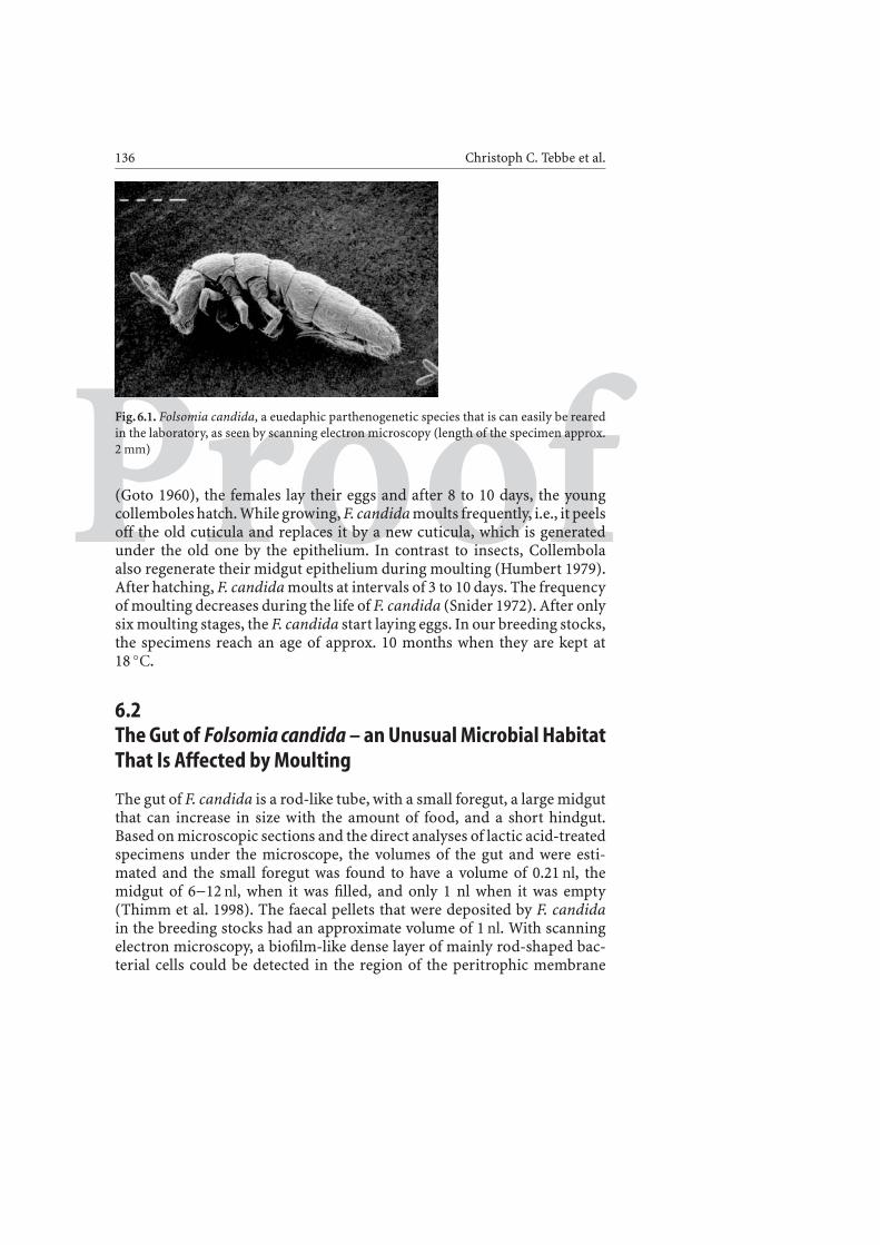
Most of our studies were conducted with the previously mentioned,euedaphic, non-pigmented collembole F. candida (Fig. 6.1), a species thatis ubiquitous and that can easily be kept in laboratory breeding stocks. Thespecies reproduces in the breeding stocks by parthenogenesis, all specimenswere female. F. candida has a typical morphology for a collembole, witha ventral tube and a springing organ, the furca. The furca, a typical featureof Collembola, is held by the tentaculum (a catch) and it is used to makejumps, by snatching out of this tentaculum. In the breeding stocks, whichcan be maintained in jars with plaster of Paris and charcoal on the bottom
Proof
136 Christoph C. Tebbe et al.
Fig. 6.1. Folsomia candida, a euedaphic parthenogenetic species that is can easily be rearedin the laboratory, as seen by scanning electron microscopy (length of the specimen approx.2 mm)
(Goto 1960), the females lay their eggs and after 8 to 10 days, the youngcollemboles hatch. While growing, F. candida moults frequently, i.e., it peelsoff the old cuticula and replaces it by a new cuticula, which is generatedunder the old one by the epithelium. In contrast to insects, Collembolaalso regenerate their midgut epithelium during moulting (Humbert 1979).After hatching, F. candida moults at intervals of 3 to 10 days. The frequencyof moulting decreases during the life of F. candida (Snider 1972). After onlysix moulting stages, the F. candida start laying eggs. In our breeding stocks,the specimens reach an age of approx. 10 months when they are kept at18 ◦C.
6.2The Gut of Folsomia candida – an Unusual Microbial HabitatThat Is Affected by Moulting
The gut of F. candida is a rod-like tube, with a small foregut, a large midgutthat can increase in size with the amount of food, and a short hindgut.Based on microscopic sections and the direct analyses of lactic acid-treatedspecimens under the microscope, the volumes of the gut and were esti-mated and the small foregut was found to have a volume of 0.21 nl, themidgut of 6−12 nl, when it was filled, and only 1 nl when it was empty(Thimm et al. 1998). The faecal pellets that were deposited by F. candidain the breeding stocks had an approximate volume of 1 nl. With scanningelectron microscopy, a biofilm-like dense layer of mainly rod-shaped bac-terial cells could be detected in the region of the peritrophic membrane

Proof
6 Collembola as a Habitat for Microorganisms 137
(Fig. 6.2). The peritrophic membrane is a layer on the gut epithelium whichis common in insects and collemboles (Wang and Granados 2001). It iscomposed of chitinous microfibrills embedded into a proteoglucan ma-trix (Terra 2001b), and its function is to facilitate the transport of thefood bolus through the gut and possibly also to protect the gut epithe-lium, e.g., from the attack of microbial pathogens (Terra 2001a). Besidethis biofilm-like region of the peritrophic membrane, bacterial cells couldalso be detected in the food bolus (Fig. 6.3), even when sterile food was
Fig. 6.2. Detection of bacterial cells forming a biofilm-like structure in the peritrophicmembrane (PM) as seen with scanning electron microscopy (SEM). Note that the mucouslayer of the PM is removed by dehydration of the samples, necessary for SEM (figure fromThimm et al. 1998, courtesy of ASM Press). Length of the left bar, 1 μm
Fig. 6.3. Bacterial cells in food bolus taken from the midgut. Note that the applied food wassterile and the bacteria originated from the region of the peritrophic membrane, shown inFig. 6.3. Length of the left bar, 1 μm

Proof
138 Christoph C. Tebbe et al.
fed. It is likely that the bacteria located in the region of the peritrophicmembrane start to colonise the ingested food and utilise it for growth.Borkott and Insam (1990) detected a total of 4×10¹¹ cfu g−1 in faeces ofF. candida, which would correspond, considering the gut volumes deter-mined in these studies, to about 106 cfu per specimen. This is about theupper limit of what we detected in our studies. During the moulting cycles,the midgut epithelium is regenerated, the old epithelium, including theperitrophic membrane, is excreted and replaced by a new epithelium thathas developed underneath the old one. During the moulting process, thespecimens stop feeding, probably because of a transiently non-functionalgut. The new epithelium is then coated again by a peritrophic membranewhich is excreted from specific epithelium cells in the foregut (Hopkin1997). Since the peritrophic membrane is densely colonised by bacterialcells, the moulting process results in a dramatic change of the bacterialpopulation in the gut. These changes in bacterial gut colonisation couldbe quantified by comparing the bacterial cell density in the gut of activelyfeeding F. candida to those that were not feeding (Thimm et al. 1998).Feeding specimens had 1.6×10⁴ to 2.7×10⁵ cfu per specimen in their gut,as determined by cultivation under aerobic conditions on yeast extractagar. In contrast, the bacterial cell densities in the non-feeding populationranged from 4.9×10² to 2.3×10⁶ cells per specimen. The high cell numberswere found in specimens immediately before, and the low cell numbersimmediately after, the excretion of the old gut epithelium and peritrophicmembrane. It can be concluded that the moulting process generates highfluctuation rates and turbulences within the bacterial community and se-
Fig. 6.4. Recolonisation of the gut by bacteria after moulting. a Pylorus region with remainingbacteria after the moulting process – a possible starting point for bacterial recolonisationof the gut; b growth of bacteria into the hindgut region. Bacterial cells (red) were detectedby FISH

Proof
6 Collembola as a Habitat for Microorganisms 139
lects for bacteria that are able to rapidly multiply in the gut. The questionemerged, from where the bacteria actually recolonise the gut after moult-ing. Probably, a main pathway for inoculation is faeces, including exuvia,on which F. candida normally feeds. Another pathway, however, may comefrom the opposite direction. We have recently identified structures in thepylorus region, located between the midgut and hindgut, in which bacterialcells were detected even in specimens that had just moulted. FISH detectionof bacterial cells in this region indicated that the bacterial recolonisationof the gut may also start from this point (Fig. 6.4).
6.3Feeding Preferences of Folsomia candidaand Fate of Ingested Bacterial CellsUnder natural conditions, F. candida seems to be an omnivore, as it feedson dead organic material as well as on fungal mycelia, nematodes or bac-teria. F. candida, in fact, is very adaptable to different food sources. Tounderstand the impact of collembolan feeding on the bacterial communitystructure and diversity in soil, we conducted feeding preference studieswith different bacterial species. At the outset of these investigations, it wasunclear whether bacteria would be digested, not affected or even stimu-lated in growth during a gut passage. The feeding preferences for bacteriawere tested with a total of twelve different bacterial strains, among themGram-positive and –negative strains from type-culture collections. Thesestrains were fed to F. candida specimens in petri dish-size microcosmsoffering pairs of choices (Thimm et al. 1998).
From a total of 66 tests, 22 showed significant preferences. Eight differentpreference classes could be differentiated. The most preferred class con-tained the type culture strain Pseudomonas putida PaW340 and a strainisolated from faeces of F. candida. As indicated by 16S rRNA gene sequenc-ing, this strain was also a P. putida or a close relative (similarity to the 16SrRNA gene of P. putida PaW340, 99.1%). The second class contained twobacterial strains, both isolated from F. candida faeces: the Gram-positiveisolate, Arthrobacter citreus and a close relative of the Gram-negative Al-caligenes faecalis. Interestingly, the A. citreus was an isolate from a differentbreeding stock. In fact, the bacteria were kept for nine years in a culturecollection (by H. Borkott, Braunschweig) before they were fed to F. candidain our studies. The lower preference classes contained different type culturestrains but no isolates from F. candida. The least preferred classes containedCorynbacterium glutamicum and Bacillus subtilis. Both species have a po-tential to live in soil. Also, Escherichia coli fell into a low preference class. Wewere aware that these choice experiments were not very realistic in regard

Proof
140 Christoph C. Tebbe et al.
to natural food sources of F. candida, as it is unlikely that F. candida findsopportunities to choose between different bacterial species in its naturalhabitat. On the other hand, the obvious feeding preference of F. candidafor its own gut bacteria indicated that these bacteria were recognised asbeneficial and probably not as a well-digestible food source. Interestingly,the isolate A. citreus exhibited chitinase activity and, thus, could possiblyact as a gut symbiont, as suggested by Borkott and Insam (1990).
The digestibility of different bacterial species was tested by feeding F.candida for one day with strains that had been genetically tagged withthe luciferase gene (luc) or the gene encoding for the green fluorescenceprotein (gfp). By this means, the bacteria could easily be differentiated fromother, indigenous bacteria. The faeces of the specimens were analysed overa period of 56 days in which only sterile food was available for the specimens.E. coli cells could only be detected in faeces one day after feeding of thebacteria. The soil bacteria Sinorhizobium meliloti or Pseudomonas stutzeriwere only detected during the first week. In contrast, P. putida cells weredetected for 20 days and A. faecalis even until the end of the experiment(56 days). The data indicated that bacterial species can differ significantlyin their capacity to survive or even colonise the gut of F. candida.
The “pulse-feed” studies were complemented by studies in which theeffect of the gut passage on ingested bacteria was quantified in more detail.Based on feeding colorised food we first determined that the period betweeningestion and excretion of food was only 35 min. When we fed E. coli, weonly detected an average of 4.3 cfu in the gut of each specimen. In contrast,with S. meliloti and A. faecalis, the other two strains tested, cell numberswere approx. four orders (!) of magnitude higher, indicating these bacteriawere not as efficiently digested as E. coli. Consequently, due to the efficientdigestion, the numbers of E. coli cells in faeces were also very low (2 cfuper faecal pellet) and the number of S. meliloti and A. faecalis were oneto two orders of magnitude higher. As estimated from uptake and releaserates of the selected strains, E. coli populations were reduced 60,000-foldwhereas A. faecalis was only reduced 500-fold (Thimm et al. 1998). Thestudies demonstrated that even though the period of time for a gut passagewas relatively short, the species specific effects were quite dramatic.
We extended this type of study on the survival of ingested bacterial cellsto another Collembola species, i.e., Protaphorura fimata (formerly Ony-chiurus fimatus; Onychiuridae, Poduromorpha, Arthropleona). P. fimatais a sexually reproducing, non-pigmented, euedaphic species that is lessassociated with the litter fraction of soils than F. candida and that can feedon mycorrhizal fungi as well as on the roots of living plants. In accordancewith our studies on F. candida, E. coli was efficiently eliminated from thegut within two days after feeding, but in contrast to F. candida, the soilbacterium S. meliloti was only detectable for two days instead of a full week
Proof
6 Collembola as a Habitat for Microorganisms 141
(Hoffmann et al. 1999). Thus, it can be expected from these results thatdifferent Collembola will impose different selective pressures on ingestedbacterial cells. Interestingly, we included a strain isolated from the gut ofF. candida, closely related to Stenotrophomonas maltophilia, in the feedingexperiments with P. fimata, and this strain persisted much longer in thegut after feeding (Hoffmann et al. 1999). We assume that some bacterialspecies, among them this relative of S. maltophilia, developed a certaincapacity to survive or even grow in the gut of Collembola. It should benoted that despite some microhabitat preferences, F. candida and P. fimatacan coexist in the same soils and may feed on the same substrates. Possibly,some soil bacteria may have evolved to utilise gut passages, as they occur inmost soils with microarthropods, to grow and maintain their populations.S. maltophilia may be good example of such an ecological adaptation (seealso Sect. 6.6).
6.4The Gut of Collembola: a Hot Spot for ConjugativeGene Transfer Between BacteriaGene transfer between microorganisms in soil has become an issue ofpublic interest in the context of the debate on the environmental risksassociated with the deliberate or accidental release of genetically engineeredmicroorganisms. Two mechanisms for gene transfer were considered tobe potentially most important for soil: transformation and conjugation.Transformation is the process in which bacterial cells take up cell-free DNAand incorporate it by recombination into their own genome. Conjugationis a process by which self-transferable, mobile genetic elements (plasmids)which carry genes for transfer, replication and possibly other properties,are transferred from a donor to a recipient strain. The transfer can be ofnarrow-host range, between only closely related bacteria, or it can be ofbroad-host range, between more distantly related species. The detection ofa gene transfer event normally requires the expression of the transferredgene in the new host organisms.
Early studies on gene transfer in soil revealed no or only low ratesof conjugative plasmid transfer from a donor to a recipient in bulk soil(Ramos-Gonzalez et al. 1991; Smit et al. 1991). However, transfer ratesin rhizospheres were much higher, probably because of the presence ofmetabolically active recipient cells (van Elsas 1992). In analogy to therhizosphere, we suspected that the gut of invertebrates, and especially thatof earthworms (Thimm et al. 2001) and Collembola could also be a “hotspot” for gene transfer. The high number of bacterial cells observed in thegut of F. candida indicated that sufficient recipients were present in the

Proof
142 Christoph C. Tebbe et al.
gut, and both the population dynamics in response to the moulting cyclesand the release of nutrients by digestion of food indicated that these gutbacteria were in fact metabolically active. In order to demonstrate bacterialgene transfer in the gut of Collembola under laboratory conditions, E. colicells with different types of plasmids were fed to F. candida in microcosmexperiments (Hoffmann et al. 1998).
We chose E. coli strains with self-transferable conjugative plasmids ofbroad or narrow-host range. In addition, E. coli strains with mobilisableplasmids were included. Mobilisable plasmids are only transferred to a re-cipient if another “mobilising” plasmid is present. These mobilising plas-mids can either be in the donor cell itself, or it can be provided by a thirdpartner, a mobilising strain, in a so-called triparental mating. Finally, wealso included an E. coli strain with a plasmid (pUC18) that was not ef-ficiently mobilisable. We suspected that this plasmid would possibly betransferred by transformation and not conjugation. A transfer of this typewas demonstrated to occur under certain conditions in mineral water asa substrate and thus it was not unlikely that it would also occur in the gutof F. candida (Baur et al. 1996). The experimental set-up for gene transferstudies was as follows. A total of 50 or 100 specimens of F. candida werefed in one arena (petri dish) with agar that was inoculated with the re-spective donor strains placed on agar cubes (Fig. 6.5). After feeding for
Fig. 6.5. Petri-dish microcosm with F. candida feeding on donor bacteria, placed on agarcubes. Note the faecal depositions (white dots) on the agar surface

Proof
6 Collembola as a Habitat for Microorganisms 143
several hours, the specimens were transferred to a new arena without food,but with an agar-surface that contained the antibiotic nalidixic acid, an in-hibitor of conjugative gene transfer (Hane 1971). The antibiotic was chosento prevent conjugative gene transfer in the faeces to show whether genetransfer would take place in the gut. The faeces that were deposited withina period of up to 24 h was then collected and analysed for the presence ofdonor bacteria, recipients and transcipients (transconjugants or transfor-mants). In order to follow the transfer of plasmids, we chose plasmids thatcarried a marker-gene (luc or gfp) and one for an antibiotic resistance. Theinhibition of the growth of donor cells was achieved by cultivating on anagar with benzoic acid as a sole source of carbon. In preparation for thosestudies we found that, in contrast to E. coli, most gut bacteria of F. candidacould utilise benzoic acid for growth.
Despite their low survival rate in the gut, several E. coli cells could in facttransfer their plasmids to indigenous bacteria of F. candida. We expectedtransformation to be important, because the digestion of the donor cellswould possibly result in the release of significant amounts of DNA whichwould have been available for transformation. However, we could not detecttransfer of the non-mobilisable plasmid. In contrast, conjugative transferof self-transferable narrow- and broad-host range plasmids to indigenousbacteria were detected. Mobilisable plasmids were only transferred whenthe mobilising genes were located in the donor cell, but not by triparentalmating with mobilising genes or plasmids provided by the bacterial com-munity in the gut. Such mobilisation by other bacteria had been shownto occur in soil amended with manure (Götz and Smalla 1997). In ourstudies, the transfer rates of the broad-host range plasmid RP4, expressedas the transconjugants-to-donor ratio in the faeces, were in the region of1×10−1. This was as high as rates can be measured under optimised lab-oratory conditions in filter-mating. The results of our studies underlinedthe importance of the feeding activity and gut microhabitat conditions ofF. candida for promoting conjugative gene transfer.
In order to confirm that the Collembolan gut is a hot spot for conjugativegene transfer we conducted similar studies as described above for F. candidawith another species, i.e., P. fimata. We were interested to see if a Collembolawith somewhat different feeding preferences than F. candida would alsoprovide suitable conditions of conjugative gene transfer in the gut – andin fact, this was the case (Hoffmann et al. 1999). In contrast to the studieswith F. candida, however, transfer of narrow-host range plasmids couldnot be detected. On the other hand, in accordance to the results withF. candida, conjugative plasmids and also mobilisable plasmids, with themobilising genes in the donor cells, were transferred to indigenous gutbacteria. Plasmid mobilisation by indigenous gut bacteria was not detected,but possibly the threshold of detection was just too insensitive in our

Proof
144 Christoph C. Tebbe et al.
studies. We assume that plasmid mobilisation in the collembolan gut ispossible, because it is likely that mobilising strains occur in the gut ofCollembola, just as they occur in soil or other environments (Smalla et al.2000).
6.5Diversity of Microorganisms in the Gut of F. candidaand Other CollembolaOnly a very limited number of studies has looked at the microbial diver-sity associated with the gut of Collembola. It could be argued that the gutof Collembola is too small, its structure too simple, and the gut passageof ingested material too quick, to allow the development of a specificallyadapted or even symbiotic microbial community. In fact, some evidencewas collected by high resolution microscopy that at least some Collem-bola do not possess any intestinal microbial community (Kilbertus andVannier 1981; Saur and Ponge 1988). An analysis of the gut contents ofF. candida, fed with hornbeam (Carpinus betulus) leaves, however, showeda high abundance of fungal mycelia (Tochot et al. 1982) but (surprisingly)no bacteria were seen. First indications that the gut of Collembola also har-bours bacteria and that these bacteria actually contribute to the digestionof food, were reported by Doeksen and Hitchen (cited by W.G. Hale 1967),who cultivated a Bacillus sp. from faeces with a capacity to degrade chitin.Chitin is an important substance in the gut as it is a major constituent offungal mycelia and the cuticules of arthropods. Chitin is also a compo-nent of the exuvia (including old cuticules), that are released during themoulting of insects and Collembola, and that many Collembola may feedon. In later studies, Borkott and Insam (1990) found that one third of theculturable bacteria from gut and faeces of F. candida showed chitin de-grading activity on agar plates. Two of these bacteria were identified; onewas Stenotrophomonas maltophilia (formerly Xanthomonas maltophilia;gamma-Proteobacteria) and one Curtobacterium sp. (Actinomycetes, Acti-nobacteria).
In an initial attempt to characterise the diversity of bacteria found inthe gut of F. candida, we isolated a total of 45 different bacterial pure cul-tures which had been kept in a breeding stock for several years (Thimmet al. 1998). These isolates could be grouped into 11 different groupsaccording to their Gram-staining, fatty acid profiles, physiological testsand ARDRA (amplified ribosomal DNA restriction fragment length anal-ysis). The abundance of each of these groups ranged from 4×10² to1.2×105 cfu perspecimen. Only one group, with an estimated abundanceof 5.3×10³ cfu per specimen, exhibited chitinase activity. The most abun-

Proof
6 Collembola as a Habitat for Microorganisms 145
dant group was represented by an isolate most closely related to Erwiniaamylovora (96.2% similarity of the almost complete 16S rRNA gene). Thesecond and third most dominant groups were represented by isolates relatedto Staphylococcus captitis and to Pantoea agglomerans. E. amylovora is animportant pathogen in orchards as it is the causative agent of the fire-blightdisease and we were interested to know if F. candida could possibly act asa vector. Feeding experiments with a pathogenic strain of E. amylovora,expressing a recombinant gfp-marker gene, however, indicated that thepathogen was, in fact, efficiently digested in the gut of F. candida (Hilde-brand et al. 2001). In the course of this cultivation-dependent detection ofmicroorganisms from the gut of F. candida, we only isolated one fungus,which was identified as a cellulose-degrading Acremonium charticola (As-comycetes) (Thimm et al. 1998). However, we did not determine whetherthis fungus was cultivated from a spore or a mycelium.
A number of different Proteobacteria were isolated as transconjugantsreceiving plasmids from E. coli cells (see previous paragraph) (Hoffmannet al. 1998). The Proteobacteria comprised one isolate from the alpha-subclass, related to Ochrobactrum anthropi (99.8% similarity of the 16SrRNA gene) and several isolates from the beta subclass with isolates relatedto Alcaligenes xylosoxidans, A. faecalis, Comamonas acidovorans and Co-mamonas testosteroni. Other isolates, among them different Pseudomonasspecies, S. maltophilia, or P. agglomerans, belonged to the gamma subclass.It should be noted that the host-range of the plasmids was responsible forthe fact that no bacteria outside of the class Proteobacteria could be found.On the other hand, the occurrence of certain bacteria like S. maltophilia orP. agglomerans in the gut of F. candida was confirmed.
Recently, a molecular approach independent of cultivation has beenused to elucidate further the bacterial diversity found in F. candida. Bymeans of PCR, 16S rRNA genes were directly amplified from DNA extractedfrom F. candida specimens. In addition to the detection of intracellularbacteria (see next paragraph), a number of different 16S rRNA genes, whichprobably originated from bacteria of the gut, were identified. These 16SrRNA genes were related to Proteobacteria of the Alpha-subclass (closestrelative: Paracoccus denitrificans), of the Gamma-subclass (100% similarityto S. maltophilia), of the Firmicutes (Bacillus weihenstephaniensis; from theBacillus cereus group) and from the Planctomycetales, the latter only with asyet uncultured relatives. We assume that the diversity of gut bacteria is muchhigher than described to date and also that the diversity will be affected bythe quality of the ingested food. However, the evidence is accumulating thatcertain bacteria from soil can utilise the gut passage through F. candida orother microarthropods to grow. We suspect that bacteria like S. maltophiliaor B. weihenstephaniensis are representatives of such a life-style. In a recentreview it was suggested that bacteria of the B. cereus group utilise the gut

Proof
146 Christoph C. Tebbe et al.
of insects to grow and survive in terrestrial habitats (Jensen et al. 2003).Collembola should also be considered in this context.
In a screening experiment, we compared the diversity of 16S rRNAamplified partial sequences from other species than F. candida. The SSCP(single strand conformation polymorphism) technique was utilised to com-pare the amplified products by generating genetic profiles (Schwieger andTebbe 1998). The profiles of seven different species indicated that differentbacteria were dominant in each species. Interestingly, the patterns of thetwo closely related species Mesaphorura macrochaeta and Mesaphoruraitalica were more similar to each other than to other species. A total of 24partial sequences were recovered and identified from these profiles, indi-cating the presence of different members from the group of Proteobacteria(alpha-, gamma-, and delta-subclass), Firmicutes, Actinobacteria and Bac-teroidetes. The sequences however, were not long enough to allow a moredetailed phylogenetic analysis. In several cases, closest relatives as indi-cated by database searches were 16S rRNA genes from uncultured bacteriafrom other environments, e.g., soil, wastewater, lake sediment, potato rhi-zosphere or, in one case, from a tissue of the honey bee (Apis mellifera).Again, a sequence related closely to S. maltophilia was detected in the DNAof P. armata, confirming this species as a gut inhabitant.
6.6Collembola Can Harbour the Reproduction ParasiteWolbachia and Other Intracellular BacteriaThe long coexistence of arthropods and bacteria for approx. 400 millionyears, has allowed the development of sophisticated interactions betweenboth groups. Striking examples are the intracellular bacteria, e.g., the en-dosymbiont Buchnera in aphids (Douglas 1998) or the parasite Wolbachia,the latter affecting the sexual reproduction patterns of many insects andother arthropods (Stouthamer et al. 1999). In Collembola, intracellularbacteria were first detected by TEM in the fat body and ovarial tissue ofF. candida by Palévody in 1972 (Palevody 1972). In 1999, Vanderkerckhoveet al., detected by PCR a 16S rRNA sequence that was closely related togroup of Wolbachia (alpha Proteobacteria) (Vanderkerckhove et al. 1999).In the same study, the authors detected intracellular bacteria in fat bodiesand intestinal cells by means of TEM. In our own laboratory, we used thefluorescence in situ hybridisation technique (FISH) with universal geneprobes for Bacteria on microscopic sections of whole F. candida specimens(and not specific tissues) and we found that in addition to the gut, severalcompartments of the body cavity were colonised by bacterial cells (Thimmand Tebbe 2003).

Proof
6 Collembola as a Habitat for Microorganisms 147
Fig. 6.6. Detection of intracellular bacteria of F. candida in the furca and neighbouringregions. The hindgut region and anus are seen in the upper part of the figure, the furcabelow. Bacterial cells (red) were by detected by FISH using the probe EUB388 for Bacteria
Intracellular bacteria were detected in fat bodies in different regionsand tissues of the body, including the furca (Fig. 6.6), brain and ovary(Thimm and Tebbe 2003). FISH with a specific gene probe for Wolbachia,however, only hybridised with bacteria that were located in the ovarialtissue or the brain region. We concluded that other bacterial species thanWolbachia must also be present in the body cavity of F. candida (Czarnet-zki and Tebbe 2004). And in fact, recently we detected a 16S rRNA generelated to the intracellular Rickettsiella grylli (gamma-Proteobacteria) ofother arthropods. We found this sequence by generating clone librariesof 16S rRNA genes amplified from total DNA extracted from F. candida.Interestingly, when we compared clone libraries generated from specimensof two different breeding stocks, we found the Rickettsiella sequence tobe dominant in one stock but completely absent in the other stock. Thephenotype of F. candida did not seem to affected by the Rickettsiella infec-tion. We therefore assume that Rickettsiella is a facultative coloniser of F.candida and probably a commensal or weak pathogen.
In contrast to Rickettsiella, we detected Wolbachia in all of five breedingstocks of F. candida analysed (Czarnetzki and Tebbe 2004). In addition,we found Wolbachia by 16S rRNA specific PCR in other parthenogenetic

Proof
148 Christoph C. Tebbe et al.
Collembola, i.e., M. macrochaeta, M. italica, and P. callipygos, but we couldnot detect it in the sexually reproducing species P. fimata or Isotomaviridis. This indicated that Wolbachia may in fact induce parthenogene-sis in Collembola. Parthenogenesis is a powerful option in the success ofpopulations in the environment as it allows organisms to multiply moreefficiently, e.g., when entering a new ecological niche (Koivisto and Braig2003). It should be noted, however, that in the case of Wolbachia andCollembola, more experimental evidence needs to be collected to confirmthis hypothesis. Our own studies, in which we tried to eliminate Wolbachiafrom F. candida, were so far unsuccessful (unpublished results). In order tounderstand better the Wolbachia-host relationships, we conducted a phy-logenetic analysis of Wolbachia with both the 16S rRNA and ftsZ genesamplified from F. candida of the different breeding stocks and from theother parthenogenetic species. The 16S rRNA genes of all F. candida breed-ing stocks was identical and in fact they were also identical to the sequencereported by Vanderkerckhove (Vanderkerckhove et al. 1999). The 16S rRNAsequences of the other Collembola were much more closely related to theF. candida sequence than to any sequence from other arthropod hosts. Infact, a monophyletic branch for Collembola could be demonstrated for thephylogenetic tree of Wolbachia.
The monophyletic branch of Wolbachia from Collembola was also seenin phylogenetic analyses based on ftsZ genes (Czarnetzki and Tebbe 2004).Here, the Wolbachia sequences from the different breeding stocks of F. can-dida showed some variations. In summary, our phylogenetic analyses in-dicated a new Supergroup E for Wolbachia in Collembola, Both studiesbased only on a single sequence from F. candida had already postulatedthat such a supergroup might exist (Vanderkerckhove et al. 1999; Lo et al.2002). Compared to the approx. 7500 species that are known in the classof Collembola it is much too early to conclude that supergroup E is anexclusive group for Collembola or that Wolbachia of other supergroupshave not option to infect Collembola. In fact, we assume that the long co-evolution of Collembola and Wolbachia make it probable that many suchexceptions exist. Our phylogenetic analyses indicated that new supergroupE is a sister group of supergroup A. Supergroup A, like supergroup B,harbours Wolbachia from a high diversity of different hosts of the ClassInsecta and there is no congruence between host and Wolbachia phylogeny.Interestingly, the other major group of the soil microarthropods, the mites,which can also be hosts for Wolbachia, have no own branch or supergroupin the Wolbachia tree. Instead, all yet detected Wolbachia sequences be-longed to Supergroup B (Breeuwer and Jacobs 1996). In our own study,a rough estimate of evolutionary rates of the different marker genes (rRNAgenes, ftsZ genes) indicated that the differences between the Collembolasupergroup E and A were much smaller than the phylogenetic distance

Proof
6 Collembola as a Habitat for Microorganisms 149
between Collembola and Insecta (Czarnetzki and Tebbe 2004). We there-fore assume that Wolbachia infections took place long after Collembola haddiversified. If Wolbachia really induces parthenogenesis in Collembola, thiswould indicate that parthenogenesis is a rather young development duringthe evolution of Collembola.
6.7Conclusions
Collembola are a quantitatively and functionally important factor in mostterrestrial ecosystems. Together with mites and some other less abundantgroups, they build up the group of microarthropods. Microarthropods en-hance the mineralisation and restructuring of organic substrates in soil.Collembola can select for specific food sources, e.g., they prefer certainfungi to others in choice experiments, but many are also quite adaptable todifferent food sources, which probably explains their high adaptability andsuccess in most soils. The size of the gut of Collembola is very small witha volume of less than 20 nl, as measured for the representative species F. can-dida. The gut passage of ingested food in F. candida can last less than 1 h.However, during this passage food is inoculated with bacteria which fur-ther enhances the degradation of these substrates. In contrast to some otherstudies, our own studies indicated that the Collembolan gut can be denselycolonised with bacterial cells and that certain bacterial species, like relativesof the type culture strains Stenotrophomonas maltophilia or Bacillus wei-henstephaniensis, have adapted to live in this microhabitat. A preconditionfor successfully colonising the collembolan gut is that the bacteria resist di-gestion by the host. Also, successful gut bacteria in Collembola need to growquickly, since moulting cycles frequently change the total bacterial popu-lation by several orders of magnitude within less than a week. The feedingactivities of Collembola in the terrestrial ecosystem thus clearly affect boththe quality of the organic substrates and the composition of the microbialcommunities. The high densities of bacterial cells and the microhabitat con-ditions provide excellent conditions for conjugative gene transfer betweenbacteria, a factor that should be considered when evaluating rates of bacte-rial gene transfer in soil. Beside the gut, other compartments of the body ofCollembola can also be colonised by bacteria. Two bacteria with an intracel-lular life-style have been detected so far: one relative of Rickettsiella grylliand one belonging to the group of Wolbachia. To our knowledge, all Wol-bachia that have been detected in Collembola are more related to each otherthan to Wolbachia from other arthropod hosts. The biological importanceof Wolbachia infections are yet unknown, but as suggested by the hosts thathave so far been analysed, such infections may induce parthenogenesis.
Proof
150 Christoph C. Tebbe et al.
Acknowledgements. We gratefully acknowledge the financial support of theGerman Ministry for Education and Research (project numbers 0310664and 0311769).
References
Agusti N, Shayler SP, Harwood JD, Vaughan IP, Sunderland KD, and Symondson WOC (2003)Collembola as alternative prey sustaining spiders in arable ecosystems: prey detectionwithin predators using molecular markers. Mol Ecol 12:3467–3475
Anderson JM and Healey IN (1972) Seasonal and interspecific variation in major componentsof gut contents of some woodland Collembola. J Animal Ecol 41:359–368
Bardgett RD, Whittaker JB, and Frankland JC (1993) The diet and food preferences ofOnychiurus procampatus (Collembola) from upland grassland soils. Biol Fertil Soils16:296–298
Baur B, Hanselmann K, Schlimme W, and Jenni B (1996) Genetic transformation in fresh-water: Escherichia coli is able to develop natural competence. Appl Environ Microbiol62:3673–3678
Bellinger PF, Christiansen KA, and Janssens F (2004) Collembola of the world. Internet;http://www.collembola.org
Bengtsson G, Hedlund K, and Rundgren S (1991) Selective odor perception in the soilCollembola Onychiurus armatus. J ChemEcol 17:2113–2125
Bilde T, Axelsen JA, and Toft S (2000) The value of Collembola from agricultural soils asfood for a generalist predator. J Appl Ecol 37:672–683
Bishop AL, McKenzie HJ, Harris AM, and Barchia IM (2001) Control strategies for thelucerne flea, Sminthurus viridis (L.) (Collembola : Sminthuridae), and their effect onother species in irrigated lucerne in the Hunter dairying region of New South Wales.Austral J Entomol 40:79–84
Bonte D and Mertens J (2003) The temporal and spatial relationship between stenotopicdwarf spiders (Erigoninae : Araneae) and their prey (Isotomidae : Collembola) in coastalgrey dunes: A numerical aggregative response or common microhabitat preference?Netherlands J Zool 52:243–253
Borkott H and Insam H (1990) Symbiosis with bacteria enhances the use of chitin by thespringtail, Folsomia candida (Collembola). Biol Fertil Soils 9:126–129
Breeuwer JAJ and Jacobs G (1996) Wolbachia: Intracellular manipulators of mite reproduc-tion. Exp. Appl. Acarology 20:421–434
Chen B, Snider RJ, and Snider RM (1995) Food preference and effects of food type on thelife history of some soil Collembola. Pedobiol 39:496–505
Chen B, Snider RJ, and Snider RM (1996) Food consumption by Collembola from northernMichigan deciduous forest. Pedobiol 40: 149–161
Czarnetzki AB and Tebbe CC (2004) Detection and phylogenetic analysis of Wolbachia inCollembola. Environ Microbiol 6:35–44
D’Haese CA (2003) Morphological appraisal of Collembola phylogeny with special empha-sis on Poduromorpha and a test of the aquatic origin hypothesis. Zoologica Scripta32:563–586
Douglas AE (1998) Nutritional interactions in insect-microbial symbioses: Aphids and theirsymbiotic bacteria Buchnera. Ann Rev Entomol 43: 17–37
Dromph KM (2001) Dispersal of entomopathogenic fungi by collembolans. Soil BiolBiochem 33: 2047–2051

Proof
6 Collembola as a Habitat for Microorganisms 151
Filser J (2002) The role of Collembola in carbon and nitrogen cycling in soil. Pedobiol 46:234–245
Frati F, Simon C, Sullivan J, and Swofford DL (1997) Evolution of the mitochondrial cy-tochrome oxidase II gene in collembola. J Mol Evol 44: 145–158
Gange A (2000) Arbuscular mycorrhizal fungi, Collembola and plant growth. Trends EcolEvol 15:369–372
Gillet S and Ponge JF (2003) Changes in species assemblages and diets of Collembola alonga gradient of metal pollution. Appl Soil Ecol 22:127–138
Goto HE (1960) Simple techniques for the rearing of collembola and a note on the use ofa fungistatic substance in the cultures. Entomologist’s Monthly Magazine 46:138–140
Götz A and Smalla K (1997) Manure enhances plasmid mobilization and survival of Pseu-domonas putida introduced into field soil. Appl Environ Microbiol 63:1980–1986
Hale WG (1967) Collembola. In: Burges A and Raw R (eds) Soil Biology. Academic Press,London, pp 397–411
Hane MW (1971) Some effects of nalidixic acid on conjugation in Escherichia coli K-12.J Bacteriol 105:46–56
Hedlund K, Bengtsson G, and Rundgren S (1995) Fungal odor discrimination in 2 sympatricspecies of fungivorous Collembolans. Functional Ecol 9:869–875
Hildebrand M, Tebbe CC, and Geider K (2001) Survival studies with the fire blightpathogen Erwinia amylovora in soil and in a soil-inhabiting insect. J Phytopathol149:635–639
Hoffmann A, Thimm T, Dröge M, Moore ERB, Munch JC, and Tebbe CC (1998) Intergenerictransfer of conjugative and mobilizable plasmids harbored by Escherichia coli in thegut of the soil microarthropod Folsomia candida (Collembola). Appl Environ Microbiol64:2652–2659
Hoffmann A, Thimm T, and Tebbe CC (1999) Fate of plasmid-bearing, luciferase marker genetagged bacteria after feeding to the soil microarthropod Onychiurus fimatus (Collem-bola). FEMS Microbiol Ecol 30:125–135
Hopkin SP (1997) Biology of springtails. Insecta: Collembola. Oxford University Press,Oxford, U.K.
Humbert W (1979) The midgut of Tomocerus minor Lubbock (Insecta, Collembola): ultra-structure, cytochemistry, aging and renewal during a moulting cycle. Cell Tissue Res196:39–57
Hunt HW, Coleman DC, Ingham RE, Elliot ET, Moore JC, Rose SL, Reid CPP, and MorenoCA (1987) The detrital food web in a shortgrass prairie. Biol Fert Soils 3:17–68
Jensen GB, Hansen BM, Eilenberg J, and Mahillon J (2003) The hidden lifestyles of Bacilluscereus and relatives. Environ Microbiol 5:631–640
Kaneko N, McLean MA, and Parkinson D (1995) Grazing preference of Onychiurus subtenuis(Collembola) and Oppiella nova (Oribatei) for fungal species inoculated on pine needles.Pedobiol 39:538–546
Kilbertus G and Vannier G (1981) Microflora-microfauna relations in the Cave of Saint-Catherine (Ariegean Pyrenees). 2. The alimentary regime of Tomocerus minor (Lub-bock) and Tomocerus problematicus Cassaghau. Rev Ecol Biol Sol 18:319–338
Koivisto RKK and Braig HR (2003) Microorganisms and parthenogenesis. Biol J LinneanSoc 79:43–58
Lee Q and Widden P (1996) Folsomia candida, a "fungivorous" collembolan, feeds prefer-entially on nematodes rather than soil fungi. Soil Biol Biochem 28:689–690
Lo N, Casiraghi M, Salati E, Bazzocchi C, and Bandi C (2002) How many Wolbachia super-groups exist? Mol Biol Evol 19:341–346
Lussenhop J (1996) Collembola as mediators of microbial symbiont effects upon soybean.Soil Biol Biochem 28:363–369
Proof
152 Christoph C. Tebbe et al.
McMillan JH (1975) Interspecific and seasonal analyses of gut contents of 3 Collembola(Family Onychiuridae). Rev Ecol Biol Sol 12:449–457
McMillan JH (1976) Laboratory observations on the food preference of Onychiurus armatus(Tullb.) Gisin (Collembola, Onychiuridae). Rev Ecol Biol Sol 13:353–364
Nakamura Y, Matsuzaki I, and Itakura J (1992) Effect of grazing by Sinella curviseta (Collem-bola) on Fusarium oxysporum F-Sp Cucumerinum causing cucumber disease. Pedobiol36:168–171
Nardi F, Spinsanti G, Boore JL, Carapelli A, Dallai R, and Frati F (2003) Hexapod origins:Monophyletic or paraphyletic? Science 299:1887–1889
Palévody C (1972) Intracellular microorganisms in ovary of Collembolae Isotomid Folsomiacandida. C R Acad Sci Paris Series D 275: 401–404
Park KH (2002) Phylogenetic analysis of Collembola based on highly variable region of 28SrRNA gene sequence. Korean J Genet 24:21–29
Ponge JF (2000) Vertical distribution of Collembola (Hexapoda) and their food resourcesin organic horizons of beech forests. Biol Fertil Soils 32:508–522
Ramos-Gonzalez MI, Duque E, and Ramos JL (1991) Conjugational transfer of recombinantDNA in cultures and in soils: host range of Pseudomonas putida TOL plasmids. ApplEnviron Microbiol 57:3020–3027
Rusek J (1998) Biodiversity of Collembola and their functional role in the ecosystem.Biodiversity and Conservation 7:1207–1219
Sabatini MA and Innocenti G (2000) Functional relationships between Collembola and plantpathogenic fungi of agricultural soils. Pedobiolog 44:467–475
Sabatini MA and Innocenti G (2001) Effects of Collembola on plant-pathogenic fungusinteractions in simple experimental systems. Biol Fertil Soils 33:62–66
Saur E and Ponge JF (1988) Alimentary studies on the collembolan Paratullbergia callipygosusing transmission electron-microscopy. Pedobiologia 31: 355–379
Schwieger F and Tebbe CC (1998) A new approach to utilize PCR-single-strand-conformation polymorphism for 16S rRNA gene-based microbial community analysis.Appl Environ Microbiol 64: 4870–4876
Sievers H and Ulber B (1990) The effects of organic manure applications on Collembolaand other small arthropods as seedling pests in sugar-beet. J Plant Diseases Protection97:588–599
Smalla K, Krögerrecklenfort E, Heuer H, Dejonghe W, Top E, Osborn M, Niewint J, TebbeC, Barr M, Bailey M, Greated A, Thomas C, Turner S, Young P, Nikolakopoulou D,Karagouni A,
Smit E, van Elsas JD, van Veen JA, and Devos WM (1991) Detection of plasmid transfer fromPseudomonas fluorescens to indigenous bacteria in soil by using bacteriophage Phi-R2ffor donor counterselection. Appl Environ Microbiol 57:3482–3488
Snider R (1972) Laboratory observations on the biology of Folsomia candida (Willem)(Collembola : Isotomidae). Rev Ecol Biol Sol 10:103–124
Stouthamer R, Breeuwer JAJ, and Hurst GDD (1999) Wolbachia pipientis: Microbial manip-ulator of arthropod reproduction. Ann Rev Microbiol 53:71–102
Tebbe CC (2003) Dissemination of genetically engineered microorganisms in terrestrialecosystems – case studies for identifying risk potentials. In: Lelley T, Balázs E, and TepferM (eds) Ecological impact of GMO dissemination in agro-ecosystems. Facultas, Wien,pp 31–44Terra WR (2001a) Special topics issue – Biological, biochemical and molecularproperties of the insect peritrophic membrane – Preface. Arch Insect Biochem Physiol47: 46
Terra WR (2001b) The origin and functions of the insect peritrophic membrane and per-itrophic gel. Arch Insect Biochem Physiology 47:47–61

Proof
6 Collembola as a Habitat for Microorganisms 153
Thimm T and Larink O (1995) Grazing preferences of some Collembola for endomycorrhizalfungi. Biol Fertil Soils 19:266–268
Thimm T and Tebbe CC (2003) Protocol for rapid fluorescence in situ hybridization ofbacteria in cryosections of microarthropods. Appl Environ Microbiol 69: 2875–2878
Thimm T, Hoffmann A, Borkott H, Munch JC, and Tebbe CC (1998) The gut of the soilmicroarthropod Folsomia candida (Collembola) is a frequently changeable but selectivehabitat and a vector for microorganisms. Appl Environ Microbiol 64:2660–2669
Thimm T, Hoffmann A, Fritz I, and Tebbe CC (2001) Contribution of the earthworm Lumbri-cus rubellus (Annelida, Oligochaeta) to the establishment of plasmids in soil bacterialcommunities. Microbial Ecol 41:341–351
Tochot F, Kilbertus G, and Vannier G (1982) Rôle d’un collembole (Folsomia candida) aucours de la dégradation des litières de charme et de chêne, en présence ou en absenced’argile. In: Lebrun P, Andrés HM, De Mets A, Grégoire-Wibo C, and Wauthy G (eds)New trends in soil biology. Proceedings of the VIII. International Colloquium of SoilZoology. Imprimeur Dieu-Brichart, Ottignies-Louvain-laNeuve, pp 269–280
Vanderkerckhove TTM, Watteyne S, Willems A, Swings JG, Mertens J, and Gillis M (1999)Phylogenetic analysis of the 16S rDNA of the cytoplasmic bacterium Wolbachia from thenovel host Folsomia candida (Hexapoda, Collembola) and its implications for wolbachialtaxonomy. FEMS Microbiol Letters 180: 279–286
van Elsas JD (1992) Antibiotic resistance gene transfer in the environment: an overview. In:Wellington EMH and van Elsas JD (eds) Genetic interactions among microorganismsin the natural environment. Pergamon Press, Oxford, pp 17–39
Visser S and Whittaker JB (1977) Feeding preferences for certain litter fungi by Onychiurussubtenuis (Collembola). Oikos 29: 320–325
Wang P and Granados RR (2001) Molecular structure of the peritrophic membrane (PM):Identification of potential PM target sites for insect control. Arch Insect Biochem Physiol47:110–118
Williams RH, Whipps JM, and Cooke RC (1998) Role of soil mesofauna in dispersal ofConiothyrium minitans: Mechanisms of transmission. Soil Biol Biochem 30: 1937–1945

Proof


