Embed Size (px)
Citation preview

Zoomorphology (2008) 127:227–239
DOI 10.1007/s00435-008-0066-4ORIGINAL PAPER
Comparative immunohistochemistry of the cephalic sensory organs in Opisthobranchia (Mollusca, Gastropoda)
Simone Faller · Sid Staubach · Annette Klussmann-Kolb
Received: 13 July 2007 / Revised: 20 May 2008 / Accepted: 1 June 2008 / Published online: 2 July 2008© Springer-Verlag 2008
Abstract The structure and function of the central ner-vous systems of opisthobranch gastropods have been stud-ied extensively. However, the organisation and function ofthe peripheral nervous system are poorly understood. Thecephalic sensory organs (CSOs) are known to be chemo-sensory structures in the head region of opisthobranchs. Inthe present study, we used immunohistochemical methodsand confocal laserscanning microscopy to comparativelyexamine the CSOs of diVerent opisthobranchs, namely Act-eon tornatilis, Aplysia punctata, Archidoris pseudoargusand Haminoea hydatis. We wanted to characterise sensoryepithelia in order to infer the function of sensory structuresand the organs they constitute. Immunoreactivity againstthe three antigens tyrosine hydroxylase, FMRFamide andserotonin was very similar in the CSOs of all investigatedspecies. Tyrosine hydroxylase-like immunoreactivity wasdetected primarily in subepidermal sensory cell bodies,which were much more abundant in the anteriorly situatedCSOs. This observation indicates that these cells and therespective organs may be involved in contact chemorecep-tion and mechanoreception. The dominant features ofFMRFamide-like immunoreactivity, especially in the pos-terior CSOs, were tightly knotted Wbres which reveal glo-merulus-like structures. This suggests an olfactory role for
these organs. Serotonin-like immunoreactivity was detectedin an extensive network of eVerent Wbres, but was not foundwithin any peripheral cell bodies. Serotonin-like immuno-reactivity was found in the same glomerulus-like structuresas FMRFamide-like immunoreactivity, indicating a func-tion of serotonin in the eVerent control of olfactory inputs.Besides this functional implication, this study could alsoadd some knowledge on the doubtful homology of theCSOs in opisthobranch gastropods.
Keywords Catecholamine · FMRFamide · Serotonin · Immunohistochemistry · Opisthobranchia
Introduction
The Opisthobranchia are morphologically extremelydiverse marine gastropods occurring almost exclusively inmarine habitats across the world. While the central nervoussystem of these animals has been well studied (Ono andMcCaman 1984; Longley and Longley 1986; Croll 1987;Elekes 1992; Sudlow et al. 1998; Croll et al. 2001; New-comb et al. 2006), relatively little is known about the orga-nisation and function of their peripheral nervous systems.Like other gastropods, all species of the Opisthobranchiapossess sensory structures in the head region, the so-calledcephalic sensory organs (CSOs). These organs have alreadybeen investigated in histological (Merton 1920; Wolter1967; Edlinger 1980; Huber 1993), ultrastructural (Storchand Welsch 1969; Emery and Audesirk 1978; Davis andMatera 1982; Boudko et al. 1999; Göbbeler and Kluss-mann-Kolb 2007), behavioural (Audesirk 1975; Wolter1967) and electrophysiological (Jahan-Parwar 1972;Jacklet 1980) studies. The CSOs can take the shape of acephalic shield, a Hancock’s organ, a lip, a lip organ, oral
S. Faller (&) · S. Staubach · A. Klussmann-KolbInstitute for Ecology, Evolution and Diversity—Phylogeny and Systematics, Goethe-University Frankfurt, Siesmayerstr. 70, 60054 Frankfurt am Main, Germanye-mail: [email protected]; [email protected]
Present Address:S. FallerDepartment of Developmental Biology and Morphology of Animals, Institute for Biology II, RWTH Aachen, Kopernikusstr. 16, 52056 Aachen, Germany
123

228 Zoomorphology (2008) 127:227–239
tentacles, an oral veil or rhinophores. According to Jahan-Parwar (1972), Audesirk (1975), Bicker et al. (1982) andCroll (1983) these organs are primarily involved in chemo-reception. Chemoreception is generally the most importantmodality for marine gastropods because auditory and visualinformation are limited (Audesirk 1975; Chase 2002; Wertzet al. 2006, 2007). Chemical senses are used in Wndingfood, avoiding predators, homing and interacting with con-speciWcs (Emery 1992); however, the CSOs are also sensi-tive to mechanical stimuli (Jahan-Parwar 1972; Bickeret al. 1982), water currents (Wolter 1967; Storch and Wel-sch 1969) and light (Chase 1979; Jacklet 1980).
The distribution of neurotransmitters in the CSOs of opis-thobranchs has been studied in diVerent opisthobranch taxa(Salimova et al. 1987; Moroz et al. 1997; Croll 2001; Crollet al. 2003; Wertz et al. 2006, 2007; Hochberg 2007; Wol-lesen et al. 2007). These studies primarily focussed on thedistribution of serotonin, whereas investigations of otherneurotransmitters or neuropeptides were less extensive.Comparative studies are lacking to date. Table 1 provides anoverview over the immunohistochemical studies labellingcatecholamines, FMRFamide and serotonin in the nervoussystem of adult euthyneuran gastropods done so far.
In the present study, we compared the distribution ofserotonin, tyrosine hydroxylase (TH) and FMRFamides inthe cephalic sensory organs of several diVerent opistho-branchs. TH is an enzyme which catalyses the initial step inthe conversion of tyrosine to the catecholamines (S.-Rozsa1984), and therefore indicative of catecholamines. Cate-cholamines have been detected in the central nervous sys-tems of diVerent pulmonate gastropods (Elekes et al. 1991;Hernadi et al. 1993; Hernadi and Elekes 1999), in the cen-tral and peripheral nervous systems of the opisthobranchsAplysia depilans, Aplysia fasciata and Aplysia californica(Salimova et al. 1987; Croll 2001) and in the central ner-vous system and CSOs of the opisthobranch Phestilla sibo-gae (Croll et al. 2001, 2003). FMRFamide (Phe-Met-Arg-Phe-NH2)-related peptides comprise a family of neuropep-tides which were isolated Wrst from the ganglia of thebivalve Macrocallista nimbosa (Lightfoot, 1786) (Price andGreenberg 1977), but are also ubiquitous in other molluscs,like in species of the Polyplacophora, Cephalopoda andGastropoda (Price et al. 1987). According to Cottrell (1989)FMRFamides are chemical messengers in both the centraland peripheral nervous systems. They have been detected inthe central and peripheral nervous systems of diVerent pul-monate gastropods (Cooke and Gelperin 1988; Elekes andNässel 1990; Elekes 1992; Suzuki et al. 1997) and in thecentral nervous system and CSOs of the opisthobranchsPhestilla sibogae (Croll et al. 2001, 2003) and Aplysia cali-fornica (Elekes 1992; Wollesen et al. 2007). Serotonin (5-HT) is a biogenic monoamine which is synthesized in thenervous system from the amino acid tryptophan (S.-Rozsa
1984). It has been investigated in numerous studies of thecentral nervous system of gastropods (Ono and McCaman1984; Longley and Longley 1986; Hernadi et al. 1989; Ele-kes 1992; Sudlow et al. 1998; Hernadi and Elekes 1999;Croll et al. 2001; Shirahata et al. 2004; Newcomb et al.2006; Hochberg 2007) and the CSOs of a variety of opis-thobranchs (Moroz et al. 1997; Croll et al. 2003; Wertzet al. 2006, 2007; Hochberg 2007; Wollesen et al. 2007).
The purpose of the current study was to investigate thedistribution of the cell bodies and Wbres containing thethree types of antigens mentioned above within diVerenttypes of CSOs and to compare these distributions amongdiVerent opisthobranch taxa in order to reveal insights intothe function and evolution of the diVerent types of CSOs.
Materials and methods
Individuals of Acteon tornatilis (Linné, 1758) (Acteonoi-dea), Aplysia punctata Cuvier, 1803 (Anaspidea) andArchidoris pseudoargus (Rapp, 1827) (Nudibranchia) werecollected in the intertidal near RoscoV (France). The speci-mens of Haminoea hydatis (Linné, 1758) (Cephalaspideas.str.) were obtained from a laboratory culture at the Goe-the-University, Frankfurt. All animals were anesthetized byinjection with 7% MgCl2. The neuroanatomy of nerves wasdetermined by macropreparation of 5–6 animals. For par-aYn histology, the CSOs of 1–2 animals per species wereexamined and for whole mounts the CSOs of a minimum of4 and a maximum of 20 animals were examined for eachprimary antibody. The CSOs were cut oV and Wxed for4–24 h at room temperature in 4% paraformaldehyde in0.1 M phosphate buVer saline (PBS, pH 7.3). For detectionof tyrosine hydroxylase, the CSOs were Wxed for 30 min in99% methanol and 1% acetic acid at ¡18°C and subse-quently transferred for 10 min each in 70, 50 and 30%methanol. After Wxation the CSOs were washed severaltimes in PBS. For the neuroanatomy of nerves the CSOswere additionally bathed overnight in 100% ethanol.
ParaYn histology
The CSOs were dehydrated in a graded isopropyl alcoholseries and transferred and embedded in paraYn. Ten-micro-metre-thick sections were cut with a Leica RM 2165 micro-tome and placed on glass microscope slides. ParaYn wasremoved with rotihistol. The sections were washed threetimes in PBS and were preincubated for 1 h with 0.2% Tri-ton and 1% normal goat serum (NGS) in PBS. Followingincubation, the sections were given another three washes inPBS and were next incubated for 24 h with either anti-FMRFamide (ImmunoStar Incorporated, Hudson, WI,USA) or anti-serotonin (Acris, Hiddenhausen,Germany) at
123

Zoomorphology (2008) 127:227–239 229
Table 1 Previous immunohistochemical studies labelling catecholamines, FMRFamide and serotonin in the nervous system of adult euthyneurangastropods
Part of Animal Taxon Type of antigen Reference
Central nervous system Opisthobranchia
Aplysia californica (Cooper, 1863) Serotonin Ono and McCaman 1984
Aplysia californica (Cooper, 1863) Serotonin Longley and Longley 1986
Aplysia californica (Cooper, 1863) FMRFamide, serotonin Elekes 1992
Aplysia californica (Cooper, 1863) TH Croll 2001
Armina californica (Cooper, 1863) Serotonin Newcomb et al. 2006
Asperspina sp. Rankin, 1979 Serotonin Hochberg 2007
Dendronotus frondosus (Ascanius, 1774) Serotonin Newcomb et al. 2006
Dendronotus iris Cooper, 1863 Serotonin Newcomb et al. 2006
Dirona albolineata MacFarland, 1905 Serotonin Newcomb et al. 2006
Flabellina trophina (Bergh, 1894) Serotonin Newcomb et al. 2006
Hermissenda crassicornis (Eschscholtz, 1831)
Serotonin Croll 1987
Hermissenda crassicornis (Eschscholtz, 1831)
Serotonin Newcomb et al. 2006
Janolus fuscus O’Donohue, 1924 Serotonin Newcomb et al. 2006
Melibe leonina (Gould, 1852) Serotonin Newcomb et al. 2006
Phestilla sibogae (Bergh, 1905) TH, FMRFamide, serotonin Croll et al. 2001
Pleurobranchaea californica MacFarland, 1966
Serotonin Sudlow et al. 1998
Tochuina tetraquetra (Pallas, 1788) Serotonin Newcomb et al. 2006
Triopha catalinae (Cooper, 1863) Serotonin Newcomb et al. 2006
Tritonia diomedea Bergh, 1894 Serotonin Sudlow et al. 1998
Tritonia diomedea Bergh, 1894 Serotonin Newcomb et al. 2006
Peripheral nervous system/ cephalic sensory organs
Aplysia californica (Cooper, 1863) TH Croll 2001
Aplysia depilans Gmelin, 1791 Catecholamines Salimova et al. 1987
Aplysia fasciata Poiret, 1798 Catecholamines Salimova et al. 1987
Aplysia punctata Cuvier, 1803 Serotonin Wertz et al. 2006
Archidoris pseudoargus (Rapp, 1827) Serotonin Wertz et al. 2007
Asperspina sp. Rankin, 1979 Serotonin Hochberg 2007
Phestilla sibogae (Bergh, 1905) TH, FMRFamide, serotonin Croll 2003
Pleurobranchaea californica MacFarland, 1966
Serotonin Moroz et al. 1997
Tritonia diomedea Bergh, 1894 Serotonin Moroz et al. 1997
Central nervous system Pulmonata
Achatina fulica (Ferussac, 1821) Serotonin Chiasson et al. 1994
Helix pomatia Linné, 1758 Serotonin Hernadi et al. 1989
Helix pomatia Linné, 1758 FMRFamide Elekes and Nässel 1990
Helix pomatia Linné, 1758 FMRFamide, serotonin Elekes 1992
Helix pomatia Linné, 1758 TH and dopamine Hernadi et al. 1993
Helix pomatia Linné, 1758 TH, serotonin, dopamine Hernadi and Elekes 1999
Limax maximus Linné, 1758 FMRFamide Cooke and Gelperin 1988
Limax valentianus Férussac, 1823 Serotonin Shirahata et al. 2004
Lymnaea stagnalis (Linné, 1758) Serotonin Kemenes et al. 1989
Lymnaea stagnalis (Linné, 1758) Dopamine Elekes et al. 1991
Lymnaea stagnalis (Linné, 1758) FMRFamide, serotonin Elekes 1992
Peripheral nervous system/ cephalic sensory organs
Helix aspersa Müller, 1774 FMRFamide Cardot and Fellman 1983
Helix pomatia Linné, 1758 TH, serotonin, dopamine Hernadi and Elekes 1999
Limax marginatus Müller, 1774 FMRFamide Suzuki et al. 1997
123

230 Zoomorphology (2008) 127:227–239
room temperature. Both antibodies were raised in rabbitand were used at 1:100 dilutions in PBS. After incubationwith primary antibody the sections were washed again threetimes in PBS and were then incubated for 24 h in goat anti-rabbit antibodies labelled with either FITC or TRITC (Jack-son ImmunoResearch Laboratories, West Groove, PA,USA). The secondary antibodies were also used at 1:100dilutions. After another several washes in PBS the sectionswere covered with glass coverslips in a solution of threeparts glycerol to one part 0.1 M TRIS buVer (pH 8.0) withthe addition of 2% n-propyl gallate. Additional controlexperiments without primary antibody incubation weredone and no staining was observed in these preparations.As positive controls the central ganglia of individuals ofAplysia punctata were incubated with the same primary andsecondary antibodies at the same dilutions. These gangliashowed an intense Xuorescence in speciWc cells, as shownby Croll et al. (2004) for Aplysia californica.
Whole mounts
The CSOs were bathed overnight in blocking solution,either of 4% Triton and 1% NGS in PBS for detection ofFMRFamide and serotonin or of 1% Triton and 1% normalsheep serum (NSS) in PBS for the detection of TH. Afterthree washes in PBS the CSOs were incubated in one of theprimary antibodies for 48 h at 4°C. Anti-FMRFamide andanti-serotonin were used in 1:500 dilutions. The monoclo-nal anti-TH antibody (ImmunoStar Incorporated, Hudson,WI, USA) was developed in mouse and was diluted 1:250.Following three washes in PBS the CSOs were incubatedeither for 24 h in goat anti-rabbit antibodies labelled withFITC (for FMRFamide) or TRITC (for serotonin) or for36 h in sheep anti-mouse antibodies labelled with TRITC(for TH) at 4°C. The secondary antibodies were used at thesame dilution as the primary antibodies. After another sev-eral washes in PBS the CSOs were mounted on objectslides in a solution of three parts glycerol to one part TRISbuVer with the addition of 2% n-propyl gallate and coveredwith glass coverslips. Positive and negative control experi-ments were performed as described for paraYn histology.
Confocal microscopy and image processing
Preparations were viewed and photographed on a confocallaser scanning microscope (Leica TCS SP5). The distancebetween the optical sections was generally 1 �m and theresulting image stack was photographed as maximal projec-tions. Cell diameters were determined by average over a mini-mum of ten cells. Image processing was performed withAdobe Photoshop 6.0 (Adobe Systems Incorporated, San Jose,CA, USA) and the schematic diagrams were prepared withCorelDRAW 11 (Corel Corporation, Ottawa, ON, Canada).
Results
Structure and innervation of the CSOs
The four investigated opisthobranch taxa possess diVerenttypes of cephalic sensory organs. Acteon tornatilis pos-sesses a cephalic shield which consists of a pair of antero-lateral lobes and a pair of postero-lateral lobes (Fig. 1a). Agroove extends along the lateral ventral margins of theanterior-lateral lobes. The cephalic shield is innervated bythree cerebral nerves. The anterior part is innervated by theNervus labialis (N2), the posterior part is innervated by theNervus rhinophoralis (N3) and the Nervus clypei captitis(Nclc). The investigated CSOs of Aplysia punctata arecompletely diVerent from those of A. tornatilis. There aretwo pairs of CSOs, the posterior rhinophores and the ante-rior oral tentacles (Fig. 1b). Both pairs are rolled and haveinner grooves. Each organ possesses a peripheral ganglionat the base of this groove. The Nervus labialis (N2) inner-vates the oral tentacles and the Nervus rhinophoralis (N3)serves the rhinophores. Archidoris pseudoargus also pos-sesses a posterior pair of rhinophores and an anterior pair oforal tentacles (Fig. 1c). The rhinophores consist of shaftsthat have about 20 apical lamellae. The oral tentacles areshort bulbs which are covered by the mantle. The oral ten-tacles are innervated by the Nervus labialis (N2) and therhinophores by the Nervus rhinophoralis (N3). A large gan-glion extends through the longitudinal axis of the rhino-phores. In Haminoea hydatis we investigated three types ofCSOs: the Hancock’s organ, the lip organ and the cephalicshield (Fig. 1d). The Hancock’s organ is situated posteri-orly and the lip organ anteriorly underneath the cephalicshield. These CSOs are innervated by four cerebral nerves.The cephalic shield is innervated by the Nervus clypei cap-titis (Nclc) and the Nervus oralis (N1). The Nervus labialis(N2) innervates the lip organ and the Nervus rhinophoralis(N3) the Hancock’s organ.
Tyrosine hydroxylase (TH)-like immunoreactivity
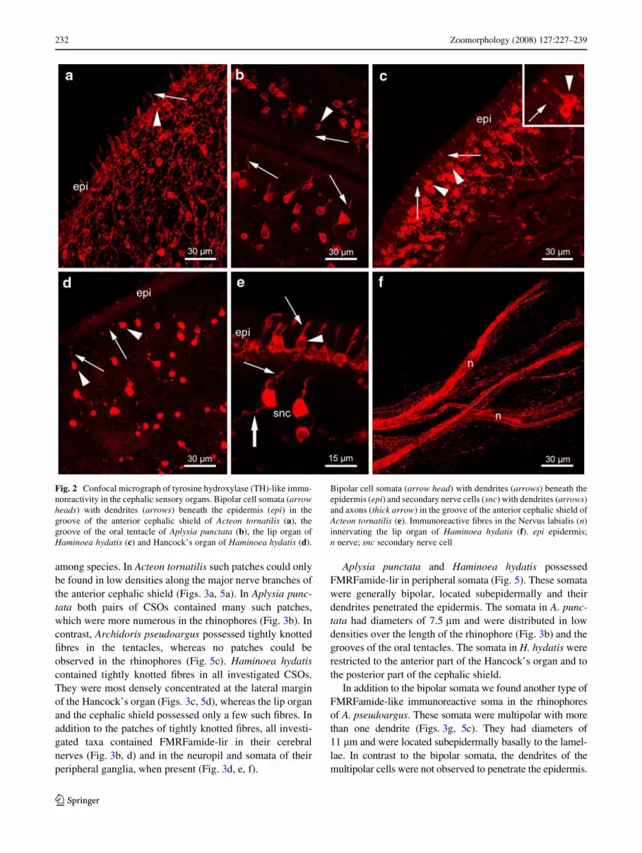
TH-like immunoreactivity (lir) was found in all investigatedCSOs of all investigated species. The predominant TH-likeimmunoreactive structures were bipolar cell somata whichhad diameters of 6.5 �m, were located subepidermally andbore dendrites that penetrated the epidermis (Fig. 5). Thedistributions of these somata varied within the diVerentCSOs of one species as well as among species. In Acteontornatilis these somata were found scattered across theentire cephalic shield, with increasing concentrations in theanterior region (Figs. 2a, 5a), especially in the lateral groove(»4,200 somata/mm²). In Aplysia punctata the number ofTH-like immunoreactive somata was less than in A. torna-tilis (Figs. 2b, 5b). The somata occurred along the complete
123

Zoomorphology (2008) 127:227–239 231
lengths of the grooves in the oral tentacle as well as the rhi-nophores, with the highest concentrations near their tips.However, the somata were more common in the oral tenta-cles (»1,000 somata/mm²) than in the rhinophores(»300 somata/mm²). This also applied to Archidoris pseud-oargus. In this species the TH-like immunoreactive somatawere located in the tissue constituting the lamellae of therhinophores as well as across the entire surface of the oraltentacles (Fig. 5c). Haminoea hydatis showed the highestdensity of TH-like immunoreactive somata in the lip organ(Fig. 2c, 5d). Here up to 6,400 somata/mm² were observed.Within the Hancock’s organ the catecholaminergic somataoccurred in smaller numbers (»1,200 somata/mm²) at theposterior end and the lateral margin (Figs. 2d, 5d). In thecephalic shield of H. hydatis we found TH-like immunore-active somata as well, but exclusively in the posterior region(»3,200 somata/mm²).
In addition to the subepidermal cell type describedabove, we found a second type of TH-like immunoreactivesomata in A. tornatilis (Fig. 2e). These somata had diame-ters of 7.5 �m and were located exclusively in the lateralgroove of the anterior cephalic shield. We observed onestriking characteristic of these somata; in contrast to thesomata described above their dendrites did not penetrate theepidermis. Instead, the dendrites seemed to extend to thesubepidermal somata of the Wrst cell type.
TH-like immunoreactivity was also observed within sub-epidermal networks of nerve Wbres. These were especiallyabundant in the cephalic shield of A. tornatilis and H. hydatisas well as in the oral tentacles of A. punctata and A. pseud-oargus. TH-lir could also be detected in cerebral nervesinnervating the CSOs, e.g. the Nervus rhinophoralis ofA. pseudoargus and the Nervus labialis of H. hydatis (Fig. 2f).
FMRFamide-like immunoreactivity
FMRFamide-like immunoreactivity (lir) was detected indiverse structures including nerves, peripheral ganglia,somata and Wbres (Fig. 5). The predominant structures thatshowed FMRFamide-lir immunoreactivity were patches oftightly knotted Wbres located along the major nervebranches (Fig. 3a). The distribution and density of thesepatches varied within the CSOs of each species as well as
Fig. 1 Schematic diagrams of frontal views of Acteon tornatilis (a),Aplysia punctata (b), Archidoris pseudoargus (c) and Haminoea hyda-tis (d), showing innervation of the investigated cephalic sensory or-gans. The left sides of the diagrams show the innervation patterns of thecerebral nerves. The right sides of the diagrams show the location ofthe cephalic sensory organs. acs anterior cephalic shield; cg cerebralganglion; cs cephalic shield; e eye; ga ganglion; gr, groove; ho Han-cock’s organ; lo lip organ; N1 nervus oralis; N2 nervus labialis; N3 ner-vus rhinophoralis; Nclc nervus clypei captitis; ot oral tentacle; pcsposterior cephalic shield; rh rhinophore
�
123
232 Zoomorphology (2008) 127:227–239
among species. In Acteon tornatilis such patches could onlybe found in low densities along the major nerve branches ofthe anterior cephalic shield (Figs. 3a, 5a). In Aplysia punc-tata both pairs of CSOs contained many such patches,which were more numerous in the rhinophores (Fig. 3b). Incontrast, Archidoris pseudoargus possessed tightly knottedWbres in the tentacles, whereas no patches could beobserved in the rhinophores (Fig. 5c). Haminoea hydatiscontained tightly knotted Wbres in all investigated CSOs.They were most densely concentrated at the lateral marginof the Hancock’s organ (Figs. 3c, 5d), whereas the lip organand the cephalic shield possessed only a few such Wbres. Inaddition to the patches of tightly knotted Wbres, all investi-gated taxa contained FMRFamide-lir in their cerebralnerves (Fig. 3b, d) and in the neuropil and somata of theirperipheral ganglia, when present (Fig. 3d, e, f).
Aplysia punctata and Haminoea hydatis possessedFMRFamide-lir in peripheral somata (Fig. 5). These somatawere generally bipolar, located subepidermally and theirdendrites penetrated the epidermis. The somata in A. punc-tata had diameters of 7.5 �m and were distributed in lowdensities over the length of the rhinophore (Fig. 3b) and thegrooves of the oral tentacles. The somata in H. hydatis wererestricted to the anterior part of the Hancock’s organ and tothe posterior part of the cephalic shield.
In addition to the bipolar somata we found another type ofFMRFamide-like immunoreactive soma in the rhinophoresof A. pseudoargus. These somata were multipolar with morethan one dendrite (Figs. 3g, 5c). They had diameters of11 �m and were located subepidermally basally to the lamel-lae. In contrast to the bipolar somata, the dendrites of themultipolar cells were not observed to penetrate the epidermis.
Fig. 2 Confocal micrograph of tyrosine hydroxylase (TH)-like immu-noreactivity in the cephalic sensory organs. Bipolar cell somata (arrowheads) with dendrites (arrows) beneath the epidermis (epi) in thegroove of the anterior cephalic shield of Acteon tornatilis (a), thegroove of the oral tentacle of Aplysia punctata (b), the lip organ ofHaminoea hydatis (c) and Hancock’s organ of Haminoea hydatis (d).
Bipolar cell somata (arrow head) with dendrites (arrows) beneath theepidermis (epi) and secondary nerve cells (snc) with dendrites (arrows)and axons (thick arrow) in the groove of the anterior cephalic shield ofActeon tornatilis (e). Immunoreactive Wbres in the Nervus labialis (n)innervating the lip organ of Haminoea hydatis (f). epi epidermis;n nerve; snc secondary nerve cell
123

Zoomorphology (2008) 127:227–239 233
Fig. 3 Confocal micrograph of FMRFamide-like immunoreactivity inthe cephalic sensory organs. Patches of tightly knotted Wbres (g) andnerves (n) in the anterior cephalic shield of Acteon tornatilis (a), therhinophore of Aplysia punctata (arrow heads indicate subepidermalsomata) (b) and the Hancock’s organ of Haminoea hydatis (c). Nervusrhinophoralis (n) and rhinophoral ganglia (ga) of Aplysia punctata (d).Higher magniWcation showing isolated somata (arrow heads) within
the rhinophoral ganglion (ga) of Aplysia punctata (e). Rhinophoralganglion (ga) and nerve (n) in the rhinophore of Archidoris pseudoar-gus (f). Multipolar cell (arrow head) with dendrites (arrow) in the rhi-nophore of Archidoris pseudoargus (g). Nerve Wbres (nf) beneath theepidermis (epi) in the anterior cephalic shield of Acteon tornatilis (h)and in the lip organ of Haminoea hydatis (i). cns central nervous sys-tem; epi epidermis; g glomerulus; ga ganglion; n nerve; nf nerve Wbre
123

234 Zoomorphology (2008) 127:227–239
All investigated CSOs possessed FMRFamide-lir in anetwork of subepidermal Wbres that did not penetrate theepidermis. In A. tornatilis, the subepidermal network ofWbres was located on the ventral side of the entire cephalicshield (Fig. 3h). Both A. punctata and A. pseudoargus pos-sessed isolated subepidermal Wbres throughout their rhino-phores and oral tentacles. In H. hydatis the subepidermalWbres were located primarily in the anterior region of theHancock’s organ, throughout the entire lip organ (Fig. 3i)and in low densities within the cephalic shield.
Serotonin-like immunoreactivity
Serotonin-like immunoreactivity (lir) was found within thesame patches of tightly knotted Wbres (Fig. 4a, b), periphe-ral ganglia (Fig. 4c, d) and nerves (Fig. 4b, c) as FMRF-amide-lir in all investigated taxa except for Haminoea
hydatis. Therefore, the distribution of serotonin is notshown in the diagrams of the whole CSOs in Fig. 5. Seroto-nin-lir was also detected in a network of subepidermalnerve Wbres in all investigated CSOs. These Wbres did notpenetrate the epidermis. In Acteon tornatilis a dense subepi-dermal network of Wbres was located on the dorsal side ofthe entire cephalic shield (Fig. 4e). In contrast, Aplysiapunctata possessed only a small number of subepidermalWbres in the rhinophores and tentacles. This distributionwas also observed in Archidoris pseudoargus, where sero-tonin-like immunoreactivity was found within a small num-ber of subepidermal Wbres in the rhinophores and on theventral surfaces of the tentacles. In H. hydatis the Wbreswere most densely concentrated in the anterior part of allinvestigated CSOs (Fig. 4f). No serotonin-like immunore-active somata were found within any of the investigatedCSOs.
Fig. 4 Confocal micrograph of serotonin-like immunoreactivity in thecephalic sensory organs. Patches of tightly knotted Wbres (g) andnerves (n) in the rhinophore (a) and oral tentacle (b) of Aplysia punc-tata. Immunoreactive Wbres within the rhinophoral ganglion (ga) andnerves (n) in the rhinophore of Aplysia punctata (c) and Archidoris
pseudoargus (d). Nerve Wbres (nf) beneath the epidermis (epi) withinthe anterior cephalic shield of Acteon tornaltilis (e) and the Hancock’sorgan of Haminoea hydatis (f). epi epidermis; g glomerulus; ga gan-glion; n nerve; nf nerve Wbre
123
Zoomorphology (2008) 127:227–239 235
Discussion
Our studies revealed that immunohistochemistry againstthe three types of neurotransmitters tyrosine hydroxylase,FMRFamide and serotonin revealed distinct structureswithin the CSOs. The distribution patterns of TH-, FMRF-amide- and serotonin-like immunoreactivity in the fourinvestigated Opisthobranchia species are summarised inFig. 5.
Tyrosine hydroxylase (TH)-like immunoreactivity
The distribution of TH-lir was very similar within the CSOsof the four investigated taxa. They all exhibited subepider-mal bipolar TH-like immunoreactive somata. These somatapossessed dendrites that penetrated the epidermis and weremuch more abundant in the anterior CSOs (e.g. the oral ten-tacles) than in the posterior CSOs (e.g. the rhinophores).These Wndings are consistent with those of Croll (2001) inAplysia californica and Croll et al. (2003) in Phestilla sibo-gae who therefore suggested that these cells function in con-tact chemoreception or mechanoreception. Acteon tornatilispossessed an additional type of TH-like immunoreactivesomata that could not be detected in any of the other investi-gated taxa. The dendrites of these bipolar cells extended tothe subepidermal sensory cells and did not penetrate the epi-dermis. These cells may be secondary nerve cells that obtainand relay information from the subepidermal primary sen-sory cells. Croll (2001) also described a second type of TH-like immunoreactive somata in A. californica that lie moredeeply within the tissue and occasionally appear to be multi-polar. He suggested they play a role in the lateral spread ofsensory information within the peripheral neural plexus.These somata may correspond to the second type of somatafound in this study. However, to clearly identify the role ofthese TH-like immunoreactive somata, further studies, espe-cially electrophysiological investigations, are needed. Inaddition to the TH-like immunoreactive somata, TH-lir wasdetected in Wbres of the nerves innervating the diVerentCSOs. These Wbres are presumably the centrally projectingaxons of the sensory somata.
FMRFamide-like immunoreactivity
The distribution of FMRFamide in the peripheral nervoussystem of adult opisthobranchs has only been investigatedby Croll et al. (2003) in the CSOs of Phestilla sibogae. Inthis study, as well as in our study, the dominant features ofFMRFamide-lir were patches of tightly knotted Wbres.These patches possibly correspond to glomerulus-likestructures (Boudko et al. 1999; Croll et al. 2003). Glome-ruli have recently been reported in the rhinophores of Aply-sia punctata (Wertz et al. 2006) and in sensory areas of
A.californica (Moroz 2006) and are also well-known in thetentacles of the pulmonate Achatina fulica (Chase and Toll-oczko 1986). In generall glomeruli are considered to beinvolved in processing of olfactory stimuli. The glomeru-lus-like structures observed in the present study were con-centrated in the posterior cephalic sensory organs of theinvestigated taxa, especially in the rhinophores of A. punc-tata and the Hancock’s organ of Haminoea hydatis. Thissuggests an olfactory role for these organs. Both the rhino-phores of A. punctata and the Hancock’s organ of H. hyda-tis have already been proposed to be involved inchemoreception by Audesirk (1975) and Edlinger (1980).While the rhinophores of A. punctata contained numerousglomeruli, the rhinophores of Archidoris pseudoargus werelacking glomeruli. These Wndings are consistent with thenew work of Wertz et al. (2007). It is questionable whetherthe rhinophores of A. pseudoargus are involved in olfactionor whether there are other structures that serve this func-tion. Instead of glomerulus-like structures, the rhinophoresof A. pseudoargus possessed FMRFamide-like immunore-active neuropil in a large ganglion that extended over theentire longitudinal axis of the rhinophore. According toBicker et al. (1982) the rhinophore and tentacular ganglionof Pleurobranchaea californica serve mainly as peripheralintegrating and relay stations for sensory inputs. Wertzet al. (2006) demonstrated by means of calcium imagingexperiments that olfactory stimuli are relayed and pro-cessed in the rhinophoral ganglion of A. punctata. It islikely that the rhinophoral ganglion also serves as a relaystation of olfactory stimuli in A. pseudoargus and thereforeglomeruli are not required. In contrast, the rhinophores ofA. punctata possessed an FMRFamide-like immunoreactiveganglion and additional glomerulus-like structures. Thiscontrast between A. punctata and A. pseudoargus could bedue to the fact that the rhinophoral ganglion of A. pseud-oargus extends over the entire length of the rhinophore,whereas the peripheral ganglion of A. punctata is restrictedto the base of the rhinophoral groove. The rhinophores of A.pseudoargus and A. punctata have completely diVerentstructures and hence may have diVerent functions or ori-gins, as already described by Gosliner (1994). The rhino-phores of A. pseudoargus consist of a shaft with lamellae,whereas those of A. punctata are rolled. Thus the rhino-phores of A. pseudoargus, in contrast to those of A. punc-tata, are not primarily olfactory organs but rather senseother modalities, e.g. detection of water currents. Theinvolvement of the rhinophores of A. pseudoargus in rheo-taxis has been described by Wolter (1967).
In addition to the FMRFamide-like immunoreactiveglomeruli-like structures and peripheral ganglia, FMRF-amide-lir was detected within a small number of subepidermalbipolar somata within the investigated CSOs of A. punctataand H. hydatis but not of A. tornatilis and A. pseudoargus.
123

236 Zoomorphology (2008) 127:227–239
123
Zoomorphology (2008) 127:227–239 237
�
Such bipolar FMRFamide-like immunoreactive somatawere also described for the tentacle tip of the pulmonateLimax marginatus (Suzuki et al. 1997). The dendrites of thecells penetrated the epidermis and therefore Suzuki et al.(1997) suggested that these cells are primary sensory neu-rons and contribute to chemical or mechanical reception, assuggested for the TH-like immunoreactive cells. Only thesomata in the rhinophore of A. pseudoargus look diVerentfrom those found in the other investigated CSOs becausethese cells are multipolar. Multipolar cells that showFMRFamide-lir have not yet been described in the opistho-branchs but as already mentioned above, Croll (2001)described the existence of TH-like immunoreactive multi-polar cells in peripheral tissues of A. californica and sug-gested a role in the lateral spread of sensory informationwithin the peripheral neural plexus. Based on our data, it isnot clear whether the FMRFamide-lir multipolar cells of A.pseudoargus correspond to the multipolar cells describedby Croll (2001) and hence may also play a role in thismodality.
Serotonin-like immunoreactivity
Unlike the distribution of tyrosine hydroxylase andFMRFamides, the distribution of serotonin has alreadybeen studied in detail in the peripheral nervous systems ofvarious opisthobranchs (Moroz et al. 1997; Croll et al.2003; Wertz et al. 2006, 2007; Hochberg 2007) as well asin the pulmonate Helix pomatia (Hernadi and Elekes 1999).No peripheral serotonin-like immunoreactive somata werefound in any of the investigated CSOs and serotonin wasfound primarily in subepidermal nerve Wbres that did notpenetrate the epidermis; therefore, these Wbres appear to beeVerent. These Wndings are consistent with the observationof only eVerent Wbres in the CSOs of A. punctata (Wertzet al. 2006), A. pseudoargus (Wertz et al. 2007), Phestillasibogae (Croll et al. 2003), Pleurobranchaea californicaand Tritonia diomedea (Moroz et al. 1997) and the pulmo-
nate H. pomatia (Hernadi and Elekes 1999). Serotonin-lirwas found in the same patches of entangled Wbres andperipheral ganglia as FMRFamide. These results agree withMoroz et al. (1997), who suggested that serotonin may playa role in the peripheral modulation of sensory inputs to thecentral nervous system. If these entangled Wbres indeed cor-respond to glomerulus-like structures, serotonin may play arole in the eVerent control of olfactory inputs.
Conclusions
Here, we demonstrate that the distribution of sensory struc-tures shows characteristic patterns for diVerent CSOs. Thedistribution of these structures within the CSOs leads us tothe conclusion that the diVerent types of CSOs have diVer-ent functions. The posterior CSOs, i.e. the rhinophores ofAplysia punctata and the Hancock’s organ of Haminoeahydatis, generally contain many glomerulus-like structuresand therefore probably primarily fulWl an olfactory func-tion, which is also supported by their location. The anteriorCSOs, i.e. the oral tentacles, the lip organ and the anteriorcephalic shield, comprise numerous bipolar sensory neu-rons which are probably involved in contact chemorecep-tion and mechanoreception. Thus, the anterior CSOs mayplay a role in these modalities. Another point which arguesfor a function in contact chemoreception and mechanore-ception is that the anterior CSOs are situated close to thesubstrate and thus may serve to discriminate between diVer-ent types of substrate.
Aside from the conclusions about the function of thediVerent CSOs, this study additionally provides insight intotheir evolution. Since the distribution of cell bodies andglomeruli containing FMRFamide is very similar especiallyin the posterior CSOs of A. punctata (Anaspidea) and H.hydatis (Cephalaspidea) and since innervation patterns forthese organs are very similar in the two species (data of sec-ond author, not shown here), we propose that these CSOs inAnaspidea and Cephalaspidea are homologous structuresindicating that the two taxa may be more closely related toeach other than to A. tornatilis (Acteonoidea) and A. pseud-oargus (Nudibranchia), where these FMRFamide-likeimmunoreactive structures are missing. This assumption isin agreement with molecular systematic studies by Vonne-mann et al. (2005) and Klussmann-Kolb et al. (2008) whorevealed a sister-group relationship between the Anaspideaand the Cephalaspidea as well as between the Nudipleura(Nudibranchia plus Pleurobranchoidea) and the Acteonoi-dea, respectively.
These sister-group relationships would either imply thatthe rhinophores of the Anaspidea and the Nudibranchiahave evolved independently from each other as alreadysuggested by Gosliner (1994) or that the rhinophores
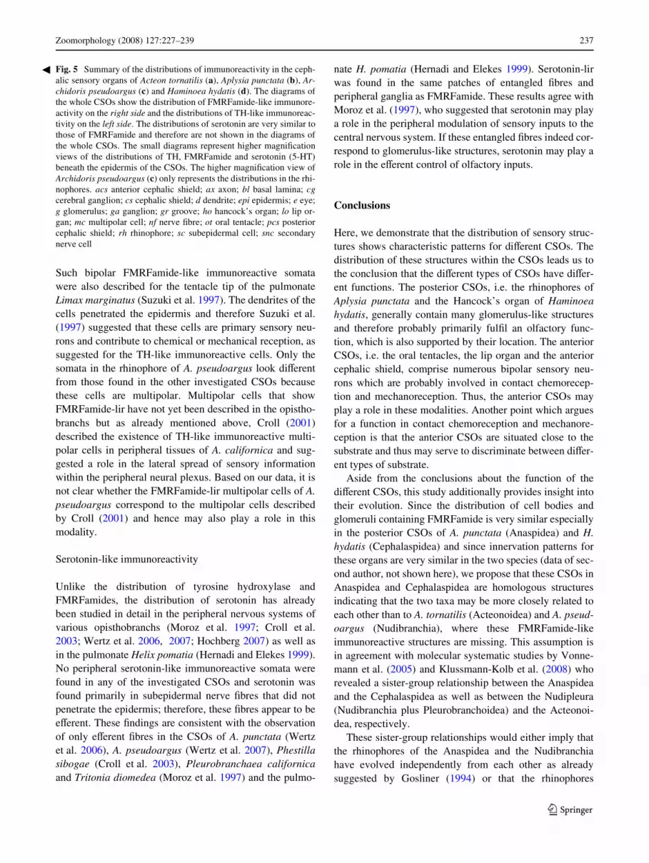
Fig. 5 Summary of the distributions of immunoreactivity in the ceph-alic sensory organs of Acteon tornatilis (a), Aplysia punctata (b), Ar-chidoris pseudoargus (c) and Haminoea hydatis (d). The diagrams ofthe whole CSOs show the distribution of FMRFamide-like immunore-activity on the right side and the distributions of TH-like immunoreac-tivity on the left side. The distributions of serotonin are very similar tothose of FMRFamide and therefore are not shown in the diagrams ofthe whole CSOs. The small diagrams represent higher magniWcationviews of the distributions of TH, FMRFamide and serotonin (5-HT)beneath the epidermis of the CSOs. The higher magniWcation view ofArchidoris pseudoargus (c) only represents the distributions in the rhi-nophores. acs anterior cephalic shield; ax axon; bl basal lamina; cgcerebral ganglion; cs cephalic shield; d dendrite; epi epidermis; e eye;g glomerulus; ga ganglion; gr groove; ho hancock’s organ; lo lip or-gan; mc multipolar cell; nf nerve Wbre; ot oral tentacle; pcs posteriorcephalic shield; rh rhinophore; sc subepidermal cell; snc secondarynerve cell
123

238 Zoomorphology (2008) 127:227–239
evolved only once within the Euthyneura. The Wrst hypoth-esis is supported by Edlinger (1980) and Huber (1993),who proposed that the rhinophores of the Anaspidea derivefrom the Hancock’s organ of the Cephalaspidea. However,this would imply that the Cephalaspidea take a more basalposition within the Opisthobranchia than the Anaspidea, ahypothesis that is not supported by current phylogeneticanalyses (Vonnemann et al. 2005; Klussmann-Kolb et al.2008).
In addition to these implications it seems that the distri-bution of some immunoreactive structures in the posteriorCSOs of the investigated opisthobranch taxa is comparableto that of pulmonate gastropods (Suzuki et al. 1997; Her-nadi and Elekes 1999) which supports a close relationshipof these two groups of Gastropoda. This is in agreementwith former phylogenetic studies (e.g. Salvini-Plawen andSteiner 1996; Ponder and Lindberg 1997; Dayrat et al.2001; Dayrat and Tillier 2002) and aYrm the secondassumption that the posterior CSOs evolved only oncewithin all Euthyneura.
Nevertheless, further immunohistochemical investiga-tions on more taxa of opisthobranchs as well as pulmonategastropods are nescessary to obtain insights in the evolutionof the CSOs within Euthyneura.
Acknowledgments We are grateful to Roger P. Croll for his helpfulcomments on an earlier version of this report. We would also like tothank the German Science Foundation (DFG) for supporting this pro-ject (KL 1303/3-1).
References
Audesirk TE (1975) Chemoreception in Aplysia californica. I. Behav-ioral localization of distance chemoreceptors used in food-Wnd-ing. Behav Biol 15:45–55. doi:10.1016/S0091-6773(75)92066-0
Bicker G, Davis WJ, Matera EM (1982) Chemoreception andmechanoreception in the gastropod mollusc Pleurobranchaeacalifornica. II. Neuroanatomical and intracellular analysis ofaVerent pathways. J Comp Physiol 149:235–250. doi:10.1007/BF00619217
Boudko DY, Switzer-Dunlap M, HadWeld MG (1999) Cellular andsubcellular structure of anterior sensory pathways in Phestillasibogae (Gastropoda, Nudibranchia). J Comp Neurol 403:39–52.doi: 10.1002/(SICI)1096-9861(19990105)403:1·39::AID-CNE4¸3.0.CO;2-B
Cardot J, Fellman D (1983) ImmunoXuorescent evidence of anFMRFamide-like peptide in the peripheral nervous system of thegastropod mollusc Helix aspersa. Neurosci Lett 43:167–172.doi:10.1016/0304-3940(83)90182-9
Chase R (1979) Photic sensitivity of the rhinophore in Aplysia. Can JZool 57:698–701
Chase R (2002) Behavior and its neural control in gastropod molluscs.Oxford University Press, New York
Chase R, Tolloczko B (1986) Synaptic glomeruli in the olfactory sys-tem of a snail, Achatina fulica. Cell Tissue Res 246:567–573.doi:10.1007/BF00215198
Chiasson BJ, Baker MW, Croll RP (1994) Morphological changesand functional recovery following axotomy of a serotonergic
cerebrobuccal neurone in the land snail Achatina fulica. J ExpBiol 192:147–167
Cooke IRC, Gelperin A (1988) Distribution of FMRFamide-likeimmunoreactivity in the nervous system of the slug Limax maxi-mus. Cell Tissue Res 253:69–76
Cottrell GA (1989) The biology of the FMRFamide-series of peptidesin molluscs with special reference to Helix. Comp Biochem Phys-iol 93A(1):41–45. doi:10.1016/0300-9629(89)90189-8
Croll RP (1983) Gastropod chemoreception. Biol Rev Camb PhilosSoc 58:293–319. doi:10.1111/j.1469-185X.1983.tb00391.x
Croll RP (1987) Distribution of monoamines in the central nervoussystem of the nudibranch gastropod, Hermissenda crassicornis.Brain Res 405:337–347. doi:10.1016/0006-8993(87)90303-9
Croll RP (2001) Catecholamine-containing cells in the central nervoussystem and periphery of Aplysia californica. J Comp Neurol441:91–105. doi:10.1002/cne.1399
Croll RP, Boudko DY, HadWeld MG (2001) Histochemical survey oftransmitters in the central ganglia of the gastropod mollusc Phe-stilla sibogae. Cell Tissue Res 305:417–432. doi:10.1007/s004410100394
Croll RP, Boudko DY, Pires A, HadWeld MG (2003) Transmitter con-tents of cells and Wbres in the cephalic sensory organs of the gas-tropod mollusc Phestilla sibogae. Cell Tissue Res 314:437–448.doi:10.1007/s00441-003-0778-1
Croll RP, Staubauch S, Klussmann-Kolb A (2004) FMRFamide-likeimmunoreactivity in the central nervous systems and periphery ofAplysia californica. World Congress of Malacology, Perth, Aus-tralia
Davis WJ, Matera EM (1982) Chemoreception in gastropod molluscs:electron microscopy of putative receptor cells. J Neurobiol13(1):79–84. doi:10.1002/neu.480130109
Dayrat B, Tillier S (2002) Evolutionary relationships of euthyneurangastropods (Mollusca): a cladistic re-evaluation of morphologicalcharacters. Zool J Linn Soc 135:403–470. doi:10.1046/j.1096-3642.2002.00018.x
Dayrat B, Tillier A, Lecointre G, Tillier S (2001) New clades of euthy-neuran gastropods (Mollusca) from 28S rRNA sequences. MolPhyl Evol 19(2):225–235. doi:10.1006/mpev.2001.0926
Edlinger K (1980) Zur Phylogenie der chemischen Sinnesorgane eini-ger Cephalaspidea (Mollusca-Opisthobranchia). Z Zool Syst Evol18:241–256
Elekes K (1992) Neurotransmitters in the gastropod CNS: comparativeimmunocytochemistry. Acta Biol Hung 43(1–4):213–220
Elekes K, Nässel DR (1990) Distribution of FMRFamide-like immuno-reactive neurons in the central nervous system of the snail Helixpomatia. Cell Tissue Res 262:177–190. doi:10.1007/BF00327760
Elekes K, Kemenes G, Hiripi L, GeVard M, Benjamin PR (1991)Dopamine-immunoreactive neurones in the central nervous sys-tem of the pond snail Lymnaea stagnalis. J Comp Neurol307:214–224. doi:10.1002/cne.903070205
Emery DG (1992) Fine structure of olfactory epithelia of gastropodmolluscs. Microsc Res Tech 22:307–324. doi:10.1002/jemt.1070220402
Emery DG, Audesirk TE (1978) Sensory cells in Aplysia. J Neurobiol9:173–179. doi:10.1002/neu.480090207
Göbbeler K, Klussmann-Kolb A (2007) A comparative ultrastructuralinvestigation of the cephalic sensory organs in Opisthobranchia(Mollusca, Gastropoda). Tissue Cell 39(6):399–414. doi:10.1016/j.tice.2007.07.002
Gosliner TM (1994) Gastropoda: Opisthobranchia. Microscopic anat-omy of invertebrates Volume 5. Mollusca I:253–355
Hernadi L, Elekes K (1999) Topographic organization of serotonergicand dopaminergic neurons in the cerebral ganglia and theirperipheral projection patterns in the head areas of the snail Helixpomatia. J Comp Neurol 411:274–287. doi: 10.1002/(SICI)1096-9861(19990823)411:2·274::AID-CNE8¸3.0.CO;2-9
123
Zoomorphology (2008) 127:227–239 239
Hernadi L, Elekes K, S-Rozsa K (1989) Distribution of serotonin-con-taining neurons in the central nervous system of the snail Helixpomatia. Cell Tissue Res 257:313–323. doi:10.1007/BF00261835
Hernadi L, Juhos S, Elekes K (1993) Distribution of tyrosine-hydrox-ylase-immunoreactive and dopamine-immunoreactive neurons inthe central nervous system of the snail Helix pomatia. Cell TissueRes 274:503–513. doi:10.1007/BF00314547
Hochberg R (2007) Serotonin-like immunoreactivity in the central andperipheral nervous systems of the interstitial acochlidean As-perspina sp. Biol Bull 213:43–54
Huber G (1993) On the cerebral nervous system of marine Heterobran-chia (Gastropoda). J Molluscan Stud 59:381–420. doi:10.1093/mollus/59.4.381
Jacklet JW (1980) Light sensitivity of the rhinophores and eyes of Aply-sia. J Comp Physiol 136(3):257–262. doi:10.1007/BF00657541
Jahan-Parwar B (1972) Behavioral and electrophysiological studies onchemoreception in Aplysia. Am Zool 12:525–537
Kemenes G, Elekes K, Hiripi L, Benjamin PR (1989) A comparison offour techniques for mapping the distribution of serotonin and sero-tonin-containing neurons in Wxed and living ganglia of the snail,Lymnaea. J Neurocytol 18(2):193–208. doi:10.1007/BF01206662
Klussmann-Kolb A, Dinapoli A, Kuhn K, Streit B, Albrecht C (2008)From sea to land and beyond—new insights into the evolution ofthe euthyneuran Gastropoda (Mollusca). BMC Evol Biol 8:57.doi:10.1186/1471–2148-8-57
Longley RD, Longley AJ (1986) Serotonin immunoreactivity of neu-rons in the gastropod Aplysia californica. J Neurobiol 17(4):339–358. doi:10.1002/neu.480170408
Merton H (1920) Untersuchungen über die Hautsinnesorgane der Mol-lusken. I. Opisthobranchia. Abhandl Senckenb Naturf Ges36(4):449–473
Moroz LL (2006) Localization of putative nitrergic neurons in periph-eral chemosensory areas and the central nervous sytem of Aplysiacalifornica. J Comp Neurol 495:10–20. doi:10.1002/cne.20842
Moroz LL, Sudlow LC, Jing J, Gillette R (1997) Serotonin-immunore-activity in peripheral tissues of the opisthobranch molluscs Pleu-robranchaea californica and Tritonia diomedea. J Comp Neurol382:176–188. doi: 10.1002/(SICI)1096-9861(19970602)382:2·176::AID-CNE3¸3.0.CO;2-0
Newcomb JM, Fickbohm DJ, Katz PS (2006) Comparative mapping ofserotonin-immunoreactive neurons in the central nervous systemof nudibranch molluscs. J Comp Neurol 499:485–505.doi:10.1002/cne.21111
Ono JK, McCaman RE (1984) Immunocytochemical localization anddirect assays of serotonin-containing neurons in Aplysia. Neuro-science 11(3):549–560. doi:10.1016/0306-4522(84)90044-7
Ponder WF, Lindberg D (1997) Towards a phylogeny of gastropodmolluscs: an analysis using morphological characters. Zool J LinnSoc 119:83–265
Price DA, Greenberg MJ (1977) PuriWcation and characterization of acardioexcitatory neuropeptide from the central ganglia of a bi-valve mollusc. Prep Biochem 7:261–281. doi:10.1080/00327487708061643
Price DA, Davies NW, Doble KE, Greenberg MJ (1987) The varietyand distribution of the FMRFamide-related peptides in molluscs.Zoolog Sci 4:395–410
Salimova NB, Sakharov DA, Milosevic I, Rakic L (1987) Catechol-amine-containing neurons in the peripheral nervous system ofAplysia. Acta Biol Hung 38(2):203–212
Salvini-Plawen L, Steiner G (1996) Synapomorphies and plesiomor-phies in higher classiWcation of Mollusca. In: Taylor J (ed) Originand evolutionary radiation of the Mollusca. Oxford UniversityPress, The Malacological Society of London, pp 29–51
Shirahata T, Watanabe S, Kirino Y (2004) Distribution of serotonin-like immunoreactive neurons in the slug Limax valentianus. CellTissue Res 315:285–290. doi:10.1007/s00441-003-0820-3
S.-Rozsa K (1984) The pharmacology of molluscan neurons. ProgNeurobiol 23:79–150. doi:10.1016/0301-0082(84)90013-3
Storch V, Welsch U (1969) Über den Bau und Funktion der Nudibran-chier-Rhinophoren. Z Zellforsch 97:528–536. doi:10.1007/BF00332801
Sudlow LC, Jing J, Moroz LL, Gillette R (1998) Serotonin-immunore-activity in the central nervous system of the marine molluscsPleurobranchaea californica and Tritonia diomedea. J CompNeurol 395:466–480. doi: 10.1002/(SICI)1096-9861(19980615)395:4·466::AID-CNE4¸3.0.CO;2-#
Suzuki H, Kimura T, Sekiguchi T, Mizukami A (1997) FMRFamide-like-immunoreactive primary sensory neurons in the olfactorysystem of the terrestrial mollusc, Limax marginatus. Cell TissueRes 289:339–345. doi:10.1007/s004410050881
Vonnemann V, Schrödl M, Klussmann-Kolb A, Wägele H (2005)Reconstruction of the phylogeny of the Opisthobranchia (Mol-lusca: Gastropoda) by means of 18S and 28S rRNA gene sequenc-es. J Molluscan Stud 71:113–125. doi:10.1093/mollus/eyi014
Wertz A, Rössler W, Obermayer M, Bickmeyer U (2006) Functionalneuroanatomy of the rhinophore of Aplysia punctata. Front Zool3:6. doi:10.1186/1742-9994-3-6
Wertz A, Rössler W, Obermayer M, Bickmeyer U (2007) Functionalneuroanatomy of the rhinophore of Archidoris pseudoargus. Hel-gol Mar Res 61:135–142. doi:10.1007/s10152-007-0061-z
Wollesen T, Wanninger A, Klussmann-Kolb A (2007) Neurogenesisof cephalic sensory organs of Aplysia californica. Cell Tissue Res330:361–379. doi:10.1007/s00441-007-0460-0
Wolter H (1967) Beiträge zur Biologie, Histologie und Sinnesphysiol-ogie (insbesondere der Chemorezeption) einiger Nudibranchier(Mollusca, Opisthobranchia) der Nordsee. Z Morphol OekolTiere 60:275–337. doi:10.1007/BF00424637
123


