Embed Size (px)
Citation preview
Int. J. Plant Sci. 177(6):481–497. 2016.q 2016 by The University of Chicago. All rights reserved.1058-5893/2016/17706-0002$15.00 DOI: 10.1086/686794
COMPARATIVE OVULE ONTOGENY IN SOME MEMBERSOF THE TABEBUIA ALLIANCE (BIGNONIACEAE)
Eduardo João Pereira Jr.1,*,† and Nelson Sabino Bittencourt Jr.*
*Departamento de Zoologia e Botânica, Instituto de Biociências, Letras e Ciências Exatas, Universidade Estadual Paulista “Júlio de MesquitaFilho,” Rua Cristóvão Colombo 2265, Jardim Nazareth, CEP 15054-000, São José do Rio Preto, São Paulo, Brasil; and †Programa de
Pós-Graduação em Ciências Biológicas (Botânica), Instituto de Biociências de Botucatu, Universidade Estadual Paulista“Júlio de Mesquita Filho,” Distrito de Rubião Junior s/n, CEP 18618-970, Botucatu, São Paulo, Brasil
Editor: Maria von Balthazar
1 Autho
Manuscriptelectronical
Premise of research. Recent phylogenetic studies placed nearly all Neotropical arboreal and shrubby spe-cies of the Bignoniaceae (excluding Jacaranda) in the Tabebuia alliance, a clade that includes all of the woodyspecies in this family with palmately compound leaves. However, the taxa assigned to this clade haveappeared in these studies as an unresolved trichotomy with Sparattosperma as its sister group. Consideringthe diversity and taxonomic significance of ovule morphogenesis in the extant angiosperms, this study aimedto contribute to the systematics of the Bignoniaceae by investigating ovule ontogeny in one representative ofeach clade of the Tabebuia alliance trichotomy: Crescentia cujete, Cybistax antisyphilitica, and Tabebuiaroseoalba.
Methodology. The analysis is based on light microscopy observations of microtome semiserial sections ofovaries and ovules at several stages of development.
Pivotal results. The ovules are anatropous, unitegmic, and tenuinucellate and originate from trizonate ovuleprimordia. The single integument shows a concurrent epidermal and subepidermal origin, and a dark staininghypostase develops at the chalaza. Meiosis of the megaspore mother cell results in a linear or T-shaped tetradof megaspores. The chalazal megaspore generates a monosporic Polygonum-type female gametophyte. In com-parison to previous studies, our analysis of ontogenetic events demonstrates that embryological features arehighly conserved in the Bignoniaceae. However, some peculiar characteristics are congruent with the systematicconsideration of this study, especially the pattern of callose wall deposition during megasporogenesis and theoccurrence of a protruding versus nonprotruding nucellus during early ovule development.
Conclusions. In the context of other embryological studies of the Bignoniaceae, our results support closerphylogenetic relationships among Crescentia, Handroanthus, and Tabebuia in comparison to Cybistax andindicate that the nonprotruding early nucellus is an additional character state that helps to segregate Tabebuias.s. from other taxa in the Tabebuia alliance with a eusyncarpous ovary.
Keywords: megasporogenesis, megagametogenesis, ovule development, callose, nucellus, embryo sac.
Introduction
The Bignoniaceae is a predominantly Neotropical familythat includes approximately 86 genera and 852 species (PlantList 2013). Since its proposal by De Candolle (1838), the genusTabebuia has presented high morphological and anatomicaldiversity, which caused a convoluted nomenclatural history(Gentry 1969, 1992; Spangler and Olmstead 1999). Mattos(1970) split the genus into two separate groups, stressing thatseveral Brazilian taxa known as ipês should not remain inTabebuia. He proposed that the new genus Handroanthusshould include those species with palmately compound leaves
481
r for correspondence; e-mail: [email protected].
received September 2015; revised manuscript received March 2016;ly published May 11, 2016.
This content downloaded from 186.2All use subject to University of Chicago Press Terms
and eight to nine series of ovules per locule, as opposed to thosespecies with simple leaves and ovaries with three to four seriesof ovules per locule in Tabebuia s.s. At first, splitting of Ta-bebuia (Mattos 1970) was not accepted in the internationaltaxonomic community (Gentry 1972). However, Grose andOlmstead (2007b) redefined the boundaries of Tabebuia andrelated taxa, resurrecting the genera Handroanthus Mattosand RoseodendronMiranda as separate clades from Tabebuia.Grose and Olmstead (2007a) recognized a clade in the Bigno-
niaceae that was comprised of all species that were previouslyassigned to Sparattosperma, Cybistax, Zeyheria, Godmania,Tabebuia, Ekmanianthe, Spirotecoma, and the Crescantieae(Parmentiera, Crescentia, and Amphitecna) and named thatclade the Tabebuia alliance. The other genera that are currentlyplaced within this alliance are Paratecoma and possibly Rome-roa (Olmstead et al. 2009). The morphological character thatunites the members of this group and distinguishes them from
17.236.064 on April 17, 2019 11:23:11 AM and Conditions (http://www.journals.uchicago.edu/t-and-c).
482 INTERNATIONAL JOURNAL OF PLANT SCIENCES
other Bignoniaceae is digitate compound leaves, although a re-duction to the unifoliate condition occurred independently insome taxa. This clade is a sister group of the Paleotropical cladesensu Olmstead et al. (2009) that consists of an assemblage ofOld World genera with a corresponding paraphyletic relation-ship to the Coleeae, a clade endemic to Madagascar. The Ta-bebuia alliance has some problems concerning the phylogeneticpositioning of its inner groups (Grose and Olmstead 2007a,2007b; Olmstead et al. 2009). In a molecular cladistic studyperformed by Olmstead et al. (2009) using the ndhF, trnL-F,and rbcL plastidDNA sequences, the small tropical SouthAmer-ican genus Sparattosperma appeared as the sister group of an un-resolved trichotomy, with one of the clades including Han-droanthus, Roseodendron, Crescentieae, and Spirotecoma; asecond clade consisting of Tabebuia and its Caribbean sistergroup Ekmanianthe; and a third clade including Cybistax,Godmania, and Zeyheria.
In plant systematics, reproductive characteristics constitutemost of the characters that have taxonomic importance (Ander-son et al. 2002). In its broadest sense, embryological informa-tion is complementary to molecular phylogenetics, and it is par-ticularly valuable in delimiting genera because embryologicalcharacters show very little plasticity in comparison to manyvegetative characters (Tobe 1989; Berg 2009). However, whilethe amount of molecular data used in phylogenetic studies isincreasing, detailed morphological/ontogenetic features arelargely missing, or the existing data must be re-evaluated (ErbarandGülden 2011), whichmakes it difficult tomap such featuresin phylogenetic reconstructions and use them in taxonomicstudies of flowering plants. Because of the hardly accessible na-ture of the embryological data, which normally requires the useof time-consuming microtechniques, and the need for a largenumber of characters in a representative group of taxa to vali-date a specific hypothesis, the use of embryological informationin phylogenetic analysis is a challenge. Despite these obstacles,efforts should be made to use embryological characters in phy-logenetic studies more frequently and more regularly becausethese characters represent an additional source of useful datawhen other types of evidence are not yet fully available or havefailed to solve systematic problems (Tobe 1989).
In this study, we investigate ovule development in Crescentiacujete, Cybistax antisyphilitica, and Tabebuia roseoalba—in-cludingmegasporogenesis andmegagametogenesis—to improveour understanding of the development and organization of fe-male gametophytes in the Bignoniaceae, to identify relevant em-bryological data that could be used in the reconstruction of thephylogeny of the family, and to elucidate the interrelationshipsamong the three major groups of the Tabebuia alliance.
Material and Methods
This studywas performed from 2008 to 2013.Tabebuia rose-oalba (Ridl.) Sandwith occurs in Eastern and Central Brazil andadjacent Paraguay, with a disjunct population in Peru (Gentry1992). This species is a 4–25-m-tall ornamental tree, which ishighly appreciated for itswhite or pale pink corollas andmassiveflowering and is broadly used for urban afforestation in Brazil.Cybistax antisyphilitica (Mart.) Mart. is a 1.5–20-m-tall treethat is commonly found in the Brazilian savannah-like Cerrado
This content downloaded from 186.2All use subject to University of Chicago Press Terms
vegetation but has a wider natural distribution. This tree occursin all of the extra-Amazonian territory of Brazil (except for thefar northeast), Paraguay, Bolivia, and the driest areas of the Am-azonian slope of the Peruvian Andes, with a disjunct populationat the extreme south of Suriname (Gentry 1992). CrescentiacujeteL. is a small tree that is up to 10m tall and apparently orig-inated from Central America andMexico but is now widely dis-tributed in the tropics (Gentry 1980; Arango-Ulloa et al. 2009).This tree is also cultivated as ornamental because of its peculiar,large, spherical to ovoid-elliptic calabash fruit. Voucher speci-mens were deposited in the São José do Rio Preto herbarium:Cy. antisyphilitica (26448, 29238), T. roseoalba (29239, 29240,29241), C. cuiete (31876, 31877).
Flowers and flower buds in several stages of developmentwere collected from three to five individuals of each species thatwere located on the campus of the São Paulo State University(Universidade Estadual Paulista “Júlio de Mesquita Filho”),which is in the municipality of São José do Rio Preto, Brazil. Af-ter dissection, the ovaries were fixed in 4% paraformaldehydeand 1% glutaraldehyde in 0.1 M sodium phosphate buffer atpH 7.2 (McDowell and Trump 1976) and stored in 70% etha-nol. For nuclear visualization, part of the ovaries were fixed for24 h in a 3∶1 solution of 95% ethanol∶acetic acid and storedin 70% ethanol before 40,6-diamidino-2-phenylindole (DAPI)staining procedures (Williams and Friedman 2002). After com-plete dehydration in an ethanol series, the samples were embed-ded in Leica historesin (glycol methacrylate) and sectioned withglass knives using a Leica RM 2255 rotary microtome (Leica,Wetzlar). The 1–4-mm-thick sections were semiserial. Theyweremounted on glass slides and subjected to histochemical testsor stained with toluidine blue O (O’Brien and McCully 1981)and sealed with Permount.
The following histochemical tests were used: the periodicacid–Schiff (PAS) reaction for the detection of insoluble poly-saccharides, after treating the sections with aldehyde blocker(O’Brien and McCully 1981); aniline blue as a fluorochromaticmethod to detect callose (Eschrich and Currier 1964); and ru-thenium red to detect acid polysaccharides, including pecticacids (Southworth 1973). To visualize the nuclei of the maturefemale gametophyte, the sections of the ovaries that were fixedin the ethanol∶acetic acid solution were immersed in 0.25 mg/mLof DAPI in a 0.05 M Tris buffer (pH 7.2) at room tempera-ture and in a light-free environment for 15 min (Williams andFriedman 2002). All bright field analyses were performed usinga Zeiss Axioskop microscope (Carl Zeiss, Jena). DAPI- and an-iline blue–stained sections were visualized using an OlympusBX51 light microscope (Olympus Optical, Tokyo) equipped witha reflected fluorescence system (mercury burner USH-103OL,cube model U-MWU2, excitation filter BP330-385, dichronicmirror DM400, and barrier filter BA420). Photographic rec-ords were made with the same Olympus microscope system us-ing an Olympus Q-Color 5 digital camera.
Results
Megasporogenesis and Integument Development
Crescentia cujete has a bicarpellate paracarpous ovary withfour placental ridges (two per carpel) on the internal surfaceof the ovary wall that delimit the major part of a single locule
17.236.064 on April 17, 2019 11:23:11 AM and Conditions (http://www.journals.uchicago.edu/t-and-c).
PEREIRA & BITTENCOURT—OVULE ONTOGENY IN TABEBUIA ALLIANCE 483
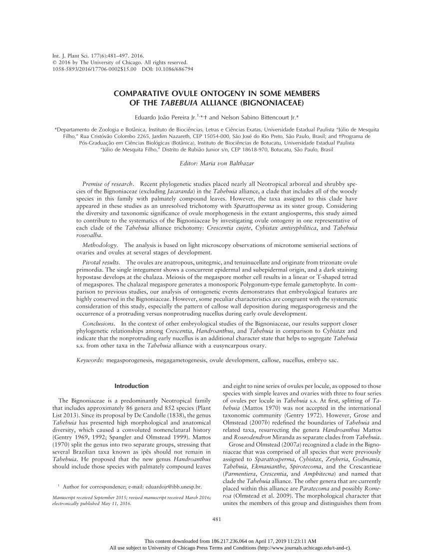
(figs. 1C, 2A). In contrast, Tabebuia roseoalba and Cybistaxantisyphilitica have a bicarpellate eusyncarpous ovary, withtwo placental ridges on each face of the septum (figs. 1A, 1B,4B, 6A). The ovules are arranged on the surfaces of the placentalridges. The number of ovules per placental ridge in a mediantransverse section of the ovary is typically just one in T. rose-oalba, four to six in Cy. antisyphilitica, and more than 16 inC. cujete. While the ovule position in both T. roseoalba andCy. antisyphilitica is pleurotropous-ventral (i.e., with the micro-pyle pointing to the side and away from the center of the carpel;fig. 1A, 1B), in C. cujete, the ovules are heterotropous (i.e., theovule position varies in orientation).
In the three species, before the initiation of ovule ontogeny,cell division patterns at the peripheral layers of the placentalmeristems generate a trizonate organization (sensu Bouman1984; figs. 2B, 4A, 6B). In the epidermal layer (zone 1), celldivisions are only anticlinally oriented. In the subepidermallayer (zone 2), anticlinal divisions predominate, although someoblique and periclinal divisions were also observed (figs. 2B,4A, 6B). There was no regular pattern for the cell divisionsin the third and subjacent layers (zone 3) of the placentae(figs. 2B, 4A, 6B), and ovule protrusion initiates via cellularproliferation in this last zone. The emerging ovule primordiaare also trizonate and show the same patterns of zone 1–3 celldivisions that were observed in the prospective placentae(figs. 2D, 4C, 6D). In each carpel of an ovary cross section,C. cujete shows a diffuse distribution of many ovule primordiaon the placental surface (fig. 2C), whereas Cy. antisyphiliticahas eight to 12 (fig. 6C), and T. roseoalba has only two ovuleprimordia per carpel (fig. 4B).
After the emergence of the ovule primordia from the pla-cental surface, a single archesporial cell typically differentiates
This content downloaded from 186.2All use subject to University of Chicago Press Terms
in the subepidermal layer at a top-lateral position in the nucel-lus. This cell exhibits a dense cytoplasm and a prominent nu-cleus. Although the archesporium expansion results in the pro-trusion of the covering nucellar epidermis in C. cujete (fig. 2E,2F) andCy. antisyphilitica (fig. 6E, 6F), this pattern was not ob-served in the first stages of archesporium differentiation in T.roseoalba (fig. 4C, 4D). This difference may be related to spacelimitation in each locule. In all species, archesporial cells developseveral small vacuoles in the cytoplasm (figs. 2F, 4E, 6F, 6G).Cell proliferation and expansion in one side of the young
ovule cause an anatropous curvature (figs. 2G, 4F, 6H). In thisstage, a group of cells on the raphal side that belong to zone 3start to elongate in longitudinal direction of the ovule and showan increasing cytoplasmic density, differentiating into the pro-vascular bundle of the funiculus (figs. 2H, 4E, 6G, 6H).The single archesporial cell does not divide, and as archespo-
rium differentiation proceeds, a single integument arises bypericlinal divisions from a set of protodermal cells (zone 1)around the base of the archesporium (figs. 2E, 4D, 6E, 6F). Sub-sequently, the subjacent cells (zone 2) also divide periclinally,pushing the cells of epidermal origin to the distal portion ofthe nucellus, where the micropyle will be formed. This processcauses a loss of boundaries between epidermal and subepider-mal cells in the developing integument (figs. 2F, 2G, 4E, 4F,6G, 6H). Zone 3 cells of the young ovule do not participatein the integument formation. The cells of the outer layer of theintegument divide anticlinally, whereas oblique and periclinaldivisions occur in the inner cell layers, which contribute to thethickening of the integument around the nucellus (figs. 2G–2I,4F, 6H–6J).The archesporium increases considerably in volume while
undergoing premeiotic cellular differentiation (PCD) withoutgiving rise to any parietal cells. Therefore, the archesporial celldifferentiates directly into a megaspore mother cell (MMC).Even when more than one archesporial cell (identified by theirlarger volume, cytoplasmic density, and nuclear size relative tothe neighboring somatic cells) is initially present in an ovule,only one of those cells (usually the larger one) undergoes PCDto form anMMC. In all of the studied species, PCDwas charac-terized not only by cell elongation but also by lateral expansion,especially at the distal (micropylar) end, which gives the MMCan oval or club shape (figs. 2G–2I, 4F–4H, 6H, 6I). Duringthe PCD phase, the centrally positioned nucleus continues to en-large, becoming up to three to four times larger in a matureMMC than in any neighboring integumentary cell. In addition,the nucleus has a prominent nucleolus (figs. 2G–2I, 4F–4H, 6H,6I). The early MMC in C. cujete has two large vacuoles, oneat the chalazal pole and the other at the micropylar pole of thecell (fig. 2G), while in T. roseoalba and Cy. antisyphilitica, theearly MMC shows a bipolar vacuome that consists of numer-ous small, rounded vacuoles (fig. 4H). However, these vacuolesare lost during prophase I of meiosis (figs. 2I, 6J), except forT. roseoalba, whose meiocyte vacuolization persists until thedyad stage (fig. 4H).Most of the MMC surface is covered by a nucellar epider-
mis, except for the proximal portion that is in close contactwith the chalaza (figs. 2G–2I, 4F–4H, 6G–6J). In T. roseoalba,amyloplast formation in all cells of the ovule primordium initi-ates simultaneously with the appearance of the archesporium(fig. 4D). InC. cujete andCy. antisyphilitica, small amyloplasts
Fig. 1 Median transverse sections of ovaries at the anthesis stagein Tabebuia roseoalba (A) and Cybistax antisyphilitica (B), both at thesynascidiate region, and inCrescentia cujete (C).Ovule rows andplacen-tal ridges are in dark gray.
17.236.064 on April 17, 2019 11:23:11 AM and Conditions (http://www.journals.uchicago.edu/t-and-c).
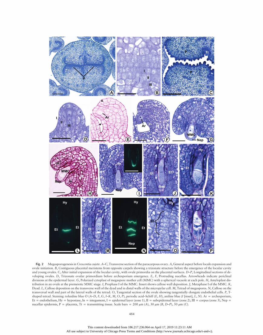
Fig. 2 Megasporogenesis inCrescentia cujete.A–C, Transverse section of the paracarpous ovary.A, General aspect before locule expansion andovule initiation. B, Contiguous placental meristems from opposite carpels showing a trizonate structure before the emergence of the locular cavityand young ovules. C, After initial expansion of the locular cavity, with ovule primordia on the placental surfaces.D–P, Longitudinal sections of de-veloping ovules. D, Trizonate ovular primordium before archesporium emergence. E, F, Protruding nucellus. Arrowheads indicate periclinaldivisions at the epidermal layer. G, Polarized cytoplast of megaspore mother cell (MMC) with a spherical vacuole at each pole. H, Amyloplast dis-tribution in an ovule at the premeiotic MMC stage. I, Prophase I of the MMC. Insert shows callose wall deposition. J, Metaphase I of the MMC. K,Dyad. L, Callose deposition on the transverse wall of the dyad and in distal walls of the micropylar cell.M, Tetrad of megaspores.N, Callose on thetransversal wall and part of the lateral walls of the tetrad. O, Tangential section of the ovule showing tangentially elongate endothelial cells. P, T-shaped tetrad. Staining: toluidine blue O (A–D, F, G, I–K, M, O, P), periodic acid–Schiff (E, H), aniline blue (I [inset], L, N). Ar p archesporium,Etp endothelium, Hyp hypostase, Inp integument, Ip epidermal layer (zone 1), IIp subepidermal layer (zone 2), IIIp corpus (zone 3), Neppnucellar epidermis, P p placenta, Tt p transmitting tissue. Scale bars p 200 mm (A), 30 mm (B, D–P), 50 mm (C).
484
This content downloaded from 186.217.236.064 on April 17, 2019 11:23:11 AMAll use subject to University of Chicago Press Terms and Conditions (http://www.journals.uchicago.edu/t-and-c).
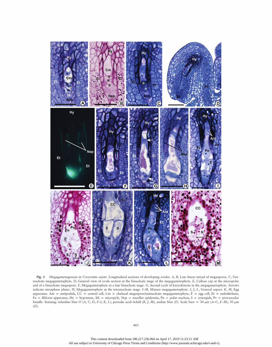
Fig. 3 Megagametogenesis in Crescentia cujete. Longitudinal sections of developing ovules. A, B, Late linear tetrad of megaspores. C, Uni-nucleate megagametophyte. D, General view of ovule section at the binucleate stage of the megagametophyte. E, Callose cap at the micropylarend of a binucleate megaspore. F, Megagametophyte at a late binucleate stage. G, Second cycle of karyokinesis in the megagametophyte. Arrowsindicate metaphase plates. H, Megagametophyte at the tetranucleate stage. I–M, Mature megagametophyte. I, J, L, General aspect. K, M, Eggapparatus. Ant p antipodals, CC p central cell, Cm p chalazal megaspore/uninucleate megagametophyte, E p egg cell, Et p endothelium,Fa p filiform apparatus, Hy p hypostase, Mi p micropyle, Nep p nucellar epidermis, Pn p polar nucleus, S p synergids, Pv p provascularbundle. Staining: toluidine blue O (A, C, D, F–I, K, L), periodic acid–Schiff (B, J, M), aniline blue (E). Scale bars p 30 mm (A–C, E–M), 50 mm(D).
485
This content downloaded from 186.217.236.064 on April 17, 2019 11:23:11 AMAll use subject to University of Chicago Press Terms and Conditions (http://www.journals.uchicago.edu/t-and-c).
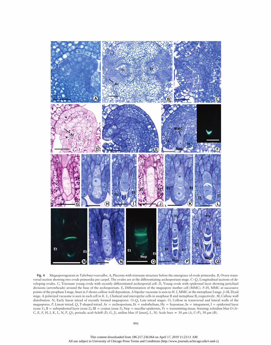
Fig. 4 Megasporogenesis in Tabebuia roseoalba.A, Placenta with trizonate structure before the emergence of ovule primordia. B, Ovary trans-versal section showing two ovule primordia per carpel. The ovules are at the differentiating archesporium stage. C–Q, Longitudinal sections of de-veloping ovules. C, Trizonate young ovule with recently differentiated archesporial cell. D, Young ovule with epidermal layer showing periclinaldivisions (arrowheads) around the base of the archesporium. E, Differentiation of the megaspore mother cell (MMC). F–H, MMC at successivepoints of the prophase I stage. Inset in F shows callose wall deposition. A bipolar vacuome is seen inH. I, MMC at the metaphase I stage. J–M, Dyadstage. A polarized vacuome is seen in each cell in K. L, Chalazal and micropylar cells in anaphase II and metaphase II, respectively.M, Callose walldistribution. N, Early linear tetrad of recently formed megaspores. O–Q, Late tetrad stages. O, Callose in transversal and lateral walls of themegaspores. P, Linear tetrad.Q, T-shaped tetrad. Arp archesporium, Etp endothelium, Hyp hypostase, Inp integument, Ip epidermal layer(zone 1), IIp subepidermal layer (zone 2), IIIp corpus (zone 3), Nepp nucellar epidermis, Ttp transmitting tissue. Staining: toluidine blue O (A–C, E, F, H, I, K, L, N, P, Q), periodic acid–Schiff (D, G, J), aniline blue (F [inset], L, N). Scale bars p 30 mm (A, C–P), 50 mm (B).
486
This content downloaded from 186.217.236.064 on April 17, 2019 11:23:11 AMAll use subject to University of Chicago Press Terms and Conditions (http://www.journals.uchicago.edu/t-and-c).
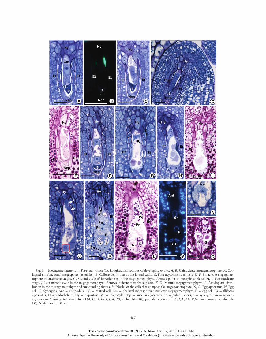
Fig. 5 Megagametogenesis in Tabebuia roseoalba. Longitudinal sections of developing ovules. A, B, Uninucleate megagametophyte. A, Col-lapsed nonfunctional megaspores (asterisks). B, Callose deposition at the lateral walls. C, First acytokinetic mitosis. D–F, Binucleate megagame-tophyte in successive stages. G, Second cycle of karyokinesis in the megagametophyte. Arrows point to metaphase plates. H, I, Tetranucleatestage. J, Last mitotic cycle in the megagametophyte. Arrows indicate metaphase plates. K–O, Mature megagametophytes. L, Amyloplast distri-bution in the megagametophyte and surrounding tissues. M, Nuclei of the cells that compose the megagametophyte. N, O, Egg apparatus. N, Eggcell. O, Synergids. Ant p antipodals, CC p central cell, Cm p chalazal megaspore/uninucleate megagametophyte, E p egg cell, Fa p filiformapparatus, Et p endothelium, Hy p hypostase, Mi p micropyle, Nep p nucellar epidermis, Pn p polar nucleus, S p synergids, Sn p second-ary nucleus. Staining: toluidine blue O (A, C, D, F–H, J, K, N), aniline blue (B), periodic acid–Schiff (E, I, L, O), 40,6-diamidino-2-phenylindole(M). Scale bars p 30 mm.
487
This content downloaded from 186.217.236.064 on April 17, 2019 11:23:11 AMAll use subject to University of Chicago Press Terms and Conditions (http://www.journals.uchicago.edu/t-and-c).
This content downloaded from 186.217.236.064 on April 17, 2019 11:23:11 AMAll use subject to University of Chicago Press Terms and Conditions (http://www.journals.uchicago.edu/t-and-c).
-.
.,
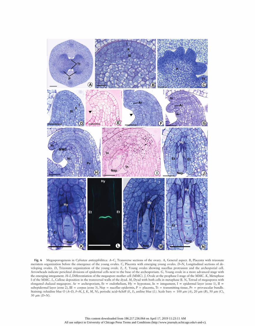
Fig. 6 Megasporogenesis in Cybistax antisyphilitica. A–C, Transverse sections of the ovary. A, General aspect. B, Placenta with trizonatemeristem organization before the emergence of the young ovules. C, Placenta with emerging young ovules. D–N, Longitudinal sections of developing ovules. D, Trizonate organization of the young ovule. E, F, Young ovules showing nucellus protrusion and the archesporial cellArrowheads indicate periclinal divisions of epidermal cells next to the base of the archesporium. G, Young ovule in a more advanced stage withthe emerging integument. H–I, Differentiation of the megaspore mother cell (MMC). J, Ovule at the prophase I stage of the MMC. K, MetaphaseI of the MMC. L, Callose deposition in the transversal walls of the dyad. M, Dyad with both cells in metaphase II. N, Tetrad of megaspores withelongated chalazal megaspore. Ar p archesporium, Et p endothelium, Hy p hypostase, In p integument, I p epidermal layer (zone 1), II psubepidermal layer (zone 2), III p corpus (zone 3), Nep p nucellar epidermis, P p placenta, Tt p transmitting tissue, Pv p provascular bundleStaining: toluidine blue O (A–D, F–H, J, K, M, N), periodic acid–Schiff (E, I), aniline blue (L). Scale bars p 100 mm (A), 20 mm (B), 50 mm (C)30 mm (D–N).
PEREIRA & BITTENCOURT—OVULE ONTOGENY IN TABEBUIA ALLIANCE 489
start to appear in the chalaza, nucellar epidermis, and integu-ment only at the end of the PCD phase of the MMC, whenthe integument border reaches halfway to the top of the nucellus(figs. 2H, 6I). In Cy. antisyphilitica and T. roseoalba, amylo-plasts were observed in the cytoplasm of the MMC, dyads,and the chalazal megaspore (figs. 4G, 4J, 6I). However, PASstaining in ovule sections from C. cujete did not reveal starchgrains in MMC, dyads, and tetrads, and nonsomatic cell amy-loplasts in this species appeared from only the bicellular stageof the developing megagametophyte onward (not shown).
By the stage in which meiotic prophase I occurs in theMMC,the single integument border reaches the top of the nucellar epi-dermis (figs. 2I, 4G, 6J). At this stage, the 1–3 inner cell layersof the integument (i.e., those layers next to the nucellar epider-mis) begin to form a poorly differentiated endothelium. In thesecells, the centrally positioned nucleus may occupy more thanhalf of the protoplasmic volume, with a prominent nucleolus(figs. 2I, 2P, 6J). These cells also show a higher cytoplasm den-sity than those of the outer cell layers of the integument (figs. 2J–2P, 4I–4Q, 6J–6N) and are tangentially elongated (fig. 2O).From the meiotic prophase stage on (fig. 4G), the inner layersof the integument (including the endothelium) begin to showan increasing gradient of number and size of amyloplasts to-ward the nucellar epidermis (figs. 3B, 3J, 3M, 5E, 5I, 5L, 7C,7I, 7P).
Even during the differentiation of the MMC, the cells of thechalaza that are located between the chalazal end of theMMC and the distal end of the provascular bundle and the cellsof the nucellar epidermis next to this region begin to differenti-ate into a hypostase. These cells generate a stronger cytoplasmicdensity than the neighboring somatic cells, and their walls, in-cluding those facing the MMC, not only thicken but also startto show an intense reddish purple staining with toluidine blueO (figs. 2I–2K, 2M, 2O–2P, 4H, 4I, 4K, 4L, 4N, 4P, 4Q, 6J,6K, 6M, 6N). These cell walls are also PAS positive (figs. 2H,3B, 4G, 4J), and ruthenium red staining (data not shown)indicates that they are rich in pectic substances. These nucellarcells have vacuoles with a dense granular content that stainsreddish purple with toluidine blue O (figs. 2J, 4Q, 6M) and isPAS positive (figs. 5E, 5I, 7C, 7I). The same vacuolar contentoccurs in most other cells of the ovule at different stages butwith a lower staining intensity. Because of its position, thehypostase clearly connects the MMC—and the developingmegagametophyte in later stages (see below)—with the provas-cular bundle (fig. 3D). Sometimes T. roseoalba shows binucle-ate cells in the hypostase, which are usually aligned with thelongitudinal axis of the archesporium/MMC/megaspores/megagametophyte and often show a bipolar vacuome (figs. 4E,4H, 4L, 4P, 5E).
During meiotic divisions of the MMC, the metaphase platesare perpendicularly oriented to the meiocyte longitudinal axis(figs. 2J, 4I, 4L, 6K, 6M), and after successive cytokinesis, a lin-ear tetrad of megaspores is generated (figs. 2J–2N, 4I–4P, 6K–6N, 7A). However, C. cujete and T. roseoalba also exhibited apercentage of T-shaped tetrads: 3.3% and 11.3% in 61 and 71examined tetrad stage ovules, respectively (figs. 2P, 4Q). Often,no synchrony is observed between the two cells of a dyad afterthe first meiotic cytokinesis because the events of the second di-vision (e.g., anaphase II chromosome migration) occur faster inthe chalazal cell (fig. 4L). In the tetrad stage, the integument
This content downloaded from 186.2All use subject to University of Chicago Press Terms
border typically covers the top of the nucellus, initiating micro-pyle organization in C. cujete and T. roseoalba (figs. 2M, 4P,4Q). InCy. antisyphilitica, themicropyle begins to be organizedprimarily after the initial expansion of the chalazal megasporeto form the uninucleate megagametophyte (fig. 7C, 7D). How-ever, in all of the plants under study, the point at which the mi-cropyle started to be organized varied from the early tetradstage to chalazal megaspore expansion.In the three species, patterns of callose deposition were ob-
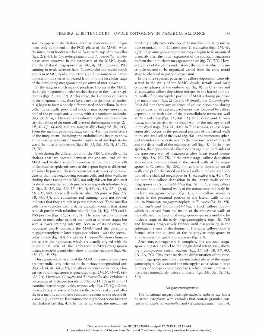
served in the walls of the MMC, dyads, tetrads, and earlycenocytic phases of the embryo sac (fig. 8). In C. cujete andT. roseoalba, callose deposition initiates in the lateral and dis-tal walls of the micropylar portion of MMCs during prophaseI or metaphase I (figs. 2I [inset], 4F [inset]), but Cy. antisyphi-litica did not show any evidence of callose deposition duringthese stages. In all species, cytokinesis was followed by callosedeposition on both sides of the pectocellulosic transverse wallat the dyad stage (figs. 2L, 4M, 6L). In C. cujete and T. rose-oalba, callose persists in the distal wall of the micropylar cellin the dyad stage (figs. 2L, 4M). In T. roseoalba, callose depo-sition also occurs in the proximal portion of the lateral wallsin the chalazal cell of the dyad (fig. 4M), and numerous spher-ical vacuoles concentrate next to the proximal wall of this celland the distal wall of the micropylar cell (fig. 4K). In the threespecies, the deposition of callose occurs again on both sides ofthe transverse wall of megaspores after linear tetrad forma-tion (figs. 2N, 4O, 7B). In the tetrad stage, callose depositionalso occurs to some extent at the lateral walls of the mega-spores in C. cujete (fig. 2N), and callose is deposited on allwalls except for the lateral and basal walls in the chalazal por-tion of the chalazal megaspore in T. roseoalba (fig. 4O). Wedid not find callose deposition in the lateral walls of themegaspores in Cy. antisyphilitica (fig. 7B). In C. cujete, callosepersists along the lateral walls of the uninucleate and early bi-nucleate megagametophyte (fig. 3E), and callose remnantspersist in the proximal portion of the lateral walls of theuni- to binucleate megagametophyte in T. roseoalba (fig. 5B).In C. cujete and Cy. antisyphilitica, a thick callose hood—which is derived from the fusion of the transverse walls ofthe collapsed nonfunctional megaspores—persists until the bi-nucleate stage of the early megagametophyte (figs. 3E, 7H)and becomes progressively thinner until disappearing in thesubsequent stages of development. The same callose hood isformed after the collapse of the micropylar megaspores inT. roseoalba but quickly disappears (fig. 5B).After megasporogenesis is complete, the chalazal mega-
spore elongates parallel to the longitudinal tetrad axis, show-ing a conspicuous central nucleus (figs. 2P, 3A, 3B, 4P, 4Q,6N, 7A, 7C). This event marks the differentiation of the func-tional megaspore into the single-nucleated phase of the mega-gametophyte. Cells around the micropylar canal show a largenumber of conspicuous amyloplasts, which persist until ovulematurity, immediately before anthesis (figs. 3M, 5E, 5I, 5L,5O).
Megagametogenesis
The functional megaspore/single-nucleate embryo sac has apolarized cytoplasm with vacuoles that contain granular con-tent in C. cujete, T. roseoalba, and Cy. antisyphilitica (figs. 3A,
17.236.064 on April 17, 2019 11:23:11 AM and Conditions (http://www.journals.uchicago.edu/t-and-c).
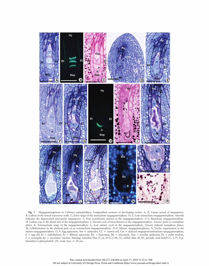
Fig. 7 Megagametogenesis in Cybistax antisyphilitica. Longitudinal sections of developing ovules. A, B, Linear tetrad of megaspores.B, Callose in the tetrad transverse walls. C, Early stage of the uninucleate megagametophyte. D, E, Late uninucleate megagametophyte. Asteriskindicates the degenerated micropylar megaspores. F, First acytokinetic mitosis in the megagametophyte. G–I, Binucleate megagametophyte.H, Callose cap at the distal end of the megagametophyte. J, Second cycle of karyokinesis in the megagametophyte. Arrows point to metaphaseplates. K, Tetranucleate stage of the megagametophyte. L, Last mitotic cycle in the megagametophyte. Arrows indicate metaphase plates.M, Cellularization in the chalazal pole of an octonucleate megagametophyte. N–P, Mature megagametophytes. N, Nuclei organization in themature megagametophyte. O, P, Egg apparatus. Ant p antipodes, CC p central cell, Cm p chalazal megaspore/uninucleate megagametophyte,E p egg cell, Et p endothelium, Fa p filiform apparatus, Hy p hypostase, Mi p micropyle, Nep p nucellar epidermis, Pn p polar nucleus,S p synergids, Sn p secondary nucleus. Staining: toluidine blue O (A, D–G, J–M, O), aniline blue (B, H), periodic acid–Schiff (C, I, P), 40,6-diamidino-2-phenylindole (N). Scale bars p 30 mm.
This content downloaded from 186.217.236.064 on April 17, 2019 11:23:11 AMAll use subject to University of Chicago Press Terms and Conditions (http://www.journals.uchicago.edu/t-and-c).
PEREIRA & BITTENCOURT—OVULE ONTOGENY IN TABEBUIA ALLIANCE 491
5A, 7A). Multiple and small vacuoles are initially dispersed inthe micropylar and chalazal portions of the cell. However,those vacuoles fuse into two large vacuoles, with one vacuoleat each pole of the single-nucleate megagametophyte (figs. 3C,7E), except for T. roseoalba, in which this fusion does not occur.
Throughout the megagametogenesis, the endothelial cellsadjacent to the nucellar epidermis sometimes are radiallyelongated (figs. 5F, 5I, 5K, 5L, 7E–7G, 7J). For all species,female gametophyte development is marked by an initial coe-nocytic phase, in which three successive cycles of acytokineticmitosis are followed by the formation of a seven-celled and
This content downloaded from 186.2All use subject to University of Chicago Press Terms
octonucleate embryo sac. The metaphase plate of the first acy-tokinetic division in the uninucleate megagametophyte is per-pendicular to the longitudinal axis of the ovule (figs. 5C, 7F).The two resulting nuclei move apart from each other towardopposite poles of an elongating coenocytic megagametophyte.At this stage, a large vacuole occupies the space between thetwo nuclei, and a smaller vacuole is also present in the chala-zal pole of the megagametophyte between the chalazal nucleusand the wall facing the hypostase. Both vacuoles contain thesame granular substance observed in the previous stages, whichis PAS positive and stains reddish purple with toluidine blue O(figs. 3F, 5E, 5F, 7G, 7I). This content also appears to be thesame as in vacuoles of somatic cells of the ovule, and it wasfound in all of the subsequent stages of female gametophytedevelopment. Amyloplasts appear around both nuclei of thebinucleate megagametophyte (figs. 5E, 7I). Remnants of thedegenerate nonfunctional megaspores are squeezed at the mi-cropylar end of the megagametophyte (figs. 5D–5F, 7G).Cells of the nucellar epidermis start to show signs of degen-
eration—such as nuclear pyknosis, plasmolysis, and cytoplas-mic coagulation—and finally collapse (figs. 3F, 5F, 7G), exceptfor those around the chalazal portion of the megagameto-phyte, which may show protoplasmic degradation but usuallymaintain their original form in subsequent stages (figs. 3F–3L,5G–5L, 7G–7M). Although these features of the degenerativeprocess are clearly evident from the binucleate stage of the coe-nocytic embryo sac onward, in several ovules they appearedeven in earlier stages (figs. 2K, 2P, 3A, 4L, 4P, 4Q, 5A, 5C,7F). During their degenerative process, the cells of the nucellarepidermis accumulate a secretion with the same staining prop-erties as the vacuole content between the collapsing protoplastand the cell wall. The secretion is subsequently released intothe micropylar canal and around the megagametophyte, nextto the degenerated nonfunctional megaspores (fig. 7G). Thewalls of collapsed cells of the nucellar epidermis remain aroundthe micropylar portion of the binucleate megagametophyte,separating it from the integument (figs. 3F, 3G, 5G, 7I, 7J).In the three species, the developing embryo sac continues to
expand, and the second acytokinetic mitotic cycle of megaga-metogenesis occurs simultaneously in the micropylar and cha-lazal nuclei of the megagametophyte (figs. 3G, 5G, 7J). Themetaphase plate of the chalazal nucleus is perpendicular oroblique to the megagametophyte longitudinal axis; on theother hand, the metaphase plate of the micropylar nucleusis parallel or oblique to the axis (figs. 3G, 5G, 7J). After kar-yokinesis, the two chalazal nuclei move away from each otheralong the longitudinal axis of the megagametophyte, while themicropylar nuclei aligns approximately perpendicular to thislongitudinal axis. The micropylar portion of the megagameto-phyte expands laterally and toward the micropyle, invadingareas that were previously occupied by amyloplast-rich cellsthat degenerated (after showing cytoplasm coagulation andkaryopyknosis) and collapse, while the megagametophyte asa whole becomes club shaped (figs. 3H, 5H, 7K). As observedfor the nucellar epidermis, remnants of the walls of cells lo-cated around the inner end of the micropylar canal also re-main around the micropylar extended portion of the mega-gametophyte, forming a pectocellulosic envelope that persistsuntil the mature megagametophyte stage (figs. 3H–3L, 5I–5L,7K–7M).
Fig. 8 Patterns of callose deposition during megasporogenesis inTabebuia roseoalba (A),Crescentia cujete (B), andCybistax antisyphili-tica (C). Callose wall deposition in black.
17.236.064 on April 17, 2019 11:23:11 AM and Conditions (http://www.journals.uchicago.edu/t-and-c).
492 INTERNATIONAL JOURNAL OF PLANT SCIENCES
A large number of conspicuous starch grains persist evenafter the degeneration and collapse of the cells at the innerend of the micropyle. These amyloplasts accumulate aroundthe megagametophyte envelope, giving the envelope a wavycontour (figs. 5L, 5O, 7P). In the preanthesis stage, amylo-plasts surrounded all of the nuclei of the mature megagameto-phyte (figs. 3J, 3M, 5L, 5O, 7P).
As during the second cycle, the third and last cycle of mitoticdivision also occurs simultaneously in the nuclei of both the mi-cropylar and the chalazal poles of the coenocytic embryo sac.In both poles, the orientation of the nuclei metaphase platestended to be perpendicular to each other, approaching a charac-teristic T configuration, although the chalazal pair showed aninverted position relative to the micropylar pair (fig. 5J). Fur-thermore, the metaphase plate of the distal nucleus in the chala-zal pair and that of the proximal nucleus in the micropylar pairapproached a transverse position (i.e., perpendicular to the lon-gitudinal axis of the megagametophyte; fig. 5J). However, de-viations from this configuration pattern were sometimes ob-served (fig. 7L).
The transition from the late tetranucleate to the octonucleatecoenocytic phase seems to be quickly followed by cellulari-zation because ovules containing noncellularized octonucleateembryo sacs were rarely observed. At this transient stage, aftercytokinesis, the proximal mitotic nucleus of the chalazal polegenerates the nuclei of two antipodal cells, and the distal nu-cleus generates the nucleus of a third antipodal cell and the cha-lazal polar nucleus of the central cell (fig. 7M). At the micropy-lar pole, the distal mitotic nucleus originates the nuclei of twosynergids, and the proximal nucleus generates the micropylarnucleus of the central cell and the egg cell nucleus (fig. 7M).Therefore, after the last mitotic cycle, the megagametophytehas eight nuclei and seven cells (figs. 3I–3M, 5K–5O, 7M–
7P). To some extent, for all species, the mature female game-tophyte and those of previous stages showed a characteristicsecretion accumulation in its middle portion between the integ-ument/collapsed nucellar epidermis and the megagametophytewall, although this pattern was more pronounced in Cy. anti-syphilitica, where the embryo sac lateral walls are displacedfrom remnants of the degenerate nucellar epidermis/endothelialcells, giving an hourglass shape to this region (fig. 7L, 7M).
The cellularization process appears to be faster in the chala-zal region because in megagametophytes in which the cell wallsof the egg apparatus are not yet evident, the thin walls that sep-arate the antipodals are already present (fig. 7M). The antip-odals contain dense cytoplasm and may contain small vacuoles(figs. 3L, 5K). These cells may organize themselves in a linearor a triangular arrangement, sometimes showing small amy-loplasts. The chalazal polar nucleus starts migrating towardthe central area of the megagametophyte before the completecellularization of the antipodals.
After cellularization, enlargement of cells of the egg appara-tus (cf. fig. 7M, 7O) and the migration of the two polar nucleito a central position in the embryo sac are the most remark-able features of the last steps of female gametophyte matura-tion. In the mature megagametophyte, the egg cell usuallyacquires an elongated pear shape, with a large vacuole intwo-thirds of the micropylar portion of the cytoplasm andthe nucleus surrounded by small vacuoles and amyloplasts atthe enlarged chalazal end of the cell (figs. 3L, 5N, 7O). The
This content downloaded from 186.2All use subject to University of Chicago Press Terms
synergids are hooked and also pear shaped, wide at the chala-zal end and narrow at the micropylar end (figs. 3K, 5K, 5O).Each synergid contains its nucleus at the micropylar pole or ata central-micropylar position and a conspicuous vacuole at thechalazal pole of the cytoplasm, with some small, rounded vac-uoles often appearing between the nucleus and the pointed mi-cropylar end of the cell (figs. 3K, 5K).
The large central cell comprises most of the mature mega-gametophyte volume and contains amyloplasts in the cyto-plasm, especially around the polar nuclei (figs. 3I, 3J, 5K,5L). The central cell has a wide vacuole with a faintly PAS-positive granulated content that also stains reddish-purple withtoluidine blue O (figs. 3I, 3J, 5K, 5L). During its expansion,the egg apparatus invades the micropylar region of the centralcell, such that a narrow space, which is filled by the centralcell cytoplasm, remains between the embryo sac envelopeand the lateral sides of the egg apparatus (figs. 3J, 5K). Thus,the egg apparatus is in direct contact with the embryo sac en-velope only at the micropylar end. The two polar nuclei fuseto form the secondary nucleus (figs. 5M, 7N). The filiform ap-paratus originates from the walls between the micropylar endsof the two synergids; this apparatus stains reddish purple withtoluidine blue O and is PAS positive (figs. 3M, 5K, 5O). Thesynergids persist until the early stages of endosperm develop-ment in fertilized embryo sacs (not shown). The events of mega-sporogenesis andmegagametogenesis showno synchrony amongthe ovules of the same ovary.
Discussion
The three species investigated in this study have bicarpellatesyncarpous gynoecia, as is common for all of the Bignonia-ceae. The presence of two placental ridges per carpel—fromwhich the ovules emerge, as observed in the species investi-gated here—is also a characteristic feature of the gynoeciumin the Bignoniaceae (Leinfellner 1973). However, while theseplacental ridges are displayed on both faces of the septum inthe eusyncarpous ovary of Cybistax antisyphilitica and Tabe-buia roseoalba, in Crescentia cujete, the placental ridges aredisplayed on the internal face of the ovary wall because ofits paracarpous condition. As is common for other asterid taxa,the Bignoniaceae has a completely syncarpous gynoecium whoserelatively long symplicate zone (and compitum) take up theentire style and at least the uppermost part of the ovary(Endress 1994). In the tribe Crescentieae, there are several in-termediate conditions between the ancestral eusyncarpousconstitution and the paracarpous ovary with parietal placen-tation, and this diversity in carpellary organization is alsofound in the Paleotropical tribe Coleeae (Gentry 1980). How-ever, on the basis of an almost completely symplicate gynoe-cium in C. cujete, which shows only a short sterile synas-cidiate zone at the base of its unilocular ovary, Leinfellner(1973) concluded that Crescentia is the unique genus inBignoniaceae, with a true parietal placentation.
The derivation of paracarpy and intermediate forms fromthe ancestral eusyncarpous condition of the ovary in Bigno-niaceae could be explained by different degrees of reductionof the synascidiate zone and a reciprocal increase of the sym-plicate zone that is involved in the ovary construction, com-bined with changes in vascularization, although additional
17.236.064 on April 17, 2019 11:23:11 AM and Conditions (http://www.journals.uchicago.edu/t-and-c).
PEREIRA & BITTENCOURT—OVULE ONTOGENY IN TABEBUIA ALLIANCE 493
studies are needed to confirm this hypothesis. However, ourobservations and the current data concerning the evolutionof syncarpy and placentation in the Bignoniaceae indicate thatovaries with some degree of a symplicate paracarpious zoneevolved independently in Coleeae and Crescentieae from syn-ascidiate eusyncarpous ancestors.
The placentae and ovule primordia of the three species in theearly stages of ovary development show a tunica-corpus orga-nization, according to the histological zonation described byBouman (1974, 1984), who traced a parallel between the cellu-lar organization of apical meristems and the ovule primordium.In the ovule primordia of all species investigated in this study,the tunica is composed of an epidermal layer with only anticli-nal cell divisions and a subepidermal layer that usually showsanticlinal cell divisions but in which oblique and periclinaldivisions eventually occur. The corpus corresponds to the cen-tral region and does not have a regular pattern of cell divisions.Only two studies investigated the histological organization ofthe ovule primordium in the Bignoniaceae: Galati and Stritt-matter (1999) reported a bizonate ovule primordium in Jaca-randa mimosifolia, and Bittencourt and Mariath (2002b)observed a trizonate ovule primordium in Handroanthus pul-cherrimus. Bouman (1984) pointed out that a bizonate ovuleprimordium initiates by periclinal divisions in the subepidermallayer of the placenta, and this type of primordium appears to besmaller than a trizonate primordium, with an epidermal layerwhose cells divide only anticlinally. On the other hand, a tri-zonate ovule primordium usually initiated by periclinal divi-sions in the central region (corpus) of the placental meristem,while cells of the two outer layers divide anticlinally (Bouman1984). Although J. mimosifolia belongs to a clade that has beenrecognized as the sister group for the rest of the Bignoniaceae(Olmstead et al. 2009), because of the scarcity of studies, it isnot possible to determine whether the bizonate ovule is aplesiomorphic condition in the family or whether it evolved in-dependently within Jacaranda.
The number of ovules per locule is a characteristic with tax-onomic relevance (Gentry 1980), and this trait was one of themorphological characters used to separate Handroanthus,which has eight to 10 ovules per locule, from Tabebuia, whichhas three to four ovules per locule (Mattos 1970; Grose andOlmstead 2007b). However, T. roseoalba (this study), Tabe-buia rosea, and Stereospemum chelonoides (Mehra and Kul-karni 1985) show only two rows of ovules per locule. The pres-ence of only two ovules versus eight to 12 ovules per locule(half of the ovules in each carpel placental ridge) in a transversesection of the ovary in T. roseoalba and Cy. antisyphilitica notonly corroborates the Mattos (1970) segregation but also ex-tends the distinctive condition (relative to Tabebuia s.s.) ofmore than three to four ovules per locule to other taxa of theBignoniaceae with a bicarpellate-eusyncarpous ovary.
The archesporial region of the nucellus in T. roseoalba (i.e.,the archesporial cell plus the nucellar epidermis) is inconspic-uous and becomes prominent only at later stages of MMC de-velopment, probably because of space limitation in the locularcavity, which differs from Cy. antisyphilitica, C. cujete, andother taxa belonging to the internal clades of the Tabebuia al-liance, in which there is an open space (a broad locular cavity)between the ovary wall and the ovule primordia and the nu-cellus is ab initio protruding (Johri et al. 1992; Bittencourt
This content downloaded from 186.2All use subject to University of Chicago Press Terms
and Mariath 2002b). The nonprotruding nucellus in earlystages of the archesporium/MMC development in T. roseoalbaalso appears to be related to the number of ovules per locule ina cross section of the ovary because eight to 10 ovules per loc-ule have been reported for Handroanthus and other specieswith a bicarpellate-eusyncarpous gynoecium, and only twoto four ovules per locule have been reported in Tabebuia s.s.(this study; Mattos 1970; Grose and Olmstead 2007b). Prelim-inary observations in T. rosea—a species that originallyoccurred from Southern Mexico to Venezuela and coastal Ecua-dor (Gentry 1992) but is now cultivated in many placesworldwide in the tropics—also indicated the occurrence of anonprotruding nucellus in the young ovules that emerges inonly two series per carpel (N. S. Bittencourt Jr., unpublisheddata), as observed in T. roseoalba. Therefore, the nonpro-truding nucellus in the initial stages of ovule developmentmay be a synapomorphic character state of the young ovulefor Tabebuia, although additional studies of other taxa in theTabebuia alliance are needed to confirm this hypothesis.The ovule in C. cujete, T. roseoalba, and Cy. antisyphilitica
is anatropous, unitegmic, and tenuinucellate. The same featureswere observed in J. mimosifolia (Galati and Strittmatter 1999),H. pulcherrimus (Bittencourt andMariath 2002b), and most ofthe species of Bignoniaceae (Davis 1966; Johri et al. 1992).Hemianatropous ovules were noticed in S. chelonoides andMillingtonia hortensis (Mehra and Kulkarni 1985). A few he-mianatropous ovules were observed in Oroxylum indicum, al-though most ovules in this species are anatropous (Ghatak1956). Kigelia africana (syn. Kigelia pinnata) and Parmentieracereifera occasionally show hemianatropous ovules (Gonvidu1950). Bouman and Schier (1979) pointed out that in uniteg-mic sympetalous plants, the integument does not always havean exclusively epidermal origin, but it can also be derived par-tially from the subepidermal layer. In fact, unitegmy evolvedseveral times in angiosperm history, and the single integumentdoes not correspond to an outer or an inner integument but isa complex structure in which both layers take part and cannotbe distinguished morphologically (Bouman and Calis 1977;Endress 2011). The integument in the three species studiedhere initiates from the epidermal layer via periclinal divisions.However, shortly after the first divisions of epidermal cells,periclinal divisions also occur in the subepidermal layer andpush the cells of epidermal origin to the top of the nucellus.This phenomenonwas named integumentary shifting by Boumanand Calis (1977). Handroanthus pulcherrimus shows the samedevelopmental pattern (Bittencourt and Mariath 2002b). Galatiand Strittmatter (1999) described a different pattern for J.mimosifolia, in which only the dermal layer participates in in-tegument development. Because in recent phylogeny studies,the tribe Jacarandeae appears as sister to the rest of the family(Spangler and Olmstead 1999; Grose and Olmstead 2007a;Olmstead et al. 2009), it is possible that the epidermal origin(zone 1 only) of the integument is a plesiomorphic conditionin Bignoniaceae from which the epidermal 1 subepidermalorigin derivates. However, Galati and Strittmatter (1999) didnot present any comment on the pattern of division in the sub-epidermal cells immediately below the group of cells of epider-mal (zone 1) origin; thus, a reevaluation of the integumentontogeny in J. mimosifolia and additional investigations inother species of Jacarandeae and the closest external groups
17.236.064 on April 17, 2019 11:23:11 AM and Conditions (http://www.journals.uchicago.edu/t-and-c).
494 INTERNATIONAL JOURNAL OF PLANT SCIENCES
of the Bignoniaceae are necessary to test the hypothesis ofplesiomorphy of the exclusive epidermal origin of the integu-ment in Bignoniaceae.
In the three species studied here, the micropyle begins to beorganized during the tetrad stage, as reported for M. horten-sis, Fernandoa adenophylla (syn. Heterophragma adenophyl-lum; Mehra and Kulkarni 1985), and H. pulcherrimus (Bit-tencourt and Mariath 2002b). However, some variations inthe point of ovule development at which the micropyle beginsto be formed appear to occur between species in the Bigno-niaceae, as this event was reported to occur in the MMC stagein T. rosea, S. chelonoides, and Dolichandrone falcata (Mehraand Kulkarni 1985) and when the MMC undergoes meiosis inJ. mimosifolia (Galati and Strittmatter 1999). Unfortunately,other embryological studies in the Bignoniaceae do not supplyinformation concerning the ovule developmental stage at whichmicropyle organization occurs.
The hypostase is a common trait in Bignoniaceae (Gonvidu1950; Ghatak 1956; Galati and Stritttmatter 1999). In somestudies, this tissue has been viewed as being involved in the in-hibition of aggressive growth of the endosperm haustorium inthe chalazal portion of the developing seed (Mehra and Kulkarni1985). In J. mimosifolia, abundant insoluble polysaccharides arepresent in hypostase cell walls, suggesting the role of this tissue incarbohydrate reserve during endosperm development (Galatiand Strittmatter 1999). The staining properties of these cells inall species here investigated indicate not only the polysacchariderichness of their walls but also the polysaccharide nature of theirvacuolar content. In fact, many authors believe that the mainfunction of the hypostase is to supply nutrients for the megaga-metophyte, although those authors disagree about the mecha-nisms by which this process occurs (Tilton 1980; Boesewinkeland Bouman 1984; Batygina and Shamrov 1999). The hypostasein the Bignoniaceae persists until the advanced stages of themegagametophyte andmay continue to develop and differentiateduring the early stages of endosperm development (Bittencourtand Morais 2010). This tissue originates adventitious embryoprecursor cells in sporophytic apomictic species ofHandroanthus(Costa et al. 2004; Souza et al. 2005; Bittencourt and Moraes2010) andAnemopaegma (Sampaio et al. 2013a, 2013b).Nodif-ferences in the features of the hypostase were found among thethree species here investigated.
MMC meiosis in Cy. antisyphilitica originates only lineartetrads of megaspores, and the chalazal megaspore gives rise toa monosporic Polygonum-type megagametophyte, as in otherpreviously studied species in the Bignoniaceae (Gonvidu 1950;Ghatak 1956; Davis 1966; Mehra and Kulkarni 1985; Johriet al. 1992; Galati and Strittmatter 1999; Souza et al. 2005;Renó et al. 2007; Sampaio 2007; Souza et al. 2008), includingH. pulcherrimus (Bittencourt and Mariath 2002a). The samepattern was observed in C. cujete and T. roseoalba, but al-though linear tetrads predominate, a small percentage of youngovules with T-shaped tetrads were observed in these species.Although there is evidence for the phylogenetic relevance ofmegaspore tetrad patterns in some taxa (Rembert 1972), inthe few studies of Bignoniaceae that investigated this issue,no information was given about the number of samples ob-served to classify the megaspore tetrad arrangement. Lineartetrads are reported for nearly all species in these studies(Duggar 1899; Govindu 1950; Ghatak 1956; Mehra and Kul-
This content downloaded from 186.2All use subject to University of Chicago Press Terms
karni 1985), but T-shaped tetrads (except for one case of anisobilateral tetrad) were noticed in K. africana (Govindu 1950),and linear and T-shaped tetrads were found in J. mimosifolia(Galati and Strittmatter 1999). In spite of the putative typicalpresence of T-shaped tetrads during megasporogenesis in K. afri-cana, on the basis of the present observations as well as obser-vations of other previously studied bignoniaceous species—such asHandroanthus ochraceus,Handroanthus chrysotrichus,and Spathodea campanulata (N. S. Bittencourt Jr., unpublisheddata)—we assume that the occurrence of T-shaped tetrads in theBignoniaceae may be purely occasional and scattered through-out the family.
In C. cujete and T. roseoalba, during meiosis, callose depo-sition was observed in the walls of the micropylar portion ofthe MMC during prophase/metaphase I. This observation con-stitutes a peculiar feature that was not reported for any otherpreviously investigated species in the Bignoniaceae. Callosealso occurs in the transverse cell walls of dyads and tetrads inall species and probably favors chalazal megaspore develop-ment due the difficulty of translocating nutrients to micropylarmegaspores (Kapil and Tiwari 1978). In the ovule, callose de-position may work like a molecular or nutritional filter by de-creasing the permeability of the cell wall and promoting a tem-porary isolation of the megaspores, which enables them toembark upon independent courses of development (Rodkiewiez1970; Bouman 1984).
The position of the functional megaspore close to the chalazais a positive factor for its development due to the nutritional roleof the hypostase and the connection of the hypostase to the fu-nicular provascular bundle tip, as reported by Bittencourt andMariath (2002b) for H. pulcherrimus. In most angiospermswith a Polygonum-type ontogeny, callose is laid down first onthe chalazal pole of the MMC wall at the onset of meiosis,followed by its temporary deposition around the entire mega-sporocyte and its subsequent disappearance from the chalazalpole (Rodkiewicz 1970; Kapil and Tiwari 1978). However, inthe present study, callose deposition was observed only on themicropylar pole of the megasporocyte cell wall in bothC. cujeteand T. roseoalba and was never observed before the dyad stagein Cy. antisyphilitica.
The presence of callose in part of the proximal half of the cha-lazal cell wall of the dyad inT. roseoalba is a feature that has notbeen reported for other members of the family and is potentiallyapomorphic at the species or genus level, although further stud-ies are needed to confirm this hypothesis. The pattern of callosedistribution that was observed in T. roseoalba megaspore tet-rads also occurs in Handroanthus pulcherimus (Bittencourt andMariath 2002b) but not in Cy. antisyphilitica, which points toa closer relationship between Tabebuia and the clade to whichCrescentia and Handroanthus belong. The callose hood thatresults from the superposition of wall remnants of the collapsedmicropylar megaspores remains at least up to the uninucleatestage of embryo sac development in all of the plants under study,but this hood appears to disappearmore quickly inT. roseoalba.The same phenomenon was reported for H. pulcherrimus (Bit-tencourt and Mariath 2002b). Crescentia cujete is the only spe-cies investigated to date to also have callose on the lateral wallsof one- to two-nucleated megagametophytes.
Kapil and Tiwari (1978) defined the endothelium as the innerepidermis of the inner integument in bitegmic ovules or the in-
17.236.064 on April 17, 2019 11:23:11 AM and Conditions (http://www.journals.uchicago.edu/t-and-c).
PEREIRA & BITTENCOURT—OVULE ONTOGENY IN TABEBUIA ALLIANCE 495
ner epidermis of the single integument in unitegmic ovules.Endress (2010) reported the presence of an endothelium inthe majority of the Lamiales taxa. Usually, the cells of this tissueare radially or tangentially elongated and have a dense cyto-plasm and prominent and often polyploid nucleus (Kapil andTiwari 1978). The endothelium in the three species studied hereis not so evident as it typically appears in ovules of other taxa(Kapil and Tiwari 1978). However, it was recognized here asone to three layers of integumentary cells in contact with the ex-ternal face of the nucellar epidermis and next to the proximalportion of the developing megegametophyte. This cells showeddense cytoplasm, high nucleus/cytoplasm ratio and prominentnucleoli, and often they appeared as tangentially or radiallyelongated cells. Despite showing only a discrete differentiationrelative to the outer layers of the ovule integument, the endothe-lium has been reported for many bignoniaceous species scat-tered throughout the family (Govindu 1950; Ghatak 1956;Mehra and Kulkarni 1985; Galati and Strittmatter 1999; Bit-tencourt and Mariath 2002b), and it continues to expand anddifferentiate around the endosperm, during the developmentof the seed (Bittencourt and Moraes 2010; Sampaio et al.2013). The endothelium canalizes nutrients to the developingmegagametophyte (Kapil and Tiwari 1978), and the occurrenceof a large number of amyloplasts in the endothelium and othercells of the inner layers of the integument, as observed here, isconsistent with this role. No differences in the general featuresof the endotheliumwere observed between species in this study.
The onset of amyloplast formation in the ovule appears tovary not only with respect to the ovule developmental stagebut also with respect to the tissue in which it occurs, appearingthroughout the ovule in the archesporium stage in T. rose-oalba, in the chalaza, nucellar epidermis and integument inthe MMC PCD stage in C. cujete and Cy. antisyphilitica (thisstudy), and in the endothelium during meiosis in H. pulcher-rimus (Bittencourt and Mariath 2002b). Because amylogene-sis was not analyzed in other studies of ovule ontogeny in theBignoniaceae, it is not possible at this point to attribute anysystematic significance to the timing of amyloplast formationonset in ovule development in Bignoniaceae. According toTilton and Lersten (1981), starch may begin to accumulatein the megagametophyte as early as the MMC stage or onlyin the stages immediately prior to fertilization, but starch isusually present by the time the megagametophyte is cellularand reaches a maximum shortly after fertilization. In fact,in this study, amyloplasts reached their largest number andsize immediately before megagametophyte maturity for allspecies. This feature appears to be a common trait in Bigno-niaceae, with C. cujete, Cy. antisyphilitica and T. roseoalbashowing amyloplasts in all cells of the egg apparatus, as de-scribed for H. pulcherrimus (Bittencourt and Mariath 2002a).
From the binucleate stage on, the megagametophyte gradu-ally becomes club-shaped due to the expansion of the diameterand length of its distal portion, which invades regions originallyoccupied by cells of the inner layers of the integument. The de-generation of these cells always follows the degeneration of thenucellar epidermis in the same area, and this phenomenon maybe causally related to the expansion of the developing megaga-metophyte. The same phenomenon has been noticed in manyspecies of Bignoniaceae (Mauritzon 1935; Swamy 1941; Go-vindu 1950; Ghatak 1956; Mehra and Kulkarni 1985; Galati
This content downloaded from 186.2All use subject to University of Chicago Press Terms
and Strittmatter 1999). Some of those authors suggested thatthe degeneration and collapse of the nucellar epidermis andintegument cells are consequences of an aggressive expansionof the megagametophyte. However, accurate examinations ofour own sections indicate that cell degeneration in the nucellarepidermis and integument appears to be the culmination of anapoptotic process because the observed collapse was precededby nuclear pyknosis, cytoplasm coagulation and plasmolysis.These signs of the degenerative process appeared sequentiallyin the nucellar epidermis and integument cells, with megagame-tophyte expansion following the cell collapse in these tissues,which may indicate a molecular cross talk between the develop-ing embryo sac and the surrounding somatic tissues that triggerstheir apoptosis.In this study, cells of the nucellar epidermis and the endothe-
lium that are in contact with the micropylar portion of theexpanding megagametophyte appeared to be involved in a ho-locrine secretion process that is related to nutrient supply andincreasing the space necessary for normal megagametophytedevelopment. From the tetranucleate stage onward, the secre-tion accumulates around the middle region of the megagameto-phyte, causing its wall to separate from the degenerating nucel-lar epidermis/integument cells and forming a bottleneck in thisregion. This phenomenon is particularly evident in Cy. antisy-philitica. The accumulation of this secretion around the medianportion of the megagametophyte has not been reported forother species of Bignoniaceae, and this study appears to bethe first report of the participation of the nucellar epidermisand integument in a holocrine secretion process that favors an-giosperm female gametophyte development. The secretion hasa PAS-positive granulated appearance and stains reddish pur-ple with toluidine blue O. These features are similar to thoseobserved for the vacuole content of the megagametophyte cellsin virtually all stages of development, as well as for some cellsof the surrounding sporophytic tissues.In all of the plants studied here, a Polygonum-type megaga-
metophyte with eight nuclei and seven cells is formed afterthree successive mitotic cycles. The events of protoplasm vac-uolization, nuclear migration and spindle positioning that wereverified in this study during the coenocytic embryo sac de-velopment are nearly identical to those described for H. pul-cherrimus (Bittencourt and Mariath 2002a). At the time ofmegagametophyte maturity, the egg apparatus has a hookedconfiguration, with only the micropylar portion of the syner-gids and the egg cell walls in direct contact with the embryosac envelope. Such features were also reported for other Big-noniaceae and appear to be common for all members of thefamily (Mauritzon 1935; Swamy 1941; Gonvidu 1950; Ghatak1956; Davis 1966; Mehra and Kulkarni 1985; Johri et al.1992; Bittencourt and Mariath 2002a). The egg cells of thethree species show a polarized cytoplasm with a morphologi-cally evident apical-basal axis, corresponding, respectively, tothe future shoot-root axis of the embryo. The nucleus is posi-tioned at the apical (chalazally oriented) end, and a large vac-uole occupies the basal (micropylarly oriented) end. This pat-tern is present in most flowering plants (Zhang and Laux2011; Lau et al. 2012). The polarity of the egg cell could bea product of the chalazal-micropylar axis organization of thesurrounding maternal tissues (Ueda and Laux 2012), althoughapomictic embryos showing inverted polarity have been found
17.236.064 on April 17, 2019 11:23:11 AM and Conditions (http://www.journals.uchicago.edu/t-and-c).
496 INTERNATIONAL JOURNAL OF PLANT SCIENCES
in polyembryonic seeds of agamic complexes in Bignoniaceae(Bittencout and Moraes 2010; Sampaio et al. 2013b). The zy-gote appears to conserve a similar cytoplasmic polarity, with anucleus at the chalazal pole and a large vacuole at the micropy-lar pole, as reported for several other members of Bignonia-ceae, such as S. campanulata (Bittencourt et al. 2003), T. rose-oalba (Gandolphi and Bittencourt 2010), H. chrysotrichus,H. ochraceus (Bittencourt and Moraes 2010), and Cy. antisy-philitica (Bittencourt et al. 2011).
Concluding Remarks
Although it is clear that embryological characteristics are no-tably conserved in Bignoniaceae, our study revealed severalnew and relevant features related to the embryology of the fam-ily. Some of those have potential systematic significance, suchas the number and orientation of ovules in transverse sectionsof the ovary, the pattern of nucellus emergence (protrudingvs. nonprotruding) in the young ovule and particularly, thepatterns of callose wall deposition during megasporogenesis,
This content downloaded from 186.2All use subject to University of Chicago Press Terms
which point to the existence of closer phylogenetic relationshipsamong Crescentia, Handroanthus, and Tabebuia in compari-son to Cybistax. The nonprotruding early nucellus appears tobe correlated with the reduced number of ovule series on theplacental column. It may be useful for separating Tabebuiafrom the other taxa in the Tabebuia alliance. Our study alsoshows that the optimization of embryological characters andcharacter states in phylogenetic trees of the Bignoniaceae is re-markably limited not only by the paucity of studies in differenttaxa but also by the incomplete characterization of ovule ontog-eny in many previous investigations and the varied approachesof their authors.
Acknowledgments
We are grateful to Coordenação de Aperfeiçoamento dePessoal de Nível Superior for a research scholarship grantedto E. J. Pereira Jr. and to Fundação de Amparo à Pesquisado Estado de São Paulo for financial support.
Literature Cited
Anderson GJ, SD Johnson, PR Neal, G Bernardello 2002 Reproduc-tive biology and plant systematics: the growth of a symbiotic asso-ciation. Taxon 51:637–653.
Arango-Ulloa J, A Bohorquez, MC Duque, BL Maass 2009 Diver-sity of the calabash tree (Crescentia cujete L.) in Colombia. AgroforSyst 76:543–553.
Batygina TB, II Shamrov 1999 New approach to interpreting theovular basic structures. Phytomorphology 49:223–231.
Berg RY 2009 Embryo sac, endosperm, and seed of Nemophila(Boraginaceae) relative to taxonomy, with a remark on embryog-eny in Pholistoma. Am J Bot 96:565–579.
Bittencourt NS Jr, PE Gibbs, J Semir 2003 Histological study ofpost-pollination events in Spathodea campanulata Beauv. (Bigno-niaceae), a species with late-acting self-incompatibility. Ann Bot91:827–834.
Bittencourt NS Jr, JEA Mariath 2002a Ovule ontogeny of Tabebuiapulcherrima Sandwith (Bignoniaceae): embryo sac development.Rev Bras Bot 25:117–127.
——— 2002b Ovule ontogeny of Tabebuia pulcherrima Sandwith(Bignoniaceae): megasporogenesis and integument development.Rev Bras Bot 25:103–115.
BittencourtNS Jr,CIGMoraes 2010 Self-fertility andpolyembryony inSouth American yellow trumpet trees (Handroanthus chrysotrichusandH. ochraceus, Bignoniaceae): a histological study of postpollina-tion events. Plant Syst Evol 288:59–76.
Bittencourt NS Jr, EJ Pereira Jr, PS São Thiago, J Semir 2011 Thereproductive biology of Cybistax antisyphilitica (Bignoniaceae), acharacteristic tree of the South American savannah-like “Cerrado”vegetation. Flora 206:872–886.
Boesewinkel FD, F Bouman 1984 The seed: structure. Pages 567–610 in BM Johri, ed. Embryology of angiosperms. Springer, Berlin.
Bouman F 1974 Developmental studies of the ovule integuments andseed in some angiosperms. PhD diss. University of Amsterdam.
——— 1984 The ovule. Pages 123–157 in BM Johri, ed. Embryologyof angiosperms. Springer, Berlin.
Bouman F, JIM Calis 1977 Integumentary shifting—a third way tounitegmy. Ber Dtsch Bot Ges 90:15–28.
Bouman F, S Schier 1979 Ovule ontogeny and seed coat develop-ment in Gentiana, with a discussion on the evolutionary origin ofthe single integument. Acta Bot Neerl 28:467–478.
Costa ME, DS Sampaio, AAS Paoli, SCAL Leite 2004 Poliembrionia easpectos da embriogênese emTabebuia ochracea (Chamisso) Standley(Bignoniaceae). Rev Bras Bot 27:395–406.
Davis OL 1966 Systematic embryology of the angiosperms. Wiley,New York.
De Candolle AP 1838 Revue sommaire de la famille des Bignonia-cées. Bibliothèque Universelle de Genève, Geneva.
Duggar BM 1899 On the development of the pollen grain and em-bryo sac in Bignonia venusta. Bull Torrey Bot Club 26:89–105.
Endress PK 1994 Floral structure and evolution of primitive angio-sperms: recent advances. Plant Syst Evol 192:79–97.
——— 2010 Flower structure and trends of evolution in eudicotsand their major subclades. Ann Mo Bot Gard 97:541–583.
——— 2011 Angiosperm ovules: diversity, development, evolution.Ann Bot 107:1465–1489.
Erbar C, C Gülden 2011 Ontogeny of the flowers in Paulowniatomentosa—a contribution to the recognition of the resurrectedmonogeneric family Paulowniaceae. Flora 206:205–218.
EschrichW, HB Currier 1964 Identification of callose by its dichromeand fluorochrome reactions. Stain Technol 39:303–307.
Galati BG, LI Strittmatter 1999 Ovule ontogeny andmegasporogenesisin Jacaranda mimosifolia D. Don. (Bignoniaceae). Phytomorphology49:67–74.
Gandolphi G, NS Bittencourt Jr 2010 Sistema reprodutivo do ipê-branco: Tabebuia roseo-alba (Ridley) Sandwith (Bignoniaceae).Acta Bot Bras 24:840–851.
Gentry AH 1969 Tabebuia: the tortuous history of a generic name(Bignon.). Taxon 18:635–642.
——— 1972 Handroanthus (Bignoniaceae): a critique. Taxon 21:113–114.
——— 1980 Bignoniaceae. I. Crescentieae and Tourrettieae. FloraNeotropica Monograph 25. New York Botanical Garden Press,Bronx, New York.
——— 1992 Bignoniaceae. II. Tribe Tecomeae. Flora NeotropicaMonograph 25. New York Botanical Garden Press, Bronx, NewYork.
Ghatak J 1956 A contribution to the life history of Oroxylum indi-cum Vent. Proc Indian Acad Sci B 43:72–87.
Govindu HC 1950 Studies in the embryology of some members ofBignoniaceae. Proc Indian Acad Sci B 32:164–178.
17.236.064 on April 17, 2019 11:23:11 AM and Conditions (http://www.journals.uchicago.edu/t-and-c).
PEREIRA & BITTENCOURT—OVULE ONTOGENY IN TABEBUIA ALLIANCE 497
Grose SO, RG Olmstead 2007a Evolution of a charismatic Neotrop-ical clade: molecular phylogeny of Tabebuia s. l., Crescentieae, andallied genera (Bignoniaceae). Syst Bot 32:650–659.
——— 2007b Taxonomic revisions in the polyphyletic genus Tabe-buia s. l. (Bignoniaceae). Syst Bot 32:660–670.
Johri BM, KB Ambegaokar, PS Srivastava 1992 Comparative em-bryology of angiosperms. Springer, Berlin.
Kapil RN, SC Tiwari 1978 The integumentary tapetum. Bot Rev44:457–490.
Lau S, D Slane, O Herud, J Kong, G Jurgens 2012 Early embryogen-esis in flowering plants: setting up the basic body pattern. AnnuRev Plant Biol 63:483–506.
LeinfellnerW 1973 Das Gynözeum der Bignoniaceen. III. Crescentieae(Amphitecna, Colea, Rhodocolea, Ophiocolea, Phyllarthron, Phyl-loctenium, Parmentiera, Enallagma und Crescentia). Oesterr Bot Z122:59–73.
Mattos JR 1970 Handroanthus, um novo gênero para os “ipês” doBrasil. Loefgrenia 50:1–4.
Mauritzon J 1935 Etwas über die Embryologie der Bignoniaceen.Bot Notiser 1935:60–77.
McDowell EM, B Trump 1976 Histological fixatives for diagnosticlight and electron microscopy. Arch Pathol Lab Med 100:405–414.
Mehra KR, AR Kulkarni 1985 Embryological studies in Bignonia-ceae. Phytomorphology 35:239–251.
O’Brien TP,MEMcCully 1981 The study of plant structure: principlesand selected methods. Termarcarphi Pty, Melbourne.
Olmstead RG, ML Zjhra, LG Lohmann, SO Grose, AJ Eckert 2009A molecular phylogeny and classification of Bignoniaceae. Am JBot 96:1731–1743.
Plant List 2013 Plant list. Version 1.1. http://www.theplantlist.org/.Accessed May 9, 2015.
Rembert DH Jr 1972 Phylogenetic significance of megaspore tetradpatterns in Leguminales. Phytomorph 21:1–9.
Renó LR, IS Moscheta, AL Braccini 2007 Morfo-anatomia do frutoe semente de amarelinho (Tecoma stans (L.) Kunth - Bignoniaceae).Rev Bras Sem 29:18–30.
Rodkiewicz B 1970 Callose in cell walls during megasporogenesis inangiosperms. Planta 93:39–47.
This content downloaded from 186.2All use subject to University of Chicago Press Terms
Sampaio DS, ME Costa, AAS Paoli 2007 Ontogenia da semente deTabebuia ochracea (Cham.) Standl. (Bignoniaceae). Rev Bras Bot30:289–302.
Sampaio DS, NS Bittencourt Jr, PE Oliveira 2013a Mating in the pseu-dogamic apomictic Anemopaegma acutifolium DC: another case ofpseudo-self-compatibility in Bignoniaceae? Plant Biol 15:919–924.
——— 2013b Sporophytic apomixis in polyploidAnemopaegma spe-cies (Bignoniaceae) from Central Brazil. Bot J Linn Soc 173:77–91.
Southworth D 1973 Cytochemical reactivity of pollen walls. J Histo-chem Cytochem 21:73–80.
Souza LA, MC Iwazaki, IS Moscheta 2005 Morphology of the peri-carp and seed of Tabebuia chrysotricha (Mart. ex DC.) Standl.(Bignoniaceae). Braz Arch Biol Technol 48:407–418.
Souza LA, SO Oyama, JC Muneratto 2008 Morfología y anatomíadel fruto en desarrollo de Macfadyena unguis-cati (L.) A. H. Gen-try, Bignoniaceae. Acta Bot Venez 31:1–14.
Spangler RE, Olmstead RG 1999 Phylogenetic analysis of Bignonia-ceae based on the cpDNA gene sequences of rbcL and ndhF. AnnMo Bot Gard 86:33–46.
Swamy BGL 1941 Contributions to the life history of Bignoniamegapotamica. J Indian Bot Soc 20:299–305.
Tilton VR 1980 Hypostase development in Ornithogalum caudatum(Liliaceae), and notes on the other types of modifications in chalazaof angiosperm ovules. Can J Bot 58:2059–2066.
Tilton VR, NR Lersten 1981 Ovule development in Ornithogalumcaudatum (Liliaceae) with a review of selected papers on angio-sperm reproduction. III. Nucellus and megagametophyte. NewPhytol 88:477–504.
Tobe H 1989 The embryology of angiosperms: its broad applicationto the systematic and evolutionary study. Bot Mag Tokyo 102:351–367.
Ueda M, T Laux 2012 The origin of the plant body axis. Curr OpinPlant Biol 15:578–584.
Williams JH, WE Friedman 2002 Identification of diploid endo-sperm in an early angiosperm lineage. Nature 415:522–526.
Zhang Z, T Laux 2011 The asymmetric division of the Arabidopsiszygote: from cell polarity to an embryo axis. Sex Plant Reprod24:161–169.
17.236.064 on April 17, 2019 11:23:11 AM and Conditions (http://www.journals.uchicago.edu/t-and-c).


