Embed Size (px)
Citation preview

Brachytherapy 7 (2008) 200e205
Loose seeds vs. stranded seeds: A comparison of criticalorgan dosimetry and acute toxicity in 125I permanent
implant for low-risk prostate cancer
Elantholi P. Saibishkumar1, Jette Borg2, Ivan Yeung2, Cheryl Cummins-Holder3,Angela Landon3, Juanita M. Crook1,*
1Department of Radiation Oncology, Princess Margaret Hospital, University Health Network, Toronto, Ontario, Canada2Department of Radiation Physics, Princess Margaret Hospital, University Health Network, Toronto, Ontario, Canada3Department of Radiation Therapy, Princess Margaret Hospital, University Health Network, Toronto, Ontario, Canada
ABSTRACT PURPOSE: To compare the critical organ dosimetry and toxicity of loose seeds (LS) with strandedseeds (SS) in 125I permanent implant for low-risk prostate cancer.METHODS AND MATERIALS: Two cohorts of 20 patients each were treated in InstitutionalReview Boardeapproved protocols designed to assess prostate edema and seed stability usingMReCT fusion on Days 0, 7, and 30 after permanent implant. 125I LS were used for one cohortand 125I SS for the other. Rectal wall dosimetry was compared for the two cohorts using RV100and RD1 cc and urethral dosimetry using UD5, UD30, and UV150. Statistical comparisons wereperformed using unpaired Student’s t test.RESULTS: At each time point (Days 0, 7, and 30), both the mean RD1 cc (SS: 123.1, 139.7, and156.1 Gy vs. LS: 90.2, 104, and 129.4 Gy, respectively) and the mean RV100 (SS: 0.63, 1.0, and1.4 cc vs. LS: 0.2, 0.4, and 0.73 cc, respectively) were significantly higher for strands (allp-values!0.01). Only 1 patient developed radiotherapy oncology group (RTOG) Grade 1 acuterectal toxicity in the loose seed cohort, whereas 3 patients had Grade 1 and 1 patient had Grade2 toxicity with strands. The mean percentage increase of UD5 (7.7% LS vs. 24.6% SS;p 5 0.004) and UD30 (5% LS vs. 15.9% SS; p 5 0.02) from preplan to Day 30 was higher forstrands. The increase in UV150 from baseline to Day 30 was significantly higher for strands (0.2vs. 0.06 cc; p 5 0.01). Urinary toxicity was similar in both cohorts.CONCLUSIONS: SS resulted in higher dose to urethra and rectal wall compared with LS on post-implant dosimetry. A trend toward higher acute rectal toxicity rate was observed for SS. � 2008American Brachytherapy Society. All rights reserved.
Keywords: Prostate cancer; Permanent implant; Critical organ dosimetry; Loose seeds; Stranded seeds
Introduction
Permanent seed prostate brachytherapy using 125I or103Pd seeds is an established treatment modality for favor-able risk prostate cancer. Migration of seeds and seed lossare important concerns with the use of loose seeds (LS) asthese may adversely affect dosimetry, tumor control,and normal tissue complication rates (1e7). Stranding
Received 24 October 2007; received in revised form 11 December
2007; accepted 28 December 2007.
* Corresponding author. Department of Radiation Oncology, Princess
Margaret Hospital, 610 University Avenue, Toronto, Ontario, Canada
M5G 2M9. Tel.: þ1-416-946-2919; fax: þ1-416-946-4586.
E-mail address: [email protected] (J.M. Crook).
1538-4721/08/$ e see front matter � 2008 American Brachytherapy Society. A
doi:10.1016/j.brachy.2007.12.005
technology to link seeds in suture material or a polymerhas become popular to reduce seed migration (8e11).The impact of stranding on dosimetry is controversial(11e17). There is limited information on the comparisonof stranded seeds (SS) with LS in terms of rectal/urethraldosimetry and toxicity (11, 14, 16, 17).
Sequential evaluations over time in permanent prostatebrachytherapy are important to assess possible changes indosimetry due to resolution of prostate edema, seed migra-tion, movement or loss, and variation in the shape and po-sition of critical organs such as the rectum (18, 19). Usingmagnetic resonance (MR)ecomputed tomography (CT) fu-sion for postimplant dosimetry on Days 0, 7, and 30, Taus-sky et al. (20) have shown that rectal wall dose increased
ll rights reserved.
201E.P. Saibishkumar et al. / Brachytherapy 7 (2008) 200e205
progressively over time and the timing of evaluation is themost important factor to affect rectal dosimetry.
The results of a sequential evaluation of prostate edemaand rectal dosimetry using MReCT fusion for postimplantdosimetry on Days 0, 7, and 30 in a prospective cohort of20 patients treated with 125I LS (19, 20) have been previ-ously reported. A similar prospective sequential evaluationwas subsequently carried out in this study for a cohort of 20patients treated with SS with the aim of studying postim-plant prostate edema and strand stability. A secondary anal-ysis was carried out on this cohort to compare prostate andcritical organ dosimetry with that of the LS cohort (20).This report compares critical organ dosimetry and acutetoxicity of these two cohorts.
Methods and materials
The permanent seed prostate brachytherapy program atPrincess Margaret Hospital, University Health Networkbegan in March 1999 and has treated more than 900 pa-tients. Between April 2006 and January 2007, 20 men withfavorable risk prostate cancer (prostate-specific antigen[PSA] < 10 ng/mL; Gleason score < 6, and clinical stageT1c/T2a) who were planned for prostate brachytherapyconsented to an Institutional Review Board (IRB)eapproved protocol to evaluate postimplant prostate edema,strand stability, and prostate and critical organ dosimetry.Patients for whom the extra visit for Day 7 scans did notrequire excessive travel were approached preferentially. Acustom-stranded product was used in which 125I seeds wereprovided in preloaded 18-gauge needles, embedded in a poly-mer strand of polyglycolic acidebioabsorbable syntheticmaterial. Strands were custom-prepared according to thepreplan, and off-loaded centrally for urethral sparing. Allneedles contained only stranded product but terminated witha loose 5-mm spacer. Prostate size (preferably !60 cc),baseline International Prostate Symptom Score (IPSS), andvoiding studies (Table 1) were similar to the previous IRB-approved loose seed cohort (September 2002eJuly 2003).None of the patients received prior hormonal treatment.
Pretreatment investigations, planning, implant proce-dure, postimplant dosimetry, and followup were performedas per the standard institutional approach (21), with theaddition of CTeMR scans on Days 0 and 7.
Prostate mapping was performed 2e4 weeks beforeimplantation by transrectal ultrasonography using a B&KLeopard 2001 (BeK Medical, Wilmington, MA) at6.5 MHz. Images were recorded every 5 mm and down-loaded to the VariSeed, version 7.1, treatment planningsystem (Varian, Palo Alto, CA). The prescribed dose was145 Gy. The planning target volume was defined as theprostate with an anterior and lateral margin of 2 mm, anda 5-mm margin in the cranial and caudal directions (marginon slice superior to prostate is removed to avoid placingseeds in bladder). No posterior margin was added at the
rectal interface. Preplan dosimetry aimed for a prostateV100 O99%, D90 120e125%, and V150 55%, with the ure-thral dose limited to !125%. On average, approximately25% of the seeds were positioned outside the prostate con-tour. The activity for strands (median, 0.31 mCi; range,0.29e0.32 mCi) was similar to the previous cohort treatedwith LS (median, 0.31 mCi; range, 0.29e0.33 mCi). Meanimplanted activity per unit volume of prostate was0.88 mCi/cc (SD 0.13) for the stranded cohort and0.99 mCi/cc (SD 0.2) for the loose seed cohort.
Implants were performed under general anesthesia withseeds inserted under ultrasound and fluoroscopic guidanceusing a standard template. During both mapping and im-plant, the urethra was identified with aerated gel. At theend of the procedure, a three-way Foley catheter was in-serted for bladder irrigation and was removed after theDay 0 MR and CT scans had been completed. Patients weredischarged from the Short Stay Unit once they were able topass urine satisfactorily; 15 of the 20 patients on the sameday and 5 patients, the next morning.
Postimplant dosimetry, using CTeMR fusion, was per-formed three times for each patient: immediately after theimplant (Day 0), and on Days 7 and 30. Axial CT imageswere taken in the supine position on a GE CT Scanner(General Electric Medical Systems, Waukesha, WI). Sliceswere obtained at 2.5-mm intervals without an interslicegap. A 14-F Foley urinary catheter was inserted for visual-ization of the urethra on Day 30 but not on Day 7. AxialMR scans (1.5 T, General Electric Medical Systems) wereobtained immediately after the CT, using T2-weighted se-quences with a pelvic coil. The slice thickness was 3 mmwith no interslice gap. CTeMR fusion was performed man-ually by the brachytherapy dosimetrist (CC-H or AL), rely-ing on the brachytherapy seeds as fiducial markers. Seedlocation was determined in VariSeed on the CT images
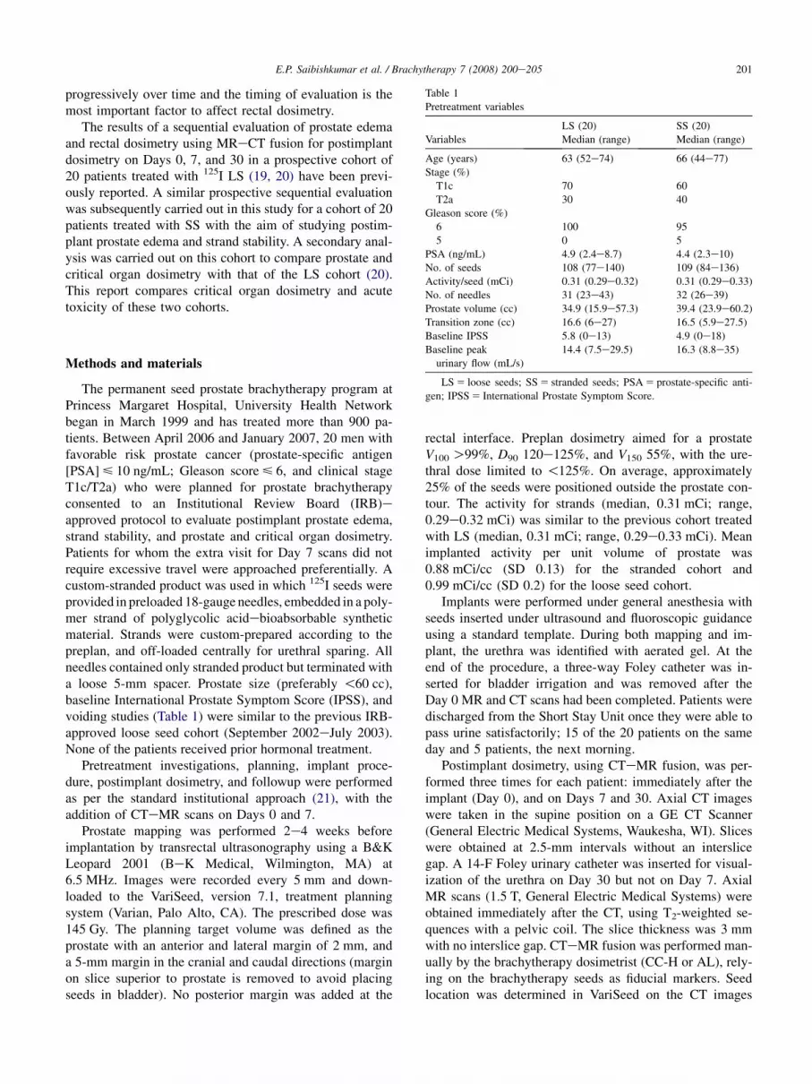
Table 1
Pretreatment variables
Variables
LS (20)
Median (range)
SS (20)
Median (range)
Age (years) 63 (52e74) 66 (44e77)
Stage (%)
T1c 70 60
T2a 30 40
Gleason score (%)
6 100 95
5 0 5
PSA (ng/mL) 4.9 (2.4e8.7) 4.4 (2.3e10)
No. of seeds 108 (77e140) 109 (84e136)
Activity/seed (mCi) 0.31 (0.29e0.32) 0.31 (0.29e0.33)
No. of needles 31 (23e43) 32 (26e39)
Prostate volume (cc) 34.9 (15.9e57.3) 39.4 (23.9e60.2)
Transition zone (cc) 16.6 (6e27) 16.5 (5.9e27.5)
Baseline IPSS 5.8 (0e13) 4.9 (0e18)
Baseline peak
urinary flow (mL/s)
14.4 (7.5e29.5) 16.3 (8.8e35)
LS 5 loose seeds; SS 5 stranded seeds; PSA 5 prostate-specific anti-
gen; IPSS 5 International Prostate Symptom Score.

202 E.P. Saibishkumar et al. / Brachytherapy 7 (2008) 200e205
and seed count was verified manually using orthogonal pel-vic X-rays. All relevant soft tissue structures were con-toured on the MR scans. Review of pree and postimplantcontouring, and the implant procedures was carried outby the same experienced physician (JC).
Critical organ contouring and dosimetry were performedas per the American Brachytherapy Society guidelines (22).The rectal wall was contoured on MR on all slices whereseeds were seen on Days 0, 7, and 30. Rectal wall dosime-try was compared for the two cohorts using RV100and RV150 (rectal wall volume in cc receiving 100% and150% of prescribed dose, respectively) and RD1 andRD2 cc (dose received by 1 and 2 cc of rectal wall, respec-tively). The urethra was identified using aerated gel on thepreplan ultrasound and by Foley catheter on Days 0 and 30CT scans and contoured from the bladder neck to the mem-branous urethra. Urethral dosimetry was described by UD5,UD30 (dose received in Gy by 5% and 30% of urethra, re-spectively), and UV150 (volume of urethra in cc receiving150% of prescribed dose).
Statistical analysis
Descriptive statistics were used for baseline characteris-tics of both cohorts. Rectal dosimetry at each time pointwas compared using two-sided unpaired Student’s t testwith equal variance. For urethral dosimetry, preplan andDay 30 values, and the percentage increase at Day 30 com-pared with preplan [100� (Day 30� preplan)/preplan]were compared using two-sided unpaired Student’s t testwith equal variance. No statistical comparisons were per-formed for acute toxicity, as the incidence was very low.
Results
Pretreatment characteristics were similar in the two co-horts (Table 1). Both the cohorts showed an increase in rec-tal wall dose with time, which may be explained by theresolution of prostatic and periprostatic edema (Table 2).At all time points (Days 0, 7, and 30), the mean edema fac-tor (percentage increase in prostate volume compared withpreimplant ultrasound volumes) was higher for LS com-pared with SS but did not reach statistical significance inthis small sample size: Day 0 (33.9% vs. 31.8%), Day 7(23.3% vs. 19.3%), and Day 30 (7.2% vs. 2.2%). All therectal dosimetry factors (RV100, RD1 cc, and RD2 cc) ex-cept RV150 were higher for the stranded cohort comparedwith the loose seed cohort at all time points and showed sta-tistical significance at all time points (Table 2). The meanRV100 at Day 30 for SS was 1.4 cc compared with0.73 cc for LS ( p 5 0.001) and the RD1 cc was 156.1 vs.129.4 Gy ( p 5 0.001), indicating rectal doses to be slightlyhigher than the desired levels for the stranded cohort (23).
To ensure that the higher rectal doses observed in thestranded cohort were not due to implant technique with
the seeds being positioned closer to the prostatic capsule,we reviewed the Day 30 scans for all patients in bothcohorts. We chose to review the Day 30 scans, rather thanDay 1 or 7 scans because the calculated rectal doses arehighest in both cohorts at Day 30. On the midprostate trans-verse slice, we measured the distance to the prostaticcapsule from the two most posterior paramedian seeds(c1 and d1). Despite the higher calculated rectal dose, theSS were actually positioned deeper within the prostate(mean, 2.25� 1.2 mm; variance, 1.5 mm) compared withLS (mean, 1.86� 1.6 mm; variance, 2.5 mm).
Similarly, we observed a significant increase in UD5,UD30, and UV150 for SS compared with the increase inthose for LS at Day 30 (Table 3). This occurred despitethe fact that the preplan values for urethral dose were sig-nificantly lower for the stranded cohort compared withthe loose seed cohort due to a deliberate attempt to limitthe urethral dose to 125% rather than 150% which was con-sidered acceptable until 2004. The percentage increases inUD5 and UD30 from preplan values were three timeshigher for SS than for LS (UD5: 24.6% vs. 7.7%; p 5 0.004and UD30: 15.9% vs. 5%; p 5 0.02, respectively). The ab-solute increase in UV150 from baseline to Day 30 was alsohigher for strands (Table 3).
The median followup for the loose seed cohort was 36months (range, 18e42) and only 6 months (range, 3e12)for the stranded cohort with 16 patients having followupO6 months. Accordingly, acute toxicity was evaluated forthe initial 6 months for both cohorts (Table 4). RTOGGrade 1 rectal toxicity was observed in 3 patients andGrade 2 toxicity in 1 patient in the stranded cohort, whereas
Table 2
Comparison of rectal dosimetry
Rectal dosimetry
parameters
LS (20)
Mean� SD
SS (20)
Mean� SD p-Value
RV100 (cc)
Day 0 0.2� 0.35 0.63� 0.38 0.0004
Day 7 0.4� 0.45 1� 0.7 0.003
Day 30 0.73� 0.5 1.4� 0.7 0.002
RV150 (cc)
Day 0 0.04� 0.15 0.05� 0.07 0.9
Day 7 0.06� 0.01 0.12� 0.16 0.3
Day 30 0.12� 0.19 0.17� 0.2 0.3
RD1 cc (Gy)
Day 0 90.2� 31.2 123.1� 22.9 0.0003
Day 7 104� 37.5 139.7� 28.6 0.002
Day 30 129.4� 32.7 156.1� 28.5 0.009
RD2 cc (Gy)
Day 0 58.8� 24.2 88.4� 23.3 0.0002
Day 7 68.9� 29.2 105.4� 30.4 0.001
Day 30 83.6� 23.4 117.7� 29.5 0.0005
LS 5 loose seeds; SS 5 stranded seeds; SD 5 standard deviation;
RV100 5 volume (cc) of rectal wall receiving 100% of prescribed dose;
RV150 5 volume (cc) of rectal wall receiving 150% of prescribed dose;
RD1 cc 5 dose received (Gy) to 1 cc of rectal wall; RD2 cc 5 dose
received (Gy) to 2 cc of rectal wall.

203E.P. Saibishkumar et al. / Brachytherapy 7 (2008) 200e205
only 1 patient in the loose seed cohort had RTOG Grade 1proctitis. There was no difference in the acute urinary tox-icity as recorded by the IPS scores at 1, 3, and 6 monthsbetween the cohorts. Two patients in each cohort developedacute urinary retention.
Although movement of seeds in the Xe and Y-axes (lat-eral and anteroposterior directions) was relatively restrictedin strands, we observed a differential movement of strandsin the Z-axis (craniocaudal). Further analysis is underwayto evaluate completely the relative seed positions andmovement over time for both cohorts.
Table 3
Comparison of urethral dosimetry
Urethral dosimetry
parameters
LS (20)
Mean� SD
SS (20)
Mean� SD p-Value
UD5 (Gy)
Preplan 196.4� 13.1 188.7� 6.6 0.03
Day 30 211.3� 26.4 234.1� 34.1 0.03
(Day 30 UD5�preplan)/preplan
7.7� 12.3% 24.6� 18.6% 0.004
UD5 (Gy)
Day 0 na 193.5� 24.8
UD30 (Gy)
Preplan 180.8� 9.7 175.4� 6.3 0.05
Day 30 190.4� 21.9 202.8� 25.7 0.0001
(Day 30 UD30�preplan)/preplan
5� 9.2% 15.9� 15.5% 0.02
UD30 (Gy)
Day 0 na 168.3� 13.5
UV150 (cc)
Preplan 0.01� 0.01 0.002� 0.004 0.03
Day 30 0.07� 0.1 0.2� 0.17 0.007
(Day 30 UV150�preplan)
0.06� 0.1 0.2� 0.17 0.005
UV150 (cc)
Day 0 na 0.04� 0.07
LS 5 loose seeds; SS 5 stranded seeds; SD 5 standard deviation;
UD5 5 dose (Gy) to 5% of urethra; UD30 5 dose (Gy) to 30% of urethra;
UV150 5 cc of urethra receiving 150% of prescribed dose; na 5 not avail-
able as urinary catheter was not used in Day 0 scan for loose seed cohort.
Table 4
Comparison of acute toxicity
Toxicity
LS (20)
Mean (range)
SS (20)
Mean (range)
Urinary IPSS
1 Month 15.2 (1e29) 12.4 (4e27)
3 Months 14.3 (0e27) 18.5 (4e30)
6 Months 11.4 (1e26) 8.6 (4e21)
Urinary retention 2 2
Rectal (RTOG)
Grade 1 1 3
Grade 2 0 1
LS 5 loose seeds; SS 5 stranded seeds; IPSS 5 International Prostate
Symptom Score; RTOG 5 Radiotherapy Oncology Group.
Discussion
Stranding of seeds has succeeded in almost eliminatingseed migration to the lungs (8e11), which has been a matterof concern for both patients and physicians in permanentprostate seed implants (1e7). The effect of stranding onprostate dosimetry is not consistent in the literature(11e16). A recent randomized trial comparing SS withLS did not demonstrate any difference (17).
Critical organ dosimetry is extremely important, as sideeffects and quality of life are major concerns in the treat-ment of favorable risk prostate cancer (11, 14, 16, 17).Fuller et al. (11) compared LS (125I or 103Pd) with SS usingDay 0 CTebased dosimetry and documented a slightlyhigher urethral D10 for strands (126.4 vs. 123.2 Gy); how-ever, urinary toxicity was not evaluated. Fagundes et al.(14) reported urinary retention rates and UV150 for 125ISS compared with 103Pd LS but the use of different isotopesprohibits comparison of loose vs. stranded format. Heyseket al. (16) compared LS (125I or 103Pd) with SS using CTscan at 3 weeks and did not observe any difference in uri-nary or rectal toxicity scores; dosimetry was not reported.In these retrospective studies (11, 14, 16), CT dosimetrywas performed at a single time point. The randomizedstudy comparing SS (125I) with LS by Reed et al. (17) usingsequential CTeMR fusion showed a trend toward higherRV100 with strands on Day 0 (0.81 vs. 0.56 cc), but byDay 30 the values were similar (1.91 vs. 1.96 cc). Toxicitywas not reported. The present study shows that dose to boththe rectal wall (Table 2) and urethra (Table 3) was signifi-cantly higher with strands at all time points evaluated. In-creased dose to the rectal wall seemed to translate toincreased acute rectal toxicity with strands despite shortfollowup (Table 4). No difference was noticed in the inci-dence or severity of urinary toxicity.
The main objective of our prospective protocol usingstrands was to evaluate prostate edema and spatial stabilityof strands, which is the subject of another manuscript. Se-quential scans provided additional opportunity to assesscritical organ dosimetry in detail and to compare this witha similar loose seed cohort treated earlier. The use ofCTeMR fusion offers an unparalleled advantage over CTin prostate definition (24) and the sequential evaluationstrack changes in dosimetry over the first month postim-plant. Although this is not a randomized comparison, thetwo cohorts have similar pretreatment characteristics, a con-sistent planning approach (with the one exception of the de-crease in preplan urethral dose from 150% in the earliercohort to 125% in the later stranded cohort) and implanttechnique, and the performance of all implants was by a sin-gle experienced physician. In addition, it should be notedthat the stranded cohort was entirely stranded, without theaddition of any LS, unlike many other reports (11, 14, 17).
The findings of higher rectal wall and urethral doseswere unexpected and the reasons for this remain specula-tive. Although the lesser degree of postimplant edema that

204 E.P. Saibishkumar et al. / Brachytherapy 7 (2008) 200e205
was observed with strands did not reach statistical signifi-cance, it may contribute to the higher central (urethral)and rectal doses. Although seed placement in the posteriorrows was actually slightly farther away from the prostaticcapsule in the stranded cohort compared with the loose seedcohort, there was more variance in the seed depth with LS.It may be that the slight variations in the depth and angle ofthe non-stranded seeds results in a lower rectal wall dose(Fig. 1). Some movement of entire strands was observedin the craniocaudal direction (Z-axis), which may be dueto the contractions of pelvic musculature as well as the factthat seeds in polymer stay well aligned and do not angulateor displace laterally. Strand kinking which would offer re-sistance to craniocaudal displacement was not observed.Movement caudally toward the genitourinary diaphragmcould increase dose to the membranous urethra and lowerrectal wall. This phenomenon was observed by McLaughlinet al. (25) on CTeMR fusion scans performed on Days0 and 14 in the evaluation of sequential changes in dosim-etry with strands. They also reported a significant increasein dose to the rectal wall and external sphincter with the Z-axis movement of strands. There was no comparison withLS in that study.
It is possible that SS encased in polymer evoke lessinflammation and mechanical trauma due to the greatlyreduced number of free foreign bodies introduced into theprostate. A smooth single strand to treat a 4-cm lengthhas only 2 ends, whereas the 4 LS and 4 spacers requiredto treat the same length have 8 loose bodies and 16 ends,all of which can irritate the prostate when muscular con-traction squeezes or deforms it. Thus strands may induceless prostatic and periprostatic edema, a factor that may in-crease urethral and rectal doses, respectively. If prostateedema is less, the central dose in the prostate will be higher.
-2.00
-1.00
0.00
1.00
2.00
3.00
4.00
5.00
6.00
1 3 5 7 9 11 13 15 17 19 21 23 25 27 29 31 33 35 37 39
Seeds ordered with increasing distance
Distan
ce to
p
ro
state cap
su
le [m
m]
loose seedsstranded
Fig. 1. Plot showing the distance of the two central seeds from prostate
capsule in Row 1 (c1 and d1) for loose seeds (LS) and stranded seeds
(SS). Y-axis shows the distance from prostate. Each point on the X-axis
represents the position of a seed with 2 seeds measured for each of the
20 patients. The 40 measurements in each group have been arranged in
ascending order. Note the increased variance in LS compared with SS.
Similarly, if the periprostatic edema at the prostateerectuminterface is less, the rectal wall is closer to the posterior rowof seeds, increasing the rectal dose. Mathematical modelingof the effect on the central urethral dose by the reducedprostatic edema seen with strands is being undertaken toquantify this as a contributing factor. Many implantapproaches that use strands continue to use LS in centrallylocated specially loaded needles which would decrease theedema-reducing effect of using strands throughout the entireimplant. An extensive analysis of individual seed positionsand movements in the sequential scans for both cohorts iscurrently underway and may elucidate our findings.
Although the increase seen in the urethral and rectaldose with the use of strands is statistically significant, theclinical significance is unknown. It is unlikely that thismagnitude of increase in urethral dose will make any differ-ence clinically (26). The early rectal toxicity observed is ofsome concern but followup is short. The increase in dose to1 cc of rectal wall from 129 Gy for the loose seed cohort to156 Gy for the stranded seed cohort may result in a slightincrease in the risk of late rectal toxicity, but one whichprobably will not be detectable in such a small sample size.Late toxicities and clinical outcomes will be reported andcompared when followup is more mature.
Strands continue to offer an undisputed advantage in essen-tially eliminating seed migration through the venous systemand we still preferentially choose to use SS in situations wherea high risk of seed loss is anticipated because of unusuallyclose proximity of periprostatic veins to the prostate observedat the time of mapping. In practices where strands are used forall cases, planning technique and placement of strands willrespond to the needs of postplan dosimetry, especially withrespect to keeping rectal doses within desired limits. Our ob-servation may be more relevant for those who implant somecases with LS and others with strands, as planning and tech-nique may not be entirely interchangeable between thesetwo physical formats. When strands are used it may be prudentto further distance them from the urethra and the posteriorcapsule of the prostate, to compensate for the higher observedrectal and urethral doses. The dosimetric consequences of thischange in planning need to be fully explored.
Conclusions
In this study, we observed an increase in rectal wall andurethral doses on sequential postimplant dosimetry usingCTeMR fusion with the use of strands of flexible polymercompared with LS. Acute rectal toxicity was higher in thestranded cohort despite urinary toxicity being similar.Further investigation of these observations is warranted.
References
[1] Steinfeld AD, Donahue BR, Plaine L. Pulmonary embolization of
iodine-125 seeds following prostate implantation. Urology 1991;37:
149e150.

205E.P. Saibishkumar et al. / Brachytherapy 7 (2008) 200e205
[2] Grimm PD, Blasko J, Ragde H, et al. Migration of iodine-125 and
palladium-103 seeds to the lung after transperineal brachytherapy
for prostate cancer. Endocurie/Hypertherm Oncol 1993;9:50.
[3] Nag S, Vivekanandam S, Martinez-Monge R. Pulmonary embolization
of permanently implanted radioactive palladium-103 seeds for carci-
noma of the prostate. Int J Radiat Oncol Biol Phys 1997;39:667e670.
[4] Older R, Snyder B, Krupski TL, et al. Radioactive implant migration
in patients treated for localized prostate cancer with interstitial bra-
chytherapy. J Urol 2001;165:1590e1592.
[5] Eshleman J, Davis B, Pisansky T, et al. Radioactive seed migration
to the chest after transperineal interstitial prostate brachytherapy:
Extraprostatic seed placement correlates with migration. Int J Radiat
Oncol Biol Phys 2004;59:419e425.
[6] Davis BJ, Pfeifer EA, Wilson TM, et al. Prostate brachytherapy seed
migration to the right ventricle found at autopsy following acute car-
diac dysrhythmia. J Urol 2000;164:1661.
[7] Davis B, Bresnahan J, Stafford S, et al. Prostate brachytherapy seed
migration to a coronary artery found during angiography. J Urol
2002;168:1103.
[8] Kumar PP, Good RR. Vicryl carrier for I-125 seeds: Percutaneous
transperineal insertion. Radiology 1986;159:276.
[9] Tapen EM, Blasko JC, Grimm PD, et al. Reduction of radioactive
seed embolization to the lung following prostate brachytherapy. Int
J Radiat Oncol Biol Phys 1998;42:1063e1067.
[10] Merrick GS, Butler WM, Dorsey AT, et al. Seed fixity in the
prostate/peri-prostatic region following brachytherapy. Int J Radiat
Oncol Biol Phys 1998;46:215e220.
[11] Fuller D, Koziol J, Feng AC. Prostate brachytherapy seed migration
and dosimetry: Analysis of stranded sources and other potential
predictive factors. Brachytherapy 2004;3:10e19.
[12] Lee R, deGuzman AF, Tomlinson S, et al. Radioactive sources em-
bedded in suture are associated with improved post-implant dosime-
try in men treated with prostate brachytherapy. Radiother Oncol
2002;65:123e127.
[13] Crook J, Pearson S, Williams T, et al. The pros and cons of linked vs.
loose seeds. Radiother Oncol 2003;69:157 (Abstract).
[14] Fagundes H, Keys R, Wojcik M, et al. Transperineal TRUS-guided
prostate brachytherapy using loose seeds versus RAPID strand: A
dosimetric analysis. Brachytherapy 2004;3:136e140.
[15] Kaplan I, Meskell P, Lieberfarb M, et al. A comparison of the preci-
sion of seeds deposited as loose seeds versus suture embedded seeds:
A randomized trial. Brachytherapy 2004;3:7e9.
[16] Heysek RV, Gwede CK, Torres-Roca J, et al. A dosimetric analysis of
unstranded seeds versus customized stranded seeds in transperineal
interstitial permanent prostate seed brachytherapy. Brachytherapy
2006;5:244e250.
[17] Reed DR, Wallner KE, Merrick GS, et al. A prospective randomized
comparison of stranded vs. loose 125I seeds for prostate brachyther-
apy. Brachytherapy 2007;6:129e134.
[18] Waterman FM, Dicker AP. Impact of post implant edema on V
(100) and D (90) in prostate brachytherapy: Can implant quality
be predicted on day 0? Int J Radiat Oncol Biol Phys 2002;53:
610e621.
[19] Taussky D, Austen L, Toi A, et al. Sequential evaluation of prostate
edema after permanent seed prostate brachytherapy using CT-MRI
fusion. Int J Radiat Oncol Biol Phys 2005;62:974e980.
[20] Taussky D, Yeung I, Williams T, et al. Rectal-wall dose dependence
on post plan timing after permanent-seed prostate brachytherapy. Int
J Radiat Oncol Biol Phys 2006;65:358e363.
[21] Crook J, Milosevic M, Catton P, et al. Interobserver variation in post-
implant computed tomography contouring affects quality assessment
of prostate brachytherapy. Brachytherapy 2002;1:66e73.
[22] Crook JM, Potters L, Stock RG, et al. Critical organ dosimetry in
permanent seed prostate brachytherapy: Defining the organs at risk.
Brachytherapy 2005;4:186e194.
[23] Snyder KM, Stock RG, Hong SM, et al. Defining the risk of develop-
ing grade 2 proctitis following 125I prostate brachytherapy using a
rectal dose-volume histogram analysis. Int J Radiat Oncol Biol Phys
2001;50:335e341.
[24] McLaughlin P, Narayana V, Kessler M, et al. Comparison of MRI
pulse sequences in defining prostate volume after permanent implan-
tation. Int J Radiat Oncol Biol Phys 2002;54:703e711.
[25] McLaughlin P, Narayana V, Pan C, et al. Comparison of day 0 and
day 14 dosimetry for permanent prostate implant using stranded
seeds. Int J Radiat Oncol Biol Phys 2005;64:144e150.
[26] Neill M, Studer G, Le L, et al. The nature and extent of urinary
morbidity in relation to prostate brachytherapy urethral dosimetry.
Brachytherapy 2007;6:173e179.


