Embed Size (px)
Citation preview

PHOTOSYNTHETICA 47 (4): 548-558, 2009
548
Comparison of thermostability of PSII between the chromatic and green leaf cultivars of Amaranthus tricolor L. Z. SHU1,4,*, L. SHAO2,*, H.-Y. HUANG5, X.-Q. ZENG1, Z.-F. LIN3, G.-Y. CHEN4, C.-L. PENG1,+ College of Life Science, Key Laboratory of Ecology and Environmental Science in Guangdong Higher Education, South China Normal University, Guangzhou 510631, China1
Department of Biology, Zhao Qing University, Zhaoqing 526061, China2
South China Botanical Garden, Chinese Academy of Sciences, Guangzhou 510650, China3 Institute of Plant Physiology and Ecology, Shanghai Institutes for Biological Sciences, Chinese Academy of Sciences, Shanghai 200032, China4 Department of Chemistry and Life science, Xiangnan College, Chenzhou,Hunan, 423000, China5 Abstract In the present study, we investigated the antioxidative potential in leaves of the chromatic (CC) versus green (GC) Amaranthus tricolor L. under moderate high-temperature stress at 45°C. Before heat stress, CC had significantly higher levels of betacyanins [about 3.2 mg g–1(FM)] than the green [1.8 mg g–1(FM) (p<0.01), while similar chlorophyll (Chl) content [about 2 mg g–1(FM)] was observed between both cultivars. After exposure to high temperature (45°C) for 6 days, betacyanins in leaves of CC were remarkably increased (about 2 times of that in control samples grown at 30°C). In contrast, betacyanins in GC significantly decreased by 56% in comparison with that of the control. Chl level in CC was higher than that in GC after heat stress for 6 days. Flavonoids and total phenolics in both cultivars were increased, but much more in CC. Significantly less H2O2 accumulation was observed in the leaves and stems of CC than in those of GC under heat stress. Interestingly, much stronger circadian oscillation in fluorescence was observed in both cultivars after treatment at 45°C, which suggested that heat stress stimulates endogenous rhythms of photosystem II (PSII). Under moderate high-temperature stress, Chl fluorescence parameters Fv/Fm (maximum quantum yield of PSII), qP (coefficient of photochemical quenching), ΦPSII (effective PSII quantum yield), and ETR (electron transport rate) exhibited a gradual decrease, NPQ (nonphotochemical quenching) showed a slight increase followed by a gradual decline, whereas Fo (minimum fluorescence of a dark-adapted leaf) increased continuously. In contrast to GC, after 120 h of high-temperature treatment, CC exhibited significantly lower Fo level, and higher levels of Fv/Fm and NPQ. It is clear that PSII in CC was more stable than that in GC. The results indicate that betacyanins are an effective antioxidant, and probably contribute greatly to the higher thermal stability of PSII and higher tolerance to heat stress. Additional key words: Amaranthus tricolor L.; betacyanins accumulation; circadian oscillation; high temperature; thermal stability of PSII. Introduction
Amaranthin, the characteristic pigment in the Amarantha-ceae, belongs to the group of betalain pigments that are most prevalent in plants of the order Centrospermae (Strack et al. 2003; Goodwin and Mercer 1983a; Goodwin and Mercer 1983b only one reference mentioned in References). Betalains are water-soluble
nitrogen-containing pigments, which comprises the red-violet betacyanins and the yellow betaxanthins. Betalains, analogous to anthocyanins, impart orange, red, purple, and bluish hues to flowers, grasses, fruits, vegetables, and grains. Anthocyanin synthesis can be induced or up-regulated by environmental stressors such as high
——— Received 15 May 2008, accepted 4 November 2009. +Corresponding author; fax: + 86-20-37252831, e-mail: [email protected] Abbreviations: CC – chromatic cultivar; ETR – electron transport rate; Fo – minimum fluorescence of a dark-adapted leaf; Fm – maximal fluorescence yield of a dark–adapted leaf; Fv/Fm – maximum quantum yield of PSII; FM – fresh mass; GC – green cultivar; MeOH – methanol; NPQ – nonphotochemical quenching; PAL – phenylalanine ammonia-lyase; PSII – photosystem II; qP – coefficient of photochemical quenching; ROS – reactive oxygen species; ΦPSII – effective PSII quantum yield. Acknowledgements: We thank Professor Fred Chow (Research School of Biology, the Australian National University) for critical reading of the manuscript and helpful suggestions. This research is financially supported by National Natural Science Foundation of China (30770173, 30870385), The State Key Basic Research and Development Plan of China (973 Program; 2009CB118504). *These authors contributed equally to this work.

THERMOSTABILITY OF PSII IN CHROMATIC VERSUS GREEN AMARANTHUS TRICOLOR
549
irradiance, extreme temperatures, UV radiation, and mechanical injury as well as resistance to pathogen and herbivory attack (Dakora 1995, Winkel-Shirley 2001). Anthocyanins have been proposed as a pigment screen contributing to shielding the photosynthetic apparatus from excess light and UV-radiation (Manetas et al. 2003); an antioxidant against harmful reactive oxygen species (ROS) (Gould et al. 2002, Neill and Gould 2003), and an inhibitor of lipid peroxidation (Tsuda et al. 1996). Our previous work suggested that anthocyanins play an important role in the protection against high temperature- and methyl viologen-induced (MV) photooxidation damage in leaves of Arabidopsis (Shao et al. 2007, 2008). How about the betalains with many similar characteristics to anthocyanins? Betalains have attracted most researchers in applied fields because of their use for food coloring and their antioxidant and radical scavenging properties for the protection against certain oxidative-stress-related disorders (Pedreno and Escribano 2000, Escribano et al. 1998, Kanner et al. 2001). Most reports focus on the extraction, chemical stability and structure, and medical value of betalains (Cai et al. 1998, Cai et al. 2001). To our best knowledge, there are few reports on physiological roles of betalains in leaves of plants under environmental stresses. Previous studies indicated that the synthesis of betalains can be induced or stimulated by low temperature (Wang and Liu 2007), darkness, high salinity (Wang et al. 2006), high UV radiation (Vogt et al. 1999), exogenous H2O2 treatment (Wang et al. 2007), and pathogen attack (Sepúlveda-Jiménez et al. 2004). Betalains are proposed as the screening pigments
that offer protection from UV-damage and pathogen attack, as an osmotic adjuster, as well as ROS-scavenger. However, still little is known about the physiological functions of betalains in plants under environmental stresses, in particular the heat one.
In the present study, we tried to find whether betalains have a physiological role under high-temperature stress. The levels of betacyanins, Chl, flavonoids and total phenolics, Chl fluorescence parameters and H2O2 accumulation in leaves of the chromactic and green cultivars of Amaranthus tricolor L. were compared under heat stress (45°C) in vivo. In Guangdong province, chromatic and green leafy cultivars of Amaranthus tricolor L. are common C4 vegetables. Leaves from CC are rich in betacyanins [about 3.2 mg g–1(FM)] during leaf expansion, particularly adjacent to the midrib, while green leaves show no appreciable betacyanins pigmentation [about 1.8 mg g–1(FM)] throughout all stages of leaf development. Interestingly, the chromatic and green leafy cultivars have similar Chl content [about 2 mg g–1(FM)]. Significant accumulation of betacyanins was observed under moderate high-temperature (45°C) stress. Our data indicated that PSII (photosystem II) in leaves of CC, rich in betacyanins, is remarkably more stable than that in the GC under moderate high-temperature stress (45°C). That is, CC of Amaranthus tricolor L. is more tolerant to heat stress than GC. To the best of our knowledge, this is the first report demon-strating the accumulation and antioxidative role of betacyanins in living plants under high-temperature stress. Our observation will provide new insights into the physiological functions of betacyanins in the protection against environmental stressors.
Materials and methods
Plants and high-temperature treatment: Seeds of CC and GC of Amaranthus tricolor L. were bought on a market in Guangzhou. Seeds were germinated in plastic pots with soil in August 2007 on the experimental field at South China Normal University, Guangzhou, China. After the 35-d growth, whole plants of each cultivar were put into the growth chamber (RXZ-500, Ningbo Jiangnan Instrument Factory, Ningbo, China) for incubation at the given temperature for 6 d. The treatment was performed at high-temperature (45°C) continuously, with a 12-h photoperiod (about 100 μmol m–2 s–1 PPFD), whereas plants grown at 30°C and the same light condition were taken as the control. The third and the fourth fully-expanded mature leaves (from the apex) from CC and GC were used for determinations. Chl a fluorescence measurements were performed every 4 h for 6 d, while quantifications of pigments, total phenolics and flavo-noids were measured every 2 d. Additionally, H2O2 histochemical localization in situ was measured at 0 d before treatment and 6 d after treatment, respectively.
Pigment quantification: Chl was extracted from leaf
pieces of 0.05 g of fresh mass (FM) taken from two sides near the midrib using 80% (v/v) acetone at 4°C in darkness. Concentrations were determined spectrophoto-metrically as described by Arnon (1949). Betacyanins were extracted and measured as described by Stintzing et al. (2002). 0.05 g of fresh leaves taken from two sides near the midrib was ground and pre-extracted with 10 ml MeOH for 30 min at 4°C. After centrifugation at 12,000 × g for 10 min at 4°C, the supernatant was dis-carded and the precipitate was re-extracted with double-distilled water. After centrifugation at 12,000 × g for 10 min at 4°C, the supernatant was taken for absorption measurement. The absorbance at 538 nm measured with a Visible-UV spectrophotometer (UV-2250, Shimadzu, Kyoto, Japan) was used to calculate the betacyanins content.
Total phenolics and flavonoids quantification: Flavo-noids and total phenolics were extracted from leaf pieces (0.05 g FM) for 48 h at 4°C in the dark in acidic methanol (1% HCl, v/v). The concentrations of flavonoids and total phenolics were estimated in methanol extracts from

Z. SHU et al.
550
absorbance at 325 nm and 280 nm, respectively (Fukumoto and Mazza 2000).
Chl a fluorescence parameters were determined using an IMAGING-PAM Chl fluorometer (Heinz Walz GmbH, Effeltrich, Germany). All lighting (modulated measuring light, actinic light and saturation pulses for measurement of Fm and Fm') were provided by blue light-emitting diodes (450 nm), and images were taken using a CCD camera. The third and the fourth fully-expanded mature leaves (from the apex) from the chromatic and green A. tricolor L. were selected for continuous Chl fluores-cence determinations for 212 h. Moreover, fluorescence signal was detected from the two adaxial sides adjacent to the midribs in both cultivars.
Fo was measured with relatively weak measuring light pulses of 0.5 μmol m–2 s–1 at a low frequency. Fm was measured during an 800-ms exposure to a PPFD of approximately 2,700 μmol m–2 s–1. The intensity of continuous actinic illumination was adjusted to 185 μmol m–2 s–1. All fluorescence measurements were started after an additional 10-min dark adaptation. The values and images of the Chl fluorescence parameters Fo (minimum fluorescence yield of dark-adapted leaf), Fm and Fm' (maximal fluorescence yield of dark-adapted and light-adapted leaf), Fv/Fm (maximal PSII quantum yield), ΦPSII (effective PSII quantum yield), NPQ (nonphotochemical quenching), qP (coefficient of photochemical quenching), were derived from the IMAGING-PAM software. There is a false color code bar shown in the given software, ranging from black via red, orange, yellow, green, blue, and violet to purple. These colors code for numbers between 0 and 1. Hence, all imaged parameters had to be normalized to values between 0 and 1 (conforming to a uniform false color scale). Among these fluorescence parameters, NPQ was normalized by dividing by 4, keeping values between 0–1. The apparent absorptivity of the leaf surface was automatically calculated pixel by pixel from the red (R)- and near infrared (NI)- images
using the formula: Abs = 1 – (R/NI). The definition of the ETR (apparent electron transport rate) assumes a uniform absorption of incident light over the whole sample area: ETR = yield × PAR × 0.5 × absorptivity, where yield = ΔF/Fm’ [ΔF = (Fm´ – Fs), where Fs is the steady state fluorescence] and PAR is the actinic light intensity, and 0.5 is 50% of the absorbed PAR distributed to PSII.
H2O2 localization in situ: H2O2 was detected in situ by diaminobenzidine (DAB) staining as described by Lin et al (2005). The leaf discs (diameter: 1.2 cm) were sampled from the fully expanded leaves of the chromatic and green A. tricolor L. Simultaneously, 2-cm-long stem sections were taken from the same plants as used for sampling leaf discs. Leafy discs and stem sections were immersed in a solution of DAB (1 mg/ml, Sigma) in 50 mM phosphate buffer (pH 7.0), and vacuum infiltrated for 10 min before incubation at room temperature for 8 h in the absence of light. Then they were illuminated until the appearance of brown spots specific to the reaction of DAB with H2O2. Samples were then bleached by immersion in boiling ethanol (75%, v/v) to visualize the brown spots and photographed. Stem sections and leaf discs of the same size and position from the chromatic and green cultivars grown at 30°C served as the control. H2O2 deposits in leaf discs were quantified by scanning spots only from leaf pictures and the number of pixels were quantified with the Adobe Photoshop 7.0.1 program (Adobe Systems Inc., San Jose, CA, USA). The results were expressed as the percentage of spots area versus total leaf area [(spot area/total leaf area)] × 100 as described by Romero-Puertas (2004). Statistical analysis: All of the data shown are means from five or six measurements. Statistical analysis is con-ducted by Origin 7.5 software (Microcal Corporation, Northampton, MA, USA). Values after ± show standard errors.
Results Levels of betacyanins and Chl were measured in leaves of two cultivars of Amaranthus tricolor L. (chromatic and green). Before high-temperature treatment, the betacyanin content in the leaves of CC was about 3.2 mg g–1(FM)] (Fig. 1A), which was significantly higher than that in GC [about 1.8 mg g–1(FM)] (p<0.01). After exposure to 45°C for 6 days (Fig. 1A), betacyanins in CC were markedly accumulated in the lower epidermis, bundle sheath cells and mesophyll layers of leaf laminae (photo not shown). In contrast, remarkable accumulation of betacyanins was not observed in the green leaf tissues (photo not shown). As to the different betacyanins levels and sequences of appearance in different tissues of the chromatic leaf laminae, we suppose that this might be attributed to the differential stress sensitivity of different leaf tissues (Kingston-Smith and Foyer 2000). Vital organs or tissues may be preserved in favor of more expendable
components such as the degradation of RuBP carboxylase and the loss of Chl (Baysdorfer et al. 1988). Hence, the lower leaf surface may be more sensitive to high-temperature stress. The extraction and quantity of betacyanins from CC (Fig. 1A) showed that betacyanins were significantly increased during 6-d high-temperature treatment, about 2 times as high as that in the control samples grown at 30°C. In contrast, betacyanins in GC decreased by about 56% under high-temperature treat-ment in comparison with the control. Betacyanins in the chromatic and green control samples (grown at 30°C) did not exhibit any remarkable change during the 6–d treat-ment [about 3.2 mg g–1(FM) in CC, while about 1.6 mg g–1(FM) in GC]. Based on the observation of tissue localization (photos not shown) and quantification of betacyanins (Fig. 1A), it is very clear that betacyanins in the chromatic leaves of A. tricolor L. were significantly

THERMOSTABILITY OF PSII IN CHROMATIC VERSUS GREEN AMARANTHUS TRICOLOR
551
Fig. 1. Changes in contents of betacyanins (A) and chlorophyll (Chl) (B) in leaves of the chromatic(CC) and green (GC) cultivars of Amaranthus tricolor L. under high-temperature stress. Beta-cyanins from methanol extract and chlorophyll in 80% (v/v) acetone were determined spectrophoto-metrically. Data show the means and standard errorof five independent repetitions.
Fig. 2. Effects of high-temperature on contents of flavonoids (A) and total phenolics (B) in leaves of the chromatic (CC) and green (GC) cultivars of Amaranthus tricolor L. Total phenolics and flavonoids contents were estimated in methanol extract from the absorbance at 280 nm and 325 nm, respectively. Data show the means and standard error of five independent repetitions.
accumulated under moderate high-temperature treatment (45°C).
Chl levels in CC and GC were similar at 0 day before treatment [2.05±0.13; 2.07±0.14 mg g–1(FM), respecti-vely] (Fig. 1B). With the progress of high-temperature treatment, Chl in the two cultivars decreased, which suggested that high-temperature had induced damage to both cultivars. However, in comparison with that of 0 d before treatment, Chl in CC showed a relatively smaller decrease (about 57% of the control) than that in GC (about 31% of the control) after heat stress for 6 d
(Fig. 1B). Chl in CC was about 1.6 times of that in GC at 6 d under heat stress. Compared to the control samples, the enhancement of accumulation of betacyanins and relatively less bleaching of Chl caused by high-temperature treatment in CC, suggested that the presence of betacyanins might protect against Chl degradation under heat stress.
Fig. 2 showed changes in the content of flavonoids and total phenolics in leaves of CC and GC under heat stress. Before treatment, flavonoids and total phenolics in GC were significantly more abundant than those in CC
Z. SHU et al.
552
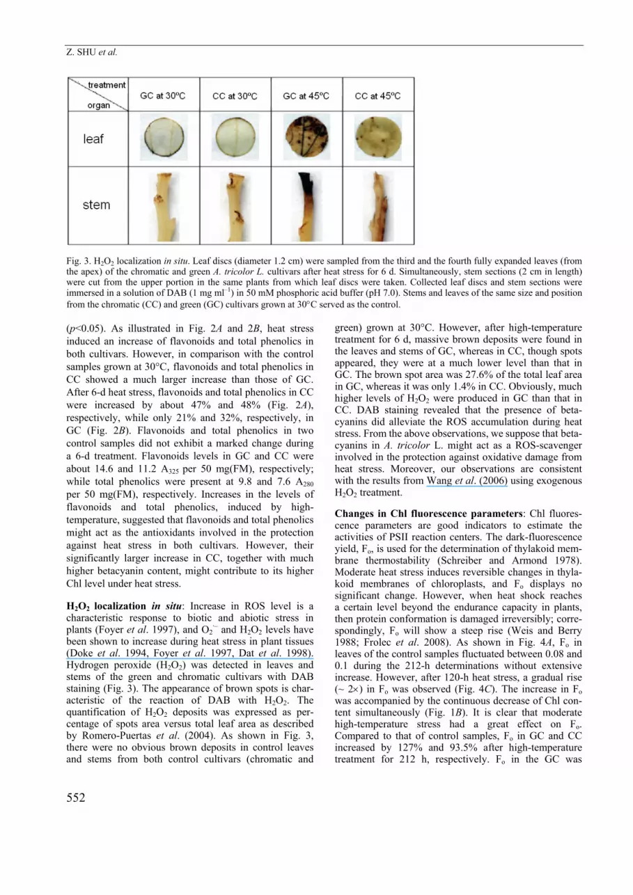
Fig. 3. H2O2 localization in situ. Leaf discs (diameter 1.2 cm) were sampled from the third and the fourth fully expanded leaves (from the apex) of the chromatic and green A. tricolor L. cultivars after heat stress for 6 d. Simultaneously, stem sections (2 cm in length) were cut from the upper portion in the same plants from which leaf discs were taken. Collected leaf discs and stem sections were immersed in a solution of DAB (1 mg ml–1) in 50 mM phosphoric acid buffer (pH 7.0). Stems and leaves of the same size and position from the chromatic (CC) and green (GC) cultivars grown at 30°C served as the control. (p<0.05). As illustrated in Fig. 2A and 2B, heat stress induced an increase of flavonoids and total phenolics in both cultivars. However, in comparison with the control samples grown at 30°C, flavonoids and total phenolics in CC showed a much larger increase than those of GC. After 6-d heat stress, flavonoids and total phenolics in CC were increased by about 47% and 48% (Fig. 2A), respectively, while only 21% and 32%, respectively, in GC (Fig. 2B). Flavonoids and total phenolics in two control samples did not exhibit a marked change during a 6-d treatment. Flavonoids levels in GC and CC were about 14.6 and 11.2 A325 per 50 mg(FM), respectively; while total phenolics were present at 9.8 and 7.6 A280 per 50 mg(FM), respectively. Increases in the levels of flavonoids and total phenolics, induced by high-temperature, suggested that flavonoids and total phenolics might act as the antioxidants involved in the protection against heat stress in both cultivars. However, their significantly larger increase in CC, together with much higher betacyanin content, might contribute to its higher Chl level under heat stress.
H2O2 localization in situ: Increase in ROS level is a characteristic response to biotic and abiotic stress in plants (Foyer et al. 1997), and O2
·– and H2O2 levels have been shown to increase during heat stress in plant tissues (Doke et al. 1994, Foyer et al. 1997, Dat et al. 1998). Hydrogen peroxide (H2O2) was detected in leaves and stems of the green and chromatic cultivars with DAB staining (Fig. 3). The appearance of brown spots is char-acteristic of the reaction of DAB with H2O2. The quantification of H2O2 deposits was expressed as per-centage of spots area versus total leaf area as described by Romero-Puertas et al. (2004). As shown in Fig. 3, there were no obvious brown deposits in control leaves and stems from both control cultivars (chromatic and
green) grown at 30°C. However, after high-temperature treatment for 6 d, massive brown deposits were found in the leaves and stems of GC, whereas in CC, though spots appeared, they were at a much lower level than that in GC. The brown spot area was 27.6% of the total leaf area in GC, whereas it was only 1.4% in CC. Obviously, much higher levels of H2O2 were produced in GC than that in CC. DAB staining revealed that the presence of beta-cyanins did alleviate the ROS accumulation during heat stress. From the above observations, we suppose that beta-cyanins in A. tricolor L. might act as a ROS-scavenger involved in the protection against oxidative damage from heat stress. Moreover, our observations are consistent with the results from Wang et al. (2006) using exogenous H2O2 treatment.
Changes in Chl fluorescence parameters: Chl fluores-cence parameters are good indicators to estimate the activities of PSII reaction centers. The dark-fluorescence yield, Fo, is used for the determination of thylakoid mem-brane thermostability (Schreiber and Armond 1978). Moderate heat stress induces reversible changes in thyla-koid membranes of chloroplasts, and Fo displays no significant change. However, when heat shock reaches a certain level beyond the endurance capacity in plants, then protein conformation is damaged irreversibly; corre-spondingly, Fo will show a steep rise (Weis and Berry 1988; Frolec et al. 2008). As shown in Fig. 4A, Fo in leaves of the control samples fluctuated between 0.08 and 0.1 during the 212-h determinations without extensive increase. However, after 120-h heat stress, a gradual rise (~ 2×) in Fo was observed (Fig. 4C). The increase in Fo was accompanied by the continuous decrease of Chl con-tent simultaneously (Fig. 1B). It is clear that moderate high-temperature stress had a great effect on Fo. Compared to that of control samples, Fo in GC and CC increased by 127% and 93.5% after high-temperature treatment for 212 h, respectively. Fo in the GC was

THERMOSTABILITY OF PSII IN CHROMATIC VERSUS GREEN AMARANTHUS TRICOLOR
553
Fig. 4. Effects of high-temperature stress on Fo (A,C) and Fv/Fm (B,D). Data are presented as means (n = 6). Whole seedlings of the chromatic (CC) and (GC) green leafy cultivars of Amaranthus tricolor L. were treated at continuous high-temperature (45°C) for 6 d, while samples grown at 30°C were taken as the control. The third and the fourth fully-expanded mature leaves were used for the 212-h determinations of chlorophyll fluorescence.
Fig. 5 Effects of high-temperature stress on qP (A,C) and NPQ/4 (B,D). Data are presented as means (n = 6). Whole seedlings of the chromatic (CC) and (GC) green leafy cultivars of Amaranthus tricolor L. were treated at continuous high-temperature (45°C) for 6 d, while samples grown at 30°C was taken as the control. The third and the fourth fully-expanded mature leaves were used for the 212 h-determinations of chlorophyll fluorescence.
Z. SHU et al.
554
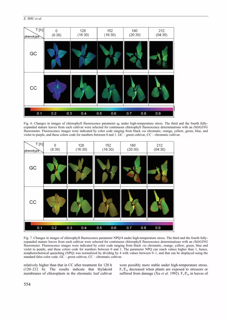
Fig. 6. Changes in images of chlorophyll fluorescence parameter qP under high-temperature stress. The third and the fourth fully-expanded mature leaves from each cultivar were selected for continuous chlorophyll fluorescence determinations with an IMAGING fluorometer. Fluorescence images were indicated by color code ranging from black via chromatic, orange, yellow, green, blue, and violet to purple, and these colors code for numbers between 0 and 1. GC – green cultivar, CC – chromatic cultivar.
Fig. 7. Changes in images of chlorophyll fluorescence parameter NPQ/4 under high-temperature stress. The third and the fourth fully-expanded mature leaves from each cultivar were selected for continuous chlorophyll fluorescence determinations with an IMAGING fluorometer. Fluorescence images were indicated by color code ranging from black via chromatic, orange, yellow, green, blue and violet to purple, and these colors code for numbers between 0 and 1. The parameter NPQ can reach values higher than 1, hence, nonphotochemical quenching (NPQ) was normalized by dividing by 4 with values between 0–1, and that can be displayed using the standard false color code. GC – green cultivar, CC – chromatic cultivar. relatively higher than that in CC after treatment for 120 h (120–212 h). The results indicate that thylakoid membranes of chloroplasts in the chromatic leaf cultivar
were possibly more stable under high-temperature stress. Fv/Fm decreased when plants are exposed to stressors or suffered from damage (Xu et al. 1992). Fv/Fm in leaves of
THERMOSTABILITY OF PSII IN CHROMATIC VERSUS GREEN AMARANTHUS TRICOLOR
555
CC and GC grown at 30°C remained at about 0.7 during 212-h measurements, and both cultivars did not show a significant difference in Fv/Fm (Fig. 4B). However, upon high-temperature treatment, Fv/Fm in leaves of the two cultivars decreased continuously (Fig. 4D), which indicated a large alteration of PSII function with prolonging heat stress. Compared to that of the control samples, Fv/Fm in leaves of GC and CC decreased by 51% and 37.9%, respectively, after high-temperature treatment for 212 h. A significant difference in Fv/Fm was observed between two cultivars after high-temperature treatment (p<0.05); CC exhibited more stable PSII under heat stress.
qP indicates the concentration of open PSII traps ready to perform charge stabilization; NPQ has been shown to be a good indicator of the potential to dissipate excess excitation energy via heat in plant (Bilger et al. 1995). The regular decrease and increase of qP during light and dark periods upon high-temperature treatment was accompanied by the opposite changes of nonphoto-chemical quenching expressed by NPQ simultaneously (Fig. 5A,B,C,D). 212-h treatment at 45°C caused a gradual decrease of qP, which reflects the decrease of the proportions of open PSII centers (Fig. 5C). Moreover, qP in chromatic leaves was relatively higher than that in the green ones from 120-144 h. NPQ showed a different pattern: an initial increase (from 0 h to 72 h), and then a slight decrease (Fig. 5D). Changes in NPQ revealed dynamically regulated capability in plants, and plants
employed heat dissipation to resist high-temperature stress. Upon high-temperature treatment for 120-212 h, NPQ in chromatic leaves was relatively higher than that in the green ones. Changes in qP and NPQ indicated that PSII in CC had stronger thermal stability than that in GC.
The third and the fourth fully-expanded mature leaves from each cultivar were selected for continuous Chl fluorescence determinations with an IMAGING fluoro-meter. Fluorescence imaging can reflect the spatio-temporal heterogeneity of photosynthesis. Images of qP and NPQ/4 are illustrated as Fig. 6 and 7. Different image colors correspond to the different numerical values. Upon high-temperature treatment for 212 h, image color of qP in leaves of CC changes from blue (value for 3rd and 4th leaves is about 0.8) to green (about 0.4), whereas qP in GC changes from blue (about 0.8) to a pale green color (about 0.28) and quite a lot of black (qP = 0). Correspondingly the value of qP in CC was significantly higher than that in GC as shown in Fig. 5C. The image color of NPQ/4 in the leaves of GC faded from green (about 0.3) to CC with slight green color (about 0.2) after 212-h high-temperature stress. Colors in fluorescence images are dominated by the chromatic and orange. However, in comparison with the green, the image color of NPQ/4 in leaves of CC showed a slight change, with a large part of green color after exposure to the high-temperature treatment for 212 h (about 0.29).
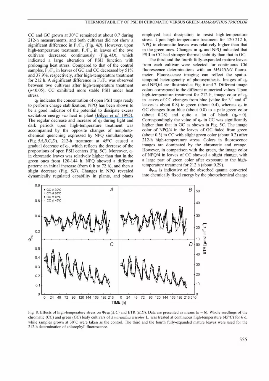
ΦPSII is indicative of the absorbed quanta converted into chemically fixed energy by the photochemical charge
Fig. 8. Effects of high-temperature stress on ΦPSII (A,C) and ETR (B,D). Data are presented as means (n = 6). Whole seedlings of the chromatic (CC) and green (GC) leafy cultivars of Amaranthus tricolor L. was treated at continuous high-temperature (45°C) for 6 d, while samples grown at 30°C were taken as the control. The third and the fourth fully-expanded mature leaves were used for the 212-h determination of chlorophyll fluorescence.

Z. SHU et al.
556
separation at PSII reaction centers. ETR in PSII is indicative of the activity of PSII (Genty et al. 1989). Upon high-temperature treatment, ΦPSII and ETR both showed the same decreasing tendency (Fig. 8C,D). In comparison with the control samples at 30°C (Fig. 8A,B), ΦPSII in GC and CC decreased by about 75% and 50%, re-spectively, after 212 h of high-temperature treatment; and ETR in GC and CC showed about the same decreasing amplitudes to ΦPSII (75% and 50%, respectively). The larger decrease in amplitudes of ΦPSII and ETR in GC suggests that PSII in CC was more stable when exposed to heat stress. Interestingly, much stronger diurnal oscillations in the Chl fluorescence parameters (Fo, qP,
NPQ, ΦPSII and ETR) in leaves of two cultivars were observed when incubated at 45°C compared to that of control samples. The results from Kouřil et al. (2004) pointed out that heat-induced fluorescence rise usually appeared at around 40–50°C, and the fluorescence rise was caused by the reduction of QA, which had been affected by several processes. In the present study, 45°C was used for heat stress. Much stronger diurnal oscillations in the Chl fluorescence parameters in both cultivars were observed under heat stress at 45°C than in the control samples at 30°C. This result is consistent with a previous report (Kouřil et al. 2004).
Discussion A. tricolor L. employs a C4 photosynthetic pathway. Lin et al. (1983) report that its optimum photosynthetic temperature is about 35°C. Although C4 plants have a higher optimum temperature than C3 plants, PN is usually inhibited when leaf temperature exceeds about 38°C (Berry and Björkman 1980, Edwards and Walker 1983). Thus 45°C used in this experiment is considered to be moderate heat stress for the growth of A. tricolor L. Photosynthesis is extremely susceptible to the inhibition caused by moderate heat stress in plants (Berry and Björkman 1980). Inhibition of photosynthesis by heat stress has been long attributed to an impairment of electron transport activity. Excess electrons are easily leaked to O2, resulting in the production of ROS. When the production exceeds the scavenging of ROS in plants, accumulated ROS will induce an oxidative damage to proteins and membrane lipids, and even the integrity of biological membranes (Asada 1992, Elstner et al. 1988).
Accumulation of betacyanins in the chromatic leaf cultivar under moderate high-temperature was accompanied by restriction of H2O2 production: After high-temperature treatment for 6 d, distribution of betacyanins in leaf tissues of CC was obviously enlarged, from the lower epidermis to the upper epidermis (photo not shown); accordingly betacyanins in chromatic leaves were increased from 3.2 mg g–1(FM) at 0 d before treatment to 6.4 mg g–1(FM) after 6 d(Fig. 1A), about 2 times as high as that before treatment. Moreover, compared to that of the control grown at 30°C, the stem in CC also was much redder after high-temperature treatment for 6 d (photo not shown). To our best knowledge, it is the first time that remarkable betacyanins accumulation in the leaf and stem of A. tricolor L. induced by moderate high-temperature stress has been reported.
The significant increase in the levels of betacyanins, flavonoids and total phenolics in both cultivars under heat stress (Figs 1A, 2), suggested that they might play a protective role in the response to heat stress. The obvious accumulation of betacyanins and much larger increases of flavonoids and total phenolics, together with less Chl degradation and much less H2O2 accumulation in the
leaves of CC, indicated that CC had much stronger antioxidative capability than GC under heat stress; and betacyanins might play an important role in the protection against oxidative damage induced by high-temperature. Betacyanins may function as a ROS scavenger alleviating oxidative damage induced by high-temperature. This result is in agreement with a previous report under low temperature by Wang et al. (2006).
PSII function in CC is more tolerant to moderate high-temperature than that in GC: Circadian oscillations in fluorescence are observed in control samples grown at 30°C and samples treated at the high-temperature of 45°C. Chl fluorescence parameters (Fo, qP, NPQ, ΦPSII and ETR) in two leafy cultivars oscillated with an approximate period of 24 h. However, oscillation in fluorescence parameters under high-temperature treatment was obviously intensified, which suggested that high-temperature might stimulate endogenous rhythms of PSII in plants. The result is consistent with fluorescence rise induced by heat around 40–50°C (Kouřil et al. 2004). With the prolongation of high-temperature stress, oscillations in ΦPSII and ETR were damped, and the mean values decreased, while the amplitude of Fo oscillations increased. Similarly, heat stress suppressed the activity of RuBP carboxylase and CO2 assimilation, thus tending to impair electron transport activity of PSII. A proportion of the photosynthetic electron flow was diverted to O2 mediated by Mehler reaction or by a violaxanthin cycle (Demmig et al. 1987), which can be illustrated by H2O2 production and the increase of NPQ. However, with the prolongation of treatment time, thylakoid membranes of chloroplasts and PSII reaction centers were damaged severely; therefore, the effective PSII quantum yield decreased and Fo increased.
Chl fluorescence parameters are good indicators reflecting physiological conditions in PSII in vivo. The observed results suggest that PSII function in CC was markedly more stable than that in GC. A significant increase in Fo under high-temperature treatment indicates that thylakoid membranes of the chloroplast were damaged severely. In comparison with that of the control samples at 30°C, Fo in GC increased by 127% after

THERMOSTABILITY OF PSII IN CHROMATIC VERSUS GREEN AMARANTHUS TRICOLOR
557
treatment for 212 h, significantly higher than that in CC (93.5%). The data indicate that damage to membranes in GC is more severe than that in CC. These results are consistent with the lower Chl content and higher production of H2O2 in GC as described. Data presented here indicate that betacyanins in CC were involved in the protection against high-temperature stress, mitigating the degradation of Chl and production of ROS. Other Chl parameters (Fv/Fm, ΦPSII, qP, and ETR) showed the same decreasing tendency under high-temperature treatment, whereas NPQ with an initial increase followed by a continuous decrease. Decrease in the levels of Fv/Fm, ΦPSII, qP, ETR and NPQ suggest that PSII was damaged severely. In contrast with that of the control samples, after high-temperature treatment for 212 h, Fv/Fm, ΦPSII,
and ETR in GC decreased by much larger magnitudes than that of in CC. Significantly lower decreasing amplitudes, together with lower level of Fo in the levels of the chromatic clearly suggest that PSII function in CC was remarkably more stable than that in the green one. It is related to higher level of betacyanins. Data from the present study indicate that betacyanins in leaves of A. tricolor L. are effective antioxidants, together with other nonenzymatic antioxidants (such as flavonoids and total phenolics, etc.) and enzymatic antioxidants (such as SOD, superoxide dismutase; APX, ascorbate peroxidase; CAT, catalase; etc.) (Foyer et al. 1994), are involved in scavenging ROS in vivo, protecting photosynthetic apparatus, thus mitigating heat damage.
References Arnon, D.I.: Copper enzymes in isolated chloroplasts – poly-
fenoloxidase in Beta valgaris. – Plant Physiol. 24: 1-15, 1949. Asada, K.: Ascorbate peroxidase - a hydrogen peroxide
scavenging enzyme in plants. – Physiol. Plant. 85: 235-241, 1992.
Baysdorfer, C., Warmbrodt, R.D., Van Der Woude, W.J.: Mechanisms of starvation tolerance in pearl-millet. – Plant Physiol. 88: 1381-1387, 1988.
Berry, J.A., Björkman, O.: Photosynthetic response and adaptation to temperature in higher-plants. – Annu. Rev. Plant Physiol. 31: 491-543, 1980.
Bilger, W., Fisahn, J., Brummet, W., Kossmann, J., Willmitzer, L.: Violaxanthin cycle pigment contents in potato and tobacco plants with genetically reduced photosynthetic capacity. – Plant Physiol. 108: 1479-1486, 1995.
Cai, Y.Z., Sun, M., Corke, H.: Colorant properties and stability of Amaranthus betacyanin pigments. – J. Agric. Food Chem. 46: 4491-4495, 1998.
Cai, Y.Z., Sun, M., Corke, H.: Identification and distribution of simple and acylated betacyanins in the Amaranthaceae. – J. Agric. Food Chem. 49: 1971-1978, 2001.
Dakora, F.D.: Plant flavonoid - biological molecules for useful exploitation. – Aust. J. Plant Physiol. 22: 87-99, 1995.
Dat, J.F., Lopez-Delgado, H., Foyer, C.H., Scott, I.M.: Parallel changes in H2O2 and catalase during thermotolerance induced by salicylic acid or heat acclimation in mustard seedlings. – Plant Physiol. 116: 1351-1357, 1998.
Demmig, B., Winter, K., Krüger, A., Czygan, F.C.: Photoinhibition and zeaxanthin formation in intact leaves - a possible role of the xanthophylls cycle in the dissipation of excess light energy. – Plant Physiol. 84: 218-224, 1987.
Doke, N., Miura, Y., Leandro, M.S., Kawakita, K.: Involvement of superoxide in signal transduction: responses to attack by pathogens, physical and chemical shocks, and UV irradiation. – In: Foyer, C.H., Mullineaux, P.M. (ed.): Causes of Photooxidative Stress and Amelioration of Defence Systems in Plants. Pp 177-197. CRC Press, Boca Raton, FL 1994.
Edwards, G.E, Walker, D.A.: C3, C4: Mechanisms and Cellular and Environmental Regulation of Photosynthesis. – Univ. California Press, Berkeley 1983.
Elstner, E.F., Wagner, G.A., Schutz, W.: Activated oxygen in green plants in relation to stress situations. – In: Randall, D.D., Blevis, D.G.,Campbell, W.H. (ed.): Current Topics in Plant Biochemistry and Physiology. Pp. 159-187. Vol. 7. Univ. Missouri, Columbia 1988.
Escribano, J., Pedreno, M.A., Garcia-Carmona, F., Munoz, R.: Characterization of the antiradical activity of betalains from Beta vulgaris L. roots. – Phytochem. Analysis 9: 124-127, 1998.
Foyer, C.H., Leiandais, M., Kunert, K.J.: Photooxidative stress in plants. – Physiol. Plant. 92: 696-717, 1994.
Foyer, C.H., Lopez-Delgado, H., Dat, J.F., Scott, I.M.: Hydrogen peroxide- and glutathione-associated mechanisms of acclimatory stress tolerance and signaling. – Physiol. Plant. 100: 241-254, 1997.
Frolec, J., Ilík, P., Krchňák, P., Sušila, P., Nauš, J.: Irreversible changes in barley leaf chlorophyll fluorescence detected by the fluorescence temperature curve in a linear heating/cooling regime. – Photosynthetica 46: 537-546, 2008
Fukumoto, L.R., Mazza, G.: Assessing antioxidant and prooxidant activities of phenolic compounds. – J. Agric. Food Chem. 48: 3597-3604, 2000.
Genty, B., Briantais, J.M., Baker, N.R.: Relationship between the quantum yield of photosynthetic electron-transport and the quenching of chlorophyll fluorescence. – Biochim. Biophys. Acta. 990: 87-92, 1989.
Goodwin, T.W. and Mercer, E.I.: Introduction to Plant Biochemistry. Vol. 1. Pergamon, Oxford 1983a.
Goodwin, T.W. and Mercer, E.I.: Introduction to Plant Biochemistry. Vol. 2. Pergamon, Oxford 1983b.
Gould, K.S., McKelvie, J., Markham, K.R.: Do anthocyanins function as antioxidants in leaves: imaging of H2O2 in red and green leaves after mechanical injury. – Plant Cell Environ. 25: 1261-1269, 2002.
Kanner, J., Harel, S., Granit, R.: Betalains - A new class of dietary cationized antioxidants. – J. Agric. Food Chem. 49: 5178-5185, 2001.
Kingston-Smith, A.H., Foyer C.H.: Bundle sheath proteins are more sensitive to oxidative damage than those of the mesophyll in maize leaves exposed to paraquat or low temperature. – J. Exp. Bot. 51: 123-130, 2000.
Kouřil, R., Lazár, D., Ilík, P., Skotnica, J., Krchňák, P., Nauš, J.: High-temperature induced chlorophyll fluorescence rise in plants at 40–50 °C: experimental and theoretical approach.- Photosynth. Res. 81: 49–66, 2004.
Lin, Z.F., Ehleringer, J.: Photosynthesis characteristics of Amaranthus tricolor, a C4 tropical leafy vegetable. – Photosynth. Res. 4: 171-178, 1983.
Lin, Z.F., Peng, C.L., Xu, X.L., Lin, G.Z., Zhang, J.L.: Thermostability of photosynthesis in two new chlorophyll b-

Z. SHU et al.
558
less rice mutants. – Sci. China C 48: 139-147, 2005. Manetas, Y., Petropoulou, Y., Psaras, G.K., Drinia, A.: Exposed
red (anthocyanic) leaves of Quercus coccifera display shade characteristics. – Funct. Plant Biol. 30: 265-270, 2003.
Neill, S.O., Gould, K.S.: Anthocyanins in leaves: light attenua-tors or antioxidants? – Funct. Plant Biol. 30: 865-873, 2003.
Pedreno, M.A., Escribano, J.: Studying the oxidation and the antiradical activity of betalain from beetroot. – J. Biol. Educ. 35: 49-51, 2000.
Romero-Puertas, M.C., Rodriguez-Serrano, M., Corpas, F.J., Gomez, M., Del Rio, L.A., Sandalio, L.M.: Cadmium-induced subcellular accumulation of O2
.– and H2O2 in pea leaves. – Plant Cell Environ. 27: 1122-1134, 2004.
Schreiber, U., Armond, P.A.: Heat-induced changes of chlorophyll fluorescence in isolated chloroplasts and related heat-damage at the pigment level. – Biochim. Biophys. Acta. 502: 138-151, 1978.
Sepúlveda-Jiménez, G., Rueda-Benítez, P., Porta, H., and Rocha-Sosa, M: Betacyanin synthesis in red beet (Beta vulgaris) leaves induced by wounding and bacterial infiltration is preceded by an oxidative burst. – Physiol. Mol. Plant Pathol. 64: 125-133, 2004.
Shao, L., Shu, Z., Peng, C.-L., Lin, Z.-F., Yang, C.-W., Gu, Q.: Enhanced sensitivity of Arabidopsis anthocyanin mutants to photooxidation: a study with fluorescence imaging. – Funct. Plant Biol. 35: 714-724, 2008.
Shao, L., Shu, Z., Sun, S.L., Peng, C.L., Wang, X.J., Lin, Z.F.: Antioxidation of Anthocyanins in photosynthesis under high temperature stress. – J. Integr. Plant Biol. 49: 1341-1351, 2007.
Stintzing, F.C., Schieber, A., Carle, R.: Betacyanins in fruits from red-purple pitaya, Hylocereus polyrhizus (Weber)
Britton & Rose. - Food Chem. 77: 101-106, 2002. Strack, D., Vogt, T., Schliemann, W.: Recent advances in
betalain research. – Phytochem. 62: 247-269, 2003. Tsuda, T., Shiga, K., Ohshima, K., Kawakishi, S., Osawa, T.:
Inhibition of lipid peroxidation and the active oxygen radical scavenging effect of anthocyanin pigment isolated from Phaseolus vulgaris L. – Biochem. Pharmacol. 52: 1033-1039, 1996.
Vogt, T., Ibdah, M., Schmidt, J., Wray, V., Nimtz, M., Strack, D.: Light-induced betacyanin and flavonol accumulation in bladder cells of Mesembryanthemum crystallinum. – Phytochemistry 52: 583-592, 1999.
Wang, C.-Q., Liu,, T.: Involvement of betacyanin in chilling-induced photoinhibition in leaves of Suaeda salsa, – Photosynthetica 45: 182-188, 2007.
Wang, C.Q., Zhao, J.Q., Chen, M., Wang, B.S.: Identification of betacyanin and effects of environmental factors on its accumulation in halophyte Suaeda salsa. – J. Plant Physiol. Mol. Biol. 31: 195-201, 2006.
Wang, C.Q., Chen, M., Wang, B.S.: Betacyanin accumulation in the leaves of C3 halophyte Suaeda salsa L. is induced by watering roots with H2O2. – Plant Sci. 172: 1-7, 2007.
Weis, E., Berry, J.A.: Plant and high temperature stress. – In: Long, S.P., Woodward, F.I. (ed.): Plant and Temperature. Pp. 329-346. The Company of Biologist, Cambridge 1988.
Winkel-Shirley, B.: Flavonoid biosynthesis. A colorful model for genetics, biochemistry, cell biology, and biotechnology. – Plant Physiol. 126: 485-493, 2001.
Xu Da-Quan, Zhang Yu-Zhong, Zhang Rong-Xian: [Photo-inhibition of photosynthesis in plants.] – Plant Physiol. Commun. 28: 237-243, 1992. [in Chin.]


