Embed Size (px)
Citation preview
COMPARISON OF THIOPENTONE SODIUM VERSUS
PROPOFOL AS INDUCTION AGENT FOR
MODIFIED ELECTROCONVULSIVE THERAPY
A STUDY OF 60 CASES
DISSERTATION SUBMITTED FOR THE DEGREE OF
DOCTOR OF MEDICINE
BRANCH – X (ANAESTHESIOLOGY)
MARCH - 2008
THE TAMILNADU DR. M.G.R. MEDICAL UNIVERSITY
CHENNAI, TAMILNADU
BONAFIDE CERTIFICATE
This is to certify that the dissertation entitled
“COMPARISON OF THIOPENTONE SODIUM VERSUS
PROPOFOL AS INDUCTION AGENT FOR MODIFIED
ELECTROCONVULSIVE THERAPY” is a bonafide record work
done by Dr. M. SELVI ANNIE GEETA under my direct supervision
and guidance, submitted to the Tamil Nadu Dr. M.G.R. Medical
University in partial fulfillment of University regulation for MD, Branch
X –Anaesthesiology.
DR.A. RAJA MANOHARAN., M.D., D.A.,
Professsor and Head Department of Anaesthesiology, Madurai Medical College and
Government Rajaji Hospital, Madurai
DECLARATION
I, Dr.M. SELVI ANNIE GEETA solemnly declare that the
dissertation titled “COMPARISON OF THIOPENTONE SODIUM
VERSUS PROPOFOL AS INDUCTION AGENT FOR MODIFIED
ELECTROCONVULSIVE THERAPY” has been prepared by me. I
also declare that this bonafide work or a part of this work was not
submitted by me or any other for any award, degree, diploma to any other
University board either in India or abroad.
This is submitted to The Tamilnadu Dr. M. G. R. Medical
University, Chennai in partial fulfillment of the rules and regulation for
the award of M.D. degree Branch X (Anaesthesiology) to be held in
March 2008.
Place: Madurai Dr. M. SELVI ANNIE GEETA
Date :

ACKNOWLEDGEMENT
I am greatly indebted to Dr. A.RajaManoharan, M.D., D.A,
Professor and Head of the Department of Anaesthesiology, Madurai
Medical College, Madurai for his guidance and encouragement in
preparing this dissertation.
My heartful thanks to Dr. I. Chandrasekaran, M.D., D.A,
Additional Professor of Anaesthesiology, Madurai Medical College,
Madurai for his guidance in doing this work.
My sincere thanks to Dr. SP. Meenakshi Sundaram, M.D., D.A,
Additional Professor of Anaesthesiology, Madurai Medical College,
Madurai for his able assistance in completing this study.
I also thank my Additional Professors Dr. S.C. Ganesh Prabhu.
M.D., D.A, and Dr.T. Thirunavukarasu. M.D., D.A., for their constant
support and guidance in performing this study.
I also thank my Assistant Professors and post graduate colleagues,
for their kind co-operation in doing this study.
My special thanks to the Professors and Assistant Professors,
Department of Psychiatry, Madurai Medical College, for permitting me
to do this study and for their guidance and cooperation in performing this
study.
My profound thanks to Dr. V.Raji, M.D., Dean, Madurai Medical
College and Government Rajaji Hospital, Madurai for permitting to
utilize the clinical materials of this hospital in the completion of my
dissertation.
I gratefully acknowledge the patients and their relatives who gave
their consent and co-operation for the study.
CONTENTS
SL. NO. TITLE PAGE NO.
1. INTRODUCTION 1
2. AIM OF THE STUDY 4
3. HISTORY 5
4. INDICATIONS FOR ECT 7
5. CONTRAINDICATIONS FOR ECT 10
6. METHODS OF ADMINISTRATION OF ECT 11
7. PHYSIOLOGICAL EFFECTS OF ECT 13
8. PRINCIPLES OF ANAESTHETIC MANAGEMENT 21
9. PHARMACOLOGY OF PROPOFOL 27
10. PHARMACOLOGY OF THIOPENTONE SODIUM 31
11. REVIEW OF LITERATURE 35
12. MATERIALS AND METHODS 45
13. OBSERVATIONS AND RESULTS 53
14. DISCUSSION 68
15. SUMMARY 73
16. CONCLUSION 76
BIBLIOGRAPHY
PROFORMA
MASTER CHART
MMRS & DMRS SCALE

INTRODUCTION
Electro convulsive therapy (ECT) is a treatment that has generated
considerable controversy since its introduction to psychiatric practice in
1938. However a series of well conducted studies since the 1970s have
conclusively established the efficiency of ECT in the treatment of
affective disorders especially depression and to a lesser extent in the
treatment of schizophrenia. Modifications of ECT practice over the last
four decades have considerably improved its safety and efficacy.
Ever since “Modified electroconvulsive therapy”is introduced in
1963, by the use of Intravenous anaesthetic agents, neuro muscular
blockade and assisted or controlled ventilation with oxygen, the
ANAESTHESIOLOGIST has a significant role to play in this modality of
treatment in psychiatry.
The Anaesthetic requirements for ECT are
1. Amnesia
2. Airway Management
3. Prevention of bodily injuries
4. Attaining haemodynamic stability
5. Smooth and rapid emergence
Anaesthesiologists must be equipped with an in-depth
understanding of the changes affecting cardio vascular system,
respiratory system and central nervous system by the treatment process
and the pharmacological measures to attenuate them.
As the treatment process of ‘Modified electroconvulsive therapy’ is
associated with significant haemodynamic disturbances, the
Anaesthesiologist should reacquaint with relevant aspects of electro
convulsive therapy, understand the effects of anaesthetic agents on the
effectiveness of electroconvulsive therapy with an aim to acquire
technical skill in this segment of Anaesthetic practice.
Electro convulsive therapy under general anaesthesia is associated
with transient but significant hypertension and tachycardia.
Cardiovascular derangements are due to lability of arterial blood pressure
resulting in systemic complications in the susceptible patients with
myocardial infarction, congestive cardiac failure and cerebrovascular
accident. Haemodynamic lability is due to parasympathetic and
sympathetic stimulation and adrenomedullary catecholamine release.
In clinical practice many different strategies have been advocated
for modification of cardiovascular changes like administering beta
blockers, calcium channel blockers, Lignocaine and fentanyl.
Use of different induction agent is one such strategy employed to
obtund the cardiovascular response to ECT.
An ideal Anaesthetic agent for ECT should provide a smooth rapid
induction, a rapid recovery and attenuation of the physiological effects of
seizure activity.
Many centres in India use Thiopentone sodium which is an ultra
short acting barbiturate. Now many other drugs like methohexitone,
midazolam, etomidate and propofol were used as alternative induction
agents.
In my study, I have compared thiopentone sodium and propofol as
induction agents for modified electroconvulsive therapy.
AIM OF THE STUDY
To compare the induction agents, thiopentone sodium and
propofol with regard to
a) Haemodynamic changes
b) Seizure duration
c) Recovery from anesthesia
d) Clinical outcome
In patients undergoing modified electroconvulsive therapy (MECT)
for treatment of depression or mania.
HISTORY
Ladislas Meduna , a Hungarian Neuropsychiatrist and
pathologist, in 1934 successfully treated a catatonic man by pentylene
tetrazol (Metrazol) which induced epileptic fits.
Convulsive therapy was induced by insulin (Sakee) and with the
inhalant Hexafluorodiethyl ether.
Electrically induced seizures, introduced by Cerelitti and Bini
(1938) are the only form of convulsive therapy currently employed
worldover.
In 1940, Bennett described the use of Curare for modifying drug
induced convulsions and not electroconvulsions. Curare was
subsequently used in electroconvulsive therapy.
During this period Bellett in 1941, Kolb in 1946 and Altschute in
1947 with their associates reported cardiovascular complications after
convulsions. Further advances in modification of convulsion during ECT
came with the introduction of gallamine by Hughenard and Bone in 1949
and introduction of succinylcholine by Holmberg and Thesieff in 1951.
Reports on the use of these drugs in electroconvulsive therapy
preceded papers on their use in General Anaesthesia .
In 1959, Friedman reported the use on intravenous methohexital for
modification of seizure activity.
By 1960’s the technique of using short acting intravenous
barbiturates and depolarizing muscle relaxants became accepted as
simple, safe regime in order to produce modified eletroconvulsive
therapy.
The early electroconvulsive therapy treatments were “Unmodified”
i.e., neither implemented by sedation, anaesthesia, neuromuscular
blockade nor by supplementary oxygenation and ventilation.
Beginning in 1963, the treatment was ‘MODIFIED’ by the use of
intravenous anaesthetic agents, neuromuscular blockade, and assisted or
controlled ventilation with 100% Oxygen. This gradually became the
practice in most of the countries.
INDICATIONS FOR ECT
The primary indications of ECT are
a) Where a need exists for rapid, definitive response on either medical
or psychiatric grounds,
b) When the risk of other treatments outweighs the risks of ECT
c) When a history of poor drug response and / or good ECT response
exists for previous episodes of illness
d) The diagnostic conditions where ECT has been shown to be useful
are :
I. DEPRESSIVE DISORDERS
People with unipolar and bipolar depressions show a 70% response
to ECT. People given ECT to treat depression show a faster response than
with antidepressants alone. ECT is indicated in depressed people with a
high suicidal risk, who are in stupor, drug non-responders or partial
responders, and are noncompliant. Depressed people with biological
disturbances or psychomotor retardation or psychotic symptoms show the
best response to ECT. Continuation therapy with antidepressants / lithium
is required to prevent relapses after treatment with ECT.
II - MANIA
ECT combined with antipsychotics results in faster recovery than
with antipsychotics or lithium alone. More frequent treatments are
possibly associated with faster recovery than less frequent treatments.
People with mania with severe disturbances show good response to ECT.
III – SCHIZOPHRENIA
ECT combined with antipsychotics produces a faster recovery from
relapses in the first 6 weeks of treatment than antipsychotics alone,
though this advantage is not always maintained over the long term. ECT
may be of benefit in some people with schizophrenia who show a limited
response to antipsychotics. When ECT is given with antipsychotics, an
adequate clinical response is usually seen within 12 treatments, though in
some instances, up to 20 treatments may be necessary. Patients with
depressive, catatonic symptoms show good response with ECT, while
people with chronic illness characterized by prominent negative
symptoms may respond only minimally or not at all.
IV - SCHIZOAFFECTIVE DISORDERS
Evidence from retrospective studies shows that ECT may be useful
in the treatment of acute episodes.
V – OTHER FUNCTIONAL PSYCHOSES
Acute psychoses usually respond well to antipsychotic medication
which is the treatment of choice, but ECT is also effective, if otherwise
indicated.
VI – SECONDARY DEPRESSION
Depression secondary to obsessive – compulsive disorder, severe
anxiety states and reactive depressions, particularly if significant
biological disturbances, psychomotor retardation and/ or psychotic
features are present, may be given ECT if required.
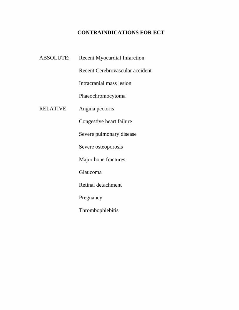
CONTRAINDICATIONS FOR ECT
ABSOLUTE: Recent Myocardial Infarction
Recent Cerebrovascular accident
Intracranial mass lesion
Phaeochromocytoma
RELATIVE: Angina pectoris
Congestive heart failure
Severe pulmonary disease
Severe osteoporosis
Major bone fractures
Glaucoma
Retinal detachment
Pregnancy
Thrombophlebitis
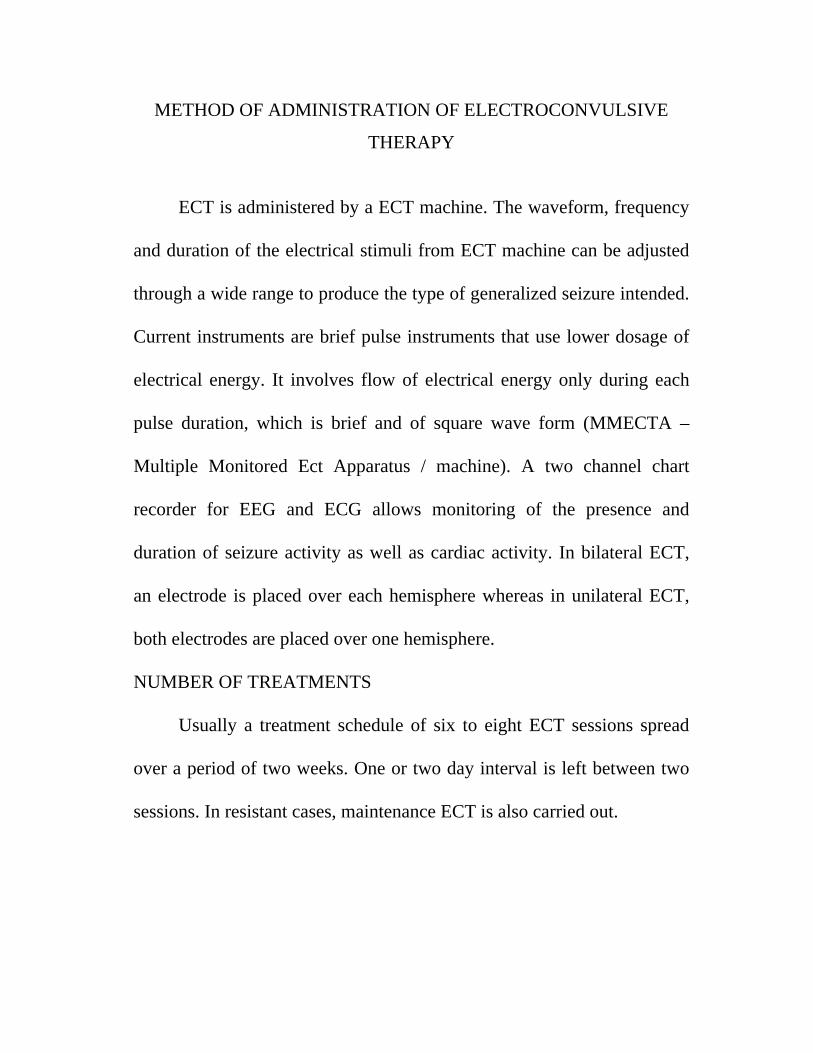
METHOD OF ADMINISTRATION OF ELECTROCONVULSIVE
THERAPY
ECT is administered by a ECT machine. The waveform, frequency
and duration of the electrical stimuli from ECT machine can be adjusted
through a wide range to produce the type of generalized seizure intended.
Current instruments are brief pulse instruments that use lower dosage of
electrical energy. It involves flow of electrical energy only during each
pulse duration, which is brief and of square wave form (MMECTA –
Multiple Monitored Ect Apparatus / machine). A two channel chart
recorder for EEG and ECG allows monitoring of the presence and
duration of seizure activity as well as cardiac activity. In bilateral ECT,
an electrode is placed over each hemisphere whereas in unilateral ECT,
both electrodes are placed over one hemisphere.
NUMBER OF TREATMENTS
Usually a treatment schedule of six to eight ECT sessions spread
over a period of two weeks. One or two day interval is left between two
sessions. In resistant cases, maintenance ECT is also carried out.
MECHANISM OF ACTION
Generalised electrically induced seizures of the central nervous
system are the sine qua non for the therapeutic effects of
electroconvulsive therapy.
The psychological mechanism responsible for the therapeutic effect
remains largely unknown.
EXPLANATIONS OFFERED FOR THE THERAPEUTIC EFFECT
a) NEUROPHYSIOLOGICAL ALTERATIONS IN
Permeability of Blood Brain Barrier
Cerebral microcirculation
Neurometabolic activity
Brain electrical activity
b) NEUROENDOCRINOLOGICAL
Acute neuroendocrine discharge of ACTH, prolactin and
hypothalamic peptides.
c) NEUROCHEMICAL CHANGES IN
Ion transport systems
Brain neurotransmitter receptor systems
Biogenic amines
d) BETA ADRENERGIC RECEPTOR STIMULATION MECHANISMS
e) ELECTROLYTE CHANGES
PHYSIOLOGICAL EFFECTS OF ELECTROCONVULSIVE
THERAPY
The physiological effects of electroconvulsive therapy can be
classified under the following headings.
1. CARDIOVASCULAR EFFECTS
2. CEREBROVASCULAR CHANGES
3. NEUROENDOCRINE RESPONSES
4. SEIZURE RESPONSE 5. ENDOCRINE RESPONSE
6. ENZYME CHANGES
7. MISCELLANEOUS EFFECTS
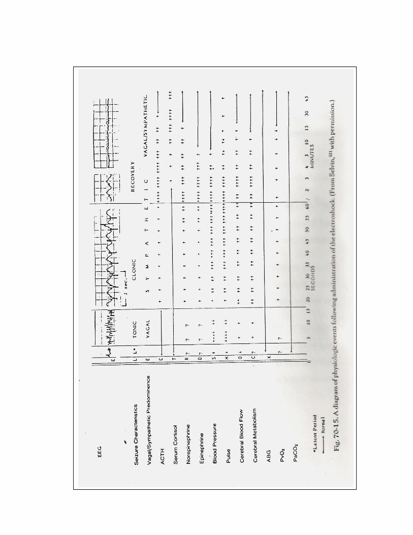
The diagram alongside illustrates the physiological events
following administration of the electroshock.
The following table gives the physiological consequences of
electroconvulsive therapy.
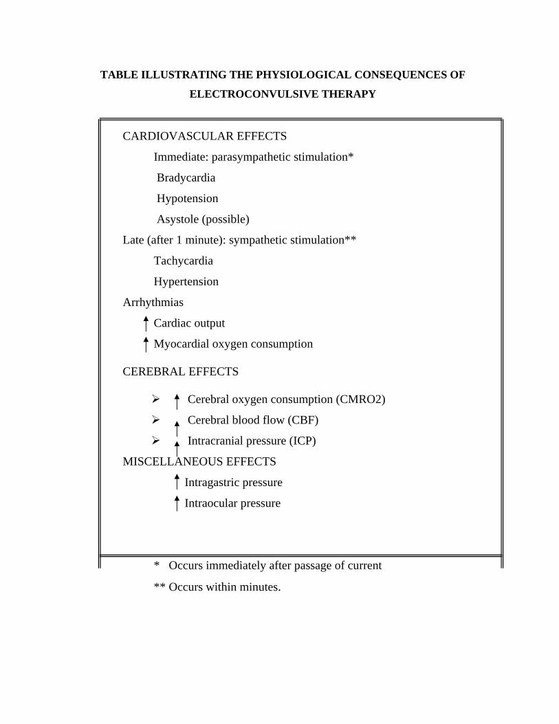
TABLE ILLUSTRATING THE PHYSIOLOGICAL CONSEQUENCES OF
ELECTROCONVULSIVE THERAPY
CARDIOVASCULAR EFFECTS
Immediate: parasympathetic stimulation*
Bradycardia
Hypotension
Asystole (possible)
Late (after 1 minute): sympathetic stimulation**
Tachycardia
Hypertension
Arrhythmias
Cardiac output
Myocardial oxygen consumption
CEREBRAL EFFECTS
Cerebral oxygen consumption (CMRO2)
Cerebral blood flow (CBF)
Intracranial pressure (ICP)
MISCELLANEOUS EFFECTS
Intragastric pressure
Intraocular pressure
* Occurs immediately after passage of current
** Occurs within minutes.
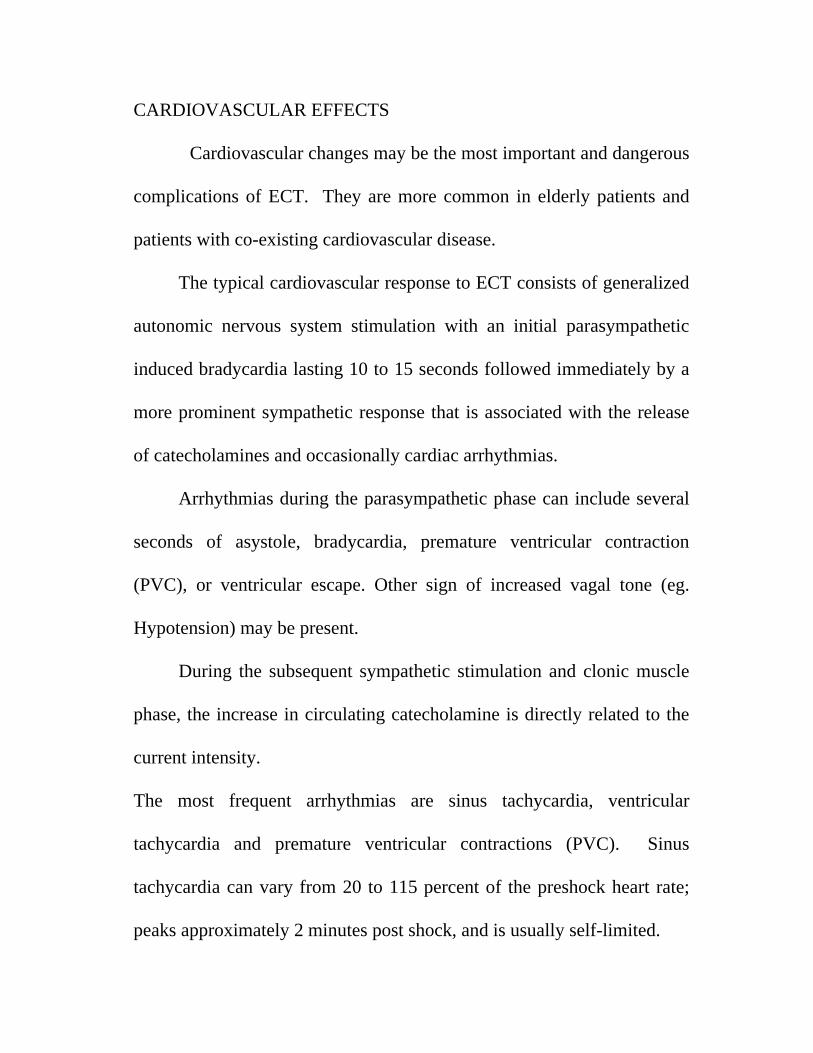
CARDIOVASCULAR EFFECTS
Cardiovascular changes may be the most important and dangerous
complications of ECT. They are more common in elderly patients and
patients with co-existing cardiovascular disease.
The typical cardiovascular response to ECT consists of generalized
autonomic nervous system stimulation with an initial parasympathetic
induced bradycardia lasting 10 to 15 seconds followed immediately by a
more prominent sympathetic response that is associated with the release
of catecholamines and occasionally cardiac arrhythmias.
Arrhythmias during the parasympathetic phase can include several
seconds of asystole, bradycardia, premature ventricular contraction
(PVC), or ventricular escape. Other sign of increased vagal tone (eg.
Hypotension) may be present.
During the subsequent sympathetic stimulation and clonic muscle
phase, the increase in circulating catecholamine is directly related to the
current intensity.
The most frequent arrhythmias are sinus tachycardia, ventricular
tachycardia and premature ventricular contractions (PVC). Sinus
tachycardia can vary from 20 to 115 percent of the preshock heart rate;
peaks approximately 2 minutes post shock, and is usually self-limited.
Systolic and diastolic blood pressures are increased by 30-40
percent. There is a two to four fold increase in the rate pressure product
(RPP) an index of myocardial oxygen consumption. Arterial blood
pressure, heart rate and arrhythmias decline in parallel with the fall in
plasma catecholamine levels. Occasionally arrhythmias and hypertension
can persist. Whole body and myocardial oxygen consumption rates
increase during ECT and venous return is decreased during the
convulsion. The clinical significance of these changes in oxygen
consumption and cardiovascular function will depend on the patient’s
general condition. This rise in circulating catecholamine can be blunted
by anaesthesia.
CEREBROVASCULAR CHANGES:
Cerebrovascular changes include a brief period of vasoconstriction
when the electrical stimulus is applied, followed by a sustained increase
in cerebral metabolism and blood flow. The increase in cerebral blood
flow can result in an increase in intracranial volume and intracranial
pressure which is of concern in patients with intracranial mass lesions,
increased intracranial pressure from any cause or cerebrovascular
anomalies.
NEUROENDOCRINE RESPONSES:
Electroconvulsive therapy activates noradrenergic systems,
enhances dopamine receptor sensitivity and reduces serotonin uptake.
ECT activates the peripheral autonomic nervous system and causes
release of secretions from many endocrine glands.
NEUROENDOCRINE RESPONSES TO ECT
1. An immediate release of ACTH with peak plasma levels at 2 to 5
minutes, which returns to normal by 45 minutes.
2. An increase in PLASMA CORTISOL, level with peak at 30
minutes, returns to normal in 2 to 4 hours
3. An increase in PLASMA EPINEPHRINE concentration to 15
time baseline by 1 minute, which returns to normal at 10 minutes.
4. An increase in PLASMA NOREPINEPHRINE level to three time
baseline at 1 minute, which returns to normal by 20 minutes.
5. Transient increased release of glucagons and inhibition of glucose-
mediated insulin secretion.
SEIZURE RESPONSE
The waveform, frequency and duration of the electrical stimulus
from the ECT machine can be adjusted through a wide range to produce
the type of generalized seizure intended.
Preceded by a latent period of 2-3 seconds, a bilateral grandmal
convulsion ensues, a tonic phase of 10-20 seconds followed by a clonic
phase of 30-50 seconds.
Reducing the duration of convulsion activity would result in
the reduction of therapeutic effect as shown by the various studies.
The threshold has been clinically lower in males than in females,
and lower in younger than elderly patients.
ENDOCRINE RESPONSES:
The therapeutic efficacy of electroshock has been ascribed to a
variety of endocrine changes occurring during therapy, most of which
have no clinical side effects.
The notable exception is the effects of ECT on Diabetes mellitus.
In the diabetic patient,electrically induced seizures produce elevations of
a rapid order of circulating catecholamines and cortisol, a transient rise in
glucagon levels, and inhibition of glucose-mediated insulin secretion.
All of these effects can result in a relative hyperglycaemia in the
diabetic patient. Since ECT appears to cause a variable effect on glucose
levels in diabetic patients, vigilance in the medical management of the
diabetic patient during a series of ECT treatment sessions is necessary.
Blood glucose levels of all diabetic patients requiring ECT should be
carefully monitored during the series and for as long as 3 weeks after
termination of the treatment.
ENZYME CHANGES
Although cardiac complications have been frequently implicated in
the morbidity and mortality, ECT per se does not appear to be involved in
direct cardiac tissue damage.
Following ECT, The total CPK concentration frequently increases,
with the highest values at 6 hours, and returns to normal values by 48
hours. However there is no elevation of the isoenzyme CPK-MB which
indicates myocardial damage. There is no elevation of LDH 1 and LDH2
and serum glutamate transaminases.
MISCELLANEOUS EFFECTS:
Electroconvulsive therapy produces significant elevations in
intraocular pressure subsequent to the onset of the seizure. Elevated
intragastric pressure is also an inevitable accompanying effect of the
convulsion.
COMPLICATIONS OF ECT
Fear and anxiety at induction
Muscle aches and headache
Memory disturbances – Anterograde and Retrograde
Damage to teeth, tongue, eyes, cutaneous structures
Prolonged seizures
Pulmonary aspiration
Laryngospasm
Prolonged apnoea
CARDIAC COMPLICATIONS
These form the main reason for mortality in electroconvulsive
therapy. Cardiovascular mortality has been reported to be 0.03%. The
complications include Atrial arrythmias, AV dissociation, ST segment
depression and a variety of changes and these have been discussed
elaborately in the section on physiological effects of ECT.
PRINCIPLES OF ANAESTHETIC MANAGEMENT
General anaesthesia for electroconvulsive therapy is intended to
provide the patient with lack of awareness of the electrical treatment,
modification of the motor effects of the seizure in order to prevent injury,
rapid recovery and minimal side effects and compatibility with
medications the patient is taking.
Brief general anaesthesia has added to the safety and comfort of
modified electroconvulsive therapy. While a simple procedure, there are
several areas in which ECT differs from other procedures requiring brief
anaesthesia. These differences call for even more collaboration,
interaction and cooperation between an anaesthesiologist and psychiatrist
than is customary.
Though the technical aspects of anaesthesia for electroconvulsive
therapy seem straight forward and are of relatively minor complexity in
an anaesthesiologist’s overall practice, there are four areas which should
command particular attention from anaesthesiologists.
First, while only a short time period is involved in modified
electroconvulsive therapy when compared to other medical procedures
requiring brief anaesthesia.
Secondly, both the physiological and pathological aspects of the
anaesthetic process itself may influence the actual treatment outcome.
Thirdly, because of the possible effect of anaesthesia on
electroconvulsive therapy and the relative brevity of the treatment itself,
more preliminary and on-the-spot coordination between anaesthesiologist
and psychiatrist is necessary.
Fourth since the procedure is repetitive, an optimum anaesthetic
regimen can be sought for each patient.
Anaesthetic requirements for successful modified electroconvulsive
therapy are fourfold.
• Rapid and smooth induction
• Attenuation of the physiological effects of ECT
• Rapid recovery after the seizure
• Minimization of any antagonistic effects on seizure
activity by anaesthetic agents.
ASSESSMENT OF PATIENTS FOR ANAESTHESIA BEFORE ECT
All patients subjected to ECT should undergo a thorough physical
examination and a detailed medical history should be taken. Particular
attention should be paid to cardiorespiratory function, allergies and
previous anesthetic experience and a full neurological assessment should
be made. The presence or absence of loose or missing teeth should be
recorded. The patient should give written consent to the procedure.
Minimum Investigation that are done
Haemoglobin
- Urine - Alb
- Sugar
- Blood - Urea
- Sugar
- Creatinine
- Chest X ray
Other tests should be performed if clinically indicated.
Records should be kept for each administration. This should
include details of the drugs given and their effects, the nature and
duration of the convulsion, the heart rate and blood pressure during the
treatment and subsequent 60 minutes and any side effects or
complications encountered.
PREANAESTHETIC PREPARATION
The immediate preanaesthetic preparation of the patients, as for any
anaesthetic, must include a period of fasting of at least 6 hours. This may
seem simple, but many of these patients are extremely unreliable and
occasionally uncooperative. Careful supervision of the patient is required
to ensure that fasting does occur. Most of the ECT sessions are scheduled
in the early morning to minimize the fasting period.
Preanaesthetic medication with sedatives or narcotics is not
required and may serve only to prolong the anaesthetic recovery time.
Reassurance from the psychiatric staff should be sufficient to allay most
of the patient’s fears regarding the upcoming treatment.
MONITORING AND CONDUCT OF ANAESTHESIA
Intravenous access is secured.
Monitoring includes pulse rate, arterial blood pressure, ECG and
arterial oxygen saturation as a minimum. Full equipment to do
Cardiopulmonary resuscitation (CPR) must be available. All the
emergency drugs should be checked and kept ready.
The ideal intravenous anaesthetic agent would provide rapid onset
short duration, attenuation of adverse physiological effects of ECT, rapid
recovery and no adverse shortening of seizure duration. The barbiturates
like thiopentone sodium and methohexital have been used. Propofol, as
an alternative IV induction drug is also preferred.
The use of neuromuscular blocking drugs has attenuate muscle
contractions associated with ECT and essentially eliminated the risks of
fractures or other injuries associated with muscle contractions.
Succinycholine in the dose of 0.5 – 1 mg/kg has been used most often.
The dosage should be modified on an individual basis.
Ventilation with an oxygen enriched mixture should be
approximately assisted or controlled upto the time of the seizure and
again following recovery from the seizure.

It is important to monitor patients closely immediately following
ECT and during the recovery room until they have recovered from
anaesthesia.
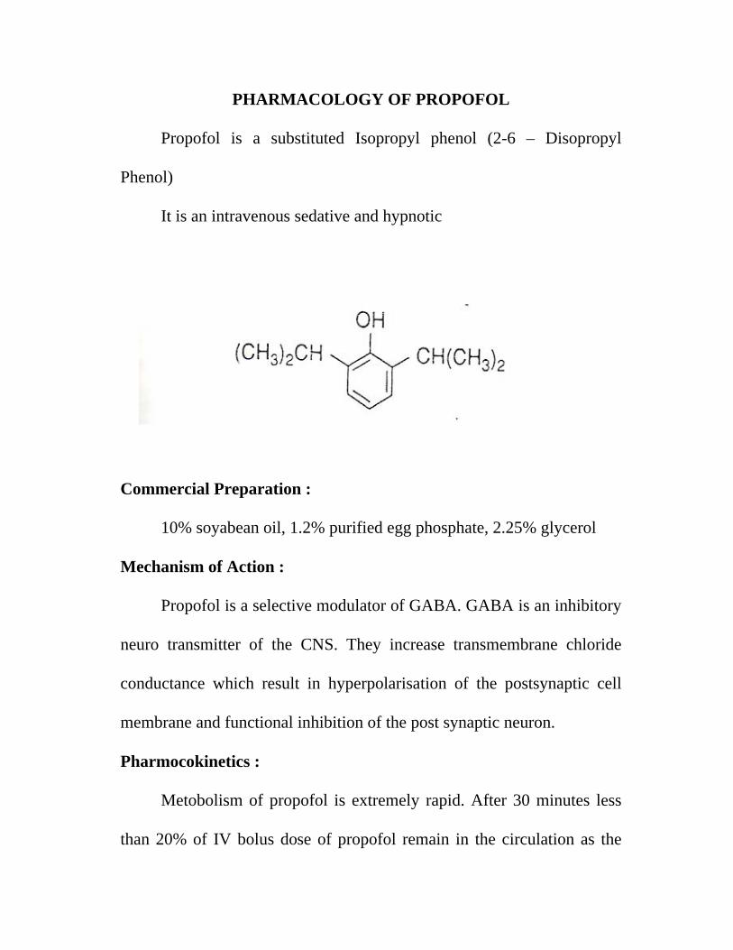
PHARMACOLOGY OF PROPOFOL
Propofol is a substituted Isopropyl phenol (2-6 – Disopropyl
Phenol)
It is an intravenous sedative and hypnotic
Commercial Preparation :
10% soyabean oil, 1.2% purified egg phosphate, 2.25% glycerol
Mechanism of Action :
Propofol is a selective modulator of GABA. GABA is an inhibitory
neuro transmitter of the CNS. They increase transmembrane chloride
conductance which result in hyperpolarisation of the postsynaptic cell
membrane and functional inhibition of the post synaptic neuron.
Pharmocokinetics :
Metobolism of propofol is extremely rapid. After 30 minutes less
than 20% of IV bolus dose of propofol remain in the circulation as the
uncharged compound. The drug is completely and rapidly metabolized to
its sulphate and glucournide compounds and other related compounds
that are eliminated via the kidney. It is also been studied that the
metabolism of drug is not markedly deranged in patients with moderate
degree of renal and hepatic dysfunction. Generally, clearance exceeds
the capacity of liver blood supply suggesting that extrahepatic
mechanisms constitute to the clearance of propofol from the blood.
Effect on Organ systems :
Cardiovascular system :
Propofol cause decrease in systemic blood pressure with
corresponding decrease in cardiac output and systemic vascular
resistance.
Negative ionotropic effect is due to decrease in intracellular
calcium availability due to inhibition of sacrolemmal Ca++ shifts.
Cardiac index remains unaltered.
Respiratory systems :
Propofol produces dose dependent depression of ventilation and
apnea may occur. Propofol decreases the tidal volume and frequency of
breathing. The ventilatory response to hypercarbia and arterial
hypoxemia are decreased by propofol.
It produce bronchodilatation and decreases the incidence of intra
operative wheezing. Hypoxic pulmonary vasconstriction is intact.
Hepatic and Renal function :
Propofol does not affect the hepatic or renal function. Prolonged
infusion of propofol may result in excretion of green urine and uric acid
excretion is increased.
Central Nervous System :
Propofol decreases the cerebral metabolic rate for oxygen
(CMRO2), cerebral blood flow and ICP. It also decreases the systemic
BP and hence decreases the cerebral perfusion pressure.
The effect of propofol on EEG activity is controversial. It has
direct anticonvulsant effect that is dose dependent. Propofol also resulted
in a shorter duration of motor and EEG activity after electro convulsive
therapy.
Propofol also has neuroprotective effect – Propofol administered
resulted in better burst suppression and better neurological outcome.
Clinical Uses:
Total Intravenous Anesthesia: Propofol has a short context sensitive
half life and also has a short effect site equilibrium time thus making it a
readily titratable drug for IV sedation.
The short context sensitive half life of propofol even with
prolonged period of infusion combined with the short effect – site
equilibrium time make this a readily titrable drug for IV sedation.
Emergence from anaesthesia was rapid (time from end of infusion until
patient being oriented was 12.8 – 18.9 min. In the recovery room patients
were described as fully alert and oriented.
Side effects: 1. Allergic reaction
Allergic component of propofol due to the phenyl nucleus and
Disopropyl side chain
2. Bacterial growth:
Propofol supports the growth of Escherichia coli & Pseudomonas
aeuroginosa – aseptic precaution during handling is a must.
3. Antioxidant property
Presence of phenolic group inhibits lipid peroxidation and
scavengers free radicals.
5. Pain on Injection:
Pain on injection is more when the drug is injected into a smaller
vein. Using 1% lidocaine, short acting opioids and changing the
composition of the carrier fat emulsion to long and medium chain
triglycerides decrease the incidence of pain on injection.
PHARMACOLOGY OF THIOPENTONE SODIUM
Sodium 5 – ethyl – ( 1- methylbutyl) 2- thiobarbiturate
Invented in 1934 by Lundy and Waters
Derivatives of bartituric acid, formed by condensation of urea and
malonic acid
Week acid – with pka above plasma pH making them largely
unionised and hence able to cross the blood brain barrier.
Dose of thiopental:
The dose of thiopental varies between 3 and 5 mg/kg, with an
effective plasma concentration of 15mcg/mL.
Pharmacodynamic effects:
Barbiturates are positive allosteric modulators at GABA and
glycine receptors. They cause increased channel opening time for
chloride, which increase inhibitory effects.
Pharmacokinetics:
The effect compartment equilibrium half-time for thiopentone is
very short – 1.2 minutes, which is faster than for propofol.
Thiopentone is highly protein bound depending on pH. Binding
falls as pH rise. Clearance is by hepatic metabolism, not renal excretion
(<1%). Plasma concentration falls rapidly after a bolus dose of
thiopentone owing to uptake by vessel rich tissue.
Hepatic extraction ratio is low. It undergoes oxidation to the
carboxylic acid derivative and to a lesser extent by S – Oxidation to
Pentobarbital, a hypnotic oxybarbiturate will slow elimination.
After a single bolus dose or short infusion pharmacodynamic decay
curves follow typical first order kinetics. With longer, high-dose infusion
hepatic metabolic capacity may be exceeded and zero-order kinetics may
be seen. Desulphuration may then become a significant metabolic
pathway. Recovery will be prolonged as a result of both reduced
metabolism and the presence of an active metabolite.
PHARMCODYNAMICS:
Central Nervous System:
Central effects of thiopentone include sedation, anaethesia,
anticonvulsion action, retrograde amnesia and depression of the
vasomotor centre.
It is a cerebral vasoconstrictor causing a reduction in cerebral blood
flow and intra cranial pressure and depression of cerebral metabolism.
There are features of neuro protection. Burst suppression of EEG can be
induced with high doses.
Respiratory system :
Thiopentone causes centrally mediated respiratory depression and
reduced sensitivity to raised CO2 which is dependent on dose and rate of
injection. Transient apnoea is common. Laryngeal reflexes are intact.
Coughing, laryngeal spasm and mild bronchoconstriction can occur
particularly in asthmatics.
Cardiovascular system :
Myocardial depressed in a dose dependent manner. Peripheral
vascular resistance falls, leading to reduced preload and cardiac output.
There is hypotension and tachycardia, which is exaggerated if there is
hypovolemia, tight aortic stenosis and tamponade.
Adverse Effects:
1. Respiratory depression, and airway obstruction. Laryngeal spasm is
more common than with propofol. Oxygen is administered by gentle
manual IPPV via a mask.
2. Circulatory collapse: This is usually due to a relative overdose
causing vasodilatation and myocardial depression. It may also be due to
anaphylaxis. Treatment: raise the legs; give oxygen by IPPV infuse
fluids fast intravenously, administer inotropes.
3. Coughing: a sign of regurgitation, salivation or over-light
anaesthesia. Hiccup occasionally seen.
4. True cutaneous allergy can occur either in the form of a scarlatiniform
rash or as true angioneurotic oedema. Photosensitivity to thiopentone in
patients recently exposed to sunlight has been reported.
5. Severe anaphylactic reactions (allergy).These reactions, although
very rare, are dangerous. They may take the form of cutaneous
manifestations, (rashes, weals, flushes, oedema), cardiovascular collapse
(hypotension, tachycardia), bronchospasm, laryngospasm and muscle
rigidity or abdominal pain.
6. Acute intermittent porphyria. Barbiturates may precipitate lower
motor neuron paralysis and perhaps death in patients with porphyria and
are absolutely contraindicated in them.
REVIEW OF LITERATURE
The first report on the cardiovascular complication ECT came from
BELLETH in 1941, KELB in 1946 and ACTSHOTE in 1947.
BOEY WK, LAIFO, Anaesthesia analog 1990, August, Department of
Anaesthesia, University of Singapore.
“Comparison of propofol and thiopentone sodium as anaesthetic
agents for electroconvulsive therapy.”
Propofol and thiopentone were compared as anaesthetic agents for
electro convulsive therapy in 31 patients on four occasions in a repeated
measure cross over study. The increase in systolic and diastolic arterial
pressures and heart rate after treatment were significantly higher with
thiopentone. Propofol gave a milder tonus and clonus during seizure
when both treatments were considered together. The time to walk 10
meters at 20 minutes was significantly better with propofol.
BONADA et al, Journal of ECT 19(3) 129-132, September 2003,
Propofol seems to be a good intravenous induction agent of choice
for ECT. Its pharmokinetic properties ensure a rapid and deep
anesthesia, of short duration, with a minimum side effects and a rapid
recovery of good quality, suitable for short repetitive procedures.
Gracia, Edwin, Rodriguez, Rev cub Med Mil Apr-June 2007.
“Propofol versus Thiopentone in electroconvulsive therapy”
50 psychiatric patients compared thiopentone sodium and propofol
in electroconvulsive therapy. Following variables were analysed, mean
blood pressure, cardiac frequency and rhythm, duration of generalized
seizure and recovery. There was a greater increase in these variables in
the thiopentone group compared to the propofol group. Generalised
seizure duration was 29.84 seconds in propofol group and 37.24 seconds
in thiopentone group. Recovery times were 6.85 min for thiopntone and
8.16 minutes for propofol. Propofol was a better hypnotic for
electroconvulsive therapy.
MITCHELL P, JORDA T, HICHIE I, BUITIE C, Australian NZJ
Psychiatry 1991 Jun 25(2)
“Propofol as an anesthetic agent for ECT, effect on outcome and
length of course”
The aim of the study was to investigate the effect of propofol on the
response to ECT.
Records of 66 patients with primary depression treated with ECT,
37 of whom had been assessed prospectively with Pre and Post ECT
Hamilton and Zung depression severity ratings. Despite demonstrating
that the individual seizure duration was significantly reduced with
propofol compared to thiopentone they found no evidence of reduced
ECT efficiency with propofol.
Park HS, Lee JH, Lee KH, “ British Journal of Anaesthesia Oct 1999
(14(3:2), Department of Anaesthesia, Presbyterian Medical centre,
Chonju, Korea.
“Comparison of Propofol and thiopentone for electroconvulsive
therapy - effects on haemodynamic changes and intraocular pressure.”
20 patients were studied during courses of ECT administration,
each patient receiving propofol or thiopentone. The induction dose was
1.6 mg / kg of propofol and 3mg/kg of thiopentone sodium. The
induction dose, rhythm and intraocular pressure were checked before
induction and after administration of succinylcholine immediately, 5
minutes and 10 minutes after ECT administration. Recovery time was
also compared between these two groups.
Results: Mean arterial pressure was lower following propofol than
thiopentone (p<0.05) immediately after ECT. Heart rate was lower
following propofol than thipentone (p<0.05) immediately 5 minutes and
10 minutes after ECT. Intraocular pressure was lower following propofol
than thiopentone (p<0.05) immediately 5 minutes and 10 minutes after
ECT. Recovery time of propofol (6.5+0.8 minutes) was shorter than
thiopentone (7.5+0.9minutes).
Conclusion: Propofol for ECT induction would seem to be an ideal drug,
as it attenuates hypertensive responses and increases in intraocular
pressure.
Kadoi. Y. Saito, Ide M. Sekimoto K. Sehi, Anaesthesia Intensive care,
2003 April : 31 (2). “Department of Intensive care medicine and
Anaesthesia school of medicine, Gunma University, Japan.
“The comparative effect of propofol versus thipentone on left ventricular
function during electro convulsive therapy”.
The purpose of this study was to compare the effect of propofol
versus thiopentone on haemodynamics during electroconvulsive therapy
as estimated by echocardiography.
Twenty eight ASA 1 & 2 patients scheduled for ECT were
randomly divided into two groups to receive propofol 1 mg / kg or
thipentone 2 mg/kg. Cardiac functions were examined by transesophageal
echocardiography prior to induction of anaesthesia and throughout ECT
until ten minutes after the seizure.
Results:
In the thiopentone group, increased end-systolic area (ESA) and
decreased fractional area change (FAC) were observed compared to the
propofol group. They have concluded that lesser haemodynamic changes
occurred after the propofol anaesthesia compared with the thiopentone
anaesthesia during ECT.
Singeri Saito, Yuifi Kadoi, Anaesthesia Analog 2000. “The comparative
effects of Propofol versus thiopentone on middle cerebral artery blood
flow velocity during electroconvulsive therapy”
In this study, they continuously compared cerebral blood flow at
the middle cerebral artery during ECT by using propofol versus
thiopentone Anaesthesia.
In the study, seizure duration was shorter in the propofol group.
The shorter duration with the propofol and hence a smaller energy
demand, may be a cause for the minor increase in cerebral blood flow
velocity in the propofol group. The systemic haemodynamic changes are
small and hence they cause a minor change in the cerebral
haemodynamics compared to thiopentone group.
Mitchell P. Smythe G. Biol Psychiatry 1991 March 15,
“Effect of Anaesthetic agent propofol on hormonal response to ECT”.
Propofol is a new anaesthetic agent that reduces electroconvulsive
therapy (ECT) seizure duration. They reviewed the endocrine response to
ECT by two distinct mechanism, decreasing prolactin by reducing the
seizure duration and decreasing ACTH and cortisal by another process,
possibly via a reduction in central nor adrenergic activation.
FEAR CF LITTLE JOHNS CS, ROUSE E, MCQUAIL P. British
Journal of Psychiatry 1995 Marh 166(3) 399-401.
“Propofol Anesthesia in electroconvulsive therapy”
“Reduced seizure duration may not be relevant”
In a prospective, randomized double blind study 20 subjects with
major depressive illness received either propofol or methohexitone
Anesthesia. The Hamilton depression rating scale was used to assess
depression before therapy, at every third treatment and at the end of
therapy. Seizure duration was measured using the cuff technique.
Results : Mean seizure duration and mean total seizure duration were
shorter in the propofol group but there was no difference in the outcome.
Conclusion: Use of propofol may not adversely affect outcome from
depression and it is not necessarily contraindicated as an induction agent
for ECT.
JOURNAL PAKISTAN MEDICAL ASSOCIATION FEB 2005 ; 50
(2) 6:3
“Comparison of thiopentone sodium and propofol for electro
convulsive therapy.”
Study conducted in department of anaesthesiology and intensive
care, Aga khan University Hospital, Karachi.
Twenty five patients each undergoing atleast 2 sessions of ECT at
the psychiatry department were included in the study. Each patient either
received thiopentone or propofol for induction of sleep in a randomized
manner. Drugs were evaluated regarding their effects on ECT induced
haemodynamic changes (Blood Pressure, heart rate, seizure duration
related to the procedure and recovery from sleep. Any side effect during
the procedure and recovery was also noted.
They have concluded that propofol offered superior haemodynamic
stability during the procedure and a quick recovery from sleep and
propofol was found to be a better induction agent for ECT compared to
thiopentone sodium.
BUTTER FIELD NN, GRAFT P, Journal of ECT, March 2004, 20(1),
“Propofol reduces cognitive impairment after electro convulsive
therapy”
Cognitive impairment is the main complication after electro
convulsive therapy (ECT). Modification of treatment parameters has been
shown to affect the magnitude of these impairments, but the role of
Anesthetic type remains unclear. This study tested whether there is a
difference in cognitive impairments immediately after ECT with propofol
compared to thiopentone.
Method: This was a randomized double blind cross over study which
included 15 patients receiving right unilateral ECT for depression. Patient
received propofol or thiopental on alternating ECT up to 6 treatments.
Immediate and delayed verbal memory, reaction speed, and executive
functions were assessed 45 minutes after each ECT.
Result: Cognitive impairment was reduced after ECT with propofol
compared to thiopentone. Time to emergence was quicker and EEG
seizure duration shorter after propofol treatment. There was no
significant correlation between seizure duration and neuropsychological
test performance.
Gaines Gy, Rees DI, Anaesthetic Analog 1986 ; 65: 1345-56
“Electroconvulsive therapy and Anaesthetic consideration.”
In this study, 85 patients who were undergoing ECT for major
depression in ASA-I, II group were included in our study. They used
thiopentone, etomidote and propofol in succession in each patient. The
effect of these agents on motor and EEG seizure times, heart rate, mean
BP and peripheral O2 saturation were compared. They have concluded
that Propofol appears to be a safe anaesthetic for ECT with minimal side
effects. It was found superior to thiopentone and etomidate in attenuating
the physiological response to ECT with milder haemodynamic changes.
AlRezah, Alinjanpour E. Journal of Babol University of Medical
Science in (JBUMS) 2005, 7 (3(27).
“Comparison of Recovery duration of Propofol and thiopentone in ECT.
The aim of the study was compare the recovery duration of
Propofol and thiopentone sodium in ECT.
Mean recovery duration of propofol and thipentone sodium was
5.49+2.57 minutes and 6.4+3.69 minutes. They have concluded that
propofol can prevent increasing haemodynamic response to ECT better
than thiopentone sodium.
MATERIALS AND METHODS
Study Design:
Place & Period:
This study was done at the ECT treatment room in the
Department of Psychiatry, Government Rajaji Hospital, Madurai attached
to Madurai Medical College, Madurai from 2005 to 2007.
Sample Size:
Sixty patients
Inclusion Criteria :
Sixty patients (19-64 yrs) of ASA grade I and II patients
diagnosed to have moderate to severe depression or mania in accordance
with the international classification of disease 10th revision (ICD-10) who
was prescribed ECT by the treating Psychiatrist were included in the
study.
Exclusion Criteria:
Patients with contraindication to ECT
Uncontrolled hypertension
Valvular heart diseases
Thyroid dysfunction
Allergy to sulfa drugs / egg protein
H/o Porphyria / Bronchial asthma
Those who were able to give written consent for ECT and
anesthesia, written consent was obtained, and for those patients who were
deemed unfit to give consent by the treating psychiatrist, consent was got
from their relatives.
Sampling Procedure:
The sixty patients in this study were randomly allocated into two
groups, one of which received thiopentone sodium and the other propofol
as the anesthetic agent for ECT.
Randomisation was done using a stratified design. Patients were
stratified according to whether they were diagnosed to have depression or
mania. Randomisation was done by the Psychiatric PG who
subsequently did not have anything to do with treatment allocation or
outcome assessment.
Outcome assessment was done by trained raters who were blind to
the drug used. Each patient’s pre-treatment and post-treatment
psychiatric ratings were done by the same rater.
The Institution’s research and ethics committee approved the study.
ECT was given thrice weekly on Tuesdays, Thursdays and Saturdays.
IMPLEMENTATION:
In all patients a detailed history, physical examination and
relevant investigations were done and medication noted. Patients who
were on benzodiazepines had the drug discontinued 12 hours prior to
ECT. All patients were fasted overnight and received.
Resuscitative equipments and emergency drugs were kept ready
before administering ECT to treat the complications if any that might
occur.
1. Cylinders with full oxygen supply
2. Laryngoscope with appropriate sized blades
3. Endotracheal tubes of appropriate size and stylets
4. Airway and mouth prop of appropriate sizes
5. Functioning suction apparatus
6. Vasopressors and drugs for resuscitation
Intravenous access secured pre oxygenation was done. Pulse rate and
oxygen saturation was monitored continuously using a pulse oximeter.
Preoperative B.P, HR and SPO2 were monitored and recorded.
After preoxygenation anesthesia was induced with either of the two
drugs. Intravenous atropine sulfate 0.6mg was given.
a) Group A - Received propofol 1.5 mg / kg body weight with 1 ml
of 2% Lignocaine hydrochloride as the anesthetic agent. Propofol
was used for each subsequent ECT in these patients
b) Group – B: Patients received Inj. Thiopentone sodium 2mg/kg
body weight and continued to receive thiopentone sodium for each
subsequent ECT.
A blood pressure cuff was applied to the right upper arm and
inflated to 40 mm of Hg above systolic B.P prior to the injection of
succinylcholine to isolate the limb for monitoring motor seizure.
In both the groups muscle relaxation was achieved with intravenous
administration of 0.5 mg/kg / bd wt of succinycholine.
Heart rate was monitored by manually palpating the radial artery
pulsation and blood pressure was measured using blood pressure monitor
at regular time intervals as indicated.
A screen was put to separate the anaesthetist giving the drug and
the anaesthetist who is performing the procedure and taking the reading.
PROCEDURE:
After establishing an I.V. line on the left forearm the calculated
dose of propofol or thiopentone was given over a period of 20 seconds.
The induction dose was considered adequate if the eyelash reflex was lost
after 30 seconds; otherwise additional agents were injected (with
increments of 0.2 mg / kg body weight of propofol or 0.5 mg / kg body
weight of thiopentone sodium)
The sleep dose was recorded for subsequent treatment.
Suxamethonium 0.5 mg/kg was given after the cuffed forearm was
isolated.
Patients were ventilated normally at the rate of 8-10 breaths / min
with 100% O2. Once the fasciculation due to suxamethonium subsided a
soft mouth prop was inserted, bitemporal electrodes were placed for ECT
and bilateral ECT was administered using brief pulse bidirectional
constant current stimuli above seizure threshold (sine wave type) was
used to administer electric shock.
The duration of motor seizure was recorded by a stop watch as well
as stimulus intensity and the number of re-stimulation required to achieve
a motor seizure of atleast 15 seconds. Any patient who did not develop a
bilateral tonic clonic motor seizure of atleast 15 seconds were
restimulated with higher stimulus doses by increasing the duration of
pulses until on adequate seizure was achieved and maximum of 3
restimulations were permitted at each session.

Oxygenation was performed between re-stimulations. Once the
motor seizure subsided patient’s ventilation was assisted with a facemask
with 100% oxygen until the patient resumed spontaneous respiration.
Any side effects like pain on injection, abnormal movement, and
prolonged seizure defined as seizure duration > 120 sec, vomiting,
bronchospasm or laryngospasm was noted.
During recovery, after 30 minutes the patient was asked some
simple questions which they were able to understand and comprehend to
assess the patient’s orientation and ability to talk. Also the patient’s
ability to walk to short distance unaided was assessed after 1 hour and
graded by the recovery room nurse who remained blinded to the
anesthetic agent given. The presence of prolonged post-ictal restlessness
or confusion was also noted.
OUTCOME ASSESSMENTS
Hamilton Depression Rating Scale (HDRS) was assessed pre and
post treatment scores for depressed patients. Modified Mania Rating
Scale (MMRS) was recorded pre and post treatment for mania patients by
trained raters who remained blind to the anesthetic agent used.
Rating Scales :
a) Hamilton Depression Rating Scale (HDRS):
It is one of the best known depression rating scales which is used to
rate the severity of symptoms. It is a validated, internationally used
assessment tool. It takes into account not only information from the
patient but also from all other sources. The original version of the scale
comprised of 21 items. But this is reduced to 17 because the items of
depersonalization / derealization, paranoia and obsessionality were found
to be uncommon and diurnal variation was felt to be more related to the
form of the illness. Higher scores indicate more severe depressive
symptom. A rating of less than 8 is usually taken to indicate the absence
of significant depressive symptoms.
b) Modified Manic Rating Scale (MMRS) :
This is a 28-item scale with ratings made on the basis of severity
based on clinical interview and on information gained from relatives and
nurses. It has satisfactory psychometric properties with good inter-rater
reliability and test-retest reliability and is sensitive to change.
Ratings were made in the mornings only to minimize the effects of
diurnal variation of symptoms. The first rating was done within 48 hours
of the first. ECT treatment and the second was done within 2 weeks of
the past ECT treatment.
The haemodynamic data was compared and analysed by the
students‘t’ test. A ‘p’ value of < 0.05 was considered statistically
significant.
Statistical Tools
The information collected regarding all the selected cases were
recorded in a Master Chart. Data analysis was done with the help of
computer using Epidemiological Information Package (EPI 2002)
developed by Centers for Disease Control and Prevention (CDC), Atlanta
for W.H.O.
Using this software, frequencies, percentage, range, mean, standard
deviation, x2 and 'p' values were calculated. A 'p' value less than 0.05 is
taken to denote significant relationship.
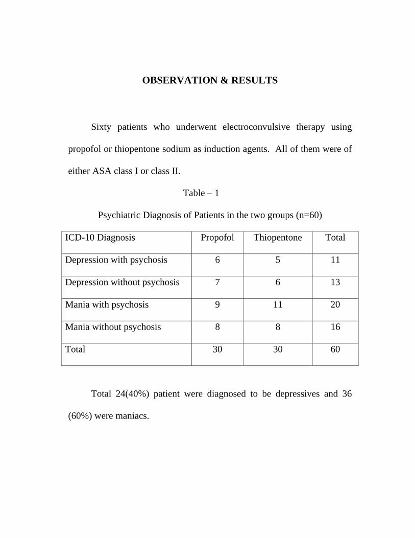
OBSERVATION & RESULTS
Sixty patients who underwent electroconvulsive therapy using
propofol or thiopentone sodium as induction agents. All of them were of
either ASA class I or class II.
Table – 1
Psychiatric Diagnosis of Patients in the two groups (n=60)
ICD-10 Diagnosis Propofol Thiopentone Total
Depression with psychosis 6 5 11
Depression without psychosis 7 6 13
Mania with psychosis 9 11 20
Mania without psychosis 8 8 16
Total 30 30 60
Total 24(40%) patient were diagnosed to be depressives and 36
(60%) were maniacs.

DEMOGRAPHIC DATA
Table - 2
Demographical Data (n=60)
Parameter Propofol Thiopentone Range P value
Age (year)
Mean + SD
37.3 + 14.52 32.6 + 9.37 18-62 0.295
Weight (kg)
Mean + SD
53.79 + 11.17 52.11 + 11.70 29-85 0.667
Male 16 23
Female 14 7
The mean age, weight and sex distribution of the included subjects
are presented in Table 2
Subjects in each group did not differ with respect to these variables.
Patients in the two group did not differ with respect to proportions
of patients who were previous drug non-responders. (Propofol 8/30,
Thiopentone – 10/30)
Equal number of patients in each group was on concurrent
medication, antidepressants or antipsychotics or both
The group did not significantly differ in proportion of patients with
pre ECT physical risk factors such as hypertension (Propofol 4/30,
Thiopentone 3/30)
Mean dose of Propofol given was 76.20 mg (SD + 14.76 mg)
Range (60-100 mg). The mean dose of thiopentone given was 120 mg
(SD + 28.30 range 100-200mg). The mean dose of succinylcholine given
was 32.3 mg (SD + 6.57 mg) (range 20-50mg).
HAEMODYNAMIC PARAMETERS
Changes in heart rate before and after ECT (n=60)
Table - 3
Parameters
Mean + SD ‘t’ test P value
Thiopentone Propofol
Pre
ECT
90.30+9.38 90.50+8.36 0.44 0.903
Not significant
Post
ECT
1st min 95.88+8.80 94.28+9.5 0.50 0.676
Not significant
2nd min 108.81+6.99 95.38+9.72 0.14 0.043
significant
5th min 106.75+6.09 97.37+6.19 0.64 0.063
Not significant
10th min 105.38+89.25 89.25+7.03 5.287 0.04
Significant
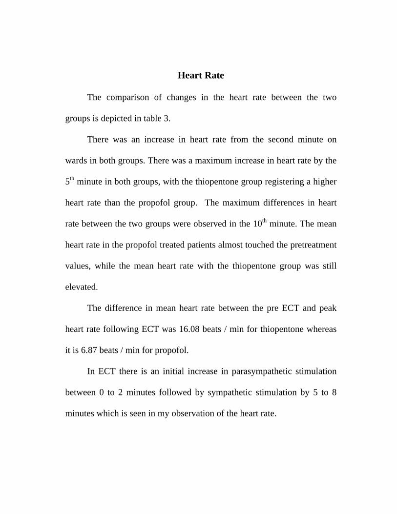
Heart Rate
The comparison of changes in the heart rate between the two
groups is depicted in table 3.
There was an increase in heart rate from the second minute on
wards in both groups. There was a maximum increase in heart rate by the
5th minute in both groups, with the thiopentone group registering a higher
heart rate than the propofol group. The maximum differences in heart
rate between the two groups were observed in the 10th minute. The mean
heart rate in the propofol treated patients almost touched the pretreatment
values, while the mean heart rate with the thiopentone group was still
elevated.
The difference in mean heart rate between the pre ECT and peak
heart rate following ECT was 16.08 beats / min for thiopentone whereas
it is 6.87 beats / min for propofol.
In ECT there is an initial increase in parasympathetic stimulation
between 0 to 2 minutes followed by sympathetic stimulation by 5 to 8
minutes which is seen in my observation of the heart rate.
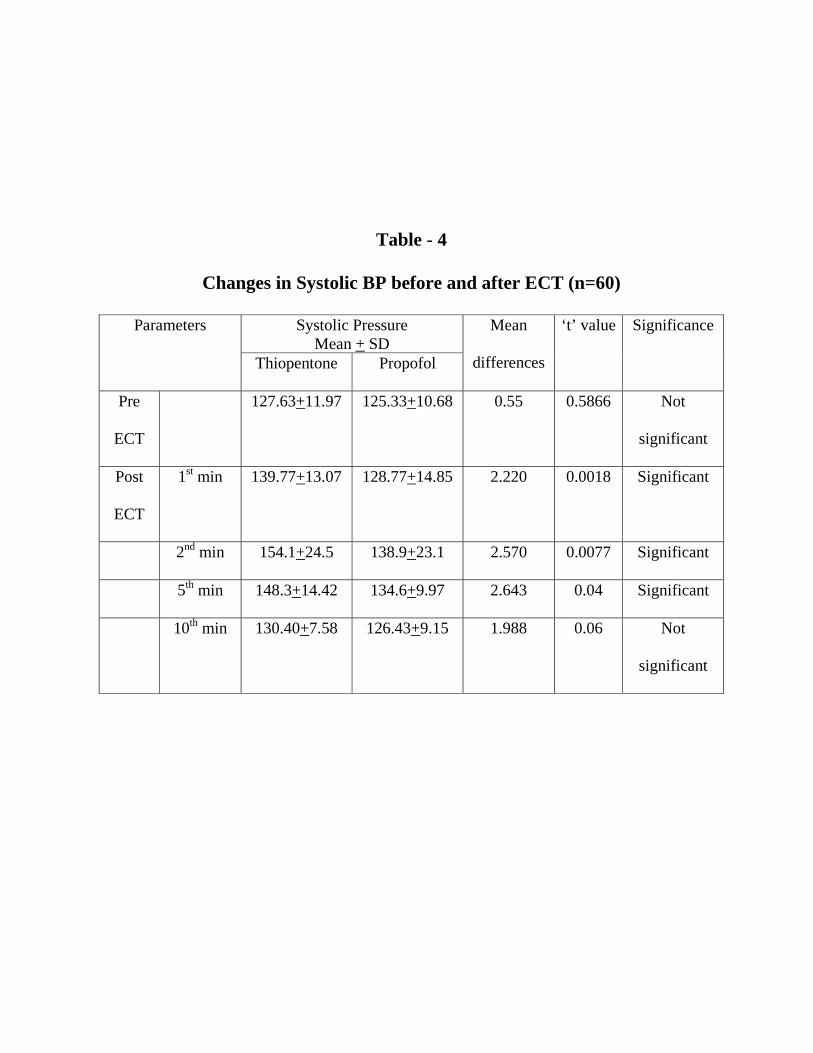
Table - 4
Changes in Systolic BP before and after ECT (n=60)
Parameters
Systolic Pressure Mean + SD
Mean
differences
‘t’ value Significance
Thiopentone Propofol
Pre
ECT
127.63+11.97 125.33+10.68 0.55 0.5866 Not
significant
Post
ECT
1st min 139.77+13.07 128.77+14.85 2.220 0.0018 Significant
2nd min 154.1+24.5 138.9+23.1 2.570 0.0077 Significant
5th min 148.3+14.42 134.6+9.97 2.643 0.04 Significant
10th min 130.40+7.58 126.43+9.15 1.988 0.06 Not
significant
Changes in Systolic BP before and after ECT
The comparison of systolic blood pressure pre induction and at
various time intervals following delivery of ECT stimulus is shown in
table 4
Baseline differences in B.P. between the two groups were not
significant.
There was an increase in systolic BP following the administration
of ECT in both groups, but the increase in systolic blood pressure is much
higher with the thiopentone group compared to the propofol group.
There was maximum increase in BP at 2nd min in both the
thiopentone and propofol group which is more with thiopentone
compared to propofol and it is statistically significant.
There was an gradual increase in BP from the 2nd minute to 5th
minute with a statistically significant increase in BP in the thiopentone
group compared to the propofol group.
The systolic BP touched the baseline pressure at around the 10th
min in both groups.
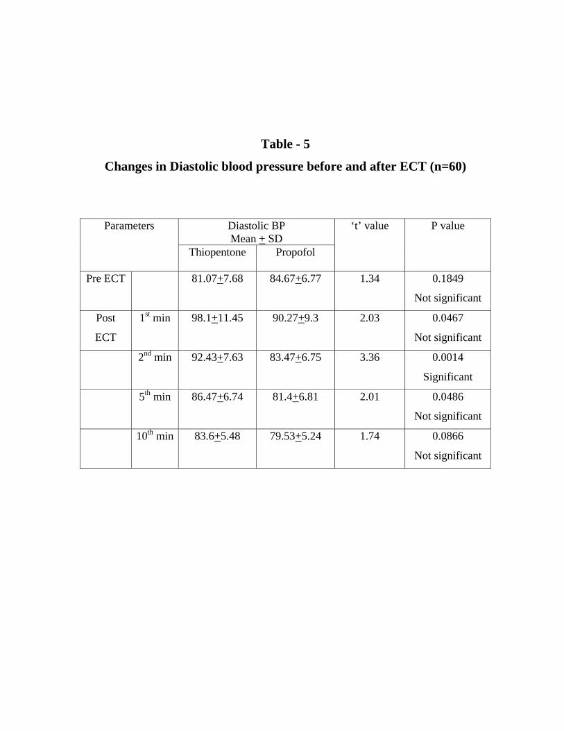
Table - 5
Changes in Diastolic blood pressure before and after ECT (n=60)
Parameters
Diastolic BP Mean + SD
‘t’ value P value
Thiopentone Propofol
Pre ECT 81.07+7.68 84.67+6.77 1.34 0.1849
Not significant
Post
ECT
1st min 98.1+11.45 90.27+9.3 2.03 0.0467
Not significant
2nd min 92.43+7.63 83.47+6.75 3.36 0.0014
Significant
5th min 86.47+6.74 81.4+6.81 2.01 0.0486
Not significant
10th min 83.6+5.48 79.53+5.24 1.74 0.0866
Not significant
Changes in diastolic blood pressure before and after ECT
The comparison of diastolic blood pressure pre induction and at
various time intervals following administration of ECT is show in the
chart.
Baseline values did not significantly differ in both the groups.
There was an increase in diastolic BP following the administration
of ECT in both the groups with the increase in diastolic BP higher with
the thiopentone group compared to the propofol group.
Diastolic BP touched the maximum level around 1-2 minutes.There
was statistically significant increase in diastolic BP in the thiopentone
group compared to the propofol group.
The diastolic BP started to decrease and reached the base line value
by 8-10 minutes.
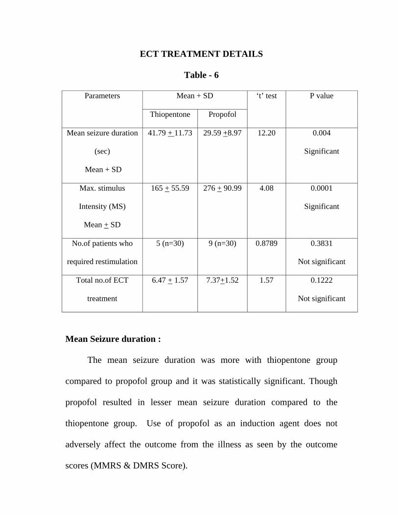
ECT TREATMENT DETAILS
Table - 6
Parameters
Mean + SD ‘t’ test P value
Thiopentone Propofol
Mean seizure duration
(sec)
Mean + SD
41.79 + 11.73 29.59 +8.97 12.20 0.004
Significant
Max. stimulus
Intensity (MS)
Mean + SD
165 + 55.59 276 + 90.99 4.08 0.0001
Significant
No.of patients who
required restimulation
5 (n=30) 9 (n=30) 0.8789 0.3831
Not significant
Total no.of ECT
treatment
6.47 + 1.57 7.37+1.52 1.57 0.1222
Not significant
Mean Seizure duration :
The mean seizure duration was more with thiopentone group
compared to propofol group and it was statistically significant. Though
propofol resulted in lesser mean seizure duration compared to the
thiopentone group. Use of propofol as an induction agent does not
adversely affect the outcome from the illness as seen by the outcome
scores (MMRS & DMRS Score).
Maximum stimulus intensity : (MS)
The maximum stimulus intensity that is required to produce seizure
is recorded. The observations showed that propofol group needed more
stimulus intensity compared to the thiopentone group which was
statistically significant.
Restimulation :
Number of patients who required restimulations were more with the
propofol group compared with the thiopentone group. 9/30 in propofol
group and 5/30 in thiopentone group, but it was not found to be
statistically significant.
Total number of ECT treatments required
The total number of ECT treatments required to produce the desired
effect is more with propofol group compared with the thiopentone group,
but it was not statistically significant.
Thus, propofol use resulted in higher stimulus intensities being
used to elicit adequate seizure, shorter seizure duration and greater
number of treatments and more number of restimulations were required
compared to the thiopentone group.
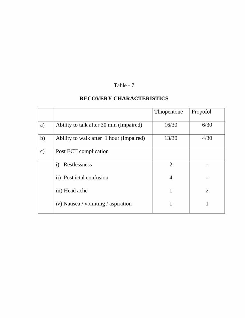
Table - 7
RECOVERY CHARACTERISTICS
Thiopentone Propofol
a) Ability to talk after 30 min (Impaired) 16/30 6/30
b) Ability to walk after 1 hour (Impaired) 13/30 4/30
c) Post ECT complication
i) Restlessness
ii) Post ictal confusion
iii) Head ache
iv) Nausea / vomiting / aspiration
2
4
1
1
-
-
2
1
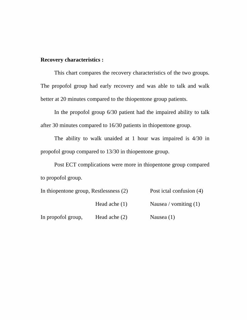
Recovery characteristics :
This chart compares the recovery characteristics of the two groups.
The propofol group had early recovery and was able to talk and walk
better at 20 minutes compared to the thiopentone group patients.
In the propofol group 6/30 patient had the impaired ability to talk
after 30 minutes compared to 16/30 patients in thiopentone group.
The ability to walk unaided at 1 hour was impaired is 4/30 in
propofol group compared to 13/30 in thiopentone group.
Post ECT complications were more in thiopentone group compared
to propofol group.
In thiopentone group, Restlessness (2) Post ictal confusion (4)
Head ache (1) Nausea / vomiting (1)
In propofol group, Head ache (2) Nausea (1)
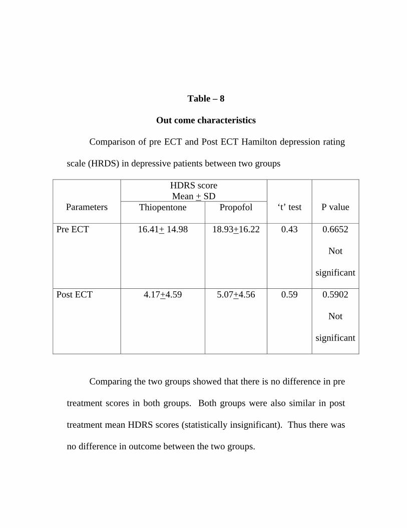
Table – 8
Out come characteristics
Comparison of pre ECT and Post ECT Hamilton depression rating
scale (HRDS) in depressive patients between two groups
Parameters
HDRS score Mean + SD
‘t’ test
P value Thiopentone Propofol
Pre ECT 16.41+ 14.98 18.93+16.22 0.43 0.6652
Not
significant
Post ECT 4.17+4.59 5.07+4.56 0.59 0.5902
Not
significant
Comparing the two groups showed that there is no difference in pre
treatment scores in both groups. Both groups were also similar in post
treatment mean HDRS scores (statistically insignificant). Thus there was
no difference in outcome between the two groups.
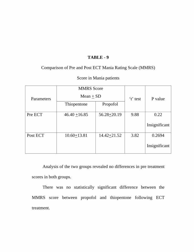
TABLE - 9
Comparison of Pre and Post ECT Mania Rating Scale (MMRS)
Score in Mania patients
Parameters
MMRS Score
Mean + SD
‘t’ test
P value Thiopentone Propofol
Pre ECT 46.40 +16.85 56.28+20.19 9.88 0.22
Insignificant
Post ECT 10.60+13.81 14.42+21.52 3.82 0.2694
Insignificant
Analysis of the two groups revealed no differences in pre treatment
scores in both groups.
There was no statistically significant difference between the
MMRS score between propofol and thiopentone following ECT
treatment.
DISCUSSION
This is a prospective study which compares Propofol with
Thiopentone on haemodynamic parameters, seizure duration, recovery,
complication and outcome in a mixed group of 60 depressed and maniac
patients.
Both the groups were matched by demographic and clinical
variables. Out of these 24 patients (40%) were diagnosed to be depressive
and 36 patients (60%) were maniac patients.
Pulse Rate :
Electroconvulsive therapy and the resultant seizures evokes an
autonomic reaction generating a parasympathetic and then sympathetic
activation sequence. The phase of sympathetic activation and a 15 fold
increase in circulating catecholamines results in severe tachycardia
Accordingly the maximum pulse rate registered in our study in a
single patient was 159/minute from his basal pulse rate of 72 beats /
minute.
There was an increase in mean heart rate from the second minute
onwards for both thiopentone and propofol group with a maximum heart
rate around the 5th minute.
Though initial tachycardia is due to the direct adrenergic outflow
through sympathetic ganglia, the sustained response is due to the further
release of epinephrine from adrenal medulla
The difference in mean heart rate between pre ECT heart rate and
peak heart rate was 16.08 beats / min for thiopentone group, whereas it is
6.87 beats/min for propofol. The finding of my study correlate with the
studies of Boey WK Lai FO, (Anaesthesia analog 1990, Aug 45) who
observed a statistically significant increase in heart rate in the thiopentone
group compared to the propofol group. Similar findings were observed
by Park Hs et al, (BJA Oct 1999, 14:32) who observed that propofol had
fewer effects on haemodynamic changes.
In our series, in which 2% of patients who were hypertensive, there
was a very significant attenuation of pulse rate. Thus, propofol use has a
very significant protective effect on the cardiovascular system as a whole.
Systolic and diastolic blood pressure
Both systolic and diastolic blood pressure is observed to increase
from 1-2 minutes post ECT. At 2 minutes post ECT, the increase in
systolic blood pressure was 40% in the thiopentone group compared to
9% in the propofol group.
In the 2nd, 5th & 10th min readings there was increase of BP more in
the thiopentone group compared to the propofol group.
All these findings correlate well with the previous studies which
showed a statistically significant difference in Blood pressure rise in
thiopentone group compared to the Propofol group.
In 2% of patients who were hypertensives, systolic blood pressure
reached basal levels at 2 minutes post ECT in the propofol group whereas
they registered a 26% rise in the thiopentone group.
The observations made in systolic and diastolic BP in my study
correlate well with the study of Kadoi Y Sait et al, (Anaesthesia Intensive
care 2003 April 31(2), who observed decreased end systolic area (ESA)
and decreased fractional area change (FAC) with the propofol group
compared to the thiopentone group. Similar observation made by Boey
WK, Lai Ko, Bonada et al, also confirmed the above findings with lesser
increase in mean systolic and diastolic pressure in the propofol group
compared to the thiopentone group.
Mean Seizure Duration:
There was statistically significant difference in mean seizure
duration with the thiopentone group compared to the propofol group.
It is observed that propofol group needed more stimulus intensity
compared to the thiopentone group which was statistically significant.
Though the total number of ECT treatments required to produce the
desired effect is more with the propofol group compared with the
thiopentone group, it was not statistically significant.
These findings of decrease in mean seizure duration in the propofol
groups were comfirmed by the studies of Fear CF Little Johns CS, et al
(BJA, 1995, March) who observed that reduced seizure duration in
propofol group may not be relevant. Similar observation was also made
by Mitchell P. Smythe G. who observed a decrease in ACTH, prolactin
and cortisol level in the propofol group compared to the thiopentone
group which is due to the decreased mean seizure duration.
Recovery characteristics:
The propofol group had early recovery and was able to talk at 30
minutes and walk better at 1 hour compared to the thiopentone group.
Post ECT complications were more with the thiopentone group
compared to the propofol group.
The earlier recovery with the propofol group compared to the
thiopentone group were consistent with the findings of Butter Field NN et
al (Journal of ECT March 2004; 20 (1)) who observed that propofol
would reduce cognitive impairment after electro convulsive therapy and
hence earlier recovery characteristics. Similar observations were made
by Gracia et al (Rev Cub Med Mil Apr. June 2007) who observed a
earlier recovery with propofol compared to the thiopentone group.
Outcome Characteristics:
There was no statistically significant difference in Pre and Post
ECT HDRS scores for depressive patients and pre and post ECT MMRS
scores for maniac patients.
SUMMARY
Modified electro convulsive therapy used to treat major affective
disorders produces significant haemodynamic disturbances.
Increase in heart rate and blood pressure can produce serious
impact on the cardiovascular system and these would be more in
hypertensives and these have to be attenuated. Hypertension and
tachycardia resulting from ECT may cause potentially serious adverse
reactions such as myocardial infarction and stroke.
Sixty patients were randomized to receive either propofol (n=30)
and thiopentone (n=30) in a prospective study to assess the
haemodynamic changes, seizure characteristics, complications and
psychological outcome. Out of 60 patients, 24 were (40%) depressive
patients and 36(60%) were maniac patients in accordance with the ICD-
10 diagnostic guidelines, who underwent modified electroconvulsive
therapy. Patients continued the prescribed medication and the number of
ECT treatments were decided by the treating clinician based on clinical
improvement. Heart rate and blood pressure were monitored before and
at varying periods up to 10 minutes after each ECT treatment. Induction
and recovery times and post ECT complications were systematically
observed. Clinical assessment of depression and mania were done by
trained raters who were blinded to the treatment assignment before the
course of ECT and within two weeks of the last ECT treatment.
The groups did not differ significantly on demographic and clinical
variables before treatment.
The base line mean heart rate was 90.30/minute in the thiopentone
group and 90.50/minute in the propofol group.
In the 2nd minute after ECT the mean heart rate was 108.81 (an
increase of 18 beats) in the thiopentone group compare to 95.38 (an
increase of 5 beats) in the propofol group which was statistically
significant. In the 5th minute, the mean heart rate was 106.75/minute in
the thiopentone group compared to 97.37 / minute in the propofol group.
By the 10th minutes, the mean heart rate continued to be higher in the
thiopentone group (105.38/minute). while in the propofol group the heart
rate touched the base line (89.25minute) which is also statistically
significant.
On comparison of the systolic and diastolic BP the pre ECT mean
systolic BP was 127.63 mmHg in the thiopentone group compared to
125.33mmHg in the propofol group. The increase in BP in 1st minute was
139.77mmHg the thiopentone group, compared to 128.77mmHg in the
propofol group, which was statistically significant. The mean systolic BP
was highest in the second minute was 154.1mmHg in the thiopentone
group whereas in the propofol group it was 138.9mmHg. which is
statistically significant.
Mean systolic pressure in the 5th minute was 148mmHg in the
thiopentone group compared to 134 mmHg in the propofol group which
is also statistically significant. By the 10th minute, BP recorded was
130.4mmHg in the thiopentone group while it was only 126.4mmHg in
the propofol group. Thus BP almost touched the base line in the propofol
group.
Patients with propofol group required higher mean stimulus
intensity to elicit adequate seizures, which was 165ms in the thiopentone
group compared to 276 ms in the propofol group. Propofol group
recorded a shorter mean duration of seizures 41.79seconds in the
thiopentone group compared to 29.59seconds in the propofol group.
Propofol group also had more number of restimulation (5/30) in the
thiopentone group compared to (9/30) in the propofol group.
Propofol group had early recovery characteristics compared to the
thiopentone group. The ability to talk oriented at 30 minutes was 14 out
of 30 patients in the thiopentone group compared to 24 out of 30 patients
in the propofol group. Similarly the ability to walk unaided at 1 hour was
17/30 patients in the thiopentone group whereas it is 26/30 patients in the
propofol group. Thus, propofol group had early recovery characteristics
compared to the thiopentone group and the post ECT complications were
less with the propofol group.
Outcome of the study by evaluating the MMRS and HDRS score
were similar in both thiopentone and propofol group.
The results of this study indicate that propofol is a safe induction
agent for modified ECT with significant advantages over thiopentone
with regard to haemodynamic changes, speed of induction and recovery
from anaesthesia with little untoward complications associated with its
use. While overall outcome was similar in patients treated with propofol
and thiopentone, use of the former was associated with longer courses of
treatment, shorter seizures and greater stimulus dosing used to ensure
adequate seizures.
These findings as well as the relatively higher cost of propofol
indicates that the use of propofol as an induction agent for modified ECT
should be reserved for those patients in whom it is required that
elevations is heart rate and blood pressure after ECT are to be minimized.

Propofol would be ideal when early post operative recovery would
be required as in day care surgeries with minimal post ECT
complications.
CONCLUSION
Modified electroconvulsive therapy used to treat major affective
disorders produces significant haemodynamic disturbances. Tachycardia
and hypertension due to sympathetic stimulation resulting from ECT may
cause potentially serious side effects especially in patients with hypertensive
heart disease, ischaemic heart disease and in patients with compromised
cardiovascular function.
Propofol when used as an induction agent was associated with a lower
increase in pulse rate and blood pressure, which would be advantages in
patients in whom tachycardia and hypertension would be detrimental.
Routine use of propofol for ECT is not encouraged because use of
propofol requires higher mean stimulus intensity to elicit adequate seizures,
(276ms) shorter mean duration of seizure,(29.59sec) more restimulations
(9/30) and more number of treatment compared to the thiopentone group and
hence propofol may be the drug of choice for induction in patients in whom
sudden haemodynamic changes following ECT are detrimental.
Propofol use resulted in earlier recovery from the effects of anaesthesia
than did the use of thiopentone. This occurred in spite of the significantly
greater stimulus dose used in propofol treated patients. The use of propofol
results in earlier recovery and less post ECT complications, which is of
importance in the treatment of large numbers of outpatients.
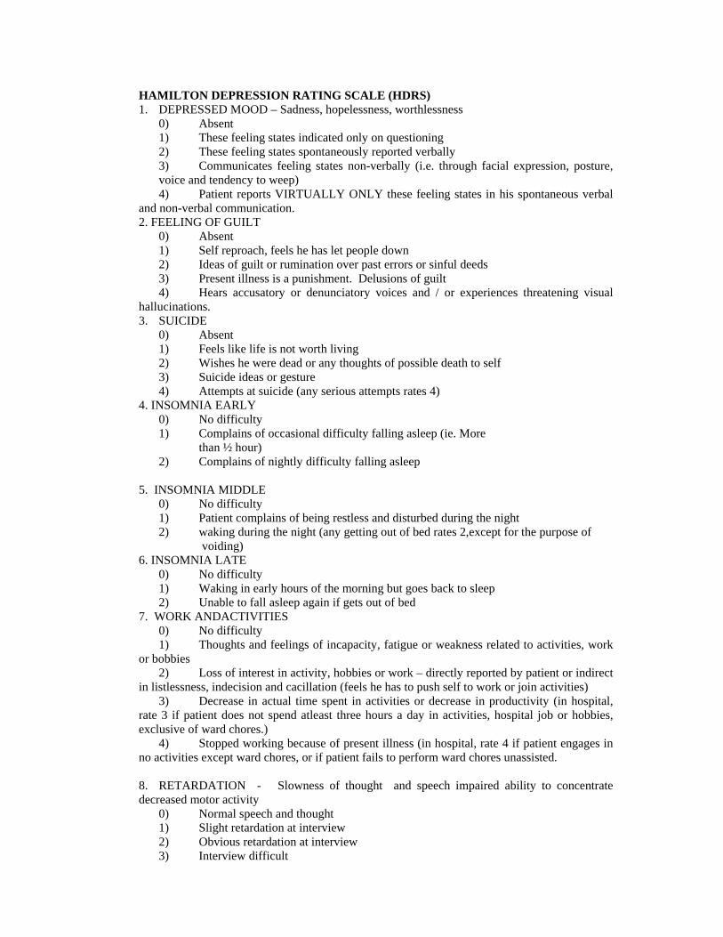
HAMILTON DEPRESSION RATING SCALE (HDRS) 1. DEPRESSED MOOD – Sadness, hopelessness, worthlessness 0) Absent 1) These feeling states indicated only on questioning 2) These feeling states spontaneously reported verbally 3) Communicates feeling states non-verbally (i.e. through facial expression, posture,
voice and tendency to weep) 4) Patient reports VIRTUALLY ONLY these feeling states in his spontaneous verbal and non-verbal communication. 2. FEELING OF GUILT 0) Absent 1) Self reproach, feels he has let people down 2) Ideas of guilt or rumination over past errors or sinful deeds 3) Present illness is a punishment. Delusions of guilt 4) Hears accusatory or denunciatory voices and / or experiences threatening visual hallucinations. 3. SUICIDE 0) Absent 1) Feels like life is not worth living 2) Wishes he were dead or any thoughts of possible death to self 3) Suicide ideas or gesture 4) Attempts at suicide (any serious attempts rates 4) 4. INSOMNIA EARLY 0) No difficulty 1) Complains of occasional difficulty falling asleep (ie. More than ½ hour) 2) Complains of nightly difficulty falling asleep 5. INSOMNIA MIDDLE 0) No difficulty 1) Patient complains of being restless and disturbed during the night 2) waking during the night (any getting out of bed rates 2,except for the purpose of
voiding) 6. INSOMNIA LATE 0) No difficulty 1) Waking in early hours of the morning but goes back to sleep 2) Unable to fall asleep again if gets out of bed 7. WORK ANDACTIVITIES 0) No difficulty 1) Thoughts and feelings of incapacity, fatigue or weakness related to activities, work or bobbies 2) Loss of interest in activity, hobbies or work – directly reported by patient or indirect in listlessness, indecision and cacillation (feels he has to push self to work or join activities) 3) Decrease in actual time spent in activities or decrease in productivity (in hospital, rate 3 if patient does not spend atleast three hours a day in activities, hospital job or hobbies, exclusive of ward chores.) 4) Stopped working because of present illness (in hospital, rate 4 if patient engages in no activities except ward chores, or if patient fails to perform ward chores unassisted. 8. RETARDATION - Slowness of thought and speech impaired ability to concentrate decreased motor activity 0) Normal speech and thought 1) Slight retardation at interview 2) Obvious retardation at interview 3) Interview difficult
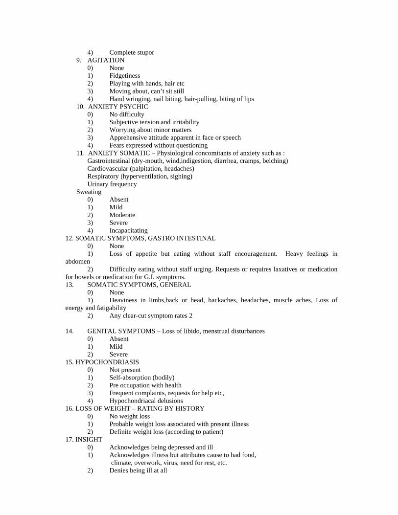
4) Complete stupor 9. AGITATION 0) None 1) Fidgetiness 2) Playing with hands, hair etc 3) Moving about, can’t sit still 4) Hand wringing, nail biting, hair-pulling, biting of lips 10. ANXIETY PSYCHIC 0) No difficulty 1) Subjective tension and irritability 2) Worrying about minor matters 3) Apprehensive attitude apparent in face or speech 4) Fears expressed without questioning 11. ANXIETY SOMATIC – Physiological concomitants of anxiety such as : Gastrointestinal (dry-mouth, wind,indigestion, diarrhea, cramps, belching) Cardiovascular (palpitation, headaches) Respiratory (hyperventilation, sighing) Urinary frequency Sweating
0) Absent 1) Mild 2) Moderate 3) Severe 4) Incapacitating
12. SOMATIC SYMPTOMS, GASTRO INTESTINAL 0) None 1) Loss of appetite but eating without staff encouragement. Heavy feelings in abdomen 2) Difficulty eating without staff urging. Requests or requires laxatives or medication for bowels or medication for G.I. symptoms. 13. SOMATIC SYMPTOMS, GENERAL 0) None 1) Heaviness in limbs,back or head, backaches, headaches, muscle aches, Loss of energy and fatigability 2) Any clear-cut symptom rates 2 14. GENITAL SYMPTOMS – Loss of libido, menstrual disturbances 0) Absent 1) Mild 2) Severe 15. HYPOCHONDRIASIS 0) Not present 1) Self-absorption (bodily) 2) Pre occupation with health 3) Frequent complaints, requests for help etc, 4) Hypochondriacal delusions 16. LOSS OF WEIGHT – RATING BY HISTORY 0) No weight loss 1) Probable weight loss associated with present illness 2) Definite weight loss (according to patient) 17. INSIGHT 0) Acknowledges being depressed and ill 1) Acknowledges illness but attributes cause to bad food,
climate, overwork, virus, need for rest, etc. 2) Denies being ill at all

MODIFIED MANIC STATE RATING SCALE (MMRS) 0 = Absent 1 = Slight flavor or suspicion of pathology by clinician 2 = Clearly noticeable but mild or occurring not more than twice during interview 3 = Clearly present and occurring 3-4 times during interview 4 = Definitely present and frequent 5 = Continuous and gross 1. Is depressed 2. Is talking excessively 3. Restlessness 4. Make threats 5. Has poor judgement 6. Is hallucinating 7. Looks happy and cheerful 8. Seeks out others 9. Is distractable 10. Has grandiose ideas 11. Has grandiose delusions 12. Is angry 13. Is suspicious 14. Has delusions of persecution 15. Is active 16. Is irritable 17. Jumps from one topic to another 18. Shows flight of ideas 19. Is careless about dress and grooming 20. Has diminished impulse control 21. Verbalizes feelings of well-being 22. Makes plans 23. Demands contact 24. Is sexually pre-occupied 25. Is emotionally labile 26. Is religious 27. Is disinhibited 28. Has disturbed sleep
BIBLIOGRAPHY
1. BEATRICE L.SELVIN M.D., Electroconvulsive therapy – 1987 ; Review
Article, Anaesthesiology 67 : 367 – 385, 1987.
2. Boey WK, Lai FO, Anaesthesia Analog, 1990, Aug 45, Comparison of
propofol and thiopentone as anaestheic agents for electroconvulsive
therapy.
3. BODLEY. P.O., FENWICK P.B., Effects of Electroconvulsive therapy on
patients with essential hypertension. BRITISH JOURNAL OF
PSYCHIATRY 112 : 1241-1249, 1966.
4. BUTTER FIELD. NN., GRAFT P – ‘Propofol reduces cognitive
impairment after electroconvulsive therapy” JOURNAL OF ECT March
2004, 20 (1).
5. CHARLES L. RICH.,N. TYSMITH., Anaesthesia for Electro convulsive
therpy : A Psychiatric view point. CANADIAN ANAESTHETISTS
SOCIETY JOURNAL 28:2: 153-157, 1981.
6. FERRIS N. PITIS., ROBERT WOODDRUFF. Drug modification of
Electroconvulsive therapy. ARCHIVES OF GENERAL PSYCHIATRY.
Vol 19, 595-598, Nov 1968.
7. Fear CF Little Johns CS, Rouse E, Mcquail P. British Journal of
Psychiatry 1995, March 166 (3) 399-401. Propofol Anaesthesia in electro
convulsive therapy.
8. GEORGE Y. GAINES., D.IAN REES., Electroconvulsive therapy and
Anaesthetic consideration – Review article. ANAESTHESIA AND
ANALGESIA 65 : 1345-1356 ; 1986.
9. HIROSHI GOTO. M.D., ANTHONY L. KOVAC et al. Comparison of
two esmolol bolus on the haemodynamic response and seizure duration
Electroconvulsive therapy. CANADIAN JOURNAL OF
ANAESTHESIA. 38 : 2 ; 204 -209, 1991.
10. KADAI. Y. SAITO, IDE M. SEHIMOTO K. “ The comparative effect of
propofol versus thiopentone on the left ventricular function during electro
convulsive therapy”. ANAESTHETIC INTENSIVE CARE, 2003, APRIL
: 31(2).
11. LEE.J.T., ERBGUTH.P.H. et al., Modification of Electro convulsive
therapy induced Hypertension with Nitroglycerine ointment.
ANAESTHESIOLOGY Vol. 62 : No. 6 : 793-796, June 1985.
12. LIU.W.S., PETTY W.C. et al., Attenuation of Haemodynamic and
hormonal responses to Electroconvulsive therapy with Propranolol,
Xylocaine, Sodium Nitroprusside or Clonidine. ANAESTHESIA AND
ANALGESIA 63 : 244, 1984.
13. MARJORIE L. BROWN., PAUL E. HUSTON., et al. Cardiovascular
changes associated with ECT in man. A.M.A. ARCHIEVES OF
NEUROLOGY ND PSYCHIATRY 601-608 ; 1952.
14. MATTHEW B. WEINGER., BRAIN L. PARTRIDGE et al. Prevention
of the cardiovascular and Neuroendocrine responses to Electroconvulsive
therapy I : Effectiveness of pretreatment regimens on Haemodynamics.
ANAESTHESIA AND ANALGESIA 73: 556 – 562, 1991.
15. MITCELLI P. SYMETHE G. “Effect of Anaesthetic agent propofol on
hormonal response to ECT, BIOL PSYCHIARTRY 1991 March 15.
16. NAKATSUKA. M.D., O’ SHANICK et al. Changes in Cardiac output
during Electroconvulsive therapy. ANAESTHESIA AND ANALGESIA.
68 : S 208 ; 1989.
17. PARK HS, LEES JH, LEE KH., “Comparison of Propofol and
thiopentone for electroconvulsive therapy – Effects on haemodynamic
changes and intraocular pressure. BRITISH JOURNAL OF
ANAESTHESIA Oct 1999, 14 (3:2)
18. RICHARD ABRAMS. M.D., Electroconvulsive therapy. PSYCHIATRIC
CLINICS OF NORTH AMERICA. Vol. 14. No.4, 1991.
19. RICHARD J. MARKS. Electroconvulsive therapy – Physiological and
Anaesthetic considerations. Review article. CANADIAN
ANAESTHETISTS SOCIETY JOURNAL. Vol 31, 541-547, Sept 1984.
20. ROBERT K. SOTELTING., STEPHEN F. DIERDORF., Anaesthesia and
Coexisting disease, Third edition. 521-523.
21. ROBERT P.KRAUS., RONALD A. REMICK., Diazoxide in the
management of severe hypertension after ECT. AMERICAN JOURNAL
OF PSYCHIATRY. 139 : 4, 504 ; April 1982.
22. RONALD M. JONES., PAUL R. KNIGHT. Cardiovascular and hormonal
responses to ECT. ANAESTHESIA. 36 : 795-799 ; 1981.
23. RONALD. D.MILLER. Anaesthesia at Remote locations.
ANAESTHESIA ; 4TH edition. 2269-2276.
24. SINGERI, SAITO, YUYI KODAI, “Comparative effect of Propofol
versus thiopentone in middle cerebral artery blood flow velocity during
electroconvulsive therapy.
25. WILLIAM HALL LEWIS., JEANNE RICHARDSON et al.
Cardiovascular disturbances and their management in Modified ECT for
psychiatric illness. THE NEW ENGLAND JOURNAL OF MEDICINE.
Vol 252. No. 24 1016-1020 ; 1955.
26. WYLIE., CHURCHILL DAVIDSON H.C., A practice of Anaesthesia.
Fifth edition. 789-790.
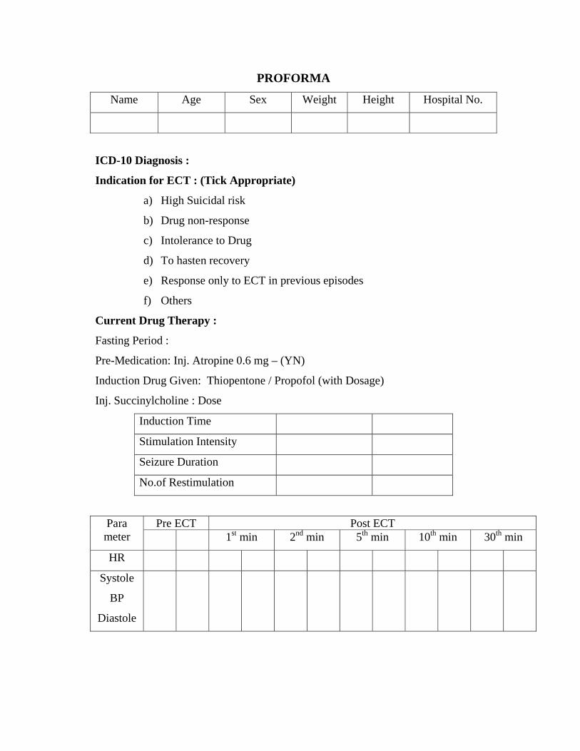
PROFORMA
Name Age Sex Weight Height Hospital No.
ICD-10 Diagnosis :
Indication for ECT : (Tick Appropriate)
a) High Suicidal risk
b) Drug non-response
c) Intolerance to Drug
d) To hasten recovery
e) Response only to ECT in previous episodes
f) Others
Current Drug Therapy :
Fasting Period :
Pre-Medication: Inj. Atropine 0.6 mg – (YN)
Induction Drug Given: Thiopentone / Propofol (with Dosage)
Inj. Succinylcholine : Dose
Induction Time
Stimulation Intensity
Seizure Duration
No.of Restimulation
Para meter
Pre ECT Post ECT 1st min 2nd min 5th min 10th min 30th min
HR
Systole
BP
Diastole
Other features: (v) Hicoughs / Branchospasms / Cough
Recovery Features
No. Action Time Ability
1. Opening of eyes
2. Ability to obey vocal commands
3. Sit up unaided
4. Ability to walk Impaired / unimpaired
5. Ability to talk Impaired/ unimpaired
Post ECT Complications: (Tick Appropriately)
Restlessness / post Ictal confusion / Head ache / Nausea / vomiting /
Aspiration
Patient Outcome:
HDRS YMRS
Pre ECT
Post ECT
MASTER CHART ABBREVIATIONS
HR - HEART RATE SP - SYSTOLIC BLOOD PRESSURE DP - DIASTOLIC BLOOD PRESSURE HDRS - HAMILTON DEPRESSION RATING SCALE MMRS - MODIFIED MANIA RATING SCALE IMP - IMPAIRED UNIMP - UNIMPAIRED N - NO Y - YES