Embed Size (px)
Citation preview

Standards in Genomic Sciences (2010) 2:38-48 DOI:10.4056/sigs.581048
The Genomic Standards Consortium
Complete genome sequence of Desulfohalobium retbaense type strain (HR100
T)
Stefan Spring1, Matt Nolan2, Alla Lapidus2, Tijana Glavina Del Rio2, Alex Copeland2, Hope Tice2, Jan-Fang Cheng2, Susan Lucas2, Miriam Land2,5, Feng Chen2, David Bruce2,3, Lynne Goodwin2,3, Sam Pitluck2, Natalia Ivanova2, Konstantinos Mavromatis2, Natalia Mikhailova2, Amrita Pati2, Amy Chen4, Krishna Palaniappan4, Loren Hauser2,5, Yun-Juan Chang2,5, Cynthia D. Jeffries2,5, Christine Munk3, Hajnalka Kiss2,3, Patrick Chain2,3, Cliff Han2,3, Thomas Bret-tin2,3, John C. Detter2,3, Esther Schüler1, Markus Göker1, Manfred Rohde7, Jim Bristow2, Jo-nathan A. Eisen2,6, Victor Markowitz8, Philip Hugenholtz2, Nikos C. Kyrpides2, and Hans-Peter Klenk1*
1 DSMZ – German Collection of Microorganisms and Cell Cultures GmbH, Braunschweig, Germany
2 DOE Joint Genome Institute, Walnut Creek, California, USA 3 Los Alamos National Laboratory, Bioscience Division, Los Alamos, New Mexico, USA 4 Lawrence Livermore National Laboratory, Livermore, California, USA 5 Oak Ridge National Laboratory, Oak Ridge, Tennessee, USA 6 University of California Davis Genome Center, Davis, California, USA 7 HZI – Helmholtz Centre for Infection Research, Braunschweig, Germany 8 Biological Data Management and Technology Center, Lawrence Berkeley National
Laboratory, Berkeley, California, USA
*Corresponding author: Hans-Peter Klenk
Keywords: sulfate-reducer, Gram-negative, mesophile, moderately halophilic, strictly anae-robic, hydrogen utilization, hypersaline lake, Desulfohalobiaceae, Deltaproteobacteria, Pro-teobacteria, GEBA
Desulfohalobium retbaense (Ollivier et al. 1991) is the type species of the polyphyletic genus Desulfohalobium, which comprises, at the time of writing, two species and represents the family Desulfohalobiaceae within the Deltaproteobacteria. D. retbaense is a moderately ha-lophilic sulfate-reducing bacterium, which can utilize H2 and a limited range of organic sub-strates, which are incompletely oxidized to acetate and CO2, for growth. The type strain HR100
T was isolated from sediments of the hypersaline Retba Lake in Senegal. Here we de-scribe the features of this organism, together with the complete genome sequence and anno-tation. This is the first completed genome sequence of a member of the family Desulfohalo-biaceae. The 2,909,567 bp genome (one chromosome and a 45,263 bp plasmid) with its 2,552 protein-coding and 57 RNA genes is a part of the Genomic Encyclopedia of Bacteria and Archaea project.
Introduction Strain HR100T (= DSM 5692) is the type strain of the species Desulfohalobium retbaense [1]. HR100T is the only strain available from culture collections belonging to this species and was isolated from surface sediments of the hypersaline Retba Lake in Senegal (Western Africa). This strain was the first cultivated sulfate-reducing bacterium, which grows in media containing NaCl concentrations up to 24% and the first described hydrogenotrophic anaerobe able to grow at salinities above 10% [1].
Interestingly, the total salt concentration of the Retba Lake was 34% at the time of sampling, which would indicate that cells of this strain were not able to proliferate in the habitat from which they were originally isolated. This phenomenon was later also reported in a study on the diversity of sulfate-reducing bacteria in hypersaline sedi-ments of the Great Salt Lake (Utah) [2]. This effect could either be explained by niches of lower salin-ity in the respective habitats, which would allow

Desulfohalobium retbaense type strain (HR100T)
39 Standards in Genomic Sciences
proliferation at distinct sites or, alternatively, that the in vitro halotolerance of these strains is differ-ent from the salt tolerance in the natural envi-ronment. One reason for the observed growth in-hibition of sulfate-reducers at salinities above 24% may be the energy expensive synthesis of compatible osmotic solutes, which are required in large amounts to retain cellular integrity at high external salt concentrations. Under anoxic condi-tions bacteria that depend on sulfate as electron acceptor gain less energy than microorganisms that use photosynthesis or denitrification for growth, so that the latter metabolic types have a selective advantage in hypersaline environments [3]. Here we present a summary classification and a set of features for D. retbaense strain HR100T, to-gether with the description of the complete ge-nomic sequencing and annotation.
Classification and features So far, no 16S rRNA gene sequences with high si-milarity (>95%) to the sequence of D. retbaense have been deposited in public databases, although several anoxic sediments with high salinity have been analyzed by cultivation independent me-thods (as of October 2009) since D. retbaense was described. Consequently, it appears that cells of sulfate-reducing bacteria related to this species are of very low abundance in most hypersaline environments. Besides several strains of the genus
Desulfovibrio, the only other member of the order Desulfovibrionales with a sequenced genome is Desulfomicrobium baculatum type strain XT [4]. D. retbaense is the type species of the genus Desul-fohalobium, which represents the recently pro-posed family Desulfohalobiaceae within the class Deltaproteobacteria [5]. The genus Desulfohalo-bium is currently polyphyletic due to the species D. utahense, which is phylogenetically more close-ly related to Desulfovermiculus halophilus, with high bootstrapping support in the 16S rRNA tree (Figure 1) Also, both share a 16S rRNA gene se-quence similarity of 96.9%, whereas the two De-sulfohalobium species display a sequence similari-ty of only 90.5%. Hence, it is possible that the spe-cies D. utahense has been misclassified, although it appears to be phenotypically more similar to D. retbaense than to Desulfovermiculus halophilus [10]. The taxonomy of the two genera thus needs to be reconsidered. Figure 1 shows the phylogenetic neighborhood of D. retbaense strain HR100T in a 16S rRNA based tree. The two 16S rRNA gene copies in the genome of strain HR100T do not differ from each other, and differ by four nucleotides from the previously published 16S rRNA sequence generated from DSM 5692 (X99235). The difference between the genome data and the reported 16S rRNA gene se-quence is most likely due to sequencing errors in the previously reported sequence data.
Figure 1. Phylogenetic tree highlighting the position of strain HR100
T, D. retbaense DSM 5692, relative to the other type strains within the family. The tree was inferred from 1,386 aligned characters [6,7] of the 16S rRNA gene sequence under the maximum likelihood criterion [8] and rooted in accordance with the type strain of the order Desulfovibrionales. The branches are scaled in terms of the expected number of substitutions per site. Numbers above branches are support values from 1,000 bootstrap replicates if larger than 60%. Lineages with type strain genome sequencing projects registered in GOLD [9] are shown in blue, published genomes in bold.
Cells of D. retbaense HR100T are straight to slightly curved rods with rounded ends (Table 1 and Fig-ure 2). They have dimensions of 0.7-0.9 x 1-3 µm and stain Gram-negative. In medium containing
lactate as substrate, cells can form filaments up to 20 µm in length. Motility is conferred by one or two polar flagella [1].

Spring et al.
http://standardsingenomics.org 40
Strain HR100T is halophilic and requires NaCl and MgCl2 for growth. The optimal NaCl concentration for growth is near 10% and salinities up to 24% are tolerated. The pH range for growth is 5.5 to 8.0 with an optimum between pH 6.5 and 7.0. Growth of this strain occurs at temperatures from 25 to 43°C and is optimal between 37 and 40°C [1]. The nutritional characteristics of strain HR100T are as follows: Vitamins and an organic carbon source
are required for growth in mineral medium. Hydrogen is utilized mixotrophically with acetate, yeast extract or biotrypcase as the carbon source, but not autotrophically. Organic carbon sources supporting growth are formate, ethanol, pyruvate and lactate. Sulfate, sulfur, thiosulfate and sulfite are used as electron acceptors and are reduced to H2S. In the absence of sulfate pyruvate can be also utilized fermentatively [1].
Figure 2. Scanning electron micrograph of cells of D. retbaense strain HR100
T
Chemotaxonomy Spectrophotometry of cell extracts indicate the presence of soluble c-type cytochromes having absorption maxima at 418.5, 522.5 and 552 nm in the reduced state, which would be characteristic for cytochrome c3. A dissimilatory sulfite reduc-tase with a similar absorption spectrum as the en-zyme of Desulfomicrobium baculatum (desulforu-bidin) was detected, but no desulfoviridin, which is diagnostic for members of the genus Desulfovi-brio [1]. The respiratory lipoquinone composition of strain HR100T has not been reported, but the moderately related species Desulfovermiculus ha-lophilus was shown to contain the menaquinone MK-7 [18]. The whole cell fatty acid pattern of strain HR100T is dominated by straight- and branched-chain saturated fatty acids (approx. 68%). Branched chain saturated fatty acids ac-
count for 30% of the total fatty acids, with iso-C15:0 predominating. In addition, the fatty acid profile contains branched-chain, mono-unsaturated fatty acids, such as iso-C17:1ω7c and branched C18:1 ω 6 [1].
Genome sequencing and annotation Genome project history This organism was selected for sequencing on the basis of its phylogenetic position, and is part of the Genomic Encyclopedia of Bacteria and Archaea project [19]. The genome project is deposited in the Genomes OnLine Database [9] and the com-plete genome sequence is available in GenBank. Sequencing, finishing and annotation were per-formed by the DOE Joint Genome Institute (JGI). A summary of the project information is shown in Table 2.

Desulfohalobium retbaense type strain (HR100T)
41 Standards in Genomic Sciences
Table 1. Classification and general features of D. retbaense strain HR100T according to the MIGS recommendations [11]
MIGS ID Property Term Evidence code
Current classification
Domain Bacteria TAS [12] Phylum Proteobacteria TAS [13] Class Deltaproteobacteria TAS [14,15] Order Desulfovibrionales TAS [14] Family Desulfohalobiaceae TAS [14] Genus Desulfohalobium TAS [1] Species Desulfohalobium retbaense TAS [1] Type strain HR100
TAS [1] Gram stain negative TAS [1]
Cell shape rod with rounded ends TAS [1]
Motility motile (one or two polar flagella) TAS [1]
Sporulation nonsporulating TAS [1]
Temperature range 25-43°C TAS [1]
Optimum temperature 37-40°C TAS [1]
Salinity >0-240 g/l (optimum 100 g/l) TAS [1] MIGS-22 Oxygen requirement obligate anaerobic TAS [1] Carbon source acetate, biotrypcase, yeast extract TAS [1]
Energy source H2, formate, lactate, ethanol, pyruvate TAS [1] MIGS-6 Habitat hypersaline sediments TAS [1] MIGS-15 Biotic relationship free living NAS MIGS-14 Pathogenicity none TAS [16] Biosafety level 1 TAS [16]
Isolation surface sediment TAS [1] MIGS-4 Geographic location Retba Lake, Senegal TAS [1] MIGS-5 Sample collection time 1989 NAS MIGS-4.1 MIGS-4.2
Latitude, Longitude 14.84, -17.23 NAS
MIGS-4.3 Depth not reported MIGS-4.4 Altitude -4 m TAS [1]
Evidence codes - IDA: Inferred from Direct Assay (first time in publication); TAS: Traceable Author Statement (i.e., a direct report exists in the literature); NAS: Non-traceable Author Statement (i.e., not directly observed for the living, isolated sample, but based on a generally accepted property for the species, or anecdotal evidence). These evidence codes are from the Gene Ontology project [17]. If the evidence code is IDA, then the property was directly observed for a live isolate by one of the authors or an expert mentioned in the acknowledgments.
Growth conditions and DNA isolationD. retbaense strain HR100T, DSM 5692, was grown anaerobically in DSMZ medium 499 [20] at 35°C. DNA was isolated from 1-1.5 g of cell paste using Qiagen Genomic 500 DNA Kit (Qiagen, Hilden, Germany) following the manufacturer's instruc-tions. Genome sequencing and assembly The genome was sequenced using a combination of 8 kb and fosmid DNA libraries. All general as-pects of library construction and sequencing per-formed at the JGI can be found at the http://www.jgi.doe.gov/. The Phred/Phrap/Consed
software package (http://www.phrap.com) was used for sequence assembly and quality assess-ment. Possible mis-assemblies were corrected with Dupfinisher [21] or transposon bombing of bridging clones [22]. Gaps between contigs were closed by editing in Consed, custom primer walk or PCR amplification. Sanger finishing reads (n=889) were produced to close gaps and to raise the quality of the finished sequence. The error rate of the completed genome sequence is less than 1 in 100,000. The final assembly consists of 42,114 Sanger reads. Together all sequence provided 10.7× coverage of the genome.

Spring et al.
http://standardsingenomics.org 42
Table 2. Genome sequencing project information
MIGS ID Property Term MIGS-31 Finishing quality Finished
MIGS-28 Libraries used Two genomic libraries - 8 kb pMCL200 and fosmid pcc1Fos
MIGS-29 Sequencing platforms ABI3730
MIGS-31.2 Sequencing coverage 10.7× Sanger MIGS-30 Assemblers phrap MIGS-32 Gene calling method Prodigal INSDC ID CP001734 (chromosome)
CP001735 (plasmid) GenBank Date of Release September 14, 2009
GOLD ID Gc01111
NCBI project ID 29199
Database: IMG-GEBA 2501939614 MIGS-13 Source material identifier DSM 5692 Project relevance Tree of Life, GEBA
Genome annotation Genes were identified using Prodigal [23] as part of the Oak Ridge National Laboratory genome an-notation pipeline, followed by a round of manual curation using the JGI GenePRIMP pipeline [24]. The predicted CDSs were translated and used to search the National Center for Biotechnology In-formation (NCBI) nonredundant database, Uni-Prot, TIGRFam, Pfam, PRIAM, KEGG, COG, and In-terPro databases. Additional gene prediction anal-ysis and manual functional annotation was per-formed within the Integrated Microbial Genomes Expert Review (IMG-ER) platform [25].
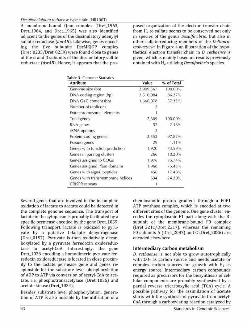
Genome properties The 2,909,567 bp genome consists of a 2,864,304 bp long chromosome and a 45,263 bp long plas-mid with a 57.3% GC content (Table 3 and Figure 3). Of the 2,609 genes predicted, 2,552 were pro-tein coding genes, and 57 RNAs; 29 pseudogenes were also identified. The majority of the protein-coding genes (73.6%) were assigned with a puta-tive function while those remaining were anno-tated as hypothetical proteins. The distribution of genes into COGs functional categories is presented in Table 4.
Insights from the genome sequence Electron donor utilization Similar to representatives of the genus Desulfovi-brio, the preferred substrates of D. retbaense are H2 and lactate, the latter which is incompletely oxidized to acetate. Several genes could be identi-
fied that are involved in H2 dependent sulfate re-duction in Desulfovibrio species. It is assumed that in species of this genus H2 is oxidized by perip-lasmic Fe- or NiFeSe-hydrogenases and the result-ing electrons are transferred to a pool of perip-lasmic cytochrome c3. Then, membrane-bound protein complexes transfer electrons from the pool of reduced cytochrome c3 to menaquinone or directly to cytoplasmic enzymes involved in the reduction of sulfate to sulfide [26]. Recently, a novel molybdopterin oxidoreductase (Mop) could be identified in Desulfovibrio desulfuricans G20 that may represent a periplasm-facing transmem-brane complex, which shuttles electrons from cy-tochrome c3 to the menaquinone pool [27]. It is thought that electrons are transferred from the reduced quinone pool to adenosine phosphosul-fate and sulfite via the membrane-bound respira-tory complexes Qmo [28] and Dsr [29], respective-ly. A similar electron transfer chain for the oxida-tion of H2 with sulfate appears to be functional in D. retbaense: The uptake of H2 is probably cata-lyzed in this species by a heterodimeric NiFe- or NiFeSe-hydrogenase encoded by the genes Dret_0265 (hydB) and Dret_0266 (hydA). Six genes of the completed genome were annotated as cy-tochromes class III containing at least one domain with homology to a tetraheme cytochrome c3. Electrons could be transferred from the reduced cytochrome c pool to menaquinone by a putative Mop complex (Dret_0270/Dret_0273) that is lo-cated in close proximity to the hydrogenase genes.
Desulfohalobium retbaense type strain (HR100T)
43 Standards in Genomic Sciences
A membrane-bound Qmo complex (Dret_1963, Dret_1964, and Dret_1965) was also identified adjacent to the genes of the dissimilatory adenylyl sulfate reductase (aprAB). Likewise, genes encod-ing the five subunits DsrMKJOP complex (Dret_0235/Dret_0239) were found close to genes of the α and β subunits of the dissimilatory sulfite reductase (dsrAB). Hence, it appears that the pro-
posed organization of the electron transfer chain from H2 to sulfate seems to be conserved not only in species of the genus Desulfovibrio, but also in other sulfate-reducing members of the Deltapro-teobacteria. In Figure 4 an illustration of the hypo-thetical electron transfer chain in D. retbaense is given, which is mainly based on results previously obtained with H2-utilizing Desulfovibrio species.
Table 3. Genome Statistics Attribute Value % of Total Genome size (bp) 2,909,567 100.00% DNA coding region (bp) 2,510,084 86.27% DNA G+C content (bp) 1,666,078 57.33% Number of replicons 2 Extrachromosomal elements 1 Total genes 2,609 100.00% RNA genes 57 2.18% rRNA operons 2 Protein-coding genes 2,552 97.82% Pseudo genes 29 1.11% Genes with function prediction 1,920 73.59% Genes in paralog clusters 266 10.20% Genes assigned to COGs 1,976 75.74% Genes assigned Pfam domains 1,968 75.43% Genes with signal peptides 456 17.48% Genes with transmembrane helices 634 24.30% CRISPR repeats 1
Several genes that are involved in the incomplete oxidation of lactate to acetate could be detected in the complete genome sequence. The transport of lactate in the cytoplasm is probably facilitated by a specific permease encoded by the gene Dret_1039. Following transport, lactate is oxidized to pyru-vate by a putative L-lactate dehydrogenase (Dret_0157). Pyruvate is then oxidatively decar-boxylated by a pyruvate ferredoxin oxidoreduc-tase to acetyl-CoA. Interestingly, the gene Dret_1036 encoding a homodimeric pyruvate fer-redoxin oxidoreductase is located in close proxim-ity to the lactate permease gene and genes re-sponsible for the substrate level phosphorylation of ADP to ATP via conversion of acetyl-CoA to ace-tate, i.e. phosphotransacetylase (Dret_1035) and acetate kinase (Dret_1034). Besides substrate level phosphorylation, genera-tion of ATP is also possible by the utilization of a
chemiosmotic proton gradient through a F0F1 ATP synthase complex, which is encoded at two different sites of the genome. One gene cluster en-codes the cytoplasmic F1 part along with the B-subunit of the membrane-bound F0 complex (Dret_2211/Dret_2217), whereas the remaining F0 subunits A (Dret_2087) and C (Dret_2086) are encoded elsewhere.
Intermediary carbon metabolism D. retbaense is not able to grow autotrophically with CO2 as carbon source and needs acetate or complex carbon sources for growth with H2 as energy source. Intermediary carbon compounds required as precursors for the biosynthesis of cel-lular components are probably synthesized by a partial reverse tricarboxylic acid (TCA) cycle. A possible pathway for the assimilation of acetate starts with the synthesis of pyruvate from acetyl-CoA through a carboxylating reaction catalyzed by

Spring et al.
http://standardsingenomics.org 44
the pyruvate ferredoxin oxidoreductase (Dret_1036). Pyruvate can then be either activated to phos-phoenolpyruvate by the enzyme pyruvate, water dikinase (Dret_0098) to enable gluconeogenesis or is further carboxylated to oxaloacetate by py-ruvate carboxylase, which is encoded by two sep-arate genes (Dret_0690 and Dret_1120). Alterna-tively, pyruvate can be also used for the synthesis of malate by malic enzyme (Dret_0778), which requires NADP+ as cofactor. The remaining major precursors for anabolic reactions can then be pro-duced starting from malate by reactions of the re-
verse TCA cycle, involving the enzymes fumarase (Dret_1068, Dret_1069), fumarate reductase (Dret_1065, Dret_1066, and Dret_1067), succinyl-CoA synthetase (Dret_0545) and 2-oxoglutarate ferredoxin oxidoreductase (Dret_1400/Dret_1403). The important five carbon precursor 2-oxo-glutarate could also be synthesized from citrate by aconitase (Dret_1771) and isocitrate dehydroge-nase (Dret_0439). Genes encoding the enzymes ATP-citrate lyase or citrate synthase were not de-tected in the annotated genome sequence, so that a closing of the TCA cycle is apparently prevented.
Figure 3. Graphical circular map of the genome. From outside to the center: Genes on forward strand (color by COG categories), Genes on reverse strand (color by COG categories), RNA genes (tRNAs green, rRNAs red, other RNAs black), GC content, GC skew.
Defense against osmotic and oxidative stressCells of D. retbaense inhabit saline environments and hence need appropriate protection against low water activity or varying salt concentrations. The accumulation of compatible solutes is a wide-spread strategy among microorganisms to protect against osmotic stress. In the distantly related moderately halophilic sulfate-reducing bacterium Desulfovibrio halophilus, the organic solutes treha-lose and glycine betaine were identified as osmo-protectants [30]. In the genome of D. retbaense
DSM 5692 several genes could be detected that may be involved in the intracellular synthesis or accumulation of the above mentioned compatible solutes. For instance, the organic solute trehalose can be synthesized from UDP-D-glucose and al-pha-D-glucose 6-phosphate by the enzymes treha-lose-6-phosphate synthase (Dret_1902) and treha-lose-6-phosphatase (Dret_1903). Alternatively, trehalose may be produced from the reserve car-bohydrate glycogen, if the enzymes malto-

Desulfohalobium retbaense type strain (HR100T)
45 Standards in Genomic Sciences
oligosyltrehalose synthase (Dret_0039) and mal-to-oligosyltrehalose trehalohydrolase (Dret_0037) are expressed. On the other hand, the gene Dret_0035 encodes a trehalose synthase that can
transform maltose directly into trehalose and vice versa, so that an excess of trehalose can be con-verted to glycogen again.
Table 4. Number of genes associated with the general COG functional categories
Code value %age Description J 151 5.9 Translation, ribosomal structure and biogenesis A 0 0.0 RNA processing and modification K 94 3.7 Transcription L 129 5.1 Replication, recombination and repair B 2 0.1 Chromatin structure and dynamics D 28 1.1 Cell cycle control, mitosis and meiosis Y 0 0.0 Nuclear structure V 21 0.8 Defense mechanisms T 168 6.6 Signal transduction mechanisms M 146 5.7 Cell wall/membrane biogenesis N 74 2.9 Cell motility Z 0 0.0 Cytoskeleton W 0 0.0 Extracellular structures U 82 3.2 Intracellular trafficking and secretion O 100 3.9 Posttranslational modification, protein turnover, chaperones C 191 7.5 Energy production and conversion G 101 4.0 Carbohydrate transport and metabolism E 188 7.4 Amino acid transport and metabolism F 53 2.1 Nucleotide transport and metabolism H 109 4.3 Coenzyme transport and metabolism I 43 1.7 Lipid transport and metabolism P 95 3.7 Inorganic ion transport and metabolism Q 20 0.8 Secondary metabolites biosynthesis, transport and catabolism R 212 8.3 General function prediction only S 152 6.0 Function unknown - 633 24.8 Not in COGs
The second osmotic solute in D. retbaense appears to be glycine betaine, which can be accumulated in two different ways: The most efficient way in terms of energy represents the uptake from the environment. Glycine betaine is produced by many cyanobacteria or halophilic anoxygenic pho-tosynthetic bacteria and is released continuously in the environment by excretion or cell lysis. The genome of D. retbaense DSM 5692 contains two distinct gene clusters (Dret_0768/Dret_0771 and Dret_22771/Dret_22773) that could encode high affinity ABC transporters for the uptake of glycine betaine. Several separate genes encoding perip-lasmic glycine betaine binding proteins were also
found and could have a function in the regulation of genes in response to the presence of glycine betaine in the environment. An alternative route for the accumulation of glycine betaine is based on the uptake of choline. This quaternary amine is an essential component of eukaryotic cell mem-branes and hence ubiquitous in most environ-ments. Two genes were found that encode puta-tive choline transporters, Dret_1055 and Dret_2376. Following transport to the cytoplasm, choline could then be oxidized to glycine betaine by the enzymes choline dehydrogenase (Dret_0130) and betaine aldehyde dehydrogenase (Dret_0129).

Spring et al.
http://standardsingenomics.org 46
Figure 4. Proposed organization of the electron transfer chain in D. ret-baense with H2 as electron donor and sulfate as electron acceptor. Gene products are designated according to the information given in Supplementary Table 1. Subunits of multiprotein complexes are labeled with capital letters. Abbreviations: APS, adenosine-5'-phosphosulfate; AMP, adenosine monophosphate; MQ, menaquinone; MQH2, dihydromenaquinone.
The sensitivity of the obligately anaerobic species D. retbaense to oxygen exposure has not been ana-lyzed in detail, but it can be assumed that it is quite moderate as in most other studied sulfate-reducing bacteria [31]. A close inspection of the annotated genome sequence revealed a complex network of antioxidant proteins protecting cells of this species against oxidative stress. Aerobic res-piration was identified as one principal mechan-ism for the detoxification of oxygen in Gram-negative sulfate-reducers [32]. In D. retbaense DSM 5692, genes for the two subunits of a cytoch-rome bd quinol oxidase (Dret_0135 and Dret_0136) were identified. This type of oxidase is the most common terminal oxidase among Gram-negative sulfate-reducers and characterized by a high-affinity to oxygen [32,33]. For the detoxifica-tion of reactive oxygen species that emerge from the contact of oxygen with cellular redox enzymes several protection systems seem to be present. A di-heme cytochrome c peroxidase (Dret_1885)
that is probably localized in the periplasmic space [34] is able to reduce H2O2 to water, whereas a catalase (Dret_1236) produces oxygen from the inactivation of H2O2. On the other hand, in the cy-toplasm multiprotein complexes containing ru-bredoxins (Dret_0886, Dret_0139), rubrerythrins (Dret_0191, Dret_1205, Dret_1644, Dret_2310) and desulfoferrodoxin (Dret_0140) could establish electron transfer systems for the reduction of su-peroxide radicals and H2O2 [35,36]. Finally, cellu-lar proteins and lipids that became damaged by reactive oxygen species could be repaired by a methionine sulfoxide reductase (Dret_2264), pe-roxiredoxin (Dret_2393) and an alkylhydroperox-idase (Dret_1223). Thus, based on the results of the genome analysis it seems that this species is very well adapted to frequent changes in salinity and redox conditions in its natural environment, the sediments of hypersaline lakes.
Desulfohalobium retbaense type strain (HR100T)
47 Standards in Genomic Sciences
AcknowledgementsWe would like to gratefully acknowledge the help of Susanne Schneider (DSMZ) for DNA extraction and quality analysis. This work was performed under the auspices of the US Department of Energy's Office of Science, Biological and Environmental Research Pro-gram, and by the University of California, Lawrence Berkeley
National Laboratory under contract No. DE-AC02-05CH11231, Lawrence Livermore National Laboratory under Contract No. DE-AC52-07NA27344, and Los Alamos National Laboratory under contract. German Research Foundation (DFG) supported DSMZ under INST 599/1-1.
References 1. Ollivier B, Hatchikian CE, Prensier G, Guezennec
J, Garcia JL. Desulfohalobium retbaense gen. nov., sp. nov., a halophilic sulfate-reducing bac-terium from sediments of a hypersaline lake in Senegal. Int J Syst Bacteriol 1991; 41:74-81.
2. Kjeldsen KU, Loy A, Jakobsen TF, Thomsen TR, Wagner M, Ingvorsen K. Diversity of sulfate-reducing bacteria from an extreme hypersaline sediment, Great Salt Lake (Utah). FEMS Microbiol Ecol 2007; 60:287-298. doi:10.1111/j.1574-6941.2007.00288.x
3. Oren A. Bioenergetic aspects of halophilism. Mi-crobiol Mol Biol Rev 1999; 63:334-348.
4. Copeland A, Spring S, Göker M, Schneider S, La-pidus A, Glavina Del Rio T, Tice H, Cheng JF, Lu-cas S, Chen F, et al. Complete genome sequence of Desulfomicrobium baculatum type strain (XT). Stand Genomic Sci 2009; 1:29-37. doi:10.4056/sigs.13134
5. Kuever J, Rainey FA, Widdel F. Family III. Desul-fohalobiaceae fam. nov. In Bergey's Manual of Systematic Bacteriology 2nd ed. (Brenner DJ, Krieg NR, Staley JT and Garrity GM, eds.), Sprin-ger Verlag, New York 2005; 2:948–949.
6. Lee C, Grasso C, Sharlow MF. Multiple sequence alignment using partial order graphs. Bioinformat-ics 2002; 18:452-464. doi:10.1093/bioinformatics/18.3.452
7. Castresana J. Selection of conserved blocks from multiple alignments for their use in phylogenetic analysis. Mol Biol Evol 2000; 17:540-552.
8. Stamatakis A, Hoover P, Rougemont J. A rapid bootstrap algorithm for the RAxML web-servers. Syst Biol 2008; 57:758-771. doi:10.1080/10635150802429642
9. Liolios K, Mavromatis K, Tavernarakis N, Kyrpides NC. The Genomes OnLine Database (GOLD) in 2007: status of genomic and metagenomic projects and their associated metadata. Nucleic Acids Res 2008; 36:D475-D479. doi:10.1093/nar/gkm884
10. Jakobsen TF, Kjeldsen KU, Ingvorsen K. Desulfo-halobium utahense sp. nov., a moderately halo-philic sulfate-reducing bacterium isolated from Great Salt Lake. Int J Syst Evol Microbiol 2006; 56:2063-2069. doi:10.1099/ijs.0.64323-0
11. Field D, Garrity G, Gray T, Morrison N, Selengut J, Sterk P, Tatusova T, Tompson N, Allen MJ, An-giuoli SV, et al. Towards a richer description of our complete collection of genomes and metage-nomes: the “Minimum Information about a Ge-nome Sequence” (MIGS) specification. Nat Bio-technol 2008; 26:541-547. doi:10.1038/nbt1360
12. Woese CR, Kandler O, Wheelis ML. Towards a natural system of organisms: proposal for the do-mains Archaea, Bacteria, and Eucarya. Proc Natl Acad Sci USA 1990; 87:4576-4579. doi:10.1073/pnas.87.12.4576
13. Garrity GM, Holt JG. Taxonomic Outline of the Archaea and Bacteria. Bergey's Manual of Syste-matic Bacteriology 2nd ed. (Boone DR and Cas-tenholz RW, eds.), Springer-Verlag, New York 2001; 1:155-166.
14. Kuever J, Rainey FA, Widdel F. Order II. Desulfo-vibrionales ord. nov. In: Garrity GM, Brenner DJ, Krieg NR, Staley JT (eds), Bergey’s Manual of Sys-tematic Bacteriology, Second Edition, Volume 2, Part C, Springer, New York, 2005, p. 925-926.
15. Editor L. Validation List No. 107. List of new names and new combinations previously effec-tively, but not validly, published. Int J Syst Evol Microbiol 2006; 56:1-6. doi:10.1099/ijs.0.64188-0
16. Anonymous. Biological Agents: Technical rules for biological agents (TRBA 466) www.baua.de
17. Ashburner M, Ball CA, Blake JA, Botstein D, But-ler H, Cherry JM, Davis AP, Dolinski K, Dwight SS, Eppig JT, et al. Gene ontology: tool for the un-ification of biology. Nat Genet 2000; 25:25-29. doi:10.1038/75556
18. Belyakova EV, Rozanova EP, Borzenkov IA, Tou-rova TP, Pusheva MA, Lysenko AM, Kolganova TV. The new facultatively chemolithoautotrophic,

Spring et al.
http://standardsingenomics.org 48
moderately halophilic, sulfate-reducing bacterium Desulfovermiculus halophilus gen. nov., sp. nov., isolated from an oil field. [english translation of Mikrobiologiya]. Microbiology 2006; 75:161-171. doi:10.1134/S0026261706020093
19. Wu D, Hugenholtz P, Mavromatis K, Pukall R, Dalin E, Ivanova N, Kunin V, Goodwin L, Wu M, Tindall BJ, et al. A phylogeny-driven genomic en-cyclopedia of Bacteria and Archaea. Nature 2009; 462:1056-1060. doi:10.1038/nature08656
20. List of growth media used at DSMZ: http://www.dsmz.de/microorganisms/ me-dia_list.php.
21. Han CS, Chain P. Finishing repeat regions auto-matically with Dupfinisher. In: Proceeding of the 2006 international conference on bioinformatics & computational biology. Hamid R Arabnia & Homayoun Valafar (eds), CSREA Press. June 26-29, 2006:141-146.
22. Sims D, Brettin T, Detter JC, Han C, Lapidus A, Copeland A, Glavina Del Rio T, Nolan M, Chen F, Susan L, et al. Complete genome sequence of Kytococcus sedentarius type strain (541T). Stand Genomic Sci 2009; 1:12-20. doi:10.4056/sigs.761
23. Anonymous. Prodigal Prokaryotic Dynamic Pro-gramming Genefinding Algorithm. Oak Ridge Na-tional Laboratory and University of Tennessee 2009 http://compbio.ornl.gov/prodigal
24. Pati A, Ivanova N, Mikhailova N, Ovchinikova G, Hooper SD, Lykidis A, Kyrpides NC. GenePRIMP: A Gene Prediction Improvement Pipeline for mi-crobial genomes. (Submitted).
25. Markowitz VM, Mavromatis K, Ivanova NN, Chen IMA, Chu K, Kyrpides NC. Expert Review of Func-tional Annotations for Microbial Genomes. Bioin-formatics 2009; 25:2271-2278. doi:10.1093/bioinformatics/btp393
26. Matias PM, Pereira IAC, Soares CM, Carrondo MA. Sulphate respiration from hydrogen in Desul-fovibrio bacteria: a structural biology overview. Prog Biophys Mol Biol 2005; 89:292-329. doi:10.1016/j.pbiomolbio.2004.11.003
27. Li X, Luo Q, Wofford NQ, Keller KL, McInerney MM, Wall JD, Krumholz LR. A molybdopterin oxidoreductase is involved in H2 oxidation in De-sulfovibrio desulfuricans G20. J Bacteriol 2009; 191:2675-2682. doi:10.1128/JB.01814-08
28. Pires RH, Lourenco AI, Morais F, Teixeira M, Xavier AV, Saraiva LM, Pereira IAC. A novel
membrane-bound respiratory complex from De-sulfovibrio desulfuricans ATCC 27774. Biochim Biophys Acta 2003; 1605:67-82. doi:10.1016/S0005-2728(03)00065-3
29. Pires RH, Venceslau SS, Morais F, Teixeira M, Xavier AV, Pereira IAC. Characterization of the Desulfovibrio desulfuricans ATCC 27774 DsrMKJOP complex—a membrane-bound redox complex involved in the sulfate respiratory path-way. Biochemistry (Mosc) 2006; 45:249-262. doi:10.1021/bi0515265
30. Welsh DT, Lindsay YE, Caumette P, Herbert RA, Hannan J. Identification of trehalose and glycine betaine as compatible solutes in the moderately halophilic sulfate reducing bacterium Desulfovi-brio halophilus. FEMS Microbiol Lett 1996; 140:203-207. doi:10.1111/j.1574-6968.1996.tb08337.x
31. Marschall C, Frenzel P, Cypionka H. Influence of oxygen on sulfate reduction and growth of sul-fate-reducing bacteria. Arch Microbiol 1993; 159:168-173. doi:10.1007/BF00250278
32. Cypionka H. Oxygen respiration by Desulfovibrio species. Annu Rev Microbiol 2000; 54:827-848. doi:10.1146/annurev.micro.54.1.827
33. Spring S, Lapidus A, Schröder M, Gleim D, Sims D, Meincke L, Glavina Del Rio T, Tice H, Copel-and A, Cheng JF, et al. Complete genome se-quence of Desulfotomaculum acetoxidans type strain (5575T). Stand Genomic Sci 2009; 1:242-253. doi:10.4056/sigs.39508
34. Goodhew CF, Wilson IBH, Hunter DJB, Pettigrew GW. The cellular location and specificity of bac-terial cytochrome c peroxidases. Biochem J 1990; 271:707-712.
35. Lumppio HL, Shenvi NV, Summers AO, Voor-douw G, Kurtz DM, Jr. Rubrerythrin and rubre-doxin oxidoreductase in Desulfovibrio vulgaris: a novel oxidative stress protection system. J Bacte-riol 2001; 183:101-108. doi:10.1128/JB.183.1.101-108.2001
36. Kawasaki S, Sakai Y, Takahashi T, Suzuki I, Nii-mura Y. O2 and reactive oxygen species detoxifi-cation complex, composed of O2-responsive NADH:rubredoxin oxidoreductase-flavoprotein A2-desulfoferrodoxin operon enzymes, rubperox-in, and rubredoxin, in Clostridium aceto-butylicum. Appl Environ Microbiol 2009; 75:1021-1029. doi:10.1128/AEM.01425-08


