Embed Size (px)
Citation preview
Neurosurg. Focus / Volume 26 / June 2009
Neurosurg Focus 26 (6):E7, 2009
1
Two randomized, multicenter prospective trials, RESCUEicp and DECRA, are currently in progress to determine whether decompressive craniectomy
improves outcome in the management of TBI.14,37 Com-plication rates of this procedure will be a major determi-nant of whether decompressive craniectomy proves su-perior to medical treatment for patients with intracranial hypertension after TBI. These procedures are performed for serious life-threatening intracranial hypertension, and mortality rates following decompressive craniectomy for severe TBI are high. During a 5-year experience with 170 consecutive decompressive hemicraniectomies for se-
vere TBI at our institution, 50% of patients died.82 Many of the complications of decompressive craniectomy arise from the normal pathophysiological changes that occur in ICP, CSF circulation, and CBF following removal of a large area of skull bone.
The rationale behind decompressive craniectomy is to convert an injury within a closed box, with a fixed volume and limited reserve, into an open system with increased capacity to accommodate mass.64 After bone removal, there is an increase in brain compliance and a shift of the pressure volume curve to the right.29,33 The RAP index, determined as a correlation between fluctua-tions in ICP with heart rate against the mean ICP aver-aged over repetitive time points, is a surrogate for cere-bral compliance.87 Improvements in RAP values as well as direct measurements using Spiegelberg compliance monitors have demonstrated improvements in cerebral
Complications of decompressive craniectomy for traumatic brain injury
Shirley i. Stiver, M.D., Ph.D.Department of Neurosurgical Surgery, School of Medicine; and The Brain and Spinal Injury Center, University of California San Francisco, California
Decompressive craniectomy is widely used to treat intracranial hypertension following traumatic brain injury (TBI). Two randomized trials are currently underway to further evaluate the effectiveness of decompressive craniec-tomy for TBI. Complications of this procedure have major ramifications on the risk-benefit balance in decision-making during evaluation of potential surgical candidates. To further evaluate the complications of decompressive craniectomy, a review of the literature was performed following a detailed search of PubMed between 1980 and 2009. The author restricted her study to literature pertaining to decompressive craniectomy for patients with TBI. An under-standing of the pathophysiological events that accompany removal of a large piece of skull bone provides a founda-tion for understanding many of the complications associated with decompressive craniectomy. The author determined that decompressive craniectomy is not a simple, straightforward operation without adverse effects. Rather, numerous complications may arise, and they do so in a sequential fashion at specific time points following surgical decompres-sion. Expansion of contusions, new subdural and epidural hematomas contralateral to the decompressed hemisphere, and external cerebral herniation typify the early perioperative complications of decompressive craniectomy for TBI. Within the 1st week following decompression, CSF circulation derangements manifest commonly as subdural hygro-mas. Paradoxical herniation following lumbar puncture in the setting of a large skull defect is a rare, potentially fatal complication that can be prevented and treated if recognized early. During the later phases of recovery, patients may develop a new cognitive, neurological, or psychological deficit termed syndrome of the trephined. In the longer term, a persistent vegetative state is the most devastating of outcomes of decompressive craniectomy. The risk of complica-tions following decompressive craniectomy is weighed against the life-threatening circumstances under which this surgery is performed. Ongoing trials will define whether this balance supports surgical decompression as a first-line treatment for TBI. (DOI: 10.3171/2009.4.FOCUS0965)
Key WorDS • traumatic brain injury • intracranial hypertension • surgical de-compression • cranioplasty • complication • trephine
1
Abbreviations used in this paper: CBF = cerebral blood flow; EDH = epidural hematoma; GCS = Glasgow Coma Scale; ICP = intracranial pressure; RAP = pressure-volume compensatory reserve; SDH = subdural hematoma; TBI = traumatic brain injury.
Unauthenticated | Downloaded 01/27/22 10:25 PM UTC
S. I. Stiver
2 Neurosurg. Focus / Volume 26 / June 2009
compliance following decompressive craniectomy.2,87 As a rescue therapy, decompressive craniectomy has been shown to reliably abort progressive increases in ICP.59,89
Decompressive craniectomy also brings about changes in CBF and CSF dynamics.7,20,24,96,97 Cine MR imaging studies have demonstrated impaired CSF flow across the craniovertebral junction at the skull base, which improves following cranioplasty repair of the skull defect.20 Decom-pressive craniectomy also induces a hyperemic response as evidenced by 99mTc SPECT, 31P MR imaging, and 133Xe CT studies demonstrating augmentation of CBF as well as cerebral metabolism.96,101 Physiological monitoring using CBF, transcranial Doppler, and brain tissue oxygen probes has demonstrated increased CBF and brain tissue oxygen following decompressive craniectomy.7,39,80 Cerebrovascu-lar reactivity is also impaired for the first 24 hours, trend-ing toward a return to intact autoregulation within the first
72 hours following decompressive craniectomy. Measure-ments of PRx, the correlation coefficient of ICP versus ar-terial blood pressure over repetitive time intervals, have shown increases indicative of loss of cerebrovascular re-activity after decompression.89,94 Loss of autoregulation in the early period following decompressive craniectomy may derive from a state of maximal vasodilation and hype-remia following relief of prolonged severe compression.4,71 These alterations in compliance, CBF and autoregulation, and CSF circulation are the basis for many of the compli-cations that arise following decompressive craniectomy.
Overview and Risks for ComplicationsComplications following decompressive craniectomy
occur at predictable time points following the surgery. They can often be anticipated and in many cases prevent-
TABLE 1: Literature summary of complications after decompressive craniectomy for TBI
Authors & Year
No. of Patients
Subdural Hygroma
Contusion/Hematoma Progression
Intra- cranial
Infection
New Contralat Extraaxial Hematoma
Hydro-cephalus
Bone Flap Re-sorption
Reoperation (excluding
cranioplasty) Comment
A arabi et al., 2006
50 25 (50%) 8 (16%) wound infection (1 [2%])
5 (10%) 6 (12%) 6 (12%) developed paren-chymal lucency possibly related to ischemia; bone flap infection after cranio-plasty (in 3)
C hibbaro and Tac-coni, 2007
48 3 (6%) 1 (2%) 1 (2%)
Fl int et al., 2008
40 23 (58%) 11 (28%) 1 2/40 (30%) new or wors-ened IVH
G aab et al., 1990
37 (18 unilat,
19 bilat)
13 (22%) 5 (13%) shunt-treated
G uerra et al., 1999
57 (31 unilat,
26 bilat)
15 (26%) 8 (14%) 1 (2%) wound infection after cranioplasty
H uang et al., 2008
38 10 (26%) 2 (5%) 2 (5%) 3 (8%) 3 (8%)
Ji ang et al., 2005
241 38 (16%) 8 (3%) in cisional CSF fistula (3%); epilepsy (3%)
P olin et al., 1997
35 2 (6%) (1 required
reop)
1 (3%) 10 (29%) shunt-treated
2 (6%) 1 (3%) reop for delayed expansion of a temporal con-tusion requiring lobectomy
1 CSF leak treated w/ lumbar drain
Y ang et al., 2003
68 18 (26%) 4 (6%) 20 (29%)
Y ang et al., 2008
108 7 (6%) w/ reop
9 (8%) shunt-treated
1 9 (18%): contralat intracranial hema-toma (7 [6%]), sub-dural effusion (3 [3%]), hydrocepha-lus (9 [8%])
Unauthenticated | Downloaded 01/27/22 10:25 PM UTC
Neurosurg. Focus / Volume 26 / June 2009
Complications of decompressive craniectomy for TBI
3
ed. Bleeding may be initiated during bone decompres-sion and this, as well as development of new or expanded contusions, may present with malignant swelling or el-evations in ICP in the first few hours or days after de-compression. In a decompressed skull, even an ICP of 20 mm Hg may be of concern. External cerebral herniation frequently evolves over the first few days following de-compressive surgery and can be problematic if the bone decompression is not sufficiently large to allow the brain to expand without constriction. In the early postoperative period (< 1 week), manifestations of deranged CSF flow become evident with evolution of subdural hygromas. Fevers are common during this time, and the need for lumbar puncture for CSF sampling needs to be weighed carefully against the risks of inducing a paradoxical her-niation syndrome. Wound infections are common. In the late postoperative period, unique complications of de-compressive craniectomy include those of syndrome of the trephined.
Patients with severe TBI are at increased risk of de-veloping complications after decompressive craniectomy (Table 1).99 In patients having sustained TBI, Yang et al.99
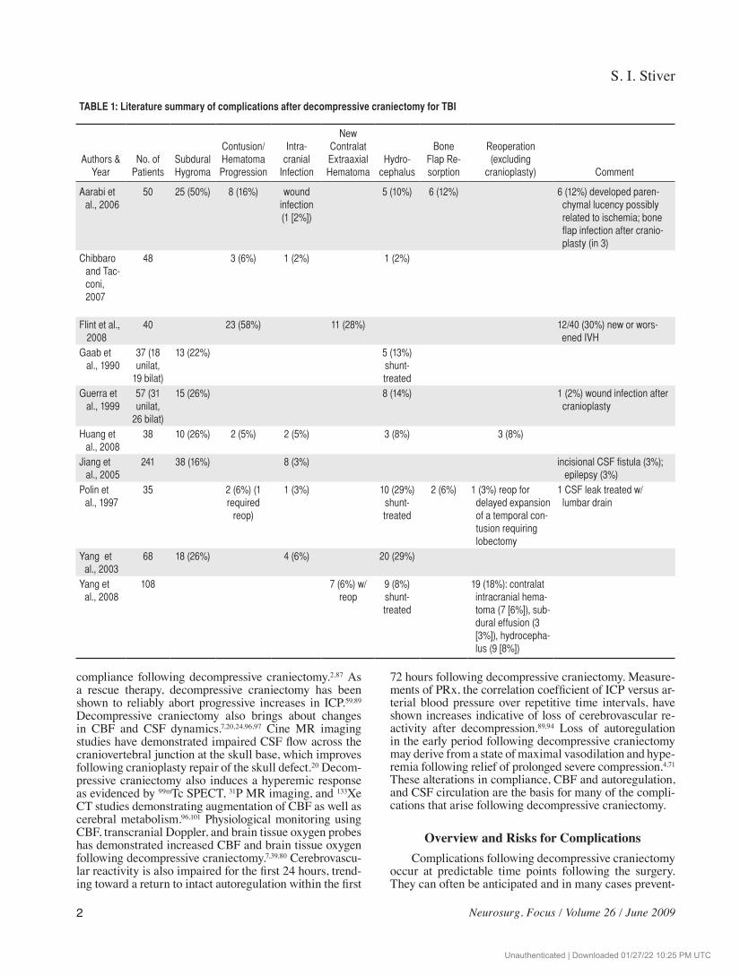
reported that the frequency of complications was 62% for GCS Scores 3–5, 39% for GCS Scores 6–9, and 36% for GCS scores > 9. Older patients (> 60 years) also tended to have more complications, but the differences compared with younger patients were not statistically significant.99 Patients taking aspirin, Plavix, and warfarin are at partic-ularly high risk for severe or nonsurvivable complications following decompressive craniectomy (Fig. 1).
ComplicationsPerioperative Complications
Blossoming of Contusions. Hemorrhagic expansion of contusions is inherent in the injury process and has been demonstrated on serial CT scanning in patients with TBI.10,58,79 Hemorrhagic contusion expansion was observed in 42% of 142 TBI patients with median GCS scores of 8,58 59% of 37 patients in a coma,77 48% of 104 children with TBI,28 and 47% of 141 patients with TBI present-ing with traumatic subarachnoid hemorrhage on their CT
Fig. 1. Coagulopathy. Preoperative noncontrast CT scans of an SDH in a patient on a regimen of warfarin (A and B). Despite reversal of the coagulopathy before surgery, marked hemorrhagic blossoming occurred as evidenced by postoperative noncon-trast images (C and D). Note in panel C the development of an extraaxial SDH contralateral to the decompressed hemisphere.
Unauthenticated | Downloaded 01/27/22 10:25 PM UTC
S. I. Stiver
4 Neurosurg. Focus / Volume 26 / June 2009
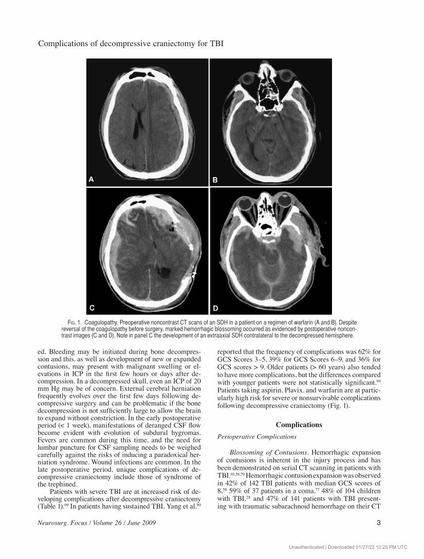
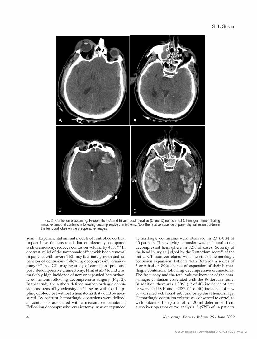
scan.12 Experimental animal models of controlled cortical impact have demonstrated that craniectomy, compared with craniotomy, reduces contusion volume by 40%.102 In contrast, relief of the tamponade effect with bone removal in patients with severe TBI may facilitate growth and ex-pansion of contusions following decompressive craniec-tomy.23,49 In a CT imaging study of contusions pre– and post–decompressive craniectomy, Flint et al.23 found a re-markably high incidence of new or expanded hemorrhag-ic contusions following decompressive surgery (Fig. 2). In that study, the authors defined nonhemorrhagic contu-sions as areas of hypodensity on CT scans with focal stip-pling of blood but without a hematoma that could be mea-sured. By contrast, hemorrhagic contusions were defined as contusions associated with a measurable hematoma. Following decompressive craniectomy, new or expanded
hemorrhagic contusions were observed in 23 (58%) of 40 patients. The evolving contusion was ipsilateral to the decompressed hemisphere in 82% of cases. Severity of the head injury as judged by the Rotterdam score48 of the initial CT scan correlated with the risk of hemorrhagic contusion expansion. Patients with Rotterdam scores of 5 or 6 had an 80% chance of expansion of their hemor-rhagic contusions following decompressive craniectomy. The frequency and the total volume increase of the hem-orrhagic contusion correlated with the Rotterdam score. In addition, there was a 30% (12 of 40) incidence of new or worsened IVH and a 28% (11 of 40) incidence of new or worsened extraaxial subdural or epidural hemorrhage. Hemorrhagic contusion volume was observed to correlate with outcome. Using a cutoff of 20 ml determined from a receiver operator curve analysis, 8 (57%) of 14 patients
Fig. 2. Contusion blossoming. Preoperative (A and B) and postoperative (C and D) noncontrast CT images demonstrating massive temporal contusions following decompressive craniectomy. Note the relative absence of parenchymal lesion burden in the temporal lobes on the preoperative images.
Unauthenticated | Downloaded 01/27/22 10:25 PM UTC
Neurosurg. Focus / Volume 26 / June 2009
Complications of decompressive craniectomy for TBI
5
with an expanded hemorrhagic contusion volume > 20 ml died within 6 months, whereas only 4 (15%) of 26 patients with contusion volumes < 20 ml (p = 0.011) died.23
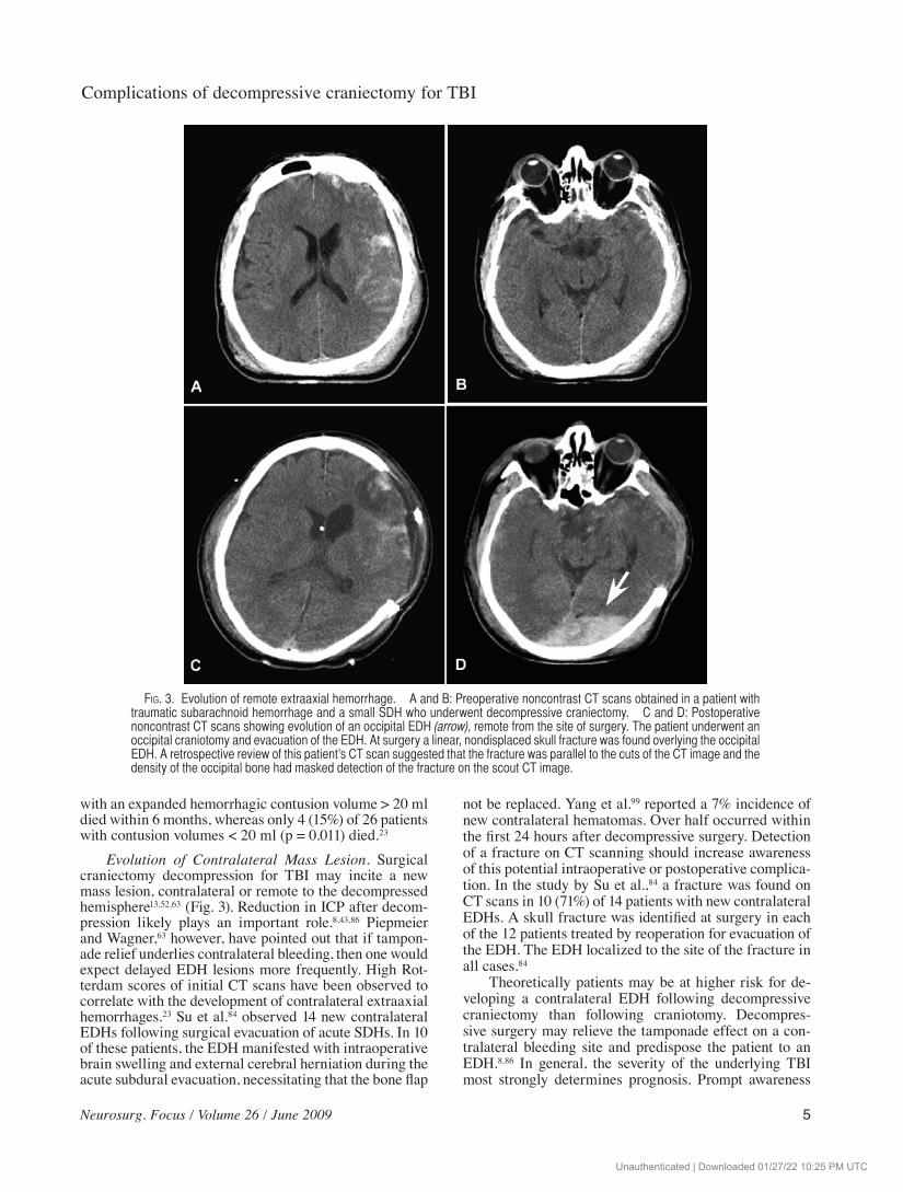
Evolution of Contralateral Mass Lesion. Surgical craniectomy decompression for TBI may incite a new mass lesion, contralateral or remote to the decompressed hemisphere13,52,63 (Fig. 3). Reduction in ICP after decom-pression likely plays an important role.8,43,86 Piepmeier and Wagner,63 however, have pointed out that if tampon-ade relief underlies contralateral bleeding, then one would expect delayed EDH lesions more frequently. High Rot-terdam scores of initial CT scans have been observed to correlate with the development of contralateral extraaxial hemorrhages.23 Su et al.84 observed 14 new contralateral EDHs following surgical evacuation of acute SDHs. In 10 of these patients, the EDH manifested with intraoperative brain swelling and external cerebral herniation during the acute subdural evacuation, necessitating that the bone flap
not be replaced. Yang et al.99 reported a 7% incidence of new contralateral hematomas. Over half occurred within the first 24 hours after decompressive surgery. Detection of a fracture on CT scanning should increase awareness of this potential intraoperative or postoperative complica-tion. In the study by Su et al.,84 a fracture was found on CT scans in 10 (71%) of 14 patients with new contralateral EDHs. A skull fracture was identified at surgery in each of the 12 patients treated by reoperation for evacuation of the EDH. The EDH localized to the site of the fracture in all cases.84
Theoretically patients may be at higher risk for de-veloping a contralateral EDH following decompressive craniectomy than following craniotomy. Decompres-sive surgery may relieve the tamponade effect on a con-tralateral bleeding site and predispose the patient to an EDH.8,86 In general, the severity of the underlying TBI most strongly determines prognosis. Prompt awareness
Fig. 3. Evolution of remote extraaxial hemorrhage. A and B: Preoperative noncontrast CT scans obtained in a patient with traumatic subarachnoid hemorrhage and a small SDH who underwent decompressive craniectomy. C and D: Postoperative noncontrast CT scans showing evolution of an occipital EDH (arrow), remote from the site of surgery. The patient underwent an occipital craniotomy and evacuation of the EDH. At surgery a linear, nondisplaced skull fracture was found overlying the occipital EDH. A retrospective review of this patient’s CT scan suggested that the fracture was parallel to the cuts of the CT image and the density of the occipital bone had masked detection of the fracture on the scout CT image.
Unauthenticated | Downloaded 01/27/22 10:25 PM UTC
S. I. Stiver
6 Neurosurg. Focus / Volume 26 / June 2009
and evacuation of the delayed EDH may not impair re-covery as long as the mass effect has not progressed to the stage of impairing brainstem function.84
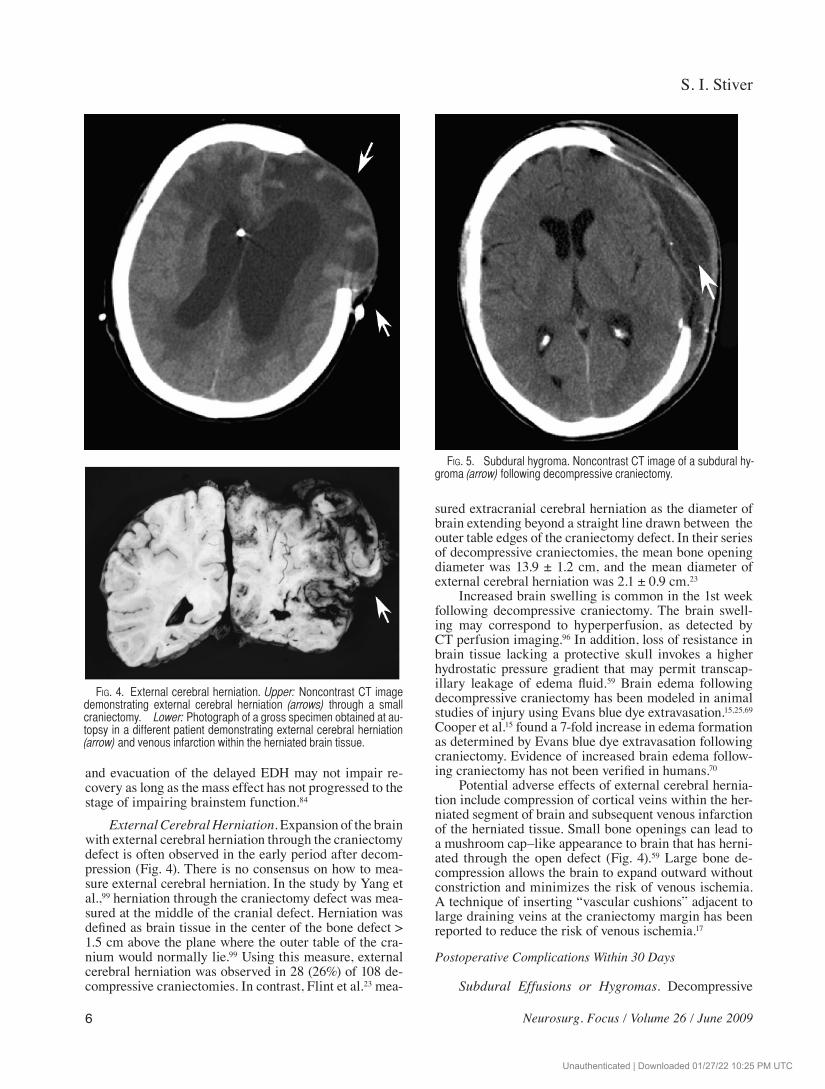
External Cerebral Herniation. Expansion of the brain with external cerebral herniation through the craniectomy defect is often observed in the early period after decom-pression (Fig. 4). There is no consensus on how to mea-sure external cerebral herniation. In the study by Yang et al.,99 herniation through the craniectomy defect was mea-sured at the middle of the cranial defect. Herniation was defined as brain tissue in the center of the bone defect > 1.5 cm above the plane where the outer table of the cra-nium would normally lie.99 Using this measure, external cerebral herniation was observed in 28 (26%) of 108 de-compressive craniectomies. In contrast, Flint et al.23 mea-
sured extracranial cerebral herniation as the diameter of brain extending beyond a straight line drawn between the outer table edges of the craniectomy defect. In their series of decompressive craniectomies, the mean bone opening diameter was 13.9 ± 1.2 cm, and the mean diameter of external cerebral herniation was 2.1 ± 0.9 cm.23
Increased brain swelling is common in the 1st week following decompressive craniectomy. The brain swell-ing may correspond to hyperperfusion, as detected by CT perfusion imaging.96 In addition, loss of resistance in brain tissue lacking a protective skull invokes a higher hydrostatic pressure gradient that may permit transcap-illary leakage of edema fluid.59 Brain edema following decompressive craniectomy has been modeled in animal studies of injury using Evans blue dye extravasation.15,25,69 Cooper et al.15 found a 7-fold increase in edema formation as determined by Evans blue dye extravasation following craniectomy. Evidence of increased brain edema follow-ing craniectomy has not been verified in humans.70
Potential adverse effects of external cerebral hernia-tion include compression of cortical veins within the her-niated segment of brain and subsequent venous infarction of the herniated tissue. Small bone openings can lead to a mushroom cap–like appearance to brain that has herni-ated through the open defect (Fig. 4).59 Large bone de-compression allows the brain to expand outward without constriction and minimizes the risk of venous ischemia. A technique of inserting “vascular cushions” adjacent to large draining veins at the craniectomy margin has been reported to reduce the risk of venous ischemia.17
Postoperative Complications Within 30 Days
Subdural Effusions or Hygromas. Decompressive
Fig. 4. External cerebral herniation. Upper: Noncontrast CT image demonstrating external cerebral herniation (arrows) through a small craniectomy. Lower: Photograph of a gross specimen obtained at au-topsy in a different patient demonstrating external cerebral herniation (arrow) and venous infarction within the herniated brain tissue.
Fig. 5. Subdural hygroma. Noncontrast CT image of a subdural hy-groma (arrow) following decompressive craniectomy.
Unauthenticated | Downloaded 01/27/22 10:25 PM UTC
Neurosurg. Focus / Volume 26 / June 2009
Complications of decompressive craniectomy for TBI
7
craniectomy alters the dynamics of CSF circulation.9,100 This may exacerbate the occurrence of subdural hygro-mas and hydrocephalus. Subdural hygromas develop ear-ly after decompressive surgery (Fig. 5). In the study by Aarabi et al.,1 subdural hygromas developed in 25 (50%) of 50 patients after a mean of 8 days following decom-pressive craniectomy. Hygromas are generally ipsilateral to the skull defect with volumes ranging from 10 to 120 ml (mean 51 ml).1 While most authors favor a mechanism of altered CSF dynamics to account for the occurrence of hygromas, others have suggested that increased cere-bral perfusion pressure that accompanies decompressive craniectomy may play a role.45
Duraplasty at the time of decompression has been observed to lower the incidence of subdural effusions.100 Wrapping the cranium after the peak time of cerebral swelling has also been suggested to help prevent effu-sions.99 In the early experience of decompressive craniec-tomy, Guerra et al.32 reported that 10 (67%) of 15 patients who developed hygromas responded to puncture treat-ment and only 5 (33%) required a ventriculo- or lumbo-peritoneal shunt. In this report, 8 (14%) of the 57 patients developed hydrocephalus. At our institution we avoid tap-ping into CSF hygromas and have found that most resolve spontaneously without need for intervention. Yang et al.99
reported a 21% incidence of subdural hygromas in their series and noted that 20 of 23 effusions resolved sponta-neously without neurological deficits. Aarabi et al.1 have also shown that subdural hygromas following decom-pressive craniectomy regress spontaneously over weeks to months. In their series, no patient required decompres-sion or shunt treatment of a hygroma collection.1 Two of the 25 patients, however, went on to develop delayed hy-drocephalus.
Paradoxical Herniation. Paradoxical herniation with compression of the brainstem and neurological deteriora-tion may present in a delayed fashion after a lumbar punc-ture in patients with decompressive craniectomy.93 Vile-la93 described a case of a patient with a decompressive
craniectomy whose condition suddenly deteriorated 38 days after a lumbar puncture to rule out meningitis. The precipitous deterioration was attributed to upright posi-tioning with mobilization of the patient as she started to participate in rehabilitation therapy. Mobilization into the upright position may have invoked a higher cranial-spinal pressure gradient and perhaps even opened up a preexist-ing small dural hole from the lumbar puncture site. Neu-rological deterioration after lumbar puncture or lumbar CSF drainage in the setting of large cranial defects has also been observed following decompressive craniecto-my for stroke.21,61 In the early period of recovery from traumatic injury following decompressive craniectomy, symptoms of paradoxical herniation may be masked and even mistaken for neurological damage from the trauma.
The concept that a negative pressure gradient be-tween the cranial and spinal compartments, provoked by a spinal CSF leak, can precipitate downward hernia-tion, even in the absence of raised ICP, has been carefully documented by many groups.5,6,42,72,73 With a large skull defect, a lumbar puncture exacerbates the negative pres-sure gradient between the atmosphere and the cranium.93 Based on this, we have adopted a high threshold for per-forming lumbar puncture in patients with a decompres-sive craniectomy. Treatment for symptomatic herniation following lumbar puncture with a skull defect is the an-tithesis of that used for herniation in the setting of raised ICP, hence the term paradoxical herniation. Patients with paradoxical herniation should be treated with intrave-nous fluids, Trendelenburg positioning, and clamping of CSF drainage; hyperosmolar therapy should be discon-tinued, and early attention given to insertion of a blood patch.61,76,93 Cranioplasty may act to reduce the negative pressure gradient and should also be considered in the management of paradoxical herniation.76,93
Delayed Complications After 1 Month
Wound Healing and Infection. There are several factors associated with decompressive craniectomy that
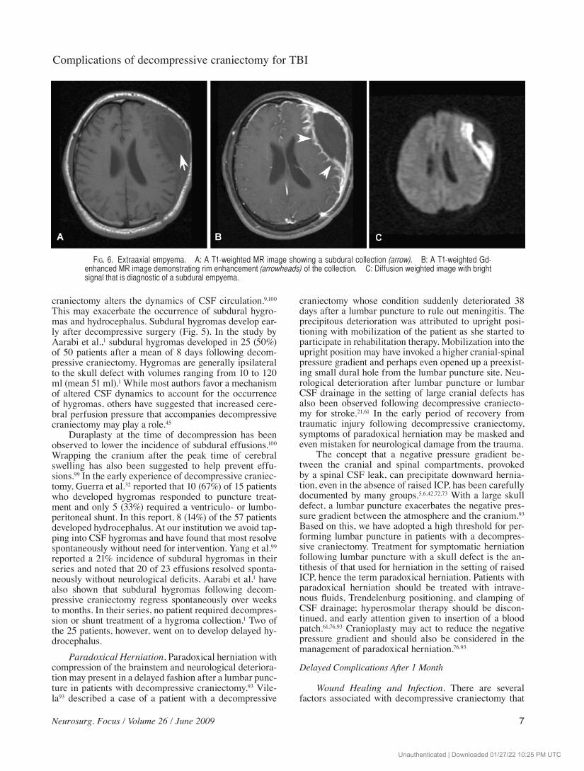
Fig. 6. Extraaxial empyema. A: A T1-weighted MR image showing a subdural collection (arrow). B: A T1-weighted Gd-enhanced MR image demonstrating rim enhancement (arrowheads) of the collection. C: Diffusion weighted image with bright signal that is diagnostic of a subdural empyema.
Unauthenticated | Downloaded 01/27/22 10:25 PM UTC
S. I. Stiver
8 Neurosurg. Focus / Volume 26 / June 2009
should lead one to expect a higher rate of infection than with standard craniotomy for general neurosurgical pro-cedures. The incision varies but the typical, large, reverse question mark incision with a long scalp pedicle on a com-paratively small base predisposes to wound breakdown along the parietal and posterior temporal limbs farthest along the flap. Bone removal is needed low in the tem-poral fossa to decompress the basal cisterns. To expose the scalp and temporalis muscle down to the level of the zygoma, the incision is carried to 1 cm below the zygoma anterior to the tragus. The urgency to decompress may not facilitate careful dissection and preservation of the superficial temporal artery. Sacrifice of the artery may impair perfusion of the scalp pedicle and negatively im-pact wound healing. The dura is not closed primarily.
Duraplasty using a dura substitute is associated with an increased risk of infection.50 If the dura is left open with-out duraplasty, a foreign synthetic material should be laid over the brain surface to prevent adherence of the scalp to the underlying brain. With the large bone flaps used for decompression, opening into the frontal sinus or air cells low in the middle fossa risks delayed meningitis and subdural empyema, especially if a duraplasty is not per-formed. Last, surgical fields in trauma surgery are often clean contaminated. The bone, soft tissues, and brain are easily contaminated by penetrating injury.
In the intensive care unit, fevers are common. We avoid tapping subdural collections and prefer to use con-trast enhancement on CT and MR imaging together with diffusion weighted MR imaging to diagnose infected
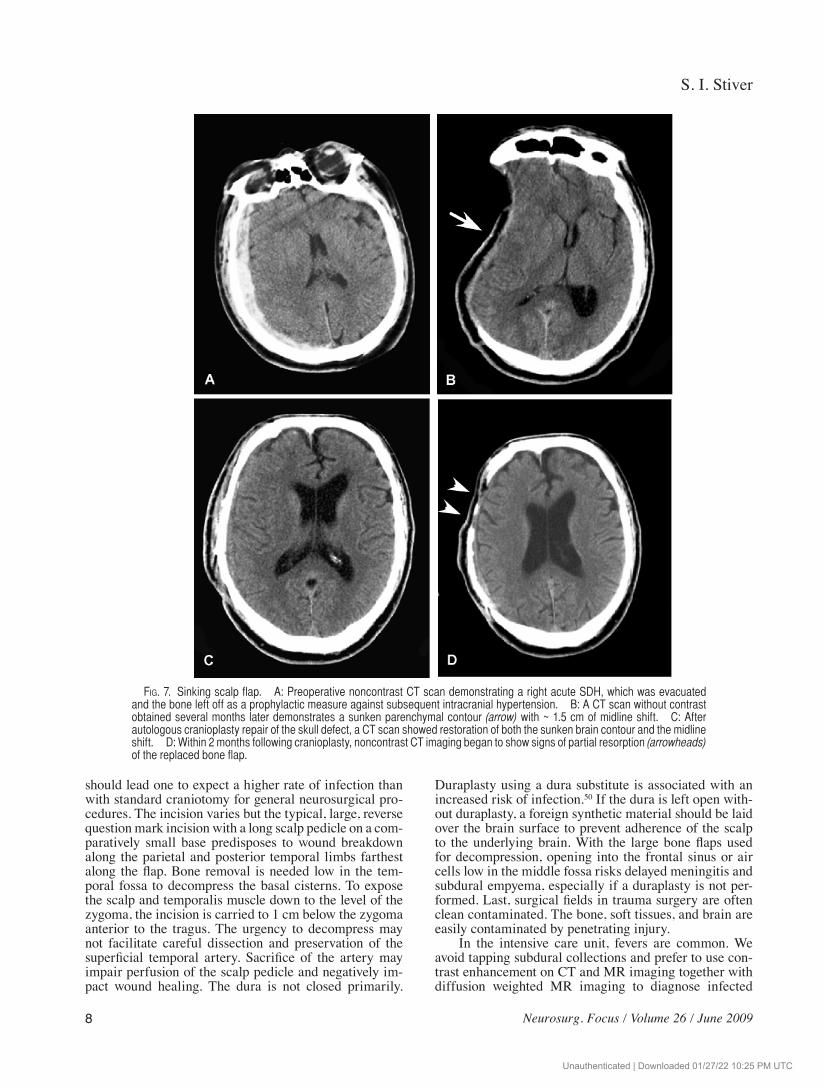
Fig. 7. Sinking scalp flap. A: Preoperative noncontrast CT scan demonstrating a right acute SDH, which was evacuated and the bone left off as a prophylactic measure against subsequent intracranial hypertension. B: A CT scan without contrast obtained several months later demonstrates a sunken parenchymal contour (arrow) with ~ 1.5 cm of midline shift. C: After autologous cranioplasty repair of the skull defect, a CT scan showed restoration of both the sunken brain contour and the midline shift. D: Within 2 months following cranioplasty, noncontrast CT imaging began to show signs of partial resorption (arrowheads) of the replaced bone flap.
Unauthenticated | Downloaded 01/27/22 10:25 PM UTC
Neurosurg. Focus / Volume 26 / June 2009
Complications of decompressive craniectomy for TBI
9
subdural collections (Fig. 6). In many cases, a superficial wound infection aids in the diagnosis of the underlying intracranial problem.
Decompressive craniectomy necessitates a second cranioplasty surgery to replace the bone. Repeat surgery inherently increases risks of infection. In our patient population, we and others have observed high rates of infection following early cranioplasty repair and prefer to wait a minimum of 3 months and generally longer, before replacing the bone.90 Storage of the bone for pro-longed periods in a freezer also increases the risk of infection.60,67,90 To minimize risks of infection through opening of communications to the frontal sinus, we do not remove scar tissue from the area of the frontal sinus during exposure of the craniectomy margins for replace-ment of the bone flap.
Adhesions between the brain and scalp may be a
source of neurological injury during subsequent expo-sure of the brain for the cranioplasty. If a duraplasty is not performed, it is important that the Gelfilm (Upjohn) or another dural substitute cover all aspects of exposed brain to prevent adhesion between the brain surface and the overlying scalp. The disadvantage of Gelfilm is that it appears to take several months for the pseudodura layer to form and completely seal a barrier layer overlying the brain. Duraplasty up front may enable an earlier cranio-plasty repair. A recent report has advocated the use of DuraGen (Integra LifeSciences Corp.) that is laid over the brain without suture closure. Over time the Duragen is replaced by ingrowth of connective tissue that has prop-erties similar to dura.34
Decompressive craniectomy is not an esthetic op-eration. The patient’s scalp contours are disfigured for a period of months. In many patients, asymmetric cerebral
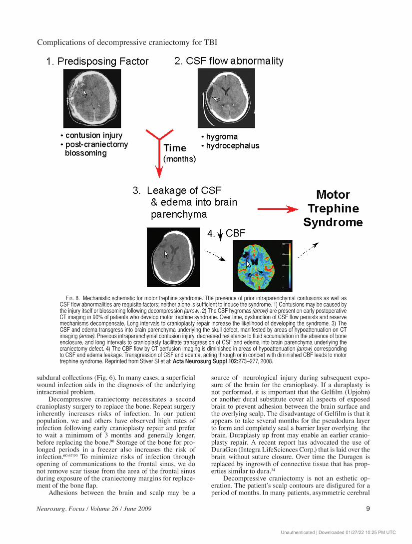
Fig. 8. Mechanistic schematic for motor trephine syndrome. The presence of prior intraparenchymal contusions as well as CSF flow abnormalities are requisite factors; neither alone is sufficient to induce the syndrome. 1) Contusions may be caused by the injury itself or blossoming following decompression (arrow). 2) The CSF hygromas (arrow) are present on early postoperative CT imaging in 90% of patients who develop motor trephine syndrome. Over time, dysfunction of CSF flow persists and reserve mechanisms decompensate. Long intervals to cranioplasty repair increase the likelihood of developing the syndrome. 3) The CSF and edema transgress into brain parenchyma underlying the skull defect, manifested by areas of hypoattenuation on CT imaging (arrow). Previous intraparenchymal contusion injury, decreased resistance to fluid accumulation in the absence of bone enclosure, and long intervals to cranioplasty facilitate transgression of CSF and edema into brain parenchyma underlying the craniectomy defect. 4) The CBF flow by CT perfusion imaging is diminished in areas of hypoattenuation (arrow) corresponding to CSF and edema leakage. Transgression of CSF and edema, acting through or in concert with diminished CBF leads to motor trephine syndrome. Reprinted from Stiver SI et al: Acta Neurosurg Suppl 102:273–277, 2008.
Unauthenticated | Downloaded 01/27/22 10:25 PM UTC
S. I. Stiver
10 Neurosurg. Focus / Volume 26 / June 2009
swelling in the early phases is followed later by markedly sunken scalp contours ipsilateral to the bone decompres-sion (Fig. 7). Cerebral atrophy secondary to brain injury can exacerbate the degree of concavity. The temporalis muscle is detached and atrophies, which may limit the excursion of mouth opening and cause difficulty chewing. Masticatory function, however, does not seem to be sig-nificantly impaired. In 1 study, even after the temporalis muscle was excised, only 2 (25%) of 8 patients experi-enced lateral deviation of the mandible on opening their mouths or had difficulties with eating.62
Hydrocephalus. Hydrocephalus and syndrome of the trephined are the most frequent complications of de-compressive craniectomy beyond 1 month. Decompres-sive craniectomy has been identified as a risk factor for
CSF alterations and development of posttraumatic hydro-cepalus.53 Hydrocephalus has been associated with poorer outcome following TBI.51 Relatively few patients require ventriculoperitoneal shunt treatment before the bone flap has been replaced. Ventriculoperitoneal shunt treatment of hydrocephalus in the setting of a large cranial defect may also risk neurological deterioration consistent with a paradoxical herniation phenomenon.47
Syndrome of the Trephined. Syndrome of the tre-phined is a frequent, delayed complication of decompres-sive craniectomy.30 Common symptoms include headaches, dizziness, irritability, concentration difficulty, memory problems, and mood disturbances, which typically arise weeks to months following decompressive craniectomy. The diagnosis is often overlooked, as many of these symp-toms are also common sequelae to postconcussion and
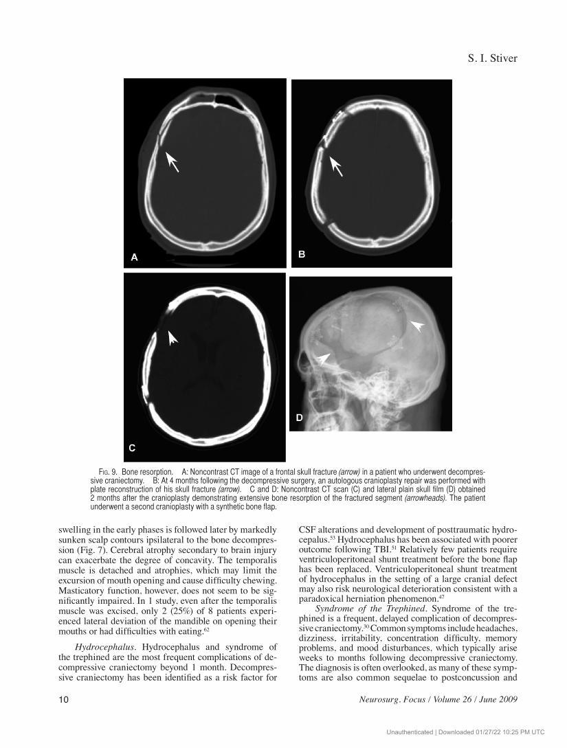
Fig. 9. Bone resorption. A: Noncontrast CT image of a frontal skull fracture (arrow) in a patient who underwent decompres-sive craniectomy. B: At 4 months following the decompressive surgery, an autologous cranioplasty repair was performed with plate reconstruction of his skull fracture (arrow). C and D: Noncontrast CT scan (C) and lateral plain skull film (D) obtained 2 months after the cranioplasty demonstrating extensive bone resorption of the fractured segment (arrowheads). The patient underwent a second cranioplasty with a synthetic bone flap.
Unauthenticated | Downloaded 01/27/22 10:25 PM UTC
Neurosurg. Focus / Volume 26 / June 2009
Complications of decompressive craniectomy for TBI
11
posttraumatic stress syndromes that accompany TBI.Less commonly, syndrome of the trephined can
present with delayed onset of new neurological deficits. We have observed a small cohort of patients who de-veloped motor weakness of the contralateral hand and upper extremity months after their decompressive sur-gery.82 We termed this motor trephine syndrome in ref-erence to Grant’s initial description of syndrome of the trephined.81,82 These motor deficits evolve in an insidi-ous and progressive manner in a limb that was otherwise normal and not affected by the injury. From a series of 170 consecutive patients with TBI who were treated with decompressive craniectomy, we studied 38 patients with long-term follow-up after cranioplasty repair.82 We found 10 patients (26%) with motor trephine syndrome who developed delayed upper-extremity weakness a mean of 5 months following decompressive craniectomy. All ex-perienced marked, rapid improvement of their weakness within a few days of cranioplasty repair with subsequent full motor recovery.
The pathophysiological mechanisms underlying syndrome of the trephined have been a subject of debat-ed theories.19 Changes in atmospheric pressure,83 altered CSF circulation,20,24 and changes in CBF68,85 have all been proposed to explain the pathophysiology underlying the syndrome. In early studies, a sunken scalp was noted in many patients with syndrome of the trephined (Fig. 7).27,98 Recently we discovered a high incidence of CT scanning abnormalities in patients who developed mo-tor trephine syndrome.82 Patients who developed motor trephine syndrome were significantly more likely to have demonstrated CSF subdural hygromas as well as frontal and temporal contusions on their early postoperative CT scans. At later time points, CT scans demonstrated ar-eas of hypodensity consistent with evolving interstitial edema in the brain parenchyma.22 This form of extracel-lular edema was attributed to transependymal flow and accumulation of water and CSF in previously contused areas of parenchyma underlying the skull defect. The CT perfusion imaging studies in 2 patients demonstrated re-duced CBF in these areas of edematous change. Rapid improvements in CBF and resolution of these hypodense areas on CT occurred within days of cranioplasty repair, simultaneous with dramatic recovery of motor function.
We have observed that motor trephine syndrome de-velops in the setting of decompressive craniectomy as a result of 2 predisposing requisite factors (Fig. 8).81,82 First, a contusion or other parenchymal injury imparts a lower threshold for fluid shifts into brain tissue. In the absence of an overlying skull, reduced resistance promotes fluid shifts into areas underlying the skull defect. Second, de-compressive craniectomy impairs normal CSF circula-tion. Over time, impairments of CSF flow, decompensate beyond the phases of subdural hygroma collections and hydrocephalus.1,19,20 De compensated CSF flow manifests at late time points with leakage of CSF and edema fluid into vulnerable areas of injured parenchyma underlying the skull defect. This process is akin to the development of transependymal edema as a late response in normal-pressure hydrocephalus.40 Microcirculatory compromise ensues within the edematous parenchyma, thereby im-
pairing CBF. The combination of decreased cerebral per-fusion and interstitial edema initiates a delayed, slowly progressive motor deficit. Cranioplasty repair quickly normalizes interstitial pressure, CSF flow, transependy-mal fluid shifts, and CBF leading to rapid recovery of mo-tor function.
Patients with decompressive craniectomy should have serial neurological assessments for syndrome of the trephined and complaints of weakness, particularly in activities such as writing and buttoning, which in-volve the small muscles of the hand. Motor testing of the contralateral upper extremity should focus attention on the grip and metacarpal extensor strength. The mo-tor trephine syndrome deficits are reversible, and phys-iotherapy should be involved to prevent development of contractures that could impair long-term function. Early cranioplasty may be considered, noting that the risks of infection are high with early replacement of the autolo-gous bone flap.98,99 However, Liang et al.46 have recently shown that titanium mesh cranioplasty can be performed as early as 5–8 weeks after decompressive craniectomy without untoward infectious complications.
Bone Resorption. In decompressive craniectomy, bone resorption of free bone flaps is common and may ap-proach an incidence as high as 50% in long-term follow-up (Figs. 7 and 9).31,38,66 Skull fractures identified at the time of the original decompression should raise concern for possible bone resorption following cranioplasty (Fig. 9). Multiple fragments should lead to consideration of an upfront synthetic cranioplasty repair. During cranioplasty repair, tight approximation of the free flap to a lengthy surface of the native craniectomy margin reduces bone resorption. In our experience, rigid plate and screw fixa-tion that permits minimal movement of the flap also ap-pears to be important. A modified craniectomy in which the bone flap is not removed but rather is hinged has been proposed.74 In 25 patients, hinge craniotomy was observed to provide adequate decompression of ICP with only 1 infection, 1 cranial deformity, no complications of bone resorption, and no cases of syndrome of the trephined.74 Osteoclast inhibitors may also be used to prevent bone resorption in the future.92
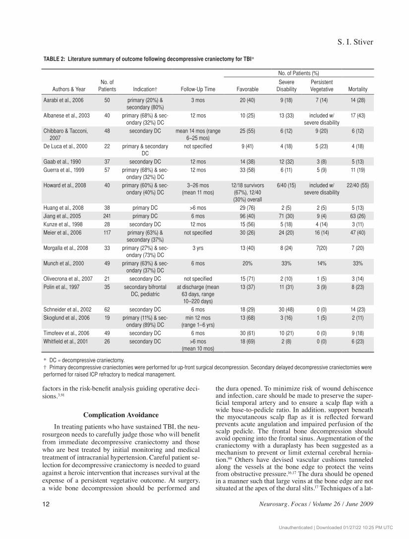
Persistent Vegetative State. Decompressive craniec-tomy is very effective in ameliorating raised ICP as a life-saving measure. While decompressive craniectomy reduces mortality, it may fail to rescue neurological func-tion from devastating injury incurred by either the pri-mary impact or secondary damage that evolves during the early resuscitation period. Risks of survival with an outcome of a persistent vegetative state after decompres-sive craniectomy have been reported to range upwards of 15 to 20% in many series (Table 2). Concerns have been raised that decompressive craniectomy facilitates surviv-al, in a persistent vegetative or minimally conscious state, for patients who would otherwise have died of cerebral herniation from raised ICP.88 Preoperative GCS scores < 6, brainstem dysfunction, older age, and longer time to decompression have been reported to be associated with a higher risk of persistent vegetative outcome, but further study is needed to precisely delineate the role of these
Unauthenticated | Downloaded 01/27/22 10:25 PM UTC
S. I. Stiver
12 Neurosurg. Focus / Volume 26 / June 2009
factors in the risk-benefit analysis guiding operative deci-sions.3,91
Complication AvoidanceIn treating patients who have sustained TBI, the neu-
rosurgeon needs to carefully judge those who will benefit from immediate decompressive craniectomy and those who are best treated by initial monitoring and medical treatment of intracranial hypertension. Careful patient se-lection for decompressive craniectomy is needed to guard against a heroic intervention that increases survival at the expense of a persistent vegetative outcome. At surgery, a wide bone decompression should be performed and
the dura opened. To minimize risk of wound dehiscence and infection, care should be made to preserve the super-ficial temporal artery and to ensure a scalp flap with a wide base-to-pedicle ratio. In addition, support beneath the myocutaneous scalp flap as it is reflected forward prevents acute angulation and impaired perfusion of the scalp pedicle. The frontal bone decompression should avoid opening into the frontal sinus. Augmentation of the craniectomy with a duraplasty has been suggested as a mechanism to prevent or limit external cerebral hernia-tion.99 Others have devised vascular cushions tunneled along the vessels at the bone edge to protect the veins from obstructive pressure.16,17 The dura should be opened in a manner such that large veins at the bone edge are not situated at the apex of the dural slits.17 Techniques of a lat-
TABLE 2: Literature summary of outcome following decompressive craniectomy for TBI*
No. of Patients (%)
Authors & YearNo. of
Patients Indication† Follow-Up Time Favorable Severe
Disability Persistent Vegetative Mortality
Aarabi et al., 2006 50 primary (20%) & secondary (80%)
3 mos 20 (40) 9 (18) 7 (14) 14 (28)
Albanese et al., 2003 40 primary (68%) & sec-ondary (32%) DC
12 mos 10 (25) 13 (33) included w/ severe disability
17 (43)
Ch ibbaro & Tacconi, 2007
48 secondary DC mean 14 mos (range 6–25 mos)
25 (55) 6 (12) 9 (20) 6 (12)
De Luca et al., 2000 22 primary & secondary DC
not specified 9 (41) 4 (18) 5 (23) 4 (18)
Gaab et al., 1990 37 secondary DC 12 mos 14 (38) 12 (32) 3 (8) 5 (13)Guerra et al., 1999 57 primary (68%) & sec-
ondary (32%) DC12 mos 33 (58) 6 (11) 5 (9) 11 (19)
Howard et al., 2008 40 primary (60%) & sec-ondary (40%) DC
3–26 mos (mean 11 mos)
12/18 survivors (67%), 12/40 (30%) overall
6/40 (15) included w/ severe disability
22/40 (55)
Huang et al., 2008 38 primary DC >6 mos 29 (76) 2 (5) 2 (5) 5 (13) Jiang et al., 2005 241 primary DC 6 mos 96 (40) 71 (30) 9 (4) 63 (26)Kunze et al., 1998 28 secondary DC 12 mos 15 (56) 5 (18) 4 (14) 3 (11) Meier et al., 2006 117 primary (63%) &
secondary (37%)not specified 30 (26) 24 (20) 16 (14) 47 (40)
Morgalla et al., 2008 33 primary (27%) & sec-ondary (73%) DC
3 yrs 13 (40) 8 (24) 7(20) 7 (20)
Munch et al., 2000 49 primary (63%) & sec-ondary (37%) DC
6 mos 20% 33% 14% 33%
Olivecrona et al., 2007 21 secondary DC not specified 15 (71) 2 (10) 1 (5) 3 (14)Polin et al., 1997 35 secondary bifrontal
DC, pediatricat discharge (mean
63 days, range 10–220 days)
13 (37) 11 (31) 3 (9) 8 (23)
Schneider et al., 2002 62 secondary DC 6 mos 18 (29) 30 (48) 0 (0) 14 (23)Skoglund et al., 2006 19 primary (11%) & sec-
ondary (89%) DCmin 12 mos
(range 1–6 yrs)13 (68) 3 (16) 1 (5) 2 (11)
Timofeev et al., 2006 49 secondary DC 6 mos 30 (61) 10 (21) 0 (0) 9 (18)Whitfield et al., 2001 26 secondary DC >6 mos
(mean 10 mos)18 (69) 2 (8) 0 (0) 6 (23)
* DC = decompressive craniectomy. † Primary decompressive craniectomies were performed for up-front surgical decompression. Secondary delayed decompressive craniectomies were performed for raised ICP refractory to medical management.
Unauthenticated | Downloaded 01/27/22 10:25 PM UTC
Neurosurg. Focus / Volume 26 / June 2009
Complications of decompressive craniectomy for TBI
13
tice duraplasty have also been suggested to limit external cerebral herniation.55
The presence of a skull fracture contralateral to the craniectomy should prompt consideration for an immedi-ate or very early postoperative CT scan to rule out de-velopment of a mass lesion underlying the fracture.84 If possible, tapping and shunt treatment of CSF subdural hygromas should be avoided. They almost always resolve over time on their own. Lumbar puncture carries a risk of paradoxical herniation at the time of puncture and even months later.
Hydrocephalus is a delayed complication of decom-pressive craniectomy. Few decompressed skulls require shunt treatment. Ventriculoperitoneal shunt treatment in patients with a skull defect can also risk paradoxical herniation. In many cases cranioplasty repair of the skull defect may normalize the impaired CSF flow and obviate the need for a shunt. The patient’s neurological examina-tion should be followed closely with particular attention for signs and symptoms of syndrome of the trephined.
ConclusionsDecompressive craniectomy is viewed by many as a
simple surgical procedure, yet complications may be as common as those associated with complex skull base sur-gery. High mortality rates relate primarily to the sever-ity of the trauma for which the operation is performed. Factors important in patient selection need to be further studied and refined to ensure the highest chances of fa-vorable outcome. Many complications of decompressive craniectomy are inherent in the pathophysiological pro-cesses that ensue following removal of a large portion of skull bone. Other complications, including high rates of infection, derive from the technical aspects of the surgery including large scalp incisions, need for bone storage, and the challenges of sterility accompanying the nature of trauma surgery.
Different types of complications occur in discrete temporal windows. Changes in interstitial pressures and brain compliance, CSF circulation, CBF, and cerebral vasoreactivity and autoregulation play a role in early postoperative complications of contusion blossoming, ex-ternal cerebral herniation, and formation of contralateral extraaxial hematomas. During the first weeks following decompression, subdural hygromas arise secondary to al-terations in brain compliance and CSF circulation. Rare paradoxical herniation syndromes highlight the abnormal pressure dynamics across the cranial-spinal compart-ments following decompression.
In the late phases following decompression, syn-drome of the trephined may lead to cognitive, psycho-logical, and neurological deficits. Patients may develop a motor trephine deficit, typically a new, delayed-onset motor deficit of the contralateral upper extremity. We postulate that these motor trephine deficits arise from de-velopment of interstitial edema in areas of contused and injured brain underlying the skull defect. Motor trephine syndrome deficits are rapidly and completely reversed following cranioplasty repair. Further studies are needed to advance our understanding of the pathophysiological
processes following decompressive craniectomy to obvi-ate complications and provide patients with TBI with the best possible outcomes from this surgery.
Disclaimer
The author reports no conflict of interest concerning the mate-rials or methods used in this study or the findings specified in this paper.
References
1. Aarabi B, Hesdorffer DC, Ahn ES, Aresco C, Scalea TM, Eisenberg HM: Outcome following decompressive craniec-tomy for malignant swelling due to severe head injury. J Neu-rosurg 104:469–479, 2006
2. Abdullah J, Zamzuri I, Awang S, Sayuthi S, Ghani A, Tahir A, et al: Preliminary report on spiegelberg pre and post-operative monitoring of severe head-injured patients who received de-compressive craniectomy. Acta Neurochir Suppl 95:311–314, 2005
3. Albanese J, Leone M, Alliez JR, Kaya JM, Antonini F, Al-liez B, et al: Decompressive craniectomy for severe traumatic brain injury: evaluation of the effects at one year. Crit Care Med 31:2535–2538, 2003
4. Bernstein M, Fleming JFR, Deck JHN: Cerebral hyperperfu-sion after carotid endarterectomy: a cause of cerebral hemor-rhage. Neurosurgery 15:50–56, 1984
5. Binder DK, Dillon WP, Fishman RA, Schmidt MH: Intrathe-cal saline infusion in the treatment of obtundation associated with spontaneous intracranial hypotension: technical case re-port. Neurosurgery 51:830–837, 2002
6. Bloch J, Regli L: Brain stem and cerebellar dysfunction after lumbar spinal fluid drainage: case report. J Neurol Neuro-surg Psychiatry 74:992–994, 2003
7. Bor-Seng-Shu E, Hirsch R, Teixeira MJ, De Andrade AF, Marino R Jr: Cerebral hemodynamic changes gauged by transcranial Doppler ultrasonography in patients with post-traumatic brain swelling treated by surgical decompression. J Neurosurg 104:93–100, 2006
8. Borovich B, Braun J, Guilburd JN, Zaaroor M, Michich M, Levy L, et al: Delayed onset of traumatic extradural hema-toma. J Neurosurg 63:30–34, 1985
9. Carvi Y, Nievas MN, Hollerhage HG: Early combined cranio-plasty and programmable shunt in patients with skull bone de-fects and CSF-circulation disorders. Neurol Res 28:139–144, 2006
10. Chang EF, Meeker M, Holland MC: Acute traumatic intra-parenchymal hemorrhage: risk factors for progression in the early post-injury period. Neurosurgery 58:647–656, 2006
11. Chibbaro S, Tacconi L: Role of decompressive craniectomy in the management of severe head injury with refractory cerebral edema and intractable intracranial pressure. Our experience with 48 cases. Surg Neurol 68:632–638, 2007
12. Chieregato A, Fainardi E, Morselli-Labate AM, Antonelli V, Compagnone C, Targa L, et al: Factors associated with neuro-logical outcome and lesion progression in traumatic subarach-noid hemorrhage patients. Neurosurgery 56:671–680, 2005
13. Cohen JE, Rajz G, Itshayek E, Umansky F: Bilateral acute epidural hematoma after evacuation of acute subdural hema-toma: brain shift and the dynamics of extraaxial collections. Neurol Res 26:763–766, 2004
14. Cooper DJ, Rosenfeld JV, Murray L, Wolfe R, Ponsford J, Da-vies A, et al: Early decompressive craniectomy for patients with severe traumatic brain injury and refractory intracranial hypertension–a pilot randomized trial. J Crit Care 23:387–393, 2008
15. Cooper PR, Hagler H, Clark WK, Barnett P: Enhancement of experimental cerebral edema after decompressive craniecto-
Unauthenticated | Downloaded 01/27/22 10:25 PM UTC
S. I. Stiver
14 Neurosurg. Focus / Volume 26 / June 2009
my: implications for the management of severe head injuries. Neurosurgery 4:296–300, 1979
16. Csokay A, Nagy L, Novoth B: Avoidance of vascular com-pression in decompressive surgery for brain edema caused by trauma and tumor ablation. Neurosurg Rev 24:209–213, 2001
17. Csokay A, Nagy L, Pentelenvi T: “Vascular tunnel” formation to improve the effect of decompressive craniectomy in the treatment of brain swelling caused by trauma and hypoxia. Acta Neurochir (Wien) 143:173–175, 2001
18. De Luca GP, Volpin L, Fornezza U, Cervellini P, Zanusso M, Casentini L, et al: The role of decompressive craniectomy in the treatment of uncontrollable post-traumatic intracranial hy-pertension. Acta Neurochir Suppl 76:401–404, 2000
19. Dujovny M, Agner C, Aviles A: Syndrome of the trephined: theory and facts. Crit Rev Neurosurg 9:271–278, 1999
20. Dujovny M, Fernandez P, Alperin N, Betz W, Misra M, Ma-fee M: Post-cranioplasty cerebrospinal fluid hydrodynamic changes: magnetic resonance imaging quantitative analysis. Neurol Res 19:311–316, 1997
21. Fields JD, Lansberg MG, Skirboll SL, Kurien PA, Wijman CA: “Paradoxical” transtentorial herniation due to CSF drainage in the presence of a hemicraniectomy. Neurology 67:1513–1514, 2006
22. Fishman RA: Cerebrospinal Fluid Disease of the Nervous System. Philadelphia: W.B. Saunders, 1980
23. Flint AC, Manley GT, Gean AD, Hemphill JC III, Rosenthal G: Post-operative expansion of hemorrhagic contusions after unilateral decompressive hemicraniectomy in severe traumat-ic brain injury. J Neurotrauma 25:503–512, 2008
24. Fodstad H, Love JA, Ekstedt J, Friden H, Liliequist B: Ef-fect of cranioplasty on cerebrospinal fluid hydrodynamics in patients with the syndrome of the trephined. Acta Neurochir (Wien) 70:21–30, 1984
25. Gaab M, Knoblich OE, Fuhrmeister U, Pflughaupt KW, Dietrich K: Comparison of the effects of surgical decompres-sion and resection of local edema in the therapy of experi-mental brain trauma. Investigation of ICP, EEG and cerebral metabolism in cats. Childs Brain 5:484–498, 1979
26. Gaab MR, Rittierodt M, Lorenz M, Heissler HE: Traumatic brain swelling and operative decompression: a prospective investigation. Acta Neurochir Suppl (Wien) 51:326–328, 1990
27. George AE, Morantz RA, Abad RM, Rovit RL, Chase N: Neu-roradiology of the posthemicraniectomy patient with special emphasis on the radiology of unilateral atrophy. Radiology 111:627–631, 1974
28. Givner A, Gurney J, O’Connor D, Kassarjian A, Lamorte WW, Moulton S: Reimaging in pediatric neurotrauma: factors associated with progression of intracranial injury. J Pediatr Surg 37:381–385, 2002
29. Grande PO, Asgeirsson B, Nordstrom CH: Physiologic prin-ciples for volume regulation of a tissue enclosed in a rigid shell with application to the injured brain. J Trauma 42:S23–S31, 1997
30. Grant F, Norcross N: Repair of cranial defects by cranioplasty. Ann Surg 110:488–512, 1939
31. Grant GA, Jolley M, Ellenbogen RG, Roberts TS, Gruss JR, Loeser JD: Failure of autologous bone-assisted cranioplasty following decompressive craniectomy in children and adoles-cents. J Neurosurg 100:163–168, 2004
32. Guerra WK, Gaab MR, Dietz H, Mueller JU, Piek J, Fritsch MJ: Surgical decompression for traumatic brain swelling: in-dications and results. J Neurosurg 90:187–196, 1999
33. Hase U, Reulen HJ, Meinig G, Schurmann K: The influence of the decompressive operation on the intracranial pressure and the pressure-volume relation in patients with severe head injuries. Acta Neurochir (Wien) 45:1–13, 1978
34. Horaczek JA, Zierski J, Graewe A: Collagen matrix in de-
compressive hemicraniectomy. Neurosurgery 63:ONS176– ONS181, 2008
35. Howard JL, Cipolle MD, Anderson M, Sabella V, Shollenberg-er D, Li PM, et al: Outcome after decompressive craniectomy for the treatment of severe traumatic brain injury. J Trauma 65:380–386, 2008
36. Huang AP, Tu YK, Tsai YH, Chen YS, Hong WC, Yang CC, et al: Decompressive craniectomy as the primary surgical inter-vention for hemorrhagic contusion. J Neurotrauma 25:1347–1354, 2008
37. Hutchinson PJ, Corteen E, Czosnyka M, Mendelow AD, Menon DK, Mitchell P, et al: Decompressive craniectomy in traumatic brain injury: the randomized multicenter RESCUE-icp study (www.RESCUEicp.com). Acta Neurochir Suppl 96:17–20, 2006
38. Iwama T, Yamada J, Imai S, Shinoda J, Funakoshi T, Sakai N: The use of frozen autogenous bone flaps in delayed cranio-plasty revisited. Neurosurgery 52:591–596, 2003
39. Jaeger M, Soehle M, Meixensberger J: Effects of decompres-sive craniectomy on brain tissue oxygen in patients with in-tracranial hypertension. J Neurol Neurosurg Psychiatry 74:513–515, 2003
40. James AE Jr, Strecker EP, Sperber E, Flor WJ, Merz T, Burns B: An alternative pathway of cerebrospinal fluid absorption in communicating hydrocephalus. Transependymal movement. Radiology 111:143–146, 1974
41. Jiang JY, Xu W, Li WP, Xu WH, Zhang J, Bao YH, et al: Ef-ficacy of standard trauma craniectomy for refractory intra-cranial hypertension with severe traumatic brain injury: a multicenter, prospective, randomized controlled study. J Neu-rotrauma 22:623–628, 2005
42. Komotar RJ, Mocco J, Ransom ER, Mack WJ, Zacharia BE, Wilson DA, et al: Herniation secondary to critical postcran-iotomy cerebrospinal fluid hypovolemia. Neurosurgery 57:286–292, 2005
43. Konig A, Laas R, Herrmann HD: Cerebellar haemorrhage as a complication after supratentorial craniotomy. Acta Neuro-chir (Wien) 88:104–108, 1987
44. Kunze E, Meixensberger J, Janka M, Sorensen N, Roosen K: Decompressive craniectomy in patients with uncontrollable intracranial hypertension. Acta Neurochir Suppl 71:16–18, 1998
45. Lang JK, Ludwig HC, Mursch K, Zimmerer B, Markakis E: Elevated cerebral perfusion pressure and low colloid osmotic pressure as a risk factor for subdural space-occupying hygro-mas? Surg Neurol 52:630–637, 1999
46. Liang W, Xiaofeng Y, Weiguo L, Gang S, Xuesheng Z, Fei C, et al: Cranioplasty of large cranial defect at an early stage after decompressive craniectomy performed for severe head trauma. J Craniofac Surg 18:526–532, 2007
47. Liao CC, Kao MC: Cranioplasty for patients with severe de-pressed skull bone defect after cerebrospinal fluid shunting. J Clin Neurosci 9:553–555, 2002
48. Maas AI, Hukkelhoven CW, Marshall LF, Steyerberg EW: Prediction of outcome in traumatic brain injury with com-puted tomographic characteristics: a comparison between the computed tomographic classification and combinations of computed tomographic predictors. Neurosurgery 57:1173–1182, 2005
49. Makino H, Yamaura A: Assessment of outcome following large decompressive craniectomy in management of serious cerebral contusion. A review of 207 cases. Acta Neurochir Suppl (Wien) 28:193–194, 1979
50. Malliti M, Page P, Gury C, Chomette E, Nataf F, Roux FX: Comparison of deep wound infection rates using a synthetic dural substitute (neuro-patch) or pericranium graft for dural closure: a clinical review of 1 year. Neurosurgery 54:599–603, 2004
51. Marmarou A, Foda MA, Bandoh K, Yoshihara M, Yamamoto
Unauthenticated | Downloaded 01/27/22 10:25 PM UTC
Neurosurg. Focus / Volume 26 / June 2009
Complications of decompressive craniectomy for TBI
15
T, Tsuji O, et al: Posttraumatic ventriculomegaly: hydrocepha-lus or atrophy? A new approach for diagnosis using CSF dy-namics. J Neurosurg 85:1026–1035, 1996
52. Matsuno A, Katayama H, Wada H, Morikawa K, Tanaka K, Tanaka H, et al: Significance of consecutive bilateral surger-ies for patients with acute subdural hematoma who develop contralateral acute epi- or subdural hematoma. Surg Neurol 60:23–30, 2003
53. Mazzini L, Campini R, Angelino E, Rognone F, Pastore I, Oliveri G: Posttraumatic hydrocephalus: a clinical, neurora-diologic, and neuropsychologic assessment of long-term out-come. Arch Phys Med Rehabil 84:1637–1641, 2003
54. Meier U, Lemcke J, Reyer T, Grawe A: Decompressive craniec-tomy for severe head injury in patients with major extracranial injuries. Acta Neurochir Suppl 96:373–376, 2006
55. Mitchell P, Tseng M, Mendelow AD: Decompressive craniec-tomy with lattice duraplasty. Acta Neurochir (Wien) 146:159–160, 2004
56. Morgalla MH, Will BE, Roser F, Tatagiba M: Do long-term results justify decompressive craniectomy after severe trau-matic brain injury? J Neurosurg 109:685–690, 2008
57. Munch E, Horn P, Schurer L, Piepgras A, Paul T, Schmiedek P: Management of severe traumatic brain injury by decom-pressive craniectomy. Neurosurgery 47:315–322, 2000
58. Oertel M, Kelly DF, McArthur D, Boscardin WJ, Glenn TC, Lee JH, et al: Progressive hemorrhage after head trauma: pre-dictors and consequences of the evolving injury. J Neurosurg 96:109–116, 2002
59. Olivecrona M, Rodling-Wahlstrom M, Naredi S, Koskinen LO: Effective ICP reduction by decompressive craniectomy in patients with severe traumatic brain injury treated by an ICP-targeted therapy. J Neurotrauma 24:927–935, 2007
60. Osawa M, Hara H, Ichinose Y, Koyama T, Kobayashi S, Sugita Y: Cranioplasty with a frozen and autoclaved bone flap. Acta Neurochir (Wien) 102:38–41, 1990
61. Oyelese AA, Steinberg GK, Huhn SL, Wijman CA: Paradoxi-cal cerebral herniation secondary to lumbar puncture after decompressive craniectomy for a large space-occupying hemispheric stroke: case report. Neurosurgery 57:E594, 2005
62. Park J, Kim E, Kim GJ, Hur YK, Guthikonda M: External decompressive craniectomy including resection of temporal muscle and fascia in malignant hemispheric infarction. J Neurosurg 110:101–105, 2009
63. Piepmeier JM, Wagner FC Jr: Delayed post-traumatic extra-cerebral hematomas. J Trauma 22:455–460, 1982
64. Plesnila N: Decompression craniectomy after traumatic brain injury: recent experimental results. Prog Brain Res 161:393–400, 2007
65. Polin RS, Shaffrey ME, Bogaev CA, Tisdale N, Germanson T, Bocchicchio B, et al: Decompressive bifrontal craniectomy in the treatment of severe refractory posttraumatic cerebral edema. Neurosurgery 41:84–92, 1997
66. Posnick JC, Goldstein JA, Armstrong D, Rutka JT: Recon-struction of skull defects in children and adolescents by the use of fixed cranial bone grafts: long-term results. Neurosur-gery 32:785–791, 1993
67. Rasmussen S, Ohrstrom JK, Westergaard L, Kosteljanetz M: Post-operative infections of osteoplastic compared with free bone flaps. Br J Neurosurg 4:493–495, 1990
68. Richaud J, Boetto S, Guell A, Lazorthes Y: Effects of cranio-plasty on neurological function and cerebral blood flow. Neu-rochirurgie 31:183–188, 1985
69. Rinaldi A, Mangiola A, Anile C, Maira G, Amante P, Ferra-resi A: Hemodynamic effects of decompressive craniectomy in cold induced brain oedema. Acta Neurochir Suppl (Wien) 51:394–396, 1990
70. Sahuquillo J, Arikan F: Decompressive craniectomy for the treatment of refractory high intracranial pressure in traumat-
ic brain injury. Cochrane Database Syst Rev: CD003983, 2006
71. Sakaki T, Tsujimoto S, Nishitani M, Ishida Y, Morimoto T: Perfusion pressure breakthrough threshold of cerebral auto-regulation in the chronically ischemic brain: an experimental study in cats. J Neurosurg 76:478–485, 1992
72. Samadani U, Huang JH, Baranov D, Zager EL, Grady MS: Intracranial hypotension after intraoperative lumbar cerebro-spinal fluid drainage. Neurosurgery 52:148–151, 2003
73. Schievink WI: Spontaneous spinal cerebrospinal fluid leaks and intracranial hypotension. JAMA 295:2286–2296, 2006
74. Schmidt JH III, Reyes BJ, Fischer R, Flaherty SK: Use of hinge craniotomy for cerebral decompression. Technical note. J Neurosurg 107:678–682, 2007
75. Schneider GH, Bardt T, Lanksch WR, Unterberg A: Decom-pressive craniectomy following traumatic brain injury: ICP, CPP and neurological outcome. Acta Neurochir Suppl 81:77–79, 2002
76. Schwab S, Erbguth F, Aschoff A, Orberk E, Spranger M, Hacke W: [“Paradoxical” herniation after decompressive tre-phining.] Nervenarzt 69:896–900, 1998 (Ger)
77. Servadei F, Nanni A, Nasi MT, Zappi D, Vergoni G, Giuliani G, et al: Evolving brain lesions in the first 12 hours after head injury: analysis of 37 comatose patients. Neurosurgery 37:899–906, 1995
78. Skoglund TS, Eriksson-Ritzen C, Jensen C, Rydenhag B: As-pects on decompressive craniectomy in patients with traumat-ic head injuries. J Neurotrauma 23:1502–1509, 2006
79. Smith JS, Chang EF, Rosenthal G, Meeker M, von Koch C, Manley GT, et al: The role of early follow-up computed to-mography imaging in the management of traumatic brain in-jury patients with intracranial hemorrhage. J Trauma 63:75–82, 2007
80. Stiefel MF, Heuer GG, Smith MJ, Bloom S, Maloney-Wilen-sky E, Gracias VH, et al: Cerebral oxygenation following decompressive hemicraniectomy for the treatment of refrac-tory intracranial hypertension. J Neurosurg 101:241–247, 2004
81. Stiver SI, Wintermark M, Manley GT: Motor trephine syn-drome: a mechanistic hypothesis. Acta Neurochir Suppl 102:273–277, 2008
82. Stiver SI, Wintermark M, Manley GT: Reversible monopare-sis following decompressive hemicraniectomy for traumatic brain injury. J Neurosurg 109:245–254, 2008
83. Stula D: Intracranial pressure measurement in large skull de-fects. Neurochirurgia (Stuttg) 28:164–169, 1985
84. Su TM, Lee TH, Chen WF, Lee TC, Cheng CH: Contralat-eral acute epidural hematoma after decompressive surgery of acute subdural hematoma: clinical features and outcome. J Trauma 65:1298–1302, 2008
85. Suzuki N, Suzuki S, Iwabuchi T: Neurological improvement after cranioplasty. Analysis by dynamic CT scan. Acta Neu-rochir (Wien) 122:49–53, 1993
86. Thibodeau M, Melanson D, Ethier R: Acute epidural hemato-ma following decompressive surgery of a subdural hematoma. Can Assoc Radiol J 38:52–53, 1987
87. Timofeev I, Czosnyka M, Nortje J, Smielewski P, Kirkpatrick P, Gupta A, et al: Effect of decompressive craniectomy on in-tracranial pressure and cerebrospinal compensation following traumatic brain injury. J Neurosurg 108:66–73, 2008
88. Timofeev I, Hutchinson PJ: Outcome after surgical decom-pression of severe traumatic brain injury. Injury 37:1125–1132, 2006
89. Timofeev I, Kirkpatrick PJ, Corteen E, Hiler M, Czosnyka M, Menon DK, et al: Decompressive craniectomy in traumatic brain injury: outcome following protocol-driven therapy. Acta Neurochir Suppl 96:11–16, 2006
90. Tokoro K, Chiba Y, Tsubone K: Late infection after cra-
Unauthenticated | Downloaded 01/27/22 10:25 PM UTC
S. I. Stiver
16 Neurosurg. Focus / Volume 26 / June 2009
nioplasty—review of 14 cases. Neurol Med Chir (Tokyo) 29:196–201, 1989
91. Ucar T, Akyuz M, Kazan S, Tuncer R: Role of decompressive surgery in the management of severe head injuries: prognostic factors and patient selection. J Neurotrauma 22:1311–1318, 2005
92. Vaananen K: Mechanism of osteoclast mediated bone resorp-tion—rationale for the design of new therapeutics. Adv Drug Deliv Rev 57:959–971, 2005
93. Vilela MD: Delayed paradoxical herniation after a decom-pressive craniectomy: case report. Surg Neurol 69:293–296, 2008
94. Wang EC, Ang BT, Wong J, Lim J, Ng I: Characterization of cerebrovascular reactivity after craniectomy for acute brain injury. Br J Neurosurg 20:24–30, 2006
95. Whitfield PC, Patel H, Hutchinson PJ, Czosnyka M, Parry D, Menon D, et al: Bifrontal decompressive craniectomy in the management of posttraumatic intracranial hypertension. Br J Neurosurg 15:500–507, 2001
96. Yamakami I, Yamaura A: Effects of decompressive craniec-tomy on regional cerebral blood flow in severe head trauma patients. Neurol Med Chir (Tokyo) 33:616–620, 1993
97. Yamakami I, Yamaura A, Isobe K: Types of traumatic brain injury and regional cerebral blood flow assessed by 99mTc-HMPAO SPECT. Neurol Med Chir (Tokyo) 33:7–12, 1993
98. Yamaura A, Sato M, Meguro K, Nakamura T, Uemura K: Cranioplasty following decompressive craniectomy–analysis of 300 cases. No Shinkei Geka 5:345–353, 1977
99. Yang XF, Wen L, Shen F, Li G, Lou R, Liu WG, et al: Surgical complications secondary to decompressive craniectomy in patients with a head injury: a series of 108 consecutive cases. Acta Neurochir (Wien) 150:1241–1248, 2008
100. Yang XJ, Hong GL, Su SB, Yang SY: Complications induced by decompressive craniectomies after traumatic brain injury. Chin J Traumatol 6:99–103, 2003
101. Yoshida K, Furuse M, Izawa A, Iizima N, Kuchiwaki H, Inao S: Dynamics of cerebral blood flow and metabolism in patients with cranioplasty as evaluated by 133Xe CT and 31P mag-netic resonance spectroscopy. J Neurol Neurosurg Psychiatry 61:166–171, 1996
102. Zweckberger K, Stoffel M, Baethmann A, Plesnila N: Effect of decompression craniotomy on increase of contusion volume and functional outcome after controlled cortical impact in mice. J Neurotrauma 20:1307–1314, 2003
Manuscript submitted February 15, 2009.Accepted April 1, 2009.Address correspondence to: Shirley Stiver, M.D., Ph.D., San
Francisco General Hospital, 1001 Potrero Avenue, Building 1, Room 101, San Francisco, California 94110-0899. email: [email protected].
Unauthenticated | Downloaded 01/27/22 10:25 PM UTC


