Embed Size (px)
Citation preview
HAL Id: hal-03283042https://hal.archives-ouvertes.fr/hal-03283042
Submitted on 9 Jul 2021
HAL is a multi-disciplinary open accessarchive for the deposit and dissemination of sci-entific research documents, whether they are pub-lished or not. The documents may come fromteaching and research institutions in France orabroad, or from public or private research centers.
L’archive ouverte pluridisciplinaire HAL, estdestinée au dépôt et à la diffusion de documentsscientifiques de niveau recherche, publiés ou non,émanant des établissements d’enseignement et derecherche français ou étrangers, des laboratoirespublics ou privés.
Conformational changes in ammonia-channelingglutamine amidotransferases
Stéphane Mouilleron, Béatrice Golinelli-Pimpaneau
To cite this version:Stéphane Mouilleron, Béatrice Golinelli-Pimpaneau. Conformational changes in ammonia-channelingglutamine amidotransferases. Current Opinion in Structural Biology, Elsevier, 2007, 17 (6), pp.653 -664. �10.1016/j.sbi.2007.09.003�. �hal-03283042�
1
Conformational changes in ammonia-channeling glutamine amidotransferases.
Stéphane Mouilleron1,2, Béatrice Golinelli-Pimpaneau1
1 Laboratoire d’Enzymologie et Biochimie structurales, CNRS Bâtiment 34, 1 avenue
de la Terrasse, 91190 Gif-sur-Yvette, France
Corresponding author: Béatrice Golinelli-Pimpaneau, tel: 33 1 69 82 42 35, fax: 33
1 69 82 31 29, e-mail: [email protected]
2 Present address: Structural Biology Laboratory, Cancer Research UK, London
Research Institute, 44 Lincoln's Inn Fields, London WC2A 3PX, United Kingdom
Running title: Ammonia-channeling glutamine amidotransferases.
* Manuscript
2
Abstract
Glutamine amidotransferases, which catalyze the synthesis of different aminated
products, channel ammonia over 10-40 Å from a glutamine substrate at the
glutaminase site to an acceptor substrate at the synthase site. Ammonia production
usually uses a cysteine-histidine-glutamate triad, or a N-terminal cysteine residue.
Crystal structures of several amidotransferaseligand complexes, mimicking
intermediates along the catalytic cycle, have now been determined. In most cases,
acceptor binding triggers glutaminase activation through domain hinged-movements
and other conformational changes. Structural information shows how flexible loops of
the synthase and glutaminase domains move to shield the two catalytic sites and
anchor the substrates, and how the ammonia channel forms, and opens or closes.
Key words
Ammonia channeling/interdomain signaling/catalytic coupling/glutamine
amidotransferases/ hinge motion
Abbreviations
AS: anthranilate synthase; AsnB: asparagine synthetase B; AT: amidotransferase;
CPS: carbamoyl-phosphate synthetase; CTPS: CTP synthetase; DON: 6-diazo-5-
oxo-L-nor-leucine; GltS: glutamate synthase; Gln: L-glutamine; GSA: glutamyl--
semialdehyde; Fru6P: D-fructose-6-phosphate; Fd-GltS: ferredoxin-dependent
glutamate synthase; FGAM: formylglycinamidine ribonucleotide; FGAR:
formylglycinamide ribonucleotide; FGAR-AT: formylglycinamidine ribonucleotide
amidotransferase; GAT: glutamine amidotransferase; GatCAB: bacterial Glu-tRNAGln
amidotransferase; GatDE: archaeal Glu-tRNAGln amidotransferase; G3P:
glyceraldehyde-3-phosphate; GlmS: glucosamine-6-phosphate synthase; GPAT:
glutamine phosphoribosylpyrophosphate amidotransferase; GMPS: GMP
synthetase; Ntn: amino-terminal nucleophile ; IGP: imidazole glycerol phosphate;
3
IGPS: imidazole glycerol phosphate synthase ; 2-OG: 2-oxo-glutarate ; PLPS: PLP
synthase ; PRA: 5-phosphoribosyl-)1-amine ; PRFAR: N1-(5’-phosphoribosyl)-
formimino-5-aminoimidazole-4-carboxamide ribonucleotide ; PRPP: 5-
phosphoribosyl-()1-pyrophosphate ; R5P: ribulose-5-phosphate
4
Introduction
Glutamine amidotransferases (GATs) play a central role in metabolism since these
enzymes are responsible for the incorporation of nitrogen into amino acids, amino
sugars, purine and pyrimidine nucleotides, coenzymes and antibiotics (Table 1). The
enzymes consist of at least two globular domains or subunits. Hydrolysis of
glutamine in the glutaminase domain yields ammonia, which is transferred to a
synthase (or ATP-dependent synthetase) domain specific for each GAT through a
channel, which is usually 10-40 Å long. The catalytic mechanism consists generally
of the nucleophilic attack of the thiol group of a catalytic cysteine residue on the -
carbonyl group of glutamine to form ammonia, that is next transferred through the
channel to act as a nucleophile on an NH3 acceptor substrate, which differs for each
GAT [1]. Depending on the active sites residues, GATs are categorized into different
unrelated classes (Table 1): class I or triad GATs use histidine and glutamate
residues for the activation of the cysteine thiol group (Fig. 1A) whereas in class II or
N-terminal nucleophile (Ntn) GATs, the catalytic cysteine is at the N-terminus and its
thiol group is activated by the -amino group (Fig. 1B). Other families of GATs,
related to amidases [2], L-asparaginases [3], or nitrilases [4] also exist. The
glutaminase domain of most triad GATs shares a common open structure fold
whereas that of Ntn GATs is composed mainly of antiparallel -sheets.
Interdomain signaling mechanisms are common to all enzymes that use substrate
channeling [5-8]. These ensure the efficacy of the reaction, since the substrate to be
channeled is not produced until the acceptor is ready for the transfer, and it is then
rapidly diffused from one active site to the other through the channel. In addition, the
solvent-inaccessible ammonia channel in GATs prevents the formation of non-
reactive ammonium ions. Hence the enzymes must synchronize their catalytic sites
through conformational changes triggered by acceptor or glutamine binding.
Recently, new structures of GATs have been reported, as well as new complexes of
previously structurally characterized enzymes (Table 2). Here we provide an
5
overview of the progress made in understanding the conformational changes
necessary for shielding the active sites, activating the glutaminase function and
forming the channel, based on the crystallographic structures.
Structural changes upon acceptor binding
Closure of the synthase site by a flexible loop
The conformational changes that occur in the synthase site upon acceptor binding
can be visualized by comparing the structures of the enzyme alone and in the
presence of acceptor substrate. Although there are none amidotransferases for
which both structures have been reported yet (Table 2), we recently solved the
structure of unliganded E. coli glucosamine-6-P synthase (GlmS) (S Mouilleron et al.,
unpublished), in which the glutaminase domains are disordered, as well as the
synthase C-terminal nonapeptide. Comparison with the acceptor-bound structure
[9] indicates that these elements become ordered upon acceptor binding and that
the C-terminal loop covers the synthase site and forms also the major part of the
channel (Fig. 2A).
In addition, the structure of glutamine phosphoribosylpyrophosphate
amidotransferase (GPAT) in the presence of the glutamine affinity analog 6-diazo-5-
oxo-L-nor-leucine (DON) can be compared to that in the presence of both DON and
acceptor substrate and give hints about the conformational changes occurring upon
acceptor binding. The occupation of the synthase site of GPAT triggers the ordering
of a 25-residues flexible loop, which closes the synthase site and forms one wall of
the channel [10,11] (Fig. 2B).
Although the acceptor-bound form of yeast imidazole glycerol phosphate synthase
(IGPS), which also contains a glutamine analog, represents only a precatalytic form
of the enzyme, the comparison of the acceptor-bound and free forms of IGPS
indicates that several loops surrounding the active site move slightly toward the
acceptor substrate when it binds, the largest conformational change being a
reorientation of the partially ordered synthase site loop connecting strand 1 to helix
6
1 [12]. This structural change, which is also observed by comparing the synthase
sites in the three molecules in the asymmetric unit of T. maritima IGPS, in which
either one or two phosphate ions are bound at the synthase site [13], causes the
side chain of Lys258 of the synthase loop to point into the synthase site and interact
with the acceptor substrate.
Recently, several structures of Glu-tRNAGln GATs have been reported (Table 2).
Bacterial Glu-tRNAGln GATs are heterotrimeric proteins composed of A, B, C
subunits (GatCAB) while archaeal enzymes are heterodimeric proteins composed of
D and E subunits (GatDE). Whereas the synthetase subunits, GatB and GatE, are
highly related, ammonia production is achieved by the structurally different GatA and
GatD subunits. The flexible synthetase 120-residues C-terminal region is ordered in
the tRNAGln-bound GatDE structure and interacts with tRNAGln [14], while it is
disordered in the unliganded GatDE [15] and in the unliganded or Gln-bound
GatCAB structures [16]. Therefore, the synthetase tail domain becomes ordered
upon Glu-tRNAGln acceptor binding.
Other examples indicating the closure of the synthase site upon acceptor binding
come from the comparison of different complexed states of enzymes from different
species (Fig. 2C, D and E). In addition, the existence of a flexible loop, which should
close on the synthase site, is indicated by its disorder in the structure in the absence
of the acceptor substrate and by the accessibility of the synthase site to solvent. For
example, the 37-terminal residues of the synthetase domain of asparagine
synthetase B (AsnB) in complex with glutamine and AMP [17] and a 22-residues
peptide of the synthetase domain of GMP synthetase (GMPS) in the absence of
acceptor and donor substrates [18] are disordered, and were suggested to be
stabilized upon acceptor binding. Likewise, a large conformational change upon ATP
and acceptor binding is expected in T. thermophilus CTP synthetase (CTPS)
because no continuous channel connects the synthase and glutaminase sites, the
synthetase active site is exposed to solvent and the binding pocket for the allosteric
effector GTP is not properly formed [19].
7
Stimulation of the glutaminase function upon acceptor binding.
GATs employ different mechanisms to incapacitate their glutaminase function until
an activation signal is received from the remote synthase site. The glutaminase
activity is usually coupled with acceptor binding to the synthase site, although there
are quantitative differences as to the degree of the glutaminase regulation
(Supplementary Table 1, Fig. 1C). It has also been demonstrated in a few cases,
that inactivation by glutamine affinity analogs is dependent upon acceptor binding or
that acceptor binding is followed by glutamine binding [1]. The mechanism of
activation of the glutaminase function upon acceptor binding varies among GATs
and comes from an increase of kcat, a lowering of KmGln or both and involves protein
conformational changes upon acceptor binding at the synthase site to reorganize the
glutaminase site.
Catalytic coupling
Formation of the glutaminase pocket
Glutamine binding results in the closure of the glutaminase site by a flexible loop (Q-
loop) in Ntn GATs (Fig. 1B). Likewise, for some triad GATs such as IGPS [12] and
CTPS [19], a loop of the glutaminase domain becomes ordered or is reorganized
upon glutamine or glutamine analog binding to cover the entrance of the glutaminase
site and shield glutamine.
To control glutaminase activity, glutamine is bound in a non-productive orientation in
the absence of the acceptor substrate, and a specificity pocket is created only at the
appropriate point in the catalytic cycle. Thus, in CTPS, a tyrosine from the synthase
domain becomes ordered only in the presence of glutamine and participates in the
glutaminase active site formation by interacting with the glutamine amide group [19].
Moreover, it was suggested that the glutaminase site of S. typhimurium anthranilate
synthase (AS) would be fully formed only in the presence of acceptor substrate since
parts of the glutaminase site are disordered in its absence [20]. In addition,
comparing the structures of DON-inactivated GPAT in the absence and in the
8
presence of stable acceptor analog [10,21] shows that the glutamine anchoring site
is formed only when the acceptor is bound, through the positioning of several
glutamine-binding residues (Fig. 1C). Finally, the presence in the structure of S.
thyphimurium FGAR-AT of a glutamylthioester intermediate together with a water
molecule positioned to hydrolyze the thioester intermediate indicates that the
reaction did not take place and suggests that completion of the reaction requires a
conformational change associated with acceptor binding [22].
Positioning of the catalytic residues
Another way GATs use to regulate glutamine hydrolysis is to optimally position the
catalytic residues and the oxyanion hole (comprising two amide nitrogens, which
stabilize the transient negative charge developing on the carbonyl oxygen during
glutamine hydrolysis), only after acceptor is bound. Because acceptor binding
stimulates glutamine hydrolysis, the conformational changes occurring upon
glutamine binding that lead to catalysis in GATs can be visualized by comparing the
structures in the presence of acceptor, and in the presence of both acceptor and
glutamine analogs, which represents the active conformation of the enzyme before
the ammonia transfer step. Such structures have been determined for only two Ntn
ATs, GlmS and ferredoxin-dependent glutamate synthase (Fd-GltS).
Glutamine binding to GlmS activates the glutaminase function by positioning the
oxyanion hole asparagine residue through a 100° rotation of its side chain, and by
enhancing the nucleophilic character of the -amino group of Cys1 [9]. Similarly, it
has been suggested that upon acceptor binding to Fd-GltS, the -amino group of
Cys1 would be activated by a hydrogen bond with residue Glu1013 of loop 4 from
the synthase domain, due to a movement of this loop [23]. Nevertheless, the
oxyanion hole asparagine is in an active conformation when the glutaminase site is
empty in Fd-GltS [24] and when the synthase site is empty in GPAT [21] and AsnB
[17].
There are other hints indicating that conformational changes in the glutaminase site
are necessary to activate the glutaminase function. Thus, in the structure of IGPS in
9
complex with a glutamine analog but no acceptor substrate, the glutaminase site is
not in a fully active state because the oxyanion hole is not completely formed [25]. It
has been proposed that a conformational change induced by acceptor binding would
reorganize the oxyanion strand and form the oxyanion hole [12]. In addition, the
glutaminase site is closed in unliganded AS [26]. Therefore, conformational changes
accompanying acceptor binding could force the glutaminase subunit to switch from a
nonfunctional to a functional conformation, allowing glutamine to enter and be
hydrolyzed.
A synthase flexible loop is involved in the coupling of the two catalytic sites
In all GATs, the communication between the two active sites is mediated by
residues, which constitute the ammonia channel. For instance, in GlmS, the C-
terminal residues, in particular Thr606, the only residue from the synthase domain
taking part to the glutaminase site (Fig. 1B), and Lys603, whose peptide bond flips
upon DON binding, are crucial elements in the coupling of the two active sites [9].
In Fd-GltS, Glu1013 (equivalent to Thr606 of GlmS, Fig. 1B) at the C-terminus of
loop 4 of the synthase domain, which covers the synthase site and forms part of the
wall of the channel [24], has been shown to be crucial for glutaminase activation and
coupling of the glutaminase and synthase sites [27]. In GPAT, Ile335 belonging to
the synthase flexible loop, which covers the synthase site, is involved in the
activation of the glutaminase function by contacting Tyr74 of the Q-loop (Fig. 2B),
which leads to the repositioning of Arg73 to interact with the -carboxyl group of
glutamine (Fig. 1C). Finally, in IGPS, Lys258, which belongs to a synthase loop
covering the synthase site and is positioned to interact with the acceptor substrate
when it binds, has been shown by mutagenesis to be implicated in signaling [28].
Hinge movement
Hinge domain motions involved in coupling catalysis in the two remote active sites
have been demonstrated for GlmS by comparing the structures of GlmS in complex
with acceptor and in complex with both acceptor and DON (Supplementary Fig. 1A)
[9,29]. This domain rotation of 23° is necessary to allow the closure of the Q-loop
10
over the glutaminase site without changing the dimer interface. Hinge movements of
the synthase and glutaminase domains relative to each other seem to be a general
property of GATs because partially closed conformations have been observed for
sulfate-bound CTPS, and for IGPS in complex with acceptor substrate and glutamine
analog (Supplementary Fig. 1B and 1C).
Conformational changes upon complex assembly
Several conformational changes occur also during assembly of the enzymatic
complex from different subunits or during oligomerization to form an active enzyme.
Thus, the recently reported structures of PLPS from B. subtilis and T. maritima
[30,31] can be compared to the structures of the individual synthase [30,32]
and glutaminase subunits [30,33,34] from several organisms to shade light on the
conformational changes occuring upon complex assembly. Upon complexation with
the glutaminase subunit, the 18-residues N-terminus of the synthase subunit
becomes ordered, as well as an extra helix 2’. In fact, the N-terminal -helix on the
synthase subunit directs the macromolecular assembly of twelve synthase and
twelve glutaminase subunits in PLPS and also forms one side of the putative
ammonia channel (Supplementary Fig. 2). Moreover, the formation of the oxyanion
hole, which is linked to the conformational change of the N-terminal -helix, occurs
only upon complexation with the synthase subunit. Similarly, a reactive conformation
of the oxyanion hole is observed for GlmS [9] and not for the glutaminase subunit
alone [35].
Then, comparison of the structures of yeast IGPS and T. maritima glutaminase
subunit alone [36], as well as mutagenesis studies [37], suggest that a lysine residue
belonging to the glutaminase loop carrying the His and Glu catalytic residues may
regulate the glutaminase function upon association of the glutaminase and synthase
domains by altering the loop conformation [37].
Finally, in the absence of ATP and acceptor substrate, CTPS maintains an
equilibrium between monomer, dimer and tetramer states in solution while in their
presence or in the crystal, the enzyme folds into an active homotetramer, with the
11
walls of the synthase active site contributed by three different subunits [19].
Formation of the channel
Distinct ammonia channels are used by GATs because the channels are formed
primarily by the synthase domains, which are not related to each other (Fig. 3).
Significant conformational changes accompanying substrate binding are required for
the formation of the channel in most GATs.
However, the channels are apparent in some structures in the absence of acceptor,
like in carbamoyl-phosphate synthetase (CPS) [38] and AsnB [17]. Nevertheless, in
the unliganded structures of IGPS, the channel is formed but obstructed by four
charged residues forming a salt-bridge ring that serve as a gate between the two
domains (Fig. 3A) [12,13,39,40]. Acceptor binding has been suggested to induce the
opening of the IGPS channel. In contrast, channel formation is dependent on
acceptor binding in GPAT [10] or GlmS (Fig. 3B) (S Mouilleron et al., unpublished)
because the flexible loop that closes the synthase site upon acceptor binding is a
major component of the channel. In Fd-GltS and NADPH-GltS, the channel, which is
formed by residues belonging to loop 4 also involved in acceptor binding [23], is fully
formed but is unfortunately obstructed by the C-terminal residues of loop 4 in the
structure in complex with DON and acceptor because of crystal packing constraints
[24] (Fig. 3C). Yet, the channel is expected to open upon substrate(s) binding.
In the unliganded E. coli CTPS structure, two cavities in the glutaminase and
synthase domains define an apparent ammonia diffusion path (Fig. 3D) [41] whereas
in T. thermophilus CTPS, the channel is not formed. In GMPS, both active sites are
exposed to solvent and the channel is not apparent [18]. Likewise, some
conformational changes are needed in AS [20,26] or PLPS (Supplementary Fig. 2)
[30,31] to create a continuous channel from the observed adjacent cavities. In
FGAR-AT, two possible paths for ammonia exist through a gate formed by two
phenylalanine residues [22].
12
Opening of the ammonia channel
Although the mechanism of ammonia channel opening has been unveiled only for a
few GATS, a model for it has be proposed in a few other cases.
The C-terminal loop of GlmS forms part of the ammonia channel, connecting the two
active sites. In the presence of the acceptor alone, the channel of GlmS is totally
closed by the indole ring of Trp74, which explains why GlmS cannot use ammonia as
a nitrogen donor. DON binding induces the opening of the ammonia channel through
rotation of the Trp74 indole side-chain, allowing the connection between the two
active sites (Fig. 3B) [9]. An opening mechanism of the same amplitude to that
occurring in GlmS has been observed for an indole-channeling enzyme, tryptophan
synthase (Fig. 3E) [42].
In GPAT, the residue equivalent to Trp74 in Glms, Tyr74, lines the ammonia channel
and has been shown by mutagenesis experiments to be a key residue in coupling
the glutamine and acceptor sites upon acceptor binding [21]. In GPAT, Tyr74 may
play a similar gate role as Trp74 in GlmS.
In E. coli CTPS, His57 has been proposed to act as the gate of the channel because
it can adopt two different conformations that open or close the ammonia channel
(Fig. 3D)[41].
Of the four charged residues blocking the channel in IGPS (Fig. 3A), the lysine
residue is the most plausible candidate for the door of the channel [25,40] because it
displays the largest conformational flexibility in the crystal structures of the enzyme
[40], and it adopts two different conformations in the structure of the isolated
synthase subunit of T. maritima IGPS. In one of them, which may represent the open
conformation of the channel, it is free from the cyclic salt bridge and makes a H-bond
with a residue of the synthase domain.
Other conformational changes
In addition, other conformational changes involving other steps of the reaction, such
as ATP binding to the synthetase domain, exist. For instance, a flexible loop of
13
FGAR-AT partially shields ATP from solvent upon complex formation [22]. Moreover,
different conformations of the B-domain of the carboxyphosphate synthetic unit in
CPS are observed depending on its liganded state [38,43,44]: occupation of the
phosphate-binding site by inorganic phosphate or by a non-hydrolyzable analog of
ATP triggers the closure of the B-domain of both synthetic units over the active-site
pocket.
Conclusion
The main functional feature of GATs is their ability to coordinate the activity of their
two functional sites in order to avoid a wasteful consumption of glutamine [1].
Generally, hydrolysis of glutamine occurs significantly only when the acceptor is
bound to the synthase domain, initiating an interdomain signal transduction that
activates the glutaminase function. Thus, the active conformation of the glutaminase
site is achieved only after acceptor binding. In GATs, flexible loops of the synthase
and glutaminase domains seem to function as gates to give access to the active site
only at proper time by adopting open and closed conformations. Yet, the ammonia
channel is formed and open at different moments during catalysis, for each GAT.
14
Figure legends
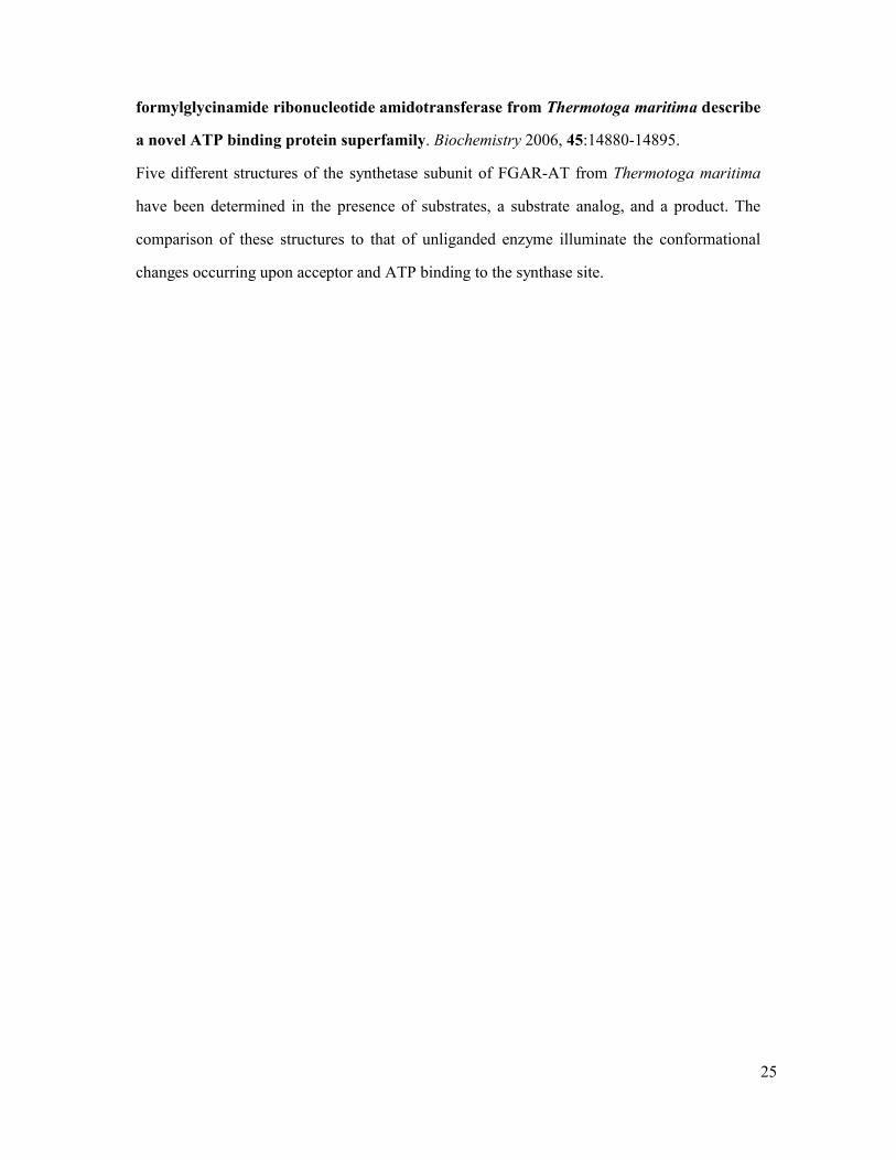
Fig. 1: Comparison of the glutaminase active sites in triad GATs and Ntn GATs.
A Triad GATs
The residue numbering is based on the AS sequence. The conserved residues of the
glutaminase site (Pro50, Gly58, Gly60, Cys85, Gln89, His172, Glu174) of different
triad GATs have been superimposed on those of AS. The residues of the
glutaminase site are shown in stick representation, colored in green (AS, PDB code
1I7Q), yellow (CPS, PDB code 1C3O), grey (CTPS, PDB code 1VCO), orange
(FGAR-AT, PDB code 1T3T), pink (GMPS, PDB code 1GPM), cyan (PLPS, PDB
code 2ISS). In this superposition, all enzymes are in complex with Gln or DON,
except GMPS and PLPS. Indeed, for GMPS, only the unliganded structure has been
reported and in the PLPSGln complex, glutamine is oriented differently in the
glutaminase site. In triad GATs, the oxyanion hole is formed by one peptide nitrogen
belonging to the residue immediately following the nucleophile cysteine and the
second to an adjacent -strand called the “oxyanion strand”. The relative position of
the residues of the catalytic triad (Cys85, His172, Glu174) and of the oxyanion hole
(NH groups of Gly58 and Leu86) is conserved. The glutamine carboxylate is bound
by the main-chain amides of Ser135 and Leu136 and by the side-chain amide of
Gln89. The -amino group of glutamine is bound either by main-chain carbonyl
groups or by oxygen atoms of an Asp/Glu residue. In the absence of a ligand at the
glutaminase site, the residues in GMPS, potentially involved in binding the
carboxylate and -amino groups of glutamine, are not in their competent binding
conformation.
B: Ntn GATs.
The residue numbering is that of GlmS. The C atoms of several conserved residues
of the glutaminase site (Arg26, Arg73, Asn98, Gly99, Asp123) of different Ntn GATs
in complex with glutamine or DON have been superimposed on those of GlmS. The
residues of the glutaminase site are shown in stick representation, colored in green
(GlmS, PDB code 2J6H, grey (E. coli GPAT, PDB code 1ECC), purple (B. subtilis
15
GPAT, PDB code 1A0O), pink (Cys1Ala mutant of AsnB, PDB code 1CT9) or yellow
(GltS, PDB code 1OFE). The hydrogen bond between the carbonyl oxygen atom of
Cys1 and the guanidinium group of Arg26 is conserved in all Ntn GATs. The side-
chain carbonyl group of glutamine or DON is bound by the oxyanion hole formed by
the backbone amide group of a conserved glycine residue and N2 of a conserved
asparagine residue. The carboxylate group of glutamine or DON forms a salt bridge
with a conserved argine residue and its amino group makes H-bonds with a
conserved aspartate residue and with the carbonyl group of the glycine involved in
the oxyanion hole. The Q-loop, which protects L-glutamine from bulk solvent is
maintained in an open conformation in the structure of acceptor-bound Fd-GltS in
complex with DON due to crystal packing constraints [24]. An oxygen atom of a
residue belonging to the synthase domain (Thr606 in GlmS, Glu1013 in GltS,
Asp387 in GPAT) or a water molecule in AsnB and E. coli GPAT H-binds to the
carboxylate group of glutamine or DON.
C: Activation of the glutaminase function upon acceptor binding.
Superposition of the glutaminase site of GPAT in complex with DON (in orange) [21],
and in complex with DON and cPRPP (in white) [10]. In GPAT, the reorganization of
the Q-loop (residues 73-84) upon acceptor binding to the DON-inactivated enzyme is
accompanied by a reorientation of Arg73 in an optimal position to form a salt bridge
with the carboxylate group of DON [10]. In addition, the carbonyl side-chain of
Asp127 and the hydroxyl group of Thr76 are positioned to make hydrogen bonds to
the amino group of the glutamine analog. These conformational changes likely
explain the 110-fold lower glutamine Km for the glutaminase activity when the
acceptor is present [21].
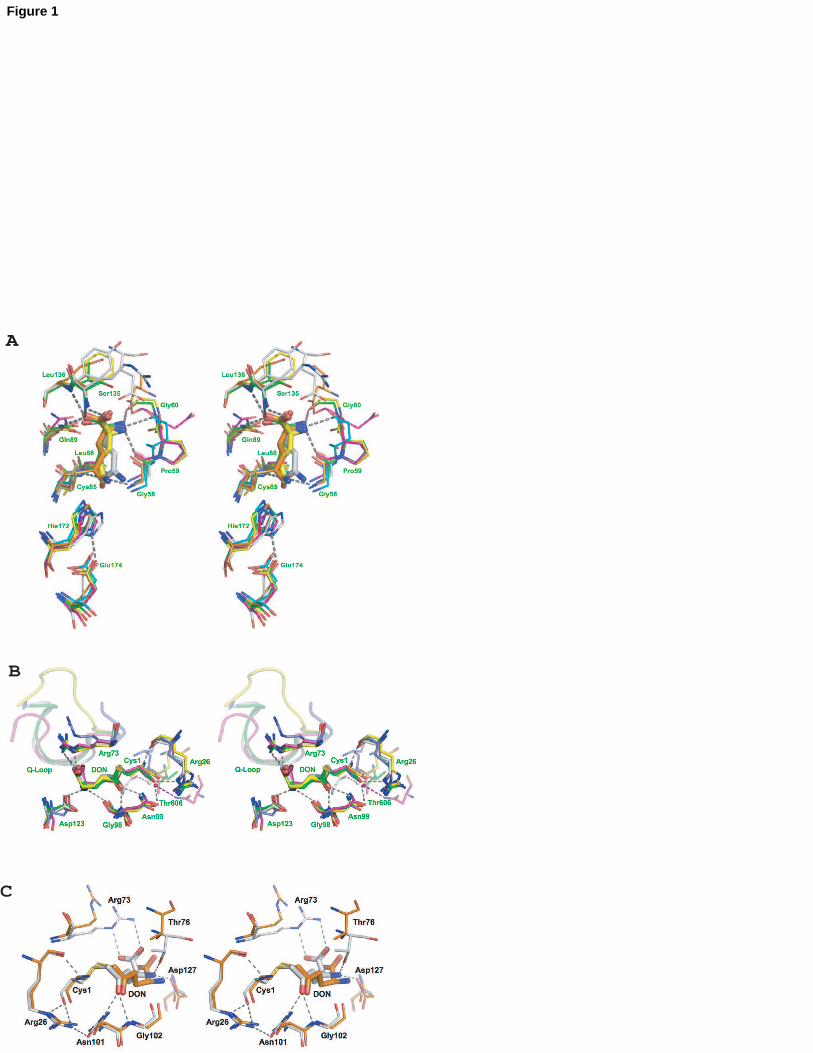
Fig. 2: Closing of the synthase site upon acceptor binding.
A GlmS
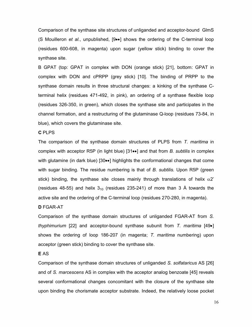
16
Comparison of the synthase site structures of unliganded and acceptor-bound GlmS
(S Mouilleron et al., unpublished, [9] shows the ordering of the C-terminal loop
(residues 600-608, in magenta) upon sugar (yellow stick) binding to cover the
synthase site.
B GPAT (top: GPAT in complex with DON (orange stick) [21], bottom: GPAT in
complex with DON and cPRPP (grey stick) [10]. The binding of PRPP to the
synthase domain results in three structural changes: a kinking of the synthase C-
terminal helix (residues 471-492, in pink), an ordering of a synthase flexible loop
(residues 326-350, in green), which closes the synthase site and participates in the
channel formation, and a restructuring of the glutaminase Q-loop (residues 73-84, in
blue), which covers the glutaminase site.
C PLPS
The comparison of the synthase domain structures of PLPS from T. maritima in
complex with acceptor R5P (in light blue) [31] and that from B. subtilis in complex
with glutamine (in dark blue) [30] highlights the conformational changes that come
with sugar binding. The residue numbering is that of B. subtilis. Upon R5P (green
stick) binding, the synthase site closes mainly through translations of helix 2’
(residues 48-55) and helix 310 (residues 235-241) of more than 3 Å towards the
active site and the ordering of the C-terminal loop (residues 270-280, in magenta).
D FGAR-AT
Comparison of the synthase domain structures of unliganded FGAR-AT from S.
thyphimurium [22] and acceptor-bound synthase subunit from T. maritima [49]
shows the ordering of loop 186-207 (in magenta; T. maritima numbering) upon
acceptor (green stick) binding to cover the synthase site.
E AS
Comparison of the synthase domain structures of unliganded S. solfataricus AS [26]
and of S. marcescens AS in complex with the acceptor analog benzoate [45] reveals
several conformational changes concomitant with the closure of the synthase site
upon binding the chorismate acceptor substrate. Indeed, the relatively loose pocket
17
of the synthase site in the unliganded structure has to narrow upon acceptor binding.
Benzoate is shown in pink stick representation.
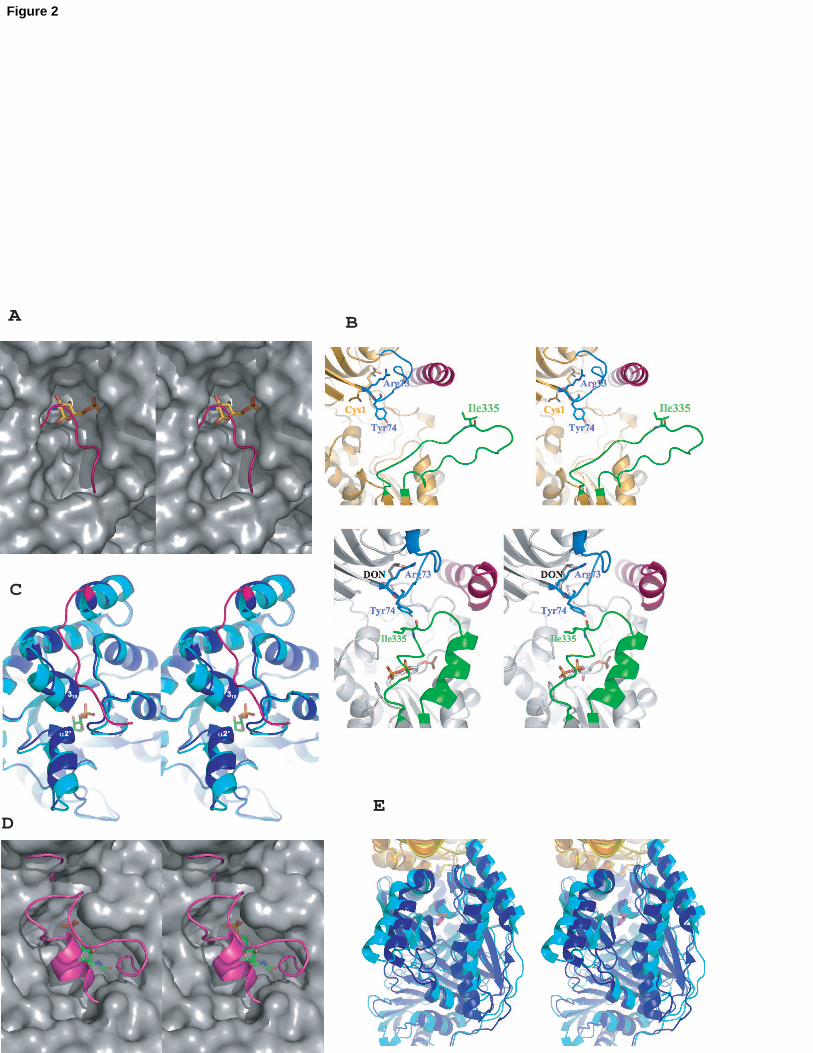
Fig. 3: Open and closed forms of channels
A IGPS
In IGPS, the core of a ()8barrel is used for the transfer of ammonia between the
glutaminase and synthase sites. The acceptor substrate PRFAR (green stick) binds
at the top of the barrel and the acivicin glutamine analog (pink stick) is located at its
bottom the interface between the glutaminase and synthase domains. A cyclic salt
bridge formed by Arg239, Glu293, Lys360, Glu465 (yeast numbering, shown in stick
representation) closes the bottom of the barrel.
B GlmS
Trp74 acts as the gate of the channel in GlmS (closed conformation as observed in
the crystal structure of GlmS in complex with fructose-6-P in yellow, open
conformation as observed in the crystal structure of GlmS in complex with DON and
Fru6P in orange) [9]. The accessible surface of the channel calculated with a
probe radius of 1.4 Å is represented as a mesh surface. The walls of the ammonia
channel are constituted by Trp74 and Arg26 from the glutaminase domain, and by
residues of the C-terminal loop of the synthase domain (orange coil).
C Fd-GltS
The channel of Fd-GltS is formed by residues of loop 4 (residues 968-1013) of the
synthase domain, which interacts with the -amino terminal cysteine and is involved
in the binding of the acceptor substrate. [23]. The accessible surface of the channel
calculated with a probe radius of 1.4 Å is represented as a mesh surface.
Unfortunately, in the structure in complex with DON and acceptor substrate (DON in
pink stick, 2OG in green stick, FMN in cyan stick), the channel is obstructed both at
the entrance (by Ser1011 and Ile1012 of loop4, and by Thr503 and Asn504 of the
central domain) and near the acceptor by Glu903 and Lys966 because crystal
packing constraints hinder a conformational change of the Q-loop that is necessary
18
to open the channel [24].
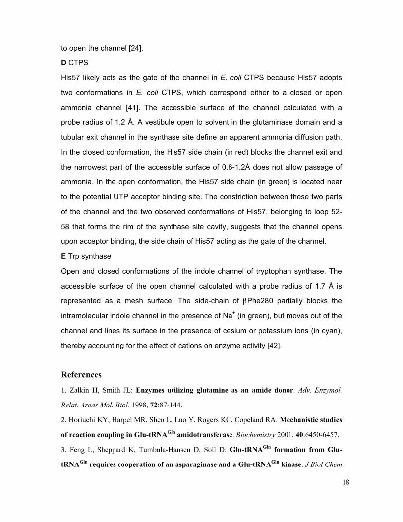
D CTPS
His57 likely acts as the gate of the channel in E. coli CTPS because His57 adopts
two conformations in E. coli CTPS, which correspond either to a closed or open
ammonia channel [41]. The accessible surface of the channel calculated with a
probe radius of 1.2 Å. A vestibule open to solvent in the glutaminase domain and a
tubular exit channel in the synthase site define an apparent ammonia diffusion path.
In the closed conformation, the His57 side chain (in red) blocks the channel exit and
the narrowest part of the accessible surface of 0.8-1.2Å does not allow passage of
ammonia. In the open conformation, the His57 side chain (in green) is located near
to the potential UTP acceptor binding site. The constriction between these two parts
of the channel and the two observed conformations of His57, belonging to loop 52-
58 that forms the rim of the synthase site cavity, suggests that the channel opens
upon acceptor binding, the side chain of His57 acting as the gate of the channel.
E Trp synthase
Open and closed conformations of the indole channel of tryptophan synthase. The
accessible surface of the open channel calculated with a probe radius of 1.7 Å is
represented as a mesh surface. The side-chain of Phe280 partially blocks the
intramolecular indole channel in the presence of Na+ (in green), but moves out of the
channel and lines its surface in the presence of cesium or potassium ions (in cyan),
thereby accounting for the effect of cations on enzyme activity [42].
References
1. Zalkin H, Smith JL: Enzymes utilizing glutamine as an amide donor. Adv. Enzymol.
Relat. Areas Mol. Biol. 1998, 72:87-144.
2. Horiuchi KY, Harpel MR, Shen L, Luo Y, Rogers KC, Copeland RA: Mechanistic studies
of reaction coupling in Glu-tRNAGln amidotransferase. Biochemistry 2001, 40:6450-6457.
3. Feng L, Sheppard K, Tumbula-Hansen D, Soll D: Gln-tRNAGln formation from Glu-
tRNAGln requires cooperation of an asparaginase and a Glu-tRNAGln kinase. J Biol Chem
19
2005, 280:8150-8155.
4. Bieganowski P, Pace HC, Brenner C: Eukaryotic NAD+ synthetase Qns1 contains an
essential, obligate intramolecular thiol glutamine amidotransferase domain related to
nitrilase. J Biol Chem 2003, 278:33049-33055.
5. Anderson KS: Fundamental mechanisms of substrate channeling. Methods Enzymol.
1999, 308:111-145.
6. Miles EW, Rhee S, Davies DR: The molecular basis of substrate channeling. J Biol
Chem 1999, 274:12193-12196.
7. Raushel FM, Thoden JB, Holden HM: The amidotransferase family of enzymes:
molecular machines for the production and delivery of ammonia. Biochemistry 1999,
38:7891-7899.
8. Huang X, Holden HM, Raushel FM: Channeling of substrates and intermediates in
enzyme-catalyzed reactions. Annu. Rev. Biochem. 2001, 70:149-180.
9. Mouilleron S, Badet-Denisot M-A, Golinelli-Pimpaneau B: Glutamine binding opens
the ammonia channel and activates glucosamine-6P synthase. J. Biol. Chem. 2006,
281:4404-4412.
The crystal structures of acceptor-bound GlmS with and without a glutamine affinity analog,
respectively at 2.35 Å and 2.05 Å resolution, show that glutamine binding activates the
enzyme through the closing of a loop to shield the glutaminase site, the positioning of several
catalytic residues and the opening of the ammonia channel thanks to a rotation of the Trp74
indole group.
10. Krahn JM, Kim JH, Burns MR, Parry RJ, Zalkin H, Smith JL: Coupled formation of an
amidotransferase interdomain ammonia channel and a phosphoribosyltransferase active
site. Biochemistry 1997, 36:11061-11068.
11. Muchmore CR, Krahn JM, Kim JH, Zalkin H, Smith JL: Crystal structure of glutamine
phosphoribosylpyrophosphate amidotransferase from Escherichia coli. Protein Sci 1998,
7:39-51.
12. Chaudhuri BN, Lange SC, Myers RS, Davisson VJ, Smith JL: Toward understanding
the mechanism of the complex cyclization reaction catalyzed by imidazole
20
glycerolphosphate synthase: crystal structures of a ternary complex and the free
enzyme. Biochemistry 2003, 42:7003-7012.
13. Douangamath A, Walker M, Beismann-Driemeyer S, Vega-Fernandez MC, Sterner R,
Wilmanns M: Structural evidence for ammonia tunneling across the (beta alpha)(8)
barrel of the imidazole glycerol phosphate synthase bienzyme complex. Structure 2002,
10:185-193.
14. Oshikane H, Sheppard K, Fukai S, Nakamura Y, Ishitani R, Numata T, Sherrer RL, Feng
L, Schmitt E, Panvert M, et al.: Structural basis of RNA-dependent recruitment of
glutamine to the genetic code. Science 2006, 312:1950-1954.
The crystal structure of GatDE from Methanothermobacter thermautotrophicus in complex
with tRNAGln at 3.1 Å resolution shows that specific recognition of Glu-tRNAGln by GatDE is
achieved by the enfolding of the acceptor-T helix of tRNA, and by shape complementarity
with its T arm and D-loop. Moreover, the use of tRNA mutants indicates that the A1-U72
base pair in tRNAGln is important for recognition by GatDE. An explanation for the specificity
of GatDE for Glu-tRNAGln in contrast to bacterial GatCABs, which act as both Glu- and Asp-
GATs, is proposed: the AspRS-like insertion synthetase domain, which is present in GatDE
but not in GatCAB, could facilitate the formation of a GluRS/GatDE complex, while
preventing association with AspRS. This complex would channel only the mysacylated Glu-
tRNAGln between the GluRS and GatDE enzymes.
15. Schmitt E, Panvert M, Blanquet S, Mechulam Y: Structural basis for tRNA-dependent
amidotransferase function. Structure 2005, 13:1421-1433.
16. Nakamura A, Yao M, Chimnaronk S, Sakai N, Tanaka I: Ammonia channel couples
glutaminase with transamidase reactions in GatCAB. Science 2006, 312:1954-1958.
The crystal structures of GatCAB from Staphylococcus aureus alone and in complex with
glutamine, asparagines, ATP analog or Mn2+ are reported. Discrimination between Glu-
tRNAGln and Glu-tRNAGlu by GatCAB is shown to be achieved by specific recognition of the
U1-A72 base pair whereas the insertion of a U in the D-loop of tRNA serves as a negative
determinant.
17. Larsen TM, Boehlein SK, Schuster SM, Richards NG, Thoden JB, Holden HM, Rayment

21
I: Three-dimensional structure of Escherichia coli asparagine synthetase B: a short
journey from substrate to product. Biochemistry 1999, 38:16146-16157.
18. Tesmer JJ, Klem TJ, Deras ML, Davisson VJ, Smith JL: The crystal structure of GMP
synthetase reveals a novel catalytic triad and is a structural paradigm for two enzyme
families. Nat Struct Biol 1996, 3:74-86.
19. Goto M, Omi R, Nakagawa N, Miyahara I, Hirotsu K: Crystal structures of CTP
synthetase reveal ATP, UTP, and glutamine binding sites. Structure 2004, 12:1413-1423.
20. Morollo AA, Eck MJ: Structure of the cooperative allosteric anthranilate synthase
from Salmonella typhimurium. Nat Struct Biol 2001, 8:243-247.
21. Kim JH, Krahn JM, Tomchick DR, Smith JL, Zalkin H: Structure and function of the
glutamine phosphoribosylpyrophosphate amidotransferase glutamine site and
communication with the phosphoribosylpyrophosphate site. J Biol Chem 1996,
271:15549-15557.
22. Anand R, Hoskins AA, Stubbe J, Ealick SE: Domain organization of Salmonella
typhimurium formylglycinamide ribonucleotide amidotransferase revealed by X-ray
crystallography. Biochemistry 2004, 43:10328-10342.
23. Van den Heuvel RH, Curti B, Vanoni MA, Mattevi A: Glutamate synthase: a
fascinating pathway from L-glutamine to L-glutamate. Cell. Mol. Life Sci. 2004, 61:669-
681.
24. Van den Heuvel RH, Svergun DI, Petoukhov MV, Coda A, Curti B, Ravasio S, Vanoni
MA, Mattevi A: The active conformation of glutamate synthase and its binding to
ferredoxin. J. Mol. Biol. 2003, 330:113-128.
25. Chaudhuri BN, Lange SC, Myers RS, Chittur SV, Davisson VJ, Smith JL: Crystal
structure of imidazole glycerol phosphate synthase: a tunnel through a (beta/alpha)8
barrel joins two active sites. Structure (Camb) 2001, 9:987-997.
26. Knochel T, Ivens A, Hester G, Gonzalez A, Bauerle R, Wilmanns M, Kirschner K,
Jansonius JN: The crystal structure of anthranilate synthase from Sulfolobus
solfataricus: functional implications. Proc Natl Acad Sci U S A 1999, 96:9479-9484.
27. Dossena L, Curti B, Vanoni MA: Activation and coupling of the glutaminase and
22
synthase reaction of glutamate synthase is mediated by E1013 of the ferredoxin-
dependent enzyme, belonging to loop 4 of the synthase domain. Biochemistry 2007,
46:4473-4485.
28. Myers RS, Jensen JR, Deras IL, Smith JL, Davisson VJ: Substrate-induced changes in
the ammonia channel for imidazole glycerol phosphate synthase. Biochemistry 2003,
42:7013-7022.
29. Mouilleron S, Golinelli-Pimpaneau B: Domain motions of glucosamine-6P synthase:
comparison of the anisotropic displacements in the crystals and the catalytic hinge-
bending rotation. Protein Sci 2007, 16:485-493.
The analysis of the anisotropic domain displacements observed in the crystals of GlmS in
complex with acceptor, and in complex with both acceptor and glutamine affinity analog,
suggests that the intramolecular mobility of the sugar-bound enzyme contributes to facilitate
the domain motion that occurs upon glutamine binding during catalysis.
30. Strohmeier M, Raschle T, Mazurkiewicz J, Rippe K, Sinning I, Fitzpatrick TB, Tews I:
Structure of a bacterial pyridoxal 5'-phosphate synthase complex. Proc Natl Acad Sci U S
A 2006, 103:19284-19289.
The structure of PLP synthase from Bacillus subtilis in complex with glutamine at 2.1 Å
resolution as well as those of the individual glutaminase and synthase subunits are reported.
The complex consists of a core of twelve synthase monomers with twelve noninteracting
glutaminase subunits attached to the core. Comparison of the different structures shows the
structural changes occurring upon complex formation.
31. Zein F, Zhang Y, Kang YN, Burns K, Begley TP, Ealick SE: Structural insights into
the mechanism of the PLP synthase holoenzyme from Thermotoga maritima.
Biochemistry 2006, 45:14609-14620.
The structure of PLP synthase from Thermotoga maritima at 2.9 Å resolution reveals that the
complex consists of a core of twelve synthase monomers with twelve noninteracting
glutaminase subunits attached to the core. The acceptor substrate, ribulose-5-P, is bound via
an imine bond to the synthase site, which allows the authors to propose a mechanism for the
reaction.
23
32. Zhu J, Burgner JW, Harms E, Belitsky BR, Smith JL: A new arrangement of
(beta/alpha)8 barrels in the synthase subunit of PLP synthase. J Biol Chem 2005,
280:27914-27923.
33. Bauer JA, Bennett EM, Begley TP, Ealick SE: Three-dimensional structure of YaaE
from Bacillus subtilis, a glutaminase implicated in pyridoxal-5'-phosphate biosynthesis. J
Biol Chem 2004, 279:2704-2711.
34. Gengenbacher M, Fitzpatrick TB, Raschle T, Flicker K, Sinning I, Muller S, Macheroux
P, Tews I, Kappes B: Vitamin B6 biosynthesis by the malaria parasite Plasmodium
falciparum: biochemical and structural insights. J Biol Chem 2006, 281:3633-3641.
35. Isupov MN, Obmolova G, Butterworth S, Badet-Denisot MA, Badet B, Polikarpov I,
Littlechild JA, Teplyakov A: Substrate binding is required for assembly of the active
conformation of the catalytic site in Ntn amidotransferases: evidence from the 1.8 A
crystal structure of the glutaminase domain of glucosamine 6-phosphate synthase.
Structure 1996, 4:801-810.
36. Korolev S, Skarina T, Evdokimova E, Beasley S, Edwards A, Joachimiak A, Savchenko
A: Crystal structure of glutamine amidotransferase from Thermotoga maritima. Proteins
2002, 49:420-422.
37. Myers RS, Amaro RE, Luthey-Schulten ZA, Davisson VJ: Reaction coupling through
interdomain contacts in imidazole glycerol phosphate synthase. Biochemistry 2005,
44:11974-11985.
38. Thoden JB, Holden HM, Wesenberg G, Raushel FM, Rayment I: Structure of carbamoyl
phosphate synthetase: a journey of 96 A from substrate to product. Biochemistry 1997,
36:6305-6316.
39. Beismann-Driemeyer S, Sterner R: Imidazole glycerol phosphate synthase from
Thermotoga maritima. Quaternary structure, steady-state kinetics, and reaction
mechanism of the bienzyme complex. J Biol Chem 2001, 276:20387-20396.
40. Omi R, Mizuguchi H, Goto M, Miyahara I, Hayashi H, Kagamiyama H, Hirotsu K:
Structure of imidazole glycerol phosphate synthase from Thermus thermophilus HB8:
open-closed conformational change and ammonia tunneling. J Biochem (Tokyo) 2002,
24
132:759-765.
41. Endrizzi JA, Kim H, Anderson PM, Baldwin EP: Crystal structure of Escherichia coli
cytidine triphosphate synthetase, a nucleotide-regulated glutamine
amidotransferase/ATP-dependent amidoligase fusion protein and homologue of
anticancer and antiparasitic drug targets. Biochemistry 2004, 43:6447-6463.
42. Rhee S, Parris KD, Ahmed SA, Miles EW, Davies DR: Exchange of K+ or Cs+ for Na+
induces local and long-range changes in the three-dimensional structure of the
tryptophan synthase alpha2beta2 complex. Biochemistry 1996, 35:4211-4221.
43. Thoden JB, Raushel FM, Benning MM, Rayment I, Holden HM: The structure of
carbamoyl phosphate synthetase determined to 2.1 A resolution. Acta Crystallogr D Biol
Crystallogr 1999, 55 ( Pt 1):8-24.
44. Thoden JB, Huang X, Raushel FM, Holden HM: The small subunit of carbamoyl
phosphate synthetase: snapshots along the reaction pathway. Biochemistry 1999,
38:16158-16166.
45. Spraggon G, Kim C, Nguyen-Huu X, Yee MC, Yanofsky C, Mills SE: The structures of
anthranilate synthase of Serratia marcescens crystallized in the presence of (i) its
substrates, chorismate and glutamine, and a product, glutamate, and (ii) its end-product
inhibitor, L-tryptophan. Proc Natl Acad Sci U S A 2001, 98:6021-6026.
46. Thoden JB, Wesenberg G, Raushel FM, Holden HM: Carbamoyl phosphate synthetase:
closure of the B-domain as a result of nucleotide binding. Biochemistry 1999, 38:2347-
2357.
47. van den Heuvel RH, Ferrari D, Bossi RT, Ravasio S, Curti B, Vanoni MA, Florencio FJ,
Mattevi A: Structural studies on the synchronization of catalytic centers in glutamate
synthase. J Biol Chem 2002, 277:24579-24583.
48. Chen S, Tomchick DR, Wolle D, Hu P, Smith JL, Switzer RL, Zalkin H: Mechanism of
the synergistic end-product regulation of Bacillus subtilis glutamine
phosphoribosylpyrophosphate amidotransferase by nucleotides. Biochemistry 1997,
36:10718-10726.
49. Morar M, Anand R, Hoskins AA, Stubbe J, Ealick SE: Complexed structures of
25
formylglycinamide ribonucleotide amidotransferase from Thermotoga maritima describe
a novel ATP binding protein superfamily. Biochemistry 2006, 45:14880-14895.
Five different structures of the synthetase subunit of FGAR-AT from Thermotoga maritima
have been determined in the presence of substrates, a substrate analog, and a product. The
comparison of these structures to that of unliganded enzyme illuminate the conformational
changes occurring upon acceptor and ATP binding to the synthase site.
A
B
C
Figure 1
AB
E
C
D
Figure 2
A B
CD
E
Figure 3
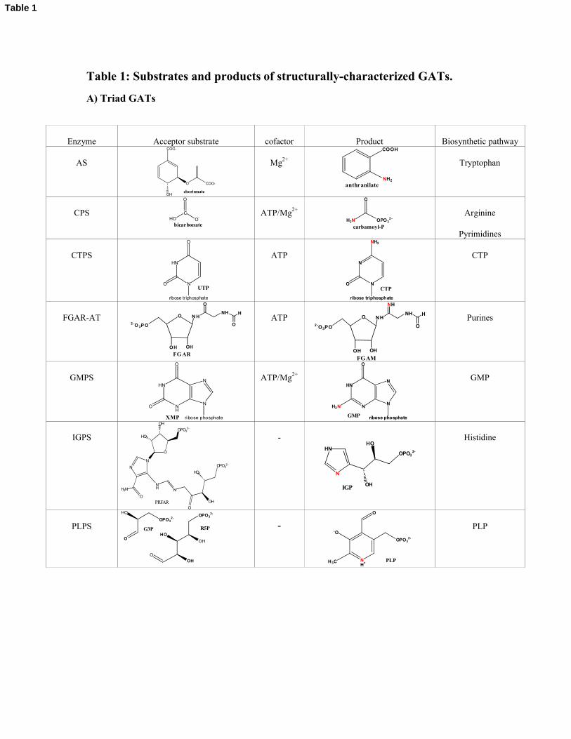
Table 1: Substrates and products of structurally-characterized GATs.
A) Triad GATs
Enzyme Acceptor substrate cofactor Product Biosynthetic pathway
AS
COO-
O
OH
COO-
chorismate
Mg2+
COOH
NH2
anthranilate
Tryptophan
CPS
O
CO-HO
bicarbonate
ATP/Mg2+
O
H2N OPO32 -
carbamoyl-P
Arginine
Pyrimidines
CTPSHN
N
O
O
ribose triphosphate
UTP
ATPN
N
NH2
O
ribose triphosphate
CTP
CTP
FGAR-AT O N H2 -O 3P O
OH OH
NH
O
H
O
FG AR
ATP O NH
2 -O3PO
OH OH
NH
O
H
NH
FGAM
Purines
GMPSHN
NH
N
N
O
ribose phosphate
O
XMP
ATP/Mg2+
HN
NN
N
O
ribose phosphate
H2N
GMP
GMP
IGPS
NH N
O
OPO32-
HO
OH
N
N
H2N
O
O
HO
OH
OPO32-
PRFAR
-HN
N
OPO32-
HO
OHIGP
Histidine
PLPSO
HO
OPO32-
OH
OH
HO
OPO32-
O
R5PG3P -
NH+
-O
H3C
OPO32-
O
PLP
PLP
Table 1
B) Ntn GATs
Enzyme Acceptor substrate cofactor Product Biosynthetic pathway
AsnB
H2N OH
OCOOH
L-AspATP/Mg2+ H2N NH2
OCOOH
L-Asn
Asparagine
GlmSO
OHHO
HO
2-O3PO
OHFru-6P
-
O
HO
HO
OH
NH3+
OPO32-
GlcNH2-6P
Hexosamines
GltS
O COO-
COO-
2-OG
FMN/FAD and
Ferredoxin
or NAD(PH
+H 3N COO-
COO-
L-Glu
Glutamate
GPAT
OO-PO2H-OPO3
2-
2- O3 PO
OH OHPRPP -
ONH22- O3 PO
OH OHPRA
Purines
C) Other class
Enzyme Acceptor substrate cofactor Product Biosynthetic pathway
Glu-tRNAGlnAT* Glu-tRNAGln ATP Gln-tRNAGln Gln-ARNtGln
* Ammonia production is achieved by the structurally different bacterial GatA or archeal GatD subunits, that do not share obvious sequence relationship with class I or class II amidotransferases. GatA and GatD belong to the glutaminase superfamily, GatA being similar to amidases [2] and GatD to L-asparaginases [3].
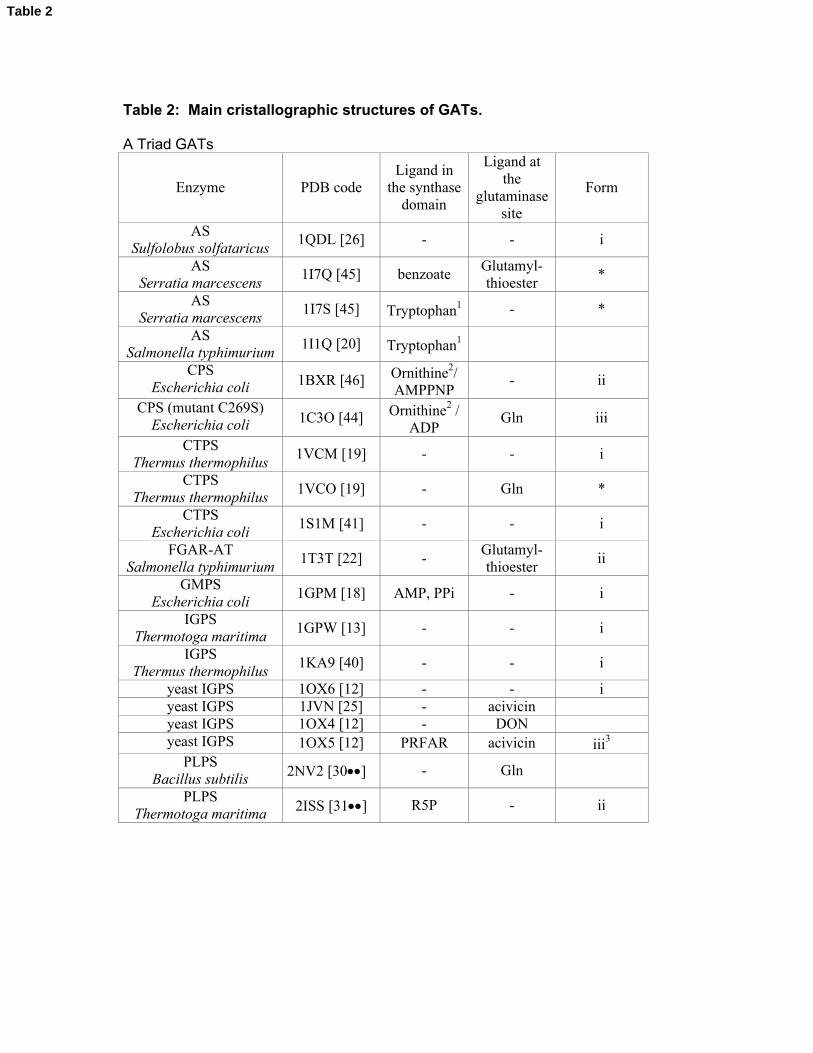
Table 2: Main cristallographic structures of GATs.
A Triad GATs
Enzyme PDB codeLigand in
the synthase domain
Ligand at the
glutaminase site
Form
ASSulfolobus solfataricus
1QDL [26] - - i
ASSerratia marcescens
1I7Q [45] benzoateGlutamyl-thioester
*
ASSerratia marcescens
1I7S [45] Tryptophan1 - *
ASSalmonella typhimurium
1I1Q [20] Tryptophan1
CPSEscherichia coli 1BXR [46] Ornithine2/
AMPPNP- ii
CPS (mutant C269S)Escherichia coli 1C3O [44] Ornithine2 /
ADPGln iii
CTPSThermus thermophilus
1VCM [19] - - i
CTPSThermus thermophilus
1VCO [19] - Gln *
CTPSEscherichia coli
1S1M [41] - - i
FGAR-AT Salmonella typhimurium
1T3T [22] -Glutamyl-thioester
ii
GMPSEscherichia coli
1GPM [18] AMP, PPi - i
IGPS Thermotoga maritima
1GPW [13] - - i
IGPS Thermus thermophilus
1KA9 [40] - - i
yeast IGPS 1OX6 [12] - - iyeast IGPS 1JVN [25] - acivicinyeast IGPS 1OX4 [12] - DONyeast IGPS 1OX5 [12] PRFAR acivicin iii3
PLPSBacillus subtilis 2NV2 [30] - Gln
PLPSThermotoga maritima 2ISS [31] R5P - ii
Table 2
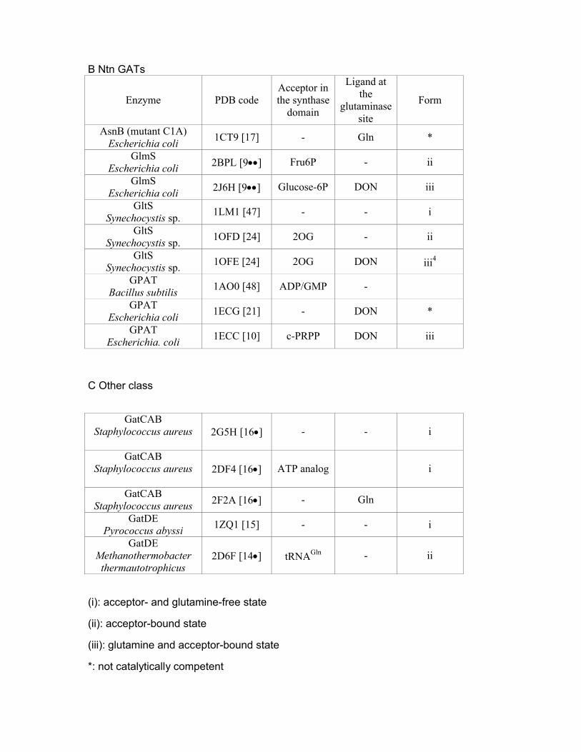
B Ntn GATs
Enzyme PDB codeAcceptor in the synthase
domain
Ligand at the
glutaminase site
Form
AsnB (mutant C1A)Escherichia coli
1CT9 [17] - Gln *
GlmSEscherichia coli 2BPL [9] Fru6P - ii
GlmSEscherichia coli 2J6H [9] Glucose-6P DON iii
GltSSynechocystis sp.
1LM1 [47] - - i
GltSSynechocystis sp.
1OFD [24] 2OG - ii
GltSSynechocystis sp.
1OFE [24] 2OG DON iii4
GPATBacillus subtilis
1AO0 [48] ADP/GMP -
GPATEscherichia coli
1ECG [21] - DON *
GPATEscherichia. coli
1ECC [10] c-PRPP DON iii
C Other class
GatCABStaphylococcus aureus 2G5H [16] - - i
GatCABStaphylococcus aureus 2DF4 [16] ATP analog i
GatCABStaphylococcus aureus 2F2A [16] - Gln
GatDEPyrococcus abyssi
1ZQ1 [15] - - i
GatDEMethanothermobacter
thermautotrophicus2D6F [14] tRNAGln - ii
(i): acceptor- and glutamine-free state
(ii): acceptor-bound state
(iii): glutamine and acceptor-bound state
*: not catalytically competent
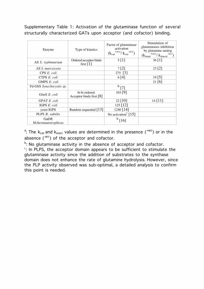
Supplementary Table 1: Activation of the glutaminase function of severalstructurally characterized GATs upon acceptor (and cofactor) binding.
Enzyme Type of kineticsFactor of glutaminase
activation(kcat
+acc/ kcat-acc)
Stimulation ofglutaminases inhibition
by glutamine analog(kinaat
+acc/ kinacat-acc)
AS S. typhimurium Ordered:acceptor bindsfirst [1]
5 [1] 36 [1]
AS S. marcescens 7 [2] 23 [2]CPS E. coli 275 [3]
CTPS E. coli 6 [4] 19 [5]GMPS E. coli 21 [6]
Fd-GltS Synechocystis sp. b [7]GlmS E. coli bi bi ordered:
Acceptor binds first [8]103 [9]
GPAT E. coli 22 [10] 14 [11]IGPS E. coli 125 [12]yeast IGPS Random sequential [13] 1240 [14]
PLPS B. subtilis No activationC [15]GatDE
M.thermautotrophicusb [16]
a: The kcat and kinact values are determined in the presence (+acc) or in theabsence (-acc) of the acceptor and cofactor.b: No glutaminase activity in the absence of acceptor and cofactor.C: In PLPS, the acceptor domain appears to be sufficient to stimulate theglutaminase activity since the addition of substrates to the synthasedomain does not enhance the rate of glutamine hydrolysis. However, sincethe PLP activity observed was sub-optimal, a detailed analysis to confirmthis point is needed.
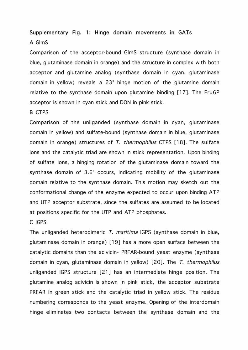
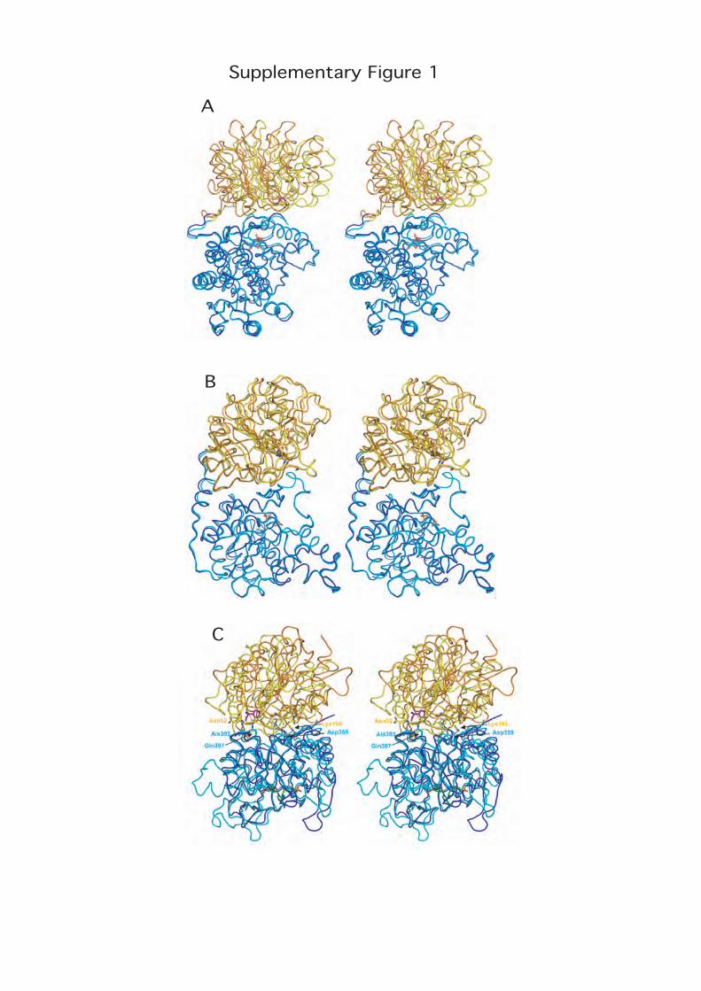
Supplementary Fig. 1: Hinge domain movements in GATs
A GlmS
Comparison of the acceptor-bound GlmS structure (synthase domain in
blue, glutaminase domain in orange) and the structure in complex with both
acceptor and glutamine analog (synthase domain in cyan, glutaminase
domain in yellow) reveals a 23° hinge motion of the glutamine domain
relative to the synthase domain upon glutamine binding [17]. The Fru6P
acceptor is shown in cyan stick and DON in pink stick.
B CTPS
Comparison of the unliganded (synthase domain in cyan, glutaminase
domain in yellow) and sulfate-bound (synthase domain in blue, glutaminase
domain in orange) structures of T. thermophilus CTPS [18]. The sulfate
ions and the catalytic triad are shown in stick representation. Upon binding
of sulfate ions, a hinging rotation of the glutaminase domain toward the
synthase domain of 3.6° occurs, indicating mobility of the glutaminase
domain relative to the synthase domain. This motion may sketch out the
conformational change of the enzyme expected to occur upon binding ATP
and UTP acceptor substrate, since the sulfates are assumed to be located
at positions specific for the UTP and ATP phosphates.
C IGPS
The unliganded heterodimeric T. maritima IGPS (synthase domain in blue,
glutaminase domain in orange) [19] has a more open surface between the
catalytic domains than the acivicin- PRFAR-bound yeast enzyme (synthase
domain in cyan, glutaminase domain in yellow) [20]. The T. thermophilus
unliganded IGPS structure [21] has an intermediate hinge position. The
glutamine analog acivicin is shown in pink stick, the acceptor substrate
PRFAR in green stick and the catalytic triad in yellow stick. The residue
numbering corresponds to the yeast enzyme. Opening of the interdomain
hinge eliminates two contacts between the synthase domain and the
glutaminase active site that occur in the yeast enzyme: the H-bond
between Gln397 of the synthase domain to the glutamine analog in the
glutaminase site and the backbone H-bond between Ala393 of the synthase
domain and Asn52 belonging to the oxyanion strand in the glutaminase
site. By contrast, the salt bridge between Asp359 from the synthase
domain and Lys196, a residue belonging to the glutaminase loop that
contains the His and Glu catalytic residues, which anchors the interface in
this region and stabilizes the glutaminase active site structure, is not
affected by the hinge motion. The occurence of a large scale hinge-closing
motion upon PRFAR binding is supported by steered molecular dynamics
simulations of the undocking of PRFAR in IGPS [22].
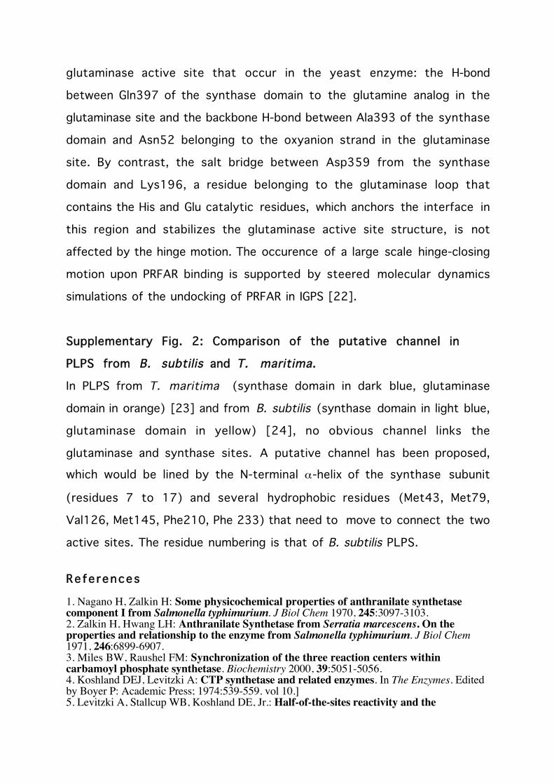
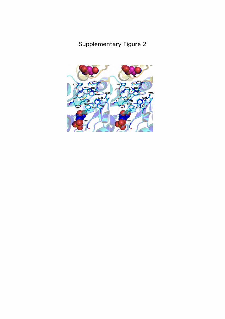
Supplementary Fig. 2: Comparison of the putative channel in
PLPS from B. subtilis and T. maritima.
In PLPS from T. maritima (synthase domain in dark blue, glutaminase
domain in orange) [23] and from B. subtilis (synthase domain in light blue,
glutaminase domain in yellow) [24], no obvious channel links the
glutaminase and synthase sites. A putative channel has been proposed,
which would be lined by the N-terminal α-helix of the synthase subunit
(residues 7 to 17) and several hydrophobic residues (Met43, Met79,
Val126, Met145, Phe210, Phe 233) that need to move to connect the two
active sites. The residue numbering is that of B. subtilis PLPS.
Refe rences
1. Nagano H, Zalkin H: Some physicochemical properties of anthranilate synthetasecomponent I from Salmonella typhimurium. J Biol Chem 1970, 245:3097-3103.2. Zalkin H, Hwang LH: Anthranilate Synthetase from Serratia marcescens. On theproperties and relationship to the enzyme from Salmonella typhimurium. J Biol Chem1971, 246:6899-6907.3. Miles BW, Raushel FM: Synchronization of the three reaction centers withincarbamoyl phosphate synthetase. Biochemistry 2000, 39:5051-5056.4. Koshland DEJ, Levitzki A: CTP synthetase and related enzymes. In The Enzymes. Editedby Boyer P: Academic Press; 1974:539-559. vol 10.]5. Levitzki A, Stallcup WB, Koshland DE, Jr.: Half-of-the-sites reactivity and the
conformational states of cytidine triphosphate synthetase. Biochemistry 1971, 10:3371-3378.6. Zalkin H, Truitt CD: Characterization of the glutamine site of Escherichia coliguanosine 5'-monophosphate synthetase. J Biol Chem 1977, 252:5431-5436.7. Ravasio S, Dossena L, Martin-Figueroa E, Florencio FJ, Mattevi A, Morandi P, Curti B,Vanoni MA: Properties of the recombinant ferredoxin-dependent glutamate synthase ofSynechocystis PCC6803. Comparison with the Azospirillum brasilense NADPH-dependent enzyme and its isolated alpha subunit. Biochemistry 2002, 41:8120-8133.8. Badet B, Vermoote P, Le Goffic F: Glucosamine synthetase from Escherichia coli:kinetic mechanism and inhibition by N3-fumaroyl-L-2,3-diaminopropionicderivatives. Biochemistry 1988, 27:2282-2287.9. Floquet N, Mouilleron S, Daher R, Maigret B, Badet B, Badet-Denisot MA: Ammoniachanneling in bacterial glucosamine-6-phosphate synthase (Glms): Moleculardynamics simulations and kinetic studies of protein mutants. FEBS Lett 2007, 581:2981-2987.10. Messenger LJ, Zalkin H: Glutamine phosphoribosylpyrophosphate amidotransferasefrom Escherichia coli. Purification and properties. J Biol Chem 1979, 254:3382-3392.11. Hartman SC: The Interaction of 6-Diazo-5-Oxo-L-Norleucine with PhosphoribosylPyrophosphate Amidotransferase. J Biol Chem 1963, 238:3036-3047.12. Klem TJ, Davisson VJ: Imidazole glycerol phosphate synthase: the glutamineamidotransferase in histidine biosynthesis. Biochemistry 1993, 32:5177-5186.13. Myers RS, Jensen JR, Deras IL, Smith JL, Davisson VJ: Substrate-induced changes inthe ammonia channel for imidazole glycerol phosphate synthase. Biochemistry 2003,42:7013-7022.14. Myers RS, Amaro RE, Luthey-Schulten ZA, Davisson VJ: Reaction coupling throughinterdomain contacts in imidazole glycerol phosphate synthase. Biochemistry 2005,44:11974-11985.15. Raschle T, Amrhein N, Fitzpatrick TB: On the two components of pyridoxal 5'-phosphate synthase from Bacillus subtilis. J Biol Chem 2005, 280:32291-32300.16. Feng L, Sheppard K, Tumbula-Hansen D, Soll D: Gln-tRNAGln formation from Glu-tRNAGln requires cooperation of an asparaginase and a Glu-tRNAGln kinase. J BiolChem 2005, 280:8150-8155.17. Mouilleron S, Golinelli-Pimpaneau B: Domain motions of glucosamine-6P synthase:comparison of the anisotropic displacements in the crystals and the catalytic hinge-bending rotation. Protein Sci 2007, 16:485-493.18. Goto M, Omi R, Nakagawa N, Miyahara I, Hirotsu K: Crystal structures of CTPsynthetase reveal ATP, UTP, and glutamine binding sites. Structure 2004, 12:1413-1423.19. Douangamath A, Walker M, Beismann-Driemeyer S, Vega-Fernandez MC, Sterner R,Wilmanns M: Structural evidence for ammonia tunneling across the (beta alpha)(8)barrel of the imidazole glycerol phosphate synthase bienzyme complex. Structure(Camb) 2002, 10:185-193.20. Chaudhuri BN, Lange SC, Myers RS, Davisson VJ, Smith JL: Toward understanding themechanism of the complex cyclization reaction catalyzed by imidazoleglycerolphosphate synthase: crystal structures of a ternary complex and the freeenzyme. Biochemistry 2003, 42:7003-7012.21. Omi R, Mizuguchi H, Goto M, Miyahara I, Hayashi H, Kagamiyama H, Hirotsu K:Structure of imidazole glycerol phosphate synthase from Thermus thermophilus HB8:open-closed conformational change and ammonia tunneling. J Biochem (Tokyo) 2002,132:759-765.22. Amaro RE, Sethi A, Myers RS, Davisson VJ, Luthey-Schulten ZA: A network ofconserved interactions regulates the allosteric signal in a glutamine amidotransferase.Biochemistry 2007, 46:2156-2173.23. Zein F, Zhang Y, Kang YN, Burns K, Begley TP, Ealick SE: Structural insights into themechanism of the PLP synthase holoenzyme from Thermotoga maritima. Biochemistry2006, 45:14609-14620.24. Strohmeier M, Raschle T, Mazurkiewicz J, Rippe K, Sinning I, Fitzpatrick TB, Tews I:Structure of a bacterial pyridoxal 5'-phosphate synthase complex. Proc Natl Acad Sci US A 2006, 103:19284-19289.
Supplementary Figure 1
A
B
C
Supplementary Figure 2


