Embed Size (px)
Citation preview
Structure 14, 577–587, March 2006 ª2006 Elsevier Ltd All rights reserved DOI 10.1016/j.str.2005.11.015
Conformational Flexibility in the MultidrugEfflux System Protein AcrA
Jonathan Mikolosko,1,5 Kostyantyn Bobyk,2
Helen I. Zgurskaya,2 and Partho Ghosh1,3,4,*1Department of Chemistry and BiochemistryUniversity of California, San Diego9500 Gilman DriveLa Jolla, California 920372Department of Chemistry and BiochemistryUniversity of Oklahoma620 Parrington OvalNorman, Oklahoma 730193Section of Molecular BiologyUniversity of California, San Diego9500 Gilman DriveLa Jolla, California 92037
Summary
Intrinsic resistance to multiple drugs in many gram-
negative bacterial pathogens is conferred by resis-tance nodulation cell division efflux pumps, which
are composed of three essential components as typi-fied by the extensively characterized Escherichia coli
AcrA-AcrB-TolC system. The inner membrane drug:proton antiporter AcrB and the outer membrane chan-
nel TolC export chemically diverse compounds out ofthe bacterial cell, and require the activity of the third
component, the periplasmic protein AcrA. The crystalstructures of AcrB and TolC have previously been de-
termined, and we complete the molecular picture ofthe efflux system by presenting the structure of a sta-
ble fragment of AcrA. The AcrA fragment resembles
the elongated sickle shape of its homolog Pseudomo-nas aeruginosa MexA, being composed of three do-
mains: b-barrel, lipoyl, and a-helical hairpin. Notably,unsuspected conformational flexibility in the a-helical
hairpin domain of AcrA is observed, which has poten-tial mechanistic significance in coupling between
AcrA conformations and TolC channel opening.
Introduction
The rising incidence of multidrug-resistant bacterialpathogens poses a serious threat to human health. In-trinsic multidrug resistance is conferred in a number ofgram-negative bacterial pathogens, including Pseudo-monas aeruginosa and Haemophilus influenzae, bytransporters belonging to the resistance nodulation celldivision (RND) family of proteins (Putman et al., 2000;Walsh, 2000). RND-type transporters utilize the protonelectrochemical gradient to energize efflux of antibioticsand other compounds out of the bacterial cell. The majormechanistic feature of these transporters is the ability totransfer multiple substrates across both the inner and
*Correspondence: [email protected] Lab address: http://pghosh.ucsd.edu5 Present address: Department of Molecular Biology, The Scripps
Research Institute, 10550 North Torrey Pines Road, La Jolla, Califor-
nia 92037.
the outer membranes of gram-negative bacteria directlyinto external media without periplasmic intermediates(Thanassi et al., 1995). To achieve drug efflux acrosstwo membranes, RND-type transporters assemble intomultiprotein efflux systems. Two of the most extensivelycharacterized RND multidrug efflux systems are Escher-ichia coli AcrA-AcrB-TolC and P. aeruginosa MexA-MexB-OprM (Hirakata et al., 2002; Ma et al., 1995).
RND systems consist of large complexes of three es-sential components. The first is an RND inner membraneprotein, which is energized by the proton-motive force.The RND inner membrane protein assembles into a tri-mer, as shown by the X-ray crystal structure of the pro-ton antiporter AcrB (Murakami et al., 2002; Yu et al.,2003). Each protomer of the trimer has 12 transmem-brane a-helical segments and two large w300 residueperiplasmic domains that extend w70 A above the planeof the inner membrane. Chemically diverse substrates,such as rhodamine 6G, ethidium, dequalinium, and ci-profloxacin, have been seen to bind through hydropho-bic interactions to a central cavity in the periplasmic do-main (Murakami et al., 2002; Yu et al., 2003). How thesecompounds are pumped outward from their bindingsites is not yet known.
The second essential component of the RND system isan outer membrane protein, also known as outer mem-brane factor (OMF), that like the RND inner membraneprotein is trimeric. The structures of the OMFs E. coliTolC, P. aeruginosa OprM, and Vibrio cholerae VceChave been determined (Akama et al., 2004a; Federiciet al., 2005; Koronakis et al., 2000), revealing similarlyshaped cylindrical channels. The trimeric channel is em-bedded in the outer membrane as a 12 stranded b-barrelthat continues w100 A into the periplasmic space as ana-helical barrel. TolC, OprM, and VceC have been visual-ized in their closed states, and hypothesized to openthrough an iris-like mechanism (Andersen et al., 2002;Koronakis et al., 2000). TolC has been shown to interactphysically with AcrB by chemical crosslinking and inter-molecular disulfide bond formation experiments (Ta-mura et al., 2005; Tikhonova and Zgurskaya, 2004; Touzeet al., 2004). This interaction suggests that substrates aretransported directly from the RND inner membrane pro-tein through the open OMF channel and out into the ex-tracellular space.
The third essential component is a periplasmic proteinthat belongs to the membrane fusion protein (MFP) fam-ily (Dinh et al., 1994; Saier et al., 1994), named for se-quence similarity in these proteins to the membrane fu-sion protein (F protein) of paramyxovirus 5 (Dinh et al.,1994; Saier et al., 1994). Interestingly, MFPs are notonly essential components of RND systems but also ofother energy-dependent transport systems, such as theATP binding cassette (ABC) system and the major facili-tator system (MFS) (Putman et al., 2000). MFPs are at-tached to the inner membrane via lipid acylation of a cys-teine residue or through an N-terminal transmembranesegment. However, membrane attachment is not essen-tial for drug efflux activity, as seen by the functionality ofsoluble, periplasmic mutants of the E. coli MFP AcrA and
Structure578Structure578
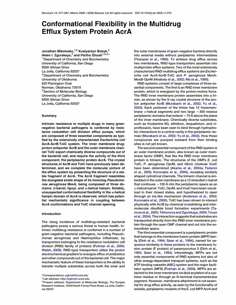
Figure 1. Domain Mapping of AcrA(26–397)
by Thermolytic Digestion
AcrA(26–397) was digested with thermolysin
for 0, 1, 2, and 4 hr (lanes A, B, C, D, respec-
tively) at a 50:1 (mass) substrate:protease
ratio and analyzed by 12% SDS-PAGE.
Molecular weights and schematics of proteo-
lytic products are shown to the right of the
corresponding fragments. Fragments were
identified by N-terminal sequencing and
mass spectrometry.
the P. aeruginosa MFP MexA (Yoneyama et al., 2000;Zgurskaya and Nikaido, 1999a). AcrA has been shownto interact physically with both AcrB and TolC (Husainet al., 2004; Tikhonova and Zgurskaya, 2004; Touzeet al., 2004; Zgurskaya and Nikaido, 2000). Recent exper-imental evidence indicates that interaction of AcrA withthese components is likely to play an active role in theefflux process (Aires and Nikaido, 2005; Zgurskaya andNikaido, 1999b).
In this study, we have determined the 2.7 A resolutionX-ray crystal structure of the stable core of AcrA, therebyhelping to complete the atomic resolution model of theAcrA-AcrB-TolC drug extrusion system and providinga point of comparison to recently determined structuresof the P. aeruginosa MFP MexA (Akama et al., 2004b;Higgins et al., 2004). The structure of AcrA is found toprovide unsuspected evidence for conformational flexi-bility in MFPs. Intriguingly, this flexibility coincides withconformational changes predicted to occur during open-ing of OMF channels by an iris-like mechanism.
Results
Domain Mapping of AcrAAcrA was subjected to limited proteolytic digestion tomap its domain architecture. For this purpose, a solubleform of mature AcrA (residues 26–397, 40 kDa), whichlacks the cleaved signal sequence (residues 1–24) andthe lipid acylation site at residue Cys-25, was expressedcytoplasmically in E. coli and purified using an intro-duced C-terminal histidine tag (Figure 1). This cytoplas-mically expressed, soluble form of AcrA has been shownto be functional in restoring in vivo drug efflux activity toa DacrA strain of E. coli (Zgurskaya and Nikaido, 1999a).After 1 hr of digestion with the relatively nonspecific pro-tease thermolysin at a 50:1 substrate:protease (mass)ratio, AcrA is found to be trimmed slightly at its N- andC-terminal ends. The resulting 38 kDa products beginheterogeneously at either residue 26 or 30 and end ho-mogeneously at residue 386, as determined by N-termi-nal sequencing and mass spectrometry. These productsare further digested over the next two to four hours toyield the stable, protease-resistant core of AcrA (Fig-ure 1). The 28 kDa stable core begins at residue 45 andends heterogeneously at residue 305 or 312. This ther-molytic fragment is very similar to a recently reportedtryptic fragment of AcrA that encompasses residues45–315 (Touze et al., 2004).
These results demonstrate that the w90 C-terminalresidues of AcrA are likely to be flexible and hence pro-teolytically sensitive. The C-terminal residues (313–397)removed from AcrA by proteolysis are digested intosmall peptides and do not yield a resistant fragment.This proteolytically sensitive C-terminal region of AcrAis required for association with the inner membraneRND protein AcrB, as shown by chimera studies usingAcrA residues 290–357 and direct binding studies usingan AcrA fragment composed of residues 172–397 (Elkinsand Nikaido, 2003; Touze et al., 2004). The C-terminal re-gion also appears to be required for interaction withTolC (Touze et al., 2004). The flexibility of both N- andC-terminal regions of AcrA is also consistent with X-raycrystal structures of the homolog MexA, the periplasmiccomponent of the P. aeruginosa MexA-MexB-OprMmultidrug efflux system (Akama et al., 2004b; Higginset al., 2004). Although intact mature MexA, with a molec-ular weight of 38 kDa, had been crystallized, electrondensity was observed only for a region of the proteinequivalent to the 28 kDa stable core of AcrA.
Structure Determination of the Stable Core of AcrATo gain functional insight, we set out to crystallize the 28kDa stable core of AcrA (residues 45–312). AlthoughAcrA(45–312) was found to readily crystallize (e.g., underone fifth of Hampton crystal screen I and II conditions),almost all crystals were found to diffract anisotropicallyto a maximum resolution of only 5–10 A. Only one condi-tion was identified to yield crystals that diffracted X-raysto a reasonable resolution limit (w3.5 A). These crystalsappeared to belong to space group P42212, but werediscovered, after several unsuccessful attempts at ob-taining phase information, to have arisen througha rare case of perfect pseudomerohedral twinning ofP212121 crystals, in which the a and b axes are fortu-itously identical in length (169 A). Analysis of intensitydistributions in local areas of reciprocal space provedinstrumental in detecting pseudomerohedral twinningin these crystals (Padilla and Yeates, 2003).
Due to great difficulties in determining structures fromperfectly twinned crystals, an alternate crystal form wassought. Untwinned crystals that diffracted X-rays iso-tropically to 2.7 A resolution were obtained from a qua-druple methionine substitution mutant (F223M, L224M,L287M, and L288M) which had been constructed forthe purpose of multiwavelength anomalous dispersion(MAD) phase determination. Wild-type AcrA(45–312)also crystallized under essentially the same conditions
Structure of AcrA579
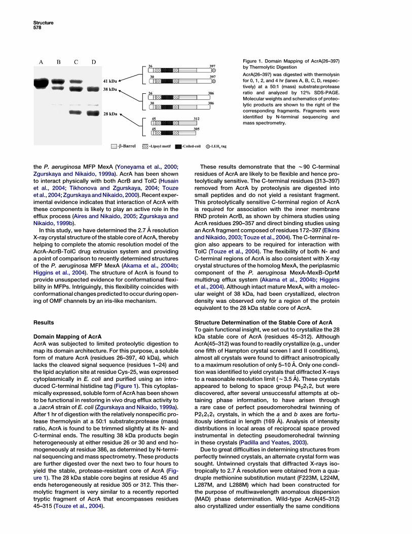
Figure 2. Structure of AcrA(45–312)-4M
(A) Ribbon representation of AcrA(45–312)-
4M, showing two apparent dimers per asym-
metric unit in the crystal.
(B) Ribbon representation of a monomer of
AcrA(45–312)-4M (molecule C), with the a-he-
lical hairpin domain in red, lipoyl domain in
green, and b-barrel domain in cyan. Molecu-
lar graphics were made with PyMOL (DeLano,
2002).
as the quadruple methionine substitution mutant, calledAcrA(45–312)-4M, but diffracted X-rays anisotropicallyto a maximum resolution of only 3.3 A resolution. Crys-tals of AcrA(45–312)-4M contain four molecules perasymmetric unit and a solvent content of 60%. The struc-ture was determined by MAD techniques using seleno-methionine-labeled protein. Continuous electron densityfor the main chain from residue 53 to 299 is observed inone of the monomers (Figure 2A, labeled C), whereassmall breaks (at residues 230–241) are observed in theother three monomers (Figure 2A, labeled A, B, and D).The model has been refined to 2.71 A resolution with anRwork of 23.7% and an Rfree of 27.5% (Table 1).
Overview of the Structure
The four molecules of AcrA(45–312)-4M in the asymmet-ric unit of the crystal pack as an apparent dimer of
dimers. The molecules labeled A and B are related toone another by approximate dyad symmetry, as arethe molecules labeled C and D; each set of dimers is inturn related to one another by yet another approximate2-fold axis (Figure 2A). Close structural relationship isseen (and detailed below) between AcrA(45–312)-4Mand MexA, which is not surprising given their 62% and73% sequence identities and similarities, respectively.Despite the close sequence and structural similarity,the two proteins do not functionally complement eachother (Tikhonova et al., 2002). Both AcrA(45–312)-4Mand MexA are elongated, sickle-shaped molecules com-prised of three domains: a b-barrel domain at one end,a lipoyl domain centrally located, and a coiled-coil a-he-lical hairpin at the other end of the molecule (Figure 2B).The existence in MFPs of the lipoyl and a-helical hairpindomains had been predicted from primary sequence
Structure580
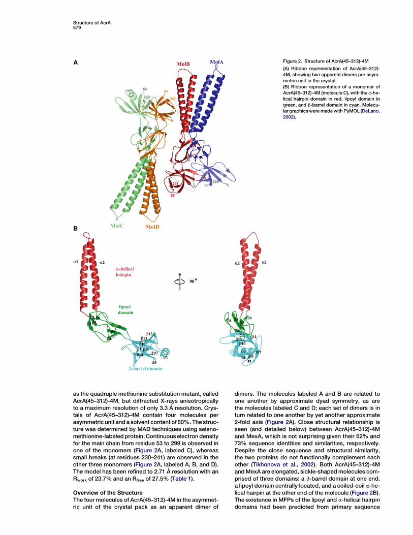
Table 1. X-Ray Data Collection and Refinement, AcrA(45–312)-4M
Data Collection
Beamline APS-BM19 ALS-8.2.2
Data set l1 l2 l3 l1
Wavelength (A) 0.97938 0.97955 0.96415 1.0000
Resolution (A)a 50–3.00 (3.11–3.00) 50–3.39 (3.59–3.39) 50–3.50 (3.63–3.50) 50–2.70 (2.77–2.70)
Completeness (%) 99.3 (99.3) 99.8 (99.9) 99.8 (99.9) 97.3 (91.1)
Redundancy 4.2 (3.4) 4.4 (4.5) 4.2 (3.8) 5.0 (3.9)
I/s 13.5 (2.3) 16.6 (4.6) 12.0 (4.0) 14.6 (2.3)
Rmergeb 13.9 (76.1) 9.8 (38.9) 18.5 (53.4) 9.0 (42.9)
Phasing
Se l1 Se l2 Se l3
Anomalous and dispersive differences (%)c
Se l1 4.2
Se l2 1.8 2.4
Se l3 2.0 3.8 3.1
Anomalous phasing powerd 1.5 0.8 0.7
Number of sites 16
Figure of merit (40–3.0 A) 0.59
Refinement
Resolution 50.0–2.71 (2.77–2.71)
Rcryst (%)e 23.7 (40.8) Rms deviation
Rfree (%)e 27.5 (47.4) Bonded B factor (A2) 3.30
Number of reflections From ideal geometry
Working set 35,426 Lengths (A) 0.010
Test set 1,873 Angles (º) 1.608
Number of atoms Average B factors (A2)
Protein 6936 Protein 89.2
Solvent 36 Solvent 64.0
a Highest resolution shell is in parentheses.
b Rmerge = 1003
P
h
P
i
jIh; i 2 IhjP
h
P
i
Ih; i, where Ih is the mean intensity of symmetry-related reflections, Ih,j.
c Anomalous and dispersive differences = 100 3 rms DF/rms F, where DF for anomalous differences is (F+h – F2h)/2 (diagonal element) and for
dispersive differences is Fli – Flj (off-diagonal).d FH00/E, with FH00 being the anomalous component of the heavy atom structure factor and E the rms lack-of-closure error.
e R factor = 1003
P
hkl
jFobs 2 FcalcjP
hkl
Fobs, where Rfree is calculated for a randomly chosen 5% of reflections (F > 0) omitted from refinement, and Rcryst is
calculated for the remaining 95% of reflections (F > 0) included in refinement.
considerations alone (Johnson and Church, 1999). Thea-helical hairpin in AcrA is seven residues longer thanin MexA, resulting in a total length of 105 A for AcrA ascompared to the 89 A length of MexA.
b-Barrel Domain
The b-barrel domain consists of six antiparallel b strandsand a short a helix (Figure 2B). The N and C termini of theAcrA fragment form two of the apposing strands (b1, res-idues 54–61 and b14, residues 292–297, respectively).This domain is predicted to be proximal in vivo to the in-ner membrane due to lipid acylation of Cys-25. The 28flexible residues connecting Cys-25 to the b-barrel do-main are more than sufficient to reach the periplasmictop of AcrB, which extends w70 A above the inner mem-brane surface (Murakami et al., 2002; Yu et al., 2003). Theshort a helix (a3, residues 222–230) is located betweenb strands 10 and 11, and closes off the end of the b-barrelnear the C terminus of the AcrA fragment. This helix in in-tact AcrA would, therefore, lie close to the flexible, pro-teolytically sensitive C-terminal 100 residues that are re-quired for interaction with AcrB and TolC (Elkins andNikaido, 2003; Touze et al., 2004).
The b-barrel domain is the site of all four methioninesubstitutions introduced for crystallization and phasingpurposes. Two of the methionine substitutions, at posi-tions 223 and 224, are located on the a3 helix, and theother two, at positions 287 and 288, on a loop connectingb strands 13 and 14 (Figure 3). Among these four substi-tutions, the only methionine that makes intermolecularcontacts (within 4 A) is Met-288 (molecules A and C),which abuts residues Val-265 and Val-267. The adjacentmethionine substitution Met-287 makes intramolecularcontacts, and is positioned against a hydrophobicpocket formed by residues Phe-254, Leu-246, and Met-291 near the center of the molecule’s b-barrel domain.Overall, the b-barrel domains of all four AcrA moleculesare found to be structurally similar (rmsd 1.02 A, 63 Caatoms).
Lipoyl and Coiled-Coil Domains
The central portion of AcrA consists of a lipoyl domain,best thought of as two lipoyl half-motifs interrupted byan a-helical hairpin formed by helices a1 and a2 (Figure2B). The N- and C-terminal lipoyl half-motifs are homol-ogous to each other (Johnson and Church, 1999) andconsist of four b strands (b2–b5 and b6–b9, respectively)
Structure of AcrA581
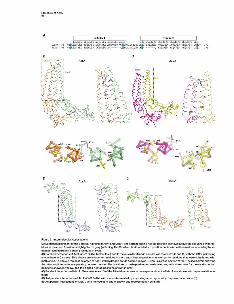
Figure 3. Intermolecular Associations
(A) Sequence alignment of the a-helical hairpins of AcrA and MexA. The corresponding heptad position is shown above the sequence with res-
idues in the c and f positions highlighted in gray (including Ala-99, which is situated at a c position but is a b position residue according to se-
quence) and hydrogen bonding residues in cyan.
(B) Parallel interactions of AcrA(45–312)-4M. Molecules A and B make similar dimeric contacts as molecules C and D, with the latter pair being
shown here in Ca trace. Side chains are shown for residues in the c and f heptad positions as well as for residues that were substituted with
methionine. The boxed region is enlarged at right, with hydrogen bonds colored in cyan. Below is a cross-section of the a-helical hairpin showing
the intra- and intermolecular packing between helices. The positions of the heptad repeat are labeled a–g with side chains for the a and d heptad
positions shown in yellow, and the e and f heptad positions shown in gray.
(C) Parallel interactions of MexA. Molecules A and B of the 13 total molecules in the asymmetric unit of MexA are shown, with representation as
in (B).
(D) Antiparallel interactions of AcrA(45–312)-4M, with molecules related by crystallographic symmetry. Representation as in (B).
(E) Antiparallel interactions of MexA, with molecules G and H shown and representation as in (B).
Structure582
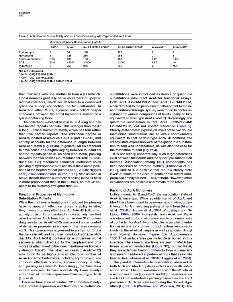
Table 2. Antimicrobial Susceptibility of E. coli Cells Expressing Wild-Type and Mutant AcrA
Minimum Inhibitory Concentration (mg/ml)
pUC18 AcrA AcrA F223M/L224Ma AcrA L287M/L288Mb AcrA-4Mc AcrA(1–312)
Erythromycin 2 64 128 128 2 2
Novobiocin 4 128 256 256 2 1
Ethidium bromide 6.25 100 100 100 6.25 6.25
SDS 62.5 >2000 >2000 >2000 62.5 40
Puromycin 2 256 256 256 2 ND
ND, not determined.a AcrA(1–397) F223M/L224M.b AcrA(1–397) L287M/L288M.c AcrA(1–397) F223M/L224M/L287M/L288M.
that intertwine with one another to form a b sandwich.Lipoyl domains generally serve as carriers of lipoyl orbiotinyl cofactors, which are attached to a conservedlysine on a loop connecting the two half-motifs. InAcrA and other MFPs, a coiled-coil a-helical hairpinintervenes between the lipoyl half-motifs instead of alysine-containing loop.
The coiled-coil a-helical hairpin is 58 A long and hasfive heptad repeats per helix. This is longer than the 47A long a-helical hairpin of MexA, which has four ratherthan five heptad repeats. The additional heptad inAcrA is located at residues 120–126 and 142–148, andentirely accounts for the difference in length betweenAcrA and MexA (Figure 3A). In general, MFPs are foundto have coiled-coil lengths varying between four and sixheptad repeats per helix. As seen with MexA, packingbetween the two helices (a1, residues 99–130; a2, resi-dues 140–172) resembles canonical knobs-into-holespacking of hydrophobic side chains in the a and d posi-tions of the heptad repeat (Akama et al., 2004b; Higginset al., 2004; Johnson and Church, 1999). Also as seen inMexA, the left-handed superhelical coiling in the a1 helixis more pronounced than in the a2 helix, so that a2 ap-pears to be relatively straighter than a1.
Functional Properties of Methionine
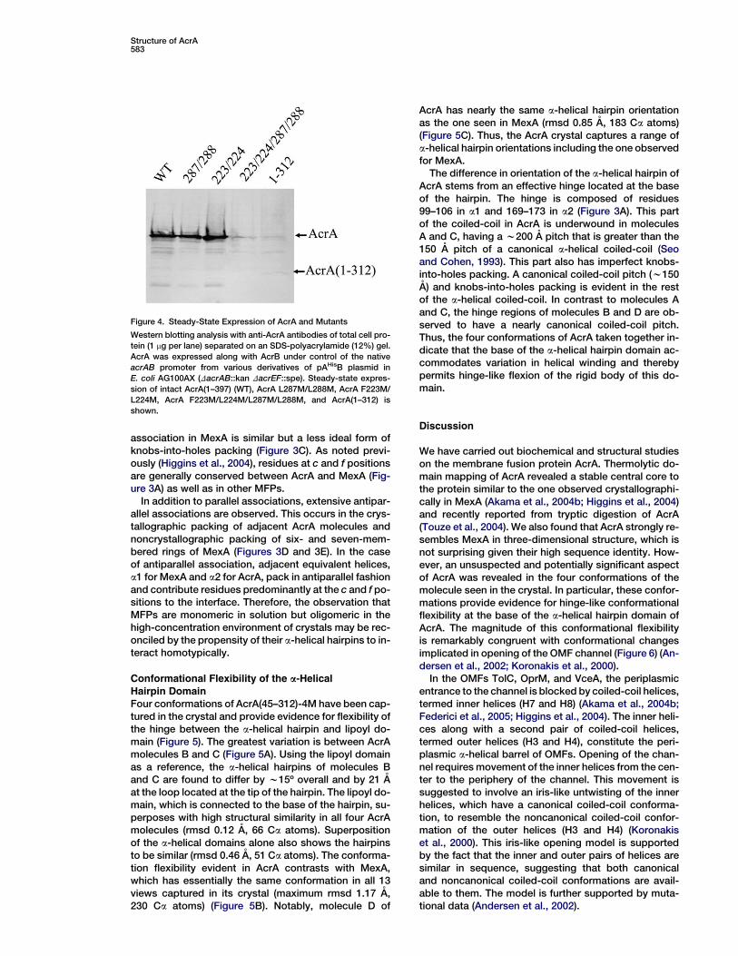
Substitution MutantsWhile the methionine mutations introduced for phasinghave no apparent effect on protein stability in vitro,they have surprising effects on AcrA-AcrB-TolC effluxactivity in vivo. To understand in vivo activity, we firstasked whether AcrA truncated at residue 312 confersdrug resistance. AcrA(1–312) was placed under controlof its native promoter in an operon that also containsacrB. This operon was expressed in a strain of E. colithat lacks AcrAB and its close homolog AcrEF (DacrAB-DacrEF). AcrA(1–312) contains its endogenous signalsequence, which directs it to the periplasm and pro-motes its attachment to the inner membrane via lipid ac-ylation of Cys-25. The strain expressing AcrA(1–312)was found to be highly susceptible to a number ofAcrA-AcrB-TolC substrates, including erythromycin, no-vobiocin, ethidium bromide, sodium dodecyl sulfate,and puromycin (Table 2). Interestingly, the truncationmutant was seen to have a drastically lower steady-state level of protein expression than wild-type AcrA(Figure 4).
Because truncation at residue 312 abrogates steady-state protein expression and function, the methionine
substitutions were introduced as double or quadruplesubstitutions into intact AcrA for functional assays.Both AcrA F223M/L224M and AcrA L287M/L288M,when directed to the periplasm for attachment to the in-ner membrane through Cys-25, were found to confer re-sistance to various compounds at levels nearly or fullyequivalent to wild-type AcrA (Table 2). Surprisingly, thequadruple substitution mutant, AcrA F223M/L224M/L287M/L288M, did not confer resistance (Table 2).Steady-state protein expression levels of the two doublemethionine substitutions are at levels approximatelyequivalent to that of wild-type AcrA. In contrast, thesteady-state expression level of the quadruple substitu-tion mutant was undetectable, as was also the case forthe truncation mutant (Figure 4).
It is not readily apparent why such large differencesexist between the double and the quadruple substitutionmutants. Association among RND components hasbeen observed to promote stability (Tikhonova et al.,2002), and so it is possible that the low steady-statelevels of some of the AcrA mutants above reflect com-promised affinity for AcrB, TolC, or both. However, otherexplanations are possible and remain to be tested.
Packing of AcrA MonomersUnlike trimeric AcrB and TolC, the association state ofAcrA is uncertain. While soluble forms of AcrA andMexA have been found to be monomeric in vitro, cross-linking of AcrA in vivo suggests a trimeric form (Akamaet al., 2004b; Higgins et al., 2004; Zgurskaya and Ni-kaido, 1999a, 2000). In crystals, both AcrA and MexAare observed to form oligomers involving similar setsof contacts. For AcrA, two molecules in parallel orienta-tion associate as a dimer through extensive contactsinvolving the a-helical hairpins as well as adjoining lipoyland b-barrel domains (Figure 3B). Approximately1650 A2 of surface area per molecule is buried at thisinterface. The same interactions are seen in MexA be-tween adjacent molecules (Figure 3C), but in MexA,they are extended beyond dimers to form unusual six-and seven-membered superhelical rings that associatehead to head (Akama et al., 2004b; Higgins et al., 2004).
The parallel intermolecular association observed inboth AcrA and MexA crystals involves antiparallel inter-action of the a1 helix of one monomer with the a2 helix ofa second monomer (Figures 3B and 3C). The associationinvolves knobs-into-holes packing of residues at c and fpositions in AcrA, as assessed using the Socket algo-rithm (Figure 3B) (Walshaw and Woolfson, 2001). The
Structure of AcrA583
association in MexA is similar but a less ideal form ofknobs-into-holes packing (Figure 3C). As noted previ-ously (Higgins et al., 2004), residues at c and f positionsare generally conserved between AcrA and MexA (Fig-ure 3A) as well as in other MFPs.
In addition to parallel associations, extensive antipar-allel associations are observed. This occurs in the crys-tallographic packing of adjacent AcrA molecules andnoncrystallographic packing of six- and seven-mem-bered rings of MexA (Figures 3D and 3E). In the caseof antiparallel association, adjacent equivalent helices,a1 for MexA and a2 for AcrA, pack in antiparallel fashionand contribute residues predominantly at the c and f po-sitions to the interface. Therefore, the observation thatMFPs are monomeric in solution but oligomeric in thehigh-concentration environment of crystals may be rec-onciled by the propensity of their a-helical hairpins to in-teract homotypically.
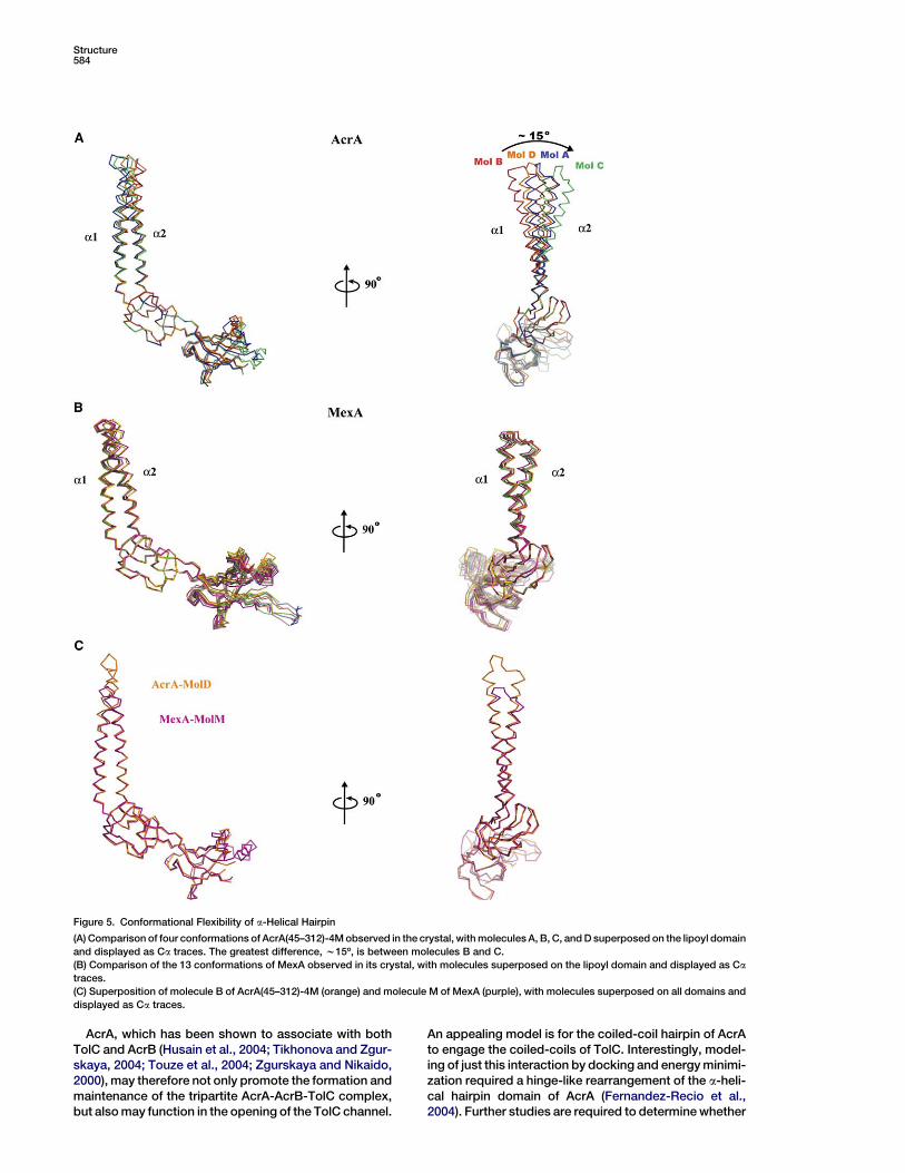
Conformational Flexibility of the a-Helical
Hairpin DomainFour conformations of AcrA(45–312)-4M have been cap-tured in the crystal and provide evidence for flexibility ofthe hinge between the a-helical hairpin and lipoyl do-main (Figure 5). The greatest variation is between AcrAmolecules B and C (Figure 5A). Using the lipoyl domainas a reference, the a-helical hairpins of molecules Band C are found to differ by w15º overall and by 21 Aat the loop located at the tip of the hairpin. The lipoyl do-main, which is connected to the base of the hairpin, su-perposes with high structural similarity in all four AcrAmolecules (rmsd 0.12 A, 66 Ca atoms). Superpositionof the a-helical domains alone also shows the hairpinsto be similar (rmsd 0.46 A, 51 Ca atoms). The conforma-tion flexibility evident in AcrA contrasts with MexA,which has essentially the same conformation in all 13views captured in its crystal (maximum rmsd 1.17 A,230 Ca atoms) (Figure 5B). Notably, molecule D of
Figure 4. Steady-State Expression of AcrA and Mutants
Western blotting analysis with anti-AcrA antibodies of total cell pro-
tein (1 mg per lane) separated on an SDS-polyacrylamide (12%) gel.
AcrA was expressed along with AcrB under control of the native
acrAB promoter from various derivatives of pAHisB plasmid in
E. coli AG100AX (DacrAB::kan DacrEF::spe). Steady-state expres-
sion of intact AcrA(1–397) (WT), AcrA L287M/L288M, AcrA F223M/
L224M, AcrA F223M/L224M/L287M/L288M, and AcrA(1–312) is
shown.
AcrA has nearly the same a-helical hairpin orientationas the one seen in MexA (rmsd 0.85 A, 183 Ca atoms)(Figure 5C). Thus, the AcrA crystal captures a range ofa-helical hairpin orientations including the one observedfor MexA.
The difference in orientation of the a-helical hairpin ofAcrA stems from an effective hinge located at the baseof the hairpin. The hinge is composed of residues99–106 in a1 and 169–173 in a2 (Figure 3A). This partof the coiled-coil in AcrA is underwound in moleculesA and C, having a w200 A pitch that is greater than the150 A pitch of a canonical a-helical coiled-coil (Seoand Cohen, 1993). This part also has imperfect knobs-into-holes packing. A canonical coiled-coil pitch (w150A) and knobs-into-holes packing is evident in the restof the a-helical coiled-coil. In contrast to molecules Aand C, the hinge regions of molecules B and D are ob-served to have a nearly canonical coiled-coil pitch.Thus, the four conformations of AcrA taken together in-dicate that the base of the a-helical hairpin domain ac-commodates variation in helical winding and therebypermits hinge-like flexion of the rigid body of this do-main.
Discussion
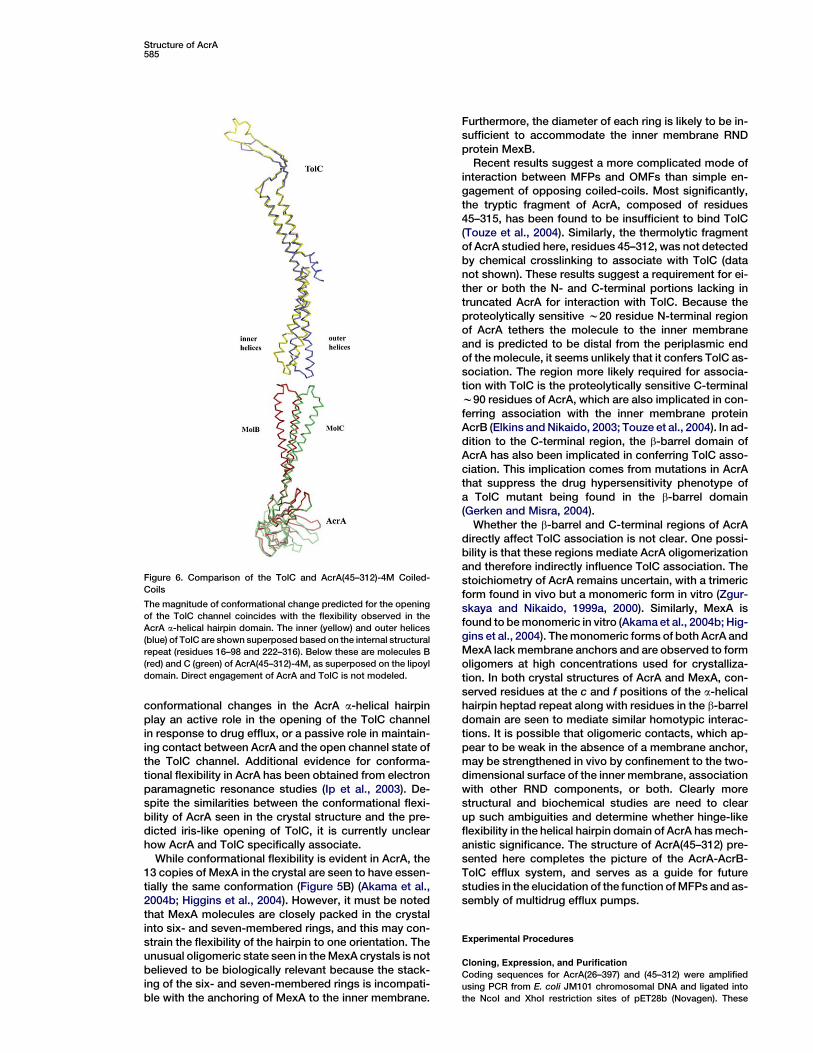
We have carried out biochemical and structural studieson the membrane fusion protein AcrA. Thermolytic do-main mapping of AcrA revealed a stable central core tothe protein similar to the one observed crystallographi-cally in MexA (Akama et al., 2004b; Higgins et al., 2004)and recently reported from tryptic digestion of AcrA(Touze et al., 2004). We also found that AcrA strongly re-sembles MexA in three-dimensional structure, which isnot surprising given their high sequence identity. How-ever, an unsuspected and potentially significant aspectof AcrA was revealed in the four conformations of themolecule seen in the crystal. In particular, these confor-mations provide evidence for hinge-like conformationalflexibility at the base of the a-helical hairpin domain ofAcrA. The magnitude of this conformational flexibilityis remarkably congruent with conformational changesimplicated in opening of the OMF channel (Figure 6) (An-dersen et al., 2002; Koronakis et al., 2000).
In the OMFs TolC, OprM, and VceA, the periplasmicentrance to the channel is blocked by coiled-coil helices,termed inner helices (H7 and H8) (Akama et al., 2004b;Federici et al., 2005; Higgins et al., 2004). The inner heli-ces along with a second pair of coiled-coil helices,termed outer helices (H3 and H4), constitute the peri-plasmic a-helical barrel of OMFs. Opening of the chan-nel requires movement of the inner helices from the cen-ter to the periphery of the channel. This movement issuggested to involve an iris-like untwisting of the innerhelices, which have a canonical coiled-coil conforma-tion, to resemble the noncanonical coiled-coil confor-mation of the outer helices (H3 and H4) (Koronakiset al., 2000). This iris-like opening model is supportedby the fact that the inner and outer pairs of helices aresimilar in sequence, suggesting that both canonicaland noncanonical coiled-coil conformations are avail-able to them. The model is further supported by muta-tional data (Andersen et al., 2002).
Structure584
Figure 5. Conformational Flexibility of a-Helical Hairpin
(A) Comparison of four conformations of AcrA(45–312)-4M observed in the crystal, with molecules A, B, C, and D superposed on the lipoyl domain
and displayed as Ca traces. The greatest difference, w15º, is between molecules B and C.
(B) Comparison of the 13 conformations of MexA observed in its crystal, with molecules superposed on the lipoyl domain and displayed as Ca
traces.
(C) Superposition of molecule B of AcrA(45–312)-4M (orange) and molecule M of MexA (purple), with molecules superposed on all domains and
displayed as Ca traces.
AcrA, which has been shown to associate with bothTolC and AcrB (Husain et al., 2004; Tikhonova and Zgur-skaya, 2004; Touze et al., 2004; Zgurskaya and Nikaido,2000), may therefore not only promote the formation andmaintenance of the tripartite AcrA-AcrB-TolC complex,but also may function in the opening of the TolC channel.
An appealing model is for the coiled-coil hairpin of AcrAto engage the coiled-coils of TolC. Interestingly, model-ing of just this interaction by docking and energy minimi-zation required a hinge-like rearrangement of the a-heli-cal hairpin domain of AcrA (Fernandez-Recio et al.,2004). Further studies are required to determine whether
Structure of AcrA585
conformational changes in the AcrA a-helical hairpinplay an active role in the opening of the TolC channelin response to drug efflux, or a passive role in maintain-ing contact between AcrA and the open channel state ofthe TolC channel. Additional evidence for conforma-tional flexibility in AcrA has been obtained from electronparamagnetic resonance studies (Ip et al., 2003). De-spite the similarities between the conformational flexi-bility of AcrA seen in the crystal structure and the pre-dicted iris-like opening of TolC, it is currently unclearhow AcrA and TolC specifically associate.
While conformational flexibility is evident in AcrA, the13 copies of MexA in the crystal are seen to have essen-tially the same conformation (Figure 5B) (Akama et al.,2004b; Higgins et al., 2004). However, it must be notedthat MexA molecules are closely packed in the crystalinto six- and seven-membered rings, and this may con-strain the flexibility of the hairpin to one orientation. Theunusual oligomeric state seen in the MexA crystals is notbelieved to be biologically relevant because the stack-ing of the six- and seven-membered rings is incompati-ble with the anchoring of MexA to the inner membrane.
Figure 6. Comparison of the TolC and AcrA(45–312)-4M Coiled-
Coils
The magnitude of conformational change predicted for the opening
of the TolC channel coincides with the flexibility observed in the
AcrA a-helical hairpin domain. The inner (yellow) and outer helices
(blue) of TolC are shown superposed based on the internal structural
repeat (residues 16–98 and 222–316). Below these are molecules B
(red) and C (green) of AcrA(45–312)-4M, as superposed on the lipoyl
domain. Direct engagement of AcrA and TolC is not modeled.
Furthermore, the diameter of each ring is likely to be in-sufficient to accommodate the inner membrane RNDprotein MexB.
Recent results suggest a more complicated mode ofinteraction between MFPs and OMFs than simple en-gagement of opposing coiled-coils. Most significantly,the tryptic fragment of AcrA, composed of residues45–315, has been found to be insufficient to bind TolC(Touze et al., 2004). Similarly, the thermolytic fragmentof AcrA studied here, residues 45–312, was not detectedby chemical crosslinking to associate with TolC (datanot shown). These results suggest a requirement for ei-ther or both the N- and C-terminal portions lacking intruncated AcrA for interaction with TolC. Because theproteolytically sensitive w20 residue N-terminal regionof AcrA tethers the molecule to the inner membraneand is predicted to be distal from the periplasmic endof the molecule, it seems unlikely that it confers TolC as-sociation. The region more likely required for associa-tion with TolC is the proteolytically sensitive C-terminalw90 residues of AcrA, which are also implicated in con-ferring association with the inner membrane proteinAcrB (Elkins and Nikaido, 2003; Touze et al., 2004). In ad-dition to the C-terminal region, the b-barrel domain ofAcrA has also been implicated in conferring TolC asso-ciation. This implication comes from mutations in AcrAthat suppress the drug hypersensitivity phenotype ofa TolC mutant being found in the b-barrel domain(Gerken and Misra, 2004).
Whether the b-barrel and C-terminal regions of AcrAdirectly affect TolC association is not clear. One possi-bility is that these regions mediate AcrA oligomerizationand therefore indirectly influence TolC association. Thestoichiometry of AcrA remains uncertain, with a trimericform found in vivo but a monomeric form in vitro (Zgur-skaya and Nikaido, 1999a, 2000). Similarly, MexA isfound to be monomeric in vitro (Akama et al., 2004b; Hig-gins et al., 2004). The monomeric forms of both AcrA andMexA lack membrane anchors and are observed to formoligomers at high concentrations used for crystalliza-tion. In both crystal structures of AcrA and MexA, con-served residues at the c and f positions of the a-helicalhairpin heptad repeat along with residues in the b-barreldomain are seen to mediate similar homotypic interac-tions. It is possible that oligomeric contacts, which ap-pear to be weak in the absence of a membrane anchor,may be strengthened in vivo by confinement to the two-dimensional surface of the inner membrane, associationwith other RND components, or both. Clearly morestructural and biochemical studies are need to clearup such ambiguities and determine whether hinge-likeflexibility in the helical hairpin domain of AcrA has mech-anistic significance. The structure of AcrA(45–312) pre-sented here completes the picture of the AcrA-AcrB-TolC efflux system, and serves as a guide for futurestudies in the elucidation of the function of MFPs and as-sembly of multidrug efflux pumps.
Experimental Procedures
Cloning, Expression, and Purification
Coding sequences for AcrA(26–397) and (45–312) were amplified
using PCR from E. coli JM101 chromosomal DNA and ligated into
the NcoI and XhoI restriction sites of pET28b (Novagen). These
Structure586
constructs contain a C-terminal His tag (LEHHHHHH) for purposes
of protein purification. The methionine substitution mutations
F223M/L224M and L287M/L288M were introduced by site-directed
mutagenesis using strand overlap extension PCR (Higuchi et al.,
1988).
Expression of AcrA(26–397) was induced in E. coli BL21 (DE3) us-
ing 1 mM isopropyl-b-D-thiogalactopyranoside (25ºC). Bacteria
were harvested by centrifugation and lysed by sonication (in 50
mM phosphate buffer [pH 8.0], 150 mM NaCl, 10 mM imidazole),
and AcrA(26–397) was purified by Ni2+ chelation chromatography
(Poros MC). Pooled fractions were dialyzed (50 mM Tris [pH 8.0],
150 mM NaCl), concentrated using Amicon ultrafiltration (MW cutoff
30 kDa; Millipore), and applied to a size exclusion column (Superdex
200). Purified AcrA(26–397)-His was concentrated to w30 mg/ml
(calculated e280 of 17,210 M21 cm21), dialyzed in 10 mM Tris (pH
8.0), and flash frozen at 280ºC. AcrA(45–397) was expressed and pu-
rified similarly.
Selenomethionine was incorporated into a quadruple methionine
mutant (F223M, L224M, L287M, and L288M) as described previously
(Budisa et al., 1995) and purified as above, except for the addition of
1 mM dithiothreitol to samples following Ni2+ chelation chromatog-
raphy. Purified protein was concentrated (calculated e280 of 11,520
M21 cm21), dialyzed in 10 mM Tris (pH 8.0), 1 mM Tris(2-carboxye-
thyl)phosphine hydrochloride (TCEP), and flash frozen at 280ºC.
Proteolytic Domain Mapping
Proteolysis experiments were carried out at 37ºC at a 50:1 AcrA:ther-
molysin mass ratio in 10 mM Tris (pH 8.0), 0.15 mM ZnSO4, 2 mM
CaCl2. The final concentration of AcrA(26–397)-His was 1 mg/ml in
a 100 ml reaction. Ten microliter samples were taken at varying times
points, mixed with 23 SDS-PAGE sample buffer, boiled, and ana-
lyzed by 12% SDS-PAGE. Proteolysis fragments were identified us-
ing N-terminal sequencing of bands isolated by SDS-PAGE, and liq-
uid chromatography followed by electrospray ionization mass
spectrometry of the proteolysis reaction.
Crystallization and Data Collection
Crystals were grown at 4ºC by the vapor diffusion method using a 1:1
mixture of AcrA(45–312)-4M and 30% 2-methyl-2,4-pentadiol, 20
mM MgCl2, 100 mM citrate (pH 5.4), 1 mM TCEP. Crystals were
briefly washed in well buffer supplemented with fresh 1 mM TCEP,
mounted in fiber loops, and flashed cooled in liquid N2. Inverse
beam, three-wavelength oscillation data (0.5º) were collected from
cryocooled crystals at beamline 19-BM (Advanced Photon Source,
Argonne, IL) to 3.5 A resolution (Table 1). Data were processed using
HKL2000 (Otwinowski and Minor, 1997). The crystals belong to
space group C2221 with cell dimensions a = 88.7 A, b = 100.0 A,
and c = 332.6 A; four protein molecules are contained in the asym-
metric unit (60% solvent).
Structure Determination and Refinement
Selenomethionine sites were initially located with SHELXL (Shel-
drick and Schneider, 1997), with a total of 16 sites being located after
refinement with Sharp (de La Fortelle and Bricogne, 1997). The mo-
lecular model was built with O (Jones et al., 1991) after solvent flat-
tening with DM (CCP4, 1994). The initial model was refined against
a 2.7 A resolution data set (beamline 8.3.2, Advanced Light Source,
Berkeley, CA) using TLS and noncrystallographic symmetry re-
strained refinement with REFMAC5 (Table 1) (CCP4, 1994). A ran-
dom 5% of data were omitted from refinement for Rfree calculations.
The model AcrA(45–312)-4M includes residues 54–299 with breaks
in the main chain density for residues 230–241 in molecules A, B,
and D. All residues are within allowed regions of the Ramachandran
plot.
Functional Analysis and Expression of AcrA Mutants
The functional integrity of AcrA truncated at residue 312, and of dou-
ble (F223M/L224M and L287M/L288M) and quadruple (F223M/
L224M/L287M/L288M) methionine substitution mutants were as-
sessed using minimum inhibitory assays, as described previously
(Tikhonova and Zgurskaya, 2004). For this purpose, PCR-amplified
fragments of acrA mutants were subcloned by replacement of the
corresponding EcoNI-XbaI (for AcrA truncated at residue 312) or
MscI-XbaI (for double and quadruple methionine substitution mu-
tants) fragments of pAHisB plasmid (Tikhonova and Zgurskaya,
2004). AcrA mutants containing C-terminal His tags were expressed
in a single operon with AcrB under control of the native acrAB pro-
moter. Constructs were verified by DNA sequencing. Plasmids
were transformed into E. coli AG100AX (DacrAB::kan DacrEF::spe)
and cells were grown to midlogarithmic phase (A600 of 1.0). Luria-
Bertani broth media containing 2-fold increasing concentrations of
the antibiotic under investigation were inoculated with 2.5 3 104
cells per ml. Cell growth was determined after overnight incubation
at 37ºC. The steady-state level of expression for these constructs
was analyzed by immunoblotting with an anti-AcrA polyclonal anti-
body, as described previously (Tikhonova and Zgurskaya, 2004).
Acknowledgments
We thank Todd Yeates for his help in the analysis of the twinned
data, and the beamline staffs at Advanced Light Source and the
Structural Biology Center at Advanced Photon Source for their as-
sistance in data collection. This work was supported by NIH T32
DK007233 (J.M.), NIH R03 AI064312 (J.M. and P.G.), NIH R01
AI052293 (H.I.Z.), and the W.M. Keck Distinguished Young Scholars
in Medicine Award (P.G.).
Received: August 28, 2005
Revised: November 15, 2005
Accepted: November 15, 2005
Published online: March 14, 2006
References
Aires, J.R., and Nikaido, H. (2005). Aminoglycosides are captured
from both periplasm and cytoplasm by the AcrD multidrug efflux
transporter of Escherichia coli. J. Bacteriol. 187, 1923–1929.
Akama, H., Kanemaki, M., Yoshimura, M., Tsukihara, T., Kashiwagi,
T., Yoneyama, H., Narita, S., Nakagawa, A., and Nakae, T. (2004a).
Crystal structure of the drug discharge outer membrane protein,
OprM, of Pseudomonas aeruginosa: dual modes of membrane an-
choring and occluded cavity end. J. Biol. Chem. 279, 52816–52819.
Akama, H., Matsuura, T., Kashiwagi, S., Yoneyama, H., Narita, S.,
Tsukihara, T., Nakagawa, A., and Nakae, T. (2004b). Crystal structure
of the membrane fusion protein, MexA, of the multidrug transporter
in Pseudomonas aeruginosa. J. Biol. Chem. 279, 25939–25942.
Andersen, C., Koronakis, E., Bokma, E., Eswaran, J., Humphreys, D.,
Hughes, C., and Koronakis, V. (2002). Transition to the open state of
the TolC periplasmic tunnel entrance. Proc. Natl. Acad. Sci. USA 99,
11103–11108.
Budisa, N., Steipe, B., Demange, P., Eckerskorn, C., Kellermann, J.,
and Huber, R. (1995). High-level biosynthetic substitution of methio-
nine in proteins by its analogs 2-aminohexanoic acid, selenomethio-
nine, telluromethionine and ethionine in Escherichia coli. Eur. J. Bio-
chem. 230, 788–796.
CCP4 (Collaborative Computational Project, Number 4) (1994). The
CCP4 suite: programs for protein crystallography. Acta Crystallogr.
D Biol. Crystallogr. 50, 760–763.
de La Fortelle, E., and Bricogne, G. (1997). Maximum-likelihood
heavy-atom parameter refinement for multiple isomorphous re-
placement and multiwavelength anomalous diffraction methods.
Methods Enzymol. 276, 472–494.
DeLano, W.L. (2002). The PyMOL Molecular Graphics System
(http://www.pymol.org).
Dinh, T., Paulsen, I.T., and Saier, M.H., Jr. (1994). A family of extra-
cytoplasmic proteins that allow transport of large molecules across
the outer membranes of Gram-negative bacteria. J. Bacteriol. 176,
3825–3831.
Elkins, C.A., and Nikaido, H. (2003). Chimeric analysis of AcrA func-
tion reveals the importance of its C-terminal domain in its interaction
with the AcrB multidrug efflux pump. J. Bacteriol. 185, 5349–5356.
Federici, L., Du, D., Walas, F., Matsumura, H., Fernandez-Recio, J.,
McKeegan, K.S., Borges-Walmsley, M.I., Luisi, B.F., and Walmsley,
A.R. (2005). The crystal structure of the outer membrane protein
VceC from the bacterial pathogen Vibrio cholerae at 1.8 A resolution.
J. Biol. Chem. 280, 15307–15314.
Structure of AcrA587
Fernandez-Recio, J., Walas, F., Federici, L., Venkatesh Pratap, J.,
Bavro, V.N., Miguel, R.N., Mizuguchi, K., and Luisi, B. (2004). A
model of a transmembrane drug-efflux pump from Gram-negative
bacteria. FEBS Lett. 578, 5–9.
Gerken, H., and Misra, R. (2004). Genetic evidence for functional in-
teractions between TolC and AcrA proteins of a major antibiotic ef-
flux pump of Escherichia coli. Mol. Microbiol. 54, 620–631.
Higgins, M.K., Bokma, E., Koronakis, E., Hughes, C., and Koronakis,
V. (2004). Structure of the periplasmic component of a bacterial drug
efflux pump. Proc. Natl. Acad. Sci. USA 101, 9994–9999.
Higuchi, R., Krummel, B., and Saiki, R.K. (1988). A general method of
in vitro preparation and specific mutagenesis of DNA fragments:
study of protein and DNA interactions. Nucleic Acids Res. 16,
7351–7367.
Hirakata, Y., Srikumar, R., Poole, K., Gotoh, N., Suematsu, T., Kohno,
S., Kamihira, S., Hancock, R.E., and Speert, D.P. (2002). Multidrug
efflux systems play an important role in the invasiveness of Pseudo-
monas aeruginosa. J. Exp. Med. 196, 109–118.
Husain, F., Humbard, M., and Misra, R. (2004). Interaction between
the TolC and AcrA proteins of a multidrug efflux system of Escheri-
chia coli. J. Bacteriol. 186, 8533–8536.
Ip, H., Stratton, K., Zgurskaya, H., and Liu, J. (2003). pH-induced
conformational changes of AcrA, the membrane fusion protein of Es-
cherichia coli multidrug efflux system. J. Biol. Chem. 278, 50474–
50482.
Johnson, J.M., and Church, G.M. (1999). Alignment and structure
prediction of divergent protein families: periplasmic and outer mem-
brane proteins of bacterial efflux pumps. J. Mol. Biol. 287, 695–715.
Jones, T.A., Zou, J.Y., Cowan, S.W., and Kjeldgaard. (1991). Im-
proved methods for building protein models in electron density
maps and the location of errors in these models. Acta. Crystallogr.
A 47, 110–119.
Koronakis, V., Sharff, A., Koronakis, E., Luisi, B., and Hughes, C.
(2000). Crystal structure of the bacterial membrane protein TolC
central to multidrug efflux and protein export. Nature 405, 914–919.
Ma, D., Cook, D.N., Alberti, M., Pon, N.G., Nikaido, H., and Hearst,
J.E. (1995). Genes acrA and acrB encode a stress-induced efflux
system of Escherichia coli. Mol. Microbiol. 16, 45–55.
Murakami, S., Nakashima, R., Yamashita, E., and Yamaguchi, A.
(2002). Crystal structure of bacterial multidrug efflux transporter
AcrB. Nature 419, 587–593.
Otwinowski, Z., and Minor, W. (1997). Processing of X-ray diffraction
data collected in oscillation mode. Methods Enzymol. 276, 307–326.
Padilla, J.E., and Yeates, T.O. (2003). A statistic for local intensity dif-
ferences: robustness to anisotropy and pseudo-centering and utility
for detecting twinning. Acta Crystallogr. D Biol. Crystallogr. 59,
1124–1130.
Putman, M., van Veen, H.W., and Konings, W.N. (2000). Molecular
properties of bacterial multidrug transporters. Microbiol. Mol. Biol.
Rev. 64, 672–693.
Saier, M.H., Jr., Tam, R., Reizer, A., and Reizer, J. (1994). Two novel
families of bacterial membrane proteins concerned with nodulation,
cell division and transport. Mol. Microbiol. 11, 841–847.
Seo, J., and Cohen, C. (1993). Pitch diversity in a-helical coiled coils.
Proteins 15, 223–234.
Sheldrick, G., and Schneider, T. (1997). SHELXL: high-resolution re-
finement. Methods Enzymol. 277, 319–343.
Tamura, N., Murakami, S., Oyama, Y., Ishiguro, M., and Yamaguchi,
A. (2005). Direct interaction of multidrug efflux transporter AcrB and
outer membrane channel TolC detected via site-directed disulfide
cross-linking. Biochemistry 44, 11115–11121.
Thanassi, D.G., Suh, G.S., and Nikaido, H. (1995). Role of outer mem-
brane barrier in efflux-mediated tetracycline resistance of Escheri-
chia coli. J. Bacteriol. 177, 998–1007.
Tikhonova, E.B., and Zgurskaya, H.I. (2004). AcrA, AcrB, and TolC of
Escherichia coli form a stable intermembrane multidrug efflux com-
plex. J. Biol. Chem. 279, 32116–32124.
Tikhonova, E.B., Wang, Q., and Zgurskaya, H.I. (2002). Chimeric
analysis of the multicomponent multidrug efflux transporters from
Gram-negative bacteria. J. Bacteriol. 184, 6499–6507.
Touze, T., Eswaran, J., Bokma, E., Koronakis, E., Hughes, C., and
Koronakis, V. (2004). Interactions underlying assembly of the
Escherichia coli AcrAB-TolC multidrug efflux system. Mol. Micro-
biol. 53, 697–706.
Walsh, C. (2000). Molecular mechanisms that confer antibacterial
drug resistance. Nature 406, 775–781.
Walshaw, J., and Woolfson, D.N. (2001). Socket: a program for iden-
tifying and analysing coiled-coil motifs within protein structures. J.
Mol. Biol. 307, 1427–1450.
Yoneyama, H., Maseda, H., Kamiguchi, H., and Nakae, T. (2000).
Function of the membrane fusion protein, MexA, of the MexA, B-
OprM efflux pump in Pseudomonas aeruginosa without an anchor-
ing membrane. J. Biol. Chem. 275, 4628–4634.
Yu, E.W., McDermott, G., Zgurskaya, H.I., Nikaido, H., and Koshland,
D.E., Jr. (2003). Structural basis of multiple drug-binding capacity of
the AcrB multidrug efflux pump. Science 300, 976–980.
Zgurskaya, H.I., and Nikaido, H. (1999a). AcrA is a highly asymmetric
protein capable of spanning the periplasm. J. Mol. Biol. 285, 409–
420.
Zgurskaya, H.I., and Nikaido, H. (1999b). Bypassing the periplasm:
reconstitution of the AcrAB multidrug efflux pump of Escherichia
coli. Proc. Natl. Acad. Sci. USA 96, 7190–7195.
Zgurskaya, H.I., and Nikaido, H. (2000). Cross-linked complex be-
tween oligomeric periplasmic lipoprotein AcrA and the inner-mem-
brane-associated multidrug efflux pump AcrB from Escherichia
coli. J. Bacteriol. 182, 4264–4267.
Accession Numbers
Coordinates and structure factors have been deposited in the Pro-
tein Data Bank under ID code 2F1M.


