Embed Size (px)
Citation preview

131
Copyright 2008 CBS Fungal Biodiversity Centre PO Box 85167 3508 AD Utrecht The Netherlands
You are free to share - to copy distribute and transmit the work under the following conditionsAttribution You must attribute the work in the manner specified by the author or licensor (but not in any way that suggests that they endorse you or your use of the work) Non-commercial You may not use this work for commercial purposes No derivative works You may not alter transform or build upon this work For any reuse or distribution you must make clear to others the license terms of this work which can be found at httpcreativecommonsorglicensesby-nc-nd30legalcode Any of the above conditions can be waived if you get permission from the copyright holder Nothing in this license impairs or restricts the authorrsquos moral rights
available online at wwwstudiesinmycologyorg StudieS in Mycology 61 131ndash136 2008doi103114sim20086113
INTRODUCTION
In recent years the clinical significance of melanized fungi involved in cutaneous infections has been underlined (Badali et al 2008a) Several of the species concerned although causing relatively mild infections are regularly encountered in dermatological specimens but usually discarded as purported contaminants Some species such as Phialophora europaea de Hoog et al and Cyphellophora laciniata de Vries however are recurrently observed on humans and their environmental niches thus far have remained unknown (de Hoog et al 2000) We here report on a species that originated from the skin of an 80-yr-old male patient who manifested with a 3 yrs history of black bilateral maculae on his feet with scales maceration and fissures The infection was caused by a Coniosporium-like fungus that could not be identified with any of the known species and is therefore introduced here as a new taxon
The genus Coniosporum is considered to comprise environmental fungi forming black spots or patches on plant leaves bamboo surface rotten wood and recently particularly on rock surfaces (Hyde et al 2002 De Leo et al 1999 Sterflinger et al 1997 2001) Species have black velvety colonies on the natural substrate and are characterized microscopically by thick-walled heavily pigmented arthroconidia with subsequent meristematic development This report concerns the first human infection caused by a Coniosporium species Coniosporium is not among the recognized human pathogens in dermatology Several melanized fungi have been reported cause mild cutaneous infections eg Cyphellophora laciniata de Vries (1962) Phialophora europaea de Hoog et al (2000b) and Cladophialophora saturnica Badali et al (2009) Such fungi are encountered fairly regularly in samples from human skin and nail (de Hoog et al 2000a) A new dermatological category may be concerned which will be introduced in this paper
MATERIALS AND METHODS
Isolation
Clinical specimens were scraped with a scalpel from superficially sterilized blackish skin lesions A skin biopsy was performed on the black lesion and histological specimens were stained with hematoxylin-eosin Samples of skin flakes were plated on Sabouraudrsquos glucose agar (SGA) with chloramphenicol and incubated at 27 degC Strain T22 (= CBS 120353) was isolated from specimens of the first visit of the patient Another isolate was recovered one year later from the same patient and turned out to be identical by sequence data Studied strains of the same species included for comparison were isolates encountered during analysis of routine dermatological specimens from Denmark and an isolate from ant garbage from Brazil Related strains studied are listed in Table 1
Morphology
Strains were transferred to malt extract agar (MEA) potato dextrose agar (PDA) cormeal agar (CMA) oatmeal agar (OA) and Czapek agar (CZA) and incubated at 25 degC and 37 degC for at least 4 wk under alternate near-ultraviolet light for growth rate determination and phenetic description of colonies For study of microscopic morphology strains were point-inoculated on PDA Blocks of agar of approximately 1 times 1 cm were excised aseptically on sterile microscope slides Blocks were inoculated covered with sterile cover slips and incubated in moist chambers for 14 d at 27 degC Structure and branching pattern of conidiophores were observed at magnifications times100 times200 and times400 in intact slide cultures under the microscope without removing the cover slips from the agar blocks For higher magnifications cover slips were removed and mounted in lactic acid with aniline blue
Coniosporium epidermidis sp nov a new species from human skin
D M Li12 GS de Hoog23 DM Lindhardt Saunte4 AHG Gerrits van den Ende and X R Chen1
1Peking University Third Hospital Beijing China 2CBS Fungal Biodiversity Centre Utrecht The Netherlands 3Insitute for Biodiversity and Ecosystem Dynamics University of Amsterdam Amsterdam The Netherlands 4Unit of Mycology and Parasitology Statens Serum Institut and Dermatology Department Bispebjerg Hospital Copenhagen Denmark
Correspondence Dong Ming Li lidm3163com
Abstract Coniosporium epidermidis sp nov is described from a superficial skin lesion with blackish discolouration in an 80-yr-old Chinese patient The species produces dark thick-walled inflated reluctantly liberating arthroconidia without longitudinal septa Sequences of the ribosomal operon as well as of the translation elongation factor 1-α support its novelty The species is found in a lineage basal to the order Chaetothyriales amidst relatives from rock but also species repeatedly isolated from human skin and nails and eventually causing mild cutaneous infections Coniosporium epidermidis is consistently found on humans either asymptomatic or symptomatic The species indicates a change of life style towards human pathogenicity which is a recurrent type of ecology in derived Chaetothyriales Superficial and cutaneous infection by melanized fungi is a new category in dermatology
Key words Black yeasts Coniosporium superficial mycosis taxonomy Taxonomic novelties Coniosporium epidermidis DM Li de Hoog Saunte amp XR Chen sp nov
132
li et al
Sequencing
Approximately 01 g of fungal material was transferred to a 2-mL Eppendorf tube containing a 21 (ww) mixture of silica gel and Celite (silica gel H Merck 7736Kieselguhr Celite 545 Machery Merck Amsterdam The Netherlands) DNA was extracted according to methods described previously (Li et al 2008) Amplifications were done with primers ITS1 and ITS4 (for rDNA Internal Transcribed Spacer ITS) NS1 BF83 OLI1 BF963 BF1438 and NS24 (for rDNA Small Subunit nucSSU) D1D2 (for rDNA Large SubUnit nucLSU) and EF1-728F and EF1-986R (for Translation Elongation Factor 1-α EF1α) PCR was performed in 50 microL volumes of a reaction mixture containing 10 mM Tris HCl (pH 83) 50 mM KCl 15 mM MgCl26H2O 001 gelatin 200 mM of each deoxynucleotide triphosphate 25 pmol of each primer 10minus100 ng rDNA and 05 U Taq DNA polymerase (Bioline GC Biotech Alphen ad Rijn The Netherlands) as follows 95 degC for 4 min followed by 35 cycles consisting of 94 degC for 45 s 52 degC for 30 s and 72 degC for 2 min Amplicons were cleaned with GFX columns (GE Healthcare Sweden) Sequence PCR was performed as follows 95 degC for one min followed by 30 cycles consisting of 95 degC for 10 s 50 degC for five s and 60 degC for two min DNA was purified with Sephadex G-50 Superfine Purified amplicons were then sequenced on both strands using the same primers described above BigDye terminator cycle sequencing Ready Reaction kits (Perkin Elmer Applied Biosystems Nieuwerkerk ad IJssel The Netherlands) were used according to the manufacturerrsquos instructions and DNA was sequenced using a DYE-ET terminator
Sequence analysis and taxonomy
Sequences were compared in GenBank and using a research database available at the Centraalbureau voor Schimmelcultures Biodiversity Centre (CBS) Utrecht The Netherlands Alignment
was done in a database using BionuMericS software v 461 (Applied Maths Kortrijk Belgium) SSU sequences were aligned with the ARB beta-package (v 22-08-2003) developed by Ludwig et al (2004) A distance tree of Coniosporium epidermidis and allied black fungi based on the completed ITS 1-2 domain including the 58S rDNA gene were reconstructed using neighbor-joining algorithm with Kimura 2 correction with 100 bootstrap replications in treefinder
RESULTS
Mycology
A skin biopsy performed on the black lesion (Fig 1) and histological specimens stained with hematoxylin-eosin (Fig 2) showed hyperkeratosis and acanthosis Numerous hyphae and swollen cells were observed in the stratum corneum (Fig 2) Pigmented hyphae and loose cells were displayed in the entire layers of epidermis predominated among the low layers Cells also penetrated the basal membrane to the dermis Direct examination of skin scrapings with KOH was positive for pigmented arthroconidia and dark-walled hyphae The disease was considered to be an infection
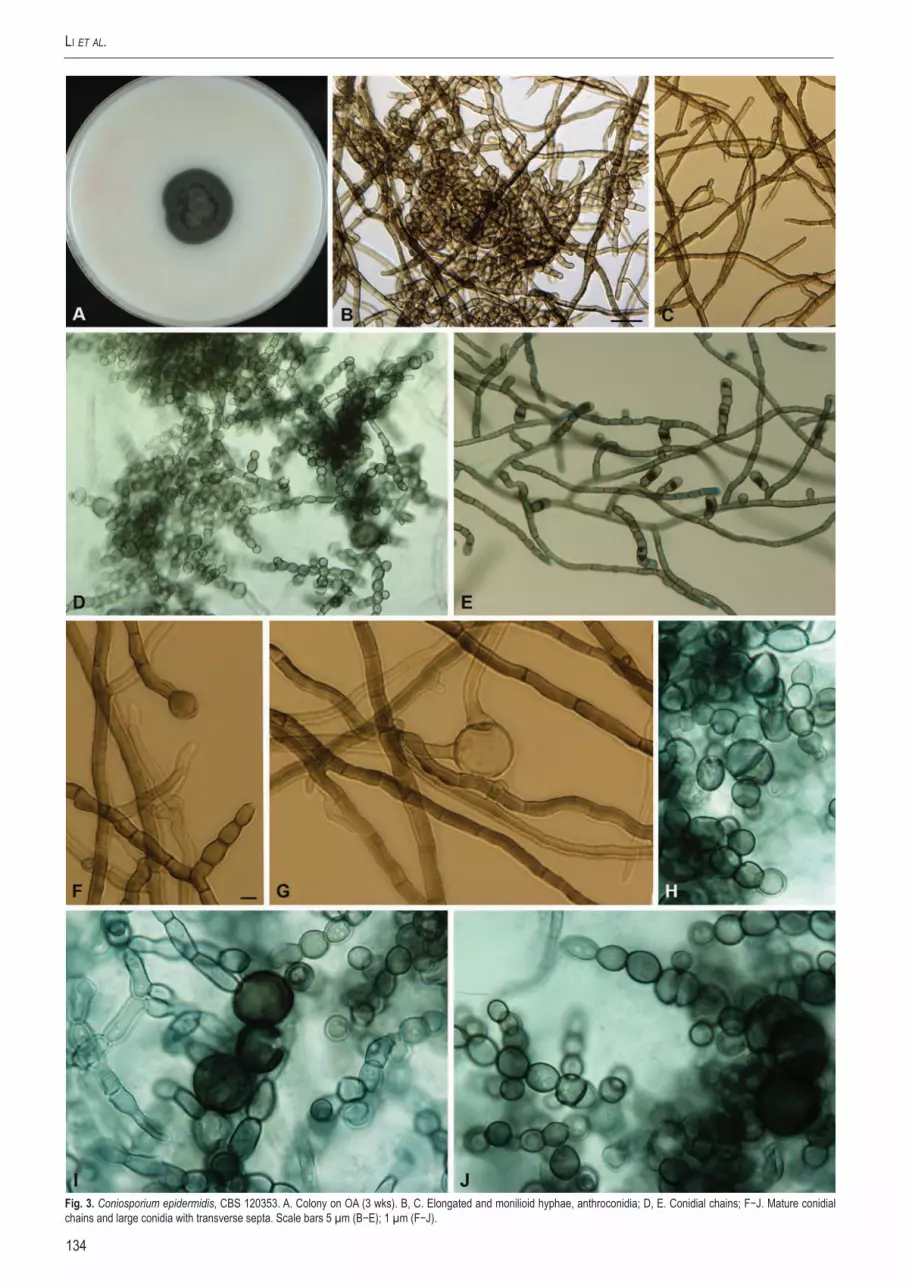
At primary isolation isolate CBS 120353 grew slowly with 8 mmwk Colonies on SGA were convex with papillate surface Obverse and reverse were black while colonies on MEA OA CMA CZA and PDA were velvety with dark brown to olive obverse Growth was stimulated under near-UV light 37 degC was tolerated Subcultures initially were cream-coloured smooth and turned black within a wk this phenomenon disappeared after several transfers Hyphae were septate olivaceous-black forming reluctantly disarticulating arthroconidia with transverse but without longitudinal septa (Fig 3) Cells were pigmented thick-walled and maturated meristematically the mother cell wall frequently rupturing in an irregular fashion With time monilioid conidia (Fig 3h i) were predominant and occasional chlamydospores occurred
Molecular data
1743 bp of the rDNA SSU gene were sequenced of strain CBS 120353 (data not shown) Phylogenetic analysis of aligned sequences revealed close relationship with species in Cladophialophora Exophiala Phialophora Rhinocladiella Fonsecaea and Capronia which all are members of the order Chaetothyriales However
nearest neighbours were Coniosporium perforans Sterflinger (CBS 66580) and C apollinis Sterflinger (CBS 35297) Of the LSU domain 616 bp were sequenced Nearest neighbour at 97 similarity was a species published by Crous et al (2007) in a tree as lsquoExophiala sp 3rsquo CPC 12173 = EU035422
Length of ITS domain of CBS 120353 was 541 bp ITS rDNA sequences compared in a dedicated black yeast data base maintained at CBS and containing about 11000 entries revealed no
Fig 1 Blackish discoloured skin of toes and toe webs with scaling Fig 2 Skin biopsy stained with HAE some fungal elements are visible (arrows) Size bar = 5microm
133wwwstudiesinmycologyorg
Coniosporium on Skin
Name Accession no Country Source GenBankAntarctic black fungus CCFEE 5323 Antarctic Thallus of Lecanora sp FJ392866
CCFEE 5314 Antarctic Thallus of Xanthoria elegans FJ392865CCFEE 5324 Antarctic Thallus of Acarospora flavocordia FJ392867
Coniosporium apollinis CBS 100213 Greece Rock AJ244271CBS 100218 Greece Marble AJ244273CBS 109867 Greece Marble CBS 100216 Spain Rock AJ244272CBS 109860 Spain Rock CBS 109865 Italy Rock
Coniosporium epidermidis CBS 123233 Denmark Hand femaledH 17028 Denmark Axilla maleCBS 123466 China Nail with onychomycosisCBS 120353 (T) China Skin infection EU730589CBS 120388 Denmark Toenail female CBS 123279 Denmark Toenail femaledH 17006 Denmark Toenail femaleCBS 123261 Denmark Toenail maledH 17086 Denmark Toenail male
Coniosporium perforans dH 17016 Denmark Axilla femaleCBS 109861 Italy Marble dH 16682 Denmark Nail male CBS 88595 (T) Greece Marble
Coniosporium species CBS119726 Italy Stone monumentdH 14071 Australia CattledH 16979 France Chronic nasal oedemadH 14084 Italy Marble monumentdH 14085 Italy Rock monumentCBS 109864 Italy Rock CBS 109866 Italy RockCBS 66580 Italy Rock
Cryptoendolithic fungus CCFEE 457 Antarctic University ValleyExophiala placitae CPC 13707 Australia Eucalyptus placita EU0402151Exophiala species CPC 12172 Canada Prunus sp
CPC 12173 Canada Prunus spCPC 12171 Canada Prunus sp EU035420
Meristematic fungus CBS119729 Italy Stone Phaeococcomyces catenatus Det M175 Netherlands Nail
CBS 65076 (T) Switzerland Air AF050277dH 11392 UnknowndH 14721 Austria Bloodplasma TRN4 Spain Rock surface AY843222
Phialophora europaea CBS 65682 France Nail FJ489612dH 12320 Germany Nail CBS 101466 (T) Netherlands Skin scales
Sarcinomyces petricola CBS 72696 (T) Guinea Dung of cow FJ489613CBS 60093 Greece Pentelic marbleCDC 2008006858 USA Scalp rash male
Uncultured ascomycete AM901753 Finland Indoor dust AM901753Abbreviations used CBS = CBS Fungal Biodiversity Centre Utrecht The Netherlands CCFEE = Culture Collection of Fungi from Extreme Environments Viterbo Italy CDC = Centers for Disease Control and Prevention Atlanta USA CPC = culture collection of Pedro Crous housed at CBS dH = GS de Hoog working collection TRN = Tino Ruibal working collectionT = ex-type culture
Table 1 Isolation data of examined strains
134
li et al
Fig 3 Coniosporium epidermidis CBS 120353 A Colony on OA (3 wks) B C Elongated and monilioid hyphae anthroconidia D E Conidial chains FminusJ Mature conidial chains and large conidia with transverse septa Scale bars 5 microm (BminusE) 1 microm (FminusJ)
135wwwstudiesinmycologyorg
Coniosporium on Skin
close match with any known species Alignment was only partially confident Among the nearest neighbours were the known rock-inhabiting species Coniosporium perforans and C apollinis as well as a number of undescribed rock-inhabiting species prevalently from the Mediterranean and from the Antarctic (Table 1) lsquoExophiala sp 3rsquo of Crous et al (2007) was also close the ITS of this species proved to be identical to a hyperparasitic lsquoConiosporium sprsquo AM901753 published by Harutyunyan et al (2008) In addition strains from human skin and nail samples were involved (Table 1) Identity was found with a group of strains from dermatological skin and nail samples from Denmark as well as with an environmental sample from Brazil all these strains either were morphologically identical to CBS 120353 or consisted of sterile melanized hyphae These strains were therefore regarded to represent a hitherto undescribed taxon which is introduced below
Coniosporium epidermidis DM Li de Hoog Saunte amp XR Chen sp nov ndash MycoBank MB512506 Figs 3 5
Coloniae primum fuscae effusae deinde elevatae velutinae vel cottoneae griseo-olivaceae reversum olivaceo-nigrum Coloniae in agaroso maltoso agaro PDA vel agaro farina avenae confecto (OSD) dicto 25 degC 10minus15 mm diam post 28 dies Mycelium immersum vel superficiale ex hyphis ramosis septatis compositum olivaceo-nigrum vel brunneum Conidiophora vix distinguenda ramose vel simplicia terminalia vel intercalaria Conidia singula primaria in apice conidiophori ellipsoidea vel subglobosa levia 2-3 microm diam Mycelium torulosum praesens Teleomorphosis ignota
Holotype dried culture in CBS herbarium (CBS-H-20167) ex-type strain CBS 120353 = T22 isolated from nigramacula superficial infection of the feet of a 80-yr-old male patient China DM Li Additional strains listed in Table 1
The following description is of CBS 120353 on PDA after 28 ds incubation at 25 degC
Colonies effuse becoming raised attaining 10-15 mm diam velvety to fluffy black blackish-brown to greyish olivaceous reverse olivaceous-black Mycelium superficial regularly and densely septate profusely branched at nearly right angles olivaceous-black or dark brown smooth- or occasionally rough-walled Cells gradually swelling at maturation up to 3-8 microm diam cell walls very thick at maturity Conidia formed by liberation of arthric cells swelling to become ellipsoidal or nearly spherical smooth-walled mostly 2-3 microm diam up to 8 microm wide then frequently the mother cell wall remaining visible on the daughter cell Truly muriform cells absent Teleomorph unknown Cardinal temperatures optimum 27 degC maximum 37 degC
73
100
70
94
90
9890
92
98
97
100
100
74
100
10090
82
9894
99
Fig 4 Phylogenetic tree of Coniosporium epidermidis and allied black fungi based on the completed ITS 1minus2 domain including the 58S rDNA gene generated with the TREEFINDER package using the Neighbor-joining algorithm and Kimura correction The tree was subjected to 100 bootstrap replications Phialophora europaea CBS 65682 was selected as outgroup
Fig 5 Microscopic morphology of Coniosporium epidermidis CBS 120353 slide culture on SGA 28 d
136
li et al
DISCUSSION
Coniosporium classically includes black slow-growing anamorphs most of which are plant colonizers (Hyde et al 2002) Species are characterized by pigmented anthroconidia and tend to develop meristematically into chains (Ellis 1971) The mother cell wall may eventually rupture during development leading to irregular cell wall ornamentation (Ellis 1976)
Coniosporium epidermidis was isolated from a tinea nigra-like skin infection on the foot Tinea nigra characterized by brown to black superficial macules is a strictly asymptomatic colonization of dead epidermis (Schwartz 2004 Bonifaz et al 2008) caused by a halophilic member of the order Capnodiales Hortaea werneckii (Zalar et al 1999) In contrast pathological slides of C epidermidis clearly showed that the fungus grew into all layers of the epidermis and some cells penetrated down to the basal membrane reaching the superficial dermis Thus the present species causes a real disease process as was also observed with the tinea nigra-like infection caused by Cladophialophora saturnica Badali et al (2008a) another member of Chaetothyriales The optimal growth temperature of C epidermidis at 27 degC (maximum 37 degC) is comparable to that of true pathogens of the skin the dermatophytes ranging between 25 and 35 degC (Weitzman amp Summerbell 1999) We thus conclude that ordinal relationships predict opportunistic potential In the course of the present study Coniosporium epidermidis was repeatedly isolated from routine dermatological samples in Denmark It is supposed that the fungus may be common in cutaneous samples but is generally discarded as a contaminant We therefore recommend to pay more attention to melanized fungi occurring on skin and nails and to establish their precise role in pathology
With SSU rDNA the black fungus recovered from affected human skin appeared to be an undescribed species located within a group of species causing mild cutaneous infections such as Phialophora europaea and Cyphellophora laciniata The group was basal to the order Chaetothyriales an order having Capronia teleomorphs and being notorious for containing numerous opportunists (Badali et al 2008b) In the corresponding ITS tree (Fig 4) the group showed considerable diversification among species The similarity of C epidermidis to the nearest described species Coniosporium perforans was less than 91 This species is however a colonizer of rock and monuments in the Mediterranean Basin (Sterflinger et al 1997) as is the case in the majority of taxa in this group The rock-inhabiting species were attributed to the genus Coniosporium although the generic type species C olivaceum Link (Ellis 1971) has as yet not been redefined according to modern standards
Sterflinger et al (1997) supposed that the Coniosporium-clade of Chaetothyriales was entirely rock-associated However with recent additions to this group we notice that it contains several undescribed taxonomic species from human skin and nail samples In addition many of the species described as having a rock-inhabiting life-style such as Sarcinomyces petricola Wollenzien amp de Hoog Coniosporium perforans and Phaeococcomyces catenatus (de Hoog amp Hermanides-Nijhof) de Hoog can also be found on human skin (Table 1) A similar dual ecology was found earlier in the unrelated meristematic fungus Catenulostroma abietis (Butin amp Pehl) Crous et al (Butin et al 1996) We suppose that these predominantly meristematic skin colonizers are taken up from the environment where they live as oligotrophs on rock leathery plant leaves and other relatively inert surfaces They are likely to display a similar oligotrophic character on human skin and thus probably behave as commensals rather than pathogens Nevertheless occasional mild infections may occur such as the
ones observed in Cladophialophora saturnica (Badali et al 2008a) and the present fungus
ACKNOWLEDGEMENTS
This work was supported partly by grants from the National Natural Science Foundation of China (C30570003) the China Exchange Programme of the Netherlands Academy of Sciences D Attili (Rio Claro Brazil) is acknowledged for donating a strain from an ant hill We thank Xin Lian for assistance in sequencing
REFERENCES
Badali H Carvalho VO Vicente V Attili-Angelis D Kwiatkowski IB Gerrits van den Ende AHG Hoog GS de (2009) Cladophialophora saturnica sp nov a new opportunistic species of Chaetothyriales revealed using molecular data Medical Mycology 47 1ndash12
Badali H Gueidan C Najafzadeh MJ Bonifaz A Gerrits van den Ende AHG Hoog GS de (2008b) Biodiversity of the genus Cladophialophora Studies in Mycology 61 175ndash191
Bonifaz A Badali H Hoog GS de Cruz M Araiza J Cruz M Fierro L Ponce RM (2008) Tinea nigra by Hortaea werneckii a report of 22 cases from Mexico Studies in Mycology 61 77ndash82
Butin H Pehl L Hoog GS de Wollenzien U (1996) Trimmatostroma abietis sp nov (Hyphomycetes) and related species Antonie van Leeuwenhoek 69 203ndash209
Crous PW Braun U Schubert K Groenewald JZ (2007) Delimiting Cladosporium from morphologically similar genera Studies in Mycology 58 33ndash56
De Leo F Urzi C Hoog GS de (1999) Two Coniosporium species from rock surfaces Studies in Mycology 43 70ndash79
Ellis MB (1971) Dematiaceous Hyphomycetes Commonwealth Mycological Institute Kew UK
Ellis MB (1976) More dematiaceous Hyphomycetes Commonwealth Mycological Institute Kew UK
Harutyunyan S Muggia L Grube M (2008) Black fungi in lichens from seasonally arid habitats Studies in Mycology 61 83ndash90
Hoog GS de Guarro J Geneacute J Figueras MJ (2000a) Atlas of Clinical Fungi ed 2 Centraalbureau voor Schimmelcultures Universitat Rovira i Virgili Utrecht Reus
Hoog GS de Mayser P Haase G Horreacute R Horrevorts AM (2000b) A new species Phialophora europaea causing superficial infections in humans Mycoses 43 409ndash416
Hoog GS de Zeng JS Harrak MJ Sutton DA (2006) Exophiala xenobiotica sp nov an opportunistic black yeast inhabiting environments rich in hydrocarbons Antonie van Leeuwenhoek 90 257ndash268
Hyde KD Zhou DQ Dalisay T (2002) Bambusicolous fungi A review Fungal Diversity 9 1ndash14
Li DM Xiu DR Li RY Samson RA Hoog GS de Wang DL (2008) Aspergillus flavus myositis in a patient after liver transplantation Clinical Transplant 22 508ndash511
Ludwig W Strunk O Westram R Richter L Meier H Yadhukumar Buchner A Lai T Steppi S Jobb G Forster W Brettske I Gerber S Ginhart AW Gross O Grumann S Hermann S Jost R Konig A Liss T Lussmann R May M Nonhoff B Reichel B Strehlow R Stamatakis A Stuckmann N Vilbig A Lenke M Ludwig T Bode A Schleifer KH (2004) ARB a software environment for sequence data Nucleic Acids Research 32 1363-1371
Schwartz RA (2004) Superficial fungal infections Lancet 364 1173ndash1182Sterflinger K De Baere R Hoog GS de De Wachter R Krumbein WE Haase G
(1997) Coniosporium perforans and C apollinis two new rockndashinhabiting fungi isolated from marble in the Sanctuary of Delos (Cyclades Greece) Antonie van Leeuwenhoek 72 349ndash353
Sterflinger K Prillinger H (2001) Molecular taxonomy and biodiversity of rock fungal communities in an urban environment (Vienna Austria) Antonie van Leeuwenhoek 80 275ndash286
Tseng SS Whittier S Miller SR Zalar GL (1999) Bilateral tinea nigra plantaris and tinea nigra plantaris mimicking melanoma Cutis 64 265ndash268
Vries GS de (1962) Cyphellophora laciniata nov gen nov sp and Dactylium fusarioides Fragoso et Ciferri Mycopathologia et Mycologia Applicata 16 147-54
Weitzman I Summerbell RC (1999) The dermatophytes Clinical Microbiology Reviews 8 240ndash259
Zalar P Hoog GS de Gunde-Cimerman N (1999) Ecology of halotolerant dothideaceous black yeasts Studies in Mycology 43 38ndash48

132
li et al
Sequencing
Approximately 01 g of fungal material was transferred to a 2-mL Eppendorf tube containing a 21 (ww) mixture of silica gel and Celite (silica gel H Merck 7736Kieselguhr Celite 545 Machery Merck Amsterdam The Netherlands) DNA was extracted according to methods described previously (Li et al 2008) Amplifications were done with primers ITS1 and ITS4 (for rDNA Internal Transcribed Spacer ITS) NS1 BF83 OLI1 BF963 BF1438 and NS24 (for rDNA Small Subunit nucSSU) D1D2 (for rDNA Large SubUnit nucLSU) and EF1-728F and EF1-986R (for Translation Elongation Factor 1-α EF1α) PCR was performed in 50 microL volumes of a reaction mixture containing 10 mM Tris HCl (pH 83) 50 mM KCl 15 mM MgCl26H2O 001 gelatin 200 mM of each deoxynucleotide triphosphate 25 pmol of each primer 10minus100 ng rDNA and 05 U Taq DNA polymerase (Bioline GC Biotech Alphen ad Rijn The Netherlands) as follows 95 degC for 4 min followed by 35 cycles consisting of 94 degC for 45 s 52 degC for 30 s and 72 degC for 2 min Amplicons were cleaned with GFX columns (GE Healthcare Sweden) Sequence PCR was performed as follows 95 degC for one min followed by 30 cycles consisting of 95 degC for 10 s 50 degC for five s and 60 degC for two min DNA was purified with Sephadex G-50 Superfine Purified amplicons were then sequenced on both strands using the same primers described above BigDye terminator cycle sequencing Ready Reaction kits (Perkin Elmer Applied Biosystems Nieuwerkerk ad IJssel The Netherlands) were used according to the manufacturerrsquos instructions and DNA was sequenced using a DYE-ET terminator
Sequence analysis and taxonomy
Sequences were compared in GenBank and using a research database available at the Centraalbureau voor Schimmelcultures Biodiversity Centre (CBS) Utrecht The Netherlands Alignment
was done in a database using BionuMericS software v 461 (Applied Maths Kortrijk Belgium) SSU sequences were aligned with the ARB beta-package (v 22-08-2003) developed by Ludwig et al (2004) A distance tree of Coniosporium epidermidis and allied black fungi based on the completed ITS 1-2 domain including the 58S rDNA gene were reconstructed using neighbor-joining algorithm with Kimura 2 correction with 100 bootstrap replications in treefinder
RESULTS
Mycology
A skin biopsy performed on the black lesion (Fig 1) and histological specimens stained with hematoxylin-eosin (Fig 2) showed hyperkeratosis and acanthosis Numerous hyphae and swollen cells were observed in the stratum corneum (Fig 2) Pigmented hyphae and loose cells were displayed in the entire layers of epidermis predominated among the low layers Cells also penetrated the basal membrane to the dermis Direct examination of skin scrapings with KOH was positive for pigmented arthroconidia and dark-walled hyphae The disease was considered to be an infection
At primary isolation isolate CBS 120353 grew slowly with 8 mmwk Colonies on SGA were convex with papillate surface Obverse and reverse were black while colonies on MEA OA CMA CZA and PDA were velvety with dark brown to olive obverse Growth was stimulated under near-UV light 37 degC was tolerated Subcultures initially were cream-coloured smooth and turned black within a wk this phenomenon disappeared after several transfers Hyphae were septate olivaceous-black forming reluctantly disarticulating arthroconidia with transverse but without longitudinal septa (Fig 3) Cells were pigmented thick-walled and maturated meristematically the mother cell wall frequently rupturing in an irregular fashion With time monilioid conidia (Fig 3h i) were predominant and occasional chlamydospores occurred
Molecular data
1743 bp of the rDNA SSU gene were sequenced of strain CBS 120353 (data not shown) Phylogenetic analysis of aligned sequences revealed close relationship with species in Cladophialophora Exophiala Phialophora Rhinocladiella Fonsecaea and Capronia which all are members of the order Chaetothyriales However
nearest neighbours were Coniosporium perforans Sterflinger (CBS 66580) and C apollinis Sterflinger (CBS 35297) Of the LSU domain 616 bp were sequenced Nearest neighbour at 97 similarity was a species published by Crous et al (2007) in a tree as lsquoExophiala sp 3rsquo CPC 12173 = EU035422
Length of ITS domain of CBS 120353 was 541 bp ITS rDNA sequences compared in a dedicated black yeast data base maintained at CBS and containing about 11000 entries revealed no
Fig 1 Blackish discoloured skin of toes and toe webs with scaling Fig 2 Skin biopsy stained with HAE some fungal elements are visible (arrows) Size bar = 5microm
133wwwstudiesinmycologyorg
Coniosporium on Skin
Name Accession no Country Source GenBankAntarctic black fungus CCFEE 5323 Antarctic Thallus of Lecanora sp FJ392866
CCFEE 5314 Antarctic Thallus of Xanthoria elegans FJ392865CCFEE 5324 Antarctic Thallus of Acarospora flavocordia FJ392867
Coniosporium apollinis CBS 100213 Greece Rock AJ244271CBS 100218 Greece Marble AJ244273CBS 109867 Greece Marble CBS 100216 Spain Rock AJ244272CBS 109860 Spain Rock CBS 109865 Italy Rock
Coniosporium epidermidis CBS 123233 Denmark Hand femaledH 17028 Denmark Axilla maleCBS 123466 China Nail with onychomycosisCBS 120353 (T) China Skin infection EU730589CBS 120388 Denmark Toenail female CBS 123279 Denmark Toenail femaledH 17006 Denmark Toenail femaleCBS 123261 Denmark Toenail maledH 17086 Denmark Toenail male
Coniosporium perforans dH 17016 Denmark Axilla femaleCBS 109861 Italy Marble dH 16682 Denmark Nail male CBS 88595 (T) Greece Marble
Coniosporium species CBS119726 Italy Stone monumentdH 14071 Australia CattledH 16979 France Chronic nasal oedemadH 14084 Italy Marble monumentdH 14085 Italy Rock monumentCBS 109864 Italy Rock CBS 109866 Italy RockCBS 66580 Italy Rock
Cryptoendolithic fungus CCFEE 457 Antarctic University ValleyExophiala placitae CPC 13707 Australia Eucalyptus placita EU0402151Exophiala species CPC 12172 Canada Prunus sp
CPC 12173 Canada Prunus spCPC 12171 Canada Prunus sp EU035420
Meristematic fungus CBS119729 Italy Stone Phaeococcomyces catenatus Det M175 Netherlands Nail
CBS 65076 (T) Switzerland Air AF050277dH 11392 UnknowndH 14721 Austria Bloodplasma TRN4 Spain Rock surface AY843222
Phialophora europaea CBS 65682 France Nail FJ489612dH 12320 Germany Nail CBS 101466 (T) Netherlands Skin scales
Sarcinomyces petricola CBS 72696 (T) Guinea Dung of cow FJ489613CBS 60093 Greece Pentelic marbleCDC 2008006858 USA Scalp rash male
Uncultured ascomycete AM901753 Finland Indoor dust AM901753Abbreviations used CBS = CBS Fungal Biodiversity Centre Utrecht The Netherlands CCFEE = Culture Collection of Fungi from Extreme Environments Viterbo Italy CDC = Centers for Disease Control and Prevention Atlanta USA CPC = culture collection of Pedro Crous housed at CBS dH = GS de Hoog working collection TRN = Tino Ruibal working collectionT = ex-type culture
Table 1 Isolation data of examined strains
134
li et al
Fig 3 Coniosporium epidermidis CBS 120353 A Colony on OA (3 wks) B C Elongated and monilioid hyphae anthroconidia D E Conidial chains FminusJ Mature conidial chains and large conidia with transverse septa Scale bars 5 microm (BminusE) 1 microm (FminusJ)
135wwwstudiesinmycologyorg
Coniosporium on Skin
close match with any known species Alignment was only partially confident Among the nearest neighbours were the known rock-inhabiting species Coniosporium perforans and C apollinis as well as a number of undescribed rock-inhabiting species prevalently from the Mediterranean and from the Antarctic (Table 1) lsquoExophiala sp 3rsquo of Crous et al (2007) was also close the ITS of this species proved to be identical to a hyperparasitic lsquoConiosporium sprsquo AM901753 published by Harutyunyan et al (2008) In addition strains from human skin and nail samples were involved (Table 1) Identity was found with a group of strains from dermatological skin and nail samples from Denmark as well as with an environmental sample from Brazil all these strains either were morphologically identical to CBS 120353 or consisted of sterile melanized hyphae These strains were therefore regarded to represent a hitherto undescribed taxon which is introduced below
Coniosporium epidermidis DM Li de Hoog Saunte amp XR Chen sp nov ndash MycoBank MB512506 Figs 3 5
Coloniae primum fuscae effusae deinde elevatae velutinae vel cottoneae griseo-olivaceae reversum olivaceo-nigrum Coloniae in agaroso maltoso agaro PDA vel agaro farina avenae confecto (OSD) dicto 25 degC 10minus15 mm diam post 28 dies Mycelium immersum vel superficiale ex hyphis ramosis septatis compositum olivaceo-nigrum vel brunneum Conidiophora vix distinguenda ramose vel simplicia terminalia vel intercalaria Conidia singula primaria in apice conidiophori ellipsoidea vel subglobosa levia 2-3 microm diam Mycelium torulosum praesens Teleomorphosis ignota
Holotype dried culture in CBS herbarium (CBS-H-20167) ex-type strain CBS 120353 = T22 isolated from nigramacula superficial infection of the feet of a 80-yr-old male patient China DM Li Additional strains listed in Table 1
The following description is of CBS 120353 on PDA after 28 ds incubation at 25 degC
Colonies effuse becoming raised attaining 10-15 mm diam velvety to fluffy black blackish-brown to greyish olivaceous reverse olivaceous-black Mycelium superficial regularly and densely septate profusely branched at nearly right angles olivaceous-black or dark brown smooth- or occasionally rough-walled Cells gradually swelling at maturation up to 3-8 microm diam cell walls very thick at maturity Conidia formed by liberation of arthric cells swelling to become ellipsoidal or nearly spherical smooth-walled mostly 2-3 microm diam up to 8 microm wide then frequently the mother cell wall remaining visible on the daughter cell Truly muriform cells absent Teleomorph unknown Cardinal temperatures optimum 27 degC maximum 37 degC
73
100
70
94
90
9890
92
98
97
100
100
74
100
10090
82
9894
99
Fig 4 Phylogenetic tree of Coniosporium epidermidis and allied black fungi based on the completed ITS 1minus2 domain including the 58S rDNA gene generated with the TREEFINDER package using the Neighbor-joining algorithm and Kimura correction The tree was subjected to 100 bootstrap replications Phialophora europaea CBS 65682 was selected as outgroup
Fig 5 Microscopic morphology of Coniosporium epidermidis CBS 120353 slide culture on SGA 28 d
136
li et al
DISCUSSION
Coniosporium classically includes black slow-growing anamorphs most of which are plant colonizers (Hyde et al 2002) Species are characterized by pigmented anthroconidia and tend to develop meristematically into chains (Ellis 1971) The mother cell wall may eventually rupture during development leading to irregular cell wall ornamentation (Ellis 1976)
Coniosporium epidermidis was isolated from a tinea nigra-like skin infection on the foot Tinea nigra characterized by brown to black superficial macules is a strictly asymptomatic colonization of dead epidermis (Schwartz 2004 Bonifaz et al 2008) caused by a halophilic member of the order Capnodiales Hortaea werneckii (Zalar et al 1999) In contrast pathological slides of C epidermidis clearly showed that the fungus grew into all layers of the epidermis and some cells penetrated down to the basal membrane reaching the superficial dermis Thus the present species causes a real disease process as was also observed with the tinea nigra-like infection caused by Cladophialophora saturnica Badali et al (2008a) another member of Chaetothyriales The optimal growth temperature of C epidermidis at 27 degC (maximum 37 degC) is comparable to that of true pathogens of the skin the dermatophytes ranging between 25 and 35 degC (Weitzman amp Summerbell 1999) We thus conclude that ordinal relationships predict opportunistic potential In the course of the present study Coniosporium epidermidis was repeatedly isolated from routine dermatological samples in Denmark It is supposed that the fungus may be common in cutaneous samples but is generally discarded as a contaminant We therefore recommend to pay more attention to melanized fungi occurring on skin and nails and to establish their precise role in pathology
With SSU rDNA the black fungus recovered from affected human skin appeared to be an undescribed species located within a group of species causing mild cutaneous infections such as Phialophora europaea and Cyphellophora laciniata The group was basal to the order Chaetothyriales an order having Capronia teleomorphs and being notorious for containing numerous opportunists (Badali et al 2008b) In the corresponding ITS tree (Fig 4) the group showed considerable diversification among species The similarity of C epidermidis to the nearest described species Coniosporium perforans was less than 91 This species is however a colonizer of rock and monuments in the Mediterranean Basin (Sterflinger et al 1997) as is the case in the majority of taxa in this group The rock-inhabiting species were attributed to the genus Coniosporium although the generic type species C olivaceum Link (Ellis 1971) has as yet not been redefined according to modern standards
Sterflinger et al (1997) supposed that the Coniosporium-clade of Chaetothyriales was entirely rock-associated However with recent additions to this group we notice that it contains several undescribed taxonomic species from human skin and nail samples In addition many of the species described as having a rock-inhabiting life-style such as Sarcinomyces petricola Wollenzien amp de Hoog Coniosporium perforans and Phaeococcomyces catenatus (de Hoog amp Hermanides-Nijhof) de Hoog can also be found on human skin (Table 1) A similar dual ecology was found earlier in the unrelated meristematic fungus Catenulostroma abietis (Butin amp Pehl) Crous et al (Butin et al 1996) We suppose that these predominantly meristematic skin colonizers are taken up from the environment where they live as oligotrophs on rock leathery plant leaves and other relatively inert surfaces They are likely to display a similar oligotrophic character on human skin and thus probably behave as commensals rather than pathogens Nevertheless occasional mild infections may occur such as the
ones observed in Cladophialophora saturnica (Badali et al 2008a) and the present fungus
ACKNOWLEDGEMENTS
This work was supported partly by grants from the National Natural Science Foundation of China (C30570003) the China Exchange Programme of the Netherlands Academy of Sciences D Attili (Rio Claro Brazil) is acknowledged for donating a strain from an ant hill We thank Xin Lian for assistance in sequencing
REFERENCES
Badali H Carvalho VO Vicente V Attili-Angelis D Kwiatkowski IB Gerrits van den Ende AHG Hoog GS de (2009) Cladophialophora saturnica sp nov a new opportunistic species of Chaetothyriales revealed using molecular data Medical Mycology 47 1ndash12
Badali H Gueidan C Najafzadeh MJ Bonifaz A Gerrits van den Ende AHG Hoog GS de (2008b) Biodiversity of the genus Cladophialophora Studies in Mycology 61 175ndash191
Bonifaz A Badali H Hoog GS de Cruz M Araiza J Cruz M Fierro L Ponce RM (2008) Tinea nigra by Hortaea werneckii a report of 22 cases from Mexico Studies in Mycology 61 77ndash82
Butin H Pehl L Hoog GS de Wollenzien U (1996) Trimmatostroma abietis sp nov (Hyphomycetes) and related species Antonie van Leeuwenhoek 69 203ndash209
Crous PW Braun U Schubert K Groenewald JZ (2007) Delimiting Cladosporium from morphologically similar genera Studies in Mycology 58 33ndash56
De Leo F Urzi C Hoog GS de (1999) Two Coniosporium species from rock surfaces Studies in Mycology 43 70ndash79
Ellis MB (1971) Dematiaceous Hyphomycetes Commonwealth Mycological Institute Kew UK
Ellis MB (1976) More dematiaceous Hyphomycetes Commonwealth Mycological Institute Kew UK
Harutyunyan S Muggia L Grube M (2008) Black fungi in lichens from seasonally arid habitats Studies in Mycology 61 83ndash90
Hoog GS de Guarro J Geneacute J Figueras MJ (2000a) Atlas of Clinical Fungi ed 2 Centraalbureau voor Schimmelcultures Universitat Rovira i Virgili Utrecht Reus
Hoog GS de Mayser P Haase G Horreacute R Horrevorts AM (2000b) A new species Phialophora europaea causing superficial infections in humans Mycoses 43 409ndash416
Hoog GS de Zeng JS Harrak MJ Sutton DA (2006) Exophiala xenobiotica sp nov an opportunistic black yeast inhabiting environments rich in hydrocarbons Antonie van Leeuwenhoek 90 257ndash268
Hyde KD Zhou DQ Dalisay T (2002) Bambusicolous fungi A review Fungal Diversity 9 1ndash14
Li DM Xiu DR Li RY Samson RA Hoog GS de Wang DL (2008) Aspergillus flavus myositis in a patient after liver transplantation Clinical Transplant 22 508ndash511
Ludwig W Strunk O Westram R Richter L Meier H Yadhukumar Buchner A Lai T Steppi S Jobb G Forster W Brettske I Gerber S Ginhart AW Gross O Grumann S Hermann S Jost R Konig A Liss T Lussmann R May M Nonhoff B Reichel B Strehlow R Stamatakis A Stuckmann N Vilbig A Lenke M Ludwig T Bode A Schleifer KH (2004) ARB a software environment for sequence data Nucleic Acids Research 32 1363-1371
Schwartz RA (2004) Superficial fungal infections Lancet 364 1173ndash1182Sterflinger K De Baere R Hoog GS de De Wachter R Krumbein WE Haase G
(1997) Coniosporium perforans and C apollinis two new rockndashinhabiting fungi isolated from marble in the Sanctuary of Delos (Cyclades Greece) Antonie van Leeuwenhoek 72 349ndash353
Sterflinger K Prillinger H (2001) Molecular taxonomy and biodiversity of rock fungal communities in an urban environment (Vienna Austria) Antonie van Leeuwenhoek 80 275ndash286
Tseng SS Whittier S Miller SR Zalar GL (1999) Bilateral tinea nigra plantaris and tinea nigra plantaris mimicking melanoma Cutis 64 265ndash268
Vries GS de (1962) Cyphellophora laciniata nov gen nov sp and Dactylium fusarioides Fragoso et Ciferri Mycopathologia et Mycologia Applicata 16 147-54
Weitzman I Summerbell RC (1999) The dermatophytes Clinical Microbiology Reviews 8 240ndash259
Zalar P Hoog GS de Gunde-Cimerman N (1999) Ecology of halotolerant dothideaceous black yeasts Studies in Mycology 43 38ndash48


