Embed Size (px)
Citation preview

J Eukunor. M i c r d ~ i o l , 47(5). 2000 pp. 481-492 0 2000 by the Society ot Pn~taznolog~rts
Contributions of the Axostyle and Flagella to Closed Mitosis in the Protists Tritrichomonas foetus and Trichomonas vaginalis
KARLA C. RIBEIRO,",' LUIZ HENRIQUE MONTEIRO-LEALb AND MARLENE BENCHIMOL' "Universidade Estadual d o Norte Fluminense, LBCT, Campos dos Goytacazes, Rio de Janeiro, Brazil, and
bL.uhoratcirio de Microscopia e Processamento de Imagem, Departumento de Histologia e Embriologia, Universidade Estadual do Rio de Janeiro, and
'Universidade Sunta Ursula and Instituto de Biofisica Carlos Ckagas Filko, Universidade Federal do Rio de Janeiro, Rua Jornalista Orlando Dmtas, 59. CEP 222-31 -010. Botaj+yh Rio de Juneiro, Brazil
ABSTRACT. Tritrichhmonas foetus and Trickomonas vaginalis are protists that undergo closed mitosis: the nuclear envelope remains intact and the spindle remains extranuclear. Here we show, in disagreement with previous studies, that the axostyle does not disappear during mitosis hut rather actively participates in it. We document the main structural modifications of the cell during its cell cycle using video enhanced microscopy and computer animation, bright field light microscopy, confocal laser scanning microscopy, and scanning and transmission electron microscopy. We propose six phases in the trichomonad's cell cycle: an orthodox interphase, a pre-mitotic phase, and four stages during the cell division process. We report that in T. foetus and T. vaginalis: a) all skeletal structures such as the costa, pelta-axostyle system, basal bodies, flagella, and associated filaments of the mastigont system are duplicated in a pre-mitotic phase; b) the axostyle does not disappear during mitosis, otherwise playing a fundamental role in this process; c) axostyles participate in the changes in the cell shape, contortion of the anterior region of the cell, and karyokinesis; d) flagella are not under assembly during mitosis, as previously stated by others, but completely formed before it; and e) cytokinesis is powered in part by cell locomotion.
Key Words. Axostyle, cell cycle, cytoskeleton, flagella, karyokinesis, protist.
ITRICHOMONAS foetus and Trichornonas vaginalis are flagellated parasitic protists of the urogenital tract of cattle
and humans, respectively, and belong to the phylum Parabasa- lia. Molecular phylogenetic studies using large and small sub- unit ribosomal RNAs indicate that these organisms are among the most early diverging eukaryotes (Cavalier-Smith and Chao 1996; Leipe et al. 1993; Viscogliosi and Brugerolle 1993) al- though one cannot discount the possibility of phylogenetic re- positioning (Embley and Hirt 1998). According to Heath (1980), trichomonads present one of the most primitive kinds of mitosis since the extranuclear microtubular spindle does not contact the chromosomes directly and nuclear envelope break- down does not take place.
Like some other parabasalian species, T. foetus possesses an axostyle, which is a ribbon of longitudinally-oriented microtu- bules running from one end of the cell to the other. Unlike axostyles found elsewhere, the axostyle of T. foetus does not present any contraction movement (Monteiro-Leal et al. 1996) and it is composed of microtubules that contain acetylated a- tubulin (Batista et al. 1988). Previous reports on the division process in trichomonads (Delgado-Viscogliosi et al. 1996; Ju- liano et al. 1986; Viscogliosi and Brugerolle 1994; Viscogliosi and Delgado-Viscogliosi 1996) claimed that the parental axo- style-pelta complex depolymerizes during mitosis, and two new axostyles grow around each set of basal bodies; the parental structures are retained in one sister kinetid and new structures are assembled in the other. Some authors (Viscogliosi and Brug- erolle 1994) also stated that the flagella were assembled during mitosis. Our previous studies were in disagreement with these reports, since they provided evidence that the pelta-axostyle complex is a stable structure (Batista et al. 1988; Silva-Filho and DeSouza 1986). Also in disagreement with our observa- tions, Delgado-Viscogliosi et al. (1996) claimed that the flagel- lum number is restored at the end of mitosis.
In view of the lack of a detailed study on the dynamics of cell structures during the cell cycle of trichomonads, we decid- ed to use several approaches, including video microscopy, con-
Corresponding Author: M. Benchimol-Telephone number: 55-21- 21-553-161s; FAX number: 5521-21-260-2364; Email: marleneb@ domain.com.br
Abbreviations: LSCM: laser scanning confocal microscopy, MT: mi- crotubules; SEM: scanning electron microscopy, SPB: spindle pole body, TEM: transmission electron microscopy.
focal laser scanning microscopy, and conventional scanning and transmission electron microscopy to follow the fate of several structures. The results obtained are described in this paper.
MATERIALS AND METHODS
Cell culture. The K strain of Tritrichomonas foetus was iso- lated by Dr. H. Guida (Embrapa, Rio de Janeiro, Brazil) from the urogenital tract of a bull from the state of Rio de Janeiro, Brazil, and has been maintained in TYM Diamond's medium (Diamond 1957). Trichomonas vaginalis, strain JT, was isolated from a patient attending the Rio de Janeiro Federal University Hospital. The cells were cultivated for 24 h at 36.5 "C, which corresponds to the end of the logarithmic phase of growth.
Immunofluorescence. Cells were washed in warm PHEM buffer (50 mM MgCl,, 70 mM KC1, 10 mM EGTA, 20 mM Hepes, 60 mM Pipes, pH 6.8) and then allowed to adhere to coverslips previously coated with poly-L-lysine. Thereafter, they were fixed with 4% paraformaldehyde and permeabilized with 0.2% Triton X-100. Alternatively, cells were fixed and permeabilized with 70% ethanol at -20 "C. Fixed cells were quenched using 50 mM ammonium chloride solution and 3% bovine serum albumin. They were then incubated for 3 h with one of the following antibodies: polyclonal anti-tubulin # 3526 (Sigma, St. Louis, MO) or monoclonal anti-alpha-tubulins # 356 (Amersham, UK), # 5168, # 6793 (Sigma), TAT (anti- body against T. brucei microtubules, kindly provided by Dr. Keith Gull (Woods et al. 1989), and anti-P-tubulin # 357 (Amersham), monoclonal against chick-brain microtubules. Af- ter thorough washing in PBS, the samples were incubated for 1 h with a FITC-labeled anti-rabbit antibody or TRITC-labeled anti-mouse antibody, respectively. Cells were examined with a Zeiss Confocal Laser Scanning Microscope (CLSM) or Axio- phot 2 (Zeiss).
Panotic staining-bright field microscopy. Cells were al- lowed to adhere to a poly-L-lysine coated coverslip. Cell fixa- tion and staining were performed using the Panotic staining Kit (Laborclin, PR, Brazil). Panotic staining was used to show, by light microscopy, the number and position of the axostyle, nu- cleus, kinetosomes, and flagella, made of a mixture of stains (unpublished by the suppliers), in a composition similar to he- matoxylin-eosin stains. Chromosomes and ribosomes are stained a deep-blue color, whereas pelta-axostyle, and flagella stay pink. Digitized images of different focal planes were ob- tained and processed with Adobe Photoshop.
48 I

482 J. EUKARYOT. MICROBIOL., VOL. 47, NO. 5 , SEPTEMBER-OCTOBER 2000
Fig. 1. Video microscopy: frozen frames of Tritrichomonas foetus from interphase (IA), through mitosis (B-F). B: Phase 1 (prophase). Arrowheads point to two straight axostyles; the nucleus (N) is seen on the upper portion of the cell. The recurrent flagella are present on each side of the cell parallel to the cell body longitudinal axis. C : Phase 2 (metaphase) in side view with cell in “heart shape.” Arrowheads point to bent axostyles. D: metaphase seen in top view. The recurrent flagella are seen redirected to a plane perpendicular to the cell’s longitudinal axis. The nucleus is under constriction, assuming an “s” shape and it is still located at the upper portion of the cell. E: Phase 3 (anaphase) karyokinesis has occurred and the two nuclei occupy the central part of the elongated cell. F: Phase 4 (telophase): cytokinesis. The two nuclei achieve maximum distance and are positioned at the anterior region of
Transmission electron microscopy (TEM). Cells were washed 3 X in PHEM buffer at 37 “C and fixed overnight at room temp. in 2.5% (v/v) glutaraldehyde in 0.1 M Na-caco- dylate buffer (pH 7.2), post-fixed in 1% OsO, in cacodylate buffer plus 5 mM CaCl, and 0.8% potassium ferricyanide. Cells were washed, dehydrated in acetone, and embedded in Epon. Thin sections were stained and observed in a Zeiss 900 electron microscope.
Video-enhanced microscopy. A 63 X , N.A. 1.4 objective lens was chosen to allow maximum resolution using differential interference contrast microscopy. Living cells from interphase through mitosis were recorded. The sequences of video mi- croscopy images were obtained using a high resolution CCD video-camera (Optronics, Goleta, CA) attached to a Zeiss Ax- ioplan microscope. The sequences were optimized by analogue process and recorded in a S-VHS video-recorder. The video images were analyzed and the best images were chosen and digitized using the IBAS System (Kontron-Zeiss, Oberkochen, Germany) or were captured with a TARGA board (True Vision, RasterOps Company).
Scanning electron microscopy (SEM). Cells on poly-l-ly- sine-coated glass coverslips were fixed in 2.5% glutaraldehyde, post-fixed for 5 min in 1% OsO,, dehydrated in ethanol, critical point dried, sputter-coated with gold-palladium, and examined in a JEOL 1200X Scanning Electron Microscope.
Model creation. Combined morphological data obtained from serial sections, scanning electron microscopy, and video microscopy led to a summary drawing in form of a set of com- puter-based pictures, illustrating the dynamic interaction of the skeletal structures with the nucleus during division process. The drawings were made using the 3D-Studio (Autodesk Inc.) graphic creation software.
RESULTS We followed Tritrichomonas foetus during division by light
microscopy and electron microscopy of living and fixed cells. By video microscopy the main modifications of the cell from interphase through mitosis were studied. It is possible to see a duplicated axostyle in all phases of the mitosis (Table 1, Fig. 1 B-E 3-7). A non-dividing cell is characterized by a tear-drop or ovoid shape, one anterior nucleus, a pelta supporting the flagellar canal, a ribbon of microtubules forming the axostyle, which runs from the basal bodies to the cell tip, and hydroge- nosomes following the axostyle and the costa (a periodic struc- ture). Tritrichomonas foetus presents three flagella at the ante- rior region and one recurrent flagellum, adhering to the cell body (Fig. IA, 8). Around 80% of the cells were in interphase when analyzed by SEM. The nucleus is located at the anterior region of the cell (Fig. 2A). We found that a cell is at onset of mitosis when an extra set of flagella are seen (Fig. 9). In this case, the cell shape is still teardrop, although more rounded. The pelta-axostyle, costa, and flagella are already duplicated before the beginning of mitosis (S/G2 phase). Mitosis starts with migration of the basal bodies (Fig. 2C-E 3B-C, 18). As a consequence, the flagella and the axostyle move together, making the cell change to a triangle or “heart” shape (Fig.lC, 2G, 10-11). In this phase, the cell presents a teardrop mor- phology. Immunofluorescence of tubulin allowed us to specif- ically visualize the pelta-axostyle, basal bodies, and flagella. Only the TAT antibody labeled the spindle (Fig. 7). We could
t
each daughter cell. E anterior flagella. The cell movement in interphase and Phase 1 of mitosis is in a forward direction while from Phase 2 to 4 the cell does not move forward but only rotates. Bar = 2 bm.

RIBEIRO ET AL.-CLOSED MITOSIS IN TRICHOMONADS 483
Fig. 2. Bright field images of Tritrichornonas jbefus stained with Panotic stain from interphase (A), through pre-mitosis (B-C), mitosis Phase 1 (D-F), Phase 2 (G-H), Phase 3 (I-L), and Phase 4 (M). In A, the interphase cell presents a single axostyle (A) and an oval nucleus (N). B-C: pre-mitosis or synthesis phase: the arrowhead points to a new growing axostyle (in B); in C duplicated kinetosomes (basal bodies) are side-by- side and close to the two axostyles (arrows). C-E: Initial to mid-Phase I , where axostyles start to move apart from a side-by-side to a face-to- face position following basal body migration and driving the nucleus in between the doubled axostyles. From D to I, the nucleus is seen under pressure by axostyles crossing (arrowheads). F: Late Phase I . The basal bodies are in the process of separation. The nucleus achieves a concave shape and is clearly seen between the doubled axostyles. G: Phase 2 showing the heart-shaped cell achieved when basal bodies are aligned 180". The axostyles are separated at the anterior portion and are together at the posterior. H: Phase 2 showing the detail of nuclear constriction by the doubled axostyles on each side of the nucleus forming a constriction waist. I: Transition from Phase 2 to Phase 3 . Axostyles are crossing. It is possible to observe that their posterior tips are separated. J-K: Phase 3 is characterized by an elongated cell body and karyokinesis. Axostyles are parallel to each other. The nucleus is very elongated or is already separated. L: Phase 3 where the cell body continues to elongate and axostyles are getting aligned. M: Phase 4: Cytokinesis. The two daughter cells are joined at the posterior region, where the axostyles are overlaid. The mitosis finishes with the cell separation. A, axostyle; N, nucleus. Bar A-H = 3 pm; I-M = 2 Frn.

484 J . EUKARYOT. MICROBIOL., VOL. 47, NO. 5, SEPTEMBER-OCTOBER 2000
Fig. 3 4 . Confocal immunofluorescence of Tritrichomorzas ,fi>rtu.s using anti-tubulin monoclonal antibody and rhodamine-conjugated second antibody. The axostyles are seen from: interphase (A); pre-mitosis or synthesis (B); early Phase 1 ( C ) where axostyles change from a side-by- side to a face-to-face position; Phase 1 (D) where axostyles are parallel and face-to-face; Phase 2 (E) where axostyles are close at their posterior tips and separated at the anterior portion; transition from Phase 2 to Phase 3 (F) where axostyles are crossing conferring on the cell an elongated shape; Phase 3 (G) parallel axostyles have moved apart; Phase 4 (H) the axostyles are aligned. Figure 3 is the phase contrast overlay from Fig. 4. In Fig. 3 the spherical structures are the hydrogenosomes. Bar = 3 km,
RIBEIRO ET AL.-CLOSED MITOSIS IN TRICHOMONADS 485
Table 1. Morphological Characteristics of Interphase and Mitotic Phases in Tritrichomonus foetus
Cell cycle Cell Cell Nucleus Basal bodies Axostyle phase shape movement shaDe & Dosition number & Dosition Dosition
Interphase Tear-drop Forward RounaAnterior Fourkingle Single, on longitudinal cell axis
Pre-mitosis Tear-drop Forward Round/Anterior Doubledside-by-side Doubled, side-by-side, on longitudinal axis
Mitosis Tear-drop Forward RoundAnterior Doubledside-by-side/ Doubled, parallel to cell Phase I in process of migra- axis
tion Mitosis Triangle (“heart rotates around its axis Elongated/anterior Doubled, separated, in Doubled, scissors shape,
Phase 2 shape”) process of migration separated on anterior portion, together on posterior
Mitosis Elongated Rotates around its axis “8” central Doubled, separated, in Doubled, separated on Phase 3 process of migration anterior and posterior
Mitosis “8” Opposite directions Round, duplicated, Doubled, separated, Doubled on longitudinal Phase 4 anterior position opposite cell poles cell axis of daughter
a portions (crossing)
of daughter cells cells
occasionally observe the spindle by TEM (Fig. 20). In some preparations, the nucleus was also stained with ethidium bro- mide (not shown) allowing us to document its size, shape, and cell position in relation to the axostyle (Table 1). We observed that as cell division progresses, the nucleus becomes elongated, whereas the axostyles press it, until the karyokinesis is achieved (Fig. 2H-K). Serial thin sections allowed us to correlate by TEM the distribution of the organelles (Fig. 18-23).
Combining the results from all the methods, we were able to describe six distinct phases of T. foetus during its cell cycle (Table 1, Fig. 2): one for interphase (Fig. 1 A, 3 A, 8), one for pre-mitosis (or synthesis phase) (Fig. 2 A, 6, 9), and four for mitosis (Fig. 10-13).
A new pelta-axostyle complex is seen in formation in a pre- mitotic phase (Fig. 2B). The “old” axostyle is maintained while the new axostyle grows. Two pelta-axostyles are seen during the entire mitotic process (Fig. 1 B-F, 2 C-M, 3 B-H, 4 B-H,
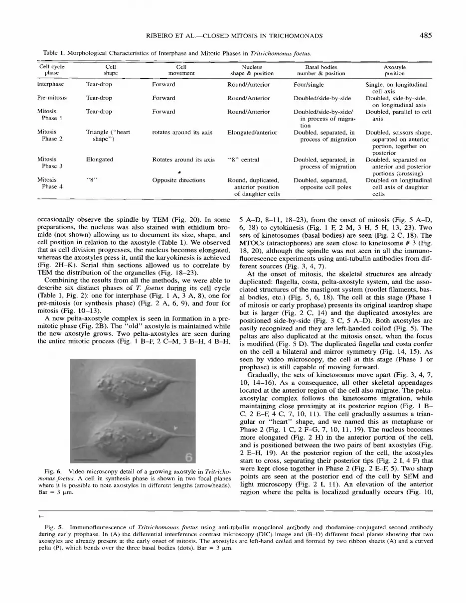
Fig. 6. Video microscopy detail of a growing axostyle in Tritricho- rnona.s,foetu.s. A cell in synthesis phase is shown in two focal planes where it is possible to note axostyles in different lengths (Hrrowheads). Bar = 3 fim.
5 A-D, 8-11, 18-23), from the onset of mitosis (Fig. 5 A-D, 6, 18) to cytokinesis (Fig. 1 F, 2 M, 3 H, 5 H, 13, 23). Two sets of kinetosomes (basal bodies) are seen (Fig. 2 C, 18). The MTOCs (atractophores) are seen close to kinetosome # 3 (Fig. 18, 20), although the spindle was not seen in all the immuno- fluorescence experiments using anti-tubulin antibodies from dif- ferent sources (Fig. 3, 4, 7).
At the onset of mitosis, the skeletal structures are already duplicated: flagella, costa, pelta-axostyle system, and the asso- ciated structures of the mastigont system (rootlet filaments, bas- al bodies, etc.) (Fig. 5 , 6, 18). The cell at this stage (Phase 1 of mitosis or early prophase) presents its original teardrop shape but is larger (Fig. 2 C, 14) and the duplicated axostyles are positioned side-by-side (Fig. 3 C, 5 A-D). Both axostyles are easily recognized and they are left-handed coiled (Fig. 5) . The peltas are also duplicated at the mitosis onset, when the focus is modified (Fig. 5 D). The duplicated flagella and costa confer on the cell a bilateral and mirror symmetry (Fig. 14, 15). As seen by video microscopy, the cell at this stage (Phase 1 or prophase) is still capable of moving forward.
Gradually, the sets of kinetosomes move apart (Fig. 3, 4, 7, 10, 14-16). As a consequence, all other skeletal appendages located at the anterior region of the cell also migrate. The pelta- axostylar complex follows the kinetosome migration, while maintaining close proximity at its posterior region (Fig. 1 B- C, 2 E-E 4 C, 7, 10, 11). The cell gradually assumes a trian- gular or “heart” shape, and we named this as metaphase or Phase 2 (Fig. 1 C, 2 F-G, 7, 10, 11, 19). The nucleus becomes more elongated (Fig. 2 H) in the anterior portion of the cell, and is positioned between the two pairs of bent axostyles (Fig. 2 E-H, 19). At the posterior region of the cell, the axostyles start to cross, separating their posterior tips (Fig. 2 I, 4 F) that were kept close together in Phase 2 (Fig. 2 E-E 5) . Two sharp points are seen at the posterior end of the cell by SEM and light microscopy (Fig. 2 I, 11). An elevation of the anterior region where the pelta is localized gradually occurs (Fig. 10,
t
Fig. 5. Immunofluorescence of Tritrichomonas foetus using anti-tubulin monoclonal antibody and rhodamine-conjugated second antibody during early prophase. In (A) the differential interference contrast microscopy (DIC) image and (B-D) different focal planes showing that two axostyles are already present at the early onset of mitosis. The axostyles are left-hand coiled and formed by two ribbon sheets (A) and a curved pelta (P), which bends over the three basal bodies (dots). Bar = 3 pm.

486 J. EUKARYOT. MICROBIOL., VOL. 47, NO. 5, SEPTEMBER-OCTOBER 2000
Fig. 7. Immunofluorescence of Trichomonas vaginalis during mitosis. Interphase is shown in two different focal planes (A); pre-mitosis or synthesis (B); early Phase 1 (C) where axostyles changed from a side-by-side to a face-to-face position (D, E); Phase 2 (F) where axostyles are bent; two focal planes of a cell at the transition from Phase 2 to Phase 3 (G) where axostyles are crossed conferring to the cell an elongated shape, and the recurrent flagella are seen following each cytoplast; Phase 3 (H) parallel axostyles are moved apart; Phase 4 (H) the axostyles are aligned. Ax, axostyles; f, anterior flagella; rf, recurrent flagella; s, spindle; p, pelta. Bar = 3 p,m
14-16). At first, the two flagellar channels are seen adjacent, but a process of cell torsion is initiated, causing them to grad- ually become positioned in opposite directions (Figs. 14-16). Another characteristic of this phase is the elevation of the costa/ recurrent flagellum to a position perpendicular to the cell axis (Fig. 11). This event redirects the recurrent flagellum propulsion force and cell movement is altered. As seen by video micros- copy, the cell no longer moves forward, but instead only rotates around its axis. This alteration leads the cell body to experience a torsion, that is also evident by SEM (Fig. 11, 14-16).
The transition of Phase 2 to Phase 3 is marked by a gradual contortion and crossing dynamics of the axostyles, whose pos- terior tips are now completely separated and pointing in op- posite directions (Fig. 1 E, 2 K-L, 12). Axostyle crossing and sliding convert the cell from triangular to elongate, character- izing Phase 3 (anaphase) as seen in cross-section (Fig. 22). Parallel to this, the elongated nucleus appears to be pressed by the axostyles (Fig. 21), which culminates with karyokinesis. Gradually, one can recognize two small, teardrop-shaped cells, linked by a straight connection (Fig. 1 F, 2 M, 4 H, 7 I, 13, 23), similar to the number eight “8”, which characterizes Phase 4 (telophase). The recurrent flagella point in opposite positions (Fig. 3 H, 13). At this stage, the two linked cells have their flagella actively beating, but are not able to move forward and remain rotating around their axis. When examined by EM, this connection between the two daughter cells consists of an over- lay of the two axostyles (Fig. 13, 23). Propulsion generated by flagella beating at each extremity further contributes to cyto- kinesis, as seen by video microscopy. Mitosis finishes when the
two small daughter cells separate at their posterior extremities and recover normal cell movement.
DISCUSSION There are few reports on the cell cycle and the process of
closed mitosis in trichomonads (Brugerolle 1975; Juliano et al. 1986; Viscogliosi and Brugerolle 1994; Viscogliosi and Del- gado-Viscogliosi 1996; Zuo et al. 1999). Since the nuclear en- velope does not break down, it is difficult to recognize by elec- tron microscopy when a trichomonad is dividing. In an attempt to explore this primitive closed mitosis process, we have used several approaches. Cells with two sets of three anterior flagella were considered by us to be in mitosis or in pre-mitosis, de- pending on the distance between the flagellar channels (by SEM) andor kinetosomes (by TEM). After comparison of these observations with those obtained by video microscopy, it was not difficult to recognize each phase of the T. foetus cell cycle. The Confocal Laser Scanning Microscopy allowed us to make optical sections and to be sure of the presence of the pelta- axostyle in all cells observed.
We characterized six distinct phases in the trichomonad cell cycle. The majority of the cells (approximately 80%) belongs to a phase in which the cells were clearly not in division: tear- drop shape, three anterior flagella, one recurrent flagellum, a rounded nucleus, one costa, and one axostyle. This phase cor- responds to interphase (Gl) of other eukaryotic cells. Next, we characterized an initial phase with indications that the cells were no longer in interphase, although they still had a teardrop shape: two sets of anterior flagella that were close together; one

RIBEIRO ET AL.-CLOSED MITOSIS IN TRlCHOMONADS 487
Fig. 8-13. SEM of Tritrichomonm ,foetus during morphogenesis from interphase (Fig. 8 through pre-mitosis (Fig. 9) and mitosis (10-13). Fig. 8. The cell is in interphase, presenting one axostyle tip (arrow) at the posterior region and a teardrop shape. Fig. 9. The cell is larger and shows the flagella already duplicated. Fig. 10. Prophase (or Phase 1). The cell assumes a triangle shape. Fig. 11. The cell is in metaphase, presenting an heart shape. Note the two axostyles tips (white arrows). Fig. 12. Phase 3. The cell is in anaphase. Note the more elongated shape and the appearance of the axostyle tips at the posterior region of the cell (white arrow). The axostyles have started to migrate and in anaphase these tips are apart from each other (arrows, Fig. 12). Fig. 13. Phase 4. The daughter cells are pointing in opposite directions but still connected. The recurrent flagella are in opposite positions (arrowheads). Notice the redirection of the recurrent flagella from a parallel position with relation to the cell body longitudinal axis on premitosis and prophase (Fig. 9-10) to a perpendicular position in relation to the longitudinal axis in Phase 2 (Fig. I I ) . The axostyle tips are indicated in all phases by white arrows. Asterisk, flagellar channel; F, anterior flagella; R, recurrent flagellum. Figs. 8 and 13, Bar = 3 pm. Figs 9-12, Bar = 2 pm.

488 J. EUKARYOT. MICROBIOL., VOL. 47, NO. 5, SEPTEMBER-OCTOBER 2000
Fig. 1&16. Details of the anterior region of Tritrichomonas ,foetus by SEM, from early prophase 14 (14), to late prophase (16) of mitosis. Notice that in Fig. 14, the Aagellar channels (stars) are side-by-side, and gradually they undergo contortion (Fig. 15) and separation (Fig. 16). Figure 14, Bar = 1 pm. Figs. 15-16. Bar = 0.5 pm.
Fig. 17. Computer animation frames of a hypothetical 3D model of the trichoinonad’s mitosis. It shows how the cytoskeletal structures com- bine to promote a karyokinesis coupled to cytokinesis. Prophase (A); nietaphase (B); anaphase (C, D) and telophase (E). Spindle microtubules (small arrow), axostyle-pelta complex (a), nucleus (n) costa, which also indicates the recurrent flagella position (c).

RIBEIRO ET AL.-CLOSED MITOSIS IN TRICHOMONADS 489
Fig. 18. Detail of the anterior region of Tritrichomonus foetus in longitudinal section by TEM during pre-mitosis/early prophase, which corresponds to Fig. 14. Notice the two sets of basal bodies (B) and related skeletal structures (stars), and the pelta (arrowheads)/axostyle (arrows) complex positioned side-by-side. Symmetrical Golgi complexes (G) and nuclei (N) are also seen. Bar = 1 ~ m .
The anterior portion of Tritrichomonus,foetus in longitudinal section by TEM is shown during Phase 2 of mitosis, which corresponds to Fig. 1 I . Axostyles (arrowheads) are bent due to separation of the uppermost part and proximity of the posterior trunk. The nucleus (N) is positioned in between both axostyles. The recurrent flagella (R) and the costa (C) are seen in cross-section due to their position perpendicular to longitudinal axis of the cell body. Golgi complex (0) and hydrogenosomes (H) are also seen. Bar = lpm.
Fig. 19.

490 J. EUKARYOT. MICROBIOL., VOL. 47, NO. 5, SEPTEMBER-OCTOBER 2000
Fig. 20. Cross-section of the anterior portion of Tritrichornonus ,/ietus by TEM during prophasehetaphase. Adjacent to basal body (B) localizes the MTOC or attractophore (A) from which emanate pole-to-pole or pole-to-envelope spindle microtubules (arrowheads). The nuclear matrix (N) is seen devoid of any chromosome condensation whereas the nuclear envelope shows polar modifications forming “finger-like- projections” (thicker arrow) extending towards the MTOCS. Pore complexes are observed along the nuclear envelope and on the projections (thin arrow). Bar = 0.5 pni
axostyle that was normal in size and position in addition to a small one (appearing from the duplicated set of kinetosomes), and one rounded anterior nucleus. These cells were larger (in width) than those in interphase. We considered it a pre-mitotic phase, which might correspond to the SlG2 phase described in other eukaryotic cells. In this phase, the cell presented its struc- tures, such as flagella, axostyle, costa, rootlets structures, and basal bodies, in a process of assembly or already duplicated, but there was not yet evidence of mitosis. Major skeletal mod- ifications and interaction with the nucleus can be followed in a hypothetical 3D model (Fig. 17). It is important to mention that flagella were omitted to facilitate the visualization of the other structures; therefore, one can follow the costa as a reference to the recurrent flagella position (Fig. 17). A cell was considered at the onset of mitosis when flagella, costa, axostyle, and other cell structures were already duplicated and when the kineto- somes started to move apart, together with associated centro- soma1 structures (Fig. 17a). As a consequence, the cell lost its teardrop shape, acquired a triangular or “heart” shape, and modified its movement. The axostyles follow the kinetosome movement, and become progressively separated at the anterior region of the cell, while remaining together at their posterior ends (Fig. 17b). The crossing of the axostyles seems to put pressure on the nucleus, now elongated, thereby starting the karyokinesis (Fig. 17c). We propose that, when the mastigont system twists, it would affect the axostyles, causing a torsion that seems to press each axostyle onto the nucleus (Fig. 17d). As the division progresses, the two cell poles separate and the cell shape becomes rather cylindrical. The migration stops when
the daughter cells are in opposite directions (Phase 4). The two cells, still connected by their posterior regions, in which the two axostyles are aligned, become easily recognizable (Fig. 17e). The flagella beating at the anterior region of each daughter cell introduce power for locomotion and it could contribute in cytokinesis (as apparent by video microscopy). The mitosis fin- ishes with cell separation.
Concerning the costa and flagella, Viscogliosi and Brugerolle (1994) stated that these skeletal elements were duplicated dur- ing the course of mitosis. In our observations, we saw that cells initiating mitosis displayed all skeletal structures already dupli- cated, costa, flagella, and pelta-axostyle, included. Thus, assem- bly of axostyle, costa, flagella, and other structures occurs dur- ing a pre-mitosis phase in T. foetus and T. vaginalis. Our results are in agreement with those of Zuo et al. (1999) who demon- strated that the flagella are already duplicated before mitosis (S phase). They also proposed an intense participation of the fla- gella during the course of mitosis in T. vaginalis.
In our studies, a new axostyleipelta complex was seen in formation in a pre-mitotic phase and it actively participated in the cell division process. We started our studies using video microscopy, following step-by-step the division process in liv- ing cells. We also found two axostyles with their normal size during all phases of the mitosis by all techniques. In the present paper we demonstrated the fluorescence images with the con- comitant phase contrast image or DIC (differential interferential contrast), which allowed us to clearly show the presence of the two axostyles. We never saw depolymerized or ejected axo- styles in our preparations. These results are contradictory to

RIBEIRO ET AL.-CLOSED MITOSIS IN TRICHOMONADS 49 1
previous studies performed with T. vaginalis (Delgado-Viscog- liosi et al. 1996; Juliano et al. 1986; Viscogliosi and Brugerolle 1994; Viscogliosi and Delgado-Viscogliosi 1996). In addition, according to these authors, the axostyle would be composed of labile microtubules. However, there are several observations that suggest that the axostyle is formed by stable microtubules: (a) incubation of T. foetus in the presence of colchicine or vin- blastine does not disturb the axostyle (unpubl. observ., Silva- Filho and DeSouza 1986); (b) the axostyle/pelta complex as observed here is a very stable microtubular structure during the whole division process; this was observed also previously using anti-acetylated-tubulin antibodies (Batista et al. 1988; Benchi- mol and DeSouza 1987); (c) our observations of trichomonads in all phases of the cell cycle always showed the presence of intact axostyles; and (d) when T. foetus and T. vaginalis were submitted to temperatures below 4 "C the axostyle did not de- polymerize (Benchimol, unpubl. observ.). We postulate that axostyle assembly may be effected by the addition of novel microtubules to preexisting ones in a manner reminiscent of the cortical microtubular skeleton of trypanosomes (Robinson et al. 1991).
Only occasionally were we able to detect the spindle micro- tubules by immunofluorescence or electron microscopy. The possible explanations could be that the anti-tubulin antibodies used here do not recognize or could not access these labile microtubules or that our preparation procedures may have led to a depolymerization of these labile structures. At present, we are trying to obtain anti-tubulin antibodies from other sources and changing experimental procedures in order to further study the spindle behavior during the mitotic process in T. foetus.
In principle, we demonstrate that karyokinesis is associated with, in addition to the extranuclear spindle action as demon- strated by Brugerolle (1975), the direct mechanical aid of the axostyle, a stable microtubular structure. The utilization of mi- crotubular structures other than the spindle apparatus to perform mitosis is, to our knowledge, a novel consideration. On the other hand, we also observed that the flagella undergo a redi- rection, introducing the power of flagellar locomotion to pro- mote celVspindle pole separation and facilitating cytokinesis. Propulsion generated by flagella beating on each extremity of the cell body contributes to cell elongation and consequent par- tition.
In conclusion, the closed mitosis in trichomonads seems to use a combination of flagellar, axostyle and spindle efforts, as summarized in the 3D hypothetical model using computer an- imation frames.
ACKNOWLEDGMENTS The authors wish to thank Dr. Wanderley de Souza, Bruce
Granger, and Yara Traub-Cseko for critically reading the man- uscript. This investigation received financial support from H- NEP (Financiadora de Estudos e Projetos), CNPq (Conselho Nacional de Desenvolvimento Cientifico e Tecnol6gico), PRO- NEX (Programa de Nucleos de Excelgncia), FAPERJ (Funda- ,.-so de Ampar0 & Pesquisa do Estado do Rio de Janeiro), CAPES (Coordena,.-Ho de AperfeiGoamento de Pessoal de Nivel superior) and AUSU ( ~ ~ ~ ~ ~ i ~ ~ ~ ~ universit&ia santa ursula).
LITERATURE CITED
Fig. 21. Cross-section of the medial region of ~ri tr ichomnnas.~~etu.r by TEM during karyokinesis. Notice that nucleus (N) is apparently constricted (arrows) by axostyles (A). Hydrogenosomes (H) show bi- lateral and mirror symmetry. This stage corresponds to Fig. 2 J or Fig. 12. Nu, nucleolus; C, costa. Bar = 0.5 pm.
TEM of Tritrichomona.~ ,fi)etus in Phase 3 after karyoki- nesis. It is possible to observe a mirror synimetry reflected by the in- ternal organization of the cell's structures, such as the axostyle ( m o w -
Fig. 22.
heads), flagella (F). hydrogenosomes (H), and nuclei (N). Bar = Ipm.
division of TritrjeAomontc,s foetus, The daughter cells are pointing in opposite directjonc, still connected by their posterior ends. The ar- rowheads point to the axostyles, H, hydrogenosomes. Bar = p ~ .
~ i ~ . 23, ~ ~ ~ ~ k i ~ ~ ~ i ~ , end of telophase or phase in the Batista, M. C. C., Benchimol, M., Cunha e Silva, L. N. 8~ De Souza, W. 1988. Localization of acetylated a-tubulin in T~-ituichomonu.sfoe- tus and Tric.homonu.s vaginalis. Cell Struct. Funct., 13:445-453.
Benchimol, M. & De Souza, W. 1987. Structural analysis of the cyto- skeleton of Tritrichomonas fiwtus. J . Suhmicrosc. Cvrol., 1: 139-147.
Brugerolle, G. 1975. Etude de la cryptopleuromitose et de la morpho- genkse de division chez plusieurs Genres de trichomonadines primi- tives. Protistologica, 4:457-468.

492 J . EUKARYOT. MICROBIOL., VOL. 47, NO. 5, SEPTEMBER-OCTOBER 2000
Cavalier-Smith, T. & Chao, E. E. 1996. Molecular phylogeny of the free-living archeozoan Trepomonas agilis and the nature of the first eukaryote. J . Mol . Evol., 43:55 1-562.
Delgado-Viscogliosi, l?, Brugerolle, G. & Viscogliosi, E. 1996. Tubulin post-translational modifications in the primitive protist Trichomonus vaginalis. Cell Mot. Cytoskel., 33:288-297.
Diamond, L. S. 1957. The establishment of various trichomonads of animals and man in axenic cultures. J. Parusitol., 43:488-490.
Embley, T. M. & Hirt, R. l? 1998. Early branching eukaryotes? Curr. Opin. Gen. Develop., 8:624-629.
Heath, B. 1980. Variant mitosis in lower eukaryotes: indicators of the evolution of mitosis? Int. Rev. Cytol., 64:6-8 1.
Juliano, C., Monaco, G., Rubino, S. & Cappuccinelli, P. 1986. Inhibition of Trirhomonas vaginalis replication by the microtubule stabilizer taxol. J . Protozool., 33:255-260.
Leipe, D. D., Gunderson, H. J., Nerad, A. T. & Sogin, L. M. 1993. Small subunit ribosomal RNA’ of Hexamita inflatu and the quest for the first eukaryotic tree. Mol. & Biochem. Parasirol., 59:41-48.
Monteiro-Leal, L. H., Farina, M. & De Souza, W. 1996. The free-move- ment of Tritrichomonas foetus in liquid medium: a video microscopy study. Cell Mot . Cyroskel., 34:206-214.
Robinson, D. R., Beattie, I? Sherwin, T. & Gull, K. 1991. Microtubules,
tubulin and microtubule-associated proteins of trypanosomes. Meth. Enzymol., 196:285-299.
Silva-Filho, E C. & De Souza, W. 1986. Effect of colchicine, vinblas- tine, and cytochalasin B on cell surface anionic sites of Tritricho- monas foetus. J . Protozool., 33:6-10.
Viscogliosi, E. & Brugerolle, G. 1993. Cytoskeleton in trichomonads. Eur. J. Protistol., 29: 160-170.
Viscogliosi, E. & Brugerolle, G. 1994. Cytoskeleton in trichomonads. 111. Study of the morphogenesis during division by using monoclonal antibodies against cytoskeletal structures. Eur. J . Protistol., 30: 129- 138.
Viscogliosi, E. & Delgado-Viscogliosi, P. 1996. Cytosquelette, division et evolution dans un groupe de protistes primitifs, les Trichomona- dines. Ann. Biol., 35:146-161.
Woods, A,, Sherwin, T., Sasse, R., MacRae, T. H., Baines, A. J. and Gull, K. 1989. Definition of individual components within the cyto- skeleton of Trypanosoma brucei by a library of monoclonal antibod- ies. J. Cell Sci., 93:491-500.
Zuo, Y., Riley E. D. & Krieger, N. J . 1999. Flagellar duplication and migration during the Trichomonas vaginalis cell cycle. J . Parasitol., 85: 203-207.
Received: 10-7-99, 1-26-00, 5-1 1-00: accepted 5-1 1-00
Erratum
The contribution numbers in the ACKNOWLEDGMENTS, page 409, of the paper “A Survey of Flagellate Diversity at Four Deep-sea Hydrothermal Vents in the Eastern Pacific Ocean Using Structural and Molecular Approaches” by Michael S. Atkins, Andreas P. Teske and 0. Roger Anderson, J. Eukaryot. Microbiol., 47(4):400-411 are correct as follows: “This is Woods Hole Oceanographic Institution contribution number 10159 and Lamont-Doherty Earth Observatory contribution number 6055.”


