Embed Size (px)
Citation preview
Control of the lettuce anthracnose (Microdochium panattonianum Berl.) using Trichoderma spp. and
their secondary metabolites
José Valentín Palacios Zevallos
ORCID N° 0000-0002-0667-2225
Summited in total fulfillment of the requirements of the degree of
Doctor of Philosophy
April 2020
Faculty of Veterinary and Agricultural Sciences
The University of Melbourne

I
This thesis comprises only my original work towards the Ph.D. and no material for
other degree in any other University.
To the best of my knowledge and belief, this thesis contains no material previously
published or written by any other person except when due reference is made in the
text, and
This thesis is fewer than 100,000 words in length, exclusive of tables, maps,
bibliographies, and appendices.
II
Acknowledgements
During these three years I have left my comfort zone so many times that now I do not know
what discomfort is. Perhaps that is the main ingredient every researcher needs to face
before discovering science.
Science, for me is the truth, the truth of how things work, it is unique. It has already been
created not invented; science is in the things that surround us. We humans do not do science,
we discover it at the end of every experiment. Tiny pieces of the universal truth come along
and coexist with us but also, they may be refuted, of course, reminding us how incipient is
our knowledge.
My path to find this little piece of science was not only through laboratories and fields but
also washing dishes and cleaning toilets and I sincerely thank to every person who
supported me when I was at the bottom.
I want to thank Professor Jim He for giving me the opportunity to continue my scientific
evolution in The University of Melbourne and supervise this research project, introducing
fellow students that contributed with efficacy to this research. Also, I thank the Australia-
China Joint Research Centre project Healthy Soils for Sustainable Healthy Food
Production and Environmental Quality (ACSRF48165) for my scholarship and funding
support.
Dr. Mary Cole, a brilliant scientist from Agpath trusted in my project from start to the end.
Her ideas and words were always along my road even in the hardest moments and I will
III
never forget her words “José, you can crash now and then, but not totally. Remember there
are always more exciting things to discover”. I will never find the ways how to express her
my deep gratitude. This thesis would not be accomplished without her support.
Dr. Helen Waite for helping review my thesis draft along those long hours of exhausting
analysis, giving me ideas for a rounder meaning.
Anita Chudleigh and Heli Thaw, you were always a helping hand in the early
microbiological phase, sharing the ABC of laboratory techniques.
Darren Corrigan from Corrigan Produce Farms and Michael Bogicevic and Nicolas
Huvelle form Coolibah herbs, for giving me the opportunity to use their lettuce crops for
field tests.
Professor Paul Taylor, Dr. Azin Moslemi, Dr. Qinglin Chen, Dr. Hangwei Hu; fellows Obed
Luchibia, Chaoyu Li, Weixia Wang and Sofia Callaghan for their willing support along the
molecular works.
Professor Francesco Vinale who introduced me to his team at The University of Naples and
performed the metabolites test for Trichoderma filtrates, and Dr. Allessia Staropoli and Dr.
Roberta Marra for their support in teaching the operation of the LC-MS/MS Q-TOF
equipment and the instrumental analysis. Also is important to note the contribution of Dr.
Sandy Clarke from The University of Melbourne statistics department.
To the South Australian Research and Development Institute (SARDI), The NSW Plant
IV
Pathology and Mycology Herbarium, Dr. Dean Metcalf, and Agrimm New Zealand, for
facilitating their Trichoderma isolates.
To Lily, Camila, Leonardo, and Rodrigo for your patience, I know there were many
weekends that I missed you and you missed me. I let you go without me, just asking, “Daddy
are you coming with us?” I hope someday you will understand.
Finally, to myself, why not? I did a great job…
V
Summary
The main objectives of this project were to: 1) show that lettuce anthracnose propagules that
persist in soil from a previous lettuce crop may be controlled by a cold tolerant strain of
Trichoderma inoculated just before the pathogen becomes active and virulent, reducing
incidence and severity, and, 2) determine if liquid cultures containing Trichoderma
secondary metabolites (TSMs) can be sprayed for maximum leaf coverage to reduce the
incidence and severity of the disease when condition for disease are ideal.
Lettuce anthracnose Microdochium panattonianum (MP) is a key winter disease in Victoria,
Australia, and other temperate regions in the world. Cultural methods to reduce the severity
of this disease are not practical nor economically feasible. While chemical control can be
achieved it is not flawless and can cause health issues.
Propagules of MP persist in soil sheltered in leaf debris and germinate with the onset of cold
and wet conditions in the subsequent winter. Unfortunately, this pathogen has received little
scientific attention but as winters become wetter this disease is causing greater loss and this
body of work offers a more sustainable management tool.
Trichoderma species are common saprophytic fungi in soils rich in organic matter. They
have a proven ability to control crop diseases with a multiplicity of strategies including
parasitism, competition, antibiosis and, at the same time, stimulating plant defences and
growth. However, these natural processes vary with species and strains of Trichoderma.
This research project showed that the incidence and severity of the lettuce anthracnose can
VI
be reduced by applying Trichoderma cold tolerant strains to the soil. Secondly, it showed
that spraying Trichoderma liquid filtrates containing metabolites to the leaves also reduced
and managed lettuce anthracnose.
From a pool of 27 Trichoderma isolates, 8 grew at 10°C. These 8 isolates were characterised
morphologically in vitro under different culturing conditions of temperature, pH and media.
Some isolates were identified by molecular techniques to species while others still require
more investigation.
Tests in vitro on solid media prepared with liquid filtrates confirmed that liquid filtrates
from one isolate related to T. viride and one from T. composticola affected the morphology
and growth of MP. These filtrates contained 6 pentyl-alpha-pyrone (6PP,) a volatile
metabolite with a characteristic coconut smell.
Other Trichoderma liquid filtrates containing metabolites such as cytosporone S and 6PP,
produced by some strains of T. aureoviride, T. atroviride and Trichoderma sp., killed the
pathogen.
Fourteen isolates produced 6PP, a well-documented Trichoderma secondary metabolite
(TSM). Of these, some have been identified as T. viride, T. composticola, T. atroviride, T
paratroviride, T. neokoningii and T. asperelloides. Others are yet to be fully identified to
species. Isolates of T. harzianum, T. polysporum. T aureoviride, T. longibrachiatum and T.
spirale did not produce this metabolite.
VII
Isolates of Trichoderma, TC1, CAROL, 71558 and 1536 require further identification
because they were outliers on the phylogenetic tree. None of these isolates produced 6PP.
Challenged isolates produced higher quantities of 6PP than did the individual liquid
cultures. Trichoderma atroviride (PMF) from New Zealand produced 137 mg/l of 6PP, but
when challenged by Trichoderma paratroviride (NSW) the 6PP concentration was 160
mg/l.
Incidence and severity of MP were reduced significantly in a field trial using the cold
tolerant isolate 356 related to T. composticola.
TSMs applied as foliar sprays have the potential to control many foliar diseases in crops and
is a new field of research.
The use of Trichoderma cold tolerant strains for control of winter active pathogens is a new
strategy for the use of Trichoderma products.
VIII
List of Appendices
Appendix 1 Differences in colony size by Trichoderma 356 at different temperatures. One-way ANOVA. Appendix 2 Differences in colony size by Trichoderma 1535 at different temperatures. One-way ANOVA. Appendix 3 Differences in colony size by Trichoderma 71556 at different temperatures. One-way ANOVA. Appendix 4 Differences in colony size by Trichoderma CAROL at different temperatures. One-way ANOVA. Appendix 5 Differences in colony size by Trichoderma 59837 at different temperatures. One-way ANOVA. Appendix 6 Differences in colony size by Trichoderma LM at different temperatures. One-way ANOVA. Appendix 7 Differences in colony size by Trichoderma LUP at different temperatures. One-way ANOVA. Appendix 8 Effect of temperature on Trichoderma isolates phialide length. One-way ANOVA. Appendix 9 Effect of temperature on Trichoderma isolates phialide width. One-way ANOVA. Appendix 10 Effect of temperature on Trichoderma isolates conidia width. One-way ANOVA. Appendix 11 Effect of pH on Trichoderma isolates phialide width. One-way ANOVA. Appendix 12 Effect of pH on Trichoderma isolates conidia length. One-way ANOVA. Appendix 13 Effect of pH on Trichoderma isolates conidia width. One-way ANOVA.
IX
Appendix 14 Effect of culture media on Trichoderma isolates phialide length. One-way ANOVA. Appendix 15 Effect of culture media on Trichoderma isolates phialide width. One-way ANOVA. Appendix 16 Effect of culturing media on Trichoderma isolates conidia length. One-way ANOVA. Appendix 17 Colony size in 7 cold tolerant Trichoderma isolates incubated at 10°C. 10 days after inoculation One-way ANOVA. Appendix 18 Colony size in 7 cold tolerant Trichoderma isolates incubated at 10°C. 14 days after inoculation One-way ANOVA. Appendix 19 Effect of pH on colony radius in 7 cold tolerant Trichoderma isolates incubated at 10°C. One-way ANOVA. Appendix 20 List of TSM in liquid filtrates. Appendix 21 Effect of Trichoderma application on number of viable plants in trays trial. One-way ANOVA. Appendix 22 Effect of lettuce variety and treatments on incidence of MP in second field trial, at harvest. One-way ANOVA. Appendix 23 Effect of lettuce variety and treatments on severity of MP second field trial, at harvest. One-way ANOVA. Appendix 24 Effect of lettuce variety and treatments on severity of MP in the field trial at week 10 (one week before harvest). One-way ANOVA.
X
List of Figures
N° Figure Page
2.1 M. panattonianum bicelled conidia (X400) 11 2.2 Lettuce plant with typical symptoms of anthracnose including the “shot hole” 11 2.3 Sporulating Trichoderma longibrachiatum colony cultured on PDA media. Notice
the yellow pigmentation in the media produced by the fungus. 14
3.1 Map of Australia showing approximate geographical origin of Trichoderma isolates: 1 Twin peaks (WA); 2 Virginia (SA); 3 Adelaide Hills (SA); 4 Belthorpe (QLD); 5 Ballina (NSW); 6 Anuello (VIC); 7 Melbourne and Mornington Peninsula; 8 Orange (NSW) and 9 Molesworth (TAS)
35
3.2 Liquid cultures containing Trichoderma isolates in PDB shaken at 200 rpm
42
3.3 Filtrates from Trichoderma isolates liquid cultures showing different natural pigmentation
43
3.4 Microdochium panattonianum colonies incubated in cold conditions. Wells were cut in square pattern.
45
3.5 Layout of complete wells experiment. 4 sets of plates. Orange pigmentation denotes older colonies that corresponded to the 7 days CF. Pale colonies corresponded to the 14 days CF.
45
3.6 Challenging Microdochium panattonianum discs cultured in presence of Trichoderma
46
3.7 Trial layout for isolate 1 placed in centre. Same layout was applied in sets of 5 plates for all 27 isolates changing only the central disc.
50
3.8 Filtration of Trichoderma cultures using sterile syringes and Minisart 0.22µ filters.
51
3.9 Lettuce seedlings transplanted in trays under open conditions in Garfield, Victoria, winter 2017
54
3.10 Plants of lettuce previously inoculated with pathogen and incubated in refrigerated conditions
56
3.11 Replicate of second pot trial in refrigerated incubator to evaluate minimum pathogen concentration to promote disease.
56
3.12 MP infected lettuce plants sprayed with Trichoderma liquid cultures to evaluate control of the disease. Plants were incubated in refrigeration to promote disease
58
3.13 First field trial showing details of the Trichoderma application technique in compost as a carrier
61
3.14 Random block design of treatments in the first field trial 63 3.15 Second field trial installed in a commercial lettuce crop in an organic certified
farm. Pearcedale, Victoria. 66
4.1 Average number of days for Trichoderma colonies to cover a 90mm Petri plate at 25° and 18°C in two culture media and three pH media values (the shorter the column the faster growing conditions). NB at both temperatures, growth on PDA (undefined) was faster than in CZA (defined)
70
4.2 Number of days for Trichoderma isolates to cover a 90mm PDA at 25°C 70 4.3 Number of days to cover a 90 mm PDA at 25°C by isolates 355 and 73
XI
BIANCA at different pH media 4.4 Trichoderma paratroviride (71556) cultured in PDA at 25°C. Left: pH 5.0,
right pH 6.5, 3 days after inoculation 74
4.5 Effect of the media pH on Trichoderma phialide width (µ) 76
4.6 Isolate MACA (Trichoderma longibrachiatum) cultured at 18°C and pH 6.5, 7 days after inoculation; left on PDA and right on CZA
77
4.7 Phylogenetic tree for genes tef1, rpb2 and acl1 80 4.8 Phylogenetic tree for genes tef and rpb2 81 4.9 Microdochium panattonianum hyphal structures at well edges showing
normal growth 82
4.10 Radius of Trichoderma isolates colonies incubated at 10°C after challenge culturing experiment
83
4.11 Colonies formed at 10°C incubation temperature. Untreated shows a healthy MP colony
83
4.12 Cold tolerant Trichoderma isolates colony radius, 10 (blue) and 14 (orange) days after inoculation incubated at 10°C from conidial suspension
84
4.13 Comparison of colony radius based on the initial inoculum 6 days after inoculation at 10°C
85
4.14 Effect of three pH media on Trichoderma cold tolerant isolates colony growth
85
4.15 Colonies of M. panattonianun cultured in A: Untreated PDA; and treated with filtrates from isolates B:1535 and C: 356, 10 days after inoculation X20
87
4.16 Concentration of 6PP in experimental liquid cultures. Challenged cultures in blue columns, individual isolate cultures in green columns
94
4.17 Liquid filtrates from challenged and single cultures with ≥ 100mg/l 6PP 96 4.18 Effect of Trichoderma sp. strain 356 on percentage of viable (marketable)
plants of lettuce in trays trial 97
4.19 Correlation between the number of MP propagules in soil and incidence of the disease
99
4.20 Incidence: Number of lettuce plants out of 140 total plants, with at least one spot of anthracnose in one leave (by variety)
100
4.21 Severity by lettuce variety: Percentage of infected plants with not commercial value
101
4.22 Incidence: Number of plants (out of 140 per plot) with at least one anthracnose spot per leaf. (by treatments)
101
4.23 Severity: Percentage of MP infected plants with not commercial value by treatment
102
5.1 T. composticola, isolate 356 (left) and 33861 (right) showing differences in their conidiation pattern at 25°C on PDA
114
XII
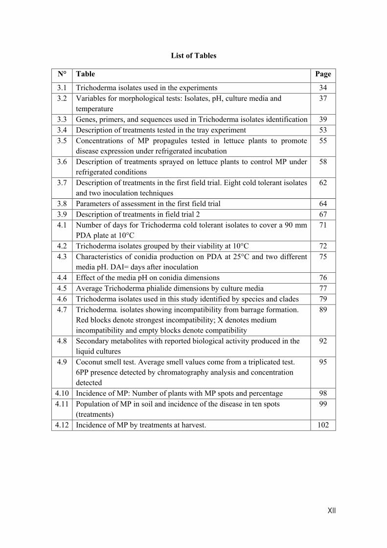
List of Tables
N° Table Page
3.1 Trichoderma isolates used in the experiments 34 3.2 Variables for morphological tests: Isolates, pH, culture media and
temperature 37
3.3 Genes, primers, and sequences used in Trichoderma isolates identification 39 3.4 Description of treatments tested in the tray experiment 53 3.5 Concentrations of MP propagules tested in lettuce plants to promote
disease expression under refrigerated incubation 55
3.6 Description of treatments sprayed on lettuce plants to control MP under refrigerated conditions
58
3.7 Description of treatments in the first field trial. Eight cold tolerant isolates and two inoculation techniques
62
3.8 Parameters of assessment in the first field trial 64 3.9 Description of treatments in field trial 2 67 4.1 Number of days for Trichoderma cold tolerant isolates to cover a 90 mm
PDA plate at 10°C 71
4.2 Trichoderma isolates grouped by their viability at 10°C 72 4.3 Characteristics of conidia production on PDA at 25°C and two different
media pH. DAI= days after inoculation 75
4.4 Effect of the media pH on conidia dimensions 76 4.5 Average Trichoderma phialide dimensions by culture media 77 4.6 Trichoderma isolates used in this study identified by species and clades 79 4.7 Trichoderma. isolates showing incompatibility from barrage formation.
Red blocks denote strongest incompatibility; X denotes medium incompatibility and empty blocks denote compatibility
89
4.8 Secondary metabolites with reported biological activity produced in the liquid cultures
92
4.9 Coconut smell test. Average smell values come from a triplicated test. 6PP presence detected by chromatography analysis and concentration detected
95
4.10 Incidence of MP: Number of plants with MP spots and percentage 98 4.11 Population of MP in soil and incidence of the disease in ten spots
(treatments) 99
4.12 Incidence of MP by treatments at harvest. 102
XIII
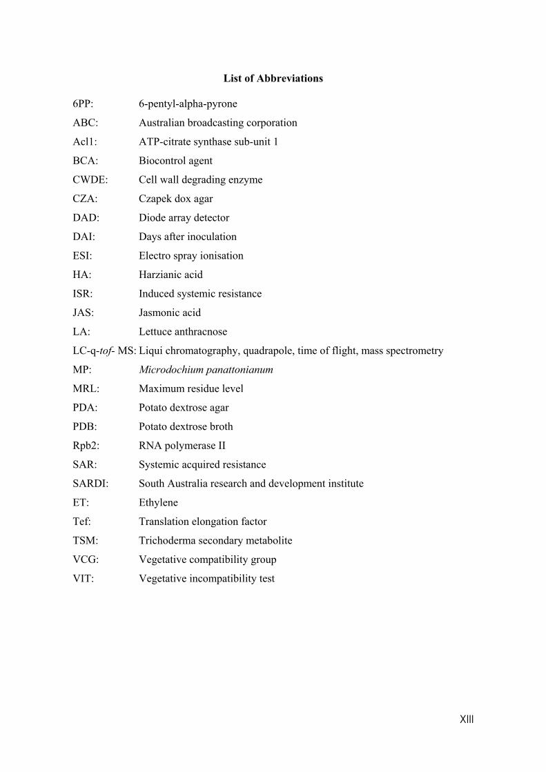
List of Abbreviations 6PP: 6-pentyl-alpha-pyrone
ABC: Australian broadcasting corporation
Acl1: ATP-citrate synthase sub-unit 1
BCA: Biocontrol agent
CWDE: Cell wall degrading enzyme
CZA: Czapek dox agar
DAD: Diode array detector
DAI: Days after inoculation
ESI: Electro spray ionisation
HA: Harzianic acid
ISR: Induced systemic resistance
JAS: Jasmonic acid
LA: Lettuce anthracnose
LC-q-tof- MS: Liqui chromatography, quadrapole, time of flight, mass spectrometry
MP: Microdochium panattonianum
MRL: Maximum residue level
PDA: Potato dextrose agar
PDB: Potato dextrose broth
Rpb2: RNA polymerase II
SAR: Systemic acquired resistance
SARDI: South Australia research and development institute
ET: Ethylene
Tef: Translation elongation factor
TSM: Trichoderma secondary metabolite
VCG: Vegetative compatibility group
VIT: Vegetative incompatibility test
XIV
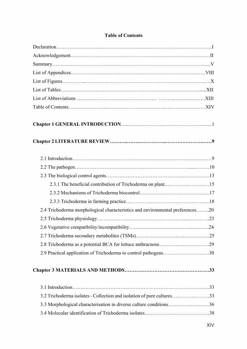
Table of Contents
Declaration………………………………………………………………………………….I
Acknowledgement…………………………………………………………………………II
Summary…………………………………………………………………………………...V
List of Appendices………………………………………………………………………VIII
List of Figures…………...………………………………………………….…………..….X
List of Tables…………………..………………………………………….…………......XII
List of Abbreviations ………………………………………… ……………………….XIII
Table of Contents…………………...……………………………..….…………………XIV
Chapter 1 GENERAL INTRODUCTION…...………………………………………......1
Chapter 2 LITERATURE REVIEW………..…….……………..………………….……9
2.1 Introduction…………………………………………………..……….…….………9
2.2 The pathogen……………………………………………………………....………10
2.3 The biological control agents………………………………………..…………….13
2.3.1 The beneficial contribution of Trichoderma on plant.…………..……….…15
2.3.2 Mechanisms of Trichoderma biocontrol…………………….………….…..17
2.3.3 Trichoderma in farming practice…………………………..……………….18
2.4 Trichoderma morphological characteristics and environmental preferences……..20
2.5 Trichoderma physiology………………..……………………………...………….23
2.6 Vegetative compatibility/incompatibility………………………………...……….24
2.7 Trichoderma secondary metabolites (TSMs)……………………..….……………25
2.8 Trichoderma as a potential BCA for lettuce anthracnose…………………………29
2.9 Practical application of Trichoderma to control pathogens…………………….…30
Chapter 3 MATERIALS AND METHODS……………………………………………33
3.1 Introduction…………………………………………………………………….….33
3.2 Trichoderma isolates - Collection and isolation of pure cultures….……….………33
3.3 Morphological characterisation in diverse culture conditions.………….……..….36
3.4 Molecular identification of Trichoderma isolates…………………………………38
XV
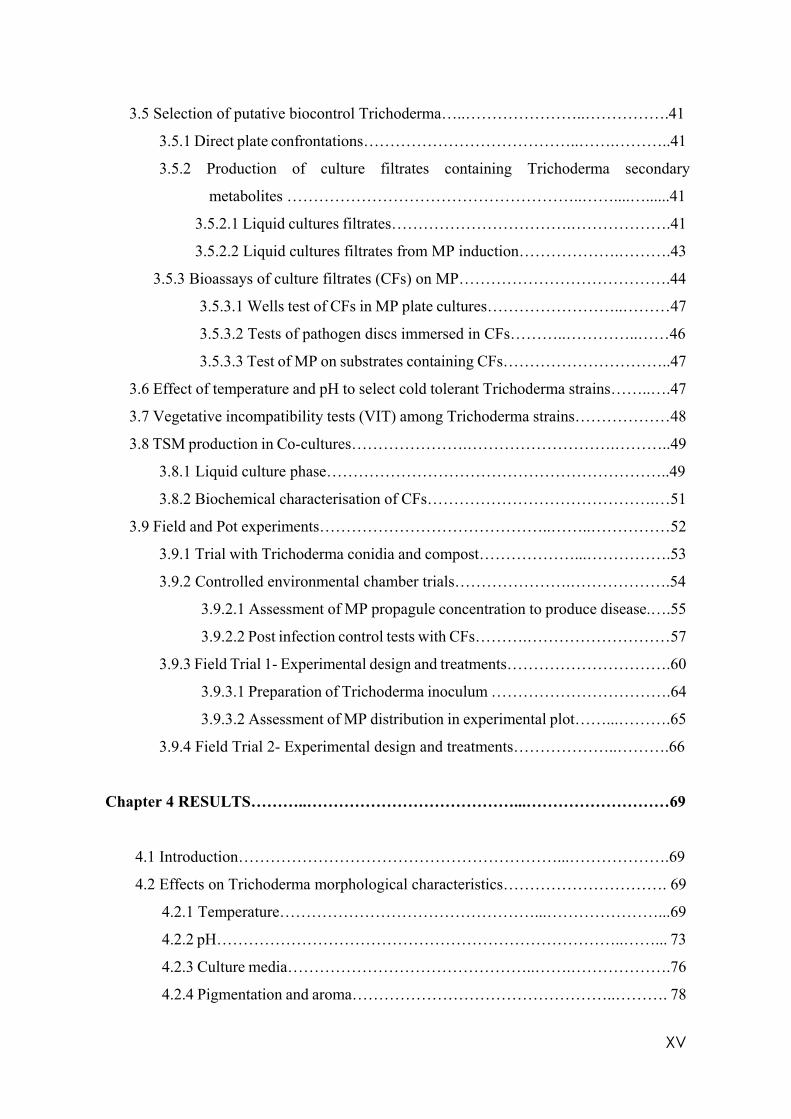
3.5 Selection of putative biocontrol Trichoderma…..…………………..…………….41
3.5.1 Direct plate confrontations…………………………………..…….………..41
3.5.2 Production of culture filtrates containing Trichoderma secondary
metabolites ………………………………………………..……....…......41
3.5.2.1 Liquid cultures filtrates…………………………….……………….41
3.5.2.2 Liquid cultures filtrates from MP induction……………….……….43
3.5.3 Bioassays of culture filtrates (CFs) on MP………………………………….44
3.5.3.1 Wells test of CFs in MP plate cultures……………………..………47
3.5.3.2 Tests of pathogen discs immersed in CFs………..…………..……46
3.5.3.3 Test of MP on substrates containing CFs…………………………..47
3.6 Effect of temperature and pH to select cold tolerant Trichoderma strains……..….47
3.7 Vegetative incompatibility tests (VIT) among Trichoderma strains………………48
3.8 TSM production in Co-cultures………………….……………………….………..49
3.8.1 Liquid culture phase………………………………………………………..49
3.8.2 Biochemical characterisation of CFs…………………………………….…51
3.9 Field and Pot experiments……………………………………..……..……………52
3.9.1 Trial with Trichoderma conidia and compost………………...…………….53
3.9.2 Controlled environmental chamber trials………………….……………….54
3.9.2.1 Assessment of MP propagule concentration to produce disease.….55
3.9.2.2 Post infection control tests with CFs……….………………………57
3.9.3 Field Trial 1- Experimental design and treatments………………………….60
3.9.3.1 Preparation of Trichoderma inoculum …………………………….64
3.9.3.2 Assessment of MP distribution in experimental plot……...……….65
3.9.4 Field Trial 2- Experimental design and treatments………………..……….66
Chapter 4 RESULTS………..…………………………………...………………………69
4.1 Introduction……………………………………………………...……………….69
4.2 Effects on Trichoderma morphological characteristics…………………………. 69
4.2.1 Temperature…………………………………………...…………………...69
4.2.2 pH…………………………………………………………………..……... 73
4.2.3 Culture media………………………………………..…….……………….76
4.2.4 Pigmentation and aroma…………………………………………..………. 78
XVI
4.3 Molecular identification and classification of Trichoderma isolates….…………. 86
4.4 Trichoderma cultured filtrates efficacy in vitro in wells……….…………………81
4.4.1 Liquid filtrates in well experiment………………………..………………..82
4.4.2 Pathogen/Trichoderma discs assessment……………………...………..….82
4.5 Growth of selected cold tolerant Trichoderma in vitro…….….…………….……84
4.6 Test of MP on substrates containing liquid filtrates….................………………. 86
4.7 Vegetative compatibility/incompatibility……………………………………….. 88
4.8 Trichoderma secondary metabolites (TSM)……………………………….……. 88
4.9 Pot, tray, and Field trials…………………………………….……………………96
4.9.1 Trial with Trichoderma conidia and compost……….…….…….………... 97
4.9.2 Pot trial in refrigerated incubator…………………………………………..98
4.9.3 Second pot trial in refrigerated incubator………………………….…….…98
4.9.4 First field trial………………………………………………………………99
4.9.5 Second field trial……………………………..………………………...…100
Chapter 5 DISCUSSION AND CONCLUSIONS….…………………………………104
5.1 Introduction…………………………………………………………………..…104
5.2 Identification and characterisation of suitable Trichoderma isolates to control
MP in soil………………………………………………………………… …….106
5.3 Identification of TSM efficacy to control MP………………….………………..114
5.4 Integrated practices to control MP……………………………. ………………..121
5.4 Recommendations for further research……………………………..…………...122
APPENDICES……………………….…...………………….………………………….123
REFERENCES………………………………………………………….………...……140
1
Chapter 1
GENERAL INTRODUCTION
Like many other human activities, agricultural practices around the world are becoming
non-sustainable because they consider only maximum profits and not a balance between
productivity and land protection. Many farmers and agricultural scientists have lost
understanding of the complexity of interactions between soil, water and air and their impact
in sustainable agricultural production. In some situations, contamination has passed the
point where soil biology can repair the damage in a reasonable time frame (Lal et al. 1997).
Natural methods or at least more sustainable ways to prepare and fertilise soils and manage
pests, weeds, and diseases are available already. These contrast with the high input system
that for decades has focused on maximum yields but actually created serious problems for
productivity and led to an ecological crisis (Rosset and Altieri 1997).
Farmers and the whole horticulture industry are under pressure as retail distributors demand
“perfect” fruits and vegetables while convincing consumers that visual perfection is the best
and safest option. This is not always correct! Retailers have strict specifications for size,
minimum disease damage and insect infestation and these conditions produce unnecessary
food waste as explained by Craig Reucassel (2017) in his ABC TV series “war on waste”.
During more than 25 years of experience as a field production agronomist, I have witnessed
many batches of wasted fresh, almost perfect product, rejected from market. Produce is
returned to the farms to be destroyed or destroyed by supermarkets at farmers’ cost. This
situation pushes farmers to increase chemical use to minimise those “imperfections” so that
2
they protect their investment.
For many years, it was thought that health problems related to agrochemical exposure were
related only to farm workers or farmers (World Health Organisation 1990), but the sad fact
is that currently there is not a human group that is completely unexposed to pesticides (Kim
2017).
Risks implicated with pesticide residues in fruits and vegetables are well documented.
Cancer (Caron-Beaudon et al. 2016), leukemia, diabetes, Parkinson’s disease and asthma
(Kim 2017) and even mental disorders such as ADHD (Attention deficit hyperactivity
disorder) (Wagner-Schuman et al. 2015) and reduced cognitive performance in children and
adults (Viel et al. 2015). Exceptionally low levels of exposure have more dramatic effects
at early human development stages making children more susceptible than adults
(Mascarelli 2013).
Among the vegetable crops in Australia, lettuce was the sixth largest in 2009 with 7411 ha,
increasing to 8000 ha in 2015. Victoria grows 50% of the Australian lettuce production
(Australian Bureau of Statistics 2014-15).
Lettuce anthracnose (MP) Microdochium panattonianum (Berl), is a winter plant pathogen
that has received minimal research efforts. This organism produces a foliar disease
commonly known as “shot hole” that can cause extensive losses in wet and cold conditions
in both head and loose-leaf lettuce. It is found in most of the lettuce cropping areas around
the world. In Australian temperate regions it is noticeably more destructive since 2009 when
winters turned from relatively dry to very wet. The unusually cold and wet conditions during
3
2010-2011 winter-spring period in south eastern Australia made it nearly impossible for
growers of outdoor lettuce to avoid severe losses due to anthracnose. (Rogers and Kimptom
2012).
Development of tolerant varieties of lettuce is an option but has not yet been developed for
this pathogen. Seed companies still do not consider “shot hole” sufficiently important for
cost effective investment. However, change in global weather conditions will make it
necessary to find alternatives to chemical sprays for control of this and other crop diseases.
This disease can be controlled effectively only with the chemical, Prochloraz (Rogers and
Kimptom 2012), a fungicide from the imidazole group that has been found to feminise
offspring of male rats after perinatal exposure by acting as an antiandrogenic (Vingaard et
al. 2006) and causing cryptorchidism - one or two of the testicles not descending from
abdomen to scrotum when in foetus stage - in sons of Danish female gardeners exposed to
this chemical (Weidner et al. 1998).
It is estimated that an average crop cycle of a winter lettuce in Victoria is 12 weeks from
transplant to harvest and during this period, plants are exposed to ideal conditions for
expression of the disease; i.e. temperatures below 15°C and wet leaves for at least 8 hours.
As a result, farmers spray Prochloraz several times for a successful crop; some of them,
desperate to protect their investment, spray to the limit of the withholding period established
on label of 7 days.
Maximum residue levels (MRL) of this chemical for closed head and loose leaf lettuce are
2 and 3 mg/kg respectively, according to the Australian Agricultural and Veterinary
4
Chemicals (APVMA) code instrument N° 4 (MRL) standard 2012 (last compilation 24th
May 2019), which means that it is legal to commercialise lettuce with residues as high as
these values, not necessarily nil. Meanwhile, over a million Australians regularly purchase
organic fruit and vegetables and beverages to reduce the risks implicated. According to the
Australian Bureau of Statistics, 2075 organic certified producers were reported in 2017, and
by 2019, the total value of organic production reached AU$ 2.6 billion (Australian Organic
Market Report 2019). Most of this produce is exported to China, the USA, Japan, South
Korea, and Singapore.
The solution is in the soil, but how can soil help?
Soils have four essential functions: soils sustain biomass production and biodiversity by a
huge pool of genes; regulate water and air quality; preserve archaeological, geological, and
astronomical records; and support of socioeconomic infrastructure. The first two of these
are crucial for environmental and agricultural processes (Lal et al. 1997). Soils provide
physical and nutritional support for crops and animal production. They are also
environmental purifiers, detoxifying and neutralising poisons, complexing heavy metals,
filtering water, air and themselves, and finally, providing a genetic bank of information in
the form of microbial diversity that has the potential to solve many agricultural problems
(Lal et al. 1997).
Many of those soil organisms that contribute to the genetic pool are identified as biological
agents to control plant diseases. They use several mechanisms to reduce crop disease
incidence and severity. One of these strategies is the production and release of secondary
metabolites, biological compounds that are a fundamental part of their relationship with
5
other organisms. Some examples of the use of these metabolites include Penicillin, a group
of antibiotics isolated from soil fungi (Gaynes 2017); the insecticide, Spinosad, employed
broadly in horticulture against caterpillars, isolated from a bacterial species,
Saccharospolyspora spinosa, and Abamectin, a powerful miticide and insecticide, produced
from fermented broths of the actinobacterium, Streptomyces avermitis (Mujica et al. 1999).
Research into these important natural substances demands standardisation of techniques,
including inoculum preparation and size, nature of the growth media, incubation, and
culturing conditions and, finally, modern instrumentation (Balouiri et al. 2015). Scientific
instrumentation has evolved and produced sophisticated equipment and techniques that
facilitate the identification of these compounds to molecular levels opening a vast area of
applied research with potential practical uses in agriculture.
Using soil biota and their metabolites as part of a strategy to control diseases in crops is a
new paradigm. Additionally, selective production and use of these active compounds
demand understanding that a single strain of organism is unable to solve all crop health
problems all the time under all conditions. In every agricultural environment, a meticulous
selection of strains must be performed prior to their application in the field.
Biological control in plant pathology is the purposeful utilisation of living organisms, other
than crop resistant varieties, to supress the activity and population of one or more plant
pathogens (Pal et al. 2006). In a simpler way is the use of microorganisms to control plant
pathogens (Parnell et al. 2016). Trichoderma species are one of the most studied biological
control agents; they are widespread opportunistic soil inhabitants, feeding on decomposed
organic matter (Brožová 2004) and parasitising and killing other microorganisms
6
(Druzhinina et al. 2011). They can adapt effectively and occupy a foreign ecological niche
and produce secondary metabolites, small molecules that are not involved directly in growth
but are important in signalling, developing, and establishing relationships with other
organisms. Antibiotics and enzymes with antimicrobial properties are included in this list
(Hoffmeister and Keller 2007; Mukherjee et al. 2012). However, not all the species and
strains of Trichoderma are adapted to all conditions of temperature, soil pH, soil moisture
where they exert control capacity. The efficacy of Trichoderma propagules to survive,
germinate and colonise leaves and control crop foliar diseases, for instance, lettuce
anthracnose, depends additionally on many other factors such as leaf exudates, relative
humidity, free water, light, wind, pollution and sprayed chemicals all of which are dynamic
variables (Elad and Kirshner 1992). Using effective secondary metabolites that are not
affected by environmental conditions to be sprayed on leaves can be a significant
contribution in the strategies to control foliar diseases.
Trichoderma spp. and their secondary metabolites are proposed in this thesis because they
constitute a novel strategy to control diseases in crops rather than only applying their
reproductive structures such as conidia and chlamydospores on soil. The combined strategy
deserves more attention.
This project had two hypotheses:
1. Living microbial control agents (such as Trichoderma) can be used for the control
of lettuce anthracnose (LA) inoculum in soil.
7
2. Trichoderma secondary metabolites (TSMs) produced in liquid cultures can be
used as a preventative/curative foliar spray to reduce LA incidence and severity on
lettuce leaves
These hypotheses will be validated by:
• Molecular identification resulting in a phylogenetic tree.
• Practical applications evaluated in pots and field experiments
With reference to hypothesis one, the aims are:
• Gathering Trichoderma isolates from native sources and commercial
formulations;
• Characterising isolates in different culturing conditions providing a broader idea
of how the environment can alter their morphology and behaviour.
With reference to hypothesis two, the aims are:
• To test incompatibility of Trichoderma isolates in couplets to predict antagonistic
activity and maximum TSM production;
• Culturing incompatible couplets from vegetative incompatibility test, filtering,
identifying and quantifying TSMs by LC-q-tof- MS technique.
This research project started with the isolation of Trichoderma species across Australia
taking advantage of the diverse climate and soil conditions. A collection was established
based on single conidia cultures. The isolates were characterised observing morphological
8
characteristics and their responses to different culturing temperatures, pH and media
composition. Finally, the isolates were identified molecularly and located in a phylogenetic
tree. Cold tolerant isolates were selected successfully and tested in vitro, pots and field
experiments. In pots and field, two application techniques were tested, the standard conidial
spray, and the use of compost as carrier of mycelia. At the same time, vegetative
compatibility tests provided evidence that several isolates cannot share the same substrate;
these couplets were cultured in liquid media to determine if that incompatibility allowed
them to produce more TSMs. Some of these liquid cultures were tested in vitro, and one of
them in an actual commercial lettuce field comparing it with copper sulphate, the standard
practice to control LA in this farm.
In general, the overall aim of this research is to validate an integrated strategy to control
lettuce anthracnose using Trichoderma isolates and their metabolites.
9
Chapter 2
LITERATURE REVIEW
2.1 Introduction
There is little research to find alternatives for control of lettuce anthracnose, Microdochium
panattonianum (Berg.) in Australia. Control of this pathogen even using chemicals is a
challenge because of the architecture of lettuce plants, especially the closed head types like
Romaine. Tight heads protect pathogenic conidia in the centre of the plants that are
transported from soil by rain splash before lettuce heads close. They incubate and start
infection protected from the environmental conditions and chemical spray. As the plants
mature the infection moves from the core to the outer leaves where conidia are released to
the environment and infect surrounding plants in a relatively short time (Moline and Polack
1975).
No reports are available of biological control agents or cultural practices being used to
control MP other than removing sick plants before incorporation into soil post-harvest.
Resistant varieties are not available yet because this disease is considered lower priority
than resistance to, for example, berry-aphid, Nasonovia ribisnigri, or downy mildew
(Rogers and Kimpton 2012). However, some lettuce cultivars like Lotus (RZ) and Zazu
(Terranova) appear more tolerant than others to anthracnose. Spraying “soft” compounds
such as copper sulphate, lime sulphur and other sulphur formulations are common practices
among the farmers, especially the organic farmers to reduce the disease impact in the winter
lettuce crops. The use of compost as a potential soil amendment to control soil borne
diseases is not fully understood by commercial farmers; those who apply compost are
focused mainly in the improvement of the physical and chemical properties of soil but not
10
in the suppressive diseases properties of composts.
2.2 The Pathogen
Lettuce anthracnose (LA), Microdochium panattonianum (Berl) previously, Marssoninia
panattoniana (Berl). Sutton, Galea and Price (Galea et al. 1986) is a leaf pathogen
congeneric with Microdochium nivale, Microdochium. oryzae and Microdochium stoveri,
that causes extensive damage in temperate regions of Australia, New Zealand, Europe, and
Asia in winter seasons (Rogers and Kimpton 2012).
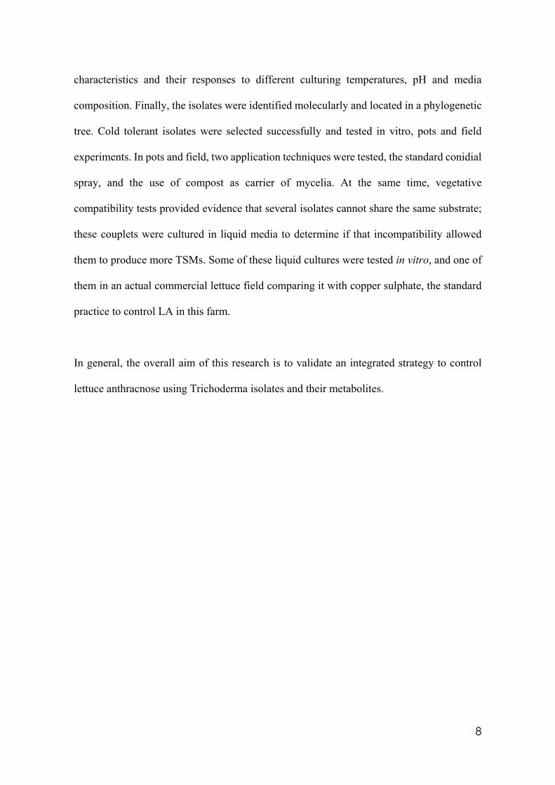
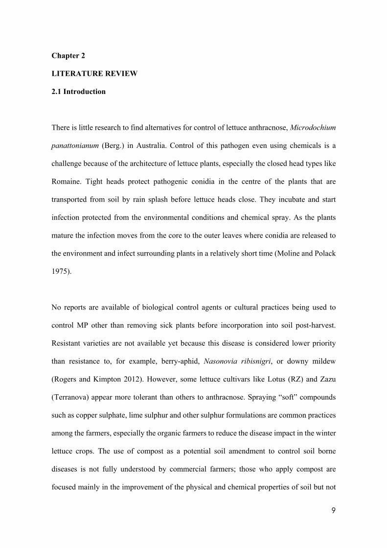
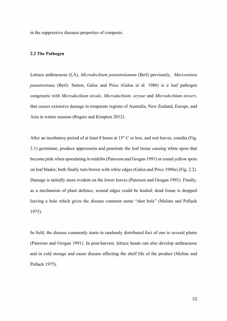
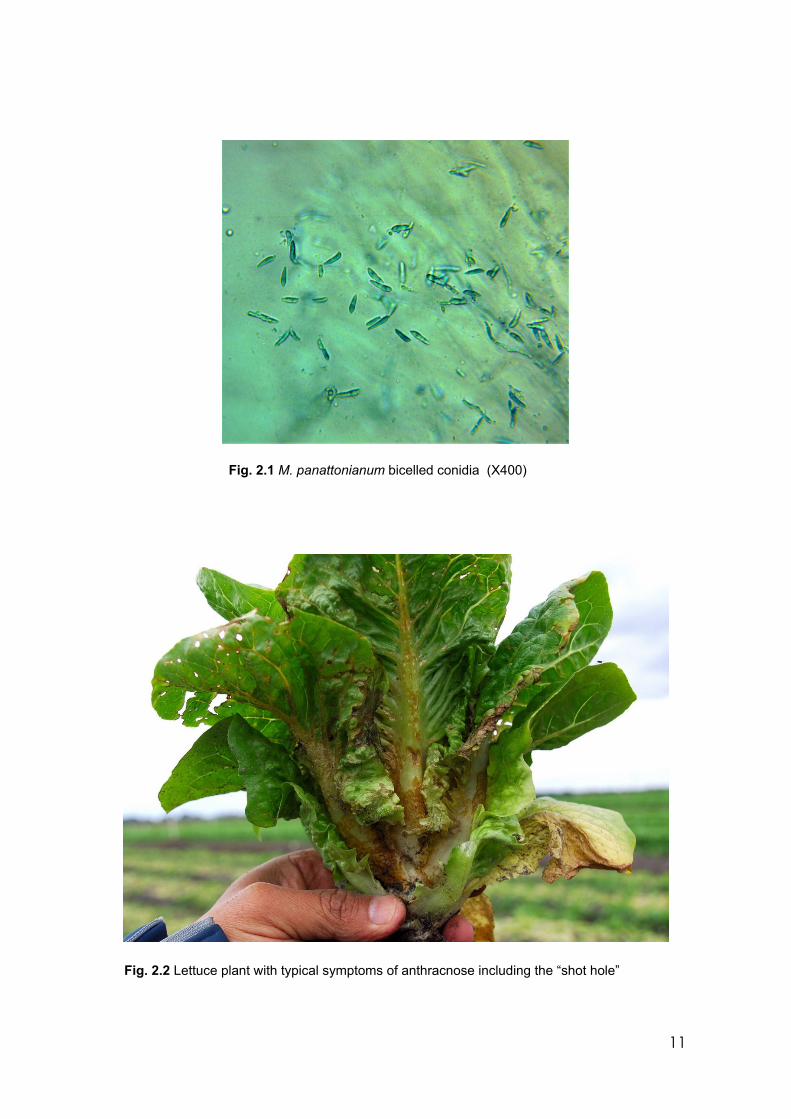
After an incubatory period of at least 8 hours at 15° C or less, and wet leaves, conidia (Fig.
2.1) germinate, produce appressoria and penetrate the leaf tissue causing white spots that
become pink when sporulating in midribs (Paterson and Grogan 1991) or round yellow spots
on leaf blades; both finally turn brown with white edges (Galea and Price 1988a) (Fig. 2.2).
Damage is initially more evident on the lower leaves (Paterson and Grogan 1991). Finally,
as a mechanism of plant defence, wound edges could be healed; dead tissue is dropped
leaving a hole which gives the disease common name “shot hole” (Moline and Pollack
1975).
In field, the disease commonly starts in randomly distributed foci of one to several plants
(Paterson and Grogan 1991). In post-harvest, lettuce heads can also develop anthracnose
and in cold storage and cause disease affecting the shelf life of the product (Moline and
Pollack 1975).
11
Fig. 2.1 M. panattonianum bicelled conidia (X400)
Fig. 2.2 Lettuce plant with typical symptoms of anthracnose including the “shot hole”
12
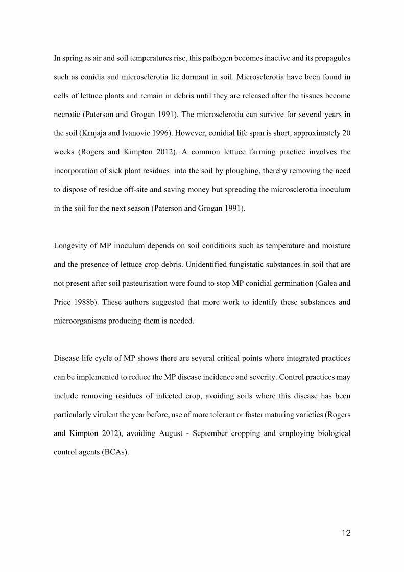
In spring as air and soil temperatures rise, this pathogen becomes inactive and its propagules
such as conidia and microsclerotia lie dormant in soil. Microsclerotia have been found in
cells of lettuce plants and remain in debris until they are released after the tissues become
necrotic (Paterson and Grogan 1991). The microsclerotia can survive for several years in
the soil (Krnjaja and Ivanovic 1996). However, conidial life span is short, approximately 20
weeks (Rogers and Kimpton 2012). A common lettuce farming practice involves the
incorporation of sick plant residues into the soil by ploughing, thereby removing the need
to dispose of residue off-site and saving money but spreading the microsclerotia inoculum
in the soil for the next season (Paterson and Grogan 1991).
Longevity of MP inoculum depends on soil conditions such as temperature and moisture
and the presence of lettuce crop debris. Unidentified fungistatic substances in soil that are
not present after soil pasteurisation were found to stop MP conidial germination (Galea and
Price 1988b). These authors suggested that more work to identify these substances and
microorganisms producing them is needed.
Disease life cycle of MP shows there are several critical points where integrated practices
can be implemented to reduce the MP disease incidence and severity. Control practices may
include removing residues of infected crop, avoiding soils where this disease has been
particularly virulent the year before, use of more tolerant or faster maturing varieties (Rogers
and Kimpton 2012), avoiding August - September cropping and employing biological
control agents (BCAs).
13
2.3 The biological control agents
Of many fungal species used as biocontrol agents (BCAs), Trichoderma species are the most
researched and widely used in commercial practice. Since their first application in the
1930’s, Trichoderma spp. have become popular BCAs to protect plants against diseases all
over the world (Ha 2010). They occur naturally in contrasting environmental conditions
such as dry and hot soils in Libya (Abadi 2008), wet and cold tundras in the Himalayas
(Ghildiyal and Pandey 2008), in the amazon rainforest soils (Delabona et al. 2012) and in
soils in Australia (Wong et al. 2002). Recently, Trichoderma species have been isolated
from marine conditions (Mukherjee et al. 2013). They are found colonising rhizospheres
(rhizosphere competence) (Ahmad and Baker 1988) and growing in plant roots in
symptomless infection (endophytes) (Druzhinina et al. 2011). Apparently, the rhizosphere-
competent Trichoderma strains are more effective for suppressing plant pathogens in a wider
spectrum of environmental conditions than the rhizosphere-incompetent strains (Brožová et
al. 2004). In a co-evolutionary process (Mukherjee et al. 2013), plants allow Trichoderma
to colonise their roots by detecting specific compounds (Woo and Lorito 2006) such as
auxin-like and proteinaceous metabolites that allow fungi to penetrate plant tissues
(Djonovic et al. 2006). Trichoderma spp. are also found growing, to a lesser extent, on aerial
parts of plants (phyllosphere competence) (Elad and Kirshner 1992).
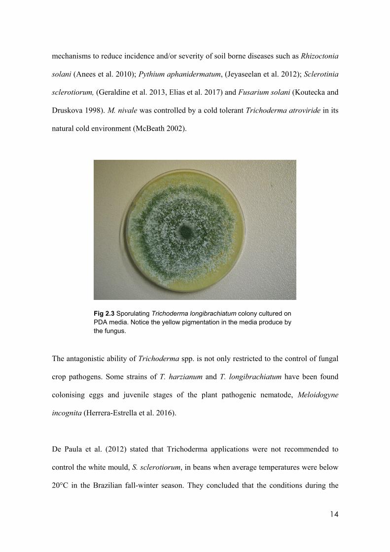
Trichoderma spp. are characterised in vitro by fast growing colonies with white, green, or
yellow cushions (Fig. 2.3). In soil, they are usually found at 101-103 colony forming units
(cfu) per gram of soil (Abadi 2008; Asha et al. 2013).
Scientific investigation of Trichoderma spp. has established their link to a variety of
14
mechanisms to reduce incidence and/or severity of soil borne diseases such as Rhizoctonia
solani (Anees et al. 2010); Pythium aphanidermatum, (Jeyaseelan et al. 2012); Sclerotinia
sclerotiorum, (Geraldine et al. 2013, Elias et al. 2017) and Fusarium solani (Koutecka and
Druskova 1998). M. nivale was controlled by a cold tolerant Trichoderma atroviride in its
natural cold environment (McBeath 2002).
Fig 2.3 Sporulating Trichoderma longibrachiatum colony cultured on PDA media. Notice the yellow pigmentation in the media produce by the fungus.
The antagonistic ability of Trichoderma spp. is not only restricted to the control of fungal
crop pathogens. Some strains of T. harzianum and T. longibrachiatum have been found
colonising eggs and juvenile stages of the plant pathogenic nematode, Meloidogyne
incognita (Herrera-Estrella et al. 2016).
De Paula et al. (2012) stated that Trichoderma applications were not recommended to
control the white mould, S. sclerotiorum, in beans when average temperatures were below
20°C in the Brazilian fall-winter season. They concluded that the conditions during the
15
experiment favoured the pathogen more than the antagonist. In a similar experiment, two
isolates of Trichoderma were selected for their ability to control sclerotia from S.
sclerotiourum in the same crop under similar temperature conditions (Morandi et al. 2007).
These authors referred to these isolates as ‘cold tolerant’ Trichoderma isolates.
Because Trichoderma strains vary in their tolerance to cold, isolates must be selected for
their activity under specific pathogen, environmental and crop conditions that promote
disease (Nelson 1991), the “pathosystem” (Fravel 2005).
In terms of Trichoderma nutrition, Khattabi et al. (2004) looking for alternatives for the
effective control of sugar beet root rot, tested nitrogenous compounds such as urea, nitrate,
ammonium and horse manure to evaluate the best combination to deplete Sclerotium rolfsii
growth by enhancing Trichoderma antagonist activity. Urea contributed to pathogen growth
inhibition, but nitrate and ammonium stimulated it. Horse manure was the best to promote
Trichoderma activity, reducing pathogen growth and controlling the disease.
2.3.1 The beneficial contribution of Trichoderma on plant
It has been demonstrated that some Trichoderma species help to improve plant health by
stimulation of the induced systemic resistance (ISR), and plant growth directly or indirectly
(Vinale et al. 2008). T. virens was reported to induce production of phytoalexins in cotton
plants improving their defence metabolism (Hanson and Howell 2004). The ISR is a
mechanism not dependent on production of salicylic acid as the systemic acquired resistance
(SAR) is, but on another two plant hormones, jasmonic acid (JAS) and ethylene (ET)
(Pieterse et al. 2014). ISR is based on stimulation of enhanced sensitivity of tissues to
16
JAS/ET rather than an increase in their synthesis (Pieterse et al. 2014). SAR and ISR cross
communicate enhancing the defence capacity of the plant (Mukherjee et al. 2013) and are
produced by different groups of microorganisms (Köhl et al. 2019) including beneficial
rhizobacteria (Harman 2000).
Clear evidence of Trichoderma activating ISR was found by Manganiello et al. (2018) when
they achieved significant reduction in levels of infection by R. solani in tomato seedlings
by up-regulation of genes related to ISR. They compared the effect of seed inoculated with
conidia and leaves sprayed with harzianic acid, a secondary metabolite produced by T.
harzianum M10, initially isolated in Western Australia. The level of plant protection was
significantly higher in both when leaves were sprayed and comparable with the protection
obtained when the seed was pre-inoculated with conidia. The outcome of the resistance
mechanism mediated by a BCAs depends on the balance between growing conditions for
the BCA, the plant physiological stage and the specific pathogen (Köhl et al. 2019).
Phytohormones such as indole acetic acid (IAA) related compounds are induced by
Trichoderma (Contreras-Cornejo et al. 2009). Some strains of Trichoderma induce root
branching and higher shoot biomass in response to the synthesis of fungal auxin-like
compounds (Contreras-Cornejo et al. 2016). More recently (Lombardi et al. 2018) found
that stressed tomato roots attracted Trichoderma by a specific and enhanced chemotropism
mechanism not recognisable by pathogens.
Harman (2000) reported higher corn yields and less nitrogen input using seeds treated with
Trichoderma harzianum T22 compared with untreated seeds. A similar response was
obtained in lettuce crops using T. virens GV41 and T. harzianum T22; increasing yield and
17
nitrogen uptake (Fiorentino et al. 2018). Altomare et al. (1999) reported improved solubility
of nutrients in vitro using both T. harzianum T22 culture and media treated with its filtrates.
They concluded that the increased bioavailability was based on chelation and reduction
processes rather than on increased acidity of media. Cucumber plants cultured in hydroponic
conditions treated with T. harzianum T203 also showed significantly increased uptake and
concentration of copper, manganese, phosphorus, and sodium in roots (Yedidia et al. 2001).
Salinity tolerance (Contreras-Cornejo et al. 2014) and drought tolerance (Contreras-Cornejo
et al. 2009) were enhanced by using Trichoderma applications.
2.3.2 Mechanisms of Trichoderma biocontrol
Trichoderma spp. are competitors for food sources and space. Mycoparasitism, antibiosis
and resistance induction are the main mechanisms Trichoderma species use to exert disease
control (Ghisalberti and Sivasithamparam 1991; Vinale et al. 2017). Antibiosis by
Trichoderma is a natural process mediated by Trichoderma secondary metabolites (TSMs)
synthesis that includes chemically diverse substances and cell wall degrading enzymes
(CWDE) (Ghisalberti and Sivasithamparam 1991). These antibiotics and enzymes often
work synergistically (Woo et al. 2006) and are linked to specific genes that could not be
expressed under laboratory conditions when they are in pure culture (Mukherjee et al. 2012).
This is because only a small set of genes producing TSMs are activated in vitro. The
remaining genes are only activated using specific elicitors (Vinale et al. 2017). Elicitors
include the presence of pathogen cell walls in the media as the challenging mechanism. Both
hydrolytic enzymes and antibiotics production increase significantly when challenge occurs
(Schirmböck et al. 1994).
18
Efficacy of TSM might be specific, i.e. filtrates of T. harzianum and T. hamatum inhibited
growth of P. aphanidermatum by 83% and 8% respectively (Sivan et al. 1984). Abundance
and diversity of TSMs are controlled by specific genes and other conditions that trigger their
expression (Mukherjee et al. 2012). Initially, in in vitro experiments it was thought that
TSMs are produced only during active fungal growth by tips of growing hyphae and released
in points of contact with the pathogen (Michalikova and Michirina 1997). Further
investigation determined that they are also produced but in less concentration in the
sporulation phase (Vinale et al. 2008). Trichorzianines, which are partially responsible for
the antifungal activity of T. harzianum, were isolated from sporulating cultures (Bodo et al.
1985). Therefore, any factor that affects the growth of mycelia and sporulation also affects
the production of TSMs. Vinale et al. (2009) found that TSM production in vitro is affected
not only by the presence of another microbe acting as the elicitor but also by its viability.
Some metabolites are produced only when the challenging organism is viable. Therefore,
the Trichoderma - pathogen co-culture technique is a good first approach to investigate more
diverse production of TSMs (Vinale et al. 2017). It is apparent that productivity of conidia
and mycelia for TSM production demands different conditions (Ferrigo et al. 2014). Talla
et al. (2015) using a strain of T. viride found that maximum mycelia growth was achieved
at 37°C but conidiation at 24°C. They suggested selective incubation temperatures for a
maximum production of conidia.
2.3.3 Trichoderma in farming practice
Many Trichoderma formulations are available commercially (Woo et al. 2014), but their
individual environmental requirements are not taken in consideration and explained to
farmers when they are introduced in farming systems (Pers. Comm. Nicholas Huvelle,
19
2016). Understanding the mode of action of a BCA is essential to achieve optimum disease
control (Köhl et al. 2019).
For efficacy in field conditions, formulations based only on conidia or chlamydospores as
mechanisms of fungal dispersion and biocontrol of crop diseases, have strong limitations
when sprayed on leaves to control foliar diseases (Pascale et al. 2017). However, by contrast,
secondary metabolites are not affected by normal environmental conditions and are
candidates for foliar disease control (Abadi 2008).
An individual species or strain of Trichoderma is not able to express all these above
advantages under all environmental or agricultural conditions. Some strains are efficient
producers of cell wall degrading enzymes and/or antibiotics (Vinale et al. 2008), while
others are specialised plant growth promoters (Nieto-Jacobo et al. 2017) or chelating agents
to enhance plant mineral nutrition (Anke et al. 1991). Alvarado and Rivera (2016) reported
differences in the antagonistic capacity of strains within the same species of Trichoderma
against Sclerotium cepivorum when challenged in in vitro conditions. They concluded that
control efficacy depends on the isolate. Likewise, different Trichoderma strains have
nutritional preferences when they colonise soils or plants and establish relationships with
crops (Carreras-Villasenor et al. 2012). Not all isolates show the same rhizosphere or
phyllosphere competence (Lo et al. 1997). Therefore, defining a specific set of parameters
for each potential BCA for growth and development (Carreras-Villasenor et al. 2012), and
factors affecting the relationship between plants, pathogen and other microorganisms,
including other Trichoderma species, requires field testing in multiple environments (Vinale
et al. 2008). Methods employed to produce, formulate, and apply these organisms in field
may affect profoundly their efficacy (Lo et al. 1997).
20
2.4 Trichoderma morphological characteristics and environmental preferences
Classic morphologically based taxonomy has been the historical foundation of fungal
species identification and the starting point for plant pathology experiments to evaluate
biological activity under different conditions (Guigón-López et al. 2010). Colony
morphology of Trichoderma isolates cultured on the defined medium, Czapek Dox Agar
(CZA), that includes nitrate as the only one source of nitrogen, was used as the first level of
identification in their classification (Gilman 1957). However, misidentification happened.
Chaverri et al. (2015) tested 4 commercial Trichoderma products labelled as T. harzianum,
and, after running molecular identification, they concluded that none were T. harzianum.
Molecular techniques, introduced at the end of the 1990’s for Trichoderma species, have the
advantage of higher precision and faster results and, therefore, are replacing the
morphological processes (Kullnig-Gradiner et al. 2002). However, the molecular
instrumentation is not as easily accessible as a microscope. Combining both techniques
leads to the better outcome (Castle et al. 1998).
Trichoderma species are classified by some authors as Deuteromycetes or mitosporic fungi
because their sexual reproduction and structures are rare, lacking, or unknown (Kullnig-
Gradinger et al. 2002; Agrios 2004). They produce single-celled, elliptical phialospore-
conidia (Kiffer and Morelet 2000), released apically without an inflated apical cell in
conidiophores irregularly branched (Watanabe 2010).
Historically, Trichoderma/Hypocrea are difficult to distinguish morphologically and
physiologically (Chaverri et al. 2003; Schuster and Schmoll 2010; Druzhinina et al. 2011),
21
despite their heterogeneous genomic structure and behaviour (Gomez et al. 1997). Initially
all were identified as T. viride (Schuster and Schmoll 2010). In 1969 their taxonomy was
clarified by Professor Mein Rifai in a major taxonomic review of this genus and species
(Verma et al. 2007). Since the taxonomic review, Trichoderma phylogenetic classification
resulted in 100 species by 2005 (Druzhinina and Kubicek 2005), greater than 1100
Hypocrea/Trichoderma strains from 75 molecularly defined species by 2011 (Druzhinina et
al. 2011) and 292 registered species by 2018 (Kubicek et al. 2019).
A current trend in Trichoderma classification is establishing chemotypes based on the
biological chemistry of isolates (Hanson 2005). However, initial isolate identification and
characterisation is the fundamental first step before specific and practical applications for
biocontrol can be considered (El-Refai et al. 2013). Additionally, it is essential that the
classified isolates are included in the phylogenetical lineage grouping system. This allows
many biological Trichoderma attributes with practical application such as production of
TSMs to be linked usefully (Chaverri et al. 2015).
Most Trichoderma species have their optimum developmental temperature between 25-
30°C (Guigón-López et al. 2010b). These authors also determined that Trichoderma strains
from warm climate and temperate climate have different temperature requirements for
biocontrol; warm climate strains work better for antibiosis than the temperate climate
strains. In locations with widely fluctuating weather conditions like southern Australia
where hot and dry summers and cold and wet winters alternate, it is possible that cold and
warm species of Trichoderma coexist but alternate in activity with the conditions described
above. Therefore, the dynamics and interactions are important factors that must be
understood (Lo et al. 1997). Among the species of Trichoderma, T. viride strains are
22
restricted to low temperature areas and T. harzianum to warm temperatures, while T.
hamatum and T. koningii have diverse climate requirements (Ghildiyal and Pandey 2008).
A cold tolerant isolate of T. atroviride tested in Alaska succeeded in controlling M. nivale
(McBeath 2002).
It is known also that fungi such as Trichoderma prefer acidic rather than alkaline conditions
with optimal growth in soil at pH between 4 to 6 (Baker 1986; Trushina et al. 2013). T.
viride and T. polysporum are more closely correlated with acidic coniferous forest soils
while T. hamatum is more correlated with neutral pH deciduous forest soils, even when both
substrates developed historically from the same base composition (Widden 1979). Domsch
et al. (1980) found that pH 3.7-4.7 is the optimal level for T. harzianum for maximum
biomass production. Rahman et al. (2009) could not find a conclusive correlation between
pH and production of colony form units (cfu.) by Trichoderma species under their
experimental conditions.
Conidial production is correlated more closely with low moisture, low organic matter, low
pH, exposure to UV-blue light and mycelial physical damage (Mukherjee et al. 2013).
Nitrate is not the preferred source of nitrogen for Trichoderma and low phosphorus and
potassium content promote conidia formation (Rahman et al. 2011). Dextrose (glucose)
rather than sucrose as the sole source of carbon produces five times more mycelial growth
in Trichoderma under in vitro conditions irrespective of the source of nitrogen used (Rossi-
Rodrigues et al. 2009).
For the above reasons Trichoderma in vitro research demands media standardisation
because it guarantees reproducibility of study conditions. Undefined media that contains
23
yeast or potato, such as potato dextrose agar (PDA), are variable from batch to batch and
even manufacturer to manufacturer (Griffith et al. 2007) when compared with defined media
such as CZA in which (Grant and Pramer, 1962) nitrogen is in inorganic form but not in
amino acids form as in PDA.
2.5 Trichoderma physiology
Trichoderma species, like all filamentous fungi, are constantly exploring their spatial
environment (Carreras-Villasenor et al. 2012). Many genes that encode proteases and
oligopeptide transporters are expressed during this process (Druzhinina et al. 2011). For
example, cell wall degrading enzymes (CWDE) (Vinale et al. 2008) are continuously
released into the microenvironment along with the expanding mycelia and are connected to
the nitrogen depletion receptors (Druzhinina et al. 2011) that are linked mostly to their
amino acid metabolism system (Seidil et al. 2009). When a suitable food source is detected,
Trichoderma mycelia grow in the direction of the nutrient gradient (Benhamou and Chet
1993); synthesising and releasing at the same time a more complex array of metabolites
(Vinale et al. 2008). The above attributes can be used in artificial production of these
compounds by the appropriate modification of fungal growing conditions (Lorito and Scala
1999).
Cell wall degrading enzymes produced by Trichoderma are not toxic to humans or animals
(Abadi 2008). They resist desiccation, are stable up to 60°C and are active over a wide range
of pH and temperature in agricultural environments (Abadi 2008). Therefore, they are easier
to manipulate than the living propagules in commercial formulations. Nevertheless, these
mechanisms and biochemical uses may represent risks for mycorrhizae, bacteria, plants,
insects, aquatic and terrestrial animals, and humans. Their effects need to be studied before
24
they are used in practical applications (Brimner and Boland 2003).
2.6 Vegetative compatibility/incompatibility
Genetically, when two strains of Trichoderma are compatible, their alleles in all vegetative
compatibility loci are the same (Malik and Vilgalys 1999, Burguess et al. 2009). They
belong to the same vegetative compatibility group (VCG) and produce heterokaryons
(Krnjaja et al. 2013). Heterokarions are cells with two or more genetically distinct nuclei
that share a common cytoplasm (Malik and Vilgalys 1999; Strom and Bushley 2016).
Heterokarions are physiologically active and they can generate independent colonies from
the two parent strains (Malik and Vilgalys 1999). According to these authors, heterokarions
are associated with more vigour because of the sexual recombination (parasexual
reproduction). They are source of future strain variation with biotechnological applications
in biocontrol, bioremediation, and production of novel pharmaceuticals (Strom and Bushley
2016). Researchers have created hybrids in vitro to use the enzyme producing ability of the
parent strains (Strom and Bushley 2016). Hassan (2014) created a hybrid by protoplast
fusion between two species of Trichoderma one of which synthesised large quantities of
chitinase and the other β-glucanase. Cole (1998) mentioned that because of a natural
restriction of the nuclear transference by allelic incompatibility in a vegetative compatibility
system within a territory, less virulent strains of plant pathogens can be produced. This may
explain the occurrence of new strains of a disease in the field.
On the other hand, if two colonies are not compatible, the cells (heterokarions) that are
produced by anastomosis (Burguess et al. 2009) are killed resulting in a visual gap (barrage),
between two colonies cultured in vitro (Moore et al. 2011), or zone lines that occur when
25
two genetically different mycelia approach (Smith et al. 2006). The barrage forming isolates
can be different species or even strains within species. For example, Gomez et al. (1997)
found barrage formation between T. harzianum strains. When antagonists become
competitors, they protect their own space and food sources and because of this territorial
defence mechanism, they can produce a more diverse range of metabolites than they would
produce unchallenged. All these biochemicals are released into the barrage area (Vinale et
al. 2008). Co-culturing strains of Trichoderma to increase metabolite productivity is a
simple process, but every strain in a collection must be challenged with every other strain
in that collection (Burguess et al. 2009).
2.7 Trichoderma secondary metabolites (TSMs)
TSMs are small molecules of relatively low molecular weight (Vinale et al. 2014) but their
chemical structure is remarkably complex (Ioca et al. 2016). These compounds are not
directly involved in growth or internal metabolic pathways but are important in signalling
and developing and establishing relationships with other microorganisms and plants
(Hoffmeister and Keller 2007). Some of these compounds are antibiotics (Ghisalberti and
Sivasithamparam 1991), with antifungal properties targeting hyphal growth or sporulation
processes; others are cell wall degrading enzymes and chelating agents (Vinale et al. 2014);
and some function like plant hormones enhancing plant growth, stimulating mineral uptake,
and therefore increasing mineral content in plant parts (Marra et al. 2019).
Dennis and Webster (1971) classified TSMs as volatile and non-volatile compounds.
Volatile TSMs can diffuse into the soil air pores and radiate further than can non-volatile
metabolites which only disperse in the soil solution. These authors were the first to describe
26
how these compounds were effective in reducing plant pathogen fungal hyphal growth and
they also stated that TSM production and distribution varies between isolates. More than
100 TSMs were reported by 2008 (Reino et al. 2008) and more than 1000 by 2014 (Hermosa
et al. 2014). The list of reported TSMs is continually increasing.
Gliotoxin was the first reported TSM, described by Richard Weindling in 1934, initially
from Trichoderma lignorum that was re-classified as Gliocladium virens (Mukherjee et al.
2012), and finally as Trichoderma virens (Vinale et al. 2014). Strains of T. virens group ‘Q’
produce large amounts of gliotoxin in liquid culture and are effective in controlling R. solani
but not in controlling protistas (oomycetes), such as Pythium or Phytophthora species,
which are controlled by another metabolite, gliovirin, produced by T. virens strains of the
group ‘P’ (Mukherjee et al. 2012 and Vinale et al. 2014).
Some species of Trichoderma, including T. harzianum, T. viride, T. koningii and T.
atroviride are profuse producers of a distinctive ‘coconut smell’ that is attributed to 6-n-
pentyl-2H-pyran-2-one (6-pentyl-α-pyrone) or simply 6PP, the most studied volatile TSM
(Vinale et al. 2014). This metabolite was discovered by accident by Dennis and Webster
(1971) when they were testing antagonistic activity of Trichoderma isolates in dual cultures.
They noticed that the most effective isolates against R. solani and G. graminis var. tritici
were characterised by their ‘coconut smell’ (Hanson 2005). 6PP is effective in controlling
sclerotia-forming pathogens and take-all fungus (Gaeumannomyces graminis) (Ghisalberti
and Sivasithamparam 1991). However, Dewan and Sivasithamparam (1988) already had
found that production of this compound was very variable between isolates of T. harzianum.
They found the isolates that did not produce or produced it in small amounts did not control
the pathogens.
27
6PP is a fungistatic and fungicidal compound (El Hassan et al. 2007); not toxic to mammals
at concentrations needed for pathogen control. This metabolite is an efficient inhibitor of R.
solani (Vinale et al 2006); Roselinia necatrix (Arjona-Girona et al 2014), Botrytis cinerea
and Alternaria brassicola (Kottb et al 2015). Arjona-Girona et al. (2014) achieved control
of white root rot (Rosellinia necatrix) by treating lupin seeds with a solution of 1 mg/l of
6PP. Vinale et al (2008) treated tomato and canola seedlings with 1-10 mg/kg stimulating
plant defence molecular triggers. El Hassan and Buchenauer (2009) found a higher dose
were required for maize seedlings to control Fusarium moniliforme (200 mg/kg).
Jeyaseelan et al. (2012) tested two isolates of T. harzianum and T. viride, synthesizing
volatile compounds that produced significant delay in Pythium aphanidermatum colony
growth in vitro after 24 hours of incubation, but not after 48 hours. This loss of efficacy
over time was also reported by Hanson (2005) when the same effect was observed with
Botrytis cinerea colonies. El-Hassan et al. (2007) reported a reduction of 93% of mycelia
growth and conidial germination of Fusarium moniliforme at 250 µg/ml and 300 µg/ml of
6PP respectively in vitro. In nature, this metabolite is degraded by hydroxylation to less
bioactive molecules, confirming that it is not persistent in the environment and useful for
practical application (Hanson 2005).
Harzianic acid, produced by strains of T. harzianum was found to control S. sclerotiorum,
R. solani and Pythium irregulare (Vinale et al. 2014). Additionally, it has been identified as
a plant growth promotor and chelating agent of soil iron Fe+3 (phytosiderophore) making
iron bioavailable for plants (Vinale et al. 2013). TSM peptaibols (Howell, 2003; Harman et
al. 2004 mentioned by Shi et al. 2012) are peptides which contain an unusual aminoacid, α-
aminoisobutyric acid (Aib), a C- terminal hydroxylated (amino alcohol) and a N-terminal-
acetylated aminoacid, with many antimicrobial properties which are largely still unknown.
28
Peptaibol is derived from ‘peptide’, Aib and alcohol (Brito et al. 2014). Shi et al. (2012)
mentioned that from 317 reported peptaibols, 190 were synthesised by Trichoderma isolates.
Fusalanipyrone is a rare fungal monoterpenoid metabolite (Abraham and Arfmann 1988)
produced by F. solani (Abraham et al. 1990) with moderate inhibitory effect on lettuce
seedlings growth (Shimizu et al. 2005). Cytosporone S was isolated from a strain of
Trichoderma sp. FK6626 with antifungal and antibacterial properties (Ishi et al. 2012).
Cytosporone A, B and C showed allelopathic activity against lettuce seeds and seedlings
(Zamberlan et al 2012) and antibacterial and antifungal activity (Ishi et al. 2012). Harzianic
acid (HA) reported as an antifungal, plant growth promoter and phytosiderophore (Vinale
et al. 2013). HA is effective in controlling P. irregulare, S. sclerotiorum and R. solani
(Vinale et al. 2009). The HA role as a chelating agent selectively protects Fe+3 for plant
nutrition, depriving pathogens of this important nutrient resulting growth suppression
(Vinale et al. 2013).
Enzymatically, T. harzianum, the most studied Trichoderma species targets organisms by
producing an enzyme that catalyses chitin breakdown, a primary component of fungal cell
walls (Zeilinger et al. 1999). Mycoparasitism by coiling and penetrating pathogen hyphae
is accompanied by antibiotic production that permeates the perforated hyphae and prevents
re-synthesis of the host cell wall (Lorito et al. 1996; Mc Beath 2002).
Formal identification to molecular level must be performed for every single isolate because
the TSM synthesis repertoire can vary widely between different isolates of the same species
(Vinale et al. 2014). T. harzianum isolates were found to produce different chemicals with
different levels of activity against ‘take all’ fungus, thus establishing a classification system
29
based on ‘chemical types’ (Ghisalberti and Sivasithamparam 1991).
2.8 Trichoderma as a potential BCA for lettuce anthracnose
Trichoderma species are well known for their capacity to colonise soils and therefore reduce
pathogen infections on plant parts. They also have an inherent ability to colonise
intercellular compartments of roots (Druzhinina et al. 2011) and leaf surfaces, a mechanism
called phylloplane competency (Schumann and D’Arcy 2006). However, the efficacy of
Trichoderma to colonise a leaf and control a foliar disease directly in field conditions
depends on many factors such as leaf exudates, temperature, relative humidity, free water,
light, radiation, wind and pollution all of which fluctuate widely (Elad and Kirshner 1992).
Trichoderma spp. as mentioned before, have efficacy in controlling many pathogens that
produce foliar diseases, but unfortunately most of the research has been performed in vitro
and in optimal conditions for the antagonist (Adedeji 2008; Rahman 2009; Evueh et al.
2011; Pakdaman et al. 2013; Thakur and Harsh 2014; Kabir et al. 2016). Laboratory
conditions are radically different from field conditions, making the accuracy of laboratory-
based predictions problematic (Elad and Kirshner 1992; Sawant 2014; Woo et al.
2014).TSMs reduced the incidence and severity of foliar diseases in grapes under field
conditions (Pascale et al. 2017) and in greenhouse tomato crops (Vinale et al, 2017), but
there is no report of MP control using TSMs.
The ability of Trichoderma species to control soil borne diseases is not only associated with
external factors as described above, but also on their growing stages. Conidia are the most
effective mechanism of survival and dispersal (Carreras-Villasenor et al. 2012) but have the
lowest capacity for control, while the actively growing mycelia gives the best control (Bae
30
and Knudsen 2005). In unfavourable conditions, Trichoderma conidia remain dormant and
they will remain inactive until conditions are optimal (Carreras-Villaseñor et al. 2012,
Ŝimkoviĉ et al. 2015). Baker (1986) impregnated seeds with T. harzianum conidia and
sowed them under suboptimal temperatures for the antagonist, finding no control of Pythium
or Rhizoctonia. Therefore, commercial formulations based on conidia or chlamydospores
sprayed in the field are not necessarily a guarantee of success because the conditions maybe
suboptimal for the antagonist (Mendoza-Mendoza et al. 2015). Screening for a successful
biocontrol agent involves its isolation from environment based upon the pathosystem of
interest (Parnel et al. 2016). Papavizas et al. (1984) found that mycelial fragments were
more effective than conidia for supressing soil borne diseases 10-20 days after application.
Time is required for an effective control and development of mycelia biomass from conidia
(Ŝimkoviĉ et al. 2015). Methods of control that demand use of conidia must be
complemented by practices that stimulate their survival and proliferation in soil (Papavizas
et al. 1985).
2.9 Practical application of Trichoderma to control pathogens.
There are several ways to increase soil borne disease suppression: (1) applying known
biological control agents (BCAs); (2) enhancing the growth and development of pre-existing
BCAs in soil and (3) applying a complex population of microorganisms (St. Martin 2015).
Thermal aerobic composts provide opportunities for consistent biological control of crop
diseases particularly if they are inoculated with BCAs (Hoitink et al. 1997, Bernal-Vicente
et al. 2012). Maximum disease suppression from compost is achieved when the BCAs
recolonise the compost in the curing to maturity stages (Hoitink et al. 1997, Bonanomi et al.
31
2018). Some authors including Parkash and Saikia (2014) consider that Trichoderma species
are compost fungal activators (CFAs). Additionally, they can reduce the composting process
time when inoculated onto the raw materials. Interestingly, Stone et al. (2003) applied paper
mill residues to a sandy soil and succeeded in controlling soil borne diseases such as
Pythium sp that cause damping off in cucumber and beans and C. lindemuthianum which
causes anthracnose in cucumber, respectively. They attributed but could not confirm this
response to the activation of plant defence mechanisms in the cucumbers and beans.
Since lettuce anthracnose is a soil borne disease that produces survival microsclerotia and
causes damage in aerial parts of plants in cold conditions, the mechanisms of control must
be used strategically, first to keep the pathogen away from the plants by applying cold
tolerant Trichoderma isolates and, secondly, by reducing the damage to leaves using TSMs.
Single or mixed combinations of culture liquids produced under challenged conditions
containing different metabolites such as CWDEs are suitable alternatives for the control of
foliar pathogens. The CWDEs are resistant to desiccation and are stable up to 60°C in a
wide pH range (Abadi 2008).
Selective production of TSMs for pathogens may be achieved by modifying culturing
conditions which include substrate composition, pH, temperature, and agitation (Lorito and
Scala 1999).
Many strains of Trichoderma are reportedly resistant to chemical fungicides (Abadi 2008).
T. atroviride P1 was selected for its resistance to Benomyl and for its compatibility with T.
harzianum T22, another strain with well-known commercial formulation. These two strains
were investigated for their compatibility with organic products and copper oxychloride
32
which showed no effect on mycelial growth in concentrations up to 5 mM (Vinale et al.
2004).
Currently, there are no references of biological control of lettuce anthracnose,
Microdochium panattonianum, using Trichoderma and or its metabolites.
33
Chapter 3
MATERIALS AND METHODS.
3.1 Introduction
Different approaches were implemented to identify and characterise suitable cold tolerant
Trichoderma isolates and identification of TSMs that reduce MP incidence and severity.
3.2 Trichoderma isolates - Collection and isolation of pure cultures
Initially, 23 Trichoderma isolates were gathered. Four more were added just after the first
screening. In total twenty-seven isolates of Trichoderma were collected from different
sources and conditions (Table 3.1 and Figure 3.1).
South Australia Research and Development Institute (SARDI) provided mixed colony
plates from pistachio isolations. New South Wales Herbarium (DAR) provided six isolates.
Agpath P/L (AG), Garfield, Victoria, provided eleven samples cultured from soil and plant
material. The remaining Trichoderma isolates (7) were commercial formulations.
Fungal material from above cultures were soaked in sterile water and an aliquot of 100 µL
from each suspension was spread onto potato dextrose agar (PDA) (Amyl Media,
Dandenong) in 90 mm Petri dishes and incubated at room temperature. This method was
repeated until pure cultures of possible Trichoderma isolates were obtained.
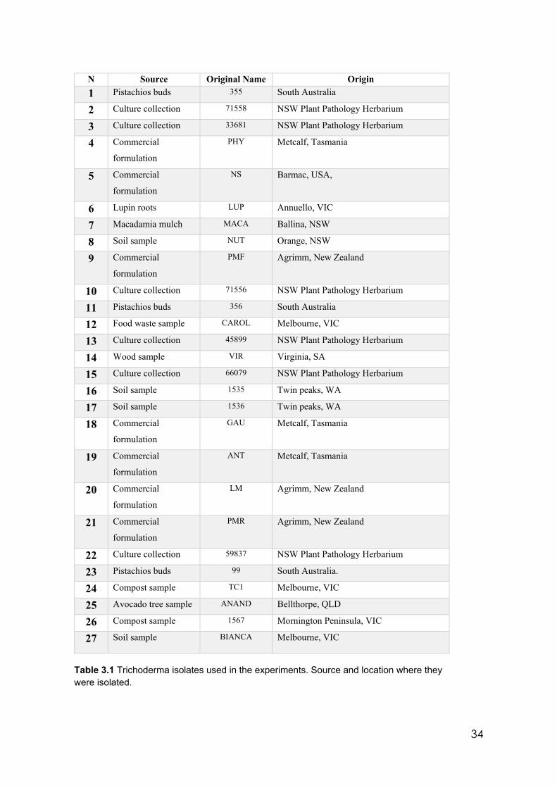
34
N Source Original Name Origin 1 Pistachios buds 355 South Australia
2 Culture collection 71558 NSW Plant Pathology Herbarium
3 Culture collection 33681 NSW Plant Pathology Herbarium
4 Commercial
formulation
PHY Metcalf, Tasmania
5 Commercial
formulation
NS Barmac, USA,
6 Lupin roots LUP Annuello, VIC
7 Macadamia mulch MACA Ballina, NSW
8 Soil sample NUT Orange, NSW
9 Commercial
formulation
PMF Agrimm, New Zealand
10 Culture collection 71556 NSW Plant Pathology Herbarium
11 Pistachios buds 356 South Australia
12 Food waste sample CAROL Melbourne, VIC
13 Culture collection 45899 NSW Plant Pathology Herbarium
14 Wood sample VIR Virginia, SA
15 Culture collection 66079 NSW Plant Pathology Herbarium
16 Soil sample 1535 Twin peaks, WA
17 Soil sample 1536 Twin peaks, WA
18 Commercial
formulation
GAU Metcalf, Tasmania
19 Commercial
formulation
ANT Metcalf, Tasmania
20 Commercial
formulation
LM Agrimm, New Zealand
21 Commercial
formulation
PMR Agrimm, New Zealand
22 Culture collection 59837 NSW Plant Pathology Herbarium
23 Pistachios buds 99 South Australia.
24 Compost sample TC1 Melbourne, VIC
25 Avocado tree sample ANAND Bellthorpe, QLD
26 Compost sample 1567 Mornington Peninsula, VIC
27 Soil sample BIANCA Melbourne, VIC
Table 3.1 Trichoderma isolates used in the experiments. Source and location where they were isolated.
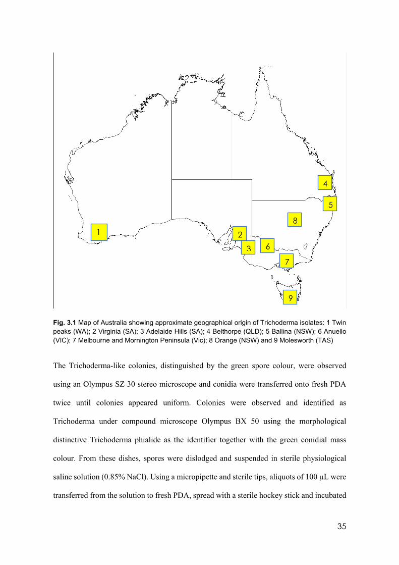
35
Fig. 3.1 Map of Australia showing approximate geographical origin of Trichoderma isolates: 1 Twin peaks (WA); 2 Virginia (SA); 3 Adelaide Hills (SA); 4 Belthorpe (QLD); 5 Ballina (NSW); 6 Anuello (VIC); 7 Melbourne and Mornington Peninsula (Vic); 8 Orange (NSW) and 9 Molesworth (TAS)
The Trichoderma-like colonies, distinguished by the green spore colour, were observed
using an Olympus SZ 30 stereo microscope and conidia were transferred onto fresh PDA
twice until colonies appeared uniform. Colonies were observed and identified as
Trichoderma under compound microscope Olympus BX 50 using the morphological
distinctive Trichoderma phialide as the identifier together with the green conidial mass
colour. From these dishes, spores were dislodged and suspended in sterile physiological
saline solution (0.85% NaCl). Using a micropipette and sterile tips, aliquots of 100 µL were
transferred from the solution to fresh PDA, spread with a sterile hockey stick and incubated
1
2 3
4 5
6 7
8
9
36
right side up at 22 ± 1° C. After 24 hours, using a stereo microscope, individual conidia
were selected and removed carefully by cutting an agar square carrying one germinated
conidium and transplanted spore face down onto fresh PDA and incubated at 25°C in a
Thermoline incubator until spore germination was observed. This procedure resulted in
genetically pure isolates. These single spore isolates became the foundation isolates for all
further research for this project.
From the commercial formulations used in these experiments a suspension of 0.1 g of freeze
dry formulation was added to 100 ml of sterile water. Aliquots of 100 µL were added to
PDA plates and spread with sterile hockey sticks and incubated at room temperature. Single
spores or single hyphal tips, in the case of fast germinating spores, were removed and treated
as above.
Mother cultures of each Trichoderma isolate were established on PDA slants in triplicate.
The established cultures were covered with sterile water to the top of the agar slant to
prevent desiccation and were refrigerated at 8°C. This collection is refreshed every 3 months
using the same protocol.
3.3 Morphological characterisation in diverse culture conditions
All Trichoderma isolates were cultured in vitro under different conditions (pH, temperature
and media) (Table 3.2) to determine how these conditions and their combinations affect
morphological characteristics such as phialide and conidia dimensions, colony shape, colour
and growth speed, and the coconut aroma that is linked to a key volatile TSM.
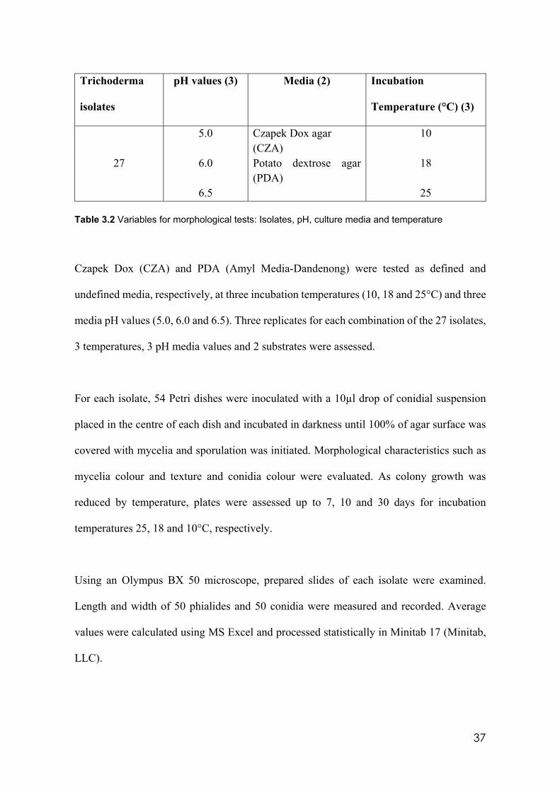
37
Trichoderma
isolates
pH values (3) Media (2) Incubation
Temperature (°C) (3)
5.0 Czapek Dox agar (CZA)
10
27 6.0 Potato dextrose agar (PDA)
18
6.5 25 Table 3.2 Variables for morphological tests: Isolates, pH, culture media and temperature
Czapek Dox (CZA) and PDA (Amyl Media-Dandenong) were tested as defined and
undefined media, respectively, at three incubation temperatures (10, 18 and 25°C) and three
media pH values (5.0, 6.0 and 6.5). Three replicates for each combination of the 27 isolates,
3 temperatures, 3 pH media values and 2 substrates were assessed.
For each isolate, 54 Petri dishes were inoculated with a 10µl drop of conidial suspension
placed in the centre of each dish and incubated in darkness until 100% of agar surface was
covered with mycelia and sporulation was initiated. Morphological characteristics such as
mycelia colour and texture and conidia colour were evaluated. As colony growth was
reduced by temperature, plates were assessed up to 7, 10 and 30 days for incubation
temperatures 25, 18 and 10°C, respectively.
Using an Olympus BX 50 microscope, prepared slides of each isolate were examined.
Length and width of 50 phialides and 50 conidia were measured and recorded. Average
values were calculated using MS Excel and processed statistically in Minitab 17 (Minitab,
LLC).
38
3.4 Molecular identification of Trichoderma isolates
For formal identification of all Trichoderma isolates, molecular tests were performed.
Isolates were cultured on PDA at 25° C for three days until all agar surfaces were covered
with mycelia (Alvarado-Marchena and Rivera-Méndez 2016).
Extraction of DNA was performed using a Qiagen DNeasy® plant mini kit following the
protocol supplied with the kit. A satisfactory quality and quantity of DNA in tube samples
was checked using both a nanodrop spectrophotometer and agarose electrophoresis test
(Sambrook et al. 1989). Tubes containing DNA were stored at -20°C until required (Griffin
et al. 2002).
For the electrophoresis tests, the medium containing 3 gr of agarose in 200ml of deionized
water (1.5%) was boiled in a microwave oven, swirling every 30 seconds, until dissolved
completely. Twenty microliters of SYBR Safe DNA stain was then added and mixed well.
Sixty ml of gel were poured in the tray; two combs were introduced and smoothly removed
after the gel solidified. The tray was introduced into the electrophoresis chamber and
covered with 1X borate-EDTA buffer solution. This solution was previously prepared
dissolving 242 gr of Tris base in 750 ml of deionized water, 57.1 ml of acetic acid and 100
ml of 100 mM EDTA solution were added, finally the volume was adjusted to 1l by adding
deionized water. From this mother stock 1ml was added to 49 ml of deionized water to
prepare a 1x solution. Three microliters of loading dye were placed on a piece of Parafilm,
one for each sample. Two microliters of each DNA sample were added and mixed well. The
mix was placed in the gel wells. The chamber lid was added checking the correct position
of cords. The voltage was set to 90 volts for 35 minutes. The gel was removed from the
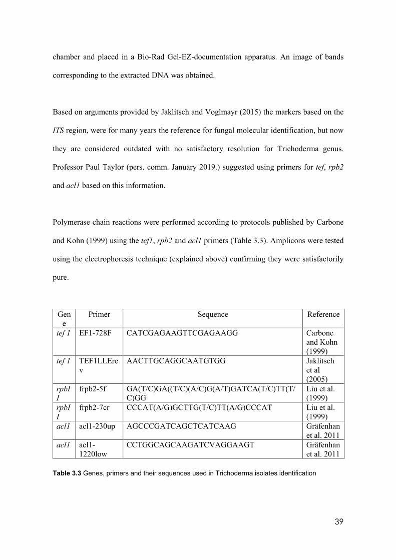
39
chamber and placed in a Bio-Rad Gel-EZ-documentation apparatus. An image of bands
corresponding to the extracted DNA was obtained.
Based on arguments provided by Jaklitsch and Voglmayr (2015) the markers based on the
ITS region, were for many years the reference for fungal molecular identification, but now
they are considered outdated with no satisfactory resolution for Trichoderma genus.
Professor Paul Taylor (pers. comm. January 2019.) suggested using primers for tef, rpb2
and acl1 based on this information.
Polymerase chain reactions were performed according to protocols published by Carbone
and Kohn (1999) using the tef1, rpb2 and acl1 primers (Table 3.3). Amplicons were tested
using the electrophoresis technique (explained above) confirming they were satisfactorily
pure.
Gene
Primer Sequence Reference
tef 1 EF1-728F CATCGAGAAGTTCGAGAAGG Carbone and Kohn (1999)
tef 1 TEF1LLErev
AACTTGCAGGCAATGTGG Jaklitsch et al (2005)
rpbII
frpb2-5f GA(T/C)GA((T/C)(A/C)G(A/T)GATCA(T/C)TT(T/C)GG
Liu et al. (1999)
rpbII
frpb2-7cr CCCAT(A/G)GCTTG(T/C)TT(A/G)CCCAT Liu et al. (1999)
acl1 acl1-230up AGCCCGATCAGCTCATCAAG Gräfenhan et al. 2011
acl1 acl1-1220low
CCTGGCAGCAAGATCVAGGAAGT Gräfenhan et al. 2011
Table 3.3 Genes, primers and their sequences used in Trichoderma isolates identification
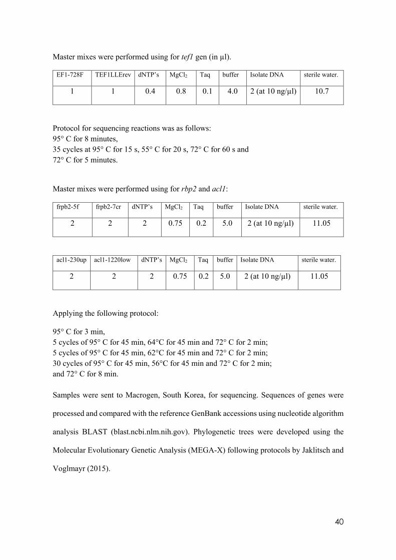
40
Master mixes were performed using for tef1 gen (in µl).
EF1-728F TEF1LLErev dNTP’s MgCl2 Taq buffer Isolate DNA sterile water.
1 1 0.4 0.8 0.1 4.0 2 (at 10 ng/µl) 10.7
Protocol for sequencing reactions was as follows: 95° C for 8 minutes, 35 cycles at 95° C for 15 s, 55° C for 20 s, 72° C for 60 s and 72° C for 5 minutes.
Master mixes were performed using for rbp2 and acl1:
frpb2-5f frpb2-7cr dNTP’s MgCl2 Taq buffer Isolate DNA sterile water.
2 2 2 0.75 0.2 5.0 2 (at 10 ng/µl) 11.05
acl1-230up acl1-1220low dNTP’s MgCl2 Taq buffer Isolate DNA sterile water.
2 2 2 0.75 0.2 5.0 2 (at 10 ng/µl) 11.05
Applying the following protocol:
95° C for 3 min, 5 cycles of 95° C for 45 min, 64°C for 45 min and 72° C for 2 min; 5 cycles of 95° C for 45 min, 62°C for 45 min and 72° C for 2 min; 30 cycles of 95° C for 45 min, 56°C for 45 min and 72° C for 2 min; and 72° C for 8 min. Samples were sent to Macrogen, South Korea, for sequencing. Sequences of genes were
processed and compared with the reference GenBank accessions using nucleotide algorithm
analysis BLAST (blast.ncbi.nlm.nih.gov). Phylogenetic trees were developed using the
Molecular Evolutionary Genetic Analysis (MEGA-X) following protocols by Jaklitsch and
Voglmayr (2015).
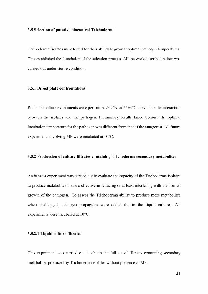
41
3.5 Selection of putative biocontrol Trichoderma
Trichoderma isolates were tested for their ability to grow at optimal pathogen temperatures.
This established the foundation of the selection process. All the work described below was
carried out under sterile conditions.
3.5.1 Direct plate confrontations
Pilot dual culture experiments were performed in vitro at 25±3°C to evaluate the interaction
between the isolates and the pathogen. Preliminary results failed because the optimal
incubation temperature for the pathogen was different from that of the antagonist. All future
experiments involving MP were incubated at 10°C.
3.5.2 Production of culture filtrates containing Trichoderma secondary metabolites
An in vitro experiment was carried out to evaluate the capacity of the Trichoderma isolates
to produce metabolites that are effective in reducing or at least interfering with the normal
growth of the pathogen. To assess the Trichoderma ability to produce more metabolites
when challenged, pathogen propagules were added the to the liquid cultures. All
experiments were incubated at 10°C.
3.5.2.1 Liquid culture filtrates
This experiment was carried out to obtain the full set of filtrates containing secondary
metabolites produced by Trichoderma isolates without presence of MP.
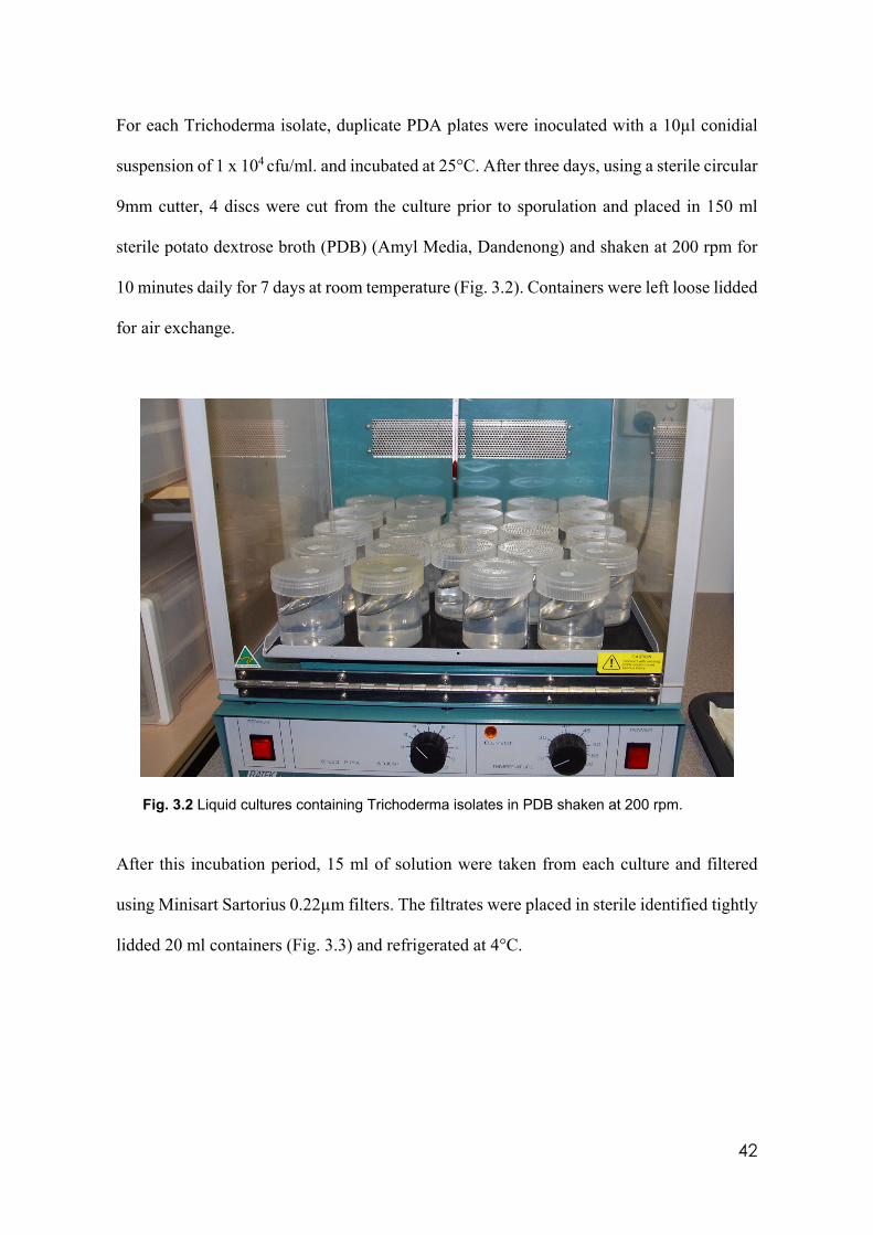
42
For each Trichoderma isolate, duplicate PDA plates were inoculated with a 10µl conidial
suspension of 1 x 10⁴ cfu/ml. and incubated at 25°C. After three days, using a sterile circular
9mm cutter, 4 discs were cut from the culture prior to sporulation and placed in 150 ml
sterile potato dextrose broth (PDB) (Amyl Media, Dandenong) and shaken at 200 rpm for
10 minutes daily for 7 days at room temperature (Fig. 3.2). Containers were left loose lidded
for air exchange.
Fig. 3.2 Liquid cultures containing Trichoderma isolates in PDB shaken at 200 rpm.
After this incubation period, 15 ml of solution were taken from each culture and filtered
using Minisart Sartorius 0.22µm filters. The filtrates were placed in sterile identified tightly
lidded 20 ml containers (Fig. 3.3) and refrigerated at 4°C.
43
Fig. 3.3 Filtrates from Trichoderma isolates liquid cultures showing different natural pigmentation
The culturing process continued with the remaining liquid for a further 7 days, after which
another 15 ml sample was filtered as above. The remaining culture was filtered and
refrigerated at 4°C for later trials.
3.5.2.2 Liquid culture filtrates from MP induction
Filtrates in this experiment used the same protocol described in 3.5.2.1, however in this case,
discs containing the sporulating pathogen were added at the same time as the Trichoderma
mycelia discs. MP colonies were cultured on PDA and incubated at 10°C for 7 days. From
these plates, two 9 mm diameter discs with growing colonies were introduced into each of
the liquid cultures. Containers were agitated for 10 minutes daily for 7 and 14 days as
described above in 3.5.2.1
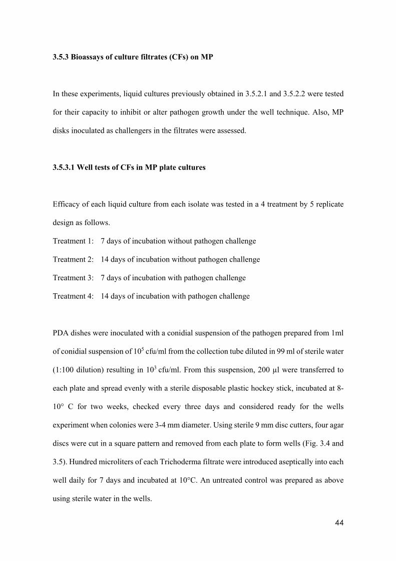
44
3.5.3 Bioassays of culture filtrates (CFs) on MP
In these experiments, liquid cultures previously obtained in 3.5.2.1 and 3.5.2.2 were tested
for their capacity to inhibit or alter pathogen growth under the well technique. Also, MP
disks inoculated as challengers in the filtrates were assessed.
3.5.3.1 Well tests of CFs in MP plate cultures
Efficacy of each liquid culture from each isolate was tested in a 4 treatment by 5 replicate
design as follows.
Treatment 1: 7 days of incubation without pathogen challenge
Treatment 2: 14 days of incubation without pathogen challenge
Treatment 3: 7 days of incubation with pathogen challenge
Treatment 4: 14 days of incubation with pathogen challenge
PDA dishes were inoculated with a conidial suspension of the pathogen prepared from 1ml
of conidial suspension of 105 cfu/ml from the collection tube diluted in 99 ml of sterile water
(1:100 dilution) resulting in 103 cfu/ml. From this suspension, 200 µl were transferred to
each plate and spread evenly with a sterile disposable plastic hockey stick, incubated at 8-
10° C for two weeks, checked every three days and considered ready for the wells
experiment when colonies were 3-4 mm diameter. Using sterile 9 mm disc cutters, four agar
discs were cut in a square pattern and removed from each plate to form wells (Fig. 3.4 and
3.5). Hundred microliters of each Trichoderma filtrate were introduced aseptically into each
well daily for 7 days and incubated at 10°C. An untreated control was prepared as above
using sterile water in the wells.
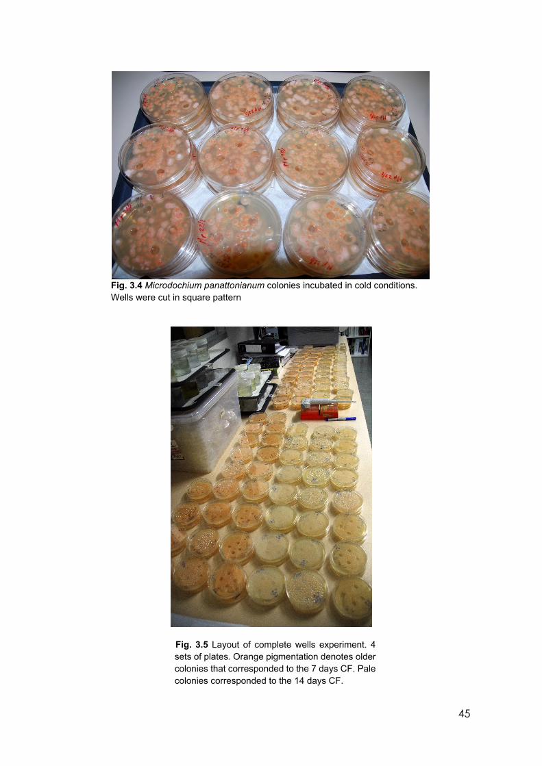
45
Fig. 3.4 Microdochium panattonianum colonies incubated in cold conditions. Wells were cut in square pattern
Fig. 3.5 Layout of complete wells experiment. 4 sets of plates. Orange pigmentation denotes older colonies that corresponded to the 7 days CF. Pale colonies corresponded to the 14 days CF.
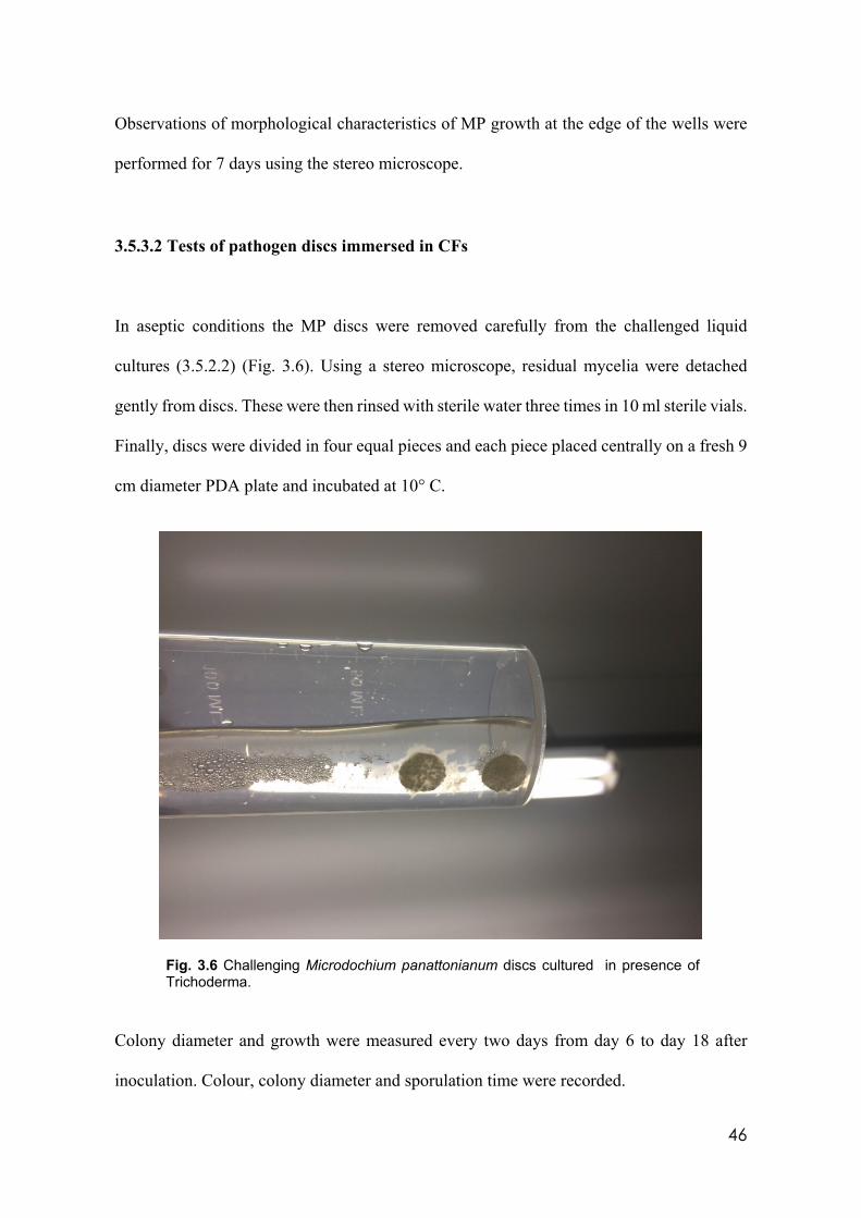
46
Observations of morphological characteristics of MP growth at the edge of the wells were
performed for 7 days using the stereo microscope.
3.5.3.2 Tests of pathogen discs immersed in CFs
In aseptic conditions the MP discs were removed carefully from the challenged liquid
cultures (3.5.2.2) (Fig. 3.6). Using a stereo microscope, residual mycelia were detached
gently from discs. These were then rinsed with sterile water three times in 10 ml sterile vials.
Finally, discs were divided in four equal pieces and each piece placed centrally on a fresh 9
cm diameter PDA plate and incubated at 10° C.
Fig. 3.6 Challenging Microdochium panattonianum discs cultured in presence of Trichoderma.
Colony diameter and growth were measured every two days from day 6 to day 18 after
inoculation. Colour, colony diameter and sporulation time were recorded.
47
3.5.3.3 Test of MP on substrates containing CFs To test the efficacy of the compounds in the liquid filtrates against MP using the previously
stored liquid cultures (3.5.2.2), a trial was set up to evaluate the ability to inhibit the
pathogen growth in vitro and to determine if they are heat stable. Only filtrates from isolates
356 and 1535 were selected for this pilot experiment because they are wild types and the
best cold tolerant isolates.
Agar modified as described above was prepared using 60 ml of the filtrate and 2.3 g of PDA
in two 200 ml Erlenmeyer flasks plugged with cotton, sealed with aluminium foil and
sterilised in a Siltex HC2 (MK-1-94) autoclave. An untreated control was prepared with
water replacing the filtrate. Treated and untreated agar plates were inoculated with a conidial
suspension of 105 cfu/ml of MP and spread with sterile hockey sticks; incubated at 10°C for
10 days and checked every two days using a stereo microscope to detect differences in the
hyphal morphology and colony growth.
3.6 Effect of temperature and pH to select cold tolerant Trichoderma strains
Following the pathogen disc assessment (3.5.3.2), it became obvious that not all
Trichoderma isolates had the same tolerance to low temperatures.
An experiment was set up to evaluate the performance of the selected cold tolerant isolates
at different media pH. This trial might help to assess potential performance of Trichoderma
isolates in different soil pHs that reflect most agricultural natural soil pH conditions. Three
pH values were considered initially: 5.5, 6.5 and 7.5. Potato dextrose agar made at label rate
in three 500 ml Pyrex glass bottles was gently swirled until all powder was in suspension,
48
then pHed using 1N Na (OH). However, after sterilisation, the pH value dropped to 5.0, 6.0
and 6.5, respectively. These pH values were considered the reference pH media values for
this experiment.
Conidial suspensions of 105 cfu/ml were prepared for each of the seven most cold tolerant
isolates (59837, 45899, LUP, 356, LM, PMF and PMR). Using three replicates per isolate
and pH, a 10µL drop of suspension was placed in the centre of each relevant PDA plate.
Plates were incubated at 10° C, checked each two days and colony diameters were measured
at 6, 8, 10, 14 and 16 days after inoculation.
3.7 Vegetative incompatibility tests (VIT) among Trichoderma strains
Two Trichoderma isolates can interact in different ways when they grow in the same
substrate. Their colonies can overlap or stop growing before physical contact forming a
barrage. A barrage which is a gap between two colonies is the expression of the inhibition
of growth mediated by metabolites from either or both isolates.
In this experiment all isolates were challenged against one another to observe their
compatibility/incompatibility and to predict if a more diverse collection of metabolites is
produced that maybe useful in controlling MP.
For each of the 27 Trichoderma isolates, 2 PDA plates were inoculated using a 1 x 10⁵
cfu/ml and incubated at 25°C until mycelia were produced. From prepared plates above, 9
mm agar discs containing mycelia were cut and placed onto PDA plates using the template
of 6 sectors and one central point (fig. 3.7). The isolate being tested was placed in the central
49
position, and in one sector 6. Other isolates were placed in the remaining sectors at 2 cm
separation. Plates were incubated at 25°C and checked daily. Plates were evaluated for
barrage formation. This trial was triplicated. Information from this experiment formed the
foundation for all the TSM experiments in 3.9. No microscopic observations regarding
heterokarion formation or dead hyphae were performed in this experiment. The barrage
formation was considered the only assessment parameter.
3.8 TSM production in co-cultures
Presence of a competitor triggers the expression of genes to produce to a more diverse TSM
production compared with a single isolate. Trichoderma isolates were cultured in couplets
according to the incompatibility chart (Table 4.7).
3.8.1 Liquid culture phase
PDA plates were prepared, inoculated with 27 isolates in triplicate and incubated at 25°C
for 3 days as before (3.3). At the same time, 20 ml of sterile PDB were transferred into 163
x 50 ml plastic Falcon conical tubes. Discs of mycelia only were cut and introduced into the
Falcon tubes in couplets (two different isolates) according to the vegetative incompatibility
chart (See Table 4.7). Additionally, 27 single isolate filtrates were prepared by introducing
two discs in the corresponding tubes to determine if there is a difference in TSM production
between challenged and unchallenged conditions. Falcon tubes were incubated at 25°C and
shaken for 5 minutes at 120 rpm daily for 7 days. Five ml from each culture were filtered
using sterile 20 ml Terumo® syringes and Merck Millex-GP 0.22 µm polyether sulphone
gamma sterilised syringe filters and placed in Technoplast 5 ml screw capped gamma sterile
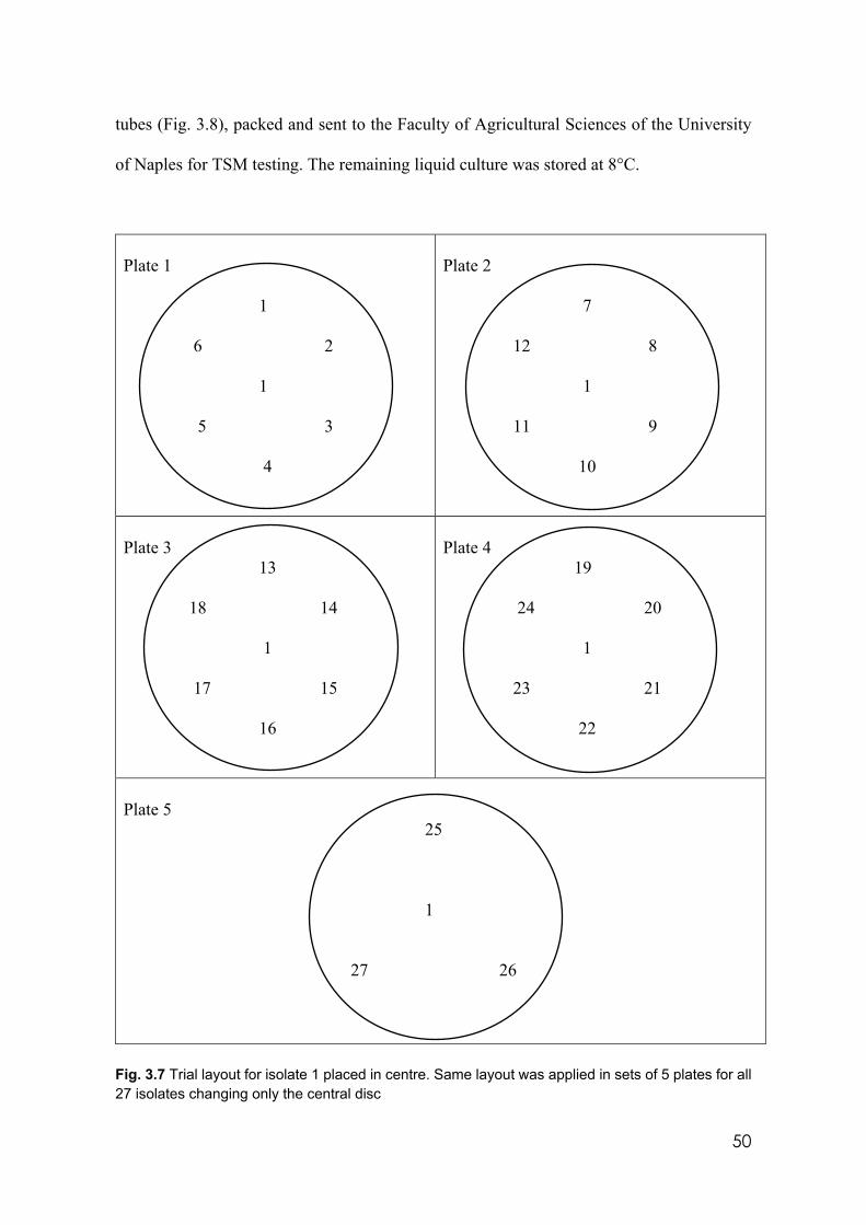
50
tubes (Fig. 3.8), packed and sent to the Faculty of Agricultural Sciences of the University
of Naples for TSM testing. The remaining liquid culture was stored at 8°C.
Plate 1 1 6 2 1 5 3 4
Plate 2 7 12 8 1 11 9 10
Plate 3 13 18 14 1 17 15 16
Plate 4 19 24 20 1 23 21 22
Plate 5 25 1 27 26
Fig. 3.7 Trial layout for isolate 1 placed in centre. Same layout was applied in sets of 5 plates for all 27 isolates changing only the central disc
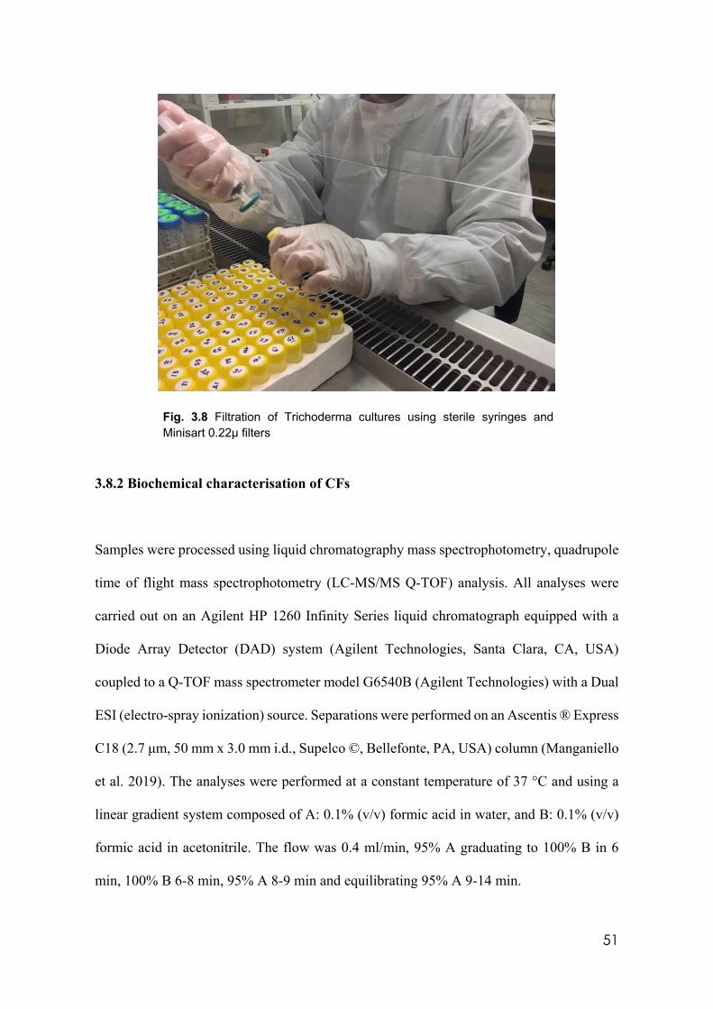
51
Fig. 3.8 Filtration of Trichoderma cultures using sterile syringes and Minisart 0.22µ filters
3.8.2 Biochemical characterisation of CFs
Samples were processed using liquid chromatography mass spectrophotometry, quadrupole
time of flight mass spectrophotometry (LC-MS/MS Q-TOF) analysis. All analyses were
carried out on an Agilent HP 1260 Infinity Series liquid chromatograph equipped with a
Diode Array Detector (DAD) system (Agilent Technologies, Santa Clara, CA, USA)
coupled to a Q-TOF mass spectrometer model G6540B (Agilent Technologies) with a Dual
ESI (electro-spray ionization) source. Separations were performed on an Ascentis ® Express
C18 (2.7 μm, 50 mm x 3.0 mm i.d., Supelco ©, Bellefonte, PA, USA) column (Manganiello
et al. 2019). The analyses were performed at a constant temperature of 37 °C and using a
linear gradient system composed of A: 0.1% (v/v) formic acid in water, and B: 0.1% (v/v)
formic acid in acetonitrile. The flow was 0.4 ml/min, 95% A graduating to 100% B in 6
min, 100% B 6-8 min, 95% A 8-9 min and equilibrating 95% A 9-14 min.
52
The UV spectra were collected by DAD every 0.4 s from 190 to 750 nm with a resolution
of 2 nm. The MS system was equipped with an ESI source and operated with Agilent
MassHunter Data Acquisition Software, rev. B.05.01 in the positive and negative mode.
Mass spectra were recorded in the range m/z 100-1700 as centroid spectra, with 3 scans per
second. Two reference mass compounds were used to perform the real-time lock mass
correction, purine (C5H4N4 at m/z 121.050873, 10 mol/L−1) and hexakis (1H,1H, 3H-
tetrafluoropentoxy)-phosphazene (C18H18O6N3P3F24 at m/z 922.009798, 2 mmol L−1).
The capillary was maintained at 2000 V, fragmenter voltage at 180 V, cone 1 (skimmer 1)
at 45 V, Oct RFV at 750 V. Gas temperature was 350 °C during the run at 11 L/min, and
the nebulizer was set at 45 psi. The injected sample volume was 7 ml.
Data analysis were evaluated using MassHunter Qualitative Analysis Software B.06.00 and
compared with known compounds included in an in-house database. The database contains
information of about 4000 known secondary metabolites isolated from more than 80
different fungal genera, and recorded according to their name, molecular formula,
monoisotopic mass and producing organism. Positive identifications of fungal metabolites
were reported if the compound was detected with a mass error below 10 ppm and with
enough score.
3.9 Field and Pot experiments
Three pot and two field experiments were carried out to evaluate efficacy of Trichoderma
isolates to reduce incidence and severity of MP.
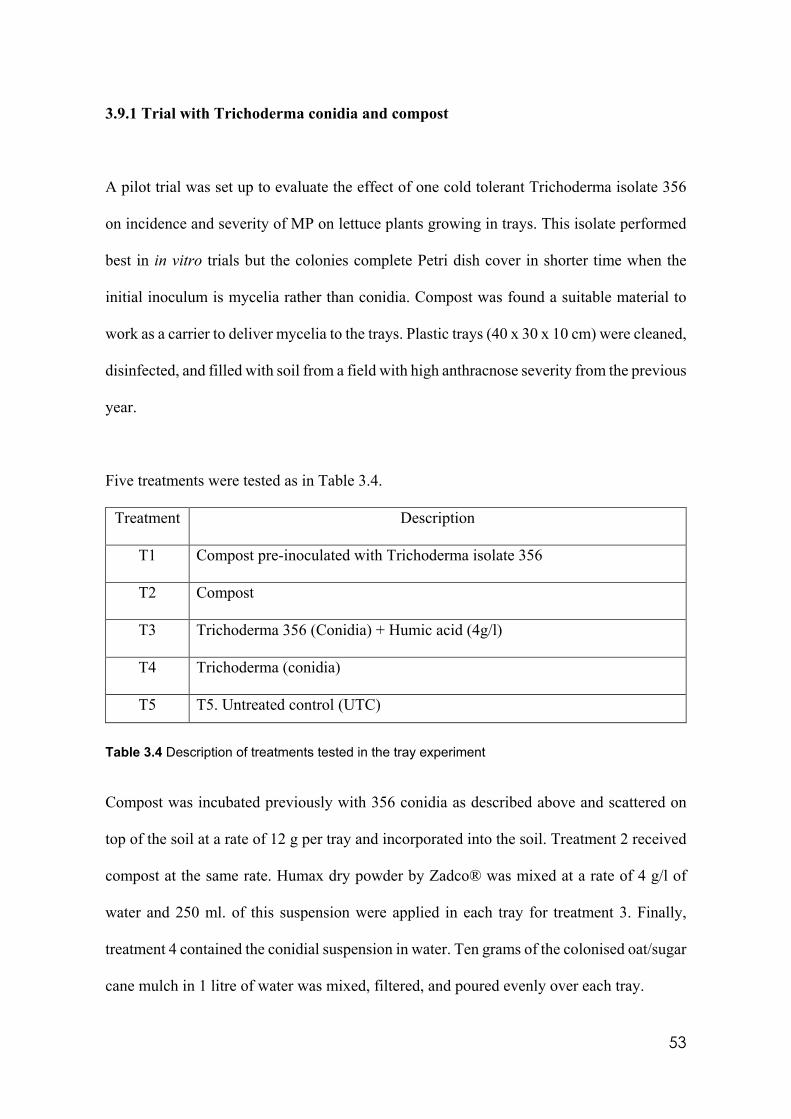
53
3.9.1 Trial with Trichoderma conidia and compost
A pilot trial was set up to evaluate the effect of one cold tolerant Trichoderma isolate 356
on incidence and severity of MP on lettuce plants growing in trays. This isolate performed
best in in vitro trials but the colonies complete Petri dish cover in shorter time when the
initial inoculum is mycelia rather than conidia. Compost was found a suitable material to
work as a carrier to deliver mycelia to the trays. Plastic trays (40 x 30 x 10 cm) were cleaned,
disinfected, and filled with soil from a field with high anthracnose severity from the previous
year.
Five treatments were tested as in Table 3.4.
Treatment Description
T1 Compost pre-inoculated with Trichoderma isolate 356
T2 Compost
T3 Trichoderma 356 (Conidia) + Humic acid (4g/l)
T4 Trichoderma (conidia)
T5 T5. Untreated control (UTC)
Table 3.4 Description of treatments tested in the tray experiment
Compost was incubated previously with 356 conidia as described above and scattered on
top of the soil at a rate of 12 g per tray and incorporated into the soil. Treatment 2 received
compost at the same rate. Humax dry powder by Zadco® was mixed at a rate of 4 g/l of
water and 250 ml. of this suspension were applied in each tray for treatment 3. Finally,
treatment 4 contained the conidial suspension in water. Ten grams of the colonised oat/sugar
cane mulch in 1 litre of water was mixed, filtered, and poured evenly over each tray.
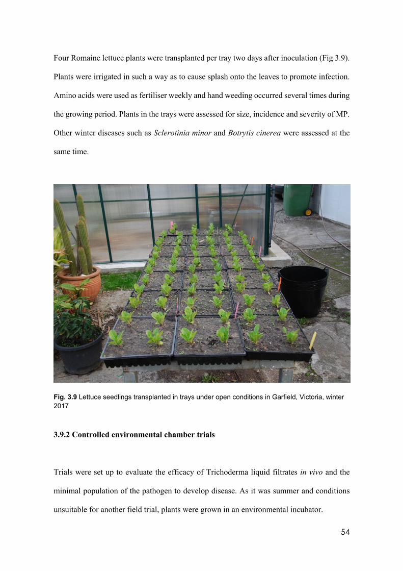
54
Four Romaine lettuce plants were transplanted per tray two days after inoculation (Fig 3.9).
Plants were irrigated in such a way as to cause splash onto the leaves to promote infection.
Amino acids were used as fertiliser weekly and hand weeding occurred several times during
the growing period. Plants in the trays were assessed for size, incidence and severity of MP.
Other winter diseases such as Sclerotinia minor and Botrytis cinerea were assessed at the
same time.
Fig. 3.9 Lettuce seedlings transplanted in trays under open conditions in Garfield, Victoria, winter 2017
3.9.2 Controlled environmental chamber trials
Trials were set up to evaluate the efficacy of Trichoderma liquid filtrates in vivo and the
minimal population of the pathogen to develop disease. As it was summer and conditions
unsuitable for another field trial, plants were grown in an environmental incubator.
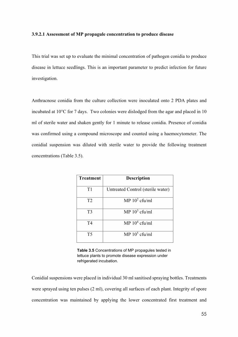
55
3.9.2.1 Assessment of MP propagule concentration to produce disease
This trial was set up to evaluate the minimal concentration of pathogen conidia to produce
disease in lettuce seedlings. This is an important parameter to predict infection for future
investigation.
Anthracnose conidia from the culture collection were inoculated onto 2 PDA plates and
incubated at 10°C for 7 days. Two colonies were dislodged from the agar and placed in 10
ml of sterile water and shaken gently for 1 minute to release conidia. Presence of conidia
was confirmed using a compound microscope and counted using a haemocytometer. The
conidial suspension was diluted with sterile water to provide the following treatment
concentrations (Table 3.5).
Treatment Description
T1 Untreated Control (sterile water)
T2 MP 102 cfu/ml
T3 MP 103 cfu/ml
T4 MP 104 cfu/ml
T5 MP 105 cfu/ml
Table 3.5 Concentrations of MP propagules tested in lettuce plants to promote disease expression under refrigerated incubation.
Conidial suspensions were placed in individual 30 ml sanitised spraying bottles. Treatments
were sprayed using ten pulses (2 ml), covering all surfaces of each plant. Integrity of spore
concentration was maintained by applying the lower concentrated first treatment and
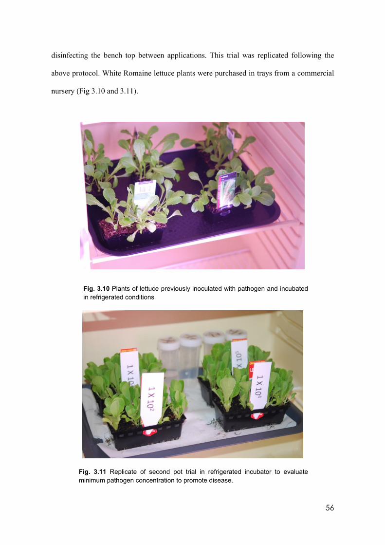
56
disinfecting the bench top between applications. This trial was replicated following the
above protocol. White Romaine lettuce plants were purchased in trays from a commercial
nursery (Fig 3.10 and 3.11).
Fig. 3.10 Plants of lettuce previously inoculated with pathogen and incubated in refrigerated conditions
Fig. 3.11 Replicate of second pot trial in refrigerated incubator to evaluate minimum pathogen concentration to promote disease.
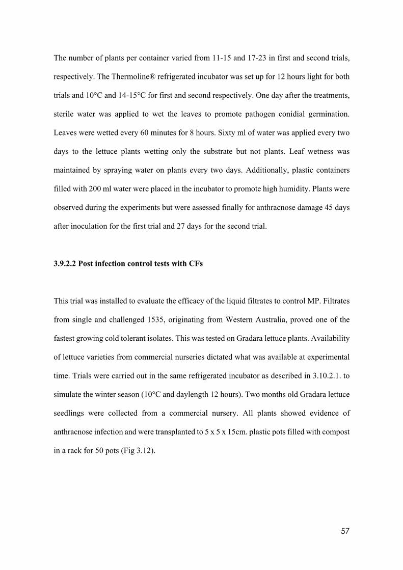
57
The number of plants per container varied from 11-15 and 17-23 in first and second trials,
respectively. The Thermoline® refrigerated incubator was set up for 12 hours light for both
trials and 10°C and 14-15°C for first and second respectively. One day after the treatments,
sterile water was applied to wet the leaves to promote pathogen conidial germination.
Leaves were wetted every 60 minutes for 8 hours. Sixty ml of water was applied every two
days to the lettuce plants wetting only the substrate but not plants. Leaf wetness was
maintained by spraying water on plants every two days. Additionally, plastic containers
filled with 200 ml water were placed in the incubator to promote high humidity. Plants were
observed during the experiments but were assessed finally for anthracnose damage 45 days
after inoculation for the first trial and 27 days for the second trial.
3.9.2.2 Post infection control tests with CFs
This trial was installed to evaluate the efficacy of the liquid filtrates to control MP. Filtrates
from single and challenged 1535, originating from Western Australia, proved one of the
fastest growing cold tolerant isolates. This was tested on Gradara lettuce plants. Availability
of lettuce varieties from commercial nurseries dictated what was available at experimental
time. Trials were carried out in the same refrigerated incubator as described in 3.10.2.1. to
simulate the winter season (10°C and daylength 12 hours). Two months old Gradara lettuce
seedlings were collected from a commercial nursery. All plants showed evidence of
anthracnose infection and were transplanted to 5 x 5 x 15cm. plastic pots filled with compost
in a rack for 50 pots (Fig 3.12).
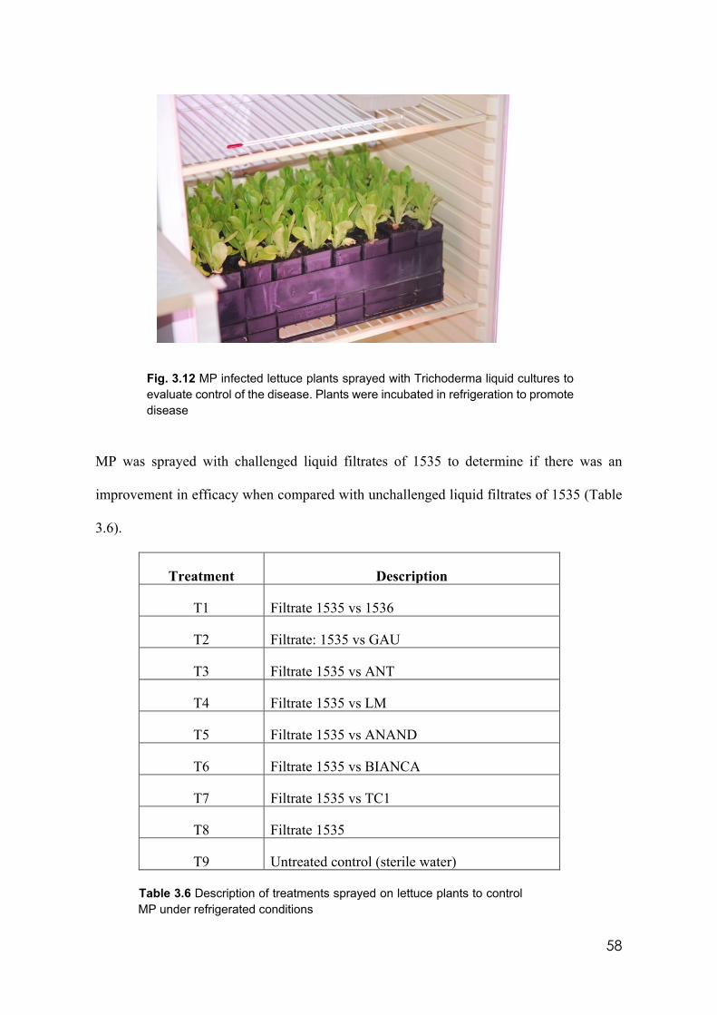
58
Fig. 3.12 MP infected lettuce plants sprayed with Trichoderma liquid cultures to evaluate control of the disease. Plants were incubated in refrigeration to promote disease
MP was sprayed with challenged liquid filtrates of 1535 to determine if there was an
improvement in efficacy when compared with unchallenged liquid filtrates of 1535 (Table
3.6).
Treatment Description
T1 Filtrate 1535 vs 1536
T2 Filtrate: 1535 vs GAU
T3 Filtrate 1535 vs ANT
T4 Filtrate 1535 vs LM
T5 Filtrate 1535 vs ANAND
T6 Filtrate 1535 vs BIANCA
T7 Filtrate 1535 vs TC1
T8 Filtrate 1535
T9 Untreated control (sterile water) Table 3.6 Description of treatments sprayed on lettuce plants to control MP under refrigerated conditions
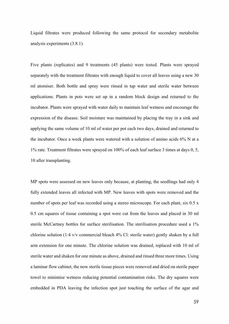
59
Liquid filtrates were produced following the same protocol for secondary metabolite
analysis experiments (3.8.1).
Five plants (replicates) and 9 treatments (45 plants) were tested. Plants were sprayed
separately with the treatment filtrates with enough liquid to cover all leaves using a new 30
ml atomiser. Both bottle and spray were rinsed in tap water and sterile water between
applications. Plants in pots were set up in a random block design and returned to the
incubator. Plants were sprayed with water daily to maintain leaf wetness and encourage the
expression of the disease. Soil moisture was maintained by placing the tray in a sink and
applying the same volume of 10 ml of water per pot each two days, drained and returned to
the incubator. Once a week plants were watered with a solution of amino acids 6% N at a
1% rate. Treatment filtrates were sprayed on 100% of each leaf surface 3 times at days 0, 5,
10 after transplanting.
MP spots were assessed on new leaves only because, at planting, the seedlings had only 4
fully extended leaves all infected with MP. New leaves with spots were removed and the
number of spots per leaf was recorded using a stereo microscope. For each plant, six 0.5 x
0.5 cm squares of tissue containing a spot were cut from the leaves and placed in 30 ml
sterile McCartney bottles for surface sterilisation. The sterilisation procedure used a 1%
chlorine solution (1:4 v/v commercial bleach 4% Cl: sterile water) gently shaken by a full
arm extension for one minute. The chlorine solution was drained, replaced with 10 ml of
sterile water and shaken for one minute as above, drained and rinsed three more times. Using
a laminar flow cabinet, the now sterile tissue pieces were removed and dried on sterile paper
towel to minimise wetness reducing potential contamination risks. The dry squares were
embedded in PDA leaving the infection spot just touching the surface of the agar and

60
incubated at 10° C for three weeks. MP colony formation was recorded.
3.9.3 Field Trial 1 – Experimental design and treatments
Eight cold tolerant isolates previously selected under in vitro tests (3.6) were tested in an
experimental plot within a commercial lettuce crop at Corrigan Produce Farms, Clyde North
(South East Melbourne) in winter.
Soil was ploughed using a disc plough incorporating residues of a previous conventional
(chemical fertilisers and pesticides used) celery crop. Two days later, a rotary plough and a
bed former completed the soil preparation. No chemical fertilisers, herbicides or fungicides
were applied to this plot at any stage of the research work. The experimental plot was
isolated using obvious exclusion signs to prevent any accidental spray applications. Physical
evidence of possible accidental spray application was identified by looking for tractor tracks
in the furrows. The experimental plot was planted in winter. The time of planting was
selected so that the most sensitive crop stage was aligned with the MP maximum virulence
period. Mid-sized Romaine lettuce seedlings of variety “Subie” by Nunhems® recognised
as a winter variety with exceptional vigour were obtained from a Berwick nursery, as a
scheduled farmer’s requirement. Plants were inspected carefully for any symptoms of MP
expression. A semi-automatic lettuce planter was used to plant the crop.
Eight Trichoderma cold tolerant isolates were tested using two inoculation methods: 1)
conidia sprayed on top the the soil surface, and 2) conidia pre-inoculated and incubated in
compost in Garfield, Victoria, and left to colonise for 7 days before being scattered on top
the soil surface. Bays of 6 beds of 6 x 1.8 m were orientated north to south and bordered by
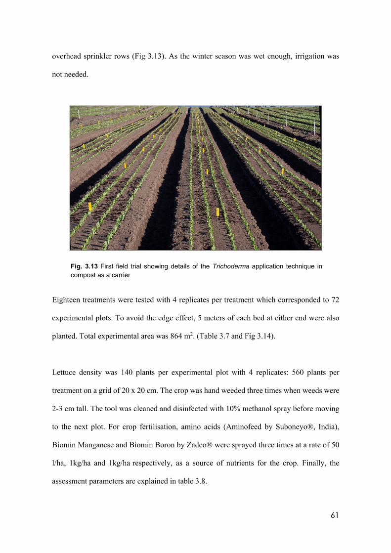
61
overhead sprinkler rows (Fig 3.13). As the winter season was wet enough, irrigation was
not needed.
Fig. 3.13 First field trial showing details of the Trichoderma application technique in compost as a carrier
Eighteen treatments were tested with 4 replicates per treatment which corresponded to 72
experimental plots. To avoid the edge effect, 5 meters of each bed at either end were also
planted. Total experimental area was 864 m2. (Table 3.7 and Fig 3.14).
Lettuce density was 140 plants per experimental plot with 4 replicates: 560 plants per
treatment on a grid of 20 x 20 cm. The crop was hand weeded three times when weeds were
2-3 cm tall. The tool was cleaned and disinfected with 10% methanol spray before moving
to the next plot. For crop fertilisation, amino acids (Aminofeed by Suboneyo®, India),
Biomin Manganese and Biomin Boron by Zadco® were sprayed three times at a rate of 50
l/ha, 1kg/ha and 1kg/ha respectively, as a source of nutrients for the crop. Finally, the
assessment parameters are explained in table 3.8.
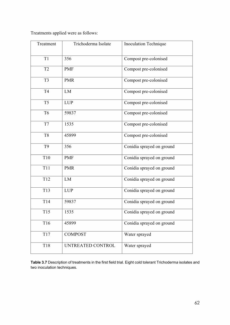
62
Treatments applied were as follows:
Treatment Trichoderma Isolate Inoculation Technique
T1 356 Compost pre-colonised
T2 PMF Compost pre-colonised
T3 PMR Compost pre-colonised
T4 LM Compost pre-colonised
T5 LUP Compost pre-colonised
T6 59837 Compost pre-colonised
T7 1535 Compost pre-colonised
T8 45899 Compost pre-colonised
T9 356 Conidia sprayed on ground
T10 PMF Conidia sprayed on ground
T11 PMR Conidia sprayed on ground
T12 LM Conidia sprayed on ground
T13 LUP Conidia sprayed on ground
T14 59837 Conidia sprayed on ground
T15 1535 Conidia sprayed on ground
T16 45899 Conidia sprayed on ground
T17 COMPOST Water sprayed
T18 UNTREATED CONTROL Water sprayed
Table 3.7 Description of treatments in the first field trial. Eight cold tolerant Trichoderma isolates and two inoculation techniques.
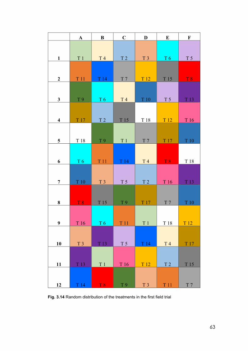
63
A B C D E F
1 T 1 T 4 T 2 T 3 T 6 T 5
2 T 11 T 14 T 7 T 12 T 15 T 8
3 T 9 T 6 T 4 T 10 T 5 T 13
4 T 17 T 2 T 15 T 18 T 12 T 16
5 T 18 T 9 T 1 T 7 T 17 T 10
6 T 6 T 11 T 14 T 4 T 8 T 18
7 T 10 T 3 T 5 T 2 T 16 T 13
8 T 8 T 15 T 9 T 17 T 7 T 10
9 T 16 T 6 T 11 T 1 T 18 T 12
10 T 3 T 13 T 5 T 14 T 4 T 17
11 T 13 T 1 T 16 T 12 T 2 T 15
12 T 14 T 8 T 9 T 3 T 11 T 7
Fig. 3.14 Random distribution of the treatments in the first field trial
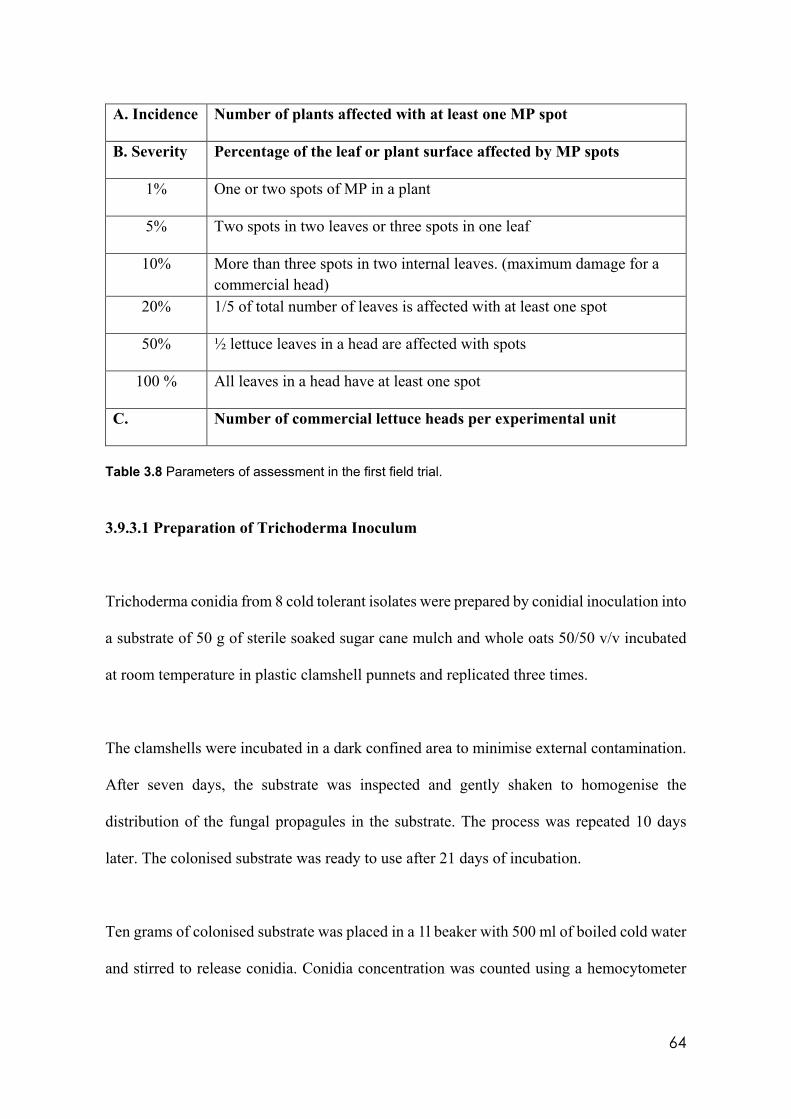
64
A. Incidence Number of plants affected with at least one MP spot
B. Severity Percentage of the leaf or plant surface affected by MP spots
1% One or two spots of MP in a plant
5% Two spots in two leaves or three spots in one leaf
10% More than three spots in two internal leaves. (maximum damage for a commercial head)
20% 1/5 of total number of leaves is affected with at least one spot
50% ½ lettuce leaves in a head are affected with spots
100 % All leaves in a head have at least one spot
C. Number of commercial lettuce heads per experimental unit
Table 3.8 Parameters of assessment in the first field trial.
3.9.3.1 Preparation of Trichoderma Inoculum
Trichoderma conidia from 8 cold tolerant isolates were prepared by conidial inoculation into
a substrate of 50 g of sterile soaked sugar cane mulch and whole oats 50/50 v/v incubated
at room temperature in plastic clamshell punnets and replicated three times.
The clamshells were incubated in a dark confined area to minimise external contamination.
After seven days, the substrate was inspected and gently shaken to homogenise the
distribution of the fungal propagules in the substrate. The process was repeated 10 days
later. The colonised substrate was ready to use after 21 days of incubation.
Ten grams of colonised substrate was placed in a 1l beaker with 500 ml of boiled cold water
and stirred to release conidia. Conidia concentration was counted using a hemocytometer
65
and adjusted to 1 x 10⁵ cfu/ml. Two hundred ml of conidial suspension was inoculated into
15 l of mature compost. After three days, the compost was inspected and evaluated for
mycelial formation, then watered, turned and stored under cover for 7 more days at 2-14°C.
Compost, as a suitable carrier for the Trichoderma propagation structures, was assessed
against the standard protocol of freeze-dried conidial formulation sprayed directly onto crop
and soil. Compost treatments were spread at a rate of 0.5 kg/m² one day after the lettuce
crop was planted.
3.9.3.2 Assessment of MP distribution in experimental plot
MP incidence and severity in field is governed by the inoculum density in the soil.
Symptoms of the disease appear in isolated areas in the plots that expand to infest more
plants following the wind direction and rain splash. It is important to know the areas with
high propagule concentration to correlate these with incidence and severity assessment.
Ten samples of soil from the field trial regardless of the treatment were taken from areas
with low, medium, and high disease incidence and severity to assess the minimum pathogen
population that causes disease. From each soil sample a 1/10 dilution was prepared, and
solids allowed to settle for one minute before a serial dilution was performed to 1/1000.
From these 10-2 and 10-3 suspensions, 200 µl were inoculated onto duplicate fresh PDA and
spread using sterile plastic hockey sticks and incubated at 8°C to promote MP colony
formation. PDA plates were assessed from day 7 and then daily until MP colonies developed
after which cfu/g of soil were calculated.
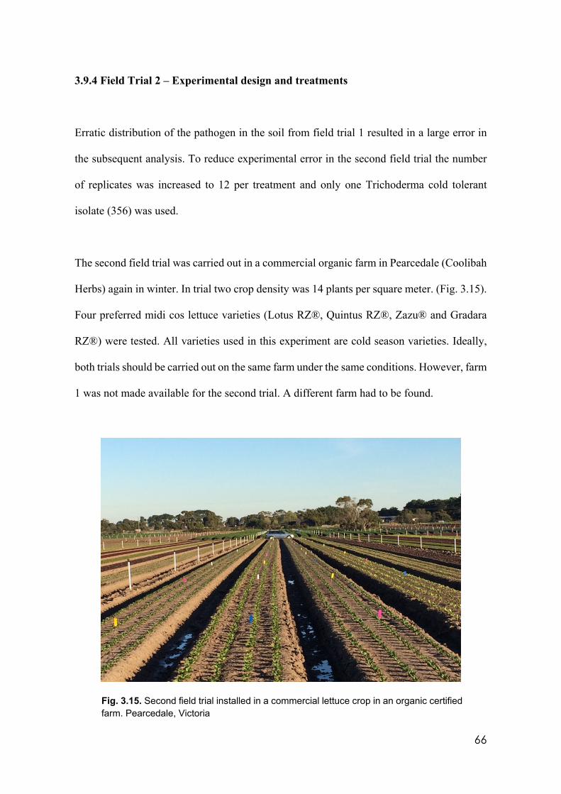
66
3.9.4 Field Trial 2 – Experimental design and treatments
Erratic distribution of the pathogen in the soil from field trial 1 resulted in a large error in
the subsequent analysis. To reduce experimental error in the second field trial the number
of replicates was increased to 12 per treatment and only one Trichoderma cold tolerant
isolate (356) was used.
The second field trial was carried out in a commercial organic farm in Pearcedale (Coolibah
Herbs) again in winter. In trial two crop density was 14 plants per square meter. (Fig. 3.15).
Four preferred midi cos lettuce varieties (Lotus RZ®, Quintus RZ®, Zazu® and Gradara
RZ®) were tested. All varieties used in this experiment are cold season varieties. Ideally,
both trials should be carried out on the same farm under the same conditions. However, farm
1 was not made available for the second trial. A different farm had to be found.
Fig. 3.15. Second field trial installed in a commercial lettuce crop in an organic certified farm. Pearcedale, Victoria
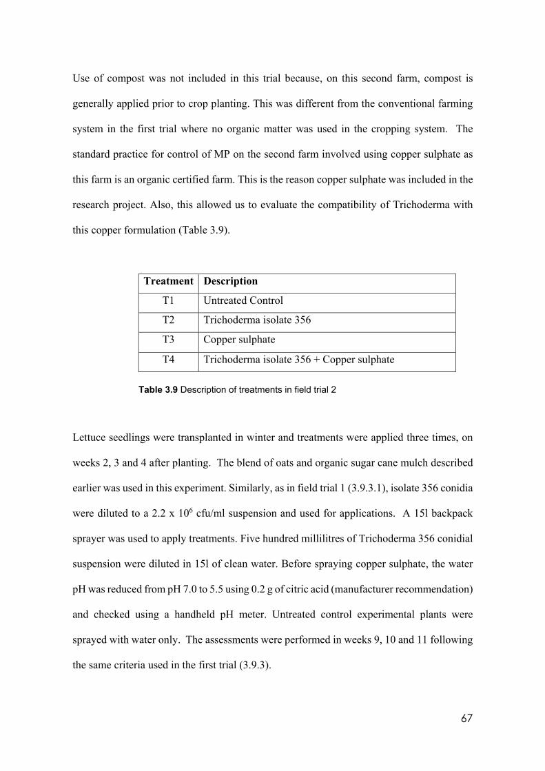
67
Use of compost was not included in this trial because, on this second farm, compost is
generally applied prior to crop planting. This was different from the conventional farming
system in the first trial where no organic matter was used in the cropping system. The
standard practice for control of MP on the second farm involved using copper sulphate as
this farm is an organic certified farm. This is the reason copper sulphate was included in the
research project. Also, this allowed us to evaluate the compatibility of Trichoderma with
this copper formulation (Table 3.9).
Treatment Description
T1 Untreated Control
T2 Trichoderma isolate 356
T3 Copper sulphate
T4 Trichoderma isolate 356 + Copper sulphate Table 3.9 Description of treatments in field trial 2
Lettuce seedlings were transplanted in winter and treatments were applied three times, on
weeks 2, 3 and 4 after planting. The blend of oats and organic sugar cane mulch described
earlier was used in this experiment. Similarly, as in field trial 1 (3.9.3.1), isolate 356 conidia
were diluted to a 2.2 x 10⁶ cfu/ml suspension and used for applications. A 15l backpack
sprayer was used to apply treatments. Five hundred millilitres of Trichoderma 356 conidial
suspension were diluted in 15l of clean water. Before spraying copper sulphate, the water
pH was reduced from pH 7.0 to 5.5 using 0.2 g of citric acid (manufacturer recommendation)
and checked using a handheld pH meter. Untreated control experimental plants were
sprayed with water only. The assessments were performed in weeks 9, 10 and 11 following
the same criteria used in the first trial (3.9.3).

68
Similarly, as in the first trial, samples of soil were collected to quantify the population of
the pathogen and its distribution in the field. Five sub-samples of approximately 2 cm3 each
of soil were collected per experimental plot at 1 cm depth (10 cm3). Four random samples
were taken per treatment (16 samples) following the protocol as in 3.9.3.2.
69
Chapter 4
RESULTS
4.1 Introduction
This chapter presents a logical sequence of data and information from all the experiments.
Starting from the classical morphological characteristics, basic information was gathered
about how colonies from different isolates behave under different temperature, pH and
media conditions. Molecular identification tests add critical data for the identification of the
isolates and location in a phylogenetic tree. Vegetative compatibility/incompatibility trials
contributed to the morphological and molecular findings to provide a critical link to the
metabolomic assessments. Results from in vitro, pots and field experiments, supported the
ability of Trichoderma to control MP.
4.2 Effects on Trichoderma morphological characteristics
Large differences in morphological characteristics were found between Trichoderma
isolates, at different incubation temperatures, culture media, and media pH.
4.2.1 Temperature
In the experiment described above (3.3), temperature was the major variable that affected
Trichoderma activity. Isolates with ability to grow in all experimental temperatures (356.
1535, 71556, CAROL, 59837, LM and LUP) showed significantly faster (p<0.05) growth
at 25°C and 18°C (Appendices 1-7). Growth was slowed by (47%) at 18°C. (Fig. 4.1).
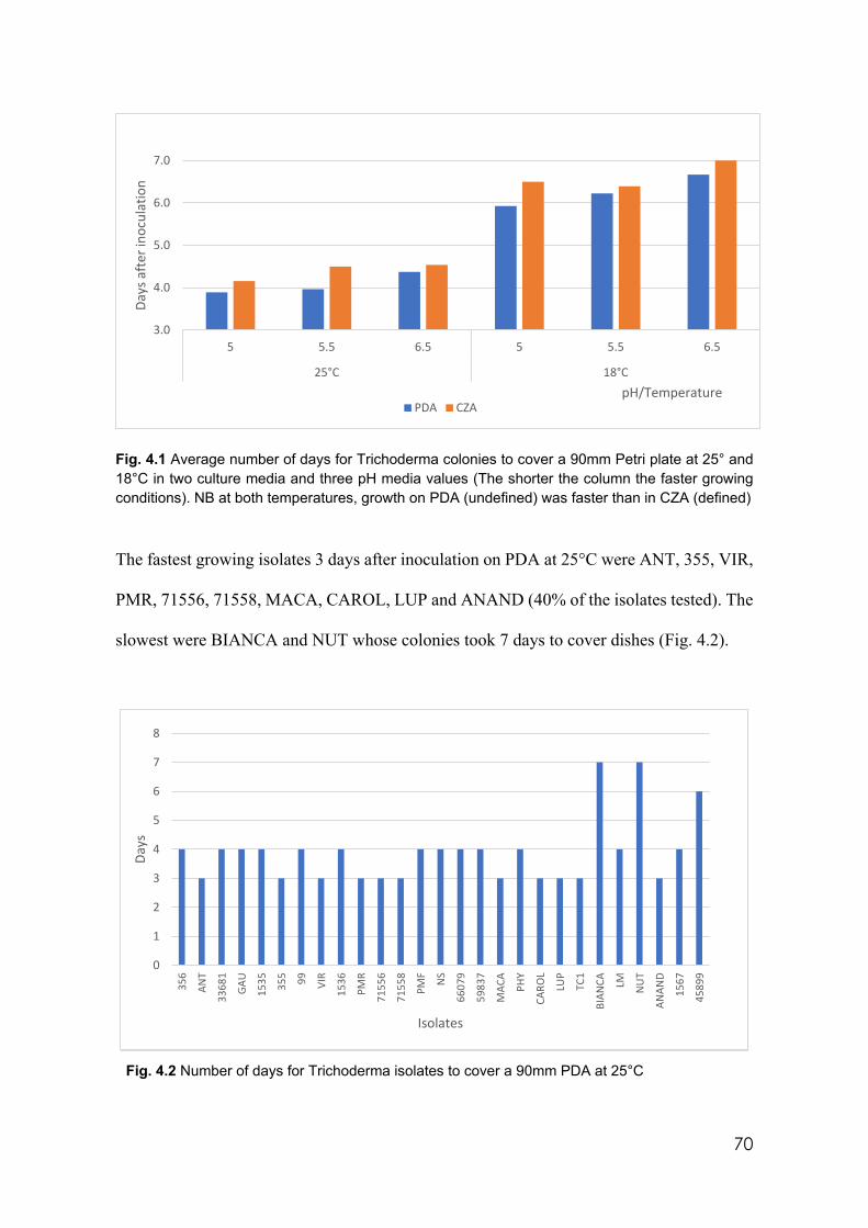
70
Fig. 4.1 Average number of days for Trichoderma colonies to cover a 90mm Petri plate at 25° and 18°C in two culture media and three pH media values (The shorter the column the faster growing conditions). NB at both temperatures, growth on PDA (undefined) was faster than in CZA (defined)
The fastest growing isolates 3 days after inoculation on PDA at 25°C were ANT, 355, VIR,
PMR, 71556, 71558, MACA, CAROL, LUP and ANAND (40% of the isolates tested). The
slowest were BIANCA and NUT whose colonies took 7 days to cover dishes (Fig. 4.2).
Fig. 4.2 Number of days for Trichoderma isolates to cover a 90mm PDA at 25°C
3.0
4.0
5.0
6.0
7.0
5 5.5 6.5 5 5.5 6.5
25°C 18°C
Days
aft
er in
ocul
atio
n
pH/TemperaturePDA CZA
0
1
2
3
4
5
6
7
8
356
ANT
3368
1
GAU
1535 35
5 99 VIR
1536
PMR
7155
6
7155
8
PMF
NS
6607
9
5983
7
MAC
A
PHY
CARO
L
LUP
TC1
BIAN
CA LM NU
T
ANAN
D
1567
4589
9
Days
Isolates
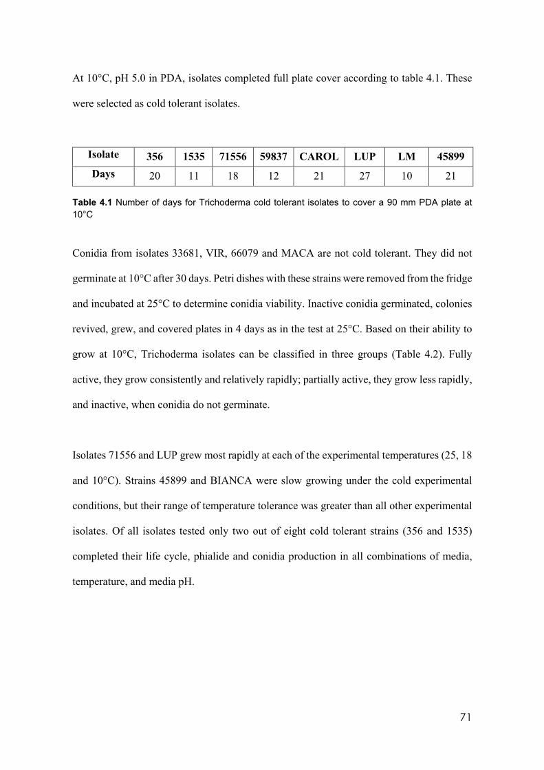
71
At 10°C, pH 5.0 in PDA, isolates completed full plate cover according to table 4.1. These
were selected as cold tolerant isolates.
Isolate 356 1535 71556 59837 CAROL LUP LM 45899 Days 20 11 18 12 21 27 10 21
Table 4.1 Number of days for Trichoderma cold tolerant isolates to cover a 90 mm PDA plate at 10°C
Conidia from isolates 33681, VIR, 66079 and MACA are not cold tolerant. They did not
germinate at 10°C after 30 days. Petri dishes with these strains were removed from the fridge
and incubated at 25°C to determine conidia viability. Inactive conidia germinated, colonies
revived, grew, and covered plates in 4 days as in the test at 25°C. Based on their ability to
grow at 10°C, Trichoderma isolates can be classified in three groups (Table 4.2). Fully
active, they grow consistently and relatively rapidly; partially active, they grow less rapidly,
and inactive, when conidia do not germinate.
Isolates 71556 and LUP grew most rapidly at each of the experimental temperatures (25, 18
and 10°C). Strains 45899 and BIANCA were slow growing under the cold experimental
conditions, but their range of temperature tolerance was greater than all other experimental
isolates. Of all isolates tested only two out of eight cold tolerant strains (356 and 1535)
completed their life cycle, phialide and conidia production in all combinations of media,
temperature, and media pH.
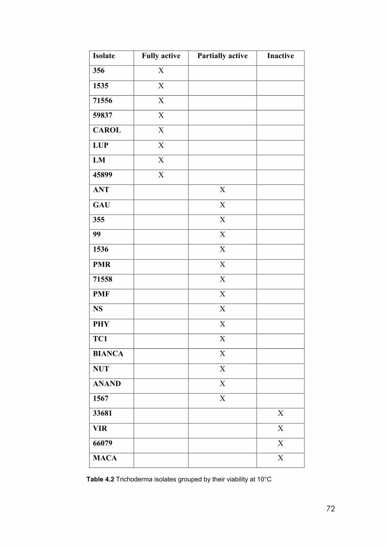
72
Isolate Fully active Partially active Inactive
356 X
1535 X
71556 X
59837 X
CAROL X
LUP X
LM X
45899 X
ANT X
GAU X
355 X
99 X
1536 X
PMR X
71558 X
PMF X
NS X
PHY X
TC1 X
BIANCA X
NUT X
ANAND X
1567 X
33681 X
VIR X
66079 X
MACA X
Table 4.2 Trichoderma isolates grouped by their viability at 10°C
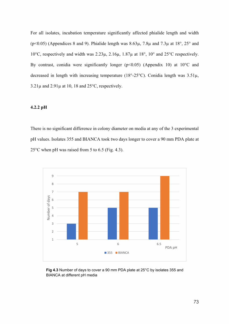
73
For all isolates, incubation temperature significantly affected phialide length and width
(p<0.05) (Appendices 8 and 9). Phialide length was 8.63µ, 7.8µ and 7.3µ at 18°, 25° and
10°C, respectively and width was 2.23µ, 2.16µ, 1.87µ at 18°, 10° and 25°C respectively.
By contrast, conidia were significantly longer (p<0.05) (Appendix 10) at 10°C and
decreased in length with increasing temperature (18°-25°C). Conidia length was 3.51µ,
3.21µ and 2.91µ at 10, 18 and 25°C, respectively.
4.2.2 pH
There is no significant difference in colony diameter on media at any of the 3 experimental
pH values. Isolates 355 and BIANCA took two days longer to cover a 90 mm PDA plate at
25°C when pH was raised from 5 to 6.5 (Fig. 4.3).
Fig 4.3 Number of days to cover a 90 mm PDA plate at 25°C by isolates 355 and BIANCA at different pH media
1
2
3
4
5
6
7
8
9
5 6 6.5
Num
ber o
f day
s
PDA pH355 BIANCA
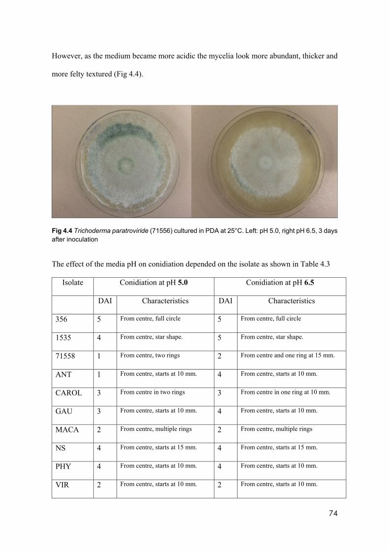
74
However, as the medium became more acidic the mycelia look more abundant, thicker and
more felty textured (Fig 4.4).
Fig 4.4 Trichoderma paratroviride (71556) cultured in PDA at 25°C. Left: pH 5.0, right pH 6.5, 3 days after inoculation
The effect of the media pH on conidiation depended on the isolate as shown in Table 4.3
Isolate Conidiation at pH 5.0 Conidiation at pH 6.5
DAI Characteristics DAI Characteristics
356 5 From centre, full circle 5 From centre, full circle
1535 4 From centre, star shape. 5 From centre, star shape.
71558 1 From centre, two rings 2 From centre and one ring at 15 mm.
ANT 1 From centre, starts at 10 mm. 4 From centre, starts at 10 mm.
CAROL 3 From centre in two rings 3 From centre in one ring at 10 mm.
GAU 3 From centre, starts at 10 mm. 4 From centre, starts at 10 mm.
MACA 2 From centre, multiple rings 2 From centre, multiple rings
NS 4 From centre, starts at 15 mm. 4 From centre, starts at 15 mm.
PHY 4 From centre, starts at 10 mm. 4 From centre, starts at 10 mm.
VIR 2 From centre, starts at 10 mm. 2 From centre, starts at 10 mm.
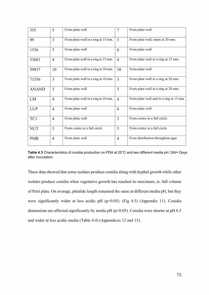
75
355 5 From plate wall 7 From plate wall
99 3 From plate wall in a ring at 15 mm. 3 From plate wall, starts at 20 mm.
1536 5 From plate wall 6 From plate wall
33681 4 From plate wall in a ring at 15 mm. 4 From plate wall in a ring at 15 mm.
59837 10 From plate wall in a ring at 10 mm. 10 From plate wall
71556 3 From plate wall in a ring at 10 mm. 3 From plate wall in a ring at 20 mm.
ANAND 3 From plate wall 3 From plate wall in a ring at 20 mm.
LM 4 From plate wall in a ring at 10 mm. 4 From plate wall and in a ring at 15 mm
LUP 4 From plate wall 6 From plate wall
TC1 4 From plate wall 3 From centre in a full circle.
NUT 5 From centre in a full circle 5 From centre in a full circle
PMR 4 From plate wall 4 Even distribution throughout agar.
Table 4.3 Characteristics of conidia production on PDA at 25°C and two different media pH. DAI= Days after inoculation.
These data showed that some isolates produce conidia along with hyphal growth while other
isolates produce conidia when vegetative growth has reached its maximum, ie. full volume
of Petri plate. On average, phialide length remained the same at different media pH, but they
were significantly wider at less acidic pH (p<0.05). (Fig 4.5) (Appendix 11). Conidia
dimensions are affected significantly by media pH (p<0.05). Conidia were shorter at pH 6.5
and wider at less acidic media (Table 4.4) (Appendices 12 and 13).
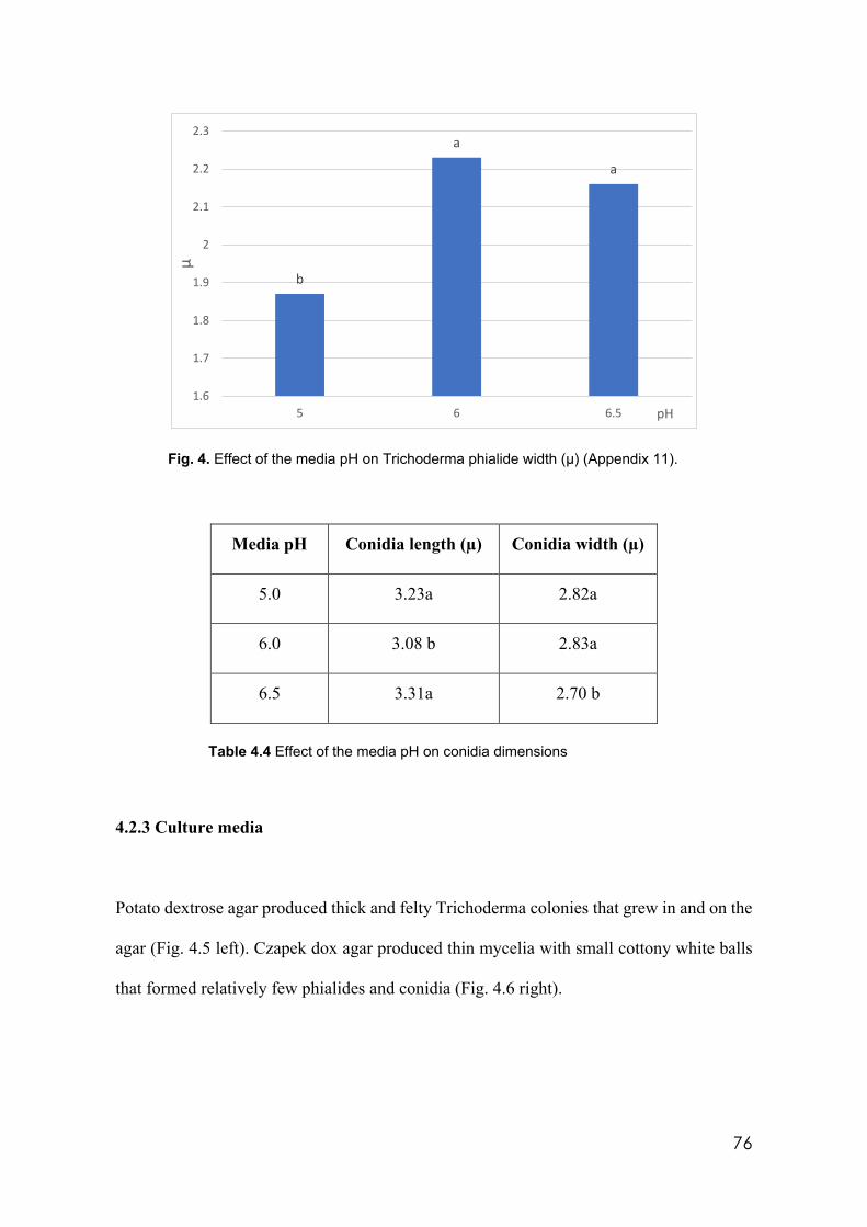
76
Fig. 4. Effect of the media pH on Trichoderma phialide width (µ) (Appendix 11).
Media pH Conidia length (µ) Conidia width (µ)
5.0 3.23a 2.82a
6.0 3.08 b 2.83a
6.5 3.31a 2.70 b
Table 4.4 Effect of the media pH on conidia dimensions
4.2.3 Culture media
Potato dextrose agar produced thick and felty Trichoderma colonies that grew in and on the
agar (Fig. 4.5 left). Czapek dox agar produced thin mycelia with small cottony white balls
that formed relatively few phialides and conidia (Fig. 4.6 right).
b
a
a
1.6
1.7
1.8
1.9
2
2.1
2.2
2.3
5 6 6.5
µ
pH
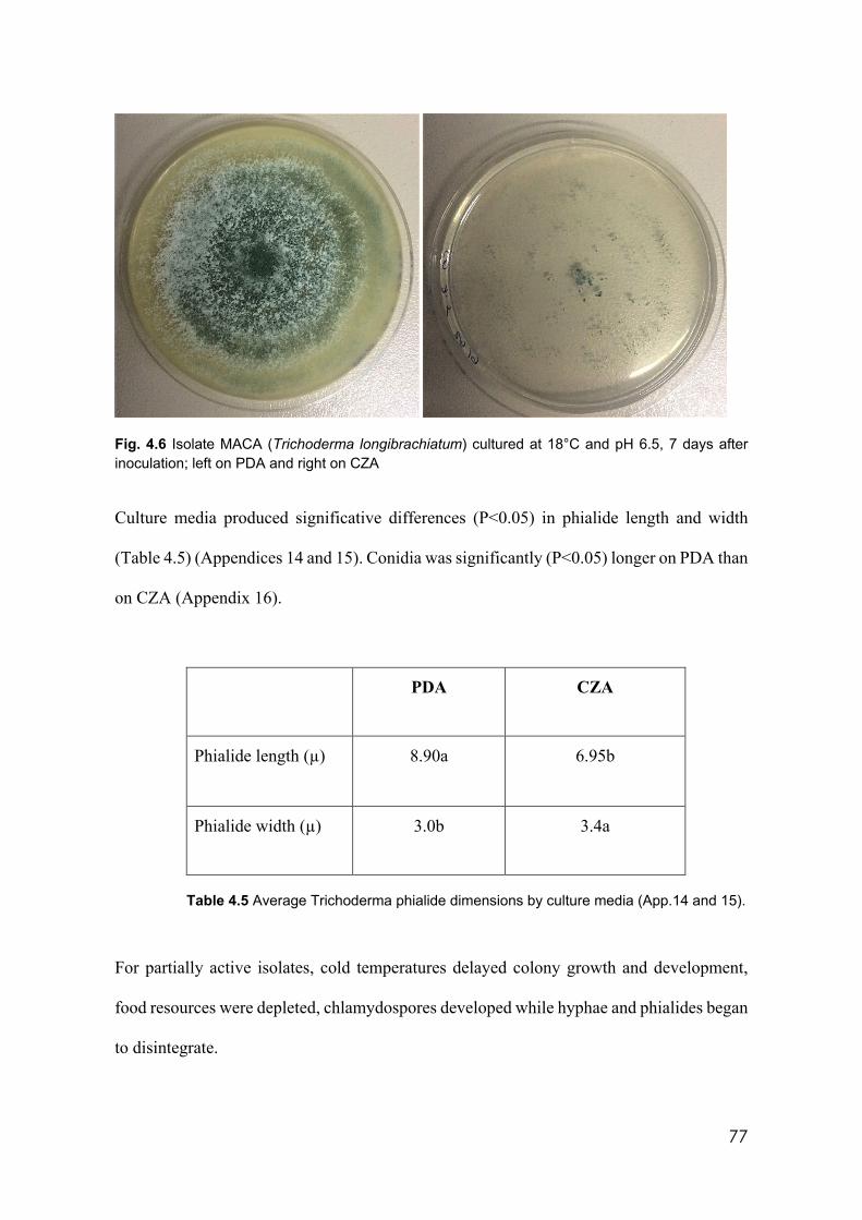
77
Fig. 4.6 Isolate MACA (Trichoderma longibrachiatum) cultured at 18°C and pH 6.5, 7 days after inoculation; left on PDA and right on CZA
Culture media produced significative differences (P˂0.05) in phialide length and width
(Table 4.5) (Appendices 14 and 15). Conidia was significantly (P˂0.05) longer on PDA than
on CZA (Appendix 16).
PDA CZA
Phialide length (µ) 8.90a 6.95b
Phialide width (µ) 3.0b 3.4a
Table 4.5 Average Trichoderma phialide dimensions by culture media (App.14 and 15).
For partially active isolates, cold temperatures delayed colony growth and development,
food resources were depleted, chlamydospores developed while hyphae and phialides began
to disintegrate.
78
4.2.4 Pigmentation and aroma
Isolates CAROL (Trichoderma sp.), VIR and 99 (T. harzianum), MACA (T.
longibachiatum) and ANAND (Trichoderma spirale) produced yellow pigmentation that
stained the PDA at all temperatures at which they grew. This pigmentation was not produced
by these isolates on CZA. Similarly, the most documented Trichoderma secondary volatile
metabolite, 6PP, developed only when selected isolates were cultured on PDA. Coconut
aroma attributed to this metabolite was identified in 11 out of 27 isolates, T. viride (1535)
T. neokoningii (355), T. composticola (356), T. hamatum (1536), T. paratroviride (71556),
Trichoderma sp. (LUP), T. asperelloides (NS), (T. atroviride) PMR, PMF, NUT and LM
when cultured on PDA but not on CZA. This aroma was also present at 10°C.
4.3 Molecular identification and classification of Trichoderma isolates
After following the protocol described by Jaklitsch and Voglmayr (2015), the isolates were
identified as in table 4.6.
After processing sequence data, only 4 isolates produced satisfactorily aligned sequences
with the three primers used (tef, rpb2 and acl1). Phylogenetic multigene analysis determined
59837 (T. viride), PMF (T. atroviride), 66079 (T. aureoviride) and LM (T. atroviride)
(Fig.4.7).
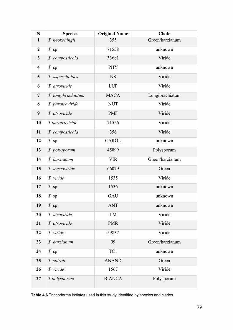
79
N Species Original Name Clade 1 T. neokoningii 355 Green/harzianum
2 T. sp 71558 unknown
3 T. composticola 33681 Viride
4 T. sp PHY unknown
5 T. asperelloides NS Viride
6 T. atroviride LUP Viride
7 T. longibrachiatum MACA Longibrachiatum
8 T. paratroviride NUT Viride
9 T. atroviride PMF Viride
10 T paratroviride 71556 Viride
11 T. composticola 356 Viride
12 T. sp CAROL unknown
13 T. polysporum 45899 Polysporum
14 T. harzianum VIR Green/harzianum
15 T. aureoviride 66079 Green
16 T. viride 1535 Viride
17 T. sp 1536 unknown
18 T. sp GAU unknown
19 T. sp ANT unknown
20 T. atroviride LM Viride
21 T. atroviride PMR Viride
22 T. viride 59837 Viride
23 T. harzianum 99 Green/harzianum
24 T. sp TC1 unknown
25 T. spirale ANAND Green
26 T. viride 1567 Viride
27 T.polysporum BIANCA Polysporum
Table 4.6 Trichoderma isolates used in this study identified by species and clades.
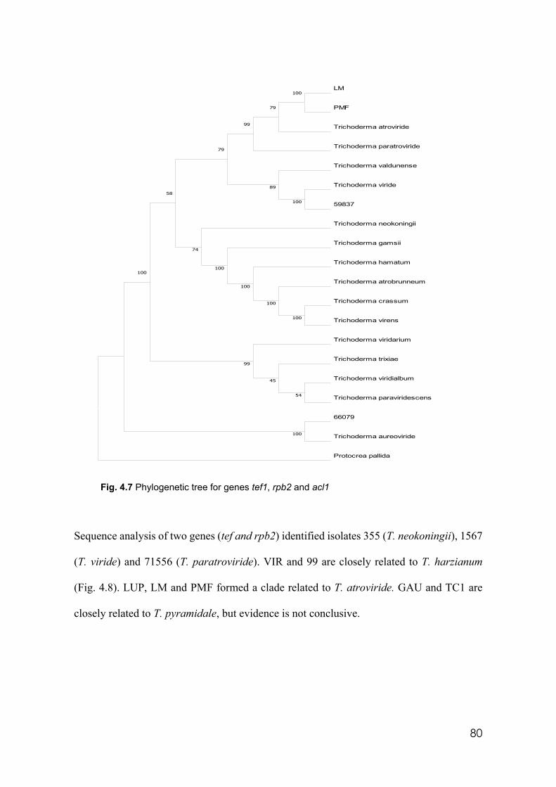
80
Fig. 4.7 Phylogenetic tree for genes tef1, rpb2 and acl1
Sequence analysis of two genes (tef and rpb2) identified isolates 355 (T. neokoningii), 1567
(T. viride) and 71556 (T. paratroviride). VIR and 99 are closely related to T. harzianum
(Fig. 4.8). LUP, LM and PMF formed a clade related to T. atroviride. GAU and TC1 are
closely related to T. pyramidale, but evidence is not conclusive.
LM
PMF
Trichoderma atroviride
Trichoderma paratroviride
Trichoderma valdunense
Trichoderma viride
59837
Trichoderma neokoningii
Trichoderma gamsii
Trichoderma hamatum
Trichoderma atrobrunneum
Trichoderma crassum
Trichoderma virens
Trichoderma viridarium
Trichoderma trixiae
Trichoderma viridialbum
Trichoderma paraviridescens
66079
Trichoderma aureoviride
Protocrea pallida
100
100
100
100
100
100
100
100
79
99
89
74
79
58
99
54
45
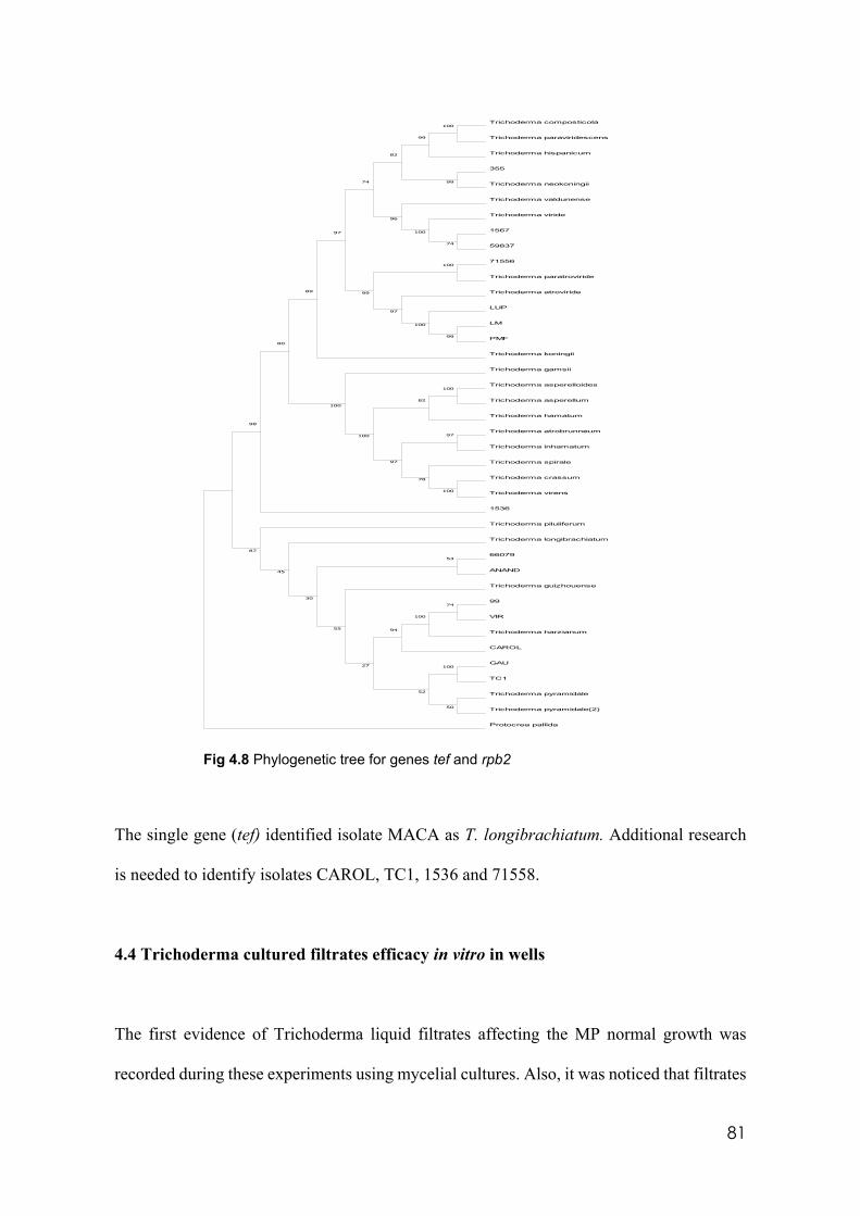
81
Fig 4.8 Phylogenetic tree for genes tef and rpb2
The single gene (tef) identified isolate MACA as T. longibrachiatum. Additional research
is needed to identify isolates CAROL, TC1, 1536 and 71558.
4.4 Trichoderma cultured filtrates efficacy in vitro in wells
The first evidence of Trichoderma liquid filtrates affecting the MP normal growth was
recorded during these experiments using mycelial cultures. Also, it was noticed that filtrates
Trichoderma composticola
Trichoderma paraviridescens
Trichoderma hispanicum
355
Trichoderma neokoningii
Trichoderma valdunense
Trichoderma viride
1567
59837
71556
Trichoderma paratroviride
Trichoderma atroviride
LUP
LM
PMF
Trichoderma koningii
Trichoderma gamsii
Trichoderma asperelloides
Trichoderma asperellum
Trichoderma hamatum
Trichoderma atrobrunneum
Trichoderma inhamatum
Trichoderma spirale
Trichoderma crassum
Trichoderma virens
1536
Trichoderma piluliferum
Trichoderma longibrachiatum
66079
ANAND
Trichoderma guizhouense
99
VIR
Trichoderma harzianum
CAROL
GAU
TC1
Trichoderma pyramidale
Trichoderma pyramidale(2)
Protocrea pallida
100
78
82
100
97
97100
100
50
74
100
100
9455
27
52
30
53
45
87
80
98
74
100
100
99
100
97
89 99
99
100
99
96
82
97
74
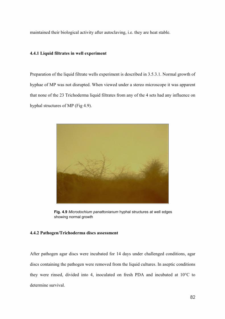
82
maintained their biological activity after autoclaving, i.e. they are heat stable.
4.4.1 Liquid filtrates in well experiment
Preparation of the liquid filtrate wells experiment is described in 3.5.3.1. Normal growth of
hyphae of MP was not disrupted. When viewed under a stereo microscope it was apparent
that none of the 23 Trichoderma liquid filtrates from any of the 4 sets had any influence on
hyphal structures of MP (Fig 4.9).
Fig. 4.9 Microdochium panattonianum hyphal structures at well edges showing normal growth
4.4.2 Pathogen/Trichoderma discs assessment
After pathogen agar discs were incubated for 14 days under challenged conditions, agar
discs containing the pathogen were removed from the liquid cultures. In aseptic conditions
they were rinsed, divided into 4, inoculated on fresh PDA and incubated at 10°C to
determine survival.
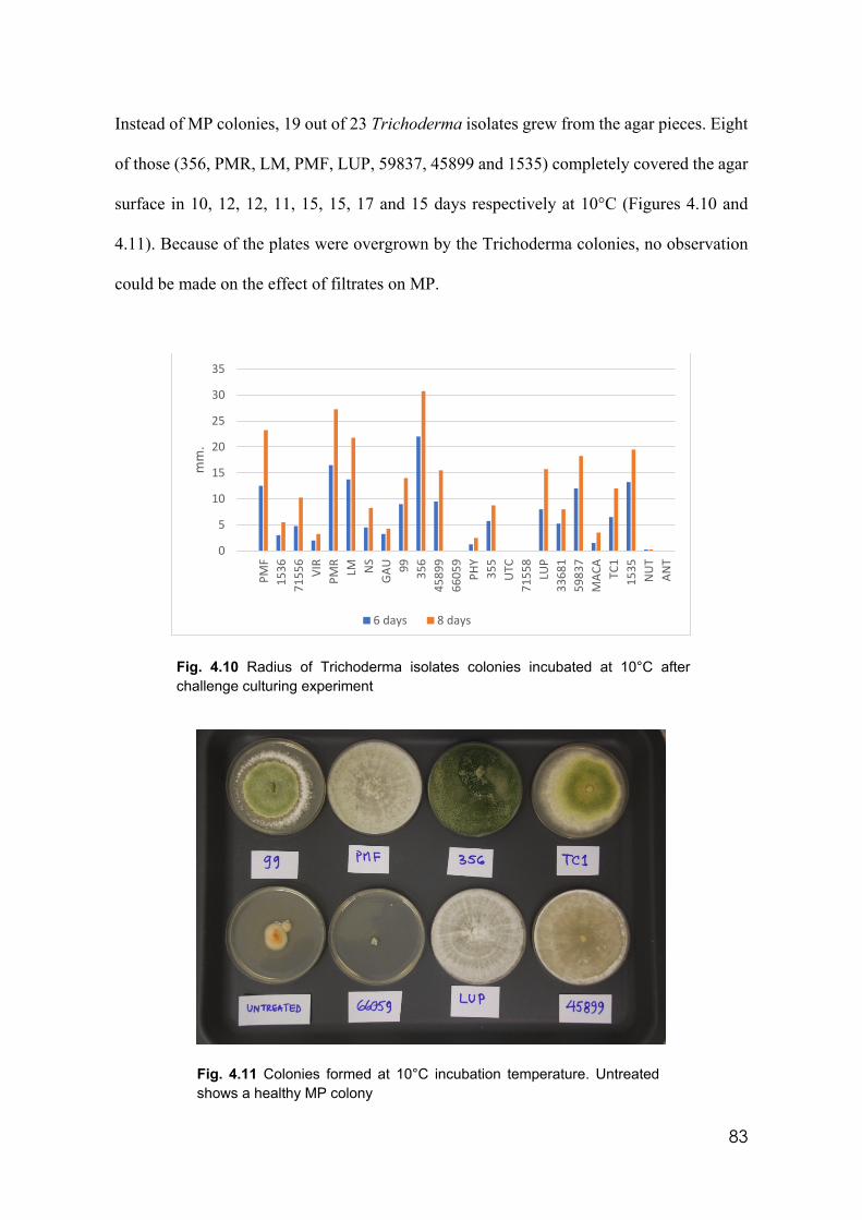
83
Instead of MP colonies, 19 out of 23 Trichoderma isolates grew from the agar pieces. Eight
of those (356, PMR, LM, PMF, LUP, 59837, 45899 and 1535) completely covered the agar
surface in 10, 12, 12, 11, 15, 15, 17 and 15 days respectively at 10°C (Figures 4.10 and
4.11). Because of the plates were overgrown by the Trichoderma colonies, no observation
could be made on the effect of filtrates on MP.
Fig. 4.10 Radius of Trichoderma isolates colonies incubated at 10°C after challenge culturing experiment
Fig. 4.11 Colonies formed at 10°C incubation temperature. Untreated shows a healthy MP colony
0
5
10
15
20
25
30
35
PMF
1536
7155
6VI
RPM
RLM N
SG
AU 99 356
4589
966
059
PHY
355
UTC
7155
8LU
P33
681
5983
7M
ACA
TC1
1535
NU
TAN
T
mm
.
6 days 8 days
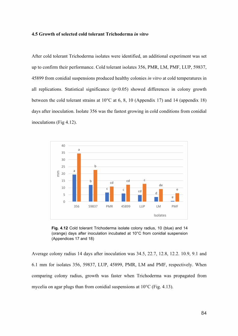
84
4.5 Growth of selected cold tolerant Trichoderma in vitro
After cold tolerant Trichoderma isolates were identified, an additional experiment was set
up to confirm their performance. Cold tolerant isolates 356, PMR, LM, PMF, LUP, 59837,
45899 from conidial suspensions produced healthy colonies in vitro at cold temperatures in
all replications. Statistical significance (p<0.05) showed differences in colony growth
between the cold tolerant strains at 10°C at 6, 8, 10 (Appendix 17) and 14 (appendix 18)
days after inoculation. Isolate 356 was the fastest growing in cold conditions from conidial
inoculations (Fig 4.12).
Fig. 4.12 Cold tolerant Trichoderma isolate colony radius, 10 (blue) and 14 (orange) days after inoculation incubated at 10°C from conidial suspension (Appendices 17 and 18)
Average colony radius 14 days after inoculation was 34.5, 22.7, 12.8, 12.2. 10.9, 9.1 and
6.1 mm for isolates 356, 59837, LUP, 45899, PMR, LM and PMF, respectively. When
comparing colony radius, growth was faster when Trichoderma was propagated from
mycelia on agar plugs than from conidial suspensions at 10°C (Fig. 4.13).
a
b
c c cd de
a
b
cd cd cde
e
0
5
10
15
20
25
30
35
40
356 59837 PMR 45899 LUP LM PMF
mm
Isolates
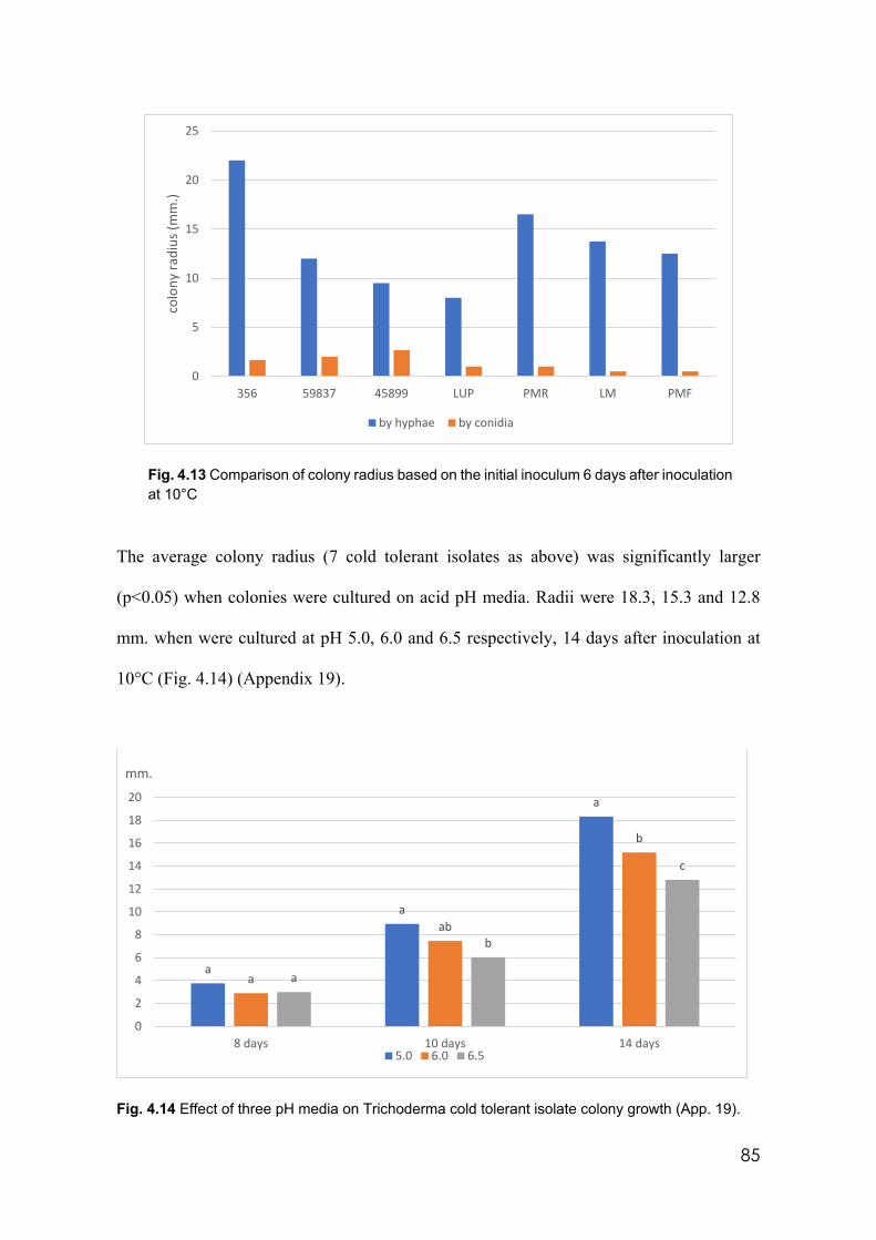
85
Fig. 4.13 Comparison of colony radius based on the initial inoculum 6 days after inoculation at 10°C
The average colony radius (7 cold tolerant isolates as above) was significantly larger
(p<0.05) when colonies were cultured on acid pH media. Radii were 18.3, 15.3 and 12.8
mm. when were cultured at pH 5.0, 6.0 and 6.5 respectively, 14 days after inoculation at
10°C (Fig. 4.14) (Appendix 19).
Fig. 4.14 Effect of three pH media on Trichoderma cold tolerant isolate colony growth (App. 19).
0
5
10
15
20
25
356 59837 45899 LUP PMR LM PMF
colo
ny ra
dius
(mm
.)
by hyphae by conidia
a
a
a
a
ab
b
a
b
c
0
2
4
6
8
10
12
14
16
18
20
8 days 10 days 14 days
mm.
5.0 6.0 6.5
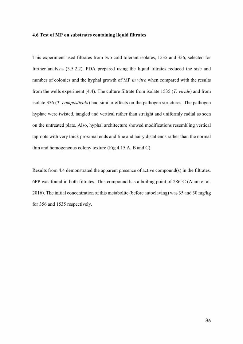
86
4.6 Test of MP on substrates containing liquid filtrates
This experiment used filtrates from two cold tolerant isolates, 1535 and 356, selected for
further analysis (3.5.2.2). PDA prepared using the liquid filtrates reduced the size and
number of colonies and the hyphal growth of MP in vitro when compared with the results
from the wells experiment (4.4). The culture filtrate from isolate 1535 (T. viride) and from
isolate 356 (T. composticola) had similar effects on the pathogen structures. The pathogen
hyphae were twisted, tangled and vertical rather than straight and uniformly radial as seen
on the untreated plate. Also, hyphal architecture showed modifications resembling vertical
taproots with very thick proximal ends and fine and hairy distal ends rather than the normal
thin and homogeneous colony texture (Fig 4.15 A, B and C).
Results from 4.4 demonstrated the apparent presence of active compound(s) in the filtrates.
6PP was found in both filtrates. This compound has a boiling point of 286°C (Alam et al.
2016). The initial concentration of this metabolite (before autoclaving) was 35 and 30 mg/kg
for 356 and 1535 respectively.
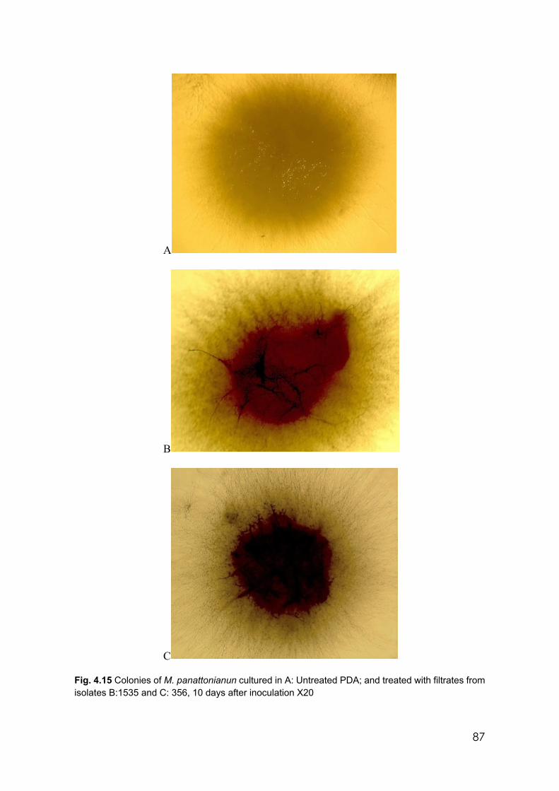
87
A
B
C Fig. 4.15 Colonies of M. panattonianun cultured in A: Untreated PDA; and treated with filtrates from isolates B:1535 and C: 356, 10 days after inoculation X20
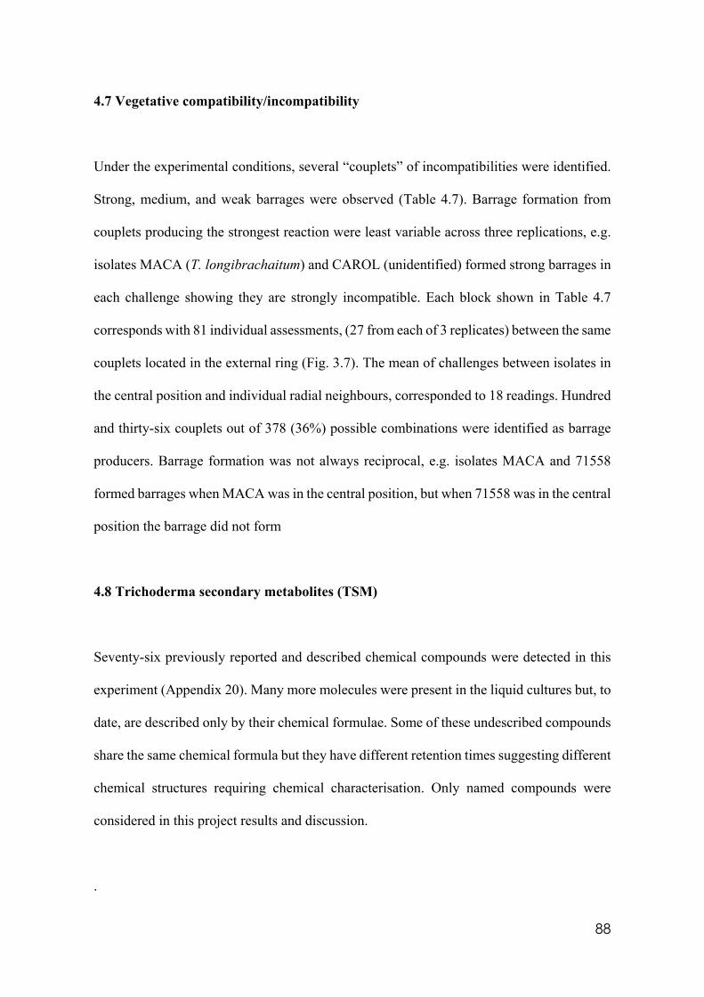
88
4.7 Vegetative compatibility/incompatibility
Under the experimental conditions, several “couplets” of incompatibilities were identified.
Strong, medium, and weak barrages were observed (Table 4.7). Barrage formation from
couplets producing the strongest reaction were least variable across three replications, e.g.
isolates MACA (T. longibrachaitum) and CAROL (unidentified) formed strong barrages in
each challenge showing they are strongly incompatible. Each block shown in Table 4.7
corresponds with 81 individual assessments, (27 from each of 3 replicates) between the same
couplets located in the external ring (Fig. 3.7). The mean of challenges between isolates in
the central position and individual radial neighbours, corresponded to 18 readings. Hundred
and thirty-six couplets out of 378 (36%) possible combinations were identified as barrage
producers. Barrage formation was not always reciprocal, e.g. isolates MACA and 71558
formed barrages when MACA was in the central position, but when 71558 was in the central
position the barrage did not form
4.8 Trichoderma secondary metabolites (TSM)
Seventy-six previously reported and described chemical compounds were detected in this
experiment (Appendix 20). Many more molecules were present in the liquid cultures but, to
date, are described only by their chemical formulae. Some of these undescribed compounds
share the same chemical formula but they have different retention times suggesting different
chemical structures requiring chemical characterisation. Only named compounds were
considered in this project results and discussion.
.
89
355 71558 33681 PHY NS LUP MACA NUT PMF 71556 356 CAROL 45899 VIR 66079 1535 1536 GAU ANT LM PMR 59837 99 TC1 ANAND 1567 BIANCA
355 X X X X X X X X
71558 X X X X X X X X
33681 X X X X X X X X X X
PHY X X X X X X X X X X X X X X X
NS X X X X X X X X X X
LUP X X X
MACA X X X X X X X X X X X X X X
NUT X X X X X X X X X X X
PMF X X X X X X X
71556 X X X X X X X X X X X X
356 X X X X X X X X
CAROL X X X X X X X X X
45899 X X X X X X X X X X X X X X X X X
VIR X X X X X X X
66079 X X X X X X
1535 X X X X X X X X X X X X X X X X
1536 X X X X X X X X X
GAU X X X X X X X X X X X X X X X X X
ANT X X X X X X X X X X X X X X X
LM X X X X X X X X X
PMR X X X X X X X X X X X X
59837 X X X X X X
99 X X X X X X X X X X
TC1 X X X X X X X X X
ANAND X X X X X X
1567 X X X X X
BIANCA X X X X X X X X X X X
Table 4.7 Trichoderma isolates showing incompatibility from barrage formation. Red blocks denote strongest incompatibility; X denotes medium incompatibility and empty blocks denote compatibility.
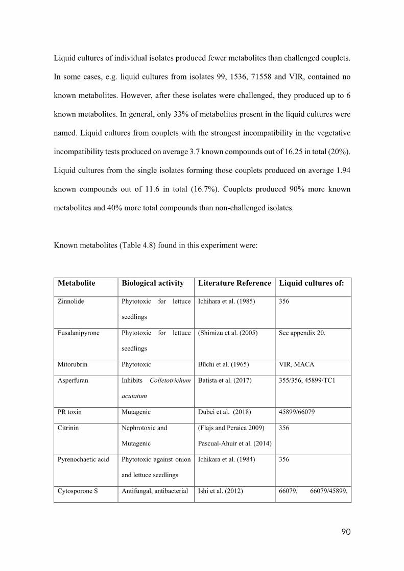
90
Liquid cultures of individual isolates produced fewer metabolites than challenged couplets.
In some cases, e.g. liquid cultures from isolates 99, 1536, 71558 and VIR, contained no
known metabolites. However, after these isolates were challenged, they produced up to 6
known metabolites. In general, only 33% of metabolites present in the liquid cultures were
named. Liquid cultures from couplets with the strongest incompatibility in the vegetative
incompatibility tests produced on average 3.7 known compounds out of 16.25 in total (20%).
Liquid cultures from the single isolates forming those couplets produced on average 1.94
known compounds out of 11.6 in total (16.7%). Couplets produced 90% more known
metabolites and 40% more total compounds than non-challenged isolates.
Known metabolites (Table 4.8) found in this experiment were:
Metabolite Biological activity Literature Reference Liquid cultures of:
Zinnolide Phytotoxic for lettuce
seedlings
Ichihara et al. (1985) 356
Fusalanipyrone Phytotoxic for lettuce
seedlings
(Shimizu et al. (2005) See appendix 20.
Mitorubrin Phytotoxic Büchi et al. (1965) VIR, MACA
Asperfuran Inhibits Colletotrichum
acutatum
Batista et al. (2017) 355/356, 45899/TC1
PR toxin Mutagenic Dubei et al. (2018) 45899/66079
Citrinin Nephrotoxic and
Mutagenic
(Flajs and Peraica 2009)
Pascual-Ahuir et al. (2014)
356
Pyrenochaetic acid Phytotoxic against onion
and lettuce seedlings
Ichikara et al. (1984) 356
Cytosporone S Antifungal, antibacterial Ishi et al. (2012) 66079, 66079/45899,
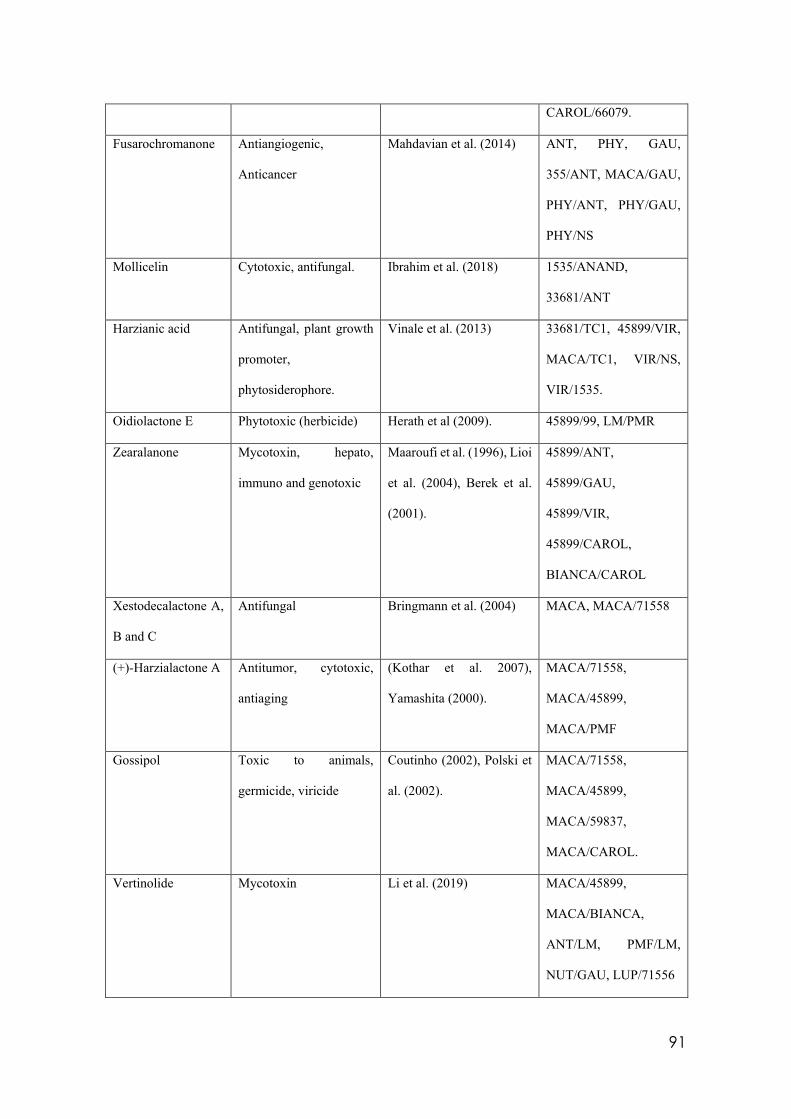
91
CAROL/66079.
Fusarochromanone Antiangiogenic,
Anticancer
Mahdavian et al. (2014) ANT, PHY, GAU,
355/ANT, MACA/GAU,
PHY/ANT, PHY/GAU,
PHY/NS
Mollicelin Cytotoxic, antifungal. Ibrahim et al. (2018) 1535/ANAND,
33681/ANT
Harzianic acid Antifungal, plant growth
promoter,
phytosiderophore.
Vinale et al. (2013) 33681/TC1, 45899/VIR,
MACA/TC1, VIR/NS,
VIR/1535.
Oidiolactone E Phytotoxic (herbicide) Herath et al (2009). 45899/99, LM/PMR
Zearalanone Mycotoxin, hepato,
immuno and genotoxic
Maaroufi et al. (1996), Lioi
et al. (2004), Berek et al.
(2001).
45899/ANT,
45899/GAU,
45899/VIR,
45899/CAROL,
BIANCA/CAROL
Xestodecalactone A,
B and C
Antifungal Bringmann et al. (2004) MACA, MACA/71558
(+)-Harzialactone A Antitumor, cytotoxic,
antiaging
(Kothar et al. 2007),
Yamashita (2000).
MACA/71558,
MACA/45899,
MACA/PMF
Gossipol Toxic to animals,
germicide, viricide
Coutinho (2002), Polski et
al. (2002).
MACA/71558,
MACA/45899,
MACA/59837,
MACA/CAROL.
Vertinolide Mycotoxin Li et al. (2019) MACA/45899,
MACA/BIANCA,
ANT/LM, PMF/LM,
NUT/GAU, LUP/71556
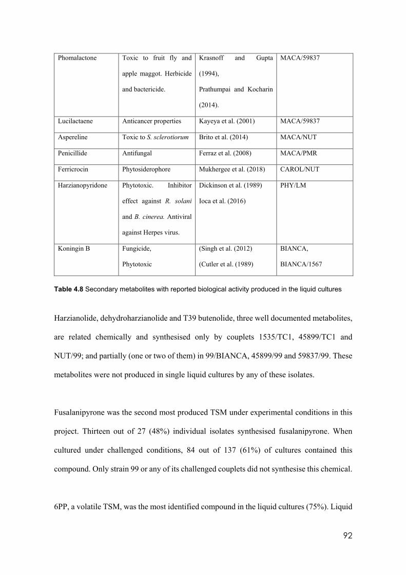
92
Phomalactone Toxic to fruit fly and
apple maggot. Herbicide
and bactericide.
Krasnoff and Gupta
(1994),
Prathumpai and Kocharin
(2014).
MACA/59837
Lucilactaene Anticancer properties Kayeya et al. (2001) MACA/59837
Aspereline Toxic to S. sclerotiorum Brito et al. (2014) MACA/NUT
Penicillide Antifungal Ferraz et al. (2008) MACA/PMR
Ferricrocin Phytosiderophore Mukhergee et al. (2018) CAROL/NUT
Harzianopyridone Phytotoxic. Inhibitor
effect against R. solani
and B. cinerea. Antiviral
against Herpes virus.
Dickinson et al. (1989)
Ioca et al. (2016)
PHY/LM
Koningin B Fungicide,
Phytotoxic
(Singh et al. (2012)
(Cutler et al. (1989)
BIANCA,
BIANCA/1567
Table 4.8 Secondary metabolites with reported biological activity produced in the liquid cultures
Harzianolide, dehydroharzianolide and T39 butenolide, three well documented metabolites,
are related chemically and synthesised only by couplets 1535/TC1, 45899/TC1 and
NUT/99; and partially (one or two of them) in 99/BIANCA, 45899/99 and 59837/99. These
metabolites were not produced in single liquid cultures by any of these isolates.
Fusalanipyrone was the second most produced TSM under experimental conditions in this
project. Thirteen out of 27 (48%) individual isolates synthesised fusalanipyrone. When
cultured under challenged conditions, 84 out of 137 (61%) of cultures contained this
compound. Only strain 99 or any of its challenged couplets did not synthesise this chemical.
6PP, a volatile TSM, was the most identified compound in the liquid cultures (75%). Liquid
93
cultures produced by isolates 355 and 356, 71556, GAU, LM, LUP, NS and PMF and all
their combinations contained 6PP. However, the highest concentrations (>100 mg/l) were
found in cultures produced by PMR, PMF, LM LUP and 71556. Isolate 1535 and all its
couplets except for 1535/TC1 also produced this metabolite (Fig 4.16). Many sets of PDA
plates were cultured to assess several variables in vitro. “Coconut” smell associated with
the presence of 6PP was present in many isolates.
Consistent results were achieved from organoleptic assessments performed three times with
isolates cultured on PDA and incubated at 25°C. These results were consistent with those
produced by the chromatography method (Table 4.9).
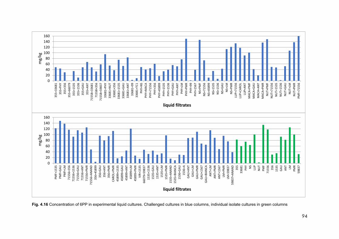
94
Fig. 4.16 Concentration of 6PP in experimental liquid cultures. Challenged cultures in blue columns, individual isolate cultures in green columns
020406080
100120140160
355+
3368
135
5+PH
Y35
5+35
635
5+66
079
355+
1535
355+
1536
355+
GAU
355+
ANT
7155
8+33
681
7155
8+35
671
558+
5983
733
681+
PHY
3368
1+N
UT
3368
1+35
633
681+
1535
3368
1+GA
U33
681+
ANT
3368
1+99
3368
1+TC
1PH
Y+N
SPH
Y+M
ACA
PHY+
7155
6PH
Y+35
6PH
Y+45
899
PHY+
1535
PHY+
1536
PHY+
GAU
PHY+
ANT
PHY+
LMPH
Y+PM
RPH
Y+99
PHY+
1567
NS+
PMF
NS+
7155
6N
S+45
899
NS+
1535
NS+
1536
NS+
GAU
NS+
LMN
S+PM
RLU
P+71
556
LUP+
CARO
LLU
P+AN
TM
ACA+
PMF
MAC
A+GA
UM
ACA+
ANT
MAC
A+PM
RN
UT+
PMF
NU
T+71
556
NU
T+15
35N
UT+
1536
NU
T+GA
UN
UT+
LMN
UT+
PMR
PMF+
7155
6
mg/
kg
liquid filtrates
020406080
100120140160
PMF+
1535
PMF+
GAU
PMF+
LM71
556+
356
7155
6+15
3671
556+
GAU
7155
6+AN
T71
556+
PMR
7155
6+AN
AND
356+
4589
935
6+GA
U35
6+AN
T35
6+PM
RCA
ROL+
PMR
4589
9+15
3545
899+
GAU
4589
9+AN
T45
899+
LM45
899+
PMR
VIR+
1535
6607
9+59
837
1535
+153
615
35+G
AU15
35+A
NT
1535
+LM
1535
+PM
R15
35+A
NAN
D15
35+B
IAN
CA15
36+G
AU15
36+A
GAU
+AN
TGA
U+L
MGA
U+P
MR
GAU
+156
7GA
U+B
IAN
CAAN
T+LM
ANT+
PMR
ANT+
1567
LM+P
MR
LM+5
9837
5983
7+AN
AND
355
3368
1PH
Y NS
LUP
NU
TPM
F71
556
356
1535
GAU
ANT
LMPM
R59
837
mg/
kg
liquid filtrates
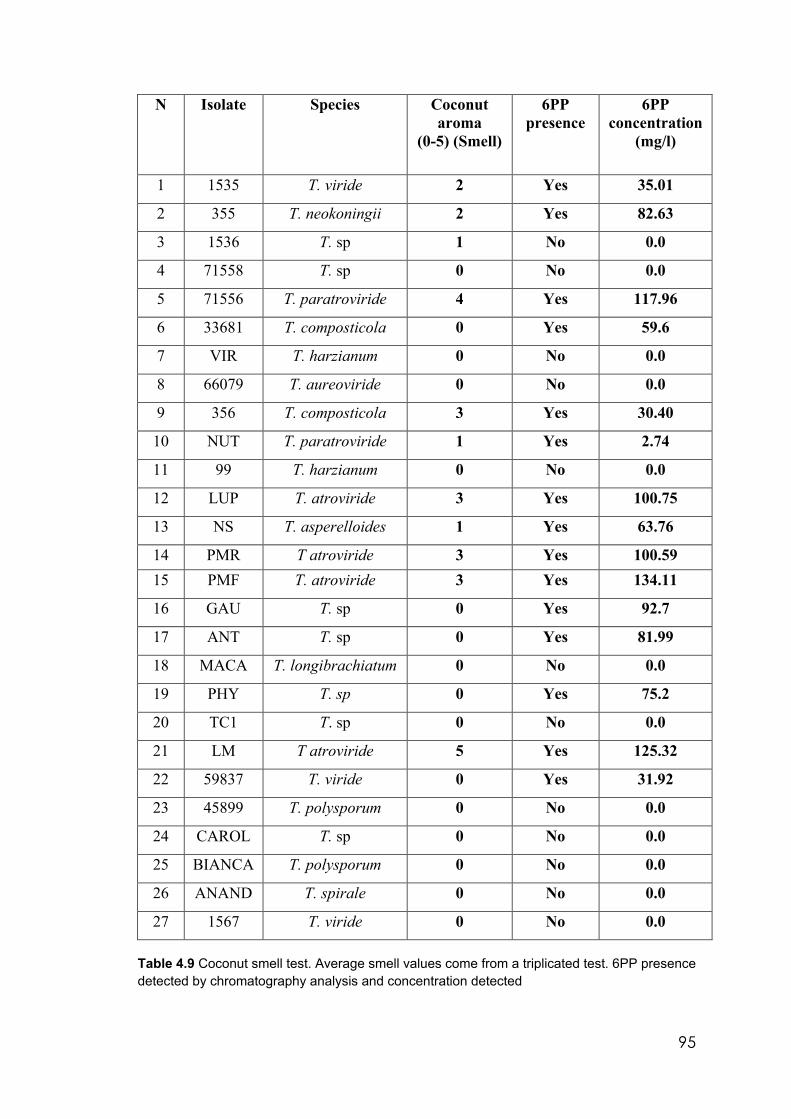
95
N Isolate Species Coconut aroma
(0-5) (Smell)
6PP presence
6PP concentration
(mg/l)
1 1535 T. viride 2 Yes 35.01
2 355 T. neokoningii 2 Yes 82.63
3 1536 T. sp 1 No 0.0
4 71558 T. sp 0 No 0.0
5 71556 T. paratroviride 4 Yes 117.96
6 33681 T. composticola 0 Yes 59.6
7 VIR T. harzianum 0 No 0.0
8 66079 T. aureoviride 0 No 0.0
9 356 T. composticola 3 Yes 30.40
10 NUT T. paratroviride 1 Yes 2.74
11 99 T. harzianum 0 No 0.0
12 LUP T. atroviride 3 Yes 100.75
13 NS T. asperelloides 1 Yes 63.76
14 PMR T atroviride 3 Yes 100.59 15 PMF T. atroviride 3 Yes 134.11
16 GAU T. sp 0 Yes 92.7
17 ANT T. sp 0 Yes 81.99
18 MACA T. longibrachiatum 0 No 0.0
19 PHY T. sp 0 Yes 75.2
20 TC1 T. sp 0 No 0.0
21 LM T atroviride 5 Yes 125.32
22 59837 T. viride 0 Yes 31.92
23 45899 T. polysporum 0 No 0.0
24 CAROL T. sp 0 No 0.0
25 BIANCA T. polysporum 0 No 0.0
26 ANAND T. spirale 0 No 0.0
27 1567 T. viride 0 No 0.0
Table 4.9 Coconut smell test. Average smell values come from a triplicated test. 6PP presence detected by chromatography analysis and concentration detected
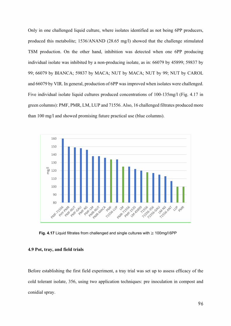
96
Only in one challenged liquid culture, where isolates identified as not being 6PP producers,
produced this metabolite; 1536/ANAND (28.65 mg/l) showed that the challenge stimulated
TSM production. On the other hand, inhibition was detected when one 6PP producing
individual isolate was inhibited by a non-producing isolate, as in: 66079 by 45899; 59837 by
99; 66079 by BIANCA; 59837 by MACA; NUT by MACA; NUT by 99; NUT by CAROL
and 66079 by VIR. In general, production of 6PP was improved when isolates were challenged.
Five individual isolate liquid cultures produced concentrations of 100-135mg/l (Fig. 4.17 in
green columns): PMF, PMR, LM, LUP and 71556. Also, 16 challenged filtrates produced more
than 100 mg/l and showed promising future practical use (blue columns).
Fig. 4.17 Liquid filtrates from challenged and single cultures with ≥ 100mg/l 6PP
4.9 Pot, tray, and field trials
Before establishing the first field experiment, a tray trial was set up to assess efficacy of the
cold tolerant isolate, 356, using two application techniques: pre inoculation in compost and
conidial spray.
80
90
100
110
120
130
140
150
160
mg/
l
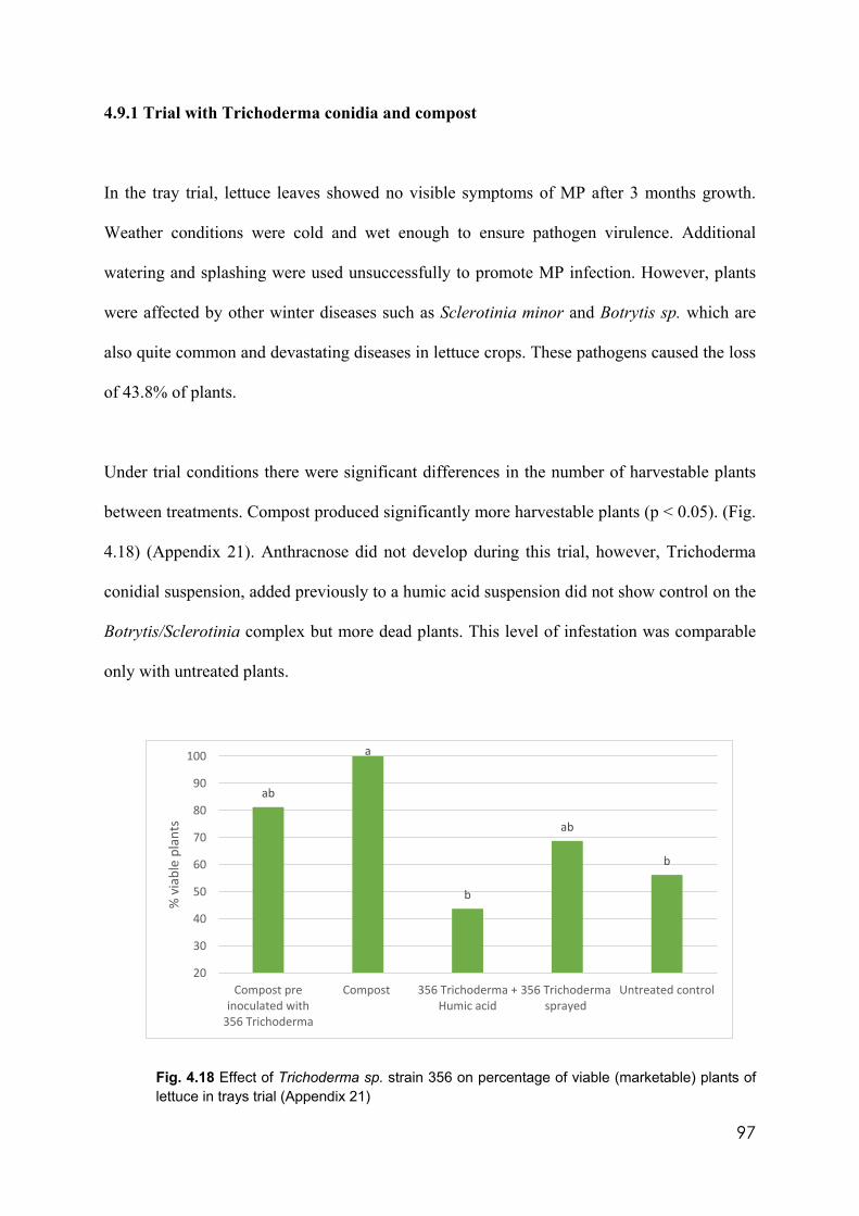
97
4.9.1 Trial with Trichoderma conidia and compost
In the tray trial, lettuce leaves showed no visible symptoms of MP after 3 months growth.
Weather conditions were cold and wet enough to ensure pathogen virulence. Additional
watering and splashing were used unsuccessfully to promote MP infection. However, plants
were affected by other winter diseases such as Sclerotinia minor and Botrytis sp. which are
also quite common and devastating diseases in lettuce crops. These pathogens caused the loss
of 43.8% of plants.
Under trial conditions there were significant differences in the number of harvestable plants
between treatments. Compost produced significantly more harvestable plants (p < 0.05). (Fig.
4.18) (Appendix 21). Anthracnose did not develop during this trial, however, Trichoderma
conidial suspension, added previously to a humic acid suspension did not show control on the
Botrytis/Sclerotinia complex but more dead plants. This level of infestation was comparable
only with untreated plants.
Fig. 4.18 Effect of Trichoderma sp. strain 356 on percentage of viable (marketable) plants of lettuce in trays trial (Appendix 21)
ab
a
b
ab
b
20
30
40
50
60
70
80
90
100
Compost preinoculated with
356 Trichoderma
Compost 356 Trichoderma +Humic acid
356 Trichodermasprayed
Untreated control
% v
iabl
e pl
ants
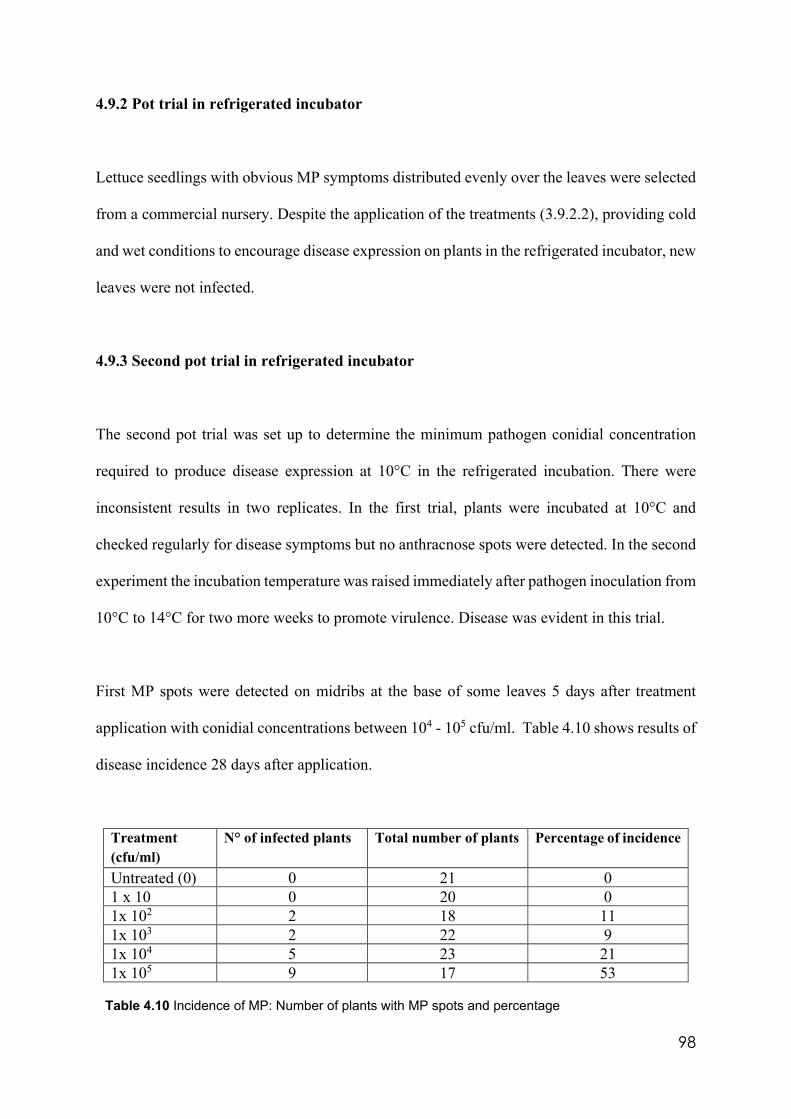
98
4.9.2 Pot trial in refrigerated incubator
Lettuce seedlings with obvious MP symptoms distributed evenly over the leaves were selected
from a commercial nursery. Despite the application of the treatments (3.9.2.2), providing cold
and wet conditions to encourage disease expression on plants in the refrigerated incubator, new
leaves were not infected.
4.9.3 Second pot trial in refrigerated incubator
The second pot trial was set up to determine the minimum pathogen conidial concentration
required to produce disease expression at 10°C in the refrigerated incubation. There were
inconsistent results in two replicates. In the first trial, plants were incubated at 10°C and
checked regularly for disease symptoms but no anthracnose spots were detected. In the second
experiment the incubation temperature was raised immediately after pathogen inoculation from
10°C to 14°C for two more weeks to promote virulence. Disease was evident in this trial.
First MP spots were detected on midribs at the base of some leaves 5 days after treatment
application with conidial concentrations between 104 - 105 cfu/ml. Table 4.10 shows results of
disease incidence 28 days after application.
Treatment (cfu/ml)
N° of infected plants Total number of plants Percentage of incidence
Untreated (0) 0 21 0 1 x 10 0 20 0 1x 10² 2 18 11 1x 10³ 2 22 9 1x 10⁴ 5 23 21 1x 10⁵ 9 17 53
Table 4.10 Incidence of MP: Number of plants with MP spots and percentage
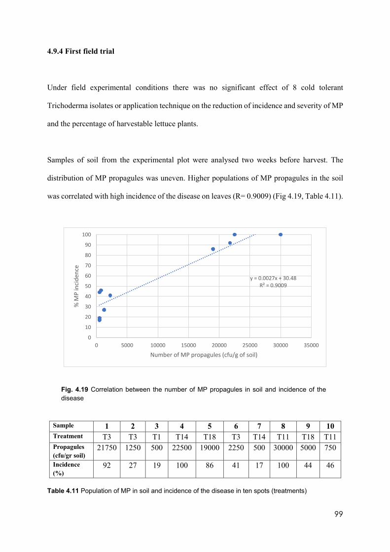
99
4.9.4 First field trial
Under field experimental conditions there was no significant effect of 8 cold tolerant
Trichoderma isolates or application technique on the reduction of incidence and severity of MP
and the percentage of harvestable lettuce plants.
Samples of soil from the experimental plot were analysed two weeks before harvest. The
distribution of MP propagules was uneven. Higher populations of MP propagules in the soil
was correlated with high incidence of the disease on leaves (R= 0.9009) (Fig 4.19, Table 4.11).
Fig. 4.19 Correlation between the number of MP propagules in soil and incidence of the disease
Sample 1 2 3 4 5 6 7 8 9 10 Treatment T3 T3 T1 T14 T18 T3 T14 T11 T18 T11 Propagules (cfu/gr soil)
21750 1250 500 22500 19000 2250 500 30000 5000 750
Incidence (%)
92 27 19 100 86 41 17 100 44 46
Table 4.11 Population of MP in soil and incidence of the disease in ten spots (treatments)
y = 0.0027x + 30.48R² = 0.9009
0
10
20
30
40
50
60
70
80
90
100
0 5000 10000 15000 20000 25000 30000 35000
% M
P in
cide
nce
Number of MP propagules (cfu/g of soil)
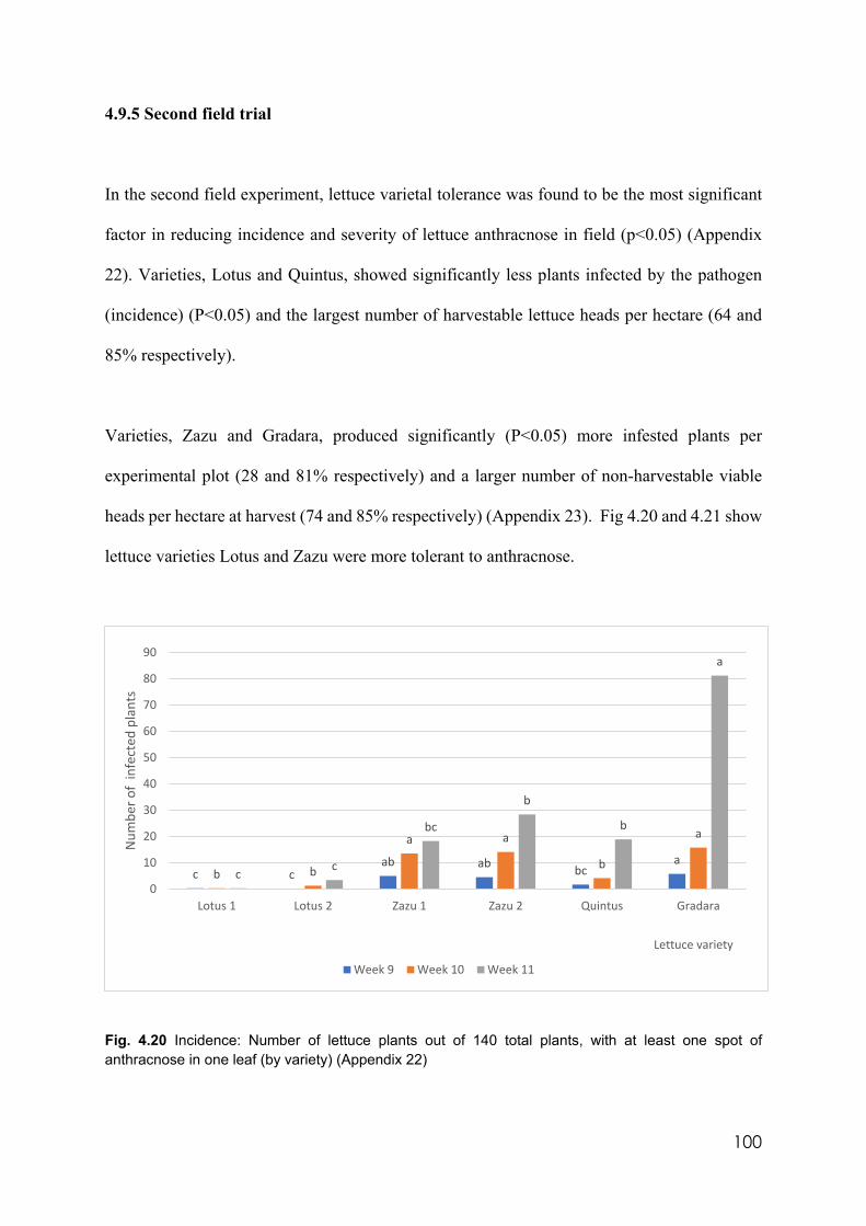
100
4.9.5 Second field trial
In the second field experiment, lettuce varietal tolerance was found to be the most significant
factor in reducing incidence and severity of lettuce anthracnose in field (p<0.05) (Appendix
22). Varieties, Lotus and Quintus, showed significantly less plants infected by the pathogen
(incidence) (P<0.05) and the largest number of harvestable lettuce heads per hectare (64 and
85% respectively).
Varieties, Zazu and Gradara, produced significantly (P˂0.05) more infested plants per
experimental plot (28 and 81% respectively) and a larger number of non-harvestable viable
heads per hectare at harvest (74 and 85% respectively) (Appendix 23). Fig 4.20 and 4.21 show
lettuce varieties Lotus and Zazu were more tolerant to anthracnose.
Fig. 4.20 Incidence: Number of lettuce plants out of 140 total plants, with at least one spot of anthracnose in one leaf (by variety) (Appendix 22)
c cab ab bc
ab b
a a
b
a
c c
bc
b
b
a
0
10
20
30
40
50
60
70
80
90
Lotus 1 Lotus 2 Zazu 1 Zazu 2 Quintus Gradara
Num
ber o
f in
fect
ed p
lant
s
Lettuce variety
Week 9 Week 10 Week 11
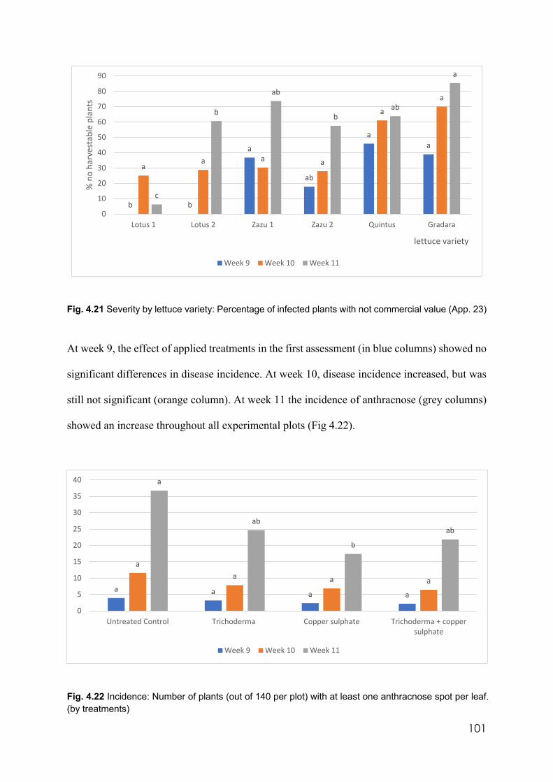
101
Fig. 4.21 Severity by lettuce variety: Percentage of infected plants with not commercial value (App. 23)
At week 9, the effect of applied treatments in the first assessment (in blue columns) showed no
significant differences in disease incidence. At week 10, disease incidence increased, but was
still not significant (orange column). At week 11 the incidence of anthracnose (grey columns)
showed an increase throughout all experimental plots (Fig 4.22).
Fig. 4.22 Incidence: Number of plants (out of 140 per plot) with at least one anthracnose spot per leaf. (by treatments)
b b
a
ab
aa
aa a a
a
a
c
b
ab
bab
a
0
10
20
30
40
50
60
70
80
90
Lotus 1 Lotus 2 Zazu 1 Zazu 2 Quintus Gradara
% n
o ha
rves
tabl
e pl
ants
lettuce variety
Week 9 Week 10 Week 11
a a a a
aa a a
a
ab
b
ab
0
5
10
15
20
25
30
35
40
Untreated Control Trichoderma Copper sulphate Trichoderma + coppersulphate
Week 9 Week 10 Week 11
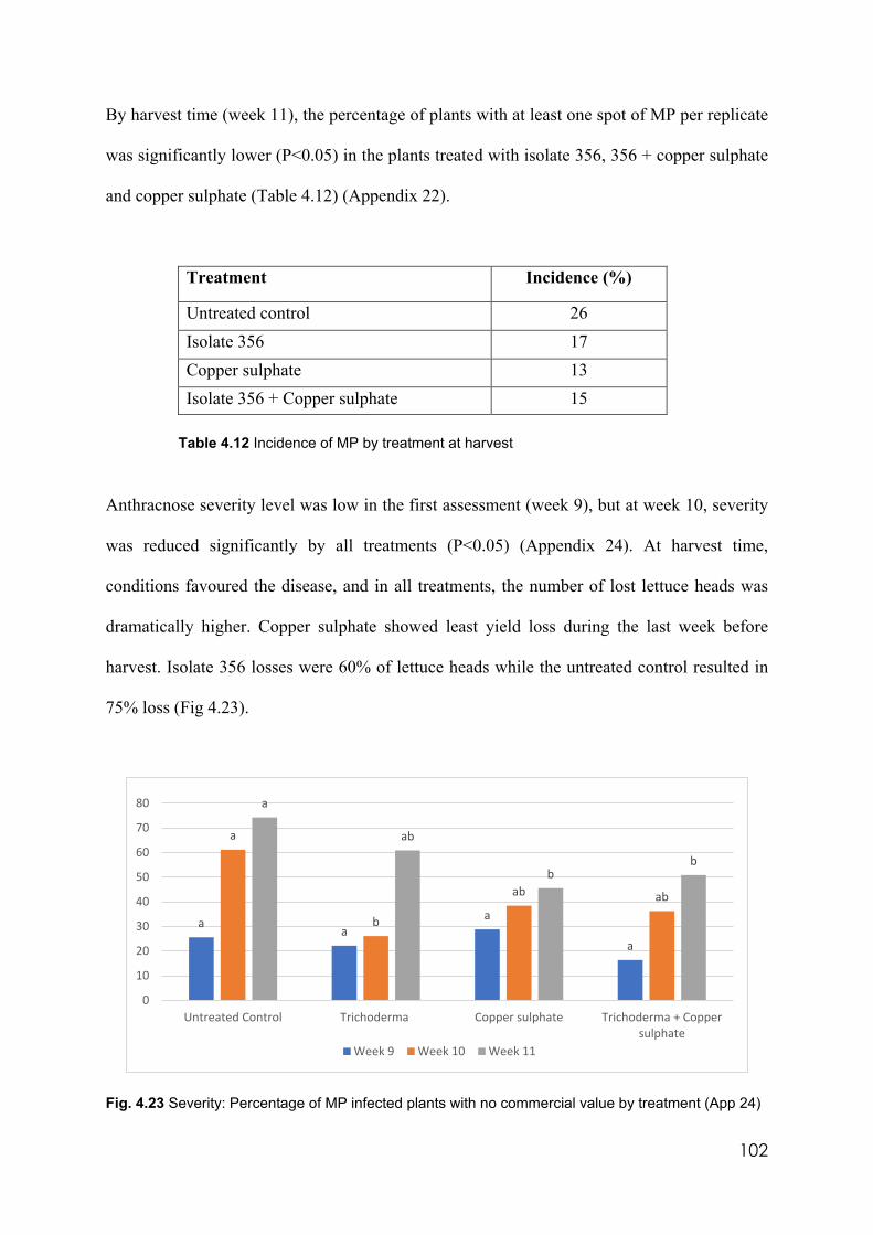
102
By harvest time (week 11), the percentage of plants with at least one spot of MP per replicate
was significantly lower (P˂0.05) in the plants treated with isolate 356, 356 + copper sulphate
and copper sulphate (Table 4.12) (Appendix 22).
Treatment Incidence (%)
Untreated control 26 Isolate 356 17 Copper sulphate 13 Isolate 356 + Copper sulphate 15
Table 4.12 Incidence of MP by treatment at harvest
Anthracnose severity level was low in the first assessment (week 9), but at week 10, severity
was reduced significantly by all treatments (P<0.05) (Appendix 24). At harvest time,
conditions favoured the disease, and in all treatments, the number of lost lettuce heads was
dramatically higher. Copper sulphate showed least yield loss during the last week before
harvest. Isolate 356 losses were 60% of lettuce heads while the untreated control resulted in
75% loss (Fig 4.23).
Fig. 4.23 Severity: Percentage of MP infected plants with no commercial value by treatment (App 24)
aa
a
a
a
b
ab ab
a
ab
bb
0
10
20
30
40
50
60
70
80
Untreated Control Trichoderma Copper sulphate Trichoderma + Coppersulphate
Week 9 Week 10 Week 11
103
In both annual field experiments, symptoms of MP disease were noticeable at eight weeks after
planting, three weeks before harvest. Therefore, the critical period for this disease was in the
last three weeks before harvest under these conditions. The second soil test (3.9.4) from 16 soil
samples showed the distribution of propagules of MP was uniform with an average of 1 colony
per plate that corresponded to 500 cfu/g of soil.
104
Chapter 5
DISCUSSION AND CONCLUSIONS
5.1 Introduction
The first objective of this thesis was to isolate, characterise and identify cold tolerant
Trichoderma isolates with the capacity to control lettuce anthracnose in soil. This objective
was fulfilled by identifying 8 cold tolerant Trichoderma species which were selected,
characterised, and identified using morphological and molecular techniques. It was found that
temperature, pH, and media had a significant influence on the morphological characteristics of
Trichoderma isolates. Cold tolerant isolates identified were classified as T. viride, T. atroviride,
T. polysporum and T. composticola by molecular techniques. Four out of 8 cold tolerant isolates
were identified as T. atroviride. All strains used in this project belonging to this species were
cold tolerant. McBeath 2002) had success controlling M. nivale (a close relative of MP) by a
strain of T. atroviride in its cold pathosystem. These findings suggest T. atroviride, formerly
T. lignorum, is potentially useful in cold environments, but prior screening is always essential.
The second objective was to identify Trichoderma secondary metabolites for lettuce
anthracnose control by foliar application. Fusalanipyrone, 6PP, cytosporone and cladoacetal A
reduced growth of MP in vitro. Additional TSMs were produced by challenging isolates in
PDB liquid cultures. Conidial suspension of the cold tolerant isolate, 356 (T. composticola),
containing TSMs were applied to lettuce in the field significantly increasing the number of
commercial lettuce heads (p<0.05) (Appendix 23).
Outcomes from this research have significant advantages for the control of MP in winter lettuce
105
crops. They provide a biological alternative for disease protection in cropping systems where
the only control method now is chemical. Song Zhu, agronomist in Coolibah Herbs (pers.
comm. 31st august 2020), identified a reduction in efficacy using Octave (prochloraz) to control
MP in field. This suggested MP has developed resistance to this chemical leaving the industry
without the only chemical with relative efficacy in controlling this pathogen. Prochloraz was
sprayed 7 times on 10 weeks lettuce crops without success.
This integrated methodology has international application in disease protection for many soil
borne and leaf borne pathogens. It gives farmers more options for sustainable food production.
Some advantages include more economical production, more sustainable production,
protection, and preservation of beneficial microbiology in agricultural soil, a safer and healthier
working environment for farm workers, and finally, a healthier product for consumers.
Therefore, this thesis fulfilled its aims and showed it is possible to control lettuce anthracnose,
a winter disease, using cold tolerant isolates of the biological control agent, Trichoderma.
Control of lettuce anthracnose was achieved when a Trichoderma sp was applied to soil prior
to planting to promote soil colonisation combined with post-planting foliar sprays of liquid
filtrates at regular intervals during the winter crop season.
Not all the isolates of Trichoderma, wild type or from commercial formulations, that were
tested could grow and express all required activity in the environmental conditions where MP
shows maximum virulence. Therefore, morphological characterisation and molecular
identification and in vitro tests in cold incubation to find effective isolates was carried out
before field trials were begun.
106
The commercial production of effective TSMs able to restrict MP growth is feasible but not
necessarily achieved using cold tolerant isolates that were essential for soil colonisation. As
the liquid cultures were produced under controlled conditions where isolates reached optimal
mycelia and TSM production, the ideal temperature for culturing conditions depended on
specific isolate requirements (18-25oC), adequate pH (5.0-6.0) and nutrient balance.
Additionally, the process of challenging isolates in co-cultures used to promote the activation
of their defence mechanisms achieved more diverse and abundant production of TSMs.
5.2 Identification and characterisation of suitable Trichoderma isolates to control MP in
soil.
Assembling a Trichoderma culture collection was the first and essential step in this research
project. Each culture was created from a single spore guaranteeing the genetic purity of each
isolate. The validity of the morphological, molecular, biochemical, and pot and field
experiment results relied on this premise. Because efficacy relies on particular characteristics
of species, it is essential that isolates used to produce useful TSMs are of single spore isolate
origin. Special care was taken to collect wild type isolates from different environmental
conditions within Australia to ensure the broadest thermal, hydric, and nutritional spectrum.
Isolates were collected from wet and dry areas, soil, mulch, roots, and plant buds, to achieve
morphological and genetic diversity and a more diverse ability to produce TSMs. Cold tolerant
isolates with consistent ability to control MP in vitro were isolated from temperate
environments that correspond to the pathosystem in study corroborating the concept coined by
Fravel (2005): the biocontrol must be successful in the environment where the pathogen is
virulent.
107
In this research project the morphological characteristics, historically considered as
fundamental for fungal identification, required molecular profiling to further separate the
species. However, at standard in vitro conditions (25°C, PDA, and pH 5.0) characteristics such
as colony growth speed, mycelia texture i.e felty, hairy or ricotta like, colony shape i.e.
symmetrical or “fried egg” shape, or conidiation pattern, i.e. from centre or from edge of
culturing plate; were necessary for grouping clades and predicting their performance as
biocontrol agents.
Carbon and nitrogen food sources had a profound effect on the biological activity of
Trichoderma isolates. Trichoderma colonies grew in different pattern when cultured on PDA
and CZA media showing that some components in PDA favour Trichoderma growth and
indirectly TSM production. It was evident from morphological studies that some species
preferred monosaccharides and specific aminoacids (Atanasova 2014).
PDA is an undefined medium, amino acids, sugars, starch, and many other biologically active
molecules are present in variable concentrations depending on the potato cultivars and on the
growing and post-harvest management conditions (Leonel et al. 2017). For example, Griffith
et al. (2017) found that low levels of copper in the tubers used to produce PDA, reduced the
production of key enzymes associated with conidial pigmentation. In a similar way the
production of 6PP was apparent only when Trichoderma 6PP producers were cultured on PDA
under all experimental incubation temperatures and pH values used in this project. This
suggested that this characteristic was not directly affected by changes in these variables. These
changes can induce or reduce the hyphal growth confirming the result from Kubicek-Pranz et
al (1998) that some monosaccharides and monosaccharides-related compounds support
growing conditions for some species over others.
108
Czapek dox agar is a defined medium with all its components as salts at standard
concentrations. Therefore, more reproducible results were possible than when organisms were
cultured in PDA. However, PDA contains sources of nitrogen other than nitrates as in CZA.
This may explain why Trichoderma isolates cultured on PDA produced abundant growth, while
growth on CZA produced thin, meagre, and hard to distinguish colonies with few phialides and
conidia (Fig 4.4 right). This confirmed results from Danielson and Davey (1973) and Rajput
(2014) that ammonium and not nitrate was the preferred nitrogen source for Trichoderma
growth. Therefore, nitrate applications in soil as a source of nitrogen for crop nutrition may
impact negatively on the antagonistic performance of Trichoderma. High concentration of
nitrates in the rhizosphere can cause a rise in pH which is not preferred by Trichoderma. N-
NO3- is up taken by the plant and an OH- is released to balance the charge (Hinsinger et al.
2003). This could be the reason why the microbial activity in the rhizosphere improves in
presence of ammonium rather than in nitrate nitrogen.
Further research to confirm the effects of ammonium and nitrate ions on Trichoderma
performance in vitro could be carried out substituting ammonium nitrate and ammonium
sulphate, as they are popular agricultural fertilisers, in the CZA recipe instead of the standard
sodium nitrate in its formulation.
Neither coconut aroma in the Trichoderma 6PP producers, nor yellow pigmentation in the agar
by pigmenting isolates were noticed when Trichoderma isolates were cultured on CZA under
any conditions of pH and temperature. These results also support the hypothesis that nitrate
reduced the biological activity and antagonistic properties of Trichoderma. Czapek dox agar,
however, induced Trichoderma isolates to produce less mycelium which tended to be more
concentrated in small cottony balls that were easy to dislodge from the agar surface allowing
109
good quality slide mounts for clearer microscopic observations to be prepared.
Modifying the culturing conditions induced fungal morphological changes and biological
control attributes, i.e. production of 6pp. In practical terms, the fertilisation (organic or
inorganic), liming, gypsum application, ploughing, irrigation, and the seasonal conditions may
also modify the behaviour and distribution of Trichoderma in the soil. These parameters must
be considered before a selection and practical application of this BCA in field conditions.
During this research work, Trichoderma isolates showed preference for pH media between 5.0
and 6.0. Colony growth decreased with decreasing acidity, reducing their capacity to supress
pathogens as found by Simon and Sivasithamparam in 1988. They determined that there was
better control of the ‘take all’ pathogen by Trichoderma in moderately acid soil. These findings
also correlated with the maximum inhibition of Fusarium sp colony growth obtained by a
higher volatile TSM production reached at pH 6.0 (Abeyratne and Desahappriya 2018). This
may be a result of more abundant Trichoderma biomass production.
The most sensitive isolates to changes in substrate acidity were 355 (T. neokoningii) and
BIANCA (possibly T. polysporum) expressed by the difference in the rate of growth on PDA
at pH 5.0 and 6.5 (Fig. 4.3). When pH increases, hyphae became stressed manifesting as
distorted hyphae. Daryaei et al 2016 reported that Trichoderma, as other fungi, have the
capacity to adapt themselves to external pH modifying it by activation of a pH-sensing system
that is genetically activated in alkaline conditions by the zinc finger transcription factor PacC
described by (Tilburn et al 1995). This system is inactive in acidic conditions. and varies
between species and strains (Moreno-Mateos et al 2007). This may explain why 355 and
BIANCA were affected by slight changes in acidity. More investigation is required to
110
determine the activity of factor PacC in all the isolates to predict their ability to modify their
environmental pH and select those that perform better in alkaline soils.
As expressed above, Trichoderma is more often reported in the international literature as a
warm temperature organism which contrasts with the MP thermal conditions. Temperature is
a regulator of mycelial growth and is considered an indirect factor in metabolite production.
The most effective Trichoderma isolates that control MP in vitro and in soil corresponded with
locations characterised by cold temperatures. The species are related to T. viride, T. atroviride,
T. polysporum and T. composticola. However, T. polysporum (BIANCA) and T. composticola
(33981) are exceptions. This suggests that the ability to survive and develop in cold
temperatures is not a species attribute but rather an environmental adaptation mechanism as
stated by Ghildiyal and Pandey (2008). For this reason, it would be expected that the best
isolates to control MP by soil inoculation should originate from cold regions. At Trichoderma
suboptimal conditions like in winter when MP shows maximum virulence, the application of
one or more fast growing biocontrol agents is critical to colonise more soil volume in the
shortest time and destroy MP propagules before they become active.
Conidial formulations are used commercially as a benchmark around the world based on their
easy dispersal and convenient storage. Conidia can be freeze dried for long shelf life and when
applied to the soil they will germinate unless conditions are not ideal. Results from in vitro
experiments suggested that when the Trichoderma cold tolerant isolates were inoculated on
PDA as mycelia and incubated at low temperatures they formed colonies faster than when they
were inoculated as conidial suspensions confirming that mycelia are less affected by fungistasis
than are conidia (Papavizas, 1985). Isolate 33681 (T. composticola) conidia were unable to
germinate in PDA at 10°C and for that reason is not considered a cold tolerant isolate. However,
111
when mycelia of this isolate were inoculated onto PDA, they produced healthy colonies under
cold incubation. Based on this observation, mycelia from cold tolerant isolates were inoculated
in an organic substrate, incubated, and then applied into soil in cold conditions in the first field
experiment. Similarly, Wong et al (2002) applied Trichoderma conidia on straw infested with
Fusarium pseudograminearum, covered them with soil and incubated in vitro with enough
moisture at 15°C obtaining 90% of reduction of pathogen survival structures. This suggests
that for better results, conidia or mycelia should be applied in a carrier that provide shelter,
moisture, and nutrients for the BCA confirming the observations of Van Elsas and Postma
(2006). A product based on hyphal fragments would cause farmers problems by blocking
sprinklers, spray nozzles and filters as well as drippers in irrigation systems. Compost was
selected as the carrier for Trichoderma cold tolerant isolates because it is relatively easy to find
and manipulate. Kok et al (1996) obtained successful control of R. solani inoculating,
incubating, and applying swine manure pellets with a T. harzinaum conidial suspension. This
material could be high in ammonia which can result in toxicity for the conidia and mycelia as
found by Montgomery et al (1980). For this reason, chicken manure pellets also high in
ammonia, popular in Victorian agriculture, were not selected as the carrier.
The tray experiment set up to evaluate the efficacy of the inoculation technique in soil using a
conidial suspension and mycelia pre-incubated in compost to control MP produced an
unexpected result. MP was not evident despite the soil used to fill the trays was sampled from
a heavily infested MP lettuce crop the winter before. Cold tolerant isolate 356 controlled B.
cinerea and Sclerotinia minor and resulted in a significantly greater number of harvestable
lettuce heads because of the improved control (p<0.05). Diverse microflora in the compost may
have an important role in controlling these pathogens confirming the conclusions of Blaya et
al (2016) demonstrating that the application of compost on farms is a valuable resource and
112
needs more research work. Commercial Humic acid (Humax) is highly alkaline (pH 9.5). This
strongly alkaline input could negatively affect the Trichoderma capacity to control these two
diseases confirming that alkaline pH may not allow Trichoderma to express their control
mechanisms as reported by (Trushina et al 2013). In relative terms, thermal aerobic composts,
have received less attention from researchers than, for example, a synthetic single nitrogen
fertiliser, urea. This observation shows that there are good reasons for serious research into the
potential disease control by these natural products.
The 8 cold tolerant isolates were tested in a commercial lettuce field under winter conditions
using conidia and mycelia pre cultured in compost. Large visible, but not statistical, differences
in the number of apparently harvestable plants were noticed when the soil was inoculated with
Trichoderma cold tolerant isolates using both application techniques. This variation was due to
the apparently uneven distribution of MP in the field which correlated with the observations of
Paterson and Grogan (1991). The pathogen distribution analysis in soil showed that areas with
more disease incidence and severity corresponded to soil with the highest infestation (R=90%).
What cannot be explained from these results, despite not having significant differences in
incidence and severity of MP from the application of cold tolerant isolates and inoculation
techniques in the soil, was the higher number of extra viable lettuce heads in the crop treated
with isolate 356. There were 40% more commercial heads per hectare than the untreated
control.
The experimental design for the second field experiment considered 12 replicates rather than 4
as in the first field experiment to reduce the experimental error from the erratic distribution of
the pathogen in the field. Unfortunately, the first plot was not available in the following winter
because it was engaged with other crops. This was a limitation for this experiment because
113
ideally the same trial should have been replicated in the same plot to assess the pathogen
dynamics one year after the treatments were applied.
Among the cold tolerant isolates, LM and PMF are remarkably close to T. atroviride in the
phylogenetic tree and they are sold commercially using this name although no conclusive
identification was obtained from the phylogenetic analysis. LUP, isolated from lupin roots near
Robinvale, Victoria, was also located in this clade but in a separate branch next to PMR, which
are sold by the same commercial company as LM and PMF (Fig. 4.6). The remaining cold
tolerant isolates were identified as 45897 (T. polysporum) which formed hyaline conidia on
PDA; 1535 and 59837 both (T. viride) and 356 (T.composticola).
Isolate 1536 (Trichoderma sp.) was not successfully identified to species and is not cold
tolerant although it was isolated from the same substrate as isolate 1535. This fact confirmed
that cold tolerant and warm tolerant Trichoderma isolates can share the same substrate and
possibly alternate dominance through warm and cold seasons as explained by (Lo et al. 1997).
Isolates 356 from pistachio buds from South Australia and 33681 from the NSW herbarium
belong to the same species (T. composticola) but, 356 is cold tolerant while 33681 is inactive
at 10°C. Morphologically, their phialides produced by both isolates are similar; very long and
pointy, but their conidiation pattern is different on PDA at 5.0 pH and 25°C (Fig 5.1). The cold
tolerant 356 began conidiation with the radial development of mycelium at 25°C suggesting
that it was not the preferred incubation temperature. Isolate 33861 did not formed conidia until
mycelium covered the available nutrient suggesting that 25°C was a suitable growth
temperature and sporulation was initiated only by depletion of nutrients. These morphological
dissimilarities confirm that there is still more variability intraspecies than interspecies
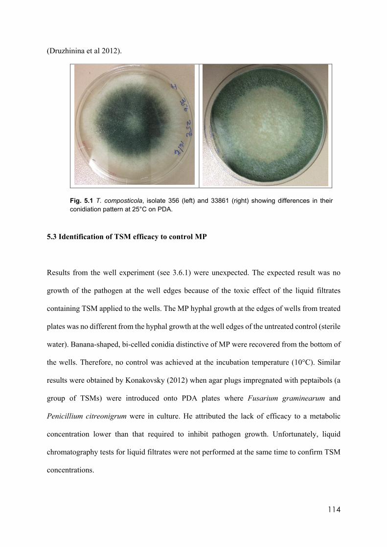
114
(Druzhinina et al 2012).
Fig. 5.1 T. composticola, isolate 356 (left) and 33861 (right) showing differences in their conidiation pattern at 25°C on PDA.
5.3 Identification of TSM efficacy to control MP
Results from the well experiment (see 3.6.1) were unexpected. The expected result was no
growth of the pathogen at the well edges because of the toxic effect of the liquid filtrates
containing TSM applied to the wells. The MP hyphal growth at the edges of wells from treated
plates was no different from the hyphal growth at the well edges of the untreated control (sterile
water). Banana-shaped, bi-celled conidia distinctive of MP were recovered from the bottom of
the wells. Therefore, no control was achieved at the incubation temperature (10°C). Similar
results were obtained by Konakovsky (2012) when agar plugs impregnated with peptaibols (a
group of TSMs) were introduced onto PDA plates where Fusarium graminearum and
Penicillium citreonigrum were in culture. He attributed the lack of efficacy to a metabolic
concentration lower than that required to inhibit pathogen growth. Unfortunately, liquid
chromatography tests for liquid filtrates were not performed at the same time to confirm TSM
concentrations.
115
However, the wells experiment produced two by-products: the Trichoderma liquid filtrates that
were stored at cold temperature, and the MP discs that were introduced as challengers in every
culture. The washed agar discs containing MP were processed (3.5.5), inoculated on PDA and
incubated at 10 °C expecting to have MP colonies characterised by the orange colour. MP
colonies were formed only on the untreated control plates (pathogen cultured in sterile water)
confirming that the pathogen survived the liquid culturing conditions. This was important
because the fluctuating submerged conditions caused variation in the access to oxygen which
is necessary for good fungal growth. The washed agar discs corresponding to isolates 66079
(T. aureoviride) and 33681 (T. composticola) when cultured on PDA showed neither growth
of the pathogen nor of the Trichoderma isolates themselves. This suggests that at least one
compound in each liquid culture killed the pathogen. At the same time, these two Trichoderma
isolates remained inactive because they were incubated below their optimal temperatures. After
20 days of cold incubation, PDA plates containing these two Trichoderma isolates were
transferred to a warm incubator at 25°C where they produced colonies confirming that these
organisms were inactive but not dead (fungistasis).
The metabolite test results (Table 4.8) for 66079 (T. aureoviride) liquid filtrate showed the
presence of cytosporone S, a compound with broad antibacterial and antifungal activity (Ishi
et al. 2013). Cytosporone S was found in liquid filtrates produced only by isolate 66079. Liquid
filtrates from isolate 33681 contained 6PP. Both cytosporone S and 6PP are potential
candidates for MP control but further research is required to determine the effects of different
conditions on the production of these two TSMs.
Results from wells and from MP agar disc experiments were contradictory. No effective control
of MP was apparent on the well edges experiment while on the agar disc experiment evidence
116
of MP control was observed. Another experiment was required to confirm or dismiss the
efficacy of the TSM to control MP. Having liquid filtrates from the wells experiment, a new
trial was designed with PDA using filtrates instead of demineralised water. Two liquid filtrates
from native cold tolerant isolates 1535 (T. viride) and 356 (T. composticola), were selected.
Both liquid filtrates contained 35 mg/l and 30.4 mg/l (Fig 4.16) of 6PP in filtrates of 1535 and
356 respectively before autoclaving (3.9). According to The US National Centre for
Biotechnology Information (2020), this compound has a boiling point of 287°C making it heat
stable at autoclaving temperature. Before autoclaving the final volume of PDA was 60 ml.
enough for 3 Petri dishes (replicates) but after autoclaving, the media volume was reduced to
40 ml. This decrease in volume may have resulted in a 33% increase in TSM concentration,
but the final concentration was not determined. This was a limitation of this trial. MP was
inoculated and incubated on this agar to determine efficacy of the TSMs from the filtrates on
the MP colonies. The growth patterns on the amended agar were abnormal suggesting that at
least one compound in each filtrate was affecting normal pathogen growth. Furthermore, this
reaction proved that at least one effective compound was not heat labile. Additional
investigation is needed to find minimum effective concentration of 6PP against MP.
After these trials there was a need to quantify the TSM production and determine if challenging
between isolates induced them to produce more or different TSMs. Vegetative
compatibility/incompatibility was a useful technique. Barrages formed by these couplets
showed incompatibility and possibly/probably the activation of genes for TSM production as
stated by Schirmböck et al. (1994) and Vinale et al. (2017). However, the position of the
isolates on the carrousel layout (Fig. 3.7) may have influenced the barrage formation; some
very fast-growing isolates surrounded and masked slow growers not allowing them to express
antagonistic activity.
117
One of the most intense barrage-forming couplets was MACA-CAROL. This couplet produced
cytosporin C, chaetoquadrim E and gossypol, this last compound is characterised by the intense
yellow pigmentation with toxic activity against P. ultimum, P. irregulare and F. oxysporum
(Mellon et al 2014). From these three metabolites, only chaetoquadrim E was produced by
CAROL (Trichoderma sp.). This couplet could be used in further investigation to control these
three plant pathogens.
MACA (T. longibrachiatum) was obtained from a mulching material employed extensively on
a macadamia farm at Alstonville in northern NSW subtropical conditions. This contrasts with
the origin of the isolate CAROL from McKinnon, Melbourne (cool temperature) which
originated from composted kitchen waste. Isolates MACA and CAROL produced the most
consistent incompatibility characterised by a thick barrage. One hundred percent of the
interactions between these two isolates produced barrages and a wide and clear gap with no
physical contact between colonies. Andrade et al. (2015) reported fungicidal compounds like
trichodermolide and sorbiquinol from filtrates of T. longibrachiatum, but none of these TSMs
were not found in MACA liquid filtrates during this research, confirming that TSM production
can vary between strains within species (Hatvani et al 2014).
Isolates VIR and 45899 formed strong and consistent barrages. Isolate VIR (possibly T.
harzianum), was isolated from Virginia, South Australia, from a piece of farm fence wood. It
grew rapidly at 25°C but was inactive in cold temperatures and produced a slight yellow
pigmentation on PDA. 45899 (T. polysporum), had a broader temperature growth range. The
single isolate VIR liquid culture contained only unreported TSMs. Isolate 45899 produced
trichodermatides A and D and mitorubrin, however, when VIR and 45899 were challenged an
118
additional TSM, harzianic acid, was produced by the couplet. This may be an additional clue
identifying VIR as T. harzianum because harzianic acid is reportedly produced by T. harzianum
(Vinale et al 2009). In this experiment harzianic acid was produced only through the
challenging process demonstrating that challenging promotes more TSM production.
Isolate BIANCA (possibly T. polysporum) originated on plates from which lettuce anthracnose
was first isolated (Pearcedale, Victoria). This isolate is aggressive under warm conditions but
in cold temperatures it is almost inactive. Isolate ANAND (possibly T. spirale) was extracted
from avocado tree samples infected by Phytophthora cinnamomi from subtropical Belthorpe,
Queensland, 100 km north west of Brisbane. ANAND and BIANCA isolates developed strong
and consistent barrages. ANAND, unchallenged, produced candidusin C, while BIANCA,
unchallenged produced dermadin, trichodermatide A and D and koningin B. However, their
challenged filtrate only produced dermadin. This result shows that less and not more TSMs are
produced and that possibly they inhibited one another as reported by Serrano-Carreón et al
(2002).
Couplets of isolates BIANCA, MACA, VIR, CAROL, 45899, 99 and 1536 developed strong
barrages between each other but moderate reactions with other isolates. Strongly cold tolerant
isolate, 1535 (Trichoderma viride), was incompatible with warm tolerant isolate 1536
(Trichoderma sp.). Both were isolated from the same mulch sample from a forest in Tween
Peaks, Western Australia. This shows that Trichoderma isolates from the same environmental
niche can exhibit different growth parameters and possibly alternate seasonally. Isolate 1535
produced fusalanipyrone, a fungicide and phytotoxic compound, and 6PP. Isolate 1536 did not
produce identified TSMs. Their couplet liquid culture contained, additionally, the peptaibol
Trichorzin PAU which, according to Leclerc et al. (1997), is almost exclusively synthesised by
119
T. harzianum strains, another clue to 1536 identification.
In general, TSM production is culture condition dependent and directly correlated with active
mycelial growth. The progressive depletion of nutrients during culturing determined the
diversity and concentration of the resulting TSMs, and efficacy as pathogen control agents
(Ghisalberti and Sivasithamparan, 1990). Similarly, it is apparent that the same two parameters
can be modified when strains are challenged in a liquid culture. Therefore, all these factors
should be studied in further investigation to improve the effectiveness of liquid cultures
containing TSMs to control not only MP but also other plant pathogens.
Challenged and unchallenged liquid filtrates of isolate 1535 were tested on lettuce seedlings.
Unchallenged filtrates from isolate 1535, which stimulated atypical MP hyphal growth in vitro,
were selected as foliar sprays on lettuce plants. Plants suspected of having MP and incubated
under cold conditions (10°C) did not express the disease on new leaves despite their initial MP
symptoms. The prior history of the seedlings (chemical treatments) was unknown.
Development of leaf disease depended on spore concentration in the environment, soil, or air.
Lettuce seedlings were sprayed twice with 101-105cfu/ml MP conidial suspensions and
maintained in the humidified refrigerated incubator. However, there were no observable signs
of the disease. Galea et al. (1986) experimented with the effects of temperature, pH and water
stress on conidial germination and germ tube growth in vitro and found the optimal temperature
for conidial germination was 10-26°C after 24 hours of incubation. Field conditions are
different from environmental chamber conditions; leaf temperature can be 4-5°C higher than
air temperatures on sunny days (Taiz and Zeiger 2010). The experiment was set up again at 14-
15°C. After 7 days, spots on the midribs of leaves inoculated with a conidia concentration of
120
1x10⁴cfu/ml were observed. Typical MP lesions were identifiable after 20 days. Perhaps the
incubation temperature for the curative test in refrigerated conditions also had to be raised.
Further research is needed.
Finally, a second field experiment was set up to evaluate the efficacy of the cold tolerant isolate
356. The treatments included conidia and TSMs from isolate 356 sprayed on lettuce leaves
alone and in combination with a copper-based fungicide. Also, different lettuce varieties were
tested as a condition of using the commercial field.
The first and second field trial sites have similar climatic and soil conditions and presence of
MP. Less lettuce anthracnose was observed in the second field experiment. Distribution of MP
propagules was more uneven in the soil in field experiment 1 than in the soil of field experiment
2. This may be the reason why, despite large differences in the number of viable lettuce plants
(yield), the effect of the application of cold tolerant Trichoderma and application technique
was not significant in reducing the crop loss because of the disease.
Soil in field trial 2 had lower and more evenly distributed pathogen propagules than soil in field
trial 1 which may explain why the incidence and severity were lower and significance was
clearly shown (p<0.05). At the time of the second field experiment (winter 2018), TSMs had
not been identified in the in vitro liquid cultures, so conidial/TSMs suspensions for spraying
were not produced in the similar way as liquid cultures. However, the strong coconut smell in
the solid cultures used to produce conidial suspension suggested the presence of 6PP. The TSM
identification was available post 2018 winter season. This was one of the limitations of this
experiment.
121
5.4 Integrated practices to control MP
As a result of this research project some practices to control MP in field have been identified:
1. Rotate lettuce crops in winter. If a lettuce crop was infested with MP in winter, the
next two winter crops in that infested patch must not be lettuce. Therefore,
operationally, the winter lettuce crop rotation in that infested patch may be
triennially.
2. Identify and plant, selectively, the best MP tolerant variety lettuce in winter.
3. The chemical control based in Prochloraz is developing resistance. Farmers should
not overuse this chemical. It is common that prochloraz is sprayed almost every
week for up to 7 times in a winter lettuce crop.
4. Select and apply Trichoderma cold tolerant isolates in compost or in direct conidial
spray as a soil application as a feasible and practical strategy. Selection of cold
tolerant isolates can be commissioned from specialised microbiology laboratories
following similar approaches trialled in this research project. Cold tolerant isolates
can be purchased as lettuce mate (LM), plant mate root (PMR) or plant mate foliar
(PMF) from Agrimm, New Zealand. Intellectual property rights reside with the
owners of all registered isolates.
5. Produce and apply Trichoderma liquid cultures with validated efficacy against MP
In field. Contractual arrangements are necessary for anyone to use the above isolates
for TSM production. Isolates 1535, CAROL and LUP which were isolated from
Australian environments must be identified fully before intellectual property
registration. Finally, liquid, or solid products could be available as commercial
products.
122
Additionally, control of lettuce anthracnose is an integrated strategy that involves every activity
on the farm. For example, practices that avoid spreading propagules from field to field must
consider vehicles (utes, tractors), implements and workers boots. Footbaths and vehicle
washing pad should be a part of general farm practices.
5.5 Recommendations for further research
It is important to understand that each isolate of Trichoderma has defined growing parameters
which include soil temperature, moisture, pH, aeration, light intensity, nutritional factors, and
surrounding microbiota. These parameters affect their efficacy particularly in the MP
pathosystem that is not the optimal for Trichoderma. As these factors fluctuate throughout the
seasons, selection, production and use of isolates should consider these conditions for
maximum efficacy. Further research is required to determine if the application of other isolates
(warm tolerant) in warmer seasons on other crops as part of the rotation plan can contribute to
reducing the MP inoculum. Minimum dose of effective TSMs such as 6PP and optimal
parameters for its maximum production using appropriate Trichoderma isolates also need more
research. General farming practices such as liming or N-nitrate applications have a strong
negative impact on the establishment of Trichoderma in soil as a BCA. This approach can be
implemented to control other soil borne diseases under unfavourable conditions such as salty
or alkaline soils and dryland agriculture, etc. In areas where compost is available, the
inoculation and incubation of selected isolates of Trichoderma spp. to compost before field
application may add value and enhance the benefits of the selected Trichoderma isolates.
Technology transfer to farmers and agricultural scientists is critical to maximise cost effective
investment and avoid loss of credibility in biological control systems.
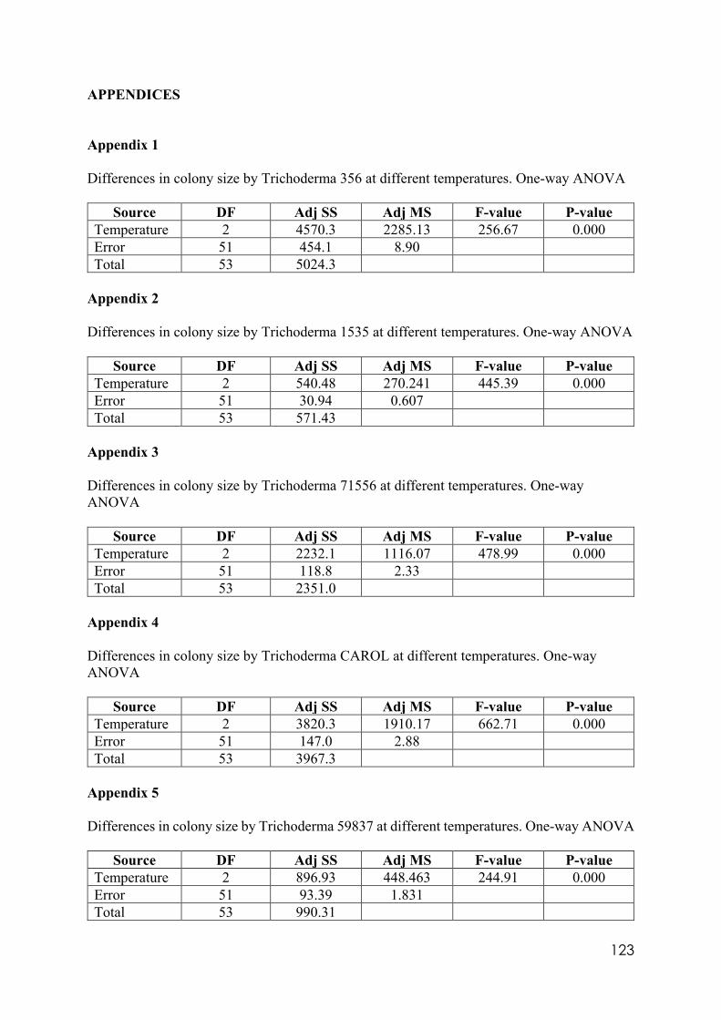
123
APPENDICES Appendix 1 Differences in colony size by Trichoderma 356 at different temperatures. One-way ANOVA
Source DF Adj SS Adj MS F-value P-value Temperature 2 4570.3 2285.13 256.67 0.000 Error 51 454.1 8.90 Total 53 5024.3
Appendix 2 Differences in colony size by Trichoderma 1535 at different temperatures. One-way ANOVA
Source DF Adj SS Adj MS F-value P-value Temperature 2 540.48 270.241 445.39 0.000 Error 51 30.94 0.607 Total 53 571.43
Appendix 3 Differences in colony size by Trichoderma 71556 at different temperatures. One-way ANOVA
Source DF Adj SS Adj MS F-value P-value Temperature 2 2232.1 1116.07 478.99 0.000 Error 51 118.8 2.33 Total 53 2351.0
Appendix 4 Differences in colony size by Trichoderma CAROL at different temperatures. One-way ANOVA
Source DF Adj SS Adj MS F-value P-value Temperature 2 3820.3 1910.17 662.71 0.000 Error 51 147.0 2.88 Total 53 3967.3
Appendix 5 Differences in colony size by Trichoderma 59837 at different temperatures. One-way ANOVA
Source DF Adj SS Adj MS F-value P-value Temperature 2 896.93 448.463 244.91 0.000 Error 51 93.39 1.831 Total 53 990.31
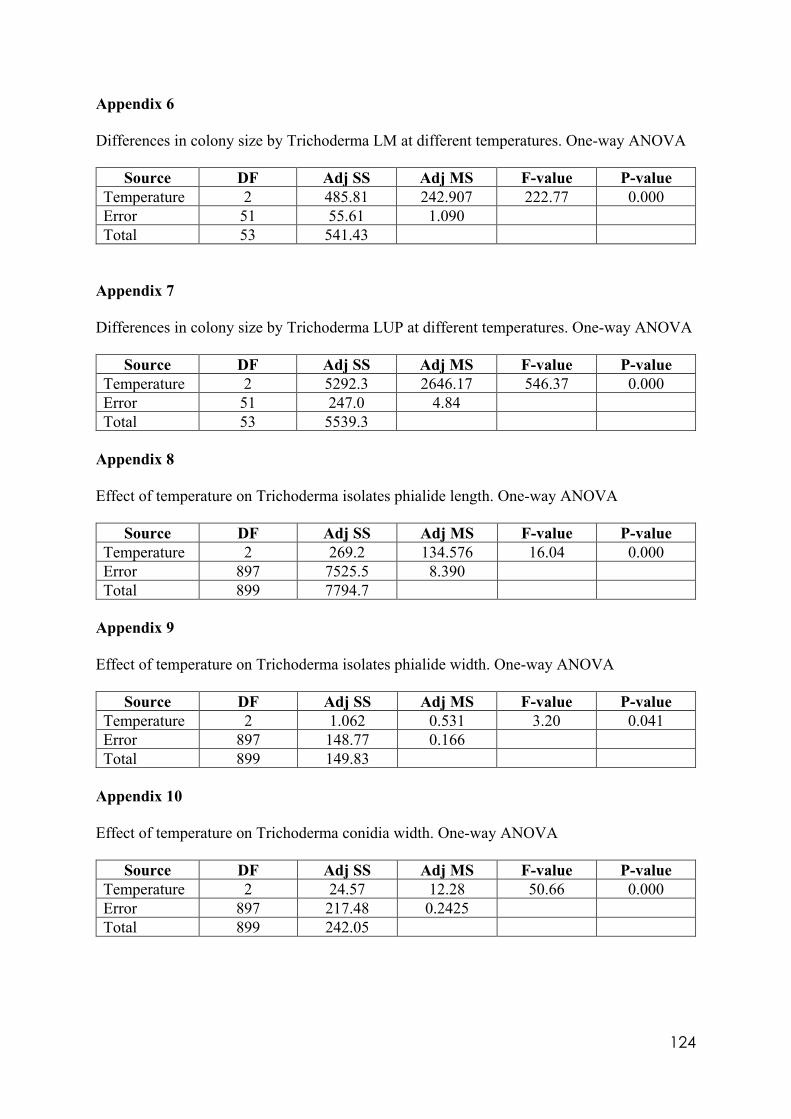
124
Appendix 6 Differences in colony size by Trichoderma LM at different temperatures. One-way ANOVA
Source DF Adj SS Adj MS F-value P-value Temperature 2 485.81 242.907 222.77 0.000 Error 51 55.61 1.090 Total 53 541.43
Appendix 7 Differences in colony size by Trichoderma LUP at different temperatures. One-way ANOVA
Source DF Adj SS Adj MS F-value P-value Temperature 2 5292.3 2646.17 546.37 0.000 Error 51 247.0 4.84 Total 53 5539.3
Appendix 8 Effect of temperature on Trichoderma isolates phialide length. One-way ANOVA
Source DF Adj SS Adj MS F-value P-value Temperature 2 269.2 134.576 16.04 0.000 Error 897 7525.5 8.390 Total 899 7794.7
Appendix 9 Effect of temperature on Trichoderma isolates phialide width. One-way ANOVA
Source DF Adj SS Adj MS F-value P-value Temperature 2 1.062 0.531 3.20 0.041 Error 897 148.77 0.166 Total 899 149.83
Appendix 10 Effect of temperature on Trichoderma conidia width. One-way ANOVA
Source DF Adj SS Adj MS F-value P-value Temperature 2 24.57 12.28 50.66 0.000 Error 897 217.48 0.2425 Total 899 242.05
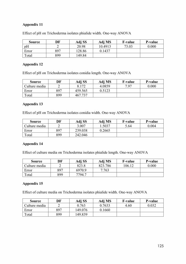
125
Appendix 11 Effect of pH on Trichoderma isolates phialide width. One-way ANOVA
Source DF Adj SS Adj MS F-value P-value pH 2 20.98 10.4913 73.03 0.000 Error 897 128.86 0.1437 Total 899 149.84
Appendix 12 Effect of pH on Trichoderma isolates conidia length. One-way ANOVA
Source DF Adj SS Adj MS F-value P-value Culture media 2 8.172 4.0859 7.97 0.000 Error 897 459.565 0.5123 Total 899 467.737
Appendix 13 Effect of pH on Trichoderma isolates conidia width. One-way ANOVA
Source DF Adj SS Adj MS F-value P-value Culture media 2 3.007 1.5037 5.64 0.004 Error 897 239.038 0.2665 Total 899 242.046
Appendix 14 Effect of culture media on Trichoderma isolates phialide length. One-way ANOVA
Source DF Adj SS Adj MS F-value P-value Culture media 2 823.8 823.786 106.12 0.000 Error 897 6970.9 7.763 Total 899 7794.7
Appendix 15 Effect of culture media on Trichoderma isolates phialide width. One-way ANOVA
Source DF Adj SS Adj MS F-value P-value Culture media 2 0.763 0.7633 4.60 0.032 Error 897 149.076 0.1660 Total 899 149.839
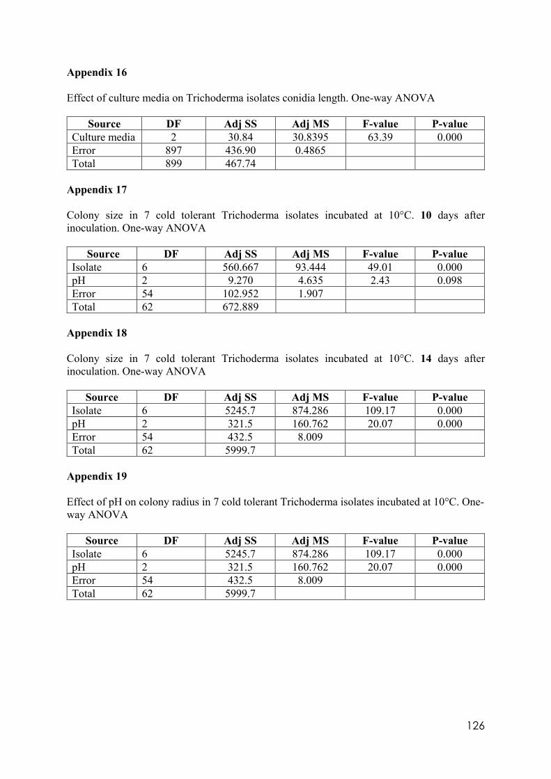
126
Appendix 16 Effect of culture media on Trichoderma isolates conidia length. One-way ANOVA
Source DF Adj SS Adj MS F-value P-value Culture media 2 30.84 30.8395 63.39 0.000 Error 897 436.90 0.4865 Total 899 467.74
Appendix 17 Colony size in 7 cold tolerant Trichoderma isolates incubated at 10°C. 10 days after inoculation. One-way ANOVA
Source DF Adj SS Adj MS F-value P-value Isolate 6 560.667 93.444 49.01 0.000 pH 2 9.270 4.635 2.43 0.098 Error 54 102.952 1.907 Total 62 672.889
Appendix 18 Colony size in 7 cold tolerant Trichoderma isolates incubated at 10°C. 14 days after inoculation. One-way ANOVA
Source DF Adj SS Adj MS F-value P-value Isolate 6 5245.7 874.286 109.17 0.000 pH 2 321.5 160.762 20.07 0.000 Error 54 432.5 8.009 Total 62 5999.7
Appendix 19 Effect of pH on colony radius in 7 cold tolerant Trichoderma isolates incubated at 10°C. One-way ANOVA
Source DF Adj SS Adj MS F-value P-value Isolate 6 5245.7 874.286 109.17 0.000 pH 2 321.5 160.762 20.07 0.000 Error 54 432.5 8.009 Total 62 5999.7
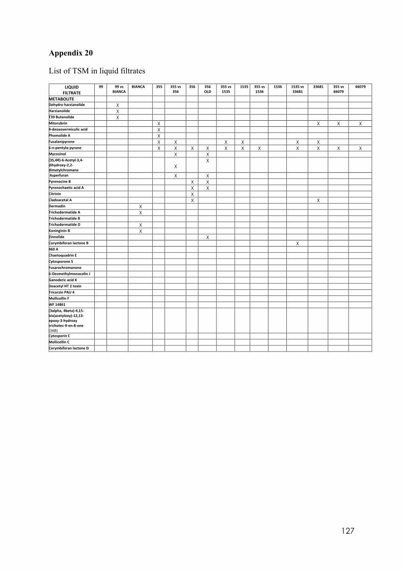
127
Appendix 20 List of TSM in liquid filtrates
LIQUID FILTRATE
99 99 vs BIANCA
BIANCA 355 355 vs 356
356 356 OLD
355 vs 1535
1535 355 vs 1536
1536 1535 vs 33681
33681 355 vs 66079
66079
METABOLITE Dehydro harzianolide X Harzianolide X T39 Butenolide X Mitorubrin X X X X 4-deoxovermiculic acid X Phomolide A X Fusalanipyrone X X X X X X 6-n-pentylα pyrone X X X X X X X X X X X Mycosinol X X (3S,4R)-6-Acetyl-3,4- dihydroxy-2,2-dimetylchromane
X
X
Asperfuran X X Pyrenocine B X X Pyrenochaetic acid A X X Citrinin X Cladoacetal A X X Dermadin X Trichodermatide A X Trichodermatide B Trichodermatide D X Koninginin B X Zinnolide X Corymbiferan lactone B X 460 A Chaetoquadrin E Cytosporone S Fusarochromanone 6-Desmethylmonacolin J Ganoderic acid K Deacetyl HT 2 toxin Tricorzin PAU 4 Mollicellin F WF 14861 (3alpha, 4beta)-4,15- bis(acetyloxy)-12,13- epoxy-3-hydroxy trichotec-9-en-8-one (3AB)
Cytosporin C Mollicellin C Corymbiferan lactone D
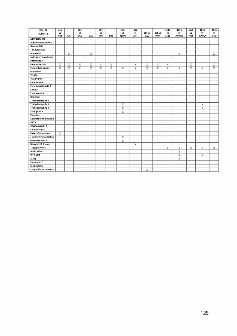
128
LIQUID FILTRATE
355 vs
ANT ANT
355 vs
GAU GAU
355 vs
PHY PHY
356 vs
45899
356 vs
ANT 356 vs GAU
356 vs PMR
1535 vs
1536
1535 vs
ANAND
1535 vs
ANT
1535 vs
BIANCA
1535 vs
GAU METABOLITE Dehydro harzianolide Harzianolide T39 Butenolide Mitorubrin X X X X 4-deoxovermiculic acid Phomolide A Fusalanipyrone X X X X X X X X X X X X 6-n-pentylα pyrone X X X X X X X X X X X X X X X Mycosinol (3S,4R) Asperfuran Pyrenocine B Pyrenochaetic acid A Citrinin Cladoacetal A Dermadin Trichodermatide A Trichodermatide B X X Trichodermatide D X X Koninginin B X Zinnolide Corymbiferan lactone B 460 A Chaetoquadrin E Cytosporone S Fusarochromanone X
6-Desmethylmonacolin J X Ganoderic acid K X Deacetyl HT 2 toxin X Tricorzin PAU 4 X X X X X Mollicellin F X WF 14861 X X (3AB) X Cytosporin C Mollicellin C Corymbiferan lactone D X
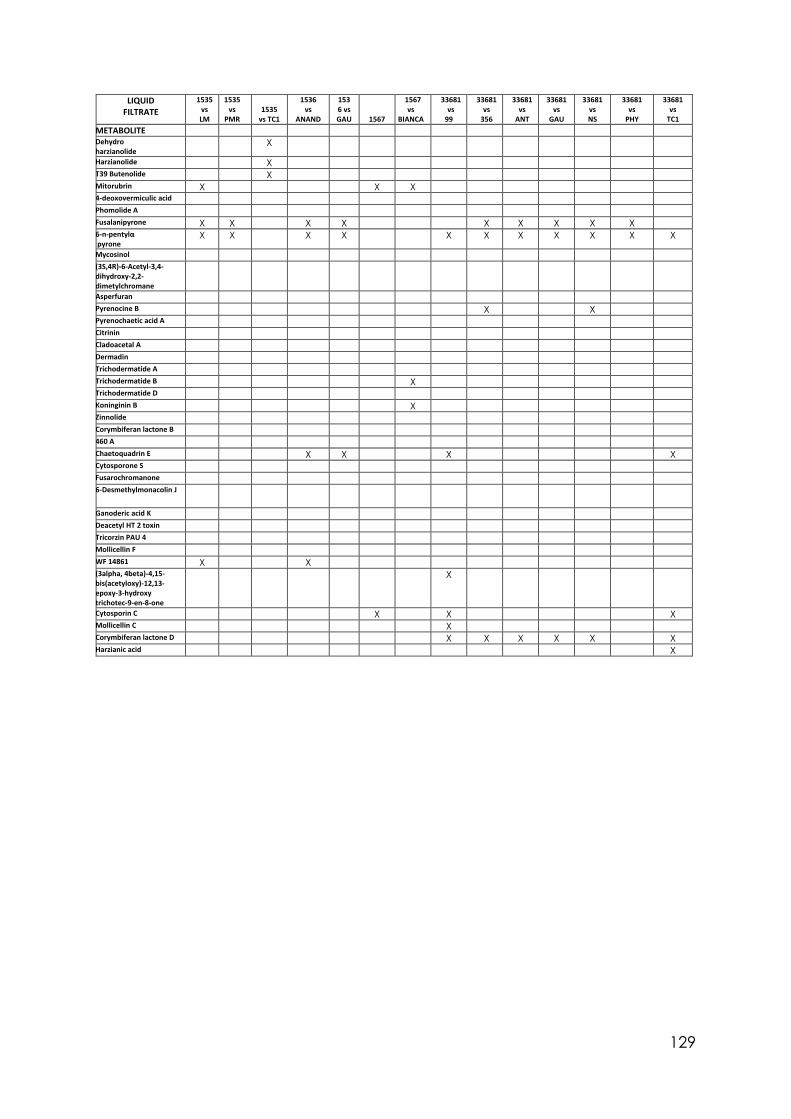
129
LIQUID FILTRATE
1535 vs LM
1535 vs
PMR 1535
vs TC1
1536 vs
ANAND
1536 vs GAU 1567
1567 vs
BIANCA
33681 vs
99
33681 vs
356
33681 vs
ANT
33681 vs
GAU
33681 vs NS
33681 vs
PHY
33681 vs
TC1 METABOLITE Dehydro harzianolide
X
Harzianolide X T39 Butenolide X Mitorubrin X X X 4-deoxovermiculic acid Phomolide A Fusalanipyrone X X X X X X X X X 6-n-pentylα pyrone
X X X X X X X X X X X
Mycosinol (3S,4R)-6-Acetyl-3,4- dihydroxy-2,2- dimetylchromane
Asperfuran Pyrenocine B X X Pyrenochaetic acid A Citrinin Cladoacetal A Dermadin Trichodermatide A Trichodermatide B X Trichodermatide D Koninginin B X Zinnolide Corymbiferan lactone B 460 A Chaetoquadrin E X X X X Cytosporone S Fusarochromanone 6-Desmethylmonacolin J
Ganoderic acid K Deacetyl HT 2 toxin Tricorzin PAU 4 Mollicellin F WF 14861 X X (3alpha, 4beta)-4,15- bis(acetyloxy)-12,13- epoxy-3-hydroxy trichotec-9-en-8-one
X
Cytosporin C X X X Mollicellin C X Corymbiferan lactone D X X X X X X Harzianic acid X
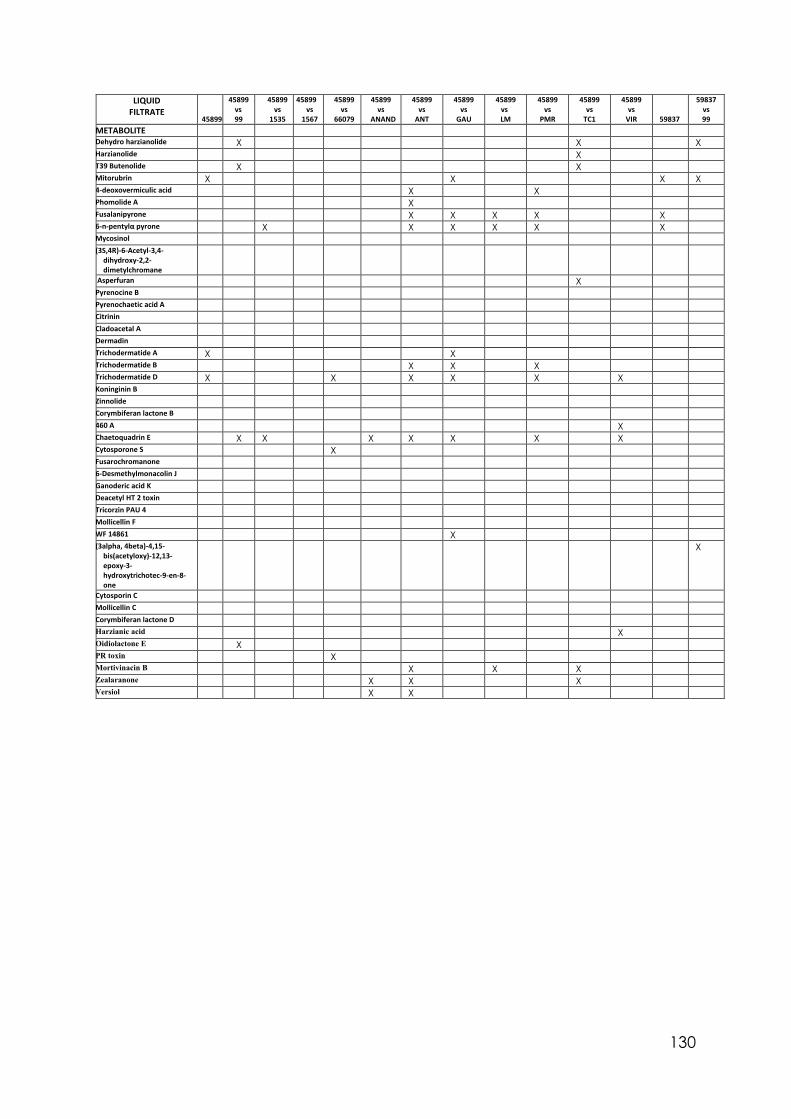
130
LIQUID FILTRATE
45899
45899 vs 99
45899 vs
1535
45899 vs
1567
45899 vs
66079
45899 vs
ANAND
45899 vs
ANT
45899 vs
GAU
45899 vs LM
45899 vs
PMR
45899 vs
TC1
45899 vs
VIR 59837
59837 vs 99
METABOLITE Dehydro harzianolide X X X Harzianolide X T39 Butenolide X X Mitorubrin X X X X 4-deoxovermiculic acid X X Phomolide A X Fusalanipyrone X X X X X 6-n-pentylα pyrone X X X X X X Mycosinol (3S,4R)-6-Acetyl-3,4-
dihydroxy-2,2-dimetylchromane
Asperfuran X Pyrenocine B Pyrenochaetic acid A Citrinin Cladoacetal A Dermadin Trichodermatide A X X Trichodermatide B X X X Trichodermatide D X X X X X X Koninginin B Zinnolide Corymbiferan lactone B 460 A X Chaetoquadrin E X X X X X X X Cytosporone S X Fusarochromanone 6-Desmethylmonacolin J Ganoderic acid K Deacetyl HT 2 toxin Tricorzin PAU 4 Mollicellin F WF 14861 X (3alpha, 4beta)-4,15-
bis(acetyloxy)-12,13-epoxy-3-hydroxytrichotec-9-en-8-one
X
Cytosporin C Mollicellin C Corymbiferan lactone D Harzianic acid X Oidiolactone E X PR toxin X Mortivinacin B X X X Zealaranone X X X Versiol X X
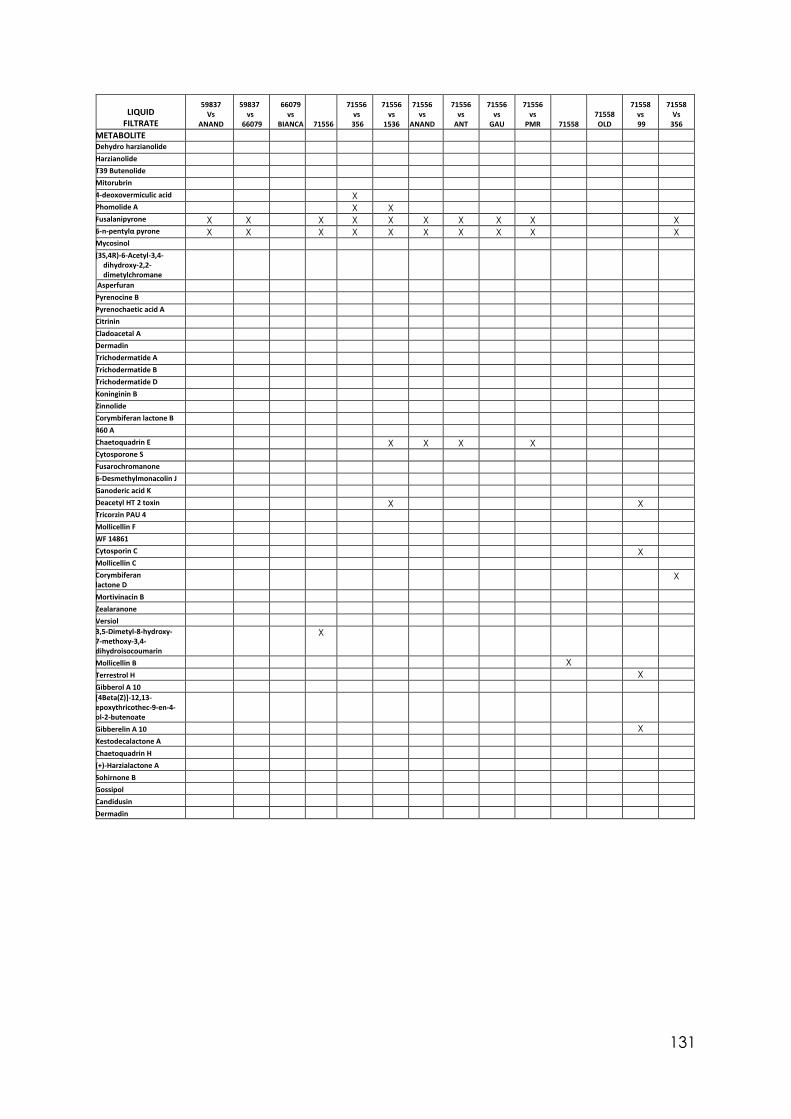
131
LIQUID
FILTRATE
59837 Vs
ANAND
59837 vs
66079
66079 vs
BIANCA 71556
71556 vs
356
71556 vs
1536
71556 vs
ANAND
71556 vs
ANT
71556 vs
GAU
71556 vs
PMR 71558 71558 OLD
71558 vs 99
71558 Vs
356 METABOLITE Dehydro harzianolide Harzianolide T39 Butenolide Mitorubrin 4-deoxovermiculic acid X Phomolide A X X Fusalanipyrone X X X X X X X X X X 6-n-pentylα pyrone X X X X X X X X X X Mycosinol (3S,4R)-6-Acetyl-3,4-
dihydroxy-2,2-dimetylchromane
Asperfuran Pyrenocine B Pyrenochaetic acid A Citrinin Cladoacetal A Dermadin Trichodermatide A Trichodermatide B Trichodermatide D Koninginin B Zinnolide Corymbiferan lactone B 460 A Chaetoquadrin E X X X X Cytosporone S Fusarochromanone 6-Desmethylmonacolin J Ganoderic acid K Deacetyl HT 2 toxin X X Tricorzin PAU 4 Mollicellin F WF 14861 Cytosporin C X Mollicellin C Corymbiferan lactone D
X
Mortivinacin B Zealaranone Versiol 3,5-Dimetyl-8-hydroxy-7-methoxy-3,4-dihydroisocoumarin
X
Mollicellin B X Terrestrol H X Gibberol A 10 [4Beta(Z)]-12,13-epoxythricothec-9-en-4-ol-2-butenoate
Gibberelin A 10 X Xestodecalactone A Chaetoquadrin H (+)-Harzialactone A Sohirnone B Gossipol Candidusin Dermadin
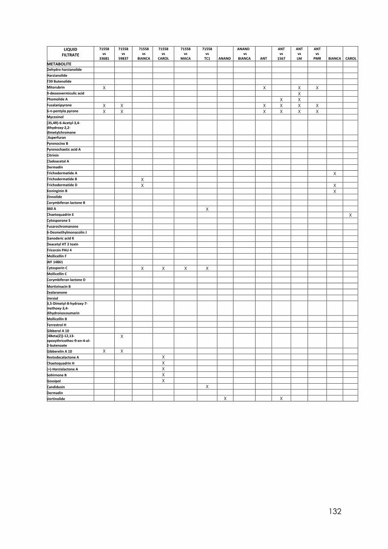
132
LIQUID FILTRATE
71558 vs
33681
71558 vs
59837
71558 vs
BIANCA
71558 vs
CAROL
71558 vs
MACA
71558 vs
TC1 ANAND
ANAND vs
BIANCA ANT
ANT vs
1567
ANT vs LM
ANT vs
PMR BIANCA CAROL METABOLITE Dehydro harzianolide Harzianolide T39 Butenolide Mitorubrin X X X X 4-deoxovermiculic acid X Phomolide A X X Fusalanipyrone X X X X X X 6-n-pentylα pyrone X X X X X X Mycosinol (3S,4R)-6-Acetyl-3,4- dihydroxy-2,2- dimetylchromane
Asperfuran Pyrenocine B Pyrenochaetic acid A Citrinin Cladoacetal A Dermadin Trichodermatide A X Trichodermatide B X Trichodermatide D X X Koninginin B X Zinnolide Corymbiferan lactone B 460 A X Chaetoquadrin E X Cytosporone S Fusarochromanone 6-Desmethylmonacolin J Ganoderic acid K Deacetyl HT 2 toxin Tricorzin PAU 4 Mollicellin F WF 14861 Cytosporin C X X X X Mollicellin C Corymbiferan lactone D Mortivinacin B Zealaranone Versiol 3,5-Dimetyl-8-hydroxy-7-methoxy-3,4-dihydroisocoumarin
Mollicellin B Terrestrol H Gibberol A 10 [4Beta(Z)]-12,13-epoxythricothec-9-en-4-ol-2-butenoate
X
Gibberelin A 10 X X Xestodecalactone A X Chaetoquadrin H X (+)-Harzialactone A X Sohirnone B X Gossipol X Candidusin X Dermadin Vertinolide X X
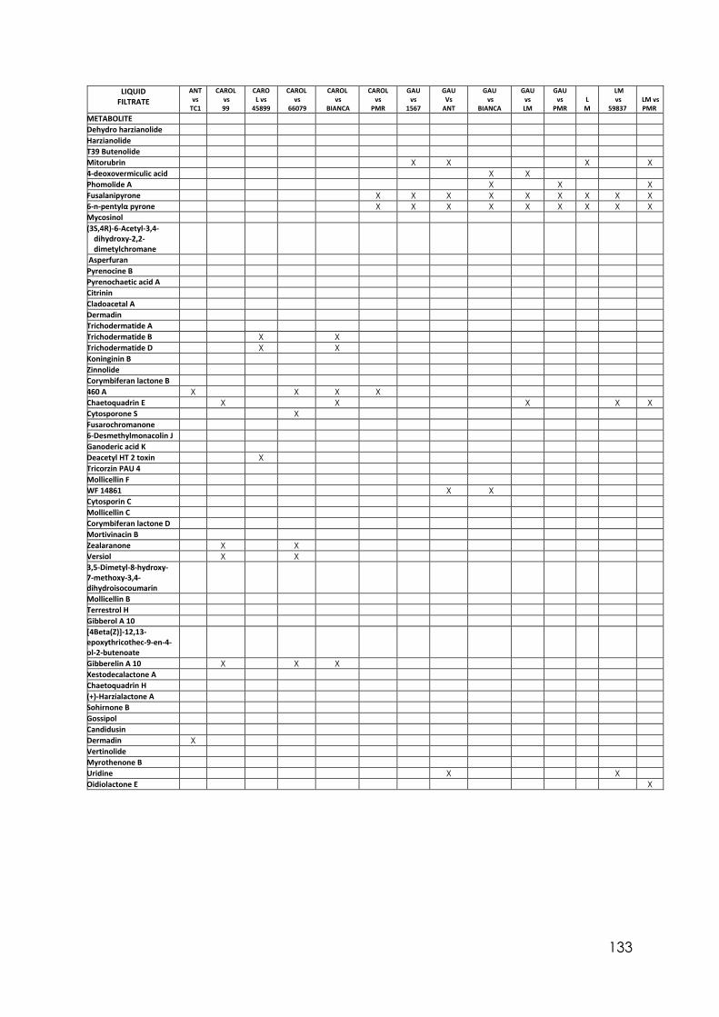
133
LIQUID FILTRATE
ANT vs
TC1
CAROL vs 99
CAROL vs
45899
CAROL vs
66079
CAROL vs
BIANCA
CAROL vs
PMR
GAU vs
1567
GAU Vs
ANT
GAU vs
BIANCA
GAU vs LM
GAU vs
PMR L
M
LM vs
59837 LM vs PMR
METABOLITE Dehydro harzianolide Harzianolide T39 Butenolide Mitorubrin X X X X 4-deoxovermiculic acid X X Phomolide A X X X Fusalanipyrone X X X X X X X X X 6-n-pentylα pyrone X X X X X X X X X Mycosinol (3S,4R)-6-Acetyl-3,4-
dihydroxy-2,2-dimetylchromane
Asperfuran Pyrenocine B Pyrenochaetic acid A Citrinin Cladoacetal A Dermadin Trichodermatide A Trichodermatide B X X Trichodermatide D X X Koninginin B Zinnolide Corymbiferan lactone B 460 A X X X X Chaetoquadrin E X X X X X Cytosporone S X Fusarochromanone 6-Desmethylmonacolin J Ganoderic acid K Deacetyl HT 2 toxin X Tricorzin PAU 4 Mollicellin F WF 14861 X X Cytosporin C Mollicellin C Corymbiferan lactone D Mortivinacin B Zealaranone X X Versiol X X 3,5-Dimetyl-8-hydroxy-7-methoxy-3,4-dihydroisocoumarin
Mollicellin B Terrestrol H Gibberol A 10 [4Beta(Z)]-12,13-epoxythricothec-9-en-4-ol-2-butenoate
Gibberelin A 10 X X X Xestodecalactone A Chaetoquadrin H (+)-Harzialactone A Sohirnone B Gossipol Candidusin Dermadin X Vertinolide Myrothenone B Uridine X X Oidiolactone E X
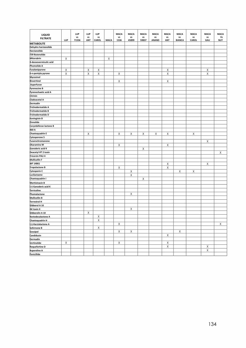
134
LIQUID
FILTRATE LUP
LUP vs
71556
LUP vs
ANT
LUP
vs CAROL
MACA
MACA
vs 1536
MACA
vs 45899
MACA
vs 59837
MACA
vs ANAND
MACA
vs ANT
MACA
vs BIANCA
MACA
vs CAROL
MACA
vs GAU
MACA
Vs NUT
METABOLITE Dehydro harzianolide Harzianolide T39 Butenolide Mitorubrin X X 4-deoxovermiculic acid Phomolide A Fusalanipyrone X X X X X 6-n-pentylα pyrone X X X X X X Mycosinol Bisvertinol X X Asperfuran Pyrenocine B Pyrenochaetic acid A Citrinin Cladoacetal A Dermadin Trichodermatide A Trichodermatide B Trichodermatide D Koninginin B Zinnolide Corymbiferan lactone B 460 A Chaetoquadrin E X X X X X X X Cytosporone S Fusarochromanone X Okaramine M X X Ganoderic acid K X Deacetyl HT 2 toxin X Tricorzin PAU 4 Mollicellin F WF 14861 X X Tropolactone B X X Cytosporin C X X X Lucilactaene X Chaetoquadrin I X Mortivinacin B (+)-Ganoderic acid K Terricoline Phomalactone X Mollicellin B Terrestrol H Gibberol A 10 AK toxin II X Gibberelin A 10 X Xestodecalactone A X Chaetoquadrin H X (+)-Harzialactone A X X Sohirnone B X Gossipol X X X Candidusin X Dermadin Vertinolide X X X Roquefortine D X X Aspereline A X Penicillide
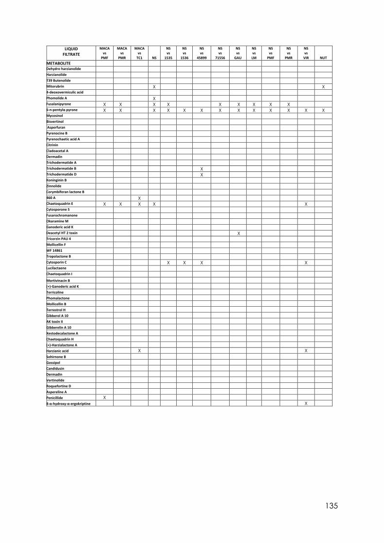
135
LIQUID FILTRATE
MACA vs
PMF
MACA vs
PMR
MACA vs
TC1 NS
NS vs
1535
NS vs
1536
NS vs
45899
NS vs
71556
NS vs
GAU
NS vs LM
NS vs
PMF
NS vs
PMR
NS vs
VIR NUT METABOLITE Dehydro harzianolide Harzianolide T39 Butenolide Mitorubrin X X 4-deoxovermiculic acid Phomolide A X Fusalanipyrone X X X X X X X X X 6-n-pentylα pyrone X X X X X X X X X X X X X Mycosinol Bisvertinol Asperfuran Pyrenocine B Pyrenochaetic acid A Citrinin Cladoacetal A Dermadin Trichodermatide A Trichodermatide B X Trichodermatide D X Koninginin B Zinnolide Corymbiferan lactone B 460 A X Chaetoquadrin E X X X X X Cytosporone S Fusarochromanone Okaramine M Ganoderic acid K Deacetyl HT 2 toxin X Tricorzin PAU 4 Mollicellin F WF 14861 Tropolactone B Cytosporin C X X X X Lucilactaene Chaetoquadrin I Mortivinacin B (+)-Ganoderic acid K Terricoline Phomalactone Mollicellin B Terrestrol H Gibberol A 10 AK toxin II Gibberelin A 10 Xestodecalactone A Chaetoquadrin H (+)-Harzialactone A Harzianic acid X X Sohirnone B Gossipol Candidusin Dermadin Vertinolide Roquefortine D Aspereline A Penicillide X 8-α-hydroxy-α-ergokriptine X
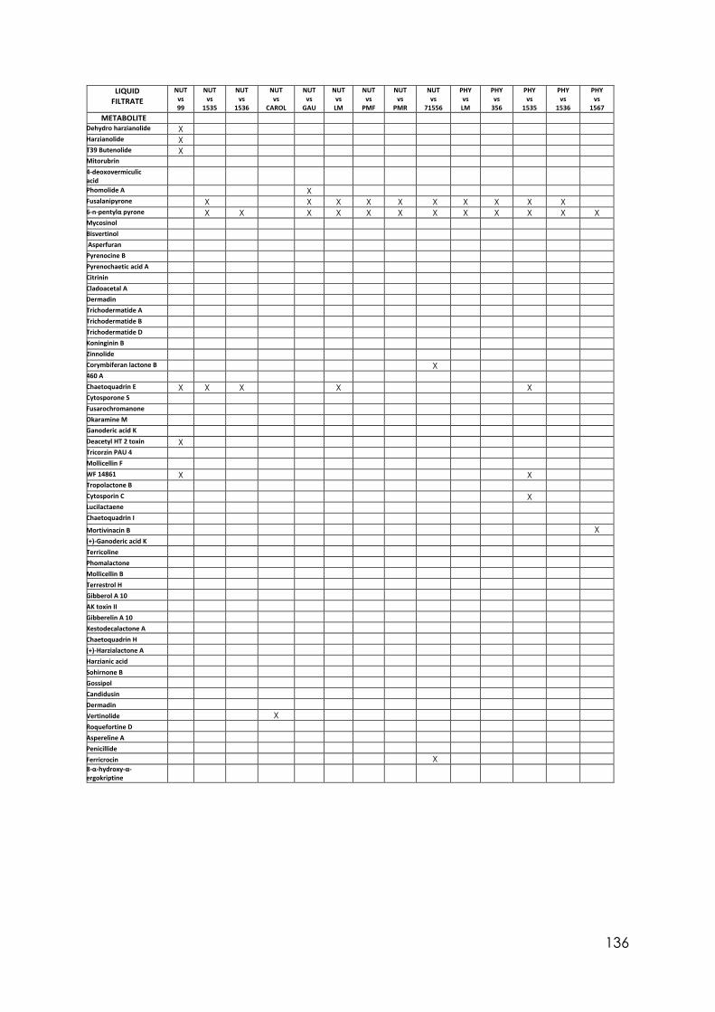
136
LIQUID FILTRATE
NUT vs 99
NUT vs
1535
NUT vs
1536
NUT vs
CAROL
NUT vs
GAU
NUT vs LM
NUT vs
PMF
NUT vs
PMR
NUT vs
71556
PHY vs LM
PHY vs
356
PHY vs
1535
PHY vs
1536
PHY vs
1567 METABOLITE
Dehydro harzianolide X Harzianolide X T39 Butenolide X Mitorubrin 4-deoxovermiculic acid
Phomolide A X Fusalanipyrone X X X X X X X X X X 6-n-pentylα pyrone X X X X X X X X X X X X Mycosinol Bisvertinol Asperfuran Pyrenocine B Pyrenochaetic acid A Citrinin Cladoacetal A Dermadin Trichodermatide A Trichodermatide B Trichodermatide D Koninginin B Zinnolide Corymbiferan lactone B X 460 A Chaetoquadrin E X X X X X Cytosporone S Fusarochromanone Okaramine M Ganoderic acid K Deacetyl HT 2 toxin X Tricorzin PAU 4 Mollicellin F WF 14861 X X Tropolactone B Cytosporin C X Lucilactaene Chaetoquadrin I Mortivinacin B X (+)-Ganoderic acid K Terricoline Phomalactone Mollicellin B Terrestrol H Gibberol A 10 AK toxin II Gibberelin A 10 Xestodecalactone A Chaetoquadrin H (+)-Harzialactone A Harzianic acid Sohirnone B Gossipol Candidusin Dermadin Vertinolide X Roquefortine D Aspereline A Penicillide Ferricrocin X 8-α-hydroxy-α- ergokriptine
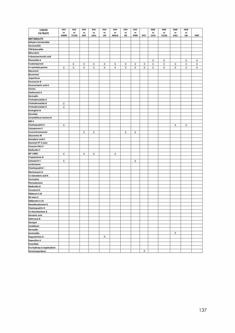
137
LIQUID FILTRATE
PHY vs
45899
PHY vs
71556
PHY vs
ANT
PHY vs
GAU
PHY vs LM
PHY vs
MACA
PHY vs NS
PHY vs
PMR PHY
PMF vs
1535
PMF vs
71556
PMF vs
GAU
PMF vs LM PMF
METABOLITE Dehydro harzianolide Harzianolide T39 Butenolide Mitorubrin 4-deoxovermiculic acid Phomolide A X X X X Fusalanipyrone X X X X X X X X X X X X X 6-n-pentylα pyrone X X X X X X X X X X X X X X Mycosinol Bisvertinol Asperfuran Pyrenocine B Pyrenochaetic acid A Citrinin Cladoacetal A Dermadin Trichodermatide A Trichodermatide B X Trichodermatide D X Koninginin B Zinnolide Corymbiferan lactone B 460 A Chaetoquadrin E X X X Cytosporone S Fusarochromanone X X X X Okaramine M Ganoderic acid K Deacetyl HT 2 toxin Tricorzin PAU 4 Mollicellin F WF 14861 X X X X Tropolactone B Cytosporin C X X Lucilactaene Chaetoquadrin I Mortivinacin B (+)-Ganoderic acid K Terricoline Phomalactone Mollicellin B Terrestrol H Gibberol A 10 AK toxin II Gibberelin A 10 Xestodecalactone A Chaetoquadrin H (+)-Harzialactone A Harzianic acid Sohirnone B Gossipol Candidusin Dermadin Vertinolide X Roquefortine D X Aspereline A Penicillide 8-α-hydroxy-α-ergokriptine Harzianopyridone X
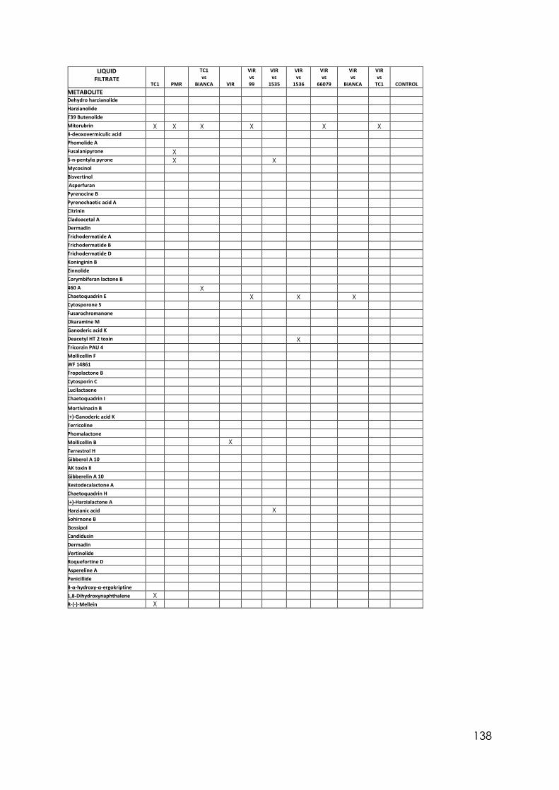
138
LIQUID FILTRATE
TC1
PMR
TC1 vs
BIANCA VIR
VIR vs 99
VIR vs
1535
VIR vs
1536
VIR vs
66079
VIR vs
BIANCA
VIR vs
TC1 CONTROL METABOLITE Dehydro harzianolide Harzianolide T39 Butenolide Mitorubrin X X X X X X 4-deoxovermiculic acid Phomolide A Fusalanipyrone X 6-n-pentylα pyrone X X Mycosinol Bisvertinol Asperfuran Pyrenocine B Pyrenochaetic acid A Citrinin Cladoacetal A Dermadin Trichodermatide A Trichodermatide B Trichodermatide D Koninginin B Zinnolide Corymbiferan lactone B 460 A X Chaetoquadrin E X X X Cytosporone S Fusarochromanone Okaramine M Ganoderic acid K Deacetyl HT 2 toxin X Tricorzin PAU 4 Mollicellin F WF 14861 Tropolactone B Cytosporin C Lucilactaene Chaetoquadrin I Mortivinacin B (+)-Ganoderic acid K Terricoline Phomalactone Mollicellin B X Terrestrol H Gibberol A 10 AK toxin II Gibberelin A 10 Xestodecalactone A Chaetoquadrin H (+)-Harzialactone A Harzianic acid X Sohirnone B Gossipol Candidusin Dermadin Vertinolide Roquefortine D Aspereline A Penicillide 8-α-hydroxy-α-ergokriptine 1,8-Dihydroxynaphthalene X R-(-)-Mellein X
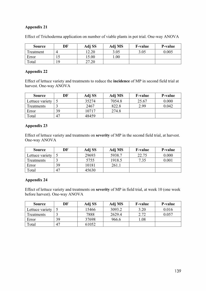
139
Appendix 21 Effect of Trichoderma application on number of viable plants in pot trial. One-way ANOVA
Source DF Adj SS Adj MS F-value P-value Treatment 4 12.20 3.05 3.05 0.005 Error 15 15.00 1.00 Total 19 27.20
Appendix 22 Effect of lettuce variety and treatments to reduce the incidence of MP in second field trial at harvest. One-way ANOVA
Source DF Adj SS Adj MS F-value P-value Lettuce variety 5 35274 7054.8 25.67 0.000 Treatments 3 2467 822.8 2.99 0.042 Error 39 10717 274.8 Total 47 48459
Appendix 23 Effect of lettuce variety and treatments on severity of MP in the second field trial, at harvest. One-way ANOVA
Source DF Adj SS Adj MS F-value P-value Lettuce variety 5 29693 5938.7 22.75 0.000 Treatments 3 5755 1918.5 7.35 0.001 Error 39 10181 261.1 Total 47 45630
Appendix 24 Effect of lettuce variety and treatments on severity of MP in field trial, at week 10 (one week before harvest). One-way ANOVA
Source DF Adj SS Adj MS F-value P-value Lettuce variety 5 15466 3093.2 3.20 0.016 Treatments 3 7888 2629.4 2.72 0.057 Error 39 37698 966.6 1.08 Total 47 61052
140
REFERENCES
Abadi K (2008) Novel plant-bioprotectants based on Trichoderma spp. strains with superior
characteristics. Dottorato di Ricerca in agrobiologia e agrochimica. Universita’ degli
studi di Napoli “Federico II”.
ABC Tv series program. War on waste: Presented by Craig Recaussel 2019.
Abeyratne G, Deshappriya N (2018) The effect of pH on the biological control activities of a
Trichoderma sp. against Fusarium sp. isolated from the commercial onion fields in Sri
Lanka. Tropical Plant Research. 5(2), 121-128.
Abraham W, Arfmann H (1988) Fusalanipyrone a monoterpenoid from Fusarium solani.
Phytochemistry. 27(10), 3310-3311.
Abraham W, Knoch I, Witte L (1990) Biosynthesis of the terpenoid polyketide Fusalipyrone.
Phytochemistry. 29(9), 2877-2878.
Adejeji A, Odebode A, Agbeniji S (2008) Bioassay of five Trichoderma strains against
Phytophthora megakaria. (Cacao pod-rot) in Nigeria. Scientific Research and Assays.
3(9), 390-394.
Agrios G (2004) Plant Pathology. Fifth edition. Elsevier Academic Press. 443 p.
Ahmad J, Baker R (1988) Rhizosphere competence of Trichoderma harzianum. Canadian
Journal of Microbiology 34(3): 229-234.
Alam I, Gupta S, Bohre A, Ahmad E, Khan T, Saha B, Haider M (2016) Development of 6-
amyl-alpha-pyrone as a potential biomass-derived platform molecule. Journal of Green
Chemistry. 18(24): 6431-6435.
Alvarado-Marchena L, Rivera-Méndez W (2016) Molecular identification of Trichoderma spp.
in garlic and onion fields and in vitro antagonism trials on Sclerotium cepivorum.
Revista Brasileira de Ciência do Solo. 40: e 0150454 pp 1-9.
Altomare C, Norvell W, Björkman T, Harman G (1999) Solubilisation of phosphates and
micronutrients by the plant-growth-promoting and biocontrol fungus Trichoderma
harzianum Rifai 1295-22. Applied and Environmental Microbiology. 65(7): 2926-
2936.
Andrade R, Ayer W, Trifonov L (2015) The metabolites of Trichoderma longibrachiatum. Part
II. The structures of trichodermolide and sorbiquinol. Canadian Journal of Chemistry.
74(3): 371-379.
141
Anees M, Tronsmo A, Edel-Hermann V, Hjeljord L, Héraud C, Steinberg C (2010)
Characterization of field isolates of Trichoderma antagonistic against Rhizoctonia
solani. Fungal Biology. 114: 691-701.
Anke H, Kinn J, Bergquist KE, Sterner O (1991) Production of siderophores by strains of the
genus Trichoderma. Biology of Metals. 4(3), 176-180
Arjona-Girona I, Vinale F, Ruano-Rosa D, M Lorito, Lopez-Herrera C (2014) Effect of
metabolites from different Trichoderma strains on the growth of Rosellinia necatrix,
the causal agent of Avocado root rot. European Journal of Plant Pathology. 140(2),
385-397.
Asha N, Raghunandan B, Shivaprakash M (2013) In vivo evaluation of Trichoderma spp. for
survival and their population in rhizosphere soil of French bean. Madras Agricultural
Journal. 100(4-6), 554-558.
Atasanova L (2014) Ecophysiology of Trichoderma in genomic perspective. In. Biotechnology
and Biology of Trichoderma. Chapter 2. Edited by Gupta et al. p.35.
Australian Government. Australian Pesticides and Veterinary Medicines Authority (APVMA)
(2012) Agricultural and veterinary chemical code instrument (MRL standard) N° 4.
Compilation N 66, May 2019.
Australian Organic Ltd. (2019) Australian organic market report. 2 p.
Azarmi R, Hajieghrari B, Giglou A (2011) Effect of Trichoderma isolates on tomato seedling
growth response and nutrient uptake. African Journal of Biotechnology. 10(31), 5850-
5855.
Bae Y, Knudsen G (2005) Soil microbial biomass influence on growth and biocontrol efficacy
by Trichoderma harzianum. Biological Control. 32(2), 236-242.
Baker R (1986) Biological control: An overview. Canadian Journal of Plant Pathology. 8,
218-221.
Balouiri M, Sadiqui M, Ibnsouda S (2015) Methods for in vitro evaluating anti-microbial
activity: A review. Journal of Pharmaceutical Analysis. 6, 71-79.
Batista B, Almeida J, Bezerra T, Acevedo J, Quencine M (2017) Describing the unexplored
microorganisms associated with guarana: A typical tropical plant. Diversity and
Benefits of Microorganisms from the Tropics. DOI 10.1007/978-3-319-55804-2_13.
Benhamou N, Chet I (1993) Hyphal interactions between Trichoderma harzianum and
Rhizoctonia solani: Ultrastructure and gold cytochemistry of the myco-parasitic
process. Phytopathology. 83, 1062-1071.
Berek L, Pietri I, Mesterházy A, Térek J, Molnár J (2001) Effect of mycotoxins on human
142
immune functions in vitro. Toxicology in vitro. 15(1), 25-30.
Bernal-Vicente A, Ros M, Pascual J (2012) Inoculation of Trichoderma harzianum during
maturation of vineyard waste compost to control muskmelon Fusarium wilt.
Bioresources 7(2), 1948-1960.
Blaya J, Marhuenda F, Pascual J, Ros M, (2016) Microbiota characterisation of compost using
omics approaches opens new perspectives for Phytophthora root rot control. PLoS
ONE 11(8), e0158048. DOI: 10.1371/journal.pone.0158048.
Bodo B, Rebuffat S, El Hajji M, Davoust D (1985) Structure of Trichorzianines A IIIc, an
antifungal peptide from Trichoderma harzianum. Journal of American Chemistry
Society. 107, 6011-6017.
Bonanomi G, Lorito M, Vinale F, Woo S (2018) Organic amendments, beneficial microbes,
and soil microbiota: Toward a unified framework for disease suppression. Annual
Review of Phytopathology. 56, 1.1-1.20.
Bringmann G, Lang G, Michel M, Heubes M (2004) Stereochemical assignment of the fungal
metabolite Xestodecalactone A by total synthesis. ChemInform. 45(13), 2829-2831.
Brimmer T, Boland G (2003) A review of the non-target effects of fungi used to biologically
control plant diseases. Agriculture, Ecosystems and Environment. 100, 3-16.
Brito J, Ramada M, Magalhães M, Silva L, Ulhoa C (2014) Peptaibols from Trichoderma
asperellum TR356 strain isolated from Brazilian soil. SpringerPlus. 3(600), 1-10.
Brožová J (2004) Review. Mycoparasitic fungi Trichoderma spp. in plant protection. Plant
Protection Science. 40(2), 63-74.
Burgess T, Bihon W, Wingfield MJ, Wingfield B (2009) A simple and rapid method to
determine vegetative compatibility groups in fungi. Inoculum Supplement to
Mycologia. 60(6), 1-2.
Carbone I, Kohn L (1999) A method for designing primers sets for speciation studies in
filamentous fungi. Mycologia. 91(3), 553-556.
Caron-Beaudoin E, Denison M, Sanderson J (2016) Effects of neonicotinoids on promoter-
specific expression and activity of aromatase (CYP 19) in human adrenocortical
carcinoma (H295R) and primary umbilical vein endothelial (HUVEC) cells.
Toxicological Sciences. 149(1), 134-144.
Carreras-Villasenor N, Sanchez-Arreguin J, Herrera-Estrella A (2012) Trichoderma: Sensing
the environment for survival and dispersal. Microbiology. 158, 3-6.
Castle A, Speranzini D, Rghei N, Alm G, Rinker D, Bissett J (1998) Morphological and
molecular identification of Trichoderma isolates on North American mushroom farms.
143
Applied and Environmental Microbiology. 64(1) 133-137.
Chaverri P, Samuels G (2003) Hypocrea/Trichoderma (Ascomycota, Hypocreales,
Hypocreaceae): species with green ascospores. Studies in Mycology. 48, 1-119.
Chaverri P, Branco Rocha F, Jaklitsch W, Gazis R, Degenkolb T, Samuels G (2015)
Systematics of the Trichoderma harzianum species and the reidentification of
commercial biocontrol strains. Mycologia. 107(3), 558-590.
Choundary A, Reddy KRN, Reddy MS (2007) Antifungal activity and genetic variability of
Trichoderma harzianum isolates. Journal of Mycology and Plant Pathology. 37(2), 1-
5.
Cole FM (1998) An investigation of the taxonomy and distribution of Stagnospora meliloti and
Acrocalymma medicaginis, fungi implicated in red-crown rot of Lucerne, Medicago
sativa. Ph.D. Thesis. Department of Biological Sciences. Monash University, Australia.
Contreras-Cornejo H, Macias-Rodríguez L, Cortez-Penagos C, López-Bucio J, (2009)
Trichoderma virens, a plant beneficial fungus, enhances biomass production and
promotes lateral root growth through an auxin-dependent mechanism in Arabidopsis.
Plant Physiology. 149, 1579-1592.
Contreras-Cornejo H, Macías-Rodríguez L, Alfaro-Cuevas R, López-Bucio J (2014)
Trichoderma spp. Improve growth of Arabidopsis seedlings under salt stress through
enhanced root development, osmolyte production and Na+ elimination through root
exudates. MPMI. 26 (6), 506-514.
Contreras-Cornejo H, Macías-Rodríguez L, del-Val Ek, Larsen J (2016) Ecological functions
of Trichoderma spp. and their secondary metabolites in the rhizosphere: Interactions
with plants. FEMS Microbiology Ecology. 92, 1-17.
Coutinho E (2002) Gossypol: A contraceptive for men. Contraception. 65, 259-263.
Cutler H, Jacyno J (1991) Biological activity of -(-) Harziano-pyridone isolated from
Trichoderma harzianum. Journal of Agricultural and Biological Chemistry. 55(10),
2629-2631.
Danielson R, Davey C (1973) The abundance of Trichoderma propagules and the distribution
of species in forest soils. Soil Biology and Biochemistry. 5(5), 485-494.
Dayaei A, Jones E, Glare T, Fallon R (2016) pH and water activity in culture media after
biological control activity of Trichoderma atroviride against Rhizoctonia solani.
Biological Control. 92, 24-30.
Delabona S, Farinas CS, da Silva MR, Azzoni SF, Pradella JG (2012) Use of a new
Trichoderma harzianum strain isolated from the Amazon rainforest with pre-treated
144
sugar cane bagasse for on-site cellulase production. Bioresource Technology. 107: 517-
521.
De Paula J, Texeira H, Faria R, Boechat M, Da Silva M, Lima R, De Sousa J (2012) Limitations
in controlling white mould on common beans with Trichoderma spp. at the fall-winter
season. Summa Phytopathologica Botucatu. 38(4), 337-340.
Dennis C, Webster J (1971) Antagonistic properties of species-groups of Trichoderma: I.
Production of non-volatile antibiotics. British Mycological Society. 51(1), 25-39.
Dewan M, Sivasithamparam K (1988) Identity and frecuency of occurrence of Trichoderma
spp.in roots of wheat ans ryegrass in Western Australia and their effect in rot root
caused by Gaeumannomyces graminis var. tritici. Plant and Soil. 109, 93-101.
Dickinson J, Hanson J, Hitchcock P, Claydon N (1989) Structure and biosynthesis of
Harzianopyridone, an antifungal metabolite of Trichoderma harzianum. Journal of the
Chemical Society Perkin Transactions 1. 21(8), DOI: 10.1039/p19890001885.
Djonovíc S, Pozo M, Dangott L, Howell C, Kenerley C (2006) Sm1 a proteinaceous elicitor
secreted by the biocontrol fungus Trichoderma virens induces plant defence responses
and systemic resistance. Molecular Plant-Microbe Interactions. 19(8), 838-853.
Domsch K, Gams W, Anderson T (1980) Compendium of soil fungi. Vol 1. Academic press.
797 p.
Druzhinina I, Kubicek C (2005) Species, concepts, and biodiversity in Trichoderma and
Hypocrea: From aggregate species to species clusters? Journal Zhejiang University
SCIENCE. 6B (2), 100-112.
Druzhinina I, Seidl-Seiboth V, Herrera-Estrella A, Horwitz B, Kenerley C, Monte E,
Mukherjee P, Zeilinger S, Grigoriev I, Kubicek C (2011) Trichoderma: the genomics
of opportunistic success. Nature Reviews. 9, 749-759.
Dubei M, Aamir M, Kaushik M, Khare S, Meena M, Singh S, Upadhyay R (2018) PR toxin-
biosynthesis, genetic regulation, toxicological potential, prevention and control
measures: Overview and challenges. Frontiers in Pharmacology. 9(288), 1-19.
Elad Y, Chet I, Katan J (1980) Trichoderma harzianum: A biocontrol agent effective against
Sclerotium rolfsii and Rhizoctonia solani. The American Phytopathological Society. 70
(2), 119-121.
Elad Y, Kirshner B (1992) Establishment of an active Trichoderma population in the
145
phylloplane and its effect on grey mould (Botrytis cinerea). Phytoparasitica. 20, 137-
141.
Elad Y (2000) Biological control of foliar pathogens by means of Trichoderma harzianum and
potential modes of action. Crop Protection. 19, 709-714.
El Hassan A, Walker F, Schöne J, Buchenauer H (2007) Antagonistic effect of 6-penthyl-alpha-
pyrone produced by Trichoderma harzianum towards Fusarium moniliforme. Journal
of Plant Diseases and Protection. 114(2), 62-68.
El Hassan A, Buchenauer H (2009) Actions of 6-pentyl-alpha-pyrone in controlling seedling
blight incited by Fusarium moniliforme and inducing defence responses in maize.
Phytopathology. 157, 697-707.
Elias P, Garrigós L, Mantovanello C, Harakava R (2017) Selection of Trichoderma spp. strains.
Pesquisa Agropecuaria Brasileira. 52(12): 1140-1148.
El-Refai I, Amer S, Assawah S, Draz M (2013) Vegetative compatibility and strain
improvement of some Egyptian Trichoderma isolates. Life Science Journal. 10(3), 187-
197.
Evueh A, Okhuoya J, Osemweigie O, Attitalla I, Ogbebor O (2011) Evaluation of Phylloplane
fungi as biocontrol agent of Corynespora leaf fall disease of rubber (Hevea brasiliensis
Muell. Arg). World Journal of Fungal and Plant Biology. 2(1), 1-5.
Ferraz H, Bombonato F, Sano M, Longo L (2008) Natural occurrence, biological activities and
synthesis of eight-, nine-, and eleven-membered ring lactones. Quimica Nova. 31(4),
885-900.
Ferrigo D, Raiola A, Rasera R, Causin R (2014) Trichoderma harzianum seed treatment
controls Fusarium verticillioides colonisation and fumonisin contamination in maize
under field conditions. Crop Protection. 65, 51-56.
Fiorentino N, Ventorino V, Woo S, Pepe O, De Rosa A, Gioia L, Romano I, Lombardi N,
Napolitano M, Colla G, Rouphael Y (2018) Trichoderma based biostimulants modulate
rhizosphere microbial populations and improve N uptake efficiency, yield and
nutritional quality of leafy vegetables. Frontiers in Plant Science. 9(743), 1-15.
Flajs D, Peraica M (2009) Toxicological properties of citrinin. Arh. Hig. Rada. Toksikol. 60
(4), 457-464.
Fravel D (2005) Commercialisation and implementation of biocontrol. Annual Review of
Phytopathology. 43, 337-359.
Galea V, Price T, Sutton B (1986) Taxonomy and biology of the lettuce anthracnose fungus.
Transactions of the British Mycological Society. 86(4), 619-628.
146
Galea V, Price T (1988a) Infection of lettuce by Microdochium panattonianum. Transactions
of the British Mycological Society. 91(3), 419-425.
Galea V, Price T (1988b) Survival of the lettuce anthracnose fungus in Victoria (Microdochium
panattonianum). Plant Pathology. 37(1), 54-63.
Gaynes R (2017) The discovery of penicillin- New insights after more than 75 years of clinical
use. Emerging Infectious Diseases. 23(5), 849-853.
Geraldine A, Cardoso F, Costa D, Barbosa E, Rodrigues A, Silva R, Ulhoa C, Junior M (2013)
Cell wall-degrading enzymes and parasitism of sclerotia are key factors on field
biocontrol of white mould by Trichoderma spp. Biological Control. 67, 308-316.
Ghildiyal A, Pandey A (2008) Isolation of cold tolerant antifungal strains of Trichoderma sp.
from glacial sites of Indian Himalayan region. Research Journal of Microbiology. 3(8),
559-564.
Ghisalberti E, Sivasithamparam K (1991) Antifungal antibiotics produced by Trichoderma spp.
Soil Biology and Biochemistry. 23(11), 1011-1020.
Gilman J (1957) A manual of soil fungi. Iowa State University. Second Edition. 212-215 pp.
Gomez I, Chet I, Herrera-Estrella A (1997) Genetic diversity and vegetative compatibility
among Trichoderma harzianum isolates. Molecular Genetics and Genomics. 256, 127-
135.
Gräfenhan T, Schroers H, Nirenberg H, Seifert K (2011) An overview of the taxonomy,
phylogeny and typification of some nectriaceous fungi classified in Cosmospora,
Acremonium, Fusarium, Stilbella and Volutella. Studies in Mycology. 68, 79-103.
Grant C, Pramer D (1962) Minor element composition of yeast extract. Journal of
Bacteriology. 84, 869-870.
Griffith G, Easton G, Detheridge A, Roderick K, Edwards A, Worgan H, Nicholson J, Perkins
W (2007) Copper deficiency in Potato dextrose agar causes reduced pigmentation in
cultures of various fungi. FEMS Microbiology letters. 276(2), 165-171.
Griffin D, Kellogg C, Peak K, Shinn E (2002) A rapid and efficient assay for extracting DNA
from fungi. Letters in Applies Microbiology. 34, 210-214.
Guigón-López C, Guerrero-Prieto V, Vargas-Albores F, Carbajal-Millán E, Ávila-Quezada G,
Bravo-Luna L, Ruocco M, Lanzuise S, Woo S, Lorito M (2010) Identificación
molecular de cepas nativas de Trichoderma sp. su tasa de crecimiento in vitro y
antagonismo contra hongos fitopatógenos. Revista Mexicana de Fitopatología. 28(2),
87-96.
Guigón-López C, Carbajal-Millán E, Ponce de Leon N, Vargas-Albores F, Bravo-Luna Leticia,
147
Guerrero-Prieto V (2010) Microcalometric measurement of Trichoderma spp. Growth
at different temperatures. Thermochimica Acta. 509, 40-45.
Ha T (2010) Using Trichoderma species for biological control of plant pathogens in Vietnam.
Journal of the International Society for Southeast Asian Agricultural Sciences. 16(1),
17-21.
Hanson J (2005) The chemistry of the biocontrol agent, Trichoderma harzianum. Science
progress. 88(4), 237-248.
Hanson L, Howell C (2004) Elicitors of plant defence responses from biocontrol strains of
Trichoderma virens. Phytopathology. 94(2), 171-176.
Harman G (2000). Myths and dogmas of biocontrol: changes in perceptions derived from
research on Trichoderma harzianum T-22. Plant Disease. 84, 377–393.
Hassan M (2014) Influence of protoplast fusion between two Trichoderma spp. on extracellular
enzymes production and antagonistic activity. Biotechnology and Biotechnological
Equipment. 28(6), 1014-1026.
Hatvani L, Vágvölgyi C, Kredics L, Irina Druzhinina (2014) DNA barcode for species in
Trichoderma. In: Chapter 3. Biotechnology and Biology of Trichoderma. Edited by
Gupta et al. 47p.
Herath H, Herath W, Carvalho P, Khan S, Tekwani B, Duke S, Tomaso-Peterson M,
Nanayakkara N (2009) Biologically active tetranorditerpenoids from the fungus
Sclerotinia homeocarpa causal agent of dollar spot in turf grass. Journal of Natural
Products. 72(12), 2091-2097.
Hermosa R, Cardoza R, Rubio B, Gutiérrez S, Monte E (2014) Secondary metabolism and
antimicrobial metabolites of Trichoderma. In: Chapter 10 Biotechnology and Biology
of Trichoderma. doi.org/10.1016/C2012-0-00434-6.
Herrera-Estrella A, Casas-Flores S, Kubicek C (2016) Chapter 13 Nematophagous fungi. In:
The Mycota IV. Druzhinina I and Kubicek C. Springer International publishing
Switzerland. 249 p.
Hinsinger P, Plassard C, Tang C, Jaillard B (2003) Origins of root-mediated pH changes in the
rhizosphere and their responses to environmental constraints: a review. Plant and Soil.
248:43–59.
Hoffmeister D and Keller N (2007) Natural products of filamentous fungi: Enzymes, genes and
their regulations. Natural Products Reports. 24(2), 396-416.
Hoitlink H, Stone A, Han D (1997) Suppression of plant diseases by composts. Hortscience.
148
32 (2),184-187.
Hoitlink H, Madden L, Dorrance A (2006) Systemic resistance induced by Trichoderma spp.:
Interactions between the host, the pathogen, the biological control agent, and the soil
organic matter quality. Phytopathology. DOI: 10.1094/PHYTO-96-0186.
Ibrahim S, Mohamed G, Al Haidari R, El Kholy A, Zayed M, Khayat M (2018) Biologically
active fungal depsidones: Chemistry, biosynthesis, structural characterisation and
bioactivities. Fitoterapia. 129, 317-365.
Ichihara A, Tazani H, Sakamura S (1985) The structure of Zinnolide, a new phytotoxin from
Alternaria solani. Agricultural and Biological Chemistry. 49(9), 2811-2812.
Ióca L, Romminger S, Santos M, Bandeira K, Rodrigues F, Kossuga M, Nicacio K, Ferreira E,
Morais-Urano R, Passos M, Kohn L, Arns C, Sette L, Berlinck R (2016) A strategy for
the rapid identification of fungal metabolites and the discovery of the antiviral activity
of Pyrenocyne A and harzianopyridone. Quimica Nova. 39(6), 709-731.
Ishi T, Nonaka K, Suga T, Masuma R, Omura S, Shiomi K (2012) Cytosporone S with
antimicrobial activity, isolated from fungus Trichoderma sp. FKI-6626. Bioorganic
and medicinal chemistry letters. 23(3), 10.1016/j.bmcl.2012.11.113.
Jaklitsch W, Komon M, Kubicek C, Druzhinina I (2005) Hypocrea volmayrii sp. nov. from
Austrian Alps represents a new phylogenetic clade in Hypocrea/Trichoderma.
Mycologia. 97(6), 1365-1378.
Jaklitsch W, Voglmayr H (2015) Biodyversity of Trichoderma (Hypocreae) in southern
Europe and Macaronesia. Studies in Mycology. 80, 1-87.
Jeswal P (1986) Citrinin-induced chromosome abnormalities in the bone marrow cells of Mus
muscullus. Cytobios. 86(344), 29-33.
Jeyaseelan E, Tharmila S, Niranjan K (2012) Antagonistic activity of Trichoderma spp. and
Bacillus spp. against Pythium aphanidermatum isolated from tomato damping off.
Applied Science Research. 4(4), 1623-1627.
Kabir S, Debnath S, Mazumder A, Dey T, Bera B (2016) In vitro evaluation of four native
Trichoderma spp. isolates against tea pathogens. Indian Journal of Fundamental and
Applied Life Sciences. 6(2), 1-6.
Khattabi N, Ezzahiri B, Louali L, Oihabi A (2004) Effect of Nitrogen fertilisers and
149
Trichoderma harzianum on Sclerotium rolfsii. Agronomie. 24, 281-288.
Kiffer E, Morelet M (2000) The Deuteromycetes. Mitosporic fungi. Classification and generic
Keys. Science Publishers USA. 162-163 pp.
Kim K, Kabir E, Jahan S (2017) Review. Exposure to pesticides and the associated human
health effects. Science of the Total Environment. 575, 525-535.
Kok C, Hageman P, Maas P, Postma J, Roozen N, Van Vuurde J (1996) Processes manure as
carrier to introduce Trichoderma harzianum: Population dynamics and biocontrol
effect against Rhizoctonia solani. Biocontrol Science and Technology. 6(2), 147-162.
Köln J, Kolnaar R, Ravensberg W (2019) Mode of action of microbial biological control agents
against plant diseases: Relevance beyond efficacy. Frontiers in Plant Science. 10(845),
1-19.
Konakovsky V (2012) Optimisation of fungal fermentation for the preparation of bioactive
metabolites. Boku, University of Natural Resources and Life Sciences, Vienna.
Master’s thesis.
Kothar S, Suryavanshi G, Sudalai A (2007) A short synthesis of (+)-Harzialactone A and (R)-
(+)-4-hexanolide via proline-catalyzed sequential α-aminooxilation and Horner-
Wadsworth-Emmons olefination of aldehydes. Asymmetry. 18, 1795-1798.
Kottb M, Gigolashvili T, Großkinski D, Piechulla B (2015) Trichoderma volatiles effecting
Arabidopsis: from inhibition to protection against phytopathogenic fungi. Frontiers in
Microbiology. 6(995), 1-14.
Koutecká J, Drušková E (1998) Interactions of soilborne pathogenic and non-pathogenic fungi
and their effects on cucumber germinating plants. Horticultural Science. 25, 109-113.
Kredics L, Antal Z, Manczinger L, Szekeres A, Kevey F, Nagy E (2003) Influence of
environmental parameters on Trichoderma strains with biocontrol potential. Food
Technology and Biothechnology. 41(1), 33-42.
Krnjaja V, Ivanovic M (1996) Microdochium panattonianum a pathogen of lettuce in Serbia.
Zastita Bilja. 47, 219-227.
Krnjaja V, Levic J, Stankovic S, Vasic T (2013) The use of vegetative compatibility tests for
identification of biodiversity of phytopathogenic fungi. Journal Pesticides and
Phytomedicine (Belgrade). 28(3), 157-165.
Kubicek C, Steindorff A, Chenthamara K, Manganiello G, Henrissat B, Zhang J, Cai F,
Kopchinskiy A, Kubicek E, Kuo A, Baroncelli R, Sarocco S, Ferreira Noronha E,
Vannacci G, Shen Q, Grigoriev I, Druzhinina I (2019) Evolution and comparative
genomics of the most common Trichoderma species. BMC Genomics. 20 (485), 2-24.
150
Kubicek-Pranz E (1998) Nutrition, cellular structure, and basic metabolic pathways in
Trichoderma and Gliocladium. In: Trichoderma and Gliocladium Vol 1. Chapter 5. 95-
98pp.
Kullnig-Gradinger C, Szakacs G, Kubicek C (2002) Phylogeny and evolution of the genus
Trichoderma; a multigene approach. Mycological Research. 106 (7), 757-767.
Lal R, Blum W, Valentin C, Stewart B (1997) Methods for assessment of soil degradation.
CRC Press. 13-15 pp.
Leclerc G, Rebuffat S, Bodo B (1997) Direct biosynthesis of peptaibol antibiotics in two
Trichoderma strains. II Structure elucidations. The Journal of Antibiotics. 51(2), 178-
184.
Leonel M, Lopes do Carmo E, Mazetti A, Peres R, Marques J, Loli E, Paes T (2017) Chemical
composition of potato tubers: the effect of cultivars and growth conditions. Journal of
Food Science and Technology. 54(8), 2372-2378.
Lioi M, Santoro A, Barbieri R, Salzano S, Ursini M (2004) Ochratoxin A and zearalenone: a
comparative study on genotoxic effects and cell death induced in bovine lymphocytes.
Mutual research. 557(1), 19-27.
Liu Y, Whelen S, Hall B (1999) Phylogenetic relationships among ascomycetes: Evidence
from an RNA polymerase II subunit. Molecular Biology and Evolution. 16(12), 1799-
1808.
Lo C, Nelson B, Hayes C, Harman G (1997) Ecological studies of transformed Trichoderma
harzianum strain 1295-22 in the rhizosphere and on phylloplane of creeping bentgrass.
The American Phytopathological society. 88(2), 129-136.
Lombardi N, Vitale S, Turrá D, Reverberi M, Fanelli C, Vinale F, Marra R, Pascale A,
D’Enrico G, Woo S, Lorito M (2018) Root exudates of stressed plats stimulate and
attract Trichoderma soil fungi. Molecular Plant-Microbe Interactions. 31(10),
DOI: 10.1094/MPMI-12-17-0310-R.
Lorito M, Farkas V, Rebuffat S, Bodo B, Kubicek C (1996) Cell wall synthesis is a major target
of mycoparasitic antagonism of Trichoderma harzianum. Journal of Bacteriology.
178(21), 6382-6385.
Lorito M, Scala F (1999) Microbial genes expresed in transgenic plants to improve disease
resistance. Journal of Plant Pathology. 81(2), 73-88.
Maaroufi K, Chekir-Ghedira L, Creppy E, Ellouz F, Bacha H (1996) Zealarenone induces
modifications of haematologicas and biochemical parameter in rats. Toxicon. 34(5),
535-540.
151
Malik M, Vilgalys R (1999) Somatic incompatibility in fungi. In: Worrall J, Structure and
dynamics of fungal populations. Chapter 6. Kluwer Academic Publisher. Netherlands,
123-138 pp.
Manganiello G, Sacco A, Ercolano M, Vinale F, Lanzuise S, Pascale A, Napolitano M,
Lombardi N, Lorito M, Woo S (2018) Modulation of tomato response to Rhizoctonia
solani by Trichoderma harzianum and its secondary metabolite Harzianic acid.
Frontiers in microbiology. 9, 1-19.
Manganiello G, Marra R, Staropoli A, Lombardi N, Vinale F, Nicoletti R (2019) The shifting
mycotoxin profiles of endophytic Fusarium strains: A case study. Agriculture. 9(7),
143 doi: 103390/agriculture9070143.
Marra R, Lombardi N, dErrico G, Troisi J, Scala G, Vinale F, Woo S, Bonanomi G, Lorito M
(2019) Appliction of Trichoderma strains and metabolites enhances soybean
productivity and nutrient content. Agricultural and Food Chemistry. 67(7), 1-31.
Mascarelli A (2013) Growing up with pesticides. Science. 341(6147), 740-741.
McBeath J (2002) Snow mould-plant-antagonist interactions: Survival of the fittest under the
snow. The Plant Health Instructor. DOI 10.1094/PHI-I-2002-1010-01.
Medina M. Gaya P, Nuñez M (1985) Production of PR toxin and roquefortine by Penicillium
roqueforti isolates from cabrales blue cheese. Journal of Food Protection. 48(2), 118-
121.
Mellon J, Dowd M, Beltz S, More G (2014). Growth inhibitory effects of gossypol and related
compounds on fungal cotton root pathogens. Letters in Applied Microbiology. 59(2),
161-168.
Mendoza-Mendoza A, Steyaert J, Nieto-Jacobo M, Holyoake A, Braithwaite M, Stewart A,
(2015) Identification of growth stage molecular markers in Trichoderma sp. ‘atroviride
type B’ and their potential application in monitoring fungal growth and developing in
soil. Microbiology. 161, 2110-2126.
Michalíková A, Michirina J (1997) Regulatory potential of Trichoderma harzianum B1 in
relation with phytopathogenous microorganisms. In: Proceedings of the XIV Slovak and
Czech Plant Protection Conference. Nitra: 291-292.
Moline H, Pollack F (1975) Conidiogenesis of Marssoninia panattoniana and its potential as
a serious post-harvest pathogen of lettuce. Post-harvest pathology and mycotoxins.
Phytopathology. 66, 669-674.
Montgomery R, Nofsinger G, Bothast R (1980) Preservation of high moisture maize- A
comparison of gaseous and liquid anhydrous ammonia with methylene Bis propionate.
152
Animal Feed Science and Technology. 5, 337-345.
Moore D, Robson G, Trinci A (2011) 21st Century Guidebook to Fungi. Cambridge University
press. 181-183 pp.
Morandi M, Pomella A, Santos E, Fernandes M, Caovila L, Fernandes A (2007) Selection of
Trichoderma spp. isolates to control the bean white mould fungus Sclerotinia
sclerotiorum in winter crops. Biological Control of Fungal and Bacterial Plant
Pathogens. 30(6), 79-82.
Moreno-Mateos M, Delgado-Jarana J, Codon A, Benitez T (2007). pH and pac1 control
development and antifungal activity in Trichoderma harzianum. Fungal Genetics and
Biology. 44, 1535-1367.
Mukherjee P, Horwitz B, Kenerley C (2012) Secondary metabolism in Trichoderma-a genomic
perspective. Mini review. Microbiology. 158, 35-45.
Mukherjee P, Horwitz B, Herrera-Estrella A, Schmoll M, Kenerley C, (2013) Trichoderma
Research in the genome era. Annual Review of Phytopathology. 51, 105-29.
Mukherjee P, Hurley J, Taylor J, Puckhaber L, Lehner S, Druzhinina I, Schumacher R,
Kenerley C (2018) Ferricrocin, the intercellular siderophore of Trichoderma virens, is
involved in growth, conidiation, gliotoxin biosynthesis and induction of systemic
resistance in Maize. Biochemical and Biophysical Research Communications. 505(2),
606-611.
Mujica N, Pravatiner M, Cisneros F (1999) Effectiveness of Abamectin and plant oil mixtures
on eggs and larvae of the leafminer fly Liriomyza huidobrensis Blanchard. Centro
Internacional de la Papa. Program report 1999-2000. 161-166 pp.
National Center for Biotechnology Information. PubChem Database. 6-Pentyl-2H-pyran-2-
one, CID=33960, https://pubchem.ncbi.nlm.nih.gov/compound/6-Pentyl-2H-pyran-2-
one (accessed on Feb. 13, 2020).
Nelson E 1991. Current limits to biological control of fungal phytopathogens Chapter 13. In:
Arora D, Rai B, Mukerji K, Knudsen G. Handbook of Applied Mycology. Vol 1 Soil
and Plants. 340 p.
Nieto-Jacobo M, Steyaert J, Salazar-Badillo F, Nguyen D, Rostas M, Braithwaite M, De Souza
J, Jimenez-Bremont J, Ohkura M, Stewart A, Mendoza-Mendoza A (2017)
Environmental growth conditions of Trichoderma spp. affects indole acetic acid
derivates, volatile organic compounds and plant growth promotion. Frontiers in Plant
Science. 8(102), doi.10.3389/fpls.2017.00102
Pakdaman B, Goltapeh E, Soltani B, Talebi A, Nadepoor M, Kruzsewska J, Pilsyk S, Sarroco
153
S, Vannacci G (2013) Toward the quantification of confrontation (Dual culture) test: A
case study on the biological control of Pythium aphanidermatum with Trichoderma
asperelloides. Journal of Biofertilizers and Biopesticides. 4(2), 1-5.
Pal K, and McSpadden Gardener (2006) Biological control of plant pathogens. The Plant
Health Instructor. DOI: 10.1094/PHI-A-2006-1117-02.
Papavizas G, Dunn M, Lewis J, Beagle-Ristaino J (1984) Liquid fermentation technology for
experimental production of biocontrol fungi. Phytopathology. 74(10), 1171-1175.
Parkash V, Saikia A (2014) Production and multiplication of native compost fungal activator
by using different substrates and its influence on growth and development of Capsicum
chinensis Jacq. “Bhut jolokia”. Biotechnology Research International. DOI:
10.1155/2015/481363.
Parnell J, Berka R, Young H, Sturino J, Kang Y, Barnhart D, DiLeo M (2016) From the lab to
the farm: Industrial perspective of plant beneficial microorganisms. Frontiers in Plant
Science. 7(1110), 1-12.
Pascale A, Vinale F, Manganiello G, Nigro M, Lanzuise S, Ruocco M, Marra R, Lombardi N,
Woo S, Lorito M (2017) Trichoderma and its secondary metabolites improve yield and
quality of grapes. Crop Protection. 92, 176-18.
Pascual-Ahuir A, Vanacloig-Pedros E, Proft M (2014) Toxicity mechanisms of the food
contaminant Citrinin: Application of a quantitative yeast model. Nutrients. 6(5), 2077-
2087.
Paterson C, Grogan R (1991) Role of microsclerotia as primary inoculum of Microdochium
panattonianum, incitant of lettuce anthracnose. Plant Disease. 75(2), 134-138.
Patil A, Patil S, Paikrao H (2016) Trichoderma secondary metabolites. Their biochemistry and
possible role in disease management. In: Choundhary D, Varma A (eds) Microbial-
Mediated Induced Systemic Resistance in Plants. Springer, Singapore. 69-102 pp.
Pieterse C, Zamioudis C, Berendsen R, Weller D, Van Wees S, Bakker P (2014) Induced
systemic resistance by beneficial microbes. Annual Review of Phytopathology. 52, 347-
375.
Pociecha E, Dziurka M (2015) Trichoderma interferes with cold acclimation by lowering
soluble sugars accumulation resulting in reducing pink snow mould (Microdochium
nivale) resistance of winter rye. Environmental and Experimental Botany. 109, 193-
200.
Polski B, Segal S, Baron P, Gold J, Ueno H, Armstrong D (1989) Inactivation of human
immunodeficiency virus in vitro by gossypol. Contraception. 39(6), 579-87.
154
Prathumpai W, Kocharin K (2014) Phomalactone optimization and production of
entomopathogenic fungi by Ophiocordyceps communis BCC1842 and BCC 2763.
Preparative Biochemistry and Biotechnology. 46(1), 44-48.
Radheshyam S, Joshi A, Chand R (2012) A brief review on mechanism of Trichoderma fungus
use as biological control agents. International Journal of Innovations in Biosciences.
2(4), 200-210.
Rahman A, Begum F, Rahman M, Bari M, Ilias G, Alam M (2011) Isolation and Identification
of Trichoderma species from different habitats and their use for bioconversion of solid
waste. Turkish Journal of Biology. 35, 183-194.
Rahman M, Begum M, Alam M (2009) Screening of Trichoderma isolates as biological control
agent against Ceratocystis paradoxa causing pineapple disease of sugarcane.
Mycrobiology. 37(4), 277-285.
Rajput A, Khanzada M, Shahzad S (2014) Effect of different organic substrates and carbon and
nitrogen sources on growth and shelf life of Trichoderma harzianum. Journal of
Agricultural Science and Technology. 16, 731-745.
Reino J, Guerrero R, Hernández-Galán R, Collado I (2008) Secondary metabolites from species
of the biocontrol agent Trichoderma. Phytochemistry. 7, 89-123.
Rogers G, Kimptom T (2012) Developing a strategy to control Anthracnose in lettuce. Project
VG 10123. Applied Horticultural Research. Horticulture Australia Ltd.
Rosset P, Altieri M (1997) Agroecology vs input substitution: A fundamental contradiction of
sustainable agriculture. Society & Natural Resources. An international journal. 10(3),
283-295.
Rossi-Rodrigues B, Brochetto-Braga M, Tauk-Tornisielo S, Cano E, Marques V, Chaud J
(2009) Comparative growth of Trichoderma strains in different nutritional sources
using bioscreen C automated system. Brazilian Journal of Microbiology. 40, 404-410.
Sambrook J, Maniatis T, Fritsch E (1989) Molecular cloning. A Laboratory Manual. Second
edition. 1, 6.3-6.13 and 14.2-14.7.
Sawant I (2014) Trichoderma-Foliar pathogen interactions. The Open Mycology Journal.
Suppl-1(M3), 58-70.
Schirmböck M, Lorito M, Wang Y, Hayes C, Arisan-Atac I, Scala F, Harman G, Kubicek C
(1994) Parallel formation and synergism of hydrolytic enzymes and peptaibols
antibiotics, molecular mechanisms involved in the antagonistic action of Trichoderma
harzianum against phytopathogenic fungi. Applied and Environmental Microbiology.
60(12), 4364-4370.
155
Schumann G, D’Arcy C (2006) Essential plant pathology. The American Phytopathological
Society. 284 p.
Schuster A, Schmoll M (2010) Biology and biotechnology of Trichoderma. Applied
Microbiology and Biotechnology. 87, 787-799.
Seidl V, Song L, Lindquist E, Gruber S, Koptchinskiy A, Zeilinger S, Schmoll M, Martínez P,
Sun J, Grigoriev I, Herrera-Estrella A, Baker S, Kubicek C (2009) Transcriptomic
response of the mycoparasitic fungus Trichoderma atroviride to the presence of a
fungal prey. BMC Genomics. 10(567), 1-13.
Serrano-Carreón L, Balderas-Ruiz K, Galindo E, Rito-Palomares M (2002) Production and
biotransformation of 6-pentyl-α-pyrone by Trichoderma harzianum in two-phase
culture systems. Applied Microbiology and Biotechnology. 58, 170-174.
Shi M, Chen L, Wang X, Zhang T, Zhao P, Song X, Sun C, Chen X, Zhou B, Zhang Y (2012)
Antimicrobial peptaibols from Trichoderma pseudokoningii induce programmed cell
death in fungal pathogens. Microbiology. 158, 166-175.
Shimizu B, Saito F, Miyagawa H, Watanabe K, Ueno T, Sakata K, Ogawa K (2005) Phytotoxic
components produced by pathogenic Fusarium against morning glory. Zeitschrift fur
Naturforschung. 60(11-12), 862-866.
Šimkovič M, Olejníková, Mat’at’a M, Žemla P, Vilimová V, Farkašová L, Varečka L (2015)
Nutrient transport into germinating Trichoderma atroviride conidia and development
of its driving force. Microbiology. 161, 1240-1250.
Simon A, Sivasithamparam K (1988) The soil environment and the suppression of saprophytic
growth of Gaeumannomyces graminis var. trictici. Canadian Journal of Microbiology.
34, 865-870.
Singh H, Singh B, Singh B, Sarma B (2012) Exploring different avenues of Trichoderma as a
potent bio-fungicidal and plant growth promoting candidate-An overview. Review of
Plant Pathology. 5, 315-426.
Sivan A, Elad Y, Chet I (1984) Biological control effects of a new isolate of Trichoderma
harzianum on Pythium aphanidermatum. Phytopathology. 74, 498-501.
Smith M, Milgroom M (2006) Heterokarion incompatibility function of barrage-associated
vegetative incompatibility genes (vic) in Cryptonectria parasitica. Mycologia. 98(1),
43-50.
St. Martin C (2014) Potential of compost tea for suppressing plant diseases. CAB Reviews.
9(32), 1-39.
Stone A, Vallad G, Rotemberg D, Darby H (2003) Effect of organic amendments on soil borne
156
and foliar diseases in field-grown snap bean and cucumbers. Plant Disease. 87(9),
1037-1042.
Stoppacher N, Kluger B, Zeilinger S, Krska R, Schuhmacher R (2010) Identification and
profiling of volatile metabolites of the biocontrol fungus Trichoderma atroviride by
HS-SPME-GC-MS. Journal of Microbiological Methods. 81, 187-193.
Strom N, Bushley K (2016) Two genomes are better than one: History, genetics and
biotechnological applications of fungal heterokarions. Fungal biology and
Biotechnology. 3(4), 1-24.
Taiz L, Zeiger E (2010) Plant physiology. Fifth edition. Sinauer Associates Inc. 762p.
Taghdi Y, Hermosa R, Domínguez S, Rubio M, Essalmani H, Nicolás C, Monte E, (2015)
Effectiveness of composts and Trichoderma strains for control Fusarium wilt of
tomato. Phytopathologia Mediterranea. 54(2), 232-240.
Thakur S, Harsh N (2014) Phylloplane fungi as biocontrol agent against Alternaria leaf spot
disease of Akarkara (Spilanthes oleracea). Bioscience Discovery. 5(2), 139-144.
Talla S, Raju A, Karri S, Kumar Y (2015) Production and antagonistic effect of Trichoderma
spp. on pathogenic microorganisms (Botrytis cinerea, Fusarium oxysporum,
Macrophomina phaseolina and Rhizoctonia solani). African Journal of Biotechnology.
14(8), 668-675.
Tilburn J, Sarkar S, Widdick D, Espeso E, Orejas M, Mungroo J, Peñalva M, Arst J (1995) The
Aspergillus PacC zinc finger transcription factor mediates regulation of both acid-and
alkaline- expressed genes by ambient pH. The EMBO Journal. 14(4), 779-790.
Trushina N, Levin M, Mukherjee P, Horwitz B (2013) PacC and pH-dependent transcriptome
of the mycotrophic fungus Trichoderma virens. BMC Genomics. 14(138), 1-21.
Van Elsas J. Postma J (2006) Suppression of soil-borne phytopathogens by composts. In:
Compost Science and Technology by Diaz et al. Chapter 10. 207p.
Verma M, Brar S, Tyagi R, Surampalli R, Valero J (2007). Antagonistic fungi, Trichoderma
spp.: Panoply of biological control. Review. Biochemical Engineering Journal. 37, 1-
20
Veverka K, Štolková J, Růžek (2017) Sensitivity of fungi to urea, ammonium nitrate and their
equimolar solution UAN. Plant Protection Science. 43(4), 157-164.
Viel J.F, Warenbourg C, LeManer-Idrissi G, Lacroix A, Limon G, Rouget F, Monfort C,
Durand G, Cordier S, Chevrier C (2015) Pyrethroid pesticide exposure and cognitive
developmental disabilities in children. The PELAGIE mother-child cohort.
Environmental International. 82, 69-75.
157
Vingaard A, Hass U, Dalgaard M, Andersen H, Bonefeld-Jørgensen E, Christiansen S, Laier
P, Poulsen M (2006) Prochloraz: and imidazole fungicide with multiple mechanisms of
action. International Journal of Andrology. 29, 186-192.
Vinale F, D’Ambrosio G, Abadi K, Scala F, Marra R., Turrà D, Woo SL, Lorito M (2004).
Application of Trichoderma harzianum T22 and Trichoderma atroviride P1 as plant
growth promoters and their compatibility with cooper oxychloride. Journal of Zhejiang
University. 30(4), 2-8.
Vinale F, Marra R, Scala F, Ghisalberti E, Lorito M, Sivasithamparam K (2006) Major
secondary metabolites produced by two commercial Trichoderma strains active against
different phytopathogens. Letters in Applied Microbiology. 43, 143-148.
Vinale F, Sivasithamparam K, Ghisalberti E, Marra R, Woo S, Lorito M (2008) Trichoderma
plant pathogen interactions. Review article. Soil Biology and Biochemistry. 40, 1-10.
Vinale F, Ghisalberti E, Sivasithamparam K, Marra R, Ritieni A, Ferracane R, Woo S, Lorito
M (2009) Factors affecting the production of Trichoderma harzianum secondary
metabolites during the interaction with different plant pathogens. Letters of Applied
Microbiology. 48, 705-709.
Vinale F, Sivasithamparam K, Ghisalberti E, Ruocco, Woo S, Lorito M (2012) Trichoderma
secondary metabolites that affect plant metabolism. Natural products communications.
7 (11), 1545-1550,
Vinale F, Nigro M, Sivasithamparam K, Flematti G, Ghisalberti E, Ruocco M, Varlese R,
Marra R, Lanzuise S, Eid A, Woo S, Lorito M (2013) Harzianic acid: a novel
siderophore from Trichoderma harzianum. FEMS Microbiology Letters. 347(2), 123-
129.
Vinale F, Sivasithamparam K, Ghisalberti E, Woo S, Nigro M, Marra R, Lombardi N, Pascale
A, Ruocco M, Lanzuise S, Manganiello G, Lorito M (2014) Trichoderma secondary
metabolites active on plants and fungal pathogens. The Open Mycology Journal. 8, 127-
129.
Vinale F, Nicoletti R, Borrelli F, Mangoni A, Parisi O, Marra R, Lombardi N, Lacatena N,
Grauso L, Finisio S, Lorito M, Woo S (2017) Co-cultures of plant beneficial microbes
as source of bioactive metabolites. 7, 1-12. 14330 | DOI:10.1038/s41598-017-14569-5.
Wagner-Schuman M, Richardson J, Auinger P, Braun J, Lanphear B, Epstein J, Yolton K,
Froehlich T (2015) Association of pyrethroid pesticide exposure with attention-
deficit/hyperactivity disorder in a nationally representative example of U.S. children.
Environmental Health. 14(44), 1-10.
158
Watanabe T (2010) Pictorial atlas of soil and seed fungi. Morphologies of cultured fungi and
key to species. Third edition. CRC press. 218-219 pp.
Weidner I, Moller H, Jensen T, Skakkebaek N (1998) Cryptorchidism and hypospadias in sons
of gardeners and farmers. Environmental Health Perspectives. 106, 793-796.
Widden P (1979) Fungal populations from forest soils in southern Quebec.Canadian Journal
of Botany. 57, 1324-1331.
Wong P, Mead J, Croft M, (2002) Effect of temperature, moisture, soil type and Trichoderma
species on survival of Fusarium pseudograminearum in wheat straw. Australasian
Plant Pathology. 31, 253-257.
Woo S, Scala F, Ruocco M, Lorito M (2006) The molecular biology of the interactions between
Trichoderma spp; Phytopahogenic Fungi and Plants. Phytopathology. 96(2), 181-185.
Woo S, Ruocco M, Vinale F, Nigro M, Marra R, Lombardi N, Pascale A, Lanzuise S,
Manganiello G, Lorito M (2014) Trichoderma-based products and their widespread use
in agriculture. The Open Mycology Journal. 8, 71-126.
World Health Organisation (1990) Public health impact of pesticides used in agriculture.49-52
pp.
Yedidia I, Srivastva K, Kapulnik Y, Chet I (2001) Effect of Trichoderma harzianum on
microelements concentrations and increased growth of cucumber plants. Plant and soil
235, 235-242.
Zamberlam C, Meza A, Leite C, Marques M, P. de Lima D, Beatriz A (2012) Total synthesis
and allelopathic activity of cytosporones A-C. Journal of Brazilian Chemistry Society.
23(1), 124-131.
Zeilinger S, Galhaup C, Payer K, Woo S, Mach R, Fekete C, Lorito M, Kubicek C (1999)
Chitinase gene expression during mycoparasitic interaction of Trichoderma harzianum
with its host. Fungal Genetic Biology. 26(2), 131-140.
Zeilinger S, Gruber S, Bansal R, Mukherjee P (2016) Secondary metabolism in Trichoderma -
Chemistry meets genomics. British Mycological Society. Fungal Biology Reviews. 30,
74-90.
Minerva Access is the Institutional Repository of The University of Melbourne
Author/s:Palacios Zevallos, Jose Valentin
Title:Control of the lettuce anthracnose (Microdochium panattonianum Berl.) using Trichodermaspp. and their secondary metabolites
Date:2020
Persistent Link:http://hdl.handle.net/11343/243120
Terms and Conditions:Terms and Conditions: Copyright in works deposited in Minerva Access is retained by thecopyright owner. The work may not be altered without permission from the copyright owner.Readers may only download, print and save electronic copies of whole works for their ownpersonal non-commercial use. Any use that exceeds these limits requires permission fromthe copyright owner. Attribution is essential when quoting or paraphrasing from these works.


