Embed Size (px)
Citation preview

Control of the photosynthetic electron transport by PQ di¡usion
microdomains in thylakoids of higher plants
Helmut Kirchho¡ *, Sebastian Horstmann, Engelbert Weis
Institut fu«r Botanik, SchloMgarten 3, D-48149 Mu«nster, Germany
Received 27 January 2000; received in revised form 12 April 2000; accepted 13 April 2000
Abstract
We investigate the role of plastoquinone (PQ) diffusion in the control of the photosynthetic electron transport. A control
analysis reveals an unexpected flux control of the whole chain electron transport by photosystem (PS) II. The contribution of
PSII to the flux control of whole chain electron transport was high in stacked thylakoids (control coefficient,
CJ(PSII) = 0.85), but decreased after destacking (CJ(PSII) = 0.25). From an `electron storage' experiment, we conclude
that in stacked thylakoids only about 50 to 60% of photoreducable PQ is involved in the light-saturated linear electron
transport. No redox equilibration throughout the membrane between fixed redox groups at PSII and cytochrome (cyt) bf
complexes, and the diffusable carrier PQ is achieved. The data support the PQ diffusion microdomain concept by Lavergne et
al. [J. Lavergne, J.-P. Bouchaud, P. Joliot, Biochim. Biophys. Acta 1101 (1992) 13^22], but we come to different conclusions
about size, structure and size distribution of domains. From an analysis of cyt b6 reduction, as a function of PSII inhibition,
we conclude that in stacked thylakoids about 70% of PSII is located in small domains, where only 1 to 2 PSII share a local
pool of a few PQ molecules. Thirty percent of PSII is located in larger domains. No small domains were found in destacked
thylakoids. We present a structural model assuming a hierarchy of specific, strong and weak interactions between PSII core,
light harvesting complexes (LHC) II and cyt bf. Peripheral LHCII's may serve to connect PSII^LHCII supercomplexes to a
flexible protein network, by which small closed lipid diffusion compartments are formed. Within each domain, PQ moves
rapidly and shuttles electrons between PSII and cyt bf complexes in the close vicinity. At the same time, long range diffusion
is slow. We conclude, that in high light, cyt bf complexes located in distant stromal lamellae (20 to 30%) are not involved in
the linear electron transport. ß 2000 Elsevier Science B.V. All rights reserved.
Keywords: Microdomain; Plastoquinone; Photosystem II; Cytochrome bf complex; Thylakoid; Photosynthesis
1. Introduction
Chloroplast thylakoid membranes contain ¢ve in-
tegral protein complexes: (1) PSII core complex,
most of which is associated with LHCII complexes,
(2) a fraction of loosely bound LHCII (mobile
LHCII), (3) the PSI core complex, associated with
LHCI complexes, (4) the cyt bf complex which medi-
ates electron £ow between PSII and PSI and (5) the
ATP-synthase complex (CFo^CF1). Of these com-
0005-2728 / 00 / $ ^ see front matter ß 2000 Elsevier Science B.V. All rights reserved.
PII: S 0 0 0 5 - 2 7 2 8 ( 0 0 ) 0 0 1 4 3 - 2
Abbreviations: cyt, cytochrome; DBMIB, 2,5-dibromo-3-
methyl-6-isopropyl-p-benzoquinone; DCMU, 3-(3,4-dichloro-
phenyl)-1,1-dimethylurea; DMBQ, 2,5-dimethyl-p-benzoquinone;
DQ, duroquinone; HEPES, N-2-hydroxyethylpiperazine-NP-2-
ethane-sulfonic acid; LHC, light harvesting complex; MV, meth-
ylviologen; PQ, plastoquinone; PS, photosystem
* Corresponding author. Fax: +49-251-8323823;
E-mail : [email protected]
Biochimica et Biophysica Acta 1459 (2000) 148^168
www.elsevier.com/locate/bba

plexes, PSI^LHCI and PSII^LHCII and ATP-syn-
thase have been demonstrated to partition in an un-
equal way between stacked grana membranes and
destacked stroma lamellae. PSII^LHCII complexes
and most of the mobile LHCII are located in the
grana stacks, whereas PSI^LHCI and ATP-synthase
are entirely excluded from stacked membranes [1^4].
Only a minor fraction of PSII, PSII L, has been
found in the destacked, stroma-exposed regions of
thylakoids. In contrast to the more or less strict com-
partmentation of these complexes, cyt bf complexes
are distributed throughout all membrane regions,
grana stacks, destacked grana regions (grana mar-
gins) and stroma lamellae [2,3]. The lateral £ow of
electrons throughout the membrane is thought to be
managed by two mobile redox carriers which are
strictly separated in two phases. Plastocyanin, a
water soluble protein, migrates in the inner thylakoid
space and mediates electron transfer between distinct
docking sites at cyt f and PSI. The mobile pool of
plastoquinone (PQ) (5 to 10 PQ per PSII) is associ-
ated with acyl lipids in the thylakoid lipid bilayer
phase and binds to speci¢c sites at PSII and cyt bf
complexes.
The lateral separation of the two PS's, in conjunc-
tion with the unique localization of cyt bf throughout
all membrane compartments raises the question of
electron shuttling between PSII and cyt bf by PQ.
Viewed from above, the grana discs are circular in
shape with a diameter of 400 to 500 nm [5]. Assum-
ing more or less random distribution of cyt bf com-
plexes throughout the membrane, electron shuttling
between all PSII and cyt bf requires the di¡usion of
PQ within a few ms (the turnover time of linear
electron £ow) over a distance of a few hundred
nm. The oxidation of PQH2 bound to the Qo site
at the cyt bf complex is assumed to be the slowest
redox step in the electron transport chain and may
limit light-saturated whole chain electron transport
[6,7]. If PQ di¡usion would be slow, PQH2 oxidation
and, hence, whole chain electron transport could be a
di¡usion limited process.
However, the situation is even more complex. As
PQ is thought to di¡use very rapidly within a lipid
bilayer phase, PQ seems to be an excellent candidate
for long distance shuttling. In pure phosphatidylcho-
line vesicles a PQ di¡usion coe¤cient of 1.3 to
3.5U1037 cm2 s31 has been determined [8]. For PQ
acting as a non-rate limiting carrier in electron £ow
throughout the membrane, the di¡usion coe¤cient
should be in the order of at least 2U1038 cm2 s31
[9]. However, it has been argued, that the actual
di¡usion coe¤cient in thylakoids is reduced because
PQ migration is restricted by closely packed trans-
membrane protein complexes. At least 50% of the
membrane is occupied by transmembrane proteins
[10,11]. As the lateral mobility of transmembrane
proteins is considerably lower than that of small lip-
ophilic molecules, such as quinones, protein density
and distribution could be important factors deter-
mining the lateral migration of small molecules
throughout the membrane [9,12,13]. The percolation
theory (summarized in [14]) predicts a certain thresh-
old (percolation threshold) where transmembrane
proteins create a network of virtually immobile ob-
stacles. Large aggregates formed by association be-
tween proteins are even more e¤cient di¡usion bar-
riers than randomly distributed single proteins. In a
¢rst approximation, the di¡usion of small molecules
in a membrane highly covered with transmembrane
proteins can be described by the percolation theory
for the di¡usion of small tracers in an archipelago of
obstacles [14,15]. The geometry of clusters formed by
obstacles is in£uenced by the interaction energy be-
tween them which in turn determines the e¡ective-
ness to function as di¡usion barriers for small tracers
[16]. Low interaction energies give more rami¢ed
clusters where in turn high interaction energies in-
duce more compact ones. In addition to the `perco-
lation' e¡ect, where proteins are primarily regarded
as obstacles in a di¡usion space, the di¡usion coef-
¢cient for small molecules in membranes may be fur-
ther reduced by speci¢c interactions between proteins
and the lipid phase. The presence of proteins is ex-
pected to decrease the £uidity of the lipid phase [17].
Mitchell et al. [9] have determined whole chain
redox kinetics in non-disturbed thylakoids and used
the data to model PQ di¡usion in thylakoids. Assum-
ing PQ percolation between randomly distributed
thylakoid proteins (with zero interaction energy),
they came to the conclusion that, despite the high
protein density, PQ di¡usion throughout the mem-
brane could still be fast enough to exclude di¡usion-
limitation of PQH2 oxidation. Recently, however,
Blackwell et al. [18] presented evidence, that the ac-
tual PQ di¡usion coe¤cient determined in thylakoid
H. Kirchho¡ et al. / Biochimica et Biophysica Acta 1459 (2000) 148^168 149

membranes (measured by pyrene £uorescence
quenching technique) is in the range of 0.3 to
3U1039 cm2 s31, i.e. two orders of magnitude lower
than values obtained in liposomes, and one order of
magnitude lower than the minimal values calculated
by Mitchell et al. [9] for non-di¡usionally controlled
PQH2 oxidation. Blackwell et al. [18] came to the
conclusion that (1) PQH2 must be di¡usion con-
trolled and (2) slow PQ di¡usion is not simply the
result of protein density. They postulated protein^
protein interactions creating virtually immobile pro-
tein boundaries, but could not specify these protein
interactions.
Recently, Lavergne et al. and Joliot et al. [11,19^
21] derived an elegant concept, which could solve the
con£ict `di¡usional' versus `chemical' limitation of
PQH2 oxidation. From their striking observation,
that no global redox equilibrium between the mobile
PQ pool and `¢xed' redox components such as QA
and cyt b6 is achieved, they proposed the existence of
small local PQ di¡usion microdomains, bound by
proteins and created by a £exible 2D network of
proteins throughout the membrane. They regard
this kind of microstructure as the result of a free
random distribution of proteins. No speci¢c associa-
tion of membrane components is assumed. From the
percolation theory, above a critical density, immobile
or slowly moving transmembrane proteins are, in-
deed, expected to form £exible 2D networks of
closed compartments preventing long range move-
ment of small molecules. The occurrence of such
compartments is predicted when at least 50% of the
surface is covered by particles [15], a value which is
close to that found for grana discs [11]. From their
redox studies, Lavergne et al. [11] suggested an aver-
age thylakoid microdomain size, in which only a
small number of PSII centers share a common local
PQ pool of about 6 PQ per PSII. Mobile PQ mole-
cules could be con¢ned within these domains, on a
short time scale. Within each domain, PQ di¡uses
rapidly. Simultaneously, due to crowding of trans-
membrane protein complexes, long-range migration
of PQ throughout the membrane is severely re-
stricted. Hence, only in the close vicinity of PSII
(in grana stacks) has PQ easy access to cyt bf com-
plexes. Provided all domains include at least one cyt
bf complex, electron £ow between PSII and cyt bf
complexes could be a non-di¡usion limited process,
while long range PQ di¡usion throughout the mem-
brane is impeded.
Lavergne and Joliot [22] discussed, that this kind
of microorganization could help to solve an inherent
thermodynamic problem associated with di¡usion-
controlled electron transport processes. They bear
the potential for dissipation of redox energy. The
thermodynamic ine¤ciency could be minimized ei-
ther by forming functional supercomplexes, thereby
excluding any free di¡usion of redox components, or
by a microorganization which substantially reduces
the di¡usion distance between ¢xed redox compo-
nents.
The microdomain concept could also be the struc-
tural basis for a functional compartmentation of thy-
lakoids on a larger scale. Recently it is suggested that
linear electron £ow between PSII and PSI occurs in
grana (from PSII in grana stacks to PSI in the grana
margins) while cyclic £ow is possibly located in the
stroma lamellae [2,3,23,24]. PQ exchange between
these compartments is assumed to be slow. About
20% of the thylakoid membrane is made up of stro-
ma lamellae. There is a 14 to 18% excess of chloro-
phylls (Chl) associated with PSI and this fraction of
Chl in association with the stromal fraction of cyt bf
complexes could be attributed to the cyclic £ow
[3,24]. The `excess Chl' possibly associated with the
cyclic £ow, would explain why about 10 quanta (in-
stead of 8 quanta) are required per evolved O2 (for a
discussion see [24]). If one further remembers that
PQH2 oxidation at the cyt bf complex is the slowest
redox step in linear £ow and fast PQ di¡usion is
possibly restricted to microdomains in the close vi-
cinity of PSII, one may predict that the light-satu-
rated whole chain linear electron £ow is only deter-
mined by the number of cyt bf complexes located in
grana stacks. Hence, the microdomain concept could
shed new light on the dynamic lateral organization of
thylakoids.
In its present form, however, the microdomain
concept is still preliminary. There is still uncertainty
about the exact stoichiometric composition and
stability of domains. Also, the model does not incor-
porate recent progress in the understanding of the
molecular architecture of thylakoid complexes. In
their model, Lavergne and Joliot [19,11] did not in-
corporate speci¢c protein interaction and, for sim-
plicity, assume circles as contours of protein com-
H. Kirchho¡ et al. / Biochimica et Biophysica Acta 1459 (2000) 148^168150

plexes. There is, however, an increasing understand-
ing of the structure of thylakoid complexes and var-
ious kinds of speci¢c associations formed between
these complexes. PSII in grana stacks seems to occur
as dimeric PSII^LHCII supercomplexes [25^27].
Three speci¢c binding sites at PSII core complexes
for `strongly', `medium' and `loosely' bound LHCII
have been identi¢ed [28]. The trimeric LHCII units
themselves can aggregate to oligomeric complexes
[29]. LHCII complexes are also essential for connect-
ing membranes to grana stacks [30,31]. It is quite
likely, that they contribute to create a more or less
stable 2D network of associated complexes. LHC
proteins are the most abundant proteins in thyla-
koids and may play an essential role not only in light
harvesting but also in the ultrastructure and organi-
zation of thylakoids (see, for example, [32]).
In this study, we examine the role of PQ di¡usion
in the linear electron transport. A control analysis of
linear electron £ow and of cyt b6 redox reveals an
unexpected high £ux control of the whole chain elec-
tron transport by PSII in stacked, but not in de-
stacked thylakoids. In stacked membranes, only
about 50 to 60% of PQ is involved in the light-satu-
rated linear £ux. The data support the microdomain
concept of restricted long range PQ di¡usion pro-
posed by Lavergne and Joliot [19]. We extend their
concept and come to di¡erent conclusions about the
size, structure and distribution of domains. Lavergne
and Joliot [19] and Lavergne et al. [11] propose a
homogeneous and broad distribution of the stoichio-
metric composition of domains, with an average
number of 3 to 5 PSII centers per domain, and do-
mains are created by random distribution of proteins
in a densely packed 2D percolation space. Speci¢c
structural interactions are not yet considered. From
a control analysis of cyt b reduction in the presence
of a di¡erent level of PSII activity we conclude a
distinct heterogeneity in the microdomains organiza-
tion. At least 70% of PSII may be located in small
domains, with an average number of only 1 to 2 PSII
centers (3 to 4 PQ per PSII) for each domain, while a
small fraction of PSII is located in large domains
(s 10 PSII centers per domain). Another important
feature of our model is the assumption that the do-
main borders are formed by a hierarchy of speci¢c
PSII^LHCII and LHCII^LHCII interactions. These
interactions are assumed to create £exible strings and
networks of associated proteins. We explain how
moderate destacking at low Mg2� concentration
could disturb this organization, by disrupting speci¢c
interactions, without changing the protein density.
We discuss a dynamic grana stack microorganization
with respect to the control of the photosynthetic £ux
in higher plants.
2. Material and methods
2.1. Preparation of thylakoids
2.1.1. From spinach
Leaves from 6 to 8 week old plants grown in hy-
potonics at 13^16³C, 10 h light period (300 Wmol
quanta m32 s31) were taken. Intact chloroplasts
were isolated as in [33] and stored on ice. Before
measurements, thylakoids were freshly prepared
from chloroplasts by osmotic shock (30 s) in 7 mM
MgCl2, 80 mM KCl and 30 mM HEPES, pH 7.6 and
stored in 7 mM MgCl2, 80 mM KCl, 30 mM
HEPES, pH 7.6 and 330 mM sorbitol (`stacked thy-
lakoids'). `Destacked thylakoids' were prepared in
the same way except MgCl2 was omitted and the
concentration of KCl was 15 mM.
2.1.2. From tobacco
Leaves from 10 to 14 weeks old plants grown in a
green house were homogenized in 50 mM KCl, 1 mM
MgCl2, 1 mM MnCl2, 1 mM EDTA, 0.5 mM
KH2PO4, 25 mM 2-(N-morpholino)-ethanesulfonic
acid, pH 6.1, 330 mM sorbitol, 10 WM sodium ascor-
bate, 0.2% (w/w) bovine serum albumin and 2%
polyvinylpyrrolidone. The homogenate was ¢ltered
and centrifuged (60 s, 2000Ug). The pellet was sep-
arated from starch, resuspended and washed in 7 mM
MgCl2, 10 mM KCl, 50 mM HEPES, pH 7.6 and
stored in a medium containing 7 mM MgCl2, 10 mM
KCl, 50 mM HEPES, pH 7.6 and 330 mM sorbitol.
Thylakoids were prepared by osmotic shock (see Sec-
tion 2.1.1) and stored and measured in a Mg2�-con-
taining (stacked) or Mg2�-free (destacked) medium.
2.2. Electron transport
Electron transport was measured using a Clark
type oxygen electrode (Hansatech) at 20³C in the
H. Kirchho¡ et al. / Biochimica et Biophysica Acta 1459 (2000) 148^168 151

storage media (s.a.) in the presence of either 100 WMMV, 1 mM Na-acid and 1 WM nigericin (whole chain
electron transport) or 1 mM DMBQ and 1 WM niger-
icin (PSII capacity) or 100 WM MV, 1 mM Na-acide,
1 mM DQ, 1 WM nigericin, 20 WM DCMU (`cyt bf
capacity'). The duroquinone-dependent electron £ux
was corrected for an electron leak between duroqui-
none and plastocyanin, which was determined in the
presence of 1 WM DBMIB. Excitation light was sat-
urating for each condition (3000^7000 Wmol quanta
m32 s31).
2.3. Chl a £uorescence
Chl a £uorescence was measured in a re£ecting
cuvette through a branched light guide. Green exci-
tation light was ¢ltered through glass ¢lters (Schott
BG 18 and Corning 9782) and a broad band passing
¢lter (450^550) by Optical Coating Lab., Inc.
(OCLI). Fluorescence was detected by a photomulti-
plier (EMI) protected by Schott AL 685 and heat
re£ecting ¢lters. Before measurement, the oxygen
concentration in the medium (10 Wg Chl ml31, 1 WMnigericin, DCMU as indicated) was reduced by
gently bubbling with nitrogen. Fluorescence induc-
tion was analyzed as in [34].
2.4. Cyt redox kinetics
Absorption transients were measured in a labora-
tory built single beam photometer. A weak measur-
ing light from a monochromator (Bausch and Lomb)
was focussed into a re£ecting cuvette (1.75 cm opti-
cal path length) and detected by a photomultiplier
(EMI), protected by broad band passing and heat
re£ecting ¢lters (LOT heat re£ecting ¢lter, FD
245Q, OCLI green). 120 ms pulses of red excitation
light (3000 to 4000 Wmol quanta m32 s31 ; Schott RG
630 cut o¡ and heat protection ¢lters) were directed
through a light guide into the cuvette (90³ position to
the measuring beam). The storage medium contained
50 WM MV, 1 mM sodium-ascorbate, 1 WM niger-
icin, 1 WM valinomycin and thylakoids at a concen-
tration equivalent to 18 WM Chl.
Repetitive pulse-induced absorbance transients
were measured at four wavelengths: 548, 554, 563
and 575 nm. It was proven that the characteristic
vpH-dependent light scattering changes and electro-
chromic changes peaking around 520 nm [35] were
eliminated by the addition of nigericin and valino-
mycin. The contribution of other non-cyt signals was
eliminated by a baseline correction of cyt spectra
(di¡erence between changes at 548 and 575 nm; see
Fig. 1) similar to that described by Joliot and Joliot
[36]. Small contributions of P700- and plastocyanin-
dependent changes were calculated on the basis of
published spectra (see under Table 1). The contribu-
tion of C550 was determined on the basis of a spec-
trum of this component derived as described below
(see spectra in Fig. 1). After baseline correction and
substraction of P700, plastocyanin and C550 contri-
bution, cyt b6- and f-dependent absorbance changes,
vabs, were calculated from the remaining changes at
554 and 563 nm as follows:
Fig. 1. Di¡erence absorbance spectra of cyt f, cyt b6 (A) and
C550 (B). Spectra were obtained from light-induced absorbance
changes in the following ways: cyt f : thylakoids were incubated
in the presence of 50 WM MV, 1 WM nigericin, 1 WM valinomy-
cin, 10 WM 2P,4P-dinitrophenylether of 2-jodo-4-nitro-thymol
(DNP-INT), 5 WM 2,3,5,6-tetramethyl-p-phenylenediamine
(DAD), 50 WM sodium ascorbate and illuminated with 500 ms
far-red pulses (Schott RG 715). The spectrum was obtained
from absorbance changes occurring between 400 and 1200 ms
after the pulse. Cyt b6 : thylakoids were incubated at pH 8.0 in
the presence of 50 WM MV, 1 WM nigericin, 1 WM valinomycin,
500 WM ferricyanide and illuminated with 500 ms light pulses
(Schott RG 630 cut o¡ and heat protection ¢lters). The spec-
trum is obtained from absorbance changes occurring during 62
and 442 ms after the pulse. C550: PSII-enriched membranes
(BBY particles) were incubated in the presence of 1 WM nigeri-
cin, 1 WM valinomycin, 10 WM DCMU and 2 mM ferricyanide
and illuminated with 10 ms light pulses. The spectrum was ob-
tained from the absorbance change during the ¢rst 10 ms after
the pulse. All spectra were normalized to published absorbance
coe¤cients (cyt f, [67] ; cyt b6, [68] ; C550, [69]). Dashed lines
indicate baselines derived from absorbance changes at 548 and
575 nm. Note the di¡erent scaling in (B).
H. Kirchho¡ et al. / Biochimica et Biophysica Acta 1459 (2000) 148^168152

vabs�cyt b� �
vabs563WvO �cyt f �5543vabs554WvO �cyt f �563vO �cyt f �554WvO �cyt b�5633vO �cyt f �563WvO �cyt b�554
�1a�
vabs�cyt f � �
vabs554WvO �cyt b�5633vabs563WvO �cyt b�554vO �cyt f �554WvO �cyt b�5633vO �cyt f �563WvO �cyt b�554
�1b�
Di¡erence absorbance coe¤cients, vO, for cyt b6 and
f and for C550 were derived from light minus dark
di¡erence spectra obtained from absorbance changes
induced by saturating light pulses under speci¢c con-
ditions (Fig. 1A and B).
2.5. P700 redox kinetics
The redox kinetics of the PSI reaction center Chl,
P700, was measured following absorption signals at
705 nm using a photometer similar as described
above, with the following modi¢cations: (1) blue
green excitation light (Schott BG18 and LOT heat
re£ecting ¢lter); (2) to minimize contamination with
a Chl a £uorescence, the multiplier (protected by a
Balzers B40 interference ¢lter) was placed 35 cm be-
hind the cuvette. The remaining £uorescence signal
was determined separately and subtracted; (3) the
medium contained 6.3 WM Chl and Na-ascorbate
was omitted and (4) a continuous far-red back-
ground light (Schott IL715 and LOT heat re£ection)
was present.
Single turnover £ashes were given by a Xenon
£ash lamp (Walz; half width 8 Ws). The exact posi-
tion of the maximum of the P700 signal (705 nm)
was determined by measuring a complete light
pulse-induced di¡erence absorption spectrum in the
absence of far-red background light and in the pres-
ence of 50 WM 2,3,5,6-tetramethyl-p-phenylenedi-
amine (DAD) and 5 mM Na-ascorbate (not shown).
3. Results
3.1. Characterization of destacked thylakoids
Thylakoid stacking is based on molecular interac-
tions between LHCII^protein complexes [31] and
controlled by a balance between hydrophobic inter-
actions and repulsive ionic forces (see [30,31]). Sur-
face charge screening by divalent cations such as
Mg2� is an important factor in stabilizing this bal-
ance [37]. In this study, we incubate thylakoids, pre-
viously isolated as stacked membranes, in either
Mg2�-free or Mg2�-containing media to compare
electron transport processes in destacked and in
stacked membranes. In the following, thylakoids in-
cubated in Mg2�-free media are called `destacked
thylakoids'. We are aware that thylakoids treated
in this way may not be Mg2�-free in a strict sense.
We expect some Mg2� still to be bound to the mem-
brane. By this treatment, membranes are less stacked
but grana are still present, while complete swelling of
thylakoids (formation of `blebs') is achieved by more
rigorous extraction of Mg2� as, for example, by che-
lating agents [38]. We have chosen the moderate
treatment to avoid the formation of blebs and to
keep the membrane in a fully functioning state.
Destacking is usually re£ected by a decrease in Chl
a £uorescence yield. Complete destacking usually
leads to a drastic decline in the variable part of £uo-
rescence, Fv, and even a signi¢cant decrease in F0,
the £uorescence with open PSII centers [38]. The
destacking treatment in this study caused only a
Table 1
Di¡erence absorbance coe¤cients after baseline correction (see text)
Wavelength
(nm)
vO (cyt f)a
(mM31 cm31)
vO (cyt b6)a
(mM31 cm31)
vO (P700)b
(mM31 cm31)
vO (plastocyanin)c
(mM31 cm31)
vO (C550)a
(mM31 cm31)
554 20.5 4.3 0.5 0.0 30.2
563 34.2 22.5 0.9 0.0 0.3
aFrom the spectra shown in Fig. 1.bFrom [70].cFrom [71].
H. Kirchho¡ et al. / Biochimica et Biophysica Acta 1459 (2000) 148^168 153

moderate decrease in Fv (about 60%; Table 2) while
F0 remained almost una¡ected (not shown). Destack-
ing is further re£ected by an increase in transmit-
tance, which is thought to re£ect thylakoid swelling
[39]. The low Mg2�-treatment caused a light trans-
mittance increase by about 30% (Table 2). This was a
moderate increase. In the case of a complete destack-
ing and bleb formation a more signi¢cant transmit-
tance increase in the order of 150% would have been
expected [39]. On the other hand, the decrease in 90³
light scattering change peaking at 535 nm (15%; Ta-
ble 2) seems to be more substantial. 20% changes
were reported for `bleb' formation [39]. These scat-
tering changes have been related to changes in the
intrinsic thylakoid membrane structure, rather than
to swelling. We take Chl a £uorescence and trans-
mittance changes as indication for a moderate reduc-
tion in the degree of stacking by which the grana
structure is not entirely destroyed. On the other
hand, destacking-induced scattering changes possibly
re£ect a substantial rearrangement of membrane
complexes.
After destacking, the light-saturated linear electron
transport (H2ODMV) was ¢rst stimulated relative
to that in stacked thylakoids (about 20%, 2 min after
incubation in Mg2�-free medium) and then declined
slowly and even fell slightly below that in stacked
thylakoids (Table 2). This secondary slow inhibition,
also seen for the PSII-dependent (H2ODDMBQ)
and the cyt bf-dependent (DQH2DMV) electron
£ux (Table 2) seems to be a consequence of a general
destabilization of thylakoid functions, often seen in
Mg2�-free preparations.
3.2. Control of linear electron transport
Table 3 shows coe¤cients for the £ux control of
whole chain electron transport (H2ODMV) by the
number of active PSII and cyt bf complexes,
CJ(PSII) and CJ(bf). We derived the control coe¤-
cients following the control theory by Kascer and
Burns [40] and de¢ne the coe¤cients as:
CJ�PSII� � d�etss�=etss=d�PSIIactive�=�PSIIactive� �2�
CJ�bf � � d�etss�=etss=d�bf active�=�bf active� �3�
[PSIIactive] and [bfactive] stand for the fraction of active
PSII centers and cyt bf complexes (after titrating
these complexes down with inhibitors), etss for steady
state electron transport (H2ODMV). CJ values were
derived from the initial slope of the relationship elec-
tron transport versus the fraction of active PSII (Fig.
2) and cyt bf (data not shown), respectively.
CJ(bf) was determined by measuring electron
transport (H2ODMV) at di¡erent levels of cyt bf
activity. Cyt bf complexes were titrated down with
the Qo-site inhibitor DBMIB. For each DBMIB con-
centration, the activity of cyt bf was determined by
the light-saturated electron transport in the presence
of 1 mM DQH2 (to keep the PQ pool reduced) and
the PSII inhibitor DCMU (results not shown). In
stacked thylakoids, the whole chain electron £ux de-
Table 2
Electron transport and optical properties of thylakoids
Destacked Stacked Ratio destacked/stacked
(H2ODMV) spinach s 15 min 468þ55 (4) 499þ 62 (4) 0.93
spinach s 2 min 560 (1) 450 (1) 1.24
tobacco s 15 min 388þ8 (10) 326þ 6 (10) 1.19
(H2ODDMBQ) spinach 998þ54 (11) 1084þ52 (11) 0.92
tobacco 1012þ 103 (3) 1030þ111 (3) 0.98
(DQH2DMV) tobacco 341 (1) 379 (1) 0.90
Fv/F0 spinach 2.76þ 0.03 (3) 5.14þ 0.31 (5) 0.54
Transmittance (548 to 575 nm) spinach 1.28þ 0.03 (5)
90³ light scattering (535 nm) spinach 85 a.u. (1) 100 a.u. (1) 0.85
Electron transport rates were measured with saturating light (s 7000 Wmol quanta m32 s31) in the presence of 1 WM nigericin; 100
WM MV, 1 mM DMBQ, 1 mM DQH2. The DQH2-dependent electron transport rates were measured in the presence of 20 WM
DCMU. 1 mM Na-acide was added when MV was used as acceptor. Electron leakage by a cyt bf `bypass' was determined by measur-
ing electron transport in the presence of 1 WM DBMIB. Electron transport is given in Wmol electron pair mg Chl31 h31. The number
of measurements is indicated in brackets.
H. Kirchho¡ et al. / Biochimica et Biophysica Acta 1459 (2000) 148^168154

creased almost linearly with the activity of cyt bf
(fraction of active cyt bf) and the value for CJ(bf)
was about 0.8 (Table 3). In destacked membranes, a
somewhat lower CJ(bf) value of about 0.5 was de-
rived. Relatively high values for CJ (bf) were ex-
pected, as PQH2 oxidation at the Qo site of cyt bf
complexes is the slowest step in the electron trans-
port chain and, hence, may signi¢cantly contribute to
the £ux control of the whole chain transport [6,7].
Values of 3.3 to 5 ms for the cyt bf turnover time
were reported in the literature (for reviews see
[41,42]). Electron transfer from cyt f to PSI (150 to
550 Ws) is much faster and its contribution to the £ux
control should be much lower.
Fig. 2 shows the e¡ect of di¡erent levels of PSII
inactivation on the linear electron transport in
stacked (A) and destacked (B) thylakoids. PSII ac-
tivity was titrated down by the addition of small
aliquots of DCMU. The fraction of PSII-centers in-
activated by DCMU was calculated from the light-
saturated PSII activity with DMBQ as an electron
acceptor (H2ODDMBQ) in an inhibited, relative
to that in a non-inhibited, sample. DMBQ is reduced
at the QB site of PSII and may not accept electrons
from so called non-QB centers [43,44]. Virtually iden-
tical results were obtained when the fraction of active
PSII-centers was derived from Chl a £uorescence in-
duction curves (data not shown). In destacked thyla-
koids (Fig. 2B), the plot electron transport versus
active PSII exhibits a curvilinear relationship and
low values for CJ(PSII) (see Table 3). CJ(PSII) could
vary between 0.20 and 0.35 for di¡erent preparations
of destacked thylakoids. Relatively low control by
PSII was expected as the overall turnover from
H2O to PQ is faster than the PQH2 oxidation at
the cyt bf complex: the transfer time from H2O to
PSII is in the order of 1 ms [45,46], the turnover time
of PSII acceptor site reactions such as QB protona-
tion [47], PQ exchange at the QA site [48] and trans-
fer from PSII to cyt bf [9] are reported to be about
2 ms. Furthermore, the number of PSII exceeds that
of cyt bf [49]. For our spinach thylakoids, we esti-
mated about 2 PSII per cyt bf (not shown). Taking
these numbers together, the capacity of PSII driven
electron transport from H2O to PQ may exceed that
of the transfer from PQ to PSI by a factor of 3 to 5.
Thus, assuming PQ to act as a di¡usable redox car-
rier, the contribution of PSII to the overall £ux con-
trol should be relatively low. The situation is de-
scribed by a simple model of electron transport
Table 3
Control coe¤cients for PSII, CJ(PSII) and for cyt bf, CJ(bf) in
stacked and destacked thylakoids
Spinach
destacked
Spinach
stacked
Tobacco stacked
CJ(PSII) 0.25 0.83 0.67
CJ(bf) 0.51 0.88
CJ(bf) is derived from two independent measurements.
Fig. 2. Linear electron transport as a function of active PSII
centers in stacked (A) and destacked (B) thylakoids. PSII-de-
pendent (H2ODDMBQ) and whole chain (H2ODMV) electron
transport was recorded from uncoupled (1 WM nigericin) spin-
ach thylakoids in the presence of di¡erent aliquots of DCMU.
The fraction of active PSII centers is calculated from PSII-de-
pendent electron transport of inhibited, relative to non-inhib-
ited, control samples. For both stacked and destacked thyla-
koids, 50% inhibition of PSII was achieved at about 50 nM
DCMU. Note that DMBQ can only be reduced from active
PSII, but not from `non-QB-reducing' centers (see text). The
solid curves in (B) are calculated using the model described in
Fig. 3 and Appendix. Further details in the text.
Fig. 3. Schematic presentation of an electron transport model
used to calculate curves 1 and 2 in Fig. 2. Details of the model
are described in Appendix. kPSII and kbf are overall rate con-
stants of PSII and cyt bf-dependent electron transport. KDPSII
and KDPSIIr are dissociation constants for PQ and PQH2 at
the QB-binding site, KDbf and KDbfr are dissociation constants
for PQH2 and PQ at the Qo-binding site.
H. Kirchho¡ et al. / Biochimica et Biophysica Acta 1459 (2000) 148^168 155

(Fig. 3) which includes PQ-binding at PSII and cyt bf
and transfer processes on the basis of published rate
constants. Furthermore, it is assumed that the `li-
gand' PQ and PQH2 can rapidly di¡use between all
`enzyme' binding sites within the reaction system (for
further description see Appendix). Curves 1 and 2 in
Fig. 2B are calculated from the model for slightly
di¡erent sets of electron transport parameters (see
Appendix). The area between the two curves ¢ts
the range of experimental data obtained with de-
stacked thylakoids.
CJ(PSII) and CJ(bf) sum up in destacked thyla-
koids to a value somewhat lower than 1 (Table 3),
indicating the contribution of other reactions (most
likely di¡usion and redox-turnover of PC) to the
overall control. However, cyt bf complexes seem to
dominate the control.
A di¡erent situation was found in stacked thyla-
koids. The plot was almost linear and CJ(PSII) val-
ues of 0.83 for spinach and 0.67 for tobacco were
found (Table 3). Consequently, CJ(PSII) and CJ(bf)
sum up to a value far above 1 (1.7 for spinach; Table
3). Obviously, the free PQ exchange concept does
adequately describe electron transport in destacked,
but not in stacked thylakoids. This is in clear con£ict
with the idea of free and fast PQ exchange between a
large number of PSII and cyt bf complexes through-
out the membrane. In contrast, there was a substan-
tial discrepancy between the model and data points
for stacked membranes (Fig. 2A).
3.3. Number of electrons stored in the PQ pool
Fig. 4 shows redox kinetics of P700 following a
120 ms saturated light pulse in the presence of far-
red background light (Vs 715 nm). P700 is kept oxi-
dized by the background light and then transiently
reduced by the strong multiple turnover (120 ms)
light pulse. Electrons for the reduction are deliber-
ated at PSII and migrate to P700 via the PQ pool
and cyt bf complexes. The area below the absorption
transient is proportional to the number of electrons
accumulated in the light and `stored' in the PQ pool
at the end of the pulse. This area was calibrated to
that induced by a single turnover £ash (equivalent to
1 electron per active PSII; see inset). The slow relax-
ation after the pulse (or £ash) re£ects the reoxidation
of P700 by the relatively weak far-red background
light. For the experiment shown in Fig. 4, we esti-
mate 7.2 electrons for stacked (curve 1) and 10.8
electrons for destacked thylakoids (curve 2) in tobac-
co (Table 4). Independently, we calculated the total
number of photoreducable PQ per PSII (PQ-pool
size, expressed in electrons) from Chl a £uorescence
induction during a 4 s illumination of anaerobic sam-
ples with relatively weak light. The maximum £uo-
Fig. 4. Light pulse-induced P700 redox kinetics measured in
stacked (curve 1) or destacked (curve 2) thylakoids. Tobacco
thylakoids were illuminated with a far-red background light (16
W m32). Absorbance changes after a 120 ms saturating green
(450^550 nm) light pulse were recorded (averaged from 30 re-
petitive signals, 0.09 Hz). The area below an absorbance transi-
ent induced by a single turnover £ash was equal in stacked and
destacked thylakoids. Further details in the text.
Table 4
Electrons stored in PQ (for explanation see text)
Tobacco destacked Tobacco stacked Spinach stacked
Electrons accumulating in PQ after a 120 ms pulse 10.8 (1) 7.2þ 0.37 (4) 6.5 þ 0.6 (3)
`PQ pool size' (number of electrons) 11.8þ 0.7 (3) 13.3 þ 1.3 (5)
Number in brackets indicate the number of independent measurements.
H. Kirchho¡ et al. / Biochimica et Biophysica Acta 1459 (2000) 148^168156

rescence was achieved after about 2s, a time assumed
to be long enough to allow complete redox equilibra-
tion between `¢xed' and mobile redox components
throughout the membrane. We derived about six
photoreducable PQ (equivalent to 12 electrons) per
PSII (Table 4). This is well within the limits of num-
bers reported by others [38]. The data in Table 4
indicate, that in destacked tobacco thylakoids, most
photoreducable PQ is reduced during the 120 ms
light pulse. It indicates, that redox equilibration is
achieved throughout the membrane. In stacked mem-
branes, however, only 7.2 electrons (6.5 for spinach)
are stored during the light pulse, compared to a total
pool size of 11.8 (13.3 for spinach). These numbers
did not change signi¢cantly by varying the light pulse
between 60 to 300 ms. The result indicates, that in
tobacco about 40% of the PQ pool (50% in spinach)
remains oxidized, i.e. has no rapid exchange with the
QB site of PSII. Obviously, in stacked thylakoids no
complete equilibrium throughout the membrane is
achieved in saturating light.
3.4. Control of cyt b6 photoreduction by PSII
Redox kinetics of cyt f and b6 induced by 120 ms
light pulses were analyzed. Photooxidation of cyt f is
followed by the reduction of cyt b6 (Fig. 5, curves 1a
and 1b). This `oxidant-induced reduction' of cyt b6 is
a ¢rst step in the Q-cycle pathway of electrons
[41,42,50,51]. From a comparison of the light-in-
duced cyt f signal with chemical di¡erence spectra
(data not shown) we conclude that virtually all cyt
f is photooxidized under such conditions. Most cyt f
was rapidly oxidized (half time about 3 ms), a small
fraction of 15 to 20% slowly oxidized (half time
about 25 ms). This `slow' cyt f could be related to
either an unknown side path for electrons or to a
plastocyanin di¡usion limitation. The slow compo-
nent may not contribute to the fast linear electron
£ux.
At the end of the 120 ms light pulse, about 0.6
heme cyt b per cyt f was reduced. This is well in
accordance with the observations of others
[41,42,50,51]. The cyt bf complex contains two b-
type hemes, b6(LP) and b6(HP). In the light, reduced
cyt b6(LP) may not signi¢cantly accumulate and,
hence, may not contribute to the signal. Cyt b reduc-
tion requires PQH2 bound to the Qo site. Under re-
ducing conditions, when there is enough PQH2 to
occupy all Qo sites, the b6 reduction level is at its
maximum. If there is not enough PQH2, less cyt b6will be reduced. Redox turnover at the Qn site seems
to have less in£uence on the cyt b6 reduction level.
Fig. 5. Light pulse-induced cyt f and cyt b6 redox kinetics in
control thylakoids (curves 1a and 1b) and in thylakoids incu-
bated in the presence of DCMU (curves 2a and 2b). Cyt redox
kinetics were derived from 30 repetitive (0.09 Hz) measurements
at 548, 554, 563 and 575 nm. Dashed lines indicate onset (left)
and o¡set (right) of the pulse. Curves 1a and 1b, controls;
curves 2a and 2b, 66% inhibition of PSII. Further details in the
text.
Table 5
Characterization of cyt f and cyt b6 redox kinetics
Destacked thylakoids Stacked thylakoids Ratio destacked/stacked thylakoids
Maximum cyt f oxidationa 1.13þ 0.03 1.17 þ 0.02 0.97
Maximum cyt b6 reductiona 0.61þ 0.03 0.65 þ 0.02 0.94
t1=2 cyt b6 oxidation (ms) 47.5þ 4.9 42.0 þ 5.1 1.13
t1=2 cyt b6 reductionb (ms) 14.1þ 1.0 12.4 þ 0.9 1.14
aValues in mmol component mol31 Chl.bA lag time of 2 ms is subtracted. The results are from four independent measurements.
H. Kirchho¡ et al. / Biochimica et Biophysica Acta 1459 (2000) 148^168 157

Thus, the cyt b reduction level may be taken as a
relative measure for Qo site occupation with PQH2.
In other words, the de¢cit in the cyt b6 reduction
level may re£ect cyt bf complexes which do not
have access to photoreduced PQ [21]. This interpre-
tation is somewhat complicated by the fact that even
under reducing conditions when all Qo sites are oc-
cupied by PQH2, some cyt b6(HP) remains oxidized
[50]. The explanation for this is still under debate.
Nevertheless, it seems reasonable to take, in a ¢rst
approximation, the maximum cyt b6 reduction level
experimentally obtained in a saturating multiple
turnover pulse as a relative measure for Qo sites oc-
cupied with PQH2.
With active PSII (not inhibited by DCMU) the
redox levels of cyt f and cyt b6 were not signi¢cantly
a¡ected by destacking (Table 5). When PSII is ti-
trated down by DCMU, the amplitude of the `fast'
cyt f remains constant, only the small slowly oxidiz-
ing component is inhibited (Fig. 5, 2a). In contrast,
the cyt b6 reduction level is reduced by DCMU (Fig.
5, 2b). We analyzed the time course of cyt b6 reduc-
tion in inhibited, relative to that in non-inhibited,
samples (Fig. 6). In moderately inhibited samples, a
constant reduction ratio was achieved rapidly, while
in highly inhibited samples (s 60%) it took up to 120
ms to achieve the maximum. Fig. 7 shows the rela-
tionship between the reduction ratio achieved after
120 ms versus PSII activity. In destacked thylakoids,
the relationship was curvilinear. Up to 50% PSII in-
hibition cyt b6 reduction was little a¡ected, indicating
that a relatively small number of PSII is able to
provide all cyt bf complexes with electrons, via the
PQ pool. In stacked membranes, the coupling be-
tween PSII activity and cyt b6 reduction is much
stronger. Over a wide range, the relationship is line-
ar, but does not extrapolate to the zero point. The
data points could be ¢tted by a complex function
which will be discussed later.
Fig. 6. The in£uence of PSII inhibition on cyt b6 reduction. (A)
Cyt b6 redox kinetics for stacked and destacked spinach thyla-
koids. Conditions as for Fig. 5. Addition of a small aliquot of
DCMU (66% PSII inhibition) causes a decrease in cyt b6 reduc-
tion. Note the di¡erent cyt b6 inhibition in stacked and de-
stacked thylakoids. The light pulse is indicated by an upward
(on) and downward (o¡) arrow. (B) Time course of the cyt b6reduction in inhibited, relative to non-inhibited stacked thyla-
koids. The curves are derived from cyt b6 kinetics (as in A) in
the presence of DCMU, divided by redox transients in the ab-
sence of DCMU. The numbers on the right side indicate the
PSII activity adjusted by DCMU.
Fig. 7. Cyt b6 reduction as a function of active PSII in stacked
(closed circles) and destacked (open circles) thylakoids. For
each level of PSII activity, the cyt b6 reduction after 120 ms il-
lumination, relative to that of a non-inhibited sample (see
curves in Fig. 6B) were taken. Dashed curves are calculated
from Eq. 6, where numbers indicate n, the number of PSII
sharing a common PQ pool. The solid line represents a two
component function from Eq. 6, with n=1.3 (70%) and ns 10
(30%). Further explanations in the text.
H. Kirchho¡ et al. / Biochimica et Biophysica Acta 1459 (2000) 148^168158

3.5. Quantum yield of PQ photoreduction
Photoreduction of the PQ pool was followed by
Chl a £uorescence induction in the absence of
DCMU. The inset in Fig. 8A shows the variable
part of £uorescence induction (Fv, £uorescence in-
crease from F0 to Fm level), measured at di¡erent
actinic light intensities. The maximal levels of Fv
from the di¡erent curves were set to one. The time
required to complete induction decreases with actinic
light intensity (curve 1 to 4). Reoxidation of PQ by
cyt bf complexes was minimized by low temperature
(7³C) and anaerobiosis (gentle N2-bubbling). Over
the range of light intensities used, the total area
above £uorescence induction correlated linearly
with the intensity, suggesting that there was no sig-
ni¢cant `electron leakage' which would have a¡ected
the £uorescence induction. A fast initial increase in
the £uorescence curve (Fig. 8A, inset) re£ects PSII
centers which do not equilibrate with the PQ pool
(possibly so called `non-QB centers'; (see [38]). PQ
photoreduction was calculated from the growth of
the area above £uorescence. To compare the quan-
tum e¤ciency of PQ photoreduction at di¡erent ac-
tinic light, PQ reduction was plotted against the
number of photoreactions (`hits') per PSII (Fig.
8A). The number of hits per PSII was derived from
a comparison with £uorescence induction in the pres-
ence of saturating DCMU (20 WM), following a pro-
cedure described by Trissel and Lavergne [34]. The
slope of these curves re£ects the quantum yield of the
PQ reduction, de¢ned as the number of quanta (hits)
required to reduce PQ. In low actinic light the PQ
photoreduction curves are almost linear over a wide
range (curves 1 and 2). Ten hits were required to
reduce 50% of the PQ pool. The total number of
photoreducable PQ was calculated to be about seven
(equivalent to 13.8 electrons) per PSII (from the area
above £uorescence induction in the absence of
DCMU, compared to that in the presence of
DCMU, see [52]). At higher actinic light the PQ
photoreduction curves were less linear and the quan-
tum requirement for 50% PQ reduction increases
(e.g. about 28 electrons at 120 Wmol quanta m32
s31 ; curve 4). Fifty percent inhibition of PSII by
DCMU caused an initial fast increase in the £uores-
cence induction (re£ecting blocked centers; Fig. 8B,
inset) and an overall decrease in the quantum yield
of PQ reduction. With 50% active PSII in low light,
about 19 hits are required to reduce 50% PQ (Fig.
8B). However, the relative decrease in the quantum
yield with increasing actinic light (s 50 Wmol quanta
m32 s31) was similar to that seen in samples without
DCMU (Fig. 8B, curves 1 to 4).
In principle, it would be interesting to obtain sim-
ilar information from destacked membranes. How-
ever, we did not carry out such experiments. Due
to LHCII detachment and energy spillover from
PSII to PSI [38], the variable part of £uorescence
in destacked membranes is highly quenched (see Ta-
ble 2) and £uorescence signals could be di¤cult to
interpret.
The normalized quantum yields derived from these
experiments (de¢ned as hits required for 50% reduc-
tion of PQ) were plotted against the time interval
(dark intervals) between hits per PSII (Fig. 9). In
low light (dark intervals 60 ms) the quantum yield
was at its maximum. Under such conditions, we as-
Fig. 8. PQ photoreduction as a function of the number of hits
per PSII at di¡erent light intensities for control (A) and thyla-
koids in the presence of DCMU (B). Stacked thylakoids were
preincubated for 15 min at 7³C in the presence of 1 WM nigeri-
cin in the dark. Under such conditions, reoxidation of PQH2 is
minimized. Chl a £uorescence was recorded at di¡erent light in-
tensities. The variable part of £uorescence was normalized. PQ
photoreduction was calculated from the growth of the area
above the £uorescence curve and recorded as a function of ac-
cumulated hits per PSII (time axis multiplied with the rate of
hits per PSII). The rate of hits per PSII was calculated for each
light intensity from £uorescence induction in the presence of
saturating DCMU (20 WM) as in [34]. The intensity of excita-
tion light (450^550 nm) was: curve 1, 22 Wmol quanta m32
s31 ; curve 2, 46 Wmol; curve 3, 90 Wmol and curve 4, 151
Wmol. (A) Control ; (B) 50% PSII inhibition. For each PQ re-
duction curve, the dashed line indicates the 50% reduction
point.
H. Kirchho¡ et al. / Biochimica et Biophysica Acta 1459 (2000) 148^168 159

sume redox equilibration between each PSII and a
large amount of PQ to occur in the dark periods
between single photoreactions. Above a certain
threshold (dark intervals6 60 ms) the quantum re-
quirement for PQ reduction increases with light.
Since the redox potential, Em, of di¡usable PQ/
PQH2 is at least 100 mV more positive then that of
the ¢xed redox couple, QA/Q3
A [48], accumulation of
Q3A is only expected if virtually all di¡usable PQ is
reduced in the vicinity of PSII centers. Thus, the
result points to a disequilibration of PQ and PQH2
throughout the membrane in high light. Obviously,
`closed' PSII centers (with Q3A) do accumulate while
a large fraction of the di¡usable PQ is still oxidized.
The time of 60 ms may re£ect the minimum time
required to achieve redox equilibrium between QA
and the PQ pool.
4. Discussion
4.1. No free di¡usion of PQ
The electron transport through the cyt bf complex
(turnover time 3.3 to 5 ms; see [41,42]) is the slowest
step in the electron transport chain [7]. The reaction
is probably limited by internal transfer reactions
within the high potential chain (Rieske center and
cyt f ; see, for example, [21]). Downstream transfer
to PSI via the mobile carrier plastocyanin is much
faster (150 to 550 Ws [7]). Turnover times of the
transfer from H2O to PSII centers [45,46] and from
QA to di¡usible PQ [47,48] are in the order of 1 and
2 ms, respectively. Considering the turnover rates
and the number of PSII centers, relative to that of
cyt bf, the capacity of electron transfer from H2O to
PQ is expected to exceed that of the cyt bf mediated
transfer by a factor of 3 to 5. If one further regards
PQ as a rapidly di¡using carrier, the contribution of
PSII to the control of whole chain electron £ux
should be low.
For destacked membranes the predictions for the
electron transport between PSII and cyt bf with PQ
as a di¡usible carrier are nearly ful¢lled. CJ(PSII) is
much lower than CJ(bf) and the two coe¤cients sum
up to values around 0.8 (Table 3). According to the
control theory [40,53], the sum of all individual con-
trol coe¤cients in a linear sequence of enzyme reac-
tions should not exceed one, provided all substrates
can migrate and interact with a large number of en-
zymes. It suggests that the electron transport chain in
destacked thylakoids is mainly controlled by cyt bf.
For stacked thylakoids, however, the situation is dif-
ferent. The whole chain electron £ux decreases al-
most linearly with the number of active PSII (Fig.
2A) and the value for CJ(PSII) is about as high as
that for CJ(bf) (Table 3). Both control coe¤cients
sum up to a value not far below 2. This is in clear
contradiction to the concept of PQ as a rapidly dif-
fusing long range carrier between randomly distrib-
uted PSII and cyt bf complexes. Rather, it points to
restricted di¡usion between a limited number of PSII
and cyt bf complexes.
Another argument against PQ as a rapidly di¡us-
ing carrier comes from the cyt b redox kinetics. In a
free exchange system, the capacity of about 50% ac-
tive PSII should be su¤cient to provide most cyt bf
complexes with electrons, as is actually seen in de-
stacked thylakoids (Fig. 7). In stacked thylakoids,
there is a much closer correlation between PSII ac-
tivity and cyt b reduction. This can be explained by
the assumption, made by Joliot et al. [11,21], that
rapid electron shuttling by PQ/PQH2 is restricted to
small di¡usion areas in the close vicinity of PSII
centers. The results indicate that by titrating down
Fig. 9. The quantum e¤ciency of PQ photoreduction as a func-
tion of dark intervals between photocycles at PSII. The quan-
tum e¤ciency, x, is de¢ned as the reciprocal of hits per PSII
required for 50% reduction (see Fig. 8). The values for x were
normalized to the highest value. Other conditions as for Fig. 8.
Closed circles: control, open circles : 50% active PSII.
H. Kirchho¡ et al. / Biochimica et Biophysica Acta 1459 (2000) 148^168160

the number of active PSII, an increasing number of
cyt bf complexes seems to have no access to photo-
reduced PQH2.
Our results and observations made by Lavergne et
al. [11] support the conclusion that due to the inabil-
ity of PQ to migrate rapidly throughout the mem-
brane, no global redox equilibrium between the
`¢xed' redox component, QA, and the di¡usible car-
rier PQ is achieved. During a 120 ms pulse, about 60
photocycles are expected to occur at each PSII center
(assuming a PSII turnover time of 2 ms) and nearly
all QA accumulates in its reduced form (from Chl a
£uorescence, not shown). This is by far enough to
photoreduce virtually all free PQ molecules in the
thylakoid membrane (about six PQ/12 electrons per
PSII center; see above), provided PQ moves rapidly
throughout the membrane. In destacked thylakoids,
nearly all PQ becomes reduced by the pulse (11 elec-
trons `stored' in the electron transport chain, equiv-
alent to 5^6 PQH2 ; see Table 4). In stacked mem-
branes, however, a large fraction of PQ remains
oxidized (about 40% in tobacco and 50% in spinach
thylakoids; Table 4).
4.2. Microdomains
To explain these observations we adopt and fur-
ther develop the microdomain organization concept
proposed by Lavergne et al. [11,19]. In this concept,
a microstructure is assumed, in which rapid PQ dif-
fusion is restricted to small lipid microdomains in the
vicinity of active PSII and surrounded by transmem-
brane protein complexes. Within each domain, PQ
moves rapidly and only a few PSII and cyt bf com-
plexes share a small PQ-pool. Long distance PQ mi-
gration is slow, as it requires crossing many domain
boundaries. This microorganization allows rapid
short distance electron shuttling between PSII and
cyt bf in a close vicinity while, at the same time,
redox equilibration throughout the membrane is
slow. This would solve the apparent discrepancy be-
tween the observed slow PQ di¡usion [18] and the
suggestion of PQH2 oxidation at the Qo site as a
non-di¡usion limited process [9].
Our electron storage experiment reveals, that in
stacked thylakoids 3^4 PQ (equivalent to 6^8 elec-
trons) per PSII center became photoreduced in satu-
rating light. Provided all active PSII is located in PQ
di¡usion domains (which is a reasonable but not a
necessary assumption) each domain may contain an
average of 3 to 4 PQ per PSII. Information about the
actual number of active PSII centers per domain can
be drawn from the DCMU titration data of cyt b6photoreduction in the following way: gradual inhibi-
tion of PSII should lead to the existence of `active'
and `inactive' domains. Inactive domains (with all
PSII centers blocked and all PQ in the oxidized state)
could be identi¢ed by the `de¢cit' of the cyt b reduc-
tion level (see experiment in Fig. 7). The occurrence
of domains, P(x), containing n (PSII) centers can be
estimated from the macroscopic relative number of
active centers x (0s xs 1) with a binomial distribu-
tion, where k is the fraction of centers blocked by
DCMU:
P�x� �n!
�n3k�!k!x�n3k��13x�k �4�
The probability of microdomains where all centers
are inactive (PSII blocked by DCMU; k= n) is then
given by:
P�x� � �13x�n �5�
As we identify those domains from the fraction of
cyt b6 not reduced in a light pulse, Eq. 5 can be
written as:
�13cyt bred� � �13x�n �6�
In Fig. 7 cyt b6 reduction in inhibited, relative to that
in non-inhibited, thylakoids was plotted as a func-
tion of active PSII centers. The photoreduction of
cyt b6 is a relatively slow process (t0:5 is about 15
ms, see Fig. 6), limited by internal transfer processes
rather than by PQH2 binding (e.g. in [21]). Accumu-
lation of photoreduced PQ and occupation of the Qo
site at cyt bf complexes should be completed much
faster. Assuming about 2 ms PSII turnover time
most PQ molecules and cyt b6 should be reduced
during the 120 ms light pulse, provided they share
a common rapid di¡usion space with active PSII
(`active domains'). At the same time, Qo sites in do-
mains with no active PSII centers (`inactive do-
mains') remain unoccupied, as re£ected by a de¢cit
of cyt b6 reduction. Thus, for each sample the rela-
tive cyt b6 reduction level will give us the fraction of
active domains (with at least one active PSII). Fur-
thermore, the curvature of the function cyt b reduc-
H. Kirchho¡ et al. / Biochimica et Biophysica Acta 1459 (2000) 148^168 161

tion versus active PSII centers re£ects the average
number of PSII per domain, n (see dashed curves
in Fig. 7). The curvature increases with n. n should
not be taken as a ¢xed stoichiometric number. The
calculated curves represent functions expected for a
homogeneous random distribution of all PSII among
domains of di¡erent size, with n as the average num-
ber (see curves in Fig. 7). For destacked thylakoids,
data points ¢t a curve for n=4 reasonably well. It
should be noted, however, that with increasing n,
data ¢tting becomes less reliable (see Fig. 7). How-
ever, whatever the exact number might be, the data
suggest that in destacked thylakoids several PSII cen-
ters share a common PQ pool and the domain size
distribution is homogeneous. In stacked membranes,
the data clearly do not ¢t a function describing a
homogeneous distribution. The solid curve in Fig. 7
seems to ¢t the data reasonably well. It represents a
two-component function, describing a distribution of
PSII between a large fraction (70%) located in very
small domains (average number n=1.3, i.e. 1 or 2
centers per domain) and a small fraction (30%) lo-
cated in large domains (ns 10).
Lavergne et al. [11,19], using stacked membranes,
arrived at the conclusion that the average number of
PSII per domain is 3 to 4, with a homogeneous dis-
tribution in the domain size. Yet, there was no direct
proof for the homogeneity in the distribution of the
stoichiometric composition of domains. They derived
the domain size from PQ reduction, relative to the
PSII activity. They found a de¢cit in PQ reduction
by about 60% with respect to the control, where
about 85% of PSII was inhibited by DCMU. From
this single titration step, they derived an average
number of 3 to 4 for n. The rational behind our
own analysis is quite similar. We compare PSII ac-
tivity and cyt b reduction, which, in turn, re£ects the
accessibility to photoreduced PQ. When we assume
homogeneity and calculate n for about 85% PSII in-
hibition (15% active PSII, see data for stacked thy-
lakoids in Fig. 7), we also arrive at a number of 3 to
4 for n. Insofar, no discrepancy between their and
our data exists. However, our analysis covers a much
wider range of PSII inhibition. The complete titra-
tion clearly suggests a non-homogeneous distribution
with one large pool of very small domains in stacked
thylakoids.
As the plot cyt b reduction versus PSII (Fig. 7)
seems to extrapolate to zero, we may assume that
all domains do contain at least one cyt bf complex.
If this is a correct assumption, it could have an in-
teresting consequence with respect to the structure of
domains. Random distribution and arrangement of
proteins (percolation model) should result in a cer-
tain fraction of cyt bf free domains [11]. If no such
`empty' domains exist, it is likely that domains ex-
hibit a more speci¢c organization, possibly by specif-
ic protein^protein interactions.
4.3. Structure of microdomains
We developed a hypothetical structural model for
microdomains in grana stacks (see Fig. 10), which is
based on conclusions drawn from the analysis in this
study and on the current knowledge about the mo-
lecular architecture of PSII, LHCII and cyt bf com-
plexes. Usually, the largest fraction of LHCII is lo-
calized in grana stacks. It should be regarded as an
important complex not only for light harvesting but
also for the ultrastructure of grana, e.g. [32]. LHCII
proteins (Lhcb1^6) are the most abundant proteins
in grana and are important factors in grana stacking.
Three of these LHCII proteins (the gene products
Lhcb1, Lhcb2 and Lhcb3) form a trimer with a
highly resolved structure [54], the remainder of these
proteins are monomeric. Some of the trimeric
LHCII's bind to CP 43 and CP 47 with the mono-
meric LHC proteins in-between [27]. PSII cores are
assembled to antiparallel dimers [25,27]. Three spe-
ci¢c binding sites at the PSII core complex for
strongly, medium and loosely bound LHCII trimers
have been identi¢ed [28]. Multiple forms of PSII^
LHCII supercomplexes, containing two antiparallel
PSII cores and at least 2 LHCII trimers (1 trimer
per PSII center) have been isolated. The actual num-
ber of LHCII trimers per PSII in situ is in the range
of 3 to 4. This exceeds the number of LHCII found
in isolated complexes and is in good agreement with
the suggestion that in addition to directly attached
trimers there exists a pool of `peripheral' LHCII
complexes [55]. This peripheral pool has possibly a
function not only in light collection but also in en-
ergy distribution and exchange between PS's. Periph-
eral LHCII may connect single LHCII^PSII super-
complexes to larger assemblies, mainly by LHCII^
LHCII interactions. Di¡erent to the relatively strong
H. Kirchho¡ et al. / Biochimica et Biophysica Acta 1459 (2000) 148^168162
associations within the PSII^LHCII supercomplexes,
the `peripheral' interactions between supercomplexes
could be weak. However, even if these interactions
are weak, they should exist : in well stacked mem-
branes, several PSII centers share a common antenna
system and excitation energy is exchanged between
PSII centers (see [56]). This exchange is mediated by
LHCII pigments [57] and requires a structural at-
tachment between PSII^LHCII supercomplexes.
Peripheral LHCII can be phosphorylated (see
[31,58]). In the phosphorylated state they are de-
tached from PSII and then tend to migrate out of
the grana into destacked regions. As a consequence,
the PSII-absorption cross section and the degree of
connectivity between PSII centers in the grana de-
creases. The migration of phosphorylated LHCII's
is accompanied by a shift of one fraction of cyt bf
complexes from stacked to destacked regions [59].
Obviously, peripheral LHCII's play an essential
role not only in the assembly of PSII^LHCII super-
complex associations, but also in the compartmenta-
tion of cyt bf complexes.
From [20] and results in this paper, the conclusion
is drawn that the di¡usion path of PQH2 is restricted
to small domains where it attempts to ¢nd a cyt bf
complex in the close vicinity of PSII centers. We
further assume that about 50% of the membrane is
occupied by integral proteins (see discussion in [11]).
Associations between PSII^LHCII supercomplexes,
as discussed above, lead to closed lipid membrane
compartments bound by a PSII^LHCII network as
illustrated in Fig. 10A. Cyt bf complexes probably
occur structurally as dimers (see discussion in [50]).
In this form, they exhibit a size comparable to that
of LHCII trimers and can be integrated into a PSII^
LHCII network. As there is no information about
the interaction between cyt bf and LHCII we assume
zero interaction energies.
Each of these compartments contains an average
of four PQ molecules per PSII (see above) and
should be seen as a PQ/PQH2 di¡usion domain. As
PSII cores are associated in an antiparallel mode, the
QB site of each center belongs to a di¡erent domain.
As the peripheral interactions between PSII^
LHCII supercomplexes are weak, the domains
should be seen as dynamic structures. They rearrange
continuously, but are quasi stable with regard to the
fast electron transport. Electrons are shuttled within
a few ms between PSII and cyt bf. The 60 ms thresh-
old for the maximum quantum yield for PQ photo-
reduction could re£ect, in a ¢rst approximation, the
average turnover time for rearranging domains. Sev-
eral electron transport cycles could occur in high
light during the average lifetime of a domain. Inter-
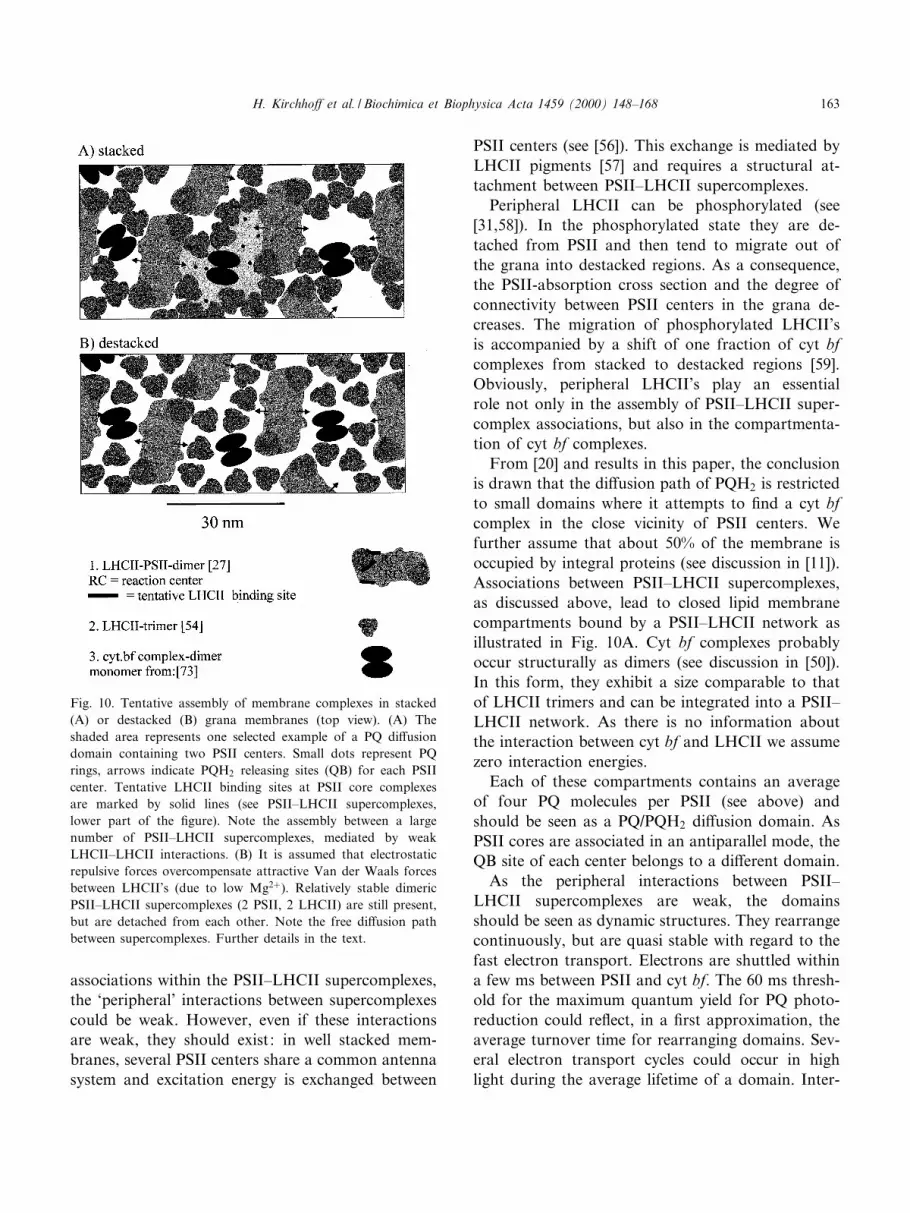
Fig. 10. Tentative assembly of membrane complexes in stacked
(A) or destacked (B) grana membranes (top view). (A) The
shaded area represents one selected example of a PQ di¡usion
domain containing two PSII centers. Small dots represent PQ
rings, arrows indicate PQH2 releasing sites (QB) for each PSII
center. Tentative LHCII binding sites at PSII core complexes
are marked by solid lines (see PSII^LHCII supercomplexes,
lower part of the ¢gure). Note the assembly between a large
number of PSII^LHCII supercomplexes, mediated by weak
LHCII^LHCII interactions. (B) It is assumed that electrostatic
repulsive forces overcompensate attractive Van der Waals forces
between LHCII's (due to low Mg2�). Relatively stable dimeric
PSII^LHCII supercomplexes (2 PSII, 2 LHCII) are still present,
but are detached from each other. Note the free di¡usion path
between supercomplexes. Further details in the text.
H. Kirchho¡ et al. / Biochimica et Biophysica Acta 1459 (2000) 148^168 163

estingly, assuming zero interaction energies between
thylakoid protein complexes and a circular shape of
these complexes, Drepper et al. [60] concluded from
Monte Carlo simulations that the average lifetime of
PQ di¡usion domains is only about 10 ms. Thus,
domains may not be stable enough to e¤ciently re-
strict long range migration of PQ. Obviously speci¢c
interactions between complexes are crucial factors in
stabilizing domains.
A decrease in the Mg2�-concentration leads to a
general increase of the electrostatic repulsive forces
between charged integral thylakoid complexes. It
causes detachment of stacked membranes (stacking
is mediated by LHCII proteins) and lateral separa-
tion of PSII^LHCII supercomplexes. The overall
protein density remains unchanged. In this state, de-
spite a high protein density, the rapidly moving PQ
molecules could worm their way between complexes
and move rapidly throughout the membrane (Fig.
10B).
The model is still hypothetical. It explains the con-
trol of electron transport and still includes important
features of the original microdomain concept by Jo-
liot and Lavergne [11]. However, di¡erent to their
concept, we propose that domains are formed by a
hierarchy of speci¢c protein interactions, rather than
by randomly distributed proteins in a percolation
space. It raises a number of questions, in particular
concerning the assembly of such a complex structure.
Possibly, phosphorylation/dephosphorylation of
Lhcb4 and Lhcb5 subunits play an important role
in controlling the assembly of this microdomain
structure.
4.4. Microdomains and electron transport
Small domains consisting of only one or two PSII
centers per domain seem to come close to the struc-
ture of PSII^cyt bf electron transport supercom-
plexes. Electron transport supercomplexes have
been proposed for bacterial systems [61]. However,
microdomains di¡er from such supercomplexes in
many respects. Firstly, supercomplexes exhibit a
¢xed stoichiometric composition, while £exibility in
its stoichiometric composition is an important fea-
ture of the domain concept. It allows dynamic ad-
justments of the stoichiometric composition of thyla-
koid complexes, an important factor in the
acclimatization of photosynthesis to environmental
and metabolic conditions [62].
Secondly, supercomplexes may be regarded as rel-
atively stable structures, by which reactions in se-
quence are catalyzed step by step, without a lateral
exchange of intermediates between single complexes.
This may be an optimal structure for bacterial sys-
tems, in which the electron transport is driven by one
photoreaction. In plant photosynthesis, optimal elec-
tron transport requires a subtle adjustment of the
rate of two photoreactions. As discussed above PQ
di¡usion domains are not entirely stable. Slow lateral
di¡usion of PQ/PQH2 is possible. This may help to
avoid transient closure of centers at low light and,
hence, to keep the quantum yield high. The domain
structure would be of relevance for the electron
transport at high electron £uxes only (with high fre-
quency of photocycles). Due to the dynamic struc-
ture of domains, a progressive lateral redox imbal-
ance will develop with increasing light throughout
the membrane: at high light PQH2 will accumulate
in grana stacks, while in stroma lamellae, PQ is kept
oxidized, even in high light. Hence, in high light, fast
electron transport is restricted to grana, while cyt bf
complexes located in stroma lamellae do not contrib-
ute to the linear £ux. Stacking and compartmenta-
tion of cyt bf complexes could then be important
factors in controlling the maximal rate of the linear
electron £ux. This kind of structural £ux control is
demonstrated by the destacking-induced stimulation
of electron transport.
Another consequence of restricted PQ mobility is a
compartmentation of the linear electron transport on
a larger scale. Since active PSII is concentrated in
grana stacks (see [24]), only the fraction of cyt bf
complexes located in stacks can be involved in the
linear £ux. In our thylakoid preparations, about 75%
of total cyt f was associated with grana (from frac-
tionation experiments; not shown), which is in good
agreement with the data from others, e.g. [59]. Such
strict compartmentation is not expected for de-
stacked thylakoids. These suggestions are in accord-
ance with the observations that (1) a substantial frac-
tion of PQ remains oxidized during a saturating light
pulse in stacked, but not in destacked membranes
(electron storage experiments; see Table 4) and (2)
the steady state electron £ux is stimulated by about
20% upon destacking (Table 2). Probably, the stim-
H. Kirchho¡ et al. / Biochimica et Biophysica Acta 1459 (2000) 148^168164

ulation is even somewhat underestimated, as it is
followed by a slight inhibition of electron transport,
possibly due to a general destabilization of mem-
brane complexes by the low salt treatment. This is
probably also the reason why no destacking-induced
stimulation in the cyt b reduction level was seen.
Repetitive spectroscopic determination of redox ki-
netics were carried out 5 to 20 min after destacking,
i.e. after the inhibition of electron transport has tak-
en place.
4.5. Plastocyanin di¡usion
There is a large number of thermodynamic argu-
ments why redox transfer by soluble carriers is less
e¤cient than transfer between carriers which are
strictly positioned in a membrane [22]. However,
during the evolution of the oxygenic photosynthesis
of plants, the spatial separation of the two PS's be-
came a necessity to avoid wasteful shortcut of exci-
tation energy between them (for a discussion see
[63]). Thylakoid stacking is the structural basis for
separation. This, however, bears the requirement for
long distance electron shuttling. Due to microdomain
formation within the lipid bilayer, long distance mi-
gration by PQ is restricted and the burden of long
range shuttling is shifted to plastocyanin in the lu-
menal space. Plastocyanin is a spherical hydrophilic
10 kDa protein which carries its redox group, a cop-
per atom ligated to 2 histidine, 1 cysteine and 1 me-
thionine, well-protected within the protein matrix
[64]. Fast electron transfer (lower Ws range) proceedsas intramolecular transfer after tight binding to spe-
ci¢c docking sites at either cyt f or PSI. Thus, dis-
sipative and short circuit reactions during long range
electron transfer should be minimal. Di¡erent to
plastocyanin, PQ is a small and relatively reactive
molecule which could undergo short circuit or even
harmful side reaction.
Actually, a shift in the plastocyanin concentration
between grana and stroma lamellae during light-dark
transition was interpreted as favoring the long dis-
tance transport between these compartments [65].
However, there are conceptual di¤culties with plas-
tocyanin di¡usion too. The lumenal space is narrow,
with a number of proteins protruding into the lumen,
and it is not yet entirely clear whether plastocyanin
can di¡use rapidly enough over the whole distance of
a few hundred nm between grana stacks and stroma
lamellae (for discussion see, for example, [50]). De-
losme [66] arrived at the conclusion that slow cyt f
oxidation can be di¡usion limited and has inter-
preted di¡ering cyt f oxidation rates to correspond
to di¡erent plastocyanin di¡usion distances. The lag
phase in the P700 oxidation after the saturating pulse
(experiment in Fig. 4) seen in destacked membranes
points to a rapid thermodynamic equilibration
throughout the membrane between PQ and P700
mediated by PC (discussed in [74]). Interestingly,
this lag phase is absent in stacked membranes. It
may re£ect a non-equilibrium situation in stacked
membranes, as expected if rapid PC di¡usion and
equilibration is restricted to domains. (However, re-
laxation kinetics are complex and the point certainly
requires further analysis.) It is actually reasonable to
believe that rapid transfer of plastocyanin from cyt
bf in grana stacks is restricted to PSI centered
around the grana in the grana margins (see discus-
sion in [24,50]). This would then raise the question
about the function of PSI located in distant stroma
lamellae. There is still a number of unsolved ques-
tions concerning the microcompartmentation of elec-
tron transport processes in thylakoids.
Acknowledgements
The authors wish to thank Dr. E. Boekema for the
communication of unpublished results, Dr. H.W.
Trissl for help in analyzing the £uorescence induction
curve from DCMU-poised thylakoids and M. in der
Stroth and U. Mukherjee for correcting and prepar-
ing the manuscript.
Appendix. Model of electron transport
We assume that the electron £uxes through PSII and cyt bf complexes, vPSII and vbf , are given by the
concentrations of enzyme^substrate complexes, (PSII-PQ) and (bf-PQH2), multiplied with an apparent intrinsic
constant for these complexes, kPSII and kbf .
H. Kirchho¡ et al. / Biochimica et Biophysica Acta 1459 (2000) 148^168 165

vPSII � kPSII�PSII3PQ� �A:1�
vbf � kbf �bf3PQH2� �A:2�
The concentrations of enzyme^substrate complexes are derived from dissociation constants of PQ and PQH2 at
the QB-site (PSII) and at the Qo-site (cyt bf complex), in conjunction with the assumption that the overall
concentrations of PSII (PSIIo), cyt bf complex (bfo) and PQ (PQo) remain constant. The corresponding ex-
pressions are shown in the following equations (see illustration in Fig. 3):
KDbf �bf WPQH2
�bf3PQH2��A:3�
KDPSII �PSIIWPQ
�PSII3PQ��A:4�
KDbf r �bf WPQ
�bf3PQ��A:5�
KDPSIIr �PSIIWPQH2
�PSII3PQH2��A:6�
bf o � bf � �bf3PQ� � �bf3PQH2� �A:7�
PSIIo � PSII� �PSII3PQ� � �PSII3PQH2� �A:8�
PQo � PQ� PQH2 � �bf3PQ� � �bf3PQH2� � �PSII3PQ� � �PSII3PQH2� �A:9�
From Eqs. A.1^A.9 and with the assumption (vbf = vPSII) for the steady state we derive an equation with
(bf3PQH2) as the only variable.
KDPSIIr�PSIIo3V�bf3PQH2��
KDPSIIr � PQH2
�Qo3PQH23bf o3PSIIo � KDbf r � PQH2
bf o3�bf3PQH2�
KDbf
KDbf r�bf3PQH2�
3
KDbf �bf3PQH2�
PQH2
�A:10a�
where
PQH2 �
�bf3PQH2�2KDbf WV3�bf3PQH2�
2KDPSIIrWVKDPSII
KDPSIIr
KDbf
KDbf r3�bf3PQH2�KDbf WPSIIo
�bf3PQH2�V Wbf o3�bf3PQH2�2V � �bf3PQH2�2VKDPSII
KDPSIIr
KDbf
KDbf r3bf oWPSIIo � �bf3PQH2�PSIIo
�A:10b�
The parameter V is the ratio of kbfUbfo/kPSIIUPSIIo. We assume that DCMU inhibited PSII-centers (1) do not
contribute to linear electron transport and (2) do not bind to PQ or PQH2. Inhibited centers reduce the number
of active centers (PSIIo). The actual number of active PSII centers enters Eq. A.10a via the V term and directly
by PSIIo.
We solve Eq. A.10a for (bf3PQH2) yielding an equation of ¢fth order with respect to V. The zero points of
this equation are calculated with Newton's approximation. The solution is selected by the condition that
06 (bf3PQH2)6 bfo. This solution is inserted in Eq. A.2.
H. Kirchho¡ et al. / Biochimica et Biophysica Acta 1459 (2000) 148^168166

References
[1] B. Andersson, J.M. Anderson, Biochim. Biophys. Acta 593
(1980) 427^440.
[2] J.M. Anderson, FEBS Lett. 138 (1982) 62^66.
[3] J.M. Anderson, Photosynth. Res. 34 (1992) 341^357.
[4] L.A. Staehelin, in: L.A. Staehelin, C.J. Arntzen (Eds.), Pho-
tosynthesis III, Encyclopedia of Plant Physiology, vol. 19,
Springer, Berlin, 1986, pp. 1^84.
[5] L.A. Staehelin, G.W.M. Van der Staay, in: D.A. Ort, C.F.
Yocum (Eds.), Oxygenic Photosynthesis : the Light Reac-
tions, Kluwer Academic Publishers, The Netherlands, 1996,
pp. 11^30.
[6] H.H. Stiehl, H.T. Witt, Z. Naturforsch. 24b (1969) 1588^
1598.
[7] W. Haehnel, Ann. Rev. Plant Physiol. 35 (1984) 659^693.
[8] M.F. Blackwell, K. Gounaris, S.J. Zara, J. Barber, Biophys.
J. 51 (1987) 735^744.
[9] R. Mitchell, A. Spillmann, W. Haehnel, Biophys. J. 58
(1990) 1011^1024.
[10] L.A. Staehelin, C.J. Arntzen, J. Cell. Biol. 97 (1983) 1327^
1337.
[11] J. Lavergne, J.-P. Bouchaud, P. Joliot, Biochim. Biophys.
Acta 1101 (1992) 13^22.
[12] D.J. Murphy, Biochim. Biophys. Acta 864 (1986) 33^94.
[13] M.F. Blackwell, J. Whitmarsh, Biophys. J. 58 (1990) 1259^
1271.
[14] M.J. Saxton, Biophys. J. 56 (1989) 615^622.
[15] M.J. Saxton, Biophys. J. 61 (1992) 119^128.
[16] W.Y. Shih, I.A. Aksay, R. Kikuchi, Phys. Rev. 36 (1987)
5015^5019.
[17] P.A. Millner, D.J. Chapman, J. Barber, Biochim. Biophys.
Acta 765 (1984) 282^287.
[18] M. Blackwell, C. Gibas, S. Gygax, D. Roman, B. Wagner,
Biochim. Biophys. Acta 1183 (1994) 533^543.
[19] J. Lavergne, P. Joliot, Trends Biochem. Sci. 16 (1991) 129^
134.
[20] P. Joliot, J. Lavergne, D. Beal, Biochim. Biophys. Acta 1101
(1992) 1^12.
[21] P. Joliot, A. Joliot, Biochim. Biophys. Acta 1102 (1992) 53^
61.
[22] J. Lavergne, P. Joliot, Photosynth. Res. 48 (1996) 127^138.
[23] J.M. Anderson, W.W. Thomson, in: W.R. Briggs (Ed.),
Photosynthsis, Alan Liss, New York, 1989, pp. 161^182.
[24] P.-A. Albertsson, Photosynth. Res. 46 (1995) 141^149.
[25] M. Ro«gner, E.J. Boekema, J. Barber, Trends Biochem. Sci.
21 (1996) 44^49.
[26] R. Bassi, P. Dianese, Eur. J. Biochim. 204 (1992) 317^326.
[27] B. Hankamer, J. Barber, E.J. Boekema, Annu. Rev. Plant
Physiol. Plant Mol. Biol. 48 (1997) 641^671.
[28] E.J. Boekema, H. van Roon, F. Calkoen, R. Bassi, J.P.
Dekker, Biochemistry 38 (1999) 2233^2239.
[29] J.P. Dekker, H. van Roon, E.J. Boekema, FEBS Lett. 449
(1999) 211^214.
[30] J. Barber, Biochim. Biophys. Acta 594 (1980) 253^308.
[31] J.F. Allen, Biochim. Biophys. Acta 1098 (1992) 275^335.
[32] R. Bassi, D. Sandona, R. Croce, Physiol. Plant. 100 (1997)
769^779.
[33] H. Laasch, Planta 171 (1987) 220^226.
[34] H.W. Trissl, J. Lavergne, Aust. J. Plant Physiol. 22 (1994)
183^193.
[35] R. Reich, R. Scheerer, K.-U. Sewe, H.T. Witt, Biochim.
Biophys. Acta 449 (1976) 285^294.
[36] P. Joliot, A. Joliot, Biochim. Biophys. Acta 765 (1984) 210^
218.
[37] J. Barber, in: M.D. Hatch, N.K. Boardman (Eds.), The Bio-
chemistry of Plants, vol. 10, 1987, pp. 75^130.
[38] G.H. Krause, E. Weis, Annu. Rev. Plant Physiol. Plant
Physiol. Mol. Biol. 42 (1991) 313^349.
[39] S. Murakami, L. Packer, Arch. Biochim. Biophys. 146
(1971) 337^347.
[40] H. Kacser, J.A. Burns, Biochem. Soc. Trans. Biochem Soc. 7
(1979) 1149^1160.
[41] W.A. Cramer, G.M. Soriano, M. Ponomarev, D. Huang, H.
We assume that the electron transport rate given by Eq. A.2 determines the rate of steady state electron
transport (see above). The dependency of the electron £ux rate on the number of active PSII-centers is analyzed
by varying PSIIo. The curves in Fig. 2B are calculated in this way for di¡erent parameter sets given in Table 6
and by normalizing Eq. A.2 to 1 if PSIIo is maximal.
It is assumed in the model, that the intermediates PQ and PQH2 can di¡use rapidly between all binding sites,
i.e. no di¡usion limitation exists.
Table 6
Parameters used for the electron transport model
Literature Curve 1 Curve 2
KDPSII 1 to 2 mMa 1.5 1.5
KDPSIIr 1 to 2 mMa 1.5 1.5
KDbf 0.05 mMb 0.05 0.05
KDbfr unknown 0.05 0.5
kPSII/kbf see text, 1.7 to 2.5c 1.7 1.7
PQ (total) 1b to 14 mMd 1 7
PSII (total) 1/6UPQ (total)e
Cyt bf (total) 1/2UPSII (total)e
a[48]b[72]cWe assume the smallest ratio for PSII to cyt bf turnover times,
i.e. (2 ms)31 for PSII (3.3 ms)31 for cyt bf.d[18]eOwn measurements.
H. Kirchho¡ et al. / Biochimica et Biophysica Acta 1459 (2000) 148^168 167

Zhang, S.E. Martinez, J.L. Smith, Annu. Rev. Plant Physiol.
Plant Mol. Biol. 47 (1996) 477^508.
[42] G. Hauska, M. Schu«tz, M. Bu«ttner, in: D.A. Ort, C.F. Yo-
cum (Eds.), Oxygenic Photosynthesis : the Light Reactions,
Kluwer Academic Publishers, The Netherlands, 1996, pp.
377^398.
[43] D.A. Ort, J. Whitmarsh, Photosynth. Res. 23 (1990) 101^
104.
[44] Govindjee, Photosynth. Res. 25 (1990) 151^160.
[45] A.W. Rutherford, Trends Biochem. Sci. 14 (1989) 227^232.
[46] Oë . Hansson, T. Wydrzynski, Photosynt. Res. 23 (1990) 131^
162.
[47] W. Ausla«nder, W. Junge, Biochim. Biophys. Acta 357 (1974)
285^298.
[48] A.B. Diner, V. Petrouleas, J.J. Wendoloski, Physiol. Plant.
81 (1991) 423^436.
[49] J.M. Anderson, W.S. Chow, D.J. Goodchild, Aust. J. Plant
Physiol. 15 (1988) 11^26.
[50] A.B. Hope, Biochim. Biophys. Acta 1143 (1993) 1^22.
[51] T. Kallas, in: D.A. Bryant (Ed.), The Molecular Biology of
Cyanobacteria, Kluwer Academic Publishers, The Nether-
lands, 1994, pp. 185^219.
[52] S. Malkin, B. Kok, Biochim. Biophys. Acta 126 (1966) 413^
432.
[53] D.A. Fell, Biochem. J. 286 (1992) 313^330.
[54] W. Ku«hlbrandt, D.N. Wang, Y. Fujiyoshi, Nature 367
(1994) 614^621.
[55] P.J. Morissey, R.R. Glick, A. Melis, Plant Cell Physiol. 30
(1989) 335^344.
[56] R. Van Grondelle, J. Amesz, in: Govindjee, J. Amesz, D.C.
Fork (Eds.), Light Emission by Plant and Bacteria, Academ-
ic Press, New York, 1986, pp. 191^223.
[57] W.L. Butler, Annu. Rev. Plant. Physiol. 29 (1978) 345^
378.
[58] J. Bennet, Annu. Rev. Plant Physiol. Plant Mol. Biol. 42
(1991) 281^311.
[59] O. Vallon, F.A. Wollman, J. Olive, Photochem. Photobio-
phys. 12 (1986) 203^220.
[60] F. Drepper, I. Carlberg, B. Andersson, W. Haehnel, Bio-
chemistry 32 (1993) 11915^11922.
[61] P. Joliot, A. Vermeglio, A. Joliot, Biochim. Biophys. Acta
1141 (1993) 151^174.
[62] J.M. Anderson, W.S. Chow, Y.-I. Park, Photosynth. Res. 46
(1995) 129^139.
[63] H.W. Trissl, C. Wilhelm, Trends Biochem. Sci. 18 (1993)
415^419.
[64] E.L. Gross, in: D.A. Ort, C.F. Yocum (Eds.), Oxygenic
Photosynthesis, Kluwer Academic Publishers, The Nether-
lands, 1996, pp. 413^429.
[65] W. Haehnel, R. Ratajczak, H. Robenek, J. Cell. Biol. 108
(1989) 1397^1405.
[66] R. Delosme, Photosynth. Res. 29 (1991) 45^54.
[67] S.U. Metzger, W.A. Cramer, J. Whitmarsh, Biochim. Bio-
phys. Acta 1319 (1997) 233^241.
[68] N. Nelson, J. Neumann, J. Biol. Chem. 247 (1972) 1817^
1824.
[69] H.J. Van Gorkum, Biochim. Biophys. Acta 347 (1974) 439^
442.
[70] B. Ke, Arch. Biochim. Biophys. 152 (1972) 70^77.
[71] S. Katoh, I. Suga, I. Shiratori, A. Takamiya, Arch. Biochim.
Biophys. 94 (1961) 136^141.
[72] D.M. Kramer, A. Joliot, P. Joliot, A.R. Crofts, Biochim.
Biophys. Acta 1184 (1994) 251^262.
[73] D. Bald, J. Kruip, E.J. Boekema, M. Ro«gner, in: N. Murata
(Ed.), Research of Photosynthesis, vol. 1, Kluwer Academic
Publishers, The Netherlands, 1992, pp. 629^632.
[74] B. Kok, P. Joliot, M.P. McCloin, Prog. Photosynth. Res. 11
(1969) 1042^1056.
H. Kirchho¡ et al. / Biochimica et Biophysica Acta 1459 (2000) 148^168168


![Id‚v A^-tbgvkv h{P-Pq-_nen hnti-jm¬ ]Xn∏vc - Kerala PSC](https://img.pdfslide.net/doc/110x75/631c87277051d371800f845f/idv-a-tbgvkv-hp-pq-nen-hnti-jm-xnvc-kerala-psc.jpg)