Embed Size (px)
Citation preview
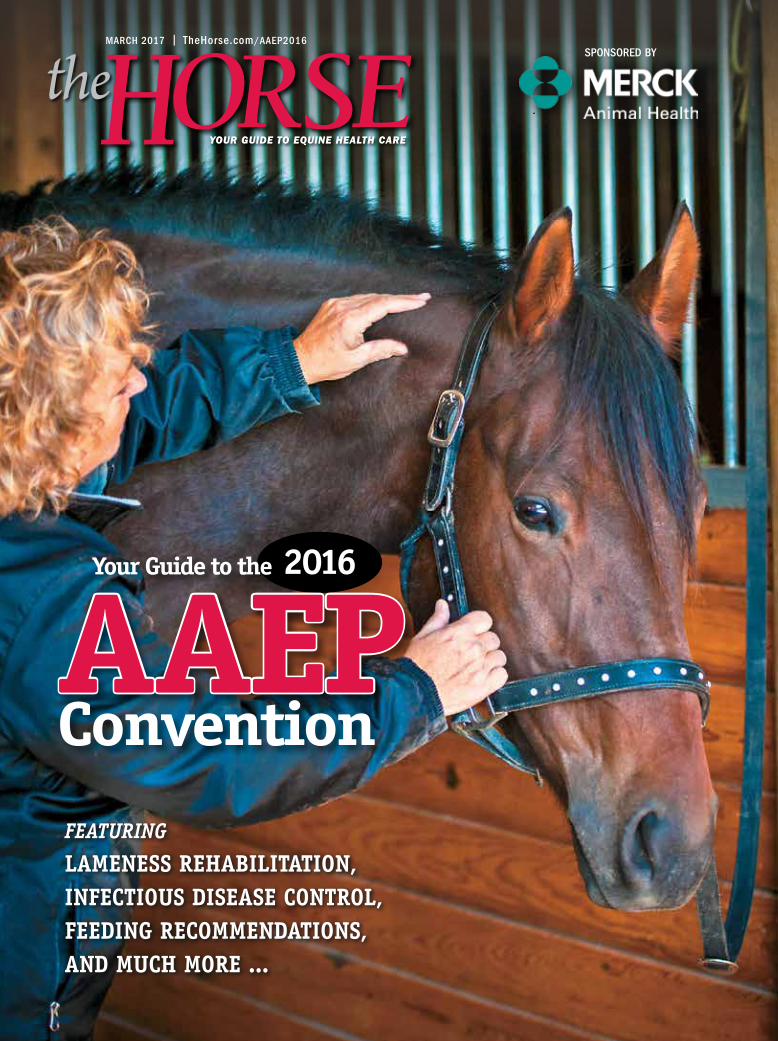
FEATURING
lameness rehabilitation,infectious Disease control,feeDing recommenDations, anD much more ...
SPONSORED BYMARCH 2017 | TheHorse.com/AAEP2016
2016Your guide to the
aaePconvention
The Science of Healthier Animals
2 Giralda Farms • Madison, NJ 07940 • merck-animal-health-usa.com • 800-521-5767Copyright © 2016 Intervet Inc., d/b/a/ Merck Animal Health, a subsidiary of Merck & Co., Inc.All rights reserved. 3526 EQ-FP AD Protazil®
The Science of Convenient
• Equine Protozoal Myeloencephalitis (EPM) is a serious neurological disease that can strike anytime, anywhere. Make treatment easy with Protazil®
• Safe and accurate dosing with a calibrated scoop
• Easier to use than paste, less stress for you and your horse
• Rapid absorption — no loading dose required1
Now that’s convenient.
Protazil® (1.56% diclazuril) is the only FDA-approved alfalfa-based top dress antiprotozoal pellet for the treatment of EPM.
Ask your veterinarian for Protazil®. Visit us at Protazil.com to learn more about Merck Animal Health and the equine products and programs that help keep horses healthy.
Use of Protazil® (1.56% dicazuril) is contraindicated in horses with known hypersensitivity to diclazuril. Safe use in horses used for breeding purposes, during pregnancy, or in lactating mares has not been evaluated. The safety of Protazil® (1.56% dicazuril) with concomitant therapies in horses has not been evaluated. See related page in this issue for details. For use in horses only. Do not use in horses intended for human consumption. Not for human use. Keep out of reach of children.
1 Hunyadi L, Papich MG, Pusterla N. Pharmacokinetics of a low-dose and DA-labeled dose of diclazuril administered orally as a pelleted top dressing in adult horses. J of Vet Pharmacology and Therapeutics (accepted) 2014, doi: 10.111/jvp.12176. The correlation between pharmacokinetic data and clinical effectiveness is unknown
3526_Protazil_8125x10875.indd 1 1/23/17 8:47 AM
AAEP Wrap-Up The horse March 2017TheHorse.com/AAEP2016
Y our horse’s veterinarian is constantly on
the go. Chances are he or she has seen a long list of patients today and hasn’t sat down for many hours (save for the drive time between appointments). All the while there’s some teaching going on, too.
My veterinarian can dispense infor-mation 100 mph as she is palpating, vaccinating, scrubbing a joint, or even trying to find her miniature Daschund, Ed, who likes to runs off after the barn cats. Sometimes I joke that I’ve had several conversations with her as she is essentially cantering toward her truck and looking back at me. (Perhaps this is why she’s always on time.)
Practitioners based at clinics also rarely lose momentum, moving seam-lessly from case to case, asking or an-swering questions or relaying informa-tion as they examine and treat.
But for five days in December, many veterinarians take time to sit down and fill up their knowledge reserves at the American Association of Equine Practitioners (AAEP) Convention. Don’t get me wrong—while they do sit down, there’s a lot of bustle, too, as they shift between session rooms, catch up with colleagues and former vet school classmates in the hallways, take and return client phone calls, and purchase equipment at the trade show.
I enjoy watching and listening to
presenting veterinarians share their hard-earned research results or deliver state-of-the-art information from their area of study. Their passion for caring for horses and advancing the field is evident in anecdotes shared from the podium and in questions coming from the audience. I find it inspiring, too … and I’m not even a veterinarian.
In these pages you’ll see dozens of research summaries organized by topic of interest. Here are some tips, one horse owner to another, on best ways to use this material:■ Don’t try to digest it all in one sitting.
Flip through, read the items that look most interesting to you now, and mark items to come back to later. There’s plenty to savor.
■ If you have questions based on these stories, write them down.
■ Next time your vet is out for a well-ness check, ask if he or she went to AAEP. If so, ask what they thought was most interesting.
■ If there’s a presentation summarized in the Wrap-Up that got you thinking and wondering, ask if your veterinar-ian has time for a few questions so you can understand a concept better. This approach will help you use
this information wisely and can be a jumping-off point for good conversa-tions with your vet. Remember, he or she knows your horse’s health history and can advise on which principles apply to your own situation, and which should be filed away for future refer-ence (or interesting barn conversation). I hope you enjoy this year’s Wrap-Up. h
learning at a canter
aaePwraP-uP2016
SPONSORED BY
STEPHANIE L. CHURCH Editor-in-Chief
Publisher: Marla Bickel Editor-in-Chief: Stephanie L. Church Managing Editor: Alexandra Beckstett News Editor: Erica Larson Digital Managing Editor: Michelle AndersonArt Director: Brian Turner Web Producer: Jennifer WhittleBrand Manager: Shawna White
EDitoriAl DEPArtMENtThe Horse, Editorial, 3101 Beaumont Centre Circle, Suite 100, Lexington, KY 40513E-MAil [email protected] All letters must include the writer’s name, address, and daytime phone number for verification.■ letters: [email protected], or by mail.■ Farm Call: [email protected], or by mail. ■ Across the Fence and Behavior Columns:
[email protected], or by mail.■ New Products: [email protected], or by mail.
EDitoriAl ADvisory BoArDScott Anderson, DVM Jerry Black, DVM Anthony Blikslager, DVM, PhD, Dipl. ACVS Tom Brokken, DVM Ann Dwyer, DVM Benjamin Espy, DVM, Dipl. ACT Jenifer R. Gold, DVM, Dipl. ACVIM, ACVECC Margo Macpherson, DVM, MS, Dipl. ACT Kyla Ortved, DVM, Dipl. ACVS Debra Taylor, DVM
Educational Partnership Disclaimer: The American Association of Equine Practitioners (AAEP), one of The Horse’s partners in equine health, has no involvement regarding editorial management or advertising content within this publication and thereby does not endorse any editorial or advertising content unless so acknowledged within the individual article or advertisement.
ADvErtisiNg sAlEs AND sErviCEs [email protected]
West Coast Advertising Executive: Yvonne Long, 859/276-6701 [email protected]
East Coast Advertising Executive: Leigh Walkup, 859/276-6710 [email protected]
sales support: Kelly Stephens, 859/276-6740 [email protected]
PuBlishED By thE horsE MEDiA grouP llC
For up-to-date news, in-depth horse health articles, and more,
go to TheHorse.com
AAEP Wrap-Up The horse March 2017TheHorse.com/AAEP2016
ISTo
Ck
.Co
m
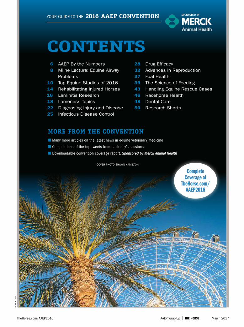
6 AAEP By the Numbers 8 milne Lecture: Equine Airway
Problems 10 Top Equine Studies of 2016 14 Rehabilitating Injured Horses16 Laminitis Research 18 Lameness Topics 22 Diagnosing Injury and Disease 25 Infectious Disease Control
28 Drug Efficacy 32 Advances in Reproduction 37 Foal Health 39 The Science of Feeding 43 Handling Equine Rescue Cases 46 Racehorse Health 48 Dental Care 50 Research Shorts
more from the convention■ Many more articles on the latest news in equine veterinary medicine
■ Compilations of the top tweets from each day’s sessions
■ Downloadable convention coverage report. Sponsored by Merck Animal Health
CovER PHoTo SHAwN HAmILToN
contents
Your GuiDE To THE 2016 aaeP convention
CompleteCoverage at
TheHorse.com/aaep2016
SPONSORED BY

AAEP Wrap-Up The horse March 2017TheHorse.com/AAEP2016
Your GuiDE To THE 2016 aaeP convention SPonSorED bY
veterinary professionals, students, guests, and exhibitors from 48 countries attended the 2016 convention
donated in support of the AAEP Foundation’s mission to improve the welfare of the horse
the 63rd annual convention will take place nov. 17-21, 2017, in san antonio, texas
Co
UR
TES
y AA
EP
BY the numbers
5,471
hours of continuing education
The average attendee
walked 25 miles over the 5 days
More than 650
#AAEP2016tweets
Ethics speaker
ChuckGallagher
gave the keynote address
347 companies showcased their products and services at the trade show
R. Reynolds Cowles Jr., DVM,
of Blue Ridge equine Clinic in earlysville, Virginia, was installed as the 63rd president of
the aaep
130
,$

AAEP Wrap-Up The horse March 2017TheHorse.com/AAEP2016
The Science of Trusted
From the broodmare to the performance horse, properly managing your mare’s hormones is critical. Rely on the product that’s trusted most.
• More than 30 years of practical use in the field by veterinarians1
• More than 200 clinical trials to determine efficacy, duration and safety1
• More than 20 million doses sold to veterinarians, trainers and horse owners1
Now that’s trusted.
Talk to your veterinarian about proper use and safe handling of Regu-Mate®. Avoid skin contact. Always wear protective gloves when administering Regu-Mate®. This product is contraindicated for use in mares with a previous or current history of uterine inflammation. Pregnant women, or women who suspect they are pregnant, should not handle this product. For complete product information, see accompanying product insert.
Regu-Mate® (altrenogest) is the name veterinarians and their clients depend on for estrus control (suppression, management).
1 Data on file, Merck Animal Heath
Ask your veterinarian for Regu-Mate®. Visit us online at merck-animal-health-equine.com to learn more about Merck Animal Health and the equine products and programs that help keep horses healthy.
The Science of Healthier Animals
2 Giralda Farms • Madison, NJ 07940 • merck-animal-health-usa.com • 800-521-5767Copyright © 2016 Intervet Inc., d/b/a/ Merck Animal Health, a subsidiary of Merck & Co., Inc.All rights reserved. 3526 EQ-FP AD Regu-Mate®
3526_Regu-Mate_8125x10875.indd 1 2/7/17 10:27 AM
AAEP Wrap-Up The horse March 2017TheHorse.com/AAEP2016
Your GuiDE To THE 2016 aaeP convention
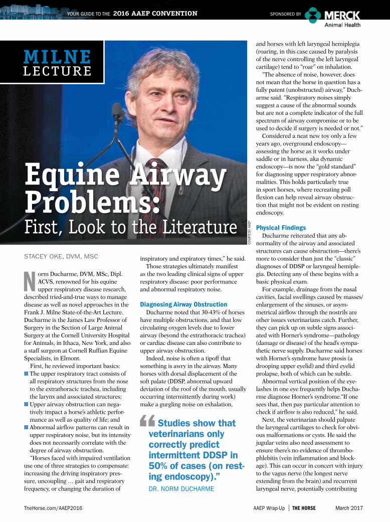
STACEy okE, Dvm, mSC
N orm Ducharme, DVM, MSc, Dipl. ACVS, renowned for his equine upper respiratory disease research,
described tried-and-true ways to manage disease as well as novel approaches in the Frank J. Milne State-of-the-Art Lecture. Ducharme is the James Law Professor of Surgery in the Section of Large Animal Surgery at the Cornell University Hospital for Animals, in Ithaca, New York, and also a staff surgeon at Cornell Ruffian Equine Specialists, in Elmont.
First, he reviewed important basics:■ The upper respiratory tract consists of
all respiratory structures from the nose to the extrathoracic trachea, including the larynx and associated structures;
■ Upper airway obstruction can nega-tively impact a horse’s athletic perfor-mance as well as quality of life; and
■ Abnormal airflow patterns can result in upper respiratory noise, but its intensity does not necessarily correlate with the degree of airway obstruction.“Horses faced with impaired ventilation
use one of three strategies to compensate: increasing the driving inspiratory pres-sure, uncoupling … gait and respiratory frequency, or changing the duration of
inspiratory and expiratory times,” he said. Those strategies ultimately manifest
as the two leading clinical signs of upper respiratory disease: poor performance and abnormal respiratory noise.
Diagnosing Airway obstructionDucharme noted that 30-43% of horses
have multiple obstructions, and that low circulating oxygen levels due to lower airway (beyond the extrathoracic trachea) or cardiac disease can also contribute to upper airway obstruction.
Indeed, noise is often a tipoff that something is awry in the airway. Many horses with dorsal displacement of the soft palate (DDSP, abnormal upward deviation of the roof of the mouth, usually occurring intermittently during work) make a gurgling noise on exhalation,
and horses with left laryngeal hemiplegia (roaring, in this case caused by paralysis of the nerve controlling the left laryngeal cartilage) tend to “roar” on inhalation.
“The absence of noise, however, does not mean that the horse in question has a fully patent (unobstructed) airway,” Duch-arme said. “Respiratory noises simply suggest a cause of the abnormal sounds but are not a complete indicator of the full spectrum of airway compromise or to be used to decide if surgery is needed or not.”
Considered a neat new toy only a few years ago, overground endoscopy— assessing the horse as it works under saddle or in harness, aka dynamic endoscopy—is now the “gold standard” for diagnosing upper respiratory abnor-malities. This holds particularly true in sport horses, where recreating poll flexion can help reveal airway obstruc-tion that might not be evident on resting endoscopy.
Physical Findings Ducharme reiterated that any ab-
normality of the airway and associated structures can cause obstruction—there’s more to consider than just the “classic” diagnoses of DDSP or laryngeal hemiple-gia. Detecting any of these begins with a basic physical exam.
For example, drainage from the nasal cavities, facial swellings caused by masses/enlargement of the sinuses, or asym-metrical airflow through the nostrils are other issues veterinarians catch. Further, they can pick up on subtle signs associ-ated with Horner’s syndrome—pathology (damage or disease) of the head’s sympa-thetic nerve supply. Ducharme said horses with Horner’s syndrome have ptosis (a drooping upper eyelid) and third eyelid prolapse, both of which can be subtle.
Abnormal vertical position of the eye-lashes in one eye frequently helps Ducha-rme diagnose Horner’s syndrome.“If one sees that, then pay particular attention to check if airflow is also reduced,” he said.
Next, the veterinarian should palpate the laryngeal cartilages to check for obvi-ous malformations or cysts. He said the jugular veins also need assessment to ensure there’s no evidence of thrombo-phlebitis (vein inflammation and block-age). This can occur in concert with injury to the vagus nerve (the longest nerve extending from the brain) and recurrent laryngeal nerve, potentially contributing
milneLecture
Co
UR
TES
y AA
EP
equine airway Problems:First, Look to the Literature
Studies show that veterinarians only correctly predict intermittent DDSP in 50% of cases (on rest-ing endoscopy).”
DR. NoRm DUCHARmE
SPonSorED bY
AAEP Wrap-Up The horse March 2017TheHorse.com/AAEP2016
to airway obstruction, as both nerves sup-ply important respiratory tract structures.
Practitioners can also identify atrophy (wasting) of the cricoarytenoid dorsalis (CAD), a small but mighty muscle that’s a major larynx abductor, via routine exam.
“generic” and Advanced testingAlthough he referred to it as a generic
approach, Ducharme emphasized the importance of resting endoscopy (or “scoping”) when assessing upper airway patency and searching for abnormalities.
“In my experience the nasal passages, nasopharynx, and larynx are best exam-ined with the animal unsedated to avoid a false diagnosis of nasopharyngeal collapse or recurrent laryngeal neuropathy,” he said. Neuropathy is disease or dysfunction of one or more peripheral nerves that typi-cally causes numbness or weakness.
Conditions veterinarians can easily diagnose with scoping include:■ Masses and cysts;■ Arytenoid chondritis (inflammation of
the cartilages);■ Persistent DDSP (pDDSP);■ Epiglottic abscesses; and■ Guttural pouch empyema (pus collec-
tion), mycosis (fungal disease), and tympany (air accumulation). With guttural pouch diseases, the vagus
and glossopharyngeal nerves can become inflamed or even paralyzed, resulting in nasopharyngeal collapse and pDDSP.
“Caution must be used when using resting endoscopy to diagnose intermit-tent DDSP (iDDSP) because even with a history of noise during exercise, studies show that veterinarians only correctly pre-dict intermittent DDSP in 50% of cases,” said Ducharme. “Even identification of an ulcer on the caudal free edge (back) of the palate, a flaccid epiglottis, and induction of DDSP by either swallowing or nasal oc-clusion are not reliable indicators.”
If a horse has an ulcer on the caudal free edge of the soft palate, he might have a subepiglottic ulcer/granuloma or inter-mittent epiglottic entrapment—when the loose skin located on the bottom of the epiglottis (hence, sub-) flips over, covering the epiglottis like a slipper.
Based on Ducharme’s comprehensive review of the literature, he deemed rest-ing endoscopy an appropriate diagnostic technique for laryngeal hemiplegia. Pri-marily caused by left recurrent laryngeal neuropathy, roaring occurs when the left
arytenoid cartilage droops into the airway at the trachea entrance (i.e., voice box). Ultrasound of the muscles associated with laryngeal function also appears to be highly valuable; however, it should not re-place resting endoscopy, he said, because other lesions can be missed. Examples in-clude arytenoid chondromas (rare benign cartilaginous tumors) and subluxations (partial dislocation), inflammation of the epiglottis (epiglottitis), and subepiglottic ulceration.
He focused heavily on athletic horses in his lecture but said that even nonper-formance horses and foals benefit from scoping, particularly ones suffering from swallowing disorders or difficulties.
Next, Ducharme described the merits of dynamic endoscopy both for recogniz-ing collapse during exercise and confirm-ing the absence of airway obstruction. Veterinarians traditionally performed dynamic endoscopy using a high-speed treadmill that assured horses reached at least near-maximal exercise intensity. Now, overground endoscopes allow veter-inarians to evaluate horses under normal work conditions, including the impact the rider or driver has on performance.
He said using overground endoscopy is preferred in Thoroughbred racehorses; but trotters and pacers have a higher prevalence of coexisting illness that often requires more testing, making high-speed treadmill endoscopic exams preferable.
Managing upper Airway DiseaseDucharme described myriad conditions
and the management strategies for each. DDSP Management Options Ducharme
recommended surgery for horses with iDDSP … but only after medical therapy (e.g., systemic anti-inflammatory medica-tions such as dexamethasone or predniso-lone) and tack changes (tongue ties, bit changes, bitless bridles, and figure-eight or dropped nosebands) in already-fit horses have failed.
Also, it’s important that young horses be allowed to mature in case they are suf-fering from an immature nasopharynx.
“Several surgical options for iDDSP ex-ist, but many have been devised without a clear understanding of the underlying condition,” Ducharme said. Current sur-gical strategies include:■ Correcting underlying issues (remov-
ing granulomas, cysts, and abnormal subepiglottic tissue);
■ Increasing the stiffness of the soft pal-ate (using cautery, laser, or chemical agents); and
■ Altering the larynx’s position to limit soft palate movement (strap muscle resection, or bilateral partial stenothy-roidectomy,) and/or tie-forward surgery. Typically, bilateral stenothyroidectomy
and/or a tie-forward procedure is the preferred surgical procedure, he said. Success ranges from 58% to 82%.
Laryngeal Hemiplegia The underly-ing cause of roaring remains unclear. Surgeons frequently turn to laryngo-plasty (aka a tie-back procedure) and removal of the vocal cords and folds ( ventriculocordetomy).
The tie-back involves pulling the flaccid cartilage out of the airway using perma-nent suture material. Success with this procedure leaves us wanting for more, he said, and “DDSP is a newly recognized complication seen after laryngoplasty.”
He also described complications and the methods for determining causes of persistent upper respiratory noise and poor performance post-surgery, as well as novel methods for managing roaring, in-cluding laryngeal reinnervation and elec-trical rehabilitation of the CAD muscle.
take-home MessageDucharme emphasized that the treat-
ment goal of any equine upper airway abnormality should be based on the most up-to-date literature and be the least-invasive approach that results in a quick return to function. h
conventiontweet
AAEP@AAEPHorseDocs
Are you in the Valencia Ballroom for the Milne lecture? Dr. Norm Ducharme is lecturing on #equine upper airways!
5 5
Your GuiDE To THE 2016 aaeP convention SPonSorED bY
AAEP Wrap-Up The horse March 2017TheHorse.com/AAEP2016
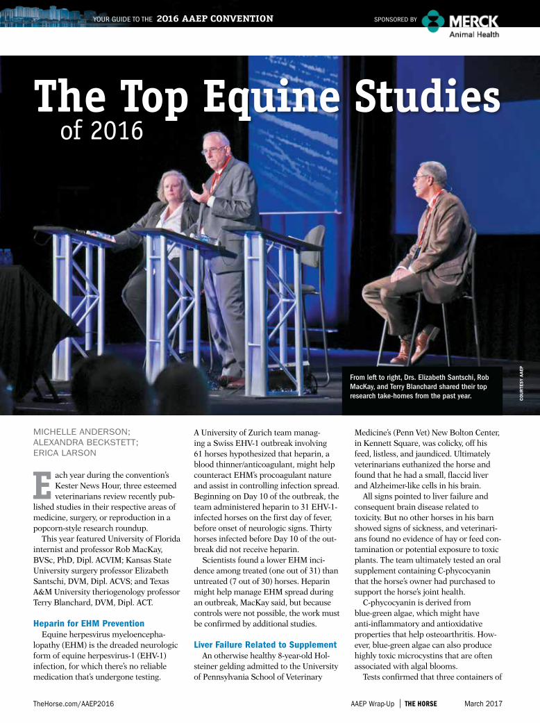
mICHELLE ANDERSoN; ALExANDRA BECkSTETT; ERICA LARSoN
e ach year during the convention’s Kester News Hour, three esteemed veterinarians review recently pub-
lished studies in their respective areas of medicine, surgery, or reproduction in a popcorn-style research roundup.
This year featured University of Florida internist and professor Rob MacKay, BVSc, PhD, Dipl. ACVIM; Kansas State University surgery professor Elizabeth Santschi, DVM, Dipl. ACVS; and Texas A&M University theriogenology professor Terry Blanchard, DVM, Dipl. ACT.
heparin for EhM PreventionEquine herpesvirus myeloencepha-
lopathy (EHM) is the dreaded neurologic form of equine herpesvirus-1 (EHV-1) infection, for which there’s no reliable medication that’s undergone testing.
A University of Zurich team manag-ing a Swiss EHV-1 outbreak involving 61 horses hypothesized that heparin, a blood thinner/anticoagulant, might help counteract EHM’s procoagulant nature and assist in controlling infection spread. Beginning on Day 10 of the outbreak, the team administered heparin to 31 EHV-1- infected horses on the first day of fever, before onset of neurologic signs. Thirty horses infected before Day 10 of the out-break did not receive heparin.
Scientists found a lower EHM inci-dence among treated (one out of 31) than untreated (7 out of 30) horses. Heparin might help manage EHM spread during an outbreak, MacKay said, but because controls were not possible, the work must be confirmed by additional studies.
liver Failure related to supplementAn otherwise healthy 8-year-old Hol-
steiner gelding admitted to the University of Pennsylvania School of Veterinary
Medicine’s (Penn Vet) New Bolton Center, in Kennett Square, was colicky, off his feed, listless, and jaundiced. Ultimately veterinarians euthanized the horse and found that he had a small, flaccid liver and Alzheimer-like cells in his brain.
All signs pointed to liver failure and consequent brain disease related to toxicity. But no other horses in his barn showed signs of sickness, and veterinari-ans found no evidence of hay or feed con-tamination or potential exposure to toxic plants. The team ultimately tested an oral supplement containing C-phycocyanin that the horse’s owner had purchased to support the horse’s joint health.
C-phycocyanin is derived from blue-green algae, which might have anti- inflammatory and antioxidative properties that help osteoarthritis. How-ever, blue-green algae can also produce highly toxic microcystins that are often associated with algal blooms.
Tests confirmed that three containers of
co
ur
TESy
AA
EP
the top equine studies of 2016
From left to right, Drs. Elizabeth Santschi, Rob MacKay, and Terry Blanchard shared their top research take-homes from the past year.
Your GuiDE To THE 2016 aaeP convention SPonSorED bY
AAEP Wrap-Up The horse March 2017TheHorse.com/AAEP2016
the supplement contained the toxins. The veterinarians concluded that consuming it had likely caused the horse’s death.
Despite professional-looking labels and marketing claims, “supplements are not always safe,” said MacKay. Furthermore, blue-green algae harvesting and process-ing for supplements is not regulated.
Drug Combo for R. Equi testedVeterinarians commonly use the an-
tibiotic rifampin in combination with a macrolide antibiotic such as clarithromy-cin to treat Rhodococcus equi infections in foals. German researchers compared the pharmacokinetics of oral clarithromy-cin (CLA) and rifampin to data gathered from foals given CLA alone. They found that CLA concentrations decreased by more than 80% in foals’ blood and bronchoalveolar cells when rifampin was co-administered with CLA.
MacKay said rifampin likely severely reduces uptake of all macrolides used to treat R. equi. Regardless, he said, the CLA/rifampin combination remains a clinically effective R. equi treatment.
“More work is needed to discern whether the perceived advantage of ri-fampin outweighs the negative effects on macrolide absorption,” MacKay said.
lyme Neuroborreliosis CharacteristicsA Penn Vet research group conducted
a retrospective study of equine neurobor-reliosis (NB, the neurologic form of Lyme disease) cases in hopes of providing a detailed description of clinical signs, diag-nostics, and pathologic findings.
Sixteen horses from New Bolton and other referral clinics qualified for the study. They had variable clinical signs, including muscle atrophy/weight loss; cranial nerve deficits; incoordination; be-havior changes; difficulty eating or swal-lowing; muscle tremors; neck stiffness; episodic respiratory distress; uveitis; fever; joint swelling; and heart irregularities.
Only six tested positive for Borrelia burgdorferi (Lyme’s causative bacterium that’s spread by ticks) infection via stan-dard immunodiagnostic testing of blood or cerebrospinal fluid (CSF). Polymerase chain reaction tests on CSF for exposure to the bacterium were negative in all seven cases tested.
There’s still a lot we don’t know about Lyme disease in horses, said MacKay.
“Signs are nonspecific and numerous,
routine bloodwork is unhelpful, and tests such as the Lyme multiplex are not diag-nostic,” he said.
PENs for treating headshakingHeadshaking is a chronic problem—
likely a response to nerve pain—with no consistently effective treatment.
But researchers from the University of Bristol, in Somerset, U.K., recently tested a new approach: percutaneous electri-cal nerve stimulation (PENS) therapy, a minimally invasive therapy used to man-age human neuropathic pain.
They treated seven headshakers that showed clinical signs at the study’s onset with a PENS probe. Each received a series of three or four treatments, with repeated treatments as headshaking recurred.
Results had MacKay calling the pro-tocol “safe and promising.” All horses tolerated it well. Two had increased signs for up to three days after the initial ses-sion. Six responded positively to their first treatment and returned to ridden work at the same level as prior to the condition’s onset. Five continued to respond to subse-quent treatments, gaining up to 20 weeks of relief after the fourth treatment.
radius Fracture outcomesResearchers behind the first study
Santschi described sought to determine survival-to-discharge rates for horses with radial fractures and examine risk factors affecting these rates in conservatively and surgically managed fractures. The radius is the large leg bone located above the horse’s knee (or carpal joint).
The team included 54 horses in the retrospective study, 13 of which were euthanized at admission. Of the remain-ing 41 horses, 14 had incomplete fractures and were managed conservatively with Robert-Jones bandages and splints; 12 survived to discharge. The other 27 had complete fractures that were repaired
surgically; 15 survived to discharge.Santschi said both conservatively
managed horses that were euthanized developed supporting-limb laminitis. Of the surgical horses that died, two were euthanized following anesthesia recovery, 11 developed surgical site infections, and eight suffered failure of the surgical repair.
Risk factors tied to surgical failure were age (older horses less likely to survive), duration (procedures longer than 168 minutes were less likely to have success-ful outcomes), and surgical site infection (trended toward decreased survival rate).
Santschi’s take-home: Younger horses and horses with incomplete fractures tend to have a good prognosis for recov-ery. Horses with open fractures are more likely to develop surgical site infection.
long-term outcomes of upward Fixation of the Patella treatment
Researchers evaluated the long-term outcome of the medial patellar ligament splitting procedure to treat upward fixa-tion of the patella (UFP). This involves inserting a blade or needle through the skin to split the proximal (inner) third part of the medial patellar ligament.
The study authors looked at the medical records of 85 horses that underwent the procedure, 83 (97.6%) of which showed complete UFP resolution immediately after or within two weeks of surgery. Santschi said UFP persisted in the re-maining two horses (2.4%), even after the procedure was repeated.
The team collected follow-up data on 78 horses (90.5%) three to 14 years after surgery. None of the horses experienced complications, and UFP recurrence wasn’t reported in horses that returned to work.
Santschi said the ligament-splitting procedure was highly effective, had a low complication rate, and offered a rapid return to function for horses with UFP.
Comparing Joint lavage techniquesWhen veterinarians detect joint con-
taminants, they use lavage to remove them and prevent infection.
Scientists evaluated lavage techniques to see which was most effective for remov-ing 1.5 million tiny microspheres from a cadaver horse’s tarsocrural joints (between the tibia and the talus in the upper hock joint). Joints were lavaged with saline via arthroscope or three 14-gauge needles, each placed in a different position.
conventiontweet
Jackie Kaufman@Jackie_Kaufman
Surprisingly, use of Rifampin with Clarithromycin decreased bioavailability of CLA by >80% in one study.
Your GuiDE To THE 2016 aaeP convention SPonSorED bY
AAEP Wrap-Up The horse March 2017TheHorse.com/AAEP2016
The team found that needles lavaged 2.5 times more microspheres from joints than did the single arthroscope. Regard-less of technique, about 80% of the microspheres were flushed out with the first liter of lavage fluid.
It’s better to place more smaller needles than one large one when lavaging joints, and one to two liters of lavage fluid is probably sufficient to remove most of the debris, said Santschi.
Conservative lower Jaw Fracture Management
Santschi described a retrospective study in which researchers evaluated the outcome of conservative management (no surgery) of unilateral mandibular (lower jaw) fractures in horses. The outcome was considered successful if horses returned to their prior use, chewed normally, and had no additional fracture-related problems.
The study group included 24 horses, aged 1 to 24 years. Sixty-seven percent of fractures occurred on the right side of the jaw, 33% occurred on the left, and 62% involved teeth. Also, 67% of fractures were open at the first veterinary exam. Santschi said 23 cases (96%) had a successful out-come. One had chronic tooth loosening, feed impaction, and chewing problems and was euthanized five years later.
Most horses with one-sided mandibu-lar fractures managed conservatively do well following treatment, said Santschi.
sarcoid treatments ComparedShe detailed another retrospective
study in which researchers compared
sarcoid treatments applied to 230 equids with 614 sarcoids.
These included surgical excision, topical treatment with imiquimod (an immune-booster) or acyclovir (an anti-viral), cryosurgery, local chemotherapy with cisplatin or carboplatin, and bacillus Calmette-Guerin (BCG) vaccine injection. Follow-up was conducted six months after the patient’s last treatment.
Overall, 74.9% of treatments were considered successful. She said treatment failure was more common when multiple sarcoids were present, and success was more likely when immunostimulation (i.e., a BCG vaccine) was administered in conjunction with another treatment.
Treatment complications included wound reopening, skin irritation, and abscess formation. No complications were noted with cryosurgery, acyclovir administration, or local chemo.
Santschi and the study authors cau-tioned that there was selection bias in this study—specific types of sarcoids in certain locations were treated with certain pro-cedures. But, study authors said results could still help clinicians select treatments and determine prognosis for equids with sarcoids treated using these procedures.
resolving Nosebleeds Caused By guttural Pouch Mycosis
In this study, researchers sought to evaluate carotid artery ligation coupled with topical treatment for epistaxis (nose-bleed) resulting from guttural pouch my-cosis, a potentially fatal fungal infection.
Bleeding from one or both nostrils
should be treated as an emergency, as horses can hemorrhage and bleed out. The surgical treatment tested in this study involves tying off the carotid artery.
The researchers performed ligation on 24 horses, applied topical treatments to 16 of those horses, and removed fungal plaques in the guttural pouch in eight. It took an average of six topical treatments to resolve lesions in the guttural pouch.
Epistaxis recurred in five horses (20.8%), four of which died as a result. Additionally, two horses died after sur-gery due to colic or pleuropneumonia.
Ultimately, Santschi said, ligation did not prove as successful as coil emboliza-tion (catheterizing the carotid artery and placing tiny coils to prevent bleeding), but it is less technically demanding. Study authors said the method could be “a salvage procedure when financial or technical constraints prevent the use” of more advanced treatments.
Post-Mating Cervical occlusion Puts Mares at risk of Endometritis
Mares with a cervix that fails to relax and allow contaminants to drain post-mating are at risk of developing endome-tritis (inflammation of the inner lining of the uterus, called the endometrium). So European researchers evaluated whether cervical occlusion (closing or blocking the cervix, which mimics the tight cervix of an older maiden mare, for instance) after artificial insemination increased uterine fluid accumulation and inflammation. They gathered endometrial swabs, biop-sies, and fluid from 29 normal mares over five estrous cycles. Then they artificially inseminated the mares during the second and fourth estrus; immediately after one of these inseminations they inserted a clamped catheter (to simulate cervical occlusion) into the uterus. Clamped cath-eter mares had more fluid accumulation and neutrophils (a type of white blood cell used for fighting infections) present than did the mares without catheters, re-sulting in declining fertility and develop-ment of periglandular fibrosis (scarring).
“Closure of the cervix after artificial insemination results in pronounced in-flammation of the endometrium and may result in permanent damage,” Blanchard said. “With mares that have a tight or fibrotic cervix, expect problems and plan on aggressive treatment after insemina-tion to get inflammation under control.”
ISTo
Ck
.Co
m
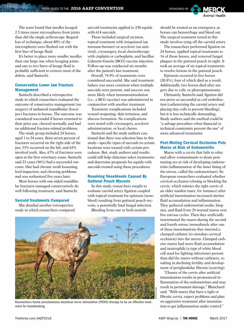
Researchers found percutaneous electrical nerve stimulation (PENS) therapy to be an effective treat-ment for headshaking.
Your GuiDE To THE 2016 aaeP convention SPonSorED bY
AAEP Wrap-Up The horse March 2017TheHorse.com/AAEP2016
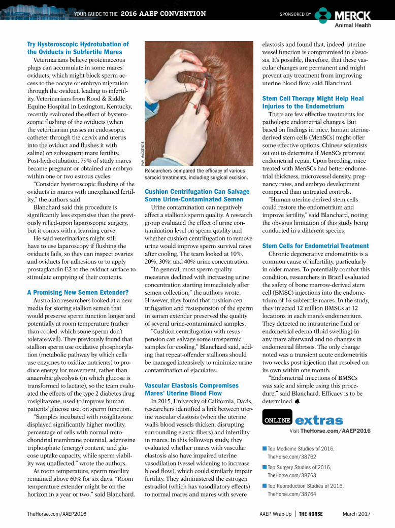
try hysteroscopic hydrotubation of the oviducts in subfertile Mares
Veterinarians believe proteinaceous plugs can accumulate in some mares’ oviducts, which might block sperm ac-cess to the oocyte or embryo migration through the oviduct, leading to infertil-ity. Veterinarians from Rood & Riddle Equine Hospital in Lexington, Kentucky, recently evaluated the effect of hystero-scopic flushing of the oviducts (when the veterinarian passes an endoscopic catheter through the cervix and uterus into the oviduct and flushes it with saline) on subsequent mare fertility. Post- hydrotubation, 79% of study mares became pregnant or obtained an embryo within one or two estrous cycles.
“Consider hysteroscopic flushing of the oviducts in mares with unexplained fertil-ity,” the authors said.
Blanchard said this procedure is significantly less expensive than the previ-ously relied-upon laparoscopic surgery, but it comes with a learning curve.
He said veterinarians might still have to use laparoscopy if flushing the oviducts fails, so they can inspect ovaries and oviducts for adhesions or to apply prostaglandin E2 to the oviduct surface to stimulate emptying of their contents.
A Promising New semen Extender?Australian researchers looked at a new
media for storing stallion semen that would preserve sperm function longer and potentially at room temperature (rather than cooled, which some sperm don’t tolerate well). They previously found that stallion sperm use oxidative phosphoryla-tion (metabolic pathway by which cells use enzymes to oxidize nutrients) to pro-duce energy for movement, rather than anaerobic glycolysis (in which glucose is transformed to lactate), so the team evalu-ated the effects of the type 2 diabetes drug rosiglitazone, used to improve human patients’ glucose use, on sperm function.
“Samples incubated with rosiglitazone displayed significantly higher motility, percentage of cells with normal mito-chondrial membrane potential, adenosine triphosphate (energy) content, and glu-cose uptake capacity, while sperm viabil-ity was unaffected,” wrote the authors.
At room temperature, sperm motility remained above 60% for six days. “Room temperature extender might be on the horizon in a year or two,” said Blanchard.
Cushion Centrifugation Can salvage some urine-Contaminated semen
Urine contamination can negatively affect a stallion’s sperm quality. A research group evaluated the effect of urine con-tamination level on sperm quality and whether cushion centrifugation to remove urine would improve sperm survival rates after cooling. The team looked at 10%, 20%, 30%, and 40% urine concentration.
“In general, most sperm quality measures declined with increasing urine concentration starting immediately after semen collection,” the authors wrote. However, they found that cushion cen-trifugation and resuspension of the sperm in semen extender preserved the quality of several urine-contaminated samples.
“Cushion centrifugation with resus-pension can salvage some urospermic samples for cooling,” Blanchard said, add-ing that repeat-offender stallions should be managed intensively to minimize urine contamination of ejaculates.
vascular Elastosis Compromises Mares’ uterine Blood Flow
In 2015, University of California, Davis, researchers identified a link between uter-ine vascular elastosis (when the uterine wall’s blood vessels thicken, disrupting surrounding elastic fibers) and infertility in mares. In this follow-up study, they evaluated whether mares with vascular elastosis also have impaired uterine vasodilation (vessel widening to increase blood flow), which could similarly impair fertility. They administered the estrogen estradiol (which has vasodilatory effects) to normal mares and mares with severe
elastosis and found that, indeed, uterine vessel function is compromised in elasto-sis. It’s possible, therefore, that these vas-cular changes are permanent and might prevent any treatment from improving uterine blood flow, said Blanchard.
stem Cell therapy Might help heal injuries to the Endometrium
There are few effective treatments for pathologic endometrial changes. But based on findings in mice, human uterine-derived stem cells (MenSCs) might offer some effective options. Chinese scientists set out to determine if MenSCs promote endometrial repair. Upon breeding, mice treated with MenSCs had better endome-trial thickness, microvessel density, preg-nancy rates, and embryo development compared than untreated controls.
“Human uterine-derived stem cells could restore the endometrium and improve fertility,” said Blanchard, noting the obvious limitation of this study being conducted in a different species.
stem Cells for Endometrial treatment Chronic degenerative endometritis is a
common cause of infertility, particularly in older mares. To potentially combat this condition, researchers in Brazil evaluated the safety of bone marrow-derived stem cell (BMSC) injections into the endome-trium of 16 subfertile mares. In the study, they injected 12 million BMSCs at 12 locations in each mare’s endometrium. They detected no intrauterine fluid or endometrial edema (fluid swelling) in any mare afterward and no changes in endometrial fibrosis. The only change noted was a transient acute endometritis two weeks post-injection that resolved on its own within one month.
“Endometrial injections of BMSCs was safe and simple using this proce-dure,” said Blanchard. Efficacy is to be determined. h
PAm
mAC
kEN
zIE
Researchers compared the efficacy of various sarcoid treatments, including surgical excision.
extrasVisit TheHorse.com/AAEP2016
■ Top Medicine Studies of 2016, TheHorse.com/38762
■ Top Surgery Studies of 2016, TheHorse.com/38763
■ Top reproduction Studies of 2016, TheHorse.com/38764
Your GuiDE To THE 2016 aaeP convention SPonSorED bY
AAEP Wrap-Up The horse March 2017TheHorse.com/AAEP2016
ERICA LARSoN
tips for rehabbing soft tissue injuries Prevention is the best treatment for any
health issue, said Andris J. Kaneps, DVM, PhD, Dipl. ACVS, ACVSMR, “but we all know that we can put a horse in a padded room, wrapped in bubble wrap, and we’ll still have issues we’ll need to address.”
Some of the most common are injuries to soft tissues such as tendons and liga-ments. Kaneps, who owns Kaneps Equine Sports Medicine and Surgery, in Beverly, Massachusetts, reviewed best practices for rehabilitating soft tissue injuries.
In a healthy tendon or ligament, “there are even, organized fiber patterns,” he said. “When the injury occurs, the fibers tear, hemorrhage, and go through an inflammatory process. The goal with treatment is to take the problem area and return it to a normal structure.”
The first step is recognizing there’s a problem, he said, often evident as pain, swelling, and sensitivity to palpation.
The next step is the most important: Stop the horse from exercising.
Then the goal should be to reduce in-flammation around the injury, which will make it easier to diagnose. He suggested using cold therapy and/or non- steroidal anti-inflammatory drugs (NSAIDs). Addi-tionally, apply a support bandage to keep the area stable.
Kaneps suggested veterinarians aim to reach a diagnosis within a day or two of injury. With soft-tissue, ultrasound is often the most useful imaging modality.
After diagnosis, it’s time to begin reha-bilitation. Options include:
Cold therapy One of the simplest measures is also one of the most effective at helping tissues heal. Kaneps said the optimal tissue temperature to reach dur-ing cold therapy is 59-66°F (15-19°C).
The gold standard for cold therapy is immersion in an ice and water slurry, he said. It takes 10 to 13 minutes to reach the thermal plateau (the coldest the limb will become), and the total soaking time should be 20 to 30 minutes. Study results have shown that ice water immersion can cool deep tissues in a horse’s limb by up to 16°C (61°F).
He recommended repeating cold therapy three to four times per day for the first 48 hours after acute injury and continuing treatment two to three times per day for about two weeks. Owners can use cold therapy following exercise when the horse returns to work to reduce inflammation at the injury site.
Kaneps said ice and cold packs can be useful, but they tend not to be as effective as ice water immersion. Cold salt water spas can also help.
Controlled exercise “This is the pri-mary, most effective treatment,” Kaneps said, adding that studies have shown that 67-71% of horses with soft-tissue injuries treated using controlled exercise had successful outcomes, compared to just 25-51% of horses treated with pasture turnout.
Kaneps recommended caretakers begin hand-walking horses shortly after the injury because tendons and ligaments require stress to heal properly. The rule of thumb is to increase exercise by 5-10% each week and reassess lameness and ultrasound scans every 60 to 90 days.
He said many controlled exercise pro-grams go something like this:■ Gradually build up to hand-walking for
30 minutes two to three times per day;■ Transition to walking under tack for
20-25 minutes per day;■ After about two weeks, add three to five
minutes of trot per day, but not until the horse is warmed up at the walk for 10-15 minutes;
■ Increase the trotting time gradually to 20-25 minutes per day;
■ Add three minutes of canter, gradually increasing that time.The veterinarian should recheck the
horse’s soundness before each workload increase, he added.
Regenerative treatments Some of these therapies can improve or shorten the healing process. Options include:■ Platelet-rich plasma (PRP), which deliv-
ers a high concentration of platelets in the form of blood plasma to a lesion, increasing the amount of growth fac-tors at the site to help the injury heal. There are commercially available PRP products, as well as a stall-side system that separates the horse’s own red and white blood cells from the plasma in a relatively short amount of time. The veterinarian injects the PRP into the lesion or the surrounding areas.
PHo
ToS
Co
UR
TES
y D
R.
AND
RIS
j.
kAN
EPSrehabilitating
injured horses
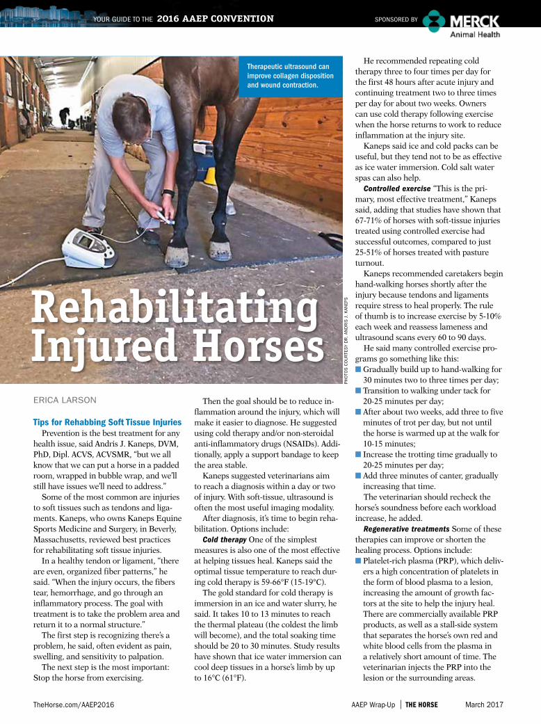
Therapeutic ultrasound can improve collagen disposition and wound contraction.
Your GuiDE To THE 2016 aaeP convention SPonSorED bY
AAEP Wrap-Up The horse March 2017TheHorse.com/AAEP2016
■ Stem cells, which Kaneps said recruit growth factors to help injured areas heal with better quality, strength, and elasticity. There are two main types of stem cells: bone marrow- and adipose (fat)-derived. He said researchers on one study showed a lower reinjury rate in horses with soft-tissue injuries treated with stem cells than without. Kaneps said the optimal time to inject
both PRP and stem cells is three to four weeks following injury, preferably using ultrasound guidance. After the injection, stop exercise and keep the limb ban-daged for about two weeks and perform a veterinary follow-up four weeks after injection.
Therapeutic ultrasound This stimulates healing by delivering heat to the injured tissue, he said, which increases local cir-culation, among other effects. He added that it can also improve collagen disposi-tion and wound contraction.
Extracorporeal shock wave therapy This approach has been shown to reduce inflammation; increase cytokines, growth factors, and osteoblasts (all important to healing); and potentially recruit stem cells to affected areas, he said, cautioning that tissue damage can occur with a too-high setting.
Laser therapy While there have been many recent advancements in this area, Kaneps said there’s still no research prov-ing it’s effective for treating soft-tissue injuries. Still, he said modern lasers could offer sufficient energy and penetration depth to reach and provide energy to the cells involved.
low-intensity Exercise Key to Muscle injury healing
“Muscle pain and injury as a cause of lameness and poor performance in the horse are poorly recognized,” said Tracy Turner, DVM, MS, Dipl. ACVS, ACVSMR, who owns Turner Equine Sports Medi-cine and Surgery, in Big Lake, Minnesota.
He said factors known to predispose horses to muscle strains and injury include cold temperatures, impaired circulation to the muscle, muscle fatigue, poor or insufficient training; and insuffi-cient warmup. Diagnosing these injuries, however, is very challenging.
Veterinarians can’t diagnose muscle in-juries using radiographs or nerve blocks, he said, and ultrasound is only useful af-ter locating the injured muscle. Palpation
isn’t always helpful because some muscle injuries are only painful during exercise or movement. And elevated muscle-related enzymes in the blood aren’t useful indicators, either.
So to make a muscle injury diagnosis, start with collecting a thorough case history.
“It is important to determine whether there was a history of a fall or other trauma, the duration of clinical signs, the presence of swelling, and whether lameness or poor performance has been documented,” he said.
Turner suggested practitioners stand the horse squarely and look and palpate for signs of muscle atrophy (wasting), fi-brosis (scarring), tension, spasm, defects, or pain. Then consider using thermog-raphy, which reveals muscle injuries as temperature increases or decreases.
Once the veterinarian has located the injury, he or she can use ultrasound to evaluate muscle fiber alignment and look for hemorrhage.
The next step is rehabilitating the horse. Turner says the general goals are improving flexibility and muscle condi-tion, strengthening, and returning to full activity. Rehab options he described include stretching, massage, therapeutic ultrasound, shock wave therapy, electrical stimulation, and pulsed electromagnetic field therapy (for more on all these, see TheHorse.com/38740)
“Regardless of the modality … I believe that the horse must remain in at least low-intensity exercise,” Turner added.
Another important component to re-hab is strengthening the muscles, he said. “Horses gain strength by flexion, through transition of gait, stress, and lateral work,” he said.
He encouraged veterinarians to con-tinue stretching exercises during and after strengthening.
Once training commences, said Turner, “I’ve found that altering the exercise program can be most beneficial, and conditioning is of utmost importance” in the horse’s long-term recovery. h
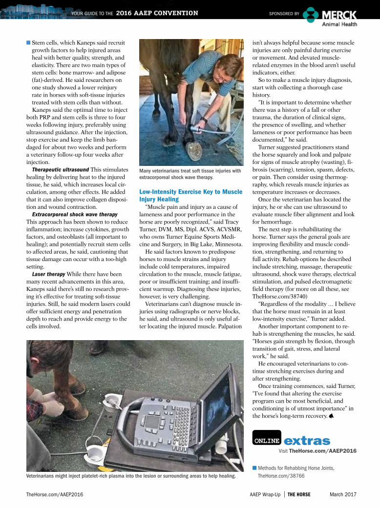
Veterinarians might inject platelet-rich plasma into the lesion or surrounding areas to help healing.
Many veterinarians treat soft tissue injuries with extracorporeal shock wave therapy.
extrasVisit TheHorse.com/AAEP2016
■ Methods for rehabbing Horse Joints, TheHorse.com/38766
Your GuiDE To THE 2016 aaeP convention SPonSorED bY
AAEP Wrap-Up The horse March 2017TheHorse.com/AAEP2016
Your GuiDE To THE 2016 aaeP convention SPonSorED bY
STEPHANIE L. CHURCH
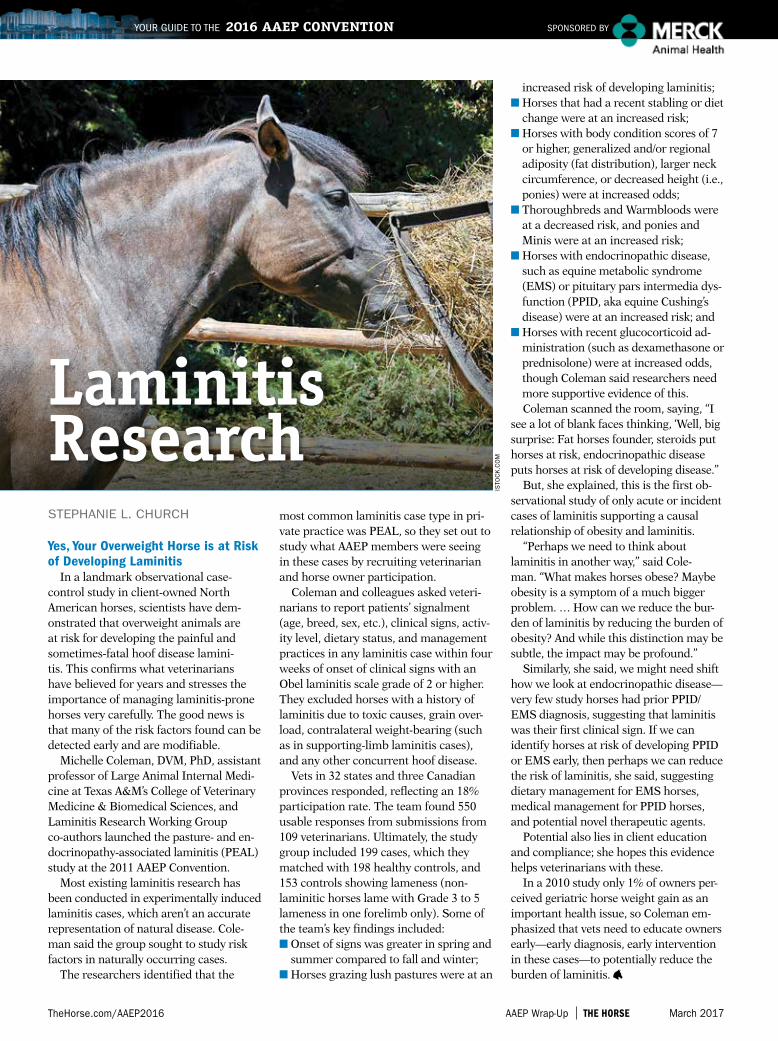
yes, your overweight horse is at risk of Developing laminitis
In a landmark observational case- control study in client-owned North American horses, scientists have dem-onstrated that overweight animals are at risk for developing the painful and sometimes-fatal hoof disease lamini-tis. This confirms what veterinarians have believed for years and stresses the importance of managing laminitis-prone horses very carefully. The good news is that many of the risk factors found can be detected early and are modifiable.
Michelle Coleman, DVM, PhD, assistant professor of Large Animal Internal Medi-cine at Texas A&M’s College of Veterinary Medicine & Biomedical Sciences, and Laminitis Research Working Group co-authors launched the pasture- and en-docrinopathy-associated laminitis (PEAL) study at the 2011 AAEP Convention.
Most existing laminitis research has been conducted in experimentally induced laminitis cases, which aren’t an accurate representation of natural disease. Cole-man said the group sought to study risk factors in naturally occurring cases.
The researchers identified that the
most common laminitis case type in pri-vate practice was PEAL, so they set out to study what AAEP members were seeing in these cases by recruiting veterinarian and horse owner participation.
Coleman and colleagues asked veteri-narians to report patients’ signalment (age, breed, sex, etc.), clinical signs, activ-ity level, dietary status, and management practices in any laminitis case within four weeks of onset of clinical signs with an Obel laminitis scale grade of 2 or higher. They excluded horses with a history of laminitis due to toxic causes, grain over-load, contralateral weight-bearing (such as in supporting-limb laminitis cases), and any other concurrent hoof disease.
Vets in 32 states and three Canadian provinces responded, reflecting an 18% participation rate. The team found 550 usable responses from submissions from 109 veterinarians. Ultimately, the study group included 199 cases, which they matched with 198 healthy controls, and 153 controls showing lameness (non-laminitic horses lame with Grade 3 to 5 lameness in one forelimb only). Some of the team’s key findings included:■ Onset of signs was greater in spring and
summer compared to fall and winter; ■ Horses grazing lush pastures were at an
increased risk of developing laminitis;■ Horses that had a recent stabling or diet
change were at an increased risk;■ Horses with body condition scores of 7
or higher, generalized and/or regional adiposity (fat distribution), larger neck circumference, or decreased height (i.e., ponies) were at increased odds;
■ Thoroughbreds and Warmbloods were at a decreased risk, and ponies and Minis were at an increased risk;
■ Horses with endocrinopathic disease, such as equine metabolic syndrome (EMS) or pituitary pars intermedia dys-function (PPID, aka equine Cushing’s disease) were at an increased risk; and
■ Horses with recent glucocorticoid ad-ministration (such as dexamethasone or prednisolone) were at increased odds, though Coleman said researchers need more supportive evidence of this.Coleman scanned the room, saying, “I
see a lot of blank faces thinking, ‘Well, big surprise: Fat horses founder, steroids put horses at risk, endocrinopathic disease puts horses at risk of developing disease.”
But, she explained, this is the first ob-servational study of only acute or incident cases of laminitis supporting a causal relationship of obesity and laminitis.
“Perhaps we need to think about laminitis in another way,” said Cole-man. “What makes horses obese? Maybe obesity is a symptom of a much bigger problem. … How can we reduce the bur-den of laminitis by reducing the burden of obesity? And while this distinction may be subtle, the impact may be profound.”
Similarly, she said, we might need shift how we look at endocrinopathic disease—very few study horses had prior PPID/EMS diagnosis, suggesting that laminitis was their first clinical sign. If we can identify horses at risk of developing PPID or EMS early, then perhaps we can reduce the risk of laminitis, she said, suggesting dietary management for EMS horses, medical management for PPID horses, and potential novel therapeutic agents.
Potential also lies in client education and compliance; she hopes this evidence helps veterinarians with these.
In a 2010 study only 1% of owners per-ceived geriatric horse weight gain as an important health issue, so Coleman em-phasized that vets need to educate owners early—early diagnosis, early intervention in these cases—to potentially reduce the burden of laminitis. h
ISTo
Ck
.Co
m
laminitis research
AAEP Wrap-Up The horse March 2017TheHorse.com/AAEP2016
From the breeding barn to the show ring and everything in between, we have the vaccines you need to help protect your horse.
• Made with the exclusive Antigen Purification System (APS™)
• Contains the Havlogen® adjuvant veterinarians know and trust
• Delivers core vaccine coverage according to American Association of Equine Practitioners recommendations
Now that’s protected.
Ask your veterinarian for Prestige®, Encevac®, Prodigy® and EquiRab® brand vaccines. Visit us at GetVaccinatingRight.com to learn more about Merck Animal Health and the equine products and programs that help keep horses healthy.
Every vaccine purchased through Merck supports the plight of the unwanted horse through the Unwanted Horse Veterinary Relief Campaign.
The Science of Protected
The Science of Healthier Animals
2 Giralda Farms • Madison, NJ 07940 • merck-animal-health-usa.com • 800-521-5767Copyright © 2016 Intervet Inc., d/b/a/ Merck Animal Health, a subsidiary of Merck & Co., Inc.All rights reserved. 3526 EQ-FP AD Vaccine Line
3526_VaccineFamily_Girl_8125x10875.indd 1 2/15/17 8:54 AM
AAEP Wrap-Up The horse March 2017TheHorse.com/AAEP2016
Your GuiDE To THE 2016 aaeP convention SPonSorED bY
ALExANDRA BECkSTETT; ERICA LARSoN
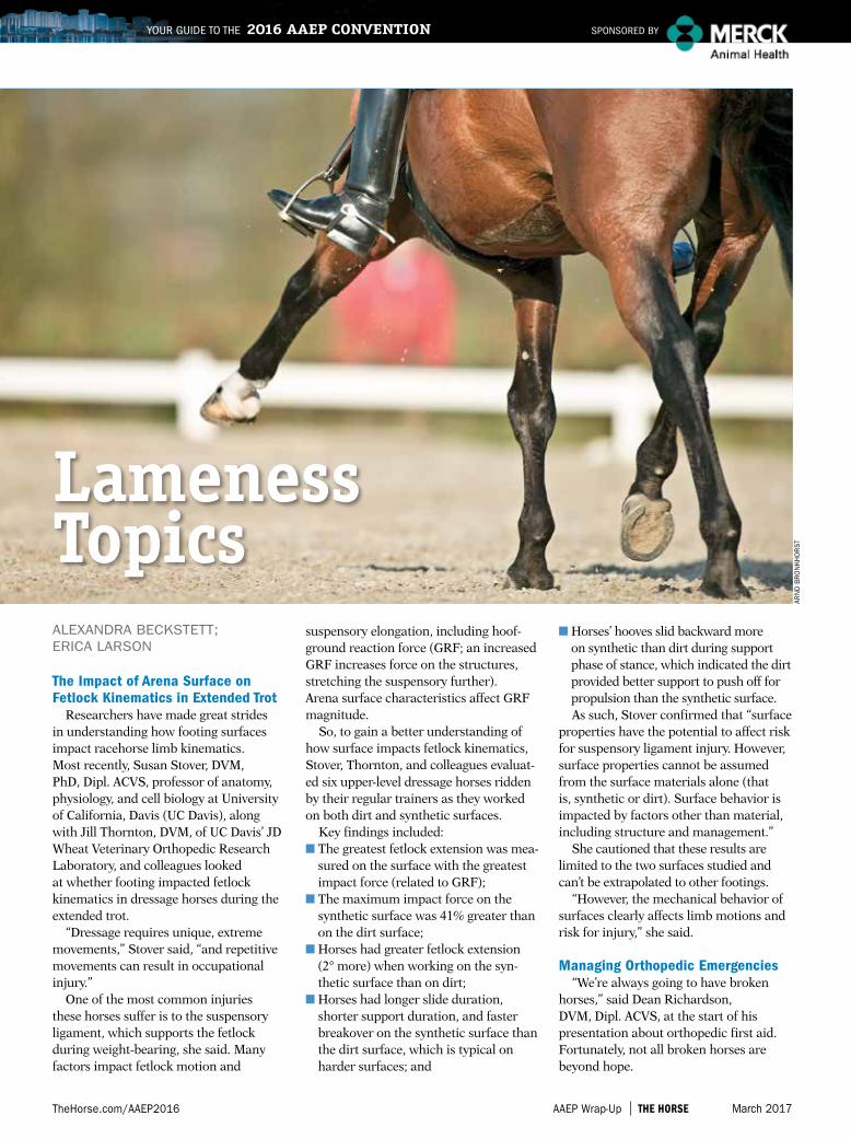
the impact of Arena surface on Fetlock Kinematics in Extended trot
Researchers have made great strides in understanding how footing surfaces impact racehorse limb kinematics. Most recently, Susan Stover, DVM, PhD, Dipl. ACVS, professor of anatomy, physiology, and cell biology at University of California, Davis (UC Davis), along with Jill Thornton, DVM, of UC Davis’ JD Wheat Veterinary Orthopedic Research Laboratory, and colleagues looked at whether footing impacted fetlock kinematics in dressage horses during the extended trot.
“Dressage requires unique, extreme movements,” Stover said, “and repetitive movements can result in occupational injury.”
One of the most common injuries these horses suffer is to the suspensory ligament, which supports the fetlock during weight-bearing, she said. Many factors impact fetlock motion and
suspensory elongation, including hoof-ground reaction force (GRF; an increased GRF increases force on the structures, stretching the suspensory further). Arena surface characteristics affect GRF magnitude.
So, to gain a better understanding of how surface impacts fetlock kinematics, Stover, Thornton, and colleagues evaluat-ed six upper-level dressage horses ridden by their regular trainers as they worked on both dirt and synthetic surfaces.
Key findings included:■ The greatest fetlock extension was mea-
sured on the surface with the greatest impact force (related to GRF);
■ The maximum impact force on the synthetic surface was 41% greater than on the dirt surface;
■ Horses had greater fetlock extension (2° more) when working on the syn-thetic surface than on dirt;
■ Horses had longer slide duration, shorter support duration, and faster breakover on the synthetic surface than the dirt surface, which is typical on harder surfaces; and
■ Horses’ hooves slid backward more on synthetic than dirt during support phase of stance, which indicated the dirt provided better support to push off for propulsion than the synthetic surface.As such, Stover confirmed that “surface
properties have the potential to affect risk for suspensory ligament injury. However, surface properties cannot be assumed from the surface materials alone (that is, synthetic or dirt). Surface behavior is impacted by factors other than material, including structure and management.”
She cautioned that these results are limited to the two surfaces studied and can’t be extrapolated to other footings.
“However, the mechanical behavior of surfaces clearly affects limb motions and risk for injury,” she said.
Managing orthopedic Emergencies“We’re always going to have broken
horses,” said Dean Richardson, DVM, Dipl. ACVS, at the start of his presentation about orthopedic first aid. Fortunately, not all broken horses are beyond hope.
ARN
D B
Ro
Nk
Ho
RS
T
lameness topics
The Science of Trusted
From the broodmare to the performance horse, properly managing your mare’s hormones is critical. Rely on the product that’s trusted most.
• More than 30 years of practical use in the field by veterinarians1
• More than 200 clinical trials to determine efficacy, duration and safety1
• More than 20 million doses sold to veterinarians, trainers and horse owners1
Now that’s trusted.
Talk to your veterinarian about proper use and safe handling of Regu-Mate®. Avoid skin contact. Always wear protective gloves when administering Regu-Mate®. This product is contraindicated for use in mares with a previous or current history of uterine inflammation. Pregnant women, or women who suspect they are pregnant, should not handle this product. For complete product information, see accompanying product insert.
Regu-Mate® (altrenogest) is the name veterinarians and their clients depend on for estrus control (suppression, management).
1 Data on file, Merck Animal Heath
Ask your veterinarian for Regu-Mate®. Visit us online at merck-animal-health-equine.com to learn more about Merck Animal Health and the equine products and programs that help keep horses healthy.
The Science of Healthier Animals
2 Giralda Farms • Madison, NJ 07940 • merck-animal-health-usa.com • 800-521-5767Copyright © 2016 Intervet Inc., d/b/a/ Merck Animal Health, a subsidiary of Merck & Co., Inc.All rights reserved. 3526 EQ-FP AD Regu-Mate®
3526_Regu-Mate_8125x10875.indd 1 2/7/17 10:27 AM
AAEP Wrap-Up The horse March 2017TheHorse.com/AAEP2016
Richardson is the chief of large animal surgery at the University of Pennsylvania School of Veterinary Medicine’s New Bolton Center, in Kennett Square. He described how veterinarians can best handle orthopedic emergencies such as extremely unstable limb fractures.
“With proper sedation and simple emergency bandaging, a large proportion of catastrophes can be humanely managed until a thoughtful decision can be made,” as to whether the injury can be repaired surgically or the horse requires euthanasia, he said.
“Many people are under the impression that you can’t do anything about injuries involving bones,” said Richardson. “The reality is that many severe lacerations and orthopedic injuries seem to be far worse than they are.”
While some extremely catastrophic injuries do require euthanasia, you don’t want to find out after you’ve put a horse down that other horses with the same injury have been treated successfully.
So, what orthopedic injuries are typi-cally treatable and which are hopeless?
Skin wound over a fracture These are not death sentences, said Richardson. Prognosis does, however, depend on the degree of fracture contamination. Super-ficial lacerations are much less likely to result in unmanageable infection, espe-cially in locations with a healthy muscle covering and blood supply.
Nondisplaced fractures Any of these injuries have a chance to heal, said Richardson. And not every horse with a
non-weight-bearing lameness will develop support-limb laminitis.
Simple vs. comminuted fractures “Simple fractures are nearly always more manageable than comminuted fractures (multiple fragments), but location is ev-erything,” he said. The higher up the limb, the less probable that it can heal on its own, with the exception of the humerus (located between the shoulder and the elbow), which he said has been managed successfully with stall rest.
Articular fractures Any displaced fracture involving a joint is best managed with surgery, and many joints return to full function if they can be reconstructed properly. Veterinarians can sometimes salvage severe injuries that cannot be reconstructed surgically by fusing the af-fected joint, said Richardson.
Fractures with vascular compromise
Any major injury with a loss of blood sup-ply is likely to be fatal.
When faced with an orthopedic injury, goals are to keep the owner’s options open, keep the skin intact, prevent further trauma, and allay both horse and owner anxiety. The horse needs a proper seda-tion dose (not so much that he loses all coordination) and analgesics for pain and, if there’s an open wound, antibiotics.
Before hauling the horse to the clinic, the owner can make some important and potentially life-saving transportation decisions. The smoothest-riding trailer is a gooseneck with a ramp. Ship the horse in a space or stall that’s as tight as pos-sible to give him something to lean on to protect his injured limb.
Load the horse so that his injured limb (whether hind or fore) is closest to the rear of the trailer. “So if you brake
Co
UR
TES
y D
R.
DEA
N R
ICH
ARD
So
N
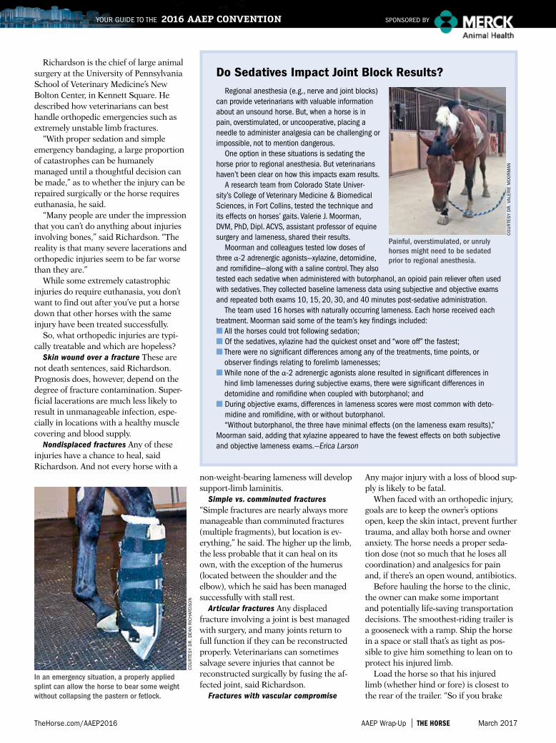
In an emergency situation, a properly applied splint can allow the horse to bear some weight without collapsing the pastern or fetlock.
Do sedatives impact Joint Block results?regional anesthesia (e.g., nerve and joint blocks)
can provide veterinarians with valuable information about an unsound horse. but, when a horse is in pain, overstimulated, or uncooperative, placing a needle to administer analgesia can be challenging or impossible, not to mention dangerous.
one option in these situations is sedating the horse prior to regional anesthesia. but veterinarians haven’t been clear on how this impacts exam results.
A research team from Colorado State univer-sity’s College of Veterinary Medicine & biomedical Sciences, in Fort Collins, tested the technique and its effects on horses’ gaits. Valerie J. Moorman, DVM, PhD, Dipl. ACVS, assistant professor of equine surgery and lameness, shared their results.
Moorman and colleagues tested low doses of three α-2 adrenergic agonists—xylazine, detomidine, and romifidine—along with a saline control. They also tested each sedative when administered with butorphanol, an opioid pain reliever often used with sedatives. They collected baseline lameness data using subjective and objective exams and repeated both exams 10, 15, 20, 30, and 40 minutes post-sedative administration.
The team used 16 horses with naturally occurring lameness. Each horse received each treatment. Moorman said some of the team’s key findings included:■ All the horses could trot following sedation;■ of the sedatives, xylazine had the quickest onset and “wore off” the fastest;■ There were no significant differences among any of the treatments, time points, or
observer findings relating to forelimb lamenesses;■ While none of the α-2 adrenergic agonists alone resulted in significant differences in
hind limb lamenesses during subjective exams, there were significant differences in detomidine and romifidine when coupled with butorphanol; and
■ During objective exams, differences in lameness scores were most common with deto-midine and romifidine, with or without butorphanol.“Without butorphanol, the three have minimal effects (on the lameness exam results),”
Moorman said, adding that xylazine appeared to have the fewest effects on both subjective and objective lameness exams.—Erica Larson
Co
UR
TES
y D
R.
vALE
RIE
mo
oR
mAN
Painful, overstimulated, or unruly horses might need to be sedated prior to regional anesthesia.
Your GuiDE To THE 2016 aaeP convention SPonSorED bY
AAEP Wrap-Up The horse March 2017TheHorse.com/AAEP2016
suddenly, his weight is on the uninjured area,” said Richardson.
Foals fatigue easily, so he said to get them into a recumbent position (lying down) on the trailer, and have an atten-dant ride with them during the trip.
Many of these horses (particularly those with unstable fractures) should be placed in a splinted bandage for trans-port. Richardson warned, however, that an ill-applied splint can do more harm than good. Make sure it’s very tight, but not too heavy with bandage material.
“Make the bandage light enough to get the splints closer to the skin, but thick enough to prevent trauma from the splint to the skin or soft tissues,” he said.
Also be sure the splint is neither too short nor too long to be of use. “A good principle is to stabilize the joint above and the joint below the injury whenever possible,” said Richardson.
He said most mistakes in these emer-gencies occur because decisions are made too quickly. These are typically high-stress scenarios with owners in a state of panic.
“It is absolutely true that some injuries are so painful and debilitating that our current techniques cannot manage them successfully, but we need to have decision-making evolve alongside improv-ing techniques,” he said. “Many injuries that would have been considered hopeless years ago can now be treated with consis-tent success.”
What’s inside the Digital Cushion?Your horse’s hooves contain several
important bone and soft tissue structures, all protected by the thick, elastic digital cushion. Veterinarians know that this important piece of anatomy, located in the rear part of the hoof, absorbs energy and forces placed on the hoof, but little more than that.
Recently, Babak Faramarzi, DVM, CVA, MSc, PhD, and colleagues sought to gain a better understanding of the structure’s connective, nervous, and adipose (fat) tis-sues, as well as its vascular components.
“Previous studies are inconsistent, with some claiming that digital cushion (DC) is primarily a fat pad,” said Faramarzi, asso-ciate professor at the Western University of Health Sciences College of Veterinary Medicine, in Pomona, California. “Col-lectively, these studies represent disagree-ment about the architecture of the DC.”
His team collected samples from the
hooves of 24 sound Quarter Horses euthanized for reasons unrelated to the study, examining samples from four regions of the cushion—axial-proximal (top center), axial-distal (bottom center), abaxial- lateral (outer edge), and abaxial-medial (inner edge)—via light microscopy.
The axial-distal region had signifi-cantly more collagen (a connective tissue protein) and fewer elastic fibers than the axial- proximal and abaxial regions.
“The presence of a moderate to large amount of elastic fiber profiles in the DC … may allow the elastic fibers to support the tensile strength of collagen bundles,” he said. “Elastic fiber-rich dynamic tis-sues are, therefore, able to deform and store energy under normal physiological loads and use this energy to drive recoil back to a resting state.”
Also, there were about four times as many nerve bundles in the axial-proximal region than in the axial-distal region, Faramarzi said, indicating there are more nerves near the limb than the hoof’s sole.
“This study, for the first time, char-acterized the architecture of differ-ent regions of the DC,” he said. “Such regional structural differences of the DC are presumably related to the different functional properties of those regions.”
Faramarzi hopes these results encour-age further research on this topic as well as the influence of age, breed, exercise, shoeing/trimming, and environmental factors. h
Regu-Mate® (altrenogest)
Solution 0.22% (2.2 mg/mL) CAUTION: Federal law restricts this drug to use by or on the order of a licensed veterinarian.
DESCRIPTION: Regu-Mate® (altrenogest) Solution 0.22% contains the active synthetic progestin, altrenogest. The chemical name is 17α-allyl-17ß-hydroxyestra- 4,9,11-trien-3-one. The CAS Registry Number is 850-52-2. The chemical structure is:
Each mL of Regu-Mate® (altrenogest) Solution 0.22% contains 2.2 mg of altrenogest in an oil solution.
ACTIONS: Regu-Mate® (altrenogest) Solution 0.22% produces a progestational effect in mares.
INDICATIONS: Regu-Mate® (altrenogest) Solution 0.22% is indicated to suppress estrus in mares. Suppression of estrus allows for a predictable occurrence of estrus following drug withdrawal. This facilitates the attainment of regular cyclicity during the transition from winter anestrus to the physiological breeding season. Suppression of estrus will also facilitate management of prolonged estrus conditions. Suppression of estrus may be used to facilitate scheduled breeding during the physiological breeding season.
CONTRAINDICATIONS: Regu-Mate® (altrenogest) Solution 0.22% is contraindicated for use in mares having a previous or current history of uterine inflammation (i.e., acute, subacute, or chronic endometritis). Natural or synthetic gestagen therapy may exacerbate existing low-grade or “smoldering” uterine inflammation into a fulminating uterine infection in some instances.
PRECAUTIONS: Various synthetic progestins, including altrenogest, when administered to rats during the embryogenic stage of pregnancy at doses manyfold greater than the recommended equine dose caused fetal anomalies, specifically masculinization of the female genitalia.
DOSAGE AND ADMINISTRATION: While wearing protective gloves, remove shipping cap and seal; replace with enclosed plastic dispensing cap. Remove cover from bottle dispensing tip and connect luer lock syringe (without needle). Draw out appropriate volume of Regu-Mate solution. (Note: Do not remove syringe while bottle is inverted as spillage may result.) Detach syringe and administer solution orally at the rate of 1 mL per 110 pounds body weight (0.044 mg/kg) once daily for 15 consecutive days. Administer solution directly on the base of the mare’s tongue or on the mare’s usual grain ration. Replace cover on bottle dispensing tip to prevent leakage. Excessive use of a syringe may cause the syringe to stick; therefore, replace syringe as necessary.
WHICH MARES WILL RESPOND TO REGU-MATE® (altrenogest) SOLUTION 0.22%: Extensive clinical trials have demonstrated that estrus will be suppressed in approximately 95% of the mares within three days; however, the post-treatment response depended on the level of ovarian activity when treatment was initiated. Estrus in mares exhibiting regular estrus cycles during the breeding season will be suppressed during treatment; these mares return to estrus four to five days following treatment and continue to cycle normally. Mares in winter anestrus with small follicles continued in anestrus and failed to exhibit normal estrus following withdrawal. Response in mares in the transition phase between winter anestrus and the summer breeding season depended on the degree of follicular activity. Mares with inactive ovaries and small follicles failed to respond with normal cycles post-treatment, whereas a higher proportion of mares with ovarian follicles 20 mm or greater in diameter exhibited normal estrus cycles post-treatment. Regu-Mate® (altrenogest) Solution 0.22% was very effective for suppressing the prolonged estrus behavior frequently observed in mares during the transition period (February, March and April). In addition, a high proportion of these mares responded with regular estrus cycles post-treatment.
SPECIFIC USES FOR REGU-MATE® (altrenogest) SOLUTION 0.22%: SUPPRESSION OF ESTRUS TO:1. Facilitate attainment of regular cycles during the transition period from winter anestrus to the
physiological breeding season. To facilitate attainment of regular cycles during the transition phase, mares should be examined to determine the degree of ovarian activity. Estrus in mares with inactive ovaries (no follicles greater than 20 mm in diameter) will be suppressed but these mares may not begin regular cycles following treatment. However, mares with active ovaries (follicles greater than 20 mm in diameter) frequently respond with regular post-treatment estrus cycles.
2. Facilitate management of the mare exhibiting prolonged estrus during the transition period. Estrus will be suppressed in mares exhibiting prolonged behavioral estrus either early or late during the transition period. Again, the posttreatment response depends on the level of ovarian activity. The mares with greater ovarian activity initiate regular cycles and conceive sooner than the inactive mares. Regu-Mate® (altrenogest) Solution 0.22% may be administered early in the transition period to suppress estrus in mares with inactive ovaries to aid in the management of these mares or to mares later in the transition period with active ovaries to prepare and schedule the mare for breeding.
3. Permit scheduled breeding of mares during the physiological breeding season. To permit scheduled breeding, mares which are regularly cycling or which have active ovarian function should be given Regu-Mate® (altrenogest) Solution 0.22% daily for 15 consecutive days beginning 20 days before the date of the planned estrus. Ovulation will occur 5 to 7 days following the onset of estrus as expected for nontreated mares. Breeding should follow usual procedures for mares in estrus. Mares may be regulated and scheduled either individually or in groups.
ADDITIONAL INFORMATION: A 3-year well controlled reproductive safety study was conducted in 27 pregnant mares, and compared with 24 untreated control mares. Treated mares received 2 mL Regu-Mate® (altrenogest) Solution 0.22% /110 lb body weight (2x dosage recommended for estrus suppression) from day 20 to day 325 of gestation. This study provided the following data: 1. In filly offspring (all ages) of treated mares, clitoral size was increased. 2. Filly offspring from treated mares had shorter interval from Feb. 1 to first ovulation than fillies
from their untreated mare counterparts.3. There were no significant differences in reproductive performance between treated and untreated
animals (mares & their respective offspring) measuring the following parameters: • interval from Feb. 1 to first ovulation, in mares only. • mean interovulatory interval from first to second cycle and second to third cycle, mares
only. • follicle size, mares only. • at 50 days gestation, pregnancy rate in treated mares was 81.8% (9/11) and untreated
mares was 100% (4/4). • after 3 cycles, 11/12 treated mares were pregnant (91.7%) and 4/4 untreated mares were
pregnant (100%). • colt offspring of treated and control mares reached puberty at approximately the same age
(82 & 84 weeks respectively). • stallion offspring from treated and control mares showed no differences in seminal volume,
spermatozoal concentration, spermatozoal motility, and total sperm per ejaculate. • stallion offspring from treated and control mares showed no difference in sexual behavior. • testicular characteristics (scrotal width, testis weight, parenchymal weight, epididymal
weight and height, testicular height, width & length) were the same between stallion offspring of treated and control mares.
REFERENCES:Shoemaker, C.F., E.L. Squires, and R.K. Shideler. 1989.Safety of Altrenogest in Pregnant Mares and on Health and Development of Offspring. Eq. Vet. Sci. (9); No. 2: 69-72.Squires, E.L., R.K. Shideler, and A.O. McKinnon. 1989.Reproductive Performance of Offspring from Mares Administered Altrenogest During Gestation. Eq. Vet. Sci. (9); No. 2: 73-76.
WARNING: Do not use in horses intended for food.
HUMAN WARNINGS: Skin contact must be avoided as Regu-Mate® (altrenogest) Solution 0.22% is readily absorbed through unbroken skin. Protective gloves must be worn by all persons handling this product. Pregnant women or women who suspect they are pregnant should not handle Regu-Mate® (altrenogest) Solution 0.22%. Women of child bearing age should exercise extreme caution when handling this product. Accidental absorption could lead to a disruption of the menstrual cycle or prolongation of pregnancy. Direct contact with the skin should therefore be avoided. Accidental spillage on the skin should be washed off immediately with soap and water.
INFORMATION FOR HANDLERS: WARNING: Regu-Mate® (altrenogest) Solution 0.22% is readily absorbed by the skin. Skin contact must be avoided; protective gloves must be worn when handling this product.
Effects of Overexposure: There has been no human use of this specific product. The information contained in this section is extrapolated from data available on other products of the same pharmacological class that have been used in humans. Effects anticipated are due to the progestational activity of altrenogest. Acute effects after a single exposure are possible; however, continued daily exposure has the potential for more untoward effects such as disruption of the menstrual cycle, uterine or abdominal cramping, increased or decreased uterine bleeding, prolongation of pregnancy and headaches. The oil base may also cause complications if swallowed. In addition, the list of people who should not handle this product (see below) is based upon the known effects of progestins used in humans on a chronic basis.
PEOPLE WHO SHOULD NOT HANDLE THIS PRODUCT. 1. Women who are or suspect they are pregnant. 2. Anyone with thrombophlebitis or thromboembolic disorders or with a history of these events. 3. Anyone with cerebral-vascular or coronary artery disease. 4. Women with known or suspected carcinoma of the breast. 5. People with known or suspected estrogen-dependent neoplasia. 6. Women with undiagnosed vaginal bleeding. 7. People with benign or malignant tumors which developed during the use of oral
contraceptives or other estrogen-containing products. 8. Anyone with liver dysfunction or disease.
ACCIDENTAL EXPOSURE: Altrenogest is readily absorbed from contact with the skin. In addition, this oil based product can penetrate porous gloves. Altrenogest should not penetrate intact rubber or impervious gloves; however, if there is leakage (i.e., pinhole, spillage, etc.), the contaminated area covered by such occlusive materials may have increased absorption. The following measures are recommended in case of accidental exposure. Skin Exposure: Wash immediately with soap and water.Eye Exposure: Immediately flush with plenty of water for 15 minutes. Get medical attention.If Swallowed: Do not induce vomiting. Regu-Mate® (altrenogest) Solution 0.22% contains an oil. Call a physician. Vomiting should be supervised by a physician because of possible pulmonary damage via aspiration of the oil base. If possible, bring the container and labeling to the physician.
CAUTION: For oral use in horses only. Keep this and all medication out of the reach of children.
Store at or below 25°C (77°F).
NADA# 131-310, Approved by FDA.
HOW SUPPLIED:Regu-Mate® (altrenogest) Solution 0.22% (2.2 mg/mL).Each mL contains 2.2 mg altrenogest in an oil solution.Available in 1000 mL plastic bottles.
* US Patents 3,453,267; 3,478,067; 3,484,462
Manufactured by:DPT Laboratories, San Antonio, TX 78215
Distributed by:Intervet Inc., Millsboro, DE 19966
DOSAGE CHART:
Approximate Weight Dose in Pounds in mL 770 7 880 8 990 9 1100 10 1210 11 1320 12
Regu-Mate Ad-HO_BR_Legal_25x9625_BW.indd 1 2/1/17 11:05 AM
extrasVisit TheHorse.com/AAEP2016
■ Soft Tissue injuries of the Western Performance Horse’s Foot, TheHorse.com/38624
■ How to Tell if Your Saddle Hurts Your Horse, TheHorse.com/38765
Many injuries that would have been considered hopeless years ago can now be treated with consis-tent success.”
DR. DEAN RICHARDSoN
Your GuiDE To THE 2016 aaeP convention SPonSorED bY
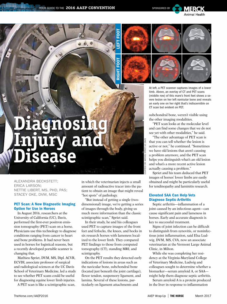
AAEP Wrap-Up The horse March 2017TheHorse.com/AAEP2016
Your GuiDE To THE 2016 aaeP convention SPonSorED bY
ALExANDRA BECkSTETT; ERICA LARSoN; NETTIE LIBERT, mS, PHD, PAS; STACEy okE, Dvm, mSC
PEt scan: A New Diagnostic imaging option for use in horses
In August 2016, researchers at the University of California (UC), Davis, performed the first-ever positron emis-sion tomography (PET) scan on a horse. Physicians use this technology to diagnose conditions ranging from cancer to heart and bone problems. It had never been used in horses for logistical reasons, but a recently developed portable scanner is changing that.
Mathieu Spriet, DVM, MS, Dipl. ACVR, ECVDI, associate professor of surgical and radiological sciences at the UC Davis School of Veterinary Medicine, led a study to see whether PET scans could be useful for diagnosing equine lower limb injuries.
A PET scan is like a scintigraphic scan,
in which the veterinarian injects a small amount of radioactive tracer into the pa-tient to obtain an image that might reveal “hot spots” of pathology.
“But instead of getting a single (two-dimensional) image, we’re getting a series of images through the body, giving us much more information than the classic scintigraphic scan,” Spriet said.
In their study, he and his colleagues used PET to capture images of the front feet and fetlocks, the knees, and hocks in six research horses with lameness local-ized to the lower limb. They compared PET findings to those from computed tomography (CT), standing MRI, and scintigraphy.
On the PET results they detected early indications of lesions in areas such as the navicular bone, subchondral bone (located just beneath the joint cartilage), flexor tendon, suspensory ligament, and lamina. Several of these lesions, par-ticularly on ligament attachments and
subchondral bone, weren’t visible using the other imaging modalities.
“PET scan looks at the molecular level and can find some changes that we do not see yet with other modalities,” he said.
“The other advantage of PET scan is that you can tell whether the lesion is active or not,” he continued. “Sometimes we have old lesions that aren’t causing a problem anymore, and the PET scan helps you distinguish what’s an old lesion and what’s a more recent active lesion actually causing a problem.”
Spriet and his team deduced that PET images of horses’ lower limbs are easily obtained and might be particularly useful for tendinopathy and laminitis research.
Elevated sAA Can help vets Diagnose septic Arthritis
Septic arthritis—inflammation of a joint caused by an infectious agent—can cause significant pain and lameness in horses. Early and accurate diagnosis is key to successful treatment.
Signs of joint infection can be difficult to distinguish from synovitis, or noninfec-tious joint inflammation, said Elsa Lud-wig, DVM, MS, CVA, now an associate veterinarian at the Vermont Large Animal Clinic, in Milton.
While she was completing her resi-dency at the Virginia-Maryland College of Veterinary Medicine, Ludwig and colleagues sought to determine whether a biomarker—serum amyloid A, or SAA—might help them diagnose septic arthritis.
Serum amyloid A is a protein produced in the liver in response to inflammation
Co
UR
TES
y D
R.
mAT
HIE
U S
PRIE
T/U
C D
AvIS
Diagnosing injury and Disease
LEFT
Fo
oT
rIG
HT
Foo
T
At left, a PET scanner captures images of a lower limb. Above, an overlay of CT and PET scans (middle row) of this mare’s front feet shows a se-vere lesion on her left navicular bone and reveals an early one on her right that’s indiscernible on CT scan but evident on PET.

AAEP Wrap-Up The horse March 2017TheHorse.com/AAEP2016
SafetyIn Numbers
Some dewormers claim just one dose of their product is the best way to deworm your horse, but that’s simply not true.
Demand Safety: You won’t find a laundry list of warnings and precautions on the PANACUR® (fenbendazole) POWERPAC label because fenbendazole has a unique mode of action that makes it safe for horses of all ages, sizes, and body conditions.
Demand Efficacy: PANACUR® POWERPAC is the only dewormer FDA approved to treat ALL STAGES of the encysted small strongyle.1
Other dewormers miss a critical stage, EL3, which can account for up to 75% of the encysted small strongyle burden. Plus, it’s the best choice for treating ascarids — which are not just a problem in young horses!
So when it comes to which dewormer to trust, don’t forget there’s safety in numbers.
Consult your veterinarian for assistance in the diagnosis, treatment and control of parasitism.
Do not use in horses intended for human consumption. When using PANACUR® (fenbendazole) Paste 10% concomitantly with trichlorfon, refer to the manufacturers labels for use and cautions for trichlorfon.
1 PANACUR® (fenbendazole) POWERPAC Equine Dewormer product label.
The Science of Healthier Animals
2 Giralda Farms • Madison, NJ 07940 • merck-animal-health-usa.com • 800-521-5767Copyright © 2016 Intervet Inc., d/b/a/ Merck Animal Health, a subsidiary of Merck & Co., Inc.All rights reserved. 3526 EG-PC-Adver
3526_Panacur_8125x10875.indd 1 2/15/17 8:55 AM
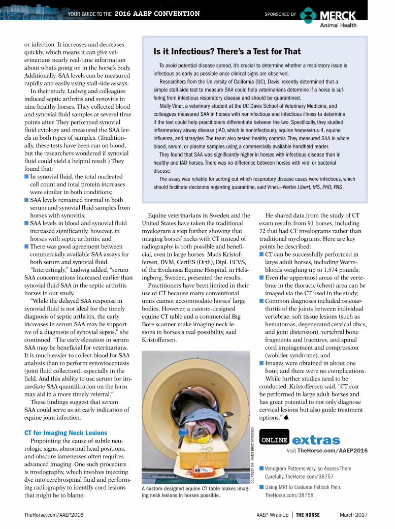
AAEP Wrap-Up The horse March 2017TheHorse.com/AAEP2016
Your GuiDE To THE 2016 aaeP convention SPonSorED bY
or infection. It increases and decreases quickly, which means it can give vet-erinarians nearly real-time information about what’s going on in the horse’s body. Additionally, SAA levels can be measured rapidly and easily using stall-side assays.
In their study, Ludwig and colleagues induced septic arthritis and synovitis in nine healthy horses. They collected blood and synovial fluid samples at several time points after. They performed synovial fluid cytology and measured the SAA lev-els in both types of samples. (Tradition-ally, these tests have been run on blood, but the researchers wondered if synovial fluid could yield a helpful result.) They found that:■ In synovial fluid, the total nucleated
cell count and total protein increases were similar in both conditions;
■ SAA levels remained normal in both serum and synovial fluid samples from horses with synovitis;
■ SAA levels in blood and synovial fluid increased significantly, however, in horses with septic arthritis; and
■ There was good agreement between commercially available SAA assays for both serum and synovial fluid.“Interestingly,” Ludwig added, “serum
SAA concentrations increased earlier than synovial fluid SAA in the septic arthritis horses in our study.
“While the delayed SAA response in synovial fluid is not ideal for the timely diagnosis of septic arthritis, the early increases in serum SAA may be support-ive of a diagnosis of synovial sepsis,” she continued. “The early elevation in serum SAA may be beneficial for veterinarians. It is much easier to collect blood for SAA analysis than to perform synoviocentesis (joint fluid collection), especially in the field. And this ability to use serum for im-mediate SAA quantification on the farm may aid in a more timely referral.”
These findings suggest that serum SAA could serve as an early indication of equine joint infection.
Ct for imaging Neck lesionsPinpointing the cause of subtle neu-
rologic signs, abnormal head positions, and obscure lamenesses often requires advanced imaging. One such procedure is myelography, which involves injecting dye into cerebrospinal fluid and perform-ing radiography to identify cord lesions that might be to blame.
Equine veterinarians in Sweden and the United States have taken the traditional myelogram a step further, showing that imaging horses’ necks with CT instead of radiography is both possible and benefi-cial, even in large horses. Mads Kristof-fersen, DVM, CertES (Orth), Dipl. ECVS, of the Evidensia Equine Hospital, in Hels-ingborg, Sweden, presented the results.
Practitioners have been limited in their use of CT because many conventional units cannot accommodate horses’ large bodies. However, a custom-designed equine CT table and a commercial Big Bore scanner make imaging neck le-sions in horses a real possibility, said Kristoffersen.
He shared data from the study of CT exam results from 91 horses, including 72 that had CT myelograms rather than traditional myelograms. Here are key points he described:■ CT can be successfully performed in
large adult horses, including Warm-bloods weighing up to 1,574 pounds;
■ Even the uppermost areas of the verte-brae in the thoracic (chest) area can be imaged via the CT used in the study;
■ Common diagnoses included osteoar-thritis of the joints between individual vertebrae, soft tissue lesions (such as hematomas, degenerated cervical discs, and joint distension), vertebral bone fragments and fractures, and spinal cord impingement and compression (wobbler syndrome); and
■ Images were obtained in about one hour, and there were no complications.While further studies need to be
conducted, Kristoffersen said, “CT can be performed in large adult horses and has great potential to not only diagnose cervical lesions but also guide treatment options.” h
Co
UR
TES
y D
R.
mAD
S k
RIS
ToFF
ERS
oN
A custom-designed equine CT table makes imag-ing neck lesions in horses possible.
extrasVisit TheHorse.com/AAEP2016
■ Venogram Patterns Vary, so Assess Them Carefully, TheHorse.com/38757
■ using Mri to Evaluate Fetlock Pain, TheHorse.com/38758
is it infectious? there’s a test for thatTo avoid potential disease spread, it’s crucial to determine whether a respiratory issue is
infectious as early as possible once clinical signs are observed. researchers from the university of California (uC), Davis, recently determined that a
simple stall-side test to measure SAA could help veterinarians determine if a horse is suf-fering from infectious respiratory disease and should be quarantined.
Molly Viner, a veterinary student at the uC Davis School of Veterinary Medicine, and colleagues measured SAA in horses with noninfectious and infectious illness to determine if the test could help practitioners differentiate between the two. Specifically, they studied inflammatory airway disease (iAD, which is noninfectious), equine herpesvirus-4, equine influenza, and strangles. The team also tested healthy controls. They measured SAA in whole blood, serum, or plasma samples using a commercially available handheld reader.
They found that SAA was significantly higher in horses with infectious disease than in healthy and iAD horses. There was no difference between horses with viral or bacterial disease.
The assay was reliable for sorting out which respiratory disease cases were infectious, which should facilitate decisions regarding quarantine, said Viner.—Nettie Libert, MS, PhD, PAS
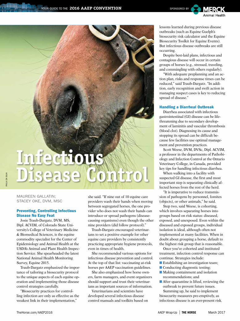
AAEP Wrap-Up The horse March 2017TheHorse.com/AAEP2016
mAUREEN gALLATIN; STACEy okE, Dvm, mSC
Preventing, Controlling infectious Disease No Easy Feat
Josie Traub-Dargatz, DVM, MS, Dipl. ACVIM, of Colorado State Uni-versity’s College of Veterinary Medicine & Biomedical Sciences, is the equine commodity specialist for the Center of Epidemiology and Animal Health at the USDA’s Animal and Plant Health Inspec-tion Service. She spearheaded the latest National Animal Health Monitoring Survey, Equine 2015.
Traub-Dargatz emphasized the impor-tance of tailoring a biosecurity protocol to the unique aspects of each equine op-eration and implementing those disease control strategies carefully.
“Biosecurity practices for control-ling infection are only as effective as the weakest link in their implementation,”
she said. “If nine out of 10 equine care providers wash their hands when moving between segregated horses, the one pro-vider who does not wash their hands can introduce or spread pathogens (disease-causing organisms) even though the other nine providers (did follow protocol).”
Traub-Dargatz encouraged veterinar-ians to set a positive example for other equine care providers by consistently practicing appropriate hygiene protocols, even in times of health.
She recommended various options for infectious disease prevention and control. At the top of that list: vaccinating at-risk horses per AAEP vaccination guidelines.
She also emphasized how horse own-ers, farm managers, and event organizers should support and trust their veterinar-ians as important sources of information.
Veterinarians and scientists have developed several infectious disease control manuals and toolkits based on
lessons learned during previous disease outbreaks (such as Equine Guelph’s biosecurity risk calculator and the Equine Biosecurity Toolkit for Equine Events). But infectious disease outbreaks are still occurring.
Despite best-laid plans, infectious and contagious disease will occur in certain groups of horses (e.g., stressed, traveling, and commingling with others regularly).
“With adequate preplanning and an ac-tion plan, risks and response times can be reduced,” said Traub-Dargatz. “In addi-tion, early recognition and swift action in managing suspect cases is key to reducing spread of disease.”
handling a Diarrheal outbreakDiarrhea associated with infectious
gastrointestinal (GI) disease can be life-threatening due to secondary develop-ment of laminitis and vascular thrombosis (blood clot). Diagnosing its cause and stopping its spread can be difficult be-cause few facilities use optimal manage-ment and prevention practices.
Scott Weese, DVM, DVSc, Dipl. ACVIM, a professor in the departments of Pathobi-ology and Infection Control at the Ontario Veterinary College, in Canada, provided key tips for handling infectious diarrhea.
When walking into a facility with suspected GI disease, the first and most important step is separating clinically af-fected horses from the rest of the herd.
“It is imperative to reduce transmis-sion of pathogens by personnel, fomites (objects), or other animals,” he said.
Step two, said Weese, is cohorting, which involves separating horses into groups based on risk status: diseased, exposed, and unexposed. Even within the diseased and exposed groups, individual isolation is ideal, although often not implemented at many facilities. When in doubt about grouping a horse, default to the highest risk group that is reasonable.
Once you’ve cohorted and instituted treatment, infection control response can continue. Strategies include:■ Establishing an investigation team;■ Conducting diagnostic testing;■ Making containment and isolation
recommendations; and■ After quarantine is lifted, reviewing the
outbreak to prevent future issues.Summing up, he said to implement
biosecurity measures pre-emptively, as infectious disease is an ever-present risk.
PAU
LA D
A S
ILvA
infectious Disease control
SPonSorED bYYour GuiDE To THE 2016 aaeP convention
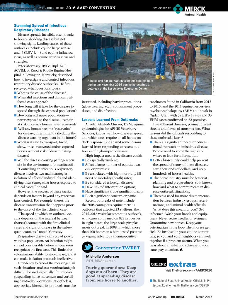
AAEP Wrap-Up The horse March 2017TheHorse.com/AAEP2016
stemming spread of infectious respiratory Diseases
Disease spreads invisibly, often thanks to horses shedding disease but not showing signs. Leading causes of these outbreaks include equine herpesvirus-1 and -4 (EHV-1, -4) and equine influenza virus, as well as equine arteritis virus and strangles.
Peter Morresey, BVSc, Dipl. ACT, ACVIM, of Rood & Riddle Equine Hos-pital in Lexington, Kentucky, described how to investigate and control infectious respiratory disease outbreaks. He first reviewed what questions to ask:■ What is the cause of the disease?■ When did infectious and clinically af-
fected cases appear?■ How long will it take for the disease to
spread through the exposed population?■ How long will naive populations—
never exposed to the disease—remain at risk once sick horses have recovered?
■ Will any horses become “reservoirs” for disease, intermittently shedding the disease-causing organism in the future?
■ When is it safe to transport, breed, show, or sell recovered and/or exposed horses without risk of disseminating disease?
■ Will the disease-causing pathogen per-sist in the environment (on surfaces)?“Controlling an infectious respiratory
disease involves two main strategies: isolation of affected individuals and iden-tifying then segregating horses exposed to clinical cases,” he said.
However, the success of these tactics depends on factors beyond any veterinar-ian’s control. For example, there’s the disease transmission that happens prior to the onset of the first clinical case.
“The speed at which an outbreak oc-curs depends on the interval between (horses’) contact with the first clinical cases and signs of disease in the subse-quent contacts,” noted Morresey.
Respiratory disease can spread slowly within a population. An infection might spread considerably before anyone even recognizes the first case. This limits the veterinarian’s ability to stop disease, and it can make isolation protocols ineffective.
A tendency to “shoot the messenger” in such situations makes a veterinarian’s job difficult, he said, especially if it involves suspending horse movement and curtail-ing day-to-day operations. Nonetheless, appropriate biosecurity protocols must be
instituted, including barrier precautions (glove wearing, etc.), containment proce-dures, and disinfection.
lessons learned From outbreaksAngela Pelzel-McCluskey, DVM, equine
epidemiologist for APHIS Veterinary Services, knows well how diseases spread and which ones require an all-hands-on-deck response. She shared some lessons learned from responding to recent out-breaks of high-impact diseases.
High-impact means the disease could: ■ Be especially virulent; ■ Affect a large number of equids, own-
ers, or premises; ■ Be associated with high morbidity (ill-
ness) or mortality (death) rates; ■ Be considered a foreign disease; ■ Have limited intervention options; ■ Have significant trade ramifications; or ■ Elicit significant concern or panic.
Recent outbreaks of note include the 2008 contagious equine metritis outbreak that affected 23 stallions; the 2015-2016 vesicular stomatitis outbreak, with cases confirmed on 823 properties in eight states; the large-scale piroplas-mosis outbreak in 2009, in which more than 400 horses in a herd tested positive; 39 equine infectious anemia-positive
racehorses found in California from 2012 to 2015; and the 2011 equine herpesvirus myeloencephalopathy (EHM) outbreak in Ogden, Utah, with 57 EHV-1 cases and 33 EHM cases confirmed on 62 premises.
Five different diseases, posing different threats and forms of transmission. What lessons did the officials responding to these outbreaks learn?■ There’s a significant need for educa-
tional outreach on infectious disease. People need to know the signs and where to look for information.
■ Better biosecurity could help prevent the spread of many of these diseases, save thousands of dollars, and keep hundreds of horses healthy.
■ The horse industry must be better at planning and preparedness, so it knows how and what to communicate in dis-ease outbreak situations.
■ There’s a need for more direct interac-tion between industry groups, veteri-narians, and animal health officials.What does this mean for you? Get
informed. Wash your hands and equip-ment. Never reuse needles or syringes. Quarantine new horses. Keep your veterinarian in the loop when horses get sick. Be involved in your equine commu-nity, so you and your neighbors can work together if a problem occurs. When you hear about an infectious disease in your state, pay attention. hconventiontweet
Michelle Anderson@TH_MNAnderson
During quarantines: Keep dogs out of barn! They are good at spreading disease from one horse to another.
Co
UR
TES
y D
R.
kAT
IE F
LyN
N
A horse and handler wait outside the isolation barn during the November 2016 equine herpesvirus-1 outbreak at the Los Angeles Equestrian Center.
extrasVisit TheHorse.com/AAEP2016
■ The role of State Animal Health officials in Pro-tecting Equine Health, TheHorse.com/38759
Your GuiDE To THE 2016 aaeP convention SPonSorED bY

From the breeding barn to the show ring and everything in between, we have the vaccines you need to help protect your horse.
• Made with the exclusive Antigen Puri� cation System (APS™)
• Contains the Havlogen® adjuvant veterinarians know and trust
• Delivers core vaccine coverage according to American Association of Equine Practitioners recommendations
Now that’s protected.
Ask your veterinarian for Prestige®, Encevac®, Prodigy® and EquiRab® brand vaccines. Visit us at GetVaccinatingRight.com to learn more about Merck Animal Health and the equine products and programs that help keep horses healthy.
Every vaccine purchased through Merck supports the plight of the unwanted horse through the Unwanted Horse Veterinary Relief Campaign.
The Scienceof Protected
The Science of Healthier Animals
2 Giralda Farms • Madison, NJ 07940 • merck-animal-health-usa.com • 800-521-5767Copyright © 2016 Intervet Inc., d/b/a/ Merck Animal Health, a subsidiary of Merck & Co., Inc.All rights reserved. 3526 EQ-FP AD Vaccine Line
3526_VaccineFamily_Woman_8125x10875.indd 1 1/23/17 9:15 AM
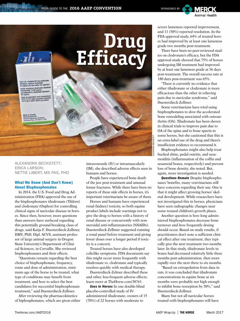
AAEP Wrap-Up The horse March 2017TheHorse.com/AAEP2016
Your GuiDE To THE 2016 aaeP convention SPonSorED bY
ALExANDRA BECkSTETT; ERICA LARSoN; NETTIE LIBERT, mS PAS, PHD
What We Know (And Don’t Know) About Bisphosphonates
In 2014, the U.S. Food and Drug Ad-ministration (FDA) approved the use of the bisphosphonates tiludronate (Tildren) and clodronate (Osphos) for controlling clinical signs of navicular disease in hors-es. Since then, however, more questions than answers have surfaced regarding this potentially ground-breaking class of drugs, said Katja F. Duesterdieck-Zellmer, DMV, PhD, Dipl. ACVS, assistant profes-sor of large animal surgery in Oregon State University’s Department of Clini-cal Sciences, in Corvallis. She reviewed bisphosphonates and their effects.
“Questions remain regarding the best choice of bisphosphonate, frequency, route and dose of administration, mini-mum age of the horse to be treated, what type of conditions may benefit from treatment, and how to select the best candidates for successful bisphosphonate treatment,” said Duesterdieck-Zellmer.
After reviewing the pharmacokinetics of biphosphonates, which are given either
intravenously (IV) or intramuscularly (IM), she described adverse effects seen in humans and horses.
People have experienced bone death of the jaw post- treatment and unusual femur fractures. While there have been no reports of these side effects in horses, it’s important veterinarians be aware of them.
Horses and humans have experienced renal (kidney) toxicity, so both equine product labels include warnings not to give the drug to horses with a history of renal disease or concurrently with non- steroidal anti-inflammatories (NSAIDs). Duesterdieck-Zellmer suggested running a renal panel before treatment and giving lower doses over a longer period if toxic-ity is a concern.
Treated horses have also developed coliclike symptoms. FDA documents say this might occur more frequently with tiludronate vs. clodronate and typically resolves quickly with medical therapy.
Duesterdieck-Zelmer described these and other, less-frequent adverse effects; learn more at TheHorse.com/38761.
Uses in Horses In one double-blind placebo-controlled study of IV- administered tiludronate, owners of 15 (70%) of 22 horses with moderate to
severe lameness reported improvement, and 11 (50%) reported resolution. In the FDA approval study, 64% of treated hors-es had improved by at least one lameness grade two months post-treatment.
There have been no peer- reviewed stud-ies on clodronate’s efficacy, but the FDA approval study showed that 75% of horses undergoing IM treatment had improved by at least one lameness grade at 56 days post-treatment. The overall success rate at 180 days post-treatment was 65%.
“There is currently no evidence that either tiludronate or clodronate is more efficacious than the other in relieving pain due to navicular syndrome,” said Duesterdieck-Zellmer.
Some veterinarians have tried using bisphosphonates to slow the accelerated bone remodeling associated with osteoar-thritis (OA). Tiludronate has been shown in clinical trials to improve pain due to OA of the spine and to bone spavin in some horses, but she cautioned that this is an extra-label use of the drug and there’s insufficient evidence to recommend it.
Bisphosphonates might also help treat bucked shins, pedal osteitis, and sesa-moiditis (inflammation of the coffin and sesamoid bones, respectively) and prevent loss of bone density, she noted. But, again, more investigation is needed.
Questions Remain Despite bisphospho-nates’ benefits, many veterinarians still have concerns regarding their use. One is that it might affect growing horses’ skel-etal development. While researchers have not investigated this in horses, physicians have seen radiographic changes near some treated children’s growth plates.
Another question is how long admin-istered bisphosphonates decrease bone turnover and how frequently dosing should occur. Based on study results, if practitioners don’t note a sufficient clini-cal effect after one treatment, they typi-cally give the next treatment two months later. In that study, tiludronate levels in bones had decreased relatively little three months post- administration, then more rapidly over the next three to six months.
“Based on extrapolation from data in rats, it was concluded that tiludronate concentrations in equine bone at six months were probably not high enough to inhibit bone resorption by 50%,” said Duesterdieck-Zellmer.
Many but not all navicular horses treated with bisphosphonates will have
ALEx
AND
RA
BEC
kS
TETT
Drug efficacy
1 UC Davis (Nicola Pusterla) & Merck Animal Health. Infectious Upper Respiratory Surveillance Program. Ongoing Research 2008-present.
2 Townsend HGG. Onset of protection against live-virus equine influenza challenge following vaccination naive horses with a modified-live vaccine. Unpublished data.
Visit us online to learn more about Merck Animal Health and the equine products and programs that help you keep horses healthy.
The Science of ExceptionalStop flu where it starts.
The Science of Healthier Animals
2 Giralda Farms • Madison, NJ 07940 • merck-animal-health-usa.com • 800-521-5767Copyright © 2016 Intervet Inc., d/b/a/ Merck Animal Health, a subsidiary of Merck & Co., Inc.All rights reserved. 3526 EQ-FluAvert-PM
Trust Flu Avert® I.N. for superior protection against relevant flu strains threatening the U.S. horse population.1
• Just ONE dose required - An ideal first flu vaccine for young horses - Provides protection at the site of infection
and long-lasting immunity• Proven safe and effective - Intranasal application leaves no risk for
injection site reactions to interfere with training or competing
• Rapid onset of protection - Onset of flu protection within five to
seven days following one dose – no matter when your horse was last vaccinated for flu.2
Now that’s exceptional.
3526_FluAvertIN_8125x10875.indd 1 1/4/17 12:56 PM
AAEP Wrap-Up The horse March 2017TheHorse.com/AAEP2016
Your GuiDE To THE 2016 aaeP convention SPonSorED bY
improved or resolved lameness, she concluded. She suggested veterinarians assess horses’ response and improvement two to six months after treatment.
Dipyrone for reducing Fever Because there are currently no
medications labeled to control fever in horses, practitioners generally administer NSAIDs to pyrexic (feverish) animals.
The NSAID dipyrone is approved in several countries—but not the United States—for use in animals and people to reduce fever and relieve pain. Emily Sun-dman, DVM, of Kindred Biosciences Inc., in Burlingame, California, investigated this drug’s safety and efficacy in horses.
She and her colleagues studied horses from 14 clinical research sites in 12 states that had a fever of at least 102°F, were older than 1 year, not pregnant, and free of severe systemic disease.
The researchers randomly assigned horses to either a treatment group that received 30 mg/kg of IV dipyrone or a control group that received a placebo. Researchers were blinded to the groups. They recorded horses’ temperatures six hours after treatment and considered it to be effective if body temperature decreased by 2°F or returned to normal. Dipyrone was effective in 77% of horses treated, said Sundman.
In the same study, she investigated the drug’s field safety. Treated horses received additional doses of 30 mg/kg of dipyrone up to three times daily. The team com-pleted a physical exam and bloodwork when each horse exited the study.
Adverse reactions to treatment were mild, occurred at similar rates among the placebo and dipyrone groups, and did not appear to cause clinical problems. Bloodwork abnormalities were what you’d see with horses suffering from fever and could have been related to the cause of disease.
In conclusion, dipyrone was safe and
effective for controlling fever in horses when given at an IV dose of 30 mg/kg, said Sundman. She said Kindred Biosciences hopes to gain pending FDA approval for dipyrone for use in horses.
Acetaminophen Could relieve laminitis Pain
Jonathan Foreman, DVM, MS, Dipl. ACVIM, presented the results of a study in which he and colleagues tested whether acetaminophen (yes, Tylenol’s active ingredient) effectively relieved equine foot pain. Foreman is a professor of veteri-nary clinical medicine at the University of Illinois College of Veterinary Medicine Veterinary Teaching Hospital, in Urbana.
Veterinarians often prescribe tradi-tional NSAIDs—such as phenylbuta-zone (PBZ) or flunixin meglumine (FM, Banamine)—to help reduce or eliminate foot pain. But such drugs carry potentially dangerous side effects, so researchers are still on the hunt for effective treatments.
Foreman and colleagues recently compared acetaminophen’s efficacy in relieving foot pain to that of FM.
The researchers induced reversible lameness in eight healthy adult horses and administered 1.1 mg/kg of FM orally, 20 mg/kg of acetaminophen orally, or left horses untreated to serve as controls. Each horse received each treatment with a week between trials.
Foreman said control horses had high-er heart rates and lameness scores than horses that received FM or acetamino-phen. However, there were no significant differences between heart rates or lame-ness scores in FM- or acetaminophen-treated horses, he said.
The team also monitored horses’ liver enzymes and found no evidence of eleva-tion, suggesting there was no liver dam-age from the 20 mg/kg acetaminophen dose, Foreman said.
Acetaminophen at this oral dosage was comparable to FM, he said.
There’s still no data regarding acet-aminophen’s pharmacodynamics or toxicity in horses, so further research is warranted. However, Foreman encour-aged practitioners to consider this medi-cation for laminitic horses. h
is a New treatment for Joint Pain on the horizon?“Equine pain management is one of the most challenging issues veterinarians face on
a daily basis,” said Melissa King, DVM, PhD, Dipl. ACVS, ACVSMr, an assistant professor of equine sports medicine at Colorado State university’s Veterinary Teaching Hospital, in Fort Collins. She has been studying a new tool to manage joint pain in horses that has significant pain-relieving effects in other species.
in the presence of tissue injury, the body produces neurotrophins, or nerve growth factor (nFG). When a damaged joint, for instance, releases nGF, a cascade of events occurs that enhances the sensation of noxious stimuli. The result? Pain. researchers have developed an-tibodies against nGF—anti-nerve growth factor (anti-nGF mAb)—to help stem this response.
in their study, King and colleagues induced synovitis (a type of noninfectious joint inflammation) in one hock joint in 24 horses. They injected a sterile saline solution into each horse’s opposite hock, which served as its control. They then split the horses into four treatment groups of six that received intra-articular anti-nGF mAb doses of either 0.1 mg, 1 mg, or 10 mg or a placebo.
The team performed a lameness examination and collected synovial fluid and mea-sured ground-reaction forces from all horses at three time points. Ten hours post-anti-nGF mAb administration they also collected tissue biopsies from the affected hocks.
in the joints treated with the 1-mg and 10-mg doses, the researchers found:■ Less pain in response to joint flexion and symmetrical limb loading; ■ Significantly less glycosaminoglycan and PGE2 (inflammatory markers) in synovial fluid;■ Significantly less inflammatory cell infiltrate on histopathology (microscopic exam); and■ no significant changes or effects on articular (joint) cartilage viability.
“Anti-nGF mAb has multiple therapeutic and disease-modifying effects, short-term safety, and is efficacious,” said King. “it’s the first new pain treatment in several years.”
While this treatment is not currently available for use in horses, King said she hopes it will be an option in the near future.—Alexandra Beckstett
20 mg/kgoral acetaminophen was comparable to 1.1 mg/kg FM
AAEP Wrap-Up The horse March 2017TheHorse.com/AAEP2016
The Science of ProtectedFrom the breeding barn to the show ring and everything in between, we have the vaccines you need to help protect your horse.
• Made with the exclusive Antigen Purification System (APS™)
• Contains the Havlogen® adjuvant veterinarians know and trust
• Delivers core vaccine coverage according to American Association of Equine Practitioners recommendations
Now that’s protected.
Ask your veterinarian for Prestige®, Encevac®, Prodigy® and EquiRab® brand vaccines. Visit us at GetVaccinatingRight.com to learn more about Merck Animal Health and the equine products and programs that help keep horses healthy.
Every vaccine purchased through Merck supports the plight of the unwanted horse through the Unwanted Horse Veterinary Relief Campaign.
The Science of Healthier Animals
2 Giralda Farms • Madison, NJ 07940 • merck-animal-health-usa.com • 800-521-5767Copyright © 2016 Intervet Inc., d/b/a/ Merck Animal Health, a subsidiary of Merck & Co., Inc.All rights reserved. 3526 EQ-FP AD Vaccine Line
3526_VaccineFamily_Roper_8125x10875.indd 1 2/17/17 1:59 PM
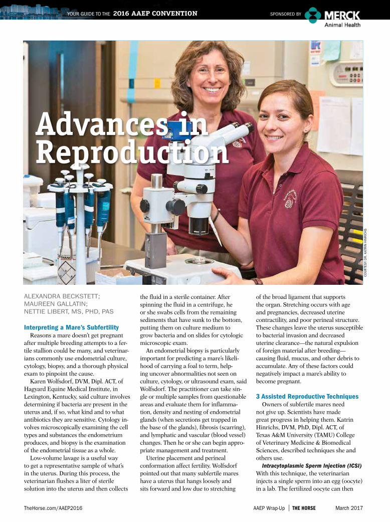
AAEP Wrap-Up The horse March 2017TheHorse.com/AAEP2016
ALExANDRA BECkSTETT; mAUREEN gALLATIN; NETTIE LIBERT, mS, PHD, PAS
interpreting a Mare’s subfertilityReasons a mare doesn’t get pregnant
after multiple breeding attempts to a fer-tile stallion could be many, and veterinar-ians commonly use endometrial culture, cytology, biopsy, and a thorough physical exam to pinpoint the cause.
Karen Wolfsdorf, DVM, Dipl. ACT, of Hagyard Equine Medical Institute, in Lexington, Kentucky, said culture involves determining if bacteria are present in the uterus and, if so, what kind and to what antibiotics they are sensitive. Cytology in-volves microscopically examining the cell types and substances the endometrium produces, and biopsy is the examination of the endometrial tissue as a whole.
Low-volume lavage is a useful way to get a representative sample of what’s in the uterus. During this process, the veterinarian flushes a liter of sterile solution into the uterus and then collects
the fluid in a sterile container. After spinning the fluid in a centrifuge, he or she swabs cells from the remaining sediments that have sunk to the bottom, putting them on culture medium to grow bacteria and on slides for cytologic microscopic exam.
An endometrial biopsy is particularly important for predicting a mare’s likeli-hood of carrying a foal to term, help-ing uncover abnormalities not seen on culture, cytology, or ultrasound exam, said Wolfsdorf. The practitioner can take sin-gle or multiple samples from questionable areas and evaluate them for inflamma-tion, density and nesting of endometrial glands (when secretions get trapped in the base of the glands), fibrosis (scarring), and lymphatic and vascular (blood vessel) changes. Then he or she can begin appro-priate management and treatment.
Uterine placement and perineal conformation affect fertility. Wolfsdorf pointed out that many subfertile mares have a uterus that hangs loosely and sits forward and low due to stretching
of the broad ligament that supports the organ. Stretching occurs with age and pregnancies, decreased uterine contractility, and poor perineal structure. These changes leave the uterus susceptible to bacterial invasion and decreased uterine clearance—the natural expulsion of foreign material after breeding—causing fluid, mucus, and other debris to accumulate. Any of these factors could negatively impact a mare’s ability to become pregnant.
3 Assisted reproductive techniquesOwners of subfertile mares need
not give up. Scientists have made great progress in helping them. Katrin Hinrichs, DVM, PhD, Dipl. ACT, of Texas A&M University (TAMU) College of Veterinary Medicine & Biomedical Sciences, described techniques she and others use.
Intracytoplasmic Sperm Injection (ICSI) With this technique, the veterinarian injects a single sperm into an egg (oocyte) in a lab. The fertilized oocyte can then
Co
UR
TES
y D
R.
kAT
RIN
HIN
RIC
HS
advances in reproduction
Your GuiDE To THE 2016 aaeP convention SPonSorED bY
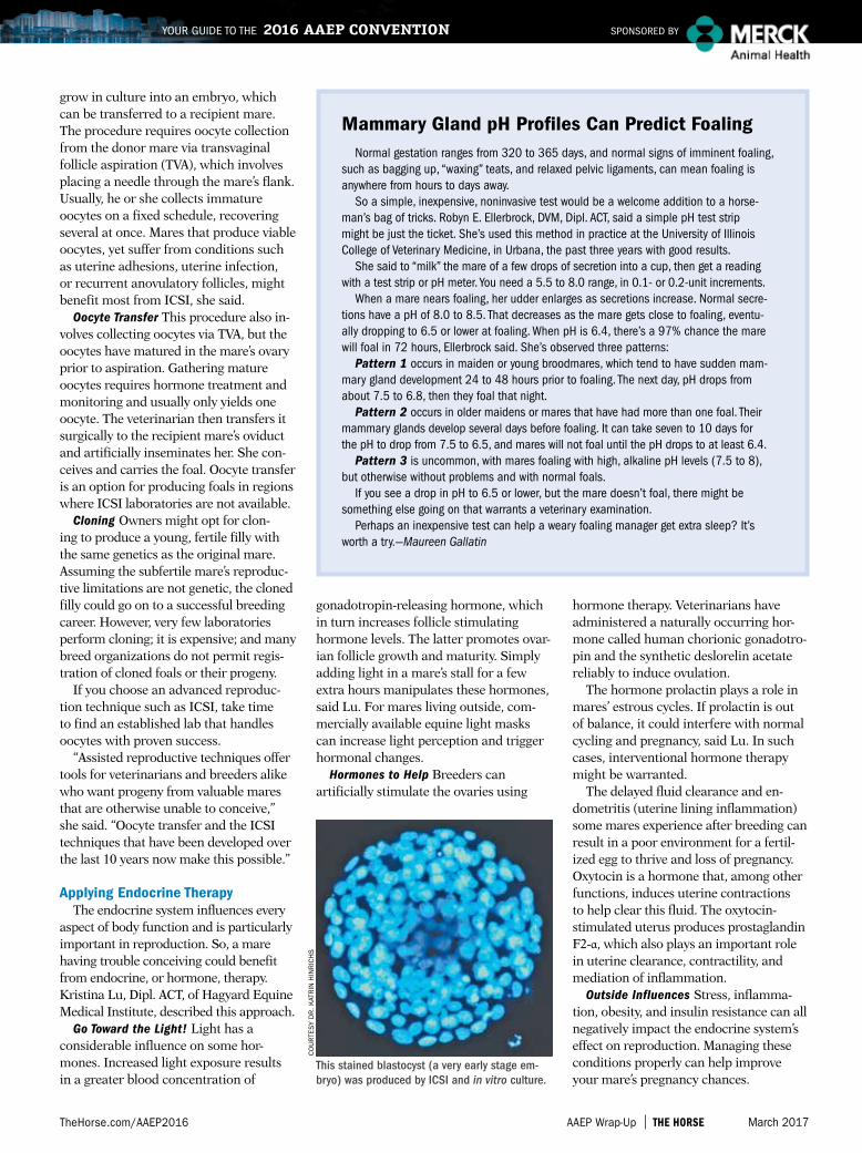
AAEP Wrap-Up The horse March 2017TheHorse.com/AAEP2016
grow in culture into an embryo, which can be transferred to a recipient mare. The procedure requires oocyte collection from the donor mare via transvaginal follicle aspiration (TVA), which involves placing a needle through the mare’s flank. Usually, he or she collects immature oocytes on a fixed schedule, recovering several at once. Mares that produce viable oocytes, yet suffer from conditions such as uterine adhesions, uterine infection, or recurrent anovulatory follicles, might benefit most from ICSI, she said.
Oocyte Transfer This procedure also in-volves collecting oocytes via TVA, but the oocytes have matured in the mare’s ovary prior to aspiration. Gathering mature oocytes requires hormone treatment and monitoring and usually only yields one oocyte. The veterinarian then transfers it surgically to the recipient mare’s oviduct and artificially inseminates her. She con-ceives and carries the foal. Oocyte transfer is an option for producing foals in regions where ICSI laboratories are not available.
Cloning Owners might opt for clon-ing to produce a young, fertile filly with the same genetics as the original mare. Assuming the subfertile mare’s reproduc-tive limitations are not genetic, the cloned filly could go on to a successful breeding career. However, very few laboratories perform cloning; it is expensive; and many breed organizations do not permit regis-tration of cloned foals or their progeny.
If you choose an advanced reproduc-tion technique such as ICSI, take time to find an established lab that handles oocytes with proven success.
“Assisted reproductive techniques offer tools for veterinarians and breeders alike who want progeny from valuable mares that are otherwise unable to conceive,” she said. “Oocyte transfer and the ICSI techniques that have been developed over the last 10 years now make this possible.”
Applying Endocrine therapyThe endocrine system influences every
aspect of body function and is particularly important in reproduction. So, a mare having trouble conceiving could benefit from endocrine, or hormone, therapy. Kristina Lu, Dipl. ACT, of Hagyard Equine Medical Institute, described this approach.
Go Toward the Light! Light has a considerable influence on some hor-mones. Increased light exposure results in a greater blood concentration of
gonadotropin-releasing hormone, which in turn increases follicle stimulating hormone levels. The latter promotes ovar-ian follicle growth and maturity. Simply adding light in a mare’s stall for a few extra hours manipulates these hormones, said Lu. For mares living outside, com-mercially available equine light masks can increase light perception and trigger hormonal changes.
Hormones to Help Breeders can artificially stimulate the ovaries using
hormone therapy. Veterinarians have administered a naturally occurring hor-mone called human chorionic gonadotro-pin and the synthetic deslorelin acetate reliably to induce ovulation.
The hormone prolactin plays a role in mares’ estrous cycles. If prolactin is out of balance, it could interfere with normal cycling and pregnancy, said Lu. In such cases, interventional hormone therapy might be warranted.
The delayed fluid clearance and en-dometritis (uterine lining inflammation) some mares experience after breeding can result in a poor environment for a fertil-ized egg to thrive and loss of pregnancy. Oxytocin is a hormone that, among other functions, induces uterine contractions to help clear this fluid. The oxytocin-stimulated uterus produces prostaglandin F2-α, which also plays an important role in uterine clearance, contractility, and mediation of inflammation.
Outside Influences Stress, inflamma-tion, obesity, and insulin resistance can all negatively impact the endocrine system’s effect on reproduction. Managing these conditions properly can help improve your mare’s pregnancy chances.
Co
UR
TES
y D
R.
kAT
RIN
HIN
RIC
HS
This stained blastocyst (a very early stage em-bryo) was produced by ICSI and in vitro culture.
Mammary gland ph Profiles Can Predict Foalingnormal gestation ranges from 320 to 365 days, and normal signs of imminent foaling,
such as bagging up, “waxing” teats, and relaxed pelvic ligaments, can mean foaling is anywhere from hours to days away.
So a simple, inexpensive, noninvasive test would be a welcome addition to a horse-man’s bag of tricks. robyn E. Ellerbrock, DVM, Dipl. ACT, said a simple pH test strip might be just the ticket. She’s used this method in practice at the university of illinois College of Veterinary Medicine, in urbana, the past three years with good results.
She said to “milk” the mare of a few drops of secretion into a cup, then get a reading with a test strip or pH meter. You need a 5.5 to 8.0 range, in 0.1- or 0.2-unit increments.
When a mare nears foaling, her udder enlarges as secretions increase. normal secre-tions have a pH of 8.0 to 8.5. That decreases as the mare gets close to foaling, eventu-ally dropping to 6.5 or lower at foaling. When pH is 6.4, there’s a 97% chance the mare will foal in 72 hours, Ellerbrock said. She’s observed three patterns:
Pattern 1 occurs in maiden or young broodmares, which tend to have sudden mam-mary gland development 24 to 48 hours prior to foaling. The next day, pH drops from about 7.5 to 6.8, then they foal that night.
Pattern 2 occurs in older maidens or mares that have had more than one foal. Their mammary glands develop several days before foaling. it can take seven to 10 days for the pH to drop from 7.5 to 6.5, and mares will not foal until the pH drops to at least 6.4.
Pattern 3 is uncommon, with mares foaling with high, alkaline pH levels (7.5 to 8), but otherwise without problems and with normal foals.
if you see a drop in pH to 6.5 or lower, but the mare doesn’t foal, there might be something else going on that warrants a veterinary examination.
Perhaps an inexpensive test can help a weary foaling manager get extra sleep? it’s worth a try.—Maureen Gallatin
Your GuiDE To THE 2016 aaeP convention SPonSorED bY
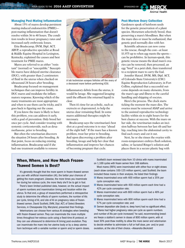
AAEP Wrap-Up The horse March 2017TheHorse.com/AAEP2016
Your GuiDE To THE 2016 aaeP convention SPonSorED bY
Managing Post-Mating inflammationAbout 15% of mares develop persistent
mating-induced endometritis (PMIE)— post-mating inflammation that doesn’t resolve within 36 to 48 hours. The condi-tion results in lower pregnancy rates and increased early embryonic losses.
Etta Bradecamp, DVM, Dipl. ACT, ABVP, a reproductive specialist at Rood & Riddle Equine Hospital in Lexington, Kentucky, explained the causes and best treatment for PMIE mares.
Mares are referred to as either “resis-tant” (normal) or “susceptible.” Suscepti-ble mares have delayed uterine clearance (DUC), with greater than 2 centimeters of fluid in the uterus when checked on ultrasound 24 hours after breeding.
Bradecamp focused on management techniques that can improve fertility in DUC mares and modulate the inflam-matory response. Knowing which of the many treatments are most appropriate and when to use them can be tricky, and it goes back to figuring out the cause.
If you know the mare is likely to have this problem, you can address it early, with a goal of prevention. Only breed her once per cycle. And consider treating with systemic corticosteroids, such as dexa-methasone, prior to breeding.
But often the veterinarian discovers the situation 24 hours after breeding. At that point, focus on reducing existing inflammation. Bradecamp said if she had one treatment available to remove
inflammatory debris from the uterus, it would be lavage. She suggested lavaging until the effluent (the returned liquid) is clear.
Then it’s time for an ecbolic, such as oxytocin or cloprostenol, to help the uterus clear remaining fluid. In some mares additional therapies might be necessary.
Bradecamp says the veterinarian’s best shot at a good outcome is to stay “ahead of the eight ball.” If the mare has a known problem, treat her prior to breeding. And upon discovering a problem after breeding, lavage and help her clear that inflammation and improve her chances of becoming pregnant that cycle.
Post-Mortem ovary Collection Gardeners speak of heirloom seeds for the genetic preservation of a plant species. Horsemen selectively breed, thus preserving a mare’s bloodlines. But when the mare dies or must be euthanized, that genetic pool normally dies with her.
Scientific advances can now come to the rescue, though the cost—at least $1,975 up to when egg meets the sperm—can be prohibitive. In layman-speak, this genetic rescue means the dead mare’s ova-ries can be removed, then processed, an egg “bred” in a petri dish, then transferred to a recipient mare to carry the foal.
Jennifer Hatzel, DVM, MS, Dipl. ACT, of Colorado State University’s (CSU) Equine Reproduction Laboratory, in Fort Collins, explained that a successful out-come depends on many elements, from the mare’s age and illness to the careful handling and shipping of oocytes.
Here’s the process. The clock starts ticking the moment the mare dies. The ovaries must be removed ASAP and arrive to the assisted reproductive technology facility within six to eight hours for the best chance at success. With the mare on her side, the veterinarian makes an inci-sion halfway between her last rib and her hip, reaching into the abdominal cavity to find each ovary and cut it out.
Next, he or she rinses the ovaries with room-temperature embryo flush solution, saline, or lactated Ringer’s solution and places them in a secure plastic bag with
Co
UR
TES
y C
AT H
ERS
H
A lab technician scrapes follicles off the ovary of a deceased mare before performing ICSI.
When, Where, and how Much Frozen-thawed semen is Best?
it’s generally thought that the more sperm in frozen-thawed semen you use with artificial insemination (Ai), the better your chances of getting the mare pregnant. Likewise, the more times you inseminate her during her estrous cycle, the more likely she’ll be to get in foal.
There’s been limited published data, however, on the actual impact of sperm numbers and insemination timing and location within the uterus. To that end, a group of researchers performed a retrospective study to determine the effects of sperm numbers, the number of Ais per cycle, timing of Ai, and site of Ai on pregnancy rates of frozen-thawed semen. David Scofield, DVM, Dipl. ACT, of Select breeders Services, in Chesapeake City, Maryland, presented their findings.
Veterinarians can choose from various Ai strategies when breeding with frozen-thawed semen. They can inseminate the mare multiple times throughout her estrous cycle using a fixed-time Ai protocol, or they can use ultrasound to determine Ai timing post-ovulation. They can inseminate the mare into her uterine body or by a deep uterine horn technique with a variable number or sperm and/or sperm doses.
Scofield’s team reviewed data from 10 clinics with mares inseminated on 1,169 cycles with frozen semen from 168 stallions.
Most mares (96%) were inseminated with either four or eight straws (each straw contains 100-125 million sperm), said Scofield; the team included these mares in their analysis. He listed their findings:■ Mares inseminated once with 400 million sperm had a 35% per
cycle conception rate;■ Mares inseminated twice with 400 million sperm each time had a
42% per cycle conception rate;■ Mares inseminated once with 800 million sperm had a 48% per
cycle conception rate;■ Mares inseminated twice with 800 million sperm each time had a
57% per cycle conception rate; and■ Semen deposition site (body vs. deep horn) had no significant effect.
“Mares had higher pregnancy rates per cycle as sperm numbers and number of Ais per cycle increased,” he said, recommending breed-ers freeze a stallion’s semen in doses of 800 million sperm, with at least 30% post-thaw motility, to allow the mare owner and veterinarian to decide whether to administer a full or half dose, pre- and/or post-ovulation, at the site of their choice.—Alexandra Beckstett

AAEP Wrap-Up The horse March 2017TheHorse.com/AAEP2016
The Science of Healthier Animals
2 Giralda Farms • Madison, NJ 07940 • merck-animal-health-usa.com • 800-521-5767Copyright © 2016 Intervet Inc., d/b/a/ Merck Animal Health, a subsidiary of Merck & Co., Inc.All rights reserved. 3526 EQ-FP AD Protazil®
The Science of Convenient
• Equine Protozoal Myeloencephalitis (EPM) is a serious neurological disease that can strike anytime, anywhere. Make treatment easy with Protazil®
• Safe and accurate dosing with a calibrated scoop
• Easier to use than paste, less stress for you and your horse
• Rapid absorption — no loading dose required1
Now that’s convenient.
Protazil® (1.56% diclazuril) is the only FDA-approved alfalfa-based top dress antiprotozoal pellet for the treatment of EPM.
Ask your veterinarian for Protazil®. Visit us at Protazil.com to learn more about Merck Animal Health and the equine products and programs that help keep horses healthy.
Use of Protazil® (1.56% dicazuril) is contraindicated in horses with known hypersensitivity to diclazuril. Safe use in horses used for breeding purposes, during pregnancy, or in lactating mares has not been evaluated. The safety of Protazil® (1.56% dicazuril) with concomitant therapies in horses has not been evaluated. See related page in this issue for details. For use in horses only. Do not use in horses intended for human consumption. Not for human use. Keep out of reach of children.
1 Hunyadi L, Papich MG, Pusterla N. Pharmacokinetics of a low-dose and DA-labeled dose of diclazuril administered orally as a pelleted top dressing in adult horses. J of Vet Pharmacology and Therapeutics (accepted) 2014, doi: 10.111/jvp.12176. The correlation between pharmacokinetic data and clinical effectiveness is unknown
3526_Protazil_8125x10875.indd 1 1/23/17 8:47 AM
AAEP Wrap-Up The horse March 2017TheHorse.com/AAEP2016
Your GuiDE To THE 2016 aaeP convention SPonSorED bY
a small amount of solution to keep them moist, double-bagging to prevent leakage.
The most challenging part (and dangerous for eggs) is ahead, with the time- and temperature-sensitive trip to the lab.
They must be maintained at a stable temperature, so flights and counter-to-counter arrangements are crucial. CSU and TAMU have been at the forefront of offering post-mortem ovary processing and ICSI services, so those are the two locations most familiar with all the pieces coming together in the right timing.
While this is happening, the owners should be selecting a stallion and making arrangements for his semen to be shipped. Either frozen or cooled/shipped semen can be used, but it must arrive by—at the latest—the day after the ovaries themselves arrive at the facility.
Someone at the lab processes ovaries on arrival so eggs can mature, then performs ICSI. The embryo is cultured and, when ready, sent for transfer or frozen.
Which Biomarkers Best Predict late-term Pregnancy loss?
Late-term pregnancy loss continues to be a significant problem in reproduction practice. In recent years, researchers have identified several biomarkers (TheHorse.com/38108) that can indicate trouble’s brewing, but they needed a large-scale field study to evaluate them.
Recently, a University of Kentucky research team examined changes that occurred in four established biomarkers in mares with both normal and abnormal pregnancy outcomes. Barry Ball, DVM, Dipl. ACT, presented their results.
The biomarkers of interest here were:■ The estrogen estradiol-17B The fetus
and placenta produce estradiol during pregnancy. Estradiol levels decrease when experimental placentitis is in-duced at nine months of gestation.
■ Progesterone The placenta plays a large role in progesterone metabolism. Pro-gesterone falls to low or nondetectable concentrations during gestation. When scientists induce experimental placenti-tis at nine months, however, these levels increase in the days before abortion.
■ Alpha fetoprotein (AFP) This protein is present in fetal fluids and can cross the placenta into the maternal circulation. Concentrations increase in mares with induced placentitis.
■ Serum amyloid A (SAA) This biomarker increases in response to any inflamma-tion in the body and has been shown to increase in pregnant mares a few days post-placentitis induction.In their study, Ball and his team
collected blood samples weekly from 700 mares on 15 Kentucky Thoroughbred farms until they foaled or aborted. From this group, 15 mares aborted or had placental lesions after foaling. For the purpose of the study, the team age-matched each of these mares with two control mares.
Looking at weekly blood sample results, Ball said estradiol levels decreased as foaling approached. At the week of pregnancy loss or foaling, all case mares had significantly lower levels than did controls. This decline was particularly steep in mares with placentitis, he said.
While progesterone levels increased as expected in both groups, they rose more rapidly in mares with placentitis. There were no significant differences between case and control mares, however, at the week of pregnancy loss or foaling.
Concentrations of AFP were signifi-cantly higher in case mares than controls and increased closer to foaling.
And, lastly, Ball said SAA levels were not significantly different between case and control mares in the two weeks before foaling or abortion.
So, from Day 290 to 300, estradiol levels decrease and progesterone levels increase in affected mares. At foaling or abortion, estradiol decreases, AFP increases, and progesterone and SAA are not predictive of outcome. In placentitis cases, progesterone increases in the four weeks prior to foaling or abortion, and estradiol decreases in the two weeks prior.
In other words, given that estradiol concentration decreases and AFP increases within seven days of foaling or abortion in affected mares, these measures could be useful for predicting late-term abortions using a single serum sample. h
FOR ORAL USE IN HORSES ONLYFor the treatment of equine protozoal myeloencephalitis (EPM) caused by Sarcocystis neurona in horses. CAUTION Federal (U.S.A.) law restricts this drug to use by or on the order of a licensed veterinarian.NADA #141-268 Approved by FDADESCRIPTION Diclazuril, (±)-2,6-dichloro-α-(4-chlorophenyl)-4-(4,5 dihydro-3,5-dioxo-1,2,4-triazin-2(3H)-yl)benzeneacetonitrile, has a molecular formula of C17H9CI3N4O2, a molecular weight of 407.64, and a molecular structure as follows:
Diclazuril is an anticoccidial (antiprotozoal) compound with activity against several genera of the phylum Apicomplexa. PROTAZIL® (diclazuril) is supplied as oral pellets containing 1.56% diclazuril to be mixed as a top-dress in feed. Inert ingredients include dehydrated alfalfa meal, wheat middlings, cane molasses and propionic acid (preservative).INDICATIONS PROTAZIL® (1.56% diclazuril) Antiprotozoal Pellets are indicated for the treatment of equine protozoal myeloencephalitis (EPM) caused by Sarcocystis neurona in horses.DOSAGE AND ADMINISTRATIONDosage: PROTAZIL® (1.56% diclazuril) is administered as a top dress in the horse’s daily grain ration at a rate of 1 mg diclazuril per kg (0.45 mg diclazuril/lb) of body weight for 28 days. The quantity of PROTAZIL® necessary to deliver this dose is 64 mg pellets per kg (29 mg pellets/lb) of body weight.Administration: To achieve this dose, weigh the horse (or use a weigh tape)). Scoop up PROTAZIL® to the level (cup mark) corresponding to the dose for the horse’s body weight using the following chart:
Weight Range of Horse (lb)
mLs of Pellets Weight Range of Horse (lb)
mLs of Pellets
275 - 524 20 1275 - 1524 60525 - 774 30 1525 - 1774 70
775 - 1024 40 1775 - 2074 801025 - 1274 50 - -
One 2-lb bucket of PROTAZIL® will treat one 1100-lb horse for 28 days. One 10-lb bucket of PROTAZIL® will treat five 1100-lb horses for 28 days.CONTRAINDICATIONS Use of PROTAZIL® (1.56% diclazuril) Antiprotozoal Pellets is contraindicated in horses with known hypersensitivity to diclazuril.WARNINGS For use in horses only. Do not use in horses intended for human consumption. Not for human use. Keep out of reach of children. PRECAUTIONS The safe use of PROTAZIL® (1.56% diclazuril) Antiprotozoal Pellets in horses used for breeding purposes, during pregnancy, or in lactating mares has not been evaluated. The safety of PROTAZIL® (1.56% diclazuril) Antiprotozoal Pellets with concomitant therapies in horses has not been evaluated.ADVERSE REACTIONS There were no adverse effects noted in the field study which could be ascribed to diclazuril. To report suspected adverse reactions, to obtain a MSDS, or for technical assistance call 1-800-224-5318.CLINICAL PHARMACOLOGY The effectiveness of diclazuril in inhibiting merozoite production of Sarcocystis neurona and S. falcatula in bovine turbinate cell cultures was studied by Lindsay and Dubey (2000).1 Diclazuril inhibited merozoite production by more than 80% in cultures of S. neurona or S. falcatula treated with 0.1 ng/mL diclazuril and greater than 95% inhibition of merozoite production (IC95) was observed when infected cultures were treated with 1.0 ng/mL diclazuril. The clinical relevance of the in vitro cell culture data has not been determined.PHARMACOKINETICS IN THE HORSE The oral bioavailability of diclazuril from the PROTAZIL® (1.56% diclazuril) Antiprotozoal Pellets at a 5 mg/kg dose rate is approximately 5%. Related diclazuril concentrations in the cerebrospinal fluid (CSF) range between 1% and 5% of the concentrations observed in the plasma. Nevertheless, based upon equine pilot study data, CSF concentrations are expected to substantially exceed the in vitro IC95 estimates for merozoite production (Dirikolu et al., 1999)2. Due to its long terminal elimination half-life in horses (approximately 43-65 hours), diclazuril accumulation occurs with once-daily dosing. Corresponding steady state blood levels are achieved by approximately Day 10 of administration.EFFECTIVENESS Two hundred and fourteen mares, stallions, and geldings of various breeds, ranging in age from 9.6 months to 30 years, were enrolled in a multi-center field study. All horses were confirmed EPM-positive based on the results of clinical examinations and laboratory testing, including CSF Western Blot analyses. Horses were administered PROTAZIL® (1.56% diclazuril) Antiprotozoal Pellets at doses of 1, 5, or 10 mg diclazuril/kg body weight as a top-dress on their daily grain ration for 28 days. The horses were then evaluated for clinical changes via a modified Mayhew neurological scale on Day 48 as follows:
0. Normal, neurological deficits not detected.1. Neurological deficits may be detectable at normal gaits; signs exacerbated with
manipulative procedures (e.g., backing, turning in tight circles, walking with head elevation, truncal swaying, etc.).
2. Neurological deficit obvious at normal gaits or posture; signs exacerbated with manipulative procedures.
3. Neurological deficit very prominent at normal gaits: horses give the impression they may fall (but do not) and buckle or fall with manipulative procedures.
4. Neurological deficit is profound at normal gait: horse frequently stumbles or trips and may fall at normal gaits or when manipulative procedures were utilized.
5. Horse is recumbent, unable to rise.Each horse’s response to treatment was compared to its pre-treatment values. Successful response to treatment was defined as clinical improvement of at least one grade by Day 48 ± conversion of CSF to Western Blot-negative status for S. neurona or achievement of Western Blot-negative CSF status without improvement of 1 ataxia grade.Forty-two horses were initially evaluated for effectiveness and 214 horses were evaluated for safety. Clinical condition was evaluated by the clinical investigator’s subjective scoring and then corroborated by evaluation of the neurological examination videotapes by a masked panel of three equine veterinarians. Although 42 horses were evaluated for clinical effectiveness, corroboration of clinical effectiveness via videotape evaluation was not possible for one horse due to missing neurologic examination videotapes. Therefore, this horse was not included in the success rate calculation.Based on the numbers of horses that seroconverted to negative Western Blot status, and the numbers of horses classified as successes by the clinical investigators, 28 of 42 horses (67%) at 1 mg/kg were considered successes. With regard to independent expert masked videotape assessments, 10 of 24 horses (42%) at 1 mg/kg were considered successes. There was no clinical difference in effectiveness among the 1, 5, and 10 mg/kg treatment group results.Adverse events were reported for two of the 214 horses evaluated for safety. In the first case, a horse was enrolled showing severe neurologic signs. Within 24 hours of dosing, the horse was recumbent, biting, and exhibiting signs of dementia. The horse died, and no cause of death was determined. In the second case, the horse began walking stiffly approximately 13 days after the start of dosing. The referring veterinarian reported that the horse had been fed grass clippings and possibly had laminitis.ANIMAL SAFETY PROTAZIL® (1.56% diclazuril) Antiprotozoal Pellets were administered to 30 horses (15 males and 15 females, ranging from 5 to 9 months of age) in a target animal safety study. Five groups of 6 horses each (3 males and 3 females) received 0, 5 (5X), 15 (15X), 25 (25X) or 50 (50X) mg diclazuril/kg (2.27mg/lb) body weight/day for 42 consecutive days as a top-dress on the grain ration of the horse. The variables measured during the study included: clinical and physical observations, body weights, food and water consumption, hematology, serum chemistry, urinalysis, fecal analysis, necropsy, organ weights, gross and histopathologic examinations. The safety of diclazuril top-dress administered to horses at 1 mg/kg once daily cannot be determined based solely on this study because of the lack of an adequate control group (control horses tested positive for the test drug in plasma and CSF). However, possible findings associated with the drug were limited to elevations in BUN, creatinine, and SDH and less than anticipated weight gain. Definitive test article-related effects were decreased grain/top-dress consumption in horses in the 50 mg/kg group.In a second target animal safety study, PROTAZIL® (1.56% diclazuril) Antiprotozoal Pellets were administered to 24 horses (12 males and 12 females, ranging from 2 to 8 years of age). Three groups of 4 horses/sex/group received 0, 1, or 5 mg diclazuril/kg body weight/day for 42 days as a top-dress on the grain ration of the horse. The variables measured during the study included physical examinations, body weights, food and water consumption, hematology, and serum chemistry. There were no test article-related findings seen during the study.STORAGE INFORMATION Store between 15°C to 30°C (59°F to 86°F).HOW SUPPLIED PROTAZIL® (1.56 % diclazuril) Antiprotozoal Pellets are supplied in 2-lb (0.9 kg) and 10-lb (4.5 kg) buckets.REFERENCES1. Lindsay, D. S., and Dubey, J. P. 2000. Determination of the activity of diclazuril against
Sarcocystis neurona and Sarcocystis falcatula in cell cultures. J. Parasitology, 86(1):164–166.2. Dirikolu, L., Lehner, F., Nattrass, C., Bentz, B. G., Woods, W. E., Carter, W. E., Karpiesiuk, W.
G., Jacobs, J., Boyles, J., Harkins, J. D., Granstrom, D. E. and Tobin, T. 1999. Diclazuril in the horse: Its identification and detection and preliminary pharmacokinetics. J. Vet. Pharmacol. Therap. 22:374–379.
May 2010Intervet Inc.56 Livingston Ave, Roseland, New Jersey 07068© 2010 Intervet Inc. All rights reserved. 08-10211.x.3.1.0
ANTIPROTOZOAL PELLETS (1.56% diclazuril)
50035 Protazil Legal-25x9625.indd 1 7/10/15 10:39 AM
extrasVisit TheHorse.com/AAEP2016
■ The Effect of Two Levels of Hemospermia on Stallion Fertility, TheHorse.com/38752
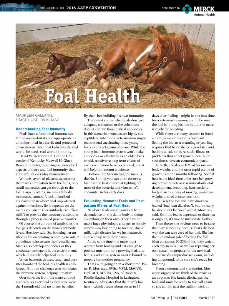
AAEP Wrap-Up The horse March 2017TheHorse.com/AAEP2016
mAUREEN gALLATIN; STACEy okE, Dvm, mSC
understanding Foal immunityFoals have a functional immune sys-
tem in utero—but it’s one appropriate to an unborn foal in a sterile and protected environment. Once that baby hits the real world, he needs real-world immunity.
David W. Horohov, PhD, of the Uni-versity of Kentucky Maxwell H. Gluck Research Center, in Lexington, described aspects of mare and foal immunity that are useful in everyday management.
With six layers of placenta separating the mare’s circulation from the fetus, only small molecules can get through to the foal. Large proteins, such as antibody molecules, cannot. A lack of antibod-ies leaves the newborn foal unprotected against infections. So it depends on the mare’s colostrum (her antibody-rich “first milk”) to provide the necessary antibodies through a process called passive transfer.
Of course, the amount of antibodies the foal gets depends on the mare’s antibody levels, Horohov said. So, boosting her an-tibodies by vaccinating according to AAEP guidelines helps assure they’re sufficient. Mares also develop antibodies as they encounter pathogens in the environment, which ultimately helps foal immunity.
When bacteria, viruses, fungi, and para-sites bombard the foal, his health is chal-lenged. But that challenge also stimulates the immune system, helping it mature.
Over time, the from-the-mare antibod-ies decay, so as critical as they once were, the 4-month-old foal no longer benefits.
By then, he’s building his own immunity.The caveat comes when foals don’t get
adequate colostrum or the colostrum doesn’t contain those critical antibodies. In this scenario, neonates are highly sus-ceptible to infections. Veterinarians might recommend vaccinating those young foals to protect against disease. While the young foal’s immune system won’t make antibodies as effectively as an older foal’s would, no adverse long-term effects of early vaccination have been noted, and it will help him mount a defense.
Bottom line: Vaccinating the mare is the No. 1 thing you can do to ensure a foal has the best chance at fighting off most of the bacteria and viruses he’ll encounter in his early days.
Evaluating Neonatal Foals and Post-partum Mares at Foal heat
Newborn foals must transition from dependence on the dam’s body to doing everything on their own. They have to make huge physiologic changes to simply survive—by beginning to breathe, digest milk, fight disease (as we just learned), and keep up with their dams.
At the same time, the mare must recover from foaling and eat enough to provide nutrition for a growing foal, and her reproductive system must rebound to prepare for another pregnancy.
That’s a lot going on in a short time. Pe-ter R. Morresey, BVSc, MVM, MACVSc, Dipl. ACT, ACVIM, CVA, of Rood & Riddle Equine Hospital in Lexington, Kentucky, advocates that the mare’s foal heat—which occurs about seven to 15
days after foaling—might be the best time for a veterinary examination to be sure the foal is hitting his marks and the mare is ready for breeding.
While there are many reasons to breed a mare, a major reason is financial. Selling the foal as a weanling or yearling requires that he or she be a good size and healthy at sale time. As such, illness or problems that affect growth, health, or soundness have an economic impact.
At birth, a foal is at 10% of his mature body weight, and the most rapid period of growth is in the months following. So foal heat is the ideal time to be sure he’s grow-ing normally. Vets assess musculoskeletal development, breathing, heart activity, body structure, way of moving, umbilicus, weight, and, of course, nutrition.
It’s likely the foal will have diarrhea (called “foal-heat diarrhea”), but normally he should not be “sick” with it, Morresey said. So if the foal is depressed or diarrhea is ongoing, it’s time to investigate further.
Then there’s the obvious need to be sure the mare is healthy, because that’s the best way she can take care of her foal. She has the tremendous job of feeding the foal (that consumes 20-25% of his body weight each day in milk!), as well as repairing her own system to prepare for the next foal.
She needs a reproductive exam, includ-ing ultrasound, to be sure she’s ready for breeding.
From a commercial standpoint, Mor-resey suggested we think of the mare as an airplane: She lands, discharges the foal, and must be ready to take off again so she can fly past the stallion, pick up
foal health
ELLE
N P
oN
S
SPonSorED bYYour GuiDE To THE 2016 aaeP convention
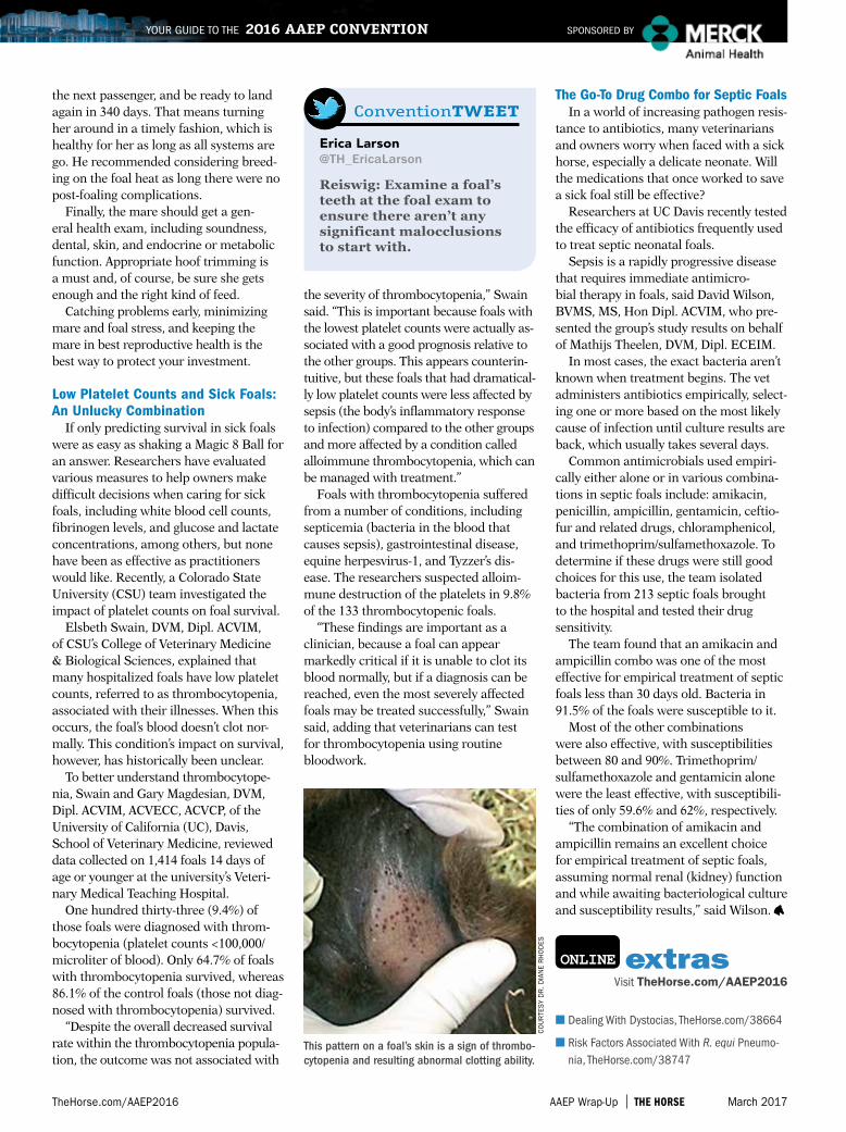
AAEP Wrap-Up The horse March 2017TheHorse.com/AAEP2016
Your GuiDE To THE 2016 aaeP convention SPonSorED bY
the next passenger, and be ready to land again in 340 days. That means turning her around in a timely fashion, which is healthy for her as long as all systems are go. He recommended considering breed-ing on the foal heat as long there were no post-foaling complications.
Finally, the mare should get a gen-eral health exam, including soundness, dental, skin, and endocrine or metabolic function. Appropriate hoof trimming is a must and, of course, be sure she gets enough and the right kind of feed.
Catching problems early, minimizing mare and foal stress, and keeping the mare in best reproductive health is the best way to protect your investment.
low Platelet Counts and sick Foals: An unlucky Combination
If only predicting survival in sick foals were as easy as shaking a Magic 8 Ball for an answer. Researchers have evaluated various measures to help owners make difficult decisions when caring for sick foals, including white blood cell counts, fibrinogen levels, and glucose and lactate concentrations, among others, but none have been as effective as practitioners would like. Recently, a Colorado State University (CSU) team investigated the impact of platelet counts on foal survival.
Elsbeth Swain, DVM, Dipl. ACVIM, of CSU’s College of Veterinary Medicine & Biological Sciences, explained that many hospitalized foals have low platelet counts, referred to as thrombocytopenia, associated with their illnesses. When this occurs, the foal’s blood doesn’t clot nor-mally. This condition’s impact on survival, however, has historically been unclear.
To better understand thrombocytope-nia, Swain and Gary Magdesian, DVM, Dipl. ACVIM, ACVECC, ACVCP, of the University of California (UC), Davis, School of Veterinary Medicine, reviewed data collected on 1,414 foals 14 days of age or younger at the university’s Veteri-nary Medical Teaching Hospital.
One hundred thirty-three (9.4%) of those foals were diagnosed with throm-bocytopenia (platelet counts <100,000/microliter of blood). Only 64.7% of foals with thrombocytopenia survived, whereas 86.1% of the control foals (those not diag-nosed with thrombocytopenia) survived.
“Despite the overall decreased survival rate within the thrombocytopenia popula-tion, the outcome was not associated with
the severity of thrombocytopenia,” Swain said. “This is important because foals with the lowest platelet counts were actually as-sociated with a good prognosis relative to the other groups. This appears counterin-tuitive, but these foals that had dramatical-ly low platelet counts were less affected by sepsis (the body’s inflammatory response to infection) compared to the other groups and more affected by a condition called alloimmune thrombocytopenia, which can be managed with treatment.”
Foals with thrombocytopenia suffered from a number of conditions, including septicemia (bacteria in the blood that causes sepsis), gastrointestinal disease, equine herpesvirus-1, and Tyzzer’s dis-ease. The researchers suspected alloim-mune destruction of the platelets in 9.8% of the 133 thrombocytopenic foals.
“These findings are important as a clinician, because a foal can appear markedly critical if it is unable to clot its blood normally, but if a diagnosis can be reached, even the most severely affected foals may be treated successfully,” Swain said, adding that veterinarians can test for thrombocytopenia using routine bloodwork.
the go-to Drug Combo for septic FoalsIn a world of increasing pathogen resis-
tance to antibiotics, many veterinarians and owners worry when faced with a sick horse, especially a delicate neonate. Will the medications that once worked to save a sick foal still be effective?
Researchers at UC Davis recently tested the efficacy of antibiotics frequently used to treat septic neonatal foals.
Sepsis is a rapidly progressive disease that requires immediate antimicro-bial therapy in foals, said David Wilson, BVMS, MS, Hon Dipl. ACVIM, who pre-sented the group’s study results on behalf of Mathijs Theelen, DVM, Dipl. ECEIM.
In most cases, the exact bacteria aren’t known when treatment begins. The vet administers antibiotics empirically, select-ing one or more based on the most likely cause of infection until culture results are back, which usually takes several days.
Common antimicrobials used empiri-cally either alone or in various combina-tions in septic foals include: amikacin, penicillin, ampicillin, gentamicin, ceftio-fur and related drugs, chloramphenicol, and trimethoprim/sulfamethoxazole. To determine if these drugs were still good choices for this use, the team isolated bacteria from 213 septic foals brought to the hospital and tested their drug sensitivity.
The team found that an amikacin and ampicillin combo was one of the most effective for empirical treatment of septic foals less than 30 days old. Bacteria in 91.5% of the foals were susceptible to it.
Most of the other combinations were also effective, with susceptibilities between 80 and 90%. Trimethoprim/ sulfamethoxazole and gentamicin alone were the least effective, with susceptibili-ties of only 59.6% and 62%, respectively.
“The combination of amikacin and ampicillin remains an excellent choice for empirical treatment of septic foals, assuming normal renal (kidney) function and while awaiting bacteriological culture and susceptibility results,” said Wilson. h
Co
UR
TES
y D
R.
DIA
NE
RH
oD
ES
This pattern on a foal’s skin is a sign of thrombo-cytopenia and resulting abnormal clotting ability.
conventiontweet
Erica Larson@TH_EricaLarson
Reiswig: Examine a foal’s teeth at the foal exam to ensure there aren’t any significant malocclusions to start with.
extrasVisit TheHorse.com/AAEP2016
■ Dealing With Dystocias, TheHorse.com/38664
■ risk Factors Associated With R. equi Pneumo-nia, TheHorse.com/38747
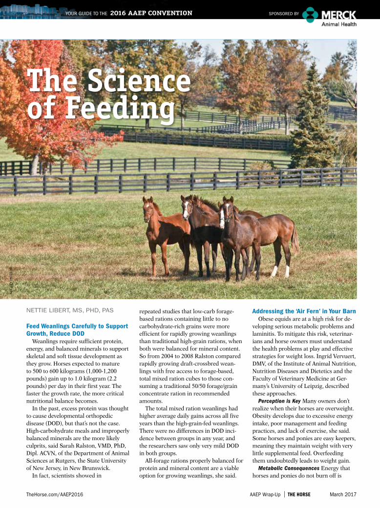
AAEP Wrap-Up The horse March 2017TheHorse.com/AAEP2016
NETTIE LIBERT, mS, PHD, PAS
Feed Weanlings Carefully to support growth, reduce DoD
Weanlings require sufficient protein, energy, and balanced minerals to support skeletal and soft tissue development as they grow. Horses expected to mature to 500 to 600 kilograms (1,000-1,200 pounds) gain up to 1.0 kilogram (2.2 pounds) per day in their first year. The faster the growth rate, the more critical nutritional balance becomes.
In the past, excess protein was thought to cause developmental orthopedic disease (DOD), but that’s not the case. High-carbohydrate meals and improperly balanced minerals are the more likely culprits, said Sarah Ralston, VMD, PhD, Dipl. ACVN, of the Department of Animal Sciences at Rutgers, the State University of New Jersey, in New Brunswick.
In fact, scientists showed in
repeated studies that low-carb forage-based rations containing little to no carbohydrate-rich grains were more efficient for rapidly growing weanlings than traditional high-grain rations, when both were balanced for mineral content. So from 2004 to 2008 Ralston compared rapidly growing draft-crossbred wean-lings with free access to forage-based, total mixed ration cubes to those con-suming a traditional 50/50 forage/grain concentrate ration in recommended amounts.
The total mixed ration weanlings had higher average daily gains across all five years than the high-grain-fed weanlings. There were no differences in DOD inci-dence between groups in any year, and the researchers saw only very mild DOD in both groups.
All-forage rations properly balanced for protein and mineral content are a viable option for growing weanlings, she said.
Addressing the ‘Air Fern’ in your Barn Obese equids are at a high risk for de-
veloping serious metabolic problems and laminitis. To mitigate this risk, veterinar-ians and horse owners must understand the health problems at play and effective strategies for weight loss. Ingrid Vervuert, DMV, of the Institute of Animal Nutrition, Nutrition Diseases and Dietetics and the Faculty of Veterinary Medicine at Ger-many’s University of Leipzig, described these approaches.
Perception is Key Many owners don’t realize when their horses are overweight. Obesity develops due to excessive energy intake, poor management and feeding practices, and lack of exercise, she said. Some horses and ponies are easy keepers, meaning they maintain weight with very little supplemental feed. Overfeeding them undoubtedly leads to weight gain.
Metabolic Consequences Energy that horses and ponies do not burn off is
SH
ELLE
y PA
ULS
oN
the science of feeding
SPonSorED bYYour GuiDE To THE 2016 aaeP convention
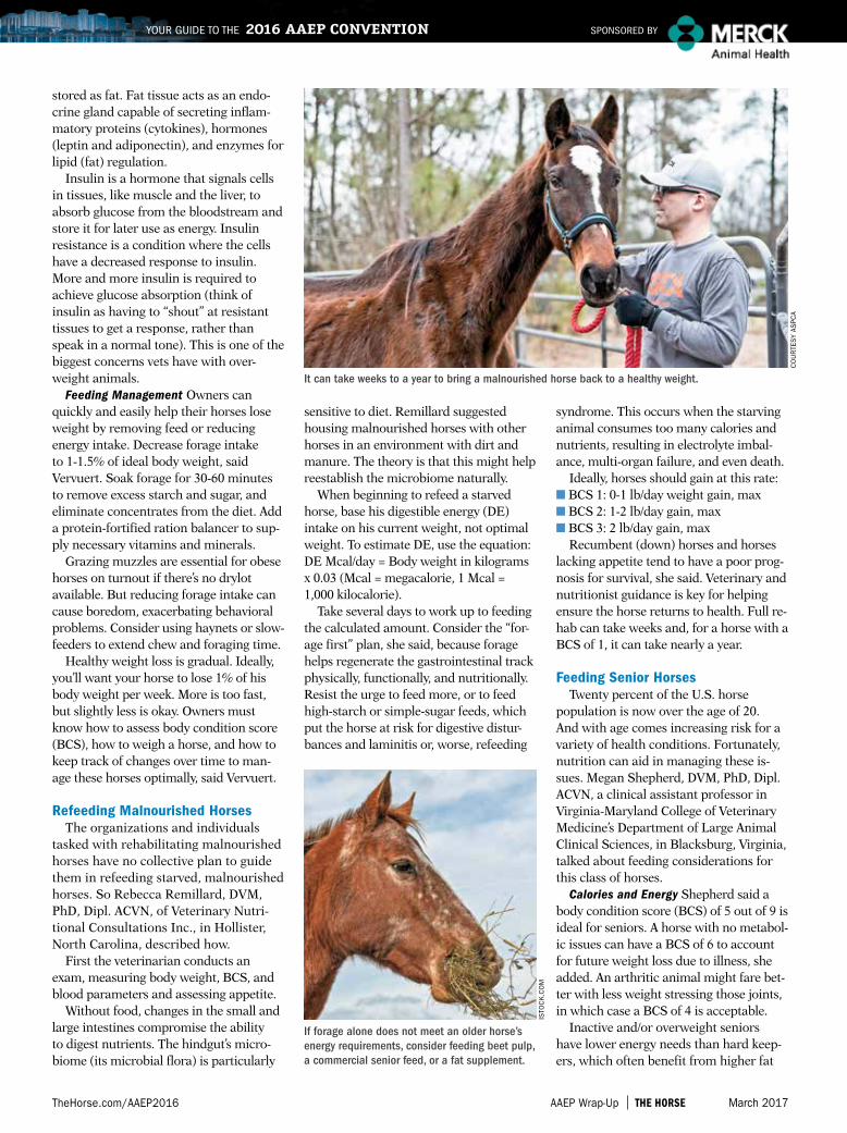
AAEP Wrap-Up The horse March 2017TheHorse.com/AAEP2016
Your GuiDE To THE 2016 aaeP convention SPonSorED bY
stored as fat. Fat tissue acts as an endo-crine gland capable of secreting inflam-matory proteins (cytokines), hormones (leptin and adiponectin), and enzymes for lipid (fat) regulation.
Insulin is a hormone that signals cells in tissues, like muscle and the liver, to absorb glucose from the bloodstream and store it for later use as energy. Insulin resistance is a condition where the cells have a decreased response to insulin. More and more insulin is required to achieve glucose absorption (think of insulin as having to “shout” at resistant tissues to get a response, rather than speak in a normal tone). This is one of the biggest concerns vets have with over-weight animals.
Feeding Management Owners can quickly and easily help their horses lose weight by removing feed or reducing energy intake. Decrease forage intake to 1-1.5% of ideal body weight, said Vervuert. Soak forage for 30-60 minutes to remove excess starch and sugar, and eliminate concentrates from the diet. Add a protein-fortified ration balancer to sup-ply necessary vitamins and minerals.
Grazing muzzles are essential for obese horses on turnout if there’s no drylot available. But reducing forage intake can cause boredom, exacerbating behavioral problems. Consider using haynets or slow-feeders to extend chew and foraging time.
Healthy weight loss is gradual. Ideally, you’ll want your horse to lose 1% of his body weight per week. More is too fast, but slightly less is okay. Owners must know how to assess body condition score (BCS), how to weigh a horse, and how to keep track of changes over time to man-age these horses optimally, said Vervuert.
refeeding Malnourished horsesThe organizations and individuals
tasked with rehabilitating malnourished horses have no collective plan to guide them in refeeding starved, malnourished horses. So Rebecca Remillard, DVM, PhD, Dipl. ACVN, of Veterinary Nutri-tional Consultations Inc., in Hollister, North Carolina, described how.
First the veterinarian conducts an exam, measuring body weight, BCS, and blood parameters and assessing appetite.
Without food, changes in the small and large intestines compromise the ability to digest nutrients. The hindgut’s micro-biome (its microbial flora) is particularly
sensitive to diet. Remillard suggested housing malnourished horses with other horses in an environment with dirt and manure. The theory is that this might help reestablish the microbiome naturally.
When beginning to refeed a starved horse, base his digestible energy (DE) intake on his current weight, not optimal weight. To estimate DE, use the equation: DE Mcal/day = Body weight in kilograms x 0.03 (Mcal = megacalorie, 1 Mcal = 1,000 kilocalorie).
Take several days to work up to feeding the calculated amount. Consider the “for-age first” plan, she said, because forage helps regenerate the gastrointestinal track physically, functionally, and nutritionally. Resist the urge to feed more, or to feed high-starch or simple-sugar feeds, which put the horse at risk for digestive distur-bances and laminitis or, worse, refeeding
syndrome. This occurs when the starving animal consumes too many calories and nutrients, resulting in electrolyte imbal-ance, multi-organ failure, and even death.
Ideally, horses should gain at this rate:■ BCS 1: 0-1 lb/day weight gain, max■ BCS 2: 1-2 lb/day gain, max■ BCS 3: 2 lb/day gain, max
Recumbent (down) horses and horses lacking appetite tend to have a poor prog-nosis for survival, she said. Veterinary and nutritionist guidance is key for helping ensure the horse returns to health. Full re-hab can take weeks and, for a horse with a BCS of 1, it can take nearly a year.
Feeding senior horsesTwenty percent of the U.S. horse
population is now over the age of 20. And with age comes increasing risk for a variety of health conditions. Fortunately, nutrition can aid in managing these is-sues. Megan Shepherd, DVM, PhD, Dipl. ACVN, a clinical assistant professor in Virginia-Maryland College of Veterinary Medicine’s Department of Large Animal Clinical Sciences, in Blacksburg, Virginia, talked about feeding considerations for this class of horses.
Calories and Energy Shepherd said a body condition score (BCS) of 5 out of 9 is ideal for seniors. A horse with no metabol-ic issues can have a BCS of 6 to account for future weight loss due to illness, she added. An arthritic animal might fare bet-ter with less weight stressing those joints, in which case a BCS of 4 is acceptable.
Inactive and/or overweight seniors have lower energy needs than hard keep-ers, which often benefit from higher fat
Co
UR
TES
y AS
PCA
It can take weeks to a year to bring a malnourished horse back to a healthy weight.
ISTo
Ck
.Co
m
If forage alone does not meet an older horse’s energy requirements, consider feeding beet pulp, a commercial senior feed, or a fat supplement.

AAEP Wrap-Up The horse March 2017TheHorse.com/AAEP2016
The Science of Trusted
From the broodmare to the performance horse, properly managing your mare’s hormones is critical. Rely on the product that’s trusted most.
• More than 30 years of practical use in the field by veterinarians1
• More than 200 clinical trials to determine efficacy, duration and safety1
• More than 20 million doses sold to veterinarians, trainers and horse owners1
Now that’s trusted.
Talk to your veterinarian about proper use and safe handling of Regu-Mate®. Avoid skin contact. Always wear protective gloves when administering Regu-Mate®. This product is contraindicated for use in mares with a previous or current history of uterine inflammation. Pregnant women, or women who suspect they are pregnant, should not handle this product. For complete product information, see accompanying product insert.
Regu-Mate® (altrenogest) is the name veterinarians and their clients depend on for estrus control (suppression, management).
1 Data on file, Merck Animal Heath
Ask your veterinarian for Regu-Mate®. Visit us online at merck-animal-health-equine.com to learn more about Merck Animal Health and the equine products and programs that help keep horses healthy.
The Science of Healthier Animals
2 Giralda Farms • Madison, NJ 07940 • merck-animal-health-usa.com • 800-521-5767Copyright © 2016 Intervet Inc., d/b/a/ Merck Animal Health, a subsidiary of Merck & Co., Inc.All rights reserved. 3526 EQ-FP AD Regu-Mate®
3526_Regu-Mate_8125x10875.indd 1 2/7/17 10:27 AM
AAEP Wrap-Up The horse March 2017TheHorse.com/AAEP2016
Your GuiDE To THE 2016 aaeP convention SPonSorED bY
diets for extra calories. Easy keepers or overweight horses generally fare well on forage-based diets with an added ration balancer.
Water Ensure senior horses have free access to fresh water to reduce colic risks. Water requirements increase with forage intake, especially in winter. During this time, said Shepherd, make sure water is ice-free, and warm it periodically. Horses with pituitary pars intermedia dysfunc-tion (PPID) tend to drink and urinate more and, so, have increased water needs.
Forage Shepherd recommends feeding a ration balancer to horses consuming forage-only diets to be sure they con-sume enough vitamins and minerals. If good-quality forage alone does not meet an older horse’s energy requirements, consider feeding beet pulp, a commer-cial senior feed, or a fat supplement to increase calorie intake.
If a horse quids, or drops chewed wads of hay, then you might replace long-stem or pasture forage with soaked pelleted or cubed forage.
Other Nutrients Horses with PPID might be insulin-resistant, meaning their cells don’t respond normally to insulin; therefore, balance these horses’ diets care-fully to limit starch and sugar intake.
Horses with PPID have decreased an-tioxidant capacity in the pituitary gland’s pars intermedia and might benefit from a vitamin E supplement. They might also have increased free radical formation, causing oxidative damage. Therefore, you might consider exceeding vitamin E requirements for these horses, said Shepherd.
For horses with joint disease, omega-3 fatty acids, particularly marine-based eicosapentaenoic acid and docosa-hexaenoic acid, might help reduce joint inflammation.
Feeding horses With Muscle issuesHorses with exertional myopathies
such as recurrent exertional rhabdomy-olysis (RER), also known as tying-up, and polysaccharide storage myopathy (Type 1 and 2 PSSM) can experience muscle fatigue, pain, cramping, and damage. Stephanie Valberg, DVM, PhD, Dipl. ACVIM, ACVSMR, of the Michigan State University College of Veterinary Medi-cine’s Department of Large Animal Clini-cal Sciences, described how to maintain them, stressing that regular exercise and
dietary management are necessary to improve.
The general rule of thumb is to keep nonstructural carbohydrate (NSC, starch and sugar) levels low and provide extra calories with fat. In RER horses, Valberg said to restrict calories from NSCs to less than 20% of the total diet to minimize ex-citability. Also provide up to 20% of calo-ries with fat. Horses receiving 5 kilograms (11 pounds) or more of grain tend to be at higher risk for an RER episode than those getting 2.5 kilograms (5.5 pounds) or less.
For Type 1 PSSM horses, keep NSCs to 10-15% of the diet. A low-NSC diet will reduce insulin secretion in response to a meal. Insulin stimulates production of glycogen synthase, so reducing the amount of circulating insulin in PSSM horses is important to help manage the amount of glycogen in the muscles. Fat needs depend on the horse’s BCS and exercise demands. Researchers have re-cently shown that Type 2 PSSM horses do not have the same high muscle glycogen concentrations and, so, might not need strict NSC restriction. Type 1 or Type 2 PSSM horses that experience decreased muscle mass might need an amino acid or protein supplement to help support muscle development.
“The easiest thing to change when a horse is diagnosed with PSSM is the diet, but I always emphasize that the diet itself is a small piece of the solution, and for muscle to properly utilize the appropriate diet, horses need to be exercised regularly in a training program,” Valberg said. For more info see TheHorse.com/38753. h
extrasVisit TheHorse.com/AAEP2016
■ How to Feed Horses With Gastric ulcers, TheHorse.com/38754
conventiontweet
Erica Larson@TH_EricaLarson
MacKay—Soaking hay doubled weight loss in obese ponies. Also removes fructans, DM, DE, and minerals from hay. Needs vet supervision.
Regu-Mate® (altrenogest)
Solution 0.22% (2.2 mg/mL) CAUTION: Federal law restricts this drug to use by or on the order of a licensed veterinarian.
DESCRIPTION: Regu-Mate® (altrenogest) Solution 0.22% contains the active synthetic progestin, altrenogest. The chemical name is 17α-allyl-17ß-hydroxyestra- 4,9,11-trien-3-one. The CAS Registry Number is 850-52-2. The chemical structure is:
Each mL of Regu-Mate® (altrenogest) Solution 0.22% contains 2.2 mg of altrenogest in an oil solution.
ACTIONS: Regu-Mate® (altrenogest) Solution 0.22% produces a progestational effect in mares.
INDICATIONS: Regu-Mate® (altrenogest) Solution 0.22% is indicated to suppress estrus in mares. Suppression of estrus allows for a predictable occurrence of estrus following drug withdrawal. This facilitates the attainment of regular cyclicity during the transition from winter anestrus to the physiological breeding season. Suppression of estrus will also facilitate management of prolonged estrus conditions. Suppression of estrus may be used to facilitate scheduled breeding during the physiological breeding season.
CONTRAINDICATIONS: Regu-Mate® (altrenogest) Solution 0.22% is contraindicated for use in mares having a previous or current history of uterine inflammation (i.e., acute, subacute, or chronic endometritis). Natural or synthetic gestagen therapy may exacerbate existing low-grade or “smoldering” uterine inflammation into a fulminating uterine infection in some instances.
PRECAUTIONS: Various synthetic progestins, including altrenogest, when administered to rats during the embryogenic stage of pregnancy at doses manyfold greater than the recommended equine dose caused fetal anomalies, specifically masculinization of the female genitalia.
DOSAGE AND ADMINISTRATION: While wearing protective gloves, remove shipping cap and seal; replace with enclosed plastic dispensing cap. Remove cover from bottle dispensing tip and connect luer lock syringe (without needle). Draw out appropriate volume of Regu-Mate solution. (Note: Do not remove syringe while bottle is inverted as spillage may result.) Detach syringe and administer solution orally at the rate of 1 mL per 110 pounds body weight (0.044 mg/kg) once daily for 15 consecutive days. Administer solution directly on the base of the mare’s tongue or on the mare’s usual grain ration. Replace cover on bottle dispensing tip to prevent leakage. Excessive use of a syringe may cause the syringe to stick; therefore, replace syringe as necessary.
WHICH MARES WILL RESPOND TO REGU-MATE® (altrenogest) SOLUTION 0.22%: Extensive clinical trials have demonstrated that estrus will be suppressed in approximately 95% of the mares within three days; however, the post-treatment response depended on the level of ovarian activity when treatment was initiated. Estrus in mares exhibiting regular estrus cycles during the breeding season will be suppressed during treatment; these mares return to estrus four to five days following treatment and continue to cycle normally. Mares in winter anestrus with small follicles continued in anestrus and failed to exhibit normal estrus following withdrawal. Response in mares in the transition phase between winter anestrus and the summer breeding season depended on the degree of follicular activity. Mares with inactive ovaries and small follicles failed to respond with normal cycles post-treatment, whereas a higher proportion of mares with ovarian follicles 20 mm or greater in diameter exhibited normal estrus cycles post-treatment. Regu-Mate® (altrenogest) Solution 0.22% was very effective for suppressing the prolonged estrus behavior frequently observed in mares during the transition period (February, March and April). In addition, a high proportion of these mares responded with regular estrus cycles post-treatment.
SPECIFIC USES FOR REGU-MATE® (altrenogest) SOLUTION 0.22%: SUPPRESSION OF ESTRUS TO:1. Facilitate attainment of regular cycles during the transition period from winter anestrus to the
physiological breeding season. To facilitate attainment of regular cycles during the transition phase, mares should be examined to determine the degree of ovarian activity. Estrus in mares with inactive ovaries (no follicles greater than 20 mm in diameter) will be suppressed but these mares may not begin regular cycles following treatment. However, mares with active ovaries (follicles greater than 20 mm in diameter) frequently respond with regular post-treatment estrus cycles.
2. Facilitate management of the mare exhibiting prolonged estrus during the transition period. Estrus will be suppressed in mares exhibiting prolonged behavioral estrus either early or late during the transition period. Again, the posttreatment response depends on the level of ovarian activity. The mares with greater ovarian activity initiate regular cycles and conceive sooner than the inactive mares. Regu-Mate® (altrenogest) Solution 0.22% may be administered early in the transition period to suppress estrus in mares with inactive ovaries to aid in the management of these mares or to mares later in the transition period with active ovaries to prepare and schedule the mare for breeding.
3. Permit scheduled breeding of mares during the physiological breeding season. To permit scheduled breeding, mares which are regularly cycling or which have active ovarian function should be given Regu-Mate® (altrenogest) Solution 0.22% daily for 15 consecutive days beginning 20 days before the date of the planned estrus. Ovulation will occur 5 to 7 days following the onset of estrus as expected for nontreated mares. Breeding should follow usual procedures for mares in estrus. Mares may be regulated and scheduled either individually or in groups.
ADDITIONAL INFORMATION: A 3-year well controlled reproductive safety study was conducted in 27 pregnant mares, and compared with 24 untreated control mares. Treated mares received 2 mL Regu-Mate® (altrenogest) Solution 0.22% /110 lb body weight (2x dosage recommended for estrus suppression) from day 20 to day 325 of gestation. This study provided the following data: 1. In filly offspring (all ages) of treated mares, clitoral size was increased. 2. Filly offspring from treated mares had shorter interval from Feb. 1 to first ovulation than fillies
from their untreated mare counterparts.3. There were no significant differences in reproductive performance between treated and untreated
animals (mares & their respective offspring) measuring the following parameters: • interval from Feb. 1 to first ovulation, in mares only. • mean interovulatory interval from first to second cycle and second to third cycle, mares
only. • follicle size, mares only. • at 50 days gestation, pregnancy rate in treated mares was 81.8% (9/11) and untreated
mares was 100% (4/4). • after 3 cycles, 11/12 treated mares were pregnant (91.7%) and 4/4 untreated mares were
pregnant (100%). • colt offspring of treated and control mares reached puberty at approximately the same age
(82 & 84 weeks respectively). • stallion offspring from treated and control mares showed no differences in seminal volume,
spermatozoal concentration, spermatozoal motility, and total sperm per ejaculate. • stallion offspring from treated and control mares showed no difference in sexual behavior. • testicular characteristics (scrotal width, testis weight, parenchymal weight, epididymal
weight and height, testicular height, width & length) were the same between stallion offspring of treated and control mares.
REFERENCES:Shoemaker, C.F., E.L. Squires, and R.K. Shideler. 1989.Safety of Altrenogest in Pregnant Mares and on Health and Development of Offspring. Eq. Vet. Sci. (9); No. 2: 69-72.Squires, E.L., R.K. Shideler, and A.O. McKinnon. 1989.Reproductive Performance of Offspring from Mares Administered Altrenogest During Gestation. Eq. Vet. Sci. (9); No. 2: 73-76.
WARNING: Do not use in horses intended for food.
HUMAN WARNINGS: Skin contact must be avoided as Regu-Mate® (altrenogest) Solution 0.22% is readily absorbed through unbroken skin. Protective gloves must be worn by all persons handling this product. Pregnant women or women who suspect they are pregnant should not handle Regu-Mate® (altrenogest) Solution 0.22%. Women of child bearing age should exercise extreme caution when handling this product. Accidental absorption could lead to a disruption of the menstrual cycle or prolongation of pregnancy. Direct contact with the skin should therefore be avoided. Accidental spillage on the skin should be washed off immediately with soap and water.
INFORMATION FOR HANDLERS: WARNING: Regu-Mate® (altrenogest) Solution 0.22% is readily absorbed by the skin. Skin contact must be avoided; protective gloves must be worn when handling this product.
Effects of Overexposure: There has been no human use of this specific product. The information contained in this section is extrapolated from data available on other products of the same pharmacological class that have been used in humans. Effects anticipated are due to the progestational activity of altrenogest. Acute effects after a single exposure are possible; however, continued daily exposure has the potential for more untoward effects such as disruption of the menstrual cycle, uterine or abdominal cramping, increased or decreased uterine bleeding, prolongation of pregnancy and headaches. The oil base may also cause complications if swallowed. In addition, the list of people who should not handle this product (see below) is based upon the known effects of progestins used in humans on a chronic basis.
PEOPLE WHO SHOULD NOT HANDLE THIS PRODUCT. 1. Women who are or suspect they are pregnant. 2. Anyone with thrombophlebitis or thromboembolic disorders or with a history of these events. 3. Anyone with cerebral-vascular or coronary artery disease. 4. Women with known or suspected carcinoma of the breast. 5. People with known or suspected estrogen-dependent neoplasia. 6. Women with undiagnosed vaginal bleeding. 7. People with benign or malignant tumors which developed during the use of oral
contraceptives or other estrogen-containing products. 8. Anyone with liver dysfunction or disease.
ACCIDENTAL EXPOSURE: Altrenogest is readily absorbed from contact with the skin. In addition, this oil based product can penetrate porous gloves. Altrenogest should not penetrate intact rubber or impervious gloves; however, if there is leakage (i.e., pinhole, spillage, etc.), the contaminated area covered by such occlusive materials may have increased absorption. The following measures are recommended in case of accidental exposure. Skin Exposure: Wash immediately with soap and water.Eye Exposure: Immediately flush with plenty of water for 15 minutes. Get medical attention.If Swallowed: Do not induce vomiting. Regu-Mate® (altrenogest) Solution 0.22% contains an oil. Call a physician. Vomiting should be supervised by a physician because of possible pulmonary damage via aspiration of the oil base. If possible, bring the container and labeling to the physician.
CAUTION: For oral use in horses only. Keep this and all medication out of the reach of children.
Store at or below 25°C (77°F).
NADA# 131-310, Approved by FDA.
HOW SUPPLIED:Regu-Mate® (altrenogest) Solution 0.22% (2.2 mg/mL).Each mL contains 2.2 mg altrenogest in an oil solution.Available in 1000 mL plastic bottles.
* US Patents 3,453,267; 3,478,067; 3,484,462
Manufactured by:DPT Laboratories, San Antonio, TX 78215
Distributed by:Intervet Inc., Millsboro, DE 19966
DOSAGE CHART:
Approximate Weight Dose in Pounds in mL 770 7 880 8 990 9 1100 10 1210 11 1320 12
Regu-Mate Ad-HO_BR_Legal_25x9625_BW.indd 1 2/1/17 11:05 AM
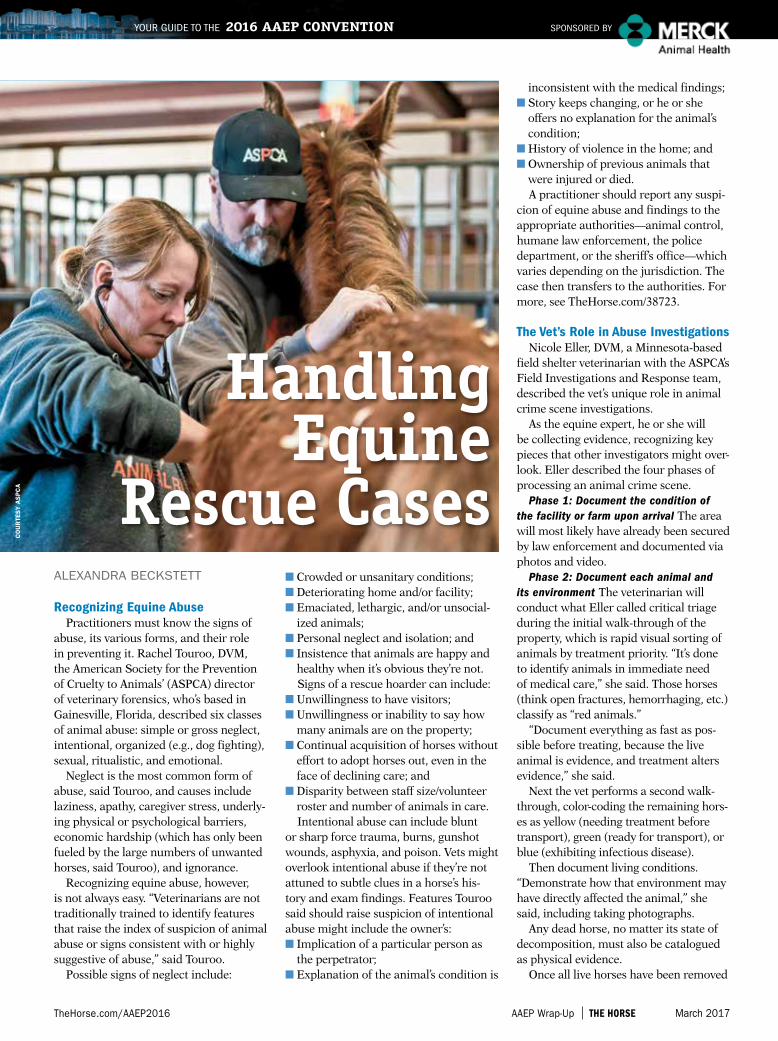
AAEP Wrap-Up The horse March 2017TheHorse.com/AAEP2016
ALExANDRA BECkSTETT
recognizing Equine AbusePractitioners must know the signs of
abuse, its various forms, and their role in preventing it. Rachel Touroo, DVM, the American Society for the Prevention of Cruelty to Animals’ (ASPCA) director of veterinary forensics, who’s based in Gainesville, Florida, described six classes of animal abuse: simple or gross neglect, intentional, organized (e.g., dog fighting), sexual, ritualistic, and emotional.
Neglect is the most common form of abuse, said Touroo, and causes include laziness, apathy, caregiver stress, underly-ing physical or psychological barriers, economic hardship (which has only been fueled by the large numbers of unwanted horses, said Touroo), and ignorance.
Recognizing equine abuse, however, is not always easy. “Veterinarians are not traditionally trained to identify features that raise the index of suspicion of animal abuse or signs consistent with or highly suggestive of abuse,” said Touroo.
Possible signs of neglect include:
■ Crowded or unsanitary conditions;■ Deteriorating home and/or facility;■ Emaciated, lethargic, and/or unsocial-
ized animals;■ Personal neglect and isolation; and■ Insistence that animals are happy and
healthy when it’s obvious they’re not.Signs of a rescue hoarder can include:
■ Unwillingness to have visitors; ■ Unwillingness or inability to say how
many animals are on the property;■ Continual acquisition of horses without
effort to adopt horses out, even in the face of declining care; and
■ Disparity between staff size/volunteer roster and number of animals in care.Intentional abuse can include blunt
or sharp force trauma, burns, gunshot wounds, asphyxia, and poison. Vets might overlook intentional abuse if they’re not attuned to subtle clues in a horse’s his-tory and exam findings. Features Touroo said should raise suspicion of intentional abuse might include the owner’s:■ Implication of a particular person as
the perpetrator;■ Explanation of the animal’s condition is
inconsistent with the medical findings;■ Story keeps changing, or he or she
offers no explanation for the animal’s condition;
■ History of violence in the home; and■ Ownership of previous animals that
were injured or died.A practitioner should report any suspi-
cion of equine abuse and findings to the appropriate authorities— animal control, humane law enforcement, the police department, or the sheriff’s office —which varies depending on the jurisdiction. The case then transfers to the authorities. For more, see TheHorse.com/38723.
the vet’s role in Abuse investigations Nicole Eller, DVM, a Minnesota-based
field shelter veterinarian with the ASPCA’s Field Investigations and Response team, described the vet’s unique role in animal crime scene investigations.
As the equine expert, he or she will be collecting evidence, recognizing key pieces that other investigators might over-look. Eller described the four phases of processing an animal crime scene.
Phase 1: Document the condition of the facility or farm upon arrival The area will most likely have already been secured by law enforcement and documented via photos and video.
Phase 2: Document each animal and its environment The veterinarian will conduct what Eller called critical triage during the initial walk-through of the property, which is rapid visual sorting of animals by treatment priority. “It’s done to identify animals in immediate need of medical care,” she said. Those horses (think open fractures, hemorrhaging, etc.) classify as “red animals.”
“Document everything as fast as pos-sible before treating, because the live animal is evidence, and treatment alters evidence,” she said.
Next the vet performs a second walk-through, color-coding the remaining hors-es as yellow (needing treatment before transport), green (ready for transport), or blue (exhibiting infectious disease).
Then document living conditions. “Demonstrate how that environment may have directly affected the animal,” she said, including taking photographs.
Any dead horse, no matter its state of decomposition, must also be catalogued as physical evidence.
Once all live horses have been removed
co
ur
TESy
ASP
cA
handling equine
rescue cases
SPonSorED bYYour GuiDE To THE 2016 aaeP convention
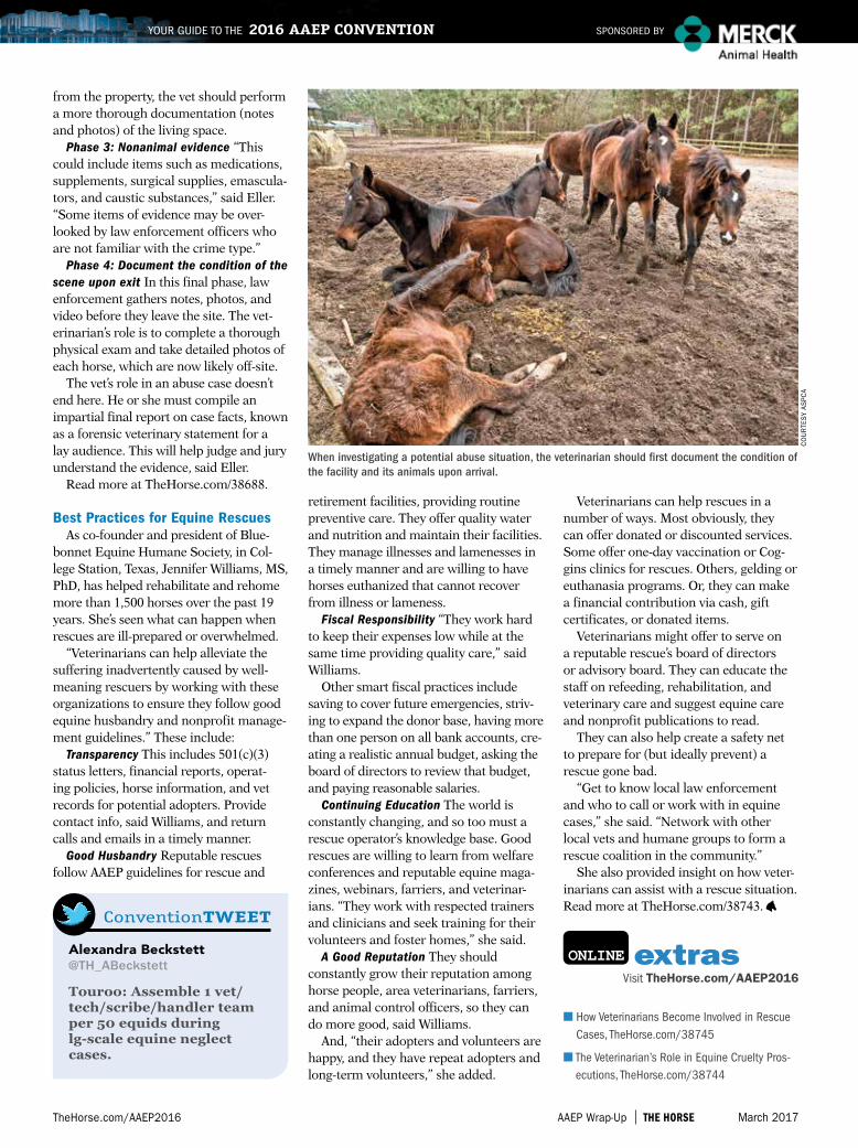
AAEP Wrap-Up The horse March 2017TheHorse.com/AAEP2016
Your GuiDE To THE 2016 aaeP convention SPonSorED bY
from the property, the vet should perform a more thorough documentation (notes and photos) of the living space.
Phase 3: Nonanimal evidence “This could include items such as medications, supplements, surgical supplies, emascula-tors, and caustic substances,” said Eller. “Some items of evidence may be over-looked by law enforcement officers who are not familiar with the crime type.”
Phase 4: Document the condition of the scene upon exit In this final phase, law enforcement gathers notes, photos, and video before they leave the site. The vet-erinarian’s role is to complete a thorough physical exam and take detailed photos of each horse, which are now likely off-site.
The vet’s role in an abuse case doesn’t end here. He or she must compile an impartial final report on case facts, known as a forensic veterinary statement for a lay audience. This will help judge and jury understand the evidence, said Eller.
Read more at TheHorse.com/38688.
Best Practices for Equine rescuesAs co-founder and president of Blue-
bonnet Equine Humane Society, in Col-lege Station, Texas, Jennifer Williams, MS, PhD, has helped rehabilitate and rehome more than 1,500 horses over the past 19 years. She’s seen what can happen when rescues are ill-prepared or overwhelmed.
“Veterinarians can help alleviate the suffering inadvertently caused by well-meaning rescuers by working with these organizations to ensure they follow good equine husbandry and nonprofit manage-ment guidelines.” These include:
Transparency This includes 501(c)(3) status letters, financial reports, operat-ing policies, horse information, and vet records for potential adopters. Provide contact info, said Williams, and return calls and emails in a timely manner.
Good Husbandry Reputable rescues follow AAEP guidelines for rescue and
retirement facilities, providing routine preventive care. They offer quality water and nutrition and maintain their facilities. They manage illnesses and lamenesses in a timely manner and are willing to have horses euthanized that cannot recover from illness or lameness.
Fiscal Responsibility “They work hard to keep their expenses low while at the same time providing quality care,” said Williams.
Other smart fiscal practices include saving to cover future emergencies, striv-ing to expand the donor base, having more than one person on all bank accounts, cre-ating a realistic annual budget, asking the board of directors to review that budget, and paying reasonable salaries.
Continuing Education The world is constantly changing, and so too must a rescue operator’s knowledge base. Good rescues are willing to learn from welfare conferences and reputable equine maga-zines, webinars, farriers, and veterinar-ians. “They work with respected trainers and clinicians and seek training for their volunteers and foster homes,” she said.
A Good Reputation They should constantly grow their reputation among horse people, area veterinarians, farriers, and animal control officers, so they can do more good, said Williams.
And, “their adopters and volunteers are happy, and they have repeat adopters and long-term volunteers,” she added.
Veterinarians can help rescues in a number of ways. Most obviously, they can offer donated or discounted services. Some offer one-day vaccination or Cog-gins clinics for rescues. Others, gelding or euthanasia programs. Or, they can make a financial contribution via cash, gift certificates, or donated items.
Veterinarians might offer to serve on a reputable rescue’s board of directors or advisory board. They can educate the staff on refeeding, rehabilitation, and veterinary care and suggest equine care and nonprofit publications to read.
They can also help create a safety net to prepare for (but ideally prevent) a rescue gone bad.
“Get to know local law enforcement and who to call or work with in equine cases,” she said. “Network with other local vets and humane groups to form a rescue coalition in the community.”
She also provided insight on how veter-inarians can assist with a rescue situation. Read more at TheHorse.com/38743. h
When investigating a potential abuse situation, the veterinarian should first document the condition of the facility and its animals upon arrival.
Co
UR
TES
y AS
PCA
extrasVisit TheHorse.com/AAEP2016
■ How Veterinarians become involved in rescue Cases, TheHorse.com/38745
■ The Veterinarian’s role in Equine Cruelty Pros-ecutions, TheHorse.com/38744
conventiontweet
Alexandra Beckstett@TH_ABeckstett
Touroo: Assemble 1 vet/tech/scribe/handler team per 50 equids during lg-scale equine neglect cases.

AAEP Wrap-Up The horse March 2017TheHorse.com/AAEP2016
SafetyIn Numbers
Some dewormers claim just one dose of their product is the best way to deworm your horse, but that’s simply not true.
Demand Safety: You won’t find a laundry list of warnings and precautions on the PANACUR® (fenbendazole) POWERPAC label because fenbendazole has a unique mode of action that makes it safe for horses of all ages, sizes, and body conditions.
Demand Efficacy: PANACUR® POWERPAC is the only dewormer FDA approved to treat ALL STAGES of the encysted small strongyle.1
Other dewormers miss a critical stage, EL3, which can account for up to 75% of the encysted small strongyle burden. Plus, it’s the best choice for treating ascarids — which are not just a problem in young horses!
So when it comes to which dewormer to trust, don’t forget there’s safety in numbers.
Consult your veterinarian for assistance in the diagnosis, treatment and control of parasitism.
Do not use in horses intended for human consumption. When using PANACUR® (fenbendazole) Paste 10% concomitantly with trichlorfon, refer to the manufacturers labels for use and cautions for trichlorfon.
1 PANACUR® (fenbendazole) POWERPAC Equine Dewormer product label.
The Science of Healthier Animals
2 Giralda Farms • Madison, NJ 07940 • merck-animal-health-usa.com • 800-521-5767Copyright © 2016 Intervet Inc., d/b/a/ Merck Animal Health, a subsidiary of Merck & Co., Inc.All rights reserved. 3526 EG-PC-Adver
3526_Panacur_8125x10875.indd 1 2/15/17 8:55 AM
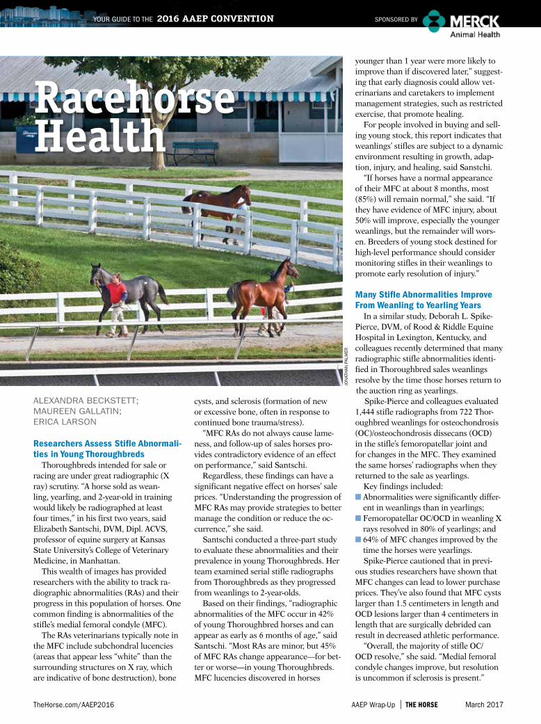
AAEP Wrap-Up The horse March 2017TheHorse.com/AAEP2016
ALExANDRA BECkSTETT; mAUREEN gALLATIN; ERICA LARSoN
researchers Assess stifle Abnormali-ties in young thoroughbreds
Thoroughbreds intended for sale or racing are under great radiographic (X ray) scrutiny. “A horse sold as wean-ling, yearling, and 2-year-old in training would likely be radiographed at least four times,” in his first two years, said Elizabeth Santschi, DVM, Dipl. ACVS, professor of equine surgery at Kansas State University’s College of Veterinary Medicine, in Manhattan.
This wealth of images has provided researchers with the ability to track ra-diographic abnormalities (RAs) and their progress in this population of horses. One common finding is abnormalities of the stifle’s medial femoral condyle (MFC).
The RAs veterinarians typically note in the MFC include subchondral lucencies (areas that appear less “white” than the surrounding structures on X ray, which are indicative of bone destruction), bone
cysts, and sclerosis (formation of new or excessive bone, often in response to continued bone trauma/stress).
“MFC RAs do not always cause lame-ness, and follow-up of sales horses pro-vides contradictory evidence of an effect on performance,” said Santschi.
Regardless, these findings can have a significant negative effect on horses’ sale prices. “Understanding the progression of MFC RAs may provide strategies to better manage the condition or reduce the oc-currence,” she said.
Santschi conducted a three-part study to evaluate these abnormalities and their prevalence in young Thoroughbreds. Her team examined serial stifle radiographs from Thoroughbreds as they progressed from weanlings to 2-year-olds.
Based on their findings, “radiographic abnormalities of the MFC occur in 42% of young Thoroughbred horses and can appear as early as 6 months of age,” said Santschi. “Most RAs are minor, but 45% of MFC RAs change appearance—for bet-ter or worse—in young Thoroughbreds. MFC lucencies discovered in horses
younger than 1 year were more likely to improve than if discovered later,” suggest-ing that early diagnosis could allow vet-erinarians and caretakers to implement management strategies, such as restricted exercise, that promote healing.
For people involved in buying and sell-ing young stock, this report indicates that weanlings’ stifles are subject to a dynamic environment resulting in growth, adap-tion, injury, and healing, said Sanstchi.
“If horses have a normal appearance of their MFC at about 8 months, most (85%) will remain normal,” she said. “If they have evidence of MFC injury, about 50% will improve, especially the younger weanlings, but the remainder will wors-en. Breeders of young stock destined for high-level performance should consider monitoring stifles in their weanlings to promote early resolution of injury.”
Many stifle Abnormalities improve From Weanling to yearling years
In a similar study, Deborah L. Spike-Pierce, DVM, of Rood & Riddle Equine Hospital in Lexington, Kentucky, and colleagues recently determined that many radiographic stifle abnormalities identi-fied in Thoroughbred sales weanlings resolve by the time those horses return to the auction ring as yearlings.
Spike-Pierce and colleagues evaluated 1,444 stifle radiographs from 722 Thor-oughbred weanlings for osteochondrosis (OC)/osteochondrosis dissecans (OCD) in the stifle’s femoropatellar joint and for changes in the MFC. They examined the same horses’ radiographs when they returned to the sale as yearlings.
Key findings included:■ Abnormalities were significantly differ-
ent in weanlings than in yearlings;■ Femoropatellar OC/OCD in weanling X
rays resolved in 80% of yearlings; and■ 64% of MFC changes improved by the
time the horses were yearlings.Spike-Pierce cautioned that in previ-
ous studies researchers have shown that MFC changes can lead to lower purchase prices. They’ve also found that MFC cysts larger than 1.5 centimeters in length and OCD lesions larger than 4 centimeters in length that are surgically debrided can result in decreased athletic performance.
“Overall, the majority of stifle OC/OCD resolve,” she said. “Medial femoral condyle changes improve, but resolution is uncommon if sclerosis is present.”
racehorse health
joN
ATH
AN P
ALm
ER
Your GuiDE To THE 2016 aaeP convention SPonSorED bY
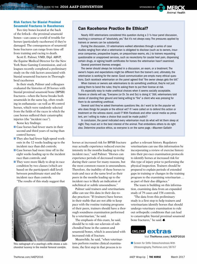
AAEP Wrap-Up The horse March 2017TheHorse.com/AAEP2016
risk Factors for Biaxial Proximal sesamoid Fractures in racehorses
Two tiny bones located at the back of the fetlock—the proximal sesamoid bones—can cause a world of trouble for horses (particularly racehorses) if they’re damaged. The consequences of sesamoid bone fractures can range from time off from training and racing to death.
Scott E. Palmer, VMD, Dipl. ABVP, the Equine Medical Director for the New York State Gaming Commission, and col-leagues recently completed a preliminary study on the risk factors associated with biaxial sesamoid fractures in Thorough-bred racehorses
In their study, Palmer and colleagues evaluated the histories of 20 horses with biaxial proximal sesamoid bone (BPSB) fractures—when the horse breaks both sesamoids in the same leg, often result-ing in euthanasia—as well as 40 control horses, which were randomly selected from the fields of the races in which the case horses suffered their catastrophic injuries (the “incident race”).
Some key findings:■ Case horses had fewer starts in their
second and third years of racing than control horses;
■ They also had fewer high-speed work-outs in the 12 weeks leading up to the incident race than did controls;
■ Case horses had more time off in the eight weeks leading up to the incident race than controls; and
■ They were more likely to drop in race condition by two classes (which are based on the participant’s skill level) between penultimate start and the incident race than controls.“The results of this study suggest that
horses at increased risk for BPSB fracture may actually experience reduced exercise intensity in the months leading up to the incident race,” said Palmer. “Horses can experience periods of decreased training during their career for many reasons, but the most common reason is unsoundness. Therefore, the inability of these horses to train and race at the same level as their peers in the months leading up to the incident race is likely an indication of subclinical or subtle unsoundness.”
Palmer said trainers and veterinarians alike can use this data in their day-to-day practices: “If (trainers) have horses in their stable that are not able to keep pace with the routine training programs of their peers, trainers should have a thor-ough soundness examination performed by a veterinarian,” he said.
The emphasis of this exam, he said, should be to rule out sclerosis of sub-chondral bone in the cannon and sesamoid bones, which is associated with increased risk of fracture.
Meanwhile, he said, “when veterinar-ians perform routine clinical examina-tions, the first step in that process is to
gather a relevant history. Regulatory veterinarians can use this information by incorporating a review of exercise history into their pre-race examination protocols to identify horses at increased risk for this type of injury prior to performing the physical inspection. Trainers should be prepared to explain periods of extended gaps in training or changes in the training program to the examining veterinarian … as part of their due diligence.”
The team is building on this informa-tion, examining data from an expanded study of 79 cases and 158 controls.
“The data from this preliminary study is a first step to help trainers and veterinarians identify horses that should undergo veterinary examination to rule out orthopedic conditions that can lead to catastrophic biaxial proximal sesamoid bone fractures,” he said. h
Co
UR
TES
y D
R.
ELIz
ABET
H S
ANTS
CH
I
This radiograph of a yearling’s stifle shows a sub-chondral lucency in the medial femoral condyle.
Can racehorse Practice Be Ethical?nearly 400 veterinarians considered this question during a 3 ½-hour panel discussion,
reaching a consensus of “absolutely, yes.” but it’s not always easy. The pressures applied by trainers or owners can be substantial.
During the discussion, 10 veterinarians walked attendees through a series of case studies ranging from what a veterinarian is obligated to disclose (such as to owners, insur-ance companies, prospective buyers, on prepurchase exams, etc.) to trainers requesting questionable or unapproved services, such as neurectomy for caudal heel pain, dispensing certain drugs, or signing health certificates for horses the veterinarian hasn’t examined.
Several prominent themes emerged. The owner should always be included in a discussion, an exam, or a treatment. The
owner’s hopes and expectations might be different from the trainer’s and, ultimately, the veterinarian is working for the owner. Good communication pre-empts many ethical ques-tions. Each racetrack veterinarian on the panel agreed that “the owner always gets the bill.”
When trainers or owners ask veterinarians to do something unethical, they’re not just asking them to bend the rules; they’re asking them to put their license at risk.
it’s especially easy to make unethical choices when it seems socially acceptable. Trainers or clients will say, “Everyone (or Dr. So and So) is doing it.” Still, veterinarians told stories of standing their ground and being willing to “fire” a client who was pressing for them to do something unethical.
Several said they’ve asked themselves questions like, do i want to be the popular vet who does things for people or the ethical vet? if i were called on to defend this action or treatment on the witness stand, could i? With Facebook and other social media so preva-lent, am i willing to make a choice that could be made public?
in conclusion, the panel indicated every veterinarian must do what will let them sleep at night. They must act in the best interest of the animal. That emboldens others to do right also. Determine practice ethics, so everyone is on the same page.—Maureen Gallatin
extrasVisit TheHorse.com/AAEP2016
■ Screen for Stifle osteochondrosis With ultrasonography, TheHorse.com/38767
Your GuiDE To THE 2016 aaeP convention SPonSorED bY
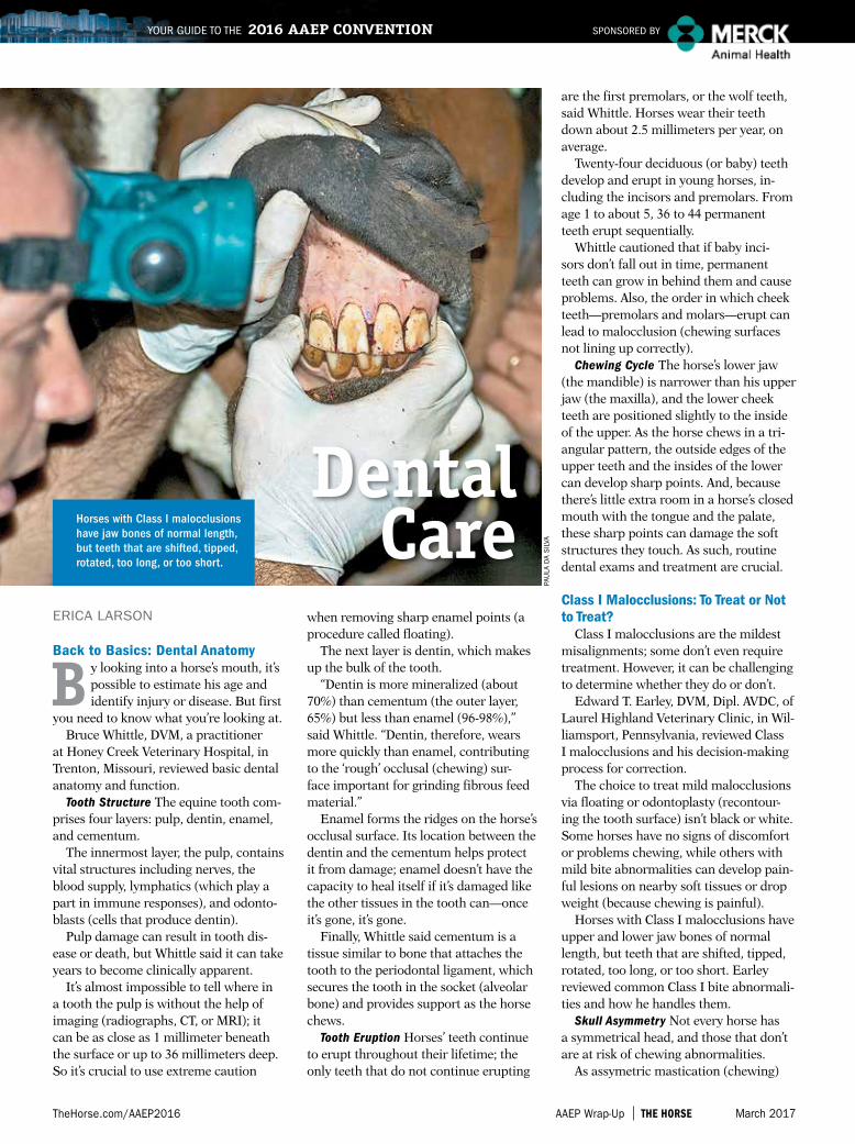
AAEP Wrap-Up The horse March 2017TheHorse.com/AAEP2016
ERICA LARSoN
Back to Basics: Dental Anatomy
b y looking into a horse’s mouth, it’s possible to estimate his age and identify injury or disease. But first
you need to know what you’re looking at.Bruce Whittle, DVM, a practitioner
at Honey Creek Veterinary Hospital, in Trenton, Missouri, reviewed basic dental anatomy and function.
Tooth Structure The equine tooth com-prises four layers: pulp, dentin, enamel, and cementum.
The innermost layer, the pulp, contains vital structures including nerves, the blood supply, lymphatics (which play a part in immune responses), and odonto-blasts (cells that produce dentin).
Pulp damage can result in tooth dis-ease or death, but Whittle said it can take years to become clinically apparent.
It’s almost impossible to tell where in a tooth the pulp is without the help of imaging (radiographs, CT, or MRI); it can be as close as 1 millimeter beneath the surface or up to 36 millimeters deep. So it’s crucial to use extreme caution
when removing sharp enamel points (a procedure called floating).
The next layer is dentin, which makes up the bulk of the tooth.
“Dentin is more mineralized (about 70%) than cementum (the outer layer, 65%) but less than enamel (96-98%),” said Whittle. “Dentin, therefore, wears more quickly than enamel, contributing to the ‘rough’ occlusal (chewing) sur-face important for grinding fibrous feed material.”
Enamel forms the ridges on the horse’s occlusal surface. Its location between the dentin and the cementum helps protect it from damage; enamel doesn’t have the capacity to heal itself if it’s damaged like the other tissues in the tooth can—once it’s gone, it’s gone.
Finally, Whittle said cementum is a tissue similar to bone that attaches the tooth to the periodontal ligament, which secures the tooth in the socket (alveolar bone) and provides support as the horse chews.
Tooth Eruption Horses’ teeth continue to erupt throughout their lifetime; the only teeth that do not continue erupting
are the first premolars, or the wolf teeth, said Whittle. Horses wear their teeth down about 2.5 millimeters per year, on average.
Twenty-four deciduous (or baby) teeth develop and erupt in young horses, in-cluding the incisors and premolars. From age 1 to about 5, 36 to 44 permanent teeth erupt sequentially.
Whittle cautioned that if baby inci-sors don’t fall out in time, permanent teeth can grow in behind them and cause problems. Also, the order in which cheek teeth—premolars and molars—erupt can lead to malocclusion (chewing surfaces not lining up correctly).
Chewing Cycle The horse’s lower jaw (the mandible) is narrower than his upper jaw (the maxilla), and the lower cheek teeth are positioned slightly to the inside of the upper. As the horse chews in a tri-angular pattern, the outside edges of the upper teeth and the insides of the lower can develop sharp points. And, because there’s little extra room in a horse’s closed mouth with the tongue and the palate, these sharp points can damage the soft structures they touch. As such, routine dental exams and treatment are crucial.
Class i Malocclusions: to treat or Not to treat?
Class I malocclusions are the mildest misalignments; some don’t even require treatment. However, it can be challenging to determine whether they do or don’t.
Edward T. Earley, DVM, Dipl. AVDC, of Laurel Highland Veterinary Clinic, in Wil-liamsport, Pennsylvania, reviewed Class I malocclusions and his decision-making process for correction.
The choice to treat mild malocclusions via floating or odontoplasty (recontour-ing the tooth surface) isn’t black or white. Some horses have no signs of discomfort or problems chewing, while others with mild bite abnormalities can develop pain-ful lesions on nearby soft tissues or drop weight (because chewing is painful).
Horses with Class I malocclusions have upper and lower jaw bones of normal length, but teeth that are shifted, tipped, rotated, too long, or too short. Earley reviewed common Class I bite abnormali-ties and how he handles them.
Skull Asymmetry Not every horse has a symmetrical head, and those that don’t are at risk of chewing abnormalities.
As assymetric mastication ( chewing)
PAU
LA D
A S
ILvA
Dental careHorses with Class I malocclusions
have jaw bones of normal length, but teeth that are shifted, tipped, rotated, too long, or too short.
Your GuiDE To THE 2016 aaeP convention SPonSorED bY
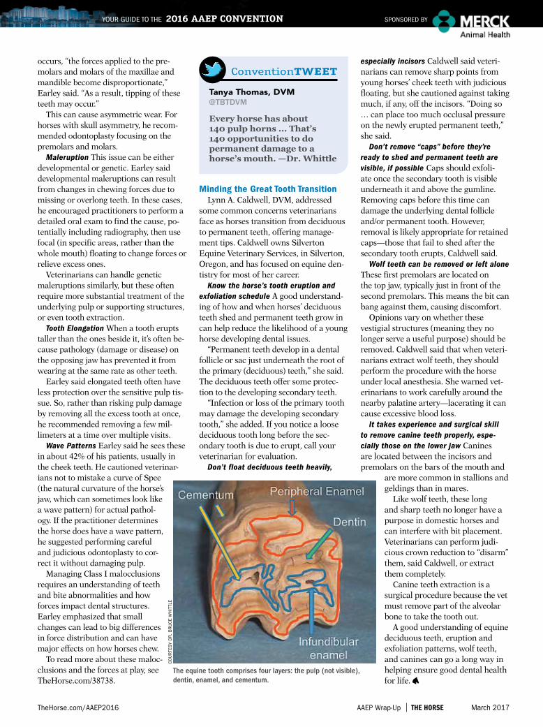
AAEP Wrap-Up The horse March 2017TheHorse.com/AAEP2016
occurs, “the forces applied to the pre-molars and molars of the maxillae and mandible become disproportionate,” Earley said. “As a result, tipping of these teeth may occur.”
This can cause asymmetric wear. For horses with skull asymmetry, he recom-mended odontoplasty focusing on the premolars and molars.
Maleruption This issue can be either developmental or genetic. Earley said developmental maleruptions can result from changes in chewing forces due to missing or overlong teeth. In these cases, he encouraged practitioners to perform a detailed oral exam to find the cause, po-tentially including radiography, then use focal (in specific areas, rather than the whole mouth) floating to change forces or relieve excess ones.
Veterinarians can handle genetic maleruptions similarly, but these often require more substantial treatment of the underlying pulp or supporting structures, or even tooth extraction.
Tooth Elongation When a tooth erupts taller than the ones beside it, it’s often be-cause pathology (damage or disease) on the opposing jaw has prevented it from wearing at the same rate as other teeth.
Earley said elongated teeth often have less protection over the sensitive pulp tis-sue. So, rather than risking pulp damage by removing all the excess tooth at once, he recommended removing a few mil-limeters at a time over multiple visits.
Wave Patterns Earley said he sees these in about 42% of his patients, usually in the cheek teeth. He cautioned veterinar-ians not to mistake a curve of Spee (the natural curvature of the horse’s jaw, which can sometimes look like a wave pattern) for actual pathol-ogy. If the practitioner determines the horse does have a wave pattern, he suggested performing careful and judicious odontoplasty to cor-rect it without damaging pulp.
Managing Class I malocclusions requires an understanding of teeth and bite abnormalities and how forces impact dental structures. Earley emphasized that small changes can lead to big differences in force distribution and can have major effects on how horses chew.
To read more about these maloc-clusions and the forces at play, see TheHorse.com/38738.
Minding the great tooth transitionLynn A. Caldwell, DVM, addressed
some common concerns veterinarians face as horses transition from deciduous to permanent teeth, offering manage-ment tips. Caldwell owns Silverton Equine Veterinary Services, in Silverton, Oregon, and has focused on equine den-tistry for most of her career.
Know the horse’s tooth eruption and exfoliation schedule A good understand-ing of how and when horses’ deciduous teeth shed and permanent teeth grow in can help reduce the likelihood of a young horse developing dental issues.
“Permanent teeth develop in a dental follicle or sac just underneath the root of the primary (deciduous) teeth,” she said. The deciduous teeth offer some protec-tion to the developing secondary teeth.
“Infection or loss of the primary tooth may damage the developing secondary tooth,” she added. If you notice a loose deciduous tooth long before the sec-ondary tooth is due to erupt, call your veterinarian for evaluation.
Don’t float deciduous teeth heavily,
especially incisors Caldwell said veteri-narians can remove sharp points from young horses’ cheek teeth with judicious floating, but she cautioned against taking much, if any, off the incisors. “Doing so … can place too much occlusal pressure on the newly erupted permanent teeth,” she said.
Don’t remove “caps” before they’re ready to shed and permanent teeth are visible, if possible Caps should exfoli-ate once the secondary tooth is visible underneath it and above the gumline. Removing caps before this time can damage the underlying dental follicle and/or permanent tooth. However, removal is likely appropriate for retained caps—those that fail to shed after the secondary tooth erupts, Caldwell said.
Wolf teeth can be removed or left alone These first premolars are located on the top jaw, typically just in front of the second premolars. This means the bit can bang against them, causing discomfort.
Opinions vary on whether these vestigial structures (meaning they no longer serve a useful purpose) should be removed. Caldwell said that when veteri-narians extract wolf teeth, they should perform the procedure with the horse under local anesthesia. She warned vet-erinarians to work carefully around the nearby palatine artery—lacerating it can cause excessive blood loss.
It takes experience and surgical skill to remove canine teeth properly, espe-cially those on the lower jaw Canines are located between the incisors and premolars on the bars of the mouth and
are more common in stallions and geldings than in mares.
Like wolf teeth, these long and sharp teeth no longer have a purpose in domestic horses and can interfere with bit placement. Veterinarians can perform judi-cious crown reduction to “disarm” them, said Caldwell, or extract them completely.
Canine teeth extraction is a surgical procedure because the vet must remove part of the alveolar bone to take the tooth out.
A good understanding of equine deciduous teeth, eruption and exfoliation patterns, wolf teeth, and canines can go a long way in helping ensure good dental health for life. h
conventiontweet
Tanya Thomas, DVM@TBTDVM
Every horse has about 140 pulp horns ... That’s 140 opportunities to do permanent damage to a horse’s mouth. —Dr. Whittle
Co
UR
TES
y D
R.
BR
UC
E w
HIT
TLE
The equine tooth comprises four layers: the pulp (not visible), dentin, enamel, and cementum.
Your GuiDE To THE 2016 aaeP convention SPonSorED bY
AAEP Wrap-Up The horse March 2017TheHorse.com/AAEP2016
Your GuiDE To THE 2016 aaeP convention SPonSorED bY
treating life-threatening hyperkalemia in horses
Potassium is an essential electrolyte for proper muscle function. However, high blood potassium levels cause a life- threatening condition known as hyperkalemia, seen most frequently in horses with a history of hyper-kalemic periodic paralysis or compromised kidney function.
A researcher at the Loomis Basin Equine Medical Center described treatment options for this condition. One is administering intravenous (IV) calcium borogluconate in a 5-liter bag of isotonic fluids. This fluid will not reduce plasma potassium concentrations, but it can help protect the heart muscle from their negative effects.
Other options include IV dextrose, beta agonists (more research is needed), and diuretics (but avoid use in dehydrated horses). Historically, researchers have recommended administering sodium bicarbonate to treat hyperkalemia, but clinical trials have shown it to offer little to no benefit.
See TheHorse.com/37848.
EEE, WNv vaccinations Do Not interfere With Disease testing
if a vaccine stimulates production of high enough levels of antibodies, diagnostic tests can’t distinguish between pre-existing infec-tion and an adequately vaccinated horse. researchers from Louisiana State university’s School of Veterinary Medicine conducted an experiment to determine if vaccines against Eastern equine encephalitis virus (EEEV) or West nile virus (WnV) resulted in this “test interference.”
“none of the tested samples had igM anti-body titers close to the 1:400 value cutoff for true disease,” said the authors. “This means the tested vaccine for EEEV and WnV does not interfere with the currently available blood test to diagnose EEEV and WnV.”
See TheHorse.com/38750.
A Potential Antibiotic for resistant synovial infections
intravenous regional limb perfusion (iVrLP) can deliver high levels of antibiotics to infected synovial structures, such as joints and tendon sheaths. but in the face of growing antibiotic resistance, selecting an effective antibiotic can be tricky. researchers at Mississippi State university’s College of Veterinary Medicine re-cently evaluated the efficacy of meropenem for iVrLP, because it has little pathogen resistance. Their results showed that the drug achieves synovial fluid concentrations that exceed the minimum inhibitor concentration (the lowest drug concentration needed to inhibit bacterial growth) for common equine pathogens for at least four hours following iVrLP. The authors said their study supports the clinical use of meropenem—but only when resistance is an issue and other antibiotics have failed.
See TheHorse.com/38751.
Blood in semen: how Much is too Much?
blood in a stallion’s ejaculate is associated with subfertility, but to what extent? research-ers from Texas A&M university evaluated ejaculate tainted with no blood, 5% blood, and 50% blood and determined that contamina-tion at least up to 5% had no effect on fertility. The 5% and 50% contaminated ejaculate, however, look the same, making it difficult to visually assess contamination. Veterinarians can consider pink-tinged ejaculate to be fertile, as this color typically has no more than 1% blood. See TheHorse.com/38752.
risk Factors Associated With R. equi PneumoniaResearchers from Texas A&M University’s College of Veterinary Medicine &
Biological Sciences looked at R. equi pneumonia risk in foals and found no associa-tion between disease and birth month, gestation duration, dam age, or whether the dam was biological or surrogate. Plasma antibody concentration (immunity from the dam’s colostrum, or first milk) at 24 hours of age was not a factor. Surprisingly, foals with concurrent respiratory illness were less likely to develop R. equi pneumonia.
Foals that received two liters of hyperimmune plasma (commonly adminstered to boost immune function) were less likely to develop R. equi pneumonia than foals that received 1 liter or none at all. See TheHorse.com/38747.
rabies vaccine Efficacy Might last longer than We thought
Researchers at the University of California, Davis, School of Veterinary Medicine, recently found that the rabies vaccine might protect horses from the fatal infection longer than previously thought.
The AAEP recommends every horse receive an annual rabies vaccine. That same vaccine only needs to be administered every three years in dogs and cats.
The team measured rabies virus neutralizing antibody (RVNA) levels in 48 healthy adult horses over the course of three years. They found that RVNA levels exceeded protective levels for the study duration in all horses that had been vaccinated previ-ously. While rabies remains an annual vaccination, this information might benefit owners of horses that have had adverse reactions to the vaccine in the past.
See TheHorse.com/38749.
Paint-on Aseptic skin Prep is Easy, Effective, and Cost-Efficient
Aseptic skin preparation—cleaning an area to remove dirt and microorganisms—is important to prevent infection during veterinary procedures. New paint-on products that don’t requiring scrubbing (called passive) are gaining popularity. But are they as effective? A student at the University of Florida College of Veterinary Medicine compared a passive preparation to an active scrub preparation and found no difference in the number of skin-associated bac-teria after each technique. Overall, passive skin prep requires less time and money and fewer supplies. See TheHorse.com/38746.
Find full stories at TheHorse.com/AAEP2016
research shorts
ALEx
AND
RA
BEC
kS
TETT
Traditionally, most practitioners have scrubbed the skin to prep it.

1 UC Davis (Nicola Pusterla) & Merck Animal Health. Infectious Upper Respiratory Surveillance Program. Ongoing Research 2008-present.
2 Townsend HGG. Onset of protection against live-virus equine influenza challenge following vaccination naive horses with a modified-live vaccine. Unpublished data.
Visit us online to learn more about Merck Animal Health and the equine products and programs that help you keep horses healthy.
The Science of ExceptionalStop flu where it starts.
The Science of Healthier Animals
2 Giralda Farms • Madison, NJ 07940 • merck-animal-health-usa.com • 800-521-5767Copyright © 2016 Intervet Inc., d/b/a/ Merck Animal Health, a subsidiary of Merck & Co., Inc.All rights reserved. 3526 EQ-FluAvert-PM
Trust Flu Avert® I.N. for superior protection against relevant flu strains threatening the U.S. horse population.1
• Just ONE dose required - An ideal first flu vaccine for young horses - Provides protection at the site of infection
and long-lasting immunity• Proven safe and effective - Intranasal application leaves no risk for
injection site reactions to interfere with training or competing
• Rapid onset of protection - Onset of flu protection within five to
seven days following one dose – no matter when your horse was last vaccinated for flu.2
Now that’s exceptional.
3526_FluAvertIN_8125x10875.indd 1 1/4/17 12:56 PM


