Embed Size (px)
Citation preview
INVESTIGATION
Convergent Evolution of Calcineurin PathwayRoles in Thermotolerance and Virulencein Candida glabrataYing-Lien Chen,* Jay H. Konieczka,† Deborah J. Springer,* Samantha E. Bowen,* Jing Zhang,‡
Fitz Gerald S. Silao,§ Alice Alma C. Bungay,§,** Ursela G. Bigol,†† Marilou G. Nicolas,**Soman N. Abraham,*,‡‡,§§ Dawn A. Thompson,† Aviv Regev,†,†† and Joseph Heitman*,1*Department of Molecular Genetics and Microbiology, ‡‡Pathology, and §§Immunology, Duke University Medical Center,Durham, North Carolina 27710, †The Broad Institute of MIT & Harvard, Cambridge, Massachusetts 02142, ††Howard HughesMedical Institute, Department of Biology, Massachusetts Institute of Technology, Cambridge, Massachusetts 02142,‡Department of Chemistry, Duke University, Durham, North Carolina 27710, §Department of Microbiology and Parasitology,University of Perpetual Help–Dr. Jose G. Tamayo Medical University, Biñan, Laguna, 4024 Philippines, **National Institutesof Health, University of the Philippines, Manila, Philippines, and ††Environment and Biotechnology Division, IndustrialTechnology Development Institute, Department of Science and Technology, Bicutan, Taguig City, 1631 Philippines
ABSTRACT Candida glabrata is an emerging human fungal pathogen that is frequently drug tolerant, resulting indifficulties in treatment and a higher mortality in immunocompromised patients. The calcium-activated proteinphosphatase calcineurin plays critical roles in controlling drug tolerance, hyphal growth, and virulence in diversefungal pathogens via distinct mechanisms involving survival in serum or growth at host temperature (37� andhigher). Here, we comprehensively studied the calcineurin signaling cascade in C. glabrata and found novel anduncharacterized functions of calcineurin and its downstream target Crz1 in governing thermotolerance, intracellulararchitecture, and pathogenesis in murine ocular, urinary tract, and systemic infections. This represents a secondindependent origin of a role for calcineurin in thermotolerant growth of a major human fungal pathogen, distinctfrom that which arose independently inCryptococcus neoformans. Calcineurin also promotes survival ofC. glabratain serum via mechanisms distinct from C. albicans and thereby enables establishment of tissue colonization ina murine systemic infection model. To understand calcineurin signaling in detail, we performed global transcriptprofiling analysis and identified calcineurin- and Crz1-dependent genes in C. glabrata involved in cell wall bio-synthesis, heat shock responses, and calcineurin function. Regulators of calcineurin (RCN) are a novel family ofcalcineurin modifiers, and two members of this family were identified in C. glabrata: Rcn1 and Rcn2. Our studiesdemonstrate that Rcn2 expression is controlled by calcineurin and Crz1 to function as a feedback inhibitor ofcalcineurin in a circuit required for calcium tolerance in C. glabrata. In contrast, the calcineurin regulator Rcn1activates calcineurin signaling. Interestingly, neither Rcn1 nor Rcn2 is required for virulence in a murine systemicinfection model. Taken together, our findings show that calcineurin signaling plays critical roles in thermotoleranceand virulence, and that Rcn1 and Rcn2 have opposing functions in controlling calcineurin signaling in C. glabrata.
KEYWORDS
phosphatasecalciumcalmodulinCrz1Rcn1Rcn2thermotolerancecell wall integrityER stressdrug tolerancepH homeostasisurinary tractinfection
ocular infectionvirulence
Candida glabrata is an emerging human fungal pathogen. Moststrains have reduced antifungal drug susceptibility, thus making treat-ment challenging (Lagrotteria et al. 2007; Pfaller et al. 2011a). Thepoor susceptibility of C. glabrata to antifungal drugs contributes to thehigh mortality rate (�50%) associated with C. glabrata candidemia(Klevay et al. 2009). Resistance to echinocandins and azoles was mostprevalent among nosocomial bloodstream infection (BSI) isolates ofC. glabrata compared with other Candida species (Pfaller et al. 2011a).Although C. albicans accounts for 48% of Candida BSIs, C. glabrataranked second, accounting for 18% of infections across multiple
Copyright © 2012 Chen et al.doi: 10.1534/g3.112.002279Manuscript received February 24, 2012; accepted for publication April 2, 2012This is an open-access article distributed under the terms of the CreativeCommons Attribution Unported License (http://creativecommons.org/licenses/by/3.0/), which permits unrestricted use, distribution, and reproduction in anymedium, provided the original work is properly cited.Supporting information is available online at http://www.g3journal.org/lookup/suppl/doi:10.1534/g3.112.002279/-/DC1Arrays have been submitted to the GEO database at NCBI as series GSE31167.1Corresponding author: Department of Molecular Genetics and Microbiology, DukeUniversity Medical Center, Durham, NC 27710. E-mail: [email protected]
Volume 2 | June 2012 | 675
geographic regions, including Asia-Pacific, Europe, Latin America,and North America (Pfaller et al. 2011b). Besides BSI, Candida speciesalso cause urinary tract infections (UTI). Fungal UTIs are often causedby Candida species, which account for �10% of all nosocomial UTIsrelated to indwelling catheters, with C. glabrata accounting for approx-imately 15% of all Candida isolates (Kauffman et al. 2000; Lundstromand Sobel 2001). Surprisingly, a study performed by Phillips andKarlowicz (1997) showed that 42% of hospital-acquired urinary tractinfections in neonatal intensive care units were caused by Candidaspecies. For Candida ocular infections, C. glabrata was previouslyrarely seen; however, several recent studies report that C. glabratainfects human corneas, especially in patients undergoing keratoplasty(Caldwell et al. 2009; Djalilian et al. 2001; Kitzmann et al. 2009; Leeet al. 2011; Tappeiner et al. 2009). The pathogenic mechanisms bywhich C. glabrata causes ocular infections have not been reported.
Calcineurin is a calcium/calmodulin-dependent serine/threonine-specific protein phosphatase that comprises a catalytic A (Cna1) anda regulatory B calcium-binding subunit (Cnb1). Upon stimulation withcalcium, calmodulin associates with the calcineurin AB heterodimer,stimulating phosphatase activity and converting signals to variousoutputs by dephosphorylating its downstream targets. Upon dephos-phorylation by activated calcineurin, the transcription factor Crz1 infungi or nuclear factor of activated T cells (NFAT) in mammals migratesto the nucleus to regulate gene expression. Because active calcineurin isan AB heterodimer, the loss of Cnb1 often results in destabilization ofCna1 (Chen et al. 2010a). Calcineurin is essential for growth at elevatedtemperatures in the human fungal pathogen Cryptococcus neoformans(Odom et al. 1997) and the protozoan parasite Leishmania major(Naderer et al. 2011), but it has not been demonstrated to have a rolein controlling thermotolerance of ascomycetes, including Candida spe-cies. The intracellular architecture defects of C. neoformans and L. majorcalcineurin mutants upon thermal stress have not been investigated. InC. glabrata, calcineurin and Crz1 have been demonstrated to play func-tions in antifungal drug tolerance, cell wall integrity, and virulence ina murine systemic infection model (Miyazaki et al. 2010). However, themechanism for C. glabrata calcineurin and Crz1 requirement in serumsurvival and virulence in other murine infection models remains elusive.
The calcineurin- and/or Crz1-regulated transcriptome has beeninvestigated in Saccharomyces cerevisiae, C. albicans, Aspergillus fumiga-tus, and the rice blast fungal pathogen Magnaporthe oryzae (Karababaet al. 2006; Kim et al. 2010; Malavazi et al. 2009; Yoshimoto et al.2002). The regulation pattern of the transcriptome is diverged amongthe four species. Because C. glabrata calcineurin and crz1 mutantsexhibited novel phenotypes distinct from these species in terms ofhypersensitivity to thermal stress, ER stress, and antifungal drugs,microarray analyses to explore the targets of calcineurin and Crz1 inC. glabrata will extend our understanding of calcineurin signaling.
Regulators of calcineurin (RCN) are a novel family of calcineurinregulators found in eukaryotic cells. In S. cerevisiae, two regulators ofcalcineurin (Rcn1 and Rcn2) have been demonstrated to control cal-cineurin signaling. In C. albicans, the function of Rcn1 has beenassociated with calcineurin signaling (Reedy et al. 2009). In C. glab-rata, the functions of Rcn1 and Rcn2 in controlling calcineurin sig-naling were recently reported (Miyazaki et al. 2011a). C. glabrata Rcn1displayed both stimulatory and inhibitory effects, whereas Rcn2showed only inhibitory activities on calcineurin signaling (Miyazakiet al. 2011a). Interestingly, Rcn1 is required for micafungin and flu-conazole tolerance in addition to ER stress induced by tunicamycin(Miyazaki et al. 2011a). However, the roles of Rcn1 and Rcn2 intemperature sensitivity, pH response, and virulence remain unclear.In mammals, the homolog of S. cerevisiae Rcn1 is named DSCR1/
Rcan1/calcipressin and is overexpressed in the brain of patients withDown’s syndrome (Rothermel et al. 2003). In addition, Baek et al.(2009) demonstrated that DSCR1, a negative regulator of calcineurinlocated on chromosome 21 and thus present in three copies inpatients with Down’s syndrome, has antiangiogenic activity and inhib-its tumor formation.
In this study, we comprehensively studied the roles of the calcineurinsignaling cascade in stress responses, antifungal drug tolerance, andpathogenesis of C. glabrata. We demonstrated that C. glabrata calci-neurin (Cna1 and Cnb1) and/or Crz1 are required for thermotolerance,ER stress, pH homeostasis, and virulence in murine ocular, urinarytract, and systemic infection models. In addition, global transcript pro-filing analysis revealed 30 calcineurin and Crz1-activated targets, whichexplain, at least in part, the requirement of calcineurin signaling in cellwall integrity, thermotolerance, and pathogenesis. Furthermore, wedemonstrated that a novel regulator of calcineurin 2 (Rcn2) is a down-stream target of calcineurin and Crz1 and acts as a feedback inhibitor ofcalcineurin signaling in response to calcium, whereas Rcn1 functions asa positive regulator of calcineurin signaling.
MATERIALS AND METHODS
Ethics statementAnimals studies conducted in the Division of Laboratory AnimalResources (DLAR) facilities at Duke University Medical Center (DUMC)were handled with good practice as defined by the United States AnimalWelfare Act and in full compliance with the guidelines of the DUMCInstitutional Animal Care and Use Committee (IACUC). The murinesystemic and urinary tract infection models were reviewed and approvedby the DUMC IACUC under protocol numbers A238-09-08 and A219-08-08, respectively.
Murine ocular infection studies conducted at the animal housefacility of the Department of Microbiology and Parasitology, Univer-sity of Perpetual Help–Dr. Jose G. Tamayo Medical University (UPH-DJGTMU) were performed in accordance with the ARVO Statementfor the Use of Animals in Ophthalmic and Vision Research, the UnitedStates Animal Welfare Act, and the Republic of the Philippines AnimalWelfare Act of 1998 (RA No. 8485), and in full conformity with theguidelines set forth in the UPH-DJGTMU research manual. The pro-tocol was formally approved by the UPH-DJGTMU institutional re-view board after review by the Regional Institute for Tropical MedicineInstitutional Animal Care and Use Committee (RITM-IACUC) underresearch protocol No. 010. All animal infection experiments were con-ducted by properly trained personnel, including licensed veterinarians.
Yeast strains, media, and chemicalsYeast strains used in this study are listed in Table 1. YPD (1% yeastextract, 2% peptone, 2% glucose) liquid medium and agar (2%) andSC (6.7 g yeast nitrogen base without amino acids, 1 · amino acidstock, 20 g glucose, 20 g agar in 1 liter) media were used in this study.YPD medium containing 100 mg/ml nourseothricin was used to selecttransformants. YPD medium containing 150 mM HEPES buffered atpH 2, 5, 8, or 9 was used to study pH tolerance. FK506 (AstellasPharma Inc.), cyclosporin A (CsA; LC Laboratories), sodium dodecylsulfate (SDS; Fisher), fetal bovine serum (Invitrogen), calcofluor white(CFW, fluorescent brightener 28; Sigma), Congo red (Sigma), fluco-nazole (Bedford Laboratories), posaconazole (Sequoia Research Prod-ucts Ltd.), ketoconazole (Sigma), voriconazole (Sigma), caspofungin(Merck), micafungin (Astellas Pharma Inc.), anidulafungin (PfizerInc.), tunicamycin (Sigma), and dithiothreitol (DTT; Sigma) wereadded to the media at the concentrations indicated.
676 | Y.-L. Chen et al.
Identification of C. glabrata gene orthologsThe C. glabrata orthologs of the genes encoding C. albicans calci-neurin subunits Cna1 and Cnb1 and the calcineurin target Crz1 wereidentified by reciprocal BLAST searches between the two species, withthe reciprocal best BLAST hit orthologs in C. glabrata being the CNA1(CAGL0L11110g), CNB1 (CAGL0L00605g), and CRZ1 (CAGL0M06831g)genes. C. glabrata YPS5 (CAGL0E01771g, putative aspartyl prote-ase) and RCN2 (CAGL0J04158g, regulator of calcineurin 2) wereidentified by reciprocal BLAST searches using the S. cerevisiae ge-nome and genes. The C. glabrata ortholog of C. albicans RCN1(regulator of calcineurin 1) was identified by reciprocal BLASTsearches between C. glabrata and C. albicans, identifying the recip-rocal best BLAST hit as C. glabrata RCN1 (CAGL0E06248).
Gene disruptions in C. glabrata
All deletion strains were generated from the prototrophic CBS138(ATCC2001) background using the SAT1 flipper (Reuss et al. 2004).All primers used in strain construction are listed in Table S1. For theCNA1 gene disruption, approximately 1 kb of the 59 (amplified withprimers JC49/JC50, Table S1) and 39 (amplified with primers JC51/JC52) noncoding regions (NCR) of the CNA1ORF were PCR-amplifiedfrom genomic DNA of the wild-type strain CBS138. The 4.2 kbSAT1 flipper sequence was amplified from plasmid pSFS2A (Reusset al. 2004) with primers JC17/JC18. The three PCR products weretreated with ExoSAP-IT (USB Corp.) to remove contaminating pri-mers and dNTPs and then combined in a 1:3:1 molar ratio (59CNA1NCR: SAT1 flipper: 39 CNA1NCR) to generate the disruption alleleby overlap PCR using flanking primers JC49/JC52, resulting in an�6.2 kb 59CNA1NCR-SAT1 flipper-39CNA1NCR CNA1 disruption al-lele. The CNA1 gene was disrupted in the wild-type strain CBS138 bytransformation with 0.2�1.0 mg of gel-purified disruption cassette
DNA extracted with the Frozen-EZ Yeast Transformation Kit(Zymo Research). Two independent nourseothricin-resistant cna1mutants (YC67 and YC98, Table 1) were obtained from two sep-arate transformations.
A similar approach was employed to disrupt the CNB1 and CRZ1genes, with �1 kb of the 59 and 39 noncoding regions used for ho-mologous recombination. To generate the �6 kb cnb1 disruption al-lele, the overlap PCR DNA products 59 CNB1NCR (amplified withprimers JC134/JC135), SAT1 flipper (amplified with primers JC17/JC18), and 39 CNB1NCR (amplified with primers JC136/JC137) weremixed in a 1:3:1 molar ratio and amplified with primers JC138/JC139(�100 bp closer to the CNB1 ORF compared with JC134/JC137, re-spectively, reserving primers JC134/JC137 for further integration con-firmation). Two independent nourseothricin-resistant cnb1 mutants(YC191 and YC193, Table 1) derived from two separate transforma-tions were obtained. To generate the �6 kb crz1 disruption allele, 59CRZ1NCR (amplified with primers JC142/JC143), SAT1 flipper (ampli-fied with primers JC17/JC18), and 39 CRZ1NCR (amplified with pri-mers JC144/JC145) were combined and amplified with primers JC146/JC147 (�100 bp closer to the CRZ1 ORF compared with JC142/JC145,respectively). Two independent nourseothricin-resistant crz1 mutants(YC267 and YC182, Table 1) derived from two separate transforma-tions were obtained.
To disrupt the RCN1 gene, 59 RCN1NCR and 39 RCN1NCR wereamplified with primers JC459/JC460 and JC461/JC462, respectively.JC463/JC464 primers were used to amplify the RCN1 disruption allelevia overlap PCR including three fragments: 59 RCN1NCR, SAT1 flipper,and 39 RCN1NCR. Two independent rcn1 mutants (YC553 and YC556)were obtained. To disrupt the RCN2 gene, 59 RCN2NCR and 39 RCN2NCR
were amplified with primers JC441/JC442 and JC443/JC444, respectively.JC445/JC446 primers were used to amplify the RCN2 disruption allele via
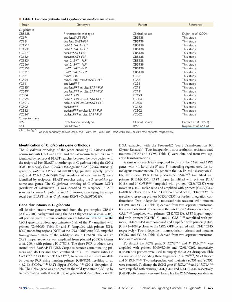
n Table 1 Candida glabrata and Cryptococcus neoformans strains
Strain Genotype Parent Reference
C. glabrataCBS138 Prototrophic wild-type Clinical isolate Dujon et al. (2004)YC67a cna1D::SAT1-FLP CBS138 This studyYC98a cna1D:: SAT1-FLP CBS138 This studyYC191b cnb1D::SAT1-FLP CBS138 This studyYC193b cnb1D::SAT1-FLP CBS138 This studyYC267c crz1D::SAT1-FLP CBS138 This studyYC182c crz1D::SAT1-FLP CBS138 This studyYC553d rcn1D::SAT1-FLP CBS138 This studyYC556d rcn1D::SAT1-FLP CBS138 This studyYC525e rcn2D::SAT1-FLP CBS138 This studyYC531e rcn2D::SAT1-FLP CBS138 This studyYC581 rcn2D::FRT YC531 This studyYC594 rcn2D::FRT rcn1D::SAT1-FLP YC581 This studyYC111 cna1D::FRT YC98 This studyYC535f cna1D::FRT rcn2D::SAT1-FLP YC111 This studyYC549f cna1D::FRT rcn2D::SAT1-FLP YC111 This studyYC504 cnb1D::FRT YC193 This studyYC599g cnb1D::FRT rcn2D::SAT1-FLP YC504 This studyYC601g cnb1D::FRT rcn2D::SAT1-FLP YC504 This studyYC502 crz1D::FRT YC182 This studyYC532h crz1D::FRT rcn2D::SAT1-FLP YC502 This studyYC534h crz1D::FRT rcn2D::SAT1-FLP YC502 This studyC. neoformansH99 Prototrophic wild-type Clinical isolate Perfect et al. (1993)KK1 cna1D::NAT H99 Kojima et al. (2006)a,b,c,d,e,f,g,h
Two independently derived cna1, cnb1, crz1, rcn1, rcn2, cna1 rcn2, cnb1 rcn2, or crz1 rcn2 mutants, respectively.
Volume 2 June 2012 | Calcineurin Signaling Cascade in C. glabrata | 677
overlap PCR including three fragments: 59 RCN2NCR, SAT1 flipper, and39 RCN2NCR. Two independent rcn2 mutants (YC525 and YC531) wereobtained.
To obtain the rcn1 rcn2 double mutant, we disrupted the RCN1gene in the rcn2 mutant background (YC581, the SAT1 flipper wasremoved via culturing YC531 in YPD medium and replicating ontonourseothricin-containing medium to confirm the loss of SAT1 flip-per). To obtain the cna1 rcn2 (YC535 and YC549), cnb1 rcn2 (YC599and YC601), and crz1 rcn2 (YC532 and YC534) double mutants, wedisrupted the RCN2 gene in SAT1 flipper-free (methods describedabove) cna1 (YC111), cnb1 (YC504), and crz1 (YC502) mutants,respectively.
Transmission electron microscopyTransmission electron microscopy (TEM) of C. glabrata was accom-plished as follows. Cells were grown overnight in YPD at 24� andwashed twice with dH2O. Then 0.1 OD600 of cells (in 100 ml) wasspread on YPD agar plates and incubated for 24 hr at 24�, 37�, and40�. Cells were collected from agar plates by washing in 0.2 M sodiumcacodylate buffer (pH¼ 6.8), collected by centrifugation (�4,000 rpm,3 min in a table-top centrifuge), resuspended in 2% glutaraldehydeplus 0.05% malachite green oxalate in 0.1 M sodium cacodylate buffer,and incubated at 4� for 2 days. Fixed cells were collected by centrifu-gation, resuspended, washed with 0.1 M sodium cacodylate buffer,centrifuged, supernatant removed, and post-fixed with 0.8% K3Fe(CN)6, 1% OsO4, 0.1 M sodium cacodylate for 2 hr at room temper-ature. The cells were washed twice with 0.1 M sodium cacodylatebuffer and stained with 1% tannic acid for 1 hr at room temperature.Then cells were washed with 0.1% sodium cacodylate buffer for 5 minfollowed by two washes in dH20 for 5–10 min each and stained with1% uranyl acetate in water overnight at 4�. Samples were washed inmolecular-grade distilled water, embedded in an agarose pellet, andprepared for embedding. The cured peg was trimmed, sectioned, andmounted on copper grids. Grids were post-stained prior to viewing.Sections were viewed and imaged with a Philips/FEI CM 12 Trans-mission EM (FEI Company, Hillsboro, OR) with Advanced Micros-copy Techniques Corp. (AMT) 2K · 2K digital camera (Danvers,MA) at the Duke University Department of Pathology.
Fluorescence microscopyCells were grown overnight in YPD at 24� and washed twice withdH2O. Then 0.1 OD600 of cells (in 100 ml) was spread on YPD agarplates and incubated for 24 hr at 24� and 37�. The Alexa Fluor 488phalloidin (Cat #A12379; Life Technologies) staining of actin wasperformed based on the protocol described by Adams and Pringle(1991) with minor modifications. In brief, cells were spun and resus-pended in 3.7% (vol/vol) formaldehyde in PBS for 1 hr, washed, andresuspended in PBS (100 ml). Cells were then stained with 20 ml of 6.6mM (or 0.2 U/ml) phalloidin stock for 1 hr in the dark. Stained cellswere washed five times with PBS, resuspended in 100 ml PBS in theabsence of mounting medium, and observed using the standard greenfluorescence filter set.
Time-kill curve for strains exposed to fluconazole,micafungin, or serumCells were grown overnight at 24�, washed twice with dH2O, countedwith a hemocytometer, and then 5 · 106 cells were added to 5 ml offresh YPD medium 6 fluconazole/micafungin or 5 ml of 100% fetalbovine serum to achieve 106 cells/ml. Cells were cultured at 24� withshaking at 250 rpm. The cells surviving after 0, 3, 6, 9, and 24 hr were
serially diluted onto YPD medium, and CFUs were counted after 48hr of incubation at 24�. The experiments were performed in triplicate,and data were plotted using Prism 5.03.
Culture growth, harvesting, and total RNA extractionsfor microarray experimentsStrains were grown overnight at 24�, washed twice with dH2O, dilutedto 0.2 OD600/ml in YPD, and incubated for 3 hr at 24�. For wild-typestrains, cells in log-phase were diluted to 0.2 OD600/ml (10 ml) in YPDin the presence or absence of FK506 (1 mg/ml) while cna1 (YC98) andcrz1 (YC182) mutants were diluted to 0.2 OD600/ml (10 ml) in YPD.Following 3 hr of incubation at 37� with shaking at 250 rpm, the 10 mlcultures were immediately added to 15 ml methanol (60%) chilled ina dry ice-ethanol bath to stop cellular processes and RNase activity.Cells were pelleted at 3000 rpm at 24�, flash frozen with liquid N2,and stored at 280� prior to total RNA extraction. The total RNAswere extracted using the RNeasy Mini Kit (Qiagen). RNA quality wasassessed with the RNA 6000 Nano Kit of the Agilent Bioanalyzer 2100to ensure RNA Integrity Number (RIN) scores $ 7.
Probe preparation, microarray hybridization,and data analysisTotal RNA samples were reverse-transcribed to cDNA and labeledwith either Cy3 or Cy5, according to a modification of the protocoldescribed by Wapinski et al. (2010). We employed a wheel hybrid-ization design to compare all samples to each other with dye-swaps,for four comparisons per sample on 10 microarrays per biologicalreplicate. The entire hybridization scheme was repeated for two bi-ological replicates. After hybridization and washing per the manufac-turer’s instructions, arrays were scanned using an Agilent scanner andanalyzed with Agilent’s Feature Extraction software V10.5.1. The me-dian intensities of probes corresponding to a specific ORF were usedto estimate the expression values for each gene. We then fit linearmodels to these gene expression data using the limma library of the RBioconductor Package (Gentleman et al. 2005), which computes foldchange, indicates the direction and quantity of the differential geneexpression between the samples, and provides summary statistics, in-cluding T- and B-statistics and an adjusted P value that takes intoaccount the false discovery rate. For each comparison in each study,Q-values were computed using the R package. Microarray results havebeen deposited at NCBI Gene Expression Omnibus (http://www.ncbi.nlm.nih.gov/geo/query/acc.cgi?acc=GSE31167).
Quantitative determination of expressionby real-time RT-PCRDNase I (Turbo DNA-free; Ambion) was used to eliminate genomicDNA contamination. One microgram of DNA-free total RNAs wasreverse-transcribed to cDNA by the Affinity Script qPCR cDNASynthesis Kit (Agilent). PCR reactions of 25 ml included 10 ng cDNA(in 10 ml), 12.5 ml of 2· qPCR master mix (Brilliant SYBR Green Kit;Agilent), 0.5 ml of 5 mM forward primer, 0.5 ml of 5 mM reverseprimer, 1.125 ml of nuclease-free H2O, and 0.375 ml of ROX dye.Quantitative PCR conditions were the following: 95�/10 min (dena-turation); 95�/15 sec, 60�/1 min (40·, cycling stage); 95�/15 sec, 60�/1 min, 95�/15 sec (melting curve). Primers for probes were designedusing Primer3 (http://frodo.wi.mit.edu/primer3/) and are listed in Ta-ble S1. The ABI PRISM 7900HT machine and StepOne v2.1 (AppliedBiosystems) were used to determine ΔΔCt and relative quantity (RQ).The bar graphs of ACT1 normalized RQ compared with the wild-type(CBS138) were created with Prism 5.03.
678 | Y.-L. Chen et al.
Mouse studies
Urinary tract infection model: Six- to eight-week-old female C3H/HeJ mice (stock #000659, n ¼ 5 for each group) and C3H/HeOuJ(stock #000635, n ¼ 8 for the group) from the Jackson Laboratorywere used in this study. C. glabrata strains were grown in 5 ml SD +CAA medium (6.7 g yeast nitrogen base minus amino acids, 6 gcasamino acids, 20 g glucose in 1 L dH2O) overnight at 24�. Cultureswere washed twice with 10 ml of phosphate buffered saline (PBS) anddiluted to yield an infection inocula of 109 cells/ml after hemocytom-eter counting. Prior to infection, mice were anesthetized with i.p. in-jection of 100 ml of 5-fold diluted pentobarbital (final concentration of10 mg/ml in PBS). Thirty microliters (3 · 107 cells) were used to infectmice via the urinary tract for 15 sec aided by polyethylene cathetertubing (BD catalog #427400) and syringe (BD catalog #309659) with30 G · 1/2 needle (BD catalog #305106). Bladder and kidney tissuesamples were harvested on day 7 and homogenized with 5 ml PBS for5 s at 17,500 rpm (for kidneys) or at 24,000 rpm (for bladder) (PowerGen 500; Fisher Scientific). Tissue homogenates were serially dilutedand 100 ml was plated onto YPD solid medium. The plates wereincubated at 24� for 72 hr to determine CFUs per organ. Appropriatedilutions of the inocula were plated onto YPD at 24� for 48 hr toconfirm cell viability. All experimental procedures were carried outaccording to NIH guidelines and Duke IACUC protocols for theethical treatment of animals.
Murine systemic infection model: Five- to six-week-old male CD1mice from the Jackson Laboratory (n ¼ 10 for each group) wereutilized in this study. C. glabrata strains were grown in 5 ml liquidYPD medium overnight at 24�. Cultures were washed twice with 10 mlof phosphate buffered saline (PBS), and the cells were then resus-pended in 2 ml of PBS. Cells were counted with a hemocytometerand resuspended in an appropriate volume of PBS to obtain an in-fection inocula concentration of 2 · 108 cells/ml. Two hundred micro-liters (4 · 107 cells) were used to infect mice by lateral tail veininjection. Appropriate dilutions of the cells were plated onto YPD solidmedium and incubated at 24� for 48 hr to confirm cell viability. C.glabrata–infected mice were sacrificed and dissected on day 7 post-infection. The kidney and spleen tissues were removed, weighed, trans-ferred to a 15 ml Falcon tube filled with 5 ml PBS, and homogenizedfor 5 s at 19,000 rpm (IKA T25; Cole-Parmer). Tissue homogenateswere serially diluted, and 100 ml was plated onto YPD solid medium.The plates were incubated at 24� for 48 hr to determine CFUs per gramof organ. The identity of organ-recovered colonies was confirmed byPCR. All experimental procedures were carried out according to NIHguidelines and Duke IACUC protocols for the ethical treatment ofanimals.
Murine ocular infection model: Candida strains were grown in YPDovernight at 25�. Cultures were washed three times with sterile PBS(pH 7.4). Cells were diluted to a concentration of 106 CFU/5 ml. Theconcentration was determined by using the spectrophotometer opticaldensity reading at a wavelength of 600 nm and multiplying it bya conversion factor of 1 OD600, equivalent to 3 · 107 cells/ml. In-oculum concentration was verified by plating on YPD for 48 hr at 25�.
For murine ocular infection, six- to eight-week-old outbred ICRmice (20–28 g; Research Institute for Tropical Medicine, Alabang,Philippines) were used in the experiment in accordance with theARVO Statement for the Use of Animals in Ophthalmic and VisionResearch. The previously described keratomycosis protocol (Chenet al. 2011) was used for Candida infection with minor modifications
and was approved by the University of Perpetual Help InstitutionalReview Board. Before the infection procedure, mice were anesthetizedby intravenous injection of Zoletil 50 (10–15 mg/kg body weight;Virac, Australia) followed by topical application of proparacaine hy-drochloride ophthalmic solution (Alcaine; Alcon-Couvreur, Belgium)to the eyes. Once animals were anesthetized, the right eye was super-ficially scarified in a grid-pattern by a sterile 25-gauge hypodermicneedle, and 5 ml of Candida solution (106 CFU) was placed into eacheye. The inoculum was distributed uniformly by rubbing the eye forfew seconds with the eyelid. A mock-infection experiment was per-formed using sterile PBS as a control. Disease severity of fungal ker-atitis was assessed for 8 days with the aid of a dissecting microscope.In this procedure, corneal involvement was assessed and scoredaccording to three parameters: (i) area of opacity, (ii) density of opac-ity, and (iii) surface regularity. A grade of 0 to 4 was assigned based oneach of these criteria to yield a maximum score of 12.
Statistical analysisStatistical analysis was conducted using Prism 5.03 software (Graph-Pad, La Jolla, CA). The significance of differences in fungal burdenwas determined using one-way ANOVA and Dunnett’s multiple com-parison tests. For optimal serum growth and real-time RT-PCR, thesignificance of differences was determined by one-way ANOVA andBonferroni’s multiple comparison tests. For shrunken cell percentageand murine ocular infection, the Student unpaired t-test was used todetermine significance. P , 0.05 was considered significant.
RESULTS AND DISCUSSION
The calcineurin pathway plays a critical rolein thermotolerance in C. glabrata
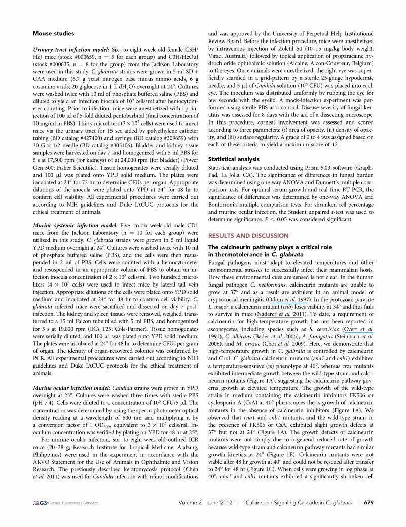
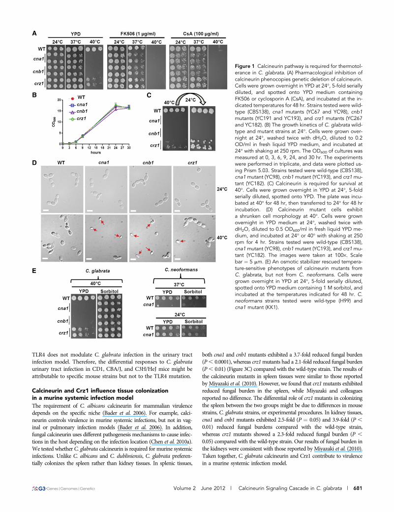
Fungal pathogens must adapt to elevated temperatures and otherenvironmental stresses to successfully infect their mammalian hosts.How these environmental cues are sensed is not clear. In the humanfungal pathogen C. neoformans, calcineurin mutants are unable togrow at 37� and as a result are avirulent in an animal model ofcryptococcal meningitis (Odom et al. 1997). In the protozoan parasiteL. major, a calcineurin mutant (cnb) loses viability at 34� and thus failsto survive in mice (Naderer et al. 2011). To date, a requirement ofcalcineurin for high-temperature growth has not been reported inascomycetes, including species such as S. cerevisiae (Cyert et al.1991), C. albicans (Bader et al. 2006), A. fumigatus (Steinbach et al.2006), and M. oryzae (Choi et al. 2009). Here, we demonstrate thathigh-temperature growth in C. glabrata is controlled by calcineurinand Crz1. C. glabrata calcineurin mutants (cna1 and cnb1) exhibiteda temperature-sensitive (ts) phenotype at 40�, whereas crz1 mutantsexhibited intermediate growth between the wild-type strain and calci-neurin mutants (Figure 1A), suggesting the calcineurin pathway gov-erns growth at elevated temperature. The growth of the wild-typestrain in medium containing the calcineurin inhibitors FK506 orcyclosporin A (CsA) at 40� phenocopies the ts growth of calcineurinmutants in the absence of calcineurin inhibitors (Figure 1A). Weobserved that cna1 and cnb1 mutants, and the wild-type strain inthe presence of FK506 or CsA, exhibited slight growth defects at37� but not at 24� (Figure 1A). The growth defects of calcineurinmutants were not simply due to a general reduced rate of growthbecause wild-type strain and calcineurin pathway mutants had similargrowth kinetics at 24� (Figure 1B). Calcineurin mutants were notviable after 48 hr growth at 40� and could not be rescued after transferto 24� for 48 hr (Figure 1C). When cells were growing in log phase at40�, cna1 and cnb1 mutants exhibited a significantly shrunken cell
Volume 2 June 2012 | Calcineurin Signaling Cascade in C. glabrata | 679
morphology [65.6 6 15.3% and 75.0 6 9.4% of the cellular popula-tion, respectively, compared with the wild-type (1.7 6 1.5%), P #
0.002; Figure 1D]. The shrunken cell morphology was also observed inwild-type cells at 40� in the presence of FK506 or cyclosporin A(Figure S1). The crz1 mutants showed an intermediate proportionof shrunken cells (29.3 6 9.4%) compared with the wild-type andcalcineurin mutants (Figure 1D). Interestingly, the growth defects ofthe calcineurin mutants at 40� were rescued by the presence of anosmotic stabilizer (1 M sorbitol; Figure 1E, left panel), which is incontrast to C. neoformans calcineurin mutant for which the osmoticstabilizer did not rescue the growth of the mutant at 37� (Figure 1E,right panel). Our data suggest a novel role for the calcineurin pathwayin controlling thermal stress response of C. glabrata and a convergentrole of calcineurin in promoting high-temperature growth of C. glab-rata and C. neoformans.
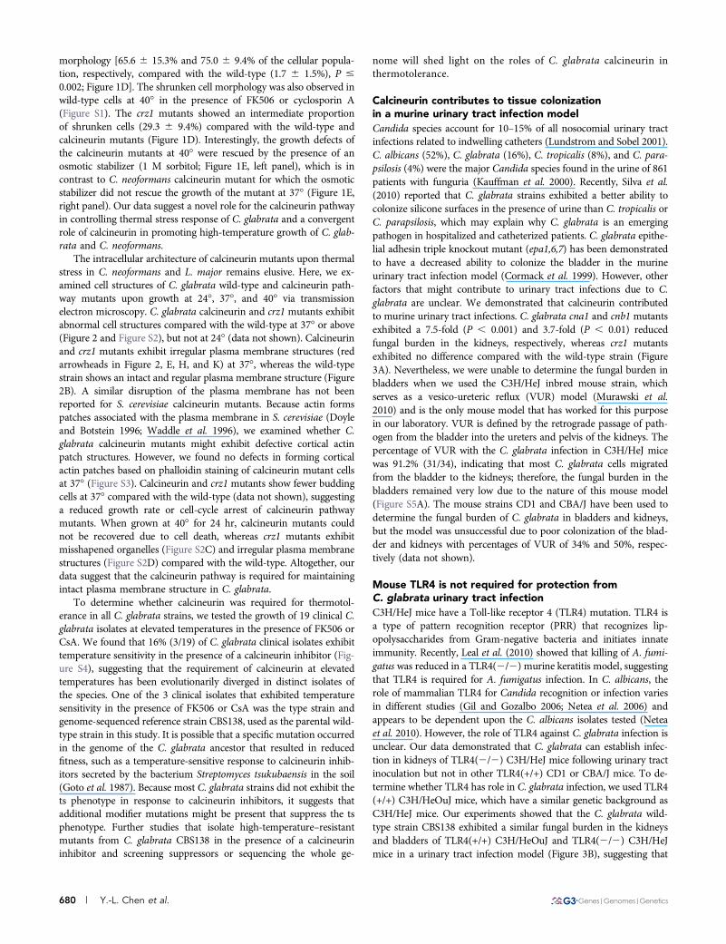
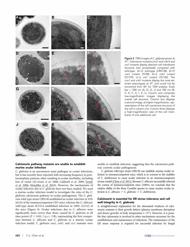
The intracellular architecture of calcineurin mutants upon thermalstress in C. neoformans and L. major remains elusive. Here, we ex-amined cell structures of C. glabrata wild-type and calcineurin path-way mutants upon growth at 24�, 37�, and 40� via transmissionelectron microscopy. C. glabrata calcineurin and crz1 mutants exhibitabnormal cell structures compared with the wild-type at 37� or above(Figure 2 and Figure S2), but not at 24� (data not shown). Calcineurinand crz1 mutants exhibit irregular plasma membrane structures (redarrowheads in Figure 2, E, H, and K) at 37�, whereas the wild-typestrain shows an intact and regular plasma membrane structure (Figure2B). A similar disruption of the plasma membrane has not beenreported for S. cerevisiae calcineurin mutants. Because actin formspatches associated with the plasma membrane in S. cerevisiae (Doyleand Botstein 1996; Waddle et al. 1996), we examined whether C.glabrata calcineurin mutants might exhibit defective cortical actinpatch structures. However, we found no defects in forming corticalactin patches based on phalloidin staining of calcineurin mutant cellsat 37� (Figure S3). Calcineurin and crz1 mutants show fewer buddingcells at 37� compared with the wild-type (data not shown), suggestinga reduced growth rate or cell-cycle arrest of calcineurin pathwaymutants. When grown at 40� for 24 hr, calcineurin mutants couldnot be recovered due to cell death, whereas crz1 mutants exhibitmisshapened organelles (Figure S2C) and irregular plasma membranestructures (Figure S2D) compared with the wild-type. Altogether, ourdata suggest that the calcineurin pathway is required for maintainingintact plasma membrane structure in C. glabrata.
To determine whether calcineurin was required for thermotol-erance in all C. glabrata strains, we tested the growth of 19 clinical C.glabrata isolates at elevated temperatures in the presence of FK506 orCsA. We found that 16% (3/19) of C. glabrata clinical isolates exhibittemperature sensitivity in the presence of a calcineurin inhibitor (Fig-ure S4), suggesting that the requirement of calcineurin at elevatedtemperatures has been evolutionarily diverged in distinct isolates ofthe species. One of the 3 clinical isolates that exhibited temperaturesensitivity in the presence of FK506 or CsA was the type strain andgenome-sequenced reference strain CBS138, used as the parental wild-type strain in this study. It is possible that a specific mutation occurredin the genome of the C. glabrata ancestor that resulted in reducedfitness, such as a temperature-sensitive response to calcineurin inhib-itors secreted by the bacterium Streptomyces tsukubaensis in the soil(Goto et al. 1987). Because most C. glabrata strains did not exhibit thets phenotype in response to calcineurin inhibitors, it suggests thatadditional modifier mutations might be present that suppress the tsphenotype. Further studies that isolate high-temperature–resistantmutants from C. glabrata CBS138 in the presence of a calcineurininhibitor and screening suppressors or sequencing the whole ge-
nome will shed light on the roles of C. glabrata calcineurin inthermotolerance.
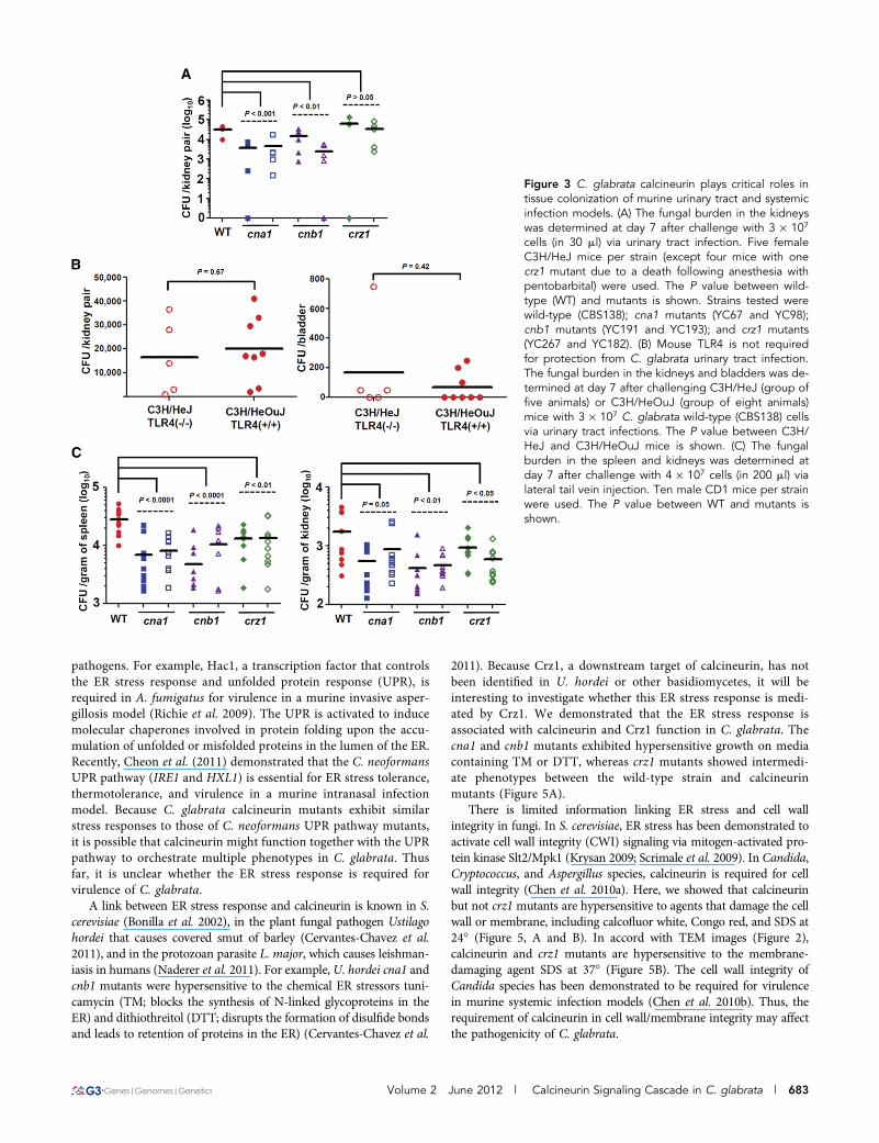
Calcineurin contributes to tissue colonizationin a murine urinary tract infection modelCandida species account for 10–15% of all nosocomial urinary tractinfections related to indwelling catheters (Lundstrom and Sobel 2001).C. albicans (52%), C. glabrata (16%), C. tropicalis (8%), and C. para-psilosis (4%) were the major Candida species found in the urine of 861patients with funguria (Kauffman et al. 2000). Recently, Silva et al.(2010) reported that C. glabrata strains exhibited a better ability tocolonize silicone surfaces in the presence of urine than C. tropicalis orC. parapsilosis, which may explain why C. glabrata is an emergingpathogen in hospitalized and catheterized patients. C. glabrata epithe-lial adhesin triple knockout mutant (epa1,6,7) has been demonstratedto have a decreased ability to colonize the bladder in the murineurinary tract infection model (Cormack et al. 1999). However, otherfactors that might contribute to urinary tract infections due to C.glabrata are unclear. We demonstrated that calcineurin contributedto murine urinary tract infections. C. glabrata cna1 and cnb1 mutantsexhibited a 7.5-fold (P , 0.001) and 3.7-fold (P , 0.01) reducedfungal burden in the kidneys, respectively, whereas crz1 mutantsexhibited no difference compared with the wild-type strain (Figure3A). Nevertheless, we were unable to determine the fungal burden inbladders when we used the C3H/HeJ inbred mouse strain, whichserves as a vesico-ureteric reflux (VUR) model (Murawski et al.2010) and is the only mouse model that has worked for this purposein our laboratory. VUR is defined by the retrograde passage of path-ogen from the bladder into the ureters and pelvis of the kidneys. Thepercentage of VUR with the C. glabrata infection in C3H/HeJ micewas 91.2% (31/34), indicating that most C. glabrata cells migratedfrom the bladder to the kidneys; therefore, the fungal burden in thebladders remained very low due to the nature of this mouse model(Figure S5A). The mouse strains CD1 and CBA/J have been used todetermine the fungal burden of C. glabrata in bladders and kidneys,but the model was unsuccessful due to poor colonization of the blad-der and kidneys with percentages of VUR of 34% and 50%, respec-tively (data not shown).
Mouse TLR4 is not required for protection fromC. glabrata urinary tract infectionC3H/HeJ mice have a Toll-like receptor 4 (TLR4) mutation. TLR4 isa type of pattern recognition receptor (PRR) that recognizes lip-opolysaccharides from Gram-negative bacteria and initiates innateimmunity. Recently, Leal et al. (2010) showed that killing of A. fumi-gatus was reduced in a TLR4(2/2) murine keratitis model, suggestingthat TLR4 is required for A. fumigatus infection. In C. albicans, therole of mammalian TLR4 for Candida recognition or infection variesin different studies (Gil and Gozalbo 2006; Netea et al. 2006) andappears to be dependent upon the C. albicans isolates tested (Neteaet al. 2010). However, the role of TLR4 against C. glabrata infection isunclear. Our data demonstrated that C. glabrata can establish infec-tion in kidneys of TLR4(2/2) C3H/HeJ mice following urinary tractinoculation but not in other TLR4(+/+) CD1 or CBA/J mice. To de-termine whether TLR4 has role in C. glabrata infection, we used TLR4(+/+) C3H/HeOuJ mice, which have a similar genetic background asC3H/HeJ mice. Our experiments showed that the C. glabrata wild-type strain CBS138 exhibited a similar fungal burden in the kidneysand bladders of TLR4(+/+) C3H/HeOuJ and TLR4(2/2) C3H/HeJmice in a urinary tract infection model (Figure 3B), suggesting that
680 | Y.-L. Chen et al.
TLR4 does not modulate C. glabrata infection in the urinary tractinfection model. Therefore, the differential responses to C. glabrataurinary tract infection in CD1, CBA/J, and C3H/HeJ mice might beattributable to specific mouse strains but not to the TLR4 mutation.
Calcineurin and Crz1 influence tissue colonizationin a murine systemic infection modelThe requirement of C. albicans calcineurin for mammalian virulencedepends on the specific niche (Bader et al. 2006). For example, calci-neurin controls virulence in murine systemic infections, but not in vag-inal or pulmonary infection models (Bader et al. 2006). In addition,fungal calcineurin uses different pathogenesis mechanisms to cause infec-tions in the host depending on the infection location (Chen et al. 2010a).We tested whether C. glabrata calcineurin is required for murine systemicinfections. Unlike C. albicans and C. dubliniensis, C. glabrata preferen-tially colonizes the spleen rather than kidney tissues. In splenic tissues,
both cna1 and cnb1 mutants exhibited a 3.7-fold reduced fungal burden(P, 0.0001), whereas crz1mutants had a 2.1-fold reduced fungal burden(P, 0.01) (Figure 3C) compared with the wild-type strain. The results ofthe calcineurin mutants in spleen tissues were similar to those reportedby Miyazaki et al. (2010). However, we found that crz1mutants exhibitedreduced fungal burden in the spleen, while Miyazaki and colleaguesreported no difference. The differential role of crz1mutants in colonizingthe spleen between the two groups might be due to differences in mousestrains, C. glabrata strains, or experimental procedures. In kidney tissues,cna1 and cnb1 mutants exhibited 2.5-fold (P ¼ 0.05) and 3.9-fold (P ,0.01) reduced fungal burdens compared with the wild-type strain,whereas crz1 mutants showed a 2.3-fold reduced fungal burden (P ,0.05) compared with the wild-type strain. Our results of fungal burden inthe kidneys were consistent with those reported by Miyazaki et al. (2010).Taken together, C. glabrata calcineurin and Crz1 contribute to virulencein a murine systemic infection model.
Figure 1 Calcineurin pathway is required for thermotol-erance in C. glabrata. (A) Pharmacological inhibition ofcalcineurin phenocopies genetic deletion of calcineurin.Cells were grown overnight in YPD at 24�, 5-fold seriallydiluted, and spotted onto YPD medium containingFK506 or cyclosporin A (CsA), and incubated at the in-dicated temperatures for 48 hr. Strains tested were wild-type (CBS138), cna1 mutants (YC67 and YC98), cnb1mutants (YC191 and YC193), and crz1 mutants (YC267and YC182). (B) The growth kinetics of C. glabrata wild-type and mutant strains at 24�. Cells were grown over-night at 24�, washed twice with dH2O, diluted to 0.2OD/ml in fresh liquid YPD medium, and incubated at24� with shaking at 250 rpm. The OD600 of cultures wasmeasured at 0, 3, 6, 9, 24, and 30 hr. The experimentswere performed in triplicate, and data were plotted us-ing Prism 5.03. Strains tested were wild-type (CBS138),cna1 mutant (YC98), cnb1 mutant (YC193), and crz1 mu-tant (YC182). (C) Calcineurin is required for survival at40�. Cells were grown overnight in YPD at 24�, 5-foldserially diluted, spotted onto YPD. The plate was incu-bated at 40� for 48 hr, then transferred to 24� for 48 hrincubation. (D) Calcineurin mutant cells exhibita shrunken cell morphology at 40�. Cells were grownovernight in YPD medium at 24�, washed twice withdH2O, diluted to 0.5 OD600/ml in fresh liquid YPD me-dium, and incubated at 24� or 40� with shaking at 250rpm for 4 hr. Strains tested were wild-type (CBS138),cna1 mutant (YC98), cnb1 mutant (YC193), and crz1 mu-tant (YC182). The images were taken at 100·. Scalebar ¼ 5 mm. (E) An osmotic stabilizer rescued tempera-ture-sensitive phenotypes of calcineurin mutants fromC. glabrata, but not from C. neoformans. Cells weregrown overnight in YPD at 24�, 5-fold serially diluted,spotted onto YPD medium containing 1 M sorbitol, andincubated at the temperatures indicated for 48 hr. C.neoformans strains tested were wild-type (H99) andcna1 mutant (KK1).
Volume 2 June 2012 | Calcineurin Signaling Cascade in C. glabrata | 681
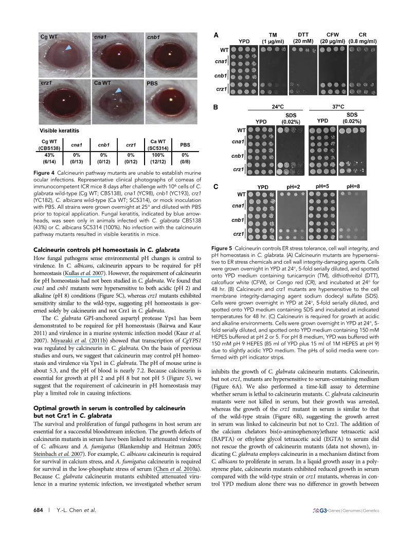
Calcineurin pathway mutants are unable to establishmurine ocular infectionC. glabrata is an uncommon yeast pathogen in ocular infections,but it has recently been reported with increasing frequency in post-keratoplasty patients, often resulting in ocular morbidity, includingloss of vision (Al-Assiri et al. 2006; Caldwell et al. 2009; Gruebet al. 2006; Muzaliha et al. 2010). However, the mechanisms ofocular infection due to C. glabrata have not been studied. We useda murine ocular infection model to investigate the roles of the C.glabrata calcineurin pathway in ocular pathogenesis. The C. glab-rata wild-type strain CBS138 established an ocular infection in 43%(6/14) of the immunocompetent CD1 mice, whereas the C. albicanswild-type strain SC5314 established infection in 100% (12/12) ofthe mice (Figure 4). Ocular infections due to C. albicans weresignificantly more severe than those caused by C. glabrata at alltime points (P , 0.05; Figure S5B), representing the first compar-ison between C. albicans and C. glabrata in a murine ocularinfection model. C. glabrata cna1, cnb1, and crz1 mutants were
unable to establish infection, suggesting that the calcineurin path-way controls ocular pathogenesis.
C. glabrata wild-type strain CBS138 can establish murine ocular in-fection in immunocompetent mice, which is in contrast to the inabilityof C. dubliniensis to cause ocular infection in an immunocompetentmouse model (Chen et al. 2011). Because C. albicans successfully infectedthe cornea of immunocompetent mice (100%), we conclude that therelative ability of the three Candida species to cause murine ocular in-fection is C. albicans . C. glabrata . C. dubliniensis.
Calcineurin is essential for ER stress tolerance and cellwall integrity in C. glabrata
A straightforward explanation for the attenuated virulence of calci-neurin mutants is their growth defects (plasma membrane disruptionand slower growth) at body temperature (�37�). However, it is possi-ble that calcineurin is involved in other mechanisms necessary for theestablishment and maintenance of infections. The maintenance of theER stress response is required for successful infection by fungal
Figure 2 TEM images of C. glabrata grown at37�. Calcineurin mutants (cna1 and cnb1) andcrz1 mutants display aberrant cell membranestructure (red arrowheads) compared withwild-type. (A–C) wild-type (CBS138). (D–F)cna1 mutant (YC98). (G–I) cnb1 mutant(YC193). (J–L) crz1 mutant (YC182). Thecna1 and cnb1 mutants display the most ab-errant phenotypes at 37� and could not berecovered from 40� for TEM analysis. Scalebar ¼ 500 nm (A, D, G, J) and 100 nm (B,C, E, F, H, I, K, L). Column one compriseslow-magnification images displaying theoverall cell structure. Column two displaysa second image, at higher magnification, rep-resentative of the cell membrane structure ofthe cell in column one. Column three displaysa high-magnification view of the cell mem-brane of one additional cell.
682 | Y.-L. Chen et al.
pathogens. For example, Hac1, a transcription factor that controlsthe ER stress response and unfolded protein response (UPR), isrequired in A. fumigatus for virulence in a murine invasive asper-gillosis model (Richie et al. 2009). The UPR is activated to inducemolecular chaperones involved in protein folding upon the accu-mulation of unfolded or misfolded proteins in the lumen of the ER.Recently, Cheon et al. (2011) demonstrated that the C. neoformansUPR pathway (IRE1 and HXL1) is essential for ER stress tolerance,thermotolerance, and virulence in a murine intranasal infectionmodel. Because C. glabrata calcineurin mutants exhibit similarstress responses to those of C. neoformans UPR pathway mutants,it is possible that calcineurin might function together with the UPRpathway to orchestrate multiple phenotypes in C. glabrata. Thusfar, it is unclear whether the ER stress response is required forvirulence of C. glabrata.
A link between ER stress response and calcineurin is known in S.cerevisiae (Bonilla et al. 2002), in the plant fungal pathogen Ustilagohordei that causes covered smut of barley (Cervantes-Chavez et al.2011), and in the protozoan parasite L. major, which causes leishman-iasis in humans (Naderer et al. 2011). For example, U. hordei cna1 andcnb1 mutants were hypersensitive to the chemical ER stressors tuni-camycin (TM; blocks the synthesis of N-linked glycoproteins in theER) and dithiothreitol (DTT; disrupts the formation of disulfide bondsand leads to retention of proteins in the ER) (Cervantes-Chavez et al.
2011). Because Crz1, a downstream target of calcineurin, has notbeen identified in U. hordei or other basidiomycetes, it will beinteresting to investigate whether this ER stress response is medi-ated by Crz1. We demonstrated that the ER stress response isassociated with calcineurin and Crz1 function in C. glabrata. Thecna1 and cnb1 mutants exhibited hypersensitive growth on mediacontaining TM or DTT, whereas crz1 mutants showed intermedi-ate phenotypes between the wild-type strain and calcineurinmutants (Figure 5A).
There is limited information linking ER stress and cell wallintegrity in fungi. In S. cerevisiae, ER stress has been demonstrated toactivate cell wall integrity (CWI) signaling via mitogen-activated pro-tein kinase Slt2/Mpk1 (Krysan 2009; Scrimale et al. 2009). In Candida,Cryptococcus, and Aspergillus species, calcineurin is required for cellwall integrity (Chen et al. 2010a). Here, we showed that calcineurinbut not crz1 mutants are hypersensitive to agents that damage the cellwall or membrane, including calcofluor white, Congo red, and SDS at24� (Figure 5, A and B). In accord with TEM images (Figure 2),calcineurin and crz1 mutants are hypersensitive to the membrane-damaging agent SDS at 37� (Figure 5B). The cell wall integrity ofCandida species has been demonstrated to be required for virulencein murine systemic infection models (Chen et al. 2010b). Thus, therequirement of calcineurin in cell wall/membrane integrity may affectthe pathogenicity of C. glabrata.
Figure 3 C. glabrata calcineurin plays critical roles intissue colonization of murine urinary tract and systemicinfection models. (A) The fungal burden in the kidneyswas determined at day 7 after challenge with 3 · 107
cells (in 30 ml) via urinary tract infection. Five femaleC3H/HeJ mice per strain (except four mice with onecrz1 mutant due to a death following anesthesia withpentobarbital) were used. The P value between wild-type (WT) and mutants is shown. Strains tested werewild-type (CBS138); cna1 mutants (YC67 and YC98);cnb1 mutants (YC191 and YC193); and crz1 mutants(YC267 and YC182). (B) Mouse TLR4 is not requiredfor protection from C. glabrata urinary tract infection.The fungal burden in the kidneys and bladders was de-termined at day 7 after challenging C3H/HeJ (group offive animals) or C3H/HeOuJ (group of eight animals)mice with 3 · 107 C. glabrata wild-type (CBS138) cellsvia urinary tract infections. The P value between C3H/HeJ and C3H/HeOuJ mice is shown. (C) The fungalburden in the spleen and kidneys was determined atday 7 after challenge with 4 · 107 cells (in 200 ml) vialateral tail vein injection. Ten male CD1 mice per strainwere used. The P value between WT and mutants isshown.
Volume 2 June 2012 | Calcineurin Signaling Cascade in C. glabrata | 683
Calcineurin controls pH homeostasis in C. glabrata
How fungal pathogens sense environmental pH changes is central tovirulence. In C. albicans, calcineurin appears to be required for pHhomeostasis (Kullas et al. 2007). However, the requirement of calcineurinfor pH homeostasis had not been studied in C. glabrata. We found thatcna1 and cnb1 mutants were hypersensitive to both acidic (pH 2) andalkaline (pH 8) conditions (Figure 5C), whereas crz1 mutants exhibitedsensitivity similar to the wild-type, suggesting pH homeostasis is gov-erned solely by calcineurin and not Crz1 in C. glabrata.
The C. glabrata GPI-anchored aspartyl protease Yps1 has beendemonstrated to be required for pH homeostasis (Bairwa and Kaur2011) and virulence in a murine systemic infection model (Kaur et al.2007). Miyazaki et al. (2011b) showed that transcription of CgYPS1was regulated by calcineurin in C. glabrata. On the basis of previousstudies and ours, we suggest that calcineurin may control pH homeo-stasis and virulence via Yps1 in C. glabrata. The pH of mouse urine isabout 5.3, and the pH of blood is nearly 7.2. Because calcineurin isessential for growth at pH 2 and pH 8 but not pH 5 (Figure 5), wesuggest that the requirement of calcineurin in pH homeostasis mayplay a limited role in causing infections.
Optimal growth in serum is controlled by calcineurinbut not Crz1 in C. glabrata
The survival and proliferation of fungal pathogens in host serum areessential for a successful bloodstream infection. The growth defects ofcalcineurin mutants in serum have been linked to attenuated virulenceof C. albicans and A. fumigatus (Blankenship and Heitman 2005;Steinbach et al. 2007). For example, C. albicans calcineurin is requiredfor survival in calcium stress, and A. fumigatus calcineurin is requiredfor survival in the low-phosphate stress of serum (Chen et al. 2010a).Because C. glabrata calcineurin mutants exhibited attenuated viru-lence in a murine systemic infection, we investigated whether serum
inhibits the growth of C. glabrata calcineurin mutants. Calcineurin,but not crz1, mutants are hypersensitive to serum-containing medium(Figure 6A). We also performed a time-kill assay to determinewhether serum is lethal to calcineurin mutants. C. glabrata calcineurinmutants were not killed in serum, but their growth was arrested,whereas the growth of the crz1 mutant in serum is similar to thatof the wild-type strain (Figure 6B), suggesting the growth arrestin serum was linked to calcineurin but not to Crz1. The addition ofthe calcium chelators bis(o-aminophenoxy)ethane tetraacetic acid(BAPTA) or ethylene glycol tetraacetic acid (EGTA) to serum didnot rescue the growth of calcineurin mutants (data not shown), in-dicating C. glabrata employs calcineurin in a mechanism distinct fromC. albicans to proliferate in serum. In a liquid growth assay in a poly-styrene plate, calcineurin mutants exhibited reduced growth in serumcompared with the wild-type strain or crz1 mutants, whereas in con-trol YPD medium alone there was no difference in growth between
Figure 4 Calcineurin pathway mutants are unable to establish murineocular infections. Representative clinical photographs of corneas ofimmunocompetent ICR mice 8 days after challenge with 106 cells of C.glabrata wild-type (Cg WT; CBS138), cna1 (YC98), cnb1 (YC193), crz1(YC182), C. albicans wild-type (Ca WT; SC5314), or mock inoculationwith PBS. All strains were grown overnight at 25� and diluted with PBSprior to topical application. Fungal keratitis, indicated by blue arrow-heads, was seen only in animals infected with C. glabrata CBS138(43%) or C. albicans SC5314 (100%). No infection with the calcineurinpathway mutants resulted in visible keratitis in mice.
Figure 5 Calcineurin controls ER stress tolerance, cell wall integrity, andpH homeostasis in C. glabrata. (A) Calcineurin mutants are hypersensi-tive to ER stress chemicals and cell wall integrity-damaging agents. Cellswere grown overnight in YPD at 24�, 5-fold serially diluted, and spottedonto YPD medium containing tunicamycin (TM), dithiothreitol (DTT),calcofluor white (CFW), or Congo red (CR), and incubated at 24� for48 hr. (B) Calcineurin and crz1 mutants are hypersensitive to the cellmembrane integrity-damaging agent sodium dodecyl sulfate (SDS).Cells were grown overnight in YPD at 24�, 5-fold serially diluted, andspotted onto YPD medium containing SDS and incubated at indicatedtemperatures for 48 hr. (C) Calcineurin is required for growth at acidicand alkaline environments. Cells were grown overnight in YPD at 24�, 5-fold serially diluted, and spotted onto YPD medium containing 150 mMHEPES buffered at pH 2 or 5. For pH 8 medium, YPD was buffered with150 mM pH 9 HEPES (85 ml of YPD plus 15 ml of 1M HEPES at pH 9)due to slightly acidic YPD medium. The pHs of solid media were con-firmed with pH indicator strips.
684 | Y.-L. Chen et al.

the calcineurin mutants and the wild-type strain (Figure 6C). Our dataalso support the roles of calcineurin in a murine systemic infectionmodel because attenuated virulence of the calcineurin mutants may bein part due to growth arrest in serum.
Antifungal drug susceptibility is controlledby calcineurin and Crz1 in C. glabrata
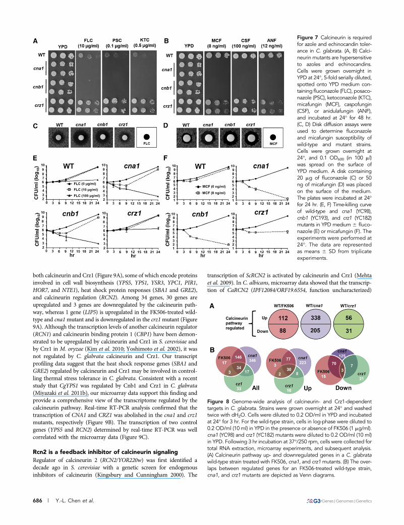
C. glabrata is notorious for its antifungal drug tolerance, which con-tributes to difficulty in treating infections associated with this species.Our studies demonstrated that calcineurin contributes to virulence inmurine urinary tract, systemic, and ocular infections due to C. glab-rata. Thus, C. glabrata calcineurin is a drug target worthy of investi-gation. Calcineurin mutants (cna1 and cnb1) are hypersensitive to theazoles fluconazole, posaconazole, and ketoconazole (Figure 7A), andto the echinocandins micafungin, caspofungin, and anidulafungin(Figure 7B). In disk diffusion assays, calcineurin mutants exhibit largerand clearer zones of inhibition in the presence of fluconazole ormicafungin than the wild-type strain (Figure 7, C and D). To confirmthe fungicidal effects of the drugs on calcineurin mutants, we per-formed time-kill assays. Fluconazole exhibited fungicidal activity at100 mg/ml, but not at 10 mg/ml, against calcineurin mutants over24 hr of incubation (Figure 7E). At a concentration of 8 ng/ml, mica-fungin showed fungicidal activity against calcineurin mutants but notthe wild-type strain (Figure 7F). Taken together, our data suggest thatcombination therapy with a calcineurin inhibitor and fluconazole ormicafungin may improve the treatment outcome of C. glabratainfections.
Interestingly, crz1 mutants showed differential responses to azolesand echinocandins. Unlike the intermediate hypersensitivity pheno-types of crz1/crz1 mutants in C. albicans and C. dubliniensis (Chenet al. 2011), C. glabrata crz1 mutants exhibit hyperresistance to azoles(Figure 7A), suggesting that Crz1 might repress genes involved inazole tolerance in C. glabrata. In response to echinocandins, crz1mutants showed an intermediate phenotype between the wild-typeand the calcineurin mutants (Figure 7B), suggesting that echinocandintolerance is mediated by a calcineurin-dependent Crz1 pathway.
Genome-wide analysis of calcineurin- andCrz1-dependent targets in C. glabrata
To better understand the multiple phenotypes of calcineurin pathwaymutants and to identify calcineurin pathway–regulated genes in C.glabrata, we compared the genome-wide transcriptome profile of awild-type strain with an FK506-treated wild-type strain, a cna1 mu-tant, and a crz1 mutant (Figure 8A). We expected that the FK506-treated wild-type strain would have a similar expression profile to thatof the cna1 mutant. The genes regulated by FK506 largely overlapped(90%, 180/200) with differentially expressed genes in a cna1 mutant(Figure 8B). By looking at calcineurin-upregulated and calcineurin-downregulated genes, the FK506 group overlapped 96% (107/112) and83% (73/88), respectively, with the cna1 mutant (Figure 8B). Interest-ingly, 38% (33/87) of Crz1-dependent genes are Cna1-independent(Figure 8B), indicating that proteins other than calcineurin mightregulate Crz1 and result in differential expression profiles comparedwith the calcineurin-dependent Crz1 pathway. The genes regulated byFK506 exhibited 29% (32/112, upregulated) and 3% (3/88, downregu-lated) overlap, respectively, with differential expressed genes in a crz1mutant (Figure 8B), suggesting a strong correlation between FK506-treated wild-type and cna1 mutant, but not with the crz1 mutant. Inaddition, FK506 treatment of wild-type cells resulted in differentialexpression of 20 genes (Table S2) distinct from those in the cna1
mutant, including genes encoding plasma membrane ATP-bindingcassette (ABC) multidrug transporters (PDR5, PDR15, and YOR1),the bile acid transporter YBT1, and the transcription factor INO2,possibly due to FK506 inhibition of FKBP12 or other FKBPs or todirect action on multidrug resistance (MDR) pumps.
Because the requirement of calcineurin in C. glabrata is differentfrom C. albicans in terms of thermal stress tolerance and optimalserum growth, it is of interest to explore the calcineurin and Crz1targets in C. glabrata. We identified 34 genes that are regulated by
Figure 6 Calcineurin, but not Crz1, is required for optimal growth inserum. (A) Cells were grown overnight in YPD at 24�, 5-fold seriallydiluted, spotted onto synthetic complete (SC) medium 6 40% fetalbovine serum, and incubated at 24� for 4 days. (B) Time-killing curve ofwild-type (CBS138), cna1 (YC98), cnb1 (YC193), and crz1 (YC182)strains grown in 100% serum. Cells surviving at 24� with shaking at250 rpm after 0, 3, 6, 9, and 24 hr were serially diluted onto YPDmedium, and CFUs were counted after 48 hr incubation at 24�. Thedata are represented as means 6 SD from triplicate experiments. (C)Calcineurin mutants cannot proliferate as well as the wild-type strain orcrz1 mutant in serum. Two microliters of 1 OD600/ml cells was addedto 100 ml of 100% serum or YPD medium in 96-well polystyrene plates,and the OD600 was measured after stationary incubation at 24� for 24hr. The data are represented as means 6 SD from triplicate experi-ments. Asterisk represents P , 0.001 compared with the wild-type.
Volume 2 June 2012 | Calcineurin Signaling Cascade in C. glabrata | 685
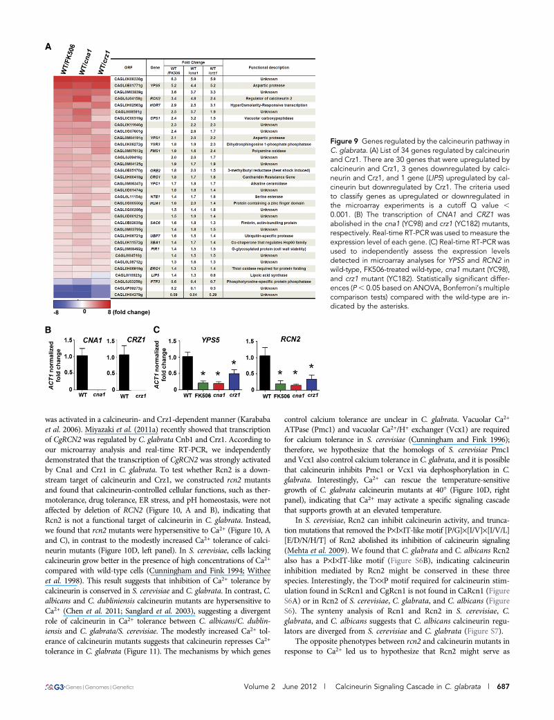
both calcineurin and Crz1 (Figure 9A), some of which encode proteinsinvolved in cell wall biosynthesis (YPS5, YPS1, YSR3, YPC1, PIR1,HOR7, and NTE1), heat shock protein responses (SBA1 and GRE2),and calcineurin regulation (RCN2). Among 34 genes, 30 genes areupregulated and 3 genes are downregulated by the calcineurin path-way, whereas 1 gene (LIP5) is upregulated in the FK506-treated wild-type and cna1mutant and is downregulated in the crz1mutant (Figure9A). Although the transcription levels of another calcineurin regulator(RCN1) and calcineurin binding protein 1 (CBP1) have been demon-strated to be upregulated by calcineurin and Crz1 in S. cerevisiae andby Crz1 in M. oryzae (Kim et al. 2010; Yoshimoto et al. 2002), it wasnot regulated by C. glabrata calcineurin and Crz1. Our transcriptprofiling data suggest that the heat shock response genes (SBA1 andGRE2) regulated by calcineurin and Crz1 may be involved in control-ling thermal stress tolerance in C. glabrata. Consistent with a recentstudy that CgYPS1 was regulated by Cnb1 and Crz1 in C. glabrata(Miyazaki et al. 2011b), our microarray data support this finding andprovide a comprehensive view of the transcriptome regulated by thecalcineurin pathway. Real-time RT-PCR analysis confirmed that thetranscription of CNA1 and CRZ1 was abolished in the cna1 and crz1mutants, respectively (Figure 9B). The transcription of two controlgenes (YPS5 and RCN2) determined by real-time RT-PCR was wellcorrelated with the microarray data (Figure 9C).
Rcn2 is a feedback inhibitor of calcineurin signalingRegulator of calcineurin 2 (RCN2/YOR220w) was first identified adecade ago in S. cerevisiae with a genetic screen for endogenousinhibitors of calcineurin (Kingsbury and Cunningham 2000). The
transcription of ScRCN2 is activated by calcineurin and Crz1 (Mehtaet al. 2009). In C. albicans, microarray data showed that the transcrip-tion of CaRCN2 (IPF12084/ORF19.6554, function uncharacterized)
Figure 7 Calcineurin is requiredfor azole and echinocandin toler-ance in C. glabrata. (A, B) Calci-neurin mutants are hypersensitiveto azoles and echinocandins.Cells were grown overnight inYPD at 24�, 5-fold serially diluted,spotted onto YPD medium con-taining fluconazole (FLC), posaco-nazole (PSC), ketoconazole (KTC),micafungin (MCF), caspofungin(CSF), or anidulafungin (ANF),and incubated at 24� for 48 hr.(C, D) Disk diffusion assays wereused to determine fluconazoleand micafungin susceptibility ofwild-type and mutant strains.Cells were grown overnight at24�, and 0.1 OD600 (in 100 ml)was spread on the surface ofYPD medium. A disk containing20 mg of fluconazole (C) or 50ng of micafungin (D) was placedon the surface of the medium.The plates were incubated at 24�for 24 hr. (E, F) Time-killing curveof wild-type and cna1 (YC98),cnb1 (YC193), and crz1 (YC182)mutants in YPD medium 6 fluco-nazole (E) or micafungin (F). Theexperiments were performed at24�. The data are representedas means 6 SD from triplicateexperiments.
Figure 8 Genome-wide analysis of calcineurin- and Crz1-dependenttargets in C. glabrata. Strains were grown overnight at 24� and washedtwice with dH2O. Cells were diluted to 0.2 OD/ml in YPD and incubatedat 24� for 3 hr. For the wild-type strain, cells in log-phase were diluted to0.2 OD/ml (10 ml) in YPD in the presence or absence of FK506 (1 mg/ml).cna1 (YC98) and crz1 (YC182) mutants were diluted to 0.2 OD/ml (10 ml)in YPD. Following 3 hr incubation at 37�/250 rpm, cells were collected fortotal RNA extraction, microarray experiments, and subsequent analysis.(A) Calcineurin pathway up- and downregulated genes in a C. glabratawild-type strain treated with FK506, cna1, and crz1mutants. (B) The over-laps between regulated genes for an FK506-treated wild-type strain,cna1, and crz1 mutants are depicted as Venn diagrams.
686 | Y.-L. Chen et al.
was activated in a calcineurin- and Crz1-dependent manner (Karababaet al. 2006). Miyazaki et al. (2011a) recently showed that transcriptionof CgRCN2 was regulated by C. glabrata Cnb1 and Crz1. According toour microarray analysis and real-time RT-PCR, we independentlydemonstrated that the transcription of CgRCN2 was strongly activatedby Cna1 and Crz1 in C. glabrata. To test whether Rcn2 is a down-stream target of calcineurin and Crz1, we constructed rcn2 mutantsand found that calcineurin-controlled cellular functions, such as ther-motolerance, drug tolerance, ER stress, and pH homeostasis, were notaffected by deletion of RCN2 (Figure 10, A and B), indicating thatRcn2 is not a functional target of calcineurin in C. glabrata. Instead,we found that rcn2 mutants were hypersensitive to Ca2+ (Figure 10, Aand C), in contrast to the modestly increased Ca2+ tolerance of calci-neurin mutants (Figure 10D, left panel). In S. cerevisiae, cells lackingcalcineurin grow better in the presence of high concentrations of Ca2+
compared with wild-type cells (Cunningham and Fink 1994; Witheeet al. 1998). This result suggests that inhibition of Ca2+ tolerance bycalcineurin is conserved in S. cerevisiae and C. glabrata. In contrast, C.albicans and C. dubliniensis calcineurin mutants are hypersensitive toCa2+ (Chen et al. 2011; Sanglard et al. 2003), suggesting a divergentrole of calcineurin in Ca2+ tolerance between C. albicans/C. dublin-iensis and C. glabrata/S. cerevisiae. The modestly increased Ca2+ tol-erance of calcineurin mutants suggests that calcineurin represses Ca2+
tolerance in C. glabrata (Figure 11). The mechanisms by which genes
control calcium tolerance are unclear in C. glabrata. Vacuolar Ca2+
ATPase (Pmc1) and vacuolar Ca2+/H+ exchanger (Vcx1) are requiredfor calcium tolerance in S. cerevisiae (Cunningham and Fink 1996);therefore, we hypothesize that the homologs of S. cerevisiae Pmc1and Vcx1 also control calcium tolerance in C. glabrata, and it is possiblethat calcineurin inhibits Pmc1 or Vcx1 via dephosphorylation in C.glabrata. Interestingly, Ca2+ can rescue the temperature-sensitivegrowth of C. glabrata calcineurin mutants at 40� (Figure 10D, rightpanel), indicating that Ca2+ may activate a specific signaling cascadethat supports growth at an elevated temperature.
In S. cerevisiae, Rcn2 can inhibit calcineurin activity, and trunca-tion mutations that removed the P·I·IT-like motif [P/G]·[I/V]·[I/V/L][E/D/N/H/T] of Rcn2 abolished its inhibition of calcineurin signaling(Mehta et al. 2009). We found that C. glabrata and C. albicans Rcn2also has a P·I·IT-like motif (Figure S6B), indicating calcineurininhibition mediated by Rcn2 might be conserved in these threespecies. Interestingly, the T··P motif required for calcineurin stim-ulation found in ScRcn1 and CgRcn1 is not found in CaRcn1 (FigureS6A) or in Rcn2 of S. cerevisiae, C. glabrata, and C. albicans (FigureS6). The synteny analysis of Rcn1 and Rcn2 in S. cerevisiae, C.glabrata, and C. albicans suggests that C. albicans calcineurin regu-lators are diverged from S. cerevisiae and C. glabrata (Figure S7).
The opposite phenotypes between rcn2 and calcineurin mutants inresponse to Ca2+ led us to hypothesize that Rcn2 might serve as
Figure 9 Genes regulated by the calcineurin pathway inC. glabrata. (A) List of 34 genes regulated by calcineurinand Crz1. There are 30 genes that were upregulated bycalcineurin and Crz1, 3 genes downregulated by calci-neurin and Crz1, and 1 gene (LIP5) upregulated by cal-cineurin but downregulated by Crz1. The criteria usedto classify genes as upregulated or downregulated inthe microarray experiments is a cutoff Q value ,0.001. (B) The transcription of CNA1 and CRZ1 wasabolished in the cna1 (YC98) and crz1 (YC182) mutants,respectively. Real-time RT-PCR was used to measure theexpression level of each gene. (C) Real-time RT-PCR wasused to independently assess the expression levelsdetected in microarray analyses for YPS5 and RCN2 inwild-type, FK506-treated wild-type, cna1 mutant (YC98),and crz1 mutant (YC182). Statistically significant differ-ences (P, 0.05 based on ANOVA, Bonferroni’s multiplecomparison tests) compared with the wild-type are in-dicated by the asterisks.
Volume 2 June 2012 | Calcineurin Signaling Cascade in C. glabrata | 687
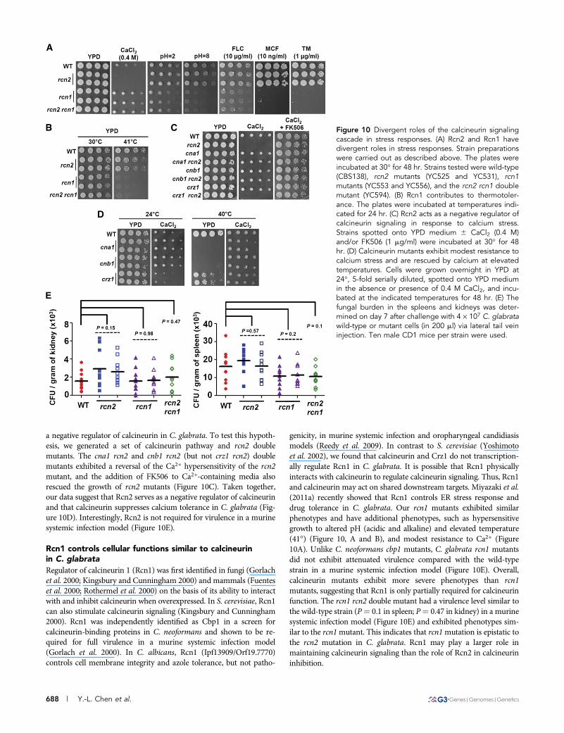
a negative regulator of calcineurin in C. glabrata. To test this hypoth-esis, we generated a set of calcineurin pathway and rcn2 doublemutants. The cna1 rcn2 and cnb1 rcn2 (but not crz1 rcn2) doublemutants exhibited a reversal of the Ca2+ hypersensitivity of the rcn2mutant, and the addition of FK506 to Ca2+-containing media alsorescued the growth of rcn2 mutants (Figure 10C). Taken together,our data suggest that Rcn2 serves as a negative regulator of calcineurinand that calcineurin suppresses calcium tolerance in C. glabrata (Fig-ure 10D). Interestingly, Rcn2 is not required for virulence in a murinesystemic infection model (Figure 10E).
Rcn1 controls cellular functions similar to calcineurinin C. glabrata
Regulator of calcineurin 1 (Rcn1) was first identified in fungi (Gorlachet al. 2000; Kingsbury and Cunningham 2000) and mammals (Fuenteset al. 2000; Rothermel et al. 2000) on the basis of its ability to interactwith and inhibit calcineurin when overexpressed. In S. cerevisiae, Rcn1can also stimulate calcineurin signaling (Kingsbury and Cunningham2000). Rcn1 was independently identified as Cbp1 in a screen forcalcineurin-binding proteins in C. neoformans and shown to be re-quired for full virulence in a murine systemic infection model(Gorlach et al. 2000). In C. albicans, Rcn1 (Ipf13909/Orf19.7770)controls cell membrane integrity and azole tolerance, but not patho-
genicity, in murine systemic infection and oropharyngeal candidiasismodels (Reedy et al. 2009). In contrast to S. cerevisiae (Yoshimotoet al. 2002), we found that calcineurin and Crz1 do not transcription-ally regulate Rcn1 in C. glabrata. It is possible that Rcn1 physicallyinteracts with calcineurin to regulate calcineurin signaling. Thus, Rcn1and calcineurin may act on shared downstream targets. Miyazaki et al.(2011a) recently showed that Rcn1 controls ER stress response anddrug tolerance in C. glabrata. Our rcn1 mutants exhibited similarphenotypes and have additional phenotypes, such as hypersensitivegrowth to altered pH (acidic and alkaline) and elevated temperature(41�) (Figure 10, A and B), and modest resistance to Ca2+ (Figure10A). Unlike C. neoformans cbp1 mutants, C. glabrata rcn1 mutantsdid not exhibit attenuated virulence compared with the wild-typestrain in a murine systemic infection model (Figure 10E). Overall,calcineurin mutants exhibit more severe phenotypes than rcn1mutants, suggesting that Rcn1 is only partially required for calcineurinfunction. The rcn1 rcn2 double mutant had a virulence level similar tothe wild-type strain (P¼ 0.1 in spleen; P¼ 0.47 in kidney) in a murinesystemic infection model (Figure 10E) and exhibited phenotypes sim-ilar to the rcn1mutant. This indicates that rcn1mutation is epistatic tothe rcn2 mutation in C. glabrata. Rcn1 may play a larger role inmaintaining calcineurin signaling than the role of Rcn2 in calcineurininhibition.
Figure 10 Divergent roles of the calcineurin signalingcascade in stress responses. (A) Rcn2 and Rcn1 havedivergent roles in stress responses. Strain preparationswere carried out as described above. The plates wereincubated at 30� for 48 hr. Strains tested were wild-type(CBS138), rcn2 mutants (YC525 and YC531), rcn1mutants (YC553 and YC556), and the rcn2 rcn1 doublemutant (YC594). (B) Rcn1 contributes to thermotoler-ance. The plates were incubated at temperatures indi-cated for 24 hr. (C) Rcn2 acts as a negative regulator ofcalcineurin signaling in response to calcium stress.Strains spotted onto YPD medium 6 CaCl2 (0.4 M)and/or FK506 (1 mg/ml) were incubated at 30� for 48hr. (D) Calcineurin mutants exhibit modest resistance tocalcium stress and are rescued by calcium at elevatedtemperatures. Cells were grown overnight in YPD at24�, 5-fold serially diluted, spotted onto YPD mediumin the absence or presence of 0.4 M CaCl2, and incu-bated at the indicated temperatures for 48 hr. (E) Thefungal burden in the spleens and kidneys was deter-mined on day 7 after challenge with 4 · 107 C. glabratawild-type or mutant cells (in 200 ml) via lateral tail veininjection. Ten male CD1 mice per strain were used.
688 | Y.-L. Chen et al.
In mammalian cells, regulator of calcineurin 1 (DSCR1/RCAN1/calcipressin) is preferentially expressed in the brain of patients withDown’s syndrome and contributes to tumor suppression, suggestingDSCR1 could be explored as a potential therapeutic target (Baek et al.2009). The difference in virulence of C. neoformans cbp1 andC. glabrata rcn1 mutants suggests a divergent role of Cbp1/Rcn1 incontrolling pathogenicity between C. neoformans and C. glabrata. Ourdata also showed that Rcn1 controls azole and echinocandin tol-erance (Figure 10A), making drug combination therapy with yet-to-be-developed Rcn1 inhibitors and azoles or echinocandins possibleapproaches to target emerging C. glabrata infections.
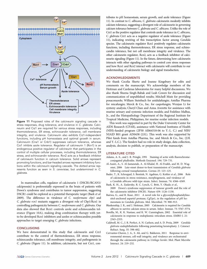
CONCLUSIONSWe have demonstrated in this study that calcineurin and Crz1contribute to the control of thermotolerance, ER stress response,echinocandin tolerance, cell membrane integrity, and pathogenesis inC. glabrata (Figure 11). In addition, calcineurin, but not Crz1, con-
tributes to pH homeostasis, serum growth, and azole tolerance (Figure11). In contrast to C. albicans, C. glabrata calcineurin modestly inhibitscalcium tolerance, suggesting a divergent role of calcineurin in governingcalcium tolerance between C. glabrata and C. albicans. Unlike the role ofCrz1 as the positive regulator that controls azole tolerance in C. albicans,C. glabrata Crz1 acts as a negative regulator of azole tolerance (Figure11), indicating rewiring of this transcription factor among Candidaspecies. The calcineurin regulator Rcn1 positively regulates calcineurinfunctions, including thermotolerance, ER stress response, and echino-candin tolerance, but not cell membrane integrity and virulence. Theother calcineurin regulator, Rcn2, acts as a feedback inhibitor of calci-neurin signaling (Figure 11). In the future, determining how calcineurininteracts with other signaling pathways to control core stress responsesand how Rcn1 and Rcn2 interact with calcineurin will contribute to ourunderstanding of calcineurin biology and signal transduction.
ACKNOWLEDGMENTSWe thank Cecelia Shertz and Joanne Kingsbury for edits andcomments on the manuscript. We acknowledge members of theHeitman and Cardenas laboratories for many helpful discussions. Wealso thank Sheena Singh-Babak and Leah Cowen for discussion andcommunication of unpublished results; Mitchell Mutz for providingposaconazole; William Steinbach for anidulafungin; Astellas Pharmafor micafungin; Merck & Co., Inc. for caspofungin; Wenjun Li forsynteny analysis; Cheryl Chan and Anna Averette for assistance withmurine urinary and systemic infection models; and Fedelino Malbas,Jr., and the Histopathology Department of the Regional Institute forTropical Medicine, Philippines, for murine ocular infection models.This work was supported in part by the Duke University Center for
AIDS Research (CFAR), assistance with National Institutes of Health(NIH)-funded program (2P30 AI064518-06 to Y.-L. C.) and NIH/NIAID R01 grant AI50438 (J.H.). This work was also supported byPilot funds from Astellas Pharma, Inc. and Merck & Co., Inc. (J.H.and Y.-L.C.). The funders had no role in study design, data collection,analysis, decision to publish, or preparation of the manuscript.
LITERATURE CITEDAdams, A. E., and J. R. Pringle, 1991 Staining of actin with fluorochrome-
conjugated phalloidin. Methods Enzymol. 194: 729–731.Al-Assiri, A., S. Al-Jastaneiah, A. Al-Khalaf, H. Al-Fraikh, and M. D. Wag-
oner, 2006 Late-onset donor-to-host transmission of Candida glabratafollowing corneal transplantation. Cornea 25: 123–125.
Bader, T., K. Schroppel, S. Bentink, N. Agabian, G. Kohler et al., 2006 Roleof calcineurin in stress resistance, morphogenesis, and virulence ofa Candida albicans wild-type strain. Infect. Immun. 74: 4366–4369.
Baek, K. H., A. Zaslavsky, R. C. Lynch, C. Britt, Y. Okada et al.,2009 Down’s syndrome suppression of tumour growth and the role ofthe calcineurin inhibitor DSCR1. Nature 459: 1126–1130.
Bairwa, G., and R. Kaur, 2011 A novel role for a glycosylphosphatidylino-sitol-anchored aspartyl protease, CgYps1, in the regulation of pH ho-meostasis in Candida glabrata. Mol. Microbiol. 79: 900–913.
Blankenship, J. R., and J. Heitman, 2005 Calcineurin is required for Candidaalbicans to survive calcium stress in serum. Infect. Immun. 73: 5767–5774.
Bonilla, M., K. K. Nastase, and K. W. Cunningham, 2002 Essential role ofcalcineurin in response to endoplasmic reticulum stress. EMBO J. 21:2343–2353.
Caldwell, M. C., J. R. Perfect, A. N. Carlson, and A. D. Proia, 2009 Candidaglabrata endophthalmitis following penetrating keratoplasty. J. CataractRefract. Surg. 35: 598–602.
Cervantes-Chavez, J. A., S. Ali, and G. Bakkeren, 2011 Response to envi-ronmental stresses, cell-wall integrity, and virulence are orchestratedthrough the calcineurin pathway in Ustilago hordei. Mol. Plant MicrobeInteract. 24: 219–232.
Figure 11 Proposed roles of the calcineurin signaling cascade instress responses, drug tolerance, and virulence in C. glabrata. Calci-neurin and Crz1 are required for various stress responses, includingthermotolerance, ER stress, echinocandin tolerance, cell membraneintegrity, and virulence. Calcineurin also exhibits Crz1-independentfunctions, including pH homeostasis and optimal growth in serum.Calcineurin (Cna1 or Cnb1) suppresses calcium tolerance, whereasCrz1 inhibits azole tolerance. Regulator of calcineurin 1 (Rcn1) is anendogenous positive regulator of calcineurin that participates in thecontrol of multiple cellular processes, including thermotolerance, ERstress, and echinocandin tolerance. Rcn2 acts as a feedback inhibitorof calcineurin function in calcium tolerance. Solid arrows representpromoting functions, and bar-headed arrows represent inhibitory func-tions within the calcineurin signaling cascade. The dotted arrow rep-resents function as seen in S. cerevisiae, but undetermined in C.glabrata.
Volume 2 June 2012 | Calcineurin Signaling Cascade in C. glabrata | 689
Chen, Y. L., L. Kozubowski, M. E. Cardenas, and J. Heitman, 2010a On theroles of calcineurin in fungal growth and pathogenesis. Curr Fungal InfectRep 4: 244–255.
Chen, Y. L., A. E. Montedonico, S. Kauffman, J. R. Dunlap, F. M. Menn et al.,2010b Phosphatidylserine synthase and phosphatidylserine decarboxy-lase are essential for cell wall integrity and virulence in Candida albicans.Mol. Microbiol. 75: 1112–1132.
Chen, Y. L., A. Brand, E. L. Morrison, F. G. S. Silao, U. G. Bigol et al.,2011 Calcineurin controls drug tolerance, hyphal growth, and virulencein Candida dubliniensis. Eukaryot. Cell 10: 803–819.
Cheon, S. A., K. W. Jung, Y. L. Chen, J. Heitman, Y. S. Bahn et al.,2011 Unique evolution of the UPR pathway with a novel bZIP tran-scription factor, Hxl1, for controlling pathogenicity of Cryptococcusneoformans. PLoS Pathog. 7: e1002177.
Choi, J. H., Y. Kim, and Y. H. Lee, 2009 Functional analysis of MCNA,a gene encoding a catalytic subunit of calcineurin, in the rice blast fungusMagnaporthe oryzae. J. Microbiol. Biotechnol. 19: 11–16.
Cormack, B. P., N. Ghori, and S. Falkow, 1999 An adhesin of the yeastpathogen Candida glabrata mediating adherence to human epithelialcells. Science 285: 578–582.
Cunningham, K. W., and G. R. Fink, 1994 Calcineurin-dependent growthcontrol in Saccharomyces cerevisiae mutants lacking PMC1, a homolog ofplasma membrane Ca2+ ATPases. J. Cell Biol. 124: 351–363.
Cunningham, K. W., and G. R. Fink, 1996 Calcineurin inhibits VCX1-dependent H+/Ca2+ exchange and induces Ca2+ ATPases in Saccharo-myces cerevisiae. Mol. Cell. Biol. 16: 2226–2237.
Cyert, M. S., R. Kunisawa, D. Kaim, and J. Thorner, 1991 Yeast has ho-mologs (CNA1 and CNA2 gene products) of mammalian calcineurin,a calmodulin-regulated phosphoprotein phosphatase. Proc. Natl. Acad.Sci. USA 88: 7376–7380.
Djalilian, A. R., J. A. Smith, T. J. Walsh, H. L. Malech, and M. R. Robinson,2001 Keratitis caused by Candida glabrata in a patient with chronicgranulomatous disease. Am. J. Ophthalmol. 132: 782–783.
Doyle, T., and D. Botstein, 1996 Movement of yeast cortical actin cyto-skeleton visualized in vivo. Proc. Natl. Acad. Sci. USA 93: 3886–3891.
Dujon, B., D. Sherman, G. Fischer, P. Durrens, S. Casaregola et al.,2004 Genome evolution in yeasts. Nature 430: 35–44.
Fuentes, J. J., L. Genesca, T. J. Kingsbury, K. W. Cunningham, M. Perez-Ribaet al., 2000 DSCR1, overexpressed in Down syndrome, is an inhibitor ofcalcineurin-mediated signaling pathways. Hum. Mol. Genet. 9: 1681–1690.
Gentleman, R., V. Cary, W. Huber, R. Irizarry, and S. Dudoit (Editors),2005 Bioinformatics and Computational Biology Solutions Using R andBioconductor (Statistics for Biology and Health). Springer, New York.
Gil, M. L., and D. Gozalbo, 2006 TLR2, but not TLR4, triggers cytokineproduction by murine cells in response to Candida albicans yeasts andhyphae. Microbes Infect. 8: 2299–2304.
Gorlach, J., D. S. Fox, N. S. Cutler, G. M. Cox, J. R. Perfect et al.,2000 Identification and characterization of a highly conserved calci-neurin binding protein, CBP1/calcipressin, in Cryptococcus neoformans.EMBO J. 19: 3618–3629.
Goto, T., T. Kino, H. Hatanaka, M. Nishiyama, M. Okuhara et al.,1987 Discovery of FK-506, a novel immunosuppressant isolated fromStreptomyces tsukubaensis. Transplant. Proc. 19: 4–8.
Grueb, M., J. M. Rohrbach, and M. Zierhut, 2006 Amphotericin B in thetherapy of Candida glabrata endophthalmitis after penetrating kerato-plasty. Cornea 25: 1243–1244.
Karababa, M., E. Valentino, G. Pardini, A. T. Coste, J. Bille et al.,2006 CRZ1, a target of the calcineurin pathway in Candida albicans.Mol. Microbiol. 59: 1429–1451.
Kauffman, C. A., J. A. Vazquez, J. D. Sobel, H. A. Gallis, D. S. McKinsey et al.,2000 Prospective multicenter surveillance study of funguria in hospi-talized patients. The National Institute for Allergy and Infectious Diseases(NIAID) Mycoses Study Group. Clin. Infect. Dis. 30: 14–18.
Kaur, R., B. Ma, and B. P. Cormack, 2007 A family of glycosylphosphati-dylinositol-linked aspartyl proteases is required for virulence of Candidaglabrata. Proc. Natl. Acad. Sci. USA 104: 7628–7633.
Kim, S., J. Hu, Y. Oh, J. Park, J. Choi et al., 2010 Combining ChIP-chipand expression profiling to model the MoCRZ1 mediated circuit forCa2+/calcineurin signaling in the rice blast fungus. PLoS Pathog. 6: e1000909.
Kingsbury, T. J., and K. W. Cunningham, 2000 A conserved family ofcalcineurin regulators. Genes Dev. 14: 1595–1604.
Kitzmann, A. S., M. D. Wagoner, N. A. Syed, and K. M. Goins,2009 Donor-related Candida keratitis after Descemet stripping auto-mated endothelial keratoplasty. Cornea 28: 825–828.
Klevay, M. J., D. L. Horn, D. Neofytos, M. A. Pfaller, and D. J. Diekema,2009 Initial treatment and outcome of Candida glabrata vs. Candidaalbicans bloodstream infection. Diagn. Microbiol. Infect. Dis. 64: 152–157.
Kojima, K., Y. S. Bahn, and J. Heitman, 2006 Calcineurin, Mpk1 and Hog1MAPK pathways independently control fludioxonil antifungal sensitivityin Cryptococcus neoformans. Microbiology 152: 591–604.
Krysan, D. J., 2009 The cell wall and endoplasmic reticulum stress re-sponses are coordinately regulated in Saccharomyces cerevisiae. Commun.Integr. Biol. 2: 233–235.
Kullas, A. L., S. J. Martin, and D. Davis, 2007 Adaptation to environmentalpH: integrating the Rim101 and calcineurin signal transduction pathways.Mol. Microbiol. 66: 858–871.
Lagrotteria, D., C. Rotstein, and C. H. Lee, 2007 Treatment of candiduriawith micafungin: a case series. Can. J. Infect. Dis. Med. Microbiol. 18:149–150.
Leal, S. M. Jr., S. Cowden, Y. C. Hsia, M. A. Ghannoum, M. Momany et al.,2010 Distinct roles for Dectin-1 and TLR4 in the pathogenesis of As-pergillus fumigatus keratitis. PLoS Pathog. 6: e1000976.
Lee, W. B., J. B. Foster, A. M. Kozarsky, Q. Zhang, and H. E. Grossniklaus,2011 Interface fungal keratitis after endothelial keratoplasty: a clinico-pathological report. Ophthalmic Surg. Lasers Imaging 42 Online: e44–48.
Lundstrom, T., and J. Sobel, 2001 Nosocomial candiduria: a review. Clin.Infect. Dis. 32: 1602–1607.
Malavazi, I., M. E. da Silva Ferreira, F. M. Soriani, T. M. Dinamarco, M.Savoldi et al., 2009 Phenotypic analysis of genes whose mRNA accu-mulation is dependent on calcineurin in Aspergillus fumigatus. FungalGenet. Biol. 46: 791–802.
Mehta, S., H. Li, P. G. Hogan, and K. W. Cunningham, 2009 Domainarchitecture of the regulators of calcineurin (RCANs) and identificationof a divergent RCAN in yeast. Mol. Cell. Biol. 29: 2777–2793.
Miyazaki, T., S. Yamauchi, T. Inamine, Y. Nagayoshi, T. Saijo et al.,2010 Roles of calcineurin and Crz1 in antifungal susceptibility and viru-lence of Candida glabrata. Antimicrob. Agents Chemother. 54: 1639–1643.
Miyazaki, T., K. Izumikawa, Y. Nagayoshi, T. Saijo, S. Yamauchi et al.,2011a Functional characterization of the regulators of calcineurin inCandida glabrata. FEMS Yeast Res. 11: 621–630.
Miyazaki, T., K. Izumikawa, S. Yamauchi, T. Inamine, Y. Nagayoshi et al.,2011b The glycosylphosphatidylinositol-linked aspartyl protease Yps1is transcriptionally regulated by the calcineurin-Crz1 and Slt2 MAPKpathways in Candida glabrata. FEMS Yeast Res. 11: 449–456.
Murawski, I. J., R. W. Maina, D. Malo, L. M. Guay-Woodford, P. Gros et al.,2010 The C3H/HeJ inbred mouse is a model of vesico-ureteric refluxwith a susceptibility locus on chromosome 12. Kidney Int. 78: 269–278.
Muzaliha, M. N., H. Adil, M. Ibrahim, and I. Shatriah, 2010 Candidaglabrata endophthalmitis following penetrating keratoplasty in a patientwith negative donor rim culture. BMC Ophthalmol. 10: 18.
Naderer, T., O. Dandash, and M. J. McConville, 2011 Calcineurin is re-quired for Leishmania major stress response pathways and for virulencein the mammalian host. Mol. Microbiol. 80: 471–480.
Netea, M. G., J. W. van der Meer, and B. J. Kullberg, 2006 Both TLR2 andTLR4 are involved in the recognition of Candida albicans. Reply to“TLR2, but not TLR4, triggers cytokine production by murine cells inresponse to Candida albicans yeasts and hyphae” by Gil and Gozalbo,Microbes and Infection 8 (2006) 2823–2824. Microbes Infect. 8: 2821–2822, author reply 2823–2824.
Netea, M. G., N. A. Gow, L. A. Joosten, I. Verschueren, J. W. van der Meeret al., 2010 Variable recognition of Candida albicans strains by TLR4and lectin recognition receptors. Med. Mycol. 48: 897–903.
690 | Y.-L. Chen et al.
Odom, A., S. Muir, E. Lim, D. L. Toffaletti, J. Perfect et al., 1997 Calcineurin isrequired for virulence of Cryptococcus neoformans. EMBO J. 16: 2576–2589.
Perfect, J. R., N. Ketabchi, G. M. Cox, C. W. Ingram, and C. L. Beiser,1993 Karyotyping of Cryptococcus neoformans as an epidemiologicaltool. J. Clin. Microbiol. 31: 3305–3309.
Pfaller, M. A., G. J. Moet, S. A. Messer, R. N. Jones, and M. Castanheira,2011a Candida bloodstream infections: comparison of species distri-butions and antifungal resistance patterns in community-onset andnosocomial isolates in the SENTRY Antimicrobial Surveillance Program,2008–2009. Antimicrob. Agents Chemother. 55: 561–566.
Pfaller, M. A., G. J. Moet, S. A. Messer, R. N. Jones, and M. Castanheira,2011b Geographic variations in species distribution and echinocandinand azole antifungal resistance rates among Candida bloodstream infec-tion isolates: report from the SENTRY Antimicrobial Surveillance Pro-gram (2008 to 2009). J. Clin. Microbiol. 49: 396–399.
Phillips, J. R., and M. G. Karlowicz, 1997 Prevalence of Candida species inhospital-acquired urinary tract infections in a neonatal intensive careunit. Pediatr. Infect. Dis. J. 16: 190–194.
Reedy, J. L., S. G. Filler, and J. Heitman, 2009 Elucidating the Candidaalbicans calcineurin signaling cascade controlling stress response andvirulence. Fungal Genet. Biol. 47: 107–116.
Reuss, O., A. Vik, R. Kolter, and J. Morschhauser, 2004 The SAT1 flipper, anoptimized tool for gene disruption in Candida albicans. Gene 341: 119–127.
Richie, D. L., L. Hartl, V. Aimanianda, M. S. Winters, K. K. Fuller et al.,2009 A role for the unfolded protein response (UPR) in virulence andantifungal susceptibility in Aspergillus fumigatus. PLoS Pathog. 5: e1000258.
Rothermel, B., R. B. Vega, J. Yang, H. Wu, R. Bassel-Duby et al., 2000 Aprotein encoded within the Down syndrome critical region is enriched instriated muscles and inhibits calcineurin signaling. J. Biol. Chem. 275:8719–8725.
Rothermel, B. A., R. B. Vega, and R. S. Williams, 2003 The role of mod-ulatory calcineurin-interacting proteins in calcineurin signaling. TrendsCardiovasc. Med. 13: 15–21.
Sanglard, D., F. Ischer, O. Marchetti, J. Entenza, and J. Bille,2003 Calcineurin A of Candida albicans: involvement in antifungaltolerance, cell morphogenesis and virulence. Mol. Microbiol. 48: 959–976.
Scrimale, T., L. Didone, K. L. de Mesy Bentley, and D. J. Krysan, 2009 Theunfolded protein response is induced by the cell wall integrity mitogen-activated protein kinase signaling cascade and is required for cell wallintegrity in Saccharomyces cerevisiae. Mol. Biol. Cell 20: 164–175.
Silva, S., M. Negri, M. Henriques, R. Oliveira, D. Williams et al.,2010 Silicone colonization by non-Candida albicans Candida species inthe presence of urine. J. Med. Microbiol. 59: 747–754.
Steinbach, W. J., R. A. Cramer Jr, B. Z. Perfect, Y. G. Asfaw, T. C. Sauer et al.,2006 Calcineurin controls growth, morphology, and pathogenicity inAspergillus fumigatus. Eukaryot. Cell 5: 1091–1103.
Steinbach, W. J., R. A. Cramer Jr, B. Z. Perfect, C. Henn, K. Nielsen et al.,2007 Calcineurin inhibition or mutation enhances cell wall inhibitorsagainst Aspergillus fumigatus. Antimicrob. Agents Chemother. 51: 2979–2981.
Tappeiner, C., D. Goldblum, S. Zimmerli, C. Fux, and B. E. Frueh,2009 Donor-to-host transmission of Candida glabrata to both recipi-ents of corneal transplants from the same donor. Cornea 28: 228–230.
Waddle, J. A., T. S. Karpova, R. H. Waterston, and J. A. Cooper,1996 Movement of cortical actin patches in yeast. J. Cell Biol. 132: 861–870.
Wapinski, I., J. Pfiffner, C. French, A. Socha, D. A. Thompson et al.,2010 Gene duplication and the evolution of ribosomal protein generegulation in yeast. Proc. Natl. Acad. Sci. USA 107: 5505–5510.
Withee, J. L., R. Sen, and M. S. Cyert, 1998 Ion tolerance of Saccharomycescerevisiae lacking the Ca2+/CaM-dependent phosphatase (calcineurin) isimproved by mutations in URE2 or PMA1. Genetics 149: 865–878.
Yoshimoto, H., K. Saltsman, A. P. Gasch, H. X. Li, N. Ogawa et al.,2002 Genome-wide analysis of gene expression regulated by the calci-neurin/Crz1p signaling pathway in Saccharomyces cerevisiae. J. Biol.Chem. 277: 31079–31088.
Communicating editor: D. J. Gresham
Volume 2 June 2012 | Calcineurin Signaling Cascade in C. glabrata | 691


