Embed Size (px)
Citation preview

Coronin 1C negatively regulates cell-matrix adhesion and motilityof intestinal epithelial cells
Stanislav N. Samarin1,*, Stefan Koch1, Andrei I. Ivanov2, Charles A. Parkos1, and AsmaNusrat1,*1Epithelial Pathobiology Research Unit, Department of Pathology and Laboratory Medicine, EmoryUniversity, Atlanta, GA, 303222Department of Medicine, University of Rochester, Rochester, NY, 14642
AbstractCoronins, WD-repeat actin-binding proteins, are known to regulate cell motility by coordinatingactin filament turnover in lamellipodia of migrating cell. Here we report a novel mechanism ofCoronin 1C-mediated cell motility that involves regulation of cell-matrix adhesion. RNAi silencingof Coronin 1C in intestinal epithelial cells enhanced cell migration and modulated lamellipodiadynamics by increasing the persistence of lamellipodial protrusion. Coronin 1C-depleted cellsshowed increased cell-matrix adhesions and enhanced cell spreading compared to control cells, whileoverexpression of Coronin 1C antagonized cell adhesion and spreading. Enhanced cell-matrixadhesion of coronin-deficient cells correlated with hyperphosphorylation of Focal Adhesion Kinase(FAK) and paxillin, and an increase in number of focal adhesions and their redistribution at the cellperiphery. siRNA depletion of FAK in coronin-deficient cells rescued the effects of Coronin 1Cdepletion on motility, cell-matrix adhesion, and spreading. Thus, our findings provide the firstevidence that Coronin 1C negatively regulates epithelial cell migration via FAK-mediated inhibitionof cell-matrix adhesion.
KeywordsCoronin; FAK; motility; adhesion
IntroductionMigration of epithelial cells plays a vital role in a number of physiological and pathologicalprocesses, such as embryogenesis, epithelial renewal, wound healing, and tumor metastasis.Mechanistically, cell migration represents a cyclic process involving extension of lamellipodiaat the leading edge, adhesion of protruded lamellipodia to the extracellular matrix, and finallyretraction of the trailing edge [1;2]. The protrusion of lamellipodia is induced by controlledturnover of actin filaments, where the Arp2/3 complex nucleates new branched actin filaments,and existing filaments are disassembled by ADF/cofilin [3]. Cell-matrix adhesion is mediated
*Corresponding authors: Asma Nusrat, Department of Pathology and Laboratory Medicine, Emory University, Whitehead ResearchBuilding, Room 105E, 615 Michael Street, Atlanta, GA 30322, Tel: (404) 727 8543, Fax: (404) 727 3321, [email protected], StanislavN. Samarin, Department of Pathology and Laboratory Medicine, Emory University, Whitehead Research Building, Room 111, 615Michael Street, Atlanta, GA 30322, Tel: (404) 712 2819, Fax: (404) 727 3321, [email protected]'s Disclaimer: This is a PDF file of an unedited manuscript that has been accepted for publication. As a service to our customerswe are providing this early version of the manuscript. The manuscript will undergo copyediting, typesetting, and review of the resultingproof before it is published in its final citable form. Please note that during the production process errors may be discovered which couldaffect the content, and all legal disclaimers that apply to the journal pertain.
NIH Public AccessAuthor ManuscriptBiochem Biophys Res Commun. Author manuscript; available in PMC 2011 January 1.
Published in final edited form as:Biochem Biophys Res Commun. 2010 January 1; 391(1): 394–400. doi:10.1016/j.bbrc.2009.11.069.
NIH
-PA Author Manuscript
NIH
-PA Author Manuscript
NIH
-PA Author Manuscript

by focal adhesions (FAs) whose main constituents are integrins and adaptor proteins. Theformer interact with the extracellular matrix whereas the latter link integrins to the actincytoskeleton and participate in intracellular signaling [4].
Coronins are evolutionary conserved WD-repeat actin-binding proteins known to regulatevarious cellular processes involving actin dynamics [5]. Coronin protein family encompasses7 proteins in mammals [6], divided into three subclasses based on sequence similarity: TypeI, II and III [5]. The Type I subclass (Coronins 1A, 1B, and 1C) is the most studied coroninsubfamily. Coronins 1A and 1B localize at the leading edge of lamellipodia [7;8;9], physicallyinteract with Arp2/3 complex, and regulate the protrusion of lamellipodia and cell migration[7;8;10]. Recently, Coronin 1B has been shown to act as a coordinator of filament nucleationand disassembly by bridging together Arp2/3 complex and slingshot 1L, an activator of cofilin,thus controlling actin filament dynamics and architecture at the leading edge of the migratingcell [11].
Coronin 1C is ubiquitously expressed in most tissues [7;12;13;14], localizes at the sites ofactive actin dynamics, such as lamellipodia and membrane ruffles [15] and co-immunoprecipitates with Arp2/3 complex and cofilin [16]. However, unlike Coronin 1A and1B, Coronin 1C has not been extensively characterized, and its role in regulating motility ofepithelial sheets is not understood. Here we report a novel mechanism for regulation of motilityof intestinal epithelial cells (IECs) by Coronin 1C, which involves negative regulation of cell-matrix adhesion through FAK-mediated signaling.
Materials and MethodsAntibodies
Anti-Coronin 1C mouse polyclonal and monoclonal, and anti-Coronin 1B mouse monoclonalantibody were purchased from Abnova (Taipei, Taiwan). Anti-paxillin mouse monoclonalantibodies were obtained from Zymed (Zymed Labs, San Francisco, CA). Anti-FAK, anti-phospho(Y118)paxillin, anti-phospho(T18/S19)RMLC rabbit polyclonal antibodies and anti-phospho(S19)RMLC mouse monoclonal antibodies were from Cell Signaling (Cell SignalingTechnology, Beverly, MA). Anti-phospho(Y397)FAK mouse monoclonal antibody were fromBD Biosciences (San Jose, CA). Rabbit polyclonal anti-actin antibodies were from Sigma(Sigma Chemical Co., St. Louis, MO). Anti-RMLC rabbit polyclonal antibodies were fromSanta Cruz Biotechnology (Santa Cruz, CA). Alexa 488/546-conjugated goat anti-mouse, goatanti-rabbit and donkey anti-rabbit antibody were purchased from Molecular Probes (Eugene,OR). Horseradish peroxidase-conjugated goat anti-rabbit and anti-mouse secondary antibodieswere from Jackson Immunoresearch Labs (West Grove, PA).
Cells, DNA transfection and RNA interferenceSK-CO15 (gift of Dr. E. Rodriguez-Boulan, Weill Medical College of Cornell University, NY)and Caco-2 (ATCC, USA) human colonic epithelial cells were grown as described previously[17]. For DNA and RNA transfection cells were plated at ∼75% confluency, transfected thenext day and were used in experiments 24 and 72 hours after transfection respectively. Fulllength Coronin 1C construct in pEGFP-C1 vector were generated as described previously[15] and transfected into cells using Lipofectamine 2000 (Invitrogen) according tomanufacturers protocol. Empty pEGFP-C1 vector (Clontech) was used as a control. HumanCoronin 1C siGENOME duplex 4 and PTK2 siGENOME SmartPool (Dharmacon, Lafayette,CO) were used to downregulate Coronin 1C and FAK respectively. Scramble duplex 2SmartPool siRNA (Dharmacon) has been used as a control. RNA transfection was carried outin OPTI-MEM I media (Invitrogen) with 50 nM siRNA using Dharmafect 1 siRNA transfectionreagent (Dharmacon) according to standard protocol.
Samarin et al. Page 2
Biochem Biophys Res Commun. Author manuscript; available in PMC 2011 January 1.
NIH
-PA Author Manuscript
NIH
-PA Author Manuscript
NIH
-PA Author Manuscript

Cell migration assay and live cell microscopyFor microscopy, confluent monolayers growing on collagen-I received a single linear woundusing a sterile 20 μL pipette tip attached to low suction. 10×10 wounds per monolayer werecreated for Western Blot analysis. Live cell microscopy was performed in CO2-independentmedium (Invitrogen) using heated cabinet and thermally-controlled microscopy stage (BrookIndustries, USA) mounted on Carl Zeiss Axiovert microscope equipped with Zeiss AxioCamMRc5 camera.
Cell adhesion/spreading assayConfluent cells were trypsinized, washed with Hanks' balanced salt solution (Sigma)containing 0.1% BSA and re-plated on coverslips coated with ECM gel (Sigma) or collagen-I (BD Biosciences, USA) at a density of 105cells/1.9cm2. For cell adhesion assay cells wereallowed to adhere for 3 hours, and non-adherent cells were removed by repeated washing withcomplete media. For cell spreading assay, cells were allowed to spread for additional 8 hours.Cells overexpressing EGFP and EGFP-Coronin 1C were detached from the substrate usingAccutase (Innovative Cell Technologies, USA) followed by resuspension in cell sorting buffer(PBS containing 25mM HEPES pH 7.0, 1mM EDTA and 0.1% BSA). EGFP-positive,propidium iodide-negative cells were sorted on a FACSVantage SE (BD Biosciences). Thepurity of sorting was greater than 95%. Cells were counted manually, and cell surface wasmeasured using MetaMorph software. A total of at least 10 microscopic fields were analyzedper group in each experiment.
Immunofluorescence labeling and confocal microscopyCells were fixed in 3.7% paraformaldehyde (PFA) for 15 min and subsequently permeabilizedwith 0.5% Triton X-100 for 10 minutes at RT. Cells were blocked in Hanks' balanced saltsolution (Sigma) containing 1.5 % BSA, and sequentially incubated with primary and Alexa-conjugated secondary antibodies (Molecular Probes) for 1 hour at RT. Nuclei were stainedwith ToPro-3 iodide (Molecular Probes), followed by mounting on slides using Pro-Long GoldAntifade medium (Molecular Probes). Stained monolayers were analyzed using Zeiss LSM510laser scanning confocal microscope (Zeiss Microimaging Inc., USA) equipped with Zeiss Plan-apochromat 63×/1.4 Oil lenses. Images shown are representative of at least three independentexperiments with multiple images taken per slide.
Image analysisWound widths were measured using Scion Image software (Scion Image Corp., USA). Fivemeasurements along the wound length were averaged to determine the wound width.Kymographs were produced using MetaMorph software (Molecular Devices, USA) by taking1-pixel-wide rectangular regions in the direction of edge movement as shown. For celladhesion/spreading assay, cells were counted manually, and cell surface was measured usingMetaMorph software. A total of at least 10 microscopic fields were analyzed per group in eachexperiment. For quantification of FAs phospho(Y118)paxillin-positive pattern wasthresholded with the same level in all specimens and the integrated surface area of FAs wasmeasured using NIS Elements software (Nikon Inc., USA). To determine the density of FAsalong the cell periphery, the total surface of FAs was devided by the cells perimeter length.
ImmunoblottingCells were scraped in RIPA buffer (20 mM Tris, pH 7.4, 150 mM NaCl, 1% Triton X-100, 1%Na deoxycholate, 2 mM EGTA, 2 mM EDTA, 0.1% SDS) containing protease inhibitorcocktail (1:100, Sigma) and phosphatase inhibitor cocktails 1 and 2 (both at 1:200, Sigma) andhomogenized using Wheaton glass homogenizers (Wheaton Industries Inc., USA). Lysateswere cleared by centrifugation at 14,000g for 10 minutes at 4°C and equalized for total protein
Samarin et al. Page 3
Biochem Biophys Res Commun. Author manuscript; available in PMC 2011 January 1.
NIH
-PA Author Manuscript
NIH
-PA Author Manuscript
NIH
-PA Author Manuscript

concentration using BCA protein quantification assay (Pierce Biotechnology, USA). Sampleswere boiled in SDS sample buffer and subjected to SDS-PAGE and Western blotting with 30μg of total protein per lane. Membranes were blocked with either bovine serum albumin (BSA)(for detection of phosphorylated proteins) or dry milk for 1 hour at room temperature (RT) andincubated with primary antibodies in blocking buffer overnight at 4°C. The results shown arerepresentative immunoblots of three independent experiments.
StatisticsAll data shown are representative of at least three independent experiments and expressed asthe mean ± standard error of the mean (S.E.). The results were compared by either two-tailedStudent's t-test, or a post-hoc Bonferroni test following repeated measures two-way ANOVAwith statistical significance assumed at p<0.05.
Results and DiscussionTo address the role of Coronin 1C in the epithelial cell motility, we down-regulated itsexpression in a SK-CO15 intestinal epithelial cells (IEC) using RNA interference. siRNAtreatment dramatically (∼85%) and specifically reduced the expression level of Coronin1C,but not the closely related Coronin 1B (Fig. 1A). Coronin 1C-depleted cells demonstratedincreased migration rate resulting in more efficient wound closure in scratch-wound assays(1.75±0.15 fold of scramble control) (Fig. 1B). This correlated with dramatic changes inlamellipodia dynamics at the leading edge of migrating IECs. While control cells rapidlyextended multiple spiked lamellipodia (Fig. 1C, upper panel) followed by their rapid retraction(Fig. 1C, upper kymograph), Coronin 1C-deficient cells formed flat and rounded lamellipodia(Fig. 1C, bottom panel), that rarely retracted (Fig. 1C, bottom kymograph), thus providingmore persistent forward motility of the leading edge and significantly enhancing the overalldistance protruded over time (Fig. 1C).
The protrusion of lamellipodia is a dynamic process involving both actin-mediated extensionsand retractions, and anchoring lamellipodia to the extracellular matrix (ECM) [18]. Wetherefore reasoned that the increased persistence of lamellipodial protrusion in Coronin 1C-deficient cells might be mediated either through decreased retraction, or enhanced cell-matrixadhesion. Retraction of lamellipodia depends on the activity of the major F-actin motor, non-muscle myosin II [19], which is regulated by phosphorylation of its regulatory light chain(RMLC) on either one (Ser19), or two (Ser19/Thr18) residues [20]. However, down-regulationof Coronin 1C did not significantly alter the levels of mono- and diphosphorylated RMLC inmigrating IECs, which suggests unaltered myosin II activity (Fig. 2A) and argues against therole of diminished retraction in the increased lammelipodial protrusion observed in Coronin1C-deficient IECs.
To test whether depletion of Coronin 1C affects cell-matrix adhesion we used two types ofECM: collagen-I and ECM gel (Sigma) composed of laminin, collagen-IV, and entactin.Coronin 1C-depleted cells attached more readily to the ECM gel (2.18±0.16 fold increase) andshowed enhanced spreading (2.46±0.15 fold cell surface area increase) following adhesion toECM compared to control siRNA-transfected cells (Fig. 2B,C). Similar results were obtainedon collagen-I and in another intestinal epithelial cell line, Caco-2 (data not shown). Conversely,over-expression of WT Coronin 1C dramatically (2.72±0.18 fold) decreased the number ofadherent cells and attenuated their spreading (1.33±0.13 fold decrease in cell surface area)compared to cells transfected with control vector only (Fig. 2D,E). Together, these data suggestthat Coronin 1C negatively regulates adhesion and spreading of IECs.
Cells adhere to the ECM by forming specialized multiprotein complexes known as focaladhesions (FAs) that consist of transmembrane integrins, and cytosolic scaffolding and
Samarin et al. Page 4
Biochem Biophys Res Commun. Author manuscript; available in PMC 2011 January 1.
NIH
-PA Author Manuscript
NIH
-PA Author Manuscript
NIH
-PA Author Manuscript

regulatory proteins [4]. Therefore we next examined whether Coronin 1C depletion effectsformation of FAs by visualizing the FA marker, Tyr118-phosphorylated (activated) paxillin inspreading IECs. Depletion of Coronin 1C dramatically increased the surface area of FAs andaltered their topography towards more dense distribution at the cells periphery (Fig. 3A, Suppl.Fig. 1). These results were consistent with increased levels of total and Tyr118-phosphorylatedpaxillin in Coronin 1C-deficient IECs (Fig. 3B).
Since phosphorylation of paxillin is mediated by focal adhesion kinase (FAK), and FAKsignaling plays a central role in regulating both cell-matrix adhesion and cell motility [21], weinvestigated the role of FAK in adhesiveness and migration of Coronin 1C-depleted IECs.Down-regulation of Coronin 1C resulted in increased activatory phosphorylation of FAK atTyr397 following attachment to and subsequent spreading on ECM (Fig. 3B).
To determine whether the effects of Coronin 1C depletion on cell-matrix adhesion andmigration were FAK-dependent, we performed a single FAK-, and dual FAK/Coronin 1CsiRNA-mediated knock-down in SK-CO15 cells. Importantly, down-regulation of FAKprotein expression (∼90%) alone did not significantly influence the level of phospho-paxillin(Fig. 3B) and the number or distribution of FAs in adhering/spreading IECs (Fig. 3C, Suppl.Fig. 1). Additionally, depletion of FAK did not significantly affect cell-matrix adhesion (Fig.3D), cell spreading (Fig. 3E), and IEC migration (Fig 4A). These data suggest that FAK is notrequired for basal adhesion and motility of IECs and reflect functional redundancy in multipleFAK-mediated signaling pathways [21]. Indeed, the complexity of FAK-mediated signalingis highlighted by a variety of reports showing either negative, or neutral, or positive role ofFAK activity in adhesion and migration in different cell types [22;23;24].
However, loss of FAK protein expression effectively reversed major effects of Coronin 1Cdown-regulation on cell adhesion and migration. Thus, double knock-down of Coronin 1C andFAK efficiently prevented paxillin hyperphosphorylation (Fig. 3B), as well as an increase innumber and changes in distribution of focal adhesions (Fig. 3C, Suppl. Fig. 1) caused byCoronin 1C depletion alone. Consistent with these results, depletion of FAK reversed increasedcell-matrix adhesion (Fig. 3D), spreading (Fig. 3E) and motility (Fig. 4A) of Coronin 1C-deficient cells. All together, these data suggest that FAK-mediated signaling is a criticaldownstream component, which mediates negative effects of Coronin 1C on both cell-matrixadhesion and motility of IECs.
The negative regulation of cell migration and cell-matrix adhesion of IECs by Coronin 1C issomewhat unexpected, since Coronin 1B is required for persistent lamellipodia protrusion andefficient motility of fibroblasts and HEK 293 cells [11], and depletion of Coronin 1C negativelyregulates wound healing in fibroblasts [16]. Our results can be reconciled with previouslypublished data by proposing a dual mechanism of Coronin 1C-dependent regulation of cellmigration. Indeed, a delicate balance between actin-mediated protrusion of lamellipodia andcell-matrix adhesion is required for the efficient cell migration, and both insufficient andexcessively strong matrix adhesion can impede cell motility [1;4;25;26], Thus, Coronin 1Ccan either promote cell migration by increasing the rate of F-actin turnover at the leading edge[11;27;28], or attenuate it by decreasing the avidity of cell-matrix adhesions (Fig. 4B). Wehypothesize that the prevalence of each regulatory mechanism is likely to be dependent on thecell type and migratory mode, being more pronounced in cells that are characterized by strongadhesion to each other and ECM, such as IECs, and less pronounced in poorly-adherent cellswith a fast actin turnover.
To confirm this hypothesis, we studied the effects of Coronin 1C depletion on cell motility,cell-matrix adhesion and spreading in HeLa cells, which have an epithelial origin, but displayfibroblast-like behavior. In contrast to IECs, knock down of Coronin 1C (Suppl. Fig. 2A)
Samarin et al. Page 5
Biochem Biophys Res Commun. Author manuscript; available in PMC 2011 January 1.
NIH
-PA Author Manuscript
NIH
-PA Author Manuscript
NIH
-PA Author Manuscript

resulted in significantly (1.4±0.1 fold) attenuated migration of HeLa cells (Suppl. Fig. 2B).This result is in good agreement with previous reports showing a positive role of coronin familymembers in regulating cell motility of fibroblasts and HEK 293 cells [7; 16]. Importantly,depletion of Coronin 1C had no apparent effect on either cell-matrix adhesion, or spreading ofHeLa cells (Suppl. Fig. 2 C,D), which further supports our hypothesis on distinct mechanismsunderlying anti-migratory and pro-migratory roles of coronins in different cell types.
Further studies are required to dissect the molecular mechanism(s) responsible for Coronin1C-dependent inhibition of FAK signaling. One possibility is that Coronin 1C forms a ternarycomplex with actin, Arp2/3 and FAK. Indeed, a direct interaction between Arp2/3 and FAKhas been reported to coordinate integrin signaling and actin dynamics [29], and coronins arebinding partners for Arp2/3 complex [7;10;16]. However, we failed to co-immunoprecipitateCoronin 1C with FAK, and our immunolocalization studies revealed no co-localization ofCoronin 1C with P(397)FAK and P(118)paxillin at focal contacts in both wounded andspreading IECs (our unpublished data). Another possibility is indirect regulation of FAK-mediated signaling via adaptor proteins involved in linking integrin-mediated focal contactswith the cytoskeleton, such as vinculin, which has been shown to co-localize and co-immunoprecipitate with neuronal tissue-restricted Coronin 2B (Clipin C) [30]. Understandingthe regulatory mechanisms of Coronin 1C action will provide a deeper insight into the basicmechanisms underlying both cell motility and cell-matrix adhesion.
Supplementary MaterialRefer to Web version on PubMed Central for supplementary material.
AcknowledgmentsWe thank Dr. Dr. E. Rodriguez-Boulan for SK-CO15 cells, Drs. A. Noegel and C. Clemen for WT Coronin 1Cconstructs, Dr. V. Glonty for help with image analysis, and Mr. R.E. Karaffa for the cell sorting. This work wassupported by Crohn's and Colitis Foundation of America (Research Fellowship Award to S.S. and S.K., and CarrierDevelopment Award to A.I.I.), National Institute of Health grants DK 61379 and DK 72564 (to C.A.P.), DK 55679,and DK 59888 (to A.N.) and a Digestive Diseases Minicenter grant DK 064399.
References1. Lauffenburger DA, Horwitz AF. Cell migration: a physically integrated molecular process. Cell
1996;84:359–69. [PubMed: 8608589]2. Le Clainche C, Carlier MF. Regulation of actin assembly associated with protrusion and adhesion in
cell migration. Physiol Rev 2008;88:489–513. [PubMed: 18391171]3. Carlier MF, Pantaloni D. Control of actin assembly dynamics in cell motility. J Biol Chem
2007;282:23005–9. [PubMed: 17576764]4. Lock JG, Wehrle-Haller B, Stromblad S. Cell-matrix adhesion complexes: master control machinery
of cell migration. Semin Cancer Biol 2008;18:65–76. [PubMed: 18023204]5. Uetrecht AC, Bear JE. Coronins: the return of the crown. Trends Cell Biol 2006;16:421–6. [PubMed:
16806932]6. de Hostos EL. The coronin family of actin-associated proteins. Trends Cell Biol 1999;9:345–50.
[PubMed: 10461187]7. Cai L, Holoweckyj N, Schaller MD, Bear JE. Phosphorylation of coronin 1B by protein kinase C
regulates interaction with Arp2/3 and cell motility. J Biol Chem 2005;280:31913–23. [PubMed:16027158]
8. Cai L, Makhov AM, Bear JE. F-actin binding is essential for coronin 1B function in vivo. J Cell Sci2007;120:1779–90. [PubMed: 17456547]
9. Cai L, Makhov AM, Schafer DA, Bear JE. Coronin 1B antagonizes cortactin and remodels Arp2/3-containing actin branches in lamellipodia. Cell 2008;134:828–42. [PubMed: 18775315]
Samarin et al. Page 6
Biochem Biophys Res Commun. Author manuscript; available in PMC 2011 January 1.
NIH
-PA Author Manuscript
NIH
-PA Author Manuscript
NIH
-PA Author Manuscript

10. Foger N, Rangell L, Danilenko DM, Chan AC. Requirement for coronin 1 in T lymphocyte traffickingand cellular homeostasis. Science 2006;313:839–42. [PubMed: 16902139]
11. Cai L, Marshall TW, Uetrecht AC, Schafer DA, Bear JE. Coronin 1B coordinates Arp2/3 complexand cofilin activities at the leading edge. Cell 2007;128:915–29. [PubMed: 17350576]
12. Rybakin V, Clemen CS. Coronin proteins as multifunctional regulators of the cytoskeleton andmembrane trafficking. Bioessays 2005;27:625–32. [PubMed: 15892111]
13. Iizaka M, Han HJ, Akashi H, Furukawa Y, Nakajima Y, Sugano S, Ogawa M, Nakamura Y. Isolationand chromosomal assignment of a novel human gene, CORO1C, homologous to coronin-like actin-binding proteins. Cytogenet Cell Genet 2000;88:221–4. [PubMed: 10828594]
14. Okumura M, Kung C, Wong S, Rodgers M, Thomas ML. Definition of family of coronin-relatedproteins conserved between humans and mice: close genetic linkage between coronin-2 and CD45-associated protein. DNA Cell Biol 1998;17:779–87. [PubMed: 9778037]
15. Spoerl Z, Stumpf M, Noegel AA, Hasse A. Oligomerization, F-actin interaction, and membraneassociation of the ubiquitous mammalian coronin 3 are mediated by its carboxyl terminus. J BiolChem 2002;277:48858–67. [PubMed: 12377779]
16. Rosentreter A, Hofmann A, Xavier CP, Stumpf M, Noegel AA, Clemen CS. Coronin 3 involvementin F-actin-dependent processes at the cell cortex. Exp Cell Res 2007;313:878–95. [PubMed:17274980]
17. Ivanov AI, McCall IC, Babbin B, Samarin SN, Nusrat A, Parkos CA. Microtubules regulatedisassembly of epithelial apical junctions. BMC Cell Biol 2006;7:12. [PubMed: 16509970]
18. Vogel V, Sheetz MP. Cell fate regulation by coupling mechanical cycles to biochemical signalingpathways. Curr Opin Cell Biol 2009;21:38–46. [PubMed: 19217273]
19. Giannone G, Dubin-Thaler BJ, Rossier O, Cai Y, Chaga O, Jiang G, Beaver W, Dobereiner HG,Freund Y, Borisy G, Sheetz MP. Lamellipodial actin mechanically links myosin activity withadhesion-site formation. Cell 2007;128:561–75. [PubMed: 17289574]
20. Matsumura F. Regulation of myosin II during cytokinesis in higher eukaryotes. Trends Cell Biol2005;15:371–7. [PubMed: 15935670]
21. Mitra SK, Hanson DA, Schlaepfer DD. Focal adhesion kinase: in command and control of cellmotility. Nat Rev Mol Cell Biol 2005;6:56–68. [PubMed: 15688067]
22. Lu Z, Jiang G, Blume-Jensen P, Hunter T. Epidermal growth factor-induced tumor cell invasion andmetastasis initiated by dephosphorylation and downregulation of focal adhesion kinase. Mol CellBiol 2001;21:4016–31. [PubMed: 11359909]
23. Sieg DJ, Hauck CR, Schlaepfer DD. Required role of focal adhesion kinase (FAK) for integrin-stimulated cell migration. J Cell Sci 1999;112(Pt 16):2677–91. [PubMed: 10413676]
24. Tilghman RW, Slack-Davis JK, Sergina N, Martin KH, Iwanicki M, Hershey ED, Beggs HE,Reichardt LF, Parsons JT. Focal adhesion kinase is required for the spatial organization of the leadingedge in migrating cells. J Cell Sci 2005;118:2613–23. [PubMed: 15914540]
25. Csucs G, Quirin K, Danuser G. Locomotion of fish epidermal keratocytes on spatially selectiveadhesion patterns. Cell Motil Cytoskeleton 2007;64:856–67. [PubMed: 17712861]
26. DiMilla PA, Stone JA, Quinn JA, Albelda SM, Lauffenburger DA. Maximal migration of humansmooth muscle cells on fibronectin and type IV collagen occurs at an intermediate attachmentstrength. J Cell Biol 1993;122:729–37. [PubMed: 8335696]
27. Kueh HY, Charras GT, Mitchison TJ, Brieher WM. Actin disassembly by cofilin, coronin, and Aip1occurs in bursts and is inhibited by barbed-end cappers. J Cell Biol 2008;182:341–53. [PubMed:18663144]
28. Brieher WM, Kueh HY, Ballif BA, Mitchison TJ. Rapid actin monomer-insensitive depolymerizationof Listeria actin comet tails by cofilin, coronin, and Aip1. J Cell Biol 2006;175:315–24. [PubMed:17060499]
29. Serrels B, Serrels A, Brunton VG, Holt M, McLean GW, Gray CH, Jones GE, Frame MC. Focaladhesion kinase controls actin assembly via a FERM-mediated interaction with the Arp2/3 complex.Nat Cell Biol 2007;9:1046–56. [PubMed: 17721515]
30. Nakamura T, Takeuchi K, Muraoka S, Takezoe H, Takahashi N, Mori N. A neurally enriched coronin-like protein, ClipinC, is a novel candidate for an actin cytoskeleton-cortical membrane-linkingprotein. J Biol Chem 1999;274:13322–7. [PubMed: 10224093]
Samarin et al. Page 7
Biochem Biophys Res Commun. Author manuscript; available in PMC 2011 January 1.
NIH
-PA Author Manuscript
NIH
-PA Author Manuscript
NIH
-PA Author Manuscript

Abbreviations
IEC intestinal epithelial cell
Arp actin-related protein
ADF actin-depolymerizing factor
RMLC regulatory myosin light chain
FA focal adhesion
FAK focal adhesion kinase
ECM extracellular matrix
Samarin et al. Page 8
Biochem Biophys Res Commun. Author manuscript; available in PMC 2011 January 1.
NIH
-PA Author Manuscript
NIH
-PA Author Manuscript
NIH
-PA Author Manuscript

Figure 1. Depletion of Coronin 1C increases motility and modulates lamellipodia dynamics in IECsSK-CO15 cells treated with control- or Coronin 1C-specific siRNAs. (A) Cells were lysed andimmunobloated for coronins 1C and 1B to ensure efficient and selective Coronin 1C knock-down and for actin to ensure equal protein loading. (B) Representative phase contrast imagesshowing wound closure in control and Coronin 1C-depleted cell monolayers. Bar, 400μm.Wound widths measured 24 hours after wounding and expressed as a % of closure of the initialwound widths at time 0 are indicated under the figure. *, p<0.01 compared to control. (D)Phase contrast images showing the shape of lamellipodia (outlined in white) at the leading edgeof the wound 120 minutes after wounding. Kymographs were generated from 180-frame 1minute interval image sequences using pixel intensities along 1-pixel wide line shown. Verticalbar, 60 seconds; horizontal bar, 20 μm. Note the fuzzy pattern on kymograph reflecting thefast withdrawal rate of the formed protrusions in control cells and smooth pattern representingthe persistence of lamellipodial protrusion in Coronin 1C-depleted cells.
Samarin et al. Page 9
Biochem Biophys Res Commun. Author manuscript; available in PMC 2011 January 1.
NIH
-PA Author Manuscript
NIH
-PA Author Manuscript
NIH
-PA Author Manuscript

Figure 2. Coronin 1C negatively regulates cell-matrix adhesion and spreading of IECsSK-CO15 cells were transfected with the indicated siRNAs. (A) Activation status of myosinII in control and Coronin1C-depleted SK-CO15 cells was compared by immunoblottinganalysis of mono- (p) and di-phosphorylated (pp) RMLC expression in cell lysates collected120 minutes after cell wounding. (B) Cells were plated on ECM gel and non-adherent cellswere removed by washing 3 hours after plating. Average count of adhered cells per low power(10×) microscopic field. (C) Adherent cells were allowed to spread for additional 8 hours.Representative phase-contrast images of spreading cells. Bar, 200 μm. The average cell surfacemeasured in spreading cells is shown. *, p<0.01 compared to control. (D,E) SK-CO15 cellswere transfected with either GFP-Coronin 1C construct or GFP alone, sorted and plated onECM as described in Methods. (D) Average number of cells per low power (20×) microscopicfield adhered to ECM 12 hours after plating. *, p<0.01 compared to control. (E) Representativefluorescent micrographs of adherent cells. Bar, 20 μm. Average cell surface area of spreadingcells is shown under the figure. *, p<0.05 compared to control.
Samarin et al. Page 10
Biochem Biophys Res Commun. Author manuscript; available in PMC 2011 January 1.
NIH
-PA Author Manuscript
NIH
-PA Author Manuscript
NIH
-PA Author Manuscript
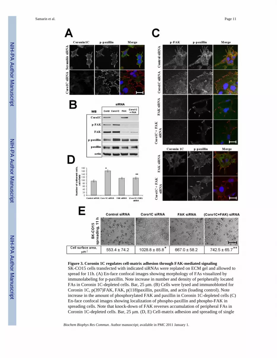
Figure 3. Coronin 1C regulates cell-matrix adhesion through FAK-mediated signalingSK-CO15 cells transfected with indicated siRNAs were replated on ECM gel and allowed tospread for 11h. (A) En-face confocal images showing morphology of FAs visualized byimmunolabeling for p-paxillin. Note increase in number and density of peripherally locatedFAs in Coronin 1C-depleted cells. Bar, 25 μm. (B) Cells were lysed and immunoblotted forCoronin 1C, p(397)FAK, FAK, p(118)paxillin, paxillin, and actin (loading control). Noteincrease in the amount of phosphorylated FAK and paxillin in Coronin 1C-depleted cells (C)En-face confocal images showing localization of phospho-paxillin and phospho-FAK inspreading cells. Note that knock-down of FAK reverses accumulation of peripheral FAs inCoronin 1C-depleted cells. Bar, 25 μm. (D, E) Cell-matrix adhesion and spreading of single
Samarin et al. Page 11
Biochem Biophys Res Commun. Author manuscript; available in PMC 2011 January 1.
NIH
-PA Author Manuscript
NIH
-PA Author Manuscript
NIH
-PA Author Manuscript

and dual Coronin 1C- and FAK knock-downs was analyzed as described in Fig. 2. (D) Averagenumber of adherent cells per microscopic field 3 hours after plaiting. (E) Representative phasecontrast image of spreading cells. Bar, 200 μm. Average cell surface area of spreading cells isshown. *, p<0.01 compared to control; ** p<0.05 compared to Coronin 1C siRNA-transfectedcells.
Samarin et al. Page 12
Biochem Biophys Res Commun. Author manuscript; available in PMC 2011 January 1.
NIH
-PA Author Manuscript
NIH
-PA Author Manuscript
NIH
-PA Author Manuscript

Figure 4. Coronin 1C regulates motility of IECs via FAK-mediated signaling(A) SK-CO15 cells were transfected with the indicated siRNAs. Representative phase contrastimages and quantification of wound closure 24 hours after wounding. Bar, 400 μm. *, p<0.01compared to control; ** p<0.05 compared to Coronin 1C siRNA-transfected cells. (B) A modelfor the regulation of cell motility by Coronin 1C. Coronin 1C increases the rate of actin filamentturnover by regulating Arp2/3/cofilin-dependent actin polymerization/disassembly.Alternatively, Coronin 1C suppresses FAK signaling leading to decreased cell-matrixadhesion. The resulting effect of these two signaling pathways on cell motility would bedependent on the dynamic equilibrium between lamellipodia protrusion and adhesion ofprotruded lamellipodia to ECM.
Samarin et al. Page 13
Biochem Biophys Res Commun. Author manuscript; available in PMC 2011 January 1.
NIH
-PA Author Manuscript
NIH
-PA Author Manuscript
NIH
-PA Author Manuscript


