Embed Size (px)
Citation preview

Isolation and directed differentiation of neural crest stemcells derived from human embryonic stem cellsGabsang Lee1, Hyesoo Kim1,4, Yechiel Elkabetz1,4, George Al Shamy2, Georgia Panagiotakos2, Tiziano Barberi3,Viviane Tabar2 & Lorenz Studer1,2
Vertebrate neural crest development depends on pluripotent, migratory precursor cells. Although avian and murine neural
crest stem (NCS) cells have been identified, the isolation of human NCS cells has remained elusive. Here we report the
derivation of NCS cells from human embryonic stem cells at the neural rosette stage. We show that NCS cells plated at clonal
density give rise to multiple neural crest lineages. The human NCS cells can be propagated in vitro and directed toward
peripheral nervous system lineages (peripheral neurons, Schwann cells) and mesenchymal lineages (smooth muscle, adipogenic,
osteogenic and chondrogenic cells). Transplantation of human NCS cells into the developing chick embryo and adult mouse
hosts demonstrates survival, migration and differentiation compatible with neural crest identity. The availability of unlimited
numbers of human NCS cells offers new opportunities for studies of neural crest development and for efforts to model and treat
neural crest–related disorders.
Human embryonic stem (hES) cells provide an in vitro assay to studyhuman development and a potential source of specialized cells for usein regenerative medicine1. Significant efforts have been devoted tocharacterizing the neural potential of hES cells, and protocols havebeen developed to achieve neural induction2 and differentiationtoward specialized neuron subtypes, including midbrain dopamine3
neurons and spinal motoneurons4,5. Although progress in the speci-fication of central nervous system (CNS) cell types from hES cells hasevolved rapidly, the ability to control peripheral nervous system (PNS)specification has remained limited. A recent study characterized neuralcrest differentiation from cloned bovine blastocysts via a neural rosetteintermediate6. Other studies report the presence of neural crestderivatives among hES cell progeny, including peripheral neuronsand melanocytes7,8. However, neural crest development is thought toemerge from pluripotent precursors, and a systematic understandingof neural crest differentiation from hES cells requires the isolation ofNCS cells.
NCS cells have been well characterized in chick and murinesystems9–11. Defects in the complex processes that choreograph neuralcrest development are involved in a wide range of human diseases12–16.Despite the importance of neural crest cell biology in developmentand disease, the isolation of an NCS cell of human origin has remainedelusive. Efficient strategies for the isolation of NCS cells will beessential for studies of human neural crest development and diseaseand will provide opportunities in regenerative medicine.
Here we report the isolation, propagation and directed differ-entiation of NCS cells from hES cells. Gene expression analysisconfirmed a molecular profile compatible with NCS cell identity.
Clonal analysis showed that hES cell–derived NCS cells are capable ofmultilineage differentiation toward neural crest lineages in vitro. Aftertransplantation into the developing chick embryo, the cells migratedextensively and contributed to neural crest structures, includingperipheral ganglia. Cells subcutaneously injected into adult murinehosts survived, did not form teratomas and contributed to mesen-chymal tissues in dermis and muscle.
RESULTS
Derivation of neural crest precursors
Previous studies have shown that hES cell–derived neural rosettes canbe directed toward various region-specific CNS fates3–5. Here wetested whether neural rosette cultures also have the potential todifferentiate toward PNS fates. Neural induction and rosette forma-tion were performed as described previously3 (using hES cell linesWA-09, I-8 and RUES1-eGFP). Rosettes were mechanically isolatedand replated on polyornithine-laminin–precoated culture dishes (pas-sage 1, P1) and characterized for the expression of neural crestprecursor markers. Expression of p75 and HNK1 was observedprimarily in cells located at the periphery of rosettes (Fig. 1a,b).Cells in the center of neural rosettes expressed the neuroepithelialmarker Pax6 (Fig. 1c). Expression of the neural crest marker AP2 wasobserved predominantly in cell clusters surrounding rosettes (Fig. 1d).
Neural crest development in vivo is modulated by extrinsic pattern-ing factors that determine dorso-ventral identity within the developingneuroepithelium. We observed a significant increase in p75+ putativeneural crest precursors upon exposure of hES cell–derived neuralrosettes to fibroblast growth factor (FGF)2 (2.5-fold ± 0.8, P o 0.05)
Received 15 May; accepted 8 November; published online 25 November 2007; corrected after print 8 July 2008; doi:10.1038/nbt1365
1Developmental Biology Program, and 2Department of Neurosurgery, Sloan-Kettering Institute, 1275 York Ave., New York, New York 10021, USA. 3Division ofNeuroscience, Beckman Research Institute of The City of Hope, 1500 E. Duarte Road, Duarte, California 91010, USA. 4These authors contributed equally to this work.Correspondence should be addressed to L.S. ([email protected]).
1468 VOLUME 25 NUMBER 12 DECEMBER 2007 NATURE BIOTECHNOLOGY
A R T I C L E S©
2008
Nat
ure
Pub
lishi
ng G
roup
ht
tp://
ww
w.n
atur
e.co
m/n
atur
ebio
tech
nolo
gy

or bone morphogenic protein (BMP)2 (2.8-fold ± 1.2, P o 0.01).Treatment with FGF and BMP antagonists inhibited induction ofp75+ cells (0.4-fold ± 0.3 fewer cells with SU5402 treatment and0.7-fold ± 0.2 fewer cells with Noggin treatment) (Fig. 1e). Thesefindings indicate that neural crest precursors spontaneously emerge incultures of hES cell–derived neural rosettes, and that their number canbe regulated by extrinsic signals.
Molecular characterization and clonal analysis
Fluorescence-activated cell sorting (FACS) analysis showed that mosthES cell–derived p75+ neural crest precursors coexpressed HNK1(Fig. 2a) and CD49d (integrin-a4 subunit; Fig. 2b), another NCScell marker17. Analysis of additional neural crest markers and markersassociated with mesenchymal, endothelial, glial or hematopoietic fateyielded a surface marker profile compatible with neural crest precursoridentity (Supplementary Table 1 online).
We next performed gene expression analysis (Affymetrix U133A) toidentity global mRNA profiles in double-positive (p75+/HNK1+)versus double-negative (p75–/HNK1–) P1 rosette progeny (Fig. 2cand Supplementary Table 2 online). Transcripts most highly enrichedin the p75+/HNK1+ cells included key markers of neural crestdevelopment (Brn3a, 32-fold; AP2, 19-fold; Pax3, eightfold; Snail,fivefold) and known NCS cell markers (Sox10, 17-fold; ErbB3, 12-fold;p75, sevenfold; integrin-a4, sevenfold). The p75–/HNK1– cells werehighly enriched in transcripts associated with CNS precursor and/
or forebrain identity. Gene expression data were confirmed byRT-PCR analysis (Supplementary Fig. 1 online) and by quantitativeimmunocytochemistry (Fig. 2d–e). Brn3A is a known marker ofmurine postmitotic sensory neurons but is also expressed in immaturemigrating neural crest precursors18. We observed strong Brn3Aexpression in Tuj1+ neurons 2 d after plating of p75+ cells, but wealso observed weak expression in Tuj1– precursors (Fig. 2f). One of
DAPI HNK1 HNK1/p75/DAPI
Pax6/AP2/DAPIp75/DAPI
p75
p75/DAPI Pax6/DAPI
4
3
2
1
0p7
5+ c
ells
(f
old
incr
ease
)N2only
BDNFAA
(BA)
BAFGF2
BAFGF2DMSO
BAFGF2
SU
BAWnt1
BADkk
BAWnt1Dkk
BABmp2
BANg
BABmp2
Ng
***
* ••
•••
a
b c d
e
104
104
103
103
102
102
101
101100
104
103
102
101
100
104
103
102
101
100
100 104103102101100 104103102101100104103102101100
No staining
No
stai
ning
Isot
ype
p75
Isotype HNK1
0.0 0.1
99.5 98.6
29.5
35.9
100
80
60
40
20
0
FL-2
p75 gated
CD49d-PEIsotype
73.7
0
25
20
15
10
5
p75 + p75 –
Mig
ratio
n ra
te [µ
m/h
] ***
100
80
60
40
20
0
Per
cent
age
of to
tal c
ells
p75 HNK1 Ki-67 Sox10 AP2
POU4F1SCN3ATFAP2BSOX10CDH1ERBB3FLJ14054GDF8MEF2CPAX3CHGAFAPDCNHAPLNITGA4NGFRNEF3HOXA2NPR3TGFBIS100A10CRYZEDNRASNAI2PHOX2B
NOTCH1EMX2LHX2SOX3EFEMP1WNT5BFABP7PLAGL1ZNF312FOXG1BSIX3HTR2CDSCR6SPRY1NPY2RFGF17FGF8
0 10 20 30Fold change [p75+]
Fold changes [p75–]20 10 0
Ki67/DAPI AP2/p75/DAPI Tuj1/Brn3A
26.3
a b
c d f
e g
Figure 1 Cells expressing neural crest markers are present in cultures of
hES cell–derived neural rosettes and can be induced through extrinsic cues.
(a) Coexpression of p75 and HNK1 in passage 1 (P1) neural rosette
cultures. (b) The expression of p75 in the periphery of rosettes. (c,d) Neural
rosette (Pax6+) was surrounded by neural crest cells (AP2+). (e) Fold
increase in the percentage of p75+ cells, measured by FACS analysis,
upon exposure to various cytokines and signaling antagonists in P1 rosette
cultures (*, P o 0.05; **, or �� , P o 0.01; ��� , P o 0.001).Scale bars, 50 mm.
Figure 2 Molecular characterization of neural
crest precursor cells in hES cell–derived neural
rosettes. (a) FACS analysis for p75 and HNK1
in P1 neural rosette cultures. (b) Representative
FACS analysis for coexpression of CD49d in p75+
gated population. (c) Graph for the top increased
(blue) and top decreased (red) genes comparing
p75+/HNK1+ versus p75–/HNK1– populations as
assessed by microarray analysis (AffymetrixU133A). (d) Immunocytochemical analysis of
FACS-purified p75+ cells for markers of cell
proliferation (Ki67) and neural crest identity
(AP2, p75). (e) Quantification of the percentage
of p75+ cells expressing a given marker
immediately after isolation by FACS. (f) Strong
expression of Brn3a in Tuj1+ neurons (arrow) and
weak expression in Tuj1– precursors (arrowhead)
derived from p75+ cells 2 d after plating.
(g) Quantification of cell migration: p75+ cells
showed significantly increased migration behavior
compared with p75– cells as assayed in 10 min
intervals over 6-h period starting 6 h after plating
of FACS-purified cells (mean ± s.e.m.; n ¼ 149
and n ¼ 125 for p75+ and p75– population;
P o 0.001). Scale bar in (d) corresponds to
50 mm in (d) and 25 mm in (f).
NATURE BIOTECHNOLOGY VOLUME 25 NUMBER 12 DECEMBER 2007 1469
A R T I C L E S©
2008
Nat
ure
Pub
lishi
ng G
roup
ht
tp://
ww
w.n
atur
e.co
m/n
atur
ebio
tech
nolo
gy

the functional features that distinguish early PNS and CNS precursorsis the migration rate on fibronectin. Time-lapse microscopy at 6 and24 h after FACS purification revealed that the migration rates of p75+
cells were more than threefold higher than those of p75– cells,(Fig. 2g) with peak migration rates of 100 mm/h (SupplementaryFig. 2 and Supplementary Movies 1 and 2 online).
Clonal analysis showed that the average plating efficiency of p75+/HNK1+ cells was 5–20%. After in vitro expansion of FACS-purifiedp75+/HNK1+ cells for 2 months, single-cell cultures were establishedand cultured for an additional 3 weeks before analysis. We observedthree major differentiated cell types: Peripherin+ and Tuj1+ neurons,glial fibrillary acidic protein (GFAP+) Schwann cells and smoothmuscle actin (SMA+) myofibroblasts. Clonal analysis showed themultipotentiality of p75+/HNK1+ cells (Fig. 3a). An average of 65%of all clones differentiated toward neurons, glia and myofibroblasts.A total of 25% of the clones differentiated to myofibroblasts andneurons, and 10% of the clones gave rise to myofibroblasts only. Allclones that yielded neuronal progeny contained a substantial fractionof Peripherin+ cells (12–89% of Tuj1+ cells).
Freshly isolated p75+/HNK1+ cells cultured in the presence of FGF2and epidermal growth factor (EGF) on ultra-low-attachment platesformed spheres. Cells within spheres expressed neural stem cellmarkers, including Nestin, Musashi1 and Vimentin (Fig. 3b–d)while retaining expression of the neural crest markers (Fig. 3e). Theestablishment of PNS sphere cultures was previously reported forprimary mouse NCS cells19. Spheres derived from p75–/HNK1– hEScell progeny were also positive for Nestin, Mushashi-1 and Vimentin(data not shown) but were mostly negative for neural crest markers(Fig. 3f). Sphere formation by p75+/HNK1+ and p75–/HNK1– cellsoccurred at similar efficiencies (Fig. 3g), and sphere formationpotential was maintained for at least three passages in vitro(Fig. 3h). Sphere formation efficiency was dependent on extrinsicaddition of FGF2 and EGF (Fig. 3i). Whereas differentiation ofp75+/HNK1+ spheres resulted in a bias toward neural crest derivatives,cultures derived from p75–/HNK1– populations were enriched inGABA+ neurons and GFAP+ astrocytes negative for O4 and MBP(Supplementary Fig. 3 online).
Directed differentiation of hES cell–derived NCS cells
We next assessed the differentiation potential of hES cell–derived NCScells after various periods of in vitro expansion in medium containingFGF2 and EGF (Fig. 4a). Neuronal differentiation (Fig. 4b,c) wasinduced by withdrawal of FGF2/EGF and exposure to BDNF, GDNF,NGF and dibutyryl cyclic AMP (dbcAMP), yielding peripheral sym-pathetic neurons (Tyrosine hydroxylase (TH)+/Peripherin+) and sen-sory neurons (Brn3a+/Peripherin+). An average of 25% of all coloniescontained Brn3A+ putative sensory neurons whereas 2% of all coloniescontained TH+ neurons. Most TH+ cells coexpressed peripherin andthe noradrenergic marker dopamine b-hydroxylase. In contrast, TH+
neurons derived from p75– cells were negative for peripherin (Sup-plementary Fig. 4 online). Schwann cell differentiation, as assessed bythe expression of S100b, GFAP and MBP+, was induced in the presenceof CNTF, neuregulin 1b and dbcAMP (Fig. 4d,e). Notably, quantifica-tion of neuronal versus glial differentiation showed that hES cell–derived NCS cells do not yield Schwann cells immediately afterisolation but only after additional in vitro culture (Fig. 4f).
These data were confirmed in clonal cultures maintained for4 weeks after p75 isolation. In such short-term-expanded clones,no GFAP+ cells were detected, whereas identical clones maintainedfor 2 months readily yielded Schwann cell progeny (Fig. 3a). In short-term-expanded clones, 55% of colonies contained peripheral neuronsand myofibroblasts. A total of 45% of the clones yielded myofibro-blasts only, and no pure neuronal clones were observed. These datasuggest that a cell-intrinsic temporal switch in competency occursduring NCS cell progression, similar to that observed for astrocyticdifferentiation during CNS stem cell progression20–23.
150
100
50
Num
ber
of s
pher
es
10010 30
Cells (×103/plate)
40
30
20
20
40
60
10
1 2 3
Passage
Ctrl
FGF2EGF
Both
Growth factor
***
p75–
p75+p75–
p75+
SMA Tuj1 GFAP SMA/GFAP/Tuj1
SMA/GFAP/Tuj1 SMA/GFAP/Tuj1
Nestin/DAPI Msh1/DAPI Vimentin/p75
Spheres [p75+] Spheres [p75–]
Ki67 Ki67AP2/p75/DAPI AP2/p75/DAPI
Sph
eres
cul
ture
sC
lona
l cul
ture
s
a
b c d
e f
g h i
Figure 3 Identification of neural crest stem cell potential by clonal assay
and sphere-forming assay. (a) Three representative examples of clonal
cultures derived from hES cell–derived NCS cells and stained with
markers of myofibroblast (SMA), neurons (Tuj1) and Schwann cells (GFAP).
(b–d) FACS-purified neural crest precursors gave rise to spheres positive
for neural stem cell markers (Nestin, Musashi1 and Vimentin). (d) Blue
color shows nucleus (DAPI). (e,f) Whereas spheres derived from both
p75+ and p75– cells were highly proliferative (Ki67), those derivedfrom p75– cells were largely negative for neural crest markers (AP2/p75).
(g,h) Quantification of the efficiency of sphere formation and the capacity
to form secondary and tertiary spheres in p75+ and p75– populations.
(i) Effect of FGF2 and EGF on sphere formation in p75+ neural crest cells
(*, P o 0.05; **, P o 0.01). Scale bars, 50 mm.
1470 VOLUME 25 NUMBER 12 DECEMBER 2007 NATURE BIOTECHNOLOGY
A R T I C L E S©
2008
Nat
ure
Pub
lishi
ng G
roup
ht
tp://
ww
w.n
atur
e.co
m/n
atur
ebio
tech
nolo
gy

Vertebrate neural crest cells are known to contribute to variousstructures outside the PNS including the head mesenchyme24,25.Clonal analyses during avian cranial neural crest developmentidentified cells with mesodermal and/or ectodermal potential26,27.To examine differentiation potential toward mesenchymal lineages,
we cultured hES cell–derived NCS cells under conditions pre-viously described for the isolation of ES cell–derived mesenchymalprecursors28,29. Under these conditions, cells with mesenchymalmorphologies and marker expression (CD73+) emerged (Fig. 5a).After an additional month of culture, most cells expressed CD73 and
Day 30 Day 60 Day 120
Day 30 Day 60 Day 120
20
15
10
5
0Ctrl Induct. Ctrl Induct. Ctrl Induct.
Per
cent
age
of to
tal c
ells
Culture period prior to differentiation
Tuj1GFAP
hES cells
NCS cellsIn vitro culture (FGF2/EGF)
Differentiation (cocktail)
Tuj1/TH Brn3/Peripherin GFAP/S100B MBP/GFAPa b c d e
f
Figure 4 Differentiation of hES cell–derived NCS cells toward peripheral nervous system
lineages. (a) Schematic illustration of experimental design. hES cell–derived NCS cells
were induced to differentiate following 30, 60 and 120-d periods of in vitro expansion.
(b–e) Representative images of differentiated hES cell-NCS cells stained with the markers
for sympathetic neurons, sensory neurons and Schwann cells. Blue color marks nucleus
(DAPI). (f) Quantification of neuronal and glial differentiation after various periods of
in vitro expansion. Scale bars, 50 mm.
100a b
c d e f
80
60
40
20
0 0
100
80
60
40
20
0
100 101 102 103 104
100 101 102 103 104 100 101 102 103 104 100 101 102 103 104 100 101 102 103 104 100 101 102 103 104
100 101 102 103 104100 101 102 103 104 100 101 102 103 104 100 101 102 103 104
400
300
200
100
0
400
500
300
200
200
250
100
100
150
0
400
500
300
200
100
400
500
300
200
100
00
50
600
400
200
00
300
200
100
0
300
400
200
100
CD73
CD73
0.2 0.1 100
99
Control
CD133 CD105 CD146 CD44
Stro-1 CD73 CD29Serum
CD73 FACS
Phase Oil red Alcian blue Collagen II Alizarin red ALP SMA/SM22α Calp/SM22α
PPARγ
GAPDH
Aggrec.
Collag II
GAPDH
H20 hES cells ctrl treat H20 hES cells ctrl treat H20 hES cells ctrl treat H20 hES cells ctrl treat
ALP
BSP
GAPDH GAPDH
SMAα
SMAγ
NC-MPC NC-MPC NC-MPC NC-MPC
hES
cel
l-NC
SC
hES
cel
l-NC
-MP
C
99.893.9
80.663.313.387.1
Figure 5 Characterization and differentiation of mesenchymal precursor cells derived from hES cell–derived NCS cells. (a) Morphology and CD73 expression
by FACS in hES cell–derived NCS cells and hES cell–derived NCMP cells upon exposure to serum-containing medium. (b) FACS analysis of the surface
marker profile of CD73+ FACS-purified NCS cell–derived mesenchymal precursors. (c–f) Adipogenic (c), chondrogenic (d), osteogenic (e) and smooth muscle
cells (f) were selectively induced from hES cell–derived NCMP cells. Blue color shows nucleus (DAPI). Scale bars, 50 mm.
NATURE BIOTECHNOLOGY VOLUME 25 NUMBER 12 DECEMBER 2007 1471
A R T I C L E S©
2008
Nat
ure
Pub
lishi
ng G
roup
ht
tp://
ww
w.n
atur
e.co
m/n
atur
ebio
tech
nolo
gy

coexpressed a set of surface markers characteristic of mesenchymalstem cell fate, including Stro-1, CD29, CD73, CD146 and CD44(Fig. 5b). We used established mesenchymal stem cell differentiationprotocols30,31 and showed that mesenchymal precursors generatedfrom hES cell–derived NCS cells were capable of adipocytic (Fig. 5c),chondrogenic (Fig. 5d) and osteogenic (Fig. 5e) differentiation.
We recently reported the isolation of skeletal muscle cells fromhES cell–derived mesenchymal precursors using a method thatinvolved isolation by FACS of neural cell adhesion molecule(NCAM)+ progeny29. Mesenchymal precursors in the previous studywere thought to arise from paraxial mesodermal precursors29.Mesenchymal precursors derived in the present study from NCS cellcultures contained 4–9% NCAM+ cells. However, when NCAM+
progeny were isolated by FACS, only few MyoD+ cells were detected,and none of these cells expressed committed myocyte lineage markerssuch as myogenin. Most NCAM+ cells expressed markers typical ofsmooth muscle fate (Fig. 5f).
In vivo analysis of hES cell–derived NCS cell progeny
We assessed the in vivo potential of hES cell–derived NCS cellsafter transplantation into the developing chick embryo and aftersubcutaneous injection into adult nonobese diabetic/severe combinedimmunodeficiencient (NOD/SCID) mice (Fig. 6a). FACS-purified hEScell–derived NCS cells, cultured for o5 d before transplantation,were grafted into the intersomite space of H&H stage 10–12 chickembryos. Human cells were identified by the expression of human-specific markers and labeling with the cell tracer DiI. To confirmfaithfulness of DiI staining, we used DiI-labeled dead cells (repeated
snap freezing/thawing cycles) as controls(Supplementary Fig. 5 online). Three daysafter in ovo transplantation, we observedextensive migration of hES cell–derived NCScell progeny throughout the embryo from thesite of transplantation following ventral,anterior and posterior migration routes(Fig. 6b–c). Although many human cellscontinued to express neural crest precursorcell markers, we found evidence for in vivo
differentiation toward Tuj1+/Islet+ neurons located within hostperipheral ganglia (Fig. 6d–e).
Histological analysis 6–8 weeks after transplantation into adultNOD/SCID mice showed NCAM+ human cells widely dispersed andoften associated with skeletal muscle or lymphatic vessels. Human cellsexpressed multiple precursor cell markers and markers characteristicof pericyte-like populations (Fig. 6f–j). We did not observe signs ofteratoma formation in any of the animals tested, as demonstratedby the absence of a-fetoprotein, Oct-4 and SSEA4 in immunohisto-chemistry analysis and by histological assessment (H&E) (data notshown). These data show in vivo survival and differentiation compa-tible with neural crest precursor identity in both embryonic andadult hosts.
DISCUSSION
We have demonstrated the isolation of NCS cells from hES cell–derived neural rosette cultures. Several observations suggest that NCScells may arise directly from neural rosettes, including the highefficiency of deriving p75+/HNK1+ cells from P1 rosette cultures,the location of p75+ cells at the periphery of rosettes and theresponsiveness of rosette-stage cultures to extrinsic cues known tospecify neural crest development in vivo. However, proof of a directlineage relationship will require clonal analysis of rosette-stage cells.Numerous studies have described molecular signals related to theinduction of neural crest, including secreted growth factors of theBMP, FGF and Wnt families32,33. Effects of FGF2 and BMP2 on neuralcrest differentiation in vitro have been reported34. In the case of hEScell–derived neural rosette cultures, we postulate that BMP2 induces
hES cells
Neuralinduction
In ovotranspl.
Rosettes
p75 FACS
NCSCShort-term
culture
DAPI hNA/Dil/DAPI SMA/Dil/DAPI
Islet/Dil/DAPI Tuj1/Dil/DAPI
hNCAM/DAPI hNestin/DAPI
Vim/hNCAM
NG2/hNCAM
hNCAM/SMA
Lyve/DAPIH & E
Subcuttranspl.in mice
a b c
d e
gf
h i j
Figure 6 In vivo transplantation of hES cell–
derived NCS cell progeny. (a) Schematic
illustration of the in vivo transplantation studies
into the developing chick embryo and into adult
NOD/SCID mice. (b–e) Analysis of cell migration
(b,c) and images for differentiation into Tuj1+/
Islet+ neurons in host sympathetic ganglion (d)
and in locations close to endogenous dorsal rootganglia (e). Red arrow in b shows location of the
injection site. The outline of neural tube is
marked with white line. The yellow box in left
panel marks the area enlarged in right panel.
(f–j) hES cell–derived NCS cells injected
subcutaneously into adult NOD/SCID mice.
Human cells (hNCAM+) were often associated
with skeletal muscle cells (f) and expressed
precursor markers such as human specific nestin
(hNestin) and Vimentin (g). Hematoxylin-Eosin
and Lyve1 staining in h and i revealed the
presence of lymphatic vessels in areas where
human cells were located. In j human cells in
these areas expressed NG2 and SMA compatible
with a pericyte- or smooth muscle phenotypes.
Scale bar, 100 mm.
1472 VOLUME 25 NUMBER 12 DECEMBER 2007 NATURE BIOTECHNOLOGY
A R T I C L E S©
2008
Nat
ure
Pub
lishi
ng G
roup
ht
tp://
ww
w.n
atur
e.co
m/n
atur
ebio
tech
nolo
gy
dorsal cell fate–determination genes35, whereas FGF2 promotes theloss of epithelial organization and the exit of neural crest precursorsfrom rosette structures25. The large number of cells with a neural crestprofile in P1 rosette cultures was surprising given that similar condi-tions are routinely used for the generation of hES cell–derived CNSprogeny. This suggests that studies aimed at generating pure popula-tions of CNS precursors should consider the possible presence ofneural crest derivatives capable of forming proliferating neurosphere-like structures in response to FGF2/EGF exposure.
Gene expression data and clonal analysis showed that p75+/HNK1+
cells in P1 rosette cultures are highly enriched in neural crestprecursors and NCS cells. However, p75+ cells also expressed markersof more differentiated neural crest derivatives. Previous studies haveused negative selection for the early Schwann cell marker P0 forfurther NCS cell purification. However, our cultures were devoid ofcells expressing Schwann cell markers at the time of isolation. Alter-native markers that could be adopted for purification of NCS cellsinclude CD29 (ref. 13) and CD49d17 or CD133 (ref. 36). The platingefficiency in our clonal assays was 5–20% which is 5–10 times lowerthan the cloning efficiency for primary NCS cells, whereas thepercentage of clones capable of multilineage differentiation (450%)was comparable to that of primary NCS cells9.
We also showed that hES cell–derived NCS cells can generateprimary, secondary and tertiary neurosphere structures in vitro similarto those produced by primary NCS cells19. We did observe a decreasein sphere formation potential in the presence of FGF2/EGF after morethan four passages, suggesting that FGF2/EGF may not be sufficientfor long-term self-renewal of hES cell–derived NCS cells in vitro.Although to our knowledge no previous study has described theprospective isolation of NCS cells from hES cell progeny, neural crestprecursors with differentiation potential toward melanocytes, neuronsand GFAP+ cells were isolated from mouse ES cells using cell sortingfor c-kit37. C-kit is a marker previously used in the identification of EScell–derived melanocytes38 and is considered a marker of committedmigratory melanocyte precursors in vivo39. It will be interesting infuture studies to assess c-kit status in FGF2/EGF-expanded anddifferentiating hES cell–derived NCS cells.
Temporal and regional changes in NCS cell competency have beenreported17,40. Our data on Schwann cell differentiation indicate thatearly NCS cells lack access to Schwann cell fate, although clonalderivatives of the same cells were capable ofgenerating Schwann cells after in vitro cul-ture. We propose that these changes in com-petency are due to cell-intrinsic temporalchanges in epigenetic state rather than to anartifact of in vitro culture. The generation ofneuronal and glial derivatives during PNSdevelopment follows a stereotypic sequence,with neurogenesis preceding gliogenesis41.Similarly, glial fate specification in CNSdevelopment follows an early wave of exclu-sively neurogenic differentiation. Schwanncell competency and lack of GFAP expressionmay be related to epigenetic changes, includ-ing direct methylation of the GFAP promoter,as shown during differentiation of fetalneural stem cells and ES cells21–23. AlthoughFGF2-mediated expansion of cell fate poten-tial has been reported in O2A precursor cellsin vitro42, we think that it is unlikely toaccount for the changes in competency
observed here given the lag of 42 months from onset of FGF2/EGFtreatment to acquisition of Schwann cell competency.
We previously reported the isolation of hES cell–derived mesench-ymal precursors using a different protocol that involves a meso-endodermal rather than a neural crest cell intermediate28,29. Inmouse and avian development, progeny from neural crest and paraxialmesoderm closely interact in the formation of connective tissueelements24. A recent study reported molecular markers that distin-guish mesenchymal precursors of mesodermal and neural crest originduring craniofacial development24,43. As shown here, the main differ-ence between mesenchymal precursors derived from NCS cells andour previously published mesenchymal precursor populations is thelack of skeletal muscle progeny and a decreased efficiency in adipocyticdifferentiation. Smooth and skeletal muscle differentiation fromcranial neural crest precursors in vivo has been described44,45. Expres-sion of HoxA2 (ref. 46) and differentiation potential toward chon-drogenic tissues47 suggest the presence of cranial neural crestprecursors in our hES cell–NCS cell cultures. However, our studyhas not shown that a single NCS cell can give rise to both mesench-ymal and neural progeny. Although we did observe Tuj1 expression insingle cell–derived clones containing CD73+ progeny, these cells werenegative for more mature markers such as Peripherin. Furthermore,serum exposure both at clonal density and in bulk culture transientlyinduced increased cell death within the p75+ population, furthercomplicating lineage analyses. Therefore, we cannot formally ruleout that mesenchymal cells arise from a distinct lineage of p75+ cellsthat is selectively capable of survival, clonal expansion and mesench-ymal differentiation in the presence of serum. Future studies arerequired to conclusively address the stem cell nature of putative cranialneural crest progeny derived from hES cells and to explore conditionsthat direct anterior-posterior fate in hES cell-NCS cell cultures,including the derivation of cultures enriched with cells of cardiac,trunk or sacral neural crest identity.
The transplantation data presented here demonstrate in vivosurvival of hES cell–derived NCS cells in both developing and adultvertebrate hosts. In vivo differentiation toward peripheral neuron,smooth muscle, but not Schwann cell fates is in agreement withour in vitro data showing that specification toward glial fates is a lateevent. This suggests that efforts aimed at restoring Schwann cellfunction after peripheral nerve damage will require the use of cells
hES cells Neural crest
Serum free
BDNF, NGF, GDNF, cAMP
Dexa, Insulin, IBXT
TGFβ3, AA
NCAM+ FACS
β-GP, Dexa, AA
CNTF, Nrg, cAMP
Serum
(p75+, HNK1+, AP2+)
p75+
FACSSorting
CD73+
FACSSorting
Mesenchymalcell
(CD73+, Stro1+)
Neuron (Peripherin+, Tuj1+)
Schwann cell (GFAP+, MBP+)
Adipogenic cell (PPARγ+, Oil red+)
Chondrogenic cell (Collagen+, Aggrecan+)
Osteogenic cell (ALP+, BSP+)
Smooth muscle cell (SM22α+, Calponin+)
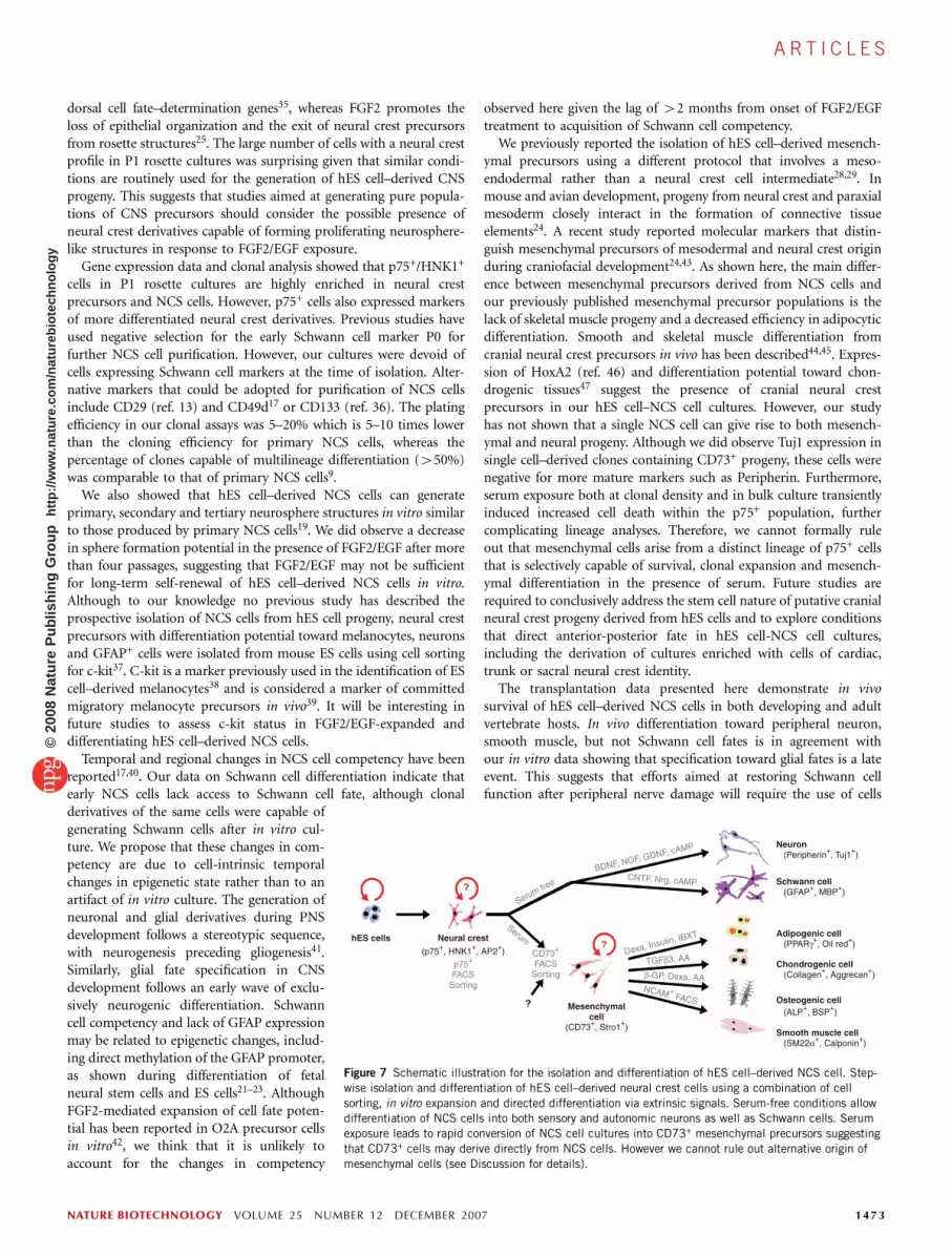
Figure 7 Schematic illustration for the isolation and differentiation of hES cell–derived NCS cell. Step-
wise isolation and differentiation of hES cell–derived neural crest cells using a combination of cell
sorting, in vitro expansion and directed differentiation via extrinsic signals. Serum-free conditions allowdifferentiation of NCS cells into both sensory and autonomic neurons as well as Schwann cells. Serum
exposure leads to rapid conversion of NCS cell cultures into CD73+ mesenchymal precursors suggesting
that CD73+ cells may derive directly from NCS cells. However we cannot rule out alternative origin of
mesenchymal cells (see Discussion for details).
NATURE BIOTECHNOLOGY VOLUME 25 NUMBER 12 DECEMBER 2007 1473
A R T I C L E S©
2008
Nat
ure
Pub
lishi
ng G
roup
ht
tp://
ww
w.n
atur
e.co
m/n
atur
ebio
tech
nolo
gy
predifferentiated to the Schwann cell stage before transplantation.Association of human cells with skeletal muscle and lymphaticstructures in the adult host suggests that hES cell–derived NCS cellscan adopt a wide range of properties depending on environmentalcues. However, transplantation into additional sites will be required tofully probe the in vivo potential of hES cell–derived NCS cells and toconfirm in vivo safety in long-term studies.
Our study reports conditions for the isolation of hES cell–derivedNCS cells and the directed differentiation of a wide range of neuralcrest derivatives, including sensory and autonomic neurons, Schwanncells, myofibroblasts, adipocytes, cartilage and bone cells (Fig. 7).These data should facilitate efforts in tissue engineering, diseasemodeling and regenerative medicine.
METHODSCell culture. Undifferentiated hES cells, H9 (WA-09), RUES1-eGFP48, I-8 were
cultured under growth conditions described previously3 on mitotically inacti-
vated mouse embryonic fibroblasts (MEFs; Chemicon) For neural induction,
hES cells were plated at 5-20 � 103 cells on a confluent layer of irradiated
(50 Gy) stromal cells (MS-5) in 60-mm cell culture plates in serum replace-
ment medium (Invitrogen) containing 2 mM L-glutamine and 10 mM
b-mercaptoethanol. After 16 d in serum replacement medium, cultures were
switched to N2 medium3. Medium was changed every 2–3 d, and growth fac-
tors were added as described previously3: 200 ng/ml sonic hedgehog, 100 ng/ml
FGF8, 20 ng/ml brain-derived neurotrophic factor (BDNF) (all R&D Systems),
and 0.2 mM ascorbic acid (AA) (Sigma–Aldrich). Rosettes structures were
harvested mechanically at day 22–28 of differentiation (termed passage 0; P0)
and gently replated on 15 mg/ml polyornithine/1 mg/ml laminin (PO/Lam)-
coated culture dishes in N2 medium (termed passage 1; P1). P1 cultures were
supplemented with FGF2, AA, and BDNF or any alternative growth factor
conditions as listed in main text. After 6–7 d of P1 culture, cells were
mechanically triturated after exposure to Ca2/Mg2-free Hanks’ balanced salt
solution (CMF-HBSS, 20 min at 25 1C) and labeled with antibodies for flow
cytometry. FACS sorting (p75, Advanced targeting systems; HNK1, Sigma) was
performed on a MoFlo (Dako). Sorted cells were plated on culture dishes
precoated with PO/Lam and 10 ng/ml fibronectin (10–30 � 103 cells/cm2). hES
cell–derived NCS cells were maintained in N2 medium supplemented with
20 ng/ml of FGF2 and 20 ng/ml of EGF changed every 2–3 d and passaged
every 7–8 d.
For sphere formation assay cells were dissociated and plated onto 6-well
ultra-low-attachment plates (Costar, Corning). For serial sphere formation
assays, cells were dissociated with Accutase (Innovative Cell Technologies).
For clonal assays, hES cell–derived NCS cells were mechanically dissociated
after exposure to CMF-HBSS for 20 min at 25 1C and spun at 200g for 5 min.
The cell pellet was resuspended with 1 ml of N2 medium and filtered through
40 mm mesh. Filtered cells were counted and directly plated on 35-mm culture
dishes coated with 15 mg/ml polyornithine/1 mg/ml laminin/10 ng/ml fibro-
nectin at clonal densities (10–30 cells/cm2)9. They were grown in N2 medium
supplemented with bFGF and EGF for 1 week followed by mitogen withdrawal
and culture in N2 medium supplemented with BDNF, nerve growth factor
(NGF, 10 ng/ml), glial cell line–derived neurotrophic factor (GDNF,
10 ng/ml), 1 mM dibutyryl cAMP, ciliary neurotropic factor (CNTF,
10 ng/ml) and neuregulin (20 ng/ml) for a period of at least 2 weeks.
For directed differentiation of hES cell–derived NCS cells toward peripheral
nerve or Schwann cells, FGF2/EGF-expanded hES cell–derived NCS cells were
differentiated upon mitogen withdrawal in medium supplemented with BDNF,
GDNF, NGF, dbcAMP (peripheral nerve) or supplemented with CNTF,
neuregulin, bFGF (10 ng/ml), dbcAMP (Schwann cells).
For mesenchymal differentiation (hES cell–derived neural crest-mesenchy-
mal precursor cells (hES cell–derived NCMP cells)), hES cell–derived NCS cells
were cultured in aMEM containing 10% FBS (FBS, Invitrogen) for 42 weeks
in uncoated tissue-culture grade dishes. FACS sorting (CD73-PE; Pharmingen)
was performed on a MoFlo as described previously28. For adipogenic
differentiation, hES cell–derived NCMP cells were grown to confluence,
followed by exposure to 1 mM dexamethasone, 10 mg/ml insulin, and
0.5 mM isobutylxanthine (all Sigma) in aMEM medium containing 10%
FBS for 43 weeks. For chondrogenic differentiation, hES cell–derived NCMP
cells were induced in pellet culture by exposure to 10 ng/ml TGFb-3 (R&D
Systems) and 200 mM AA in aMEM medium containing 10% FBS for 44
weeks. For osteogenic differentiation, hES cell–derived NCMP cells were plated
at low density (1 � 103 cells/cm2) on tissue culture–treated dishes in the
presence of 10 mM b-glycerol phosphate (Sigma), 0.1 mM dexamethasone, and
200 mM AA in aMEM medium containing 10% FBS for 3–4 weeks. For
myogenic differentiation, hES cell–derived NCMP cells were passaged for 2–3
weeks in aMEM medium with 10% heat inactivated FBS (FBS). At that stage
FACS sorting for NCAM (5.1H11, DSHB) was performed. NCAM+ cells were
grown until confluent in aMEM medium with 10% FBS and then induced to
terminally differentiate in N2 medium.
Immunocytochemistry and flow cytometry. Cells were fixed in 4% parafor-
maldehyde/0.15% picric acid and stained with the primary antibodies (Sup-
plementary Table 3 online). Appropriate Alexa 350, Alexa 488, Alexa 568, Cy5
labeled secondary antibodies (Molecular Probes) and/or DAPI counterstaining
was used for visualization. For flow cytometry, cells were mechanically
dissociated after exposure to CMF-HBSS for 20 min at 25 1C. To eliminate
dead cell populations in FACS analysis, we used 7-AAD accord-
ing to manufacturer’s recommendation. Cells were analyzed using FACScan
(Becton Dickinson) and FlowJo software (Tree Star, Inc.).
RT-PCR and Affymetrix analysis. Total RNA was extracted using the RNeasy
kit and DNAse I treatment (Qiagen) to avoid genomic contamination.
Undifferentiated hES cells, mouse fetal fibroblasts (feeder layer for hES cells),
mouse stromal cells (MS-5), hES cell–derived p75+/HNK1+, p75–/HNK1– cells
and chondrogenic, osteogenic, adipogenic and myogenic cells derived from hES
cell–derived NCMP cells were collected and frozen for further RNA extraction.
Total RNA (2 mg for each sample) was reverse transcribed (Superscript,
Invitrogen). Primer sequences, cycle numbers and annealing temperatures are
provided (Supplementary Table 4 online). For microarray, 5 mg of total RNA
from p75+/HNK1+ and p75–/HNK1– cells were processed by the MSKCC
Genomic core facility and hybridized on Affymetrix U133A human oligo-
nucleotide arrays.
Time-lapse analysis. Cells were monitored 6–24 h after FACS purification of
p75+ and p75– cells and monitored for 1- to 24-h intervals on an Olympus
IX81 microscope under Nomarski optics using a Hamamatsu ORCA CCD
camera, in an enclosed chamber with temperature and CO2 control (Weather
Station, Precision Control). Quantification was performed at 6 h after FACS
and for a 6-h or 24-h observation interval. Cell migration was quantified by
computer-assisted tracking of the center of the cell body in 10-min intervals
(five 2-min frames) using a commercial imaging software package (Slidebook).
Data were quantified for 6-h periods measured in 2-min intervals (180 frames
at 1344 � 1024 resolution).
In vivo transplantation. For in ovo transplantation, fertile eggs (CBT farms)
were incubated at 37 1C in a humidified incubator. The hES cell–NCS cells
in 3–4 d after cell sorting from P1 stage were stained with DiI solution
(Invitrogen) according to manufacturer’s recommendation. After repeated
washings, DiI-labeled hES cell–derived NCS cells were transplanted into
the intersomite space of H&H Stage 10–12 chick embryos49. Eggs were
incubated for 3 d post-transplantation. For subcutaneous transplantations,
200,000 hES cell–derived NCS cells were injected into the neck region of
adult NOD/SCID mice. Animals were monitored for tumor formation
6–8 weeks after transplantation. Tissues from chick embryo and adult mouse
were fixed in 4% paraformaldehyde and cryosectioned for immunohisto-
chemical analysis.
Statistical analysis. The data were processed using Prism 4.0c or Statistica
software. Values are reported as means ± s.e.m. if not indicated otherwise.
Comparisons among values for all groups were performed by one-way ANOVA.
Bartlett’s test for equal variances and Newman-Keuls Multiple Comparison Test
were used to determine the level of significance.
Note: Supplementary information is available on the Nature Biotechnology website.
1474 VOLUME 25 NUMBER 12 DECEMBER 2007 NATURE BIOTECHNOLOGY
A R T I C L E S©
2008
Nat
ure
Pub
lishi
ng G
roup
ht
tp://
ww
w.n
atur
e.co
m/n
atur
ebio
tech
nolo
gy

ACKNOWLEDGMENTSWe would like to thank Z. Dincer and M. Tomishima for technical advice andthe Tri-institutional Stem Cell Research Facility at the Memorial Sloan-KetteringCancer Center (MSKCC) for help in the time-lapse studies. We also would liketo thank J. Itskovitz and M. Amit for providing the I-8 cell line, the MSKCCgenomics core for performing microarray hybridizations, and members of theStuder, Tabar and Tomishima labs for helpful discussions. This work wassupported through the Tri-Institutional Stem Cell Initiative funded by theStarr Foundation.
AUTHOR CONTRIBUTIONSG.L., T.B., V.T. and L.S. designed the study. G.L., V.T. and L.S. analyzed thedata and wrote the manuscript. G.L., H.K., Y.E., G.A.S., G.P. and L.S. performedthe experiments.
Published online at http://www.nature.com/naturebiotechnology/
Reprints and permissions information is available online at http://npg.nature.com/
reprintsandpermissions
1. Joseph, N.M. & Morrison, S.J. Toward an understanding of the physiological function ofMammalian stem cells. Dev. Cell 9, 173–183 (2005).
2. Zhang, S.C., Wernig, M., Duncan, I.D., Brustle, O. & Thomson, J.A. In vitro differentia-tion of transplantable neural precursors from human embryonic stem cells. Nat.Biotechnol. 19, 1129–1133 (2001).
3. Perrier, A.L. et al. From the Cover: Derivation of midbrain dopamine neurons fromhuman embryonic stem cells. Proc. Natl. Acad. Sci. USA 101, 12543–12548(2004).
4. Li, X.J. et al. Specification of motoneurons from human embryonic stem cells. Nat.Biotechnol. 23, 215–221 (2005).
5. Lee, H.J. et al. Directed differentiation and transplantation of human embryonic stemcell derived motoneurons. Stem Cells 25, 1931–1939 (2007).
6. Lazzari, G. et al. Direct derivation of neural rosettes from cloned bovine blastocysts: amodel of early neurulation events and neural crest specification in vitro. Stem Cells 24,2514–2521 (2006).
7. Pomp, O., Brokhman, I., Ben-Dor, I., Reubinoff, B. & Goldstein, R.S. Generation ofperipheral sensory and sympathetic neurons and neural crest cells from humanembryonic stem cells. Stem Cells 23, 923–930 (2005).
8. Fang, D. et al. Defining the conditions for the generation of melanocytes from humanembryonic stem cells. Stem Cells 24, 1668–1677 (2006).
9. Morrison, S.J., White, P.M., Zock, C. & Anderson, D.J. Prospective identification,isolation by flow cytometry, and in vivo self-renewal of multipotent mammalian neuralcrest stem cells. Cell 96, 737–749 (1999).
10. Wong, C.E. et al. Neural crest-derived cells with stem cell features can be traced backto multiple lineages in the adult skin. J. Cell Biol. 175, 1005–1015 (2006).
11. Yoshida, S. et al. Isolation of multipotent neural crest-derived stem cells from the adultmouse cornea. Stem Cells 24, 2714–2722 (2006).
12. Gitler, A.D., Brown, C.B., Kochilas, L., Li, J. & Epstein, J.A. Neural crest migration andmouse models of congenital heart disease. Cold Spring Harb. Symp. Quant. Biol. 67,57–62 (2002).
13. Iwashita, T., Kruger, G.M., Pardal, R., Kiel, M.J. & Morrison, S.J. Hirschsprung diseaseis linked to defects in neural crest stem cell function. Science 301, 972–976 (2003).
14. Fuchs, S. & Sommer, L. The neural crest: understanding stem cell function indevelopment and disease. Neurodegener. Dis. 4, 6–12 (2007).
15. Edery, P. et al. Mutations of the RET proto-oncogene in Hirschsprung’s disease. Nature367, 378–380 (1994).
16. Pingault, V. et al. SOX10 mutations in patients with Waardenburg-Hirschsprungdisease. Nat. Genet. 18, 171–173 (1998).
17. Bixby, S., Kruger, G.M., Mosher, J.T., Joseph, N.M. & Morrison, S.J. Cell-intrinsicdifferences between stem cells from different regions of the peripheral nervous systemregulate the generation of neural diversity. Neuron 35, 643–656 (2002).
18. Fedtsova, N.G. & Turner, E.E. Brn-3.0 expression identifies early post-mitotic CNSneurons and sensory neural precursors. Mech. Dev. 53, 291–304 (1995).
19. Molofsky, A.V. et al. Bmi-1 dependence distinguishes neural stem cell self-renewalfrom progenitor proliferation. Nature 425, 962–967 (2003).
20. Molne, M. et al. Early cortical precursors do not undergo LIF-mediated astrocyticdifferentiation. J. Neurosci. Res. 59, 301–311 (2000).
21. Song, M.R. & Ghosh, A. FGF2-induced chromatin remodeling regulates CNTF-mediated gene expression and astrocyte differentiation. Nat. Neurosci. 7, 229–235(2004).
22. Shimozaki, K., Namihira, M., Nakashima, K. & Taga, T. Stage- and site-specificDNA demethylation during neural cell development from embryonic stem cells.J. Neurochem. 93, 432–439 (2005).
23. Fan, G. et al. DNA methylation controls the timing of astrogliogenesis throughregulation of JAK-STAT signaling. Development 132, 3345–3356 (2005).
24. Noden, D.M. & Trainor, P.A. Relations and interactions between cranial mesoderm andneural crest populations. J. Anat. 207, 575–601 (2005).
25. Kang, P. & Svoboda, K.K. Epithelial-mesenchymal transformation during craniofacialdevelopment. J. Dent. Res. 84, 678–690 (2005).
26. Baroffio, A., Dupin, E. & Le Douarin, N.M. Clone-forming ability and differentiationpotential of migratory neural crest cells. Proc. Natl. Acad. Sci. USA 85, 5325–5329(1988).
27. Baroffio, A., Dupin, E. & Le Douarin, N.M. Common precursors for neural and mesec-todermal derivatives in the cephalic neural crest. Development 112, 301–305(1991).
28. Barberi, T., Willis, L., Socci, N.D. & Studer, L. Derivation of multipotent mesenchymalprecursors from human embryonic stem cells. PLoS Med. 2, e161 (2005).
29. Barberi, T. et al. Derivation of engraftable skeletal myoblasts from human embryonicstem cells. Nat. Med. 13, 642–648 (2007).
30. Pittenger, M.F. et al. Multilineage potential of adult human mesenchymal stem cells.Science 284, 143–147 (1999).
31. Johnstone, B., Hering, T.M., Caplan, A.I., Goldberg, V.M. & Yoo, J.U. In vitrochondrogenesis of bone marrow-derived mesenchymal progenitor cells. Exp. CellRes. 238, 265–272 (1998).
32. Knecht, A.K. & Bronner-Fraser, M. Induction of the neural crest: a multigene process.Nat. Rev. Genet. 3, 453–461 (2002).
33. Le Douarin, N.M. & Dupin, E. Multipotentiality of the neural crest. Curr. Opin. Genet.Dev. 13, 529–536 (2003).
34. Sailer, M.H. et al. BMP2 and FGF2 cooperate to induce neural-crest-like fates fromfetal and adult CNS stem cells. J. Cell Sci. 118, 5849–5860 (2005).
35. Mizuseki, K. et al. Generation of neural crest-derived peripheral neurons and floor platecells from mouse and primate embryonic stem cells. Proc. Natl. Acad. Sci. USA 100,5828–5833 (2003).
36. Marzi, I. et al. Purging of the neuroblastoma stem cell compartment and tumorregression on exposure to hypoxia or cytotoxic treatment. Cancer Res. 67,2402–2407 (2007).
37. Motohashi, T., Aoki, H., Chiba, K., Yoshimura, N. & Kunisada, T. Multipotent cell fate ofneural crest-like cells derived from embryonic stem cells. Stem Cells 25, 402–410(2007).
38. Yamane, T., Hayashi, S., Mizoguchi, M., Yamazaki, H. & Kunisada, T. Derivation ofmelanocytes from embryonic stem cells in culture. Dev. Dyn. 216, 450–458 (1999).
39. Wilson, Y.M., Richards, K.L., Ford-Perriss, M.L., Panthier, J.J. & Murphy, M. Neuralcrest cell lineage segregation in the mouse neural tube. Development 131,6153–6162 (2004).
40. White, P.M. et al. Neural crest stem cells undergo cell-intrinsic developmental changesin sensitivity to instructive differentiation signals. Neuron 29, 57–71 (2001).
41. Kalcheim, C. & Burstyn-Cohen, T. Early stages of neural crest ontogeny: formation andregulation of cell delamination. Int. J. Dev. Biol. 49, 105–116 (2005).
42. Kondo, T. & Raff, M. Oligodendrocyte precursor cells reprogrammed to becomemultipotential CNS stem cells. Science 289, 1754–1757 (2000).
43. Bhattacherjee, V. et al. Neural crest and mesoderm lineage-dependent gene expressionin orofacial development. Differentiation 75, 463–477 (2007).
44. Etchevers, H.C., Vincent, C., Le Douarin, N.M. & Couly, G.F. The cephalic neural crestprovides pericytes and smooth muscle cells to all blood vessels of the face andforebrain. Development 128, 1059–1068 (2001).
45. Korn, J., Christ, B. & Kurz, H. Neuroectodermal origin of brain pericytes and vascularsmooth muscle cells. J. Comp. Neurol. 442, 78–88 (2002).
46. Trainor, P.A., Ariza-McNaughton, L. & Krumlauf, R. Role of the isthmus and FGFsin resolving the paradox of neural crest plasticity and prepatterning. Science 295,1288–1291 (2002).
47. Abzhanov, A., Tzahor, E., Lassar, A.B. & Tabin, C.J. Dissimilar regulation of celldifferentiation in mesencephalic (cranial) and sacral (trunk) neural crest cells invitro. Development 130, 4567–4579 (2003).
48. James, D., Noggle, S.A., Swigut, T. & Brivanlou, A.H. Contribution of human embryonicstem cells to mouse blastocysts. Dev. Biol. 295, 90–102 (2006).
49. Goldstein, R.S. Transplantation of human embryonic stem cells to the chick embryo.Methods Mol. Biol. 331, 137–151 (2006).
NATURE BIOTECHNOLOGY VOLUME 25 NUMBER 12 DECEMBER 2007 1475
A R T I C L E S©
2008
Nat
ure
Pub
lishi
ng G
roup
ht
tp://
ww
w.n
atur
e.co
m/n
atur
ebio
tech
nolo
gy

nature biotechnology volume 26 number 7 july 2008 831
Corrigendum: Large-scale chemical dissection of mitochondrial functionBridget K Wagner, Toshimori Kitami, Tamara J Gilbert, David Peck, Arvind Ramanathan, Stuart L Schreiber, Todd R Golub & Vamsi K MoothaNat. Biotechnol. 26, 343–351 (2008); published online 24 February 2008; corrected after print 8 July 2008
In the version of this article initially published, on p.348, column 2, paragraph 2, line 7, the following sentence was incorrect: “Statins block the synthesis of cholesterol—a precursor to ubiquinone….” It should have read “Statins block the synthesis of mevalonate, a precursor not only of cholesterol but also ubiquinone, ….” The error has been corrected in the HTML and PDF versions of the article.
Corrigendum: Isolation and directed differentiation of neural crest stem cells derived from human embryonic stem cellsGabsang Lee, Hyesoo Kim, Yechiel Elkabetz, George Al Shamy, Georgia Panagiotakos, Tiziano Barberi, Viviane Tabar & Lorenz StuderNat. Biotechnol. 25, 1468–1475 (2007); published online 25 November 2007; corrected after print 8 July 2008
In the version of this article initially published, a reference was missing from the first paragraph. A new sentence and the reference (no. 6) have been added: “A recent study characterized neural crest differentiation from cloned bovine blastocysts via a neural rosette intermediate6. Other….” Subsequent references have been renumbered. The corrections have been made in the HTML and PDF versions of the article.
Erratum: Looking forward, looking backAnonymousNat. Biotechnol. 26, 475 (2008); published online May 2008; corrected after print 13 June 2008
In the version of this article initially published, in paragraph 4, the generic name and ligand given for Avastin are incorrect. The correct generic name is bevacizumab and its target is VEGF (vascular endothelial growth factor). The error has been corrected in the HTML and PDF versions of the article.
Erratum: Is personalized medicine finally arriving?Malorye AllisonNat. Biotechnol. 26, 509–517 (2008); published May, 2008; corrected after print 8 July 2008
In the version of this article initially published, Table 1 (pp. 510–511) contained two errors. In the entry for Agendia, the product Mammaprint was described as providing information on chemotherapy options for breast cancer patients. In fact Mammaprint is a prognostic test. In the entry for Genomic Health, the product Oncotype Dx was described as providing information on breast cancer recurrence. Oncotype DX also provides information on the response to chemotherapy. The error has been corrected in the HTML and PDF versions of the article.
corr igenda and errata©
2008
Nat
ure
Pub
lishi
ng G
roup
ht
tp://
ww
w.n
atur
e.co
m/n
atur
ebio
tech
nolo
gy


![Corrigendum to “Intravenous neural stem cells abolish nociceptive hypersensitivity and trigger nerve regeneration in experimental neuropathy” [Pain 153 (4) (2012) 850–861]](https://img.pdfslide.net/doc/110x75/63378f575b59cce6920b25b7/corrigendum-to-intravenous-neural-stem-cells-abolish-nociceptive-hypersensitivity.jpg)


