Embed Size (px)
Citation preview

ISSN 1591-223X
DOSSIER209-2011
Criteria
for appropriate use
of FDG-PET
in esophageal cancer
ORIentamenti 4
Osservatorio regionaleper l’innovazione


209-2011
Criteria
for appropriate use
of FDG-PET
in esophageal cancer
ORIentamenti 4
Osservatorio regionaleper l’innovazione

This document should be cited as / Il presente documento deve essere citato come
Ballini L, Vignatelli L, Negro A, Maltoni S, Longo G. Criteria for appropriate use of FDG-PET in
esophageal cancer. Dossier 209 - Agenzia sanitaria e sociale regionale, Regione Emilia-Romagna.
2011.
La collana Dossier è curata dal Sistema comunicazione, documentazione, formazione
dell’Agenzia sanitaria e sociale regionale dell’Emilia-Romagna
responsabile Marco Biocca
redazione e impaginazione Federica Sarti
Stampa Regione Emilia-Romagna, Bologna, maggio 2011
Copia del volume può essere richiesta a
Federica Sarti - Agenzia sanitaria e sociale regionale dell’Emilia-Romagna - Sistema CDF
viale Aldo Moro 21 - 40127 Bologna
e-mail [email protected]
oppure può essere scaricata dal sito Internet
http://asr.regione.emilia-romagna.it/wcm/asr/collana_dossier/doss209.htm
Chiunque è autorizzato per fini informativi, di studio o didattici, a utilizzare e duplicare i contenuti
di questa pubblicazione, purché sia citata la fonte.

The report has been prepared by / Il rapporto è stato redatto da
Luciana Ballini Agenzia sanitaria e sociale regionale dell’Emilia-Romagna
Luca Vignatelli Agenzia sanitaria e sociale regionale dell’Emilia-Romagna
Antonella Negro Agenzia sanitaria e sociale regionale dell’Emilia-Romagna
Susanna Maltoni Agenzia sanitaria e sociale regionale dell’Emilia-Romagna
Giuseppe Longo Azienda ospedaliero-universitaria di Modena
The literature search was carried out by /
La ricerca in letteratura è stata effettuata da
Maria Camerlingo Agenzia sanitaria e sociale regionale dell’Emilia-Romagna
This report has been peer-reviewed by /
Il rapporto è stato letto e commentato da
Maurizio Dondi Section Head - Nuclear Medicine
Division of Human Health
International Atomic Energy Agency - Vienna
Eduardo Rosenblatt Section Head - Radiation Oncology
Division of Human Health
International Atomic Energy Agency - Vienna
Barry Siegel Professor of Radiology and Medicine
Director, Division of Nuclear Medicine
Mallinckrodt Institute of Radiology
Washington University School of Medicine
The working group is very grateful to our peer reviewers for their constructive
and useful comments.

Panel members list / Gruppo di lavoro
Monica Agostini Nuclear physician, Azienda USL di Cesena
Salvatore Bacciu Ear, nose and throat specialist
Azienda ospedaliero-universitaria di Parma
Luciana Ballini Coordinator
Agenzia sanitaria e sociale regionale dell’Emilia-Romagna
Alessandra Bologna Oncologist, Azienda ospedaliera di Reggio Emilia
Athos Borghi Internist, Azienda ospedaliero-universitaria di Modena
Alba Brandes Oncologist, Azienda USL di Bologna
Paolo Campioni Radiologist, Azienda ospedaliero-universitaria di Ferrara
Luigi Cavanna Nuclear physician, Azienda USL di Piacenza
Roberto De Maria Surgeon, Azienda USL di Modena
Ermanno Emiliani Radiotherapist, Azienda USL di Ravenna
Stefano Fanti Nuclear physician, Azienda ospedaliero-universitaria di Bologna
Giovanni Frezza Radiotherapist, Azienda USL di Bologna
Andrea Gardini Surgeon, Azienda USL di Forlì
Tiziana Giovannini Agenzia sanitaria e sociale regionale dell’Emilia-Romagna
Cinzia Iotti Radiotherapist, Azienda ospedaliera di Reggio Emilia
Giuseppe Longo Oncologist, Azienda ospedaliero-universitaria di Modena
Moreno Marani Ear, nose and throat specialist, Azienda USL di Forlì
Federica Matteucci Nuclear physician, Azienda USL di Forlì
Renzo Mazzarotto Radiotherapist, Azienda ospedaliero-universitaria di Bologna
Maurizio Miselli Health Director, Azienda ospedaliero-universitaria di Modena
Manlio Monti Oncologist, IRST Meldola
Cristina Nanni Nuclear physician, Azienda ospedaliero-universitaria di Bologna
Antonella Negro Agenzia sanitaria e sociale regionale dell’Emilia-Romagna
Silvia Palazzi Radiotherapist, Azienda USL di Ravenna
Micaela Piccoli Surgeon, Azienda ospedaliero-universitaria di Modena
Monica Silvotti Radiologist, Azienda ospedaliera di Reggio Emilia
Annibale Versari Nuclear physician, Azienda ospedaliera di Reggio Emilia
Claudio Vicini Ear, nose and throat specialist, Azienda USL di Forlì
Luca Vignatelli Agenzia sanitaria e sociale regionale dell’Emilia-Romagna
Elena Zamagni Hematologist, Azienda ospedaliero-universitaria di Bologna

Index
List of abbreviations 7
Sintesi dei risultati 9
Summary of results 13
Foreword 17
1. Introduction and objectives 19
1.1. Use of FDG-PET in esophageal cancer: objectives 21
1.2. Context 21
2. Methods 23
2.1. Clinical questions to be addressed 23
2.2. Systematic review of literature 26
2.3. Level of evidence 28
2.4. Voting process 29
2.5. Definition of criteria of appropriateness 30
3. Systematic review of literature 33
3.1. Overall results 33
4. N staging of patients with primary esophageal
cancer
35
4.1. Systematic review of literature: results 36
4.2. Clinical outcomes 39
4.3. Voting results 39
4.4. Conclusions 40
5. M staging of patients with primary esophageal
cancer
41
5.1. Systematic review of literature: results 42
5.2. Clinical outcomes 44
5.3. Voting results 45
5.4. Conclusions 46
6. Target volume definition of curative radiation
treatment
47
6.1. Systematic review of literature: results 48
6.2. Clinical outcomes 49
6.3. Voting results 49
6.4. Conclusions 50

7. Evaluation of early response to neoadjuvant
therapy
51
7.1. Systematic review of literature: results 52
7.2. Clinical outcomes 56
7.3. Voting results 57
7.4. Conclusions 57
8. Evaluation of response to neoadjuvant therapy
at the end of treatment
59
8.1. Systematic review of literature: results 60
8.2. Clinical outcomes 63
8.3. Voting results 63
8.4. Conclusions 64
9. Follow up in patients with no suspicion of
recurrence
65
9.1. Systematic review of literature: results 66
9.2. Clinical outcomes 67
9.3. Voting results 68
9.4. Conclusions 68
10. Diagnosis and staging of suspect distant
recurrence
69
10.1. Systematic review of literature: results 70
10.2. Clinical outcomes 71
10.3. Voting results 71
10.4. Conclusions 72
Conclusions 73
References 75
Peer review reports 89
Appendices 91
Appendix 1. Voting forms 93
Appendix 2. Systematic review of literature: search strategy
and tables of evidence
113

Criteria for appropriate use of FDG-PET in esophageal cancer
Dossier 209
7
List of abbreviations
AIOM Associazione italiana oncologia medica
ASSR Agenzia sanitaria e sociale regionale
CDSR Cochrane database of systematic reviews
CCT controlled clinical trial
CENTRAL Central register of controlled trials - the Cochrane Library
CRD Centre for Reviews and Dissemination
CT computed tomography
CTV clinical target volume
DARE database of abstracts of reviews of effects
ESMO European Society of Medical Oncology
EUS endoscopic ultrasonography
FDG fluoro-deoxyglucose
FN false negatives
FP false positives
GVT gross target volume
LR likelihood ratio
MA meta-analysis
MRI magnetic resonance imaging
NICE National Institute of Clinical Excellence
PET positron emission tomography
PVT planned target volume
RCT randomized controlled trial
RER Regione Emilia-Romagna
RT radiotherapy
SIGN Scottish Intercollegiate Guidelines Network
SR systematic review
TN true negatives
TP true positives
US ultrasonography


Criteria for appropriate use of FDG-PET in esophageal cancer
Dossier 209
9
Sintesi dei risultati
Criteri per l’uso appropriatodella tomografia ad emissionedi positroni con FDG (FDG-PET)nel tumore dell’esofago
Il panel ha esaminato e stabilito il ruolo della FDG-PET per le seguenti indicazioni
cliniche:
stadiazione N di pazienti con tumore primitivo dell’esofago -
Incerto (livello di evidenza: molto basso)
stadiazione M di pazienti con tumore primitivo dell’esofago -
Appropriato (livello di evidenza: moderato)
definizione del target volume nel trattamento radiante con intento curativo -
Inappropriato (livello di evidenza: molto basso)
valutazione della risposta precoce alla terapia neoadiuvante -
Inappropriato (livello di evidenza: basso)
valutazione della risposta alla terapia neoadiuvante al termine del trattamento -
Incerto (livello di evidenza: molto basso)
follow up di pazienti con nessun sospetto di recidiva -
Inappropriato (livello di evidenza: molto basso)
diagnosi e stadiazione di sospetta recidiva a distanza -
Incerto (livello di evidenza: molto basso)
STADIAZIONE N DI PAZIENTI CON TUMORE PRIMITIVO DELL’ESOFAGO - INCERTO
Il panel ha raggiunto l’accordo nel giudicare incerto l’uso della FDG-PET, in sostituzione
dell’ecografia endoscopica (endoscopic ultrasonography - EUS), nella stadiazione dei
linfonodi regionali in pazienti con tumore dell’esofago.
Il livello di evidenza dell’accuratezza diagnostica della FDG-PET è risultato molto basso,
a causa dell’eterogeneità delle stime di sensibilità e specificità.
Tutti gli esiti, correlati a una corretta selezione dei pazienti rispetto alla chemio-
radioterapia neoadiuvante, sono stati considerati importanti (mediana del punteggio 6).
Data l’alta probabilità pre-test di avere linfonodi positivi nei pazienti con tumore primitivo
dell’esofago, il panel ha ritenuto auspicabile l’applicazione di un test meno invasivo
rispetto all’EUS. Tuttavia, l’incertezza sull’accuratezza diagnostica della FDG-PET ha
indotto il panel a emettere un giudizio molto prudente riguardo il ruolo della FDG-PET
nell’indirizzare le opzioni terapeutiche successive.

Criteria for appropriate use of FDG-PET in esophageal cancer
Dossier 209
10
STADIAZIONE M DI PAZIENTI CON TUMORE PRIMITIVO LOCALMENTE AVANZATO DELL’ESOFAGO
- APPROPRIATO
Il panel ha raggiunto l’accordo alla prima votazione nel giudicare appropriato l’uso della
FDG-PET per la stadiazione delle metastasi a distanza nei pazienti con tumore
dell’esofago localmente avanzato, con lo scopo di indirizzare le successive scelte
terapeutiche.
Il livello di evidenza dell’accuratezza diagnostica della FDG-PET è risultato moderato, e la
prestazione della FDG-PET è migliore rispetto a quella della TC. Tuttavia il panel non
suggerisce una sostituzione della TC da parte della FDG-PET ma ribadisce che debba
essere tenuta in considerazione la migliore accuratezza diagnostica di quest’ultima. Il
panel ha invece fortemente suggerito che, nel caso di utilizzo di una FDG-PET/TC, venga
pianificata una TC diagnostica con contrasto e che venga organizzata una lettura
congiunta dei risultati tra radiologo e medico nucleare.
Le conseguenze per i veri positivi - correttamente stadiati a un livello superiore e
indirizzati in maniera appropriata al trattamento palliativo - e per i falsi positivi - la
incorretta stadiazione a un livello superiore e la rinuncia a un trattamento chirurgico con
intento curativo - hanno ottenuto un punteggio mediano di 8. Gli esiti per i veri e falsi
negativi hanno ottenuto un punteggio mediano di 7. Di conseguenza tutti e quattro gli
esiti sono stati considerati “critici” da parte del panel.
DEFINIZIONE DEL TARGET VOLUME NEL TRATTAMENTO RADIANTE CON INTENTO CURATIVO -
INAPPROPRIATO
Dopo un forte disaccordo iniziale - con i singoli punteggi distribuiti in tutte le categorie di
appropriato, incerto e inappropriato - il panel ha raggiunto l’accordo nel giudicare
inappropriato l’uso della FDG-PET per la definizione del target volume nel trattamento
radiante con intento curativo.
Sul ruolo della FDG-PET nella definizione del campo da irradiare sono stati trovati dati
insufficienti (sparse). Dato lo scopo e la quantità di dose radiante generalmente erogata,
il panel non ha ritenuto necessaria una più accurata definizione del campo rispetto a
quella ottenuta con la diagnostica per immagini attualmente disponibile.
Il panel ha evidenziato il fatto che, dato l’uso appropriato della FDG-PET per la
stadiazione M dei pazienti con tumore dell’esofago, l’immagine ottenuta a tale scopo può
essere utilizzata anche per supportare la stadiazione N o la definizione del campo da
irradiare. Tuttavia questi dati vanno interpretati con molta cautela e le decisioni non
possono essere basate solo su di essi.
VALUTAZIONE DELLA RISPOSTA PRECOCE ALLA TERAPIA NEOADIUVANTE - INAPPROPRIATO
Dopo un forte disaccordo iniziale - con i singoli punteggi distribuiti in tutte le categorie di
appropriato, incerto e inappropriato - il panel ha raggiunto l’accordo nel giudicare
inappropriato l’uso della FDG-PET per la valutazione della risposta precoce alla terapia
neoadiuvante.

Criteria for appropriate use of FDG-PET in esophageal cancer
Dossier 209
11
Il livello di evidenza dell’accuratezza diagnostica della FDG-PET è risultato basso a causa
della eterogeneità delle stime di sensibilità (comprese tra 44 e 100%).
Dato che la percentuale di pazienti che rispondono alla chemio-radioterapia neoadiuvante
è attorno al 43%, il panel ha giudicato “critici” gli esiti correlati alla possibile corretta o
scorretta sospensione del trattamento, con un punteggio maggiore per i pazienti falsi non
responder (mediana del punteggio 8, range 2-9). Le conseguenze per i falsi responder -
che completano un trattamento inefficace - sono state considerate meno importanti
(mediana del punteggio 5, range 2-9).
Nonostante sia sentita la necessità di un test che distingua correttamente i pazienti
responder da quelli non responder, il panel ha giudicato l’accuratezza della FDG-PET e il
possibile danno derivato da una scorretta sospensione di un trattamento efficace
maggiore rispetto ai possibili benefici ottenuti dall’interruzione di un trattamento
inefficace.
VALUTAZIONE DELLA RISPOSTA ALLA TERAPIA NEOADIUVANTE AL TERMINE DEL TRATTAMENTO
- INCERTO
In entrambe le votazioni è stato registrato disaccordo tra i membri del panel, con i singoli
punteggi distribuiti in tutte le categorie alla prima votazione e compresi tra incerto e
inappropriato alla seconda votazione. Pertanto l’uso della FDG-PET in aggiunta alla TC
nella valutazione della risposta alla terapia neoadiuvante al termine del trattamento, allo
scopo di decidere tra trattamento curativo o palliativo, è risultato incerto per disaccordo.
Il livello di evidenza dell’accuratezza diagnostica della FDG-PET è risultato molto basso, a
causa della eterogeneità sia della sensibilità che della specificità.
Gli esiti per i pazienti che risultano responder alla terapia - veri e falsi responder - e per i
falsi non responder sono stati giudicati “critici” (mediana del punteggio pari a 7), mentre
gli esiti per i veri non responder sono stati considerati “importanti”.
FOLLOW UP DI PAZIENTI CON NESSUN SOSPETTO DI RECIDIVA - INAPPROPRIATO
Dopo un iniziale leggero disaccordo tra giudizio inappropriato e incerto, il panel ha
raggiunto l’accordo nel giudicare inappropriato l’uso della FDG-PET nel follow up dei
pazienti con nessun sospetto di recidiva.
Il livello di evidenza dell’accuratezza diagnostica della FDG-PET è risultato molto basso,
in quanto basato su tre soli studi di bassa qualità metodologica.
Gli esiti per i pazienti con recidiva - veri positivi e falsi negativi - così come gli esiti per
i pazienti correttamente diagnosticati come negativi sono stati giudicati “importanti”
(mediana del punteggio pari a 4). Un punteggio lievemente superiore (mediana 6, range
1-9) è stato assegnato all’esito dei pazienti incorrettamente giudicati positivi per
metastasi a distanza, a causa del successivo inutile carico di ansia e di stress provocati
da una diagnosi errata.

Criteria for appropriate use of FDG-PET in esophageal cancer
Dossier 209
12
DIAGNOSI E STADIAZIONE DI SOSPETTA RECIDIVA A DISTANZA - INCERTO
È stato registrato un disaccordo tra i membri del panel in entrambe le votazioni, con
i singoli punteggi compresi tra le categorie di incerto e appropriato. L’uso della FDG-PET
come test aggiuntivo per la diagnosi e lo staging della recidiva a distanza nei pazienti con
sospetto di recidiva o con risultato incerto alla diagnostica per immagini convenzionale
è quindi risultato incerto per disaccordo.
Il livello di evidenza dell’accuratezza diagnostica della FDG-PET è risultato molto basso,
in quanto basato su un solo studio con pochi pazienti.
Gli esiti per i veri e falsi positivi, così come per i falsi negativi, sono stati considerati
“critici” (mediana del punteggio pari a 7). Per i pazienti correttamente risultati negativi
gli esiti sono stati giudicati importanti (mediana del punteggio pari a 6).

Criteria for appropriate use of FDG-PET in esophageal cancer
Dossier 209
13
Summary of results
Criteria for the appropriate use ofpositron emission tomography withFDG (FDG-PET) in esophagealcancer
The panel examined and assessed the role of FDG-PET for the following clinical
indications:
N staging of primary esophageal cancer -
Uncertain (level of evidence: very low)
M staging of primary esophageal cancer -
Appropriate (level of evidence: moderate)
target volume definition of curative radiation treatment -
Inappropriate (level of evidence: very low)
evaluation of early response to neoadjuvant therapy -
Inappropriate (level of evidence: low)
evaluation of response to neoadjuvant therapy at the end of treatment -
Uncertain (level of evidence: very low)
follow up in patients with no suspicion of recurrence -
Inappropriate (level of evidence: very low)
diagnosis and staging of suspect distant recurrence -
Uncertain (level of evidence: very low)
N STAGING OF PRIMARY ESOPHAGEAL CANCER - UNCERTAIN
The panel agreed to judge as uncertain the use of FDG-PET in staging patients with
esophageal cancer for regional lymph nodes, in replacement of endoscopic
ultrasonography (EUS).
The level of evidence for diagnostic accuracy of FDG-PET was very low, with
heterogeneous estimates for both sensitivity and specificity.
All outcomes, related to the correct selection of patients eligible for neoadjuvant
chemoradiation therapy were considered “important” (median score 6). A less invasive
test was also deemed highly desirable, given the high pre-test probability of patients
diagnosed for primary esophageal cancer having positive lymph node. However the
uncertainty on the diagnostic accuracy of FDG-PET made the panel very cautious in
suggesting the use of FDG-PET results to direct therapeutic options.

Criteria for appropriate use of FDG-PET in esophageal cancer
Dossier 209
14
M STAGING OF PRIMARY ESOPHAGEAL CANCER - APPROPRIATE
The panel agreed at the first round in rating as appropriate the use of FDG-PET in
staging patients with esophageal cancer for distant metastasis, in order to decide on
subsequent appropriate therapeutic approach.
The level of evidence for diagnostic accuracy of FDG-PET was moderate, with FDG-PET
performing better than CT.
However the panel did not suggest that FDG-PET should replace CT, but that its higher
accuracy in detecting distant metastases should be taken into account. Rather, it was
strongly suggested that when using FDG-PET/CT scanners, a diagnostic CT with contrast
should be planned and joint results readings between radiologists and nuclear physicians
arranged.
The consequences for true positives - correct upstage and appropriate palliative
treatment - and for false positives - incorrect upstage and denial of surgical curative
treatment - received a median score of 8. Outcomes for true and false negatives
obtained a median score of 7, meaning that all four outcomes were considered “critical”
by the panel.
TARGET VOLUME DEFINITION OF CURATIVE RADIATION TREATMENT - INAPPROPRIATE
After an initial strong disagreement, with ratings falling in all regions of appropriateness,
uncertainty and inappropriateness, the panel reached an agreement in judging the use of
FDG-PET for the field definition of radiation treatment as inappropriate.
Only sparse data evaluating diagnostic accuracy of FDG-PET in the target volume
definition were found, and given the scope and dose delivery of the radiation treatment,
the panel expressed no particular need for more accurate field definition than that
conveyed by available imaging.
It was highlighted by the panel that having judged as appropriate the use of FDG-PET for
M staging of patients diagnosed with esophageal cancer, available FDG-PET images can
be examined, alongside other test results, in support of N staging or of radiation field
definition. However, great caution should be placed in interpreting these data and
decisions should not rely solely on them.
EVALUATION OF EARLY RESPONSE TO NEOADJUVANT THERAPY - INAPPROPRIATE
After an initial strong disagreement, with ratings falling in all regions of appropriateness,
uncertainty and inappropriateness, the panel reached an agreement in judging the use of
FDG-PET for the evaluation of early response to neoadjuvant therapy as inappropriate.
The level of evidence for diagnostic accuracy of FDG-PET was low, due also to the
heterogeneity of estimates for sensitivity (ranging from 44 to 100%).
Given that the proportion of patients responding to neoadjuvant chemoradiation is
around 43%, the panel voted “critical” the outcomes related to the possibility of correctly
or incorrectly suspending the treatment, with a higher score of importance for the
patients resulting false non responders (median score 8; range 2-9). Consequences for

Criteria for appropriate use of FDG-PET in esophageal cancer
Dossier 209
15
false responders - completing ineffective therapy - were considered less important
(median score 5; range 2-9). Though expressing the need for a test that could correctly
discriminate responders from non responders, given the low accuracy of FDG-PET, the
panel judged the accuracy of FDG-PET as insufficient and the risk of incorrectly
suspending an effective treatment higher than the possible benefits of interrupting an
ineffective one.
EVALUATION OF RESPONSE TO NEOADJUVANT THERAPY AT THE END OF TREATMENT -
UNCERTAIN
A disagreement among panelists was registered in both rounds of voting, with ratings
falling in all three regions in the first round, and ratings falling within the uncertain and
inappropriate regions in the second round. The use of FDG-PET, in addition to CT, in the
evaluation of response to neoadjuvant therapy at the end of treatment, in order to
decide between curative or palliative therapeutic course of action, resulted as uncertain
due to disagreement.
The level of evidence for diagnostic accuracy was very low, with heterogeneity for both
sensitivity and specificity. Outcomes for patients testing as responders - true and false
responders - and for false non responders were voted “critical” (median score 7), while
outcomes for true non responders were considered “important”.
FOLLOW UP IN PATIENTS WITH NO SUSPICION OF RECURRENCE - INAPPROPRIATE
After an initial slight disagreement between inappropriate and uncertain, the panel
agreed to judge as inappropriate the use of FDG-PET for patients in follow up with no
suspicion of recurrence.
Level of evidence for diagnostic accuracy of FDG-PET in follow up was very low and
coming from three primary studies of low methodological quality. Outcomes for patients
with recurrence - true positives and false negatives, as well as outcomes for patients
correctly diagnosed as negatives were voted “important” (median score 4). A slightly
higher score (median 6; range 1-9) was assigned to the outcomes of patients incorrectly
testing positive for distant metastases and experiencing unnecessary stress and anxiety.
DIAGNOSIS AND STAGING OF SUSPECT DISTANT RECURRENCE - UNCERTAIN
A disagreement among panelists was registered in both round of voting, with ratings
falling in both the uncertain and appropriate region. The use of FDG-PET as an add on
test for the diagnosis and staging of distant recurrence in patients with clinical suspicion
of recurrence or unclear conventional imaging results resulted as uncertain due to
disagreement.
Level of evidence for diagnostic accuracy of FDG-PET was very low, coming from only
one study with very few patients. Outcomes were considered “critical” (median score 7)
for true and false positives, as well as for false negatives, and “important” (median score
6) for patients correctly found negatives.


Criteria for appropriate use of FDG-PET in esophageal cancer
Dossier 209
17
Foreword
The Regional Observatory for Innovation (ORI) is a research unit within the Regional
Health and Social Agency of Emilia-Romagna (Italy), which support the Local Authority
and its individual health care organizations in governing the adoption of health
technologies.
The Dossiers are developed with multidisciplinary working groups representative of the
regional professional networks. Conclusions are made on both adoption of the technology
and on necessary research projects.
The work leading to the development of the present Dossier on the criteria of appropriate
use of FDG-PET in esophageal cancer has been carried out between September 2010 and
January 2011.
All members of the panel have completed and signed a declaration of conflict of interests
and further details of these are available on request.
This Dossier was also reviewed in draft form by independent and external expert referees
and their comments are reported in full at the end of the document.
The evidence base was synthesized in accordance with the GRADE methodology and the
consensus process was based on the RAND/UCLA Appropriateness Method.
This Dossier is published in 2011 and will be considered for review in five years. Any
update in the interim period will be noted on the ASSR website
http://asr.regione.emilia-romagna.it


Criteria for appropriate use of FDG-PET in esophageal cancer
Dossier 209
19
1. Introduction and objectives
PET imaging is a non invasive nuclear medicine examination based on the detection of
metabolic abnormalities of disease processes through the use of short-lived
radiopharmaceuticals.
Since its introduction in the Emilia-Romagna Regional Health Service the Agenzia
sanitaria e sociale regionale (ASSR) has been committed to promote and support regional
research programs aimed at assessing clinical indications for FDG-PET and supporting
programming policies.
The first research program, conducted with a multidisciplinary panel of regional experts,
resulted in the publication in 2003 of the first regional report on the appropriate use of
FDG-PET in 16 types of tumor, for a total of 47 clinical indications. The results of this first
report were used to carry out a first clinical audit on the use of FDG-PET in the only FDG-
PET centre present in the region in 2002. Of the 452 FDG-PET scans, consecutively
registered and analyzed between January and July 2002, about one third (38.7%)
resulted to be appropriate, while 26.1% were inappropriate (Graph 1).
Following the increase in number of PET scanners (from 1 to 6) an update of the 2003
report was commissioned to a second regional panel and published in 2007. The second
report addressed the role of FDG-PET in 18 types of cancer for a total of 65 clinical
indications, and a second clinical audit was carried out in the 6 regional PET centres.
From the 600 consecutive PET exams analyzed, 56% resulted to be appropriate, 23.4%
fell in the uncertain categories and just over 3% were inappropriate (Graph 2). While
appropriate use had substantially increased since the previous clinical audit (and
inappropriateness had also decreased quite considerably), the increase from around 8%
to 17% of use of FDG-PET in clinical indications not included in the report suggested that
the evaluation had not been sufficiently comprehensive of most clinical and diagnostic
questions addressed in clinical practice.
The present update of the criteria for appropriate use of FDG-PET in oncology, which
involves a much larger multidisciplinary panel of regional experts, is a research project
financed by a national research program of the Ministry of Health. The project proposes
a new methodology for the definition of clinical questions, covering most clinical
situations occurring in routine practice, for the evaluation of the available evidence on
FDG-PET diagnostic accuracy and for the development of criteria of appropriate clinical
use. The critical appraisal of the available literature would be also directed at the
identification of main research gaps, in order to set a list of high priority research
questions that could be addressed by a future research program. With currently
8 authorized PET scanners in Emilia-Romagna region, a further aim of this project is to
explore whether and to what extent criteria of appropriate use can be used for the
programming of policies and services’ activities.

Criteria for appropriate use of FDG-PET in esophageal cancer
Dossier 209
20
Graph 1. Clinical audit 2002 - appropriate use of FDG-PET (452 FDG-PET scans)
Distribution of appropriateness
38,7%
8,0%
18,8%
26,1%
8,4%
0%
10%
20%
30%
40%
50%
60%
70%
80%
90%
100%
1
appropriate uncertain a) uncertain b) inappropriate other indications
Graph 2. Clinical audit 2006 - appropriate use of FDG-PET (588 FDG-PET scans)
Distribution of appropriateness
56,0%
11,1% 12,1%
3,1%0,5%
17,3%
0%
10%
20%
30%
40%
50%
60%
appropriate uncertain a) uncertain b) inappropriate other indications indeterminate

Criteria for appropriate use of FDG-PET in esophageal cancer
Dossier 209
21
1.1. Use of FDG-PET in esophageal cancer: objectives
This work is part of a wider research program covering the use of PET in a total of 20
types of cancer.
The objective of the present report was to define criteria for appropriate use of FDG-PET
for patients with esophageal cancer.
The criteria reported in this document are to be intended as guidance for programs of
clinical governance aimed at:
supporting clinicians on the use of FDG-PET in esophageal cancer
post hoc analyses of appropriate use of FDG-PET
contributing to the planning of the regional health service.
The purpose of this report is not to produce clinical recommendations for the use of FDG-
PET in esophageal cancer.
1.2. Context
Incidence of esophageal cancer
Crude incidence rate of esophageal cancer in Emilia-Romagna Region in 2004 (RER
2009): 5.2 per 100 000 male inhabitants per year and 1.9 per 100 000 female inhabitants
per year.
Prevalence of esophageal cancer
Cumulative 10 years prevalence estimate of esophageal cancer in Emilia-Romagna
Region at 1/1/2005 (RER 2009): 8.9 per 100 000 male inhabitants, corresponding to 180
cases in Emilia-Romagna region, and 3.3 per 100 000 female inhabitants, corresponding
to 70 cases.


Criteria for appropriate use of FDG-PET in esophageal cancer
Dossier 209
23
2. Methods
A panel of 26 experts, comprising nuclear physicians, radiologists, radiotherapists,
surgeons, oncologists, ENT specialists, hematologists and health directors working in
Health Trusts and Teaching Hospitals of Emilia-Romagna was convened to discuss and
agree on the methodology for a research program aimed at defining the criteria for
appropriate use of PET in oncology.
At the first meeting the group decided upon the following issues:
clinical questions to be addressed,
systematic review of literature,
grading of level of evidence,
voting process,
definition of criteria of appropriateness.
2.1. Clinical questions to be addressed
On the basis of the clinical pathway of patients with esophageal cancer (Figure 2.1),
shared by most international clinical practice guidelines, the panel examined and
assessed the role of FDG-PET for seven clinical indications (Table 2.1).
As the diagnosis of esophageal cancer is placed by endoscopic biopsy with histology
(ESMO 2010), the use of FDG-PET in the diagnosis has not been considered by the panel.
Table 2.1. Clinical indications selected by the panel
N staging of primary esophageal cancer
M staging of primary esophageal cancer
Target volume definition of curative radiation treatment
Evaluation of early response to neoadjuvant therapy
Evaluation of response to neoadjuvant therapy at the end of treatment
Follow up in patients with no suspicion of recurrence
Diagnosis and staging of suspect distant recurrence

Criteria for appropriate use of FDG-PET in esophageal cancer
Dossier 209
24
Figure 2.1. Clinical pathway for esophageal cancer
The starting point for the development of answerable “research questions”, based on the
PICO structure (Patient Intervention Comparator Outcome), has been the broad
definition of appropriateness of a diagnostic test, which implies:
an initial diagnosis and the therapeutic approach following the initial diagnosis;
the capacity of the new test (i.e. FDG-PET) to modify the initial diagnosis (or stage of
the disease);
the subsequent change in the therapeutic approach;
the clinical benefit expected from the change in the therapeutic approach endorsed
by the test result.
As for the previously published report, the evidence profile necessary to comprehensively
assess and evaluate the role of a diagnostic test was defined and is represented in Figure
2.2.

Criteria for appropriate use of FDG-PET in esophageal cancer
Dossier 209
25
Figure 2.2. Evidence profile for a diagnostic test
Cl in ic al effecti veness of a diagn osti c tes t
D iag n o s tic
p er fo rm a n ce
Dia g n o stic
a cc ura c y
I m pa c t o n th e
t her ap e u tica p p ro ac h
Co st -
e ffe c tiv ene s s
A va il ab l e r es ea r ch r es ul t s R e se a r ch g a p s
I m p a ct o n the
d ia g n o st icp at h wa y
I mp a c t o n
clin ic alo u t co m e s
S af et y
fe a sib i lity
The persistent gap in research evaluating the impact on therapeutic approach, clinical
outcomes and costs, that is common to most diagnostic tests, was acknowledged and
answerable clinical questions were developed as follows.
To build the PICOs on of FDG-PET clinical appropriateness, participants were identified as
patients in one of the clinical situations selected by the panel (Table 2.1).
Potentials for change in patient’s management following the test results was stated in the
rationale supporting the diagnostic role of FDG-PET and were backed up by either
evidence from studies on change in management or by the pre-test probability calculated
from the raw data extracted from the studies on diagnostic accuracy, representing the
expected percentage of change of approach over the whole patients population.
The intervention was either FDG-PET or CT/PET with a specific role within the diagnostic
pathway and with a pre-defined position in relation to the comparator (replacement,
triage, add on) as defined by Bossuyt et al. (Bossuyt 2006).
The comparator was identified as the currently used or existing test for the diagnostic
role under consideration.
Diagnostic accuracy (sensitivity and specificity) of FDG-PET was identified as the outcome
conveying the test’s capacity to modify the initial diagnosis.
As randomized clinical trials providing robust data on clinical effectiveness of diagnostic
tests are very difficult to perform, and seldom found by systematic literature search, we
decided to adopt the GRADE (Grading of Recommendations, Assessment, Development
and Evaluation) approach to evaluate benefits expected from the change in the
therapeutic approach endorsed by the test’s results (Schünemann 2008). This approach
suggests to state clinical consequences for patients testing positive (true and false
positive) and for patients testing negative (true and false negative). Data of effectiveness
related to important clinical outcomes are replaced by judgments of experts and panelists
are asked to assign a score from 1 to 9 stating the level of importance of patient
outcomes as the result of being a true or false positive or a true or false negative. The

Criteria for appropriate use of FDG-PET in esophageal cancer
Dossier 209
26
balance or trade off between the presumed benefits and the presumed harms, together
with the quality of evidence on diagnostic accuracy, are used by panel members to judge
the level of appropriateness of a test.
2.2. Systematic review of literature
Search methods for the identification of the studies
The following databases were searched for the period between January 2006 - date of
the literature search for the precedent update - and July 2010:
Cochrane Database of Systematic Reviews (CDSR - The Cochrane Library);
Database of Abstracts of Reviews of Effects (DARE - Centre for Reviews and
Dissemination);
Health Technology Assessment Database (Centre for Reviews and Dissemination
CRD);
Cochrane Central Register of Controlled Trials (CENTRAL - The Cochrane Library);
National Library of Medicine’s MEDLINE database (PubMed);
Elsevier’s EMBASE.
Language restrictions: English, Italian, French and Spanish.
Reference lists of identified articles were checked for additional references.
Full details of search terms used are given in Appendix 2.
Selection criteria
Type of studies systematic reviews, RCTs, CCTs, cross sectional diagnostic studies,
prospective or retrospective cohort studies, case series of at least
10 patients
Participants patients with esophageal cancer
Intervention FDG-PET or CT/PET
Reference standard histology or clinical follow up (for diagnostic accuracy studies)
Comparator any other imaging technique
Outcomes sensitivity, specificity, LR, accuracy in clinical target volume (CTV)
definition, metabolic/tumor response, time to recurrence, local,
locoregional and distant recurrence, disease free survival, disease
survival, overall survival

Criteria for appropriate use of FDG-PET in esophageal cancer
Dossier 209
27
Assessment of methodological quality of studies
The following criteria have been used for the quality assessment of different study
designs.
Systematic reviews criteria drawn from the AMSTAR checklist (Shea 2007)
Diagnostic cross sectional studies
criteria drawn from the QUADAS checklist (Whiting 2003)
Randomized controlled trials
criteria suggested by the Cochrane Handbook (Higgins 2009)
Case control studies and cohort studies
criteria drawn from the New Castle-Ottawa checklist
Case series no standardized checklists have been published for the assessment
of methodological quality of case series; the following two criteria
have been used: prospective vs retrospective recruitment;
consecutive recruitment
Data collection and analysis
One review author assessed all abstracts of potentially relevant articles against the study
inclusion criteria, analyzed all articles acquired in full text and assessed methodological
quality for risk of bias addressing selection bias and blind interpretation of results of
index and verification tests.
Data were extracted regarding study design, study population, intervention, comparator,
reference standard and outcomes, and pre-test probabilities were calculated. Data
extracted are reported in single study table of evidence and summarized in synoptic
tables (Appendix 2).
Data synthesis
The following data were extracted from the included studies and provided to the panel:
median of the pre-test probability to have the initial diagnosis modified (for example
to have distant metastasis) or to be in a specific clinical situation (for example
histopathologic response to chemotherapy);
estimates of diagnostic accuracy (sensitivity and specificity) of FDG-PET and
comparator.
When available from meta-analyses (MA), diagnostic accuracy pooled estimates and
clinical outcomes pooled estimates were reported.
When no pooled estimates were given, the median values with ranges were calculated
and test for heterogeneity was carried out with the Cochrane’s chi square heterogeneity
test (Meta-Disc Version 1.4). When heterogeneity was found (p<0.1), only the range of
estimates (minimum and maximum values) were given.

Criteria for appropriate use of FDG-PET in esophageal cancer
Dossier 209
28
With SRs/MA and primary studies available, if patients included in primary studies
published after systematic reviews or meta-analyses added up to a number smaller than
the patients included in the SRs/MA, results from primary studies were analyzed only for
consistency. With SRs/MA and primary studies available, if patients included in primary
studies published after SRs/MA added up to a number greater than the patients included
in the SRs/MA, estimates of all studies have been pooled and re-calculated and
heterogeneity of diagnostic estimates of FDG-PET has been tested.
2.3. Level of evidence
Randomized controlled trials, cross sectional or cohort studies in patients with diagnostic
uncertainty and direct comparison of test results with an appropriate reference standard
were considered of high quality, but their quality was downgraded if any of the following
situations occurred (Guyatt 2008):
study limitations (retrospective or non consecutive recruitment of patients, selection
and spectrum bias, verification bias, lack of concealment, large losses to follow up,
lack of blinding in results reading for index and reference test);
inconsistency of results (heterogeneity or variability in results; unexplained
inconsistency in sensitivity, specificity);
indirectness of results (if important differences exist between the population included
in the studies and population of interest, or between the chosen comparator and
routine practice testing);
imprecision of results (if results come from sparse data, i.e. from few studies - less
than two studies - or an overall small number of patients - less than 200).
Level of evidence for estimates of diagnostic accuracy were assigned according to the
GRADE categorization of the quality of evidence (Guyatt 2008), and defined as follows:
high no risk of bias or important study limitations, consistent results from several
studies and a large number of patients
moderate some study limitations, possible risk of bias, consistent results from several
studies and a large number of patients
low presence of bias, inconsistency and heterogeneity of results for one estimate
of diagnostic accuracy (either sensitivity or specificity), results coming from
several studies and a large number of patients
very low presence of bias, sparse data or inconsistency and heterogeneity of results
for both estimates of diagnostic accuracy (sensitivity and specificity)

Criteria for appropriate use of FDG-PET in esophageal cancer
Dossier 209
29
2.4. Voting process
The panel met twice to discuss and vote on the use of FDG-PET in esophageal cancer.
Each member of the panel, except for the methodologists, voted each clinical question
individually. When voting the level of appropriateness, panelists were asked to take into
consideration:
the role of PET in the diagnostic-therapeutic pathway of the patients;
the change in management brought in by the introduction of FDG-PET and the
effectiveness of the therapeutic approach following FDG-PET results;
the proportion of patients who would have the initial diagnosis changed by FDG-PET;
the level of evidence for the diagnostic accuracy of FDG-PET;
the impact on clinical outcomes resulting from the therapeutic course of action
determined by FDG-PET results;
the balance between benefits and risks resulting from acting on FDG-PET results.
Voting forms
For each clinical question panelists were presented with a voting form (Appendix 1)
containing the following background information:
clinical rationale in support of the use of FDG-PET
clinical effectiveness of therapeutic approach resulting from test results
suggested role of FDG-PET in diagnostic pathway
pre-test probability as a surrogate for change in management or evidence from
studies on change in management when available
estimates of diagnostic accuracy for FDG-PET and comparator
level of evidence
a matrix reporting presumed clinical outcomes for patients testing true and false
positive or negative
estimates of impact on clinical outcomes - when available - and level of evidence
All the above data and information were discussed and approved by the panel during the
first meeting and before proceeding to the vote.
Each panelist voted the level of importance of the clinical outcomes, i.e. the importance
for patients of the consequences from resulting true or false negative or true or false
positive. Scores from 1 to 3 deemed the consequence and resulting outcomes as “not
important”, from 4 to 6 as “important” and from 7 to 9 as “critical”.
When in presence of high, moderate or low level of evidence for diagnostic accuracy,
a matrix of “natural frequencies” (Gigerenzer 2007) reporting absolute numbers for true
and false positive and negative results per 100 patients was given, using the pre-test
probability estimates as prevalence and the estimates of sensitivity and specificity
obtained from the systematic review process.

Criteria for appropriate use of FDG-PET in esophageal cancer
Dossier 209
30
After viewing all the above information, panelists were asked to place a vote on
appropriateness (1 to 3 for “inappropriate”, 4 to 6 for “uncertain” and 7 to 9 for
“appropriate”).
Voting procedure
One round of vote was required for the importance of the clinical outcomes and results
on median scores were presented to the panel.
Two rounds of voting were requested for the judgment of appropriateness and results
were analyzed using the RAND/UCLA Appropriateness Method,1 which allows to measure
both the rating on appropriateness and the level of agreement or disagreement among
the panelists’ rating.
Results from the first round of voting were presented to the panel at the second meeting,
which served the purpose to discuss disagreements and unresolved judgment.
At the end of the two rounds of votes the use of PET for a specific clinical indication was
judged as appropriate when, after discarding one extreme high and one extreme low
rating, all remaining ratings fell within the 7-9 score region. The use of PET was judged
as inappropriate when, after discarding one extreme high and one extreme low rating, all
remaining ratings fell within the 1-3 score region. Finally the use of PET was judged as
uncertain when, after discarding one extreme high and one extreme low rating, all
remaining ratings fell within the 4-6 score region or when no agreement was reached
after the second round of voting.
Results from the voting rounds are reported for each clinical question addressed by the
panel.
2.5. Definition of criteria of appropriateness
To assign a level of appropriateness to the use of FDG-PET, the working group agreed on
the following definitions of appropriate, uncertain and inappropriate use. A fourth
category (indeterminate) was added to take into account clinical indications considered
relevant by the panel, but for which no research results are available.
APPROPRIATE
Clinical indications for which there is a rationale for change in management related to
a patient-important clinical outcome, there is a high or moderate level of evidence for
diagnostic accuracy of PET and the presumed benefit - resulting from the test results -
is greater than the presumed harm.
1 http://www.rand.org/pubs/monograph_reports/MR1269.html
(last access May 25, 2011)
Criteria for appropriate use of FDG-PET in esophageal cancer
Dossier 209
31
UNCERTAIN
Clinical indications for which there is a rationale for change in management related to a
patient-important clinical outcome, but there is a low or very low level of evidence for
diagnostic accuracy of FDG-PET.
INAPPROPRIATE
Clinical indications for which there is NO rationale for change in management related
to a patient-important clinical outcome
Clinical indications for which there is a rationale for change in management related to
a patient-important clinical outcome, there is a high or moderate level of evidence on
diagnostic accuracy of FDG-PET and the presumed harm - resulting from the test
results - is greater than the presumed benefit.
INDETERMINATE
Clinical indications for which there is a rationale for change in management related to a
patient-important clinical outcome, but there are no data on diagnostic accuracy of FDG-
PET.
Clinical indications for which the panel does not reach an agreement on level of
appropriateness after two rounds of voting also fall in the UNCERTAIN category.


Criteria for appropriate use of FDG-PET in esophageal cancer
Dossier 209
33
3. Systematic reviewof literature
3.1. Overall results
Methods and results of the systematic review of literature are reported in full in Appendix
2. The initial search identified 634 records; 128 were excluded because duplicates and
a further 400 did not meet the inclusion criteria. Full text was acquired for the remaining
potentially eligible 106 records, from which 50 studies were excluded on the basis of
inclusion criteria while another 22 resulted already included in systematic reviews. Thirty-
four studies were finally included.
Table 3.1 reports number and type of studies for each clinical question and endpoint as
well as conclusions from the previous 2007 report (Liberati 2007 - Dossier 157).
Only three studies evaluating impact on clinical outcomes were found and included, and
the remaining 31 included studies evaluated only diagnostic accuracy of FDG-PET.

Criteria for appropriate use of FDG-PET in esophageal cancer
Dossier 209
34
Table 3.1. Number of included studies for questions and endpoints
Clinical question
Endpoint
Staging Target Volumedefinition for
radical RT
Early response totherapy (during
treatment)
Response totherapy (end of
treatment)
Follow up Detection andstaging ofsuspectedrecurrence
Diagnostic accuracy Systematic reviews: 1
N staging
primary studies: 12
M staging
primary studies: 4
Systematic reviews: 1
Primary studies: 1
Systematic reviews: 2
Primary studies: 1
Systematic reviews: 3
Primary studies: 7
Systematic reviews: 0
Primary studies: 3
Systematic reviews: 0
Primary studies: 1
Impact on clinical
outcomes
Systematic reviews: 0
Primary studies: 2
Systematic reviews: 0
Primary studies: 0
Systematic reviews: 0
Primary studies: 1
Systematic reviews: 0
Primary studies: 0
Systematic reviews: 0
Primary studies: 0
Systematic reviews: 0
Primary studies: 0
Dossier 157 N staging
not considered
M staging
appropriate
Not considered Not considered Not considered Not considered Potentially useful
(uncertain A)

Criteria for appropriate use of FDG-PET in esophageal cancer
Dossier 209
35
4. N staging of patientswith primary esophagealcancer
Rationale
Surgical treatment is the therapy of choice for all patients with potentially curable
esophageal cancer and who are fit for major surgery (AIOM 2009; ESMO 2010; NCCN
2010; SIGN 2006).
Accurate pre-operative staging is necessary to correctly direct patients to curative
surgery, non curative surgery or non surgical therapy (combined chemoradiation).
N staging is used to decide on need for neoadjuvant treatment.
Diagnostic role of FDG-PET
It is suggested that FDG-PET could represent a less invasive diagnostic test for the
correct identification and selection of patients candidate to neoadjuvant treatment.
Treatment effectiveness
The expected 2-year survival after curative surgical treatment (without neoadjuvant
chemoradiotherapy) ranges between 20 and 50%. In case of regional lymph node
involvement long-term survival does not exceed 25%. In patients with locally advanced
cancer pre-operative chemoradiotherapy improves the 2-year survival by 13% (absolute
difference) compared to surgical treatment only (Gebski 2007).
Pre-test probability and change in management
The median pre-test probability of cancer involvement of regional nodes is 59.9% (range
6.9-95.2%; data from studies on FDG-PET in van Vliet 2008), which could be considered
to be the hypothetical maximum extent of change in management, achievable through
accurate N staging.
Research question: FDG-PET as replacement
Is FDG-PET better (i.e. has higher diagnostic accuracy) than the available comparators
(CT and EUS) in staging regional lymph nodes of patients with esophageal cancer?

Criteria for appropriate use of FDG-PET in esophageal cancer
Dossier 209
36
4.1. Systematic review of literature: results
Results from update of systematic review of literature from Jan 2006
Only studies evaluating diagnostic accuracy were found and results are reported below.
Systematic reviews
One systematic review (van Vliet 2008), comparing the diagnostic accuracy of endoscopy
ultrasonography (EUS), computed tomography (CT) and FDG-PET in staging regional
lymph node, has been included (Table 4.1). The characteristics of recruited patients were
not reported. Methodological quality of this systematic review is judged as intermediate.
According to the authors virtually all studies included in the review are prone to
verification bias, and some of them are not blind.
Table 4.1. Main results of the van Vliet’s 2008 systematic review on N staging
Reference van Vliet 2008
Update to January 2006
Number of studies 10
Number of patients 424
(median 43, range 21-81)
FDG-PET / PET-CT sensitivity: pooled 57% (95% CI 43-70)
specificity: pooled 85% (95% CI 76-95)
Comparator EUS (31 studies, 1 841 patients)
sensitivity: pooled 80% (95% CI 75-84)
specificity: pooled 70% (95% CI 65-75)
CT (17 studies, 943 patients)
sensitivity: pooled 50% (95% CI 41-60)
specificity: pooled 83% (95% CI 77-89)
Reference standard resection
fine needle aspiration
autopsy/follow up

Criteria for appropriate use of FDG-PET in esophageal cancer
Dossier 209
37
Primary studies
Twelve studies evaluating diagnostic accuracy of FDG-PET in the staging of patients with
esophageal cancer published after the above reported systematic review were included
(Table 4.2; Buchmann 2006; Choi 2010; Hsu 2009; Hu 2009; Kato 2008; Katsoulis 2007;
Little 2007; Okada 2009; Roedl 2009a; Sandha 2008; Schreurs 2008; Yuan 2006). Eight
studies applied FDG-PET and 4 FDG-PET/CT. Studies included patients with squamous
cell carcinoma (6) or adenocarcinoma (2) or both (4).
As number of patients of primary studies not included in the van Vliet’s 2008 systematic
review added up to a number greater than those included in van Vliet 2008, all studies
have been pooled and heterogeneity of diagnostic estimates of FDG-PET tested (Table
4.3).
Table 4.2. Main results of primary studies on N staging published after van Vliet’s
2008 systematic review
Reference Buchmann 2006; Choi 2010; Hsu 2009; Hu 2009; Kato 2008; Katsoulis
2007; Little 2007; Okada 2009; Roedl 2009a; Sandha 2008; Schreurs
2008; Yuan 2006
Number of studies 12
Number of patients 622 (median 47.5, range 18-173)
FDG-PET/PET-CT sensitivity: median 68% (0-100%)
specificity: median 92% (67-100%)
Comparator EUS (3 studies, 261 patients)
sensitivity: range 41.8-91.7%
specificity: range 60-97.6%
CT (5 studies, 429 patients)
sensitivity: median 48.3% (range 33.3-75%)
specificity: median 92.6% (range 66.7-100%)
Reference standard resection
fine needle aspiration
autopsy/follow up

Criteria for appropriate use of FDG-PET in esophageal cancer
Dossier 209
38
Table 4.3. Main results on diagnostic accuracy of studies on N staging
Diagnostic accuracy
Number of studies 22
Number of patients 957 (median 45, range 12-173)
Pre-test probability median 59.9% (6.9-95.2%)
FDG-PET/PET-CT sensitivity: median 62% (range 0-100%)
heterogeneity chi-squared = 106.50 (d.f. = 19) p = 0.000
inconsistency (I-square) = 82.2%
specificity: median 89% (range 60-100%)
heterogeneity chi-squared = 58.11 (d.f. = 19) p = 0.000
inconsistency (I-square) = 67.3%
Reference standard resection
fine needle aspiration
autopsy/follow up
References primary studies from van Vliet 2008; Buchmann 2006; Choi 2010; Hsu
2009; Hu 2009; Kato 2008; Katsoulis 2007; Little 2007; Okada 2009;
Roedl 2009a; Sandha 2008; Schreurs 2008; Yuan 2006
Comments of ASSR reviewer
For N staging a great variability in the estimates of diagnostic accuracy is reported.
Without careful analysis of source of variability, it proves difficult to draw conclusion
regarding the applicability of FDG-PET for N staging.
Diagnostic accuracy estimates
FDG-PET sensitivity: (heterogeneous) range 0-100%
FDG-PET specificity: (heterogeneous) range 60-100%
EUS sensitivity:* (pooled) 80%
EUS specificity:* (pooled) 70%
* data from studies evaluating FDG-PET included in van Vliet 2008.
LEVEL OF EVIDENCE: VERY LOW

Criteria for appropriate use of FDG-PET in esophageal cancer
Dossier 209
39
4.2. Clinical outcomes
To evaluate the balance between benefits and risks, the panel agreed to consider the
presumed patient-important outcomes reported below (Table 4.4), and voted on the level
of importance for each outcome. Median scores and ranges are reported for each
outcome.
All outcomes were voted “important”.
No studies investigating the impact of FDG-PET on the above clinical outcomes were
found.
No matrix of “natural frequencies” was provided because of heterogeneity of both
estimates.
Table 4.4. Patient-important clinical outcomes and median scores of importance
Patient-important outcomes Median score
(range)
Consequences of test for patients with involvement of regional nodes
True positives - patients are correctly upstaged and undergo neodjuvant
therapy, which could improve survival
6
(4-9)
False negatives - patients are incorrectly downstaged and do not receive
necessary neoadjuvant therapy, which could have improved survival
6
(4-9)
Consequences of test for patients without involvement of regional nodes
True negatives - patients proceed directly to curative resection of primary
tumor, aimed at improving survival
6
(3-9)
False positives - patients are incorrectly upstaged and have to undergo
unnecessary neodjuvant therapy, with no improvement on survival and
possible unnecessary peri/post-operative adverse effects.
6
(3-9)
4.3. Voting results
After an initial slight disagreement, with ratings falling in the uncertain and appropriate
regions (median score 6; range 4-8), the second voting round registered an agreement
on uncertain with a median score of 5 and range from 4 to 6.
FINAL RATING FOR THE USE OF FDG-PETFOR N STAGING OF PRIMARY ESOPHAGEAL CANCER:
UNCERTAIN

Criteria for appropriate use of FDG-PET in esophageal cancer
Dossier 209
40
4.4. Conclusions
The panel agreed to judge as uncertain the use of FDG-PET in staging patients with
esophageal cancer for regional lymph nodes, in replacement of endoscopic
ultrasonography (EUS).
The level of evidence for diagnostic accuracy of FDG-PET was very low, with
heterogeneous estimates for both sensitivity and specificity.
All outcomes, related to the correct selection of patients eligible for neoadjuvant
chemoradiation therapy were considered “important” (median score 6). A less invasive
test was also deemed highly desirable, given the high pre-test probability of patients
diagnosed for primary esophageal cancer having positive lymph node. However the
uncertainty on the diagnostic accuracy of FDG-PET made the panel very cautious in
suggesting use of FDG-PET results to direct therapeutic options.

Criteria for appropriate use of FDG-PET in esophageal cancer
Dossier 209
41
5. M staging of patientswith primary esophagealcancer
Rationale
Tumor stage at diagnosis and comorbidity are strong predictors of outcome and
determinants of survival. M staging has a role in identifying and selecting patients
candidate to curative surgery.
Diagnostic role of FDG-PET
It is suggested that FDG-PET could be more accurate in discriminating patients eligible
for curative surgery from patients eligible for treatment of distant metastases.
Treatment effectiveness
Only palliative treatment is available for metastatic esophageal cancer, aimed at
improving quality of life.
Pre-test probability and change in management
The median pre-test probability of occurrence of distant metastases is 35.7% (range
8.6-54.2%; data from studies on FDG-PET in van Vliet 2008).
Evidence from 18 studies on change in management following FDG-PET exams shows a
median estimate of 20%, with almost all patients upstaged, with a change from curative
to palliative intent treatment (Berrisford 2008; Buchmann 2006; Chatterton 2009; Duong
2006a; Gananadha 2008; Katsoulis 2007; Malik 2006; McDonough 2008; Meyers 2007;
Noble 2009; Pfau 2007; Pifarré-Montaner 2009; Salahudeen 2008; Smith 2009; van
Westreenen 2007; Walker 2011; Williams 2009).
Research question: FDG-PET as replacement
Is FDG-PET better (i.e. has higher diagnostic accuracy) than the available comparator
(CT) in staging patients with primary esophageal cancer for distant metastasis?

Criteria for appropriate use of FDG-PET in esophageal cancer
Dossier 209
42
5.1. Systematic review of literature: results
Results from update of systematic review of literature from Jan 2006
One systematic review and four primary studies evaluating diagnostic accuracy were
found, as well as two studies evaluating impact of FDG-PET on clinical outcomes. Results
are reported below.
DIAGNOSTIC ACCURACY
Systematic reviews
One systematic review (van Vliet 2008) comparing the diagnostic accuracy of computed
tomography (CT) and FDG-PET in staging distant metastases, has been included (Table
5.1). The characteristics of recruited patients were not reported. Methodological quality
of this systematic review is judged as intermediate. According to the authors, virtually all
studies included in the review are prone to verification bias, and some of them are not
blind.
Table 5.1. Results of systematic review on M staging (distant metastases)
Reference van Vliet 2008
Update to January 2006
Number of studies 9
Number of patients 475 (median 48, range 35-81)
FDG-PET/PET-CT sensitivity: pooled 71% (95% CI 62-79)
specificity: pooled 93% (95% CI 89-97)
Comparator CT
sensitivity: pooled 52% (95% CI 33-71)
specificity: pooled 91% (95% CI 86-96)
Reference standard resection
fine needle aspiration
autopsy/follow up

Criteria for appropriate use of FDG-PET in esophageal cancer
Dossier 209
43
Primary studies
Four studies evaluating diagnostic accuracy of FDG-PET in the staging of patients with
esophageal cancer published after the above reported systematic review were included
(Buchmann 2006; Katsoulis 2007; Little 2007; Noble 2009). All studies applied FDG-PET.
Studies included patients with adenocarcinoma (1) or squamous cell carcinoma and
adenocarcinoma (3). The studies have been retrieved and assessed only for overall
consistency with results on diagnostic accuracy of the above reported systematic review
(Table 5.2).
As results of primary studies are consistent with those of the systematic reviews, the
latter’s pooled estimates were chosen.
Table 5.2. Results of primary studies on M staging
Reference Buchmann 2006; Katsoulis 2007; Little 2007; Noble 2009
Number of studies 4
Number of patients 291 (median 40, range 20-191)
FDG-PET/PET-CT sensitivity: median 88% (range 60-91%)
specificity: median 94.5% (range 86-100%)
Reference standard resection
fine needle aspiration
autopsy/follow up
Comments of ASSR reviewer
Results from the systematic review (SR) for detection of distant metastases show a
higher performance for FDG-PET compared to CT. Specificity is higher than sensitivity.
Due to possible verification bias all results could overestimate diagnostic accuracy.
As results from primary studies published since 2006 confirm the diagnostic accuracy
estimates of van Vliet’s SR (2008) - i.e. a better performance of FDG-PET in M staging
than N staging and higher values of specificity than sensitivity - estimates of diagnostic
accuracy were based on the SR’s pooled estimates.
Diagnostic accuracy estimates
FDG-PET sensitivity: (pooled) 71%
FDG-PET specificity: (pooled) 93%
CT* specificity: (pooled) 52%,
CT* specificity: (pooled) 91%
* data from studies evaluating FDG-PET
LEVEL OF EVIDENCE: MODERATE

Criteria for appropriate use of FDG-PET in esophageal cancer
Dossier 209
44
IMPACT ON CLINICAL OUTCOMES
Primary studies
Two studies evaluating secondary clinical outcomes (burden of diagnostic test) were
found (Meyers 2007; Westerterp 2008). The first study (Meyers 2007) included 189
patients eligible for curative surgery and found that 2 patients (1%) suffered the adverse
consequences of the change in management due to a false positive FDG-PET result. The
first patient underwent adrenalectomy with subsequent therapy for adrenal insufficiency,
the second patient had a wound complication following confirmatory procedure. The
second study (Westerterp 2008) included 82 patients eligible for curative surgery, who
carried out a subjective comparative evaluation of the burden, in terms of discomfort,
embarrassment and anxiety, of diagnostic tests performed for the staging, such as FDG-
PET, CT, US (with or without fine needle aspiration) and EUS (with or without fine needle
aspiration). The perceived burden of FDG-PET was lower than that of EUS, and higher
than that of CT, although the large majority of subjects reported “none” or “little” burden
for all tests and all dimensions.
Comments of ASSR reviewer
No studies investigating the main aspects of impact on clinical outcomes of FDG-PET
were found, but two studies investigating two ancillary aspects were retrieved (one study
reporting the adverse consequences of the change in management due to a false positive
FDG-PET result and another study investigating the patient burden for the different
imaging tests during staging). Due to the paucity of data no firm conclusion could be
drawn.
LEVEL OF EVIDENCE: VERY LOW
5.2. Clinical outcomes
To evaluate the balance between benefits and risks, the panel agreed to consider the
presumed patient-important outcomes reported below (Table 5.3), and voted on the level
of importance for each outcome. Median scores and ranges are reported for each
outcome.
All outcomes were considered “critical” by the panel with consequences for true and false
positives receiving a median score of 8, and outcomes for true and false negatives a
median score of 7.
No studies investigating the impact of FDG-PET on the above clinical outcomes were
found.
A matrix of “natural frequencies” was provided (Table 5.4).

Criteria for appropriate use of FDG-PET in esophageal cancer
Dossier 209
45
Table 5.3. Patient-important clinical outcomes and median scores of importance
Patient-important outcomes Median score
(range)
Consequences of test for patients with distant metastases
True positives - patients are correctly upstaged and proceed to palliative
treatment, aimed at improving quality of life
8
(2-9)
False negatives - patients are incorrectly downstaged and undergo
unnecessary curative surgical treatment, which might not improve survival
7
(3-9)
Consequences of test for patients without distant metastases
True negatives - patients correctly proceed to curative surgical treatment,
which could improve survival
7
(2-9)
False positives - patients are incorrectly upstaged and denied necessary
curative surgical treatment, which could have improved survival, and
proceed to palliative treatment.
8
(3-9)
Table 5.4. “Natural frequencies” of patients staged for distant metastasis
N of patients out of 100 submitted to the exam
According to FDG-PET According to CT
True positives 26 19Patients with
distant metastasis False negatives 10 17
True negatives 60 58Patients without
distant metastasis False positives 4 6
100 100
5.3. Voting results
The first voting round registered an agreement (median score 8; range 7-9) on
appropriate rating.
FINAL RATING FOR THE USE OF FDG-PETFOR M STAGING OF PRIMARY ESOPHAGEAL CANCER:
APPROPRIATE

Criteria for appropriate use of FDG-PET in esophageal cancer
Dossier 209
46
5.4. Conclusions
The panel agreed at the first round in rating as appropriate the use of FDG-PET
in staging patients with esophageal cancer for distant metastasis, in order to decide on
subsequent appropriate therapeutic approach.
The level of evidence for diagnostic accuracy of FDG-PET was moderate, with FDG-PET
performing better than CT.
However the panel did not suggest that FDG-PET should replace CT, but that its higher
accuracy in detecting distant metastases should be taken into account. Rather, it was
strongly suggested that when using FDG-PET/CT scanners, a diagnostic CT with contrast
should be planned and joint results readings between radiologists and nuclear physicians
arranged.
The consequences for true positives - correct upstage and appropriate palliative
treatment - and for false positives - incorrect upstage and denial of surgical curative
treatment - received a median score of 8. Outcomes for true and false negatives
obtained a median score of 7, meaning that all four outcomes were considered “critical”
by the panel.

Criteria for appropriate use of FDG-PET in esophageal cancer
Dossier 209
47
6. Target volume definition ofcurative radiation treatment
Rationale
Radiotherapy (with chemotherapy) is recommended as neoadjuvant treatment for locally
advanced esophageal cancer by the majority of guidelines (AIOM 2009; ESMO 2010;
NCCN 2010). All examined guidelines (AIOM 2009; ESMO 2010; NCCN 2010; SIGN 2006)
propose radiotherapy (with or without chemotherapy) with curative intent for patients
unfit for or unwilling to undergo surgery.
Diagnostic role of FDG-PET
A more precise diagnostic tool allowing a better definition of target volume could reduce
adverse effects of radiation treatment.
Treatment effectiveness
For patients with locally advanced disease or operable esophageal cancer who decline
surgery or who are unfit for surgery, chemoradiation may be an appropriate alternative
(SIGN 2006).
Change in management
No available data.
Research question: FDG-PET in addition to CT
Does adding FDG-PET imaging lead to a better target volume definition of curative
radiotherapy in patients with esophageal cancer?

Criteria for appropriate use of FDG-PET in esophageal cancer
Dossier 209
48
6.1. Systematic review of literature: results
Results from update of systematic review of literature from Jan 2006
One systematic review on volume definition and one study evaluating diagnostic accuracy
were found. Results are reported below.
Systematic reviews
Only one systematic review assessing the role of FDG-PET in tumor volume definition in
radiotherapy treatment planning in esophageal cancer was included (Muijs 2010), that
incorporated also the studies included and assessed by a previous systematic review (van
Baardwijk 2006). Methodological quality was judged as low (Table 6.1).
Table 6.1. Results of systematic review on diagnostic accuracy of FDG-PET in the field
definition of curative radiotherapy
Reference Muijs 2010
Update to 2009
Number of studies 10
Number of patients 231
Results changes in the delineation of target volumes (GTV/CTV/PTV*) in a
proportion of patients ranging from 20 to 94% compared to CT (data from
6 studies, 142 patients); TV increases in a proportion of 10-31% of
patients; TV decreases in a proportion of 10-62.5% of patients
3 out of 4 studies (89 patients) reported a significant positive correlation
(r raging from 0.74 to 0.89) between FDG-PET tumor length and
pathologic findings
Reference standard histopathology (4 studies)
autopsy/follow up
* GTV = gross target volume
CTV = clinical target volume
PTV = planned target volume

Criteria for appropriate use of FDG-PET in esophageal cancer
Dossier 209
49
Primary studies
One study (Shimizu 2009), not included in the SR by Muijs 2010, was found. Twenty
patients with squamous cell carcinoma of the esophagus who underwent surgical
esophagectomy were examined by CT and FDG-PET/CT in order to evaluate the
diagnostic accuracy in the definition of the CTV of metastatic lymph nodes compared to
histopathological verification after surgery. It was found that CTV did not cover the
hystopatologically detected positive lymph nodes in 8 out of 20 patients undergoing CT
and in 7 out of 20 patients undergoing FDG-PET/CT. The study was limited by a possible
bias in the selection of patients; however the imaging reading lecture was blinded.
Comments of ASSR reviewer
From the systematic review the use of FDG-PET/CT resulted in changes of target
volumes in comparison with CT alone planning. Nevertheless there are no data providing
evidence, in term of diagnostic accuracy estimates, that FDG-PET-based changes in
target volume represent better pathological tumor coverage than CT-based volume
delineation.
Data from one small study suggest that there is no difference in accuracy of CTV
delineation of metastatic lymph nodes between FDG-PET/CT and CT alone.
LEVEL OF EVIDENCE: VERY LOW
6.2. Clinical outcomes
After a lengthy discussion the panel agreed that, given the scope and dose delivery of
radiation treatment, conventional imaging is more than adequate for field definition and
no patient-important outcomes have been proposed and voted.
6.3. Voting results
The first voting round registered a strong disagreement, with ratings falling in the
inappropriate, uncertain and appropriate regions (median score 4; range 2-9). The
second voting round registered an agreement on inappropriate (median score 2; range
1-3).
FINAL RATING FOR THE USE OF FDG-PET FOR TARGET VOLUME
DEFINITION OF CURATIVE RADIATION TREATMENT:
INAPPROPRIATE

Criteria for appropriate use of FDG-PET in esophageal cancer
Dossier 209
50
6.4. Conclusions
After an initial strong disagreement, with ratings falling in all regions of appropriateness,
uncertainty and inappropriateness, the panel reached an agreement in judging the use of
FDG-PET for the field definition of radiation treatment as inappropriate.
Only sparse data evaluating diagnostic accuracy of FDG-PET in the definition of the
radiation field were found, and given the scope and dose delivery of the radiation
treatment, the panel expressed no particular need for more accurate field definition than
that conveyed by available imaging.

Criteria for appropriate use of FDG-PET in esophageal cancer
Dossier 209
51
7. Evaluation of early responseto neoadjuvant therapy
Rationale
As pre-operative chemotherapy/chemoradiotherapy could increase the risk of post-
operative mortality (ESMO 2010), a selection of responders to chemotherapy/
chemoradiotherapy after the first cycles could spare non responders the risks of a futile
full-length chemotherapy/chemoradiotherapy, hypothetically improving survival of these
patients.
Diagnostic role of FDG-PET
To identify non responders who could interrupt ineffective treatment and proceed to
curative surgery, sparing them the risks associated with primary systemic therapy.
Treatment effectiveness
There is evidence that in patients with locally advanced cancer, pre-operative
chemoradiation improves the 2-year survival by 13% (absolute difference) compared to
surgical treatment only (Gebski 2007). Pre-operative chemotherapy could increase the
risk of post-operative mortality (ESMO 2010).
Pre-test probability and change in management
The median pre-test probability of histopathologic response after pre-operative
chemotherapy/chemoradiotherapy is 43% (range 10-57%; data from studies on FDG-PET
in Ngamruengphong 2010 and Lorenzen 2007), which could be considered to be the
hypothetical maximum extent of change in management, achievable with an accurate
evaluation of early response to pre-operative therapy.
Research question: FDG-PET as replacement
Is FDG-PET accurate in evaluating the early response to pre-operative chemoradiation of
patients treated for locally advanced esophageal cancer?

Criteria for appropriate use of FDG-PET in esophageal cancer
Dossier 209
52
7.1. Systematic review of literature: results
Results from update of systematic review of literature from Jan 2006
Two systematic reviews and one primary study evaluating diagnostic accuracy were
found, as well as one non randomized controlled study evaluating impact of FDG-PET on
clinical outcomes. Results are reported below.
DIAGNOSTIC ACCURACY
Systematic reviews
Two systematic reviews (Ngamruengphong 2010; Rebollo Aguirre 2009) have been
included, assessing the diagnostic accuracy of FDG-PET in evaluating response to therapy
in patients with esophageal cancer during neoadjuvant therapy prior to surgical
treatment (Table 7.1). The recruited patients have a stage II or III cancer of different
histological type. Neoadjuvant therapy consisted in varied cytotoxic drugs, mostly
including platinum-based agents, associated with radiotherapy. Methodological quality
was judged high for both reviews. According to the review authors’ QUADAS quality
assessment (Ngamruengphong 2010) several studies included in the review are prone to
verification bias.
Table 7.1. Results of systematic reviews
Reference Ngamruengphong 2009 Rebollo Aguirre 2009
Update to February 2008 August 2006
Number of studies 6 3
Number of patients 293 (median 35, range 13-119) 84
FDG-PET/ PET-CT all studies both during and after
therapy
sensitivity: range 42-100%
specificity: range 27-100%
AUC: 0.80 (95% CI 0.72-0.89)
FDG-PET during therapy
AUC: 0.78 (95% CI 0.62-0.93)
no significant differences between
accuracy during and after therapy
sensitivity: range 75-93%
specificity: range 75-87%
Reference standard pathologic confirmation histopathology
other imaging techniques
clinical follow up of at least 1 year

Criteria for appropriate use of FDG-PET in esophageal cancer
Dossier 209
53
Primary studies
One study (Lorenzen 2007), not included in the above reported SRs, was found (Table
7.2). Eleven patients with adenocarcinoma or squamous cell carcinoma underwent a
neoadjuvant therapy regimen (capecitabine and docetaxel) and performed a FDG-PET
evaluation at baseline and after two weeks of treatment. Metabolic response according to
FDG-PET was compared with clinical response according to RECIST guidelines (including
CT) at the end of treatment (reference standard). The study was limited by the low
number of patients and the uncertainty on blinding of index test (FDG-PET) when
evaluating the reference test (RECIST guidelines criteria).
As a metanalysis of studies was not performed in the above cited systematic reviews
(Ngamruengphong 2010; Rebollo Aguirre 2009), the whole number of studies was pooled
and heterogeneity of diagnostic estimates of FDG-PET tested (Table 7.3).
Table 7.2. Results of primary studies
References Lorenzen 2007
Number of patients 11
Pre-test probability
(prevalence of
responders)
45%
FDG-PET/PET-CT sensitivity: 100%
specificity: 60%
Reference standard clinical response according to RECIST guidelines
Table 7.3. Overall results for diagnostic accuracy
Diagnostic accuracy
Number of studies 7
Number of patients 269 (median 32, range 11-104)
Pre-test probability median 42% (range 10-57%)
FDG-PET/PET-CT sensitivity: range 44-100%
heterogeneity chi-squared = 21.30 (d.f. = 6) p = 0.0016
inconsistency (I-square) = 71.8%
specificity: median 74% (range 52-88%)
heterogeneity chi-squared = 8.00 (d.f. = 6) p = 0.238
inconsistency (I-square) = 25%
Reference standard resection, fine needle aspiration, autopsy/follow up
References primary studies from Ngamruengphong 2009 and Rebollo Aguirre 2009;
Lorenzen 2007

Criteria for appropriate use of FDG-PET in esophageal cancer
Dossier 209
54
Comments of ASSR reviewer
Estimates of sensitivity of FDG-PET in evaluating the response to therapy during
neoadjuvant treatment are heterogeneous and range from 44 to 100%. Estimates of
specificity on the other hand do not show heterogeneity, resulting in a median value of
74%.
Diagnostic accuracy estimates
FDG-PET sensitivity: (heterogeneous) range 44-100%
FDG-PET specificity: (median) 74%
Comparator current practice: all patients complete pre-operative treatment
LEVEL OF EVIDENCE: LOW
IMPACT ON CLINICAL OUTCOMES
Primary studies
One non randomized controlled study (Lordick 2007) assessed the impact on clinical
outcome following FDG-PET-response guided neoadjuvant chemotherapy to patients with
locally advanced (cT3 or cT4) adenocarcinoma of the esophageal junction (Table 7.4).
A cohort of 110 patients was assigned to 2 weeks of platinum and fluorouracil-based
induction chemotherapy. Those showing a decrease in tumor glucose standard uptake
values (SUVs) (predefined decreases of 35% or more at the end of the evaluation period)
were defined as metabolic responders. Responders continued to receive neoadjuvant
chemotherapy for 12 weeks and then proceeded to surgery. Metabolic non responders
discontinued chemotherapy after the 2-week evaluation period and proceeded to surgery.
The overall survival and event-free (death or relapse) survival were compared between
the two groups after a median follow up of 2.3 years (IQR 1.7-3.0); the diagnostic
predictive value of metabolic response of FDG-PET was verified with the histopathological
tumor response evaluated on the surgical specimens. The results are reported in the
table below. According to the Newcastle-Ottawa Quality Assessment Scale the study has
the limitation of a narrow representativeness of the studied cohort, an uncertain blinding
in assessing the outcomes and a partial control of confounding factors.

Criteria for appropriate use of FDG-PET in esophageal cancer
Dossier 209
55
Table 7.4. Results of primary studies
Reference Lordick 2007
Number of patients 110 (104 underwent surgical resection)
Metabolic response 54/110 (49%)
Histopathological response (6 patients lost) 29/104 (28%) (all metabolic responders)
Metabolic non responders 56/110 (51%)
Histopathological non responders
(6 patients lost)
75/104 (72%) (includes all metabolic non
responders)
Metabolic responders vs metabolic non responders
Overall survival
Event-free survival
3 or 4 grade adverse events
diarrhoea
emesis
nausea
fatigue
death
HR 2.13 (95% CI 1.14-3.99), p = 0.015
HR 2.18 (95% CI 1.32-3.62), p = 0.002
9 pts vs 0
5 pts vs 2
4 pts vs 2
5 pts vs 1
2 pts vs 0
Metabolic and histopathological responders vs
metabolic responders and histopathological non
responders
Overall survival
Event-free survival
HR 4.55 (95% CI 1.37-15.04), p = 0.004
HR 3.03 (95% CI 1.28-7.16), p = 0.006
Metabolic responders and histopathological non
responders vs metabolic non responders (all
histopathological non responders)
Overall survival
Event-free survival
HR 1.21 (95% CI 0.56-2.63), p = 0.549
HR 1.29 (95% CI 0.69-2.45), p = 0.430
Reference standard clinical response according to RECIST
guidelines
Comments of ASSR reviewer
Limited to the group of adenocarcinoma of the junctional esophagus, FDG-PET evaluation
of metabolic response after two weeks of neoadjuvant chemotherapy for locally
advanced cancer seems to have a good negative predictive value of the histopathological
response, with correct classification of all histopathological responders and of a
consistent number of non responders. A better overall and event-free survival was
observed for the metabolic responder group, which suffered a higher incidence of
chemotherapy related adverse events than the non responders group.
LEVEL OF EVIDENCE: VERY LOW

Criteria for appropriate use of FDG-PET in esophageal cancer
Dossier 209
56
7.2. Clinical outcomes
To evaluate the balance between benefits and risks, the panel agreed to consider the
presumed patient-important outcomes reported below (Table 7.5), and voted on the level
of importance for each outcome. Median scores and ranges are reported for each
outcome.
All consequences were voted “critical”, except for false responders who would continue
therapy as at present.
No studies evaluating the impact of FDG-PET on the above clinical outcomes were found.
The following matrix of “natural frequencies” was provided (Table 7.6).
Table 7.5. Patient-important clinical outcomes and median scores of importance
Patient-important outcomes Median score
(range)
Consequences of test for responders
True responders - responders complete clinically effective pre-operative
treatment, which could improve survival but might carries some risk of post-
operative mortality
7
(3-9)
False non responders - responders interrupt clinically effective treatment,
which could have improved survival, and proceed directly to surgery
8
(2-9)
Consequences of test for non responders
True non responders - non responders interrupt ineffective treatment, which
would not have improved survival, and proceed directly to surgery, with
lower risks of post-operative mortality
7
(3-9)
False responders - non responders complete ineffective pre-operative
treatment, with no possible gain in survival but with some risk of post-
operative mortality
5
(2-9)
Table 7.6. “Natural frequencies” of patients assessed for response to therapy
N of patients out of 100 submitted to the exam
According toFDG-PET
According tocurrent practice
True responders 19 - 43 43Patients
responders False non responders 24 - 0 0
True non responders 42 0Patients non
responders False responders 15 57
100 100

Criteria for appropriate use of FDG-PET in esophageal cancer
Dossier 209
57
7.3. Voting results
The first voting round registered a strong disagreement with ratings falling in the
inappropriate, uncertain and inappropriate regions (medians score 4; range 2-9), while in
the second voting round agreed on the inappropriate rating (median score 2; range 1-3).
FINAL RATING FOR THE USE OF FDG-PET FOR THE EVALUATION
OF EARLY RESPONSE TO NEOADJUVANT THERAPY:
INAPPROPRIATE
7.4. Conclusions
After an initial strong disagreement, with ratings falling in all regions of appropriateness,
uncertainty and inappropriateness, the panel reached an agreement in judging the use of
FDG-PET for the evaluation of early response to neoadjuvant therapy as inappropriate.
The level of evidence for diagnostic accuracy of FDG-PET was low, due also to the
heterogeneity of estimates for sensitivity (ranging from 44% to 100%).
Given that the proportion of patients responding to neoadjuvant chemoradiation is
around 43%, the panel voted “critical” the outcomes related to the possibility of correctly
or incorrectly suspending the treatment, with a higher score of importance for the
patients resulting false non responders (median score 8; range 2-9). Consequences for
false responders - completing ineffective therapy - were considered less important
(median score 5; range 2-9). Though expressing the need for a test that could correctly
discriminate responders from non responders, given the low accuracy of FDG-PET, the
panel judged the accuracy of FDG-PET as insufficient and the risk of incorrectly
suspending an effective treatment higher than the possible benefits of interrupting an
ineffective one.


Criteria for appropriate use of FDG-PET in esophageal cancer
Dossier 209
59
8. Evaluation of responseto neoadjuvant therapyat the end of treatment
Rationale
In patients with locally advanced disease treated with neoadjuvant chemotherapy/
chemoradiotherapy, their response to treatment should be evaluated in order to decide
for subsequent type and aim of treatment - surgical, curative, palliative (SIGN 2006). At
least 10% of patients with locally advanced disease could proceed to potentially curative
resection following a good response to chemotherapy (data from a trial of patients with
locally advanced or metastatic esophageal or gastric cancer; Ross 1996).
Diagnostic role of FDG-PET
FDG-PET could be a non invasive test useful to evaluate response to neoadjuvant
treatment in order to discriminate patients candidate to curative therapy from those
eligible for palliative treatment.
Treatment effectiveness
Surgical treatment is the therapy of choice for all patients with potentially curable
esophageal cancer and who are fit for major surgery (AIOM 2009; ESMO 2010; NCCN
2010; SIGN 2006).
Pre-test probability and change in management
The median pre-test probability of histopathologic response after pre-operative
chemotherapy/chemoradiotherapy is 42% (range 16-84%; Ngamruengphong 2009;
Kwee 2010), which could be considered the hypothetical maximum degree of change in
management achievable with an accurate evaluation of response to treatment.
The only one study (53 patients; Duong 2006b) found dealing with change in
management disclosed a 9.4% of change (5.6% from curative to palliative intent and
3.8% from palliative to curative intent).
Research question: FDG-PET in add on
Does adding FDG-PET to CT lead to a more accurate evaluation of response to
neoadjuvant therapy at the end of treatment?

Criteria for appropriate use of FDG-PET in esophageal cancer
Dossier 209
60
8.1. Systematic review of literature: results
Results from update of systematic review of literature from Jan 2006
Only studies evaluating metabolic response to therapy were found and results are
reported below.
Systematic reviews
Three systematic reviews (Kwee 2010; Ngamruengphong 2009; Rebollo Aguirre 2009)
have been included, assessing the diagnostic accuracy of FDG-PET in evaluating response
at the end of treatment for patients with esophageal cancer receiving neoadjuvant
therapy prior to surgical treatment (Table 8.1). The recruited patients have a stage II or
III cancer of different histological types. Neoadjuvant therapy consisted in different
cytotoxic drugs, mostly including platinum-based agents, associated with radiotherapy.
Methodological quality was judged as high for all reviews.

Criteria for appropriate use of FDG-PET in esophageal cancer
Dossier 209
61
Table 8.1. Results of systematic reviews
Reference Kwee 2010 Ngamruengphong 2009 Rebollo Aguirre 2009
Update to June 2009 February 2008 August 2006
Number of
studies
20 11 4
Number of
patients
849
(median 32, range 11-104)
555
(median 38, range 13-103)
164
FDG-PET/
PET-CT
sensitivity: range 33-100%
specificity: range 30-100%
significant heterogeneity for
both
AUC: 0.7815
all studies (both during and
after therapy)
sensitivity: range 42-100%
specificity: range 27-100%
AUC: 0.80
(95% CI 0.72-0.89)
all studies after therapy
AUC: 0.80
(95% CI 0.71-0.89)
FDG CT/PET studies after
therapy
AUC: 0.77
(95% CI 0.39-1.00)
no significant differences
between PET and CT/PET
no significant differences
between accuracy during and
after therapy
primary tumor response
sensitivity: range 27-93%
specificity: range 42-95%
N restaging
sensitivity: range 16-
67.5%
specificity: range 86-100%
meta-analysis not performed
because of significant
heterogeneity
Comparator EUS
sensitivity: range 20-100%
specificity: range 36-100%
AUC: 0.86
(95% CI 0.77-0.96)
from the SR of Westerterp
2005 indirect comparisons
with
EUS
sensitivity: range 50-100%
specificity: range 36-100%
CT
sensitivity: range 33-55%
specificity: range 50-71%
Reference
standard
histopathology pathologic confirmation histopathology
other imaging techniques
clinical follow up of at least 1
year
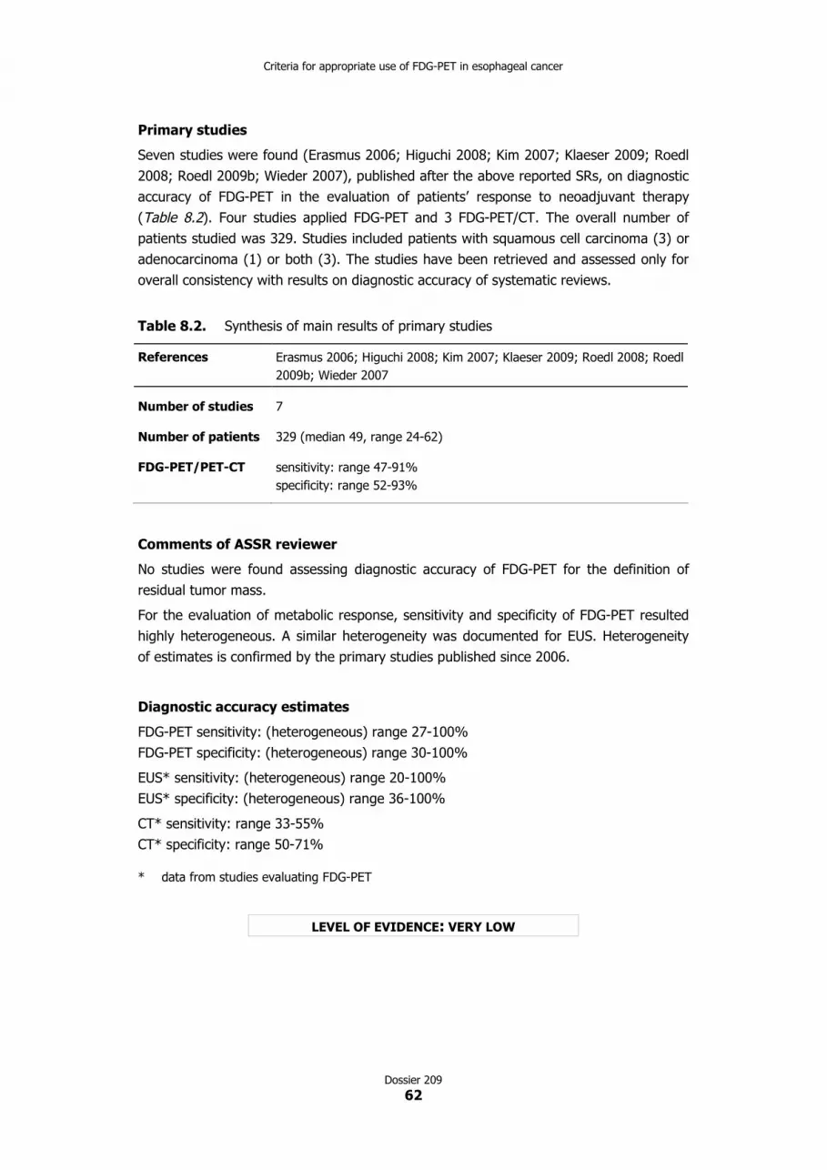
Criteria for appropriate use of FDG-PET in esophageal cancer
Dossier 209
62
Primary studies
Seven studies were found (Erasmus 2006; Higuchi 2008; Kim 2007; Klaeser 2009; Roedl
2008; Roedl 2009b; Wieder 2007), published after the above reported SRs, on diagnostic
accuracy of FDG-PET in the evaluation of patients’ response to neoadjuvant therapy
(Table 8.2). Four studies applied FDG-PET and 3 FDG-PET/CT. The overall number of
patients studied was 329. Studies included patients with squamous cell carcinoma (3) or
adenocarcinoma (1) or both (3). The studies have been retrieved and assessed only for
overall consistency with results on diagnostic accuracy of systematic reviews.
Table 8.2. Synthesis of main results of primary studies
References Erasmus 2006; Higuchi 2008; Kim 2007; Klaeser 2009; Roedl 2008; Roedl
2009b; Wieder 2007
Number of studies 7
Number of patients 329 (median 49, range 24-62)
FDG-PET/PET-CT sensitivity: range 47-91%
specificity: range 52-93%
Comments of ASSR reviewer
No studies were found assessing diagnostic accuracy of FDG-PET for the definition of
residual tumor mass.
For the evaluation of metabolic response, sensitivity and specificity of FDG-PET resulted
highly heterogeneous. A similar heterogeneity was documented for EUS. Heterogeneity
of estimates is confirmed by the primary studies published since 2006.
Diagnostic accuracy estimates
FDG-PET sensitivity: (heterogeneous) range 27-100%
FDG-PET specificity: (heterogeneous) range 30-100%
EUS* sensitivity: (heterogeneous) range 20-100%
EUS* specificity: (heterogeneous) range 36-100%
CT* sensitivity: range 33-55%
CT* specificity: range 50-71%
* data from studies evaluating FDG-PET
LEVEL OF EVIDENCE: VERY LOW

Criteria for appropriate use of FDG-PET in esophageal cancer
Dossier 209
63
8.2. Clinical outcomes
To evaluate the balance between benefits and risks, the panel agreed to consider the
presumed patient-important outcomes reported below (Table 8.3), and voted on the level
of importance for each outcome. Median scores and ranges are reported for each
outcome.
The panel voted “critical” the consequences for responders - true and false responders -
and for false non responders, while outcomes for true non responders were considered
“important”.
No studies investigating the impact of FDG-PET on the above clinical outcomes were
found.
No matrix of “natural frequencies” was provided because of heterogeneity of both
estimates.
Table 8.3. Patient-important clinical outcomes and median scores of importance
Patient-important outcomes Median score
(range)
Consequences of test for responders
True responders - responders proceed to curative, radical treatment, which
could improve survival
7
(1-9)
False non responders - responders do not receive curative, radical
treatment, which could have improved survival, and proceed to less radical,
palliative treatment
7
(3-9)
Consequences of test for non responders
True non responders - non responders proceed to less radical, palliative
treatment
6
(1-9)
False responders - non responders proceed to curative, radical treatment,
with no possible gain in survival
7
(3-9)
8.3. Voting results
The first voting round registered a strong disagreement between panelists, with ratings
falling in the inappropriate, uncertain and appropriate regions (median score 5; range
3-8). The disagreement decreased in the second voting round, with ratings between
inappropriate and uncertain and a median score of 3 (range 1-6).
FINAL RATING FOR THE USE OF FDG-PET FOR EVALUATION
OF RESPONSE TO THERAPY AT THE END OF TREATMENT:
UNCERTAIN

Criteria for appropriate use of FDG-PET in esophageal cancer
Dossier 209
64
8.4. Conclusions
A disagreement among panelists was registered in both round of voting, with ratings
falling in all three regions in the first round, and ratings falling within the uncertain and
inappropriate regions in the second round. The use of FDG-PET, in addition to CT, in the
evaluation of response to neoadjuvant therapy at the end of treatment, in order to
decide between curative or palliative therapeutic course of action, resulted as uncertain
due to disagreement.
The level of evidence for diagnostic accuracy was very low, with heterogeneity for both
sensitivity and specificity. Outcomes for patients testing as responders - true and false
responders - and for false non responders were voted “critical” (median score 7), while
outcomes for true non responders were considered “important”.

Criteria for appropriate use of FDG-PET in esophageal cancer
Dossier 209
65
9. Follow up in patients with nosuspicion of recurrence
Rationale
Follow up of patients with esophageal or gastric cancer should monitor symptoms, signs
and nutritional status in order to detect disorders of function either related to recurrent
disease or affecting quality of life (SIGN 2006).
No guideline recommends any kind of active follow up (AIOM 2009; ESMO 2010; NCCN
2010; SIGN 2006).
Anticipating the diagnosis of recurrence could lead to a curative resection of a solitary
metastasis or an early start of chemotherapy/chemoradiotherapy.
Diagnostic role of FDG-PET
To anticipate identification of patients with potential relapse in order to start appropriate
therapy earlier.
Treatment effectiveness
There is no evidence that regular follow up after initial therapy may influence the
outcome (ESMO 2010) and no evidence has been identified to support regular imaging or
measurement of serum tumor markers in the follow up of patients with esophageal
cancer (SIGN 2006). Aim of treatment is purely palliative.
Pre-test probability and change in management
The median pre-test probability of cancer recurrence is 56% (range 55-57%; data from
three studies: Roedl 2008; Sun 2009; Teyton 2009), which could be considered the
hypothetical maximum degree of change in management achievable with a correct
detection of recurrence in asymptomatic patients treated for esophageal cancer.
Evidence from 1 study (Sun 2009) on change in management following the application of
FDG-PET shows an estimate of 60% (no details about the kind of change are reported).
Research question
Is FDG-PET useful during follow up of patients with no suspicion of recurrence?

Criteria for appropriate use of FDG-PET in esophageal cancer
Dossier 209
66
9.1. Systematic review of literature: results
Results from update of systematic review of literature from Jan 2006
Only three primary studies on diagnostic accuracy and no systematic reviews were found.
Results are reported below.
Primary studies
Three studies (Roedl 2008; Sun 2009; Teyton 2009) were included, 1 study applying
FDG-PET/CT, 1 applying FDG-PET and 1 applying both (Table 9.1). The overall number of
patients studied was 108 (range 20-47). Studies included patients that, after curative
surgery, with or without previous neoadjuvant chemoradiotherapy, underwent FDG-PET
evaluation during follow up every 6 months for at least 24 months. All studies used
biopsy of the suspected recurrence and clinical follow up as reference standard. The
studies were all limited by the low number of patients, possible verification bias, and
possible unblinding of the index test when evaluating the reference standard.
Table 9.1. Results of primary studies
References Roedl 2008; Sun 2009; Teyton 2009
Number of studies 3
Number of patients 108 (range 20-47)
Pre-test probability
(frequency of
recurrence)
range 55-57%
FDG-PET/PET-CT FDG-PET
sensitivity: range 89-100%
specificity: range 55-85.3%
FDG-PET/CT
sensitivity: range 89-100%
specificity: range 67-75%
Comparator CT
sensitivity: 65%
specificity: 91.2%
Reference standard biopsy of the suspected lesion or clinical follow up

Criteria for appropriate use of FDG-PET in esophageal cancer
Dossier 209
67
Comments of ASSR reviewers
The evidence about the validity of the FDG-PET in detecting recurrence of esophageal
cancer in patients with no suspicion of recurrence after definitive treatment (follow up)
comes from only three studies on a limited number of patients.
Diagnostic accuracy estimates
Estimates of diagnostic accuracy are not robust and show an heterogeneity for
specificity.
LEVEL OF EVIDENCE: VERY LOW
9.2. Clinical outcomes
To evaluate the balance between benefits and risks, the panel agreed to consider the
presumed patient-important outcomes reported below (Table 9.2), and voted on the level
of importance for each outcome. Median scores and ranges are reported for each
outcome.
All outcomes were voted “important”.
No studies investigating the impact of FDG-PET on the above clinical outcomes were
found.
No matrix of “natural frequencies” was provided because of sparse data.
Table 9.2. Patient-important clinical outcomes and median scores of importance
Patient-important outcomes Median score
(range)
Consequences of test for patients relapsing
True positives - patients undergo further test to confirm positive results and
proceed to palliative treatment
4
(1-79)
False negatives - patients remain in follow up until symptoms occur 4
(1-9)
Consequences of test for patients not relapsing
True negatives - patients remain in follow up and are reassured, after certain
amount of stress
4
(1-9)
False positives - patients undergo unnecessary further tests to prove
negative and are exposed to unnecessary anxiety
6
(1-9)

Criteria for appropriate use of FDG-PET in esophageal cancer
Dossier 209
68
9.3. Voting results
The first voting round registered a light disagreement of ratings between inappropriate
and uncertain (median score 2.5; range 1-4), while the second voting round registered
an agreement on inappropriate (median score 2; range 1-3).
FINAL RATING FOR THE USE OF FDG-PET IN FOLLOW UP OF PATIENTS
WITH NO SUSPICION OF RECURRENCE:
INAPPROPRIATE
9.4. Conclusions
After an initial slight disagreement between inappropriate and uncertain, the panel
agreed to judge as inappropriate the use of FDG-PET for patients in follow up with non
suspicion of recurrence.
Level of evidence for diagnostic accuracy of FDG-PET in follow up was very low and
coming from three primary studies of low methodological quality.
Outcomes for patients with recurrence - true positives and false negatives, as well as
outcomes for patients correctly diagnosed as negatives were voted “important” (median
score 4). A slightly higher score (median 6; range 1-9) was assigned to the outcomes of
patients incorrectly testing positive for distant metastases and experiencing unnecessary
stress and anxiety.

Criteria for appropriate use of FDG-PET in esophageal cancer
Dossier 209
69
10. Diagnosis and stagingof suspect distant recurrence
Rationale
After curative surgery it could be difficult to interpret occurring symptoms and signs and
correctly differentiate local recurrence from scar.
Diagnostic role of FDG-PET
As more specific test, to resolve ambiguities resulting from conventional imaging and
correctly identify relapsing patients needing treatment.
Treatment effectiveness
Recurrences of patients treated with radiation therapy with curative intent could be
treated with surgery (NCCN 2010), while patients relapsing after surgery should be
treated with palliative intent (AIOM 2009).
Pre-test probability and change in management
80.4% of patients with suspected recurrence have metastases (Guo 2007).
Research question: FDG-PET in add on
Has FDG-PET sufficient specificity to be used as an add on test to diagnose recurrence in
patients with unclear results from conventional imaging?

Criteria for appropriate use of FDG-PET in esophageal cancer
Dossier 209
70
10.1. Systematic review of literature: results
Results from update of systematic review of literature from Jan 2006
Only one study evaluating diagnostic accuracy of FDG-PET in suspected recurrence and
no systematic reviews were found. Results are reported below.
Primary studies
One study (Guo 2007) was found (Table 10.1). Fifty-six patients with suspected
recurrence of esophageal squamous cell carcinoma after definitive treatment performed a
FDG-PET. FDG-PET validity in diagnosing recurrence was assessed comparing results with
histopathology or clinical follow up (reference standard). The study was limited by the
low number of patients belonging to a subgroup of esophageal cancer, by a possible
verification bias and absence of blinding about the index test (FDG-PET) when evaluating
the reference standard.
Table 10.1. Results of primary studies
Reference Guo 2007
Number of patients 56
Pre-test probability
(frequency of recurrence)
80.4%
FDG-PET/PET-CT sensitivity: 95.6%
specificity: 54.5%
Reference standard histopatology or follow up
Comments of ASSR reviewer
The only evidence about the validity of the FDG-PET in diagnosing recurrence of
esophageal cancer after definitive treatment comes from just one study on very few
patients.
Diagnostic accuracy estimates
Not available as data on the validity of FDG-PET in diagnosing recurrence of esophageal
cancer after definitive treatment comes from only one study on very few patients.
LEVEL OF EVIDENCE: VERY LOW

Criteria for appropriate use of FDG-PET in esophageal cancer
Dossier 209
71
10.2. Clinical outcomes
To evaluate the balance between benefits and risks, the panel agreed to consider the
presumed patient-important outcomes reported below (Table 10.2), and voted on the
level of importance for each outcome. Median scores and ranges are reported for each
outcome.
Outcomes were considered by the panel “critical” for all patients, except for true negative
patients, whose consequences were voted “important”.
No studies investigating the impact of FDG-PET on the above clinical outcomes were
found.
No matrix of “natural frequencies” was provided because of sparse data.
Table 10.2. Patient-important clinical outcomes and median scores of importance
Patient-important outcomes Median score
(range)
Consequences of test for patients with recurrence
True positives - patients proceed to treatment for recurrence 7
(5-9)
False negatives - patients delay start of treatment until symptoms occur,
with a possible negative impact on quality of life
7
(2-9)
Consequences of test for patients without recurrence
True negatives - patients remain in follow up, after a considerable amount of
stress
6
(2-7)
False positives - patients undergo unnecessary treatment, with a possible
negative impact on quality of life and suffer unnecessary distress
7
(4-9)
10.3. Voting results
Both voting rounds registered a slight disagreement with ratings fallings between
uncertain and appropriate with a median score of 6 (range 4-7) in the first round and a
median score of 7 (range 6-7) in the second round.
FINAL RATING FOR THE USE OF FDG-PET FOR DIAGNOSIS AND STAGING
OF SUSPECT DISTANT RECURRENCE:
UNCERTAIN

Criteria for appropriate use of FDG-PET in esophageal cancer
Dossier 209
72
10.4. Conclusions
A disagreement among panelists was registered in both round of votes, with ratings
falling in both the uncertain and appropriate region. The use of FDG-PET as an add on
test for the diagnosis and staging of distant recurrence in patients with clinical suspicion
of recurrence or unclear conventional imaging results resulted as uncertain due to
disagreement.
Level of evidence for diagnostic accuracy of FDG-PET was very low, coming from only
one study with very few patients. Outcomes were considered “critical” (median score 7)
for true and false positives, as well as for false negatives, and “important” (median score
6) for patients correctly found negatives.

Criteria for appropriate use of FDG-PET in esophageal cancer
Dossier 209
73
Conclusions
The present work is part of a larger research program dedicated to the update of the
2007 Report on the appropriate use of FDG-PET in oncology.
At the end of the research program results of the present Dossier will be used for an
overall analysis and estimate of PET scans need in Emilia-Romagna region and for setting
up priorities for future research programs on the clinical use of FDG-PET in oncology.


Criteria for appropriate use of FDG-PET in esophageal cancer
Dossier 209
75
References
AIOM 2009 - Linee guida AIOM. Tumori dell’esofago. 2009.
http://www.aiom.it/Attivit%E0+Scientifica/Linee+guida/Tumori+dell%92esofago/1,131
7,0, (last access May 18, 2011)
Berrisford 2008 - Berrisford RG, Wong WL, Day D, Toy E, Napier M, Mitchell K, Wajed S.
The decision to operate: role of integrated computed tomography positron emission
tomography in staging oesophageal and oesophagogastric junction cancer by the
multidisciplinary team. Eur J Cardiothorac Surg, 33: 1112-1116, 2008.
Blackstock 2006 - Blackstock AW, Farmer MR, Lovato J, Mishra G, Melin SA, Oaks T,
Aklilu M, Clark PB, Levine EA. A prospective evaluation of the impact of 18-F-fluoro-
deoxy-D-glucose positron emission tomography staging on survival for patients with
locally advanced esophageal cancer. Int J Radiat Oncol Biol Phys, 64: 455-460, 2006.
Bossuyt 2006 - Bossuyt PM, Irwig L, Craig J, Glasziou P. Comparative accuracy: assessing
new tests against existing diagnostic pathways BMJ, 332: 1089-1092, 2006.
Buchmann 2006 - Buchmann I, Hansen T, Brochhausen C, Kneist W, Oberholzer K,
Junginger T, Schreckenberger M, Bartenstein P.FDG-PET in the initial staging of
squamous cell oesophageal carcinoma. Nuklearmedizin, 45 (6): 235-241, 2006.
Chatterton 2009 - Chatterton BE, Ho S, I, Baldey A, Lenzo N, Patrikeos A, Kelley B, Wong
D, Ramshaw JE, Scott AM. Positron emission tomography changes management and
prognostic stratification in patients with oesophageal cancer: results of a multicentre
prospective study. Eur J Nucl Med Mol Imaging, 36: 354-361, 2009.
Choi 2010 - Choi J, Kim SG, Kim JS, Jung HC, Song IS. Comparison of endoscopic
ultrasonography (EUS), positron emission tomography (PET), and computed
tomography (CT) in the preoperative locoregional staging of resectable esophageal
cancer. Surg Endosc, 24: 1380-1386, 2010.
Dossier 157 - Liberati A, Longo G, Ballini L, De Palma R. FDG-PET in oncologia - Criteri
per un uso appropriato. Dossier 157 - Agenzia sanitaria regionale, Regione Emilia-
Romagna, 2007.
Duong 2006a - Duong CP, Demitriou H, Weih L, Thompson A, Williams D, Thomas RJ,
Hicks RJ. Significant clinical impact and prognostic stratification provided by FDG-PET in
the staging of oesophageal cancer. Eur J Nucl Med Mol Imaging, 33: 759-769, 2006.
Duong 2006b - Duong CP, Hicks RJ, Weih L, Drummond E, Leong T, Michael M, Thomas
RJS. FDG-PET status following chemoradiotherapy provides high management impact
and powerful prognostic stratification in oesophageal cancer. Eur J Nucl Med Mol
Imaging, 33: 770-778, 2006.

Criteria for appropriate use of FDG-PET in esophageal cancer
Dossier 209
76
Erasmus 2006 - Erasmus JJ, Munden RF, Truong MT, Ho JJ, Hofstetter WL, Macapinlac
HA, Correa AM, Wu TT, Bruzzi JF, Marom EM, Sabloff BS, Ajani JA, Komaki R, Liao Z,
Lee JH, Fukami N, Bresalier R, Swisher SG. Preoperative chemo-radiation-induced
ulceration in patients with esophageal cancer: a confounding factor in tumor response
assessment in integrated computed tomographic-positron emission tomographic
imaging. J Thorac Oncol, 1: 478-486, 2006.
ESMO 2010 - Stahl M, Budach W, Meyer HJ, Cervantes A, on behalf of the ESMO
Guidelines Working Group. Esophageal cancer: clinical practice guidelines for diagnosis,
treatment and follow up. Ann Oncol, 21 (Supplement 5): v46-v49, 2010.
Gananadha 2008 - Gananadha S, Hazebroek EJ, Leibman S, Berry H, Osgood L, Shon IH,
Pavlakis N, Marx G, Smith GS. The utility of FDG-PET in the preoperative staging of
esophageal cancer. Dis Esophagus, 21: 389-394, 2008.
Gebski 2007 - Gebski V, Burmeister B, Smithers BM, Foo K, Zalcberg J, Simes J, for the
Australasian Gastro-Intestinal Trials Group. Survival benefi ts from neoadjuvant
chemoradiotherapy or chemotherapy in oesophageal carcinoma: a meta-analysis.
Lancet Oncol, 8: 226-234, 2007.
Gigerenzer 2008 - Gigerenzer G, Gaissmaier W, Kurz-Milcke E, Schwartz LM, Woloshin S.
Helping doctors and patients make sense of health statistics. PSPI, 8 (2): 53-96, 2007.
Guo 2007 - Guo H, Zhu H, Xi Y, Zhang B, Li L, Huang Y, Zhang J, Fu Z, Yang G, Yuan S,
Yu J. Diagnostic and prognostic value of 18F-FDG PET/CT for patients with suspected
recurrence from squamous cell carcinoma of the esophagus. J Nucl Med, 48: 1251-
1258, 2007.
Guyatt 2008 - Guyatt GH, Oxman AD, Kunz R, Vist GE, Falck-Ytter Y, Schünemann HJ
and for the GRADE Working Group. What is “quality of evidence” and why is it
important to clinicians? BMJ, 336: 995-998, 2008.
Higgins 2009 - Higgins JPT, Green S (editors). Cochrane Handbook for Systematic
Reviews of Interventions. Version 5.0.2 [updated September 2009]. The Cochrane
Collaboration, 2009. Available from http://www.cochrane-handbook.org.
Higuchi 2008 - Higuchi I, Yasuda T, Yano M, Doki Y, Miyata H, Tatsumi M, Fukunaga H,
Takiguchi S, Fujiwara Y, Hatazawa J, Monden M. Lack of fludeoxyglucose F 18 uptake
in post treatment positron emission tomography as a significant predictor of survival
after subsequent surgery in multimodality treatment for patients with locally advanced
esophageal squamous cell carcinoma. J Thorac Cardiovasc Surg, 136: 205-212, 2008.
Hsu 2009 - Hsu WH, Hsu PK, Wang SJ, Lin KH, Huang CS, Hsieh CC, Wu YC. Positron
emission tomography-computed tomography in predicting locoregional invasion in
esophageal squamous cell carcinoma. Ann Thorac Surg, 87: 1564-1568, 2009.
Hu 2009 - Hu Q, Wang W, Zhong X, Yuan S, Fu Z, Guo H, Yu J. Dual-time-point FDG PET
for the evaluation of locoregional lymph nodes in thoracic esophageal squamous cell
cancer. Eur J Radiol, 70: 320-324, 2009.

Criteria for appropriate use of FDG-PET in esophageal cancer
Dossier 209
77
Kato 2008 - Kato H, Kimura H, Nakajima M, Sakai M, Sano A, Tanaka N, Inose T, Faried
A, Saito K, Ieta K, Sohda M, Fukai Y, Miyazaki T, Masuda N, Fukuchi M, Ojima H,
Tsukada K, Oriuchi N, Endo K, Kuwano H. The additional value of integrated PET/CT
over PET in initial lymph node staging of esophageal cancer. Oncol Rep, 20: 857-862,
2008.
Katsoulis 2007 - Katsoulis IE, Wong WL, Mattheou AK, Damani N, Chambers J,
Livingstone JI. Fluorine-18 fluorodeoxyglucose positron emission tomography in the
preoperative staging of thoracic oesophageal and gastro-oesophageal junction cancer:
a prospective study. Int J Surg, 5: 399-403, 2007.
Kim 2007 - Kim MK, Ryu JS, Kim SB, Ahn JH, Kim SY, Park SI, Kim YH, Song HY, Shin JH,
Jung HY, Lee GH, Choi KD, Cho KJ, Kim JH. Value of complete metabolic response by
(18)F-fluorodeoxyglucose-positron emission tomography in oesophageal cancer for
prediction of pathologic response and survival after preoperative chemoradiotherapy.
Eur J Cancer, 43: 1385-1391, 2007.
Klaeser 2009 - Klaeser B, Nitzsche E, Schuller JC, Koberle D, Widmer L, Balmer-Majno S,
Hany T, Cescato-Wenger C, Brauchli P, Zünd M, Pestalozzi BC, Caspar C, Albrecht S,
von Moos R, Ruhstaller T. Limited predictive value of FDG-PET for response assessment
in the preoperative treatment of esophageal cancer: results of a prospective multi-
center trial (SAKK 75/02). Onkologie, 32: 724-730, 2009.
Kwee 2010 - Kwee RW. Prediction of Tumor Response to Neoadjuvant Therapy in
Patients with Esophageal Cancer with Use of 18 F FDG PET: A Systematic Review.
Radiology, 254: 707-717, 2010.
Liberati 2007 - Liberati A, Longo G, Ballini L, De Palma R. FDG-PET in oncologia - Criteri
per un uso appropriato. Dossier 157 - Agenzia sanitaria regionale, Regione Emilia-
Romagna, 2007.
http://asr.regione.emilia-romagna.it/wcm/asr/collana_dossier/doss157.htm
(last access May 18, 2011)
Little 2007 - Little SG, Rice TW, Bybel B, Mason DP, Murthy SC, Falk GW, Rybicki LA,
Blackstone EH. Is FDG-PET indicated for superficial esophageal cancer? Eur J
Cardiothorac Surg, 31: 791-796, 2007.
Lordick 2007 - Lordick F, Ott K, Krause BJ, Weber WA, Becker K, Stein HJ, Lorenzen S,
Schuster T, Wieder H, Herrmann K, Bredenkamp R, Höfler H, Fink U, Peschel C,
Schwaiger M, Siewert JR. PET to assess early metabolic response and to guide
treatment of adenocarcinoma of the oesophagogastric junction: the MUNICON phase II
trial. Lancet Oncol, 8: 797-805, 2007.
Lorenzen 2007 - Lorenzen S, Herrmann K, Weber WA, Wieder H, Hennig M, Ott K,
Bredenkamp R, Peschel C, Schwaiger M, Lordick F. Visualisation of metastatic
oesophageal and gastric cancer and prediction of clinical response to palliative
chemotherapy using 18FDG PET. Nuklearmedizin, 46: 263-270, 2007.
Malik 2006 - Malik V, Keogan M, Gilham C, Duffy G, Ravi N, Reynolds JV. FDG-PET
scanning in the management of cancer of the oesophagus and oesophagogastric
junction: early experience with 100 consecutive cases. Ir J Med Sci, 175: 48-54, 2006.

Criteria for appropriate use of FDG-PET in esophageal cancer
Dossier 209
78
McDonough 2008 - McDonough PB, Jones DR, Shen KR, Northup PG, Galysh RL,
Hernandez A et al. Does FDG-PET add information to EUS and CT in the initial
management of esophageal cancer? A prospective single center study. Am J
Gastroenterol, 103: 570-574, 2008.
Meyers 2007 - Meyers BF, Downey RJ, Decker PA, Keenan RJ, Siegel BA, Cerfolio RJ,
Landreneau RJ, Reed CE, Balfe DM, Dehdashti F, Ballman KV, Rusch VW, Putnam JB Jr;
American College of Surgeons Oncology Group Z0060. The utility of positron emission
tomography in staging of potentially operable carcinoma of the thoracic esophagus:
results of the American College of Surgeons Oncology Group Z0060 trial. J Thorac
Cardiovasc Surg, 133: 738-745, 2007.
Moher 2009 - Moher D, Liberati A, Tetzlaff J, Altman DG, The PRISMA Group (2009).
Preferred Reporting Items for Systematic Reviews and Meta-Analyses: The PRISMA
Statement. BMJ, 339: b2535, doi: 10.1136/bmj.b2535, 2009.
Muijs 2010 - Muijs CT, Beukema JC, Pruim J, Mul VE, Groen H, Plukker JT, Langendijk JA.
A systematic review on the role of FDG-PET/CT in tumour delineation and radiotherapy
planning in patients with esophageal cancer. Radiother Oncol, Jun 9, 2010. [Epub
ahead of print] PubMed PMID: 20541273.
NCCN 2010 - NCCN clinical practice guidelines in oncology. Esophageal cancer. 2010
www.nccn.org (last access 29th September 2010).
Ngamruengphong 2010 - Ngamruengphong S, Sharma VK, Nguyen B, Das A. Assessment
of response to neoadjuvant therapy in esophageal cancer: an updated systematic
review of diagnostic accuracy of endoscopic ultrasonography and fluorodeoxyglucose
positron emission tomography. Dis Esophagus, 23: 216-231, 2010.
Noble 2009 - Noble F, Bailey D, Tung K, Byrne JP. Impact of integrated PET/CT in the
staging of oesophageal cancer: a UK population-based cohort study. Clin Radiol, 64:
699-705, 2009.
Okada 2009 - Okada M, Murakami T, Kumano S, Kuwabara M, Shimono T, Hosono M,
Shiozaki H. Integrated FDG-PET/CT compared with intravenous contrast-enhanced CT
for evaluation of metastatic regional lymph nodes in patients with resectable early
stage esophageal cancer. Ann Nucl Med, 23: 73-80, 2009.
Pfau 2007 - Pfau PR, Perlman SB, Stanko P, Frick TJ, Gopal DV, Said A, Zhang Z, Weigel
T. The role and clinical value of EUS in a multimodality esophageal carcinoma staging
program with CT and positron emission tomography. Gastrointest Endosc, 65: 377-384,
2007.
Pifarré-Montaner 2009 - Pifarre-Montaner P, Fernandez-Leon A, de Juan R, Ama-Salvador
ME, Galan M, Gamez-Cenzano C, Sans M. [Impact of (18)F-FDG PET/CT on the
therapeutic management in the initial staging of the esophageal cancer]. Rev Esp Med
Nucl, 28: 101-105, 2009.

Criteria for appropriate use of FDG-PET in esophageal cancer
Dossier 209
79
Rebollo Aguirre 2009 - Rebollo Aguirre AC, Ramos-Font C, Villegas Portero R, Cook GJR,
Llamas Elvira JM, Tabares AR. 18F-Fluorodeoxiglucose Positron Emission Tomography
for the Evaluation of Neoadjuvant Therapy Response in Esophageal Cancer. Systematic
Review of the Literature. Ann Surg, 250: 247-254, 2009.
Roedl 2008 - Roedl JB, Harisinghani MG, Colen RR, Fischman AJ, Blake MA, Mathisen DJ,
Mueller PR. Assessment of treatment response and recurrence in esophageal carcinoma
based on tumor length and standardized uptake value on positron emission
tomography-computed tomography. Ann Thorac Surg, 86: 1131-1138, 2008.
Roedl 2009a - Roedl JB, Blake MA, Holalkere NS, Mueller PR, Colen RR, Harisinghani MG.
Lymph node staging in esophageal adenocarcinoma with PET-CT based on a visual
analysis and based on metabolic parameters. Abdom Imaging, 34: 610-617, 2009.
Roedl 2009b - Roedl JB, Halpern EF, Colen RR, Sahani DV, Fischman AJ, Blake MA.
Metabolic tumor width parameters as determined on PET/CT predict disease-free
survival and treatment response in squamous cell carcinoma of the esophagus. Mol
Imaging Biol, 11: 54-60, 2009.
Ross 1996 - Ross P, Nicolson M, Cunningham D, Valle J, Seymour M, Harper P, Price T,
Anderson H, Iveson T, Hickish T, Lofts F, Norman A. Prospective randomized trial
comparing mitomycin, cisplatin, and protracted venous-infusion fluorouracil (PVI 5-FU)
with epirubicin, cisplatin, and PVI 5-FU in advanced esophagogastric cancer. J Clin
Oncol, 20: 1996-2004, 1996.
Salahudeen 2008 - Salahudeen HM, Balan A, Naik K, Mirsadraee S, Scarsbrook AF.
Impact of the introduction of integrated PET-CT into the preoperative staging pathway
of patients with potentially operable oesophageal carcinoma. Clin Radiol, 63: 765-773,
2008.
Sandha 2008 - Sandha GS, Severin D, Postema E, McEwan A, Stewart K. Is positron
emission tomography useful in locoregional staging of esophageal cancer? Results of a
multidisciplinary initiative comparing CT, positron emission tomography, and EUS.
Gastrointest Endosc, 67: 402-409, 2008.
Schreurs 2008 - Schreurs LM, Verhoef CC, Van der Jagt EJ, van Dam GM, Groen H,
Plukker JT. Current relevance of cervical ultrasonography in staging cancer of the
esophagus and gastroesophageal junction. Eur J Radiol, 67: 105-111, 2008.
Schünemann 2008 - Schünemann HJ, Oxman AD, Brozek J, Glasziou P, Jaeschke R, Vist
GE, Williams JW, Kunz R, Craig J, Montori VM, Bossuyt P, Guyatt GH. GRADE: grading
quality of evidence and strength of recommendations for diagnostic tests and
strategies. BMJ, 336: 1106-1110, 2008.
Shea 2007 - Shea B, Grimshaw J, Wells G, Boers M, Andersson N, Hamel C, Porter A,
Tugwell P, Moher D, Bouter L. Development of AMSTAR: a measurement tool to assess
the methodological quality of systematic reviews. BMC Medical Research Methodology,
7: 10 doi:10.1186/1471-2288-7-10, 2007.

Criteria for appropriate use of FDG-PET in esophageal cancer
Dossier 209
80
Shimizu 2009 - Shimizu S, Hosokawa M, Itoh K, Fujita M, Takahashi H, Shirato H. Can
hybrid FDG-PET/CT detect subclinical lymph node metastasis of esophageal cancer
appropriately and contribute to radiation treatment planning? A comparison of image-
based and pathological findings. Int J Clin Oncol, 14: 421-425, 2009.
SIGN 2006 - SIGN. Management of oesophageal and gastric cancer. Guideline No. 87,
ISBN 1 899893 59 8, June 2006
Smith 2009 - Smith JW, Moreira J, Abood G, Aranha GV, Nagda S, Wagner RH, Shoup M.
The influence of (18)flourodeoxyglucose positron emission tomography on the
management of gastroesophageal junction carcinoma. Am J Surg, 197: 308-312, 2009.
Sun 2009 - Sun L, Su XH, Guan YS, Pan WM, Luo ZM, Wei JH, Zhao L, Wu H. Clinical
usefulness of 18F-FDG PET/CT in the restaging of esophageal cancer after surgical
resection and radiotherapy. World J Gastroenterol, 15: 1836-1842, 2009.
Teyton 2009 - Teyton P, Metges JP, Atmani A, Jestin-Le Tallec V, Volant A, Visvikis D,
Bail JP, Pradier O, Lozac’h P, Cheze Le Rest C. Use of positron emission tomography in
surgery follow-up of esophageal cancer. J Gastrointest Surg, 13: 451-458, 2009.
van Baardwijk 2006 - van Baardwijk A, Baumert BG, Bosmans G, van Kroonenburgh M,
Stroobants S, Gregoire V, Lambin P, De Ruysscher D. The current status of FDG-PET in
tumour volume definition in radiotherapy treatment planning. Cancer Treatment
Reviews, 32: 245-260, 2006.
van Westreenen 2007 - van Westreenen HL, Westerterp M, Sloof GW, Groen H, Bossuyt
PM, Jager PL, Comans EF, van Dullemen HM, Fockens P, Stoker J, van der Jagt EJ, van
Lanschot JJ, Plukker JT. Limited additional value of positron emission tomography in
staging oesophageal cancer. Br J Surg, 94: 1515-1520, 2007.
Westerterp 2005 - Westerterp M, Van Westreenen HL, Reitsma JB, Hoekstra OS, Stoker
J, Fockens P, Jager PL, Van Eck-Smit BL, Plukker JT, van Lanschot JJ, Sloof GW.
Esophageal cancer: CT, endoscopic US, and FDG PET for assessment of response to
neoadjuvant therapy-systematic review. Radiology, 236: 841-851, 2005.
Westerterp 2008 - Westerterp M, van Westreenen HL, Deutekom M, Stoker J, Fockens P,
Comans EF, Plukker JT, Bossuyt PM, van Lanschot JJ, Sloof GW. Patients’ perception of
diagnostic tests in the preoperative assessment of esophageal cancer. Patient Prefer
Adherence, 2: 157-162, 2008.
Whiting 2003 - Whiting P, Rutjes AWS, Reitsma JB, Bossuyt PMM, Kleijnen J. The
development of QUADAS: a tool for the quality assessment of studies of diagnostic
accuracy included in systematic reviews. BMC Medical Research Methodology, 3: 25,
2003.
Wieder 2007 - Wieder HA, Ott K, Lordick F, Becker K, Stahl A, Herrmann K, Fink U,
Siewert JR, Schwaiger M, Weber WA. Prediction of tumor response by FDG-PET:
comparison of the accuracy of single and sequential studies in patients with
adenocarcinomas of the esophagogastric junction. Eur J Nucl Med Mol Imaging, 34:
1925-1932, 2007.

Criteria for appropriate use of FDG-PET in esophageal cancer
Dossier 209
81
Williams 2009 - Williams RN, Ubhi SS, Sutton CD, Thomas AL, Entwisle JJ, Bowrey DJ.
The early use of PET-CT alters the management of patients with esophageal cancer. J
Gastrointest Surg, 13: 868-873, 2009.
Yuan 2006 - Yuan S, Yu Y, Chao KS, Fu Z, Yin Y, Liu T, Chen S, Yang X, Yang G, Guo H,
Yu J. Additional value of PET/CT over PET in assessment of locoregional lymph nodes in
thoracic esophageal squamous cell cancer. J Nucl Med, 47: 1255-1259, 2006.
List of excluded studies on the basis of inclusion criteria
Abate E, DeMeester SR, Zehetner J, Oezcelik A, Ayazi S, Costales J et al. Recurrence
after esophagectomy for adenocarcinoma: defining optimal follow-up intervals and
testing. J Am Coll Surg, 210: 428-435, 2010.
Berrisford RG, Wong WL, Day D, Toy E, Napier M, Mitchell K et al. The decision to
operate: role of integrated computed tomography positron emission tomography in
staging oesophageal and oesophagogastric junction cancer by the multidisciplinary team.
Eur J Cardiothorac Surg, 33: 1112-1116, 2008.
Blackstock AW, Farmer MR, Lovato J, Mishra G, Melin SA, Oaks T et al. A prospective
evaluation of the impact of 18-F-fluoro-deoxy-D-glucose positron emission tomography
staging on survival for patients with locally advanced esophageal cancer. Int J Radiat
Oncol Biol Phys, 64: 455-460, 2006.
Cerfolio RJ, Bryant AS. Maximum standardized uptake values on positron emission
tomography of esophageal cancer predicts stage, tumor biology, and survival. Ann
Thorac Surg, 82: 391-394, 2006.
Cheze-Le Rest C, Metges JP, Teyton P, Jestin-Le Tallec V, Lozac’h P, Volant A et al.
Prognostic value of initial fluorodeoxyglucose-PET in esophageal cancer: a prospective
study. Nucl Med Commun, 29: 628-635, 2008.
Duong CP, Demitriou H, Weih L, Thompson A, Williams D, Thomas RJ et al. Significant
clinical impact and prognostic stratification provided by FDG-PET in the staging of
oesophageal cancer. Eur J Nucl Med Mol Imaging, 33: 759-769, 2006.
Fiore D, Baggio V, Ruol A, Bocus P, Casara D, Corti L et al. Multimodal imaging of
esophagus and cardia cancer before and after treatment. Radiol Med, 111: 804-817,
2006.
Gananadha S, Hazebroek EJ, Leibman S, Berry H, Osgood L, Shon IH et al. The utility
of FDG-PET in the preoperative staging of esophageal cancer. Dis Esophagus, 21: 389-
394, 2008.
Gardner-Thorpe J, Hardwick RH, Dwenyhouse SJ. Salvage oesophagectomy after local
failure of definitive chemoradiotherapy. Br J Surg, 94: 1059-1066, 2007.
Hyun SH, Choi JY, Shim YM, Kim K, Lee SJ, Cho YS et al. Prognostic value of metabolic
tumor volume measured by 18F-fluorodeoxyglucose positron emission tomography in
patients with esophageal carcinoma. Ann Surg Oncol, 17: 115-122, 2010.

Criteria for appropriate use of FDG-PET in esophageal cancer
Dossier 209
82
Jadvar H, Henderson RW, Conti PS. 2-deoxy-2-[F-18]fluoro-D-glucose-positron
emission tomography/computed tomography imaging evaluation of esophageal cancer.
Mol Imaging Biol, 8: 193-200, 2006.
Javeri H, Xiao L, Rohren E, Komaki R, Hofstetter W, Lee JH et al. Influence of the
baseline 18F-fluoro-2-deoxy-D-glucose positron emission tomography results on survival
and pathologic response in patients with gastroesophageal cancer undergoing
chemoradiation. Cancer, 115: 624-630, 2009.
Javeri H, Xiao L, Rohren E, Lee JH, Liao Z, Hofstetter W et al. The higher the decrease
in the standardized uptake value of positron emission tomography after chemoradiation,
the better the survival of patients with gastroesophageal adenocarcinoma. Cancer, 115:
5184-5192, 2009.
Jingu K, Kaneta T, Nemoto K, Takeda K, Ogawa Y, Ariga H et al. (18)F-
fluorodeoxyglucose positron emission tomography immediately after chemoradiotherapy
predicts prognosis in patients with locoregional postoperative recurrent esophageal
cancer. Int J Clin Oncol, 15: 184-190, 2010.
Kajiyama Y, Iwanuma Y, Tomita N, Amano T, Isayama F, Matsumoto T et al. Size
analysis of lymph node metastasis in esophageal cancer: Diameter distribution and
assessment of accuracy of preoperative diagnosis. Esophagus, 3: 189-195, 2006.
Kato H, Fukuchi M, Miyazaki T, Nakajima M, Tanaka N, Inose T et al. Prediction of
response to definitive chemoradiotherapy in esophageal cancer using positron emission
tomography. Anticancer Res, 27: 2627-2633, 2007.
Kato H, Nakajima M, Sohda M, Tanaka N, Inose T, Miyazaki T et al. The clinical
application of (18)F-fluorodeoxyglucose positron emission tomography to predict survival
in patients with operable esophageal cancer. Cancer, 115: 3196-3203, 2009.
Keswani RN, Early DS, Edmundowicz SA, Meyers BF, Sharma A, Govindan R et al.
Routine positron emission tomography does not alter nodal staging in patients
undergoing EUS-guided FNA for esophageal cancer. Gastrointest Endosc, 69: 1210-1217,
2009.
Konski AA, Cheng JD, Goldberg M, Li T, Maurer A, Yu JQ et al. Correlation of molecular
response as measured by 18-FDG positron emission tomography with outcome after
chemoradiotherapy in patients with esophageal carcinoma. Int J Radiat Oncol Biol Phys,
69: 358-363, 2007.
Korst RJ, Kansler AL, Port JL, Lee PC, Kerem Y, Altorki NK. Downstaging of T or N
predicts long-term survival after preoperative chemotherapy and radical resection for
esophageal carcinoma. Ann Thorac Surg, 82: 480-484, 2006.
Krause BJ, Herrmann K, Wieder H, Zum Buschenfelde CM. 18F-FDG PET and 18F-FDG
PET/CT for assessing response to therapy in esophageal cancer. J Nucl Med, 50 (suppl.
1): 89S-96S, 2009.
Lan XL, Zhang YX, Wu ZJ, Jia Q, Wei H, Gao ZR. The value of dual time point 18F-FDG
PET imaging for the differentiation between malignant and benign lesions. Clin Radiol, 3:
756-764, 2008.

Criteria for appropriate use of FDG-PET in esophageal cancer
Dossier 209
83
Makino T, Doki Y, Miyata H, Yasuda T, Yamasaki M, Fujiwara Y et al. Use of (18)F-
fluorodeoxyglucose-positron emission tomography to evaluate responses to neoadjuvant
chemotherapy for primary tumor and lymph node metastasis in esophageal squamous
cell carcinoma. Surgery, 144: 793-802, 2008.
Malik V, Keogan M, Gilham C, Duffy G, Ravi N, Reynolds JV. FDG-PET scanning in the
management of cancer of the oesophagus and oesophagogastric junction: early
experience with 100 consecutive cases. Ir J Med Sci, 175: 48-54, 2006.
Mamede M, El Fakhri G, Abreu-e-Lima, Gandler W, Nose V, Gerbaudo VH. Pre-operative
estimation of esophageal tumor metabolic length in FDG-PET images with surgical
pathology confirmation. Ann Nucl Med, 21: 553-562, 2007.
McDonough PB, Jones DR, Shen KR, Northup PG, Galysh RL, Hernandez A et al. Does
FDG-PET add information to EUS and CT in the initial management of esophageal
cancer? A prospective single center study. Am J Gastroenterol, 103: 570-574, 2008.
Miyata H, Doki Y, Yasuda T, Yamasaki M, Higuchi I, Makari Y et al. Evaluation of clinical
significance of 18F-fluorodeoxyglucose positron emission tomography in superficial
squamous cell carcinomas of the thoracic esophagus. Dis Esophagus, 21: 144-150, 2008.
Omloo JM, Sloof GW, Boellaard R, Hoekstra OS, Jager PL, van Dullemen HM et al.
Importance of fluorodeoxyglucose-positron emission tomography (FDG-PET) and
endoscopic ultrasonography parameters in predicting survival following surgery for
esophageal cancer. Endoscopy, 40: 464-471, 2008.
Omloo JM, van Heijl M, Smits NJ, Phoa SS, Berge Henegouwen MI, Sloof GW et al.
Additional value of external ultrasonography of the neck after CT and PET scanning in the
preoperative assessment of patients with esophageal cancer. Dig Surg, 26: 43-49, 2009.
Ott K, Weber W, Siewert JR. The importance of PET in the diagnosis and response
evaluation of esophageal cancer. Dis Esophagus, 19: 433-442, 2006.
Pfau PR, Perlman SB, Stanko P, Frick TJ, Gopal DV, Said A et al. The role and clinical
value of EUS in a multimodality esophageal carcinoma staging program with CT and
positron emission tomography. Gastrointest Endosc, 65: 377-384, 2007.
Pifarre-Montaner P, Fernandez-Leon A, de Juan R, Ama-Salvador ME, Galan M, Gamez-
Cenzano C et al. [Impact of (18)F-FDG PET/CT on the therapeutic management in the
initial staging of the esophageal cancer]. Rev Esp Med Nucl, 28: 101-105, 2009.
Pinto C, Di Fabio F, Siena S, Cascinu S, Rojas Llimpe FL, Ceccarelli C et al. Phase II
study of cetuximab in combination with FOLFIRI in patients with untreated advanced
gastric or gastroesophageal junction adenocarcinoma (FOLCETUX study). Ann Oncol, 18:
510-517, 2007.
Rizk N, Downey RJ, Akhurst T, Gonen M, Bains MS, Larson S et al. Preoperative 18[F]-
fluorodeoxyglucose positron emission tomography standardized uptake values predict
survival after esophageal adenocarcinoma resection. Ann Thorac Surg, 81: 1076-1081,
2006.
Criteria for appropriate use of FDG-PET in esophageal cancer
Dossier 209
84
Rizk NP, Tang L, Adusumilli PS, Bains MS, Akhurst TJ, Ilson D et al. Predictive value of
initial PET-SUVmax in patients with locally advanced esophageal and gastroesophageal
junction adenocarcinoma. J Thorac Oncol, 4: 875-879, 2009.
Roedl JB, Prabhakar HB, Mueller PR, Colen RR, Blake MA. Prediction of metastatic
disease and survival in patients with gastric and gastroesophageal junction tumors: the
incremental value of PET-CT over PET and the clinical role of primary tumor volume
measurements. Acad Radiol, 16: 218-226, 2009.
Salahudeen HM, Balan A, Naik K, Mirsadraee S, Scarsbrook AF. Impact of the
introduction of integrated PET-CT into the preoperative staging pathway of patients with
potentially operable oesophageal carcinoma. Clin Radiol, 63: 765-773, 2008.
Schreurs LM, Pultrum BB, Koopmans KP, Verhoef CC, Jager PL, van Dam GM et al.
Better assessment of nodal metastases by PET/CT fusion compared to side-by-side
PET/CT in oesophageal cancer. Anticancer Res, 28: 1867-1873, 2008.
Sepesi B, Raymond DP, Polomsky M, Watson TJ, Litle VR, Jones CE et al. Does the
value of PET-CT extend beyond pretreatment staging? An analysis of survival in surgical
patients with esophageal cancer. J Gastrointest Surg, 13: 2121-2127, 2009.
Shenfine J, Barbour AP, Wong D, Thomas J, Martin I, Gotley DC et al. Prognostic value
of maximum standardized uptake values from preoperative positron emission
tomography in resectable adenocarcinoma of the esophagus treated by surgery alone.
Dis Esophagus, 22: 668-675, 2009.
Teyton P, Metges JP, Jestin-Le Tallec V, Lozac’h P, Volant A, Visviki D et al. Prognostic
value of initial FDG PET in esophageal cancer. Med Nucl, 32: 323-331, 2008.
Tonkopi E, Chi PC, Mawlawi O, Riegel AC, Rohren EM, Macapinlac HA et al. Average CT
in PET studies of colorectal cancer patients with metastasis in the liver and esophageal
cancer patients. J Appl Clin Med Phys, 11: 3073, 2010.
Vali FS, Nagda S, Hall W, Sinacore J, Gao M, Lee SH et al. Comparison of Standardized
Uptake Value-Based Positron Emission Tomography and Computed Tomography Target
Volumes in Esophageal Cancer Patients Undergoing Radiotherapy. Int J Radiat Oncol Biol
Phys, 78 (4): 1057-1063, 2010.
Vallbohmer D, Holscher AH, Dietlein M, Bollschweiler E, Baldus SE, Monig SP et al.
[18F]-Fluorodeoxyglucose-positron emission tomography for the assessment of
histopathologic response and prognosis after completion of neoadjuvant chemoradiation
in esophageal cancer. Ann Surg, 250: 888-894, 2009.
van Heijl M, Omloo JM, Berge Henegouwen MI, van Lanschot JJ, Sloof GW, Boellaard R.
Influence of ROI definition, partial volume correction and SUV normalization on SUV-
survival correlation in oesophageal cancer. Nucl Med Commun, 31: 652-658, 2010.
van Westreenen HL, Westerterp M, Sloof GW, Groen H, Bossuyt PM, Jager PL et al.
Limited additional value of positron emission tomography in staging oesophageal cancer.
Br J Surg, 94: 1515-1520, 2007.

Criteria for appropriate use of FDG-PET in esophageal cancer
Dossier 209
85
Veeramachaneni NK, Zoole JB, Decker PA, Putnam JB, Jr., Meyers BF. Lymph node
analysis in esophageal resection: American College of Surgeons Oncology Group Z0060
trial. Ann Thorac Surg, 86: 418-421, 2008.
Walker AJ, Spier BJ, Perlman SB, Stangl JR, Frick TJ, Gopal DV et al. Integrated PET/CT
Fusion Imaging and Endoscopic Ultrasound in the Pre-operative Staging and Evaluation
of Esophageal Cancer. Mol Imaging Biol, 13 (1): 166-171, 2011.
Whitfield GA, Jackson A, Moore C, Price P. Radical chemoradiotherapy for
adenocarcinoma of the distal oesophagus and oesophagogastric junction: What planning
margins should we use? Br J Radiol, 81: 921-934, 2008.
Williams RN, Ubhi SS, Sutton CD, Thomas AL, Entwisle JJ, Bowrey DJ. The early use of
PET-CT alters the management of patients with esophageal cancer. J Gastrointest Surg,
13: 868-873, 2009.
List of excluded studies
as already included in systematic reviews
Bruzzi JF, Swisher SG, Truong MT, Munden RF, Hofstetter WL, Macapinlac HA et al.
Detection of interval distant metastases: clinical utility of integrated CT-PET imaging in
patients with esophageal carcinoma after neoadjuvant therapy. Cancer, 109: 125-134,
2007.
Gillham CM, Lucey JA, Keogan M, Duffy GJ, Malik V, Raouf AA et al. (18)FDG uptake
during induction chemoradiation for oesophageal cancer fails to predict
histomorphological tumour response. Br J Cancer, 95: 1174-1179, 2006.
Gondi V, Bradley K, Mehta M, Howard A, Khuntia D, Ritter M et al. Impact of hybrid
fluorodeoxyglucose positron-emission tomography/computed tomography on
radiotherapy planning in esophageal and non-small-cell lung cancer. Int J Radiat Oncol
Biol Phys, 67: 187-195, 2007.
Han D, Yu J, Yu Y, Zhang G, Zhong X, Lu J et al. Comparison of (18)F-fluorothymidine
and (18)F-fluorodeoxyglucose PET/CT in delineating gross tumor volume by optimal
threshold in patients with squamous cell carcinoma of thoracic esophagus. Int J Radiat
Oncol Biol Phys, 76: 1235-1241, 2010.
Hong TS, Killoran JH, Mamede M, Mamon HJ. Impact of manual and automated
interpretation of fused PET/CT data on esophageal target definitions in radiation
planning. Int J Radiat Oncol Biol Phys, 72: 1612-1618, 2008.
Leong T, Everitt C, Yuen K, Condron S, Hui A, Ngan SY et al. A prospective study to
evaluate the impact of FDG-PET on CT-based radiotherapy treatment planning for
oesophageal cancer. Radiother Oncol, 78: 254-261, 2006.
Levine EA, Farmer MR, Clark P, Mishra G, Ho C, Geisinger KR et al. Predictive value of
18-fluoro-deoxy-glucose-positron emission tomography (18F-FDG-PET) in the
identification of responders to chemoradiation therapy for the treatment of locally
advanced esophageal cancer. Ann Surg, 243: 472-478, 2006.

Criteria for appropriate use of FDG-PET in esophageal cancer
Dossier 209
86
Lordick F, Ott K, Krause BJ, Weber WA, Becker K, Stein HJ et al. PET to assess early
metabolic response and to guide treatment of adenocarcinoma of the oesophagogastric
junction: the MUNICON phase II trial. Lancet Oncol, 8: 797-805, 2007.
Mamede M, Abreu-e-Lima, Oliva MR, Nose V, Mamon H, Gerbaudo VH. FDG-PET/CT
tumor segmentation-derived indices of metabolic activity to assess response to
neoadjuvant therapy and progression-free survival in esophageal cancer: correlation with
histopathology results. Am J Clin Oncol, 30: 377-388, 2007.
McLoughlin JM, Melis M, Siegel EM, Dean EM, Weber JM, Chern J et al. Are patients
with esophageal cancer who become PET negative after neoadjuvant chemoradiation free
of cancer? J Am Coll Surg, 206: 879-886, 2008.
Muijs CT, Schreurs LM, Busz DM, Beukema JC, van der Borden AJ, Pruim J et al.
Consequences of additional use of PET information for target volume delineation and
radiotherapy dose distribution for esophageal cancer. Radiother Oncol, 93: 447-453,
2009.
Ott K, Weber WA, Lordick F, Becker K, Busch R, Herrmann K et al. Metabolic imaging
predicts response, survival, and recurrence in adenocarcinomas of the esophagogastric
junction. J Clin Oncol, 24: 4692-4698, 2006.
Port JL, Lee PC, Korst RJ, Liss Y, Meherally D, Christos P et al. Positron emission
tomographic scanning predicts survival after induction chemotherapy for esophageal
carcinoma. Ann Thorac Surg, 84: 393-400, 2007.
Roedl JB, Colen RR, Holalkere NS, Fischman AJ, Choi NC, Blake MA. Adenocarcinomas
of the esophagus: response to chemoradiotherapy is associated with decrease of
metabolic tumor volume as measured on PET-CT. Comparison to histopathologic and
clinical response evaluation. Radiother Oncol, 89: 278-286, 2008.
Schmidt M, Bollschweiler E, Dietlein M, Monig SP, Kobe C, Vallbohmer D et al. Mean
and maximum standardized uptake values in [18F]FDG-PET for assessment of
histopathological response in oesophageal squamous cell carcinoma or adenocarcinoma
after radiochemotherapy. Eur J Nucl Med Mol Imaging, 36: 735-744, 2009.
Schreurs LM, Busz DM, Paardekooper GM, Beukema JC, Jager PL, Van der Jagt EJ et al.
Impact of 18-fluorodeoxyglucose positron emission tomography on computed
tomography defined target volumes in radiation treatment planning of esophageal
cancer: reduction in geographic misses with equal inter-observer variability*. Dis
Esophagus, 23 (6): 493-501, 2010.
Smith JW, Moreira J, Abood G, Aranha GV, Nagda S, Wagner RH et al. The influence of
(18)flourodeoxyglucose positron emission tomography on the management of
gastroesophageal junction carcinoma. Am J Surg, 197: 308-312, 2009.
Smithers BM, Couper GC, Thomas JM, Wong D, Gotley DC, Martin I et al. Positron
emission tomography and pathological evidence of response to neoadjuvant therapy in
adenocarcinoma of the esophagus. Dis Esophagus, 21: 151-158, 2008.

Criteria for appropriate use of FDG-PET in esophageal cancer
Dossier 209
87
Vesprini D, Ung Y, Dinniwell R, Breen S, Cheung F, Grabarz D et al. Improving observer
variability in target delineation for gastro-oesophageal cancer--the role of (18F)fluoro-2-
deoxy-D-glucose positron emission tomography/computed tomography. Clin Oncol (R Coll
Radiol), 20: 631-638, 2008.
Westerterp M, Omloo JM, Sloof GW, Hulshof MC, Hoekstra OS, Crezee H et al.
Monitoring of response to pre-operative chemoradiation in combination with
hyperthermia in oesophageal cancer by FDG-PET. Int J Hyperthermia, 22: 149-160, 2006.
Yu W, Fu XL, Zhang YJ, Xiang JQ, Shen L, Jiang GL et al. GTV spatial conformity
between different delineation methods by 18FDG PET/CT and pathology in esophageal
cancer. Radiother Oncol, 93: 441-446, 2009.
Zhong X, Yu J, Zhang B, Mu D, Zhang W, Li D et al. Using 18F-fluorodeoxyglucose
positron emission tomography to estimate the length of gross tumor in patients with
squamous cell carcinoma of the esophagus. Int J Radiat Oncol Biol Phys, 73: 136-141,
2009.


Criteria for appropriate use of FDG-PET in esophageal cancer
Dossier 209
89
Peer review reports
Reviewers 1 & 2
The methodology followed is that of a systematic review of the literature (evidence-
based) followed by discussion and voting to reach the ultimate objective: the definition of
criteria for the appropriate use of PET in patients with esophageal cancer.
This is an outstanding work that should not be limited to use in the Emilia-Romagna
Region but its conclusions are valid for the whole of Italy and beyond. We think the work
has to be published in the peer-reviewed literature and probably the authors are aware
of this.
The conclusions will be particularly useful for both, the routine medical practice but also
for the definition of criteria for funding by national or insurance bodies.
Thank you for sharing this valuable work.
Eduardo Rosenblatt MD
Section Head - Radiation Oncology
Maurizio Dondi MD
Section Head - Nuclear Medicine
Division of Human Health
International Atomic Energy Agency (IAEA)
Vienna
April 27th 2011

Criteria for appropriate use of FDG-PET in esophageal cancer
Dossier 209
90
Reviewer 3
Although I know these reviews are stated not to be recommendations for whether or not
to reimburse for PET in specific circumstances, there is always a concern that these will
adopted outright and rigidly applied, thus not allowing for use of clinical judgment.
With regard to the esophageal cancer review:
interim PET does reliably predict outcome of neoadjuvant therapy, but to date this
has not been translated into response-adapted clinical strategies. One hopes this
recommendation won’t keep that from happening;
the review of response assessment at the end of neoadjuvant therapy should also
have addressed the frequency of upstaging to M1 disease, thus precluding surgery, in
these patients.
Barry A. Siegel, M.D.
Professor of Radiology and Medicine
Director, Division of Nuclear Medicine
Mallinckrodt Institute of Radiology
Washington University School of Medicine
May 2nd 2011

Criteria for appropriate use of FDG-PET in esophageal cancerAppendices
Dossier 209
91
Appendices


Criteria for appropriate use of FDG-PET in esophageal cancerAppendices
Dossier 209
93
Appendix 1.Voting forms


Criteria for appropriate use of FDG-PET in esophageal cancerAppendices
Dossier 209
95
CRITERIA FOR APPROPRIATE USEOF POSITRON EMISSION TOMOGRAPHY
IN ONCOLOGY
2010-2011
ESOPHAGEAL CANCER
VOTING FORMS
NAME__________________________________


Criteria for appropriate use of FDG-PET in esophageal cancerAppendices
Dossier 209
97
CLINICAL QUESTION
Staging patients with primary esophageal cancer
a - N staging of patients with primary esophageal cancer
Rationale
Surgical treatment is the therapy of choice for all patients with potentially curable
esophageal cancer and who are fit for major surgery.
Accurate pre-operative staging is necessary to correctly direct patients to curative
surgery, non curative surgery or nonsurgical therapy (combined chemoradiation).
N staging is used to decide on need for neoadjuvant treatment.
Treatment effectiveness
The expected 2-year survival after curative surgical treatment (without neoadjuvant
chemoradiotherapy) ranges between 20 and 50%. In case of regional lymph node
involvement long-term survival does not exceed 25%. In patients with locally advanced
cancer pre-operative chemoradiotherapy improves the 2-year survival by 13% (absolute
difference) compared to surgical treatment only (Gebski Lancet Oncology 2007)
Research question: FDG-PET as replacement
Is FDG-PET better (i.e. has higher diagnostic accuracy) than the available
comparator (EUS) for N staging of regional lymph nodes in patients with
esophageal cancer?
Pre-test probability
61.3% of patients diagnosed with primary esophageal cancer have positive lymph node
(van Vliet 2008).
Diagnostic accuracy estimates Level of evidence: very low
FDG-PET sensitivity: (heterogeneous) range 0-100%
specificity: (heterogeneous) range 60-100%
Comparator EUS* sensitivity: (pooled) 80%
specificity: (pooled) 70%
* data from studies evaluating FDG-PET included in van Vliet 2008

Criteria for appropriate use of FDG-PET in esophageal cancerAppendices
Dossier 209
98
Consequences of TEST for Level of importance*
(1-9)
True positives:
patients are correctly upstaged and undergo
neoadjuvant therapy, which could improve survivalPatients with
involvement of
regional nodesFalse negatives:
patients are incorrectly downstaged and do not
receive necessary neoadjuvant therapy, which could
have improved survival
True negatives:
patients proceed directly to curative resection of
primary tumor, aimed at improving survival
Patients without
involvement of
regional nodes
False positives:
patients are incorrectly upstaged and have to
undergo unnecessary neoadjuvant therapy, with no
improvement on survival and possible unnecessary
peri/post-operative adverse effects
* not important (score 1-3)
important (4-6)
critical (7-9)
to a decision
CLINICAL QUESTION
Role of FDG-PET in N staging of patients with primary esophagealcancer
APPROPRIATENESS of FDG-PET
1-2-3 inappropriate
4-5-6 uncertain
7-8-9 appropriate 1 2 3 4 5 6 7 8 9
INDETERMINATE

Criteria for appropriate use of FDG-PET in esophageal cancerAppendices
Dossier 209
99
b - M staging of patients with primary esophageal cancer
Rationale
Tumor stage at diagnosis and comorbidity are strong predictors of outcome and
determinants of survival. M staging is used to identify and select patients eligible for
curative surgery.
Treatment effectiveness
Only palliative treatment is available for metastatic esophageal cancer, aimed at
improving quality of life.
Research question: FDG-PET as replacement
Is FDG-PET better (i.e. has higher diagnostic accuracy) than the available
comparator (CT) in staging patients with primary esophageal cancer for
distant metastasis?
Pre-test probability
35.7% of patients diagnosed for primary esophageal cancer has distant metastases (van
Vliet 2008).
Diagnostic accuracy estimates Level of evidence: moderate
FDG-PET sensitivity: (pooled) 71%
specificity: (pooled) 93%
Comparator CT* sensitivity: (pooled) 52%
specificity: (pooled) 91%
* data from studies evaluating FDG-PET

Criteria for appropriate use of FDG-PET in esophageal cancerAppendices
Dossier 209
100
Consequences of TEST for Level of importance*
(1-9)
True positives:
patients are correctly upstaged and proceed to
palliative treatment, aimed at improving quality of
lifePatients with distant
metastasesFalse negatives:
patients are incorrectly downstaged and undergo
unnecessary curative surgical treatment, which
might not improve survival
True negatives:
patients correctly proceed to curative surgical
treatment, which could improve survival
Patients without
distant metastasesFalse positives:
patients are incorrectly upstaged and denied
necessary curative surgical treatment, which could
have improved survival, and proceed to palliative
treatment
* not important (score 1-3)
important (4-6)
critical (7-9)
to a decision
M staging
N of patients out of 100 submitted to the exam
According to FDG-PET According to CT
True positives 26 19Patients with
distant
metastases False negatives 10 17
True negatives 60 58Patients without
distant
metastases False positives 4 6
100 100

Criteria for appropriate use of FDG-PET in esophageal cancerAppendices
Dossier 209
101
CLINICAL QUESTION
Role of FDG-PET in M staging of patients with primaryesophageal cancer
APPROPRIATENESS of FDG-PET
1-2-3 inappropriate
4-5-6 uncertain
7-8-9 appropriate 1 2 3 4 5 6 7 8 9
INDETERMINATE

Criteria for appropriate use of FDG-PET in esophageal cancerAppendices
Dossier 209
102
CLINICAL QUESTION
Target volume definition of radiation treatment with curativeintent in patients with esophageal cancer
Rationale
Radiotherapy (with chemotherapy) is recommended as neoadjuvant treatment for locally
advanced esophageal cancer by the majority of guidelines (AIOM 2009; ESMO 2010;
NCCN 2010). Moreover consistently all the guidelines considered (AIOM 2009; ESMO
2010; NCCN 2010; SIGN 2006) propose (chemo)RT with curative intent for patients unfit
for or unwilling surgery.
Research question: FDG-PET as add on
Does adding FDG-PET imaging lead to a better field definition of curative RT in
patients with esophageal cancer?
Treatment effectiveness
For patients with locally advanced disease or operable esophageal cancer who decline
surgery or who are unfit for surgery, chemoradiation may be an appropriate alternative
(SIGN 2006).
Level of evidence: none
Diagnostic accuracy estimates
No data on diagnostic accuracy available.
CLINICAL QUESTION
Role of FDG-PET in the target volume definition of radiationtreatment with curative intent in patients with esophagealcancer
APPROPRIATENESS of FDG-PET
1-2-3 inappropriate
4-5-6 uncertain
7-8-9 appropriate 1 2 3 4 5 6 7 8 9
INDETERMINATE
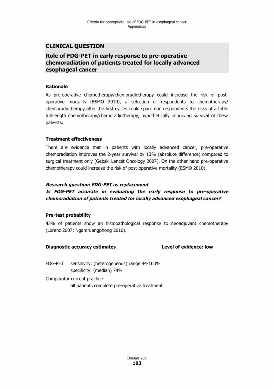
Criteria for appropriate use of FDG-PET in esophageal cancerAppendices
Dossier 209
103
CLINICAL QUESTION
Role of FDG-PET in early response to pre-operativechemoradiation of patients treated for locally advancedesophageal cancer
Rationale
As pre-operative chemotherapy/chemoradiotherapy could increase the risk of post-
operative mortality (ESMO 2010), a selection of respondents to chemotherapy/
chemoradiotherapy after the first cycles could spare non respondents the risks of a futile
full-length chemotherapy/chemoradiotherapy, hypothetically improving survival of these
patients.
Treatment effectiveness
There are evidence that in patients with locally advanced cancer, pre-operative
chemoradiation improves the 2-year survival by 13% (absolute difference) compared to
surgical treatment only (Gebski Lancet Oncology 2007). On the other hand pre-operative
chemotherapy could increase the risk of post-operative mortality (ESMO 2010).
Research question: FDG-PET as replacement
Is FDG-PET accurate in evaluating the early response to pre-operative
chemoradiation of patients treated for locally advanced esophageal cancer?
Pre-test probability
43% of patients show an histopathological response to neoadjuvant chemotherapy
(Lorenz 2007; Ngamruengphong 2010).
Diagnostic accuracy estimates Level of evidence: low
FDG-PET sensitivity: (heterogeneous) range 44-100%
specificity: (median) 74%
Comparator current practice
all patients complete pre-operative treatment

Criteria for appropriate use of FDG-PET in esophageal cancerAppendices
Dossier 209
104
Consequences of TEST for Level of importance*
(1-9)
True responders:
responders complete clinically effective pre-
operative treatment, which could improve survival
but might carries some risk of post-operative
mortalityPatients responders
False non responders:
responders interrupt clinically effective treatment,
which could have improved survival, and proceed
directly to surgery
True non responders:
non responders interrupt ineffective treatment,
which would not have improved survival, and
proceed directly to surgery, with lower risks of post-
operative mortalityPatients non
responders
False responders:
non responders complete ineffective pre-operative
treatment, with no possible gain in survival but with
some risk of post-operative mortality
* not important (score 1-3)
important (4-6)
critical (7-9)
to a decision
N of patients out of 100 submitted to the exam
According to FDG-PET According to current practice
True responders 19 - 43 43Patients
responders False non responders 24 - 0 0
True non responders 42 0Patients non
responders False responders 15 57
100 100

Criteria for appropriate use of FDG-PET in esophageal cancerAppendices
Dossier 209
105
CLINICAL QUESTION
Role of FDG-PET in early response to pre-operativechemoradiation of patients treated for locally advancedesophageal cancer
APPROPRIATENESS of FDG-PET
1-2-3 inappropriate
4-5-6 uncertain
7-8-9 appropriate 1 2 3 4 5 6 7 8 9
INDETERMINATE
Criteria for appropriate use of FDG-PET in esophageal cancerAppendices
Dossier 209
106
CLINICAL QUESTION
Role of FDG-PET in evaluating response to neoadjuvant therapyat the end of treatment
Rationale
In patients with locally advanced disease treated with neoadjuvant chemotherapy/
chemoradiotherapy, their response to treatment should be evaluated in order to decide
for subsequent type and aim of treatment (surgical, curative, palliative).
Treatment effectiveness
Surgical treatment is the therapy of choice for all patients with potentially curable
esophageal cancer and who are fit for major surgery (AIOM 2009; ESMO 2010; NCCN
2010; SIGN 2006).
Research question: FDG-PET as add on
Does adding FDG-PET to CT lead to a more accurate evaluation of response to
neoadjuvant therapy at the end of treatment?
Pre-test probability
42% of patients show an histopathological response to neoadjuvant chemotherapy
(Kwee 2010; Ngamruengphong 2010).
Diagnostic accuracy estimates Level of evidence: very low
High heterogeneity in both estimates of diagnostic accuracy are reported
FDG-PET sensitivity: range 27-100
specificity: range 30-100
EUS* sensitivity: range 20-100
specificity: range 36-100
CT* sensitivity: range 33-55
specificity: range 50-71
* data from studies evaluating FDG-PET

Criteria for appropriate use of FDG-PET in esophageal cancerAppendices
Dossier 209
107
Consequences of TEST for Level of importance*
(1-9)
True responders:
responders proceed to curative, radical treatment,
which could improve survival
Patients respondersFalse non responders:
responders do not receive curative, radical
treatment, which could have improved survival, and
proceed to less radical, palliative treatment
True non responders:
non responders proceed to less radical, palliative
treatmentPatients non
respondersFalse responders:
non responders proceed to curative, radical
treatment, with no possible gain in survival
* not important (score 1-3)
important (4-6)
critical (7-9)
to a decision
CLINICAL QUESTION
Role of FDG-PET in evaluating response to neoadjuvant therapyat the end of treatment
APPROPRIATENESS of FDG-PET
1-2-3 inappropriate
4-5-6 uncertain
7-8-9 appropriate 1 2 3 4 5 6 7 8 9
INDETERMINATE

Criteria for appropriate use of FDG-PET in esophageal cancerAppendices
Dossier 209
108
CLINICAL QUESTION
Role of FDG-PET during follow up of patients treated foresophageal cancer with no suspicion of recurrence
Rationale
Follow up of patients with esophageal or gastric cancer should monitor symptoms, signs
and nutritional status in order to detect disorders of function either related to recurrent
disease or affecting quality of life (SIGN 2006).
Treatment effectiveness
There is no evidence that regular follow up after initial therapy may influence the
outcome (ESMO 2010) and no evidence has been identified to support regular imaging or
measurement of serum tumor markers in the follow up of patients with esophageal
cancer (SIGN 2006). Aim of treatment is purely palliative.
Research question: FDG-PET introduced as new test
Is FDG-PET useful during follow up of patients with no suspicion of
recurrence?
Pre-test probability
55-57% of patients have distant recurrence (Roedl 2008; Sun 2009; Teyton 2009).
Diagnostic accuracy estimates Level of evidence: very low
The evidence about the validity of the FDG-PET in detecting recurrence of esophageal
cancer in patients with no suspicion of recurrence after definitive treatment (follow up)
comes from three low quality study providing heterogeneous estimates for both
sensitivity and specificity.

Criteria for appropriate use of FDG-PET in esophageal cancerAppendices
Dossier 209
109
Consequences of TEST for Level of importance*
(1-9)
True positives:
patients undergo further test to confirm positive
results and proceed to palliative treatmentPatients relapsing
False negative:
patients remain in follow up until symptoms occur
True negatives:
patients remain in follow up and are reassured, after
certain amount of stress
Patients not relapsing
False positives:
patients undergo unnecessary further tests to prove
negative and are exposed to unnecessary anxiety
* not important (score 1-3)
important (4-6)
critical (7-9)
to a decision
CLINICAL QUESTION
Role of FDG-PET during follow up of patients treated foresophageal cancer with no suspicion of recurrence
APPROPRIATENESS of FDG-PET
1-2-3 inappropriate
4-5-6 uncertain
7-8-9 appropriate 1 2 3 4 5 6 7 8 9
INDETERMINATE

Criteria for appropriate use of FDG-PET in esophageal cancerAppendices
Dossier 209
110
CLINICAL QUESTION
Role of FDG-PET in the diagnosis and staging of suspectedrecurrence in patients treated for esophageal cancer with clinicalsuspicion of recurrence or unclear conventional imaging results
Rationale
After curative surgery it could be difficult to interpret occurring symptoms and signs and
correctly differentiate local recurrence from scar. In case of a local recurrence a new
resection of the tumor could be planned.
Treatment effectiveness
Recurrences of patients treated with radiation therapy with curative intent could be
treated with surgery (NCCN 2010), while patients relapsing after surgery should be
treated with palliative intent (AIOM 2009).
Research question: FDG-PET as add on
Has FDG-PET sufficient specificity to be used as an add on test to diagnose
recurrence in patients with unclear results from conventional imaging?
Pre-test probability
80.4% of patients with suspected recurrence have metastases (Guo 2007).
Diagnostic accuracy estimates Level of evidence: very low
The evidence about the validity of FDG-PET in diagnosing recurrence of esophageal
cancer after definitive treatment comes from only one study on very few patients.

Criteria for appropriate use of FDG-PET in esophageal cancerAppendices
Dossier 209
111
Consequences of TEST for Level of importance*
(1-9)
True positives:
patients proceed to treatment for recurrence
Patients with
recurrenceFalse negative:
patients delay start of treatment until symptoms
occur, with a possible negative impact on quality of
life
True negatives:
patients remain in follow up, after a considerable
amount of stress
Patients without
recurrence False positives:
patients undergo unnecessary treatment, with a
possible negative impact on quality of life and suffer
unnecessary distress
* not important (score 1-3)
important (4-6)
critical (7-9)
to a decision
CLINICAL QUESTION
Role of FDG-PET in the diagnosis and staging of suspectedrecurrence in patients treated for esophageal cancer with clinical
suspicion of recurrence or unclear conventional imaging results
APPROPRIATENESS of FDG-PET
1-2-3 inappropriate
4-5-6 uncertain
7-8-9 appropriate 1 2 3 4 5 6 7 8 9
INDETERMINATE


Criteria for appropriate use of FDG-PET in esophageal cancerAppendices
Dossier 209
113
Appendix 2.Systematic reviewof literature: search strategyand tables of evidence


Criteria for appropriate use of FDG-PET in esophageal cancerAppendices
Dossier 209
115
CRITERIA FOR APPROPRIATE USEOF POSITRON EMISSION TOMOGRAPHY
IN ONCOLOGY
ESOPHAGEAL CANCER
SEARCH STRATEGY AND
TABLES OF EVIDENCE


Criteria for appropriate use of FDG-PET in esophageal cancerAppendices
Dossier 209
117
SEARCH STRATEGY
The following databases were searched for the period between January 2006 and July
2010:
Cochrane Database of Systematic Reviews (CDSR - The Cochrane Library)
Database of Abstracts of Reviews of Effects (DARE - The Cochrane Library)
Health Technology Assessment Database (HTA Database - The Cochrane Library)
Cochrane Central Register of Controlled Trials (CENTRAL - The Cochrane Library)
National Library of Medicine’s MEDLINE database (PubMed)
Elsevier’s EMBASE
Reference lists of identified articles were checked for additional references.
Criteria for appropriate use of FDG-PET in esophageal cancerAppendices
Dossier 209
118
CDSR, DARE, HTA database, CENTRAL search strategy
1. “Positron-Emission Tomography” [MeSH descriptor explode all trees]
2. “Fluorodeoxyglucose F18” [MeSH descriptor explode all trees]
3. “positron emission tomography”: ti,ab,kw
4. pet*: ti,ab,kw
5. pet scan*: ti,ab,kw
6. “Fluorodeoxyglucose F18”: ti,ab,kw or
7. fdg NEAR/2 18: ti,ab,kw
8. 1/7 OR
9. “Esophageal Neoplasms” MeSH descriptor explode all trees
10. “esophageal cancer”: ti,ab,kw
11. “Esophageal Neoplasm”: ti,ab,kw
12. “Esophagus Cancer”: ti,ab,kw
13. “Esophagus Neoplasm”: ti,ab,kw
14. “esophageal cancer”: ti,ab,kw
15. “esophagus cancer”: ti,ab,kw
16. 10/15 OR
17. 8 AND 16
Publication date: 2006-2010

Criteria for appropriate use of FDG-PET in esophageal cancerAppendices
Dossier 209
119
MEDLINE search strategy
1. “Fluorodeoxyglucose F18”[Mesh]
2. “2-Fluoro-2-deoxyglucose” [All Fields]
3. “18F Fluorodeoxyglucose” [All Fields]
4. “F 18 Fluorodeoxyglucose” [All Fields]
5. Fludeoxyglucose* [All Fields]
6. “2 fluoro 2 deoxy d glucose”[All Fields]
7. 18fluorodesoxyglucose*[All Fields]
8. fluorodeoxyglucose*[All Fields]
9. “fluorine 18 fluorodeoxyglucose” [All Fields]
10. 18fdg*[All Fields])
11. 18fluorodeoxyglucose*[All Fields]
12. 18fdg [All Fields]
13. 18 fdg* [All Fields]
14. fdg 18* [All Fields]
15. fdg/* [All Fields]
16. “fdg pet”[All Fields]
17. “Positron-Emission Tomography”[Mesh]
18. “positron emission tomography” [title/abstract]
19. pet [title/abstract]
20. “pet scan” [All Fields]
21. “pet scans” [All Fields]
22. “pet scanner” [All Fields]
23. petscan [All Fields]
24. 1/24 OR
25. “esophageal cancer”[All Fields]
26. “esophagus cancer”[All Fields]
27. “Esophageal Neoplasms”[Mesh]
28. “Esophageal Neoplasm”
29. “Esophageal Cancer”
30. “Esophagus Neoplasm”
31. “Esophagus Cancer”
32. 25/31 OR
33. 24 AND 32
34. “editorial”[Publication Type]
35. “comment”[Publication Type]

Criteria for appropriate use of FDG-PET in esophageal cancerAppendices
Dossier 209
120
36. “letter”[Publication Type]
37. “review”[Publication Type]
38. “case reports”[Publication Type]
39. 34/38 OR
40. 33 NOT 39
Limits: humans
Publication date: 2006-2010
Languages: English, French, Italian, Spanish

Criteria for appropriate use of FDG-PET in esophageal cancerAppendices
Dossier 209
121
EMBASE search strategy
1. ‘esophagus cancer’/exp
2. ‘esophagus cancer’
3. ‘esophagus cancer’/syn
4. ‘esophageal NEXT (cancer OR neoplasm OR tumor)
5. 1/4 OR
6. ‘positron emission tomography’/syn
7. ‘fluorodeoxyglucose f 18’/exp
8. (‘fluorodeoxyglucose f 18’/syn
9. ‘computer assisted emission tomography’/exp
10. ‘computer assisted emission tomography’ OR
11. pet
12. ‘pet scans’
13. ‘pet scanner’
14. ‘pet scan’
15. ‘pet/ct scan’
16. ‘pet/ct scans’
17. ‘pet/ct’
18. ‘positron emission tomography/computed tomography’
19. pet NEAR/4 scan*
20. pet NEAR/4 ct
21. 6/20 OR
22. 5 AND 21
Limits: humans
Publication date: 2006-2010
Languages: English, French, Italian, Spanish
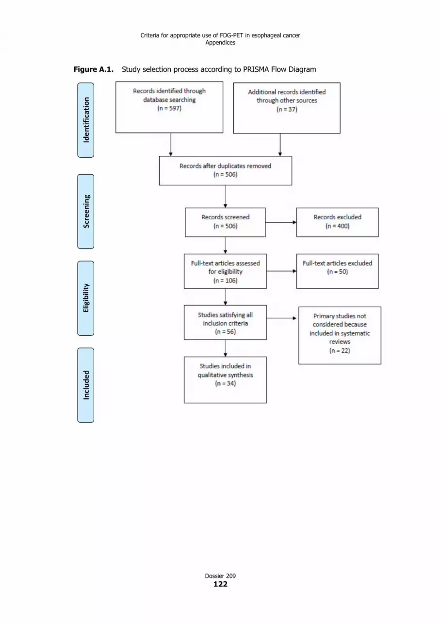
Criteria for appropriate use of FDG-PET in esophageal cancerAppendices
Dossier 209
122
Figure A.1. Study selection process according to PRISMA Flow Diagram

Criteria for appropriate use of FDG-PET in esophageal cancerAppendices
Dossier 209
123
TABLES OF EVIDENCE
Chapter 4
N staging of patients with primary esophageal cancer
Diagnostic accuracy
Systematic reviews
Author, year van Vliet 2008
Technology FDG-PET
Disease esophageal cancer
Objective to assess:
▪ staging
Inclusion criteria P patients with newly diagnosed esophageal cancer who did
not received previous radiation or chemotherapy
I FDG-PET
C endoscopic ultrasonography (EUS), CT
R histopathology following resection or FNA, clinical follow up
with or without radiological examination
O diagnostic accuracy for N staging, M staging
S diagnostic accuracy studies with prospective or
retrospective recruitment
Years covered by the search up to January 2006
Study selection data abstraction,
quality assessment performed by
two authors independently
yes
Comprehensive bibliographic
search: at least two databases
searched
no
Medline
Searched also specialized register,
conference proceedings, reviews,
textbooks and reference list of
retrieved studies
yes (only reference lists)
Searched also unpublished studies no
Language restriction not specified
Overall number of references
retrieved and n. of included studies
reported
yes

Criteria for appropriate use of FDG-PET in esophageal cancerAppendices
Dossier 209
124
N. and references of excluded
studies reported, reason given
no
Characteristics of included studies
clearly reported in tables
yes
Methodological quality of primary
studies assessed; criteria reported
no; but two methodological criteria used in the regression
model (consecutive or not recruitment, blinded or not
interpretation of results)
Results of quality assessment used
to formulate results and
conclusions
yes; two methodological criteria used in the regression model
(consecutive or not recruitment, blinded or not interpretation
of results)
Meta-analysis performed with
appropriate statistic methods
yes
Publication bias assessed yes
N. of included studies
Study design
EUS 31 for N staging
CT 17 for N staging, 7 for M staging
FDG-PET 10 for N staging, 9 for M staging
Patients of included studies not reported data on patients characteristics
Pre-test probability when given
N. of included patients EUS 1 841
CT N staging 943, M staging 437
FDG-PET N staging 424, M staging 475
Reference standard histopathology following resection or FNA
clinical follow up with or without radiological examination
Comparator EUS, CT
Performance results N staging
EUS sensitivity: 80% (95% CI 75-84)
specificity: 70% (95% CI 65-75)
CT sensitivity: 50% (95% CI 41-60)
specificity: 83% (95% CI 77-89)
FDG-PET sensitivity: 57% (95% CI 43-70)
specificity: 85% (95% CI 76-95)
Impact on management not assessed
Impact on clinical outcome not assessed

Criteria for appropriate use of FDG-PET in esophageal cancerAppendices
Dossier 209
125
Authors’ recommendations and
conclusions
EUS was significantly more sensitive but less specific than CT
and PET for the detection of regional lymph nodes. The overall
diagnostic performance of the three tests was similar. PET is
more sensitive and specific in the detection of distant
metastases compared with CT.
The presence of malignant regional lymph nodes can be
determined by EUS, CT or FDG-PET. To exclude the presence
of positive lymph nodes, EUS should be used, whereas
detected lesions should be confirmed with FNA or,
alternatively, with CT or PET. Both CT and PET can be used to
detect distant metastases, however the results suggest that
PET has higher diagnostic performance.
Comments of ASSR reviewers indirect comparisons

Criteria for appropriate use of FDG-PET in esophageal cancerAppendices
Dossier 209
126
Synoptic table of primary studies on N staging
Author, year Technology Patient number Population Staging Sensitivity (%) Specificity (%)
FDG-PET 81,71 87,3Yuan 2006
FDG-PET/CT45 SCC N staging
93,9 92,06
Buchmann 2006 FDG-PET 20 SCC N staging 20 100
Little 2007 FDG-PET 58 AC N staging 0 94
Katsoulis 2007 FDG-PET 22 SCC, AC N staging 71 67
FDG-PET 117 55 86Kato 2008
FDG-PET/CT 50SCC N staging
75,9 81
Sandha 2008 FDG-PET 29 SCC, AC N staging 36 100
125 100 98FDG-PET
55 100 95
125 100 99Schreurs 2008
FDG-PET/CT55
SCC, AC N staging
100 98
regional N staging 57,1 83,3Hsu 2009 FDG-PET 45 SCC
not regional N staging 36,4 82,4
single time point FDG-PET N staging 76,06 85,16Hu 2009
dual time point FDG-PET28 SCC
N staging 88,73 91,87

Criteria for appropriate use of FDG-PET in esophageal cancerAppendices
Dossier 209
127
Author, year Technology Patient number Population Staging Sensitivity (%) Specificity (%)
regional N staging 60 99,5Okada 2009 FDG-PET 18 SCC
paratracheal N staging 56 97,3
FDG-PET 75 85Roedl 2009
FDG-PET/CT81 AC N staging
76 96
Choi 2010 FDG-PET 109 SCC N staging 49 87
Alberini 2009 FDG-PET/CT 62 95% 100% 33.3%
SCC = squamous cell carcinoma; AC = adenocarcinoma
Criteria for appropriate use of FDG-PET in esophageal cancerAppendices
Dossier 209
128
Chapter 5
M staging of patients with primary esophageal cancer
Diagnostic accuracy
Systematic reviews
Author, year van Vliet 2008
Technology FDG-PET
Disease esophageal cancer
Objective to assess:
▪ staging
Inclusion criteria P patients with newly diagnosed esophageal cancer who did
not received previous radiation or chemotherapy
I FDG-PET
C endoscopic ultrasonography (EUS), CT
R histopathology following resection or FNA, clinical follow up
with or without radiological examination
O diagnostic accuracy for N staging, M staging
S Diagnostic accuracy studies with prospective or
retrospective recruitment
Years covered by the search up to January 2006
Study selection data abstraction,
quality assessment performed by
two authors independently
yes
Comprehensive bibliographic
search: at least two databases
searched
no
Medline
Searched also specialized register,
conference proceedings, reviews,
textbooks and reference list of
retrieved studies
yes (only reference lists)
Searched also unpublished studies no
Language restriction not specified
Overall number of references
retrieved and n. of included studies
reported
yes
N. and references of excluded
studies reported, reason given
no
Characteristics of included studies
clearly reported in tables
yes

Criteria for appropriate use of FDG-PET in esophageal cancerAppendices
Dossier 209
129
Methodological quality of primary
studies assessed; criteria reported
no; but two methodological criteria used in the regression
model (consecutive or not recruitment, blinded or not
interpretation of results)
Results of quality assessment used
to formulate results and
conclusions
yes; two methodological criteria used in the regression model
(consecutive or not recruitment, blinded or not interpretation
of results)
Meta-analysis performed with
appropriate statistic methods
yes
Publication bias assessed yes
N. of included studies
Study design
EUS 31 for N staging
CT 17 for N staging, 7 for M staging
FDG-PET 10 for N staging, 9 for M staging
Patients of included studies not reported data on patients characteristics
Pre-test probability when given
N. of included patients EUS 1 841
CT N staging 943, M staging 437
FDG-PET N staging 424, M staging 475
Reference standard histopathology following resection or FNA
clinical follow up with or without radiological examination
Comparator EUS, CT
Performance results M staging
CT sensitivity: 52% (95% CI 33-71)
specificity: 91% (95% CI 86-96)
FDG-PET sensitivity: 71% (95% CI 62-79)
specificity: 93% (95% CI 89-97)
Impact on management not assessed
Impact on clinical outcome not assessed
Authors’ recommendations and
conclusions
EUS was significantly more sensitive but less specific than CT
and PET for the detection of regional lymph nodes. The overall
diagnostic performance of the three tests was similar. PET is
more sensitive and specific in the detection of distant
metastases compared with CT.
The presence of malignant regional lymph nodes can be
determined by EUS, CT or PET. To exclude the presence of
positive lymph nodes, EUS should be used, whereas detected
lesions should be confirmed with FNA or, alternatively, with CT
or PET. Both CT and PET can be used to detect distant
metastases, however the results suggest that PET has higher
diagnostic performance.
Comments of ASSR reviewers indirect comparisons

Criteria for appropriate use of FDG-PET in esophageal cancerAppendices
Dossier 209
130
Synoptic table of primary studies on M staging
Author, year Technology Patient number Population Staging Sensitivity (%) Specificity (%)
Buchmann 2006 FDG-PET 20 SCC M staging 60 86
Little 2007 FDG-PET 58 AC M staging n.c. 95
Katsoulis 2007 FDG-PET 22 SCC, AC M staging 88 100
Noble 2009 FDG-PET 191 SCC, AC M staging 91 94
SCC = squamous cell carcinoma; AC = adenocarcinoma

Criteria for appropriate use of FDG-PET in esophageal cancerAppendices
Dossier 209
131
Impact on clinical outcomes
Primary studies on adverse events of change in management according to FDG-PET results during staging
Author, year Technology Limits Patient
number
Patient
characteristics
Standard practice Verification test Results
Meyers 2007 FDG-PET not
consecutive
sample
189 patients with
locally advanced
esophageal
cancer (T1-3,
N0-1, M0)
usual clinical
management (CT,
MRI, scintigraphy,
NOT EUS)
additional studies or
biopsies in case of
abnormalities by FDG-
PET that suggested
metastases
2 patients (1%) with false positive
FDG-PET result
▪ 1 adrenalectomy with subsequent
therapy for adrenal insufficiency
▪ 1 wound complication after a
confirmatory procedure
Primary studies on patient burden of FDG-PET during staging
Author, year Technology Limits Patient n. Patient characteristics Comparator Results
Westerterp 2008 FDG-PET - 82 (67 males, 15 females)
Their mean age was 64.3
(SD ± 8.3) years
After conventional workup
▪ 5 patients had a T1 tumor
▪ 10 patients a T2 tumor
▪ 63 patients a T3 tumor
▪ 4 patients had a T4 tumor
CT, US with/out
fine needle
aspiration, EUS
with/out fine
needle aspiration
For most tests and most dimensions of burden, the
large majority of subjects was in categories 1 and 2
Embarrassment/discomfort: 4 (very) or 5 (very
much) score:
EUS: 7 pts (8.5%)
US: 3 pts (3.6%)
FDG-PET: 3 pts (3.6%)
CT: 1 pt (1.2%)
Anxiety 4 (very) or 5 (very much) score:
FDG-PET: 6 pts (7.3%)
EUS: 5 pts (6.1%)
US: 2 pts (2.4%)
CT: 2 pt (2.4%)

Criteria for appropriate use of FDG-PET in esophageal cancerAppendices
Dossier 209
132
Chapter 6
Target volume definition of curative radiation treatment
Diagnostic accuracy
Systematic reviews
Author, year Muijs 2010
Technology FDG-PET
FDG CT/PET
Disease esophageal cancer
Objective to assess:
▪ curative intent RT field definition (only solid tumors)
Inclusion criteria P patients with esophageal cancer eligible for curative treatment
I CT/PET
C CT
R histopathology
O diagnostic accuracy in target volume delineation; evaluation of
the consequences for radiotherapy treatment planning with
regard to either target volumes or organs at risk
S cross sectional studies with prospective or retrospective
recruitment with at least 10 patients
Years covered by the search up to 2009
Study selection data abstraction,
quality assessment performed by
two authors independently
no
Comprehensive bibliographic
search: at least two databases
searched
yes
Medline, Cochrane Library
Searched also specialized register,
conference proceedings, reviews,
textbooks and reference list of
retrieved studies
yes (only reference lists)
Searched also unpublished studies no
Language restriction yes (English)
Overall number of references
retrieved and n of included studies
reported
yes
N. and references of excluded
studies reported, reason given
no
Characteristics of included studies
clearly reported in tables
yes

Criteria for appropriate use of FDG-PET in esophageal cancerAppendices
Dossier 209
133
Methodological quality of primary
studies assessed; criteria reported
no
Results of quality assessment used
to formulate results and
conclusions
no
Meta-analysis performed with
appropriate statistic methods
not applicable
Publication bias assessed no
N. of included studies
Study design
CT/PET: 12 studies (considering the questions of interest)
prospective or retrospective cross sectional studies
Patients of included studies not reported data on patients characteristics
Pre-test probability when given
Patients of included studies not reported
n. of included patients 247 (considering the questions of interest)
Reference standard histopathology (4 studies)
Comparator CT
Performance results ▪ changes in the delineation of target volumes (CTV/GTV/PTV) in
a proportion of patients ranging from 20 to 94% with respect
to CT (data from 6 studies: 142 patients); TV increase in a
proportion of 10-31% of patients; TV decrease in a proportion
of 10-62.5% of patients
▪ 3 out of 4 studies (89 patients) reported a significant positive
correlation (r raging from 0.74 to 0.89) between FDG-PET
tumor length and pathologic findings
Impact on management ▪ 1 study (16 patients): inadequate dose coverage in 38% of
patients with treatment plan based on CT alone with respect to
FDG-PET/CT-based PTV; no difference in radiation doses to the
near organs
▪ 1 study (34 patients): changes in dose distribution to normal
tissues in “virtually all patients”
▪ 1 study (21 patients): significant changes in dose distribution
to heart and lungs
Impact on clinical outcome no studies retrieved
Recommendations and conclusions The use of FDG-PET/CT resulted in changes of target volumes,
and consequently in changes in treatment planning. However,
evidence supporting the validity of the use of FDG-PET/CT in the
tumor delineation process is very limited. Tumor length
comparison as pathological validation has important shortcomings
and seems therefore unreliable. Furthermore, there are no studies
demonstrating the use of PET/CT in terms of improved
locoregional control or survival. Standard implementation of FDG-
PET/CT into the tumor delineation process for radiation treatment
seems therefore unjustified at this moment and needs further
clinical validation first.

Criteria for appropriate use of FDG-PET in esophageal cancerAppendices
Dossier 209
134
Primary studies
Author, year Shimizu 2009
Country Japan
Technology PET/CT
Disease esophageal cancer
Objective to assess:
▪ curative intent RT field definition
Patients characteristics 20 (1 764 lymph nodes), 15 males and 5 females; median age
61 years (range 47-75 years), with squamous cell carcinoma
of the esophagus, who consented to receive surgical
esophagectomy.
There were: 3 stage I patients, 8 stage II patients, 9 stage III
patients.
Index test FDG-PET/CT
Comparator CT, PET/CT with EUS, CT with EUS
Verification test histopathology after surgery
Outcomes considered diagnostic accuracy: clinical target volume (CTV) of lymph
node metastases
Results 53 (3.0%) of 1 764 nodes in the 20 patients were
histopathologically positive for cancer cells.
CTV not adequate to cover histopathologically detected
positive lymph nodes
▪ 7/20 (35%) PET/CT
▪ 8/20 (40%) on CT
▪ 5/20 (25%) on PET/CT+EUS
▪ 5/20 (25%) on CT+EUS
Study design prospective cohort
Consecutive recruitment not known
independent and blind
interpretation of index test and
verification test results
yes
Authors’ recommendations and
conclusions
The detection rate of subclinical lymph node metastasis did
not improve with the use of PET-CT, for either the cervical
and supraclavicular, mediastinal, or abdominal regions. It is
not recommended to use FDG-PET or PET-CT alone as a
diagnostic tool to determine CTV if pathologically involved
lymphatic regions are to be included in the CTV in the
treatment protocol. The accuracy of PET/CT must be further
improved in order to better detect positive nodes and improve
the definition of the CTV.

Criteria for appropriate use of FDG-PET in esophageal cancerAppendices
Dossier 209
135
Chapter 7
Evaluation of early response to neoadjuvant therapy
Diagnostic accuracy
Systematic reviews
Author, year Rebollo Aguirre 2009
Technology FDG-PET
PET/CT
Disease esophageal cancer
Objective to assess:
▪ early response to therapy (FDG-PET during treatment) only
when not adjuvant therapy
Inclusion criteria P patients with proven esophageal cancer treated with
neoadjuvant therapy
I FDG-PET, CT/PET
C none
R histopathology, other imaging techniques, clinical follow up
of at least 1 year
O diagnostic accuracy evaluating response during treatment
S diagnostic accuracy studies with prospective or
retrospective recruitment with at least 10 patients
Years covered by the search from 2004 to August 2006 (updating of the Westerterp 2005
systematic review)
N. of included studies
Study design
FDG-PET: 3 assessment of response during treatment (none
CT/PET)
Patients of included studies patients with stage II and III with different histologic types.
Neoadjuvant therapy consisted of varied cytotoxic drugs,
mostly including platinum-based agents, + RT (except 1 study
only chemotherapy)
Pre-test probability when given not given
N. of included patients FDG-PET during therapy: 84
CT/PET: 48
Reference standard histopathology
other imaging techniques
clinical follow up of at least 1 year
Comparator
Performance results primary tumor response
sensitivity: range 75-93%
specificity: range 75-87%
meta-analysis not performed because of significant
heterogeneity

Criteria for appropriate use of FDG-PET in esophageal cancerAppendices
Dossier 209
136
Impact on management not assessed
Impact on clinical outcome not assessed
Authors’ recommendations and
conclusions
The systematic review by Westerterp 2005 concluded that CT
is inaccurate in evaluating response to neoadjuvant therapy
because it does not distinguish between scar tissue and
neoplastic tissue; EUS is as accurate as PET but is an invasive
method not always feasible with some subjectivity and is
operator dependent; so PET is the best method to evaluate
induction therapy response. This updating, according to the
authors, confirmed these results.
Note ASSR reviewers both direct and indirect comparisons

Criteria for appropriate use of FDG-PET in esophageal cancerAppendices
Dossier 209
137
Author, year Ngamruengphong 2009
Technology PET
CT/PET
Disease esophageal cancer
Objective to assess:
▪ early response to therapy (PET during treatment) only
when not adjuvant therapy
Inclusion criteria P patients with esophageal cancer treated with neoadjuvant
therapy
I FDG-PET, CT/PET
C EUS
R histopathology
O diagnostic accuracy for restaging after treatment and for
assessing response during treatment
S diagnostic accuracy studies with prospective or
retrospective recruitment with at least 10 patients
Years covered by the search up to February 2008
N. of included studies
Study design
EUS: 7
PET: 6 during treatment (none with CT/PET)
Patients of included studies patients with stage II and III with different histologic types.
Neoadjuvant therapy consisted of varied cytotoxic drugs,
mostly including platinum-based agents. Three studies
performed interim FDG-PET after 2 weeks of treatment
Pre-test probability when given median prevalence of responders (i.e. pre-test probability) is
42% (range 18-58%)
N. of included patients EUS: 352
PET: 293
Reference standard histopathology
Comparator EUS
Performance results EUS
sensitivity: range 20-100%
specificity: range 36-100%
AUC: 0.86 (95% CI 0.77-0.96)
FDG-PET and CT/PET all studies (both during and after
therapy)
sensitivity: range 42-100%
specificity: range 27-100%
AUC: 0.80 (95% CI 0.72-0.89)
FDG-PET during therapy
AUC: 0.78 (95% CI 0.62-0.93)
no significant differences between accuracy during and after
therapy
Impact on management not assessed

Criteria for appropriate use of FDG-PET in esophageal cancerAppendices
Dossier 209
138
Impact on clinical outcome not assessed
Recommendations and conclusions EUS and PET have similar overall diagnostic accuracy for
assessment of response to neoadjuvant therapy. Each
modality has its unique advantages and limitations and should
be considered as complimentary rather than competing
technologies.
While in the subgroups analysis no significant differences were
shown between PET and CT/PET, this technologies has been
available only in the last few years and only three studies on
CT/PET were included; it is expected that the use of
integrated PET/CT scanner will replace PET only machines and
that would likely increase the overall diagnostic accuracy of
PET studies.
Comments of ASSR reviewers both direct and indirect comparisons

Criteria for appropriate use of FDG-PET in esophageal cancerAppendices
Dossier 209
139
Primary studies
Author, year Lorenzen 2007
Country Germany
Technology FDG-PET
Disease esophageal cancer or gastric cancer
Objective to assess:
▪ early response to therapy (FDG-PET during treatment) only
when not adjuvant therapy
Inclusion criteria P patients with esophageal cancer treated with chemotherapy
I FDG-PET after two weeks of chemotherapy
C none
R clinical response according to RECIST guidelines (including
CT)
O diagnostic accuracy for restaging after treatment and for
assessing response during treatment
S diagnostic accuracy studies with prospective or
retrospective recruitment with at least 10 patients
Study design prospective
Spectrum of participants
representative of practice
no
Selection criteria described? yes
Reference standard likely to
classify correctly
uncertain
Period between reference standard
and index test short
no
Whole sample or a random
selection verification using the
reference standard
no
Participants receive the same
reference standard
yes
Reference standard independent of
the index test
yes
Index test described in sufficient
detail
yes
Reference standard described in
sufficient detail
no
Index test interpreted without
knowledge of reference standard
yes
Reference standard interpreted
without knowledge index test
uncertain

Criteria for appropriate use of FDG-PET in esophageal cancerAppendices
Dossier 209
140
Same clinical data available when
the test results were interpreted as
would be available when the test is
used in practice
yes
Uninterpretable, indeterminate or
intermediate test results reported
yes
Withdrawals explained yes
N. of included patients 11
Patients of included studies 10 males and 1 female; median age 56 years (range 49-72); 5
adenocarcinoma and 6 squamous cell carcinoma. Neoadjuvant
therapy consisted of oral capecitabine (administered at a dose
of 1 000 mg m-2 twice daily on days 1-14 every three weeks)
and docetaxel administered at a dose of 75 mg m-2 i.v. on
day 1 every three weeks. Docetaxel was given 1 h before the
first oral dose of capecitabine. Patients were treated until best
response or until there was evidence of disease progression
Pre-test probability when given clinical response rate 45% at the end of treatment
Reference standard clinical response according to RECIST guidelines (including
CT)
Comparator none
Performance results FDG-PET during therapy
sensitivity: 100%
specificity: 60%
Impact on management not assessed
Impact on clinical outcome not assessed
Recommendations and conclusions In this heterogeneous study population, FDG-PET had a
limited accuracy in predicting clinical response. However, the
metabolic response prediction was particularly good in the
subgroup of patients with esophageal squamous cell cancer.
Therefore, FDG-PET and assessment of cancer therapy clearly
merits further investigation in circumscribed patient
populations with metastatic disease.

Criteria for appropriate use of FDG-PET in esophageal cancerAppendices
Dossier 209
141
Impact on clinical outcomes
Primary studies
Author, year Lordick 2007
Country Germany
Technology FDG-PET
Disease esophageal cancer
Objective to assess:
▪ early response to therapy (FDG-PET during treatment) only
when not adjuvant therapy
Patients characteristics 111 underwent surgical resection.
Patients with locally advanced adenocarcinoma of the
esophagogastric junction (AEG) type 1 (distal esophageal
adenocarcinoma) or type 2 (gastric-cardia-adenocarcinoma)
according to Siewert’s classification) were eligible. Patients
were staged as cT3 or cT4 based on CT and endoscopic
ultrasonography. Haematogenous metastases were excluded
by FDG-PET.
62 years (IQR 52-67). 77 patients (69%) had AEG type 1 and
34 patients (31%) had AEG type 2.
49 (44%) were treated with cisplatin, folinic acid and
fluorouracil; 45 (41%) received additional paclitaxel, and 17
(15%) received oxaliplatin, folinic acid and fluorouracil. Of 110
patients who were evaluable for response (one patient died
before assessment), cisplatin, folinic acid, and fluorouracil
were given to 26 responders and 22 non responders;
oxaliplatin, folinic acid, and fluorouracil were administered to 8
responders and 9 non responders; paclitaxel, cisplatin, folinic
acid and fluorouracil were given to 20 responders and 25 non
responders.
Intervention 1. FDG-PET WITH metabolic response after two weeks of
neoadjuvant chemotherapy (two cycles of cisplatin and
folinic acid plus fluorouracil on days 1, 8, 15, 22, 29, and
36, all repeated on day 49. For patients with a glomerular
filtration rate of less than 60 mL/Kg/min, oxaliplatin
replaced cisplatin. Patients aged 60 years or younger with
a good health status were additionally given paclitaxel on
days 0, 14, and 28) and continuation until the end of
treatment plan. Then patients with AEG type 1 tumors
underwent abdominothoracic esophagectomyand those
with AEG type 2 tumors had transhiatal extended
gastrectomy if the resection margins were tumor-free, or
underwent additional abdominothoracic esophagectomy if
the resection margins contained tumor tissue
2. FDG-PET WITH metabolic response (the same as above)
AND histopathological response

Criteria for appropriate use of FDG-PET in esophageal cancerAppendices
Dossier 209
142
Comparator 1. FDG-PET WITHOUT metabolic response after two weeks of
neoadjuvant chemotherapy (two cycles of cisplatin and
folinic acid plus fluorouracil on days 1, 8, 15, 22, 29, and
36, all repeated on day 49. For patients with a glomerular
filtration rate of less than 60 mL/Kg/min, oxaliplatin
replaced cisplatin. Patients aged 60 years or younger with
a good health status were additionally given paclitaxel on
days 0, 14, and 28) and interruption of chemotherapy then
performance of surgical resection. Patients with AEG type
1 tumors underwent abdominothoracic esophagectomyand
those with AEG type 2 tumors had transhiatal extended
gastrectomy if the resection margins were tumor-free, or
underwent additional abdominothoracic esophagectomy if
the resection margins contained tumor tissue
2. FDG-PET WITH metabolic response (the same as the
Intervention group) WITHOUT histopathological response
3. FDG-PET WITHOUT metabolic response (the same as the
Intervention group) WITHOUT histopathological response
Verification test istopathological tumor regression according to scoring system
Outcomes considered overall survival
event-free survival
adverse events
diagnostic accuracy (metabolic response vs histopathological
response)
Results Two patients (2%) died during chemotherapy (one non
assessable patient and one responder). One of these had a
sudden cardiac event, potentially induced by fluorouracil;
retrospective analysis showed a skip mutation in exon 14 of
the dihydropyrimidine dehydrogenase gene. The other patient
had a lethal stroke of unknown relation to chemotherapy.
After 2 weeks of chemotherapy, metabolic response was
assessable in 110 patients (one patient died before
assessment): 54 patients (49% [95% CI 39-59]) were
metabolic responders and 56 patients (51% [95% CI 41-61])
were metabolic non responders. No significant differences
were noted in the baseline characteristics of responders
versus non responders with regard to age, sex, performance
status, tumor localisation, T-category tumor size and N-
category nodal status, and histological subtype. By contrast,
the tumors of metabolically responsive patients tended to be
less differentiated, and these patients had significantly higher
median baseline SUVs of 8.3 (IQR 6.3-11.0-) versus 6.8 (IQR
5.1-9.0-) in non responders, p = 0.018. The number of
patients with metabolic responses were not significantly
different between the different chemotherapy regimens (p =
0.245).
(continues)
Criteria for appropriate use of FDG-PET in esophageal cancerAppendices
Dossier 209
143
Of the 104 patients who had their tumors resected, 88
patients (85%) had tumor-free resection margins (R0
resection) and 16 patients (15%) had microscopically affected
resection margins (R1). R0 resections could be done in 48 of
50 responding patients (96%) versus 40 of 54 non-responding
patients (74%, p = 0·002).
Post-operative deaths (30-day and in-hospital mortality)
occurred in 2 of 104 patients (2%), and post-operative
complications were reported in 35 patients (34%), with no
statistical difference for metabolic responders versus non
responders.
In the metabolic responder group, 29 of 50 patients (58%
[95% CI 48-67]) achieved a major histopathological response
(<10% residual tumor): 8 patients (16%) achieved complete
tumor remission; and 21 patients (42%) had subtotal
remission. No histological response was noted in metabolic
non responders. A higher number of low stage tumors was
reported in metabolic responders than in non responders.
Median event-free survival of 29·7 months (95% CI 23.6-35.7)
for metabolic responders compared with 14.1 months (7.5-
20.6) for non responders (HR 2.18 [1.32-3.62], p = 0·002).
Median overall survival was not reached in metabolic
responders, whereas nonresponders had a median overall
survival of 25.8 months (19.4-32.2; HR 2.13 [95% CI 1.14-
3.99], p = 0.015).
Metabolic responders who also had a major histological
response (n = 29) had a significantly better event-free
survival (HR 3.03 [1.28-7.16], p=0.006) and overall survival
(HR 4.55 [1.37-15.04], p=0.004) than did metabolic
responders who did not achieve a histological response
(n=21).
By contrast, event free survival (HR 1.29 [0.69-2.45],
p=0.430) and overall survival (HR 1.21 [0.56-2.63], p=0.549)
did not differ significantly when comparing metabolic
responders without a histological response (n=21) with
metabolic non responders (n=54)
Diagnostic accuracy of metabolic response
sensitivity: 100%
specificity: 72%
LR+ = 3.6
LR- = - infinito
Study design prospective series
Representativeness of the exposed
cohort selected group of users
Selection of the non exposed
cohort
from the same community as the exposed cohort

Criteria for appropriate use of FDG-PET in esophageal cancerAppendices
Dossier 209
144
Ascertainment of exposure secure record
Demonstration that outcome of
interest was not present at start of
study
yes
Comparability of cohorts on the
basis of the design or analysis
study controls for some important factors (select the most
important factor)
study controls for any additional factor
Assessment of outcome no description
Was follow up long enough for
outcomes to occur
yes
Adequacy of follow up of cohorts complete follow up - all subjects accounted for
subjects lost to follow up unlikely to introduce bias - small
number lost X
Authors’ recommendations and
conclusions
This study confirmed prospectively the usefulness of early
metabolic response evaluation, and shows the feasibility of a
PET-guided treatment algorithm. These findings might enable
tailoring of multimodal treatment in accordance with individual
tumor biology in future randomised trials.

Criteria for appropriate use of FDG-PET in esophageal cancerAppendices
Dossier 209
145
Chapter 8
Evaluation of response to neoadjuvant therapy at the end of
treatment
Diagnostic accuracy
Systematic reviews
Author, year Rebollo Aguirre 2009
Technology FDG-PET
PET/CT
Disease esophageal cancer
Objective to assess:
▪ response to therapy at the end of treatment
Inclusion criteria P patients with proven esophageal cancer treated with
neoadjuvant therapy
I FDG-PET, CT/PET
C none
R histopathology, other imaging techniques, clinical follow up
of at least 1 year
O diagnostic accuracy evaluating response during treatment
S diagnostic accuracy studies with prospective or
retrospective recruitment with at least 10 patients
Years covered by the search from 2004 to August 2006 (updating of the Westerterp 2005
systematic review)
N. of included studies
Study design
FDG-PET: 4 assessment of response after therapy (1 study
CT/PET)
Patients of included studies patients with stage II and III with different histologic types.
Neoadjuvant therapy consisted of varied cytotoxic drugs,
mostly including platinum-based agents, + RT
Pre-test probability when given not given
N. of included patients FDG-PET after therapy: 164
CT/PET: 48
Reference standard histopathology
other imaging techniques
clinical follow up of at least 1 year
Comparator

Criteria for appropriate use of FDG-PET in esophageal cancerAppendices
Dossier 209
146
Performance results primary tumor response
sensitivity: range 27.3-93.3%
specificity: range 41.7-95.2%
N restaging
sensitivity: range 16-67.5%
specificity: range 85.7-100%
meta-analysis not performed because of significant
heterogeneity
Impact on management not assessed
Impact on clinical outcome not assessed
Authors’ recommendations and
conclusions
The systematic review by Westerterp 2005 concluded that CT
is inaccurate in evaluating response to neoadjuvant therapy
because it does not distinguish between scar tissue and
neoplastic tissue; EUS is as accurate as PET but is an invasive
method not always feasible with some subjectivity and is
operator dependent; so PET is the best method to evaluate
induction therapy response. This updating, according to the
authors, confirmed these results.
Comments of ASSR reviewers both direct and indirect comparisons

Criteria for appropriate use of FDG-PET in esophageal cancerAppendices
Dossier 209
147
Author, year Ngamruengphong 2009
Technology FDG-PET
CT/PET
Disease esophageal cancer
Objective to assess:
▪ response to therapy at the end of treatment
Inclusion criteria P patients with esophageal cancer treated with neoadjuvant
therapy
I FDG-PET, CT/PET
C EUS
R histopathology
O diagnostic accuracy for restaging after treatment and for
assessing response during treatment
S diagnostic accuracy studies with prospective or
retrospective recruitment with at least 10 patients
Years covered by the search up to February 2008
N. of included studies
Study design
EUS: 7
FDG-PET: 11 assessment of response after therapy (3
CT/PET)
Patients of included studies patients with stage II and III with different histologic types.
Neoadjuvant therapy consisted of varied cytotoxic drugs,
mostly including platinum-based agents. Three studies
performed interim FDG-PET after 2 weeks of treatment
Pre-test probability when given median prevalence of responders (i.e. pre-test probability) is
42% (range 16-66%)
N. of included patients EUS: 352
PET: 555 (CT/PET: 180)
Reference standard histopathology
Comparator EUS
Performance results EUS
sensitivity: range 20-100%
specificity: range 36-100%
AUC: 0.86 (95% CI 0.77-0.96)
PET and CT/PET all studies (both during and after therapy)
sensitivity: range 42-100%
specificity: range 27-100%
AUC: 0.80 (95% CI 0.72-0.89)
PET and CT/PET after therapy
AUC: 0.80 (95% CI 0.71-0.89)
CT/PET after therapy
AUC: 0.77 (95% CI 0.39-1.00)
no significant differences between PET and CT/PET
no significant differences between accuracy during and after
therapy

Criteria for appropriate use of FDG-PET in esophageal cancerAppendices
Dossier 209
148
Impact on management not assessed
Impact on clinical outcome not assessed
Recommendations and conclusions EUS and PET have similar overall diagnostic accuracy for
assessment of response to neoadjuvant therapy. Each
modality has its unique advantages and limitations and should
be considered as complimentary rather than competing
technologies.
While in the subgroups analysis no significant differences were
shown between PET and CT/PET, this technologies has been
available only in the last few years and only three studies on
CT/PET were included; it is expected that the use of
integrated PET/CT scanner will replace PET only machines and
that would likely increase the overall diagnostic accuracy of
PET studies.
Comments of ASSR reviewers both direct and indirect comparisons

Criteria for appropriate use of FDG-PET in esophageal cancerAppendices
Dossier 209
149
Author, year Kwee 2010
Technology PET
CT/PET
Disease esophageal cancer
Objective to assess:
▪ response to therapy at the end of treatment
Inclusion criteria P patients with suspected recurrence after definitive
treatment of esophageal squamous cell carcinoma
I CT/FDG-PET
C none
R histopathology
O diagnostic accuracy evaluating response after treatment
S diagnostic accuracy studies with prospective or
retrospective recruitment with at least 10 patients
Years covered by the search up to June 2009
Study selection data abstraction,
quality assessment performed by
two authors independently
no
Comprehensive bibliographic
search: at least two databases
searched
yes
Medline, Embase, Cochrane Library
Searched also specialized register,
conference proceedings, reviews,
textbooks and reference list of
retrieved studies
yes
Searched also unpublished studies yes
Language restriction no
Overall number of references
retrieved and n of included studies
reported
yes
N. and references of excluded
studies reported, reason given
yes (only reasons, not references)
Characteristics of included studies
clearly reported in tables
yes
Methodological quality of primary
studies assessed; criteria reported
yes
QUADAS checklist
Results of quality assessment used
to formulate results and
conclusions
yes
Meta-analysis performed with
appropriate statistic methods
yes
Publication bias assessed no

Criteria for appropriate use of FDG-PET in esophageal cancerAppendices
Dossier 209
150
N. of included studies
Study design
FDG-PET: 20 (7 CT/PET)
prospective or retrospective cross sectional studies
Patients of included studies patients with predominantly stage II and III.
7 studies included a single specific histopathology subtype,
whereas the rest included different histologic types.
Neoadjuvant therapy consisted of varied cytotoxic drugs,
mostly including platinum-based agents, and 1 study including
hyperthermia. All but 4 studies used concomitant therapeutic
regimens of RT. Every study performed at least 2 FDG-PET
scans per patient, one before the neoadjuvant treatment and
another at the end.
Pre-test probability when given hstopatologic response prevalence: median 42%, range 16-
84%
N. of included patients 849
Reference standard histopathology
Comparator none
Performance results sensitivity:
range 33-100%; pooled estimate 67% (95% CI 62-72%)
specificity:
range 30-100%; pooled estimate 68% (95% CI 64-73%)
area under the sROC curve was 0.7815
significant heterogeneity in both the sensitivity and specificity
of the included studies (p < 0.0001)
Spearman r between the logit of sensitivity and the logit of 1 -
specificity was 0.086 (p = 0.719), which suggested that there
was no threshold effect
studies performed outside of the United States and studies of
higher methodologic quality yielded significantly higher overall
accuracy
Impact on management not assessed
Impact on clinical outcome not assessed
Recommendations and conclusions on the basis of current evidence, 18F-FDG PET should not yet
be used in routine clinical practice to guide neoadjuvant
therapy decisions in patients with esophageal cancer

Criteria for appropriate use of FDG-PET in esophageal cancerAppendices
Dossier 209
151
Synoptic table of primary studies evaluating response to neoadjuvant therapy at the end of treatment
Author, year Technology Patient number Population Staging Sensitivity (%) Specificity (%)
Erasmus 2006 FDG-PET/CT 52 SCC, AC, chemo and/or RT T staging 47 58
Wieder 2007 FDG-PET 24 AC, chemoT T staging 75 87
Kim 2007 FDG-PET 62 SCC, chemoT and RT T staging 51,2 66,7
Higuchi 2008 FDG-PET 50 SCC, chemoT and/or RT T staging 85,7 93,1
Klaeser 2009 FDG-PET or FDG-PET/CT 45 SCC, AC, chemoT T staging 68 52
Roedl 2009 FDG-PET or FDG-PET/CT 49 SCC, chemoT and RT T staging 86,5 91
Roedl 2008 FDG-PET or FDG-PET/CT 47 SCC, AC, chemoT and RT T staging 91 92
SCC = squamous cell carcinoma; AC = adenocarcinoma
chemoT = chemotherapy
RT = radiotherapy
Criteria for appropriate use of FDG-PET in esophageal cancerAppendices
Dossier 209
152
Chapter 9
Follow up in patients with no suspicion of recurrence
Diagnostic accuracy
Synoptic table of primary studies on follow up of patients with no suspicion of recurrence
Author, year Technology Limits Patient
number
Cancer
characteristics
Treatment Comparator Reference
standard
Recurrence
rate
Results
Roedl 2008 FDG-PET,
FDG-PET/CT
small study,
B
47 squamous,
adenoCa
surgical resection
with neoadjuvant
chemoradiotherapy
none biopsy of the
suspected
lesion or clinical
follow up
57% FDG-PET
sensitivity 89%
specificity 55%
FDG-PET/CT
sensitivity 89%
specificity 75%
Sun 2009 FDG-PET/CT small study,
S, R, B
20 not specified surgical resection
followed by
radiotherapy
none biopsy of the
suspected
lesion or clinical
follow up
55% FDG-PET/CT
sensitivity 100%
specificity 67%
Teyton 2009 FDG-PET small study,
B
41 squamous,
adenoCa
surgical resection
with/without
neoadjuvant
chemoradiotherapy
CT biopsy of the
suspected
lesion or clinical
follow up
56% FDG-PET
sensitivity 100%
specificity 85.3%
CT
sensitivity 65%
specificity 91.2%
Criteria for appropriate use of FDG-PET in esophageal cancerAppendices
Dossier 209
153
Primary studies
Author, year Roedl 2008
Country USA
Technology FDG-PET and FDG-PET/CT
Disease esophageal cancer
Objective to assess:
▪ follow up in asymptomatic patients
Inclusion criteria P patients with squamous cell carcinoma and
adenocarcinoma of the esophagus who underwent
neoadjuvant chemoradiotherapy followed by surgery were
included in the study. The clinical stage of all patients
before neoadjuvant therapy was stage II or stage III
I FDG-PET/CT e FDG-PET every 3 or 6 months till 24th month
C none
R biopsy of the suspected lesion or follow up with EUS
O diagnostic accuracy during follow up
Study design prospective consecutive
Spectrum of participants
representative of practice
yes
Selection criteria described? yes
Reference standard likely to
classify correctly
yes (short follow up)
Period between reference standard
and index test short
no
Whole sample or a random
selection verification using the
reference standard
yes
Participants receive the same
reference standard
no
Reference standard independent of
the index test
yes
Index test described in sufficient
detail
yes
Reference standard described in
sufficient detail
yes
Index test interpreted without
knowledge of reference standard
yes
Reference standard interpreted
without knowledge index test
uncertain

Criteria for appropriate use of FDG-PET in esophageal cancerAppendices
Dossier 209
154
Same clinical data available when
the test results were interpreted as
would be available when the test is
used in practice
yes
Uninterpretable, indeterminate or
intermediate test results reported
uncertain
Withdrawals explained not pertinent
N. of included patients 47
Patients characteristics patients with squamous cell carcinoma and adenocarcinoma of
the esophagus who underwent neoadjuvant
chemoradiotherapy followed by surgery were included in the
study. The clinical stage of all patients before neoadjuvant
therapy was stage II or stage III
Outcomes considered diagnostic validity: sensitivity/specificity
Pre-test probability recurrence rate 57,4% (27/47)
Performance results FDG-PET
locoregional recurrence sensitivity 83%
specificity 56%
lymph nodes sensitivity 68%
specificity 67%
distant metastasis sensitivity 84%
specificity 75%
patient based sensitivity 89%
specificity 55%
FDG-PET/CT
locoregional recurrence sensitivity 92%
specificity 81%
lymph nodes sensitivity 82%
specificity 89%
distant metastasis sensitivity 89%
specificity 85%
patient based sensitivity 89%
specificity 75%
Authors’ recommendations and
conclusions
The present study demonstrates that the decrease of tumor
length between the initial and post treatment PET-CT scan
more accurately predicts treatment response and disease-free
survival than does the decrease of SUV. In the evaluation of
tumor recurrence, PET-CT was more accurate than PET both
in a patient and in a lesion-based analysis; and PET-CT should
be used routinely between 12 and 24 months after surgery to
screen for recurrent sites.

Criteria for appropriate use of FDG-PET in esophageal cancerAppendices
Dossier 209
155
Author, year Sun 2009
Country China
Technology FDG-PET/CT
Disease esophageal cancer
Objective to assess:
▪ follow up in asymptomatic patients
Inclusion criteria P patients after treatment of esophageal cancer treated with
combined surgical treatment and radiotherapy
management with or without clinically and/or radiologically
suspicious findings for restaging
I FDG-PET/CT
C none
R the standard reference for tumor recurrence consisted of
histopathological confirmation or clinical follow up for at
least ten months after 18F-FDG PET/CT
O diagnostic accuracy after treatment or of suspected
recurrence
Study design retrospective
Spectrum of participants
representative of practice
uncertain
Selection criteria described? yes
Reference standard likely to
classify correctly
uncertain (short follow up?)
Period between reference standard
and index test short
no
Whole sample or a random
selection verification using the
reference standard
yes
Participants receive the same
reference standard
no
Reference standard independent of
the index test
yes
Index test described in sufficient
detail
yes
Reference standard described in
sufficient detail
no
Index test interpreted without
knowledge of reference standard
yes
Reference standard interpreted
without knowledge index test
uncertain

Criteria for appropriate use of FDG-PET in esophageal cancerAppendices
Dossier 209
156
Same clinical data available when
the test results were interpreted as
would be available when the test is
used in practice
yes
Uninterpretable, indeterminate or
intermediate test results reported
uncertain
Withdrawals explained no
n. of included patients 20
Patients characteristics patients with esophageal cancer after surgical resection and
following radiotherapy (15 males and 5 females; age range
39-68 years; mean age 55.1 years)
Outcomes considered diagnostic validity: sensitivity/specificity
Pre-test probability recurrence rate: 55% (11/20)
Performance results recurrence
sensitivity: 100%
specificity: 67%
accuracy: 85%
negative predictive value (NPV): 100%
positive predictive value (PPV): 78.6%
the 3 false positive FDG-PET/CT findings were chronic
inflammation of mediastinal lymph nodes (2) and anastomosis
inflammation (1)
clinical decisions of treatment were changed in 12 (60%)
patients after introducing 18F-FDG PET/CT into their
conventional post-treatment follow up program
Authors’ recommendations and
conclusions
Whole body 18F-FDG PET/CT is effective in detecting relapse
of esophageal cancer after surgical resection and
radiotherapy. It could also have important clinical impact on
the management of esophageal cancer, influencing both
clinical restaging and salvage treatment of patients.
Criteria for appropriate use of FDG-PET in esophageal cancerAppendices
Dossier 209
157
Author, year Teyton 2009
Country France
Technology FDG-PET
Disease esophageal cancer
Objective to assess:
▪ follow up in asymptomatic patients
Inclusion criteria P patients with esophageal cancer after initial radical
esophagectomy
I FDG-PET about every 6 months
C CT
R biopsy of the suspected lesion or clinical follow up (median
48 months)
O diagnostic accuracy during follow up
Study design prospective consecutive
Spectrum of participants
representative of practice
yes (of the lower stages)
Selection criteria described? yes
Reference standard likely to
classify correctly
yes
Period between reference standard
and index test short
no
Whole sample or a random
selection verification using the
reference standard
yes
Participants receive the same
reference standard
no
Reference standard independent of
the index test
yes
Index test described in sufficient
detail
yes
Reference standard described in
sufficient detail
yes
Index test interpreted without
knowledge of reference standard
yes
Reference standard interpreted
without knowledge index test
uncertain
Same clinical data available when
the test results were interpreted as
would be available when the test is
used in practice
yes
Uninterpretable, indeterminate or
intermediate test results reported
uncertain

Criteria for appropriate use of FDG-PET in esophageal cancerAppendices
Dossier 209
158
Withdrawals explained not pertinent
N. of included patients 41
Patients characteristics Thirty-eight were male (93%) and the mean age at the time
of diagnosis was 60.7 ± 9.4 years. Most of the tumors were
squamous cell carcinoma (76%) and most of the patients had
a well differentiated or moderately differentiated tumor
(90%). The majority of the tumors originated from the middle
and lower esophagus (93%). In the population included in this
study, 51% of the patients had an early stage disease (stage I
or IIa), while 58% of the patients had a T3 primary lesion.
Twenty patients (48%) had lymph node metastases (N1) at
presentation
Outcomes considered diagnostic validity: sensitivity/specificity/accuracy
Pre-test probability recurrence rate: 56.1% (23/41)
Performance results FDG-PET
locoregional recurrence sensitivity 93.3%
specificity 97.4%
accuracy 96.2%
distant metastasis sensitivity 100%
specificity 89.4%
accuracy 92.5%
patient based sensitivity 100%
specificity 85.3%
accuracy 90.7%
CT
locoregional recurrence sensitivity 60%
specificity 100%
accuracy 88.9%
distant metastasis sensitivity 66.6%
specificity 92.1%
accuracy 84.9%
patient based sensitivity 65%
specificity 91.2%
accuracy 81.5%

Criteria for appropriate use of FDG-PET in esophageal cancerAppendices
Dossier 209
159
Authors’ recommendations and
conclusions
Surgery remains a major option in the management of
esophageal neoplasms. Early diagnosis of recurrence in
asymptomatic patients could be a good way to improve the
management of those patients. The present study is the first
prospective study systematically using FDG-PET in the follow
up of surgically resected patients and it has shown that FDG-
PET is accurate for the detection of early recurrence of
esophageal cancer after initial surgery. Based on the
presented results, FDG-PET could be included in the routine
protocol for the evaluation of asymptomatic patients after
surgery, as early as 6 months after the initial operative
procedure. The use of FDG-PET in comparison with the use of
endoscopy, CT scan, and/or echography remains to be
demonstrated in terms of cost-effectiveness.

Criteria for appropriate use of FDG-PET in esophageal cancerAppendices
Dossier 209
160
Chapter 10
Diagnosis and staging of suspect distant recurrence
Diagnostic accuracy
Primary studies
Author, year Guo 2007
Technology PET/CT
Disease esophageal cancer
Objective to assess:
▪ diagnosis of suspected recurrence and staging of
recurrence
Inclusion criteria P patients with suspected recurrence (indication of possible
recurrence, such as questionable symptoms or signs,
equivocal diagnosis by CT, EUS, MRI, or barium swallow)
after definitive treatment of esophageal squamous cell
carcinoma
I FDG-PET/CT after suspected recurrence
C none
R histopatology or follow up
O diagnostic accuracy of suspected recurrence
Study design prospective non consecutive
Spectrum of participants
representative of practice
no
Selection criteria described? yes
Reference standard likely to
classify correctly
yes
Period between reference standard
and index test short
no
Whole sample or a random
selection verification using the
reference standard
yes
Participants receive the same
reference standard
no
Reference standard independent of
the index test
yes
Index test described in sufficient
detail
yes
Reference standard described in
sufficient detail
yes
Index test interpreted without
knowledge of reference standard
yes

Criteria for appropriate use of FDG-PET in esophageal cancerAppendices
Dossier 209
161
Reference standard interpreted
without knowledge index test
no
Same clinical data available when
the test results were interpreted as
would be available when the test is
used in practice
yes
Uninterpretable, indeterminate or
intermediate test results reported
uncertain
Withdrawals explained yes
N. of included patients 56
Patients characteristics patients with suspected recurrence (indication of possible
recurrence, such as questionable symptoms or signs,
equivocal diagnosis by CT, EUS, MRI, or barium swallow) after
definitive treatment of esophageal squamous cell carcinoma.
47 males and 9 females; age range 38-77 years. There were 5
stage IIb patients, 38 stage III patients, and 13 stage IV
patients.
The dominant treatment modalities preceding FDG-PET/CT
were primarily surgery (surgery alone 4 patients; surgery plus
radiation 11 patients; surgery plus chemoradiation 8 patients;
surgery plus chemotherapy 3 patients) or radiotherapy
(radiotherapy alone 3 patients; radiotherapy plus
chemotherapy 27 patients).
Outcomes considered diagnostic validity: sensitivity/specificity/accuracy
Pre-test probability recurrence rate: 80.4% (45/56)
Performance results Local recurrence
sensitivity 96.9%
specificity 50%
accuracy 84.1%
Regional recurrence
sensitivity 89.5%
specificity 81.8%
accuracy 86.7%
Distant metastasis
sensitivity 90.5%
specificity 92.9%
accuracy 91.4%
Patient based
sensitivity 95.6%
specificity 54.5%
accuracy 87.5%
(continues)

Criteria for appropriate use of FDG-PET in esophageal cancerAppendices
Dossier 209
162
Among the 9 false positive (FP) interpretations rendered on
FDG-PET/CT, 5 lesions exhibited distinctly increased 18F-FDG
uptake at the esophagogastric anastomosis but revealed no
biopsy-proven recurrence by repeated endoscopy. The
remaining 4 FP interpretations - including 1 focus near the
back wall of gastric pull-up, 2 at hilar nodes, and 1 in the left
lower lung - had also been excluded from recurrence after
close follow up.
FDG-PET/CT also had 5 false-negative (FN) interpretations.
The first one occurred in a patient who had no positive FDG-
PET/CT findings after radical radiotherapy. Five months later,
he had dysphagia, and endoscopy validated malignancy at the
location of primary tumor. The second patient had a subcarina
lymph node measuring 1.2 cm in short-axis diameter on FDG-
PET/CT (maximum SUV: 2.3) at 2 months after the completion
of radiation therapy. Three months after the FDG-PET/CT
scan, the shortest diameter of this node in the transverse axis
increased to approximately 3 cm on followup CT. The third
patient had a persistent dry cough starting at 2 months after
surgery, but both subsequent CT and FDG-PET/CT showed no
trace of a cancer-related focus. However, a paratracheal
lymph node measuring 2.3 cm in diameter was detected on
CT at 6-months follow up after surgery. The fourth FN
interpretation involved a supraclavicular lymph node in a
patient with upper-segment esophageal cancer that was
treated with radiation at both the primary tumor and the
regional nodes. The last FN interpretation was a 0.5-cm lung
nodule at the right lower lobe. It was first detected by
contrast-enhanced CT after radiation and was not visible on
the concurrent FDG-PET/CT image. Serial CT images during
follow up confirmed this lesion to be a distant metastasis.
Authors’ recommendations and
conclusions
PET/CT has displayed a remarkable sensitivity and a high
specificity and accuracy at regional and distant sites for
recurrent ESCC. 18F-FDG PET/CT can be recommended as a
preferred tool for patients who have elusive clinical
manifestations or equivocal results from conventional imaging
modalities. The SUV and the disease pattern (with or without
systemic recurrence) on PET/CT can offer incremental
information in the prognostic evaluation. Although the
specificity at the local area tends to be lower due to a high
rate of FP findings, PET/CT remains valuable because positive
PET/CT can encourage clinicians to make efforts to establish a
definitive diagnosis through biopsies or close follow up, and
the patients confirmed with recurrence would benefit from
subsequent salvage therapy.

1
1(*) volumi disponibili presso l’Agenzia sanitaria e sociale regionale. Sono anche scaricabili dal sito
http://asr.regione.emilia-romagna.it/wcm/asr/collana_dossier/archivio_dossier_1.htm
1990
1. Centrale a carbone “Rete 2”: valutazione dei rischi. Bologna. (*)
2. Igiene e medicina del lavoro: componente della assistenza sanitaria di base. Servizi di igiene e medicina del lavoro.
(Traduzione di rapporti OMS). Bologna. (*)
3. Il rumore nella ceramica: prevenzione e bonifica. Bologna. (*)
4. Catalogo collettivo dei periodici per la prevenzione. I edizione - 1990. Bologna. (*)
5. Catalogo delle biblioteche SEDI - CID - CEDOC e Servizio documentazione e informazione dell’ISPESL. Bologna. (*)
1991
6. Lavoratori immigrati e attività dei servizi di medicina preventiva e igiene del lavoro. Bologna. (*)
7. Radioattività naturale nelle abitazioni. Bologna. (*)
8. Educazione alimentare e tutela del consumatore “Seminario regionale Bologna 1-2 marzo 1990”. Bologna. (*)
1992
9. Guida alle banche dati per la prevenzione. Bologna.
10. Metodologia, strumenti e protocolli operativi del piano dipartimentale di prevenzione nel comparto rivestimenti
superficiali e affini della provincia di Bologna. Bologna. (*)
11. I Coordinamenti dei Servizi per l’Educazione sanitaria (CSES): funzioni, risorse e problemi. Sintesi di un’indagine
svolta nell’ambito dei programmi di ricerca sanitaria finalizzata (1989 - 1990). Bologna. (*)
12. Epi Info versione 5. Un programma di elaborazione testi, archiviazione dati e analisi statistica per praticare
l’epidemiologia su personal computer. Programma (dischetto A). Manuale d’uso (dischetto B). Manuale introduttivo.
Bologna.
13. Catalogo collettivo dei periodici per la prevenzione in Emilia-Romagna. 2a edizione. Bologna. (*)
1993
14. Amianto 1986-1993. Legislazione, rassegna bibliografica, studi italiani di mortalità, proposte operative. Bologna. (*)
15. Rischi ambientali, alimentari e occupazionali, Attività di prevenzione e controllo nelle USL dell’Emilia-Romagna.
1991. Bologna. (*)
16. La valutazione della qualità nei Servizi di igiene pubblica delle USL dell’Emilia-Romagna, 1991. Bologna. (*)
17. Metodi analitici per lo studio delle matrici alimentari. Bologna. (*)
1994
18. Venti anni di cultura per la prevenzione. Bologna.
19. La valutazione della qualità nei Servizi di igiene pubblica dell’Emilia-Romagna 1992. Bologna. (*)
20. Rischi ambientali, alimentari e occupazionali, Attività di prevenzione e controllo nelle USL dell’Emilia-Romagna.
1992. Bologna. (*)
21. Atlante regionale degli infortuni sul lavoro. 1986-1991. 2 volumi. Bologna. (*)
22. Atlante degli infortuni sul lavoro del distretto di Ravenna. 1989-1992. Ravenna. (*)
23. 5a Conferenza europea sui rischi professionali. Riccione, 7-9 ottobre 1994. Bologna.
COLLANADOSSIERacuradell’Agenziasanitariaesocialeregionale

1995
24. La valutazione della qualità nei Servizi di igiene pubblica dell’Emilia-Romagna 1993. Bologna. (*)
25. Rischi ambientali, alimentari e occupazionali, Attività di prevenzione e controllo nelle USL dell’Emilia-Romagna.
1993. Bologna. (*)
1996
26. La valutazione della qualità nei Servizi di igiene pubblica dell’Emilia-Romagna. Sintesi del triennio 1992-1994. Dati
relativi al 1994. Bologna. (*)
27. Lavoro e salute. Atti della 5a Conferenza europea sui rischi professionali. Riccione, 7-9 ottobre 1994. Bologna. (*)
28. Gli scavi in sotterraneo. Analisi dei rischi e normativa in materia di sicurezza. Ravenna. (*)
1997
29. La radioattività ambientale nel nuovo assetto istituzionale. Convegno Nazionale AIRP. Ravenna. (*)
30. Metodi microbiologici per lo studio delle matrici alimentari. Ravenna. (*)
31. Valutazione della qualità dello screening del carcinoma della cervice uterina. Ravenna. (*)
32. Valutazione della qualità dello screening mammografico del carcinoma della mammella. Ravenna. (*)
33. Processi comunicativi negli screening del tumore del collo dell’utero e della mammella (parte generale). Proposta di
linee guida. Ravenna. (*)
34. EPI INFO versione 6. Ravenna. (*)
1998
35. Come rispondere alle 100 domande più frequenti negli screening del tumore del collo dell’utero. Vademecum per gli
operatori di front-office. Ravenna.
36. Come rispondere alle 100 domande più frequenti negli screening del tumore della mammella. Vademecum per gli
operatori di front-office. Ravenna. (*)
37. Centri di Produzione Pasti. Guida per l’applicazione del sistema HACCP. Ravenna. (*)
38. La comunicazione e l’educazione per la prevenzione dell’AIDS. Ravenna. (*)
39. Rapporti tecnici della Task Force D.Lgs 626/94 - 1995-1997. Ravenna. (*)
1999
40. Progetti di educazione alla salute nelle Aziende sanitarie dell’Emilia Romagna. Catalogo 1995 - 1997. Ravenna. (*)
2000
41. Manuale di gestione e codifica delle cause di morte, Ravenna.
42. Rapporti tecnici della Task Force D.Lgs 626/94 - 1998-1999. Ravenna. (*)
43. Comparto ceramiche: profilo dei rischi e interventi di prevenzione. Ravenna. (*)
44. L’Osservatorio per le dermatiti professionali della provincia di Bologna. Ravenna. (*)
45. SIDRIA Studi Italiani sui Disturbi Respiratori nell’Infanzia e l’Ambiente. Ravenna. (*)
46. Neoplasie. Rapporto tecnico per la definizione di obiettivi e strategie per la salute. Ravenna.
2001
47. Salute mentale. Rapporto tecnico per la definizione di obiettivi e strategie per la salute. Ravenna.
48. Infortuni e sicurezza sul lavoro. Rapporto tecnico per la definizione di obiettivi e strategie per la salute. Ravenna.
(*)
49. Salute Donna. Rapporto tecnico per la definizione di obiettivi e strategie per la salute. Ravenna.
50. Primo report semestrale sull’attività di monitoraggio sull’applicazione del D.Lgs 626/94 in Emilia-Romagna. Ravenna.
(*)

51. Alimentazione. Rapporto tecnico per la definizione di obiettivi e strategie per la salute. Ravenna. (*)
52. Dipendenze patologiche. Rapporto tecnico per la definizione di obiettivi e strategie per la salute. Ravenna.
53. Anziani. Rapporto tecnico per la definizione di obiettivi e strategie per la salute. Ravenna. (*)
54. La comunicazione con i cittadini per la salute. Rapporto tecnico per la definizione di obiettivi e strategie per la
salute. Ravenna. (*)
55. Infezioni ospedaliere. Rapporto tecnico per la definizione di obiettivi e strategie per la salute. Ravenna. (*)
56. La promozione della salute nell’infanzia e nell’età evolutiva. Rapporto tecnico per la definizione di obiettivi e
strategie per la salute. Ravenna. (*)
57. Esclusione sociale. Rapporto tecnico per la definizione di obiettivi e strategie per la salute. Ravenna.
58. Incidenti stradali. Proposta di Patto per la sicurezza stradale. Rapporto tecnico per la definizione di obiettivi e
strategie per la salute. Ravenna. (*)
59. Malattie respiratorie. Rapporto tecnico per la definizione di obiettivi e strategie per la salute. Ravenna. (*)
2002
60. AGREE. Uno strumento per la valutazione della qualità delle linee guida cliniche. Bologna.
61. Prevalenza delle lesioni da decubito. Uno studio della Regione Emilia-Romagna. Bologna.
62. Assistenza ai pazienti con tubercolosi polmonare nati all’estero. Risultati di uno studio caso-controllo in Emilia-
Romagna. Bologna. (*)
63. Infezioni ospedaliere in ambito chirurgico. Studio multicentrico nelle strutture sanitarie dell’Emilia-Romagna.
Bologna. (*)
64. Indicazioni per l’uso appropriato della chirurgia della cataratta. Bologna. (*)
65. Percezione della qualità e del risultato delle cure. Riflessione sugli approcci, i metodi e gli strumenti. Bologna. (*)
66. Le Carte di controllo. Strumenti per il governo clinico. Bologna. (*)
67. Catalogo dei periodici. Archivio storico 1970-2001. Bologna.
68. Thesaurus per la prevenzione. 2a edizione. Bologna. (*)
69. Materiali documentari per l’educazione alla salute. Archivio storico 1970-2000. Bologna. (*)
70. I Servizi socio-assistenziali come area di policy. Note per la programmazione sociale regionale. Bologna. (*)
71. Farmaci antimicrobici in età pediatrica. Consumi in Emilia-Romagna. Bologna. (*)
72. Linee guida per la chemioprofilassi antibiotica in chirurgia. Indagine conoscitiva in Emilia-Romagna. Bologna. (*)
73. Liste di attesa per la chirurgia della cataratta: elaborazione di uno score clinico di priorità. Bologna. (*)
74. Diagnostica per immagini. Linee guida per la richiesta. Bologna. (*)
75. FMEA-FMECA. Analisi dei modi di errore/guasto e dei loro effetti nelle organizzazioni sanitarie. Sussidi per la
gestione del rischio 1. Bologna.
2003
76. Infezioni e lesioni da decubito nelle strutture di assistenza per anziani. Studio di prevalenza in tre Aziende USL
dell’Emilia-Romagna. Bologna. (*)
77. Linee guida per la gestione dei rifiuti prodotti nelle Aziende sanitarie dell’Emilia-Romagna. Bologna. (*)
78. Fattibilità di un sistema di sorveglianza dell’antibioticoresistenza basato sui laboratori. Indagine conoscitiva in
Emilia-Romagna. Bologna. (*)
79. Valutazione dell’appropriatezza delle indicazioni cliniche di utilizzo di MOC ed eco-color-Doppler e impatto sui tempi
di attesa. Bologna. (*)
80. Promozione dell’attività fisica e sportiva. Bologna. (*)
81. Indicazioni all’utilizzo della tomografia ad emissione di positroni (FDG - PET) in oncologia. Bologna. (*)
82. Applicazione del DLgs 626/94 in Emilia-Romagna. Report finale sull’attività di monitoraggio. Bologna. (*)
83. Organizzazione aziendale della sicurezza e prevenzione. Guida per l’autovalutazione. Bologna.

84. I lavori di Francesca Repetto. Bologna, 2003. (*)
85. Servizi sanitari e cittadini: segnali e messaggi. Bologna. (*)
86. Il sistema di incident reporting nelle organizzazioni sanitarie. Sussidi per la gestione del rischio 2. Bologna.
87. I Distretti nella Regione Emilia-Romagna. Bologna. (*)
88. Misurare la qualità: il questionario. Sussidi per l’autovalutazione e l’accreditamento. Bologna. (*)
2004
89. Promozione della salute per i disturbi del comportamento alimentare. Bologna. (*)
90. La gestione del paziente con tubercolosi: il punto di vista dei professionisti. Bologna. (*)
91. Stent a rilascio di farmaco per gli interventi di angioplastica coronarica. Impatto clinico ed economico. Bologna. (*)
92. Educazione continua in medicina in Emilia-Romagna. Rapporto 2003. Bologna. (*)
93. Le liste di attesa dal punto di vista del cittadino. Bologna. (*)
94. Raccomandazioni per la prevenzione delle lesioni da decubito. Bologna. (*)
95. Prevenzione delle infezioni e delle lesioni da decubito. Azioni di miglioramento nelle strutture residenziali per
anziani. Bologna. (*)
96. Il lavoro a tempo parziale nel Sistema sanitario dell’Emilia-Romagna. Bologna. (*)
97. Il sistema qualità per l’accreditamento istituzionale in Emilia-Romagna. Sussidi per l’autovalutazione e
l’accreditamento. Bologna.
98. La tubercolosi in Emilia-Romagna. 1992-2002. Bologna. (*)
99. La sorveglianza per la sicurezza alimentare in Emilia-Romagna nel 2002. Bologna. (*)
100. Dinamiche del personale infermieristico in Emilia-Romagna. Permanenza in servizio e mobilità in uscita. Bologna.
(*)
101. Rapporto sulla specialistica ambulatoriale 2002 in Emilia-Romagna. Bologna. (*)
102. Antibiotici sistemici in età pediatrica. Prescrizioni in Emilia-Romagna 2000-2002. Bologna. (*)
103. Assistenza alle persone affette da disturbi dello spettro autistico. Bologna.
104. Sorveglianza e controllo delle infezioni ospedaliere in terapia intensiva. Indagine conoscitiva in Emilia-Romagna.
Bologna. (*)
2005
105. SapereAscoltare. Il valore del dialogo con i cittadini. Bologna.
106. La sostenibilità del lavoro di cura. Famiglie e anziani non autosufficienti in Emilia-Romagna. Sintesi del progetto.
Bologna. (*)
107. Il bilancio di missione per il governo della sanità dell’Emilia-Romagna. Bologna. (*)
108. Contrastare gli effetti negativi sulla salute di disuguaglianze sociali, economiche o culturali. Premio Alessandro
Martignani - III edizione. Catalogo. Bologna.
109. Rischio e sicurezza in sanità. Atti del convegno Bologna, 29 novembre 2004. Sussidi per la gestione del rischio 3.
Bologna.
110. Domanda di care domiciliare e donne migranti. Indagine sul fenomeno delle badanti in Emilia-Romagna. Bologna.
111. Le disuguaglianze in ambito sanitario. Quadro normativo ed esperienze europee. Bologna.
112. La tubercolosi in Emilia-Romagna. 2003. Bologna. (*)
113. Educazione continua in medicina in Emilia-Romagna. Rapporto 2004. Bologna. (*)
114. Le segnalazioni dei cittadini agli URP delle Aziende sanitarie. Report regionale 2004. Bologna. (*)
115. Proba Progetto Bambini e antibiotici. I determinanti della prescrizione nelle infezioni delle alte vie respiratorie.
Bologna. (*)
116. Audit delle misure di controllo delle infezioni post-operatorie in Emilia-Romagna. Bologna. (*)

2006
117. Dalla Pediatria di comunità all’Unità pediatrica di Distretto. Bologna. (*)
118. Linee guida per l’accesso alle prestazioni di eco-color doppler: impatto sulle liste di attesa. Bologna. (*)
119. Prescrizioni pediatriche di antibiotici sistemici nel 2003. Confronto in base alla tipologia di medico curante e medico
prescrittore. Bologna. (*)
120. Tecnologie informatizzate per la sicurezza nell’uso dei farmaci. Sussidi per la gestione del rischio 4. Bologna.
121. Tomografia computerizzata multistrato per la diagnostica della patologia coronarica. Revisione sistematica della
letteratura. Bologna. (*)
122. Tecnologie per la sicurezza nell’uso del sangue. Sussidi per la gestione del rischio 5. Bologna. (*)
123. Epidemie di infezioni correlate all’assistenza sanitaria. Sorveglianza e controllo. Bologna.
124. Indicazioni per l’uso appropriato della FDG-PET in oncologia. Sintesi. Bologna. (*)
125. Il clima organizzativo nelle Aziende sanitarie - ICONAS. Cittadini, Comunità e Servizio sanitario regionale. Metodi e
strumenti. Bologna. (*)
126. Neuropsichiatria infantile e Pediatria. Il progetto regionale per i primi anni di vita. Bologna. (*)
127. La qualità percepita in Emilia-Romagna. Strategie, metodi e strumenti per la valutazione dei servizi. Bologna. (*)
128. La guida DISCERNere. Valutare la qualità dell’informazione in ambito sanitario. Bologna. (*)
129. Qualità in genetica per una genetica di qualità. Atti del convegno Ferrara, 15 settembre 2005. Bologna. (*)
130. La root cause analysis per l’analisi del rischio nelle strutture sanitarie. Sussidi per la gestione del rischio 6. Bologna.
131. La nascita pre-termine in Emilia-Romagna. Rapporto 2004. Bologna. (*)
132. Atlante dell’appropriatezza organizzativa. I ricoveri ospedalieri in Emilia-Romagna. Bologna. (*)
133. Reprocessing degli endoscopi. Indicazioni operative. Bologna. (*)
134. Reprocessing degli endoscopi. Eliminazione dei prodotti di scarto. Bologna. (*)
135. Sistemi di identificazione automatica. Applicazioni sanitarie. Sussidi per la gestione del rischio 7. Bologna. (*)
136. Uso degli antimicrobici negli animali da produzione. Limiti delle ricette veterinarie per attività di
farmacosorveglianza. Bologna. (*)
137. Il profilo assistenziale del neonato sano. Bologna. (*)
138. Sana o salva? Adesione e non adesione ai programmi di screening femminili in Emilia-Romagna. Bologna. (*)
139. La cooperazione internazionale negli Enti locali e nelle Aziende sanitarie. Premio Alessandro Martignani - IV
edizione. Catalogo. Bologna.
140. Sistema regionale dell’Emilia-Romagna per la sorveglianza dell’antibioticoresistenza. 2003-2005. Bologna. (*)
2007
141. Accreditamento e governo clinico. Esperienze a confronto. Atti del convegno Reggio Emilia, 15 febbraio 2006.
Bologna. (*)
142. Le segnalazioni dei cittadini agli URP delle Aziende sanitarie. Report regionale 2005. Bologna. (*)
143. Progetto LaSER. Lotta alla sepsi in Emilia-Romagna. Razionale, obiettivi, metodi e strumenti. Bologna. (*)
144. La ricerca nelle Aziende del Servizio sanitario dell’Emilia-Romagna. Risultati del primo censimento. Bologna. (*)
145. Disuguaglianze in cifre. Potenzialità delle banche dati sanitarie. Bologna. (*)
146. Gestione del rischio in Emilia-Romagna 1999-2007. Sussidi per la gestione del rischio 8. Bologna. (*)
147. Accesso per priorità in chirurgia ortopedica. Elaborazione e validazione di uno strumento. Bologna. (*)
148. I Bilanci di missione 2005 delle Aziende USL dell’Emilia-Romagna. Bologna. (*)
149. E-learning in sanità. Bologna. (*)
150. Educazione continua in medicina in Emilia-Romagna. Rapporto 2002-2006. Bologna. (*)
151. “Devo aspettare qui?” Studio etnografico delle traiettorie di accesso ai servizi sanitari a Bologna. Bologna. (*)
152. L’abbandono nei Corsi di laurea in infermieristica in Emilia-Romagna: una non scelta? Bologna. (*)
153. Faringotonsillite in età pediatrica. Linea guida regionale. Bologna. (*)
154. Otite media acuta in età pediatrica. Linea guida regionale. Bologna. (*)
155. La formazione e la comunicazione nell’assistenza allo stroke. Bologna. (*)
156. Atlante della mortalità in Emilia-Romagna 1998-2004. Bologna. (*)
157. FDG-PET in oncologia. Criteri per un uso appropriato. Bologna. (*)
158. Mediare i conflitti in sanità. L’approccio dell’Emilia-Romagna. Sussidi per la gestione del rischio 9. Bologna. (*)
159. L’audit per il controllo degli operatori del settore alimentare. Indicazioni per l’uso in Emilia-Romagna. Bologna. (*)
160. Politiche e piani d’azione per la salute mentale dell’infanzia e dell’adolescenza. Bologna. (*)
2008
161. Sorveglianza dell’antibioticoresistenza e uso di antibiotici sistemici in Emilia-Romagna. Rapporto 2006. Bologna. (*)
162. Tomografia computerizzata multistrato per la diagnostica della patologia coronarica. Revisione sistematica della
letteratura e indicazioni d’uso appropriato. Bologna. (*)
163. Le Aziende USL dell’Emilia-Romagna. Una lettura di sintesi dei Bilanci di missione 2005 e 2006. Bologna. (*)
164. La rappresentazione del capitale intellettuale nelle organizzazioni sanitarie. Bologna. (*)
165. L’accreditamento istituzionale in Emilia-Romagna. Studio pilota sull’impatto del processo di accreditamento presso
l’Azienda USL di Ferrara. Bologna. (*)
166. Assistenza all’ictus. Modelli organizzativi regionali. Bologna. (*)
167. La chirurgia robotica: il robot da Vinci. ORIentamenti 1. Bologna. (*)
168. Educazione continua in medicina in Emilia-Romagna. Rapporto 2007. Bologna. (*)
169. Le opinioni dei professionisti della sanità sulla formazione continua. Bologna. (*)
170. Per un Osservatorio nazionale sulla qualità dell’Educazione continua in medicina. Bologna. (*)
171. Le segnalazioni dei cittadini agli URP delle Aziende sanitarie. Report regionale 2007. Bologna. (*)
2009
172. La produzione di raccomandazioni cliniche con il metodo GRADE. L’esperienza sui farmaci oncologici. Bologna. (*)
173. Sorveglianza dell’antibioticoresistenza e uso di antibiotici sistemici in Emilia-Romagna. Rapporto 2007.
Bologna. (*)
174. I tutor per la formazione nel Servizio sanitario regionale dell’Emilia-Romagna. Rapporto preliminare. Bologna. (*)
175. Percorso nascita e qualità percepita. Analisi bibliografica. Bologna. (*)
176. Utilizzo di farmaci antibatterici e antimicotici in ambito ospedaliero in Emilia-Romagna. Rapporto 2007.
Bologna. (*)
177. Ricerca e innovazione tecnologica in sanità. Opportunità e problemi delle forme di collaborazione tra Aziende
sanitarie e imprenditoria biomedicale. Bologna. (*)
178. Profili di assistenza degli ospiti delle strutture residenziali per anziani. La sperimentazione del Sistema RUG III in
Emilia-Romagna. Bologna. (*)
179. Profili di assistenza e costi del diabete in Emilia-Romagna. Analisi empirica attraverso dati amministrativi (2005 -
2007). Bologna. (*)
180. La sperimentazione dell’audit civico in Emilia-Romagna: riflessioni e prospettive. Bologna. (*)
181. Le segnalazioni dei cittadini agli URP delle Aziende sanitarie. Report regionale 2008. Bologna. (*)
182. La ricerca come attività istituzionale del Servizio sanitario regionale. Principi generali e indirizzi operativi per le
Aziende sanitarie dell’Emilia-Romagna. Bologna. (*)
183. I Comitati etici locali in Emilia-Romagna. Bologna. (*)
184. Il Programma di ricerca Regione-Università. 2007-2009. Bologna. (*)

185. Il Programma Ricerca e innovazione (PRI E-R) dell’Emilia-Romagna. Report delle attività 2005-2008.
Bologna. (*)
186. Le medicine non convenzionali e il Servizio sanitario dell’Emilia-Romagna. Un approccio sperimentale. Bologna. (*)
187. Studi per l’integrazione delle medicine non convenzionali. 2006-2008. Bologna. (*)
2010
188. Misure di prevenzione e controllo di infezioni e lesioni da pressione. Risultati di un progetto di miglioramento nelle
strutture residenziali per anziani. Bologna. (*)
189. “Cure pulite sono cure più sicure” - Rapporto finale della campagna nazionale OMS. Bologna. (*)
190. Infezioni delle vie urinarie nell’adulto. Linea guida regionale. Bologna. (*)
191. I contratti di servizio tra Enti locali e ASP in Emilia-Romagna. Linee guida per il governo dei rapporti di
committenza. Bologna. (*)
192. La governance delle politiche per la salute e il benessere sociale in Emilia-Romagna. Opportunità per lo sviluppo e il
miglioramento. Bologna. (*)
193. Il mobbing tra istanze individuali e di gruppo. Analisi di un’organizzazione aziendale attraverso la tecnica del focus
group. Bologna. (*)
194. Linee di indirizzo per trattare il dolore in area medica. Bologna. (*)
195. Indagine sul dolore negli ospedali e negli hospice dell’Emilia-Romagna. Bologna. (*)
196. Evoluzione delle Unità di terapia intensiva coronarica in Emilia-Romagna. Analisi empirica dopo implementazione
della rete cardiologica per l’infarto miocardico acuto. Bologna. (*)
197. TB FLAG BAG. La borsa degli strumenti per l’assistenza di base ai pazienti con tubercolosi. Percorso formativo per
MMG e PLS. Bologna. (*)
198. La ricerca sociale e socio-sanitaria a livello locale in Emilia-Romagna. Primo censimento. Bologna. (*)
199. Innovative radiation treatment in cancer: IGRT/IMRT. Health Technology Assessment. ORIentamenti 2. Bologna.
(*)
200. SIRS - Servizio Informativo per i Rappresentanti per la Sicurezza. (in fase di predisposizione)
201. Sorveglianza dell’antibioticoresistenza e uso di antibiotici sistemici in Emilia-Romagna. Rapporto 2008. Bologna. (*)
202. Master in Politiche e gestione nella sanità, Europa - America latina. Tracce del percorso didattico in Emilia-
Romagna, 2009-2010. Bologna. (*)
2011
203. Buone pratiche infermieristiche per il controllo delle infezioni nelle Unità di terapia intensiva. Bologna. (*)
204. Le segnalazioni dei cittadini agli URP delle Aziende sanitarie. Report regionale 2009. Bologna. (*)
205. L’informazione nella diagnostica pre-natale. Il punto di vista delle utenti e degli operatori. Bologna. (*)
206. Contributi per la programmazione e la rendicontazione distrettuale. Bologna. (*)
207. Criteria for appropriate use of FDG-PET in breast cancer. ORIentamenti 3. Bologna. (*)
208. Il ruolo dei professionisti nell’acquisizione delle tecnologie: il caso della protesi d’anca. Bologna. (*)
209. Criteria for appropriate use of FDG-PET in esophageal cancer. ORIentamenti 4. Bologna. (*)
210. Sorveglianza dell’antibioticoresistenza e uso di antibiotici sistemici in Emilia-Romagna. Rapporto 2009. Bologna. (*)


