Embed Size (px)
Citation preview

ARTICLE IN PRESS
0969-806X/$ - s
doi:10.1016/j.ra
�Correspondogy, 3001, 12
Tel.: +1819 56
E-mail addr
(P. Moz’ejko).
Radiation Physics and Chemistry 73 (2005) 77–84
www.elsevier.com/locate/radphyschem
Cross sections for electron scattering from selected componentsof DNA and RNA
Pawe" Moz’ejkoa,b,�, Leon Sanchea
aGroupe en Sciences des Radiations, Faculte de Medecine, Universite de Sherbrooke, Sherbrooke, Quebec, Canada J1H5N4bAtomic Physics Division, Faculty of Applied Physics and Mathematics, Gdansk University of Technology, 870-952 Gdansk, Poland
Received 9 August 2004; received in revised form 22 October 2004; accepted 26 October 2004
Abstract
Differential and integral cross sections for elastic collisions between electrons and selected analogues of components
of the backbone of deoxyribonucleic acid (DNA) and ribonucleic acid (RNA) are calculated using the independent
atom method with a static-polarization model potential. They are presented for tetrahydrofuran, 3-hydroxytetrahy-
drofuran, a-tetrahydrofurfuryl alcohol, and phosphoric acid within 50–2000 eV electron energy range. Cross sectionsfor electron-impact ionization of these molecular targets are also derived using the binary-encounter-Bethe model in the
energy range from the ionization threshold to 4000 eV. Single electron-impact ionization cross sections for the sugar-
phosphate unit are also approximately derived. The results are compared with available data.
r 2004 Elsevier Ltd. All rights reserved.
PACS: 34.80
Keywords: DNA; RNA; Sugar-phosphate backbone; Tetrahydrofuran; 3-hydroxytetrahydrofuran; a-tetrahydrofurfuryl alcohol;Phosphoric acid; Ionizing radiation; Damage to DNA and RNA; Electron scattering; Elastic electron scattering; Electron impact
ionization
1. Introduction
Monte Carlo track structure codes, such as, e.g.,
CPA100, OREC, PARTRAC, NOREC, (Terrissol and
Beaudre, 1990; Ritchie et al., 1991; Friedland et al.,
1998, 1999; Semenenko et al., 2003), and many others
provide very useful tools to study damage to living cells
induced by ionizing radiation (i.e. b-rays, X-rays, or g-rays). In such simulations, the transport and energy
deposition of primary particles and secondary species,
ee front matter r 2004 Elsevier Ltd. All rights reserv
dphyschem.2004.10.001
ing author. Nuclear Medicine and Radiobiol-
Avenue Nord, Sherbrooke, Canada J1H 5N4.
4 5403; fax: +1 819 564 5442.
ess: [email protected]
including electrons, through the cellular environment is
investigated. While DNA targets are represented by
models of different order of complexity (Nikjoo et al.,
1999; Bernhardt et al., 2003), it is often assumed that
cross section, per valence orbital, for electron interac-
tions with DNA differs only slightly from those for
liquid water (Friedland et al., 1998, 1999; Nikjoo et al.,
1999; Bernhardt and Paretzke, 2003; Moissenko et al.,
1998). Consequently, the input data sets include, mainly,
cross sections for interactions of primary particles and
secondary electrons with water in liquid and/or gaseous
phase (Uehara et al., 1999). However, it has been shown
that for collision energies below 250 eV, electron-impact
ionization cross section of liquid water per valence
electron is smaller than that of DNA (Bernhardt and
Paretzke, 2003). Moreover, recent studies on low- and
ed.

ARTICLE IN PRESS
O
O N NH
O
O
P
OO
O
O
O
O
O
P
OO
O
N N
O
NH2
N
N
N
N
NH2
O
-
-
OH P OHOH
O
O
O
OH
OHO
Fig. 1. A short-chain segment of the single-stranded deoxyr-
ibose backbone of DNA and chemical structure of molecules
investigated.
P. Moz’ejko, L. Sanche / Radiation Physics and Chemistry 73 (2005) 77–8478
intermediate-energy electron collisions with DNA and
its constituents have shown that such projectiles can
induce significant damage to DNA, including single- and
double-strand breaks (Folkard et al., 1993; Boudaiffa
2000a, b; Sanche, 2002; Huels et al., 2003). Such breaks
in DNA and its constituents can be induced via both
direct and resonance interactions (Huels et al., 2003;
Abdoul-Carime et al., 2001; Barrios et al., 2002; Pan et
al., 2003; Li et al., 2003; Abdoul-Carime et al., 2004; Feil
et al., 2004; Ptasinska et al., 2004). The former creates
ionization and dissociative states, which can break the
backbone. Resonances (i.e. transient anions) can decay
to dissociative neutral states, but transient anions can
also dissociate into neutral and anionic fragments(Huels
et al., 2003; Abdoul-Carime et al., 2001; Barrios et al.,
2002; Pan et al., 2003; Li et al., 2003; Abdoul-Carime et
al., 2004; Feil et al., 2004; Ptasinska et al., 2004). Thus, a
detailed description of the interaction of all primary and
the secondary species, including low- and intermediate-
energy electrons, with complex biomolecular systems are
necessary for a complete description and understanding
of ionizing radiation damage to DNA, RNA and living
cells. As a consequence, the complete set of cross
sections for electron collision with DNA and RNA and/
or its building blocks are needed as input data in Monte
Carlo analysis.
Up to now only few cross sections for electron
interaction with DNA and RNA bases can be found in
the literature. Electron impact ionization cross sections
for adenine, cytosine, guanine, thymine and the sugar-
phosphate backbone have been calculated using
Deutsch-Mark (DM) and the Binary-Encounter-Bethe
(BEB) formalisms for an energy range between the
ionization threshold and 1 keV by Bernhardt and
Paretzke (Bernhardt and Paretzke, 2003). Recently, we
have presented differential and integral elastic cross
sections for electron scattering by the purine (adenine
and guanine) and pyrimidine (thymine, cytosine and
uracil) bases calculated with the independent atom
method (IAM) for collision energies ranging from 50
to 4000 eV (Moz’ejko and Sanche, 2003)1. For the same
targets, using the BEB formalism, we have calculated
electron impact ionization cross sections for energies
between the ionization threshold and 5 keV (Moz’ejko
and Sanche, 2003). Very recently, absolute partial cross
sections for positive and negative ion formation by
electron impact on uracil have been measured and
reported (Feil et al., 2004). In the same work, normal-
ized total single ionization cross sections have been
presented for uracil. Inelastic electron interactions with
1Please note that due to some typographical errors, the elastic
cross sections given in the last row ðEnergy ¼ 4000 eVÞ in Table
4 in (Moz’ejko and Sanche, 2003) are incorrect. They should
read 2.943 for guanine, 2.712 for adenine, 2.481 for thymine,
2.207 for cytosine and 2.128 for uracil.
2-deoxy-D ribose (deoxyribose) have been studied with a
monochromatic electron beam and a quadrupole mass
spectrometer by Ptasinska et al. (2004). While cross
sections for the formation of some negative ions via
dissociative electron attachment to deoxyribose have
been recorded on an absolute scale, the positive ion
yields have been presented only in the arbitrary units
(Ptasinska et al., 2004).
The objective of the present study is to provide
reliable elastic and ionization cross sections for electron
scattering from molecules, which due to their structure
and/or functional groups, are similar to those found in
the backbone of DNA and RNA (Fig. 1). As analogues
of the sugar in the sugar-phosphate backbone, we chose
tetrahydrofuran (C4H8O), 3-hydroxytetrahydrofuran
(C4H8O2) and a-tetrahydrofurfuryl alcohol (C5H10O2).As analogues of the phosphate group we have studied
phosphoric acid (H3PO4). The chemical structures of
these molecules are shown in Fig. 1 with an indication to
the corresponding segments of the backbone. For
comparison, elastic integral cross sections for electron
scattering from tetraphosphorus hexoxide (P4O6) and
phosphorus (III) oxide (P2O3) have also been calculated.
2. Computational procedures
The theoretical approach and computational proce-
dures applied in the present work are essentially the
same as those used in our previous calculations

ARTICLE IN PRESSP. Moz’ejko, L. Sanche / Radiation Physics and Chemistry 73 (2005) 77–84 79
(Moz’ejko and Sanche, 2003; Moz’ejko et al., 2002).
Thus, only a short description of the methods and
procedures is given here.
Elastic cross sections between 50 and 2000 eV have
been calculated with the IAM (Mott and Massey, 1965)
with a static + polarization model potential. In this
method, electron-molecule collisions are reduced to
electron interactions with individual atoms of the target
molecule. This reduction is based on the following
assumptions: (i) each atom of the molecule scatters
independently; (ii) redistribution of atomic electrons due
to molecular binding is unimportant; and (iii) multiple
scattering within the molecule is negligible (Mott and
Massey, 1965). It has been shown that IAM can provide
reasonable results only above about 50 eV (e.g. see
(Moz’ejko et al., 2002; Joshipura and Vinodkumar, 1997;
Maji et al., 1998) and references therein).
In the IAM method, the differential cross section
(DCS) for elastic electron scattering on a molecule,
taking into account all possible orientations of the
intermolecular axis in the space, is given as
dsdO
¼XN
i
XN
j
f iðy; kÞf�j ðy; kÞ
sinðsrijÞ
srij
; (1)
where N is the number of atoms within a molecule, y isthe scattering angle and f iðy; kÞ and f jðy; kÞ are complexscattering amplitudes due to the ith and jth atom of the
molecule, respectively. s ¼ 2k sinðy=2Þ is the magnitudeof the momentum transfer during the collision and k ¼ffiffiffiffiffiffi2E
pis the wave number of the incident electron. The
distance between the ith and jth atom is denoted as rij : Inall equations, regarding elastic scattering, we adopted
atomic units in which e ¼ m ¼ _ ¼ 1; although all
presented and discussed results of our calculation are
given in the SI units. The distances rij between atoms of
the studied molecules were obtained using the optimiza-
tion procedure of the GAMESS code (Schmidt et al.,
1993).
The integral cross section for elastic scattering is given
by
sðEÞ ¼4pk
XN
i¼1
Im f iðy ¼ 0; kÞ ¼XN
i
siðEÞ; (2)
where siðEÞ is the integral elastic cross section of the ith
atom of the target molecule and E is the energy of the
incident electron.
We obtained the elastic electron–atom cross sections
and atomic scattering amplitudes by partial wave
analysis and solved numerically the radial Schrodinger
equation
d2
dr2�
lðl þ 1Þ
r2� 2ðV statðrÞ þ VpolarðrÞÞ þ k2
� �ulðrÞ ¼ 0
(3)
under the boundary conditions
ulð0Þ ¼ 0; ulðrÞ �!r!1
Al jlðkrÞ � BlnlðkrÞ; (4)
where |lðkrÞ and nlðkrÞ are the spherical Bessel–Riccati
and Neumann–Riccati functions, respectively. V statðrÞ is
the static potential expressed in the form proposed by
Salvat et al. (1987). The polarization potential VpolarðrÞ
was expressed in the form proposed by Padial and
Norcross (1984)
VpolarðrÞ ¼vðrÞ; rprc
�a=2r4; r4rc;
�(5)
where vðrÞ is the free-electron-gas correlation energy
(Pedrew and Zunger, 1981) and a is the static electricdipole polarizability of atom. rc is the first crossing point
of the vðrÞ and �a=2r4 curves (Zhang et al., 1992). Thephase shifts dl are connected to the asymptotic form of
the wave function, ulðrÞ; by
tan dl ¼Bl
Al
: (6)
Electron-impact ionization cross sections have been
obtained within the binary-encounter-Bethe (BEB)
formalism (Hwang et al., 1996). Within this formalism
the electron-impact ionization cross section per mole-
cular orbital is given by
sBEB ¼S
t þ u þ 1
ln t
21�
1
t2
� �þ 1�
1
t�ln t
t þ 1
� �; (7)
where u ¼ U=B; t ¼ T=B; S ¼ 4pa20NR2=B2; a0 ¼
0:5292 (A; R ¼ 13:61 eV; and T is the energy of impinging
electrons. Finally, the total cross section for electron-
impact ionization, sion; was obtained as the sum of sBEB
for all molecular orbitals (MO)
sion ¼XnMOi¼1
sBEBnMO: (8)
The electron binding energy B, kinetic energy of the
orbital, U, and orbital occupation number, N, were
obtained for the ground states of the molecules
with the Hartree–Fock method using the GAMESS
code (Schmidt et al., 1993), and Gaussian 6-311G
basis set. Because the valence orbital energies
obtained in this way usually differ slightly from
experimental ones, we performed also outer valence
Green function calculations of correlated electron
affinities and ionization potentials (Zakrzewski and
von Niessen, 1994) with the GAUSSIAN code (Gaus-
sian 98, 2001).

ARTICLE IN PRESSP. Moz’ejko, L. Sanche / Radiation Physics and Chemistry 73 (2005) 77–8480
3. Results
3.1. Elastic scattering
Results of differential elastic cross section calculations
for electron collisions with H3PO4; C4H8O; C4H8O2 andC5H10O2 at energies of 50, 100, 200 and 500 eV are
presented in Figs. 2(a–d), respectively. To our knowl-
edge, there is no other experimental or theoretical data
available for comparison. Generally, like for purine and
pyramidine bases (Moz’ejko and Sanche, 2003), the cross
sections decrease with increasing electron energy. Only
for small scattering angles, i.e. for forward and near-to-
forward scattering, does elastic DCS increase with
collision energy. The angular dependence of elastic cross
sections for C4H8O; C4H8O2 and C5H10O2 is similar,which is related to similarities in the geometry of the
targets.
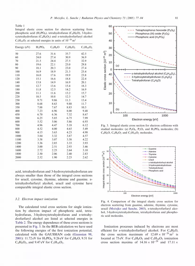
Integral elastic cross sections for phosphoric acid,
tetrahydrofuran, 3-hydroxytetrahydrofuran and a-tetra-hydrofurfuryl alcohol, computed according to Eq. (2),
are listed, in numerical form, at selected electron
energies in Table 1. Their energy dependence between
50 and 2000 eV is shown in Figs. 3(a) and (b),
respectively. It is important to note that according to
the main assumption of the IAM, the magnitude of the
cross section for low collision energies will be over-
estimated, mainly due to the neglect of bond distortion
and multiple scattering within the molecule. On the
other hand, for some small and intermediate size
molecules like silane and germane, even for energies as
low as 20 eV, the IAM method predicts reasonable
elastic cross sections (Moz’ejko et al., 2002). On the
0.01
0.1
1
10
100
1000Phosphoric acid (H3PO4)
Diff
eren
tial c
ross
sec
tion
[10-2
0 m2 sr
-1]
(c)
(a)
0 20 40 60 80 100 120 140 160 1
0.01
0.1
1
10
100
10003-hydroxytetrahydrofuran (C4H8O2)
Scattering
18
Fig. 2. Differential cross section for elastic electron collisions with s
(� �) 500 eV.
other hand, since in the present approach exchange and
absorption effects are neglected, one can expect that
resulting differential and integral cross sections can be
overestimated for energies lower than 300 eV (Khare
et al., 1994). For all studied molecules, the magnitude of
integral elastic cross section decreases with increasing
collision energy. a-tetrahydrofurfuryl alcohol has thehighest elastic cross section. It is about 19% and 17%
higher at low- and intermediate-energies, respectively,
than those for 3-hydroxytetrahydrofuran. The integral
elastic cross section for tetrahydrofuran is smaller than
the cross section for 3-hydroxytetrahydrofuran by more
than 13%. For H3PO4; the integral elastic cross sectionis smaller than those for the sugar analogues for
collision energies below 250 eV. For higher energies that
cross section decreases more slowly with energy than
those for C4H8O; C4H8O2; and C5H10O2: Consequently,for collision energies higher than 1.1 keV the cross
section for H3PO4 molecules exceeds those for C4H8O
and C4H8O2: This energy dependence of the integralcross sections is typical of some molecules. In our case, it
builds up from phosphorous and oxygen. This can be
shown by examining integral elastic cross sections for
larger molecules containing these atoms, such as those
for P2O3 and P4O6 presented in Fig. 3(a). The integral
elastic cross sections for H3PO4; P2O3; and P4O6depends strongly on the molecular size. On the other
hand, their shape at higher collision energies, especially
for H3PO4 and P2O3 is very similar. In Fig. 4, integral
elastic cross section computed for the studied targets are
compared with those calculated for DNA and RNA
bases (Moz’ejko and Sanche, 2003). In the investigated
energy range, the integral cross sections for phosphoric
(d)
(b)
Tetrahydrofuran (C4H8O)
80
angle [deg]
0 20 40 60 80 100 120 140 160 1800
α-tetrahydrofurfuryl alcohol (C5H10O2)
tudied targets at: (—) 50 eV, ð� � �Þ 100 eV, ( ) 200 eV and
ARTICLE IN PRESS
Table 1
Integral elastic cross section for electron scattering from
phosphoric acid (H3PO4), tetrahydrofuran (C4H8O), 3-hydro-
xytetrahydrofuran (C4H8O2) and a-tetrahydrofurfuryl alcoholC5H10O2 at selected energies in units of 10
�20 m2
Energy ðeVÞ H3PO4 C4H8O C4H8O2 C5H10O2
50 27.6 31.6 35.7 42.5
60 24.0 27.4 30.9 36.9
70 21.5 24.4 27.5 32.9
80 19.6 22.1 25.0 29.8
90 18.1 20.3 23.0 27.4
100 16.9 18.9 21.3 25.4
110 16.0 17.6 19.9 23.8
120 15.1 16.6 18.8 22.4
140 13.8 14.9 16.9 20.1
160 12.7 13.6 15.4 18.3
180 11.8 12.5 14.2 16.9
200 11.1 11.6 13.2 15.7
220 10.5 10.8 12.3 14.7
250 9.71 9.86 11.3 13.4
300 8.68 8.63 9.88 11.7
350 7.88 7.67 8.83 10.5
400 7.23 6.96 8.00 9.48
450 6.70 6.36 7.32 8.67
500 6.25 5.85 6.75 7.99
600 5.52 5.06 5.86 6.93
700 4.96 4.47 5.18 6.12
800 4.52 4.00 4.65 5.49
900 4.15 3.63 4.23 4.98
1000 3.84 3.32 3.87 4.57
1100 3.58 3.07 3.58 4.22
1200 3.36 2.85 3.33 3.93
1400 3.00 2.51 2.93 3.46
1600 2.72 2.25 2.64 3.10
1800 2.50 2.06 2.40 2.83
2000 2.32 1.90 2.23 2.62
0
10
20
30
40
50
60
(b)
(a)
Tetraphosphorus hexoxide (P4O6) Phosphorus (III) oxide (P2O3) Phosphoric acid (H3PO4)
Ela
stic
cro
ss s
ectio
n [1
0-20
m2 ]
100 10000
10
20
30
40 α -tetrahydrofurfuryl alcohol (C5H10O2) 3-hydroxytetrahydrofuran (C4H8O2) Tetrahydrofuran (C4H8O)
Electron energy [eV]
Fig. 3. Integral elastic cross section for electron collisions with
studied molecules: (a) P4O6; P2O3 and H3PO4 molecules; (b)C4H8O; C4H8O2 and C5H10O2 molecules.
10
20
30
40
50 Guanine Adenine Thymine Cytosine Uracilα-tetrahydrofurfuryl alcohol 3-hydroxytetrahydrofuran Tetrahydrofuran Phosphoric acid
Inte
gral
ela
stic
cro
ss s
ectio
n [1
0-20 m
2]
P. Moz’ejko, L. Sanche / Radiation Physics and Chemistry 73 (2005) 77–84 81
acid, tetrahydrofuran and 3-hydroxytetrahydrofuran are
always smaller than those of the integral cross sections
for uracil, cytosine, thymine, adenine and guanine. a-tetrahydrofurfuryl alcohol, uracil and cytosine have
comparable integral elastic cross section.
100 10000
Electron energy [eV]
Fig. 4. Comparison of the integral elastic cross section for
electron scattering from guanine, adenine, thymine, cytosine,
uracil (Moz’ejko and Sanche, 2003), a-tetrahyrofurfuryl alco-hol, 3-hydroxytetrahydorfuran, tetrahydrofuran and phospho-
ric acid molecules.
3.2. Electron impact ionization
The calculated total cross sections for single ioniza-
tion by electron impact of phosphoric acid, tetra-
hydrofuran, 3-hydroxytetrahydrofuran and a-tetrahy-drofurfuryl alcohol are listed at selected energies in
Table 2. The energy dependence of these cross sections is
presented in Fig. 5. In the BEB calculation we have used
the following energies of the first ionization potential,
calculated with the GAUSSIAN code (Gaussian 98,
2001): 11.72 eV for H3PO4; 9.26 eV for C4H8O; 9.51 forC4H8O2; and 9.47 eV for C5H10O2:
Ionization processes induced by electrons are most
efficient for a-tetrahydrofurfuryl alcohol. For C4H8O;the cross section maximum of 12:69� 10�20 m2 islocated at 75 eV. For C4H8O2 and C5H10O2 ionization
cross section maxima of 14:16� 10�20 and 17:11�

ARTICLE IN PRESS
Table 2
Total cross section for electron impact ionization of phosphoric
acid (H3PO4), tetrahydrofuran (C4H8O), 3-hydroxytetrahydro-
furan (C4H8O2) and a-tetrahydrofurfuryl alcohol C5H10O2 atselected energies in units of 10�20 m2
Energy ðeVÞ H3PO4 C4H8O C4H8O2 C5H10O2
12.0 0.0313 0.370 0.432 0.500
13.0 0.155 0.740 0.771 1.006
14.0 0.377 1.161 1.230 1.575
15.0 0.676 1.627 1.746 2.219
16.0 1.025 2.167 2.303 2.922
17.0 1.397 2.721 2.890 3.636
18.0 1.770 3.300 3.485 4.385
19.0 2.131 3.852 4.082 5.114
20.0 2.476 4.375 4.651 5.806
22.5 3.281 5.557 5.944 7.388
25.0 4.040 6.634 7.121 8.802
27.5 4.727 7.554 8.136 10.04
30 5.329 8.366 9.034 11.12
35 6.311 9.670 10.49 12.86
40 7.077 10.63 11.57 14.16
45 7.701 11.32 12.39 15.12
50 8.188 11.82 12.99 15.81
55 8.564 12.18 13.42 16.31
60 8.852 12.41 13.72 16.66
65 9.071 12.57 13.93 16.89
70 9.234 12.65 14.06 17.03
75 9.351 12.69 14.14 17.10
80 9.433 12.69 14.16 17.11
85 9.484 12.65 14.15 17.09
90 9.511 12.60 14.11 17.02
95 9.519 12.52 14.05 16.94
100 9.510 12.43 13.97 16.83
110 9.454 12.21 13.76 16.56
120 9.362 11.97 13.52 16.26
140 9.113 11.46 13.00 15.60
160 8.822 10.94 12.45 14.92
180 8.520 10.45 11.92 14.26
200 8.220 9.980 11.41 13.64
225 7.858 9.442 10.82 12.93
250 7.517 8.954 10.28 12.27
275 7.200 8.511 9.789 11.68
300 6.903 8.109 9.339 11.13
350 6.376 7.411 8.555 10.19
400 5.922 6.827 7.894 9.394
450 5.529 6.331 7.331 8.718
500 5.186 5.906 6.846 8.138
600 4.619 5.214 6.055 7.191
700 4.168 4.675 5.436 6.453
800 3.802 4.242 4.938 5.859
900 3.499 3.888 4.528 5.371
1000 3.243 3.591 4.186 4.963
1500 2.393 2.621 3.061 3.626
2000 1.911 2.080 2.432 2.880
2500 1.598 1.733 2.027 2.399
3000 1.377 1.489 1.743 2.063
3500 1.213 1.309 1.533 1.814
4000 1.085 1.170 1.370 1.621
P. Moz’ejko, L. Sanche / Radiation Physics and Chemistry 73 (2005) 77–8482
10�20 m2; respectively, are peaked at 80 eV. Among allstudied targets phosphoric acid has the lowest ionization
cross section, with a maximum of 9:52� 10�20 m2 at95 eV.
Fig. 6 shows comparison between total cross sections
for electron-impact ionization of DNA and RNA bases
(i.e. guanine, adenine, thymine, cytosine and uracil)
(Moz’ejko and Sanche, 2003) and those for ionization of
C4H8O; C4H8O2; C5H10O2 and PO4H3 for electronenergies from the ionization threshold to 120 eV. With
exception of some specific variation of the cross sections
near the ionization threshold, generally, in the whole
energy range investigated, the magnitude of the ioniza-
tion cross section obeys the following trend:
sPO4H3osC4H8OosC4H8O2osuraciloscytosineosC5H10O2osthymineosadenineosguanine: ð9Þ
It has been shown that, in some cases and within some
approximation the electron-impact ionization cross
section for polyatomic molecules can be calculated
taking into account only basic atomic properties
(Margreiter et al., 1990). From this assumption, we
approximate ionization cross sections for the sugar-
phosphate backbone of DNA adding ionization cross
sections for H3PO4 and C5H10O2: In Fig. 7 the resultingvalues are compared with recent calculations by
Bernhardt and Paretzke (Bernhardt and Paretzke,
2003) of ionization cross sections for the sugar-
phosphate unit of DNA. At low (i.e. near to the
ionization threshold) energies, the magnitude of the
summed cross sections is distinctively overestimated for
sugar-phosphate unit (at 20 eV it is higher of more than
27%), but at higher energies discrepancies between both
cross sections are reasonably smaller. While summed
ionization cross section is always higher than those
calculated for the sugar-phosphate unit, discrepancies
do not exceed 10% at 30 eV and become smaller with
increasing electron energies. At 1 keV both cross section
differ by less than 7%.
10 100 1000
2
4
6
8
10
12
14
16
18
Tot
al io
niza
tion
cros
s se
ctio
n [1
0-20 m
2 ]
Electron energy [eV]
Fig. 5. Electron-impact ionization cross section for H3PO4;C4H8O; C4H8O2 and C5H10O2 molecules.

ARTICLE IN PRESS
0 20 40 60 80 100 1200
2
4
6
8
10
12
14
16
18
20
22
Guanine Adenine Thymine Cytosine Uracilα-tetrahydrofurfuryl alcohol 3-hydroxytetrahydrofuran Tetrahydrofuran Phosphoric acid
Ioni
zatio
n cr
oss
sect
ion
[10-2
0 m2]
Electron energy [eV]
Fig. 6. Comparison of the electron-impact ionization cross
section for guanine, adenine, thymine, cytosine, uracil (Moz’ej-
ko and Sanche, 2003), a-tetrahyrofurfuryl alcohol, 3-hydro-xytetrahydorfuran, tetrahydrofuran and phosphoric acid
molecules.
10 100 10000
5
10
15
20
25
30
H3PO4 + C5H10O2
sugar-phosphate backbone unit
Ioni
zatio
n cr
oss
sect
ion
[10-2
0 m2 ]
Electron energy [eV]
Fig. 7. Comparison between cross section for electron impact
ionization of the sugar-phosphate backbone unit with the sum
of electron-impact ionization cross sections of phosphoric acid
and a-tetrahyrofurfuryl alcohol.
P. Moz’ejko, L. Sanche / Radiation Physics and Chemistry 73 (2005) 77–84 83
4. Summary
Using relatively simple but reliable approaches, i.e.
the independent atom method and binary-encounter-
Bethe formalism, cross sections for elastic electron
collisions and electron-impact ionization from C4H8O;C4H8O2; C5H10O2 and H3PO4 have been computed forwide range of the collision energies. The cross section for
electron-impact ionization of the sugar phosphate unit
of DNA has been approximated as the sum of the
ionization cross section of the H3PO4 and C5H10O2molecules. We found reasonable agreement with results
of more sophisticated calculations for energies higher
than 30 eV. The present results can be useful in a
detailed analysis of ionizing radiation damage to
complex biolomolecules such as DNA and RNA, via
Monte Carlo simulations.
Acknowledgements
This work was supported by the Canadian Institutes
of Health Research (CIHR). Pawe" Moz’ejko acknowl-edges financial support from the CIHR in the form of a
Post-doctoral Fellowship.
References
Abdoul-Carime, H., Cloutier, P., Sanche, L., 2001. Low-energy
(5–40 eV) electron-stimulated desorption of anions from
physisorbed DNA bases. Radiat. Res. 155, 633–645.
Abdoul-Carime, H., Gohlke, S., Illenberger, E., 2004. Site-
specific dissociation of DNA bases by slow electrons at early
stages of irradiation. Phys. Rev. Lett. 92 (16), 168103.
Barrios, R., Skurski, P., Simons, J., 2002. Mechanism for
damage to DNA by low-energy electrons. J. Phys. Chem. B
106, 7991–7994.
Bernhardt, Ph., Paretzke, H.G., 2003. Calculation of electron
impact ionization cross section of DNA using the Deutsch-
Mark and Binary-Encounter-Bethe formalisms. Int. J. Mass
Spectrom. 223–224, 599–611.
Bernhardt, Ph., Friedland, W., Jacob, P., Paretzke, H.G., 2003.
Modeling of ultrasoft X-ray induced DNA damage using
structured higher order DNA targets. Int. J. Mass
Spectrom. 223/224, 579–597.
Boudaiffa, B., Cloutier, P., Hunting, D., Huels, M.A., Sanche,
L., 2000a. Resonant formation of DNA strand breaks by
low-energy (3–20 eV) electrons. Science 287, 1658–1660.
Boudaiffa, B., Hunting, D., Cloutier, P., Huels, M.A., Sanche,
L., 2000b. Induction of single- and double-strand breaks in
plasmid DNA by 100–1500 eV electrons. Int. J. Radiat. Biol.
76, 1209–1221.
Feil, S., Gluch, K., Matt-Lebner, S., Scheier, P., Limtrakul, J.,
Probst, M., Deutsch, H., Becker, K., Stamatovic, A., Mark,
T.D., 2004. Partial cross sections for positive and negative
ion formation following electron impact on uracil. J. Phys.
B 37, 3013–3020.
Folkard, M., Prise, K.M., Vojnovic, B., Davies, S., Roper,
M.J., Michael, B.D., 1993. Measurement of DNA damage
by electrons with energies between 25 and 4000 eV. Int. J.
Radiat. Biol. 64, 651–658.
Friedland, W., Jacob, P., Paretzke, H.G., Stork, T., 1998.
Monte Carlo simulation of the production of short DNA
fragments by low-linear energy transfer radiation using
higher-order DNA models. Radiat. Res. 150, 170–182.
Friedland, W., Jacob, P., Paretzke, H.G., Merzagora, M.,
Ottolenhi, A., 1999. Simulation of DNA fragment distribu-
tions after irradiation with photons. Radiat. Environ.
Biophys. 38, 39–47.
Gaussian, 98, Revision A.11.2, Frisch, M.J., Trucks, G.W.,
Schlegel, H.B., Scuseria, G.E., Robb, M.A., Cheeseman,
J.R., Zakrzewski, V.G., Montgomery Jr., J.A., Stratmann,
R.E., Burant, J.C., Dapprich, S., Millam, J.M., Daniels,
A.D., Kudin, K.N., Strain, M.C., Farkas, O., Tomasi J.,

ARTICLE IN PRESSP. Moz’ejko, L. Sanche / Radiation Physics and Chemistry 73 (2005) 77–8484
Barone, V., Cossi, M., Cammi, R., Mennucci, B., Pomelli,
C., Adamo, C., Clifford, S., Ochterski, J., Petersson, G.A.,
Ayala, P.Y., Cui Q., Morokuma, K., Rega, N., Salvador,
P., Dannenberg, J.J., Malick, D.K., Rabuck, A.D., Ragha-
vachari, K., Foresman, J.B., Cioslowski, J., Ortiz, J.V.,
Baboul, A.G., Stefanov, B.B., Liu, G., Liashenko, A.,
Piskorz, P., Komaromi, I., Gomperts, R., Martin, R.L.,
Fox, D.J., Keith, T., Al-Laham, M.A., Peng, C.Y.,
Nanayakkara, A., Challacombe, M., Gill, P.M.W., John-
son, B., Chen, W., Wong, M.W., Andres, J.L., Gonzalez,
C., Head-Gordon, M., Replogle, E.S., Pople, J.A., Gaus-
sian, Inc., Pittsburgh PA, 2001.
Huels, M.A., Boudaiffa, B., Cloutier, P., Hunting, D., Sanche,
L., 2003. Single, double and multiple double strand breaks
induced in DNA by 3–100 ev electrons. J. Am. Chem. Soc.
125, 4467–4477.
Hwang, W., Kim, Y.K., Rudd, M.E., 1996. New model for
electron-impact ionization cross sections of molecules. J.
Chem. Phys. 104, 2956–2966.
Joshipura, K.N., Vinodkumar, M., 1997. Total cross sections of
electron collisions with S atoms: H2S; OCS and SO2molecules (EiX50 eV). Z. Phys. D 41, 133–137.
Khare, S.P., Raj, D., Sinha, P., 1994. Absorption effects in the
elastic scattering of electrons by the CF4 molecule at
intermediate energies. J. Phys. B 27 (12), 2569–2576.
Li, X.F., Sevilla, M.D., Sanche, L., 2003. Density functional
theory studies of electron interaction with DNA: Can zero
eV electrons induce strand breaks? J. Am. Chem. Soc. 125
(45), 13668–13669.
Maji, S., Basavaraju, G., Bharathi, S.M., Bhushan, K.G.,
Khare, S.P., 1998. Elastic scattering of electrons by
polyatomic molecules in the energy range 300–1300 eV:
CO, CO2; CH4; C2H4 and C2H6: J. Phys. B 31, 4975–4990.Margreiter, D., Deutsch, H., Schmidt, M., Mark, T.D., 1990.
Electron impact ionization cross sections of molecules. Part
II. Theoretical determination of total (counting) ionization
cross sections of molecules: a new approach. Int. J. Mass
Spectrom. Ion Proc. 100, 157–176.
Moissenko, V.V., Hamm, R.N., Walker, A.J., Rrestwich, W.V.,
1998. Modelling DNA damage induced by different energy
photons and tritium beta-particles. Int. J. Radiat. Biol. 74,
533–550.
Mott, N.F., Massey, H.S.W., 1965. The Theory of Atomic
Collisions. Oxford University Press, Oxford.
Moz’ejko, P., Sanche, L., 2003. Cross section calculations for
electron scattering from DNA and RNA bases. Radiat.
Environ. Biophys. 42 (3), 201–211.
Moz’ejko, P., Z’ ywicka-Moz’ejko, B., Szmytkowski, Cz., 2002.
Elastic cross section calculations for electron collisions with
XY4 (X ¼ Si, Ge; Y ¼ H, F, Cl, Br, I) molecules. Nucl.
Instr. and Meth. Phys. Res. B 196, 245–252.
Nikjoo, H., O’Neill, P., Terrissol, M., Goodhead, D.T., 1999.
Quantitative modelling of DNA damage using Monte Carlo
track structure method. Radiat. Environ. Biophys. 38,
31–38.
Padial, N.T., Norcross, D.W., 1984. Parameter-free model of
the correlation-polarization potential for electron-molecule
collisions. Phys. Rev. A 29, 1742–1748.
Pan, X., Cloutier, P., Hunting, D., Sanche, L., 2003.
Dissociative electron attachment to DNA. Phys. Rev. Lett.
90, 208102/1–208102/4.
Pedrew, J.P., Zunger, A., 1981. Self-interaction correction to
density-functional approximations for many-electron sys-
tems. Phys. Rev. B 23, 5048–5079.
Ptasinska, S., Denfil, S., Scheier, P., Mark, T.D., 2004. Inelastic
electron interaction (attachment/ionization with) deoxyri-
bose. J. Chem. Phys. 120 (18), 8505–8511.
Ritchie, R.H., Hamm, R.N., Turner, J.E., Wright, H.A., Bloch,
W.E., 1991. Radiation interactions and energy transport in
the condensed phase. In: Glass, W.A., Varma, N.M. (Eds.),
Physical and Chemical Mechanisms in Molecular Radiation
Biology. Plenum Press, New York, pp. 99–135.
Salvat, F., Martinez, J.D., Mayol, R., Parellada, J., 1987.
Analytical Dirac–Hartree–Fock–Slater screening function
for atoms (Z ¼ 1292Þ: Phys. Rev. A 36, 467–474.Sanche, L., 2002. Nanoscopic aspects of radiobiological
damage: fragmentation induced by secondary electrons.
Mass Spectrom. Rev. 21, 349–369.
Schmidt, M.W., Baldridge, K.K., Boatz, J.A., Elbert, S.T.,
Gordon, M.S., Jensen, J.H., Koseki, S., Matsunaga, N.,
Nguyen, K.A., Su, S., Windusn, T.L., Dupuis, M.,
Montgomery Jr., J.A., 1993. General atomic and molecular
electronic structure system. J. Comp. Chem. 14, 1347–1363.
Semenenko, V.A., Turner, J.E., Borak, T.B., 2003. NOREC, a
Monte Carlo code for simulating electron tracks in liquid
water. Radiat. Environ. Biophys. 42 (3), 213–217.
Terrissol, M., Beaudre, A., 1990. Simulation of space and time
evolution of radiolytic species induced by electrons in water.
Radiat. Prot. Dosim. 31, 171–175.
Uehara, S., Nikjoo, H., Goodhead, D.T., 1999. Comparison
and assessment of electron cross sections for Monte Carlo
track structure codes. Radiat. Res. 152, 202–213.
Zakrzewski, V.G., von Niessen, W., 1994. Vectorizable algo-
rithm for Green function and many-body perturbation
methods. J. Comp. Chem. 14, 13–18.
Zhang, X., Sun, J., Liu, Y., 1992. A new approach to the
correlation polarization potential–low-energy electron elas-
tic scattering by He atoms. J. Phys. B 25, 1893–1897.