Embed Size (px)
Citation preview
Crosslinked poly(1-vinyl-2-pyrrolidinone) as a vitreous substitute
Ye Hong,’ Traian V. Chirila,’., Sarojini Vijayasekaran,l Paul D. Dalton,l Sjakon G . Tahija? Maximiliaan J. H. Cuypers; and Ian J. Constable’ ‘Lions Eye Institute, 2 Verdun Street, Nedlands, Western Australia 6009, Australia; and 2Department of Ophthalmology, University of Indonesia, Jl. Salemba 6 , Jakarta, Indonesia
A hydrogel with a high water content was produced and tested as a possible vitreous substitute. The polymer (poly[l- vinyl-2-pyrrolidinone]) (PVP) was synthesized by free radi- cal bulk polymerization of 1-vinyl-2-pyrrolidinone (VP) in the presence of 0.25% divinyl glycol (DVG) as a crosslinking agent. The fully hydrated polymer, containing about 98% water, was clear, transparent, autoclavable, and easily in- jected through a small-gauge needle with minimum fragmen- tation, and without changes in its optical properties. Dynamic mechanical analysis of the hydrogel indicated a covalently crosslinked elastic network both before and after injection. The resilience of hydrogel decreased after being subjected to shear stress during the injection process. A cytotoxicity
bioassay of the hydrogel in vitro, using cultured mouse (Balb/ c-3T3) fibroblasts, showed cytostatic but not cytocidal effects. The hydrogel was injected into the vitreous cavity of rabbits and followed up to 4 weeks. The gel was clinically well tolerated, however opacities in the vitreous body were ob- served following the insertion of the gel. Histopathological examination revealed no adverse reactions to the retina, but the presence of loose polymer particles indicated the possibil- ity of the biodegradation of the polymer. These results sug- gest the potential use of crosslinked PVP hydrogels as vitre- ous substitutes, provided that their biodegradation is not significant, a matter that should be further investigated. 0 1996 John Wiley & Sons, Inc.
INTRODUCTION
Over the last hundred years many modified bio- polymers and synthetic polymers have been used to replace in the eye the vitreous body damaged during the surgical treatment of various vitreoretinal patholo- gies. The vitreous body itself can become dysfunc- tional due to specific diseases, hemorrhage, trauma, or age-related degeneration, cases in which its replace- ment is mandatory. Unfortunately, none of the mate- rials tested were successful as long-term vitreous substitutes.’,’ Postoperative complications such as opacification of the vitreous body or crystalline lens, inflammatory reactions, corneal edema, glaucoma, and chorioretinal atrophy were frequently caused by the various polymers injected in the vitreous body. It appeared, however, that clear and transparent hydrogels may be appropriate as vitreous substitutes if they could fulfill at least two prerequisites, i.e., (1) they should be viscoelastic materials, able to tamponade the retina to a proper position, but also able to pass through a small-gauge needle with
*To whom correspondence should be addressed.
minimum fragmentation; and (2) they should be biocompatible with the adjacent ocular tissue, and should not induce any toxic reaction. There are still many other requirements for an ideal permanent vitreous ~ubstitute,’-~ and a synthetic hydrogel to fulfill all criteria is yet to be found.
Poly(1 -vinyl-2-pyrrolidinone) (PVP) is a water-solu- ble polymer which was used for decades as a blood- plasma extender, and is currently used in various pharmaceutical compositions. It was used also as a viscoelastic agent in the anterior chamber of the eye during surgery for cataract e~traction.~ PVP induces only minor storage-related functional changes in or- gans and its cytotoxicity is extremely As a solution in water, PVP was the first synthetic polymer proposed and tested as a replacement for the vitreous b0dy.6,~ Although PVP induced less ocular inflamma- tion, as compared with air or saline, the material was retained in the vitreous cavity for only a short time, as normally it should have been expected from an aqueous solution. Considering the good tolerance of PVP in the vitreous cavity of the eye, as well as the proven supposition8 that if an uncrosslinked polymer is soluble in a liquid penetrant, the cross-
Journal of Biomedical Materials Research, Vol. 30, 441 -448 (1996) 0 1996 John Wiley & Sons, Inc. CCC 0021 -9304/96/040441-08
HONG ET AL. 442
linked version of the same polymer will be swollen only by that liquid (in this case, water) without any dissolution, we propose that the crosslinked PVP gels may perform better in the long term as an artificial vitreous. Therefore, a large number of cross- linked hydrogels based on 1-vinyl-2-pyrrolidinone (VP), with high water content, were synthesized and investigated in our laboratory. PVP crosslinked with divinyl glycol (DVG) was eventually selected as a potential candidate for a functional vitreous substi- tute. We now report preparation and evaluation of this synthetic hydrogel.
MATERIALS AND METHODS
Preparation of PVP hydrogel
VP was obtained from BASF Australia Ltd, with a purity of 99 wt %. Prior to polymerization, VP was additionally purified by vacuum distillation (b.p. 48”C/2 mmHg).
A homopolymer of VP was synthesized by bulk polymerization in the presence of 0.25 wt % crosslink- ing agent DVG (Polysciences, Inc., USA) and 0.1 wt % 2,2-azo-bis-(2,4-dimethyl valeronitrile) as an initiator (Wako Pure Chemical Industries, Ltd., Ja- pan). The crosslinking agent and the initiator were dissolved in purified VP using an ultrasonic bath. The clear and homogeneous liquid was then equally distributed (approximately 2.2 mL) among molds made of polypropylene, in a molding unit designed by us. The unit was closed and sealed, and vacuum was produced by pump-evacuating the air for 5 min. High-purity nitrogen was then admitted through an inlet valve until an overpressure of 5 kPa was created in the molding unit. The unit was placed into a water bath in which the temperature was controlled by a digital program controller with a built-in micro- processor. A temperature program was run, consist- ing of a number of steps, which allowed 30 h at 45°C and 8 h at 70°C.
At the end of polymerization, the solid polymer buttons were removed from the molds and hydrated in deionized/distilled water for 2 weeks. The hydro- gel specimens were then placed in dialysis cellulose- membrane bags (Medicell International Ltd., UK), and thoroughly extracted in a Soxhlet extractor for 24 h. The pH of extracted hydrogel specimens was between 7.2 and 7.4. The neutral gel specimens were placed in Pyrex@ glass containers with phosphate- buffered saline (PBS, pH 7.4), and sterilized in an autoclave at 130°C for 20 min. Until further evalua- tion, the samples were stored at 4°C to minimize
the opportunity for bacterial growth and consequent toxin production.
Physical evaluation
Hydration was performed in deionized/distilled water for 2 weeks, and the equilibrium water content (W), as weight percentage, was calculated using the equation W = 100 (wl - w2)/wl, where w1 and w2 are the weights of the fully hydrated specimen and the dried specimen, respectively.
Injectability of the gels was evaluated using a device designed by us, consisting of a pressure regulator, a pressure gauge, a flask with the gel, a syringe needle, and a measuring vial. One milliliter of hydrogel was injected through a 23-gauge needle under a con- stant pressure (250 mmHg). The duration of the in- jection was recorded, and fragmentation and cohe- siveness of the hydrogel during injection were also observed.
Optical properties of the gel specimens were evalu- ated according to the method proposed by Refojo.’ The samples were injected into 1-cm UV spectrophotome- ter glass cells, which are optically clear, and were first subjected to visual-acuity tests. A person with normal visual acuity was required to read three different clini- cal visual-acuity test charts in front of one eye and with the other eye closed or covered, through a cell filled with PBS or with the gel, as well as without any obstruction (i.e., no cell in the visual path). The three test charts employed include: (1) the USAF 1951 resolu- tion test target in which the number of line pairs per 1 mm doubles with every seventh target element. (An element consists of two patterns of three lines each, at right angles to each other. These six ele- ments are known as a group.); (2) Rodenstock Roda- vist projector; and (3) Bailey-Lovie chart number 5. Charts (2) and (3) are optotypes based on Snellen’s principle.’O
Light transmission of the specimens were then mea- sured using an optical bench containing a halogen light source which projects a parallel beam perpendicular onto a photometric cell. Zero-percent transmission was set in dark conditions, and total transmission (100%) was set by adjusting the intensity of the light projected through a 1-cm spectrophotometric glass cell filled with PBS. Readings of transmission were then per- formed using cells filled with the gel. Transmittance of the 550-nm wavelength radiation was determined using an LKB Ultrospec I1 spectrophotometer. The refractive index of the gels was measured using an Abbe refractomerer (Bellingham & Stanley Ltd., UK). All measurements were performed at room tempera- ture.
The injectability and optical properties of the PVP hydrogels were compared with those of PBS, and de- termined under the same conditions.
PVP VITREOUS SUBSTITUTE 443
Dynamic mechanical analysis
Mechanical spectra of the PVP hydrogel were re- corded using a controlled stress rheometer (Bohlin CS- 10, Sweden). A gel specimen was placed between a 4"/4O-mm-diameter cone and a 60-mm-diameter lower plate. The Iogarithmic plots of the complex shear mod- ulus (real part as the storage modulus G', and imagi- nary part as the loss modulus G") of the gel before and after injection through a 30-gauge needle were recorded versus the frequency of a small-deformation oscillatory shear strain. The target strain was set at 0.5, with the applied stress varying to accommodate this, in order to ensure that the obtained results were in the linear viscoelastic region. During the measurements, the temperature was kept at 25°C.
The resilience of the material was also assessed. Re- silience (R) is an inverse term of the damping property, which is characterized by the dissipation factor, also known as the loss tangent (tan aC = G"/G'). The re- silience is usually estimated" as R = 1 - 2.rr G / G ' = 1 - 2a tan aG.
Cytotoxicity evaluation in cell culture
Cultured mouse (Balb/c-3T3) fibroblasts were used to determine the cytotoxicity of the PVP hydrogel by cell proliferation using an immunocytochemical sys- tem. The RPN 20 celI proliferation kit was provided by Amersham, UK.
The bioassay took four days to complete. On day 1, the confluent fibroblasts were trypsinized and plated (approximately 5000 cells in 400-pL/well) in RPMI 1640 medium with L-glutamine, 5% fetal calf serum (FCS), and 1% penicillin/streptomycin, on an 8-well chamber slide (Nunc, #177402, Denmark) and then incubated at 37°C in a humidified 5% C02/air atmosphere. Twenty-four hours later the cells were confluent, and the medium in the wells was replaced by serum-free medium and incubated. At 48 h, 40 p L PBS was added into the first well as negative control, 40 pL FCS into the second well as positive control, 4 drops of PVP gel from a microsyringe into the third well, and 4 drops of gel together with 40 pL FCS into the fourth well. At 72 h, the cellular effects were assessed by monitoring cell proliferation and cell death, Proliferating cells were labeled with the reagent 5-bromo-2'-deoxyuridine (BrdU) for 1 h in an incubator. The cells were fixed with a solution of 90% ethanol, 5% acetic acid, and 5% water, fol- lowed by additional incubation for 1 h with mono- clonal anti-BrdU in order to detect BrdU incorporated into cellular DNA. The incorporation of BrdU into cellular DNA indicates that the cells are dividing, and those cells are called BrdU-positive cells. They were detected using peroxidase-conjugated antibody
to mouse immunoglobulin. The slides were stained with diaminobenzidine (DAB), which is polymerized in the presence of cobalt, nickel, and 1% solution of hydrogen peroxide, giving brown staining at the sites of BrdU incorporation. The slides were counterstained lightly with hematoxylin, then dehydrated and mounted.
This assay was repeated 5 times. The BrdU-positive cells (brown nuclei), pyknotic cells (dark blue nuclei), and BrdU-negative cells (blue nuclei) were counted from 15 fields of view at 40-fold magnification from each of the four samples, for each assay. The following comparisons were made with respect to BrdU-positive cells and pyknotic cells. The proportions of proliferat- ing cells (A) in the wells with polymer and without polymer were compared in each of the serum-free and 10% FCS condition, respectively. The same com- parison for the proportion of pyknotic cells (B) also was performed. A significant reduction of proliferat- ing cells in the presence of polymer was assigned as a cytostatic effect. A significant increase of pyknotic cells in the presence of polymer was assigned as a cytocidal effect. A cytostatic or cytocidal effect of polymer was assigned when there was a statistically significant difference (p < 0.05) between tests and controls. Comparisons were made by analysis of variance using the general linear models procedure of SAS@ (Version 6) statistical package (SAS Institute, Inc., USA). The proportions of proliferating and pyk- notic cells were calculated as follows: A = 100 N,/ N, %; B = 100 N,/N, %, where A is proportion of proliferating cells, B is proportion of pyknotic cells, N, is the number of BrdU-positive cells, Nb is the number of pyknotic cells, and N, is the total number of cells in 15 fields.
In vivo evaluation
Six New Zealand half-lop rabbits were used for in vivo experiments which were conducted in accordance with The Australian Code of Practice for the Care and Use of Animals for Scientific Purposes (1990). All rab- bits were anesthetized before operation by intramuscu- lar injection of ketamine (40 mg/kg body weight) and xylazine (7.5 mg/kg), while the pupils were dilated with 5% phenylephrine and 0.25% tropicamide. Topi- cal anesthetic (proparacaine hydrochloride) and antibi- otic drops (gentamicin, 0.5%) were also administered before surgery.
In the first stage, a gas-mediated vitrectomy was performed in the left eye of each anesthetized rabbit, according to a technique previously described,12 and the right eye (unoperated) was used as a control during the postoperative clinical examination. Briefly, the sur- face of the eye and the surrounding area was sterilized with a solution of 5% Povidone-iodine. Approximately
444 HONG ET AL.
0.2 mL of aqueous humor was then aspirated from the anterior chamber of the eye to lower the intraocular pressure. Using a I-mL syringe attached to a 30-gauge needle, 0.3 mL of pure perfluoropropane, which had been filtered through a 0.45-p Millex-HA filter, was injected into the vitreous cavity, 3 mm posteriorly from the limbus in the 5 o’clock meridian.
After 3 days, when the gas bubble had been ex- panded to occupy approximately two-thirds of the vit- reous cavity, the rabbits were reanesthetized, and a gas-gel exchange was undertaken using the bimanual te~hnique.‘~ Two 30-gauge needles attached to 5-mL syringes, one of them being filled up with the PVP gel, were inserted superiorly, so that as the gel (1.3-1.5 mL) was injected into the cavity through one needle, the gas was vented through the second needle. The intraocular pressure was monitored with a Schiotz to- nometer.
Clinical examination
After surgery, the animals were treated with chlor- amphenicol eye ointment, homatropine 2%, and pred- nisolone 0.5% eye drops 4 times daily. The eyes were examined daily for the first week, and subsequently on a weekly basis until sacrifice, using biomicroscopy and indirect ophthalmoscopy to evaluate the cornea, anterior chamber, lens, optic nerve, and retina. The intraocular pressure was measured, and photographs of the fundus were taken. The unoperated eyes were examined at the same time as controls.
Histology
The operated animals were sacrificed by injecting intravenously Lethobarb 4 weeks after vitreous substi- tution. The eyes were immediately enucleated, a small slit made just below the pars plana, and fixed in a solution of 2.5% glutaraldehyde in 0.1M phosphate buffer for 1 h. The corneas were removed and the eye cups returned to the fixative solution, where they were kept overnight and then transferred to a solution of 10% sucrose. After 2 days, pieces (2 X 2 mm) of full- thickness retina and choroid were taken from the dor- sal and ventral regions of the periphery and center, optic disc, and from areas where any abnormality was seen. The pieces of tissue were postfixed in osmium tetroxide, dehydrated in graded series of ethanol, and embedded in epoxy resin (DurcupanB ACM, Fluka AG, Switzerland) for light transmission electron mi- croscopy. Semithin (2 pm) sections were cut using an ultramicrotome (LKB 2088 Ultrotome V, Sweden), with a diamond knife, and stained with toluidine blue for light microscopy. Ultrathin (0.1 pm) sections were cut
from the selected areas and stained with uranyl acetate and lead citrate.
RESULTS
Physical properties of PVP hydrogel
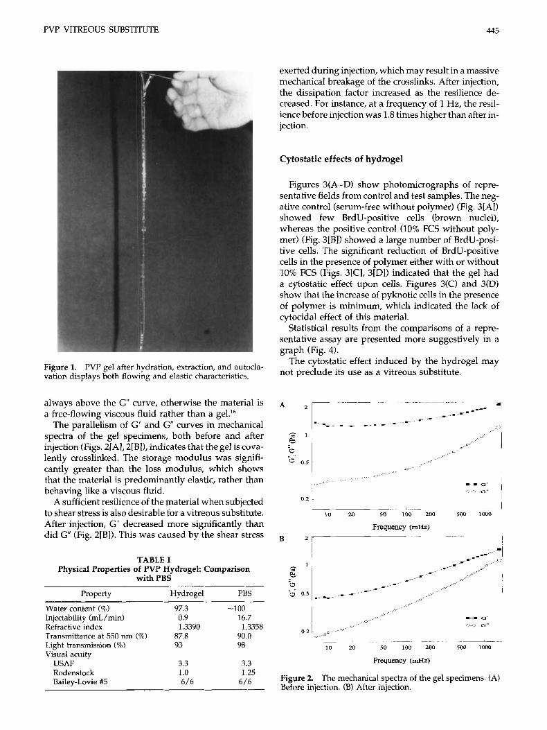
Transparent solid buttons of PVP were obtained at the end of the polymerization process. Upon hydration in pure water, the PVP hydrogel swelled to a high- equilibrium water content (an average of 97.3%). The hydrated gel was clear, transparent, and co- hesive. After extraction and sterilization, the gel not only maintained its transparency but continued to display certain cohesiveness and viscoelastic behav- ior (Fig. 1).
The transparency of the PVP hydrogel did not change after its passage through a 23-gauge needle under constant pressure, and the injecting was easy to perform. Its fragmentation was minor, and no individual particles were distinguishable after injec- tion. The results of the visual acuity tests are shown in Table I, indicating that the gel is as clear as PBS. The other optical characteristics (refractive index, transmittance of radiation at 550 nm, and visible light transmission) are also similar to those of PBS (Table I). These outstanding optical properties after injection encouraged us in using the gel as a vitre- ous substitute.
Dynamic mechanical properties of PVP hydrogel
As a vitreous substitute, any material should be gen- uinely viscoelastic like the natural vi t re~us.’~ Literally, the term viscoelasticity means the combination of vitre- ous and elastic properties, i.e., the material exhibits both dissipation and storage of energy. When the mate- rial is deformed, the dissipation of energy is deter- mined by the loss modulus (G”), and the recoverable energy, stored as elastic energy, by the storage modu- lus (G’).
The changes in viscoelastic properties depend on time, and their measurement is known as dynamic mechanical analysis. The resulting mechanical spectra are commonly used to analyze the viscoelasticity of polymers, and to discriminate between covalently crosslinked gel networks and entanglement gel net- w o r k ~ . ~ ~ In the case of entanglement networks, there is a crossover in G’ and G plots as the frequency decreases. At very low frequencies (in the “terminal zone”), they flow like viscous fluids. For the cross- linked gel networks G’ and G” plots are parallel and largely frequency insensitive.” The G’ curve should be
PVP VITREOUS SUBSTITUTE 445
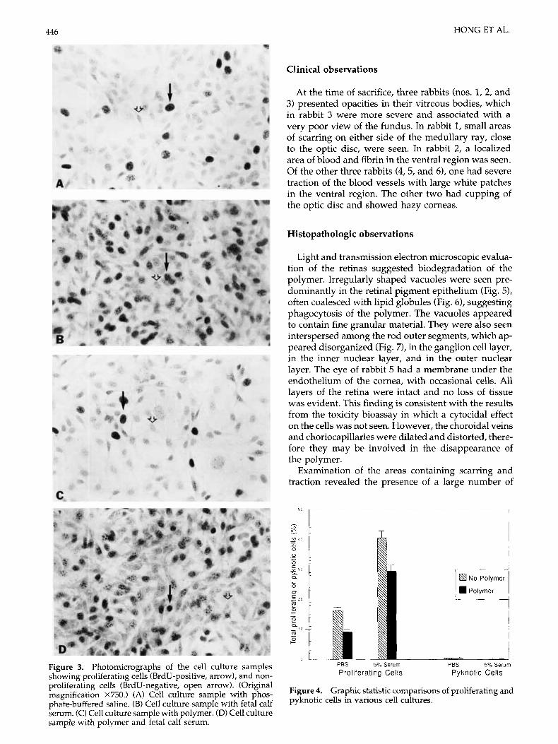
exerted during injection, which may result in a massive mechanical breakage of the crosslinks. After injection, the dissipation factor increased as the resilience de- creased. For instance, at a frequency of 1 Hz, the resil- ience before injection was 1.8 times higher than after in- jection.
Cytostatic effects of hydrogel
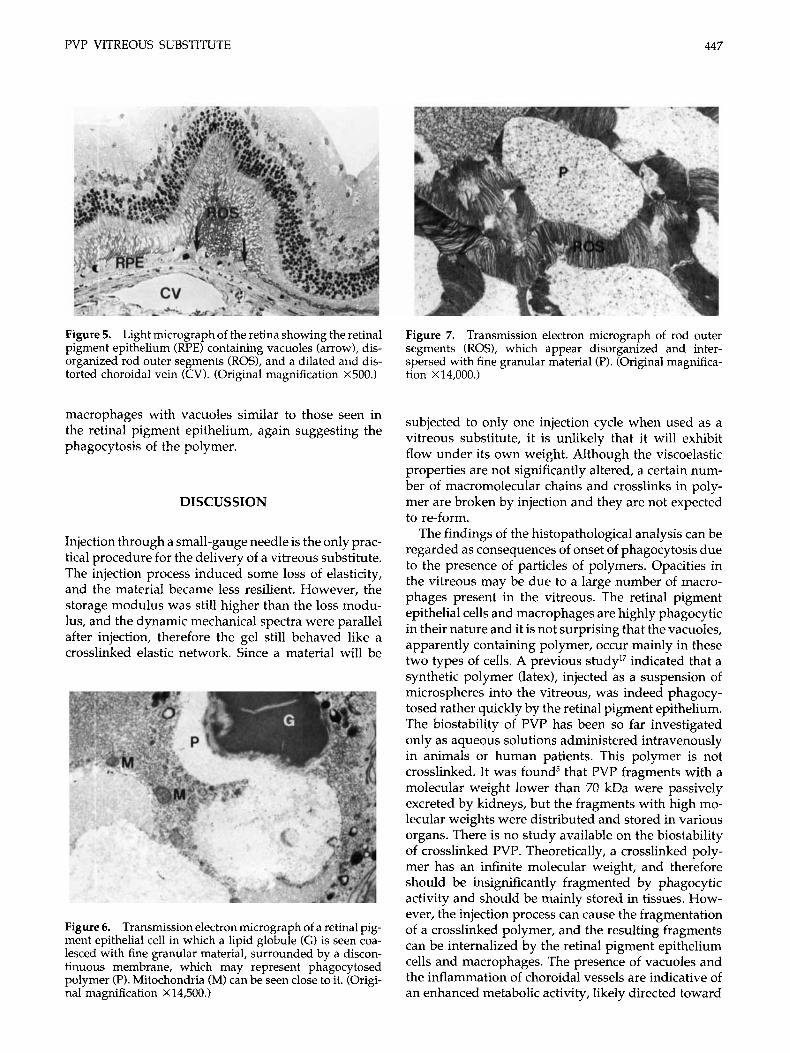
Figures 3(A-D) show photomicrographs of repre- sentative fields from control and test samples. The neg- ative control (serum-free without polymer) (Fig. 3[A]) showed few BrdU-positive cells (brown nuclei), whereas the positive control (10% FCS without poly- mer) (Fig. 3[BI) showed a large number of BrdU-posi- tive cells. The significant reduction of BrdU-positive cells in the presence of polymer either with or without 10% FCS (Figs. 3[Cl, 3[Dl) indicated that the gel had a cytostatic effect upon cells. Figures 3(C) and 3(D) show that the increase of pyknotic cells in the presence of polymer is minimum, which indicated the lack of cytocidal effect of this material.
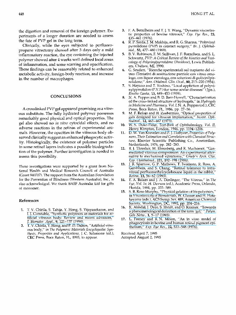
Statistical results from the comparisons of a repre- sentative assay are presented more suggestively in a graph (Fig. 4).
The cytostatic effect induced by the hydrogel may not preclude its use as a vitreous substitute. Figure 1. PVP gel after hydration, extraction, and autocla-
vation displays both flowing and elastic characteristics.
always above the G” curve, otherwise the material is a free-flowing viscous fluid rather than a ge1.16
The parallelism of G’ and G” curves in mechanical spectra of the gel specimens, both before and after injection (Figs. 2[A], 2[B]), indicates that the gel is cova- lently crosslinked. The storage modulus was signifi- cantly greater than the loss modulus, which shows that the material is predominantly elastic, rather than behaving like a viscous fluid.
A sufficient resilience of the material when subjected to shear stress is also desirable for a vitreous substitute. After injection, G’ decreased more significantly than did G” (Fig. 2[B]). This was caused by the shear stress
TABLE I Physical Properties of PVP Hydrogel: Comparison
with PBS
Property Hydrogel PBS ~~~~~
Water content (%) Injectability (mL / min) Refractive index Transmittance at 550 nm (%) Light transmission (%) Visual acuity
USAF Rodenstock Bailey-Lovie #5
97.3 0.9 1.3390
87.8 93
3.3 1 .o 6 / 6
-100 16.7
90.0 98
1.3358
3.3 1.25
6 / 6
C G
0 2
- 1 50 loo 200 500 1000 10 20
Frequency (a) - ~. B z + I
A- I I-- I
50 100 200 500 1000 10 20
Frequency (mHz)
Figure 2. The mechanical spectra of the gel specimens. (A) Before injection. (B) After injection.
446 HONG ET AL.
Clinical observations
Figure 3. Photomicrographs of the cell culture samples showing proliferating cells (BrdU-positive, arrow), and non- proliferating cells (BrdU-negative, open arrow). (Original magnification X750.) (A) Cell culture sample with phos- phate-buffered saline. (B) Cell culture sample with fetal calf serum. (C) Cell culture sample with polymer. (D) Cell culture sample with polymer and fetal calf serum.
At the time of sacrifice, three rabbits (nos. 1, 2, and 3) presented opacities in their vitreous bodies, which in rabbit 3 were more severe and associated with a very poor view of the fundus. In rabbit 1, small areas of scarring on either side of the medullary ray, close to the optic disc, were seen. In rabbit 2, a localized area of blood and fibrin in the ventral region was seen. Of the other three rabbits (4,5, and 6), one had severe traction of the blood vessels with large white patches in the ventral region. The other two had cupping of the optic disc and showed hazy corneas.
Histopathologic observations
Light and transmission electron microscopic evalua- tion of the retinas suggested biodegradation of the polymer. Irregularly shaped vacuoles were seen pre- dominantly in the retinal pigment epithelium (Fig. 5), often coalesced with lipid globules (Fig. 6), suggesting phagocytosis of the polymer. The vacuoles appeared to contain fine granular material. They were also seen interspersed among the rod outer segments, which ap- peared disorganized (Fig. 7), in the ganglion cell layer, in the inner nuclear layer, and in the outer nuclear layer. The eye of rabbit 5 had a membrane under the endothelium of the cornea, with occasional cells. All layers of the retina were intact and no loss of tissue was evident. This finding is consistent with the results from the toxicity bioassay in which a cytocidal effect on the cells was not seen. However, the choroidal veins and choriocapillaries were dilated and distorted, there- fore they may be involved in the disappearance of the polymer.
Examination of the areas containing scarring and traction revealed the presence of a large number of
Proliferating Cells Pyknotlc Ceis
Graphic statistic comparisons of proliferating and cells in various cell cultures.
PVP VITREOUS SUBSTITUTE 447
Figure 5. Light micrograph of the retina showing the retinal pigment epithelium (RPE) containing vacuoles (arrow), dis- organized rod outer segments (ROS), and a dilated and dis- torted choroidal vein (CV). (Original magnification X500.)
Figure 7. Transmission electron micrograph of rod outer segments (ROS), which appear disorganized and inter- spersed with fine granular material (P). (Original magnifica- tion X 14,000.)
macrophages with vacuoles similar to those seen in the retinal pigment epithelium, again suggesting the phagocytosis of the polymer.
DISCUSSION
Injection through a small-gauge needle is the only prac- tical procedure for the delivery of a vitreous substitute. The injection process induced some loss of elasticity, and the material became less resilient. However, the storage modulus was still higher than the loss modu- lus, and the dynamic mechanical spectra were parallel after injection, therefore the gel still behaved like a crosslinked elastic network. Since a material will be
Figure 6. Transmission electron micrograph of a retinal pig- ment epithelial cell in which a lipid globule (G) is seen coa- lesced with fine granular material, surrounded by a discon- tinuous membrane, which may represent phagocytosed polymer (P). Mitochondria (M) can be seen close to it. (Origi- nal magnification X14,500.)
subjected to only one injection cycle when used as a vitreous substitute, it is unlikely that it will exhibit flow under its own weight. Although the viscoelastic properties are not significantly altered, a certain num- ber of macromolecular chains and crosslinks in poly- mer are broken by injection and they are not expected to re-form.
The findings of the histopathological analysis can be regarded as consequences of onset of phagocytosis due to the presence of particles of polymers. Opacities in the vitreous may be due to a large number of macro- phages present in the vitreous. The retinal pigment epithelial cells and macrophages are highly phagocytic in their nature and it is not surprising that the vacuoles, apparently containing polymer, occur mainly in these two types of cells. A previous studyI7 indicated that a synthetic polymer (latex), injected as a suspension of microspheres into the vitreous, was indeed phagocy- tosed rather quickly by the retinal pigment epithelium. The biostability of PVP has been so far investigated only as aqueous solutions administered intravenously in animals or human patients. This polymer is not crosslinked. It was found5 that PVP fragments with a molecular weight lower than 70 kDa were passively excreted by kidneys, but the fragments with high mo- lecular weights were distributed and stored in various organs. There is no study available on the biostability of crosslinked PVP. Theoretically, a crosslinked poly- mer has an infinite molecular weight, and therefore should be insignificantly fragmented by phagocytic activity and should be mainly stored in tissues. How- ever, the injection process can cause the fragmentation of a crosslinked polymer, and the resulting fragments can be internalized by the retinal pigment epithelium cells and macrophages. The presence of vacuoles and the inflammation of choroidal vessels are indicative of an enhanced metabolic activity, likely directed toward
448 HONG ET AL.
the digestion and removal of the foreign polymer. Ex- periments of a longer duration are needed to assess the fate of PVP gel in the long term.
Clinically, while the eyes subjected to perfluoro- propane vitrectomy showed after 3 days only a mild inflammatory reaction, the eye containing the injected polymer showed after 4 weeks well defined local areas of inflammation, and some scarring and opacification. These findings can be reasonably related to enhanced metabolic activity, foreign-body reaction, and increase in the number of macrophages.
CONCLUSIONS
A crosslinked PVP gel appeared promising as a vitre- ous substitute. The fully hydrated polymer possesses remarkably good physical and optical properties. The gel also showed no in vitvo cytocidal effects, and no adverse reactions in the retinas of experimental ani- mals. However, the opacities in the vitreous body ob- served clinically suggest an enhanced phagocytic activ- ity. Histologically, the existence of polymer particles in some retinal layers indicates a possible biodegrada- tion of the polymer. Further investigation is needed to assess this possibility.
These investigations were supported by a grant from Na- tional Health and Medical Research Council of Australia (Grant 940707). The support from the Australian Foundation for the Prevention of Blindness (Western Australia), Inc., is also acknowledged. We thank BASF Australia Ltd for gifts of monomer.
References
1. T. V. Chirila, S. Tahija, Y. Hong, S. Vijayasekaran, and I. J. Constable, ”Synthetic polymers as materials for ar- tificial vitreous body: Review and recent advances,“ 7. Biomater. Appl., 9, 121-137 (1994).
2. T. V. Chirila, Y. Hong, and P. D. Dalton, ”Artificial vitre- ous body,” in The Polymeric Materials Encyclopedia: Syn- thesis, Properties and Applications, J. c. Salamone (ed.), CRC Press, Boca Raton, FL, 1995, to appear.
3.
4.
5.
6.
7.
8.
9.
10.
11.
12.
13.
14.
15.
16.
17.
F. A. Bettelheim and T. J. Y. Wang, “Dynamic viscoelas- tic properties of bovine vitreous,” Exp . Eye Res., 23,
R. P. Sarda, J. M. Makhija, and R. G. Sharma, ”Polyvinyl pyrrolidone (PVP) in cataract surgery,” Br. I. Ophthal- mol., 53, 477-480 (1969). B. V. Robinson, F. M. Sullivan, J. F. Borzelleca, and S. L. Schwartz, PVP: A Critical Review of the Kinetics and Toxi- cology of Polyvinylpyrrolidone (Pouidone), Lewis Publish- ers, Chelsea, MI, 1990. G. Scuderi, “Ricerche sperimentali sul trapianto del vi- treo (Tentativi di sostituzione parziale con vitreo omo- logo, con liquor eterologo, con soluzioni di polivinilpir- rolidone,” Ann. Ottalrnol. Clin. Ocul., 80,213-220 (1954). S . Hayano and T. Yoshino, ”Local appliction of polyvi- nylpyrrolidon (P.V.P.) for some ocular diseases” (Jpn.), Rinsho Ganka, 13,449-453 (1959). N. A. Peppas and B. D. Barr-Howell, “Characterization of the cross-linked structure of hydrogels,” in Hydrogels in Medicine and Pharmacy. Vol. I , N. A. Peppas (ed.), CRC Press, Boca Raton, FL, 1986, pp. 27-56. M. F. Refojo and H. Zauberman, ”Optical properties of gels designed for vitreous implantation,” Invest. Oph- thalmol., 12, 465-467 (1973). W. S. Duke-Elder, Text-Book of Ophthalmology, Vol. 11, Henry Kimpton, London, 1946, pp. 1194-1206. D. W. Van Krevelen and P. J. Hoftyzer, Properties of Poly- mers, Their Estimation and Correlation with Chemical Struc- ture, Elsevier Scientific Publishing Co., Amsterdam, Netherlands, 1976, pp. 282-283. R. J. Thresher, M. Ehrenberg, and R. Machemer, ”Gas- mediated vitreous compression: An experimental alter- native to mechanized vitrectomy,” Graefe’s Arch. Clin. Exp . Ophthalmol., 221, 192-198 (1984). J. R. Sparrow, G. P. Mathews, T. Iwamoto, R. Ross, A. Gershbein, and S. Chang, “Retinal tolerance to intra- vitreal perfluoroethylcyclohexane liquid in the rabbit,” Retina, 13, 56-62 (1993). E. A. Balazs and J. A. Denlinger, ”The Vitreus,” in The Eye, Vol. l a , H. Davson (ed.), Academic Press, Orlando, Florida, 1984, pp. 533-589. S. B. Ross-Murphy, “Physical gelation of biopolymers,” in Viscoelasticity of Biomaterials, W. Glasser and H. Hata- keyama (eds.), ACS Symp. Ser. 489, American Chemical Society, Washington, DC, 1992, pp. 204-216. K. Almdal, J. Dyre, S. Hvidt, and 0. Kramer, ”Towards a phenomenological definition of the term ’gel,’ ” Polym. Gels Netw., 1, 5-17 (1993). L. Feeney and R. N. Mixon, “An in vitro model of phagocytosis in bovine and human retinal pigment epi- thelium,” Exp. Eye Res., 22, 533-548 (1976).
435-441 (1976).
Received April 7, 1995 Accepted August 2, 1995


