Embed Size (px)
Citation preview
Hindawi Publishing CorporationNeural PlasticityVolume 2012, Article ID 304045, 9 pagesdoi:10.1155/2012/304045
Research Article
Crossmodal Recruitment of the Ventral Visual Stream inCongenital Blindness
Maurice Ptito,1, 2, 3 Isabelle Matteau,1 Arthur Zhi Wang,3, 4 Olaf B. Paulson,3, 5
Hartwig R. Siebner,3 and Ron Kupers1
1 Ecole d’Optometrie, Universite de Montreal, Montreal, QC, Canada H3T 1P12 Department of Neuroscience and Pharmacology, Panum Institute, University of Copenhagen, Blegdamsvej 3B,2200 Copenhagen, Denmark
3 Danish Research Centre for Magnetic Resonance, Copenhagen University Hospital Hvidovre, 2650 Hvidovre, Denmark4 Department of Radiology, Beijing Hospital, 100054 Beijing, China5 Neurobiology Research Unit, Rigshospitalet, Copenhagen University Hospital, 2100 Copenhagen, Denmark
Correspondence should be addressed to Ron Kupers, [email protected]
Received 22 January 2012; Accepted 19 April 2012
Academic Editor: Pietro Pietrini
Copyright © 2012 Maurice Ptito et al. This is an open access article distributed under the Creative Commons Attribution License,which permits unrestricted use, distribution, and reproduction in any medium, provided the original work is properly cited.
We used functional MRI (fMRI) to test the hypothesis that blind subjects recruit the ventral visual stream during nonhaptictactile-form recognition. Congenitally blind and blindfolded sighted control subjects were scanned after they had been trainedduring four consecutive days to perform a tactile-form recognition task with the tongue display unit (TDU). Both groups learnedthe task at the same rate. In line with our hypothesis, the fMRI data showed that during nonhaptic shape recognition, blindsubjects activated large portions of the ventral visual stream, including the cuneus, precuneus, inferotemporal (IT), cortex, lateraloccipital tactile vision area (LOtv), and fusiform gyrus. Control subjects activated area LOtv and precuneus but not cuneus, ITand fusiform gyrus. These results indicate that congenitally blind subjects recruit key regions in the ventral visual pathway duringnonhaptic tactile shape discrimination. The activation of LOtv by nonhaptic tactile shape processing in blind and sighted subjectsadds further support to the notion that this area subserves an abstract or supramodal representation of shape. Together with ourprevious findings, our data suggest that the segregation of the efferent projections of the primary visual cortex into a dorsal andventral visual stream is preserved in individuals blind from birth.
1. Introduction
It is well established that early-onset blindness leads towidespread neuroplastic changes. For instance, studies haveshown that the senses of hearing and touch are more devel-oped in blind than sighted individuals [1–6], probablydue to training-induced plasticity. The enlargement of thesomatic and motor area representation of the index fingerin proficient Braille readers is a clear example of this expe-rience-dependent plastic process [7]. Brain imaging studiesusing 18F-fluoro-deoxyglucose-positron emission tomogra-phy (FDG-PET) have shown that, despite the absence ofvisual input, the occipital cortex of congenitally blind in-dividuals shows a supranormal metabolism at rest [8, 9]. Thisindicates that the visually deprived cortex is still functionally
active and can be recruited by other modalities such astouch, hearing, and smell. Indeed, studies using a varietyof brain imaging tools such as PET, functional, magneticresonance imaging (fMRI), event-related potentials andmagnetoencephalography, all concur on a recruitment ofthe visual cortex of early blind individuals during variousnonvisual tasks (e.g., [10–14]).
Numerous brain imaging studies have consistently foundactivations of occipital cortical areas when blind subjects per-form a range of tactile tasks such as Braille character recog-nition, vibrotactile discrimination, and haptic object explo-ration [15–20]. Previous work from our laboratory showedthat blind subjects who had been trained to use the TDUin an orientation and motion discrimination task [13, 14]or in a navigation task [21] activate their visual cortex. The
2 Neural Plasticity
observed activation patterns within the visual cortex wereremarkably similar to those observed in sighted individualsperforming corresponding visual tasks. The modularity ofthese activations is further substantiated by the observa-tion that in early blind individuals, transcranial magneticstimulation (TMS) of the reorganized visual cortex elicitssomatotopically organized tactile sensations [22, 23]. TMSstudies have also provided evidence for the functionalimplication of the occipital cortex in tactile processing.For example, the demonstration that repetitive TMS of theoccipital cortex disrupts Braille reading performance in theblind [24, 25] suggests that the contribution of the visually-deprived occipital cortex to nonvisual functions is indeedfunctionally relevant. Together, these findings indicate thatthe visually deprived posterior cortical regions are muchmore adaptable than previously thought and may act eitheras a high-level multisensory area [26] or undergoes a cross-modal plastic reorganization [27].
The visual system is grossly subdivided into a dorsaland a ventral processing stream [28]. Area hMT+, a criticalpart of the dorsal visual pathway, involved in visual motionprocessing, is recruited during tactile and auditory motiondiscrimination task in early blind subjects [14, 29–31],suggesting that the dorsal “where” processing stream isfunctionally preserved in subjects lacking vision from birth.This raises the question whether the ventral processingstream, known to participate in object and shape recognition[30–39], is also preserved in blind subjects. Evidence insupport of this hypothesis comes from a study by Pietriniet al. [12] that showed category-specific recruitment of theventral temporal cortex by haptic exploration of objects incongenitally blind and sighted individuals. Therefore, theaim of this study was to investigate whether the ventralstream will also be activated by nonhaptic exploration ofshapes.
2. Materials and Methods
2.1. Subjects. Ten sighted control (five females; mean age:21 ± 11 y.) and eight blind (seven congenitally and oneearly blind) individuals with no recollection of any visualexperience (four females; mean age: 31 ± 10 y.) participatedin this study. Causes of blindness were retinopathy ofprematurity [7] and Leber’s congenital amaurosis [1]. Visualinspection of the structural brain MRI scans by a trainedneuroradiologist did not reveal macroscopic abnormalitiesand none of the subjects had a history of psychiatric orneurological illness. The study protocol was approved by thelocal ethics committee (Project ID: KF-01328723) and allsubjects provided written informed consent.
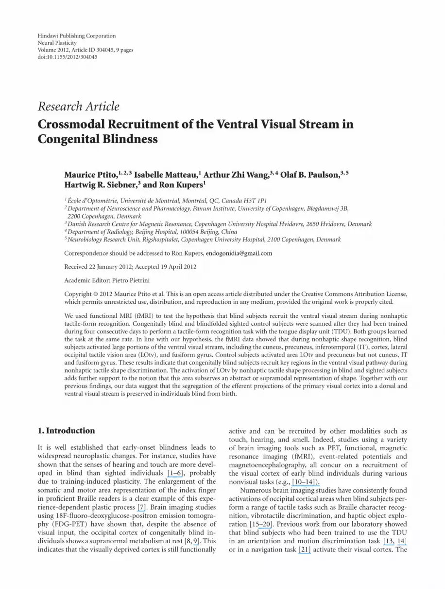
2.2. Electro-Tactile Stimulation of the Tongue. The apparatushas been described in detail elsewhere [13]. Briefly, it consistsof a tongue display unit (TDU, Wicab Inc.), an electrodearray (3 × 3 cm) with 144 gold-plated contacts arranged ina 12 × 12 matrix and a laptop with custom-made software(Figure 1(a)). Computer-generated geometric shapes wereconverted into electrical pulses and delivered to the tonguevia the electrode array. Stimulation intensity was controlled
(a)
10 s 10 s 10 s 10 s 10 s3 s 3 s 3 s 3 s ×7
(b)
Figure 1: Experimental setup. (a) The tongue display unit (TDU)and its components. (b) The fMRI block design. During each ofthe two fMRI runs, 7 stimulation blocks were presented. One blockconsisted of a rest period and Shape stimuli used during trainingand fMRI.
by the subject and could be adjusted at any time to allowoptimal perception of the stimuli.
2.3. Behavioural Training. Both blind and blindfolded con-trol subjects were trained during 10 sessions, stretched over4 consecutive days. Each session lasted around 15 minutesand comprised 28 trials. During training, subjects learnedto use the TDU to recognize four different shapes that wererandomly presented: a square, a triangle, a rectangle, and theletter E (Figure 1(b)). Participants were given a maximumof 30 seconds to identify each stimulus and they receivedimmediate feedback about the correctness of their response.Both the reaction time and the response accuracy weremeasured. It was stressed that correctness of responses wasmore important than speed. Stimuli were presented staticallyand participants could not explore the images by usingexploratory movements of a computer mouse as was the casein our previous PET study on orientation discrimination[13]. Prior to the training sessions, participants were famil-iarized with the TDU and the experimental procedures. Theywere told the forms that were going to be used and that theirtask was to correctly identify the shape that was presented.Blind participants were asked whether they were familiarwith the shapes that were going to be presented and they weregiven the opportunity to explore haptically plastic copies ofthe four shapes if necessary. Training sessions were limitedto a maximum of 15 minutes to avoid habituation to tonguestimulation. Participants were given two or maximum 3training sessions per day. Between two successive sessionsthere was a minimum time interval of 30 minutes. Wechose to work with a limited set of shapes in order notto overload memory and cognitive processing since duringthe fMRI session. Subjects had to indicate their response bypressing one of the four keys on a response pad (1: square;

Neural Plasticity 3
2: triangle; 3: rectangle and; 4: letter E). All participants weretrained by the same experimenter (IM). The criterion forsuccessful learning was set to 85% correct responses in twoconsecutive sessions. Participants who reached this criterioncould participate the next day in the fMRI examination.Statistical analysis of the behavioural data was carried outusing ANOVA (SPSS18, Chicago, Ill, US). Values of P < 0.05were considered as statistically significant.
2.4. MRI Experimental Design. Following behavioural train-ing, subjects performed the shape recognition task duringwhole-brain fMRI. We used an fMRI block design withperiods of rest (the electrode array was placed on the tonguebut no electrotactile stimulation was administered) and task(i.e., nonhaptic shape recognition). The same shapes werepresented in the fMRI session as during behavioural training.Two fMRI runs were carried out, each lasting 7 minutes and40 seconds. Each run consisted of alternating rest and taskblocks (Figure 1(b)). During a task block, four stimuli, onefor each form, were presented in a random order. This wasrepeated seven times, resulting in 35 blocks per fMRI run.Each stimulus lasted 10 s and was followed by a 3 s intervalduring which subjects had to indicate which form had beenpresented by pressing one of 4 buttons on a keypad withtheir right hand. Each button corresponded to one of thestimulus forms. Prior to scanning, subjects practiced to usethe appropriate corresponding response button.
2.5. Image Acquisition and Analysis. Task-related changes inthe (blood oxygenation level-dependent BOLD signal weremeasured with whole-brain fMRI using a Siemens Trio 3Tesla MR Scanner (Siemens, Erlangen, Germany), equippedwith an 8-channel head coil. The multislice gradient echo-planar imaging sequence had a repetition time (TR) =2500 ms, echo time (TE) = 50 ms, flip angle (FA) = 90◦,and field of view (FOV) of 192 mm (matrix: 64 × 64). Eachvolume consisted of 42 slices in an inclined axial plane,aligned to the AC-PC axis, with a slice thickness of 4 mm,resulting in a voxel size of 4 × 4 × 4 mm. A total of 368functional brain volumes were acquired per subject. Afterthe fMRI session, a high-resolution structural T1-weightedthree-dimensional brain scan (MPRAGE) was acquired usinga gradient echo pulse sequence (TE = 9.20 ms; flipangle =30◦; FOV = 256 mm; matrix = 256 × 256; voxelsize =1 mm3).
The MRI data were analyzed using Statistical ParametricMapping software (SPM5, Wellcome Department of Cog-nitive Neurology, London, UK). Functional volumes weremotion-corrected using SINC interpolation and spatiallynormalized to the reference space defined by the MRI tem-plate supplied by the Montreal Neurological Institute (MNI).Images were spatially smoothed with an 8-mm wide Gaus-sian kernel to improve the signal-to-noise ratio.
For the statistical analysis, active conditions were fittedwith a box-car function convolved with the hemodynamicresponse function. Low-frequency temporal drifts wereremoved by applying a 128-s high-pass filter. The duration ofall conditions was modelled, except for the 10 s rest periods,
which served as baseline. In order to estimate the effectsassociated with the experimental design, we evaluated BOLDsignal changes associated with the contrast active task(shapes) compared to the control task (rest). Following singlesubject analyses, we performed a random-effect analysiswithin and between groups using the individual contrastestimates for each functional run. Activation maps werethresholded at P < 0.01, corrected for multiple comparisonsusing the false discovery rate (FDR) [40]. We applied aconservative extent threshold of 20 contiguous voxels.
3. Results
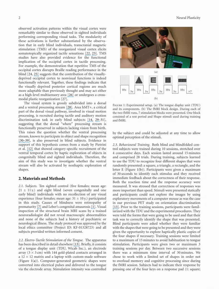
3.1. Behavioural Training. Both blind and sighted controlsubjects learned the tactile form discrimination task withinthe 10 sessions. Figure 2 illustrates the learning curves forpercentage of correct responses and reaction times. Astatistical analysis of the time× group interactions yielded nosignificant differences in the percentage of correct responses(F = 0.728; P > 0.05) or reaction times at the end of thetraining (F = 1.016; P > 0.05).
3.2. Functional MRI. Blind subjects but not blindfoldedsighted controls activated large areas of occipital (cuneus,inferior and middle occipital gyri and lingual gyrus) andoccipito-temporal (fusiform gyrus) cortices (Figure 3).Both blind and sighted controls showed increased BOLDresponses in the inferotemporal cortex (including areaLOtv), post-central gyrus, superior and inferior parietal lob-ule, precuneus, prefrontal cortex, cingulate gyrus, insula, andcerebellum. Task-related activations for blind and controlsubjects are listed in Table 1. A direct comparison of theactivation maps in both groups showed that BOLD increasesin the inferior temporal gyrus, middle occipital cortex,and precuneus were significantly stronger in blind subjects(Figure 4). In contrast, blindfolded-sighted control subjectsshowed a relative larger BOLD response increase in the rightpostcentral gyrus (BA3) and the left anterior cingulate cortex(BA24) only (data not shown). We also observed activationin both blind and controls in left and right premotor areasthat are probably due to the subject’s preparation to respondto the tactile stimulation. An increased BOLD response wasalso found in bilateral somatosensory cortex for both groupsof subjects.
4. Discussion
In this study, we report that congenitally blind but notsighted subjects activated large parts of the occipital cor-tex when performing a nonhaptic shape recognition task.Our data further showed that both groups recruited theinferotemporal cortex, including area LOtv, in response to2D tactile shape information extracted from electrotactilestimulation of the tongue. Previous studies showed that areaLOtv processes form information in the absence of visualinput through haptic [12] or auditory modalities [15, 30].The present data extend these findings by showing that areaLOtv processes form information even when tactile stimuli

4 Neural Plasticity
40
50
60
70
80
90
100
Cor
rect
res
pon
ses
(%)
1 2 3 4 5 6 7 8 9 10
(a)
0
5
10
15
20
Rea
ctio
n t
ime
(s)
Session
1 2 3 4 5 6 7 8 9 10
BlindControls
(b)
Figure 2: Learning curves for shape recognition in congenitallyblind and blindfolded control subjects. (a) Mean percentage chan-ges± SEM of correct responses and (b) mean reaction times± SEM.No significant differences in performance were observed betweenthe groups.
are delivered nonhaptically and to a body part, like thetongue, that is not primarily devoted to shape recognition.
4.1. Activation of IT/LOtv Complex. We found strong task-related activation along the occipital/inferior temporal cor-tical border in both sighted and blind subjects. WhereasIT/LOtv was activated bilaterally in blind subjects, it wasactivated only in the right hemisphere in sighted participants.This might be due to the relatively small sample size.Indeed, when using a less stringent criterion for statisticalsignificance (P < 0.01, uncorrected), an increased BOLDresponse was also noted in the left hemisphere. Moreover,a conjunction analysis of the activation patterns in bothgroups confirms the bilateral activation of IT/LOtv althoughthe cluster size was markedly larger in the right comparedto the left hemisphere (data not shown)The activationpattern in both groups encompassed a region that Amediand coworkers [36, 37] have coined the lateral occipitaltactile visual area. The stereotactic coordinates of our LOtvactivation in both groups (see Table 1) are very close to
−10 10 20 300
Figure 3: Axial maps showing brain activations for the contrast“shapes-rest” in congenitally blind (upper row) and blindfoldedcontrol (lower row) subjects. The color-coded t-maps illustratevoxels showing a task-related increase in activation at P < 0.01,FDR-corrected. Right side of the brain is to the right of the image.
x = −22 z = 12 z = 38
4
6
Figure 4: Cortical maps showing brain areas where activity wassignificantly larger in congenitally blind compared to blindfoldedcontrol subjects. The color-coded t-map shows the voxels with arelative increase in task-related activation in the blind group relativeto controls at P < 0.01, FDR-corrected. Right side of the brain is tothe right of the image.
those reported by others [12, 36, 41]. LOtv is a subregionwithin the human lateral occipital cortex (LOC) that isrobustly activated during both visual and tactile objectrecognition. Amedi and coworkers [37] demonstrated thatfor both modalities, LOtv has a preference for objects com-pared to textures and scrambled objects; this area is onlyweakly activated by the motor, naming and visual imagerycomponents of object recognition [37]. Area LOtv is alsorecruited by tactile exploration of novel, meaningless three-dimensional clay objects, suggesting that it responds more toform than to semantic features of objects [39]. Our findingof LOtv activation in both groups during the presentationof tactile stimuli is in accordance with previous resultsreported in normally sighted [12, 36, 36, 42–44] and blind[12] participants. The results further show for the first timethat not only three-dimensional tactile stimuli but also two-dimensional nonhaptic tactile information can recruit areaLOtv, adding further support that this area subserves anabstract or supramodal representation of shape information[45].
4.2. Occipital Cortex. Only blind subjects showed a signifi-cant BOLD response in several regions within the occipitalcortex including the cuneus, the lingual gyrus and theinferior, middle, and superior occipital gyri. The activationpattern in the blind following training shows remarkable

Neural Plasticity 5
Table 1: Activation clusters for “shapes versus rest” in blind and sighted subjects.
Anatomical area of activation BA
Congenitally blind Sighted controls
Talairach coordinates Talairach coordinates
x y z t x y z t
Occipital cortex
Cuneus18 −26 −75 15 8.12
12 −85 19 6.13
Lingual gyrus17 −8 −87 4 5.84
8 −85 3 4.38
Inferior occipital gyrus19 −48 −80 −3 4.02
34 −76 −8 4.78
Middle occipital gyrus19 −36 −85 6 4.71
37 53 −61 −9 6.36
Superior occipital gyrus 19 36 −73 24 4.54
Temporal cortex
Fusiform gyrus19 −36 −74 −10 3.48
38 −64 −5 5.76
Inferior temporal gyrus (LOtv)19 −51 −66 −2 4.60
37/20 55 −53 −4 5.36 51 −52 −7 4.67
Middle temporal gyrus37 −50 −60 0 5.41 −55 −51 −4 5.43
53 −62 0 4.59
Superior temporal gyrus22 −57 4 2 6.29
55 10 3 8.49
Parietal Cortex
Precuneus31/7 −26 −75 15 8.12 −10 −64 44 5.63
19/7 26 −70 35 7.57 12 −62 49 7.11
Inferior parietal lobule40 −46 −39 44 6.96 −44 −31 49 9.73
42 −31 44 5.92 46 −33 44 8.72
Superior parietal lobule7 −30 −54 51 6.82 −20 −53 58 5.75
30 −54 52 6.50 32 −52 49 6.82
Postcentral gyrus2 −53 −25 40 5.65 −50 −25 44 9.49
3/2 59 −20 34 5.73 51 −28 53 9.61
Prefrontal and frontal cortices
Precentral gyrus6/44 −61 0 35 4.48 −50 2 11 6.81
62 5 29 3.73 50 8 9 8.09
Inferior frontal gyrus9 −55 5 29 5.91 −57 5 26 6.98
47/9 34 17 −3 6.02 53 9 28 7.58
Middle frontal gyrus6 −26 −2 46 4.87 −38 −3 54 5.87
30 −1 48 6.37 40 2 50 7.00
Medial frontal gyrus6 −2 −1 50 6.63
4 1 50 6.52
Superior frontal gyrus6 −18 5 62 4.33
18 5 62 5.08
Cingulate and Insular cortices
Cingulate gyrus 32 2 10 42 6.31 2 10 42 8.53
Insula13 −40 5 13 4.76 −34 14 1 8.28
38 3 15 5.43 34 21 3 7.55
Cerebellum−32 −74 −22 5.36 −28 −67 −15 8.16
4 −73 −13 4.34 28 −67 −15 6.35

6 Neural Plasticity
similarities with that observed in normal seeing subjectsduring the performance of visual form discrimination tasks(see review by [45]). Pietrini and coworkers [12] reportedactivations in portions of the ventral stream such as thelingual and fusiform gyri and inferior occipital cortex duringhaptic object recognition in blind subjects. Other studieshave shown that the visual cortex in blind subjects can alsobe recruited by auditory and olfactory stimuli and cognitiveprocesses [38, 46–48] providing further evidence that thevisual cortex can be reorganized to mediate a variety of non-visual functions in the blind.
Another issue is the potential role of mental imageryin the visual cortex activation. A number of previous brainimaging studies on haptic processing [49, 50] and audito-ry-based sensory substitution [51] in blindfolded sightedsubjects have suggested that neural activity related to visualimagery may account for the activation in the occipitalcortex. For the following reasons, it is unlikely that visualimagery explains the current findings. First, our blind sub-jects never had visual experiences and during the debriefingfollowing the experiments, they did not report that they hadengaged in visual imagery during the orientation detectiontask (see also [12]). Secondly, if mental imagery wouldbe at the basis of the activation in the occipital cortex,sighted controls should activate the visual cortex to a largerextent compared to congenitally blind participants, whichwas clearly not the case. Finally, the question whether con-genitally blind subjects have true “visual imagery” (insteadof imagery) remains a matter of debate [52].
In accordance with several other studies (e.g., [13, 14,53, 54]), we report here a lack of activation in the occipitalcortex of our blindfolded controls. Previous neuroimagingstudies in normally sighted subjects have yielded inconsistentresults regarding the implication of V1 in tactile processing:some studies showing no activation of V1, others showingactivation of V1 only [18, 55], activation of V1 accompaniedby a deactivation of extrastriate areas [56], or activationof extrastriate cortical areas only [12]. Of note, moststudies showing V1 activation in tactile processing in normalsubjects used 3D stimuli that were palpated haptically withthe hand or fingers. In our study, we used 2-D shape stimulipresented passively to the tongue and thus, required no activehaptic exploration.
4.3. Possible Mechanisms for Cross-Modal Responses. A crit-ical question in the study of cross-modal processing in theblind is whether the recruitment of the occipital cortexoccurs through changes of existing neural network orthrough the formation of new neural connections. In thisstudy, as well as in our previous studies using the samesensory substitution device, cross-modal responses werealready observed after only a four to seven day period ofintensive training [13, 22]. The speed with which these neu-roplastic changes occur suggest that they are mediated by theunmasking or strengthening of preexisting cortico-corticalconnections [13, 22]. The observed striate and extrastriateactivations in the blind have been attributed to a cortico-cortical feedback pathway from primary somatosensorycortex (S1) through the posterior parietal cortex [22, 23].
The posterior parietal cortex is a highly multimodal asso-ciation area. Investigations in macaque and humans havedemonstrated that the anterior intraparietal area (AIP) andventral intraparietal area (VIP) are likely regions wherevisuotactile multimodal information of object features andmotion processing is integrated in sighted participants.Neurons in the macaque AIP, for instance, are sensitive tothree-dimensional features of objects such as size, shape,and orientation during object manipulation under visualcontrol [57, 58]. Neuroimaging studies in humans havealso demonstrated recruitment of AIP during tactile shapeprocessing [59, 60] and during orientation discrimination ofvisual stimuli [13, 61]. In blind subjects, who lack bottom-up visual processing, tactile inputs from these multimodalareas may then lead to a recruitment of the visual cortex viathese multimodal areas. This assumption is supported by thestrong activation of the posterior parietal cortex observed inthe blind in the present study and is moreover reinforced bythe results of several additional neuroimaging studies [13, 17,38]. This hypothesis is also in line with a recent report thatused dynamic causal modeling of fMRI data to investigate thecross-modal plasticity of effective connectivity in the blindduring a Braille reading task [62]. It is also possible thatnew aberrant subcortical projections could be responsiblefor the evoked activity in the visual cortex of congenitalblind individuals. For example, animal models of bilateralenucleation in hamsters [60], congenital blindness in mice[63, 64], and natural very low vision like the blind mole rat[65] have indicated the formation of new ectopic projectionsfrom the inferior colliculus to the lateral geniculate nucleus,the thalamic primary visual relay. More advanced methods,such as functional connectivity analysis, will be helpful tobetter understand through which pathways nonhaptic tactileinformation is funnelled to the visual cortex of the blind.
4.4. Methodological Considerations. The main limitation ofthis study is the sample size. While eight subjects are consid-ered to be a relatively small sample size for a classical fMRIstudy, we would like to emphasize that congenitally blindindividuals represent an exceptionally rare population, evenmore so when strict selection requirements are enforced, asin this study. We would further like to stress that samplesizes of congenitally blind individuals reported in most fMRIstudies in the literature are similar or smaller as compared tothe present one [12–18, 29–31, 38, 39, 54, 55, 62, 66]. Largernumbers of subjects certainly are required to make rigorousstatistical comparisons between the sighted and congenitallyblind groups in terms of distribution and extent of brainresponse to shape recognition following stimulation by TDU.Nevertheless, the data were obtained using a random-effectsanalysis and FDR-corrected statistical thresholds. A finallimitation is that we did not use functional localizer scans inour sighted subjects to identify subregions within the ventralstream.
5. Conclusion
The question we have addressed in this and our previousstudies is whether the functional segregation of the visual

Neural Plasticity 7
cortex in a dorsal and ventral visual pathway is preservedin individuals who were born without vision or who losttheir sight at a very early age. The present results signifi-cantly extend to our previously published data on motionprocessing via the tongue in the blind [14], showing thatboth pathways are preserved in this population and add togrowing evidence that the visual cortex can be reorganized tomediate non-visual functions in the blind.
Acknowledgments
This work was supported by the Harland Sanders founda-tion (MP), the Lundbeck Foundation (R. Kupers), DanishMedical Research Council (M. Ptito and RK; Grant no. 09-065632). A. Z. Wang and I. Matteau held a studentshipfrom, respectively, the Dana Foundation and the CanadianInstitutes of Health Research (CIHR). The 3 Tesla MRIscanner was donated by the Simon Spies foundation.
References
[1] O. Collignon, G. Charbonneau, M. Lassonde, and F. Lepore,“Early visual deprivation alters multisensory processing inperipersonal space,” Neuropsychologia, vol. 47, no. 14, pp.3236–3243, 2009.
[2] D. R. Chebat, J. K. Chen, F. Schneider, A. Ptito, R. Kupers, andM. Ptito, “Alterations in right posterior hippocampus in earlyblind individuals,” NeuroReport, vol. 18, no. 4, pp. 329–333,2007.
[3] A. Fieger, B. Roder, W. Teder-Salejarvi, S. A. Hillyard, and H.J. Neville, “Auditory spatial tuning in late-onset blindness inhumans,” Journal of Cognitive Neuroscience, vol. 18, no. 2, pp.149–157, 2006.
[4] D. Goldreich and I. M. Kanics, “Tactile acuity is enhanced inblindness,” Journal of Neuroscience, vol. 23, no. 8, pp. 3439–3445, 2003.
[5] D. Goldreich and I. M. Kanics, “Performance of blind andsighted humans on a tactile grating detection task,” Perceptionand Psychophysics, vol. 68, no. 8, pp. 1363–1371, 2006.
[6] R. W. Van Boven, J. E. Ingeholm, M. S. Beauchamp, P. C. Bikle,and L. G. Ungerleider, “Tactile form and location processingin the human brain,” Proceedings of the National Academy ofSciences of the United States of America, vol. 102, no. 35, pp.12601–12605, 2005.
[7] A. Pascual-Leone and F. Torres, “Plasticity of the sensorimotorcortex representation of the reading finger in Braille readers,”Brain, vol. 116, no. 1, pp. 39–52, 1993.
[8] A. G. De Volder, A. Bol, J. Blin et al., “Brain energy metabolismin early blind subjects: neural activity in the visual cortex,”Brain Research, vol. 750, no. 1-2, pp. 235–244, 1997.
[9] R. Kupers, P. Pietrini, E. Ricciardi, and M. Ptito, “The natureof consciousness in the visually-deprived brain,” Frontiers inPsychology, vol. 2, no. 4, 2011.
[10] L. B. Merabet, J. F. Rizzo, A. Amedi, D. C. Somers, and A.Pascual-Leone, “What blindness can tell us about seeing again:merging neuroplasticity and neuroprostheses,” Nature ReviewsNeuroscience, vol. 6, no. 1, pp. 71–77, 2005.
[11] U. Noppeney, “The effects of visual deprivation on functionaland structural organization of the human brain,” Neuroscienceand Biobehavioral Reviews, vol. 31, no. 8, pp. 1169–1180, 2007.
[12] P. Pietrini, M. L. Furey, E. Ricciardi et al., “Beyond sensoryimages: object-based representation in the human ventral
pathway,” Proceedings of the National Academy of Sciences ofthe United States of America, vol. 101, no. 15, pp. 5658–5663,2004.
[13] M. Ptito, S. M. Moesgaard, A. Gjedde, and R. Kupers, “Cross-modal plasticity revealed by electrotactile stimulation of thetongue in the congenitally blind,” Brain, vol. 128, no. 3, pp.606–614, 2005.
[14] M. Ptito, I. Matteau, A. Gjedde, and R. Kupers, “Recruitmentof the middle temporal area by tactile motion in congenitalblindness,” NeuroReport, vol. 20, no. 6, pp. 543–547, 2009.
[15] A. Amedi, W. M. Stern, J. A. Camprodon et al., “Shapeconveyed by visual-to-auditory sensory substitution activatesthe lateral occipital complex,” Nature Neuroscience, vol. 10, no.6, pp. 687–689, 2007.
[16] C. Buchel, C. Price, R. S. J. Frackowiak, and K. Friston,“Different activation patterns in the visual cortex of late andcongenitally blind subjects,” Brain, vol. 121, no. 3, pp. 409–419, 1998.
[17] H. Burton, “Visual cortex activity in early and late blind peo-ple,” Journal of Neuroscience, vol. 23, no. 10, pp. 4005–4011,2003.
[18] H. Burton, D. G. McLaren, and R. J. Sinclair, “Readingembossed capital letters: an fMRI study in blind and sightedindividuals,” Human Brain Mapping, vol. 27, no. 4, pp. 325–339, 2006.
[19] N. Sadato, A. Pascual-Leone, J. Grafman et al., “Activation ofthe primary visual cortex by Braille reading in blind subjects,”Nature, vol. 380, no. 6574, pp. 526–528, 1996.
[20] N. Sadato, A. Pascual-Leone, J. Grafman, M. P. Deiber, V.Ibanez, and M. Hallett, “Neural networks for Braille readingby the blind,” Brain, vol. 121, no. 7, pp. 1213–1229, 1998.
[21] R. Kupers, D. R. Chebat, K. H. Madsen, O. B. Paulson, andM. Ptito, “Neural correlates of virtual route recognition incongenital blindness,” Proceedings of the National Academy ofSciences of the United States of America, vol. 107, no. 28, pp.12716–12721, 2010.
[22] R. Kupers, A. Fumal, A. M. De Noordhout, A. Gjedde, J.Schoenen, and M. Ptito, “Transcranial magnetic stimulationof the visual cortex induces somatotopically organized qualiain blind subjects,” Proceedings of the National Academy ofSciences of the United States of America, vol. 103, no. 35, pp.13256–13260, 2006.
[23] M. Ptito, F. C. G. Schneider, O. B. Paulson, and R. Kupers,“Alterations of the visual pathways in congenital blindness,”Experimental Brain Research, vol. 187, no. 1, pp. 41–49, 2008.
[24] L. G. Cohen, P. Celnik, A. Pascual-Leone et al., “Functionalrelevance of cross-modal plasticity in blind humans,” Nature,vol. 389, no. 6647, pp. 180–183, 1997.
[25] R. Kupers, M. Pappens, A. M. De Noordhout, J. Schoenen, M.Ptito, and A. Fumal, “rTMS of the occipital cortex abolishesBraille reading and repetition priming in blind subjects,”Neurology, vol. 68, no. 9, pp. 691–693, 2007.
[26] C. Buchel, “Cortical hierarchy turned on its head,” NatureNeuroscience, vol. 6, no. 7, pp. 657–658, 2003.
[27] P. Pietrini, M. Ptito, and R. Kupers, “Blindness and con-sciousness: new lights from the dark,” in The Neurology ofConsciousness, S. Laureys and G. Tononi, Eds., pp. 360–374,Elsevier, Amsterdam, The Netherlands, 2009.
[28] M. A. Goodale and A. David Milner, “Separate visual pathwaysfor perception and action,” Trends in Neurosciences, vol. 15, no.1, pp. 20–25, 1992.
[29] C. Poirier, O. Collignon, C. Scheiber et al., “Auditory motionperception activates visual motion areas in early blind sub-jects,” NeuroImage, vol. 31, no. 1, pp. 279–285, 2006.
8 Neural Plasticity
[30] C. C. Poirier, A. G. De Volder, D. Tranduy, and C. Scheiber,“Neural changes in the ventral and dorsal visual streamsduring pattern recognition learning,” Neurobiology of Learningand Memory, vol. 85, no. 1, pp. 36–43, 2006.
[31] E. Ricciardi, N. Vanello, L. Sani et al., “The effect of visualexperience on the development of functional architecture inhMT+,” Cerebral Cortex, vol. 17, no. 12, pp. 2933–2939, 2007.
[32] J. V. Haxby, M. I. Gobbini, M. L. Furey, A. Ishai, J. L. Schouten,and P. Pietrini, “Distributed and overlapping representationsof faces and objects in ventral temporal cortex,” Science, vol.293, no. 5539, pp. 2425–2430, 2001.
[33] I. Matteau, R. Kupers, E. Ricciardi, P. Pietrini, and M.Ptito, “Beyond visual, aural and haptic movement perception:hMT+ is activated by electrotactile motion stimulation of thetongue in sighted and in congenitally blind individuals,” BrainResearch Bulletin, vol. 82, no. 5-6, pp. 264–270, 2010.
[34] L. G. Underleider and M. Mishkin, “Two cortical visual sys-tems,” in Analysis of Visual Behavior, M. A. Ingle, M. I.Goodale, and R. J. W. Masfield, Eds., pp. 549–586, MIT Press,Cambridge, Mass, USA, 1982.
[35] R. Farivar, “Dorsal-ventral integration in object recognition,”Brain Research Reviews, vol. 61, no. 2, pp. 144–153, 2009.
[36] A. Amedi, G. Jacobson, T. Hendler, R. Malach, and E. Zohary,“Convergence of visual and tactile shape processing in thehuman lateral occipital complex zohary,” Cerebral Cortex, vol.12, no. 11, pp. 1202–1212, 2002.
[37] A. Amedi, R. Malach, T. Hendler, S. Peled, and E. Zohary,“Visuo-haptic object-related activation in the ventral visualpathway,” Nature Neuroscience, vol. 4, no. 3, pp. 324–330, 2001.
[38] A. Amedi, N. Raz, P. Pianka, R. Malach, and E. Zohary,“Early ’visual’ cortex activation correlates with superior verbalmemory performance in the blind,” Nature Neuroscience, vol.6, no. 7, pp. 758–766, 2003.
[39] T. W. James, G. K. Humphrey, J. S. Gati, P. Servos, R. S. Menon,and M. A. Goodale, “Haptic study of three-dimensionalobjects activates extrastriate visual areas,” Neuropsychologia,vol. 40, no. 10, pp. 1706–1714, 2002.
[40] C. R. Genovese, N. A. Lazar, and T. Nichols, “Thresholdingof statistical maps in functional neuroimaging using the falsediscovery rate,” NeuroImage, vol. 15, no. 4, pp. 870–878, 2002.
[41] T. Kassuba, C. Klinge, C. Holig et al., “The left fusiform gyrushosts trisensory representations of manipulable objects,” Neu-roImage, vol. 56, no. 3, pp. 1566–1577, 2011.
[42] S. C. Prather and K. Sathian, “Mental rotation of tactile stim-uli,” Cognitive Brain Research, vol. 14, no. 1, pp. 91–98, 2002.
[43] C. L. Reed, S. Shoham, and E. Halgren, “Neural substratesof tactile object recognition: an fMRI study,” Human BrainMapping, vol. 21, no. 4, pp. 236–246, 2004.
[44] M. R. Stoesz, M. Zhang, V. D. Weisser, S. C. Prather, H.Mao, and K. Sathian, “Neural networks active during tactileform perception: common and differential activity duringmacrospatial and microspatial tasks,” International Journal ofPsychophysiology, vol. 50, no. 1-2, pp. 41–49, 2003.
[45] A. Amedi, K. Von Kriegstein, N. M. Van Atteveldt, M.S. Beauchamp, and M. J. Naumer, “Functional imaging ofhuman crossmodal identification and object recognition,” Ex-perimental Brain Research, vol. 166, no. 3-4, pp. 559–571, 2005.
[46] B. Roder, O. Stock, S. Bien, H. Neville, and F. Rosler, “Speechprocessing activates visual cortex in congenitally blindhumans,” European Journal of Neuroscience, vol. 16, no. 5, pp.930–936, 2002.
[47] F. Gougoux, F. Lepore, M. Lassonde, P. Voss, R. J. Zatorre, andP. Belin, “Neuropsychology: pitch discrimination in the earlyblind,” Nature, vol. 430, no. 6997, p. 309, 2004.
[48] M. Beaulieu-Lefebvre, F. C. Schneider, R. Kupers, and M.Ptito, “Odor perception and odor awareness in congenitalblindness,” Brain Research Bulletin, vol. 84, no. 3, pp. 206–209,2011.
[49] M. Zhang, V. D. Weisser, R. Stilla, S. C. Prather, and K.Sathian, “Multisensory cortical processing of object shapeand its relation to mental imagery,” Cognitive, Affective andBehavioral Neuroscience, vol. 4, no. 2, pp. 251–259, 2004.
[50] S. Lacey, P. Flueckiger, R. Stilla, M. Lava, and K. Sathian,“Object familiarity modulates the relationship between visualobject imagery and haptic shape perception,” NeuroImage, vol.49, no. 3, pp. 1977–1990, 2010.
[51] C. Poirier, A. De Volder, D. Tranduy, and C. Scheiber, “Patternrecognition using a device substituting audition for vision inblindfolded sighted subjects,” Neuropsychologia, vol. 45, no. 5,pp. 1108–1121, 2007.
[52] D. Kaski, “Revision: is visual perception a requisite for visualimagery?” Perception, vol. 31, no. 6, pp. 717–731, 2002.
[53] N. Sadato, T. Okada, M. Honda, and Y. Yonekura, “Criticalperiod for cross-modal plasticity in blind humans: a func-tional MRI study,” NeuroImage, vol. 16, no. 2, pp. 389–400,2002.
[54] T. Harada, D. N. Saito, K. I. Kashikura et al., “Asymmet-rical neural substrates of tactile discrimination humans: afunctional magnetic resonance imaging study,” Journal ofNeuroscience, vol. 24, no. 34, pp. 7524–7530, 2004.
[55] D. N. Saito, T. Okada, M. Honda, Y. Yonekura, and N.Sadato, “Practice makes perfect: the neural substrates of tactilediscrimination by Mah-Jong experts include the primaryvisual cortex,” BMC Neuroscience, vol. 7, article 79, 2006.
[56] L. B. Merabet, J. D. Swisher, S. A. McMains et al., “Combinedactivation and deactivation of visual cortex during tactilesensory processing,” Journal of Neurophysiology, vol. 97, no. 2,pp. 1633–1641, 2007.
[57] A. Murata, V. Gallese, M. Kaseda, and H. Sakata, “Parietalneurons related to memory-guided hand manipulation,”Journal of Neurophysiology, vol. 75, no. 5, pp. 2180–2186, 1996.
[58] H. Sakata, M. Taira, A. Murata, and S. Mine, “Neuralmechanisms of visual guidance of hand action in the parietalcortex of the monkey,” Cerebral Cortex, vol. 5, no. 5, pp. 429–438, 1995.
[59] A. Bodegard, S. Geyer, C. Grefkes, K. Zilles, and P. E. Roland,“Hierarchical processing of tactile shape in the human brain,”Neuron, vol. 31, no. 2, pp. 317–328, 2001.
[60] L. Jancke, A. Kleinschmidt, S. Mirzazade, N. J. Shah, and H. J.Freund, “The role of the inferior parietal cortex in linking thetactile perception and manual construction of object shapes,”Cerebral Cortex, vol. 11, no. 2, pp. 114–121, 2001.
[61] E. Shikata, F. Hamzei, V. Glauche et al., “Surface orientationdiscrimination activates caudal and anterior intraparietalsulcus in humans: an event-related fMRI study,” Journal ofNeurophysiology, vol. 85, no. 3, pp. 1309–1314, 2001.
[62] T. Fujii, H. C. Tanabe, T. Kochiyama, and N. Sadato, “Aninvestigation of cross-modal plasticity of effective connectivityin the blind by dynamic causal modeling of functional MRIdata,” Neuroscience Research, vol. 65, no. 2, pp. 175–186, 2009.
[63] N. Chabot, S. Robert, R. Tremblay, D. Miceli, D. Boire, andG. Bronchti, “Audition differently activates the visual systemin neonatally enucleated mice compared with anophthalmicmutants,” European Journal of Neuroscience, vol. 26, no. 8, pp.2334–2348, 2007.
[64] N. Chabot, V. Charbonneau, M. E. Laramee, R. Tremblay, D.Boire, and G. Bronchti, “Subcortical auditory input to the

Neural Plasticity 9
primary visual cortex in anophthalmic mice,” NeuroscienceLetters, vol. 433, no. 2, pp. 129–134, 2008.
[65] G. Bronchti, P. Heil, R. Sadka, A. Hess, H. Scheich, and Z.Wollberg, “Auditory activation of ’visual’ cortical areas inthe blind mole rat (Spalax ehrenbergi),” European Journal ofNeuroscience, vol. 16, no. 2, pp. 311–329, 2002.
[66] H. Burton, R. J. Sinclair, and D. G. McLaren, “Cortical activityto vibrotactile stimulation: an fMRI study in blind and sightedindividuals,” Human Brain Mapping, vol. 23, no. 4, pp. 210–228, 2004.


