Embed Size (px)
Citation preview
Biochemical and Biophysical Research Communications 448 (2014) 163–168
Contents lists available at ScienceDirect
Biochemical and Biophysical Research Communications
journal homepage: www.elsevier .com/locate /ybbrc
Crystal structure and biochemical properties of the (S)-3-hydroxybutyryl-CoA dehydrogenase PaaH1 from Ralstonia eutropha
http://dx.doi.org/10.1016/j.bbrc.2014.04.1010006-291X/� 2014 Elsevier Inc. All rights reserved.
⇑ Corresponding author. Address: Structural and Molecular Biology Laboratory,School of Life Sciences, Kyungpook National University, Daehak-ro 80, Buk-ku,Daegu 702-701, Republic of Korea. Fax: +82 53 955 5522.
E-mail address: [email protected] (K.-J. Kim).1 These authors contributed equally to this work.
Jieun Kim a,1, Jeong Ho Chang b,1, Kyung-Jin Kim a,⇑a School of Life Sciences, KNU Creative BioResearch Group (BK21 Plus Program), Kyungpook National University, Daehak-ro 80, Buk-ku, Daegu 702-701, Republic of Koreab Department of Biology, Teachers College, Kyungpook National University, Daehak-ro 80, Buk-ku, Daegu 702-701, Republic of Korea
a r t i c l e i n f o a b s t r a c t
Article history:Received 3 April 2014Available online 29 April 2014
Keywords:(S)-3-Hydroxybutyryl-CoA dehydrogenaseRalstonia eutrophan-ButanolCrystal structure
3-Hydroxybutyryl-CoA dehydrogenase is an enzyme involved in the synthesis of the biofuel n-butanol byconverting acetoacetyl-CoA to 3-hydroxybutyryl-CoA. To investigate the molecular mechanism ofn-butanol biosynthesis, we determined crystal structures of the Ralstonia eutropha-derived 3-hydroxybu-tyryl-CoA dehydrogenase (RePaaH1) in complex with either its cofactor NAD+ or its substrate acetoacetyl-CoA. While the biologically active structure is dimeric, the monomer of RePaaH1 comprises two separateddomains with an N-terminal Rossmann fold and a C-terminal helical bundle for dimerization. In thisstudy, we show that the cofactor-binding site is located on the Rossmann fold and is surrounded by fiveloops and one helix. The binding mode of the acetoacetyl-CoA substrate was found to be that the aden-osine diphosphate moiety is not highly stabilized compared with the remainder of the molecule. Residuesinvolved in catalysis and substrate binding were further confirmed by site-directed mutagenesis exper-iments, and kinetic properties of RePaaH1were examined as well. Our findings contribute to the under-standing of 3-hydroxybutyryl-CoA dehydrogenase catalysis, and will be useful in enhancing the efficiencyof n-butanol biosynthesis by structure based protein engineering.
� 2014 Elsevier Inc. All rights reserved.
1. Introduction
Due to issues such as limited fossil fuel availability, greenhousegas emissions, and the requirement for increased energy securityor diversity, there is increased public and scientific interest inenergy alternatives such as biofuels. A wide range of biofuels canbe derived from plant or microbial biomass [1]. The two major bio-fuels in use today are ethanol and butanol, which can be combinedwith gasoline for use in conventional engines [2,3]. However, eth-anol has a low energy efficiency compared to gasoline and highvaporizability [4]. Alternatively, n-butanol produced by microbialfermentation has characteristics that are closer to those ofmotor-vehicle fuels and could serve as a better replacement [5].The anaerobic bacterium Clostridium acetobutylicum can efficientlyproduce n-butanol through a carbohydrate catabolic pathway [6,7].In comparison with bio-ethanol, the advantage of the biosynthe-
sized n-butanol is that it has a high energy content, low corrosion,increased solubility, and easier to blend with gasoline [8–10].
Even if n-butanol is considered a potential next generation bio-fuel source, its biosynthetic efficiency must be improved, and therehave been multiple attempts to do so [11]. For example, manyengineering efforts ranging from genetic modifications to micro-bial culture optimization, have aimed to increase n-butanol pro-duction during ABE fermentation. However, the n-butanolsynthetic titers do not exceed 1 g/L in heterologous host cells thatexpress clostridial n-butanol biosynthetic machinery [7–10]. Veryrecently, alternative methods to enhance the n-butanol yield havebeen reported; these involve the use of metabolically engineeredhosts such as Escherichia coli, Pseudomonas putida, and Bacillus subtilisin the n-butanol biosynthetic pathway to improve biofuel productionfrom small organic molecules [12–14].
A next step to produce large amount of n-butanol is the engi-neering of non-solventogenic microbes [15]. It has been shownthat the n-butanol inhibits E. coli growth for example, the growthis almost ceased at approximately n-butanol concentrations of 1%[16], therefore, the toxicity effects of n-butanol in bacterial cellsshould be moderated [17]. Another issue is that the additive path-ways for n-butanol synthesis disrupt the balance of energy carrierssuch as NADH/NAD+, which results in a decrease in n-butanol
164 J. Kim et al. / Biochemical and Biophysical Research Communications 448 (2014) 163–168
production [18]. These multiple issues have necessitated an opti-mization of the heterologous metabolic pathways to maximizethe n-butanol biosynthetic yield by the use of engineered non-sol-ventogenic microbes [18,19].
In contrast to C. acetobutylicum, which is a representativen-butanol producing host, Ralstonia eutropha has a broader spectrumand is used in the production of polymers such as polyhydroxybuty-rate (PHB). It has been reported that the 3-hydroxybutyryl-CoAdehydrogenase PaaH1 is involved in n-butanol biosynthesis [20],and R. eutropha-derived PaaH1 is proposed as a homolog of Clostrid-ium butyricum 3-hydroxybutyryl-CoA dehydrogenase (CbHBD) thatis involved in the second step of n-butanol biosynthesis [21]. Here,we report the first crystal structure of R. eutropha 3-hydroxybuty-ryl-CoA dehydrogenase (RePaaH1), an enzyme that catalyzes thesecond step of n-butanol biosynthesis and converts acetoacetyl-CoA to 3-hydroxybutyryl-CoA. Kinetic properties and mutagenesisexperiments were also reported.
2. Materials and methods
2.1. Preparation of RePaaH1
Cloning, expression, purification, and crystallization of RePaaH1will be described elsewhere. Briefly, the RePaaH1 coding gene(Met1-Lys284, M.W. 32 kDa) was amplified by polymerase chainreaction (PCR) using R. eutropha chromosomal DNA as a template.The PCR product was then subcloned into pET30a (Invitrogen) with6-histag at the C-terminus. The expression construct was trans-formed into an E. coli B834 strain, which was grown in 1 L of LBmedium containing kanamycin (50 mg/ml) at 37 �C. After induc-tion via the addition of 1.0 mM IPTG, the culture medium was fur-ther maintained for 20 h at 18 �C. The culture was harvested bycentrifugation at 5000 � g at 4 �C. The cell pellet was resuspendedin buffer A (40 mM Tris–HCl at pH 8.0 and 5 mM b-mercap-toethanol) and then disrupted by ultrasonication. The cell debriswas removed by centrifugation at 11,000 � g for 1 h, and lysatewas bound to Ni–NTA agarose (QIAGEN). After washing with bufferA containing 20 mM imidazole, the bound proteins were elutedwith 300 mM imidazole in buffer A. A trace amount of contamina-tion was removed by applying HiLoad 26/60 Superdex 200 prepgrade (GE Healthcare) size exclusion chromatography. The purifiedprotein showed �95% purity on SDS–PAGE, was concentrated to50 mg/ml in 40 mM Tris–HCl, pH 8.0, 1 mM dithiothreitol.
2.2. Crystallization, data collection, and structure determination ofRePaaH1
Suitable crystals for diffraction experiments were obtained at22 �C within 7 days from the precipitant of 2 M (NH4)2SO4, 0.1 MCacodylate pH 6.5 and 0.2 M Sodium Chloride. The crystals weretransferred to cryoprotectant solution containing 2 M (NH4)2SO4,0.1 M Cacodylate pH 6.5, 0.2 M Sodium Chloride and 30% glycerol,fished out with a loop larger than the crystals and flash-frozen byimmersion in liquid nitrogen at �173 �C. The data were collectedto a resolution of 2.6 Å at 7A beamline of the Pohang AcceleratorLaboratory (PAL, Pohang, Korea) using a Quantum 270 CCD detec-tor (ADSC, USA). The data were then indexed, integrated, andscaled using the HKL2000 suite [22]. The data statistics are sum-marized in Table 1. Crystals of an apo-form belonged to spacegroup p3221, with unit cell parameters of a = b = 135.43 Å,c = 97.17 Å, a = b = 90 and c = 120. Assuming 3 molecules ofRePaaH1 per asymmetric unit, the crystal volume per unit of pro-tein mass was 2.68 Å3 Da�1 [23], which corresponds to a solventcontent of approximately 54.12%. RePaaH1 crystals in complexwith NAD+ and with acetoacetyl-CoA were crystallized with the
same crystallization condition supplemented with 20 mM each ofNAD+ and acetoacetyl-CoA. Crystals in complex with NAD+
belonged to space group C2, with unit cell parameters ofa = 235.08 Å, b = 135.59 Å, c = 97.45 Å, a = c = 90 and b = 90.1.Assuming 9 molecules of RePaaH1 per asymmetric unit, the crystalvolume per unit of protein mass was 2.70 Å3 Da�1 [23], which cor-responds to a solvent content of approximately 54.41%. Crystals incomplex with acetoacetyl-CoA belonged to the same space withRePaaH1-NAD+ complex crystals with similar unit cell parameters.SeMet-substituted apo-form crystals were obtained using the samecrystallization condition as used for the native protein crystal. Sin-gle-wavelength anomalous dispersion (SAD) data were collectedfrom an SeMet protein crystal on beamline 7A at PAL to a wave-length of 0.97855 Å. 36 Se atoms out of the expected 39 in theasymmetric unit were identified at 2.42 Å resolution using SOLVE[24]. The electron density was improved by density modificationusing RESOLVE [25], resulting in 54% of the cloned residues beingautomatically built.
Further model building was performed manually using the pro-gram WinCoot [26] and the refinement was performed with REF-MAC5 [27]. The structures of RePaaH1 in complex with NAD+ andwith acetoacetyl-CoA were solved by molecular replacement usingthe crystal structure of the apo-form of RePaaH1. The refinedmodel of apo-form of C RePaaH1 and those in complex withNAD+ and with acetoacetyl-CoA were deposited in the Protein DataBank (pdb code 4PZC for apo-form of RePaaH1, and 4PZD and4PZE for NAD+ and acetoacetyl-CoA bound forms of RePaaH1,respectively).
2.3. 3-Hydroxybutyryl-CoA dehydrogenase activity measurement
All assays were performed with reaction mixture of 1 ml totalvolume. The reaction mixture contained 100 mM MOPS (pH 8.0),100 lM of NADH, 100 lM acetoacetyl-CoA, 1 mM DTT (dithiothre-itol), and 32 lM of RePaaH1 enzyme. After pre-incubation at 30 �Cfor 5 min, the reaction was initiated by the addition of enzyme. Thedecrease in NADH was then measured at 340 nm and 30 �C usingan extinction coefficient of 6.3 � 103 [28,29]. The enzyme kineticsexperiments were performed by addition of various concentrationsof acetoacetyl-CoA substrate, such as 10, 20, 40, 60, 80 and 100 lM.
3. Results and discussion
3.1. Overall structure of RePaaH1
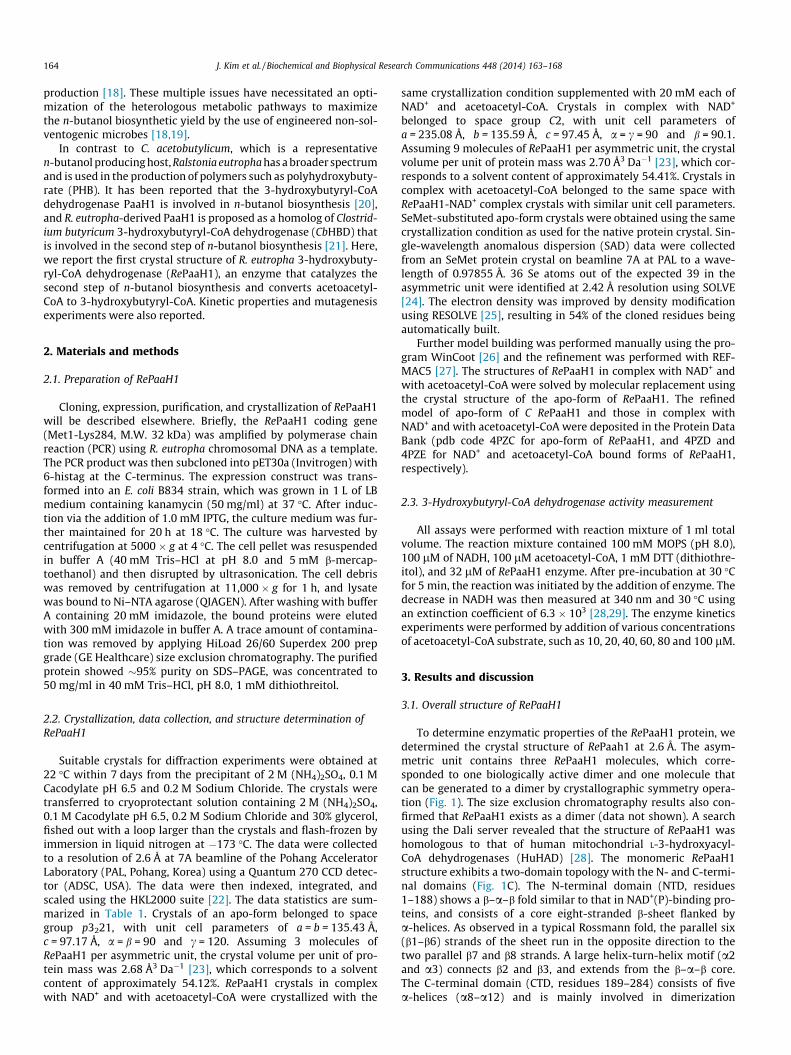
To determine enzymatic properties of the RePaaH1 protein, wedetermined the crystal structure of RePaah1 at 2.6 Å. The asym-metric unit contains three RePaaH1 molecules, which corre-sponded to one biologically active dimer and one molecule thatcan be generated to a dimer by crystallographic symmetry opera-tion (Fig. 1). The size exclusion chromatography results also con-firmed that RePaaH1 exists as a dimer (data not shown). A searchusing the Dali server revealed that the structure of RePaaH1 washomologous to that of human mitochondrial L-3-hydroxyacyl-CoA dehydrogenases (HuHAD) [28]. The monomeric RePaaH1structure exhibits a two-domain topology with the N- and C-termi-nal domains (Fig. 1C). The N-terminal domain (NTD, residues1–188) shows a b–a–b fold similar to that in NAD+(P)-binding pro-teins, and consists of a core eight-stranded b-sheet flanked bya-helices. As observed in a typical Rossmann fold, the parallel six(b1–b6) strands of the sheet run in the opposite direction to thetwo parallel b7 and b8 strands. A large helix-turn-helix motif (a2and a3) connects b2 and b3, and extends from the b–a–b core.The C-terminal domain (CTD, residues 189–284) consists of fivea-helices (a8–a12) and is mainly involved in dimerization
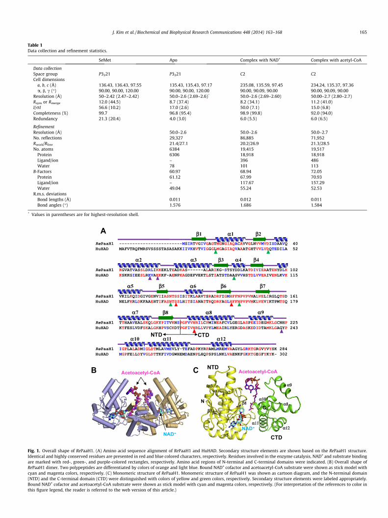
Table 1Data collection and refinement statistics.
SeMet Apo Complex with NAD+ Complex with acetyl-CoA
Data collectionSpace group P3221 P3221 C2 C2Cell dimensions
a, b, c (Å) 136.43, 136.43, 97.55 135.43, 135.43, 97.17 235.08, 135.59, 97.45 234.24, 135.37, 97.36a, b, c (�) 90.00, 90.00, 120.00 90.00, 90.00, 120.00 90.00, 90.09, 90.00 90.00, 90.09, 90.00
Resolution (Å) 50–2.42 (2.47–2.42) 50.0–2.6 (2.69–2.6)* 50.0–2.6 (2.69–2.60) 50.00–2.7 (2.80–2.7)Rsym or Rmerge 12.0 (44.5) 8.7 (37.4) 8.2 (34.1) 11.2 (41.0)I/rI 56.6 (10.2) 17.0 (2.6) 50.0 (7.1) 15.0 (6.8)Completeness (%) 99.7 96.8 (95.4) 98.9 (99.8) 92.0 (94.0)Redundancy 21.3 (20.4) 4.0 (3.0) 6.0 (5.5) 6.0 (6.5)
RefinementResolution (Å) 50.0–2.6 50.0–2.6 50.0–2.7No. reflections 29,327 86,885 71,952Rwork/Rfree 21.4/27.1 20.2/26.9 21.3/28.5No. atoms 6384 19,415 19,517
Protein 6306 18,918 18,918Ligand/ion – 396 486Water 78 101 113
B-Factors 60.97 68.94 72.05Protein 61.12 67.99 70.93Ligand/ion – 117.67 157.29Water 49.04 55.24 52.53
R.m.s. deviationsBond lengths (Å) 0.011 0.012 0.011Bond angles (�) 1.576 1.686 1.584
* Values in parentheses are for highest-resolution shell.
Fig. 1. Overall shape of RePaaH1. (A) Amino acid sequence alignment of RePaaH1 and HuHAD. Secondary structure elements are shown based on the RePaaH1 structure.Identical and highly conserved residues are presented in red and blue colored characters, respectively. Residues involved in the enzyme catalysis, NAD+ and substrate bindingare marked with red-, green-, and purple-colored rectangles, respectively. Amino acid regions of N-terminal and C-terminal domains were indicated. (B) Overall shape ofRePaaH1 dimer. Two polypeptides are differentiated by colors of orange and light blue. Bound NAD+ cofactor and acetoacetyl-CoA substrate were shown as stick model withcyan and magenta colors, respectively. (C) Monomeric structure of RePaaH1. Monomeric structure of RePaaH1 was shown as cartoon diagram, and the N-terminal domain(NTD) and the C-terminal domain (CTD) were distinguished with colors of yellow and green colors, respectively. Secondary structure elements were labeled appropriately.Bound NAD+ cofactor and acetoacetyl-CoA substrate were shown as stick model with cyan and magenta colors, respectively. (For interpretation of the references to color inthis figure legend, the reader is referred to the web version of this article.)
J. Kim et al. / Biochemical and Biophysical Research Communications 448 (2014) 163–168 165
A Arg52
NAD+ Lys56
Asn143
Ser119
Mol A
166 J. Kim et al. / Biochemical and Biophysical Research Communications 448 (2014) 163–168
(Fig. 1C). The a8 and a9 helices of each subunit mediate dimeriza-tion through hydrophobic interactions with residues such asVal188, Val189, Ile192, Leu193, Pro195, Met196, Val203, andL208. Positively charged resides are located along the cleft betweenNTD and CTD, and some of these mediate the binding of the ade-nine diphosphate moiety of the acetoacetyl-CoA substrate, whichwill be described later.
B
Acetoacetyl-CoA
Asn223His140
Asn190
RePaah1NTD
RePaah1CTD
HuHADNTD
HuHADCTD
Acetoacetyl-CoA
αα
α
α
α2
3
9
10
Mol B
Fig. 3. Substrate-binding mode of RePaaH1. (A) The RePaaH1 structure was shownas a cartoon model with gray and light orange colors for each of two monomers, andlabeled as Mol A and Mol B. The bound NAD+ and acetoacetyl-CoA were shown asstick models with cyan and magenta colors, respectively. Residues involved in theacetoacetyl-CoA stabilization through hydrogen bonds were shown as stick modelswith orange color, and labeled appropriately. (B) Comparison of domain shiftingbetween RePaaH1 and HuHAD upon the substrate binding. Structures of RePaaH1and HuHAD in complex with substrate were superposed based on the C-terminaldomains of the proteins. The N- and C-terminal domains of RePaaH1 were shownwith orange and red colors, respectively, and those of HuHAD were with light blue
3.2. RePaaH1-NAD+ cofactor complex
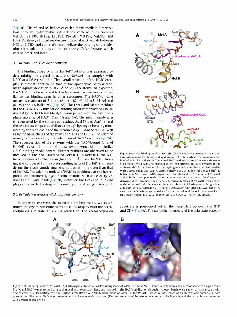
The binding property with the NAD+ cofactor was examined bydetermining the crystal structure of RePaaH1 in complex withNAD+ at a 2.6 Å resolution. The overall structure of the NAD+ com-plex is almost identical to that of the apoenzyme, with a root-mean-square deviation of 0.25 Å on 295 Ca atoms. As expected,the NAD+ cofactor is bound to the N-terminal Rossmann fold, sim-ilar to the binding seen in other structures. The NAD+-bindingpocket is made up of 5 loops (b1–a1, b2–a2, b4–a5, b5–a6 andb6–a7) and 1 a helix (a5) (Fig. 2A). The Thr13 and Met14 residuesin the G-x-G-x-x-G nucleotide-binding motif comprised of Gly10-Ala11-Gly12-Thr13-Met14-Gly15 were paired with the two phos-phate moieties of NAD+ (Figs. 1A and 3A). The nicotinamide ringis recognized by the conserved residues Asn117 and Asn143, andthe two ribose rings are stabilized through hydrogen bonding med-iated by the side chains of the residues Asp 33 and Ser119 as wellas by the main chains of the residues Ala38 and Glu92. The adeninemoiety is positioned by the side chain of Tyr77 residue (Fig. 2A).The superposition of the enzyme with the NAD+-bound form ofHuHAD reveals that although these two enzymes share a similarNAD+-binding mode, several distinct residues are observed to beinvolved in the NAD+-binding of RePaaH1. In RePaaH1, the a11helix position is further away (by about 3 Å) from the NAD+ bind-ing site compared to the corresponding helix of HuHAD, thus ren-dering the nicotinamide ring binding pocket more open than thatof HuHAD. The adenine moiety of NAD+ is positioned at the hydro-phobic cleft formed by hydrophobic residues such as Ile34, Tyr77,Ala90, Leu96 and Ile100 (Fig. 2B). However, the Tyr 77 residue alsoplays a role in the binding of this moiety through a hydrogen bond.
and green colors, respectively. The bound acetoacetyl-CoA substrate was presentedas a stick model with magenta color. (For interpretation of the references to color inthis figure legend, the reader is referred to the web version of this article.)
3.3. RePaaH1-acetoacetyl-CoA substrate complexIn order to examine the substrate-binding mode, we deter-mined the crystal structure of RePaaH1 in complex with the aceto-acetyl-CoA substrate at a 2.5 Å resolution. The acetoacetyl-CoA
Ala38
Tyr77
Met14Thr13
Asn117Ser119
Glu92
Asp33
NAD+
Asn143
A
Fig. 2. NAD+-binding mode of RePaaH1. (A) Cartoon presentation of NAD+-binding modeThe bound NAD+ was presented as a stick model with cyan color. Residues involved inorange color. (B) Electrostatic potential surface presentation of NAD+-binding mode ofpresentation. The bound NAD+ was presented as a stick model with cyan color. (For interweb version of this article.)
substrate is positioned within the deep cleft between the NTDand CTD (Fig. 3A). The pantothenic moiety of the substrate appears
NAD+
B
of RePaaH1. The RePaaH1 structure was shown as a cartoon model with gray color.the NAD+ stabilization through hydrogen bonds were shown as stick models withRePaaH1. The RePaaH1 structure was shown as an electrostatic potential surface
pretation of the references to color in this figure legend, the reader is referred to the
J. Kim et al. / Biochemical and Biophysical Research Communications 448 (2014) 163–168 167
to be stabilized mainly by the side chain of Asn143 as well as theside chain of Asn223 from the other subunit of dimer throughhydrogen-bond interactions. The acetoacetyl moiety is located atits binding pocket and positioned near the conserved catalytic res-idues Ser119, His140, and Asn190, which correspond to the Ser137,His158, and Asn208 residues of HuHAD (Fig. 3A). Interestingly,even though the two positively charged residues Arg52 andLys56 seemed to be involved in hydrogen bonding with the aden-osine moiety, the electron density map of the adenosine diphos-phate moiety was not clear, indicating that the adenosinediphosphate moiety was not fully stabilized.
It is reported that a significant conformational change in theNAD+-binding domain occurs when the domain rotates towardthe C-terminal domain upon the binding of the substrate in HuHAD(Fig. 3B) [30]. However, in RePaaH1, the positions of the twodomains in the acetoacetyl-CoA substrate complex structure were
0
20
40
60
80
0 25 50 75 100
0
0.01
0.02
0.03
0.04
0 0.025 0.05 0.075 0.1
0
50
100
150
200
250
WT R52A K56A S119A N140A
A
B
C
Enzy
me
activ
ity[m
M/m
in]
substrate [ M]
1/substrate [ Mμ -1]
1/V
Act
ivity
(%)
μ
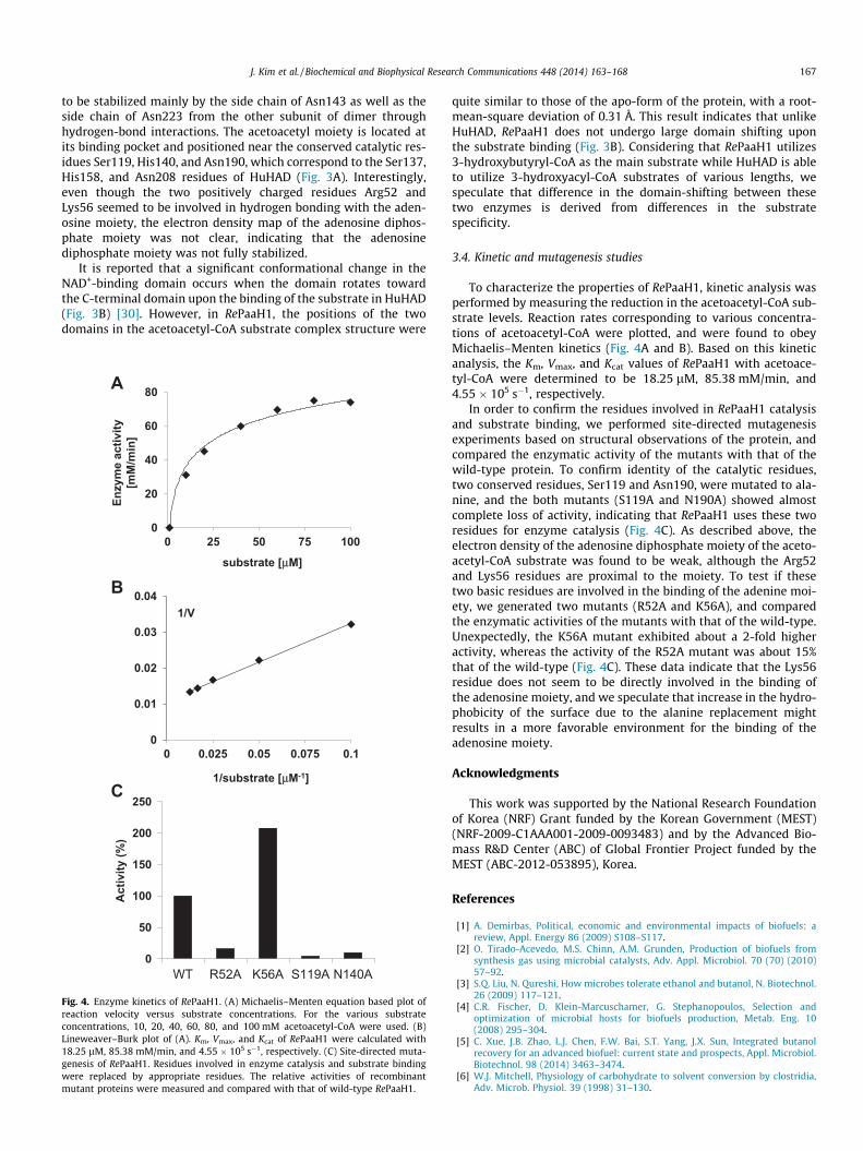
Fig. 4. Enzyme kinetics of RePaaH1. (A) Michaelis–Menten equation based plot ofreaction velocity versus substrate concentrations. For the various substrateconcentrations, 10, 20, 40, 60, 80, and 100 mM acetoacetyl-CoA were used. (B)Lineweaver–Burk plot of (A). Km, Vmax, and Kcat of RePaaH1 were calculated with18.25 lM, 85.38 mM/min, and 4.55 � 105 s�1, respectively. (C) Site-directed muta-genesis of RePaaH1. Residues involved in enzyme catalysis and substrate bindingwere replaced by appropriate residues. The relative activities of recombinantmutant proteins were measured and compared with that of wild-type RePaaH1.
quite similar to those of the apo-form of the protein, with a root-mean-square deviation of 0.31 Å. This result indicates that unlikeHuHAD, RePaaH1 does not undergo large domain shifting uponthe substrate binding (Fig. 3B). Considering that RePaaH1 utilizes3-hydroxybutyryl-CoA as the main substrate while HuHAD is ableto utilize 3-hydroxyacyl-CoA substrates of various lengths, wespeculate that difference in the domain-shifting between thesetwo enzymes is derived from differences in the substratespecificity.
3.4. Kinetic and mutagenesis studies
To characterize the properties of RePaaH1, kinetic analysis wasperformed by measuring the reduction in the acetoacetyl-CoA sub-strate levels. Reaction rates corresponding to various concentra-tions of acetoacetyl-CoA were plotted, and were found to obeyMichaelis–Menten kinetics (Fig. 4A and B). Based on this kineticanalysis, the Km, Vmax, and Kcat values of RePaaH1 with acetoace-tyl-CoA were determined to be 18.25 lM, 85.38 mM/min, and4.55 � 105 s�1, respectively.
In order to confirm the residues involved in RePaaH1 catalysisand substrate binding, we performed site-directed mutagenesisexperiments based on structural observations of the protein, andcompared the enzymatic activity of the mutants with that of thewild-type protein. To confirm identity of the catalytic residues,two conserved residues, Ser119 and Asn190, were mutated to ala-nine, and the both mutants (S119A and N190A) showed almostcomplete loss of activity, indicating that RePaaH1 uses these tworesidues for enzyme catalysis (Fig. 4C). As described above, theelectron density of the adenosine diphosphate moiety of the aceto-acetyl-CoA substrate was found to be weak, although the Arg52and Lys56 residues are proximal to the moiety. To test if thesetwo basic residues are involved in the binding of the adenine moi-ety, we generated two mutants (R52A and K56A), and comparedthe enzymatic activities of the mutants with that of the wild-type.Unexpectedly, the K56A mutant exhibited about a 2-fold higheractivity, whereas the activity of the R52A mutant was about 15%that of the wild-type (Fig. 4C). These data indicate that the Lys56residue does not seem to be directly involved in the binding ofthe adenosine moiety, and we speculate that increase in the hydro-phobicity of the surface due to the alanine replacement mightresults in a more favorable environment for the binding of theadenosine moiety.
Acknowledgments
This work was supported by the National Research Foundationof Korea (NRF) Grant funded by the Korean Government (MEST)(NRF-2009-C1AAA001-2009-0093483) and by the Advanced Bio-mass R&D Center (ABC) of Global Frontier Project funded by theMEST (ABC-2012-053895), Korea.
References
[1] A. Demirbas, Political, economic and environmental impacts of biofuels: areview, Appl. Energy 86 (2009) S108–S117.
[2] O. Tirado-Acevedo, M.S. Chinn, A.M. Grunden, Production of biofuels fromsynthesis gas using microbial catalysts, Adv. Appl. Microbiol. 70 (70) (2010)57–92.
[3] S.Q. Liu, N. Qureshi, How microbes tolerate ethanol and butanol, N. Biotechnol.26 (2009) 117–121.
[4] C.R. Fischer, D. Klein-Marcuschamer, G. Stephanopoulos, Selection andoptimization of microbial hosts for biofuels production, Metab. Eng. 10(2008) 295–304.
[5] C. Xue, J.B. Zhao, L.J. Chen, F.W. Bai, S.T. Yang, J.X. Sun, Integrated butanolrecovery for an advanced biofuel: current state and prospects, Appl. Microbiol.Biotechnol. 98 (2014) 3463–3474.
[6] W.J. Mitchell, Physiology of carbohydrate to solvent conversion by clostridia,Adv. Microb. Physiol. 39 (1998) 31–130.
168 J. Kim et al. / Biochemical and Biophysical Research Communications 448 (2014) 163–168
[7] M. Inui, M. Suda, S. Kimura, K. Yasuda, H. Suzuki, H. Toda, S. Yamamoto, S.Okino, N. Suzuki, H. Yukawa, Expression of Clostridium acetobutylicum butanolsynthetic genes in Escherichia coli, Appl. Microbiol. Biotechnol. 77 (2008)1305–1316.
[8] P. Durre, Fermentative butanol production: bulk chemical and biofuel, Ann.N.Y. Acad. Sci. 1125 (2008) 353–362.
[9] P. Durre, Biobutanol: an attractive biofuel, Biotechnol. J. 2 (2007) 1525–1534.[10] S.K. Lee, H. Chou, T.S. Ham, T.S. Lee, J.D. Keasling, Metabolic engineering of
microorganisms for biofuels production: from bugs to synthetic biology tofuels, Curr. Opin. Biotechnol. 19 (2008) 556–563.
[11] Y.S. Jang, J.M. Park, S. Choi, Y.J. Choi, Y. Seung do, J.H. Cho, S.Y. Lee, Engineeringof microorganisms for the production of biofuels and perspectives based onsystems metabolic engineering approaches, Biotechnol. Adv. 30 (2012) 989–1000.
[12] K.T. Tran, T. Maeda, T.K. Wood, Metabolic engineering of Escherichia coli toenhance hydrogen production from glycerol, Appl. Microbiol. Biotechnol. 98(2014) 4757–4770.
[13] P.I. Nikel, V. de Lorenzo, Robustness of Pseudomonas putida KT2440 as a hostfor ethanol biosynthesis, N. Biotechnol. (2014), http://dx.doi.org/10.1016/j.nbt2014.02.006 [Epub ahead of print].
[14] K.Y. Choi, D.G. Wernick, C.A. Tat, J.C. Liao, Consolidated conversion of proteinwaste into biofuels and ammonia using Bacillus subtilis, Metab. Eng. (2014),http://dx.doi.org/10.1016/j.ymben.2014.02.007 [Epub ahead of print].
[15] M. Scheel, T. Lutke-Eversloh, New options to engineer biofuel microbes:development and application of a high-throughput screening system, Metab.Eng. 17 (2013) 51–58.
[16] T. Ezeji, C. Milne, N.D. Price, H.P. Blaschek, Achievements and perspectives toovercome the poor solvent resistance in acetone and butanol-producingmicroorganisms, Appl. Microbiol. Biotechnol. 85 (2010) 1697–1712.
[17] D.T. Jones, D.R. Woods, Acetone-butanol fermentation revisited, Microbiol.Rev. 50 (1986) 484–524.
[18] S. Atsumi, A.F. Cann, M.R. Connor, C.R. Shen, K.M. Smith, M.P. Brynildsen, K.J.Chou, T. Hanai, J.C. Liao, Metabolic engineering of Escherichia coli for 1-butanolproduction, Metab. Eng. 10 (2008) 305–311.
[19] D.R. Nielsen, E. Leonard, S.H. Yoon, H.C. Tseng, C. Yuan, K.L. Prather,Engineering alternative butanol production platforms in heterologousbacteria, Metab. Eng. 11 (2009) 262–273.
[20] G.W. Haywood, A.J. Anderson, L. Chu, E.A. Dawes, The role of NADH- andNADPH-linked acetoacetyl-CoA reductases in the poly-3-hydroxybutyratesynthesizing organism Alcaligenes eutrophus, FEMS Microbiol. Lett. 52 (1988)259–264.
[21] H.B. Machado, Y. Dekishima, H. Luo, E.I. Lan, J.C. Liao, A selection platform forcarbon chain elongation using the CoA-dependent pathway to produce linearhigher alcohols, Metab. Eng. 14 (2012) 504–511.
[22] Z. Otwinowski, W. Minor, Processing of X-ray diffraction data collected inoscillation mode, Macromol. Crystallogr. 276 (Pt A) (1997) 307–326.
[23] B.W. Matthews, Solvent content of protein crystals, J. Mol. Biol. 33 (1968) 491–497.
[24] T.C. Terwilliger, J. Berendzen, Automated MAD and MIR structure solution,Acta Crystallogr. D Biol. Crystallogr. 55 (1999) 849–861.
[25] T.C. Terwilliger, Maximum-likelihood density modification, Acta Crystallogr. DBiol. Crystallogr. 56 (2000) 965–972.
[26] P. Emsley, K. Cowtan, Coot: model-building tools for molecular graphics, ActaCrystallogr. D Biol. Crystallogr. 60 (2004) 2126–2132.
[27] G.N. Murshudov, A.A. Vagin, E.J. Dodson, Refinement of macromolecularstructures by the maximum-likelihood method, Acta Crystallogr. D Biol.Crystallogr. 1 (1997) 240–255.
[28] J.J. Barycki, L.K. O’Brien, J.M. Bratt, R. Zhang, R. Sanishvili, A.W. Strauss, L.J.Banaszak, Biochemical characterization and crystal structure determination ofhuman heart short chain L-3-hydroxyacyl-CoA dehydrogenase provideinsights into catalytic mechanism, Biochemistry 38 (1999) 5786–5798.
[29] R.C. Taylor, A.K. Brown, A. Singh, A. Bhatt, G.S. Besra, Characterization of abeta-hydroxybutyryl-CoA dehydrogenase from Mycobacterium tuberculosis,Microbiology 156 (2010) 1975–1982.
[30] J.J. Barycki, L.K. O’Brien, A.W. Strauss, L.J. Banaszak, Sequestration of the activesite by interdomain shifting. Crystallographic and spectroscopic evidence fordistinct conformations of L-3-hydroxyacyl-CoA dehydrogenase, J. Biol. Chem.275 (2000) 27186–27196.


