Embed Size (px)
Citation preview

Eijsink and Mats SandgrenSamejima, Jerry Ståhlberg, Vincent G. H. Westereng, Kiyohiko Igarashi, MasahiroMichael F. Crowley, Svein J. Horn, Bjørge Christina M. Payne, Michael E. Himmel,Larsson, Takuya Ishida, Seonah Kim, Miao Wu, Gregg T. Beckham, Anna M. Phanerochaete chrysosporiumfrom the Basidiomycota Fungus Polysaccharide Monooxygenase GH61DCharacterization of the Lytic Crystal Structure and ComputationalEnzymology:
doi: 10.1074/jbc.M113.459396 originally published online March 22, 20132013, 288:12828-12839.J. Biol. Chem.
10.1074/jbc.M113.459396Access the most updated version of this article at doi:
.JBC Affinity SitesFind articles, minireviews, Reflections and Classics on similar topics on the
Alerts:
When a correction for this article is posted•
When this article is cited•
to choose from all of JBC's e-mail alertsClick here
Supplemental material:
http://www.jbc.org/content/suppl/2013/03/22/M113.459396.DC1.html
http://www.jbc.org/content/288/18/12828.full.html#ref-list-1
This article cites 72 references, 19 of which can be accessed free at
at NATL RENEWABLE ENERGY on July 31, 2013http://www.jbc.org/Downloaded from

Crystal Structure and Computational Characterization of theLytic Polysaccharide Monooxygenase GH61D from theBasidiomycota Fungus Phanerochaete chrysosporium*□S
Received for publication, February 6, 2013, and in revised form, March 15, 2013 Published, JBC Papers in Press, March 22, 2013, DOI 10.1074/jbc.M113.459396
Miao Wu‡, Gregg T. Beckham§¶1,2,3, Anna M. Larsson‡, Takuya Ishida�, Seonah Kim§1, Christina M. Payne**‡‡1,2,Michael E. Himmel**1, Michael F. Crowley**1, Svein J. Horn§§4, Bjørge Westereng§§4, Kiyohiko Igarashi�5,Masahiro Samejima�, Jerry Ståhlberg‡, Vincent G. H. Eijsink4, and Mats Sandgren‡6
From the ‡Department of Molecular Biology, Swedish University of Agricultural Sciences, P.O. Box 7026, SE-750 07 Uppsala,Sweden, the §National Bioenergy Center and **Biosciences Center, National Renewable Energy Laboratory, Golden, Colorado80401, the ¶Department of Chemical Engineering, Colorado School of Mines, Golden, Colorado 80401, the �Department ofBiomaterial Sciences, Graduate School of Agricultural and Life Sciences, University of Tokyo, 1-1-1 Yayoi, Bunkyo-ku, Tokyo 113-8657, Japan, the ‡‡Department of Chemical and Materials Engineering, University of Kentucky, Lexington, Kentucky 40506, and the§§Department of Chemistry, Biotechnology, and Food Science, Norwegian University of Life Sciences, N-1432 Ås, Norway
Background: Lytic polysaccharide monooxygenases (LPMOs) represent a recently discovered enzymatic route to cleavecarbohydrates.Results:We report the first basidiomycete LPMO structure and describe enzyme-cellulose interactions with simulation.Conclusion: We characterize the copper-containing active site and identify loops important for substrate recognition andbinding.Significance: This structure is the first LPMO from a model basidiomycete fungus that contains many LPMO genes.
Carbohydrate structures are modified and degraded in thebiosphere by a myriad of mostly hydrolytic enzymes. Recently,lytic polysaccharide mono-oxygenases (LPMOs) were discov-ered as a new class of enzymes for cleavage of recalcitrant poly-saccharides that instead employ an oxidative mechanism.LPMOs employ copper as the catalyticmetal and are dependenton oxygen and reducing agents for activity. LPMOs are found inmany fungi and bacteria, but to date no basidiomycete LPMOhas been structurally characterized. Here we present the three-dimensional crystal structure of the basidiomycete Phanero-chaete chrysosporium GH61D LPMO, and, for the first time,measure the product distribution of LPMOaction on a lignocel-lulosic substrate. The structure reveals a copper-bound activesite common to LPMOs, a collection of aromatic and polar res-idues near the binding surface thatmay be responsible for regio-selectivity, and substantial differences in loop structures nearthe binding face compared with other LPMO structures. Theactivity assays indicate that this LPMO primarily produces
aldonic acids. Last, molecular simulations reveal conforma-tional changes, including the binding of several regions to thecellulose surface, leading to alignment of three tyrosine residueson the binding face of the enzyme with individual cellulosechains, similar to what has been observed for family 1 carbohy-drate-binding modules. A calculated potential energy surfacefor surface translation indicates that P. chrysosporium GH61Dexhibits energy wells whose spacing seems adapted to the spac-ing of cellobiose units along a cellulose chain.
Nature employs mixtures of glycoside hydrolases (GHs)7 toconvert carbohydrate polymers found in plant, fungal, and algalcell walls to soluble sugars (1). Recently, a new class of enzymeswas discovered that uses copper-dependent oxidative pathwaysfor the cleavage of glycosidic linkages (2–5). These oxidativeenzymes, referred to here as lytic polysaccharide monooxyge-nases (LPMOs), have garnered significant interest because theyenhance degradation of recalcitrant polysaccharides, such aschitin and cellulose, when added to GH mixtures (6, 7). Vaaje-Kolstad et al. (2) first observed lytic activity of chitin-bindingprotein 21 (CBP21) from the bacterium Serratia marcescens on�-chitin, which produced soluble C1-oxidized chito-oligosac-charides (aldonic acids) in the presence of reductants. CBP21was originally classified as a family 33 carbohydrate-bindingmodule (CBM33), which are prevalent proteins in biomass-de-grading bacteria (8). Soon after, a CBM33 enzyme was charac-
* This work was supported in part by the Faculty for Natural Resources andAgriculture at the Swedish University of Agricultural Sciences through theresearch program MicroDrivE and by the Japan Society for the Promotionof Science (JSPS) through a fellowship (to T. I.).
□S This article contains supplemental Table S1, Figs. S1–S5, and Movie S1.The atomic coordinates and structure factors (code 4B5Q) have been deposited in
the Protein Data Bank (http://wwpdb.org/).1 Supported by the Department of Energy Office of the Biomass Program.2 Supported by Norwegian Research Council Grant 218425.3 To whom correspondence may be addressed: National Bioenergy Center,
National Renewable Energy Laboratory, 1617 Cole Blvd., Golden CO 80401.Tel.: 303-384-7806; E-mail: [email protected].
4 Supported in part by Norwegian Research Council Grants 193817, 196885,and 214613.
5 Supported by the Advanced Low Carbon Technology Research and Devel-opment Program of the Japan Science and Technology Agency.
6 To whom correspondence may be addressed. Tel.: 46-18-673179; Fax:46-18-536971; E-mail: [email protected].
7 The abbreviations used are: GH, glycoside hydrolase; LPMO, lytic polysac-charide monooxygenase; PDB, Protein Data Bank; CBM, carbohydrate-binding module; RMSD, root mean square deviation; RMSF, root meansquare fluctuations; PASC, phosphoric acid-swollen cellulose; MD, molec-ular dynamics; PES, potential energy surface.
THE JOURNAL OF BIOLOGICAL CHEMISTRY VOL. 288, NO. 18, pp. 12828 –12839, May 3, 2013Published in the U.S.A.
12828 JOURNAL OF BIOLOGICAL CHEMISTRY VOLUME 288 • NUMBER 18 • MAY 3, 2013 at NATL RENEWABLE ENERGY on July 31, 2013http://www.jbc.org/Downloaded from

terized that also produces soluble aldonic acids from cellulose(3). Similarities in the structures of CBM33s and family 61 GHs(GH61s) were noted when the structure of Hypocrea jecorinaGH61B was determined (9), including a conserved surface-lo-cated metal coordination site (2, 9). Shortly after the initialreport onCBP21 (2), it was shownby several groups thatGH61salso employ ametal-dependent oxidative pathway to cleave gly-cosidic bonds in cellulose (4, 5, 10–12). The consensus betweenCBM33 and GH61 activity to date is that enzymes from bothfamilies utilize copper as the catalytic metal (5, 13) and thatreducing agents, including cellobiose dehydrogenases (4, 11),ascorbate, reduced glutathione, gallate (2, 3, 5, 10, 12), or non-carbohydrate species present in biomass (6, 14), are required foractivity.To date, there are five LPMO structures from five different
fungal GH61s available:H. jecorinaGH61B (Protein Data Bank(PDB) code 2VTC) (9), Thielavia terrestris GH61E (PDB codes3EII and 3EJA) (6), Thermoascus aurantiacus GH61A (PDBcodes 2YET and 3ZUD) (5), andNeurospora crassa PMO-2 andPMO-3 (PDB codes 4EIR and 4EIS, respectively) (15). Addi-tionally, there are structures of four different CBM33 enzymesavailable: S. marcescens CBP21 (PDB codes 2LHS, 2BEM, and2BEN) (7, 16), Vibrio choleraeCBM33 (PDB code 2XWX) (17),Enterococcus faecalisCBM33 (PDB code 4A02) (18), and Burk-holderia pseudomallei CBM33 (PDB code 3UAM). The GH61structures all contain ametal ion in the putative catalytic centerof the enzyme, whereas not all CBM33 structures solved to datecontain ametal ion. The catalytic centers in all of these enzymesare embedded in a flat protein face containing aromatic andpolar residues for putative binding to the surfaces of celluloseand chitin. Aromatic residues are not usually dominating, butsome GH61s show arrangements similar to what is found onthe binding faces of family 1 CBMs (19–21). The flat catalyticbinding surfaces of LPMOs are putatively suited to cleave gly-cosidic linkages without decrystallizing polymer chains (13, 22,23), whereas endoglucanases, with a catalytic cleft, are thoughttomainly act onmore accessible, amorphous regions (24). Thismay explain why these two enzyme classes are synergistic (2).To date, most LPMOs have been found to oxidize the C1
position (2–5, 11, 12). However, oxidation of the C4 carbon inthe scissile bond to form a 4-keto-aldose moiety has beendescribed for an LPMO fromN. crassa (10). Oxidation has alsobeen suggested at the C6 carbon (5, 25). There is no generalconsensus yet on the spectrum of oxidative chemistry poten-tially employed by LPMOs, let alone the structural basis of theselectivity of oxidation. There is significant incentive to under-stand the structural basis of LPMO action because of theirobserved activity improvements to industrial mixtures (6).Because biomass-degrading enzyme mixtures remain a majorcost driver in production of biofuels (26, 27), including LPMOsin the industrial enzyme mixtures offers the potential for sig-nificant cost reductions for enzymatic hydrolysis of biomass.Thus, determining the LPMO mechanism of action, screeningLPMO activities from natural diversity, and enzyme engineer-ing for higher activity and stability are now under way (8).It is noteworthy that the knownGH61 structures andmost of
the recent progress on mechanism elucidation are withenzymes from ascomycete fungi. However, wood decomposi-
tion in nature is predominantly conducted by basidiomycetefungi (28, 29), which are broadly divided into brown rot andwhite rot fungi (28). Multiple putative and identified GH61genes have been found in genomes of both types (28–31), withthe number of genes appearing to be larger in white rot than inbrown rot fungi (28). It is thus of significant interest to studyLPMO structures from basidiomycete fungi. Phanerochaetechrysosporium, in particular, is one of the most extensivelystudied white rot fungi, and as such, its genome was the firstbasidomycete sequenced (31). Up to 17 putative P. chrysospo-rium genes encoding GH61 enzymes (PchGH61s) were initiallyidentified (31).We previously cloned the P. chrysosporiumGH61D gene and
expressed the protein, referred to here as PchGH61D (JointGenome Institute Protein ID: 4691 in Pichia pastoris) (12). Weshowed that PchGH61D is a copper-dependent LPMO withactivity onAvicel, filter paper, and phosphoric acid-swollen cel-lulose, which oxidizes at the C1 carbon. No soluble sugars oxi-dized at C4 or C6 were detected (12). In the present study, wepresent the crystal structure of PchGH61D, the first LPMOstructure from a basidiomycete fungus, and we use x-rayabsorption fine structure scanning to analyze metal binding.We conduct a structure-based alignment of the PchGH61Dstructure with other LPMOs to examine the conservation ofsurface and active site residues. Additionally, we show for thefirst time the profile of released products when an LPMOenzyme acts on a real biomass substrate, namely pretreatedspruce. Last, we use MD simulation to study aspects of theinteraction of PchGH61D with the cellulose surface. Overall,this study contributes to the expanding repertoire of LPMOstructures and identifies key interactions with the hydrophobicface of cellulose, which will aid in describing the mechanismand specificity of these important enzymes.
EXPERIMENTAL PROCEDURES
Protein Preparation and Crystallization—RecombinantPchGH61D was expressed in P. pastoris and purified usinghydrophobic interaction and ion exchange chromatographyafter endoglycosidase H treatment, as described previously(12). The purified protein solution was incubated with 10 mM
EDTA for 3 h and then diluted into 10 mM sodium acetatebuffer, pH 5.0, with 1 mM CuSO4 for 30 min. A PD-10 column(GEHealthcare) was used for buffer exchange to 10mM sodiumacetate buffer, pH 5.0. After buffer exchange, the protein wasconcentrated to 12 mg/ml using a VIVASPIN-6 centrifugalconcentrator (10,000 molecular weight cut-off polyethersul-fone membrane; Sigma-Aldrich).The initial search for crystallization conditions for
PchGH61D was done with sitting drop vapor diffusion tech-niques at 20 °C in a MRC2 well crystallization plate (HamptonResearch) using the JCSG� Suite sparse matrix screen (Qia-gen). Crystals for structure determination were obtained at20 °C with 2.1 M DL-malic acid, pH 7.0, as precipitant, mixed 1:1(v/v) with 12 mg/ml PchGH61D in 10 mM sodium acetate, pH5.0. Prior to data collection, crystals were soaked briefly in crys-tallization solution mixed with glycerol at 20% (v/v) final con-centration as cryoprotectant and then flash-frozen in liquidN2.
P. chrysosporium GH61D Structure and Dynamics
MAY 3, 2013 • VOLUME 288 • NUMBER 18 JOURNAL OF BIOLOGICAL CHEMISTRY 12829 at NATL RENEWABLE ENERGY on July 31, 2013http://www.jbc.org/Downloaded from

Data Collection and Structure Determination—The x-rayabsorption spectrum scan of a PchGH61D crystal was recordedby measuring the fluorescence signal during energy scan nearthe copper absorption edge, using the PyMCA program at theID23-1 beamline at the European Synchrotron Radiation Facil-ity (ESRF) (Grenoble, France). X-ray diffraction data were col-lected at beamline ID14-1 (ESRF) using a single PchGH61Dcrystal. The diffraction data set was reduced and scaled usingthe XDS program (32, 33) and the CCP4 program suite (33).Diffraction data to 1.75 Å resolution were used in the scalingand throughout structure refinement.The PchGH61D structure was solved by molecular replace-
ment using Phaser (34). The search model was a homologymodel of PchGH61D (12), based on the Thielavia terrestrisGH61E (PDB code 3EII) structure, built by the SWISS-MODELServer (35). REFMAC5 (36) was used for structure modelrefinements, andmanualmodel rebuilding was performedwithCoot (37), using maximum likelihood (�A) weighted 2Fo � Fcelectron density maps (38). For cross-validation and R and Rfreecalculations, 5% of the data was excluded from the structurerefinement (39). Solvent molecules were automatically addedusing the automatic water picking function in the ARP/wARPpackage (40). Picked water molecules were selected or dis-carded manually by visual inspection of the 2Fo � Fc electrondensity map. The copper ion bound in the active site was intro-duced at a final stage of the structure refinement. The coordi-nates for the final structure model and the structure factorshave been deposited in the PDB (41) with accession code 4B5Q.The search for similar structures was carried out using the
Dali server (42). The Lsqman program (43) in the Uppsala Soft-ware Factory suite was used to provide root mean square devi-ation (RMSD) values and structure comparison statistics (44).Coot was used for structural analysis (37), and MacPyMOL(Schrödinger, LLC) was used for the preparation of structuralfigures.Structure-based Sequence Alignment—Sequences of GH61
enzymes Phchr1�4691 (also known as PchGH61D), 41563,41650, 41123, 31049, 129325, 121193, 122129, and 10320 (thenumbers indicate Protein ID) were retrieved from the P. chrys-osporium version 2.0 genome database at the Department ofEnergy Joint Genome Institute (45). A structure-basedsequence alignment of the catalytic domains of GH61s withknown crystal structure (PchGH61D, PDB code 4B5Q;TteGH61E, 3EJA; NcrPMO-2, 4EIR; NcrPMO-3, 4EIS;HjeGH61B, 2VTC;TauGH61A, 3ZUD)wasmadewith the helpof Dali server constraints (42), to which the other PchGH61sequences were aligned using the MAFFT program (46) (sup-plemental Fig. S1). The secondary structure elements ofPchGH61Dwere assigned using the program STRIDE (47). Thesequence alignment tablewas edited in ESPript version 2.2 (48).PchGH61D Activity Assay—Degradation experiments with
PchGH61D were conducted using 0.1% phosphoric acid-swol-len cellulose (PASC), prepared as described (49), or 0.5% steam-exploded (225 °C, 10 min) and washed spruce wood chips (50)in 25mM sodium acetate, pH 5.3, as substrate. The enzyme andascorbic acid concentrations were 34 �g/ml and 1.5 mM,respectively. The reactions were incubated for 20 h at 50 °Cwith 900 rpm vertical shaking in an Eppendorf Thermo mixer
and then centrifuged at 21,000 � g for 3 min. The content ofsoluble oxidized oligosaccharides in the supernatants was ana-lyzed by high performance anion exchange chromatography, asdescribed previously (3, 51).Computational Study of PchGH61D-Cellulose Interactions—
To conduct classicalMD simulations of PchGH61Dwith a cop-per ion bound in the enzyme, the charge redistribution in theactive center upon copper binding was examined with elec-tronic structure calculations, as described in the supplementalmaterial and shown in supplemental Fig. S2. CHARMM (52)was used for all simulations. PchGH61D was placed on thehydrophobic face of cellulose 1� with the active site facing thecellulose surface, as shown in supplemental Fig. S2. The cellu-lose model was taken from a 10-ns equilibrated structure forcellulose 1� from previous work (23). We note that 10 ns waspreviously demonstrated to be a sufficient equilibration timefor studying cellulose surface behavior (53). The hydrophobicface of cellulose onwhich the protein was placed contains threecellodextrin chains, and the copper atom in the PchGH61Dactive site was placed directly above a glycosidic linkage on themiddle chain. In this orientation, Tyr-28 andTyr-198 align overthe middle chain on the crystal surface, and Tyr-75 aligns overthe edge chain. The orientation of the enzyme with respect tocellulose was chosen to be similar to that of the family 1 CBMfromH. jecorinaCel7A in that the enzymewas placed such thatTyr-28 and Tyr-198 align along a single chain. Shorter simula-tions were also conducted with PchGH61D rotated 180° in theopposite direction, which yielded similar results in terms ofhow the enzyme active site stabilized over the active site (datanot shown). Additional details related to the simulation setupandmethods can be found in the supplemental material. Last, apotential energy surface (PES) for the PchGH61D-celluloseinteraction was also constructed to examine the location of sta-ble energetic wells. This closely follows previous work con-ducted on the family 1 CBM (19, 54). Details of the PES con-struction can be found in the supplemental material.
RESULTS AND DISCUSSION
Overall Structure of PchGH61D—PchGH61D crystallized inspace group C2 with unit-cell parameters of a � 149.3 Å, b �37.5 Å, c� 79.8 Å and with a � angle of 117.4°. The asymmetricunit of the crystal contains two non-crystallographic symme-try-relatedmolecules (A and B) related by a 2-fold rotation axis,giving a Matthews coefficient of 2.0 (55). The structure wassolved by molecular replacement using a homology model ofPchGH61D (12) and was refined at 1.75 Å resolution. The finalPchGH61D structure model exhibits crystallographic R andRfree values of 18.6 and 22.3% and contains a total of 3,781 non-hydrogen atoms, including all 434 amino acid residues, twocopper atoms, one mannose residue (in chain A), two glycerolmolecules, and 366 water molecules. The amino acid residuesare numbered according to themature protein after signal pep-tide cleavage, starting with His-1. Statistics of diffraction dataand structure refinement are summarized in Table 1. TheRMSD values between all C� atompairs of the twomolecules inthe asymmetric unit is 0.11 Å.The overall fold of PchGH61D (Fig. 1A) is a �-sandwich fold
consisting of two �-sheets, formed by in total eight �-strands.
P. chrysosporium GH61D Structure and Dynamics
12830 JOURNAL OF BIOLOGICAL CHEMISTRY VOLUME 288 • NUMBER 18 • MAY 3, 2013 at NATL RENEWABLE ENERGY on July 31, 2013http://www.jbc.org/Downloaded from

One �-sheet, the front sheet in Fig. 1, includes the �1, �3, and�8 strands, whereas the other includes strands �4, �5, �9, and�10. Strand�2 is involved in forming both�-sheets, which packonto each other to form the core of the protein. The proposedcatalytic center of the enzyme is positioned on a flat surface onone side of the �-sandwich fold. The PchGH61D structureshows three extended loops, which are all involved in shapingthe potential substrate-binding surface. The L2 loop region(residues 17–57) includes two short �-helices. The long C-ter-minal loop (LC loop, residues 170–217) contains no secondarystructure elements. In the tip of this loop, the backbone atomsof residues 201–204 (Pro-201, Lys-202, Asn-203, and Phe-204)display much higher B factors (34.8, 40.0, 39.7, and 26.9 Å2,respectively) than the average B factor for the protein (16.7 Å2),indicating high flexibility in this region (Fig. 1, green). The third,shorter LS loop (residues 109–124) forms hydrophobic inter-actions with the C-terminal LC loop and contains a �-hairpinmotif (residues 110–117; �-strands �6 and �7). Residues 109–124 exhibit elevated B factors (average 24.6 Å2), indicating thatalso the LS loop is quite flexible. Cys-43 and Cys-163 form adisulfide bridge between the L2 loop and strand �10.PchGH61D contains two potentialN-linked glycosylation sites,Asn-173 and Asn-203. Both are located on the long C-terminalLC loop (shown in supplemental Fig. S1). Asn-203 is positionedin the more flexible region of the loop, close to the potentialsubstrate-binding surface. Previous results indicated that theprotein is indeedN-glycosylated, because the apparent size wasreduced upon treatment with endoglycosidase H (12). How-ever, there is no clear electron density at either site for theGlcNAc residue that should remain after deglycosylation withendoglycosidase H. On the other hand, one mannose residue
O-linked to Ser-11 is visible and included inmolecule A but notinmolecule B, where the density is too weak at the correspond-ing position (Fig. 1A). The electron density map of thePchGH61D structure does not show any indication of methyla-tion of theN�2 ofHis-1. Such post-translationalmodification isvisible in the electron densities of some GH61s that have beenstructurally described so far (5, 9, 15), all of which are fromfilamentous fungi. It may be possible that PchGH61D is notmethylated at His-1 because it has been expressed in yeast(P. pastoris). At this stage,we donot knowwhethermethylationoccurs when the enzyme is produced by P. chrysosporium itself.Nevertheless, it is notable that the non-methylated PchGH61Denzyme is active (see Ref. 12; see below). It thus seems thatmethylation of His-1 is not strictly necessary for GH61 activity.Notably highly active CBM33-type LPMOs are not methylated(16, 18).Like other known LPMO structures, PchGH61D exhibits a
flat putative binding surface in which the proposed catalyticcenter is embedded, as shown in detail in Fig. 1B. It is likely thatthe details of this binding face dictate catalytic specificity togiven carbon atoms in cellulose substrates as well as specificityto other polysaccharide substrates (2, 5, 12, 13, 18). Fig. 1Bhighlights several residues of potential interest, which are dis-cussed further below.Copper Binding and the Structure of the Catalytic Center in
PchGH61D—The x-ray absorption spectrumof the PchGH61Dcrystal shows a characteristic absorption edge at 8.9841 keV(Fig. 2), which indicates that the protein binds copper. Conse-quently, copper atoms were modeled in the catalytic centers ofboth protein molecules with full occupancy, based on thestrong positive peak in the Fo � Fcmap (Fig. 3A). The B factorsof the copper atoms in the final model are 14.8 and 16.2 Å2 formolecule A and B, respectively. These low B factors, which arein the same range as the B factors for the protein backbone,indicate that copper is strongly bound.In the final structure, there were unmodeled Fo � Fc electron
densities within 2.0 Å from the copper ion in both the A and Bmolecule, which may reflect movement of the metal ionbetween different putative reaction states, similar to what hasbeen suggested for TauGH61A (5). Published GH61 structuresshow water molecules, a peroxide ion, oxygen molecules, or asulfate ion in this position (5, 6, 9, 15). The corresponding spacein PchGH61D is occupied by contiguous electron density thatwas interpreted as a glycerol molecule. The glycerol moleculemight be stabilized in this position by hydrogen bonds to theside chains of Gln-158, His-149, and Tyr-75 (Fig. 3A).The copper-binding site in PchGH61D is a type II copper
center, which exhibits a hexacoordination. In the geometry, asquare planar coordination was created by nitrogen or nitro-gen/oxygen atoms (56, 57). In the PchGH61D structure, squarecoordination is provided by the main-chain amide group (2.1Å), N� (1.9 Å) of His-1, and N� of His-76 (2.0 Å), whereas thereis no ligand at the fourth coordination position. In HjeGH61B(PDB code 2VTC (9)), the corresponding position is occupiedby a water molecule. The hydroxyl group of Tyr-160 (2.8 Å)occupies one of the axial positions, whereas the other axial posi-tion is empty in the hexacoordination geometry. Protein atomsthus occupy four coordination positions, leaving two sites avail-
TABLE 1Diffraction data and refinement statistics for the PchGH61D structure(PDB code 4B5Q)
Data collectionBeamlinea ID14:EH1Wavelength (Å) 0.933Space group C2Unit cell dimensionsa, b, c (Å) 149.3, 37.5, 79.8�, �, � (°) 90.0, 117.4, 90.0
Rmerge (%)b,c 8.6 (56.3)I/�(I)b 10.9 (2.1)Completeness (%)b 98.5 (97.5)Multiplicityb 3.4 (3.3)
RefinementResolution(Å) 40.7–1.75 (1.84–1.75)Rwork/Rfree (%) 18.6/22.3RMSD, bonds (Å)d 0.007RMSD, angles (degrees)d 1.165No. of protein residues 434No. of water molecules 366No. of metal atoms 2Average B factorOverall (Å2)e 20.0Protein (Å2)e 16.7Metals (Å2)e 15.5Organic ligands (Å2)e 34.1Waters (Å2)e 26.3
Ramachandran outliersf (%) 0.8a Beamlines at the European Synchrotron Radiation Facility (Grenoble, France).b Values in parentheses are those for the highest resolution shell.c Rmerge � �hkl�i�I � �I���hkl�i�I�.d Data from Engh and Huber (71).e Calculated using MOLEMAN2 (72).f Calculated using a strict boundary Ramachandran definition given by Kleywegtand Jones (73).
P. chrysosporium GH61D Structure and Dynamics
MAY 3, 2013 • VOLUME 288 • NUMBER 18 JOURNAL OF BIOLOGICAL CHEMISTRY 12831 at NATL RENEWABLE ENERGY on July 31, 2013http://www.jbc.org/Downloaded from
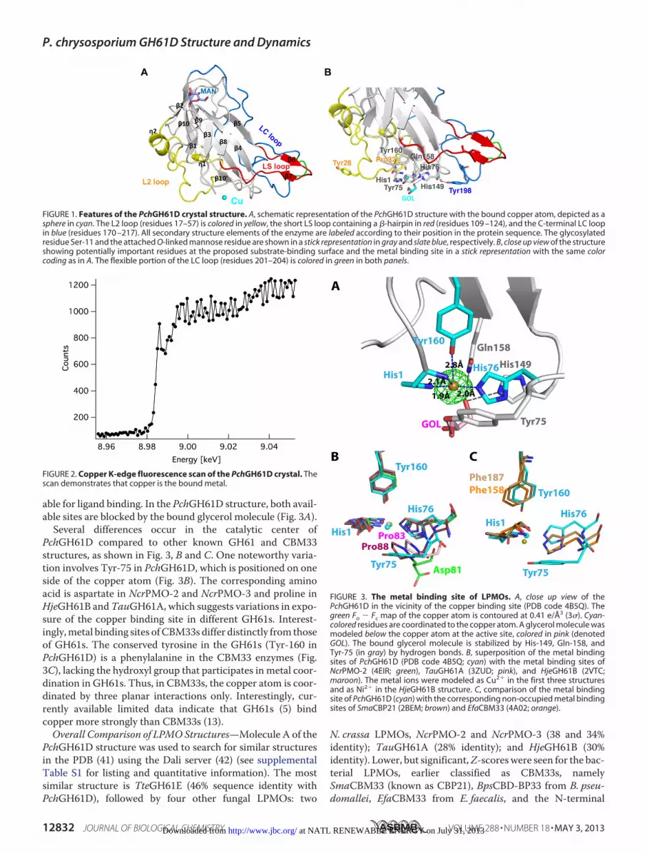
able for ligand binding. In the PchGH61D structure, both avail-able sites are blocked by the bound glycerol molecule (Fig. 3A).
Several differences occur in the catalytic center ofPchGH61D compared to other known GH61 and CBM33structures, as shown in Fig. 3, B and C. One noteworthy varia-tion involves Tyr-75 in PchGH61D, which is positioned on oneside of the copper atom (Fig. 3B). The corresponding aminoacid is aspartate in NcrPMO-2 and NcrPMO-3 and proline inHjeGH61B andTauGH61A, which suggests variations in expo-sure of the copper binding site in different GH61s. Interest-ingly,metal binding sites ofCBM33s differ distinctly from thoseof GH61s. The conserved tyrosine in the GH61s (Tyr-160 inPchGH61D) is a phenylalanine in the CBM33 enzymes (Fig.3C), lacking the hydroxyl group that participates inmetal coor-dination in GH61s. Thus, in CBM33s, the copper atom is coor-dinated by three planar interactions only. Interestingly, cur-rently available limited data indicate that GH61s (5) bindcopper more strongly than CBM33s (13).Overall Comparison of LPMO Structures—Molecule A of the
PchGH61D structure was used to search for similar structuresin the PDB (41) using the Dali server (42) (see supplementalTable S1 for listing and quantitative information). The mostsimilar structure is TteGH61E (46% sequence identity withPchGH61D), followed by four other fungal LPMOs: two
N. crassa LPMOs, NcrPMO-2 and NcrPMO-3 (38 and 34%identity); TauGH61A (28% identity); and HjeGH61B (30%identity). Lower, but significant,Z-scoreswere seen for the bac-terial LPMOs, earlier classified as CBM33s, namelySmaCBM33 (known as CBP21), BpsCBD-BP33 from B. pseu-domallei, EfaCBM33 from E. faecalis, and the N-terminal
FIGURE 1. Features of the PchGH61D crystal structure. A, schematic representation of the PchGH61D structure with the bound copper atom, depicted as asphere in cyan. The L2 loop (residues 17–57) is colored in yellow, the short LS loop containing a �-hairpin in red (residues 109 –124), and the C-terminal LC loopin blue (residues 170 –217). All secondary structure elements of the enzyme are labeled according to their position in the protein sequence. The glycosylatedresidue Ser-11 and the attached O-linked mannose residue are shown in a stick representation in gray and slate blue, respectively. B, close up view of the structureshowing potentially important residues at the proposed substrate-binding surface and the metal binding site in a stick representation with the same colorcoding as in A. The flexible portion of the LC loop (residues 201–204) is colored in green in both panels.
FIGURE 2. Copper K-edge fluorescence scan of the PchGH61D crystal. Thescan demonstrates that copper is the bound metal.
FIGURE 3. The metal binding site of LPMOs. A, close up view of thePchGH61D in the vicinity of the copper binding site (PDB code 4B5Q). Thegreen Fo � Fc map of the copper atom is contoured at 0.41 e/Å3 (3�). Cyan-colored residues are coordinated to the copper atom. A glycerol molecule wasmodeled below the copper atom at the active site, colored in pink (denotedGOL). The bound glycerol molecule is stabilized by His-149, Gln-158, andTyr-75 (in gray) by hydrogen bonds. B, superposition of the metal bindingsites of PchGH61D (PDB code 4B5Q; cyan) with the metal binding sites ofNcrPMO-2 (4EIR; green), TauGH61A (3ZUD; pink), and HjeGH61B (2VTC;maroon). The metal ions were modeled as Cu2� in the first three structuresand as Ni2� in the HjeGH61B structure. C, comparison of the metal bindingsite of PchGH61D (cyan) with the corresponding non-occupied metal bindingsites of SmaCBP21 (2BEM; brown) and EfaCBM33 (4A02; orange).
P. chrysosporium GH61D Structure and Dynamics
12832 JOURNAL OF BIOLOGICAL CHEMISTRY VOLUME 288 • NUMBER 18 • MAY 3, 2013 at NATL RENEWABLE ENERGY on July 31, 2013http://www.jbc.org/Downloaded from

domain of Gbp-A from V. cholera. For example, the structuralcomparison ofPchGH61DwithEfaCBM33 included only 95C�
atoms, with an RMSD value of 1.82Å (Z-value� 9.3). Note thatto date, there is no structural information for CBM33 domainsknown to act on cellulose. Fig. 4 shows structural superposi-tions of PchGH61D with the aforementioned nine differentfungal and bacterial LPMOs. The most prominent structuraldifferences are shown and indicate potential determinants ofbinding affinity and substrate specificity, as discussed below.At the metal binding site, His-149 and Gln-158 are highly
conserved in all fungal LPMOs. Their side chains point toward
the copper atom, but they are too far away to form coordinationinteractions (4.7 and 3.9 Å, respectively). His-149 is suitablypositioned to provide a hydrogen bond to any ligand binding toone of the two copper coordination positions that are availablefor substrate binding. In PchGH61D, the His-149 imidazolering is rotated 180° compared with its counterpart in otherstructures, probably because it makes a hydrogen bond to onehydroxyl of the glycerol ligand (Figs. 1B and 3A). One imidazolenitrogen is close to the side chain oxygen of Gln-158 (3.4 Å),and if rotated 180°, the distance would be even shorter (3.1 Å).This suggests an interaction between His-149 and Gln-158,
FIGURE 4. Structural comparison of LPMOs. A, superimposed structures of PchGH61D (gray) with other LPMOs (purple): NcrPMO2 (PDB code 4EIR); TteGH61E(3EJA); NcrPMO-3 (4EIS); TauGH61A (3ZUD); HjeGH61B (2VTC); BpsCBD-BP33 (3UAM); SmaCBP21 (2BEM); EfaCBD-CBM33 (4A02); and VchGlc-binding protein A(2XWM). Yellow, blue, and red regions correspond to the L2 loop, LC loop, and LS loop, respectively, in the PchGH61D structure. B, aromatic residues (Tyr-28,Tyr-75, and Tyr-198) on the flat substrate binding surface of PchGH61D are shown on the molecular surface in cyan. The corresponding residues or additionalaromatic residues on the surface of other GH61s are colored as follows. Pink, NcrPMO2 (PDB code 4EIR); red, TteGH61E (3EJA); yellow, NcrPMO-3 (4EIS); orange,TauGH61A (3ZUD); green, HjeGH61B (2VTC). The residue numbers are indicated beside the depicted residues. C, superposition of the residues shown in B withthe corresponding color, in a stick representation. Tyr-25 in NcrPMO-2 occurs in two conformations in pink.
P. chrysosporium GH61D Structure and Dynamics
MAY 3, 2013 • VOLUME 288 • NUMBER 18 JOURNAL OF BIOLOGICAL CHEMISTRY 12833 at NATL RENEWABLE ENERGY on July 31, 2013http://www.jbc.org/Downloaded from
although the geometry is far from ideal for a hydrogen bond.Gln-158 in turn interacts with the hydroxyl group of the con-servedTyr-160 that is axially coordinated to the copper atom. Itremains to be seen if these residues are conserved because theyparticipate directly in the catalytic mechanism or if their pri-mary role is to maintain the shape and electrostatic propertiesof the metal binding site.Structure and sequence comparisons (Fig. 4 and supplemen-
tal Fig. S1) show variations in the three loop regions that com-prise the putative substrate-binding surfaces in LPMOs. Gen-erally, the LC and LS loop regions are more extended in fungalLPMOs compared with the bacterial LPMOs (Fig. 4A). The L2loop varies within fungal LPMOs as well; PchGH61D,TteGH61E, and NcrPMO-2 have shorter L2 loops comparedwith HjeGH61B, TauGH61A, and NcrPMO-3 (Fig. 4A). Theonly conserved amino acid present in the L2 region is a cysteine,Cys-43 in PchGH61D, that forms a disulfide bond to Cys-163 inthe�10 strand. Despite the overall structural diversity shown inFig. 4A, there are similarities in the exposure of aromatic resi-dues that may impact binding, as highlighted in Fig. 4B. Theextended L2 loops in three of the GH61 structures containtyrosines (Tyr-23 inHjeGH61B; Tyr-24 inTauGH61A;Tyr-20/Tyr-24 inNcrPMO-3) that occupy spatially similar locations onthe surface as Tyr-28 in PchGH61D and Tyr-25 in NcrPMO-2(Fig. 4, B and C). It is noted that the L2 loop in NcrPMO-3actually contains two tyrosine residues side-by-side at this loca-tion. In PchGH61D, weak electron density and high B factorsindicate that the Tyr-28 side chain is flexible. Similar flexibilityis not seen for the tyrosines in theNcrPMO structures, possiblybecause they are involved in the crystal packing (15). InTteGH61E, Glu-23 replaces Tyr-28 of PchGH61D. Instead,TteGH61E has an additional exposed tyrosine, Tyr-192, next insequence to the highly conserved tyrosine in the LC loop, whichthe other enzymes do not have (Fig. 4B). Tyr-191 and Tyr-192form a similar substrate-binding motif in TteGH61E as presentin CBM1s (20).P. chrysosporium LPMO Comparison—Nine PchGH61s,
including PchGH61D, were chosen for inclusion in thesequence alignment (supplemental Fig. S1) because previousstudies had indicated that theymay be important for growth onlignocellulosic substrates (58–60). Six of the enzymes contain aC-terminal family 1 CBM (Phchr1�41563, 41650, 31049,129325, 121193, and 10320), whereas three do not(Phchr1�41123 and 122129 and PchGH61D). Only the pre-dicted catalytic domains were included in the sequence align-ment of supplemental Fig. S1. As with LPMO sequences acrossspecies, PchGH61s exhibit significant sequence variability.Three PchGH61s exhibit longer L2 loops (Phchr1�129325,121193, and 10320) similar to HjeGH61B, TauGH61A, andNcrPMO-3,whereas there is nomajor length variation in the LSor LC loop regions.With respect to the active site residues, the histidine residues
around the copper atom are conserved with the exception ofPhchr1�122129, which contains an arginine residue instead ofHis-76 (PchGH61D numbering). The axial tyrosine residue inPchGH61D, Tyr-160, is conserved with the exception ofPhchr1�41123, which contains a gap at this position. As in the
known LPMO structures, Gln-158 is completely conserved inall PchGH61s.
In terms of conspicuous residues on the putative PchGH61Dbinding surface (Fig. 4, B and C), five PchGH61s display aro-matic residues in sequence positions similar to Tyr-28, whereasPhchr1�41123, 31049, and 122129 donot. Residues correspond-ing to Tyr-75 vary considerably within PchGH61s as well andcan be aspartate, proline, glycine, alanine, or asparagines. Nota-bly, as discussed above (Fig. 3), Tyr-75 may indirectly affectcopper binding, so variation at this position may result in vari-ation in the active sites of PchGH61s (Fig. 3). The third surface-located aromatic residue, Tyr-198, is completely conservedamong PchGH61s.Degradation of PASC and Steam-exploded Spruce by
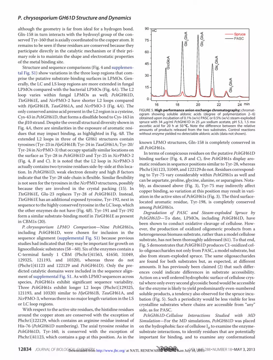
PchGH61D—To date, LPMOs, including PchGH61D, havebeen shown to conduct oxidative cleavage of cellulose. How-ever, the production of oxidized oligomeric products from aheterogeneous biomass substrate, rather than amodel cellulosesubstrate, has not been thoroughly addressed (61). To that end,Fig. 5 demonstrates that PchGH61D produces C1-oxidized cel-lo-oligosaccharides not only fromPASC, amodel substrate, butalso from steam-exploded spruce. The same oligosaccharidesare found for both substrates but, as expected, at differentamounts. It has previously been claimed (2) that such differ-ences could indicate differences in substrate accessibility.Action on a well ordered hydrophobic surface of cellulose crys-tal where only every second glycosidic bondwould be accessiblefor the enzyme is likely to yield predominantly even-numberedsoluble products, a tendency also observed for the spruce incu-bation (Fig. 5). Such a periodicity would be less visible for lesscrystalline substrates where chains are accessible from “any”side, as for PASC.PchGH61D-Cellulose Interactions Studied with MD
Simulation—For the MD simulations, PchGH61D was placedon the hydrophobic face of cellulose I� to examine the enzyme-substrate interactions, to identify residues that are potentiallyimportant for binding, and to examine any conformational
FIGURE 5. High performance anion exchange chromatography; chromat-ogram showing soluble aldonic acids (degree of polymerization 2– 6)obtained upon incubation of 0.1% (w/v) PASC or 0.5% (w/v) steam-explodedspruce with 34 �g/ml PchGH61D in 25 �M sodium acetate, pH 5.3, 1.5 mM
ascorbic acid for 20 h at 50 °C. Note the difference between the relativeamounts of products released from the two substrates. Control reactionswithout enzyme yielded no detectable aldonic acids (data not shown).
P. chrysosporium GH61D Structure and Dynamics
12834 JOURNAL OF BIOLOGICAL CHEMISTRY VOLUME 288 • NUMBER 18 • MAY 3, 2013 at NATL RENEWABLE ENERGY on July 31, 2013http://www.jbc.org/Downloaded from

changes that occur upon interaction with cellulose. The initialsystem geometry is shown in supplemental Fig. S3. It has notbeen determined if LPMOs bind to and perform oxidation onthe hydrophobic face of cellulose or chitin microfibrils. How-ever, it has been hypothesized (6, 9) that the aromatic and polarresidues on the flat surfaces exhibit structural similarities toTypeACBMs, several of which are known to bind to the hydro-phobic face of cellulose I (62, 63). Additionally, the orientationof LPMOs relative to the surface of cellulose is also currentlyunknown (15). Given the similarity of aromatic residues liningthe putative binding face, we aligned PchGH61D in an orienta-tion similar to the orientation of the family 1CBM fromH. jeco-rina (21, 64). AnMDsimulationwas conducted for 100 ns (sup-plementalMovie S1). Supplemental Fig. S4 shows the RMSD ofthe protein relative to the crystal structure, the root meansquare fluctuations (RMSF) per residue, and for comparison,the B factors of the PchGH61D crystal structure. SupplementalFig. S5 shows schematic representations of PchGH61D coloredby B factor and RMSF as well for further comparison. TheRMSD values indicate that after the initial equilibration of cel-lulose, conformational changes are minor, and the RMSFresults indicate that the primary fluctuations arose almost com-pletely from the LC and LS loops. The B factor results shown insupplemental Fig. S5, although not strictly comparable becausethe chemical environments of the crystal and simulatedPchGH61D enzymes are different, suggest that the LC and LSloops are the most flexible in both cases.Fig. 6A shows a cluster diagram of PchGH61D on the cellu-
lose surfacewith the protein backbone colored by RMSF, whichshows that there is significant flexibility in the LC and the LSloops. Fig. 6B shows the distance from the active site copper tothe hydrogen atom on the C1 carbon, which fluctuates near 5.0
Å during the MD simulation. Although the binding pose ofmolecular oxygen to copper is not yet known definitively forLPMO enzymes, a distance of 5 Å could most likely bring thesuperoxo intermediate that is hypothesized to be generated onthe copper (13) sufficiently close to abstract a hydrogen atomorconduct nucleophilic attack of the C1 carbon. Fig. 6, C and D,shows the initial and final states of PchGH61D on the cellulosesurface from two views. PchGH61D is apparently a C1 oxidizer,but further studies on the catalytic mechanism and on LPMOswith other oxidation preferences are needed to understand thespecificity for C1, C4, or possible C6 oxidation.During the simulation, the LC loop becomes quite mobile at
�30 ns (supplemental Fig. S4A and Fig. 6A). This conforma-tional flexibility is related to a conformational change in theside chains of Phe-112 (in another relatively flexible region) andPhe-204 relative to one another during the MD simulation.This observation agrees with the structural data presentedabove, showing that part of the LC loop exhibits significantlyhigher B factors than the rest of the protein (supplemental Figs.S4C and S5). Additionally, concomitantly with the Phe-112conformational change, the LS loop undergoes a significanttranslational motion toward the cellulose surface, where itforms hydrogen bonds to an edge cellodextrin chain via bothbackbone and side chain atoms.From the MD simulation, we examined the role of the three
tyrosine residues (Tyr-28, Tyr-75, and Tyr-198) that may beimportant for binding, we looked for other conspicuous resi-dues in the putative binding face, and we studied the active siteposition over cellulose. Table 2 lists the average interactionenergy of relevant protein residues with the cellulose surface.The energetic cut-off for examining interactionswas 3 kcal/molon average over the 100-ns MD simulation. As shown, many of
FIGURE 6. Simulation results for PchGH61D on the hydrophobic surface of cellulose. A, cluster view of PchGH61D with snapshots taken every 5 ns, coloredby RMSF from blue (low) to red (high). The tyrosine side chains (Tyr-28, Tyr-75, and Tyr-198) are shown in pink stick format in the conformation obtained after 100ns. B, the copper (shown as a cyan sphere) fluctuates at �5 Å from the hydrogen atom on the C1 carbon during the MD simulation. C, side view of PchGH61D onthe cellulose surface at t � 0 ns and t � 100 ns. The loops are colored as in Fig. 1. D, back view of PchGH61D on the cellulose surface at t � 0 ns and t � 100 ns.
P. chrysosporium GH61D Structure and Dynamics
MAY 3, 2013 • VOLUME 288 • NUMBER 18 JOURNAL OF BIOLOGICAL CHEMISTRY 12835 at NATL RENEWABLE ENERGY on July 31, 2013http://www.jbc.org/Downloaded from

the residues that form the putative cellulose binding face in thecrystal structure are present in the interaction energy analysis.However, some residues that are not initially bound to the cel-lulose also appear. Notably, the LS loop that contains Asn-114,Gly-115, and Gln-116 forms long lived contacts with the cellu-lose surface.Fig. 7A shows the positions of the three tyrosine residues
and the PchGH61D active site over the cellulose surface.Upon docking of PchGH61D on the cellulose surface, Tyr-28and Tyr-198 align over the same chain, and their position isquite stable during the simulation. Tyr-198 hydrogen-bondswith the adjacent chain as well during the simulation. Addi-tionally, Tyr-75 is bound to the adjacent cellulose chain onthe edge of the crystal upon docking, and it retains this con-formation over 100 ns. The active site position (defined asthe center of mass of His-1, His-76, Tyr-160, and the copperatom) remains in the same position over the cellulose surfaceduring theMD simulation directly above a glycosidic bond inthe middle chain. The results obtained here suggest that,overall, the active site is stable near the proposed site ofattack.Also, a PES was constructed for the PchGH61D-cellulose
interaction, as described in the supplementalmaterial. The PESwas constructed with explicit solvation using methods similarto those used in previous work conducted on a family 1 CBM(19, 54). The PES in Fig. 7B suggests that the enzyme is enthal-pically stable above glycosidic linkages separated by �10 Å,where the enzyme can abstract accessible hydrogen atoms. Thestabilization every 10 Å, which is approximately the length of acellobiose unit, is similar to that observed for a family 1 CBM(19, 54).We note that this study is limited toMD simulations of the
PchGH61D over the putative site of attack, which does notaccount for diffusion on the surface or the chemical reaction.In a previous study to examine the diffusion and orienta-tional preferences of a family 1 CBM on cellulose, diffusionand orientation on cellulose required 43 �s to reach conver-gence (21). Because LPMOs are substantially larger, under-standing their orientational preferences and studying theirdiffusion along the surface would probably require simula-tion times on the order of hundreds of �s without the use ofenhanced sampling methods. Thus, the questions ofPchGH61D diffusion and orientation on the cellulose surfaceare outside the scope of the present study. Additionally, we
note that LPMOs generally present a challenge to typicalprotein simulations because of the active site. The approachused here wherein an ad hoc potential was developed may begeneralized to other copper monooxygenases, but we stressthat the potential developed here is only appropriate forPchGH61D and only then for examining questions for whichcopper ion diffusion out of the active site is not relevant.Quantum mechanics/molecular mechanics approaches willbe necessary to study the reaction mechanism and questionsrelated to binding of other metals in the active site.
CONCLUSIONS
Here, we have solved the first LPMO structure from abasidiomycete fungus, the wood-degrading model organism,P. chrysosporium. This fungus contains up to nine LPMOsknown to be expressed when grown on lignocellulosic sub-strates (12, 59). Because extensive work has been done onother model glycoside hydrolase (65–68) and dehydrogen-ase enzymes (69, 70) from P. chrysosporium, the P. chrys-osporium LPMOs offer an excellent model system to under-stand the need for multiple oxidative activities to degradebiomass. The PchGH61D structure revealed potentiallyimportant residues around the active site and putative bind-ing surface that may impart differences in LPMO specificityand activity. With simulation, we demonstrated that severalconformational changes occur upon PchGH61D binding tocellulose, which suggest roles of conserved loops in substratebinding. Going forward in the burgeoning field of LPMObiochemistry, it is likely that a combination of structural,biophysical, and computational studies, such as thatpresented here, will be necessary to fully understand thediversity of LPMO structures as well as its functionalconsequences.
TABLE 2Residues that interact with cellulose in the MD simulations
ResidueAverage interaction
energy
kcal/molHis-1 �5.42Tyr-28 �10.86Ser-29 �6.79Tyr-75 �10.17Asn-114 �3.81Gly-115 �3.21Gln-116 �7.40His-149 �4.56Val-150 �5.23Tyr-198 �9.50Asn-199 �5.03
FIGURE 7. A, histogram of the tyrosine residues (Tyr-28, Tyr-75, and Tyr-198)and the PchGH61D active site positions on the cellulose surface. The bottomtwo layers of cellulose are not shown, and the cellulose chains are truncated,both for visual clarity. The color code denotes the position on a 0.1 � 0.1-Ågrid on the cellulose surface, with red being low density and blue being thehighest density. B, PES of PchGH61D on cellulose. The x direction is along thechains of cellulose, and the y direction is perpendicular to the cellulose chains.Energy minima are found over the putative site of attack, with �10-Å separa-tion (i.e. a distance corresponding to a cellobiose unit).
P. chrysosporium GH61D Structure and Dynamics
12836 JOURNAL OF BIOLOGICAL CHEMISTRY VOLUME 288 • NUMBER 18 • MAY 3, 2013 at NATL RENEWABLE ENERGY on July 31, 2013http://www.jbc.org/Downloaded from

Acknowledgments—Computer time for this research was provided bythe National Renewable Energy Laboratory Computational SciencesCenter, supported by the Department of Energy Office of Energy Effi-ciency andRenewable Energy underContractDE-AC36-08GO28308,and by the National Institute of Computational Science Kraken clus-ter under National Science Foundation XSEDE Grant MCB090159.
REFERENCES1. Cantarel, B. L., Coutinho, P. M., Rancurel, C., Bernard, T., Lombard, V.,
and Henrissat, B. (2009) The Carbohydrate-Active EnZymes database(CAZy). An expert resource for glycogenomics. Nucleic Acids Res. 37,D233–D238
2. Vaaje-Kolstad, G., Westereng, B., Horn, S. J., Liu, Z., Zhai, H., Sørlie, M.,and Eijsink, V. G. (2010) An oxidative enzyme boosting the enzymaticconversion of recalcitrant polysaccharides. Science 330, 219–222
3. Forsberg, Z., Vaaje-Kolstad, G., Westereng, B., Bunæs, A. C., Stenstrøm,Y.,MacKenzie, A., Sørlie,M., Horn, S. J., and Eijsink, V. G. (2011) Cleavageof cellulose by a CBM33 protein. Protein Sci. 20, 1479–1483
4. Phillips, C. M., Beeson, W. T., Cate, J. H., and Marletta, M. A. (2011)Cellobiose dehydrogenase and a copper-dependent polysaccharide mo-nooxygenase potentiate cellulose degradation by Neurospora crassa. ACSChem. Biol. 6, 1399–1406
5. Quinlan, R. J., Sweeney, M. D., Lo Leggio, L., Otten, H., Poulsen, J.-C.,Johansen, K. S., Krogh, K. B., Jørgensen, C. I., Tovborg,M., Anthonsen, A.,Tryfona, T., Walter, C. P., Dupree, P., Xu, F., Davies, G. J., and Walton,P.H. (2011) Insights into the oxidative degradation of cellulose by a coppermetalloenzyme that exploits biomass components. Proc. Natl. Acad. Sci.108, 15079–15084
6. Harris, P. V., Welner, D., McFarland, K. C., Re, E., Navarro Poulsen, J. C.,Brown, K., Salbo, R., Ding, H., Vlasenko, E., Merino, S., Xu, F., Cherry, J.,Larsen, S., and Lo Leggio, L. (2010) Stimulation of lignocellulosic biomasshydrolysis by proteins of glycoside hydrolase family 61. Structure andfunction of a large, enigmatic family. Biochemistry 49, 3305–3316
7. Vaaje-Kolstad, G., Horn, S. J., van Aalten, D. M., Synstad, B., and Eijsink,V. G. (2005) The non-catalytic chitin-binding protein CBP21 from Serra-tia marcescens is essential for chitin degradation. J. Biol. Chem. 280,28492–28497
8. Horn, S. J., Vaaje-Kolstad, G., Westereng, B., and Eijsink, V. G. (2012)Novel enzymes for the degradation of cellulose. Biotechnol. Biofuels 5, 45
9. Karkehabadi, S., Hansson, H., Kim, S., Piens, K.,Mitchinson, C., and Sand-gren, M. (2008) The structure of a glycoside hydrolase family 61 member,Cel61B from the Hypocrea jecorina. J. Mol. Biol. 383, 144–154
10. Beeson, W. T., Phillips, C. M., Cate, J. H., and Marletta, M. A. (2012)Oxidative cleavage of cellulose by fungal copper-dependent polysaccha-ride monooxygenases. J. Am. Chem. Soc. 134, 890–892
11. Langston, J. A., Shaghasi, T., Abbate, E., Xu, F., Vlasenko, E., and Sweeney,M. D. (2011) Oxidoreductive cellulose depolymerization by the enzymescellobiose dehydrogenase and glycoside hydrolase 61. Appl. Environ. Mi-crobiol. 77, 7007–7015
12. Westereng, B., Ishida, T., Vaaje-Kolstad, G., Wu, M., Eijsink, V. G., Iga-rashi, K., Samejima, M., Ståhlberg, J., Horn, S. J., and Sandgren, M. (2011)The putative endoglucanase PcGH61D from Phanerochaete chrysospo-rium is a metal-dependent oxidative ezyme that cleaves cellulose. PLoSONE 6, e27807
13. Aachmann, F. L., Sørlie, M., Skjåk-Bræk, G., Eijsink, V. G., and Vaaje-Kolstad, G. (2012) NMR structure of a lytic polysaccharide monooxyge-nase provides insight into copper binding, protein dynamics, and sub-strate interactions. Proc. Natl. Acad. Sci. U.S.A. 109, 18779–18784
14. Dimarogona, M., Topakas, E., Olsson, L., and Christakopoulos, P. (2012)Lignin boosts the cellulase performance of a GH61–61 enzyme from Spo-rotrichum thermophile. Bioresour. Technol. 110, 480–487
15. Li, X., Beeson, W. T., 4th, Phillips, C. M., Marletta, M. A., and Cate, J. H.(2012) Structural basis for substrate targeting and catalysis by fungal poly-saccharide monooxygenases. Structure 20, 1051–1061
16. Vaaje-Kolstad, G., Houston, D. R., Riemen, A. H., Eijsink, V. G., and vanAalten, D. M. (2005) Crystal structure and binding properties of the Ser-
ratia marcescens chitin-binding protein CBP21. J. Biol. Chem. 280,11313–11319
17. Wong, E., Vaaje-Kolstad, G., Ghosh, A., Hurtado-Guerrero, R., Konarev,P. V., Ibrahim, A. F., Svergun, D. I., Eijsink, V. G., Chatterjee, N. S., and vanAalten, D. M. (2012) The Vibrio cholerae colonization factor GbpA pos-sesses a modular structure that governs binding to different host surfaces.PLoS Pathog. 8, e1002373
18. Vaaje-Kolstad, G., Bøhle, L. A., Gåseidnes, S., Dalhus, B., Bjørås, M., Ma-thiesen, G., and Eijsink, V. G. (2012) Characterization of the chitinolyticmachinery of Enterococcus faecalis V583 and high-resolution structure ofits oxidative CBM33 enzyme. J. Mol. Biol. 416, 239–254
19. Beckham, G. T., Matthews, J. F., Bomble, Y. J., Bu, L., Adney, W. S., Him-mel, M. E., Nimlos, M. R., and Crowley, M. F. (2010) Identification ofamino acids responsible for processivity in a family 1 carbohydrate-bind-ing module from a fungal cellulase. J. Phys. Chem. B 114, 1447–1453
20. Kraulis, J., Clore, G.M., Nilges,M., Jones, T. A., Pettersson, G., Knowles, J.,and Gronenborn, A. M. (1989) Determinationn of the 3-dimensional so-lution structure of the C-terminal domain of cellobiohydrolase I fromTrichoderma reesei-A study using nuclear magnetic-resonance and hy-brid distance geometry dynamical simulated annealing. Biochemistry 28,7241–7257
21. Nimlos, M. R., Beckham, G. T., Matthews, J. F., Bu, L., Himmel, M. E., andCrowley,M. F. (2012) Binding preferences, surface attachment, diffusivity,and orientation of a family 1 carbohydrate-binding module on cellulose.J. Biol. Chem. 287, 20603–20612
22. Beckham, G. T., and Crowley, M. F. (2011) Examination of the �-chitinstructure and decrystallization thermodynamics at the nanoscale. J. Phys.Chem. B 115, 4516–4522
23. Beckham, G. T., Matthews, J. F., Peters, B., Bomble, Y. J., Himmel, M. E.,and Crowley, M. F. (2011) Molecular-level origins of biomass recalci-trance. Decrystallization free energies for four common cellulose poly-morphs. J. Phys. Chem. B 115, 4118–4127
24. Kleywegt, G. J., Zou, J. Y., Divne, C., Davies, G. J., Sinning, I., Stahlberg, J.,Reinikainen, T., Srisodsuk, M., Teeri, T. T., and Jones, T. A. (1997) Thecrystal structure of the catalytic core domain of endoglucanase I fromTrichoderma reesei at 3.6 Å resolution, and a comparison with relatedenzymes. J. Mol. Biol. 272, 383–397
25. Bey,M., Zhou, S., Poidevin, L., Henrissat, B., Coutinho, P.M., Berrin, J. G.,and Sigoillot, J. C. (2012) Comparison of two lytic polysaccharide mo-nooxygenases (GH61) from Podospora anserina reveals differences uponcello-oligosaccharides oxidation. Appl. Environ. Microbiol. 287,3147–3155
26. Chundawat, S. P., Beckham, G. T., Himmel, M. E., and Dale, B. E. (2011)Deconstruction of lignocellulosic biomass to fuels and chemicals. Annu.Rev. Chem. Biomol. Eng. 2, 121–145
27. Himmel, M. E., Ding, S. Y., Johnson, D. K., Adney, W. S., Nimlos, M. R.,Brady, J. W., and Foust, T. D. (2007) Biomass recalcitrance. Engineeringplants and enzymes for biofuels production. Science 315, 804–807
28. Eastwood, D. C., Floudas, D., Binder, M., Majcherczyk, A., Schneider, P.,Aerts, A., Asiegbu, F.O., Baker, S. E., Barry, K., Bendiksby,M., Blumentritt,M., Coutinho, P. M., Cullen, D., de Vries, R. P., Gathman, A., Goodell, B.,Henrissat, B., Ihrmark, K., Kauserud, H., Kohler, A., LaButti, K., Lapidus,A., Lavin, J. L., Lee, Y. H., Lindquist, E., Lilly, W., Lucas, S., Morin, E.,Murat, C., Oguiza, J. A., Park, J., Pisabarro, A. G., Riley, R., Rosling, A.,Salamov, A., Schmidt, O., Schmutz, J., Skrede, I., Stenlid, J., Wiebenga, A.,Xie, X., Kües, U., Hibbett, D. S., Hoffmeister, D., Högberg, N., Martin, F.,Grigoriev, I. V., Watkinson, S. C. (2011) The plant cell wall-decomposingmachinery underlies the functional diversity of forest fungi. Science 333,762–765
29. Fernandez-Fueyo, E., Ruiz-Duenas, F. J., Ferreira, P., Floudas, D., Hibbett,D. S., Canessa, P., Larrondo, L. F., James, T. Y., Seelenfreund, D., Lobos, S.,Polanco, R., Tello, M., Honda, Y., Watanabe, T., Watanabe, T., Ryu, J. S.,San, R. J., Kubicek, C. P., Schmoll, M., Gaskell, J., Hammel, K. E., St John,F. J., VandenWymelenberg, A., Sabat, G., Splinter BonDurant. S., Syed, K.,Yadav, J. S., Doddapaneni, H., Subramanian, V., Lavín, J. L., Oguiza, J. A.,Perez, G., Pisabarro, A. G., Ramirez, L., Santoyo, F., Master, E., Coutinho,P. M., Henrissat, B., Lombard, V., Magnuson, J. K., Kües, U., Hori, C.,Igarashi, K., Samejima, M., Held, B. W., Barry, K. W., LaButti, K. M.,
P. chrysosporium GH61D Structure and Dynamics
MAY 3, 2013 • VOLUME 288 • NUMBER 18 JOURNAL OF BIOLOGICAL CHEMISTRY 12837 at NATL RENEWABLE ENERGY on July 31, 2013http://www.jbc.org/Downloaded from

Lapidus, A., Lindquist, E. A., Lucas, S. M., Riley, R., Salamov, A. A.,Hoffmeister, D., Schwenk, D., Hadar, Y., Yarden, O., de Vries, R. P.,Wiebenga, A., Stenlid, J., Eastwood, D., Grigoriev, I. V., Berka, R. M.,Blanchette, R. A., Kersten, P., Martinez, A. T., Vicuna, R., and Cullen, D.(2012) Comparative genomics of Ceriporiopsis subvermispora and Phan-erochaete chrysosporium provide insight into selective ligninolysis. Proc.Natl. Acad. Sci. U.S.A. 109, 5458–5463
30. Olson, A., Aerts, A., Asiegbu, F., Belbahri, L., Bouzid, O., Broberg, A.,Canback, B., Coutinho, P. M., Cullen, D., Dalman, K., Deflorio, G., vanDiepen, L. T., Dunand, C., Duplessis, S., Durling, M., Gonthier, P., Grim-wood, J., Fossdal, C. G., Hansson, D., Henrissat, B., Hietala, A., Himmel-strand, K., Hoffmeister, D., Högberg, N., James, T. Y., Karlsson, M.,Kohler, A., Kües, U., Lee, Y.H., Lin, Y. C., Lind,M., Lindquist, E., Lombard,V., Lucas, S., Lundén, K.,Morin, E.,Murat, C., Park, J., Raffaello, T., Rouzé,P., Salamov, A., Schmutz, J., Solheim, H., Ståhlberg, J., Vélëz, H., de Vries,R. P.,Wiebenga, A.,Woodward, S., Yakovlev, I., Garbelotto,M.,Martin, F.,Grigoriev, I. V., and Stenlid, J. (2012) Insight into trade-off between wooddecay and parasitism from the genome of a fungal forest pathogen. NewPhytol. 194, 1001–1013
31. Martinez, D., Larrondo, L. F., Putnam,N., Gelpke,M.D., Huang, K., Chap-man, J., Helfenbein, K. G., Ramaiya, P., Detter, J. C., Larimer, F., Coutinho,P. M., Henrissat, B., Berka, R., Cullen, D., and Rokhsar D. (2004) Genomesequence of the lignocellulose degrading fungusPhanerochaete chrysospo-rium strain RP78. Nat. Biotechnol. 22, 695–700
32. Kabsch,W. (1993) Automatic Processing of rotation diffraction data fromcrystals of initially unknown symmetry and cell constants. J. Appl. Crys-tallogr. 26, 795–800
33. Potterton, E., Briggs, P., Turkenburg, M., and Dodson, E. (2003) A graph-ical user interface to the CCP4 program suite. Acta Crystallogr. D Biol.Crystallogr. 59, 1131–1137
34. McCoy, A. J., Grosse-Kunstleve, R. W., Adams, P. D., Winn, M. D., Sto-roni, L. C., and Read, R. J. (2007) Phaser crystallographic software. J. Appl.Crystallogr. 40, 658–674
35. Arnold, K., Bordoli, L., Kopp, J., and Schwede, T. (2006) The SWISS-MODELWorkspace. A web-based environment for protein structure ho-mology modelling. Bioinformatics 22, 195–201
36. Murshudov, G., Vagin, A., and Dodson, E. (1996) Application of maxi-mum likelihood refinement. in The Refinement of Protein Structures: Pro-ceedings of the Daresbury Study Weekend, Science and Engineering Re-search Council, Daresbury, UK
37. Emsley, P., Lohkamp, B., Scott,W. G., and Cowtan, K. (2010) Features anddevelopment of Coot. Acta Crystallogr. D Biol. Crystallogr. 66, 486–501
38. Pannu, N. S., and Read, R. J. (1996) Improved structure refinementthrough maximum likelihood. Acta Crystallogr. A 52, 659–668
39. Brunger, A. T. (1992) Free R value. Anovel statistical quantity for assessingthe accuracy of crystal structures. Nature 355, 472–475
40. Evrard, G. X., Langer, G. G., Perrakis, A., and Lamzin, V. S. (2007) Assess-ment of automatic ligand building in ARP/wARP.Acta Crystallogr. D Biol.Crystallogr. 63, 108–117
41. Bernstein, F. C., Koetzle, T. F.,Williams, G. J.,Meyer, E. F., Jr., Brice,M.D.,Rodgers, J. R., Kennard, O., Shimanouchi, T., and Tasumi, M. (1977) TheProtein Data Bank. A computer-based archival file for macromolecularstructures. J. Mol. Biol. 112, 535–542
42. Holm, L., and Rosenstrom, P. (2010) Dali server. Conservationmapping in3D. Nucleic Acids Res. 38,W545–W549
43. Kleywegt, G. J. (1996) Use of non-crystallographic symmetry in proteinstructure refinement. Acta Crystallogr. D Biol. Crystallogr. 52, 842–857
44. Kleywegt, G. J., and Jones, T. A. (1997) Detecting folding motifs and sim-ilarities in protein structures.Methods Enzymol. 277, 525–545
45. Vanden Wymelenberg, A., Minges, P., Sabat, G., Martinez, D., Aerts, A.,Salamov, A., Grigoriev, I., Shapiro, H., Putnam, N., Belinky, P., Dosoretz,C., Gaskell, J., Kersten, P., and Cullen, D. (2006) Computational analysis ofthe Phanerochaete chrysosporium v2.0 genome database and mass spec-trometry identification of peptides in ligninolytic cultures reveal complexmixtures of secreted proteins. Fungal Genet. Biol. 43, 343–356
46. Katoh, K., Misawa, K., Kuma, K., and Miyata, T. (2002) MAFFT. A novelmethod for rapid multiple sequence alignment based on fast Fouriertransform. Nucleic Acids Res. 30, 3059–3066
47. Heinig, M., and Frishman, D. (2004) STRIDE. A web server for secondarystructure assignment from known atomic coordinates of proteins.NucleicAcids Res. 32,W500–W502
48. Gouet, P., Courcelle, E., Stuart, D. I., and Metoz, F. (1999) ESPript. Anal-ysis of multiple sequence alignments in PostScript. Bioinformatics 15,305–308
49. Wood, T. M. (1971) The cellulase of Fusarium solani. Purification andspecificity of the�-(1–4)-glucanase and the�-D-glucosidase components.Biochem. J. 121, 353–362
50. Horn, S. J., Nguyen, Q. D., Westereng, B., Nilsen, P. J., and Eijsink, V. G.(2011) Screening of steam explosion conditions for glucose productionfrom non-impregnated wheat straw. Biomass Bioenergy 35, 4879–4886
51. Westereng, B., Agger, J. W., Horn, S. J., Vaaje-Kolstad, G., Aachmann,F. L., Stenstrøm, Y. H., and Eijsink, V. G. (2013) Efficient separation ofoxidized cello-oligosaccharides generated by cellulose degrading lyticpolysaccharide monooxygenases. J. Chromatogr. A 1271, 144–152
52. Brooks, B. R., Brooks, C. L., 3rd, Mackerell, A. D., Jr., Nilsson, L., Petrella,R. J., Roux, B., Won, Y., Archontis, G., Bartels, C., Boresch, S., Caflisch, A.,Caves, L., Cui,Q., Dinner, A. R., Feig,M., Fischer, S., Gao, J., Hodoscek,M.,Im,W., Kuczera, K., Lazaridis, T., Ma, J., Ovchinnikov, V., Paci, E., Pastor,R. W., Post, C. B., Pu, J. Z., Schaefer, M., Tidor, B., Venable, R. M., Wood-cock, H. L., Wu, X., Yang, W., York, D. M., and Karplus, M. (2009)CHARMM. The biomolecular simulation program. J. Comput. Chem. 30,1545–1614
53. Matthews, J. F., Beckham, G. T., Bergenstrahle-Wohlert, M., Brady, J. W.,Himmel, M. E., and Crowley, M. F. (2012) Comparison of cellulose I �
simulations with three carbohydrate force fields. J. Chem. Theory Comput.8, 735–748
54. Bu, L., Beckham, G. T., Crowley, M. F., Chang, C. H., Matthews, J. F.,Bomble, Y. J., Adney, W. S., Himmel, M. E., and Nimlos, M. R. (2009) Theenergy landscape for the interaction of the family 1 carbohydrate-bindingmodule and the cellulose surface is altered by hydrolyzed glycosidic bonds.J. Phys. Chem. B 113, 10994–11002
55. Matthews, B.W. (1968) Solvent content of protein crystals. J.Mol. Biol.33,491–497
56. Mccracken, J., Peisach, J., and Dooley, D. M. (1987) Cu(II) coordinationchemistry of amine oxidases. Pulsed EPR studies of histidine imidazole,water, and exogenous ligand coordination. J. Am. Chem. Soc. 109,4064–4072
57. Klinman, J. P. (1996) Mechanisms whereby mononuclear copper proteinsfunctionalize organic substrates. Chem. Rev. 96, 2541–2562
58. Vanden Wymelenberg, A., Gaskell, J., Mozuch, M., Kersten, P., Sabat, G.,Martinez, D., and Cullen, D. (2009) Transcriptome and secretome analy-ses of Phanerochaete chrysosporium reveal complex patterns of gene Ex-pression. Appl. Environ. Microbiol. 75, 4058–4068
59. Hori, C., Igarashi, K., Katayama, A., and Samejima, M. (2011) Effects ofxylan and starch on secretome of the basidiomycete Phanerochaete chrys-osporium grown on cellulose. FEMS Microbiol. Lett. 321, 14–23
60. Adav, S. S., Ravindran, A., and Sze, S. K. (2012) Quantitative proteomicanalysis of lignocellulolytic enzymes by Phanerochaete chrysosporium ondifferent lignocellulosic biomass. J. Proteomics 75, 1493–1504
61. Cannella, D., Hsieh, C.-W., Felby, C., and Jørgensen, H. (2012) Productionand effect of aldonic acids during enzymatic hydrolysis of lignocellulose athigh dry matter content. Biotechnol. Biofuels 5, 26
62. Lehtio, J., Sugiyama, J., Gustavsson, M., Fransson, L., Linder, M., andTeeri, T. T. (2003) The binding specificity and affinity determinants offamily 1 and family 3 cellulose binding modules. Proc. Natl. Acad. Sci.U.S.A. 100, 484–489
63. Boraston, A. B., Bolam, D. N., Gilbert, H. J., and Davies, G. J. (2004) Car-bohydrate-binding modules. Fine-tuning polysaccharide recognition.Biochem. J. 382, 769–781
64. Divne, C., Ståhlberg, J., Teeri, T. T., and Jones, T. A. (1998) High-resolu-tion crystal structures reveal how a cellulose chain is bound in the 50angstrom long tunnel of cellobiohydrolase I from Trichoderma reesei. J.Mol. Biol. 275, 309–325
65. Tsukada, T., Igarashi, K., Yoshida,M., and Samejima,M. (2006)Molecularcloning and characterization of two intracellular�-glucosidases belongingto glycoside hydrolase family 1 from the basidiomycete Phanerochaete
P. chrysosporium GH61D Structure and Dynamics
12838 JOURNAL OF BIOLOGICAL CHEMISTRY VOLUME 288 • NUMBER 18 • MAY 3, 2013 at NATL RENEWABLE ENERGY on July 31, 2013http://www.jbc.org/Downloaded from

chrysosporium. Appl. Microbiol. Biotechnol. 73, 807–81466. Kawai, R., Igarashi,K., andSamejima,M. (2006)Gene cloning andheterologous
expressionof glycosidehydrolase family 55�-1,3-glucanase fromthebasidiomy-cetePhanerochaete chrysosporium.Biotechnol. Lett.28, 365–371
67. Igarashi, K., Ishida, T., Hori, C., and Samejima,M. (2008) Characterizationof an endoglucanase belonging to a new subfamily of glycoside hydrolasefamily 45 of the basidiomycete Phanerochaete chrysosporium. Appl. Envi-ron. Microbiol. 74, 5628–5634
68. Henriksson, G., Nutt, A., Henriksson, H., Pettersson, B., Ståhlberg, J., Jo-hansson, G., and Pettersson, G. (1999) Endoglucanase 28 (cel12A), a newPhanerochaete chrysosporium cellulase. Eur. J. Biochem. 259, 88–95
69. Uzcategui, E., Ruiz, A., Montesino, R., Johansson, G., and Pettersson, G.(1991) The 1,4-�-D-glucan cellobiohydrolases from Phanerochaete chrys-
osporium. 1. A system of synergistically acting enzymes gomologous toTrichoderma reesei. J. Biotechnol. 19, 271–285
70. Habu, N., Igarashi, K., Samejima, M., Pettersson, B., and Eriksson, K. E.(1997) Enhanced production of cellobiose dehydrogenase in cultures ofPhanerochaete chrysosporium supplemented with bovine calf serum. Bio-technol. Appl. Biochem. 26, 97–102
71. Engh, R. A., and Huber, R. (1991) Accurate bond and angle parametersfor x-ray protein-structure refinement. Acta Crystallogr. A 47,392–400
72. Kleywegt, G. J. (1997) Validation of protein models from C-� coordinatesalone. J. Mol. Biol. 273, 371–376
73. Kleywegt, G. J., and Jones, T. A. (1996) Phi/psi-chology. Ramachandranrevisited. Structure 4, 1395–1400
P. chrysosporium GH61D Structure and Dynamics
MAY 3, 2013 • VOLUME 288 • NUMBER 18 JOURNAL OF BIOLOGICAL CHEMISTRY 12839 at NATL RENEWABLE ENERGY on July 31, 2013http://www.jbc.org/Downloaded from


