Embed Size (px)
Citation preview
Journal of Biomedical Optics 11�3�, 034010 �May/June 2006�
Cutaneous melanin exhibiting fluorescence emissionunder near-infrared light excitation
Zhiwei Huang*Haishan ZengBritish Columbia Cancer Research CentreCancer Imaging Department675 West Tenth AvenueVancouver, British ColumbiaV5Z 1L3 CanadaE-mail: [email protected]
Iltefat HamzaviAbdulmajeed AlajlanEileen TanDavid I. McLeanHarvey LuiUniversity of British ColumbiaDepartment of Dermatology and Skin ScienceVancouver Coastal Health Research Institute835 West Tenth AvenueVancouver, British ColumbiaV5Z 4E8 Canada
Abstract. Under ultraviolet and visible light excitation, melanin is es-sentially a nonfluorescent substance. This work reports our study onnear-infrared �NIR� fluorescence properties of melanins, and explorespotential applications of NIR fluorescence techniques for evaluatingskin disorders involving melanin. The NIR fluorescence spectrum isobtained using a fiber optic NIR spectrometer under 785-nm laserexcitation. In vitro measurements are performed on synthetic dihy-droxyphenylalanine �DOPA� melanin, melanin extracted from Sepiaink sacs, human hair, animal fur, and bird feathers. Paired spectralcomparisons of white and black skin appendages show that melaniza-tion of hair, fur, or feathers more than doubles the NIR fluorescence.In vivo NIR autofluorescence of normal dorsal and volar forearm skinof 52 volunteers is measured. Dorsal forearm skin, which is darkerthan volar skin, exhibits significantly greater NIR fluorescence. Pa-tients with vitiligo �n=4�, compound nevus �n=3�, nevus of Ota �n=1�, superficial spreading melanoma �n=3�, and postinflammatoryhyperpigmentation �n=1� are also evaluated. NIR fluorescence isgreater within the lesion than the surrounding normal skin for all theseconditions except vitiligo, where the converse was true. The observedmelanin NIR fluorescence provides a new approach to in vitro and invivo melanin detection and quantification that may be particularlyuseful for evaluating pigmented skin lesions. © 2006 Society of Photo-OpticalInstrumentation Engineers. �DOI: 10.1117/1.2204007�
Keywords: melanin; near-infrared fluorescence; tissue spectroscopy; in vivodiagnosis; skin.Paper 05224RR received Aug. 5, 2005; revised manuscript received Jan. 13, 2006;accepted for publication Jan. 16, 2006; published online May 23, 2006.
1 IntroductionOver the last decade, optical spectroscopy has rapidly evolvedas an in vivo tool for diagnosis and characterization of humanskin.1–6 Like all biological tissues, the skin is composed ofbiomolecules with specific biophysical properties that are de-termined by their chemical structures and microscopic envi-ronments. The biophysical properties of cutaneous chro-mophores such as melanin, hemoglobin, collagen, keratin, andlipids can be studied noninvasively and in vivo using a varietyof optical spectroscopic techniques such as diffuse reflec-tance, fluorescence, and Raman scattering.2,4,7–13
Melanins are ubiquitous pigments that largely determineskin, hair, and eye color. Functionally, melanins appear toplay somewhat conflicting roles in terms of serving as naturalsunscreens while potentially also initiating skin cancers.14–16
The unique photochemical and photobiological properties ofmelanins in the skin have been analyzed by opticalspectroscopy.8,17–21 Among cutaneous chromophores, melaninis unusual in that its absorption decreases monotonically withincreasing wavelengths from 300 to 1100 nm with an absence
*Current affiliation: National University of Singapore, Singapore 11576.Address all correspondence to Haishan Zeng, Cancer Imaging Dept., BritishColumbia Canger Research Centre, 675 West 10th Avenue, Vancouver, BC V5Z
1L3 Canada; Tel: 604–675–8083; Fax: 604–675–8099; E-mail: [email protected]Journal of Biomedical Optics 034010-
of discrete spectral bands or peaks for identifying its struc-tural subunits.17,20,22 Nevertheless, some structural informa-tion about melanin can be derived from visible or near-infrared �NIR� absorption spectroscopy.13,17 Spectroscopy thatassesses the apparent absorption of tissue derived from reflec-tance measurements has been used as the basis for skin cancerdiagnosis. In this instance, diagnosis is based on analyzing theabsorbance of melanin, oxyhemoglobin �HbO2�, deoxyhemo-globin �Hb�, and water �H2O� from 400 to 1100 nm withinsuspicious skin lesions.1,23,24
Fluorescence spectra of biologic tissues may often be moreinformative than optical absorption in terms of molecularstructure and functional groups. Moreover, the fluorescence ofspecific tissue components is strongly influenced by themicroenvironment.18,19,25 Under ultraviolet �UV� or short vis-ible �VIS� light excitation melanin is a relatively low quantumyield fluorophore, and any fluorescence that it does emit ispresumably attenuated by melanin’s broad spectralabsorption.19 Because of this optical behavior under UV-VISlight excitation, melanin has previously been regarded as anonfluorescent pigment that is distinct from the autofluores-cent lipopigment family.25,26 However, we have observed thatthe wavelength of excitation is important in obtaining fluores-
1083-3668/2006/11�3�/034010/6/$22.00 © 2006 SPIE
May/June 2006 � Vol. 11�3�1
Huang et al.: Cutaneous melanin exhibits fluorescence emission¼
cence from melanin, which may explain why others have re-garded melanin as a nonfluorescent pigment.
In this study, we extend the previous work of our groupand others on UV/visible autofluorescence2,3,27 to the NIR do-main by studying melanin samples in vitro as well as normal,sun-exposed, and diseased human skin in vivo under 785-nmlaser excitation. Melanins within skin appendages such as hairand feathers were also evaluated. We have observed that syn-thetic and natural melanins exhibit significant fluorescenceemission under near-infrared �NIR� light excitation at785 nm, which is strikingly different from their apparent non-fluorescent behavior under UV-VIS light excitation.
2 Materials and MethodsThis study was approved by the Clinical Research EthicsBoard of the University of British Columbia. NIR autofluo-rescence spectra were acquired from in vitro melanin as wellas from hair, feathers, and in vivo human skin.
2.1 Melanin, Hair, and Feather SamplesNatural melanin isolated from cuttlefish ink sacs of Sepia of-ficinalis �M2649� and synthetic DOPA-melanin �M8631� wereobtained from the Sigma-Aldrich Company, Saint Louis, Mis-sousri. Hair samples were obtained from ten human volun-teers �black hair=5, white hair=5� and a domestic house catwith black and white fur. Black and white chicken featherswere also measured.
2.2 Human VolunteersIn vivo NIR autofluorescence spectroscopy was carried out on52 healthy volunteers by measuring and comparing spectrafrom the sun-exposed, dorsal forearm and the less sun-exposed volar forearm skin of each subject. Patients with thefollowing skin disorders involving melanin were also evalu-ated by comparing NIR spectra from the abnormal skin le-sions to that of the immediately surrounding normal skin: viti-ligo �n=4�, benign compound melanocytic nevus �n=3�,nevus of Ota �n=1�, malignant melanoma �n=3�, and postin-flammatory hyperpigmentation �n=1�. Visible blue light au-tofluorescence spectra were also acquired in parallel fromthese skin lesions.
2.3 InstrumentationThe spectrometer employed for NIR autofluorescence mea-surement has previously been described in detail.4 This sys-tem comprises a diode laser at 785 nm �maximum output300 mW, SDL Incorporated, San Jose, California�; a trans-missive imaging spectrograph �HoloSpec f/2.2-NIR, KaiserOptical Systems Incorporated, Ann Arbor, Michigan� with avolume phase technology �VPT� holographic grating �HSG-785-LF, Kaiser Optical Systems Incorporated, Ann Arbor,Michigan�; a NIR-optimized back-illuminated, deep-depletioncharge-coupled device �CCD� detector �LN/CCD-EEV 1024�256, QE�75% at 900 nm, Princeton Instruments, Tren-ton, New Jersey�; and a specially designed skin fiber-opticprobe.4 This skin probe comprising two optical arms was de-signed to maximize the collection of tissue NIR fluorescencesignals while reducing the interference of Rayleigh scatteredlight and fiber fluorescence. One optical arm of the probe
integrating a collimating lens, a bandpass filterJournal of Biomedical Optics 034010-
�785±2.5 nm�, and a focusing lens delivers the laser lightonto the skin surface with a spot size of 3.5 mm. The otheroptical arm with collimating and refocusing lenses and a ho-lographic notch plus filter �OD�6.0 at 785 nm, Kaiser Op-tical Systems Incorporated, Ann Arbor, Michigan� is used forcollecting tissue NIR autofluorescence emission. The holo-graphic notch filter was placed between the two lenses toblock the Rayleigh scattered excitation laser light while al-lowing the NIR autofluorescence signals to pass. The refocus-ing lens then focused the filtered beam onto the circular endof the fiber bundle �58�100-�m core diameter fibers, NA=0.22�. The 785-nm laser is coupled to a 200-�m core diam-eter fiber �NA=0.22�, which in turn is connected to the skinprobe via an SMA connector. The NIR autofluorescence thatis collected by the fiber bundle probe passes through an inte-grated holographic notch plus filter �OD�6.0 at 785 nm,Kaiser Optical Systems Incorporated� and is then fed into thetransmissive spectrograph. The holographic grating dispersesthe incoming light onto the liquid nitrogen-cooled, PC-controlled CCD detector. NIR autofluorescence spectra aredisplayed on the computer screen in real time and can besaved for further analysis. The system acquires spectra overthe range 820 to 920 nm, each within 1 s at an incident irra-diance of 1.56 W/cm2. For visible autofluorescencemeasurements,28 a separate system was used consisting of aportable spectrometer equipped with a 2048-element CCD ar-ray detector �S2000-FL spectrometer, Ocean Optics Incorpo-rated, Dunedin, Florida�, a high-pressure mercury light source�M60100, Oriel Instruments, Stratford, Connecticut�, and aquartz Y-type fiber optic skin probe �R200-7, Ocean OpticsIncorporated, Dunedin, Florida�. The mercury light sourcewas filtered to generate blue light �437±10 nm band;power=9.5 mW� for fluorescence excitation, and the hand-held fiber optic probe was used to conduct both the excitationlight to the skin and the emitted fluorescence to the spectrom-eter. Visible autofluorescence emission spectra were measuredfrom 460 to 750 nm. All spectral measurements were per-formed in a dark room, and all spectra �including both NIRand visible� were corrected for the wavelength dependence oftheir respective spectrometer systems using a standard lamp�RS-10, EG&G Gamma Scientific, San Diego, California�.
2.4 Statistical AnalysisTo evaluate the mean difference in visible and NIR autofluo-rescence between the dorsal and volar forearm measurements,paired student t-tests were applied at each recorded wave-length. A two-sided alpha of �0.05 was considered signifi-cant.
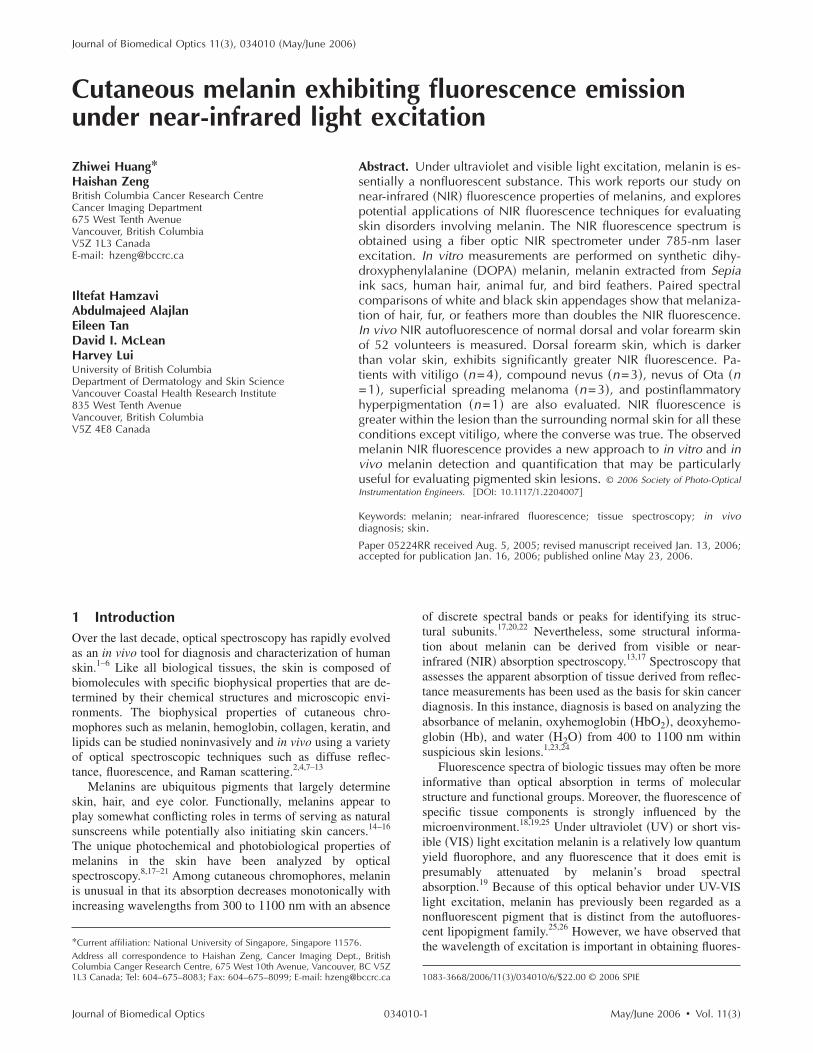
3 ResultsSolid-state in vitro melanins �Sepia and synthetic DOPA mela-nins� exhibit prominent fluorescence in the NIR range under785 nm excitation �Fig. 1�a��, with the spectrum beingslightly higher in intensity at longer emission wavelengths.The two broadbands with peaks near 880 and 890 nm repre-sent Raman scattering by melanin.29 NIR-excited autofluores-cence from black human hair, feline fur, and chicken feathers,which are replete with melanin �Figs. 1�b�–1�d��, is similar tothat of Sepia and synthetic melanin. White hair and white
feathers, which presumably differ from their black counter-May/June 2006 � Vol. 11�3�2
W; an
Huang et al.: Cutaneous melanin exhibits fluorescence emission¼
parts only in terms of their relative absence of eumelanin,exhibit a markedly lower NIR autofluorescence signal.
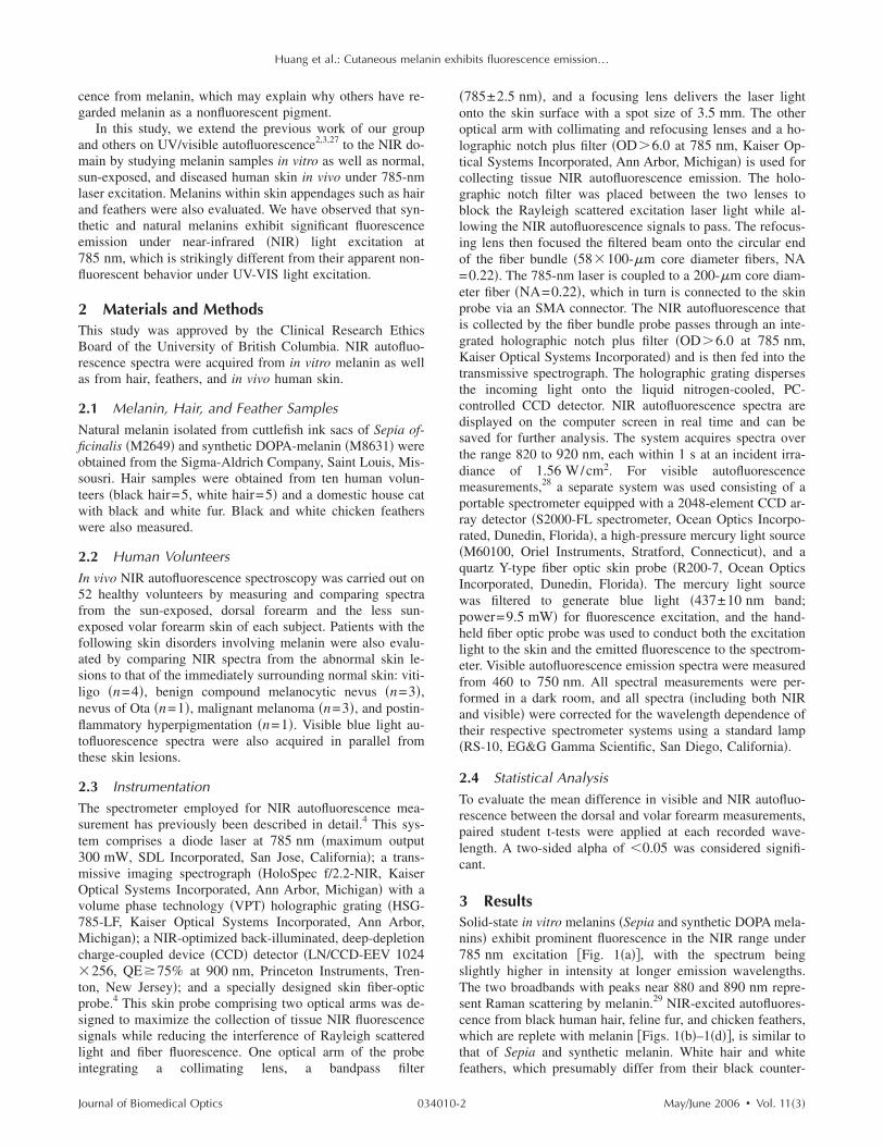
The visibly discernible differences in the degree of skinmelanization between darker dorsal and lighter volar forearmskin of normal volunteers could be demonstrated by in vivoNIR autofluorescence and the corresponding autofluorescencedifference spectrum �Fig. 2�a��, with the dorsal forearm show-ing greater autofluorescence than that of the volar aspect inthe 820- to 920-nm range �p�0.0001 at all wavelengths�.Consistent with this finding was the observation that the de-pigmented skin of vitiligo lesions had a much lower NIR au-tofluorescence intensity than that of the normally pigmentedsurrounding skin �Figs. 2�b��. Furthermore, the spectra re-vealed the presence of several embedded Raman signals �e.g.,857, 871, 874, 885, 901, and 909 nm� from both pigment-bearing �normal� and amelanotic �vitiligo� skin. These signalsare due to cutaneous proteins and lipids,4 and their consistentpresence in all skin measurements would be expected, sincethese components are unaffected by vitiligo.
Dark-colored skin lesions due to abnormal hypermelaniza-tion �i.e., benign compound nevus, nevus of Ota, melanoma,and postinflammatory hyperpigmentation� all showed higher
Fig. 1 NIR autofluorescence spectra �820 to 920 nm� obtained from: �chicken feathers. Excitation wavelength 785 nm; light intensity 170 m
NIR autofluorescence when compared to the adjacent normal
Journal of Biomedical Optics 034010-
skin �Figs. 2�c�–2�f��. For heavy pigmented skin lesions, thedifference spectra in Figs. 2�c�–2�f� showed an increasingtrend of intensity at longer wavelengths, which is in consistentwith NIR fluorescence spectra observed from synthetic or ex-tracted melanin samples and melanin-rich black hair or blackfeathers �Fig. 1�. Therefore, melanin is an important fluoro-phore accounting for in vivo skin in the NIR range 820 to920 nm.
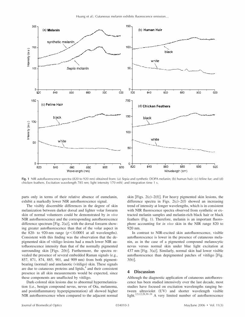
In contrast to NIR-excited skin autofluorescence, visibleautofluorescence is lower in the presence of cutaneous mela-nin, as in the case of a pigmented compound melanocyticnevus versus normal skin under blue light excitation at437 nm �Fig. 3�a��. Similarly, normal skin had lower visibleautofluorescence than depigmented patches of vitiligo �Fig.3�b��.
4 DiscussionAlthough the diagnostic application of cutaneous autofluores-cence has been studied intensively over the last decade, moststudies have focused on excitation wavelengths ranging be-tween ultraviolet �UV� and shorter wavelength visible
2,5,12,28,30–34
a and synthetic DOPA melanin; �b� human hair; �c� feline fur; and �d�d integration time 1 s.
a� Sepi
light. A very limited number of autofluorescence
May/June 2006 � Vol. 11�3�3

Huang et al.: Cutaneous melanin exhibits fluorescence emission¼
studies have utilized longer red-to-NIR wavelengths, but thesewere mostly centered on examining tissue fluorescence in re-lation to porphyrins.35–37
We have characterized the NIR autofluorescence propertiesof melanin obtained by chemical synthesis and tissue extrac-tion, as well as through direct in vivo or in vitro measurementsof skin, melanin-containing appendages �e.g., human hair, fe-line fur, and chicken feathers�, and partially purified melanin.It has been shown by observation and histochemical methodsthat regardless of ethnicity, epidermal melanin content is sig-nificantly greater in chronically sun-exposed skin than it is incorresponding sun-protected skin sites �up to two-fold�.15,21 Inthis study, sun-exposed skin �dorsal forearm� exhibited sig-nificantly more NIR autofluorescence compared to sun-protected skin �volar forearm�, confirming that NIR autofluo-rescence of epidermal melanin parallels the visible degree ofpigmentation. The results clearly demonstrate the ability ofmelanin from a variety of biological sources to fluoresce posi-tively under NIR excitation in a measurable fashion. In con-trast, both visible absorption and fluorescence spectroscopy
Fig. 2 Paired mean NIR autofluorescence spectra �820 to 920 nm� anvolar sites of the forearm �gray curve is the mean difference spectrumvitiligo �n=4�; �c� compound melanocytic nevus �n=3�; �d� nevus ofmatory hyperpigmentation �n=1�. Excitation wavelength 785 nm; ligh
d the corresponding mean difference spectra obtained from: �a� dorsal and± SD �n=52�� and skin lesions and their normal surrounding skin sites; �b�Ota �n=1�; �e� superficial spreading melanoma �n=3�; and �f� postinflam-t intensity 170 mW; and integration time 1 s.
rely on the notion that the relative absence of a spectral signal
Journal of Biomedical Optics 034010-
Fig. 3 Paired autofluorescence spectra in the visible range �460 to750 nm� obtained from skin lesions and their normal surrounding skinsite: �a� compound melanocytic nevus, and �b� vitiligo. Excitationwavelength 437 nm±10 nm; light intensity 9.5 mW; and integration
time 0.1 s.May/June 2006 � Vol. 11�3�4
Huang et al.: Cutaneous melanin exhibits fluorescence emission¼
implies the presence of melanin, and that the degree of sup-pression of a signal can be used to indirectly estimate thequantity of melanin present.7,8 The ability to measure a “posi-tive” optical signal in the presence of melanin within the NIRexcitation domain thus provides a fundamentally different andmore direct approach for in vitro and in vivo melanin detec-tion.
That melanin exhibits strong NIR autofluorescence andRaman emission under NIR wavelength excitation is perhapssurprising, given the conventional acceptance that melaninwas essentially nonfluorescent when measured in the UV andshort visible wavelengths. The biophysical basis for melaninautofluorescence remains largely unknown. It has been sug-gested that it may be induced by partial oxidative breakdownof melanin, and therefore depends on structurally defectivemelanin polymers.19,38 Our data show that prominent NIR au-tofluorescence emission appears to be a common feature ofnatural and synthetic melanins, and that melanin is one of themajor fluorophores responsible for cutaneous NIR autofluo-rescence. The observation that the melanin-poor vitiligo le-sions still exhibit significant NIR autofluorescence confirmsthat other fluorophores such as hemoglobin, porphyrin, col-lagen, etc. in the skin also contribute to cutaneous NIRautofluorescence.4,29
The NIR fluorescence line shape of white hair, whitefeather, and light colored skin tissue observed in this study issimilar to that of other tissue types �e.g., nonskin tissues withno melanin content� reported in the literature.36 The commonfeature of this fluorescence line shape is that the fluorescenceintensity monotonically decreases with wavelength and showsno maxima. In comparison, the NIR fluorescence line shapeof melanin �Fig. 1� is quite unique in that the fluorescenceintensity increases with wavelength and appears to have amaximum. A complete understanding of these phenomenawarrants further investigations.
In Fig. 2, all the difference spectra have positive values,confirming the significant contributions of melanin to in vivoskin tissue NIR fluorescence. However, one may note that inFig. 2, not all the difference spectra follow the line shape ofthe pure melanin spectrum. The difference spectrum betweennormal and vitiligo �normal–vitiligo� does not show a maxi-mum and is slightly titled toward the shorter wavelength end.We are building a tissue optics model and planning a MonteCarlo simulation approach to under this phenomenon. Tenta-tively, we have the following explanation for this phenom-enon.
We use the “normal–vitiligo” difference spectrum as anexample and assume that the only difference between vitiligoand its surrounding normal skin is melanin. Due to the lack ofmelanin absorption in vitiligo, excitation light can penetratedeeper down into the tissue than in normal skin; re-emittedfluorescence will also be easier to escape. Thus, when mea-suring vitiligo we are sampling a larger tissue volume andwith higher detection efficiency than measuring normal skin.We are getting more nonmelanin-related fluorescence signalfrom vitiligo than from normal skin. The proportion of thisextra nonmelanin related fluorescence signal should increasewith wavelength, since longer wavelength fluorescence pho-tons have higher chances to escape out of the tissue. On theother hand, melanin adds a strong fluorescence signal to the
normal skin. Therefore, the line shape of the difference spec-Journal of Biomedical Optics 034010-
trum �normal–vitiligo� will not be exactly the same as thefluorescence spectrum of pure melanins. Instead, it will bedetermined by the balance between the extra melanin fluores-cence signal generated in normal skin and the extranonmelanin-related fluorescence signal detected from vitiligo.Similar considerations can also be applied to other spectralpairs shown in Fig. 2. Therefore, for heavy pigmented lesions�Figs. 2�c� and 2�e��, the difference spectra have a shape moreclose to that of the pure melanin, while for more lightly pig-mented lesions �such as normal skin versus vitiligo in Fig.2�b��, the difference spectra deviate significantly from a puremelanin spectral shape and the maximum may disappear. Weexpect that our Monte Carlo modeling, which accurately ac-counts for all the tissue optics effects on the difference spec-tra, will either confirm this consideration or slightly modify it.
We have observed two obvious characteristic Raman bandsnear 880 and 895 nm from melanin samples, black hair, andblack feather �Fig. 1�. For in vivo spectra of melanin-rich skinlesions shown in Fig. 2, these two Raman bands are not veryobvious. They may have been masked, to a certain extent, byother strong Raman signals that originate from other biomol-ecules, such as proteins, lipids, porphyrin, etc., in skin.4,29,39
However, if we remove the fluorescence background fromthese in vivo spectra and concentrate on looking at the Ramansignals, the melanin’s contribution to the total Raman signalsbecomes more evident �data not shown�.
Changes in NIR autofluorescence spectra directly corre-spond to visible differences arising from melanin in patho-logical conditions such as vitiligo, malignant melanoma, be-nign compound melanocytic nevi, nevus of Ota, andinflammatory hyperpigmentation. Thus, NIR autofluorescencespectroscopy can potentially capture differences in melaninsfor pigmented skin lesions in vivo. It appears that the NIRautofluorescence technique may be particularly beneficial tothe diagnosis of pigmented lesions, because these dark skinlesions can be lit up under NIR excitation. With UV/VIS ex-citation, melanin largely behaves as a photon absorber �Fig.3�, making visible fluorescence or reflectance measurementsless informative. As previously mentioned, a “positive” NIRautofluorescence signal may be particularly useful for nonin-vasive analysis of lesions such as malignant melanoma, whichcan be difficult to differentiate from other pigmented but non-melanin containing lesions using UV-VIS fluorescence or re-flectance �i.e., apparent absorption� spectroscopictechniques.24
5 ConclusionsIn aggregate, our results demonstrate that under NIR excita-tion, melanins exhibit prominent autofluorescence within theskin and its pigmented appendages, whereas with UV-visibleexcitation, any potential melanin fluorescence is essentiallyundetectable. Practical applications for melanin quantificationand diagnostic evaluation of pigmented skin lesions are sup-ported by this observation. Furthermore, it appears clear thatthe fluorescence properties of melanins are highly wavelengthdependent, which in turn merits further detailed exploration.
AcknowledgmentsThe authors wish to thank Dr. Jerry Shapiro and Ms. Michelle
Zeng for their assistance with the experiments. This work wasMay/June 2006 � Vol. 11�3�5
Huang et al.: Cutaneous melanin exhibits fluorescence emission¼
supported by the National Cancer Institute of Canada withfunds from the Canadian Cancer Society, the Canadian Der-matology Foundation, and the VGH & UBC Hospital Foun-dation In It for Life Fund.
References1. R. Marchesini, M. Brambilla, C. Clemente, M. Maniezzo, A. E.
Sichirollo, A. Testori, D. R. Venturoli, and N. Cascinelli, “In vivospectrophotometric evaluation of neoplastic and non-neoplastic skinpigmented lesions - I. Reflectance measurements,” Photochem. Pho-tobiol. 53, 77–84 �1991�.
2. H. Zeng, C. MacAulay, D. I. McLean, and B. Palcic, “Spectroscopicand microscopic characteristics of human skin autofluorescence emis-sion,” Photochem. Photobiol. 61, 639–645 �1995�.
3. L. Brancaleon, A. J. Durkin, J. H. Tu, G. Menaker, J. D. Fallon, andN. Kollias, “In vivo fluorescence spectroscopy of nonmelanoma skincancer,” Photochem. Photobiol. 73, 178–183 �2001�.
4. Z. Huang, H. Zeng, I. Hamzavi, D. I. McLean, and H. Lui, “Rapidnear-infrared Raman spectroscopy system for real-time in vivo skinmeasurements,” Opt. Lett. 26, 1782–1784 �2001�.
5. N. Kollias and G. N. Stamatas, “Optical non-invasive approaches todiagnosis of skin diseases,” J. Investig. Dermatol. Symp. Proc. 7,64–75 �2002�.
6. P. J. Caspers, G. W. Lucassen, and G. J. Puppels, “Combined in vivoconfocal Raman spectroscopy and confocal microscopy of humanskin,” Biophys. J. 85, 572–580 �2003�.
7. N. Kollias and A. Baqer, “Spectroscopic characteristics of humanmelanin in vivo,” J. Invest. Dermatol. 85, 38–42 �1985�.
8. N. Kollias and A. Baqer, “On the assessment of melanin in humanskin in vivo,” Photochem. Photobiol. 43, 49–54 �1986�.
9. R. Gillies, G. Zonios, R. R. Anderson, and N. Kollias, “Fluorescenceexcitation spectroscopy provides information about human skin invivo,” J. Invest. Dermatol. 115, 704–707 �2000�.
10. G. Zonios, J. Bykowski, and N. Kollias, “Skin melanin, hemoglobin,and light scattering properties can be quantitatively assessed in vivousing diffuse reflectance spectroscopy,” J. Invest. Dermatol. 117,1452–1457 �2001�.
11. K. J. Jeon, S. J. Kim, K. K. Park, J. W. Kim, and G. Yoon, “Nonin-vasive total hemoglobin measurement,” J. Biomed. Opt. 7�1�, 45–50�2002�.
12. N. Kollias, G. Zonios, and G. N. Stamatas, “Fluorescence spectros-copy of skin,” Vib. Spectrosc. 28, 17–23 �2002�.
13. A. Matas, M. G. Sowa, G. Taylor, and H. H. Mantsch, “Melanin as aconfounding factor in near infrared spectroscopy of skin,” Vib. Spec-trosc. 28, 45–52 �2002�.
14. M. R. Chedekel, “Photochemistry and photobiology of epidermalmelanins,” Photochem. Photobiol. 35, 881–885 �1982�.
15. S. Alaluf, D. Atkins, K. Barrett, M. Blount, N. Carter, and A. Heath,“Ethnic variation in melanin content and composition in photoex-posed and photoprotected human skin,” Pigment Cell Res. 15, 112–118 �2002�.
16. J. P. Ortonne, “Photoprotective properties of skin melanin,” Br. J.Dermatol. 146, 7–10 �2002�.
17. I. A. Menon, S. Persad, H. F. Haberman, and C. J. Kurian, “A com-parative study of the physical and chemical properties of melaninsisolated from human black and red hair,” J. Invest. Dermatol. 80,202–206 �1983�.
18. S. D. Kozikowski, L. J. Wolfram, and R. R. Alfano, “Fluorescencespectroscopy of eumelanins,” IEEE J. Quantum Electron. QE-20,1379–1382 �1984�.
19. J. M. Gallas and M. Eisner, “Fluorescence of melanin-dependenceupon excitation wavelength and concentration,” Photochem. Photo-biol. 45, 595–600 �1987�.
20. N. Kollias and A. H. Baqer, “Absorption mechanisms of humanmelanin in the visible, 400-720 nm,” J. Invest. Dermatol. 89, 384–388 �1987�.
21. S. Alaluf, D. Atkins, K. Barrett, M. Blount, N. Carter, and A. Heath,
Journal of Biomedical Optics 034010-
“The impact of epidermal melanin on objective measurements of hu-man skin color,” Pigment Cell Res. 15, 119–126 �2002�.
22. M. Shimada, Y. Yamada, M. Itoh, and T. Yatagai, “Melanin and bloodconcentration in human skin studied by multiple regression analysis:experiments,” Phys. Med. Biol. 46, 2385–2395 �2001�.
23. R. Marchesini, N. Cascinelli, M. Brambilla, C. Clemente, L.Mascheroni, E. Pignoli, A. Testori, and D. R. Venturoli, “In vivospectrophotometric evaluation of neoplastic and non-neoplastic skinpigmented lesions - II: Discriminant analysis between nevus andmelanoma,” Photochem. Photobiol. 55, 515–522 �1992�.
24. M. Elbaum, A. W. Kopf, H. S. Rabinovitz, R. G. Langley, H. Ka-mino, M. C. Mihm Jr, A. J. Sober, G. L. Peck, A. Bogdan, D.Gutkowicz-Krusin, M. Greenebaum, S. Keem, M. Oliviero, and S.Wang, “Automatic differentiation of melanoma from melanocyticnevi with multispectral digital dermoscopy: a feasibility study,” J.Am. Acad. Dermatol., 44, 207–218 �2001�.
25. M. Elleder and J. Borovansky, “Autofluorescence of melanins in-duced by ultraviolet radiation and near ultraviolet light. A histochemi-cal and biochemical study,” Histochem. J. 33, 273–281 �2001�.
26. M. Senba, “Staining properties of melanin and lipofuscin pigments,”Am. J. Clin. Pathol. 86, 556–557 �1986�.
27. L. Brancaleon, G. Lin, and N. Kollias, “The in vivo fluorescence oftryptophan moieties in human skin increases with UV exposure and isa marker for epidermal proliferation,” J. Invest. Dermatol. 113, 977–982 �1999�.
28. H. Zeng, C. MacAulay, D. I. McLean, H. Lui, and B. Palcic, “Min-iature spectrometer and multi-spectral imager as a potential diagnos-tic aid for dermatology,” Proc. SPIE 2387, 57–61 �1995�.
29. Z. Huang, H. Lui, X. K. Chen, D. I. McLean, and H. Zeng, “Ramanspectroscopy of in vivo cutaneous melanin,” J. Biomed. Opt. 9�6�,1198–1205 �2004�.
30. K. T. Schomacker, J. K. Frisoli, C. C. Compton, T. J. Flotte, J. M.Richter, N. S. Nishioka, and T. F. Deutsch, “Ultraviolet laser-inducedfluorescence of colonic tissue: basic biology and diagnostic poten-tial,” Lasers Surg. Med. 12, 63–78 �1992�.
31. H. Zeng, C. MacAulay, B. Palcic, and D. I. McLean, “A computer-ized autofluorescence and diffuse reflectance spectroanalyser systemfor in vivo skin studies,” Phys. Med. Biol. 38, 231–240 �1993�.
32. H. Zeng, C. MacAulay, D. I. McLean, and B. Palcic, “Reconstructionof in vivo skin autofluorescence spectrum from microscopic proper-ties by Monte Carlo simulation,” J. Photochem. Photobiol., B 38,234–240 �1997�.
33. H. Zeng, C. MacAulay, D. I. McLean, B. Palcic, and H. Lui, “Thedynamics of laser induced autofluorescence decay in human skin-experimental measurements and theoretical modeling,” Photochem.Photobiol. 68, 227–236 �1998�.
34. S. K. Chang, M. Follen, A. Malpica, U. Utzinger, G. Staerkel, D.Cox, E. N. Atkinson, C. MacAulay, and R. Richards-Kortum, “Opti-mal excitation wavelengths for discrimination of cervical neoplasia,”IEEE Trans. Biomed. Eng. 49, 1102–1111 �2002�.
35. E. B. Hanlon, I. Itzkan, R. R. Dasari, M. S. Feld, R. J. Ferrante, A. C.McKee, D. Lathi, and N. W. Kowall, “Near-infrared fluorescencespectroscopy detects Alzheimer’s disease in vitro,” Photochem. Pho-tobiol. 70, 236–242 �1999�.
36. G. Zhang, S. G. Demos, and R. R. Alfano, “Far-red and NIR Spectralwing emission from tissues under 532 and 632 nm photo-excitation,”Lasers Life Sci. 9, 1–16 �1999�.
37. S. G. Demos, M. Staggs, L. Gandour-Edwards, R. Ramsamooj, andR. White, “Tissue imaging for cancer detection using NIR autofluo-rescence,” Proc. SPIE 4613, 31–34 �2002�.
38. S. Ito, “Advances in the pigmentary system,” in Physiology andPathophysiology, J. J. Nordlund, R. E. Boissy, V. J. Hearing, R. A.King, and J. P. Ortonne, Eds., pp. 439–450, Oxford University Press,New York �1998�.
39. Z. Huang, H. Lui, D. I. McLean, M. Korbelik, and H. Zeng, “Ramanspectroscopy in combination with background near-infrared autofluo-rescence enhances the in vivo assessment of malignant tissues,” Pho-
tochem. Photobiol. 81, 1219–1226 �2005�.May/June 2006 � Vol. 11�3�6


