Embed Size (px)
Citation preview
of January 12, 2022.This information is current as
Expansion in the Absence of TLR SignalingHematopoietic Stem and Progenitor Cell Cutting Edge: Bacterial Infection Induces
Lyle L. MoldawerAl-Quran, Ian Bovio, Shizuo Akira, Yutaro Kumagai andDelano, Jason S. Weinstein, Alex G. Cuenca, Samer Philip O. Scumpia, Kindra M. Kelly-Scumpia, Matthew J.
http://www.jimmunol.org/content/184/5/2247doi: 10.4049/jimmunol.0903652February 2010;
2010; 184:2247-2251; Prepublished online 3J Immunol
MaterialSupplementary
2.DC1http://www.jimmunol.org/content/suppl/2010/02/01/jimmunol.090365
Referenceshttp://www.jimmunol.org/content/184/5/2247.full#ref-list-1
, 11 of which you can access for free at: cites 22 articlesThis article
average*
4 weeks from acceptance to publicationFast Publication! •
Every submission reviewed by practicing scientistsNo Triage! •
from submission to initial decisionRapid Reviews! 30 days* •
Submit online. ?The JIWhy
Subscriptionhttp://jimmunol.org/subscription
is online at: The Journal of ImmunologyInformation about subscribing to
Permissionshttp://www.aai.org/About/Publications/JI/copyright.htmlSubmit copyright permission requests at:
Email Alertshttp://jimmunol.org/alertsReceive free email-alerts when new articles cite this article. Sign up at:
Print ISSN: 0022-1767 Online ISSN: 1550-6606. Immunologists, Inc. All rights reserved.Copyright © 2010 by The American Association of1451 Rockville Pike, Suite 650, Rockville, MD 20852The American Association of Immunologists, Inc.,
is published twice each month byThe Journal of Immunology
by guest on January 12, 2022http://w
ww
.jimm
unol.org/D
ownloaded from
by guest on January 12, 2022
http://ww
w.jim
munol.org/
Dow
nloaded from
Cutting Edge: Bacterial Infection Induces HematopoieticStem and Progenitor Cell Expansion in the Absence of TLRSignalingPhilip O. Scumpia,*,1 Kindra M. Kelly-Scumpia,*,1 Matthew J. Delano,*Jason S. Weinstein,† Alex G. Cuenca,* Samer Al-Quran,‡ Ian Bovio,‡ Shizuo Akira,x
Yutaro Kumagai,x and Lyle L. Moldawer*
Bone marrow (BM) hematopoietic stem and progenitorcells (HSPCs) can be activated by type I IFNs, TLR ago-nists, viruses, and bacteria to increase hematopoiesis.In this study, we report that endotoxin treatmentin vivo induces TLR4,MyD88, and Toll/IL-1 resistancedomain-containing adaptor-inducing IFN-b (TRIF)-dependent expansion of BMHSPCs. Bacterial infectionby Staphylococcus aureus or cecal ligation and puncturealso induces HSPC expansion, but MyD88, TRIF, typeI IFN, cytokine, PG, or oxidative stress pathways are notrequired for their expansion. S. aureus-induced HSPCexpansion in MyD882/2TRIF2/2 mice is also normal,but is associated with BM remodeling as granulocytestores are released peripherally. Importantly, reductionin BM cellularity alone can reproduce HSPC expansion.These data show in vivo HSPC responses to bacterialinfection are complex and not absolutely dependentupon key inflammatory signaling pathways. TheJournal of Immunology, 2010, 184: 2247–2251.
Long-term reconstituting (LTR)hematopoietic stemcells(LT-HSCs) are the source of all circulating blood cellsand are defined by their capacity for self-renewal and
multilineage differentiation. Mouse hematopoietic stem andprogenitor cells (HSPCs), including LT-HSCs, can be identifiedwithin the lineage2/lowSca-1+c-kit+ (LSK) subset of cells (1–3),and LT-HSCs can be differentiated from those with short-lived,rapidly dividing HSPCs short-term reconstituting-HSCs (ST-HSC) by the expression of additional cell surface markers (4).Although LT-HSCs maintain blood cell production duringhomeostasis, HSPCs expand in vivo following IFN-a treatment(4–6), bone marrow (BM) ablation (7), or bleeding (8), dra-matically increasing the production of leukocytes.
TLRs are central to innate immunity by recognizing con-served molecular patterns found on pathogens. All TLRs,except TLR3, signal via the adaptor protein, MyD88, leadingto NF-kB activation. TLR4, the receptor for LPS, and TLR3also signal through the adaptor protein, Toll/IL-1 resistancedomain-containing adaptor-inducing IFN-b (TRIF)/Toll-IL1receptor domain-containing adaptor molecule 1, which leadsto IFN-I production. HSPCs express TLRs and are activatedby TLR2 and TLR4 agonists in vitro through MyD88 sig-naling (9). HSPCs are also activated in vivo by the TLR3agonist polyinosinic-polycytidylic acid through IFN-I signal-ing (5, 6). Similarly, vaccinia virus infection causes MyD88-dependent HSPC expansion (10). Although Escherichia coliinfection induces LSK expansion (11), the mechanism(s) ofbacterial-induced HSPC activation have not been fully elu-cidated. In this study, we used two infectious models,Staphylococcus aureus infection and polymicrobial sepsis in-duced by cecal ligation and puncture (CLP), to examine theregulation of in vivo HSPC expansion.
Materials and MethodsMice
C57BL/6,TLR4mutantC3H/HeJmice, and controlC3H/HeOuJ,TRIF2/2, IL-1R2/2 IL-62/2, and Rag12/2 mice on a B6 background were from The JacksonLaboratory (Bar Harbor, ME). IFN-abR/A1292/2 (IFNAR2/2) and wild-typeSvEv.129 mice were purchased from B&K Universal (Grimston, Aldbrough,U.K.).B6.MyD882/2micewereobtained fromS.Akira throughA.Ayala atRhodeIsland Hospital (Providence, RI). Experiments utilizing MyD882/2TRIF2/2
double knockout (DKO) mice were performed at Osaka University (Osaka,Japan). Mice were maintained in specific pathogen-free conditions, and ex-periments were performed under institutional guidelines.
Reagents
Anti–Gr-1 (RB6-8C5) and anti-human IL-4 (IgG2b isotype) were kindlyprovided by P. Heyworth from Schering-Plough Biopharma (Palo Alto,CA). Endotoxin-reduced staphylococcal enterotoxin B (SEB), E. coli endo-toxin (LPS; strain O55:B5), N-acetyl cysteine (used at 100 mg/kg), and
*Department of Surgery, †Department of Medicine, and ‡Department of Pathology,University of Florida College of Medicine, Gainesville, FL 32610; and xLaboratory ofHost Defense, World Premier International Immunology Frontier Research Center,Osaka University, Osaka, Japan
1P.O.S. and K.M.K.-S. contributed equally to this work.
Received for publication November 19, 2009. Accepted for publication January 8, 2010.
This work was supported in part by Grants R01 GM-40586-21 and R01 GM-81923-02awarded by the National Institute of General Medical Sciences.
Address correspondence and reprint requests to Dr. Lyle L. Moldawer, Department ofSurgery, University of Florida College of Medicine, 1600 SW Archer Road, Room 6116,Gainesville, FL 32610-0286. E-mail address: [email protected]
The online version of this article contains supplemental material.
Abbreviations used in this paper: BM, bone marrow; CLP, cecal ligation and puncture;DKO, double knockout; HSPC, hematopoietic stem and progenitor cell; IFNAR2/2,IFN-abR/A1292/2; Lin, lineage mixture; LSK, lineage2/lowSca-1+c-kit+; LT-HSC,long-term reconstituting hematopoietic stem cell; LTR, long-term reconstituting; SEB,staphylococcal enterotoxin B; ST-HSC, short-term reconstituting hematopoietic stemcell; TRIF, Toll/IL-1 resistance domain-containing adaptor-inducing IFN-b.
Copyright� 2010 by TheAmerican Association of Immunologists, Inc. 0022-1767/10/$16.00
www.jimmunol.org/cgi/doi/10.4049/jimmunol.0903652
by guest on January 12, 2022http://w
ww
.jimm
unol.org/D
ownloaded from
apocynin (50 mg/kg) were purchased from Sigma-Aldrich (St. Louis, MO).Pegylated soluble TNFR1 was a kind gift of Amgen (Thousand Oaks, CA;used at 100 mg/kg).
Mouse infection models
To induce polymicrobial sepsis, mice underwent CLP or sham procedure (12)to obtain an expected mortality of ∼10% by 7 d. For S. aureus infection, micewere inoculated with 100 ml of midlogarithmic phase S. aureus, SH1000, insterile saline, s.c., i.p. (5 3 107 CFUs), or i.v. (5 3 106 CFUs).
For toxin administration, mice were treated i.p. with 5 mg LPS or 10 mgSEB. Mice were sacrificed 18–36 h later, and BM was harvested for furtheranalysis. To deplete mature neutrophils, mice were treated with 500 mg anti–Gr-1 or control Ab twice separated by 18 h.
Flow cytometry
BM cells were analyzed as previously described (12). Abs were from eBio-science (San Diego, CA) and included anti-CD16/32 Fc block, anti-mousebiotin Lineage mixture (Lin; BD Biosciences, San Jose, CA), anti-CD150,anti–Sca-1, anti–c-kit, anti-CD135/Flk-2, anti–Gr-1, and anti-CD11b. Allsamples were stained with SYTOX Blue (Invitrogen, Carlsbad, CA) for cellviability analysis and collected on a Becton-Dickinson LSRII flow cytometerusing FACSDiva software (BD Biosciences). Total cell numbers from selectexperiments were calculated and are shown in Supplemental Fig. 7A–D.
Cell purification and culture
Lin2 progenitor cells were obtained using biotin-Lin followed by antibiotinmagnetic beads (Miltenyi Biotec, Auburn, CA). To obtain LT-HSC–enrichedcells, the Lin2 cell fraction was incubated with Lin, c-kit, sca-1, CD150, andCD135 Abs, and cells were sorted using an FACSAria sorter (BD Bio-sciences). Lin2 progenitors were cultured in Methocult H4100 base meth-ylcellulose media (Stem Cell Technologies, Vancouver, British Columbia,Canada) supplemented with erythropoietin, GM-CSF, G-CSF, or IL-7(R&D Systems, Minneapolis, MN) for 14 d.
Histologic analysis
Samples were fixed in 10% neutral formalin (Sigma-Aldrich), paraffin-em-bedded, decalcified with EDTA, sectioned (5-mm sections), and mounted forH&E staining.
Statistics
Differences among groups were evaluated by ANOVA for multiple groups andthe Student t test for two groups. Tukey test was used for post hoc analysiswhen p , 0.05 by ANOVA.
Results and DiscussionLPS-induced HSPC expansion involves TLR4 inflammatory signalingthrough MyD88 and TRIF
The LSK population is heterogeneous, and ,10% are truerepopulating HSCs (2). Many additional markers have beenused to obtain higher purities of LT-HSCs. Enriched HSCpopulations with higher LTR potential can be purified withinthe CD150+ (13, 14) and the Flk-2/CD1352 (15) populations.LSKs expressing CD150 retain their LTR ability even whenobtained from cyclophosphamide orG-CSF–treatedmice (16),whereas CD135 expression defines ST-HSCs in an activatedstate (15, 17). Because CD150+CD1352 LSKs have been usedas a novel identification strategy for LT-HSCs (18, 19), weexamined the effects of infection on the CD150+CD1352 LT-HSC–enriched population, as well as the activatedCD1502CD135+ ST-HSC population (13, 15, 20).Because LPS is a potent activator of HSPC proliferation
in vitro, we initially examined whether LPS affects HSPCresponses in vivo. A single dose of LPS (100-fold lower than theLD50 dose) induces a transient LSK expansion (8- to 9-fold;Supplemental Fig. 1A–C), beginning 18 h postinjection, lasting72 h before declining (data not shown). When examining thesubpopulations of expanding LSKs following LPS injection,
both LT-HSC–enriched and highly proliferative ST-HSCssubtypes of LSK expand (Supplemental Fig. 1C).Expectedly, we find LPS-induced HSPC expansion is absent
in TLR4 mutant C3H/HeJ mice in vivo, confirming the de-pendence on TLR4 (Supplemental Fig. 1B, 1C). However,unlike the absolute requirement of MyD88 for LPS-inducedin vitro LSK proliferation (9), in vivo LPS treatment inducespartial LSK expansion inMyD882/2 or TRIF2/2mice to 61%and 36% of wild-type levels, respectively (Supplemental Fig.1D), indicating participation of both TLR adaptors in vivo.Ly6A/E (Sca-1), one of the primary markers of HSCs, is an
IFN-I–inducible gene, and IFN-I signaling is necessary for in vivopolyinosinic-polycytidylic acid/TLR3-induced HSPC activation(5, 6). We therefore examined whether IFN-I signaling con-tributes to TLR4-induced HSPC activation. Using IFNAR2/2
mice, we found that HSPC expansion is completely intact in theabsence of IFN-I signaling (Supplemental Fig. 1E), indicatingthat LPS stimulates in vivo HSPC activation through TLR4-mediated inflammatory, but not IFN-I–dependent, signaling.
Bacterial infection induces expansion of LT-HSCs and ST-HSCs
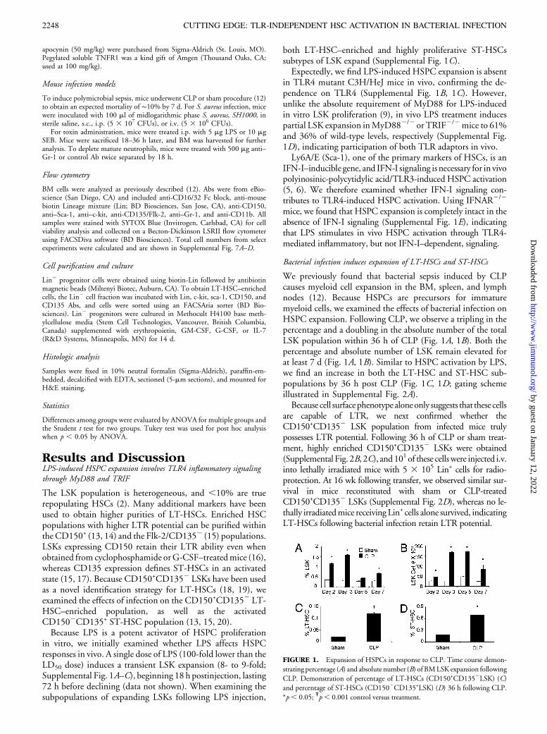
We previously found that bacterial sepsis induced by CLPcauses myeloid cell expansion in the BM, spleen, and lymphnodes (12). Because HSPCs are precursors for immaturemyeloid cells, we examined the effects of bacterial infection onHSPC expansion. Following CLP, we observe a tripling in thepercentage and a doubling in the absolute number of the totalLSK population within 36 h of CLP (Fig. 1A, 1B). Both thepercentage and absolute number of LSK remain elevated forat least 7 d (Fig. 1A, 1B). Similar to HSPC activation by LPS,we find an increase in both the LT-HSC and ST-HSC sub-populations by 36 h post CLP (Fig. 1C, 1D; gating schemeillustrated in Supplemental Fig. 2A).Because cell surfacephenotypealoneonly suggests that these cells
are capable of LTR, we next confirmed whether theCD150+CD1352 LSK population from infected mice trulypossesses LTR potential. Following 36 h of CLP or sham treat-ment, highly enriched CD150+CD1352 LSKs were obtained(Supplemental Fig. 2B, 2C), and101of these cellswere injected i.v.into lethally irradiated mice with 5 3 105 Lin+ cells for radio-protection. At 16 wk following transfer, we observed similar sur-vival in mice reconstituted with sham or CLP-treatedCD150+CD1352 LSKs (Supplemental Fig. 2D), whereas no le-thally irradiatedmice receiving Lin+ cells alone survived, indicatingLT-HSCs following bacterial infection retain LTR potential.
FIGURE 1. Expansion of HSPCs in response to CLP. Time course demon-
strating percentage (A) and absolute number (B) of BMLSK expansion following
CLP. Demonstration of percentage of LT-HSCs (CD150+CD1352LSK) (C)and percentage of ST-HSCs (CD1502CD135+LSK) (D) 36 h following CLP.
*p, 0.05; Ŧp, 0.001 control versus treatment.
2248 CUTTING EDGE: TLR-INDEPENDENT HSC ACTIVATION IN BACTERIAL INFECTION
by guest on January 12, 2022http://w
ww
.jimm
unol.org/D
ownloaded from
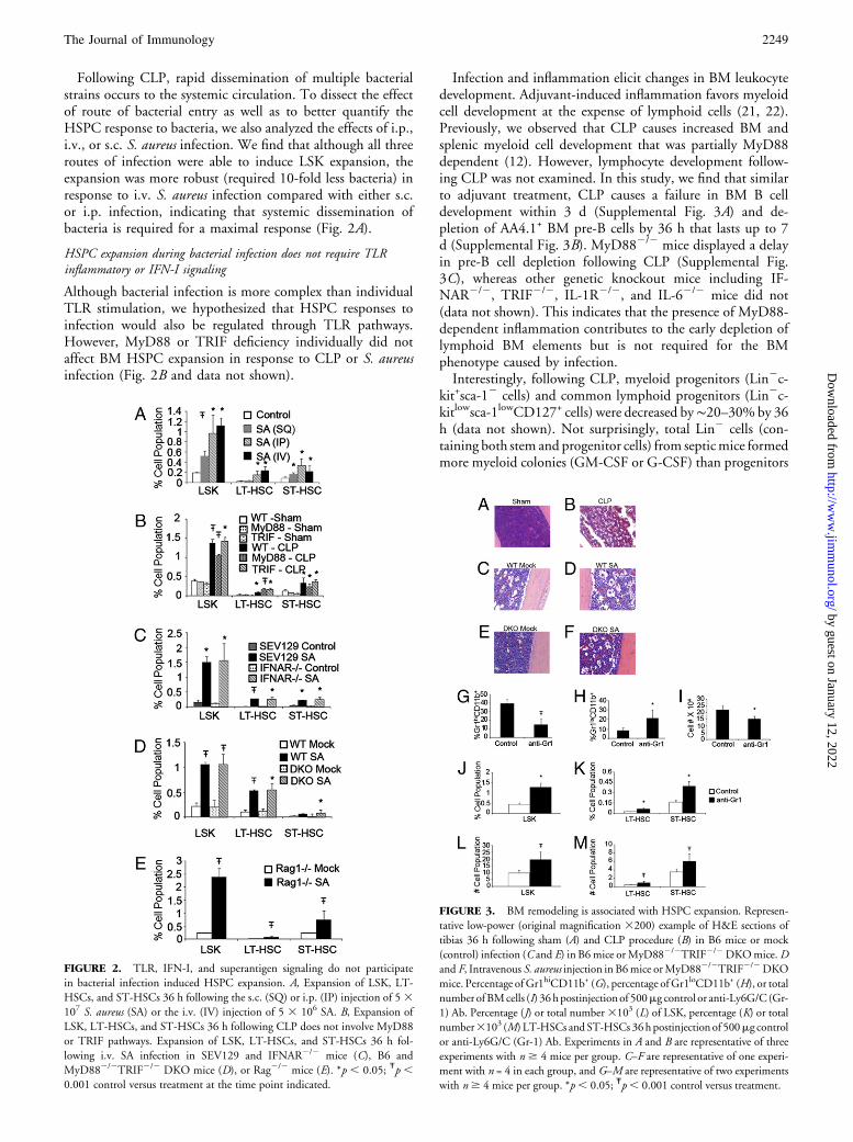
Following CLP, rapid dissemination of multiple bacterialstrains occurs to the systemic circulation. To dissect the effectof route of bacterial entry as well as to better quantify theHSPC response to bacteria, we also analyzed the effects of i.p.,i.v., or s.c. S. aureus infection. We find that although all threeroutes of infection were able to induce LSK expansion, theexpansion was more robust (required 10-fold less bacteria) inresponse to i.v. S. aureus infection compared with either s.c.or i.p. infection, indicating that systemic dissemination ofbacteria is required for a maximal response (Fig. 2A).
HSPC expansion during bacterial infection does not require TLRinflammatory or IFN-I signaling
Although bacterial infection is more complex than individualTLR stimulation, we hypothesized that HSPC responses toinfection would also be regulated through TLR pathways.However, MyD88 or TRIF deficiency individually did notaffect BM HSPC expansion in response to CLP or S. aureusinfection (Fig. 2B and data not shown).
Infection and inflammation elicit changes in BM leukocytedevelopment. Adjuvant-induced inflammation favors myeloidcell development at the expense of lymphoid cells (21, 22).Previously, we observed that CLP causes increased BM andsplenic myeloid cell development that was partially MyD88dependent (12). However, lymphocyte development follow-ing CLP was not examined. In this study, we find that similarto adjuvant treatment, CLP causes a failure in BM B celldevelopment within 3 d (Supplemental Fig. 3A) and de-pletion of AA4.1+ BM pre-B cells by 36 h that lasts up to 7d (Supplemental Fig. 3B). MyD882/2 mice displayed a delayin pre-B cell depletion following CLP (Supplemental Fig.3C), whereas other genetic knockout mice including IF-NAR2/2, TRIF2/2, IL-1R2/2, and IL-62/2 mice did not(data not shown). This indicates that the presence of MyD88-dependent inflammation contributes to the early depletion oflymphoid BM elements but is not required for the BMphenotype caused by infection.Interestingly, following CLP, myeloid progenitors (Lin2c-
kit+sca-12 cells) and common lymphoid progenitors (Lin2c-kitlowsca-1lowCD127+ cells) were decreased by∼20–30% by 36h (data not shown). Not surprisingly, total Lin2 cells (con-taining both stem and progenitor cells) from septicmice formedmore myeloid colonies (GM-CSF or G-CSF) than progenitors
FIGURE 2. TLR, IFN-I, and superantigen signaling do not participate
in bacterial infection induced HSPC expansion. A, Expansion of LSK, LT-
HSCs, and ST-HSCs 36 h following the s.c. (SQ) or i.p. (IP) injection of 53107 S. aureus (SA) or the i.v. (IV) injection of 5 3 106 SA. B, Expansion of
LSK, LT-HSCs, and ST-HSCs 36 h following CLP does not involve MyD88
or TRIF pathways. Expansion of LSK, LT-HSCs, and ST-HSCs 36 h fol-
lowing i.v. SA infection in SEV129 and IFNAR2/2 mice (C), B6 and
MyD882/2TRIF2/2 DKO mice (D), or Rag2/2 mice (E). *p , 0.05; Ŧp ,0.001 control versus treatment at the time point indicated.
FIGURE 3. BM remodeling is associated with HSPC expansion. Represen-
tative low-power (original magnification 3200) example of H&E sections of
tibias 36 h following sham (A) and CLP procedure (B) in B6 mice or mock
(control) infection (C and E) in B6mice orMyD882/2TRIF2/2DKOmice.Dand F, Intravenous S. aureus injection in B6mice orMyD882/2TRIF2/2DKO
mice. Percentage ofGr1hiCD11b+ (G), percentage ofGr1loCD11b+ (H), or total
number ofBMcells (I) 36hpostinjectionof 500mg control or anti-Ly6G/C(Gr-
1) Ab. Percentage (J) or total number3103 (L) of LSK, percentage (K) or totalnumber3103 (M) LT-HSCs andST-HSCs 36hpostinjectionof 500mg control
or anti-Ly6G/C (Gr-1) Ab. Experiments in A and B are representative of three
experiments with n $ 4 mice per group. C–F are representative of one experi-
ment with n = 4 in each group, and G–M are representative of two experiments
with n$ 4 mice per group. *p , 0.05; Ŧp, 0.001 control versus treatment.
The Journal of Immunology 2249
by guest on January 12, 2022http://w
ww
.jimm
unol.org/D
ownloaded from
from sham mice (Supplemental Fig. 3D). Surprisingly, theseLin2 cells from septic mice possessed a similar ability to formerythroid (erythropoietin) or lymphoid (IL-7) colonies whencompared with sham progenitors (Supplemental Fig. 3D). Thissuggests that the inflammatory BM microenvironment causesan in vivo block of erythroid/lymphoid cell developmentwithout depleting these cells from the BM.Blockade of other inflammatory factors that regulate he-
matopoiesis, including TNF-a, IL-1, IL-6, PG, or free radi-cal/oxidative stress, also had no effect on CLP-induced HSPCexpansion (Supplemental Fig. 4). Not surprisingly, givena lack of effect of IFN-I on LPS-induced HSPC expansion,loss of IFN-I signaling also did not affect HSPC expansion toS. aureus infection or CLP (Fig. 2C and data not shown).
S. aureus induces HSPC expansion in the absence of all TLR signaling
Because the lack of individual MyD88 or TRIF signaling didnot affect HSPC expansion, an interpretation of the data maybe that MyD88 and TRIF signaling are redundant, and ac-tivation of either pathway alone is sufficient for completeHSPC expansion. To eliminate this possibility, we usedMyD882/2TRIF2/2 (DKO) mice, which lack all TLR sig-naling. Surprisingly, HSPC expansion was completely intactin DKO mice (Fig. 2D) in response to S. aureus infection,indicating that TLR signaling is not required for HSPC ac-tivation to S. aureus infection.Because S. aureus can produce superantigens, and super-
antigenic stimulation of TCR–MHC complexes in DKOmice may induce HSPC expansion, we tested whether thismay be participating in our model. We found that i.p.treatment of mice with SEB is capable of HSPC expansion(Supplemental Fig. 5A). However, when we treated RAG2/2
mice (which cannot mount a T cell response to the super-antigen) with S. aureus, we find that HSPC responses aresimilar to WT mice (Fig 2E), indicating that by itself, su-perantigen production by S. aureus is not the dominantpathway resulting in HSPC expansion.
Infection induces BM remodeling, and Ab-mediated neutrophildepletion is capable of BM remodeling and is sufficient for HSPCexpansion
The BM is a fixed space with limited space for expansion of cellpopulations. Following an infectious or inflammatory insult,BM leukocyte stores are released into the blood. Despite this,many bacterial infections of mice cause an initial leukopeniafollowed by a delayed leukocytosis when the hematopoieticsystem is activated sufficiently (12; Supplemental Fig. 5B). Weexamined serial H&E sections of BM from sham and CLP-treated mice to determine the effects of infection on BMhomeostasis. By 12 h, the BM of septic mice shows dilatedsinusoids and release of mature neutrophils (SupplementalFig. 6A). Within 36 h, the BM shows extreme sinusoidaldilitation with loss of mature neutrophils and increased space(Fig. 3A, 3B, Supplemental Fig. 6B). Immature islands of cellsare located close to bone-lining osteoblasts and BM vascularcells. By 96 h, the BM regains cellularity, predominated bydeveloping myeloid cells, but a persistent paucity in cellularityremains (Supplemental Fig. 6C). By 7 d, however, the BM iscompletely repopulated with mostly myeloid cells (Supple-mental Fig. 6D). This increased space correlates well withtotal BM cellularity (Supplemental Fig. 6E) and was also
observed in mice following S. aureus infection, occurring tothe same extent in wild-type and MyD882/2TRIF2/2 DKOmice (Fig. 3C–F). These results led us to speculate whetheremigration of myeloid cells to the periphery causes pheno-typic changes in the BM, allowing the BM to be more con-ducive to HSPC expansion. To test this theory, mice weretreated with anti-Ly6G/C (Gr-1) Ab to deplete mature neu-trophils. Following 2 d of treatment, there is loss of Gr-1high
neutrophils from the BM (Fig. 3G) and an increase in thedeveloping Gr-1low myeloid cells (Fig. 3H). Total BM cel-lularity is decreased by ∼30% (Fig. 3I). With decreased cel-lularity, there is an increased percentage and an approximatedoubling of total LSK number (Fig. 3J, 3L). When examiningLSK subpopulations, we find expansion of LT-HSCs and ST-HSCs by both percentage and total number (Fig. 3K, 3M).Although inflammation induced by clearance of dying neu-trophils within the BM post Ab administration likely partic-ipates in HSPC expansion following anti–Gr-1 treatment,these data suggest a possible BM intrinsic mechanism wherebyHSPCs interacting with the BM microenvironment respondto decreased cellularity in their microenvironment caused byinflammatory, infectious, or cytotoxic agents, causing them toexpand to repopulate the area left by the cellular void.In summary, we demonstrate that although they likely
contribute, the absence of key innate immune pathways do notsolely govern the highly conserved processes of HSPC acti-vation and expansion in vivo following bacterial infection.Perhaps the most surprising finding of this study is the lack ofin vivo requirement for TLR signaling in HSPC activationusing two different infectious models, despite the fact that LPSrequired TLR signals to induce HSPC expansion. These resultshighlight the likely presence of other innate immune bacterialrecognition pathways during infection that can completelycompensate for the loss of TLR signaling to induce hemato-poietic activation. This pathway likely affects global signalingand does not use single inflammatory mediators to inducetheir response.We found that the BM microenvironment is considerably
altered with dilated BM sinusoids, disruption of the BMmatrix, and loss of mature cells from the BM, resulting inincreased space. Although not specifically addressed in thispaper, we hypothesize that complex cross talk between HSPCsinteracting with supporting stromal cells within their micro-environment may sense the loss of cells from the BM andprovide the signals to expand within the BM space left voidfollowing infection or chemotherapeutic BM ablation, al-though further investigation is necessary.
AcknowledgmentsWe thank N. Benson for technical assistance with the cell sorting and L. Miller
for kindly providing the S. aureus.
DisclosuresThe authors have no financial conflicts of interest.
References1. Spangrude, G. J., S. Heimfeld, and I. L. Weissman. 1988. Purification and char-
acterization of mouse hematopoietic stem cells. Science 241: 58–62.2. Uchida, N., and I. L. Weissman. 1992. Searching for hematopoietic stem cells:
evidence that Thy-1.1lo Lin- Sca-1+ cells are the only stem cells in C57BL/Ka-Thy-1.1 bone marrow. J. Exp. Med. 175: 175–184.
2250 CUTTING EDGE: TLR-INDEPENDENT HSC ACTIVATION IN BACTERIAL INFECTION
by guest on January 12, 2022http://w
ww
.jimm
unol.org/D
ownloaded from
3. Uchida, N., H. L. Aguila, W. H. Fleming, L. Jerabek, and I. L. Weissman. 1994.Rapid and sustained hematopoietic recovery in lethally irradiated mice transplantedwith purified Thy-1.1lo Lin-Sca-1+ hematopoietic stem cells. Blood 83: 3758–3779.
4. Bryder, D., D. J. Rossi, and I. L. Weissman. 2006. Hematopoietic stem cells: theparadigmatic tissue-specific stem cell. Am. J. Pathol. 169: 338–346.
5. Essers, M. A., S. Offner, W. E. Blanco-Bose, Z. Waibler, U. Kalinke, M. A.Duchosal, and A. Trumpp. 2009. IFNa activates dormant haematopoietic stemcells in vivo. Nature 458: 904–908.
6. Sato, T., N. Onai, H. Yoshihara, F. Arai, T. Suda, and T. Ohteki. 2009. Interferonregulatory factor-2 protects quiescent hematopoietic stem cells from type I in-terferon-dependent exhaustion. Nat. Med. 15: 696–700.
7. Morrison, S. J., D. E. Wright, and I. L. Weissman. 1997. Cyclophosphamide/granulocyte colony-stimulating factor induces hematopoietic stem cells to proliferateprior to mobilization. Proc. Natl. Acad. Sci. USA 94: 1908–1913.
8. Cheshier, S. H., S. S. Prohaska, and I. L. Weissman. 2007. The effect of bleedingon hematopoietic stem cell cycling and self-renewal. Stem Cells Dev. 16: 707–717.
9. Nagai, Y., K. P. Garrett, S. Ohta, U. Bahrun, T. Kouro, S. Akira, K. Takatsu, andP. W. Kincade. 2006. Toll-like receptors on hematopoietic progenitor cells stim-ulate innate immune system replenishment. Immunity 24: 801–812.
10. Singh, P., Y. Yao, A. Weliver, H. E. Broxmeyer, S. C. Hong, and C. H. Chang.2008. Vaccinia virus infection modulates the hematopoietic cell compartments inthe bone marrow. Stem Cells 26: 1009–1016.
11. Zhang, P., S. Nelson, G. J. Bagby, R. Siggins, 2nd, J. E. Shellito, and D. A. Welsh.2008. The lineage-c-Kit+Sca-1+ cell response to Escherichia coli bacteremia in Balb/cmice. Stem Cells 26: 1778–1786.
12. Delano, M. J., P. O. Scumpia, J. S. Weinstein, D. Coco, S. Nagaraj, K. M.Kelly-Scumpia, K. A. O’Malley, J. L. Wynn, S. Antonenko, S. Z. Al-Quran, et al.2007. MyD88-dependent expansion of an immature GR-1+CD11b+ populationinduces T cell suppression and Th2 polarization in sepsis. J. Exp. Med. 204:1463–1474.
13. Kiel, M. J., O. H. Yilmaz, T. Iwashita, O. H. Yilmaz, C. Terhorst, and S. J.Morrison. 2005. SLAM family receptors distinguish hematopoietic stem and pro-genitor cells and reveal endothelial niches for stem cells. Cell 121: 1109–1121.
14. Kiel, M. J., O. H. Yilmaz, and S. J. Morrison. 2008. CD150- cells are transientlyreconstituting multipotent progenitors with little or no stem cell activity. Blood 111:4413–4414, author reply 4414–4415.
15. Christensen, J. L., and I. L. Weissman. 2001. Flk-2 is a marker in hematopoieticstem cell differentiation: a simple method to isolate long-term stem cells. Proc. Natl.Acad. Sci. USA 98: 14541–14546.
16. Yilmaz, O. H., M. J. Kiel, and S. J. Morrison. 2006. SLAM family markers areconserved among hematopoietic stem cells from old and reconstituted mice andmarkedly increase their purity. Blood 107: 924–930.
17. Hasumura, M., C. Imada, and K. Nawa. 2003. Expression change of Flk-2/Flt-3 onmurine hematopoietic stem cells in an activating state. Exp. Hematol. 31: 1331–1337.
18. Czechowicz, A., D. Kraft, I. L. Weissman, and D. Bhattacharya. 2007. Efficienttransplantation via antibody-based clearance of hematopoietic stem cell niches.Science 318: 1296–1299.
19. Papathanasiou, P., J. L. Attema, H. Karsunky, J. Xu, S. T. Smale, and I. L.Weissman. 2009. Evaluation of the long-term reconstituting subset of hematopoi-etic stem cells with CD150. Stem Cells 27: 2498–2508.
20. Wilson, A., E. Laurenti, G. Oser, R. C. van der Wath, W. Blanco-Bose, M. Jaworski,S. Offner, C. F. Dunant, L. Eshkind, E. Bockamp, et al. 2008. Hematopoietic stemcells reversibly switch from dormancy to self-renewal during homeostasis and repair.Cell 135: 1118–1129.
21. Ueda, Y., M. Kondo, and G. Kelsoe. 2005. Inflammation and the reciprocal produc-tion of granulocytes and lymphocytes in bone marrow. J. Exp. Med. 201: 1771–1780.
22. Ueda, Y., K. Yang, S. J. Foster, M. Kondo, and G. Kelsoe. 2004. Inflammationcontrols B lymphopoiesis by regulating chemokine CXCL12 expression. J. Exp.Med. 199: 47–58.
The Journal of Immunology 2251
by guest on January 12, 2022http://w
ww
.jimm
unol.org/D
ownloaded from