Embed Size (px)
Citation preview

Rapid Publication
Cyclosporin A Inhibits CD40 Ligand Expression in T LymphocytesRamsay Fuleihan, * Narayanaswamy Ramesh, * Anthony Homer, * Deborah Ahern, * Peter J. Belshaw,11 David G. Alberg,"Ivan Stamenkovic,I William Harmon,* and Raif S. Geha*Divisions of *Immunology and tNephrology, The Children's Hospital, and §Departments ofPediatrics and Pathology, Harvard MedicalSchool, Boston, Massachusetts 02115; and I1Department ofChemistry, Harvard University, Cambridge, Massachusetts 02138
Abstract
The ligand for CD40 is expressed on activated T lymphocytesand delivers contact-dependent activation signals to B lympho-cytes. The mechanisms regulating CD40 ligand gene expres-sion are largely unknown. Optimal expression of CD40 ligandrequired activation of protein kinase C and a rise in intracellu-lar calcium concentration. CD40 ligand expression was inhib-ited by pretreatment ofT cells with cyclosporin A. CyclosporinA analogues inhibited CD40 ligand expression with a potencymirroring the ability of each compound to inhibit calcineurinactivity, indicating that calcineurin plays a key role in CD40ligand gene expression. Cyclosporin A inhibited IL4-drivenCD40 ligand-dependent IgE isotype switching in PBMC butdid not inhibit IgE synthesis induced by CD40 mAb plus IL-4.PBMC derived from transplant patients receiving cyclosporinA failed to express CD40 ligand upon stimulation. These re-sults suggest that patients receiving cyclosporin A may be defi-cient in CD40 ligand-dependent T cell help. (J. Clin. Invest.1994. 93:1315-1320.) Key words: gene activation * calcineurin-transcription factors * transplantation - immunosuppressants
Introduction
Humoral immunity is critically dependent on interaction be-tween T and B lymphocytes. T cells secrete cytokines necessaryfor B cell differentiation and activate B cells in a contact-de-pendent manner (1). A prime candidate for the T cell surfacemolecule that delivers the contact-dependent signal is the re-cently described ligand for the B cell antigen CD40 (2-5). TheCD40 ligand (CD40L)' is a 39-kD type II membrane glycopro-tein expressed on activated T cells. It is homologous to tumornecrosis factor (4), while its counterreceptor on B cells, CD40,is a member of the tumor necrosis factor receptor/nerve
Address correspondence to Ramsay Fuleihan, M.D., Division ofImmu-nology, The Children's Hospital, 300 Longwood Avenue, Boston, MA02115.
Receivedfor publication 14 May 1993 and in revisedform 23 No-vember 1993.
1. Abbreviations used in this paper: [Ca2+]i, intracellular calcium;CD40L, CD40 ligand; CsA, cyclosporin A; IL-2Ra, IL-2 receptor alphachain; NF-AT, nuclear factor of activated T cells; PKC, protein kinaseC; sCD40, soluble CD40.
growth factor receptor family (6). Engagement ofCD40 on Bcells by monoclonal antibody or by CD40L results in B cellactivation, proliferation, aggregation, and immunoglobulinisotype switching (2, 4, 7-11). Soluble forms of CD40(sCD40) inhibit T cell-dependent B cell activation and immu-noglobulin isotype switching (3, 12). We ( 13, 14) and others( 15-18) have recently reported that a defect in the gene encod-ing CD40L is responsible for X-linked immunoglobulin defi-ciency with normal or elevated IgM (X-linked hyper IgM).These patients are unable to synthesize immunoglobulinsother than IgM or IgD ( 19, 20), suggesting that interactionbetween CD40 on B cells and its ligand on activated T cells iscritical for immunoglobulin isotype switching. They also sufferfrom recurrent infections, particularly Pneumocystis carinjipneumonia, autoimmune disease, and lymphoproliferativedisease (21 ). The role ofCD40L in preventing these diseases isunknown. An understanding ofthe mechanisms regulating theexpression ofCD40L in T cells is essential to define its role inthe immune response. In this report, we demonstrate that acti-vation ofprotein kinase C (PKC) and ofcalcineurin is requiredfor CD40L gene expression and that cyclosporin A (CsA) in-hibits CD40L gene expression and CD40L-dependent T cellfunction.
Methods
Cells and reagents. PBMC and T cells were isolated from healthy volun-teers or from transplant patients receiving CsA as previously described( 1 ). T cells were > 98% CD3 positive as determined by flow cytometry.PMA, 20 ng/ml, and ionomycin, 0.5 ,gM, were obtained from Calbio-chem-Novabiochem Corp. (San Diego, CA). CsA was kindly providedby Sandoz Inc. (East Hanover, NJ). CsA analogues MeBm2t '-CsA andMeAla6-CsA were previously described (22).
Cell surface staining andflow cytometry. Cell surface staining forCD40L was performed 6 h after stimulation, unless otherwise indi-cated, using sCD40, a product of fusion ofcDNA segments encodingthe extracellular domain of CD40 (6) to genomic DNA segments en-coding human IgG, (3) as previously described ( 13 ). sCD44, a productof fusion of cDNA segments encoding the extracellular domain ofCD44 (23) to genomic DNA segments encoding human IgG, (24),was used as an isotype control. Cell surface expression of the IL-2receptor alpha chain (IL-2Ra) was determined 24 h after stimulationusing a fluorescein-labeled CD25 mAb (Beckton Dickinson Immuno-cytometry Systems, Mountain View, CA). Stained cells were analyzedby flow cytometry on a FACScang (Beckton Dickinson Immunocytom-etry Systems).
Northern blot analysis. Total RNA was extracted from stimulatedT cells at 3 h after stimulation, unless otherwise indicated, and North-ern blot analysis was performed as previously described ( 13 ). Northernblots were hybridized with a random primer 32P-labeled (PharmaciaLKB Biotechnology Inc., Piscataway, NJ) human CD40L probe (gift
Cyclosporin A Inhibits CD40 Ligand Expression in T Lymphocytes 1315
J. Clin. Invest.©3 The American Society for Clinical Investigation, Inc.0021-9738/94/03/1315/06 $2.00Volume 93, March 1994, 1315-1320
of Dr. A. Aruffo, Bristol-Myers Squibb Pharmaceutical Research Insti-tute, Seattle, WA), human IL-2 probe (American Type Culture Collec-tion, Rockville, MD), and human IL-2Ra probe (gift of Dr. W.Greene, Gladstone Institute, San Francisco, CA).
IgE assay. Induction ofIgE synthesis was performed and assayed aspreviously described (1). PBMC were stimulated with 5 ng/ml recom-binant IL4 (R&D Systems, Inc., Minneapolis, MN) or with IL-4 plus5 /g/ml CD40 mAb 626.1 (25) (gift of Dr. S. M. Fu, University ofVirginia School ofMedicine, Charlottesville, VA). 100 ng/ml CsA wasadded to the cultures as indicated. Culture supernatants were collectedon day 10 and assayed for IgE as described ( 1). Background IgE levelswere detected in the presence of 100 Ag/ml cycloheximide and werealways < 500 pg/ml. The average of two replicate samples is given inthe results and represents net IgE synthesis above background.
Patients. Three patients receiving CsA were studied after obtaininginformed consent. The first patient (CsA patient 1) was a 12-yr-oldmale who was studied before a kidney transplant. PBMC were derivedfrom blood samples obtained before and 3 h after a single dose of 4mg/kg CsA. He had not received any other immunosuppressant at thetime of the study. CsA patient 2 was a 19-yr-old female, and CsA pa-tient 3 was a 15-yr-old male, both of whom were receiving CsA afterkidney transplantation and, at the time of the study, had trough bloodCsA levels of 124 and 84 ng/ml (whole blood HPLC), respectively.Both patients were also receiving 15 mg prednisone every other day.Control subjects included healthy adults and a 20-yr-old female asth-matic patient who was receiving 15 mg of prednisone every other day.
Results
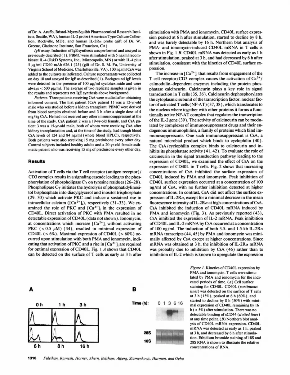
Activation ofT cells via the T cell receptor (antigen receptor)/CD3 complex results in a signaling cascade leading to the phos-phorylation ofphospholipase Cy on tyrosine residues (26-28).Phospholipase C-y initiates the hydrolysis ofphosphatidylinosi-tol bisphosphate into diacylglycerol and inositol trisphosphate(29, 30) which activate PKC and induce a sustained rise inintracellular calcium ([Ca2+]i), respectively (31-33). We ex-amined the role of PKC and [Ca2+]i in the expression ofCD40L. Direct activation of PKC with PMA resulted in nodetectable expression ofCD40L (data not shown). lonomycin,at concentrations which increase [Ca2+]i without activatingPKC (<0.5 IiM) (34), resulted in minimal expression ofCD40L (' 6%). Maximal expression of CD40L (> 60%) oc-curred upon stimulation with both PMA and ionomycin, indi-cating that activation ofPKC and a rise in [Ca2+]i are requiredfor optimal expression of CD40L. Fig. 1 A shows that CD40Lcan be detected on the surface of T cells as early as 3 h after
stimulation with PMA and ionomycin. CD40L surface expres-sion peaked at 6 h after stimulation, started to decline by 8 h,and was barely detectable by 16 h. Northern blot analysis ofPMA- and ionomycin-induced CD40L mRNA in T cells isshown in Fig. 1 B. CD40L mRNA was detected as early as 1 hafter stimulation, peaked at 3 h, and had decreased by 6 h afterstimulation, consistent with the kinetics ofCD40L surface ex-pression.
The increase in [Ca2"]i that results from engagement oftheT cell receptor/CD3 complex causes the activation of Ca2"/calmodulin-dependent enzymes including the protein phos-phatase calcineurin. Calcineurin plays a key role in signaltransduction in T cells (35, 36). Calcineurin dephosphorylatesthe cytoplasmic subunit ofthe transcription factor, nuclear fac-tor ofactivated T cells (NF-AT) (37, 38), which translocates tothe nucleus where together with other proteins it forms a func-tionally active NF-AT complex that regulates the transcriptionofthe IL-2 gene (39). The activity ofcalcineurin can be modu-lated by complexes of immunosuppressant drugs and their en-dogenous immunophilins, a family of proteins which bind im-munosuppressants. One such immunosuppressant is CsA, anatural microbial product which binds to cyclophilin (40).The CsA/cyclophilin complex binds to calcineurin and in-hibits its phosphatase activity (41, 42). To evaluate the role ofcalcineurin in the signal transduction pathway leading to theexpression of CD40L, we examined the effect of CsA on theexpression of CD40L in T cells. Fig. 2 shows that increasingconcentrations of CsA inhibited the surface expression ofCD40L induced by PMA and ionomycin. Peak inhibition ofCD40L surface expression occurred at a concentration of 100ng/ml of CsA, with no further inhibition detected at higherconcentrations. In contrast, CsA did not affect the surface ex-pression of IL-2Ra, except for a minimal decrease in the meanfluorescence intensity ofIL-2Ra at high concentrations ofCsA.CsA inhibited the induction of CD40L mRNA induced byPMA and ionomycin (Fig. 3). As previously reported (43),CsA inhibited the expression of IL-2 mRNA. Peak inhibitionofCD40L and IL-2 mRNA by CsA occurred at a concentrationof 100 ng/ml. The induction of both 3.5- and 1.5-kb IL-2RamRNA transcripts (44,45) by PMA and ionomycin was mini-mally affected by CsA except at higher concentrations. SincemRNA was obtained at 3 h, the inhibition of IL-2Ra mRNAwas probably due to inhibition by CsA (46) rather than toinhibition of IL-2 which is known to upregulate the expression
B
I h 3h
8 h
Thne (h): 0 1
18S16 h
Figure 1. Kinetics ofCD40L expression byPMA and ionomycin. T cells were stimu-lated by PMA and ionomycin for the indi-cated periods of time. (A) Cell surfacestaining for CD40L. CD4OL (continuouslines) was detected on the surface ofT cellsat 3 h ( 15%), peaked at 6 h (60%), and
3 6 16started to decline by 8 h (50%) with mini-mal expression ofCD40L remaining by 16
_'~ h (< 5%) after stimulation. There was nodetectable binding ofsCD44 (dotted lines)at any time point. (B) Northern blot anal-ysis of CD40L mRNA expression. CD40LmRNA was detected as early as 1 h, peakedat 3 h, and decreased by 6 h after stimula-tion. Ethidium bromide staining of 18S and28S RNA is shown to illustrate the relativeconcentrations of RNA.
1316 Fuleihan, Ramesh, Horner, Ahern, Belshaw, Alberg, Stamenkovic, Harmon, and Geha
A
O h
Ki:^ ef6 h

0 0
+ + +
1 10 100 1000
Figure 2. Inhibition ofCD40L surface expression by CsA. T cells were preincubated with medium alone or with the indicated concentrations ofCsA for 1 h before stimulation with PMA and ionomycin. CD40L and CD25 are shown by a continuous line, and relevant isotype controls areshown by a dotted line. CsA inhibited cell surface expression ofCD40L in T cells in a concentration-dependent manner. CD40L was expressed by62% ofthe T cells in the absence ofCsA and decreased to 58, 51, 4, and 8% with 1, 10, 100, and 1,000 ng/ml CsA, respectively. IL-2Ra expres-sion was minimally affected by CsA. All stimulated T cells expressed IL-2Ra in the presence or absence of CsA. CsA caused a minimal decreasein the mean fluorescence intensity ofIL-2Ra ( 144 in untreated vs 105 at 1 qg/ml CsA). The results are representative ofthree different experiments.
of its receptor (47). Inhibition of CD40L expression by CsAwas not due to inhibition of IL-2 expression because additionofIL-2 (100 U/ml) did not reverse CsA-mediated inhibition ofCD40L mRNA or surface expression (data not shown).
The role of calcineurin in CD40L expression was furtherinvestigated using analogues ofCsA with different affinities forcyclophilin and calcineurin. MeBm2t '-CsA, which has a muchlower affinity for cyclophilin than CsA (K, 500 nM vs 6.0) butwhose cyclophilin complex has a higher affinity for calcineurin(Ki 13 nM vs 40) (22), inhibited PMA- and ionomycin-in-duced CD40L surface expression (Fig. 4). MeBm2t'-CsA at150 ng/ml almost completely inhibited PMA- and ionomycin-induced CD40L expression and therefore was only slightly lessactive than CsA despite an 80-90-fold weaker affinity for cy-clophilin. MeAla6-CsA, which has a slightly lower affinity forcyclophilin than CsA (Ki 9.0 nM vs 6.0) but whose cyclophilincomplex has a much lower affinity for calcineurin (K, > 1x 10³ nM vs 40) (22), did not inhibit PMA- and ionomycin-induced CD40L surface expression even at concentrations ashigh as I ,ug/ml (Fig. 4). These results indicate that calcineurin
2 30 0 1 10 10 10-+ + ++ +
S..
,B ..
*09 Uit
ma.."
Figure 3. Inhibition ofCD40L mRNA expres-sion by CsA. T cellswere preincubated withthe indicated concentra-tions ofCsA for 1 h be-fore stimulation withPMA and ionomycin.CsA inhibited CD4OLand IL-2 mRNA ex-pression in a concentra-tion-dependent manner.IL-2Ra mRNA expres-sion was minimally af-fected by CsA. Ethi-dium bromide stainingof 18S and 28S RNAis shown to illustrate therelative concentrationsof RNA. The results arerepresentative of threedifferent experiments.
has a direct role in the signaling pathway regulating CD40Lexpression.
The functional importance of CsA-induced inhibition ofCD40L expression was investigated by assessing the effect ofCsA on immunoglobulin isotype switching to IgE. IL-4-in-duced IgE isotype switching in PBMC is T cell dependent (1)and is inhibited by sCD40 (12), indicating that CD40L expres-sion by T cells plays a critical role in T cell-dependent IL-4-driven IgE isotype switching. Table I shows that CsA inhibitedT cell-dependent IL-4-induced IgE synthesis in PBMC de-rived from three different individuals. The effect of CsA wasexerted at the T cell level because CsA did not inhibit T cell-in-dependent IgE synthesis induced by CD40 mAb and IL-4.Therefore inhibition ofCD40L expression by CsA is associatedwith inhibition of CD40L-mediated T cell-dependent B cellfunction.
Finally, we investigated CD40L expression in PBMC de-rived from CsA-treated transplant recipients. One patient wasstudied before and 3 h after the administration of a single doseof4 mg/kg CsA within the period when blood CsA levels peak,2-4 h after oral administration (48). Fig. 5 A shows that PBMCderived from the patient before the administration of CsA ex-pressed CD40L after stimulation with PMA and ionomycin.Administration of a single dose of CsA inhibited, within 3 h,CD40L expression in PBMC. This effect was selective toCD40L because administration of CsA did not inhibit PMA-and ionomycin-induced IL-2Ra expression. Two other kidneytransplant patients who were receiving CsA and low dose alter-nate day prednisone were also studied. In contrast to PBMCderived from healthy adult subjects or from an asthmatic pa-tient receiving equivalent doses of alternate day steroids,PBMC derived from the transplant patients receiving CsA andalternate day steroids failed to express CD40L on their surfaceupon stimulation with PMA and ionomycin (Fig. 5 B). PBMCfrom patients and controls expressed IL-2Ra normally. There-fore, administration ofCsA in vivo inhibits PMA- and ionomy-cin-induced CD40L expression in PBMC.
Discussion
Our results suggest that CD40L gene expression in T lympho-cytes is under strict regulatory control. Both activation ofPKC
Cyclosporin A Inhibits CD40 Ligand Expression in T Lymphocytes 1317
PMA+Io:
CM (g):
CD40L
IL-2RA ILAKL
CsA (ng/ml):PMA+lo:
CD40L
IL-2
IL-2R
28S
RNA
18S

K1(nM) N (nW) CD40LCompound Concentrtion for for surface
nglml cyclophilin calcineurin expreson
- - 60%
CsA
MIeBny-A
100 6.0 40 <1%
150
beAla -CsA 1000
500 13 10%
9.0 ,1OOO 58%
Figure 4. Effect of CsA analogues on the expression ofCD40L. T cells were preincubated with medium aloneor with increasing concentrations of CsA or its analoguesMeBm2t '-CsA and MeAla6-CsA for 1 h before stimula-tion with PMA and ionomycin. The relative affinities ofCsA and its analogues to cyclophilin and to calcineurinare shown (22). MeBm2t '-CsA inhibited CD4OL expres-sion by 60% at 100 ng/ml (data not shown) and by 85%at 150 ng/ml. However, MeAla6-CsA did not inhibitCD40L expression at concentrations ranging from 100ng/ml to 1 zg/ml (data for 1 jlg/ml shown). Binding ofsCD44 and sCD40 are shown in dotted and continuouslines, respectively. The results are representative of threedifferent experiments.
and a rise in [Ca2"Ji are required for CD40L gene expression.CD40L mRNA is detectable as early as 1 h after stimulationbut disappears by 16 h. Similarly, CD40L surface expression isdetectable as early as 3 h after stimulation and is barely detect-able by 16 h.
CsA inhibited CD40L mRNA and surface expression with-out significantly affecting IL-2Ra mRNA or surface expres-sion. CsA appears to exert its inhibitory effect through inhibi-tion of the Ca2+/calmodulin-dependent protein phosphatasecalcineurin. The ability of CsA analogues to inhibit CD40Lexpression correlated with the ability of each compound toinhibit calcineurin activity. These results indicate that calci-neurin plays a key role in the activation ofCD40L gene expres-sion.
Experiments involving overexpression of calcineurin inJurkat T cells suggest that NF-AT is a target of calcineurin(35). More recently, NF-AT has been shown to be a substratefor calcineurin in vitro (38). It is therefore likely that NF-ATmay play an important role in the regulation oftranscription ofthe CD40L gene. A role for NF-AT may also explain the re-quirement for activation of PKC. PKC activates several nu-clear transcription factors including members of the fos andjun family ofproteins which participate in a functional NF-AT
Table I. Effect ofCsA on CD40L-dependent IgE Isotype Switching
IL-4 CD40 mAb plus IL-4
Subject Medium - +CsA - +CsA
1 290 13,900 2,700 24,300 28,2002 100 27,700 20 22,900 24,0003 10 4,300 50 37,800 39,300
PBMC derived from three healthy volunteers were stimulated for IgEsynthesis (picograms per milliliter) as described in Methods. CsA wasadded where indicated at a concentration of 100 ng/ml. CsA inhibitedIL-4-induced CD40L-dependent IgE synthesis but did not inhibit Tcell-independent IgE synthesis induced by CD40 mAb plus IL-4.
complex (49-51 ). However, it is also possible that other tran-scription factors activated by calcineurin and/or by PKC, suchas NF-IL2A (35) may play an important role in the expressionof the CD40L gene. The exact determination of the regulatoryelements which control transcription ofthe CD40L gene awaitsdetailed characterization of the human CD40L 5' regulatoryregion. Preliminary data generated in our laboratory indicatethe presence of NF-AT-like sequences in the 5' region of themouse CD40L gene and that nuclear extracts obtained fromactivated T cells bind to these sequences in electromobilityshift assays. This supports the notion that CsA may inhibitCD40L expression by inhibiting calcineurin-dependent NF-AT activation.
Inhibition of CD40L expression by CsA prevented T cellsfrom delivering CD40L-dependent signals to B cells. The in-duction ofIgE synthesis in PBMC is T cell dependent (1 ) and ismediated by CD40L (12). In contrast, CD40 mAb and IL-4induce T cell-independent IgE synthesis. CsA inhibited T cell-dependent IL-4-driven IgE synthesis in PBMC but did notinhibit IgE synthesis induced by CD40 mAb and IL-4. Theseresults indicate that CsA inhibited CD40L-dependent IgE iso-type switching.
Inhibition ofCD40L expression by CsA may have implica-tions for patients receiving CsA. PBMC derived from trans-plant patients receiving CsA failed to express CD40L uponstimulation with PMA and ionomycin. Inhibition of CD40Lexpression was not due to the concurrent use of steroids be-cause PBMC derived from an asthmatic patient, receiving anequivalent dose of alternate day steroids, expressed CD40Lupon stimulation. In addition, one of the three patients wasstudied after receiving one dose ofCsA and before the adminis-tration of steroids. It is therefore likely that T lymphocytes, intransplant patients receiving CsA, are unable to interact with Bcells through the CD40L/CD40 axis in vivo. Since CD40L isessential for immunoglobulin isotype switching ( 1 3), patientsreceiving CsA may not be able to undergo immunoglobulinisotype switching in response to newly encountered antigens.Administration of intravenous immunoglobulin may decreasethe frequency ofinfections in these patients. This may be partic-
1318 Fuleihan, Ramesh, Horner, Ahern, Belshaw, Alberg, Stamenkovic, Harmon, and Geha

CD40L CD40L
CD25 CD25
Figure 5. Administration of CsA in vivo inhibits CD40L in PBMC. PBMC were stained for CD40L 6 h after stimulation with PMA and iono-mycin and for IL-2Ra 24 h after stimulation. CD4OL and CD25 are shown by a continuous line, and relevant isotype controls are shown by a
dotted line. (A) PBMC derived from a transplant patient expressed CD4OL before the administration ofCsA but not 3 h after the administrationofa single dose ofCsA. IL-2Ra expression was not inhibited by CsA. (B) PBMC derived from two transplant patients receiving CsA and alternateday steroids failed to express CD4OL but expressed IL-2Ra normally. PBMC derived from control subject and from an asthmatic patient re-
ceiving alternate day steroids expressed CD4OL and IL-2Ra after stimulation.
ularly significant in pediatric patients who may not have hadthe opportunity to develop a wide range of antibodies beforereceiving CsA. Further studies are needed to demonstrate aneffect of CsA on in vivo antibody responses.
Acknowledgments
We thank Mary Drew for her assistance.This work was supported by U.S. Public Health Service grants RR-
02172, A131136, and A131541 to R. S. Geha and CA55735 to I. Sta-menkovic; by a grant from the March of Dimes 6-0760 (R. S. Geha);and by grants from Caremark, Critical Care of America and HomeNutritional Services. R. Fuleihan is supported by grants from the Im-mune Deficiency Foundation (Cutter Award) and the Lucille P. Mar-key trust. I. Stamenkovic is a scholar ofthe Leukemia Society ofAmer-ica. Work done by D. G. Alberg and P. J. Belshaw in the laboratories ofS. L. Schreiber (Department of Chemistry, Harvard University) wassupported by grants from the National Institute of General MedicalSciences (GM-38627 and GM-40660 to S. L. Schreiber), a NationalSciences and Engineering Research Council postgraduate fellowship(to P. J. Belshaw), and a National Institutes of Health postdoctoralfellowship (to D. G. Alberg).
References
1. Vercelli, D., H. H. Jabara, K.-I. Arai, and R. S. Geha. 1989. Induction ofhuman IgE synthesis requires interleukin 4 and T/B cell interactions involvingthe T cell receptor/CD3 complex and MHC class II antigens. J. Exp. Med.169:1295-1307.
2. Armitage, R. J., W. C. Fanslow, L. Strockbine, T. A. Sato, K. N. Clifford,B. M. Macduff, D. M. Anderson, S. D. Gimpel, T. Davis-Smith, C. R. Malis-zewski, et al. 1992. Molecular and biologic characterization ofa murine ligand forCD40. Nature (Lond.). 357:80-82.
3. Noelle, R. J., M. Roy, D. M. Shepherd, I. Stamenkovic, J. A. Ledbetter, andA. Aruffo. 1992. A 39-kDa protein on activated helper T cells binds CD40 andtransduces the signal for cognate activation ofB cells. Proc. Nall. Acad. Sci. USA.89:6550-6554.
4. Hollenbaugh, D., L. S. Grosmaire, C. D. Kullas, N. J. Chalupny, S.Braesch-Anderson, R. J. Noelle, I. Stamenkovic, J. A. Ledbetter, and A. Aruffo.1992. The human T cell antigen gp39, a member of the TNF gene family, is aligand for the CD40 receptor: expression of a soluble form of gp39 with B cellco-stimulatory activity. EMBO (Eur. Mol. Biol. Organ.) J. 1 1:4313-4321.
5. Graf, D., U. Korthauer, H. W. Mages, G. Senger, and R. A. Kroczek. 1992.Cloning of TRAP, a ligand for CD40 on human T cells. Eur. J. Immunol.22:3191-3194.
6. Stamenkovic, I., E. A. Clark, and B. Seed. 1989. A B-lymphocyte activationmolecule related to the nerve growth factor receptor and induced by cytokines incarcinomas. EMBO (Eur. Mol. Biol. Organ.) J. 8:1403-1410.
7. Jabara, H. H., S. M. Fu, R. S. Geha, and D. Vercelli. 1990. CD40 and IgE:
Synergism between anti-CD40 monoclonal antibody and interleukin 4 in theinduction of IgE synthesis by highly purified human B cells. J. Exp. Med.172:1861-1864.
8. Barrett, T. B., G. Shu, and E. A. Clark. 1991. CD40 signalling activatesCDl la/CD18 (LFA-I )-mediated adhesion in B cells. J. Immunol. 146:1722-1729.
9. Lederman, S., M. J. Yellin, A. Krichevsky, J. Belko, J. J. Lee, and L. Chess.1992. Identification of a novel surface protein on activated CD4' T cells thatinduces contact-dependent B cell differentiation (help). J. Exp. Med. 175:1091-1101.
10. Spriggs, M. K., R. J. Armitage, L. Strockbine, K. N. Clifford, B. M.Macduff, T. A. Sato, C. R. Maliszewski, and W. C. Fanslow. 1992. Recombinanthuman CD40 ligand stimulates B cell proliferation and immunoglobulin E secre-tion. J. Exp. Med. 176:1543-1550.
11. Lane, P., T. Brocker, S. Hubele, E. Padovan, A. Lanzavecchia, and F.McConnell. 1993. Soluble CD40 ligand can replace the normal T cell-derivedCD40 ligand signal to B cells in T cell-dependent activation. J. Exp. Med.177:1209-1213.
12. Fanslow, W., D. Anderson, K. H. Grabstein, E. A. Clark, D. Cosman, andR. Armitage. 1992. Soluble forms ofCD40 inhibit biologic responses ofhuman Bcells. J. Immunol. 149:655-660.
13. Fuleihan, R., N. Ramesh, R. Loh, H. Jabara, F. S. Rosen, T. Chatila, S. M.Fu, I. Stamenkovic, and R. S. Geha. 1993. Defective expression of the CD40ligand in X chromosome-linked immunoglobulin deficiency with normal or ele-vated IgM. Proc. Nall. Acad. Sci. USA. 90:2170-2173.
14. Ramesh, N., R. Fuleihan, V. Ramesh, S. Sharma, F. S. Rosen, and R. S.Geha. 1993. Mutations in the T cell antigen CD40 ligand in X-linked immuno-globulin deficiency with normal or elevated IgM (HIGMX-1 ). Int. Immunol.5:769-773.
15. Aruffo, A., M. Farrington, D. Hollenbaugh, X. Li, A. Milatovich, S.Nonoyama, J. Bajorath, L. S. Grosmaire, R. Stenkamp, M. Neubauer, et al. 1993.The CD40 ligand, gp39, is defective in activated T cells from patients with X-linked hyper IgM syndrome. Cell. 72:291-300.
16. Korthauer, U., D. Graf, H. W. Mages, F. Briere, M. Padayachee, S. Mal-colm, A. G. Ugazio, L. D. Notarangelo, R. J. Levinsky, and R. A. Kroczek. 1993.Defective expression of T-cell CD40 ligand causes X-linked immunodeficiencywith hyper-IgM. Nature (Lond). 361:539-541.
17. DiSanto, J. P., J. Y. Bonnefoy, J. F. Gauchat, A. Fischer, and G. de SaintBasile. 1993. CD40 ligand mutations in X-linked immunodeficiency with hyper-IgM. Nature (Lond.). 361:541-543.
18. Allen, R. C., R. J. Armitage, M. E. Conley, H. Rosenblatt, N. A. Jenkins,N. G. Copeland, M. A. Bedell, S. Edelhoff, C. M. Disteche, D. K. Simoneaux et al.1993. CD40 ligand gene defects responsible for X-linked hyper-IgM syndrome.Science (Wash. DC). 259:990-993.
19. Geha, R. S., N. Hyslop, S. Alami, F. Farah, E. E. Schneeberger, and F. S.Rosen. 1979. Hyper immunoglobulin M immunodeficiency (dysgammaglobu-linemia). Presence ofimmunoglobulin M-secreting plasmacytoid cells in periph-eral blood and failure ofimmunoglobulin M-immunoglobulin G switch in B-celldifferentiation. J. Clin. Invest. 64:385-391.
20. Levitt, D., P. Haber, K. Rich, and M. D. Cooper. 1983. Hyper IgM immu-nodeficiency. A primary dysfunction ofB lymphocyte isotype switching. J. Clin.Invest. 72:1650-1657.
21. Notarangelo, L. D., M. Duse, and A. G. Ugazio. 1992. Immunodeficiencywith hyper-IgM (HIM). Immunol. Rev. 3:101-122.
Cyclosporin A Inhibits CD40 Ligand Expression in T Lymphocytes 1319
APre CsA Post CsA
B Normal Control CsA Patient 2 CsA Patient 3 Asthma Control

22. Liu, J., M. W. Albers, T. J. Wandless, S. Luan, D. G. Alberg, P. J. Belshaw,P. Cohen, C. MacKintosh, C. B. Klee, and S. L. Schreiber. 1992. Inhibition ofTcell signaling by immunophilin-ligand complexes correlates with loss of calci-neurin phosphatase activity. Biochemistry. 31:3896-3901.
23. Stamenkovic, I., M. Amiot, J. M. Pesando, and B. Seed. 1989. A lympho-cyte molecule implicated in lymph node homing is a member ofthe cartilage linkprotein family. Cell. 56:1057-1062.
24. Aruffo, A., I. Stamenkovic, M. Melnick, C. B. Underhill, and B. Seed.1990. CD44 is the principal cell surface receptor for hyaluronate. Cell. 61:1303-1313.
25. Gruber, M. F., J. M. Bjomdahl, S. Nakamura, and S. M. Fu. 1989. Anti-CD45 inhibition of human B cell proliferation depends on the nature of activa-tion signals and the state of B cell activation. J. Immunol. 142:4144-4152.
26. Park, D. J., H. W. Rho, and S. G. Rhee. 1991. CD3 stimulation causesphosphorylation of phospholipase C-y 1 on serine and tyrosine residues in a hu-man T cell line. Proc. Natl. Acad. Sci. USA. 88:5453-5456.
27. Secrist, J. P., L. Karnitz, and R. T. Abraham. 1991. T-cell antigen receptorligation induces tyrosine phosphorylation of phospholipase C-ey 1. J. Biol. Chem.266:12135-12139.
28. Weiss, A., G. Koretzky, R. C. Schatzman, and T. Kadlecek. 1991. Func-tional activation of the T cell antigen receptor induces tyrosine phosphorylationof phospholipase C-yl. Proc. Natl. Acad. Sci. USA. 88:5484-5488.
29. Dasgupta, J. D., C. Granja, B. Druker, L. Lin, E. J. Yunis, and V. Relias.1992. Phospholipase Cy I association with CD3 structure in T cells. J. Exp. Med.175:285-288.
30. Nishibe, S., M. I. Wahl, S. M. Hemandez-Stomomayor, N. K. Tonk, S. G.Rhee, and G. Carpenter. 1990. Increase in the catalytic activity of phospholipaseC--y I by tyrosine phosphorylation. Science (Wash. DC). 250:1253-1256.
31. Nishizuka, Y. 1984. The role of protein kinase C in cell surface signaltransduction and tumour promotion. Nature (Lond.). 308:693-698.
32. Berridge, M. J., and R. F. Irvine. 1984. Inositol trisphosphate, a novelsecond messenger in cellular signal transduction. Nature (Lond.). 312:315-321.
33. Imboden, J. B., and J. D. Stobo. 1985. Transmembrane signaling by the Tcell antigen receptor. Perturbation ofthe T3-antigen receptor complex generatesinositol phosphates and releases calcium ions from intracellular stores. J. Exp.Med. 161:446-456.
34. Chatila, T., L. Silverman, R. Miller, and R. Geha. 1989. Mechanisms ofTcell activation by the calcium ionophore ionomycin. J. Immunol. 143:1283-1289.
35. Clipstone, N. A., and G. R. Crabtree. 1992. Identification ofcalcineurin asa key signalling enzyme in T-lymphocyte activation. Nature (Lond.). 357:695-697.
36. O'Keefe, S. J., and J. T. Tamura. 1992. FK-506- and CsA-sensitive activa-tion ofthe interleukin-2 promoter by calcineurin. Nature (Lond.). 357:692-694.
37. Schreiber, S. L., and G. R. Crabtree. 1992. The mechanism of action ofcyclosporin A and FK506. Immunol. Today. 13:136-142.
38. Jain, J., P. G. McCaffrey, Z. Miner, T. K. Kerppola, J. N. Lambert, G. L.Verdine, T. Curran, and A. Rao. 1993. The T-cell transcription factorNFATp is asubstrate for calcineurin and interacts with Fos and Jun. Nature (Lond.).365:352-355.
39. Flanagan, W. F., B. Corthesy, R. J. Bram, and G. R. Crabtree. 1991.Nuclear association ofa T-cell transcription factor blocked by FK-506 and cyclo-sporin A. Nature (Lond.). 352:803-807.
40. Handschumacher, R. E., M. W. Harding, J. Rice, R. J. Drugge, and D. W.Speicher. 1984. Cyclophilin: a specific cytosolic binding protein for cyclosporinA. Science (Wash. DC). 226:544-546.
41. Liu, J., J. D. Farmer, W. S. Lane, J. Friedman, I. Weissman, and S. L.Schrieber. 1991. Calcineurin is a common target of cyclophilin-cyclosporin Aand FKBP-FK506 complexes. Cell. 66:807-815.
42. Fruman, D. A., C. B. Klee, B. E. Bierer, and S. J. Burakoff. 1992. Calci-neurin phophatase activity in T lymphocytes is inhibited by FK 506 and cyclo-sporin A. Proc. Natl. Acad. Sci. USA. 89:3686-3690.
43. Kronke, M., W. J. Leonard, J. M. Depper, S. K. Arya, F. Wong-Staal,R. C. Gallo, T. A. Waldmann, and W. C. Greene. 1984. Cyclosporin A inhibits Tcell growth factor gene expression at the level ofmRNA transcription. Proc. Natl.Acad. Sci. USA. 81:5214-5218.
44. Leonard, W. J., J. M. Depper, G. R. Crabtree, S. Rudikoff, J. Pumphrey,R. J. Robb, M. Kronke, P. B. Svetlik, N. J. Peffer, T. A. Waldmann, and W. C.Greene. 1984. Molecular cloning and expression ofcDNAs for the human inter-leukin-2 receptor. Nature (Lond.). 311:626-63 1.
45. Nikaido, T., A. Shimizu, N. Ishida, H. Sabe, K. Teshigawara, M. Maeda,T. Uchiyama, J. Yodoi, and T. Honjo. 1984. Molecular cloning ofcDNA encod-ing human interleukin-2 receptor. Nature (Lond.). 311:631-635.
46. Reed, J. C., A. H. Abidi, J. D. Alpers, R. G. Hoover, R. J. Robb, and P. C.Nowell. 1986. Effect ofcyclosporin A and dexamethasone on interleukin 2 recep-tor gene expression. J. Immunol. 137:150-154.
47. Depper, J. M., W. J. Leonard, C. Drogula, M. Kronke, T. A. Waldmann,and W. C. Greene. 1985. Interleukin 2 (IL-2) augments transcription ofthe IL-2receptor gene. Proc. Natl. Acad. Sci. USA. 82:4230-4234.
48. Handschumaker, R. E. 1990. Immunosuppressive agents. In The Pharma-cological Basis of Therapeutics. A. G. Gilman, T. W. Rall, A. S. Nies, P. Taylor,editors. McGraw-Hill Inc., New York. 1264-1274.
49. Castigli, E., T. A. Chatila, and R. S. Geha. 1993. A protein of the AP-1family is a component of nuclear factor of activated T-cells (NFAT). J. Im-munol. 150:3284-3290.
50. Boise, L. H., B. Petryniak, X. Mao, C. H. June, C.-Y. Wang, T. Lindsten,R. Bravo, K. Kovary, J. M. Leiden, and C. B. Thompson. 1993. The NFAT-1DNA binding complex in activated T cells contains Fra- 1 and JunB. Mol. Cell.Biol. 13:1911-1919.
51. Jain, J., P. G. McCaffrey, V. E. Valge-Archer, and A. Rao. 1992. Nuclearfactor of activated T cells contains Fos and Jun. Nature (Lond.). 356:801-804.
1320 Fuleihan, Ramesh, Horner, Ahern, Belshaw, Alberg, Stamenkovic, Harmon, and Geha


