Embed Size (px)
Citation preview

ANTIMICROBIAL AGENTS AND CHEMOTHERAPY, Aug. 2009, p. 3226–3235 Vol. 53, No. 80066-4804/09/$08.00�0 doi:10.1128/AAC.00189-09Copyright © 2009, American Society for Microbiology. All Rights Reserved.
Cyclosporine Inhibits Flavivirus Replication through Blocking theInteraction between Host Cyclophilins and Viral NS5 Protein�
Min Qing,1,2 Feng Yang,3 Bo Zhang,2 Gang Zou,1,2 John M. Robida,3 Zhiming Yuan,1Hengli Tang,3* and Pei-Yong Shi2,4*
State Key Laboratory of Virology, Wuhan Institute of Virology, Chinese Academy of Sciences, Wuhan 430071, China1; Wadsworth Center,New York State Department of Health, Albany, New York 122012; Department of Biological Science, Florida State University,
Tallahassee, Florida 323063; and Department of Biomedical Sciences, University at Albany,State University of New York, Albany, New York 122084
Received 11 February 2009/Returned for modification 27 March 2009/Accepted 12 May 2009
Although flaviviruses cause significant human diseases, no effective therapy is currently available. Hostfactors essential for viral replication are potential targets for antiviral development. Here we report thatcyclophilins (CyPs), a family of cellular peptidyl-prolyl isomerases (PPIases), play a role in flavivirus repli-cation. Huh-7.5 cells with knockdown of different isoforms of CyP were less efficient than parental cells insupporting flavivirus replication, including West Nile virus (WNV), dengue virus, and yellow fever virus. Thelow viral replication in CyP A (CyPA) knockdown cells could be rescued by trans supplying of a wild-type CyPAbut not by trans supplying of a mutant CyPA (defective in the PPIase activity), indicating that the isomeraseactivity of CyPA is critical for viral replication. Immunoprecipitation and biochemical pulldown analysesshowed that CyPA interacts with WNV genomic RNA and viral NS5 protein in the replication complex.Furthermore, antiviral experiments demonstrated that cyclosporine (Cs; an 11-amino-acid cyclic peptideknown to block the PPIase activity of CyPA) inhibits flavivirus replication in cell culture at nontoxic concen-trations. Time-of-addition and transient replicon results indicated that Cs inhibits flavivirus at the step of viralRNA synthesis. Biochemical analysis showed that Cs directly blocks the interaction between CyPA and WNVNS5 protein. Our results suggest that host CyPA is a component of flavivirus replication complex and couldbe targeted for potential antiviral development.
The family Flaviviridae includes three genera: Flavivirus, Pes-tivirus, and Hepacivirus. Many members from the genus Flavi-virus are arthropod-borne and cause significant human mor-bidity and mortality, such as the four serotypes of dengue virus(DENV), West Nile virus (WNV), yellow fever virus (YFV),Japanese encephalitis virus (JEV), and tick-borne encephalitisvirus. The DENV alone poses a risk to 2.5 billion peopleworldwide and causes 50 to 100 million human cases each year.YFV and JEV infect 200,000 and 50,000 people every year,respectively (17). Since the outbreak of WNV in New YorkCity in 1999, the virus has caused thousands of human infec-tions in the United States, representing the largest meningo-encephalitis outbreak in the Western Hemisphere and the big-gest WNV outbreak ever reported (23). No effective antiviraltherapy has been approved for clinical treatment of flavivirusinfections. Human vaccines are currently available only forYFV, JEV, and TBEV (17). Understanding the molecularmechanism of viral replication is essential for the preventionand treatment of flavivirus infections.
Flaviviruses are spherical in shape, with a diameter of 50 nm(24). The viral genome is a single-stranded, plus-sense RNA, ofabout 11,000 nucleotides (nt) in length (27). The single open
reading frame of the viral genome encodes a polyprotein,which is processed by viral and cellular proteases into threestructural proteins (capsid [C], premembrane [prM] or mem-brane [M], and envelope [E]) and seven nonstructural proteins(NS1, NS2A, NS2B, NS3, NS4A, NS4B, and NS5) (27). Thestructural proteins are involved in viral particle formation. TheNS proteins are responsible for viral RNA replication but havealso been shown to function in viral assembly (25, 30, 39) andto evade host immune response (3, 18, 29, 37, 38). NS3 acts asa protease (with NS2B as a cofactor) (15, 26), a nucleotidetriphosphatase (51, 54), an RNA triphosphatase (2), and ahelicase (6). NS5 functions as a methyltransferase (13, 43, 57)and an RNA-dependent RNA polymerase (RdRp) (1, 19).Upon translation, viral NS proteins and host factors form thereplication complex to synthesize minus-sense RNA, which, inturn, serves as a template for synthesis of more plus-senseRNA (27).
Cyclophilins (CyPs) are cellular peptidyl-prolyl isomerases(PPIases) which catalyze the isomerization of peptide bondsfrom trans to cis form at proline residues and facilitates proteinfolding. CyPs were shown to be critical for the replication ofhuman immunodeficiency virus (HIV) and hepatitis C virus(HCV). Specifically, CyP A (CyPA) and CyPB were reportedto bind specifically to the HIV-1 Gag polyprotein (33), as wellas bind to the HCV NS5B (53, 56). These virus-host interac-tions have been targeted for antiviral development, as exem-plified by cyclosporine (Cs) and its derivatives, which bind toCyPs with high affinity and block its PPIase activity (40). CsAis a 11-amino-acid cyclic peptide that has been used as animmunosuppressant drug in patients after organ transplants
* Corresponding authors. Mailing address for P.-Y. Shi: 10 BiopolisRoad, #05-01 Chromos, Singapore 138670. Phone: (65) 67222909. Fax:(65) 67222916. E-mail: [email protected]. Mailing address forH. Tang: King Building, Stadium Dr., Department of Biological Science,Florida State University, Tallahassee, FL 32306-4370. Phone: (850)645-2402. Fax: (850) 645-8447. E-mail: [email protected].
� Published ahead of print on 18 May 2009.
3226

(47). The immunosuppression function of Cs is partially ex-plained by the fact that the Cs/CyP complex binds to andinhibits calcineurin, a cellular phosphatase and a key mediatorof T-cell activation (28). Cs was found to inhibit HIV-1 (4, 40)and HCV replication (52). Furthermore, analogues of Cs(NIM811 and DEBIO-025) that bind to CyPs with high affinitybut lack calcineurin-mediated immunosuppressive activity arecurrently in clinical development for HCV treatment (16, 35).DEBIO-025 showed a potent anti-HCV effect in patients coin-fected with hepatitis C and HIV (16).
In the present study, we show that CyP functions in flavivirusreplication. Knockdown of CyP expression in Huh-7.5 cellsreduced flavivirus replication; the replication suppressioncould be rescued by trans supplying of a wild-type (WT) CyPA.An antibody against CyPA could immunoprecipitate WNVRNA from replication complexes, and recombinant CyPAcould pull down viral NS5 protein. The functional role of CyPsin viral replication was further supported by the results that Csinhibited flaviviruses in cell culture. Time-of-addition and tran-sient replicon results indicate that Cs inhibits flavivirus at thestep of RNA synthesis. Biochemical analysis showed that Cscould directly suppress the interaction between CyPA and fla-vivirus NS5 protein. The results suggest that Cs and its clinicalcandidates are potentially useful for the treatment of flavivirusinfections.
MATERIALS AND METHODS
Viruses, compounds, and antibodies. We used the following viruses: WNV(strain 3356), YFV (17D vaccine strain), DENV-2 (strain New Guinea C),Western equine encephalitis virus (WEEV; strain Cova 746), and vesicularstomatitis virus (VSV; New Jersey serotype). The sources of these viruses werereported previously (42). Cs was purchased from Alexis Corp. and was dissolvedin 100% ethanol for antiviral experiments. Antibodies against Myc and CyPAwere obtained from Santa Cruz Biotechnology and Biomol International LP,respectively. Monoclonal antibody against WNV NS5 was generated by immu-nizing mice in-house with recombinant NS5 protein.
CyP knockdown cell lines. An HIV-based lentiviral vector was used to expressall short hairpin RNAs (shRNAs) (50). Huh-7.5 cells were used to establish thefollowing cell lines, each of which contained a stably expressed shRNA directedat a distinct CyP isoform: sh-A161 for CyPA, sh-B710 for CyPB, and sh-C454 forCyPC. A control cell line sh-Fluc, with a shRNA targeting firefly luciferase(Fluc), was also established. These cell lines were generated and reported pre-viously (44, 56). A similar protocol was used in the present study to prepare anew cell line sh-AB, with both CyPA and CyPB knockdown. Figure 1A summa-rizes the target sequences of sh-A, sh-B, sh-C, and sh-AB and their alignmentswith the three isoforms of CyP. Stable cells expressing shRNAs were obtained byselection with 1 �g/ml of puromycin (MP Biomedicals) for 3 weeks. The knock-down cells were maintained in Dulbecco modified Eagle medium with 10% fetalbovine serum, 0.1 mM nonessential amino acids, and 1 �g/ml of puromycin.Huh-7.5 cells were maintained in a similar medium without puromycin.
WNV and DENV-1 replicon-containing cells. The replicon-harboring Verocells for WNV and DENV-1 were established previously (32, 42). Both replicons(WNV-Rluc-Neo-Rep and DENV-1-Rluc-Neo-Rep) contained dual reporters, aRenilla luciferase (Rluc) and a neomycin phosphotransferase (Neo). Both rep-licon cell lines were maintained in Dulbecco modified Eagle medium with 10%fetal bovine serum plus 1 mg/ml of G418. All cells in the present study weremaintained in 5% CO2 at 37°C.
Packaging of VLPs. Flavivirus viruslike particles (VLPs) were prepared bytrans supplying of viral structural proteins to Renilla luciferase-reporting replicon(Rluc2A-Rep) (46). Briefly, 10 �g of replicon RNAs of WNV, DENV-1, or YFVwere electroporated into 8 � 106 BHK-21 cells. Each of the WNV, DENV-1, andYFV replicons has a Renilla luciferase and the foot-and-mouth disease virus 2Asequence substituting for the viral structural genes. The WNV and DENV-1replicons were described before (31, 41); the YFV replicon was a generous giftfrom Richard Kuhn (Purdue University) (22). We used an alphavirus SemlikiForest virus (SFV) replicon to express flavivirus structural genes (SFV-CprME-
Rep) (22, 41). At 24 h posttransfection (p.t.) of replicon RNAs, the cells wereelectroporated again with 10 �g of SFV-CprME-Rep RNAs (expressing homol-ogous viral structural proteins). At 24 h after the second transfection, culturefluids were harvested, divided into aliquots, and stored at �80°C. Both flavivirusRluc2A-Rep RNA and SFV-CprME-Rep RNA were in vitro transcribed fromlinearized DNAs, using a T7 and a SP6 mMESSAGE mMACHINE kit (Am-bion), respectively.
We used indirect immunofluorescence assay (IFA) to estimate the VLP titers.Vero cells in a four-chamber slide (Nalge Nunc International) were infected withserial dilutions of VLP samples, IFA was performed on the infected cells at 24 hpostinfection (p.i.), and IFA-positive cell foci were counted. The VLP titers wereestimated by the number of positive IFA-positive foci and expressed in focus-forming units (FFU)/ml. Immune mouse ascetic fluid of WNV, DENV-1, andYFV (American Type Culture Collection) and goat anti-mouse immunoglobulinG (IgG) conjugated with Texas Red were used as primary and secondary anti-bodies, respectively.
Co-IP and RT-PCR. WNV-Rluc-Neo-Rep Vero cells (5 � 106) were seededinto a T-75 flask 1 day before the coimmunoprecipitation (Co-IP) experiment.After 24 h, the cells were lysed in 1 ml of immunoprecipitation buffer (50 mMTris-HCl [pH 8.0], 150 mM NaCl, 1 mM phenylmethylsulfonyl fluoride, and 0.5%NP-40). Two hundred units of RNaseOUT (Invitrogen) were added to thesupernatant after centrifugation at 12,000 � g for 15 min. The supernatant wasthen added to 50 �l of 75% protein G slurry containing either anti-CyPA orrabbit IgG. The binding was allowed to proceed at 4°C overnight, after which theprotein G-beads were washed with immunoprecipitation buffer four times. RNAwas extracted from the beads with an RNeasy kit (Qiagen). Reverse transcrip-tion-PCR (RT-PCR) was then performed to detect the RdRp region of WNV
FIG. 1. Characterization of Huh-7.5 cells with knockdown of vari-ous CyP isoforms. (A) Sequence alignments of shRNA targets ofdifferent CyP isoforms. Mismatched nucleotide were underlined. Thesequences of CyPA, CyPB, and CyPC are derived from GenBankaccession numbers Y00052 (CyPA), NM_000942 (CyPB), andNM_000943 (CyPC). (B) Silencing of CyP mRNAs in stable Huh-7.5cells transduced with shRNAs. Equal amounts of total cellular RNA (1�g) were subjected to real-time RT-PCR using primer-probe setsspecific for CyPA, CyPB, CyPC, and GAPDH (glyceraldehyde-3-phos-phate dehydrogenase) (56). The amounts of CyPs in Huh-7.5 cells(mock) were set as 100%. The percentages of CyPs in other cell lineswere calculated and indicated after normalization to the GAPDHlevels. The data represent means and standard deviations (n � 3).
VOL. 53, 2009 CYCLOPHILINS FUNCTION IN FLAVIVIRUS REPLICATION 3227

NS5, using the primers 8706-forward (5�-CATGGCCATGACTGACACTACTC-3�) and 9093-reverse (5�-CTTGGCCTTTCCGAACTCTCCG-3�) (45).
Viral growth kinetics. The knockdown cell lines, sh-AB and sh-Fluc, wereseeded in a 12-well plate (4 � 105 cells/well). At 24 h after seeding, the cells wereinfected with WNV, DENV-2, YFV, WEEV, or VSV at an multiplicity ofinfection (MOI) of 0.1. For VSV, samples of culture medium were collected at12, 16, and 24 h p.i. For the other four viruses, samples were collected on 16 h,24 h, 48 h p.i. All virus titers were determined by a double-layer plaque assay onVero cells, as described previously (42).
Transient replicon assay. Portions (10 �g) of luciferase replicon (Rluc2A-Rep) RNA of WNV, YFV, or DENV-1 were electroporated into Huh-7.5 cells(46). The cells were seeded in a 12-well plate (4 � 105 cells/well) and immedi-ately treated with 8 �M Cs or treated with 1% ethanol as a control. At varioustime points, the cells were washed once with cold phosphate-buffered saline(PBS) and added with 250 �1 of 1� lysis buffer (Promega). The plates containingthe lysis buffer were sealed with Parafilm and stored at �80°C. Once the samplesfor all time points had been collected, 20 �l of cell lysis was transferred to a96-well plate and assayed for luciferase signals in a Turner BioSystems lumi-nometer (Promega).
Recombinant CyPA production and GST binding assay. The recombinantprotein of CyPA, with a N-terminal glutathione S-transferase (GST) tag, wasprepared as described previously (56). For the GST pulldown assay, 20 �g ofGST or GST-CyPA was brought to a final volume of 200 �l with binding buffer(20 mM Tris-HCl [pH 7.5], 100 mM KCl, 2 mM CaCl2, 2 mM MgCl2, 5 mMdithiothreitol, 0.5% NP-40, 0.5 mM phenylmethylsulfonyl fluoride, 5% glycerol).WNV-Rluc-Neo-Rep Vero cells (8 � 105) were lysed in 400 �l of immunopre-cipitation buffer supplemented with 1 mM dithiothreitol and 1 mM EDTA.Then, 50 �l portions of the cell lysate were added to the tubes containing GSTor GST-CyPA. The samples were mixed by rotating at 4°C for 1 h. Glutathione-Sepharose 4B beads (25 �l of a 50% slurry; GE Healthcare) were then added toeach sample. After the tubes were rotated at 4°C for 30 min, the beads werewashed three times with binding buffer and then pelleted at 500 � g for 5 min.Proteins bound to the beads were analyzed by sodium dodecyl sulfate-polyacryl-amide gel electrophoresis, followed by Western blotting. To examine the effectsof Cs on interaction between CyPA and WNV NS5, we incubated Cs with therecombinant GST-CyPA and replicon cell lysates; the GST pulldown experimentwas performed exactly as described above.
Coelectroporation of WNV replicon RNA and plasmid DNA expressing CyPA.A 5 �g portion of WNV Rluc2A-Rep RNA was mixed with 9 �g of an emptyplasmid pcDNA3.1, a plasmid expressing WT CyPA, or a plasmid expressingmutant (MT) CyPA. The MT CyPA contained an Ala substitution for Arg atamino acid position 55 of CyPA, resulting in an inactivation of PPIase(Arg55Ala; AGA3GCA) (58). The N termini of both WT and MT CyPAs werefused with three copies of Myc tag (3Myc) for antibody detection. The RNA andDNA mixtures were electroporated into 7.5 � 106 of sh-A cells, which were thenplated onto a 12-well plate (4 � 105 cells per well). The cells were assayed forluciferase activities at various time points p.t. (49).
VLP and virus titer reduction assays. For the virus titer reduction assay,Huh-7.5 cells were seeded in a 12-well plate (4 � 105 cells per well). At 24 h afterseeding, the cells were infected with WNV, DENV-2, YFV, WEEV, or VSV atan MOI of 0.1. The infected cells were immediately treated with Cs at 8 or 20�M. Virus titers in culture medium were quantified by plaque assays at theindicated time points (42).
MTT assay. A cell proliferation-based MTT [3-(4,5-dimethyl-2-thiazolyl)-2,5-diphenyl-2H-tetrazolium bromide] assay (American Type Culture Collection)was used to examine the compound cytotoxicity. Approximately 2 � 104 Verocells or Huh-7.5 cells in 100 �l were seeded in a 96 well-plate. After 16 h ofincubation, the cells were treated with 0, 2.5, 5, 8, 10, 20, and 40 �M Cs. Cs wasdissolved in 100% ethanol and was tested at a 1% final ethanol concentration.After 48 h of incubation, 10 �l of MTT reagent was added to each well. The cellswere incubated for another 4 h, after which 100 �l of detergent reagent wasadded to each well. The plates were swirled gently and left in the dark at roomtemperature overnight. The absorbance was then recorded in a microliter platereader (Molecular Devices Corp.) with a 550-nm filter.
Time-of-addition analysis. Approximately 4 � 105 Huh-7.5 cells were seededin a 12-well plate per well, followed by incubation for 24 h for cell attachment.The cells were infected with WNV or DENV-2 at an MOI of 3 for 1 h at 4°C,followed by three rounds of cold PBS washes to remove the unabsorbed virus. Atdifferent time points p.i., Cs (20 �M for WNV and 8 �M for DENV-2) was addedto the infected cells without a medium change. Culture medium were collectedat 24 h p.i. and assayed for virus titers by using plaque assays (42). As negativecontrols, ethanol was added to the infected cells at a final concentration of 1%at 0, 10, and 20 h p.i. to estimate its effect on viral production.
RESULTS
Knockdown of CyP isoforms reduces the replication of fla-vivirus VLPs. To examine the role of CyP on flavivirus repli-cation, we prepared a panel of CyP knockdown cell lines usingshRNA (Fig. 1). Huh-7.5 cells with individual CyPA, CyPB, orCyPC knockdown were generated by using isoform-specificsh-A, sh-B, and sh-C, respectively; a double-knockdown (CyPAplus CyPB) cell line was established by using sh-AB (Fig. 1A);a control cell line was prepared by using shRNA targetingfirefly luciferase (sh-Fluc). Figure 1A summarizes the targetsequences of sh-A, sh-B, sh-C, and sh-AB and their alignmentswith the three isoforms of CyP. Real-time RT-PCR analysisshowed a significant difference in mRNA expression of the CyPisoforms in the knockdown cells (Fig. 1B). The mRNA levelsfor all three CyPs in parental Huh-7.5 (set as 100% for eachisoform) were similar to those in sh-Fluc cells. In sh-A cells,the mRNA level of CyPA was reduced by 92%, but the CyPBand CyPC mRNAs were increased by 50 and 112%, respec-tively. In sh-B cells, CyPB mRNA was decreased by 86%,whereas the CyPC mRNA was increased by 83%. In sh-C cells,the CyPC level dropped only by 51%, and the CyPA and CyPBexpression did not change substantially (�20%). In the dou-ble-knockdown sh-AB cells, CyPA and CyPB levels were, re-spectively, decreased by 92 and 84%, and the CyPC level wasnot dramatically changed (10%). These results indicated thatspecific isoforms of CyP were knocked down in the cell lines;however, the expression of untargeted isoforms may increasein some of the knockdown cells (e.g., sh-A and sh-B cells),possibly to compensate for the decrease of the sh-RNA-tar-geted isoform.
We used VLPs containing a luciferase reporter to quantifythe effect of CyP knockdown on flavivirus replication. VLPs ofWNV, YFV, and DENV-1 were prepared by trans supplyingluciferase replicons (Flavi-Rluc2A-Rep) with viral structureproteins (Fig. 2A). The structural proteins were expressedthrough an alphavirus SFV replicon (SFV-Flavi-CprME). Thetiters of VLPs were approximately 2.5 � 106, 2 � 105, and2.4 � 103 FFU/ml for WNV, YFV, and DENV-1, respectively.Because of the dramatic difference in VLP titers, we infectedthe knockdown cells with WNV, YFV, and DENV-1 VLPs at1, 0.1, and 0.01 FFU/cell, respectively. Luciferase signals (mea-sured at 24 h p.i.) showed that VLP replication was reduced inalmost all knockdown cells (Fig. 2B); the reduction of lucifer-ase signal is statistically significant (Student t test P � 0.01).One exception was observed, in which the replication of YFVVLP in sh-C cells increased by 15% (Fig. 2B). Of the five celllines, the sh-AB double-knockdown cells yielded the lowestluciferase signals (decreased by 68 to 85%) for all three flavi-virus VLPs (Fig. 2B). Changing the FFU/cell of VLP of theabove experiments did not substantially affect the relative lu-ciferase activities among different CyP knockdown cells (datanot shown). Taken together, the results indicate that CyPs,especially CyPA and CyPB, play a role in flavivirus replication.
The sh-AB cells are compromised in supporting the repli-cation of a number of RNA viruses. To validate the roles ofCyPA and CyPB in flavivirus infection, we compared growthkinetics of authentic WNV, DENV-2, and YFV between thesh-AB cells and the control sh-Fluc cells (Fig. 3). After the cellswere infected under an identical condition (MOI of 0.1), the
3228 QING ET AL. ANTIMICROB. AGENTS CHEMOTHER.

sh-Fluc cells produced more viruses than the sh-AB did. Thedifference in virus titer was most apparent at 24 h p.i.; specif-ically, the sh-AB cells produced 67% (WNV), 89% (DENV-2),and 75% (YFV) less virus than the sh-Fluc cells did. However,at 48 h p.i., the difference became less apparent for DENV-2and YFV; almost no difference was observed for WNV. Inter-estingly, the knockdown of CyPA and CyPB also reduced viralyields of two nonflaviviruses: a plus-strand RNA alphavirus(WEEV) and a negative RNA rhabdovirus (VSV) (Fig. 3).Similar to the flaviviruses, the difference in virus titer reductionwas more dramatic at early time points (�24 h p.i. for WEEVand �16 h p.i. for VSV) than those at late time points. TheStudent t test showed that the growth kinetics of the WNVbetween the two cell lines are not statistically significant, witha P value of 0.19. However, the growth curves of DENV-2,YFV, WEEV, and VSV between the two cell lines are statis-tically significant, with P values of 0.04, 0.03, 0.001, and 0.002,respectively. Overall, the results indicate that CyPA and/orCyPB function in both flavivirus and nonflavivirus replications.
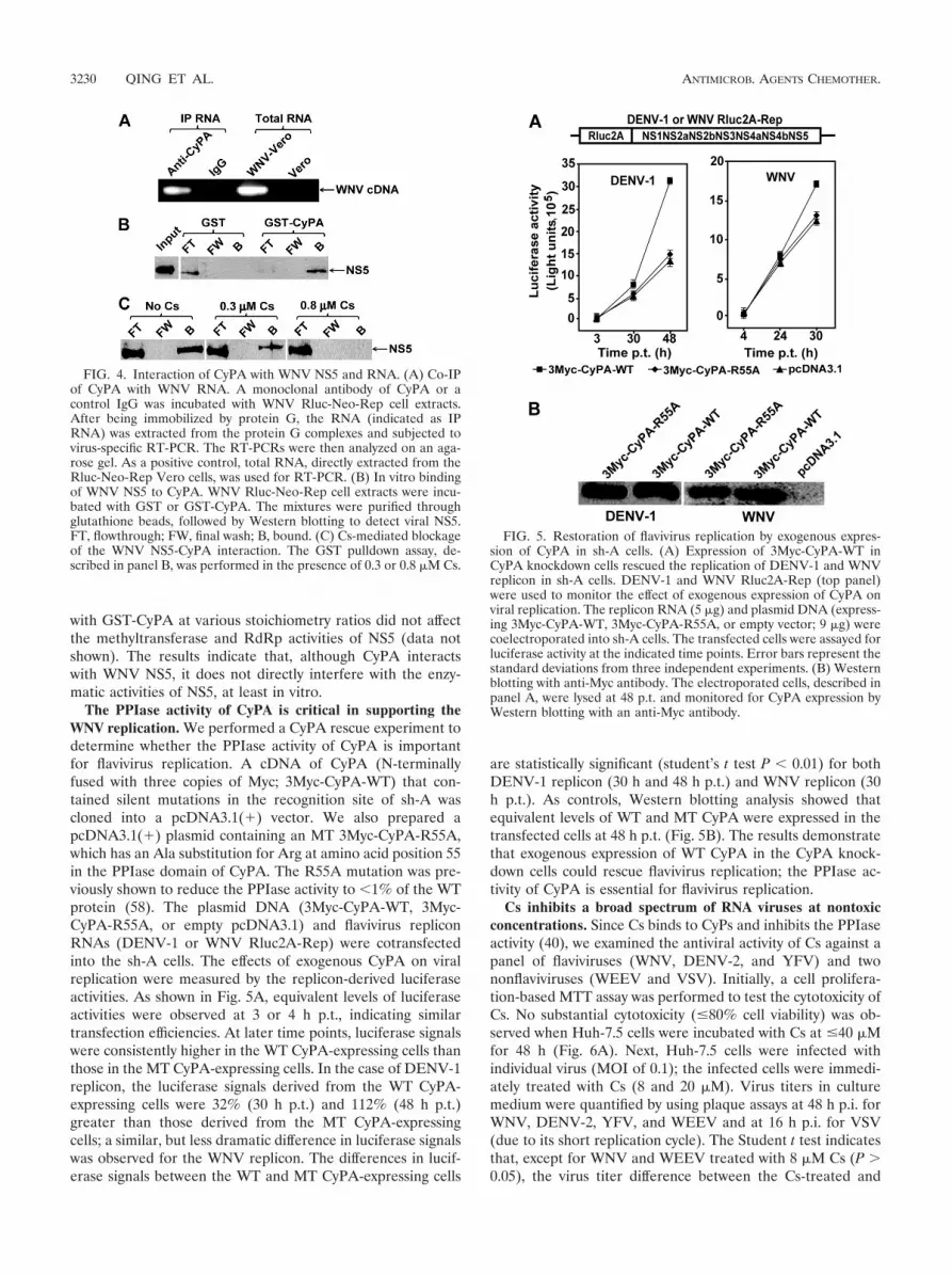
Host CyPA interacts with WNV RNA and NS5. To explorethe potential mechanism of CyPs in flavivirus replication, weperformed Co-IP experiments with a monoclonal antibody ofCyPA. Cell extracts prepared from the WNV replicon-contain-ing Vero cells (Rluc-Neo-Rep; Fig. 4A) were immunoprecipi-tated with the CyPA antibody. Total RNA was extracted fromthe immunoprecipitated complexes and was subjected to virus-specific RT-PCR amplification. Agarose gel analysis of theRT-PCR showed an expected DNA fragment of viral sequence(Fig. 4A). In contrast, no RT-PCR product was detected whenthe same cell extract was immunoprecipitated using a rabbitcontrol IgG. The results suggest that CyPA may participate inviral replication complex formation.
Next, we examined whether CyPA directly interacts withviral NS5 in the replication complexes. Recombinant GST-CyPA protein was incubated with cell extracts prepared fromthe WNV Rluc-Neo-Rep Vero cells. Viral NS5 was pulleddown by GST-CyPA after the mixture was incubated withglutathione-Sepharose beads (Fig. 4B). As a negative control,no detectable level of NS5 was pulled down by GST. Inclusionof Cs in the binding reaction inhibited the binding of NS5 toGST-CyPA in a dosage-dependent manner (Fig. 4C; detailsare described below). The observed CyPA-NS5 interactionprompted us to test whether CyPA could affect the NS5 en-zyme activities. Coincubation of recombinant NS5 of WNV
FIG. 2. Effects of CyP knockdown on flavivirus VLP infection.(A) Production of flavivirus VLPs. Flavivirus replicon (Flavi-Rluc2A-Rep) contains a Renilla luciferase gene (Rluc) which substitutes for thedeleted viral structural genes. The SFV vector (SFV-Flavi-CprME) wasused to express flavivirus structural proteins. Double electroporationswere performed to sequentially transfect Flavi-Rluc2A-Rep and SFV-Flavi-CprME into BHK-21 cells, leading to the production of VLPs inculture medium (see Materials and Methods for details). (B) Infection ofCyP knockdown cells with flavivirus VLPs. Approximately 2 � 104 cells ofeach cell line were seeded per well in 96-well plates. At 24 h after seeding,the cells were infected with VLPs of WNV (1 FFU/cell), YFV (0.1 FFU/cell), or DENV-1 (0.01 FFU/cell). At 24 h p.i., the 96-well plates wereassayed for luciferase activity. The percentages of the luciferase activitiesfrom the CyP knockdown cells versus on the sh-Fluc cells (set as 100%)are indicated. Average results, with standard deviations (error bars), offour independent experiments are shown.
FIG. 3. Comparison of viral growth kinetics on sh-AB and sh-Fluc cells. The sh-AB and sh-Fluc cells were infected with WNV, DENV-2, YFV,WEEV, and VSV (MOI of 0.1). Culture fluids at the indicated time points were assayed for virus titers by a double-layer plaque assay. See detailsin Materials and Methods. Average results and standard deviations (n � 3) are presented.
VOL. 53, 2009 CYCLOPHILINS FUNCTION IN FLAVIVIRUS REPLICATION 3229
with GST-CyPA at various stoichiometry ratios did not affectthe methyltransferase and RdRp activities of NS5 (data notshown). The results indicate that, although CyPA interactswith WNV NS5, it does not directly interfere with the enzy-matic activities of NS5, at least in vitro.
The PPIase activity of CyPA is critical in supporting theWNV replication. We performed a CyPA rescue experiment todetermine whether the PPIase activity of CyPA is importantfor flavivirus replication. A cDNA of CyPA (N-terminallyfused with three copies of Myc; 3Myc-CyPA-WT) that con-tained silent mutations in the recognition site of sh-A wascloned into a pcDNA3.1(�) vector. We also prepared apcDNA3.1(�) plasmid containing an MT 3Myc-CyPA-R55A,which has an Ala substitution for Arg at amino acid position 55in the PPIase domain of CyPA. The R55A mutation was pre-viously shown to reduce the PPIase activity to �1% of the WTprotein (58). The plasmid DNA (3Myc-CyPA-WT, 3Myc-CyPA-R55A, or empty pcDNA3.1) and flavivirus repliconRNAs (DENV-1 or WNV Rluc2A-Rep) were cotransfectedinto the sh-A cells. The effects of exogenous CyPA on viralreplication were measured by the replicon-derived luciferaseactivities. As shown in Fig. 5A, equivalent levels of luciferaseactivities were observed at 3 or 4 h p.t., indicating similartransfection efficiencies. At later time points, luciferase signalswere consistently higher in the WT CyPA-expressing cells thanthose in the MT CyPA-expressing cells. In the case of DENV-1replicon, the luciferase signals derived from the WT CyPA-expressing cells were 32% (30 h p.t.) and 112% (48 h p.t.)greater than those derived from the MT CyPA-expressingcells; a similar, but less dramatic difference in luciferase signalswas observed for the WNV replicon. The differences in lucif-erase signals between the WT and MT CyPA-expressing cells
are statistically significant (student’s t test P � 0.01) for bothDENV-1 replicon (30 h and 48 h p.t.) and WNV replicon (30h p.t.). As controls, Western blotting analysis showed thatequivalent levels of WT and MT CyPA were expressed in thetransfected cells at 48 h p.t. (Fig. 5B). The results demonstratethat exogenous expression of WT CyPA in the CyPA knock-down cells could rescue flavivirus replication; the PPIase ac-tivity of CyPA is essential for flavivirus replication.
Cs inhibits a broad spectrum of RNA viruses at nontoxicconcentrations. Since Cs binds to CyPs and inhibits the PPIaseactivity (40), we examined the antiviral activity of Cs against apanel of flaviviruses (WNV, DENV-2, and YFV) and twononflaviviruses (WEEV and VSV). Initially, a cell prolifera-tion-based MTT assay was performed to test the cytotoxicity ofCs. No substantial cytotoxicity (�80% cell viability) was ob-served when Huh-7.5 cells were incubated with Cs at �40 �Mfor 48 h (Fig. 6A). Next, Huh-7.5 cells were infected withindividual virus (MOI of 0.1); the infected cells were immedi-ately treated with Cs (8 and 20 �M). Virus titers in culturemedium were quantified by using plaque assays at 48 h p.i. forWNV, DENV-2, YFV, and WEEV and at 16 h p.i. for VSV(due to its short replication cycle). The Student t test indicatesthat, except for WNV and WEEV treated with 8 �M Cs (P �0.05), the virus titer difference between the Cs-treated and
FIG. 4. Interaction of CyPA with WNV NS5 and RNA. (A) Co-IPof CyPA with WNV RNA. A monoclonal antibody of CyPA or acontrol IgG was incubated with WNV Rluc-Neo-Rep cell extracts.After being immobilized by protein G, the RNA (indicated as IPRNA) was extracted from the protein G complexes and subjected tovirus-specific RT-PCR. The RT-PCRs were then analyzed on an aga-rose gel. As a positive control, total RNA, directly extracted from theRluc-Neo-Rep Vero cells, was used for RT-PCR. (B) In vitro bindingof WNV NS5 to CyPA. WNV Rluc-Neo-Rep cell extracts were incu-bated with GST or GST-CyPA. The mixtures were purified throughglutathione beads, followed by Western blotting to detect viral NS5.FT, flowthrough; FW, final wash; B, bound. (C) Cs-mediated blockageof the WNV NS5-CyPA interaction. The GST pulldown assay, de-scribed in panel B, was performed in the presence of 0.3 or 0.8 �M Cs.
FIG. 5. Restoration of flavivirus replication by exogenous expres-sion of CyPA in sh-A cells. (A) Expression of 3Myc-CyPA-WT inCyPA knockdown cells rescued the replication of DENV-1 and WNVreplicon in sh-A cells. DENV-1 and WNV Rluc2A-Rep (top panel)were used to monitor the effect of exogenous expression of CyPA onviral replication. The replicon RNA (5 �g) and plasmid DNA (express-ing 3Myc-CyPA-WT, 3Myc-CyPA-R55A, or empty vector; 9 �g) werecoelectroporated into sh-A cells. The transfected cells were assayed forluciferase activity at the indicated time points. Error bars represent thestandard deviations from three independent experiments. (B) Westernblotting with anti-Myc antibody. The electroporated cells, described inpanel A, were lysed at 48 p.t. and monitored for CyPA expression byWestern blotting with an anti-Myc antibody.
3230 QING ET AL. ANTIMICROB. AGENTS CHEMOTHER.
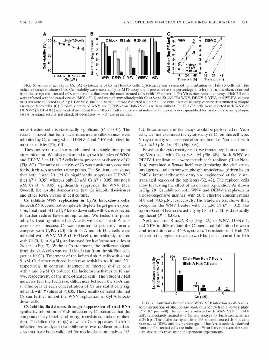
mock-treated cells is statistically significant (P � 0.05). Theresults showed that both flaviviruses and nonflaviviruses wereinhibited by Cs, among which DENV-2 and YFV exhibited themost sensitivity (Fig. 6B).
These antiviral results were obtained at a single time pointafter infection. We also performed a growth kinetics of WNVand DENV-2 on Huh-7.5 cells in the presence or absence of Cs(Fig. 6C). The antiviral activity of Cs was consistently observedfor both viruses at various time points. The Student t test showsthat both 8 and 20 �M Cs significantly suppresses DENV-2titer (P � 0.05), whereas only 20 �M Cs (P � 0.05) but not 8�M Cs (P � 0.05) significantly suppresses the WNV titer.Overall, the results demonstrate that Cs inhibits flavivirusesand other RNA viruses in cell culture.
Cs inhibits WNV replication in CyPA knockdown cells.Since shRNA could not completely deplete target gene expres-sion, treatment of the CyP knockdown cells with Cs is expectedto further reduce flavivirus replication. We tested this possi-bility by treating infected sh-A cells with Cs. The sh-A cellswere chosen because Cs was reported to primarily form acomplex with CyPA (20). Both sh-A and sh-Fluc cells wereinfected with WNV VLP (1 FFU/cell), immediately treatedwith Cs (0, 4, or 8 �M), and assayed for luciferase activities at24 h p.i. (Fig. 7). Without Cs treatment, the luciferase signalfrom the sh-A cells was ca. 51% of that from the sh-Fluc cells(set as 100%). Treatment of the infected sh-A cells with 4 and8 �M Cs further reduced luciferase activities to 10 and 2%,respectively. In contrast, treatment of infected sh-Fluc cellswith 4 and 8 �M Cs reduced the luciferase activities to 18 and4%, respectively, of the mock-treated cells. The Student t testindicates that the luciferase differences between the sh-A andsh-Fluc cells at each concentration of Cs are statistically sig-nificant, with P values of �0.05. These results demonstrate thatCs can further inhibit the WNV replication in CyPA knock-down cells.
Cs inhibits flaviviruses through suppression of viral RNAsynthesis. Inhibition of VLP infection by Cs indicates that thecompound may block viral entry, translation, and/or replica-tion. To define the step(s) at which Cs suppresses flavivirusinfection, we analyzed the inhibitor in two replicon-based as-says that have been validated for mode-of-action analysis (12,
42). Because some of the assays would be performed on Verocells, we first examined the cytotoxicity of Cs on this cell type.No cytotoxicity was observed after treatment of Vero cells withCs at �10 �M for 48 h (Fig. 8A).
Based on the cytotoxicity result, we treated replicon-contain-ing Vero cells with Cs at �8 �M (Fig. 8B). Both WNV orDENV-1 replicon cells were tested; each replicon (Rluc-Neo-Rep) contained a Renilla luciferase (replacing the viral struc-tural genes) and a neomycin phosphotransferase (driven by anEMCV internal ribosome entry site engineered at the 3� un-translated region of the replicon) (32, 42). The replicon cellsallow for testing the effect of Cs on viral replication. As shownin Fig. 8B, Cs inhibited both WNV and DENV-1 replicons ina dose-responsive manner, with 50% effective concentrationsof 8 and �0.5 �M, respectively. The Student t test shows that,except for the WNV treated with 0.5 �M Cs (P � 0.2), thesuppression of luciferase activity by Cs in Fig. 8B is statisticallysignificant (P � 0.005).
Next, we used Rluc2A-Rep (Fig. 2A) of WNV, DENV-1,and YFV to differentiate the Cs-mediated inhibition betweenviral translation and RNA synthesis. Transfection of Huh-7.5cells with this replicon reveals two Rluc peaks, one at 1 to 10 h
FIG. 6. Antiviral activity of Cs. (A) Cytotoxicity of Cs in Huh-7.5 cells. Cytotoxicity was examined by incubation of Huh-7.5 cells with theindicated concentrations of Cs. Cell viability was measured by an MTT assay and is presented as the percentage of colorimetric absorbance derivedfrom the compound-treated cells compared to that from the mock-treated cells (with 1% ethanol). (B) Virus titer reduction assays. Huh-7.5 cellswere infected with indicated viruses (MOI of 0.1) and treated immediately with Cs at 8 and 20 �M. For WNV, DENV-2, YFV, and WEEV, culturemedium were collected at 48 h p.i. For VSV, the culture medium was collected at 16 h p.i. The virus titers of all samples were determined by plaqueassays on Vero cells. (C) Growth kinetics of WNV and DENV-2 on Huh-7.5 cells with or without Cs. Huh-7.5 cells were infected with WNV orDENV-2 (MOI of 0.1) and treated with Cs at 8 and 20 �M. Culture medium at indicated time points were quantified for viral yields by using plaqueassays. Average results and standard deviations (n � 3) are presented.
FIG. 7. Antiviral effect of Cs on WNV VLP infection on sh-A cells.After incubation of sh-Fluc and sh-A cells for 24 h in a 96-well plate(2 � 104 per well), the cells were infected with WNV VLP (1 FFU/cell), immediately treated with Cs, and assayed for luciferase activitiesat 24 h p.i. The luciferase signals from 1% ethanol-treated sh-Fluc cellswere set as 100%, and the percentages of luciferase activities derivedfrom the Cs-treated cells are indicated. Error bars represent the stan-dard deviations from three independent experiments.
VOL. 53, 2009 CYCLOPHILINS FUNCTION IN FLAVIVIRUS REPLICATION 3231

p.t. and another at �10 h p.t., which represent viral translationand RNA replication, respectively (22, 31, 42). The replicon-transfected Huh-7.5 cells were immediately treated with 8 �MCs and assayed for luciferase activities at the indicated timepoints. As shown in Fig. 8C, Cs did not suppress luciferaseactivities at �6 h p.t. In contrast, Cs inhibited luciferase activ-ities by �70% at �20 h p.t.; statistical analysis indicated thatthe Cs-mediated luciferase suppression is significant, with Pvalues of �0.05. These results suggest that Cs inhibits WNV,DENV-1, and YFV virus through suppression of viral RNAreplication.
Time-of-addition experiments were performed to furthercharacterize the mode-of-action for Cs. Huh-7.5 cells were
synchronously infected with WNV, DENV-2, or YFV. Itshould be noted that, although all of the replicon experimentsfor DENV described above were derived from serotype 1(DENV-1), we chose DENV-2 for the time-of-addition exper-iments because DENV-2 generates better plaques thanDENV-1 does in our plaque assays. After the synchronousinfection, Cs was added to the infected cells (without changingmedium) at various time points p.i. Virus titers in the culturemedium were determined at 24 h p.i. For mock treatment, 1%ethanol was added at 0, 10, and 20 h p.i. to estimate its effecton viral production. Because WNV was not very sensitive toCs, we treated the WNV-infected Huh-7.5 cells with 20 �M Cs(80% cell viability; Fig. 6A); whereas the DENV-2- and YFV-
FIG. 8. Mechanism of Cs-mediated inhibition of flavivirus. (A) Cytotoxicity of Cs in Vero cells. Cytotoxicity was examined by incubation ofVero cells with the indicated concentrations of Cs for 48 h. Cell viability was determined by an MTT assay. The average results from threeexperiments are shown. (B) Antiviral activity of Cs in flavivirus replicon cells. Vero cells containing WNV or DENV Rluc-Neo-Rep were seededin a 96-well plate (2 � 104 per well). At 24 h after seeding, the cells were treated with Cs at the indicated concentrations, and the luciferase activitieswere measured at 24 h posttreatment. The percentages of luciferase signals from the Cs-treated cells compared to those from the mock-treatedcells (set as 100%) are indicated. Average results from three independent experiments are presented. (C) Analysis of Cs using transient replicon.Luciferase replicon (Rluc2A-Rep; 10 �g) of WNV, DENV-1, or YFV was electroporated into Huh-7.5 cells. The transfected cells wereimmediately incubated with 8 �M Cs and measured for luciferase activity at the indicated time points. Error bars indicate the standard deviationsfrom three independent experiments. (D) Time-of-addition analysis of Cs in flavivirus infection. Huh-7.5 cells were infected with WNV, DENV-2or YFV at an MOI of 3 at 4°C for 1 h. The infected cells were washed three times with cold PBS. At the indicated time points p.i., Cs was addedto the infected cells at 20 �M for WNV and at 8 �M for DENV-2 and YFV. The supernatants were assayed to determine the virus titers at 24 hp.i. As controls, 1% ethanol was added to the infected cells at 0, 10, and 20 h p.i. to estimate its effect on viral production. Error bars representthe standard deviations from three independent experiments.
3232 QING ET AL. ANTIMICROB. AGENTS CHEMOTHER.

infected cells were treated with 8 �M Cs. Consistent with thetransient replicon results (Fig. 8C), a significant level of sup-pression in virus titer was observed when the compound wasadded during the initial 6 h (WNV) and 8 h (DENV-2 andYFV) of infection (Fig. 8D). The compound partially lost theantiviral activity when added between 6 and 20 h p.i. TheStudent t test indicates that the virus titer difference betweenthe Cs-treated and the mock-treated cells in Fig. 8D is statis-tically significant (P � 0.001). It should be noted that thereplication kinetics of raw viral infection are faster than thoseof the transfected replicon, possibly because the delivery ofviral RNA through infection is more productive than thatthrough electroporation. Specifically, during flavivirus infec-tion, RNA synthesis could be detected at 6 p.i., with the releaseof infectious virus beginning at 12 h (10). Overall, the time-of-addition results were in agreement with the transient repliconresults, indicating that Cs inhibits flavivirus at the step of viralRNA synthesis. However, in the case of DENV, treatment with8 �M Cs reduced replicon RNA synthesis by �10-fold (Fig. 8Cmiddle panel), whereas the same concentration of Cs (added at20 h p.i.) suppressed the virus titer by �103-fold (Fig. 8D,middle panel). The results suggest that, in addition to RNAsynthesis, Cs may also affect the assembly of DENV.
Cs does not inhibit WNV protease, methyltransferase, andRdRp. Since the above results indicate that Cs inhibits viralRNA synthesis, we tested the compound in our previouslyestablished enzyme assays, including WNV protease (withNS2B), NTPase, methyltransferase, and RdRp (43, 55). Noneof the enzymatic activities were suppressed by the compoundat concentrations up to 100 �M (data not shown), suggestingthat Cs does not directly target the enzyme functions of viralNS3 or NS5 proteins.
Cs directly blocks the interaction between host CyPA andviral NS5. To examine whether Cs could interfere with theinteraction between CyPA and NS5, we performed the GST-CyPA pulldown experiments in the presence or absence of Cs.Different concentrations of Cs were incubated with GST-CyPAand WNV replicon cell extracts; the GST-CyPA were thenimmobilized onto glutathione beads, and viral NS5 (coimmo-bilized with GST-CyPA) was monitored by Western blotting.As shown in Fig. 4C, increasing concentrations of Cs decreasedthe mounts of NS5 that could be pulled down by GST-CyPA.No NS5 was detected when the experiment was performedwith 0.8 �M Cs. These results demonstrate that Cs targets theassociation of CyPA with viral NS5.
DISCUSSION
A number of host proteins have been reported to be impor-tant for flavivirus replication, among which eEF1 and TIA-1(T-cell intracellular antigen 1) are the two best characterized(11, 14). The eEF1 binds to the 3� terminal stem-loop offlavivirus genomic; this interaction is critical for the synthesisof minus-strand RNA (11). In contrast, TIA-1 binds to the3�-terminal stem-loop of WNV minus-strand RNA. This inter-action negatively affects genomic RNA amplification (14). Thepresent study demonstrated that, in addition to eEF1 andTIA-1, cellular CyPs also play an important role in flavivirusreplication. We show that knockdown of different isoforms ofCyP reduced flavivirus production in Huh-7.5 cells. However,
the knockdown cells did not allow us to conclude which iso-form(s) of CyP is most critical for flavivirus replication, be-cause most cell lines with knockdown of one isoform exhibitedenhanced expression of other isoforms (e.g., sh-A and sh-B;Fig. 1B). Only the double sh-AB knockdown cells specificallyreduced the expression of CyPA (by 92%) and CyPB (by 84%),without a significant change in the CyPC expression (90% ofthe WT level). Compared to the parental cells, infection of thesh-AB cells with flavivirus VLPs produced 68 to 85% lessluciferase signals at 24 h p.i. (Fig. 2B). Similarly, infection ofthe sh-AB cells with authentic flaviviruses yielded 67 to 89%less viruses at 24 h p.i. (Fig. 3). Furthermore, the low viralreplication in sh-A cells could be restored by trans supplying ofWT CyPA (Fig. 5A). These results clearly indicate that CyPAplays a role in flavivirus replication, but the potential functionof CyPB and CyPC could not be excluded at this point.
Our results suggest that CyPA may serve as a component ofthe replication complex. An CyPA antibody was shown tocoimmunoprecipitate WNV RNA from the replicon-contain-ing cell extracts (Fig. 4A). Additionally, WNV NS5 in thereplicon cell extracts could be pulled down by recombinantGST-CyPA protein (Fig. 4B). The latter results, together withthe observation that only WT CyPA (but not the MT CyPAdefective in PPIase activity) could rescue viral replication insh-A cells (Fig. 4A), raised the possibility that CyPA maymodulate the enzyme activities of WNV NS5 through the PPIase.However, biochemical analysis showed that recombinant CyPAaffected neither the methyltransferase nor the RdRp activitiesof NS5 of WNV (data not shown). These data suggest thatCyPA may not directly act on NS5, and an unidentified factormay mediate the CyPA-NS5 interaction. Alternatively, theconformation of recombinant CyPA and NS5 in vitro are dif-ferent from that in the replication complex in vivo; in thecontext of replication complex, CyPA may directly bind to andenhance the NS5 activities. The fact that the PPIase activity ofCyPA is required to facilitate viral replication suggests thatCyPA serves as a molecular chaperone to keep the replicationcomplex in an active conformation, leading to a more produc-tive viral replication.
Consistent with the CyP knockdown results, Cs was shown toinhibit flavivirus replication in cell culture (Fig. 6). The com-pound could further suppress viral replication in the sh-Aknockdown cells (Fig. 7). Mode-of-action analysis indicatedthat Cs inhibited viral replication at the step of RNA synthesis,possibly through direct blockage of the CyPA-NS5 interaction(Fig. 4C). It should be noted that, besides inhibiting the PPIaseactivity of CyPA (5, 40), Cs could also bind to and inhibit thePPIase activity of CyPB (21, 36), leading to the suppression ofviral replication.
We found that sh-AB knockdown cells were less efficient insupporting nonflavivirus replication (WEEV and VSV; Fig. 3).A previous study reported that knockdown of CyPA did notaffect the replication of VSV (56). Combining these results,one might conclude that CyPB contributes to the low replica-tion of VSV in the sh-AB cells. However, care should be takenwhen interpreting these results, because the two studies useddifferent methods to quantify the VSV yields. In the earlystudy, equal amounts (PFU) of VSV were directly plaqued onparental and sh-A Huh-7.5 cells; the resulting numbers ofplaques were compared to indicate the effect of CyPA on VSV
VOL. 53, 2009 CYCLOPHILINS FUNCTION IN FLAVIVIRUS REPLICATION 3233

replication. The incubation time of plaque assay was �24 h. Inthe present study, we infected parental and sh-AB cells at anMOI of 0.1. The culture media at different time points weremeasured for viral yields by plaque assay. The method used inthe present study is expected to be more sensitive than the onedescribed in the previous study. Nevertheless, a role of one ormore of the CyPs in VSV and WEEV replication was furthersupported by the results that Cs inhibited both viruses in pa-rental Huh-7.5 cells (Fig. 6B).
CyPs were previously reported to regulate HCV and HIVreplication. Both CyPA and CyPB were initially shown to co-precipitate with HCV NS5B protein (53, 56) and with HIVGag (8, 33), but RNA silencing experiments yielded conflictingresults as to whether CyPA or CyPB, or both, are required forthe viral replication cycle. CyPA was later demonstrated to bemore important for HCV replication (56) and HIV infection(9). Moreover, CyPA was reported to be packaged into HIV-1virion and to catalyze cis to trans isomerization of the viralcapsid protein (7). The anti-HIV and anti-HCV actions of Cswas shown to rely on its capacity to bind to CyPs (33, 34, 48).Toward therapeutic development, two derivatives of Cs,NIM811 and DEBIO-025, are currently in clinical trials forHCV treatment (16, 35). Recent studies showed that the com-bination of NIM811 with HCV protease or RdRp inhibitorsenhanced antiviral activity and suppressed the emergence ofresistance (35) and that DEBIO-025 achieved proof-of-con-cept efficacy in HCV patients (16). It remains to be examinedwhether the two HCV clinical candidates could inhibit flavivi-ruses in cell culture and in animal models.
One major advantage of targeting host factors for antiviraldevelopment is the higher genetic barrier to the emergence ofviral escape mutants. Due to the error-prone nature of RdRp,drug resistance to virus-specific inhibitors can quickly emergeboth in vitro and in patients. Since viral inhibitors usually bindto defined pockets of viral proteins, mutations in the viralgenome that weaken or disrupt the binding would reduce thecompound efficacy and lead to resistance. In this regard, hostinhibitors do not directly bind to viral targets, creating agreater barrier for the emergence of resistant viruses. On theother hand, one major challenge for the development of hostinhibitors is to identify a therapeutic dose that does not affectthe normal function of host targets and, therefore, does notlead to significant side effect.
In summary, we found that host CyP plays a role in flavivirusreplication. CyPA may function as a component of viral repli-cation complex. The PPIase activity is essential for CyPA tofunction in WNV replication. Cs, a ligand and inhibitor ofPPIase of CyP, inhibits flaviviruses in cell culture. Mechanism-of-action analysis showed that Cs directly blocks the interac-tion between cellular CyPA and WNV NS5 protein. The re-sults suggest that CyPs represent a potential target forflavivirus antiviral development.
ACKNOWLEDGMENTS
We thank the Molecular Genetics Core and the Cell Culture Facilityat the Wadsworth Center for the DNA sequencing and maintenance ofthe BHK-21 and Vero cells, respectively.
This study was partially supported by federal funds from the Na-tional Institute of Allergy and Infectious Disease, National Institutesof Health (NIH), under contract N01-AI-25490, by NIH grants1U01AI061193 and U54-AI057158 (Northeast Biodefense Center),
and by the James Esther King Biomedical Research Program fromFlorida Department of Health.
REFERENCES
1. Ackermann, M., and R. Padmanabhan. 2001. De novo synthesis of RNA bythe dengue virus RNA-dependent RNA polymerase exhibits temperaturedependence at the initiation but not elongation phase. J. Biol. Chem. 276:39926–39937.
2. Bartelma, G., and R. Padmanabhan. 2002. Expression, purification, andcharacterization of the RNA 5�-triphosphatase activity of dengue virus type2 nonstructural protein 3. Virology 299:122–132.
3. Best, S. M., K. L. Morris, J. G. Shannon, S. J. Robertson, D. N. Mitzel, G. S.Park, E. Boer, J. B. Wolfinbarger, and M. E. Bloom. 2005. Inhibition ofinterferon-stimulated JAK-STAT signaling by a tick-borne flavivirus andidentification of NS5 as an interferon antagonist. J. Virol. 79:12828–12839.
4. Billich, A., F. Hammerschmid, P. Peichl, R. Wenger, G. Zenke, V. Quesni-aux, and B. Rosenwirth. 1995. Mode of action of SDZ NIM 811, a nonim-munosuppressive cyclosporin A analog with activity against human immu-nodeficiency virus (HIV) type 1: interference with HIV protein-cyclophilin Ainteractions. J. Virol. 69:2451–2461.
5. Borel, J. F., F. Di Padova, J. Mason, V. Quesniaux, B. Ryffel, and R. Wenger.1990. Pharmacology of cyclosporine (sandimmune). I. Introduction. Phar-macol. Rev. 41:239–242.
6. Borowski, P., A. Niebuhr, O. Mueller, M. Bretner, K. Felczak, T. Kulikowski,and H. Schmitz. 2001. Purification and characterization of West Nile virusnucleoside triphosphatase (NTPase)/helicase: evidence for dissociation ofthe NTPase and helicase activities of the enzyme. J. Virol. 75:3220–3229.
7. Bosco, D. A., E. Z. Eisenmesser, S. Pochapsky, W. I. Sundquist, and D. Kern.2002. Catalysis of cis/trans isomerization in native HIV-1 capsid by humancyclophilin A. Proc. Natl. Acad. Sci. USA 99:5247–5252.
8. Braaten, D., H. Ansari, and J. Luban. 1997. The hydrophobic pocket ofcyclophilin is the binding site for the human immunodeficiency virus type 1Gag polyprotein. J. Virol. 71:2107–2113.
9. Braaten, D., and J. Luban. 2001. Cyclophilin A regulates HIV-1 infectivity,as demonstrated by gene targeting in human T cells. EMBO J. 20:1300–1309.
10. Chambers, T. J., C. S. Hahn, R. Galler, and C. M. Rice. 1990. Flavivirusgenome organization, expression, and replication. Annu. Rev. Microbiol.44:649–688.
11. Davis, W., J. Blackwell, P. Shi, and M. Brinton. 2007. Interaction betweenthe cellular protein eEF1A and the 3� terminal stem-loop of the West Nilevirus genomic RNA facilitates viral RNA minus strand synthesis. J. Virol.10172–10187.
12. Deas, T. S., I. Binduga-Gajewska, M. Tilgner, P. Ren, D. A. Stein, H. M.Moulton, P. L. Iversen, E. B. Kauffman, L. D. Kramer, and P.-Y. Shi. 2005.Inhibition of flavivirus infections by antisense oligomers specifically sup-pressing viral translation and RNA replication. J. Virol. 79:4599–4609.
13. Egloff, M. P., D. Benarroch, B. Selisko, J. L. Romette, and B. Canard. 2002.An RNA cap (nucleoside-2�-O-)-methyltransferase in the flavivirus RNApolymerase NS5: crystal structure and functional characterization. EMBO J.21:2757–2768.
14. Emara, M. M., H. Liu, W. G. Davis, and M. A. Brinton. 2008. Mutation ofmapped TIA-1/TIAR binding sites in the 3� terminal stem-loop of West Nilevirus minus-strand RNA in an infectious clone negatively affects genomicRNA amplification. J. Virol. 82:10657–10670.
15. Falgout, B., R. H. Miller, and C. J. Lai. 1993. Deletion analysis of denguevirus type 4 nonstructural protein NS2B: identification of a domain requiredfor NS2B-NS3 protease activity. J. Virol. 67:2034–2042.
16. Flisiak, R., A. Horban, P. Gallay, M. Bobardt, S. Selvarajah, A. Wiercinska-Drapalo, E. Siwak, I. Cielniak, J. Higersberger, J. Kierkus, C. Aeschlimann,P. Grosgurin, V. Nicolas-Metral, J. M. Dumont, H. Porchet, R. Crabbe, andP. Scalfaro. 2008. The cyclophilin inhibitor Debio-025 shows potent anti-hepatitis C effect in patients coinfected with hepatitis C and human immu-nodeficiency virus. Hepatology 47:817–826.
17. Gubler, D., G. Kuno, and L. Markoff. 2007. Flaviviruses, p. 1153–1253. InD. M. Knipe, P. M. Howley, D. E. Griffin, R. A. Lamb, M. A. Martin, B.Roizman, and S. E. Straus (ed.), Fields virology, 5th ed. Lippincott-RavenPublishers, Philadelphia, PA.
18. Guo, J., J. Hayashi, and C. Seeger. 2005. West nile virus inhibits the signaltransduction pathway of alpha interferon. J. Virol. 79:1343–1350.
19. Guyatt, K. J., E. G. Westaway, and A. A. Khromykh. 2001. Expression andpurification of enzymatically active recombinant RNA-dependent RNApolymerase (NS5) of the flavivirus Kunjin. J. Virol. Methods 92:37–44.
20. Handschumacher, R. E., M. W. Harding, J. Rice, R. J. Drugge, and D. W.Speicher. 1984. Cyclophilin: a specific cytosolic binding protein for cyclospo-rin A. Science 226:544–547.
21. Hasel, K. W., J. R. Glass, M. Godbout, and J. G. Sutcliffe. 1991. An endo-plasmic reticulum-specific cyclophilin. Mol. Cell. Biol. 11:3484–3491.
22. Jones, C., C. Patkar, and R. Kuhn. 2005. Construction and applications ofyellow fever virus replicons. Virology 331:247–259.
23. Kramer, L., J. Li, and P.-Y. Shi. 2007. West Nile virus. Lancet Neurol.6:171–182.
24. Kuhn, R. J., W. Zhang, M. G. Rossmann, S. V. Pletnev, J. Corver, E.
3234 QING ET AL. ANTIMICROB. AGENTS CHEMOTHER.

Lenches, C. T. Jones, S. Mukhopadhyay, P. R. Chipman, E. G. Strauss, T. S.Baker, and J. H. Strauss. 2002. Structure of dengue virus: implications forflavivirus organization, maturation, and fusion. Cell 108:717–725.
25. Kummerer, B. M., and C. M. Rice. 2002. Mutations in the yellow fever virusnonstructural protein NS2A selectively block production of infectious par-ticles. J. Virol. 76:4773–4784.
26. Li, H., S. Clum, S. You, K. E. Ebner, and R. Padmanabhan. 1999. The serineprotease and RNA-stimulated nucleoside triphosphatase and RNA helicasefunctional domains of dengue virus type 2 NS3 converge within a region of20 amino acids. J. Virol. 73:3108–3116.
27. Lindenbach, B. D., H.-J. Thiel, and C. M. Rice. 2007. Flaviviridae: the virusand their replication, p. 1101–1152. In D. M. Knipe, P. M. Howley, D. E.Griffin, R. A. Lamb, M. A. Martin, B. Roizman, and S. E. Straus (ed.), Fieldsvirology, 5th ed. Lippincott-Raven Publishers, Philadelphia, PA.
28. Liu, J., J. D. Farmer, Jr., W. S. Lane, J. Friedman, I. Weissman, and S. L.Schreiber. 1991. Calcineurin is a common target of cyclophilin-cyclosporin Aand FKBP-FK506 complexes. Cell 66:807–815.
29. Liu, W., X. Wang, V. Mokhonov, P.-Y. Shi, R. Randall, and A. Khromykh.2005. Inhibition of interferon signaling by the New York 99 strain and Kunjinsubtype of West Nile virus involves blockage of STAT1 and STAT2 activa-tion by nonstructural proteins. J. Virol. 79:1934–1942.
30. Liu, W. J., H. B. Chen, and A. A. Khromykh. 2003. Molecular and functionalanalyses of Kunjin virus infectious cDNA clones demonstrate the essentialroles for NS2A in virus assembly and for a nonconservative residue in NS3in RNA replication. J. Virol. 77:7804–7813.
31. Lo, L., M. Tilgner, K. Bernard, and P.-Y. Shi. 2003. Functional analysis ofmosquito-borne flavivirus conserved sequence elements within 3� untrans-lated region of West Nile virus using a reporting replicon that differentiatesbetween viral translation and RNA replication. J. Virol. 77:10004–10014.
32. Lo, L., M. Tilgner, and P.-Y. Shi. 2003. A potential high-throughput assay forscreening inhibitors of West Nile virus replication. J. Virol. 77:12901–12906.
33. Luban, J., K. L. Bossolt, E. K. Franke, G. V. Kalpana, and S. P. Goff. 1993.Human immunodeficiency virus type 1 Gag protein binds to cyclophilins Aand B. Cell 73:1067–1078.
34. Ma, S., J. E. Boerner, C. TiongYip, B. Weidmann, N. S. Ryder, M. P.Cooreman, and K. Lin. 2006. NIM811, a cyclophilin inhibitor, exhibits potentin vitro activity against hepatitis C virus alone or in combination with alphainterferon. Antimicrob. Agents Chemother. 50:2976–2982.
35. Mathy, J. E., S. Ma, T. Compton, and K. Lin. 2008. Combinations of cyclo-philin inhibitor NIM811 with hepatitis C Virus NS3-4A Protease or NS5Bpolymerase inhibitors enhance antiviral activity and suppress the emergenceof resistance. Antimicrob. Agents Chemother. 52:3267–3275.
36. Mikol, V., J. Kallen, and M. D. Walkinshaw. 1994. X-ray structure of acyclophilin B/cyclosporin complex: comparison with cyclophilin A and de-lineation of its calcineurin-binding domain. Proc. Natl. Acad. Sci. USA91:5183–5186.
37. Munoz-Jordan, J. L., M. Laurent-Rolle, J. Ashour, L. Martinez-Sobrido, M.Ashok, W. I. Lipkin, and A. Garcia-Sastre. 2005. Inhibition of alpha/betainterferon signaling by the NS4B protein of flaviviruses. J. Virol. 79:8004–8013.
38. Munoz-Jordan, J. L., G. G. Sanchez-Burgos, M. Laurent-Rolle, and A. Gar-cia-Sastre. 2003. Inhibition of interferon signaling by dengue virus. Proc.Natl. Acad. Sci. USA 100:14333–14338.
39. Patkar, C. G., and R. J. Kuhn. 2008. Yellow Fever virus NS3 plays anessential role in virus assembly independent of its known enzymatic func-tions. J. Virol. 82:3342–3352.
40. Ptak, R. G., P. A. Gallay, D. Jochmans, A. P. Halestrap, U. T. Ruegg, L. A.Pallansch, M. D. Bobardt, M. P. de Bethune, J. Neyts, E. De Clercq, J. M.Dumont, P. Scalfaro, K. Besseghir, R. M. Wenger, and B. Rosenwirth. 2008.Inhibition of human immunodeficiency virus type 1 replication in humancells by Debio-025, a novel cyclophilin binding agent. Antimicrob. AgentsChemother. 52:1302–1317.
41. Puig-Basagoiti, F., T. S. Deas, P. Ren, M. Tilgner, D. M. Ferguson, and P.-Y.Shi. 2005. High-throughput assays using luciferase-expressing replicon, vi-
rus-like particle, and full-length virus for West Nile virus drug discovery.Antimicrob. Agent. Chemother. 49:4980–4988.
42. Puig-Basagoiti, F., M. Tilgner, B. Forshey, S. Philpott, N. Espina, Went-worth, S. Goebel, P. S. Masters, B. Falgout, P. Ren, Ferguson, and P. Y. Shi.2006. Triaryl pyrazoline compound inhibits flavivirus RNA replication. An-timicrob. Agents Chemother. 50:1320–1329.
43. Ray, D., A. Shah, M. Tilgner, Y. Guo, Y. Zhao, H. Dong, T. Deas, Y. Zhou,H. Li, and P.-Y. Shi. 2006. West Nile Virus 5�-cap structure is formed bysequential guanine N-7 and ribose 2�-O methylations by nonstructural pro-tein 5. J. Virol. 80:8362–8370.
44. Robida, J. M., H. B. Nelson, Z. Liu, and H. Tang. 2007. Characterization ofhepatitis C virus subgenomic replicon resistance to cyclosporine in vitro.J. Virol. 81:5829–5840.
45. Shi, P. Y., E. B. Kauffman, P. Ren, A. Felton, J. H. Tai, A. P. Dupuis, 2nd,S. A. Jones, K. A. Ngo, D. C. Nicholas, J. Maffei, G. D. Ebel, K. A. Bernard,and L. D. Kramer. 2001. High-throughput detection of West Nile virus RNA.J. Clin. Microbiol. 39:1264–1271.
46. Shi, P. Y., M. Tilgner, M. K. Lo, K. A. Kent, and K. A. Bernard. 2002.Infectious cDNA clone of the epidemic West Nile virus from New York City.J. Virol. 76:5847–5856.
47. Starzl, T. E., G. B. Klintmalm, K. A. Porter, S. Iwatsuki, and G. P. Schroter.1981. Liver transplantation with use of cyclosporin A and prednisone.N. Engl. J. Med. 305:266–269.
48. Steinkasserer, A., R. Harrison, A. Billich, F. Hammerschmid, G. Werner, B.Wolff, P. Peichl, G. Palfi, W. Schnitzel, E. Mlynar, et al. 1995. Mode of actionof SDZ NIM 811, a nonimmunosuppressive cyclosporin A analog with ac-tivity against human immunodeficiency virus type 1 (HIV-1): interferencewith early and late events in HIV-1 replication. J. Virol. 69:814–824.
49. Tilgner, M., and P.-Y. Shi. 2004. Structure and function of the 3� terminal sixnucleotides of the West Nile virus genome in viral replication. J. Virol.78:8159–8171.
50. Waninger, S., K. Kuhen, X. Hu, J. E. Chatterton, F. Wong-Staal, and H.Tang. 2004. Identification of cellular cofactors for human immunodeficiencyvirus replication via a ribozyme-based genomics approach. J. Virol.78:12829–12837.
51. Warrener, P., J. K. Tamura, and M. S. Collett. 1993. RNA-stimulated NT-Pase activity associated with yellow fever virus NS3 protein expressed inbacteria. J. Virol. 67:989–996.
52. Watashi, K., M. Hijikata, M. Hosaka, M. Yamaji, and K. Shimotohno. 2003.Cyclosporin A suppresses replication of hepatitis C virus genome in culturedhepatocytes. Hepatology 38:1282–1288.
53. Watashi, K., N. Ishii, M. Hijikata, D. Inoue, T. Murata, Y. Miyanari, and K.Shimotohno. 2005. Cyclophilin B is a functional regulator of hepatitis C virusRNA polymerase. Mol. Cell 19:111–122.
54. Wengler, G., and G. Wengler. 1991. The carboxy-terminal part of the NS 3protein of the West Nile flavivirus can be isolated as a soluble protein afterproteolytic cleavage and represents an RNA-stimulated NTPase. Virology184:707–715.
55. Wong, S. J., R. H. Boyle, V. L. Demarest, A. N. Woodmansee, L. D. Kramer,H. Li, M. Drebot, R. A. Koski, E. Fikrig, D. A. Martin, and P.-Y. Shi. 2003.An immunoassay targeting nonstructural protein 5 to differentiate West Nilevirus infection from dengue and St. Louis encephalitis virus infections, andform flavivirus vaccination. J. Clin. Microbiol. 41:4217–4223.
56. Yang, F., J. M. Robotham, H. B. Nelson, A. Irsigler, R. Kenworthy, and H.Tang. 2008. Cyclophilin A is an essential cofactor for hepatitis C virusinfection and the principal mediator of cyclosporine resistance in vitro.J. Virol. 82:5269–5278.
57. Zhou, Y., D. Ray, Y. Zhao, H. Dong, S. Ren, Z. Li, Y. Guo, K. Bernard, P.-Y.Shi, and H. Li. 2007. Structure and function of flavivirus NS5 methyltrans-ferase. J. Virol. 81:3891–3903.
58. Zydowsky, L. D., F. A. Etzkorn, H. Y. Chang, S. B. Ferguson, L. A. Stolz, S. I.Ho, and C. T. Walsh. 1992. Active site mutants of human cyclophilin Aseparate peptidyl-prolyl isomerase activity from cyclosporin A binding andcalcineurin inhibition. Protein Sci. 1:1092–1099.
VOL. 53, 2009 CYCLOPHILINS FUNCTION IN FLAVIVIRUS REPLICATION 3235


