Embed Size (px)
Citation preview
Durham E-Theses
Cyclotron production of short-lived radionuclides and
labelled compounds for use in biomedical research and
clinical diagnosis
Clark, John Charles
How to cite:
Clark, John Charles (1994) Cyclotron production of short-lived radionuclides and labelled compounds for
use in biomedical research and clinical diagnosis, Durham theses, Durham University. Available at DurhamE-Theses Online: http://etheses.dur.ac.uk/5495/
Use policy
The full-text may be used and/or reproduced, and given to third parties in any format or medium, without prior permission orcharge, for personal research or study, educational, or not-for-pro�t purposes provided that:
• a full bibliographic reference is made to the original source
• a link is made to the metadata record in Durham E-Theses
• the full-text is not changed in any way
The full-text must not be sold in any format or medium without the formal permission of the copyright holders.
Please consult the full Durham E-Theses policy for further details.

Academic Support O�ce, Durham University, University O�ce, Old Elvet, Durham DH1 3HPe-mail: [email protected] Tel: +44 0191 334 6107
http://etheses.dur.ac.uk
2
CYCLOTRON PRODUCTION OF SHORT-LIVED RADIONUCLK)ES AND LABELLED COMPOUNDS FOR USE IN BIOMEDICAL RESEARCH AND CLINICAL DIAGNOSIS
John C Clark BSc (Durham)
Submission for the Degree of DSc in the Uruversity of Durham 1994.
ABSTRACT
The works submitted in this thesis cover the development of
methods for the production in a cyclotron of a variety of radionuclides and
their incorporation in radio-labelled compounds for use in biomedical
research. In addition, papers are included which describe biomedical
applications of such radio-tracers. My co-authorship of these pubhcations
reflects my interest in the design and execution of experiments in the realm
of interdiscipUnary research. The original contributions to science embodied
in the publications submitted include examples of novel radiochemistry
applied in the areas of cyclotron production of short half-Ufe radionuclides
and their radiochemical purification. In many cases the use of these
radionuclides in biomedical research has added new information to the body
of medical scientific Tcnowledge.
Novel radiolabelling strategies using very short half-life
radionuclides are included. These have necessitated the development of
rapid radio-organic syntheses, several of which have been achieved using
automated microchemical engineering process plants of my design. I have
also developed novel systems for the administration of radionuchdes and
radio-labelled compounds of pharmaceutical quality, widely acknowledged
to be "World Firsts." My invention of the siKr^ radionuclide generator
resulted in publications covering a wide range of medical applications.
These are included with the thesis. The device is now produced in many
countries around the world for use both in routine clinical diagnosis and in
research, particularly in lung disease. More recently, I have created an
automated bedside infuser of H2i50, which has revolutionised
measurements of regional cerebral blood flow using the technique of
Positron Emission Tomography for in vivo regional mapping of brain
activity.
CYCLOTRON PRODUCTION OF SHORT-LIVED
RADIONUCLIDES AND LABELLED COMPOUNDS
FOR USE IN BIOMEDICAL RESEARCH AND
CLINICAL DIAGNOSIS
in
TWO VOLUMES
VOLUME I
John Charles Clark
Submission for the Degree of DSc
in the University of Durham
1994
The copyright of this thesis rests with the author.
No quotation from it should be pubHshed without
his prior written consent and information derived
from it should be acknowledged.
1 3 NOV 1995
TABLE OF CONTENTS
Introduction and Synopsis of volumes I & I I
Publications submitted
References 1 - 40 1966 - 1978
Book Short-lived Radioactive Gases for Clinical Use (ref 26)
J C Clark & P D Buckingham, London Butterworths 1975
(submitted separately)
DECLARATION
None of the material included in this thesis has previously been
submitted for a degree in either the University of Durham or any
other University. My contributions to the joint publications
enclosed within this thesis are clearly indicated beside each
reference in the list of publications submitted.
STATEMENT OF COPYRIGHT
The copyright of this thesis rests with the author. No quotation
from it should be published without his prior written consent and
information derived from it should be acknowledged.
INTRODUCTION
The publications within these volumes and the appended book represent the
development of cyclotron methods for the production of a variety of
radionuclides and radio-labelled compounds for use by biomedical researchers
to address biological and medical scientific questions with the goal of
achieving a better understanding of disease, its diagnosis and and the effect of
treatment, in order to develop rational therapies.
In addition publications are included which describe biomedical applications
of such radio-tracers. My co-authorship of these publications reflects my
profound interest in experimental design and execuhon in the realm of
interdisciplinary research.
ORIGINAL CONTRIBUTIONS TO SCIENCE
The original contributions to science embodied in the publications submitted
include examples of novel radiochemistry appUed in the areas of cyclotron
production of short half-life radionuclides and their radiochemical
purification (2, 3, 4, 12, 14, 15, 17, 18, 19, 20, 48, 54). In many cases the use of
these radionuclides in biomedical research has led to original contributions to
medical scientific knowledge (1, 5, 6, 7, 8, 16, 28, 33, 41, 46, 52, 53, 55, 56, 63, 76,
77).
Novel radiolabelling strategies using very short half-life radionuclides which
have necessitated the development of rapid radio-organic syntheses are
included (21, 22, 23, 43, 44, 57, 59, 65, 72, 78). Several of these have been
achieved using automated microchemical engineering process plants of my
design (57, 65, 72, 78).
I have also developed novel delivery systems for radionuclides and radio-
labelled compounds of pharmaceutical quality, widely acknowledged to be
"World Firsts." The First is my invention of the siKrm radionucUde generator
(27, 36). A wide range of medical applications of these siKrm generators are
included (9, 10,13, 30, 31, 35, 37, 38, 39, 40, 42, 47). The device is now produced
in many countries around the world for use in routine clinical diagnosis
particularly in lung disease. The Second is the automated bedside infusor of
H2I5O (71, 78).
The availabihty of the H2^50 infuser has revolutionised measurements of
regional cerebral blood flow (RCBF) using the technique of Positron Emission
Tomography (PET) (63, 76, 77). Many centres around the world are now either
commissioning my manufacture of the device or assembling devices to my
design.
FORMAT
Refereed Journal publications within these volumes are clearly marked (RJ)
in the reference list. Interspersed with these in chronological order are
articles which represent contributions to books, reviews and conference
proceedings. Published abstracts of scientific presentations have been
included where the published input did not of itself justify a ful l paper, but
which formed the basis upon which studies by other groups were founded.
The book "Short-lived Radioactive Gases for Clinical Use" which contains a
wealth of information has been in constant demand since it was published in
1975. Although now out of print I continue to receive requests for copies to be
released from my now rapidly dwindling stock. It is perhaps timely that a
revised edition be prepared.
Some of the publications presented take the form of reviews which have
more often than not been commissioned as part of a teaching commitment (
32, 34,45, 40, 67, 68, 73, 78).
PERSONAL CONTRIBUTION
Most of the publications presented here are the result of multi author
endeavours. My contribution to these papers is shown clearly in the reference
list. Two figures are given : the first indicates the fraction (percent) of the
radiochemistry, for which I personally was responsible, and the second the
fraction (percent) for the whole work. In many cases I was the only chemist
involved, this my contribution to the radiochemistry is shown as 100%
PERSONAL SCIENTIFIC RESUME
My 3 year undergraduate chemistry studies in Durham were followed by a
further year of post graduate studies in the Londonderry Laboratory for
Radiochemistry with Graham Martin, then Reader in Radiochemistry. The
use of radioisotopes as a tool for medical research became a great attraction to
me and I eagerly expanded my skills both in the theory and practice of
radiochemistry which quickly led me into an involvement in biological
applications of tiie radiotracer methodology. During the course of my

postgraduate training I was re-acquainted with one of Graham's former PhD
students who had recently returned from Brookhaven National Laboratory to
join the Medical Research Council team at Hammersmith Hospital. I was
intrigued to hear from him the plans at Hammersmith to use a cyclotron to
produce radionuclides with short half-lives for use in biomedical research. I
was fortunate in being able to join the Hammersmith team myself in 196L I
found the team very receptive to many new concepts I introduced for the use
of the cyclotron to produce radionucUdes. The opportunities of working with
interdisciplinary teams of researchers was mutually rewarding.
The teams include basic scientists and clinical scientists who at Hammersmith
particularly have a long established tradition of interdisciplinary research. I
found myself working alongside mechanical, electrical and electronic
engineers, medical and nuclear physicists, biochemists, pharmacologists,
pharmacists, neuroscientists, physiologists and cell biologists in the basic
sciences.
In the clinical sciences I work with radiotherapists, radiologists, respirologists,
paediatricians, obstetricians and gynaecologists, cardiologists, neurologists,
surgeons, endocrinologists, gastroenterologists and psychiatrists.
The publications on the applications of radionuclide tracers will be seen to be
co-authored by members of these diverse teams.
EVOLUTION OF THE USE OF RADIONUCLIDES IN BIOMEDICINE
Although the first radionuclides to be used for biomedical research had been
made using a cyclotron before the 1939-45 war the advent of the nuclear
reactor with its prolific neutron flux enabled the production of many more
radiotracers in the post war years. They were for the most part neutron rich
radionuclides with half-lives commensurate with the need to be able to ship
them from the few accessible reactors to the users. This usually meant that
only tracers with half-lives greater than a few days were available. The
Medical Research Council foresaw the potential for the re-introduction of the
cyclotron for radiotracer production close to the site of use, thus opening up
the exciting possibility of using neutron deficient tracers with half-lives of
only minutes. This opened up the intriguing possibihty of using multiple
doses of radiotracer in test and retest biomedical experimental protocols as the
tracer could be allowed to decay between measurements. In the early days we
made these measurements using discrete fixed external radiation detectors
(probes) located close to the organ or region of interest. At the birth of medical
imaging using radionuclides, probes were mechanically driven to "scan" the
region of interest and the radiation events collected and recorded
simultaneously on a two dimensional display. The externally detectable
gamma rays were "focused" by the probes either by the use of heavy lead
collimators or by exploiting the coincidence detection of positron annihilation
radiations (2 x 511 KeV photons with 180° correlation) using opposed pairs of
detectors set electronically only to produce a signal where the two photons
were detected in time coincidence. As many of the tracers produced with the
cyclotron were neutron deficient and decayed by positron ((3+) emission.
positron radionuclide imaging could be exploited in many of the
Hammersmith pioneering biomedical studies.
The use of simple chemical forms of the positron emitting tracers eg ^^02 was
soon expanded to the radiolabelling of much more complex molecules with
Carbon-11 (half life 20 mins) and fluorine-18 (half life 110 mins) which in
many cases involved the design and execution of rapid organic
radiosyntheses and purifications to achieve the desired goal of radiolabelled
metabolic substrates such as i^C-glucose, pharmaceuticals such as ^iC-
methylspiperone and related compounds such as isp-fluoro amino acids and
labelled chlorofluorocarbon propellants for drug inhalers.
The ability to exploit positron emitting radiotracers was revolutionised with
the advent of^ositron Emission Tomography (PET) imaging systems. These
systems still exploit the coincidence detection technique referred to above but
due to the large number of detectors employed (typically around 2500 pairs)
large data sets of coincident events can be collected. Using computer
reconstruction algorithms these data can be processed for display as
quantitative functional images of living subjects in cross section sometimes
referred to as in vivo autoradiographs. The pioneering work by teams at
Hammersmith, in bringing together these new exciting concepts, has
contributed to a world wide expansion of specialist PET centres which employ
a small cyclotron and PET scanners to apply these techniques to a wide variety
of research studies and clinical diagnostic tests.
ACKNOWLEDGEMENTS
Several hundred of my colleagues have played a part in bringing many of
these works to fruition. To name them individually would be a daunting
task. There are however a few key individuals who were highly supportive in
the endeavours reported in these volumes. I hope all will become aware of
my gratitude to them as I come to the point of submitting these works for
consideration for the degree of DSc.
Of the few I would like to thank Mr D D Vonberg CBE, the Director of the
Cyclotron Unit throughout most of the period covered in these volumes and
Dr D J Silvester and Dr T Jones, my close scientific colleagues and friends who
have challenged and supported me through thick and thin. I would also like
to convey my sincere thanks to my two research assistants and sometimes co
authors Mr P D Buckingham and the late Mr P L Horlock whose enthusiasm
and skills so often contributed to steering a productive course.
Finally I would like to acknowledge the untiring support of Dr Hazel A Jones,
my partner sometimes co-author and mother of our two boys, for providing
me with continuing education in biomedicine and for her moral support.
Finally I would like to acknowledge the University of Durham for my
undergraduate and post graduate training which prepared me well for the
years of exciting and productive scientific endeavours culminating in the
reports in these volumes.
International Links
An integral part of my commitment to science has always been my eagerness
to collaborate with scientists internationally and in some of the submitted
works my international co-authors will be found as follows:
Mr Roman Bucharest Romania (21,23)
F Skrabal Innsbruck Austria (8,11)
F Fazio
R D Finn et al
G L Lenzi
P Buranapong
H A O'Brien et al
F Oberdorfer
R E Forster
I Kanno
H Tochon-Danguy
J Link
T Ruth
D A Silbersweig &
E Stern
J-L Morelle
G Firnau
Pisa, Italy (25)
Miami, USA (31,45,48)
Sierra, Italy (28,33)
Bangkok, Thailand (39)
Los Alamos Nat. Lab. USA (47, 49, 53)
Heidelberg, Germany (51)
Philadelphia USA (52)
Akita, Japan
Geneva, Switzerland
and Melbourne, Australia (63, 71)
Seattle, USA (73)
Vancouver, Canada (73)
New York, USA
Louvain la Neuve, Belgium
Hamilton, Canada (59)
In addition I have had continuous commitment to an EEC initiative for PET
chemistry which can be found in refs. 61, 64, 74, 75.
PUBLICATIONS SUBMITTED % of % of Chemistry Total
1. Matthews CME., Dollery CT., Clark JC and West JB., 100 20 Radioactive gases. Radioactive Pharmaceuticals: ed Andrews WS., RM Kinsley and HN Wagner. U.S.A.E.C. Division of Technical Information, Oak Ridge, Tennessee., 1966. pp. 567-592.
2. Clark JC, Glass HI and Silvester DJ. In vitro 70 30 labelling of red cells with carbon-11. Free 2nd Int Conf. on methods of preparing and storing labelled compounds.. Brussels, 1966: pp 603-609.
3. Clark JC and Silvester DJ. A cyclotron method for RJ 70 70 the production of fluorine-18. Int T Appl Radiat Isotopes.. 1966:17:151-154
4. Clark JC, Matthews CME., Silvester DJ and Vonberg RJ 50 30 DD. Using cyclotron-produced isotopes at Hammersmith Hospital., Nucleonics. 1967: 25: 54.
5. Glass HL, Brant A., Clark JC, De Garreta AC and RJ 100 20 Day LG. Measurement of blood volume using red cells labelled with radioactive carbon monoxide. J Nucl Med., 1968: 9: 571-575
6. Glass HI., Jacoby J., Westerman B., Clark JC, RJ 100 15 Arnott RN and Dixon HG. Placental localisation by inhalation of radioactive carbon monoxide.
I Nucl Med.. 1968: 9: 468-470
7. Glass HI., Edwards RHT., de Gareta AC and Clark RJ 100 20 JC. UCO red cell labelling for blood volume and total haemoglobin in athletes: effect of training.
I Appl Physiol. 1969: 26:131-134.
8. Skrabal F., Glass HI., Clark JC, Jeyasingh K and RJ 100 20 Jophn GF. A simplified method for simultaneous electrolyte studies in man utilising potassium-43. Int T Appl Radiat Isotopes.. 1969: 20: 677-681
9. Clark JC, Jones T and Mackintosh A. 81mKr an ultra 100 40 short lived inert gas tracer for lung ventilation and perfusion studies with the scintillation camera. Radioaktive Isotope in Klinik und Forschung: 9th Int Symposium: Bad Gastem, 1979. Ed. F Fellinger and R Hofer. Munich, Urban & Schwarzenberg., 1970: 9: 444-450.
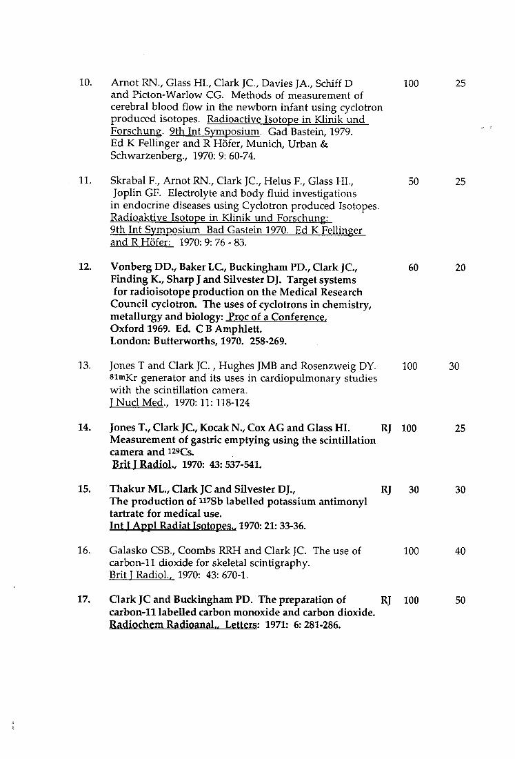
10. Arnot RN., Glass HI., Clark JC, Davies JA., Schiff D 100 25 and Picton-Warlow CG. Methods of measurement of cerebral blood flow in the newborn infant using cyclotron produced isotopes. Radioactive Isotope in Klinik und Forschung. 9th Int Symposium. Gad Bastein, 1979. Ed K Fellinger and R Hofer, Munich, Urban & Schwarzenberg., 1970: 9: 60-74.
11. Skrabal F., Arnot RN., Clark JC, Helus F., Glass HI., 50 25 Joplin GF. Electrolyte and body fluid investigations
in endocrine diseases using Cyclotron produced Isotopes. Radioaktive Isotope in Klinik und Forschung: 9th Int Symposium Bad Gastein 1970. Ed K Fellinger and R H5fer: 1970: 9: 76 - 83.
12. Vonberg DD., Baker L C , Buckingham PD., Clark JC, 60 20 Finding K., Sharp J and Silvester DJ. Target systems for radioisotope production on the Medical Research
Council cyclotron. The uses of cyclotrons in chemistry, metallurgy and biology: Proc of a Conference. Oxford 1969. Ed. CBAmphlett. London: Butterworths, 1970. 258-269.
13. Jones T and Clark JC., Hughes JMB and Rosenzweig DY. 100 30 simKr generator and its uses in cardiopulmonary studies with the scintillation camera. T Nucl Med.. 1970:11:118-124
14. Jones T., Clark JCKocakN., Cox AG and Glass HL RJ 100 25 Measurement of gastric emptying using the scintillation camera and i29Cs. Brit J Radiol.. 1970: 43: 537-541.
15. Thakur ML., Clark JC and Silvester DJ., RJ 30 30 The production of n sb labelled potassium antimonyl tartrate for medical use. Int T Appl Radiat Isotopes.. 1970:21: 33-36.
16. Galasko CSB., Coombs RRH and Clark JC. The use of 100 40 carbon-11 dioxide for skeletal scintigraphy. Brit 1 Radiol.. 1970: 43: 670-1.
17. Clark JC and Buckingham FD. The preparation of RJ 100 50 carbon-11 labelled carbon monoxide and carbon dioxide. Radiochem RadiQanal., Letters: 1971: 6:281-286.

18. Clark JC and Buckingham PD. The preparation and RJ 100 50 storage of carbon-11 labelled gases for clinical use. Int T Appl Radiat Isotopes.. 1971:22:639-646.
19. Buckingham PD and Qark JC. Nitrogen-13 solutions RJ 100 50 for research studies in pulmonary physiology. Int T Appl Radiat Isotopes.. 1972: 23: 5-8.
20. Clark JC, Thakur ML and Watson lA. The production RJ 50 30 of potassiiun-43 for medical use. Int I Appl Radiat Isotopes.. 1972:23:229-335.
21. Palmer AJ., Clark JCGoulding RW and Roman M. 30 30 Preparation of isp labelled DL-3-Fluorotyrosine labelled radiopharmaceuticals. Proc of Symposium on Radiopharmaceuticals and Labelled Compounds. Copenhagen, 1973. Vienna, I.A.E.A. STl/PUB/344: Vol 1, pp 291-302.
22. Clark JC, Goulding RW and Palmer AJ. Preparation 50 40 of isp-labelled fluorocarbons for use in pharmacodynamic studies. Proc of Symposium on Radiopharmaceuticals and Labelled Compounds. Copenhagen, 1973. Vienna, I.A.E.A. STl/PUB/344: Vol 1, pp 411-421.
23. Clark J C , Goulding RW., Roman M and Palmer AJ. RJ 50 40 The preparation of fluorine-18 labelled compounds using a recirculatory neon target. Radiochem Radioanal. Letters. 1973:14:101-108.
24. Williams FM., Draffan GH., Dollery CT., Clark JC, RJ 70 20 Palmer AJ and Vernon P. Use of 18F-labelled fluorocarbon-11 to investigate the fate of inhaled fluorocarbons in man and in the rat. Thorax., 1974:29:99-103.
25. Fazio R, Clark JC, Buckingham PD., Rhodes CG., RJ 100 15 Hudson FR., Jones HA., Jones T and Hughes JMB. An external counting method for regional measurements of extravascular lung water. Prog Resp Res., 1975: 9: 249-253.
26. Clark JC and Buckingham PD: Short-lived Book 100 50 Radioactive Gases for Clinical Use. Butterworth 1975: ISBN 0 407 39770 1
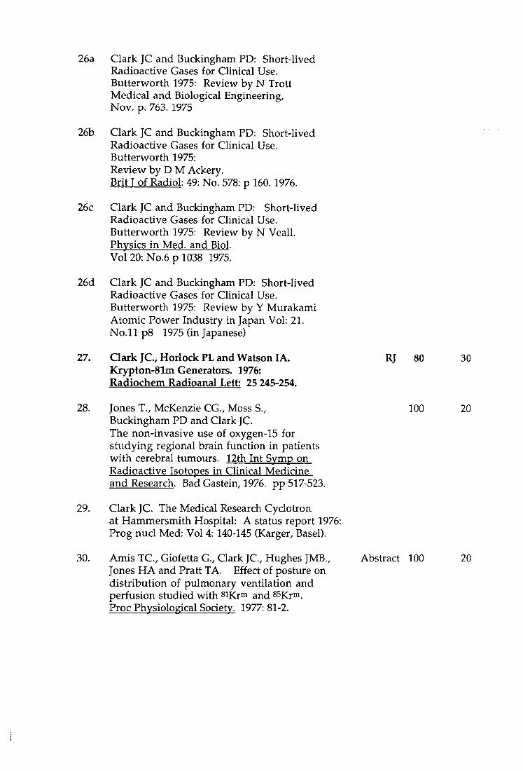
26a Clark JC and Buckingham PD: Short-lived Radioactive Gases for Clinical Use. Butterworth 1975: Review by N Trott Medical and Biological Engineering, Nov. p. 763. 1975
26b Clark JC and Buckingham PD: Short-lived Radioactive Gases for Clinical Use. Butterworth 1975: Review by D M Ackery. Brit T of Radiol: 49: No. 578: p 160. 1976.
26c Clark JC and Buckingham PD: Short-lived Radioactive Gases for Clinical Use. Butterworth 1975: Review by N Veall. Physics in Med, and Biol. Vol 20: No.6 p 1038 1975.
26d Clark JC and Buckingham PD: Short-lived Radioactive Gases for Clinical Use. Butterworth 1975: Review by Y Murakami Atomic Power Industry in Japan Vol: 21. No. 11 p8 1975 (in Japanese)
27. Clark J C , Horlock PL and Watson lA. RJ 80 30 Krypton-81m Generators. 1976: Radiochem Radioanal Lett: 25 245-254.
28. Jones T., McKenzie CG., Moss S., 100 20 Buckingham PD and Clark JC. The non-invasive use of oxygen-15 for studying regional brain function in patients with cerebral tumours. 12th Int Symp on Radioactive Isotopes in Clinical Medicine and Research. Bad Gastein, 1976. pp 517-523.
29. Clark JC. The Medical Research Cyclotiron at Hammersmith Hospital: A status report 1976: Prog nucl Med: Vol 4: 140-145 (Karger, Basel).
30. Amis TC, Giofetta G., Clark JC, Hughes JMB., Abstract 100 20 Jones HA and Pratt TA. Effect of posture on distribution of pulmonary ventilation and perfusion studied with siKr^ and 85Krm. Proc Physiological Society. 1977: 81-2.
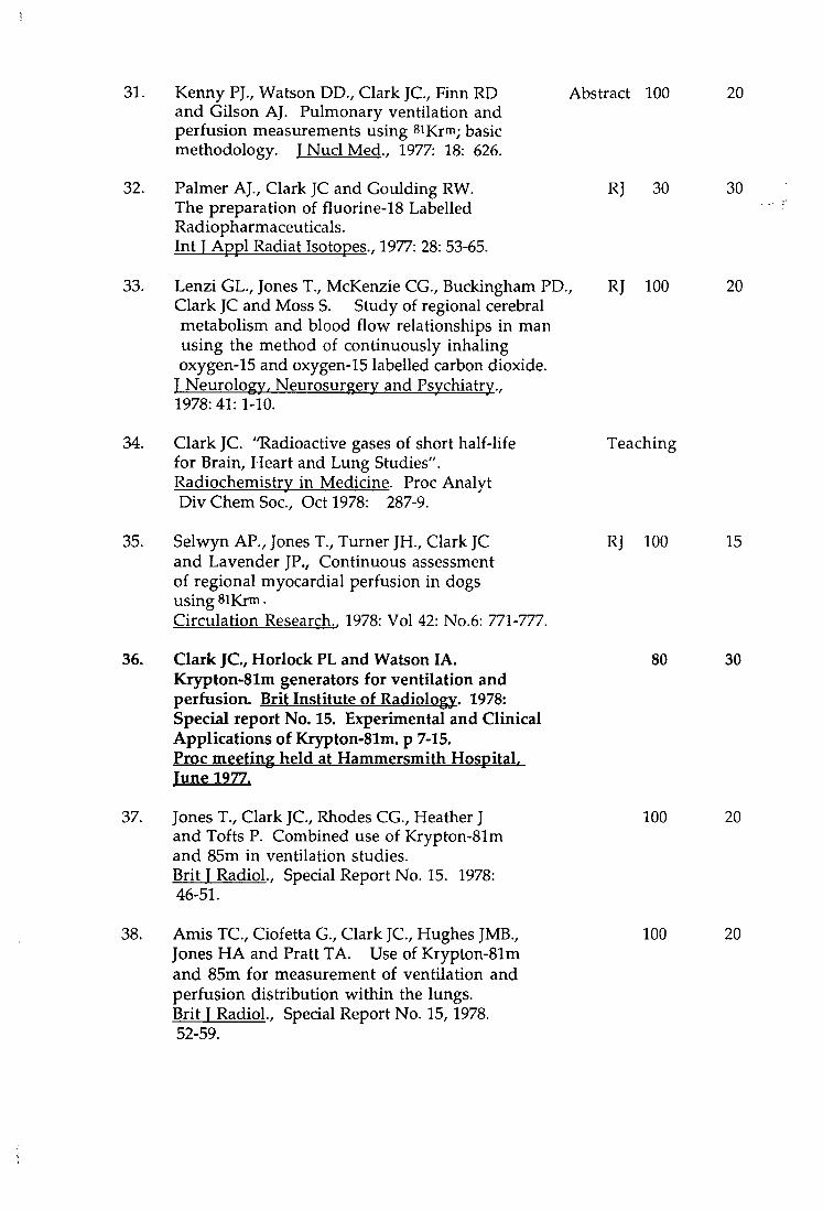
31. Kenny PJ., Watson DD., Clark JC, Finn RD Abstract 100 20 and Gilson AJ. Pulmonary ventilation and perfusion measurements using siKrm; basic methodology. I Nucl Med., 1977: 18: 626.
32. Palmer AJ., Clark JC and Goulding RW. RJ 30 30 The preparation of fluorine-18 Labelled Radiopharmaceuticals. Int T Appl Radiat Isotopes., 1977: 28: 53-65.
33. Lenzi GL., Jones T., McKenzie CG., Buckingham PD., RJ 100 20 Clark JC and Moss S. Study of regional cerebral metabolism and blood flow relationships in man using the method of continuously inhaling oxygen-15 and oxygen-15 labelled carbon dioxide.
J Neurology, Neurosurgery and Psychiatry., 1978: 41: 1-10.
34. Clark JC. "Radioactive gases of short half-life Teaching for Brain, Heart and Lung Studies". Radiochemistry in Medicine. Proc Analyt Div Chem Soc, Oct 1978: 287-9.
35. Selwyn AR, Jones T., Turner JH., Clark JC RJ 100 15 and Lavender JP., Continuous assessment of regional myocardial perfusion in dogs using silCrm. Circulation Research.. 1978: Vol 42: No.6: 771-777.
36. Clark JC, Horlock PL and Watson lA. 80 30 Krypton-81m generators for ventilation and perfusion. Brit Institute of Radiology. 1978: Special report No. 15. Experimental and Clinical Applications of Krypton-81m. p 7-15. Proc meeting held at Hammersmith Hospital. Tune 1977.
37. Jones T., Clark JC, Rhodes CG., Heather J 100 20 and Tofts P. Combined use of Krypton-81m and 85m in ventilation studies. Brit T Radiol. Special Report No. 15. 1978: 46-51.
38. Amis TC, Ciofetta G., Clark JC, Hughes JMB., 100 20 Jones HA and Pratt TA. Use of Krypton-81m and 85m for measurement of ventilation and perfusion distribution within the lungs. Brit T Radiol., Special Report No. 15,1978. 52-59.
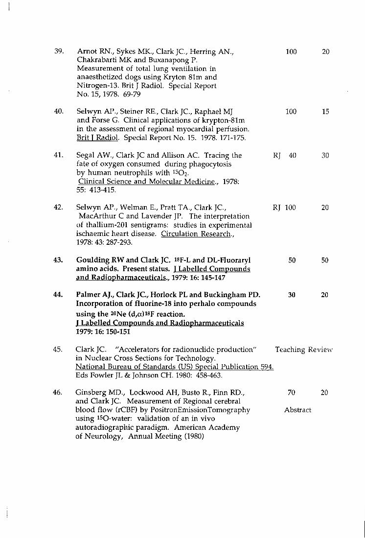
39. Arnot RN. , Sykes MK., Clark J C , Herring AN. , 100 20 Chakrabarti M K and Buxanapong P. Measurement of total lung ventilation in anaesthetized dogs using Kryton 81m and Nitrogen-13. Brit J Radiol. Special Report No. 15,1978. 69-79
40. Selwyn AP., Steiner RE. , Clark J C , Raphael MJ 100 15 and Forse G. Clinical applications of krypton-81m in the assessment of regional myocardial perfusion. Brit T Radiol. Special Report No. 15. 1978. 171-175.
41. Segal AW., Clark JC and Allison A C . Tracing the RJ 40 30 fate of oxygen consumed during phagocytosis by human neutrophils with 1502. Clinical Science and Molecular Medicine., 1978:
55: 413-415.
42. Selwyn AP., Welman E . , Pratt TA., Clark J C , RJ 100 20 Mac Arthur C and Lavender JP. The interpretation
of thallium-201 sentigrams: studies in experimental ischaemic heart disease. Circulation Research., 1978: 43: 287-293.
43. Goulding R W and Clark JC. " F - L and DL-Huoraryl 50 50 amino acids. Present status. T Labelled Compoxmds and Radiopharmaceuticals.. 1979:16:145-147
44. Palmer AJ., Clark J C , Horlock PL and Buckingham PD. 30 20 Incorporation of fluorine-18 into perhalo compounds using the 20Ne (d,a)i8F reaction. T Labelled Compounds and Radiopharmaceuticals 1979:16:150-151
45. Clark JC. "Accelerators for radionuchde production" Teaching Review in Nuclear Cross Sections for Technology. National Bureau of Standards (US) Special Publication 594. Eds Fowler JL & Johnson C H . 1980: 458-463.
46. Ginsberg MD., Lockwood A H , Busto R., Finn RD., 70 20 and Clark JC. Measurement of Regional cerebral blood flow (rCBF) by PositronEmissionTomography Abstract using ISO-water: validation of an in vivo autoradiographic paradigm. American Academy of Neurology, Annual Meeting (1980)
47. Arnot RN. , Clark J C , Herring AN. , Chakrabarti MK RJ 100 20 and Sykes MK. Measurements of regional ventilation using nitrogen-13 and krypton-81m in mechanically
ventilated dogs. Clin Phys Physiol Meas.. 1981: 2: 183-196.
48. Horlock PL. , Q a r k J C , Goodier IW., Barnes JW., RJ 15 15 Bentley G E . , Grant PM and O'Brien HA. The preparation of a rubidiixm-82 radionuclide generator. J Radioanalytical Chemistry.. 1981:64:257-265.
49. Finn R., Vora M., Boothe T., Campbell J., Carroll S RJ 50 15 and Clark J. Radiation-induced defects as illustrated by the 81Rb-81Krm target system. Int J Appl Radiat Isotopes., 1982: 33: 349-353.
50. Selwyn AP., Allan RM., L'Abbate A., Horlock PL. , RJ 20 20 Camici P., Clark J C , O'Brien H A and Grant P. Relation between regional myocardial uptake of rubidium-82 and perfusion: absolute reduction of cation uptake in ischaemia. Am T Cardiol., 1982: 50: 112-121.
51. Clark JC and Oberdorfer F. Thermal characteristics of the 50 50 release of fluorine-18 from an inconel-600 gas target. T Labelled Compounds and Radiopharmaceuticals., 1982: 19:1337-1339.
52. Jones HA. , Clark J C , Davies E E . , Forster RE and RJ 70 20 Hughes JMB. Rate of uptake of carbon monoxide at different inspired concentrations. T Appl Physiol. 1982: 52 (1): 109-113.
53. Jones HA. , Stradling JR., Clark J C , Davies E E and RJ 72 20 Rozkovec A. Quantitative measurement of intrapulmonary and extrapulmonary right-to-left shunt. I Appl Physiol, 1983: 54: 1434-1438.
54. Waters S L . , Butler K R . , Clark J C , Horlock PL. , RJ 10 10 Kensett MJ., Goodier IW., Makepeace J . , Smith D.,
Woods MJ., Barnes JW., Bentley G E . , Grant P M and O'Brien H A . Radioassay problems associated with
the clinical use of Rb-82 radionuclide generator. Int J N u d Med Biol., 1983:10: 2/3, 69-74.
55. Kanno I . , Lammertsma AA. , Heather JD., Gibbs JM., RJ 70 10 Rhodes C G . , Clark JC and Jones T. Measurement of cerebral blood flow using bolus inhalation of C1502 and Positron Emission Tomography : Description of the
Method and its Comparison with the C15O2 Continuous Inhalation Method. T Cerebral Blood Flow and MetaboUsm., 1984: 4: 224-234.
56. Spinks TJ., Bateman JE., Flesher A C , Clark J C , RJ 100 30 Hopkins N F G and Jones T. Blood flow in the feet of diabetic patients measured with a MWPC positron camera and inhalation of C15O2. Phys Med Biol.. 1984: 29: 873-879.
57. Pike VW., Horlock PL. , Brown C and Clark JC. RJ 30 25 The remotely-controlled preparation of a nC-labelled radiopharmaceutical - [l-iiC]Acetate.
Int T Appl Radiat Isotopes. 1984: 35:623-627.
58. Herold S., Leenders K L . , Turton DR., Kensett MJ., 10 10 Pike VW., Clark J C , Brooks DJ., Crow TJ., Owen F., Cooper S., Johnstone E C . Dopamine receptor binding in Schizophrenic patients as measured with i i C methyl-spiperone and PET.
T Cerebral Blood Flow and Metabolism. 1985. Abstract Vol: 5: Suppl 1. S191-2
59. Leenders K L . , Palmer AJ., Quinn N., Clark J C , RJ 10 10 F imau G. , Garnett ES. , Nahmias C , Jones T and Marsden C D . Brain dopamine metabolism in patients with Parkinson's Disease measured with positron emission tomography. T Neurology. Neurosurgery. Psychiatry.. 1986:49:853-860.
60. Clark JC. Positrons from generators. 67-84 Teaching Review In: Progress in Radiotherapy. Eds Cox PM et al. Martinus Nijhoff, Netherlands, 1986.
61. Clark, J C , Crouzel C , Meyer GJ and Strijckmans K. RJ 30 25 Current Methodology for Oxygen-15 production for Clinical Use., Appl Radiat Isot: 38.8 1987: 597-600.
62. Jones AJ., Rhodes C G . , Law MNP., Becket JM., Clark J C , RJ 10 20 Boobis AR., and Taylor GW. Rapid analysis for metabolites of llC-labelled drugs: fate of [llC]-S-4-(tert-butylamino-2-hydroxypropoxy)- benzimidazol-2-one in the dog. T. Chromatography.. 1991:570, 361-370.
63. Colebatch JG., Adams L . , Murphy K., Martin AJ., RJ 50 15 Lammertsma AA. , Tochon-Danguy HJ., Clark J C , KJ Friston and Guz A. Regional Cerebral Blood Flow during Volitional Breathing in Man. J Physiology.. 1991:443,91-103.
64. Guillaume M., Luxen A., Nebeling B., Argentini M., Clark JC and Pike VW. Recommendations for fluorine-18 production. Appl Radiat Isot.. 42.8: 1991: 749-762.
RJ 20 20
65. Brady P., Luthra SK., Tochon-Danguy HJ., Steel CJ. , RJ 10 10 Waters W L . , Kensett MJ., Landais P., Shah F., Jaeggi KA. , Drake A., Clark JC and Pike VW. Assymetric synthesis of a precursor for the automated radiosynthesis of S-(3'-t-Butylamino-2'- hydroxypropoxy)-benzimidazol-2-[iiC] one (S-[ii]CGP 12177) as a preferred radioligand
for P -Adrenergic Receptors. Appl Radiat Lsot. 1991: 42,7: 621-628.
66. Ranicar ASO., WiUiams CW. , Schnorr L . , Clark J C , RJ 25 Rhodes C G . , Bloomfield PM and Jones T.
The on-line monitoring of continuously withdrawn arterial blood during PET studies using a single B G O / photomultipUer assembly and non-stick tubing. Medical Progress through Technology: 1991:17: 259-264,1991.
67. Clark JC. Production and AppUcation of Oxygen-15. Radiopharmacy aspects. Progress in Radiopharmacy (1992) 91-107: Eds Schubiger PA and Westera G. Kulwer Academic Publishers.
Teaching 100
68. Clark JC. (1992) Production of Radiopharmaceuticals Teaching 100 for PET. Proceedings of the 1992 Advanced School on Medical Physics. Phvsica Medica Vol 13: Suppl 1:1992: 43-47.
69 Clark JC. MorelleJ-L. An Oxygen-15 Generator: 50 50 Early Experience with the IBA Cyclone-3 Proceedings of the IVth International Workshop on Targetry and Target Chemistry: PSI-Proceedings ISSN 1019-6447. August 1992. pp 34-35. E d Regin Weinreich. Paul Scherrer Institut, 1992.
70. Clark JC. Status report from Hanunersmith. Proceedings of the IVth International Workshop on Targetry and Target Chemistry: PSI-Proceedings ISSN 1019-6447. pp 237-241. E d Regin Weinreich. Paul Scherrer Institut, 1992.
71. Clark JC and Tochon-Danguy H. , "R2D2", 70 70 A Bedside IOxygen-15] Water Infuser. Proceedings of the IVth International Workshop on Targetry and Target Chemistry:
Status report
PSI-Proceedings ISSN 1019-6447. pp 234-235. E d Regin Weinreich. Paul Scherrer Institut, 1992.
72 Clark JC and Dowsett K. Automated Carbon-11 70 50 Radiopharmaceutical Production. Proceedings of the IVth International Workshop on Targetry and Target Chemistry: PSI-Proceedings ISSN 1019-6447. pp 207-209. E d Regin Weinreich. Paul Scherrer Institut, 1992.
73. Link JM., Clark JC and Ruth TJ., Introduction: 40 40 State of the Art in Automated Syntheses of Short-Lived Chair persons Radiopharmaceuticals. Review Proceedings of the IVth International Workshop on Targetry and Target Chemistry: PSI-Proceedings ISSN 1019-6447. pp pp 174-185. E d Regin Weinreich. Paul Scherrer Institut, 1992.
74. Qaim SM., Clark J C C , Crouzel C , Guillaume M, E E C Chapter Helmeke HJ., Nebeling B., Pike VW and StockUn G. P E T radionuclide production. Radiopharmaceuticals for Positron Emission Tomography - Methodologica 1 Aspects. Kluwer Academic Pubis. 1993. pp 1 - 44.
75. Crouzel C , Clark J C , Brihaye C , Langstrom B., E E C Chapter Lemaire C , Meyer G-J., Nebeling B and Stone-Elander S. Radiochemistry automation for PET. Radiopharmaceuticals for Positron Emission Tomography - Methodological Aspects. Kluwer Academic Pubis. 1993: pp 45 - 90.
76. Ramsay S C , Murphy K., Shea SA., Friston KJ., RJ 100 20 Lammertsma AA., Clark J C , Adams L . , Guz A and Frackowiak RSJ. Changes in global cerebral blood flow in humans: Effect on regional cerebral blood flow during a neural activation task. T of Physiology 1993: 471: pp 521-534
77. Silbersweig DA., Stern E . , Frith CD. , Cahill C , RJ 100 20 Schnorr L . , Grootoonk S., Spinks T., Clark J C , Frackowiak RSJ and Jones T. Detection of Thirty-Second Cognitive Activations in single subjects with positron emission tomography: A new low-dose Hi^^O regional cerebral blood flow three-dimensional imaging technique. T Cerebral Blood Flow and Metabolism 1993: 13: pp 617-629.
78. Clark J C . The Hammersmith Philosophy for P E T Teaching Chemistry Automation to be published in American Chemical Society monograph 1994. "Chemist views of Imaging Centers" Symposium held at A C S fall meeting Chicago, August 1993.
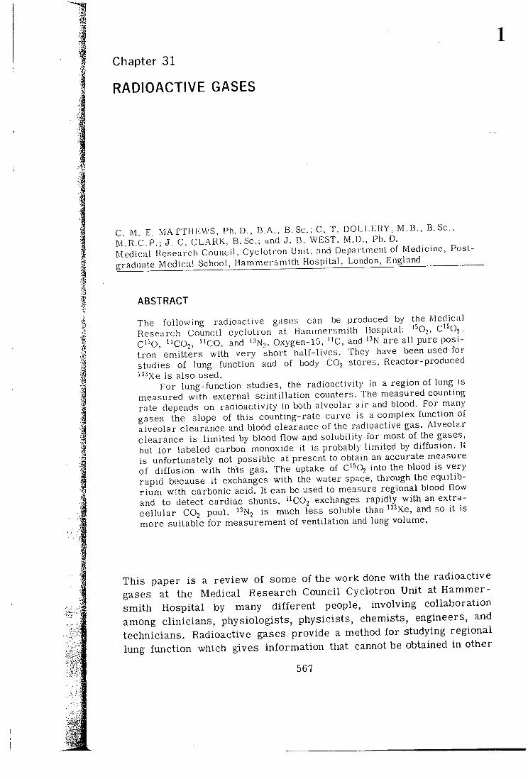
Chapter 31
RADIOACTIVE GASES
C M E MATTHEWS, Ph. D., D.A., B. Sc.; C. T. DOLLERY, M.B., B.Sc, M R C P • J C. CLARK, B.Sc; and J. B. WEST, M.D., Ph. D. Medical Research Council, Cyclotron Unit, and Department of Medicine, Post-graduate Medical School, Hammersmith Hospital, London, England
ABSTRACT
The following radioactive gases can be produced by the Medical Research Council cyclotron at Hammersmith Hospital: '^02, C'^Oj, C^='0, "CO2, "CO, and I^N^. Oxygen-15, "C, and '^N are all pure positron emitters with very short half-lives. They have been used for studies of lung function and of body CO2 stores, lieactor-produced '•'• Xe is also used.
For lung-function studies, the radioactivity in a region of lung is measured with external scintillation counters. The measured counting rate depends on radioactivity in both alveolar air and blood. For many gases the slope of this counting-rate curve is a complex function of alveolar clearance and blood clearance of the radioactive gas. Alveolar clearance is limited by blood flow and solubility for most of the gases, but for labeled carbon monoxide it is probably limited by diffusion. It is unfortunately not possible at present to obtain an accurate measure of diffusion with this gas. The uptake of C'^Oj into the blood is very rapid because it exchanges with the water space, through the equilibr ium with carbonic acid. It can be used to measure regional blood flow and to detect cardiac shunts. '^C02 exchanges rapidly with an extracellular CO2 pool. '^Nj is much less soluble than '^^Xe, and so it is more suitable for measurement of ventilation and lung volume.
Th i s paper i s a review of some of the work done wi th the radioact ive gases at the Medica l Research Council Cyclot ron Uni t at H a m m e r smi th Hospi ta l by many d i f f e ren t people, involving col laborat ion among c l in ic ians , physiologis ts , physicis ts , chemists, engineers, and technicians. Radioactive gases provide a method f o r studying regional lung func t ion which gives in fo rma t ion that cannot be obtained in other
567
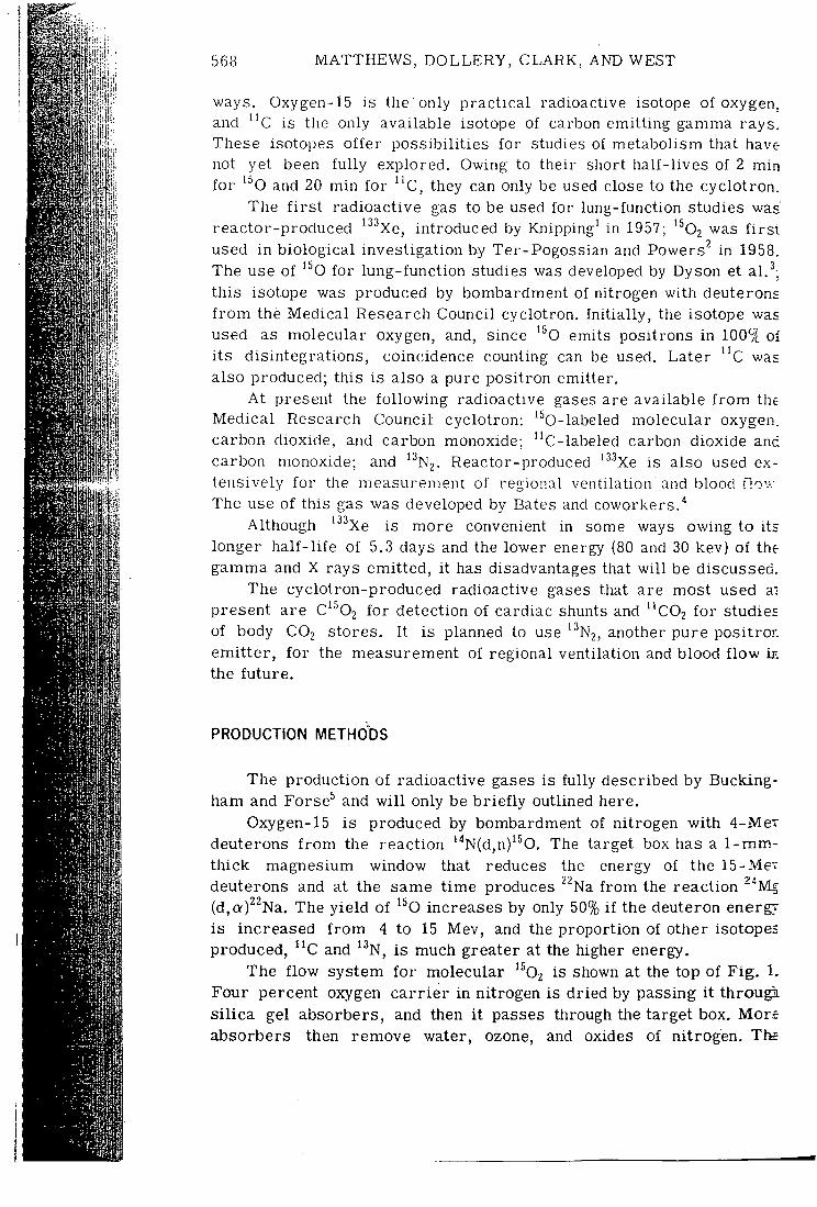
568 MATTHEWS, D O L L E R Y , CLARK, AND WEST
d'rH
ways. Oxygen-15 is the only p rac t i ca l radioactive isotope of oxygen, and " C is the only available isotope of carbon emit t ing gamma rays. These isotopes o f f e r poss ib i l i t ies for studies of metabolism that have not yet been fu l l y explored. Owing to their short ha l f - l ives of 2 min fo r '^O and 20 min fo r " c , they can only be used close to the cyc lo t ron .
The f i r s t radioact ive gas to be used for lung-function studies was reactor-produced '^'^Xe, introduced by Knipping' in 1957; '^©2 was f i r s t used in b io logica l invest igat ion by Ter-Pogossian and Powers^ in 1958. The use of '^O fo r lung-funct ion studies was developed by Dyson et al.'^; th is isotope was produced by bombardment of nitrogen wi th deuterons f r o m the Medical Research Council cyclotron. In i t i a l ly , the isotope was used as molecular oxygen, and, since '^O emits positrons in 100% oi i t s d is integrat ions , coincidence counting can be used. Later " c was also produced; this is also a pure posi t ron emitter .
At present the f o l l o w i n g radioactive gases are available f r o m the Medica l Research Counci l cyclo t ron: '^0-labeled molecular oxygen, carbon dioxide, and carbon monoxide; "C-labeled carbon dioxide and carbon monoxide; and '^N2. Reactor-produced ''^''Xe is also used extensively f o r the measurement of regional venti lat ion ' and blood flov-.-The use of this gas was developed by Bates and coworkers. ' '
Although ''^^Xe is more convenient in some ways owing to its longer h a l f - l i f e of 5.3 days and the lower energy (80 and 30 kev) of the gamma and X rays emit ted , i t has disadvantages that w i l l be discussed.
The cyclot ron-produced radioactive gases that are most used ai present are C'^Oj f o r detection of cardiac shunts and " C O 2 f o r studies of body CO2 stores. I t is planned to use ''^N2, another pure positrOE emi t t e r , f o r the measurement of regional venti lat ion and blood f l o w in the f u t u r e .
PRODUCTION METHODS
The product ion of radioactive gases is fu l ly described by Buckingham and Forse^ and w i l l only be b r i e f l y outlined here.
Oxygen-15 is produced by bombardment of ni trogen w i t h 4-MeT deuterons f r o m the react ion ' ' 'N(d,n)'^0. The target box has a 1-mm-th ick magnesium window that reduces the energy of the 15-Me-deuterons and at the same t ime produces ^^Na f r o m the react ion "^Mg (d,Q:)^^Na. The y i e ld of ^^O increases by only 50% i f the deuteron energy is increased f r o m 4 to 15 Mev, and the propor t ion of other isotopes produced, ^ C and ^^N, is much greater at the higher energy.
The f l ow system f o r molecular ^^02 is shown at the top of F i g . 1. Four percent oxygen c a r r i e r in nitrogen is dr ied by passing i t through s i l i c a gel absorbers , and then i t passes through the target box. More absorbers then remove water, ozone, and oxides of n i t rogen. The
's. ve un m. •as rst 38.
ms /as
of ;as
the en, ind 3X-j W .
i ts the ed.
at ies ron ; in
ng-
Aev im-Aev 'Mg rgy pes
. 1. ugh Mi ore The
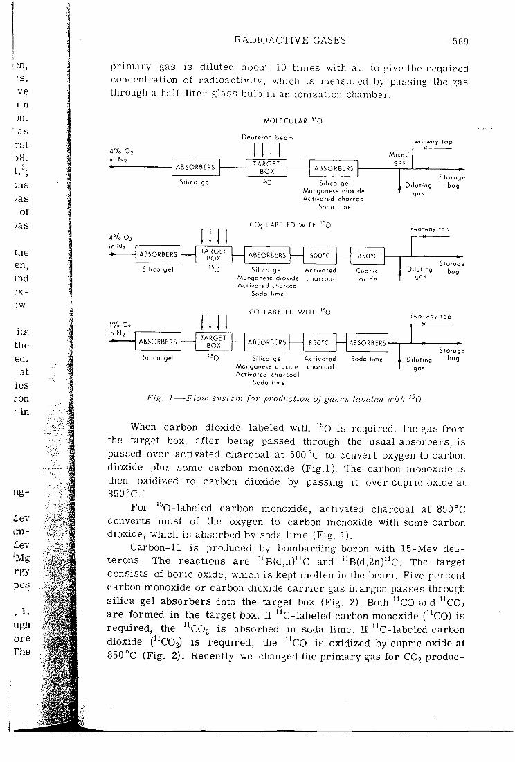
R A D I O A C T I V E GASES 569
p r i m a r y gas is di luted about 10 t imes with a i r to give the required concentration of rad ioac t iv i ty , winch is measured by passing the gas through a h a l f - l i t e r glass bulb m an ionization chamber.
M O L E C U L A R ' o
Deuteron beam
4 % O2 in N2
Two-way t a p
A B S O R B E R S T A R G E T A B S O R B E R S A B S O R B E R S
B O X A B S O R B E R S
S i l i ca gel '50 M
Silico gel angonese dioni
Mixed gas
Ac two ted c h a r c o a l Soda lime
4V0 O2 in No
CO2 L A B E L E D W I T H '^O
Storage Diluting bog
gas
TwO'wOy t o p
A B S O R B E R S T A R G E T ABSORBERS 5 0 0 ° C A B S O R B E R S
BOX ABSORBERS 5 0 0 ° C
S i l i ca ge l '5o Si l i ca gel Manganese dioxide Act iva ted charcool
Act ivated charcoa l
Cupr ic ox ide
Diluting ga s
StOfOge bog
47o O2 in N2
Sodo l.n
C O L A B E L E D W I T H '^O Two-way top
A B S O R B E R S T A R G E T
A B S O R B E R S ABSORBERS A B S O R B E R S BOX A B S O R B E R S ABSORBERS
Si l ica gel '^0 Si l ica gel Manganese dioxide Act ivated chorcool
Act ivoted charcoal
Soda lime Diluting gas
Storage bog
Soda
Fig. ]—Flow system for production oj gases Labeled ivUh '^0.
When carbon dioxide labeled wi th '^O is required, the gas f r o m the target box, a f t e r being passed through the usual absorbers, is passed over activated charcoal at 500 °C to- convert oxygen to carbon dioxide plus some carbon monoxide ( F i g . l ) . The carbon monoxide is then oxidized to carbon dioxide by passing i t over cupric oxide at 850°C . -
For ^^O-labeled carbon monoxide, activated charcoal at 850°C converts most of the oxygen to carbon monoxide wi th some carbon dioxide, which is absorbed by soda l ime (Fig . 1).
Carbon-11 is produced by bombarding boron wi th 15-Mev deu-terons. The react ions are ^°B(d,n)^'C and '^B(d,2n)'^C. The target consists of bor ic oxide, which i s kept molten in the beam. Five percent carbon monoxide or carbon dioxide c a r r i e r gas i n argon passes through s i l i c a gel absorbers into the target box (Fig. 2). Both ^^CO and '^C02 are f o r m e d i n the target box. I f ^'C-labeled carbon monoxide (^^CO) is requi red , the ^^COi is absorbed in soda l i m e . If "C- labe led carbon dioxide (^^COa) is requ i red , the '^CO is oxidized by cupric oxide at 850°C (Fig . 2). Recently we changed the p r i m a r y gas f o r CO2 produc-
570 M A T T H E W S , DOLLERY, CLARK, AND WEST
C O ' L A B E L E D W I T H " C
Deureron beom
in Ar A B S O R B E R S
T A R G E T BOX
A B S O R B E R A B S O R B E R S T A R G E T
BOX A B S O R B E R
Si l ico gel S o d a lime
C O 2 LABELED W ITH " C
Oeoteron b e a m
in He A B S O R B E R S
T A R G E T 850% A B S O R B E R S BOX
850%
Si l i ca gel "c C u p r i c oxide
Two-way t a p
Sforoge Di lut ing bog .
g a s
Two-way tap
Storage D i lu t ing bog
g o j
mi Deuteron b e a m
I I A B S O R B E R S
T A R G E T /sec A B S O R B E R A B S O R B E R S BOX /sec A B S O R B E R
S i l i ca gel 13N C o p p e r Soda lime
Two-way top
S loroge
Di lut ing g o s
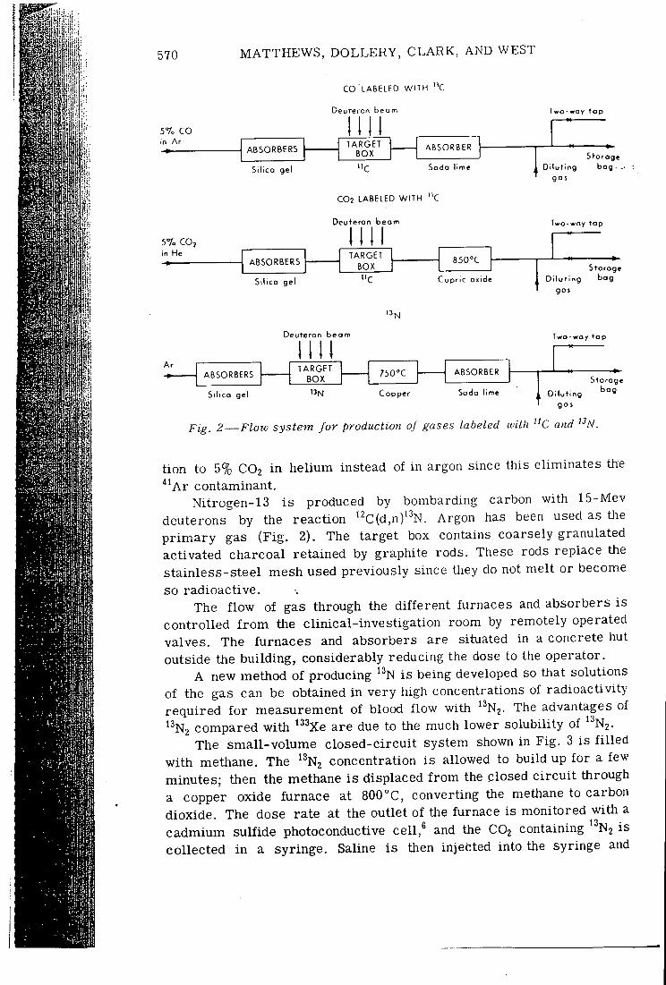
Fig. 2—Flow system for production of gases labeled with "C and "A'.
t ion to 5% CO2 i n he l ium instead of in argon since this e l iminates the ' ' ' A r contaminant.
Nitrogen-13 i s produced by bombarding carbon wi th 15-Mev deuterons by the react ion '^C(d,n)' ' 'N. Argon has been used as the p r i m a r y gas ( F i g . 2). The target box contains coarsely granulated activated charcoal retained by graphite rods. These rods replace the s ta in less-s tee l mesh used previously since tliey do not mel t or become so radioact ive .
The f l o w of gas through the d i f f e r en t furnaces and absorbers is con t ro l l ed f r o m the c l in ica l - inves t iga t ion room by remotely operated valves. The furnaces and absorbers are situated in a concrete hut outside the bu i ld ing , considerably reducing the dose to the operator .
A new method of producing '^N i s being developed so that solutions of the gas can be obtained in ve ry high concentrations of rad ioac t iv i ty r equ i r ed f o r measurement of blood f l ow wi th '^N2. The advantages of '^N2 compared w i t h ' ^^e are due to the much lower solubi l i ty of '^N2.
The sma l l -vo lume c losed-c i r cu i t system shown in F ig . 3 is f i l l e d w i t h methane. The '^N2 concentration i s allowed to bui ld up f o r a few minutes; then the methane i s displaced f r o m the closed c i r c u i t through a copper oxide fu rnace at 800°C , convert ing the methane to carbon dioxide . The dose rate at the outlet of the furnace is moni tored wi th a cadmium sulf ide photoconductive cel l ,^ and the CO2 containing '^N2 is co l lec ted i n a syr inge . Saline i s then injected into the syringe and
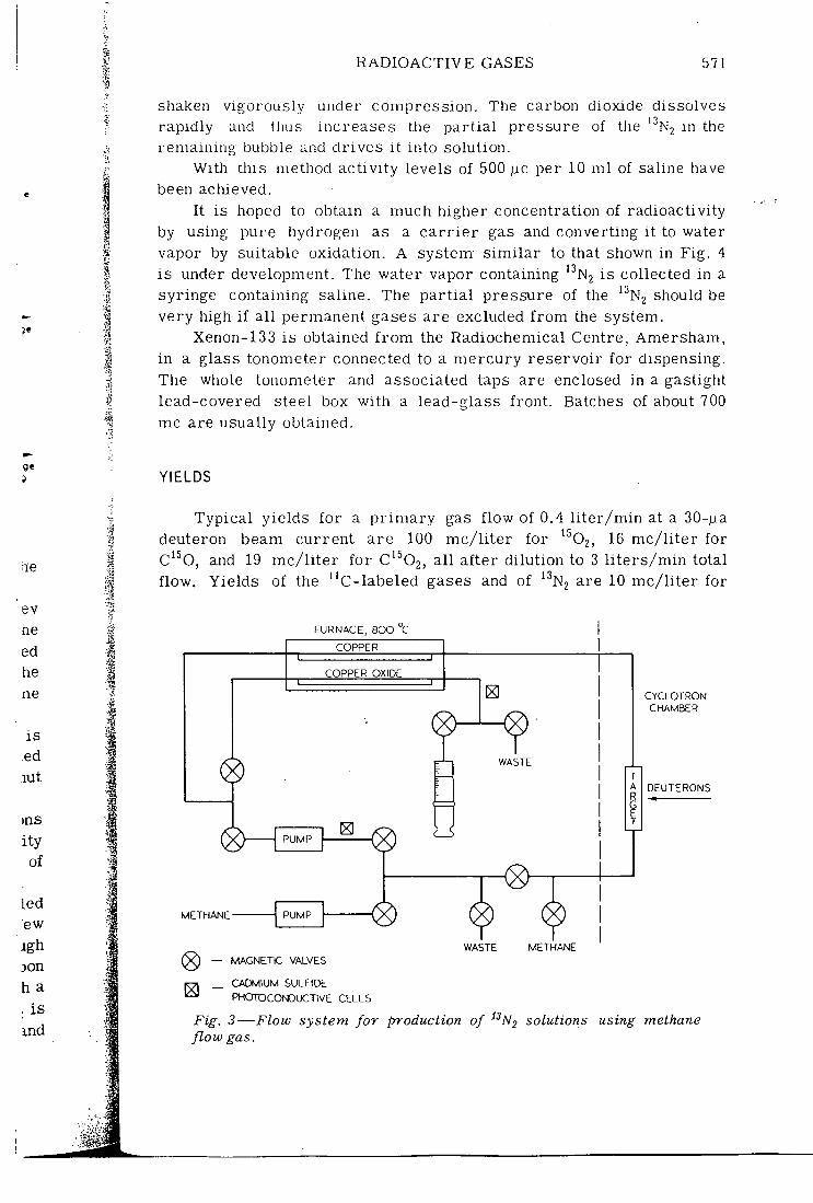
R A D I O A C T I V E G A S E S 571
shaken vigorously under compress ion. The carbon dioxide dissolves rapid ly and thus increases the pa r t i a l pressure of the '•'N2 in the remain ing bubble and d r ives i t into solut ion.
With this method ac t iv i ty levels of 500 |ic per 10 m l of saline have been achieved.
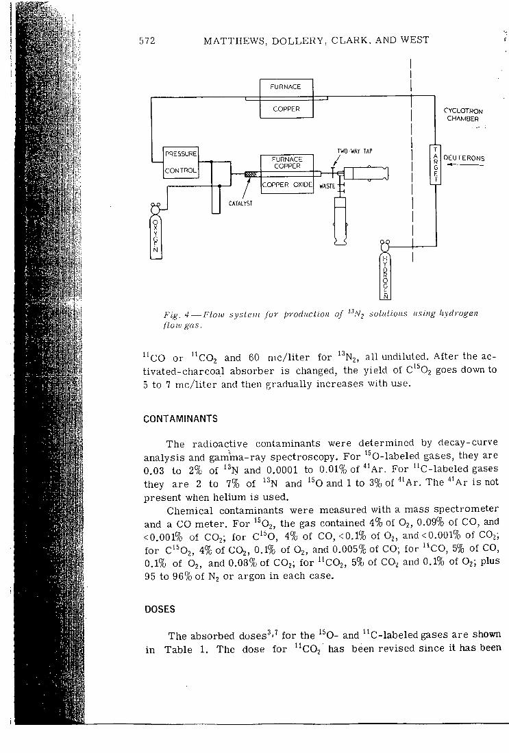
I t is hoped to obtain a much higher concentration of radioact iv i ty by using pure hydrogen as a c a r r i e r gas and convert ing i t to water vapor by suitable oxidat ion. A system- s i m i l a r to that shown in F ig . 4 is under development. The water vapor containing ' ' 'N2 is collected in a syringe containing saline. The p a r t i a l pressure of the '^N2 should be ve ry high i f a l l permanent gases are excluded f r o m the system.
Xenon-133 i s obtained f r o m the Radiochemical Centre, Amersham, in a glass tonometer connected to a m e r c u r y r e se rvo i r f o r dispensing. The whole tonometer and associated taps are enclosed in a gastight lead-covered steel box wi th a lead-glass f ron t . Batches of about 700 mc are usually obtained.
9 YIELDS
Typica l yields fo r a p r i m a r y gas f low of 0.4 l i t e r / m i n at a 30-fj.a deuteron beam cur ren t are 100 m c / l i t e r f o r ^^62, 16 m c / l i t e r for C'^0, and 19 m c / l i t e r f o r C^^Oj, a l l a f te r d i lu t ion to 3 l i t e r s / m i n total f l ow . Yields of the ^^C-labeled gases and of '•^N2 are 10 m c / l i t e r f o r
FURNACE, BOO C
COPPER
COPPER OXIDE
PUMP
WASTE
METHANE- PUMP I PUMP \
(8) WASTE METHANE
MAGNETIC VALVES
CADMIUM SULFIDE PHOTOCONDUCTIVE CELLS
CYCLOTRON CHAMBER
OEUTERONS
Fig. 3—Flow system for production of ^^Nj solutions using methane flow gas.
572 MATTHEWS, D O L L E R Y , CLARK, AND WEST
FURNACE
COPPER
PRESSURE
CONTROL
FURNACE COPPER
COPPER OXIDE
TWO-WAY TAP
/ W A S T E
C A T A L Y S T
CYCLOTRON CHAMBER
DEUTERONS
Fig. 4—Flow system for produclion of "/V^ soliUioiis using liydrogen flow gas.
' ' c O or ' 'cOo and 60 m c / l i t e r fo r 13 N2, a l l undiluted. A f t e r the ac-t ivated-char coal absorber is changed, the y ie ld of c'^02 goes down to 5 to 7 m c / l i t e r and then gradual ly increases wi th use.
CONTAMINANTS
The radioact ive contaminants were determined by decay-curve analysis and gamma-ray spectroscopy. For '^0-labeled gases, they are 0.03 to 2% of ' ^N and 0.0001 to 0.01% of ^ ' A r . For "C- labe led gases they are 2 to 7% of ' ^N and '^O and 1 to 3% of ^ ' A r . The ^ ' A r is not present when he l ium is used.
Chemical contaminants were measured wi th a mass spectrometer and a CO meter . For ' ^Oj , the gas contained 4% of O2, 0.09% of CO, and <0.001% of CO2; f o r C'^0, 4% of CO, <0.1% of O2, and<0.001% of CO2; f o r C'^02, 4% of CO2, 0.1% of O2, and 0.005% of CO; fo r ' 'CO, 5% of CO, 0.1% of O2, and 0.08% of CO2; f o r ' ' C O 2 , 5% of CO2 and 0.1?o of O j ; plus 95 to 96% of N2 or argon i n each case.
DOSES
i n
The absorbed doses^'^ f o r the '^O- and ' 'C- labe led gases are shown Table 1. The dose f o r ' ' C O 2 has been revised since i t has been
ERONS
RADIOACTIVE GASES 573
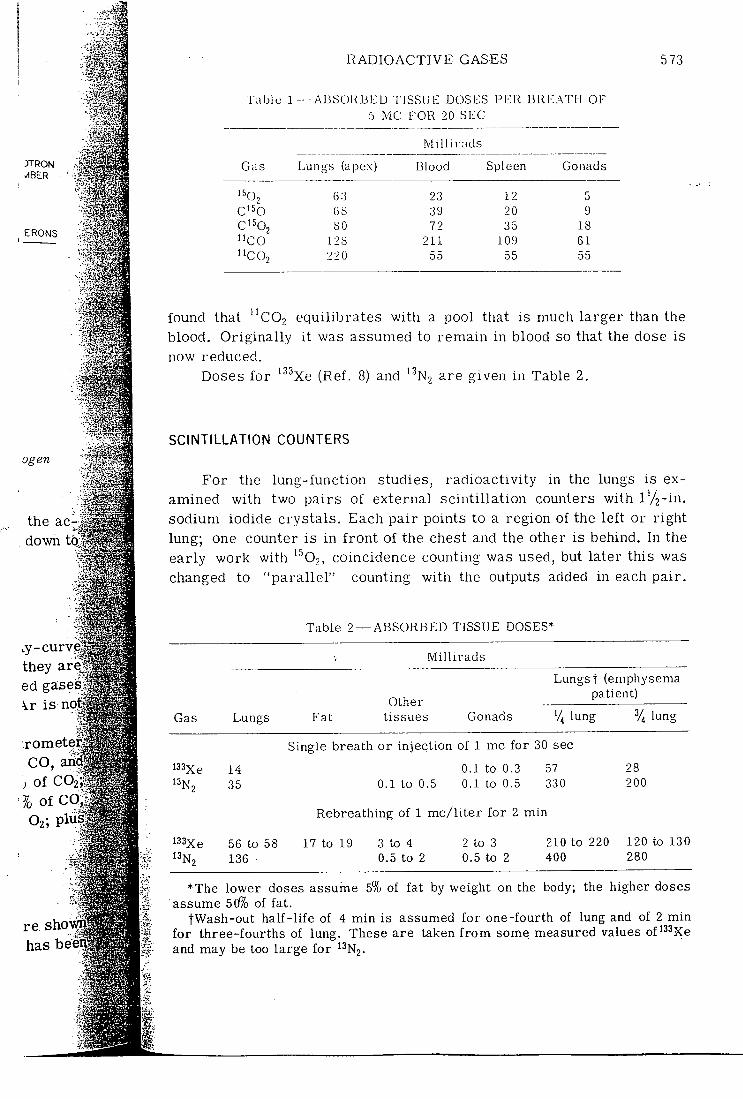
Tabic 1 — AiJSOKBl:: D T ISSUE DOSES PER Bi l l •:ATI-I OF 5 MC FOR 20 SEC
.Millirads
Gas Lungs (apex) Blood Spleen Gonads
'^02 63 23 12 5 C'50 68 39 20 9 C'^02 SO 72 35 18 "CO 128 211 109 61 "CO2 220 55 55 55
found that " C O 2 equil ibrates wi th a pool that is much larger than the blood. Or ig ina l ly i t was assumed to remain in blood so that the dose is now reduced.
Doses f o r '^^Xe (Ref. 8) and ^^Nj are given in Table 2.
ogen
the acr:, down to.
SCINTILLATION COUNTERS
For the lung-funct ion studies, rad ioac t iv i ty in the lungs is examined wi th two pa i rs of external sc in t i l l a t ion counters wi th l ' / 2 - i n . sodium iodide crys ta ls . Each pa i r points to a region of the l e f t or r ight lung; one counter is in f r o n t of the chest and the other is behind. In the early work wi th '^02, coincidence counting was used, but later this was changed to " p a r a l l e l " counting wi th the outputs added in each pa i r .
.y-curve they are ed ga;ses Vr i s not
.rometer' ft 'iSf C O , and'^i^^ll
, of C 0 2 ; ^ # ^ ;
7o of CQ,
re shov^r^ has beeiil
Gas
i33xe
Table 2 — A B S O R B E D TISSUE DOSES*
Millirads
Lungs Fat Other
tissues Gonads
Lungst (emphysema patient)
% lung lung
Single breath or injection of 1 mc for 30 sec
14 35
56 to 58 136
0.1 to 0.3 0.1 to 0.5 0.1 to 0.5
Rebreathing of 1 mc/l i ter for 2 min
57 330
28 200
17 to 19 3 to 4 0.5 to 2
2 to 3 0.5 to 2
210 to 220 120 to 130 400 280
*The lower doses assume 5% of fat by weight on the body; the higher doses assume 5(/7o of fat.
tWash-out half- l ife of 4 min is assumed for one-fourth of lung and of 2 min for three-fourths of lung. These are taken f rom some measured values of'^Sjce and may be too large for ^^NJ.
574 MATTHEWS, DOLLERY, C L A R K , AND WEST
Wi th the co l l ima to r s used this gives a much higher counting rate than coincidence counting, and the resolution is quite adequate. At present two types of co l l imator are used for c l in ica l work. One is conical w i th a f i e l d of view about 9 cm in diameter at 15 cm f r o m its end. The other is a s l i t co l l ima to r , which views a slab of lung about 4 cm th ick and 17 cm wide at 15 cm f r o m the end of the co l l ima tor . A shor t mul t iho le co l l ima to r is used for ''^''Xe for experimental work. Pulse-height analysis is used fo r ''^"'Xe, and the 30-kev X ray is cut out. For the pos i t ron emi t te r s only a single d i sc r imina to r bias is used; i t i s set to include the whole spectrum for coincidence but only the photoelectr ic peak fo r p a r a l l e l counting.
A t present the same counters are used for both the positron emi t t e r s and ''^''Xe, a system that is not ideal for either; in the future i t is planned to use two d i f f e r en t counting systems.
For p r o f i l e scanning the counters are moved up the lung by means of an e l ec t r i ca l l y d r iven hydraul ic pump.
UPTAKE, DISTRIBUTION, AND FATE OF RADIOACTIVE GASES IN THE BODY
There are f ive stages in the fate of the radioactive gases in the body. F i r s t , the i n i t i a l uptake of the gas in a region of lung af ter a single brea th w i l l depend on the vent i la t ion to that region. Next, during a subsequent breath-holding per iod, the radioact ivi ty w i l l be removed f r o m the alveolar a i r by the blood. The extent to which this occurs may be l i m i t e d by d i f fu s ion or by solubi l i ty and blood f low. T h i r d , the rad ioac t iv i ty i s removed f r o m the lungs by the blood f low. Fourth , it may exchange wi th tissue pools i n the systemic cap i l l a r i es . Final ly, r ad ioac t iv i ty re turns to the lungs in the venous blood and may be expired.
The calculat ion of the d i f f e r en t clearances may be applied to a region of lung or to the whole lung i f i t is u n i f o r m .
m
Initial Uptake
The i n i t i a l uptake, which depends on vent i la t ion, w i l l be s i m i l a r f o r a l l gases, but, f o r those rapidly cleared by the blood, this i n i t i a l uptake w i l l be d i f f i c u l t to measure.
Alveolar Clearance
The a lveolar clearance can be defined as the f r ac t ion of rad ioac t iv i ty i n a lveolar a i r removed per unit t ime by the blood dur ing brea th holding. I f i t is assumed that dur ing a short breath-holding per iod a negl igible amount of radioact iv i ty re turns in the venous blood, then alveolar clearance. A, is given by the expression
RADIOACTIVE GASES 575
•ate . A t e is
i t s )Out tor . Drk. cut
s is Dnly
:ron :ure
ians
3 Y
the •^r a -ing >ved •urs the
1 , i t i i y ,
/ be
to a
Rate of removal of stable gas by arterial blood (liters per unit time) ^ " Initial liters of stable gas in alveolar air ± liters of stable gas added or removed
i l a r t i a l
l i o -."ing l i n g lOUS
Q C 1 " x 7
dX, ""dT (1)
where Q = blood f low V^ = alveolar volume
F = f r a c t i o n of stable gas in alveolar a i r at t ime t = alveolar pa r t i a l pressure ^ (barometr ic pressure - pressure
of water vapor) C = concentration of stable gas in a r t e r i a l blood
X^ = radioac t iv i ty in alveolar a i r
For gases whose uptake is l i m i t e d by solubi l i ty and blood f low, C is the concentration fo r e q u i l i b r i u m wi th alveolar a i r .
In general , F w i l l be a funct ion of t i m e , but for very short t imes i t may be approximately constant.
Molecular Oxygen Dyson, S inc la i r , and West^ der ived a s i m i l a r expression fo r '^02 a lveolar clearance and demonstrated the rapid uptake of '^Oj compared wi th '^02 in the lung. This more rapid uptake is s imply explained by the fact that a l l the ^^02 s tar ts in the lung, and so i n i t i a l l y none is coming back in the venous blood. Dyson et a l . s i m p l i f y the expression fo r a lveolar radioact iv i ty by assuming that the ra t io of ^^02 to ^^02 p a r t i a l pressure is constant (i.e.", X ^ / F = constant) so that a lveolar rad ioac t iv i ty decreases l inear ly wi th t i m e . A better app rox i mation is probably to assume that uptake of ^^02 by the a r t e r i a l blood is negl igible f o r a b rea th-hold ing per iod of about 10 to 15 sec; so that F and the re fo re A are constant, and alveolar ^^Oj decreases exponent i a l l y wi th t i m e .
Dyson, S inc la i r , and West^ also investigated the va r ia t ion of ^ Oo concentration along the lung cap i l la ry by means of a stepwise calculat ion . They found that equ i l ib ra t ion between blood and alveolar a i r was not quite reached at the end of the capi l la ry except when a high-oxygen mix tu re was being breathed. W i t h '^02, equi l ibra t ion f a i l s to occur only when 1% of O2 in n i t rogen is being breathed. Thus the foregoing equat ion , which assumes equ i l ib ra t ion , is not quite correc t fo r '^02 , but t.he e r r o r is s m a l l .
Oxygen-15-labeled Carbon Dioxide West and Do l l e ry ' " found that the alveola r clearance of ^^O-labeled carbon dioxide was at least four t imes fas ter than that f o r carbon dioxide labeled w i t h ^^C. Th i s i s due to the exchange of the ^^O label w i t h oxygen in water through the e q u i l i b r i u m between carbon dioxide plus water and carbonic acid . Thus C'^02 exchanges w i t h the ent i re water volume, although th is does not occur un t i l the C^^02 reaches the blood since the presence of carbonic anhy-
576 MATTHEWS. DOLLERY, CLARK, AND WEST
J -
ill
I
1 lii
iw' ? liiHiiif I
J t jH l i l t W
drase is requi red to accelerate the reaction. Hence the rate of uptake of C'^02 is very rapid owing to the high so lubi l i ty , and possibly uptake is l i m i t e d by d i f fus ion rather than by blood f low and so lub i l i ty . ' "
Carbon-ll-labeled Carbon Dioxide It has been found" that " C O 2 exchanges very rapidly wi th the extracel lular f l u i d CO2, and so the concentration-C in Eq. 1 w i l l now represent mean concentration in blood plus ex t ra ce l lu la r f lu id in the lung.
Nitrogen l S and Xenon-133 For the iner t gases the ra t io of blood concent r a t i on to alveolar a i r f r ac t ion is simply equal to the so lubi l i ty , which is a constant.
Hence
Qa_ VA
1 dX, (2)
where a is the so lub i l i ty . Thus uptake is exponential as long as re turn of rad ioac t iv i ty in the venous blood is negligible. The solubi l i ty of n i t rogen in blood is 0.013, and so l i t t l e uptake of '^N2 occurs even if the gas is rebreathed fo r several minutes. For'^^'Xe, on the other hand, the solubi l i ty is about 0.16, the exact value depending on the heniato-c r i t . ' ^ ' ' ^ For a single breath with a breath-holding per iod of about 10 sec, the uptake w i l l s t i l l be smal l ; but, i f the gas is rebreathed f o r a few minutes, a considerable uptake w i l l occur; so i t is no longer co r r ec t to assume that a negligible amount of radioact ivi ty returns in the venous blood.
Carbon Monoxide Labeled with Oxygen-15 or Carbon-11 The alveolar clearance
Of this gas is probably l i m i t e d by d i f fus ion , and therefore the alveolar clearance is given by the equation
DP. 1 dX, X A dt
(3)
where D is the d i f fus ing capacity ( m l / m m Hg/uni t t ime) and Pg is the b a r o m e t r i c pressure minus the pressure of water vapor. Here again A is constant and uptake is exponential.
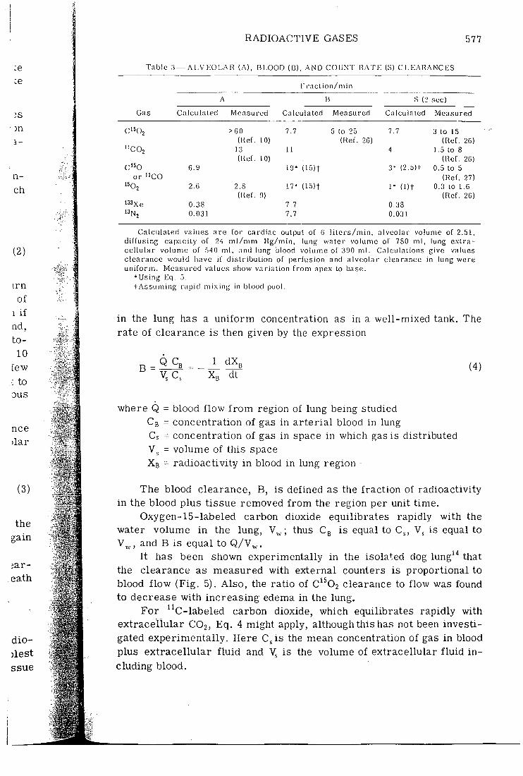
Approximate calculated or measured values of the alveolar c lear ances fo r the var ious radioactive gases for a short per iod of brea th holding are given in Table 3.
Blood Clearance
The blood clearance is important since i t affects the f a l l in r a d i o ac t iv i ty i n a region of lung measured by external counters. The s imples t t rea tment is to assume that the radioactive gas in the blood and t issue
:e ce
;s
1 -
n-ch
RADIOACTIVE GASES 577
T a b l e 3 — A I .V I-O R (A) , B L O O D (B) , AND C O U N T R A T I ; (S) C L E A R A N C E S
E r a c l i o n / i i i i i i
B S (2 sec)
G a s Calcuia lec l Measured Calcu la ted M e a s u r e d Ca I aula led Measured
C ' S Q J > 6 0 7 . 7 5 lo 2 5 7 . 7 3 to 1 5
(Ref. 1 0 ) (Ref. 2 6 ) (Ref . 2 6 )
"CO2 1 3 1 1 4 1 . 5 to 8
c'^o (Ref. 1 0 ) (Ref. 2 6 )
c'^o 6.9 19* (15)t 3 - ( 2 . 5 ) t 0 . 5 to 5
or " C O (Ref. 2 7 )
'502 2 . 6 2 . 8 1 7 * ( 1 5 ) 1 1 - ( l ) t 0 . 3 to 1 . 6
(Ref. 9) (Ref. 2 6 )
l33xe 0 . 3 8 7 . 7 0 . 3 8
'3N2 0 . 0 3 1 7 . 7 0 . 0 3 1
C a l c u l a t e d values a r e for c a r d i a c output of 6 l i t e r s / m i n , a lveo lar volume of 2.51, di f fus ing capaci ty of 2 4 m l / m m Hg/min , lung water volume of 7 8 0 ml , lung e x t r a c e l l u l a r volume of 5 4 0 ml , and lung blood volume of 3 9 0 m l . Calcu lat ions give values c l e a r a n c e would have if distr ibution of perfusion and a l v e o l a r c l e a r a n c e in lung were u n i f o r m . M e a s u r e d values show variat ion from apex to base.
* Using E q . 5 . t . ' \ ssuming rapid mi.xing in blood pool.
in the lung has a uniform concentration as in a well-mixed tank. The rate of clearance is then given by the expression
B = Q c. 1 dX„ X „ dt
(4)
(3)
where Q = blood flow f r o m region of lung being studied Cg = concentration of gas in arterial blood in lung Cs = concentration of gas in space in which gas is distributed
= volume of this space Xb = radioactivity in blood in lung region -
The blood clearance, B, is defined as the fraction of radioactivity in the blood plus tissue removed f rom the region per unit time.
Oxygen-15-labeled carbon dioxide equilibrates rapidly with the water volume in the lung, ; thus Cg is equal to C , is equal to V „ , and B is equal to Q /V^ .
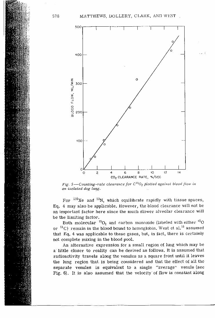
It has been shown experimentally in the isolated dog lung'"* that the clearance as measured with external counters is proportional to blood flow (Fig. 5). Also, the ratio of C^^02 clearance to flow was found to decrease with increasing edema in the lung.
For ^^C-labeled carbon dioxide, which equilibrates rapidly with extracellular C O 2 , Eq. 4 might apply, although this has not been investigated experimentally. Here C^is the mean concentration of gas in blood plus extracellular f lu id and is the volume of extracellular f luid i n cluding blood.
Em
1 mm
if!
578 MATTHEWS, DOLLERY, CLARK, AND WEST
5 0 0
4 0 0
3 0 0
o _J u. o o o 2 0 0
too
4 6 8 10 12
C O z C L E A R A N C E R A T E , % / S E C
14
Fig. 5—Counting-rate clearance for plotted against blood jlow in an isolated dog lung.
For "'' Xe and " N , which equilibrate rapidly with tissue spaces, Eq. 4 may also be applicable. However, the blood clearance wi l l not be an important factor here since the much slower alveolar clearance wi l l be the l imi t ing factor.
Both molecular ^^©2 and carbon monoxide (labeled with either '^O or ^^C) remain in the blood bound to hemoglobin. West et a l /^ assumed that Eq. 4 was applicable to these gases, but, in fact, there is certainly not complete mixing in the blood pool.
An alternative expression for a small region of lung which may be a l i t t l e closer to reality can be derived as follows. It is assumed that radioactivity travels along the venules as a square front until it leaves the lung region that, is being considered and that the effect of a l l the separate venules is equivalent to a single "average" venule (see Fig. 6). It is also assumed that the velocity of flow is constant along
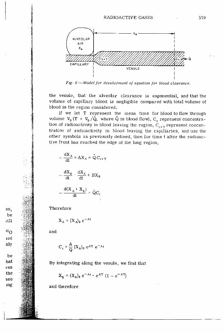
RADIOACTIVE GASES 579
A L V E O L A R
A I R
C A P I L L A R Y
V E N U L E
Fig. 6—Model for development of equation for blood clearance.
the venule, that the alveolar clearance is exponential, and that the volume of capillary blood is negligible compared with total volume of blood in the region considered.
If we let T represent the mean time for blood to flow through volume Vg (T = Vg/Q, where Q is blood flow), C represent concentration of radioactivity in blood leaving the region, C^ . . represent concentration of radioactivity in blood leaving the capillaries, and use the other symbols as previously defined, then for time t after the radioactive front has reached the edge of the lung region,
dX^ "dT = A X A = QCt+T
dt dt
dt QC,
Therefore
X A = (XA)O e-^'
and
Ct = | ( X A ) o e -r e-At
By integrating along the venule, we find that
Xg = (XA)O e-A» • e -r (1 - e'^'O
and therefore
580 MATTHEWS, DOLLERY, CLARK, AND WEST
B A 1 - e -AT (5)
III
mi"
Hence B is constant and independent of time and therefore blood clearance is exponential; however, i t is a function of alveolar clearance.
Another possible model is to consider that clearance from tlie blood is exponential owing to an exponential distribution of passage times along the venules f rom the capillary to the edge of tlie region considered. Thus = Coe~ ('"'"^ i f the radioactivity is instantaneously taken up by the blood. If to is very small, i t can be shown that the equations are the same as for two well-mixed pools but with t - to substituted for t.
Table 3 shows approximate calculated or measured values for blood clearances for tlie radioactive gases.
Exchange with Tissue Pools and Return in Venous Blood
Carbon monoxide labeled with ' O or "C remains bound to hemoglobin witli very l i t t le exchange with the tissues.
On the other hand, when molecular '^02 taken up by the blood reaches the systemic capillaries, i t w i l l take part in metabolism and some wi l l return to the blood as ^^O-labeled water. Ter-Pogossiar. et al.^^ studied the kinetics of uptake and desaturation of '^02 in red cells and the rate of appearance of labeled water in plasma in the dog. These authors and Dollery and West'^ showed that the plasma radioactivity was in the fo rm of water and not carbon dioxide.
Oxygen-15-labeled carbon dioxide exchanges with the entire water pool, as has already been mentioned. Hence the radioactivity returning in the venous blood is negligibly small since i t is so diluted. However, i f there is a shunt vin the heart through which radioactivity can return more rapidly to the lungs without much dilution, a secondary recirculation peak can be seen when counting rate over the lungs is recorded.
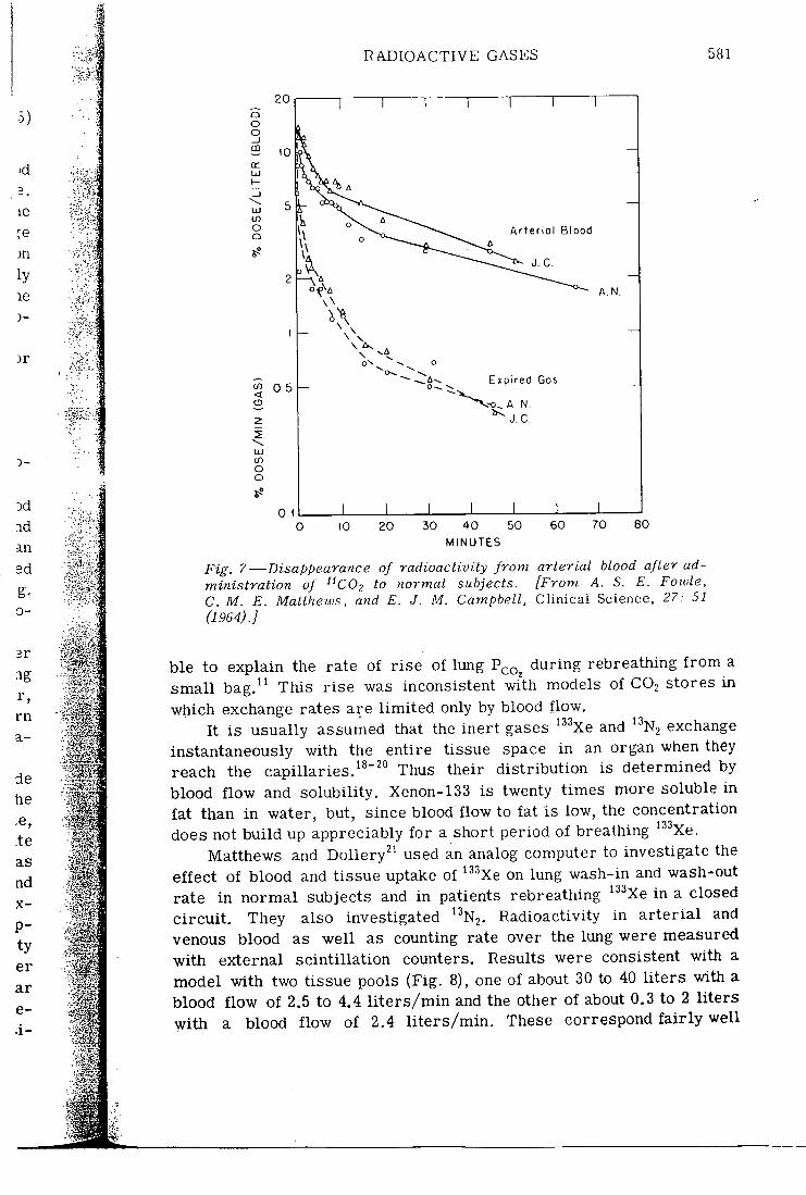
Fowle, Matthews, and Campbell'' used "C-labeled carbon dioxide to investigate the distribution of carbon dioxide in body stores. The " C O 2 was rebreathed f rom a small bag for about a quarter of a minute, and arterial samples were taken at short intervals. Within half a minute f rom the end of administration, the radioactivity in arterial blood was already diluted in a pool that was much larger than the blood CO, and approximately equal to extracellular C O 2 . Subsequently the "COo exchanged much more slowly with another pool that corresponded approximately with intracellular C O 2 . Figure 7 shows the radioactivity in ar ter ia l blood plotted vs. time. The distribution of tritiated water was also consistent with very rapid exchange with the extracellular pool and slower exchange with the intracellular pool. With this hypothesis of slow extracellular —intracellular exchange of C O 2 , i t was possi-
RADIOACTIVE GASES 581
5)
le
)n ly le )-
)r
3-
Dd i d an ed g-0 -
3r
•ig r , rn a-
A r t e r i a l B l o o d
E x p i r e d G o s 0 .5
A . N .
O O
0.1 I 10 2 0 3 0 4 0 5 0
M I N U T E S
6 0 7 0 8 0
Fig. 7—Disappearance of radioactivity from arterial blood after administration of "CO^ to normal subjects. [From A. S. E. Fowle, C. M. E. Matthews, and E. J. M. Campbell, Clinical Science, 27: 51 (1964). J
hie to explain the rate of rise of lung P ^ q during rebreathing f rom a small bag.^^ This rise was inconsistent with models of C O 2 stores in which exchange rates are l imited only by blood f jow.
It is usually assumed that the inert gases '^^Xe and ^•'N2 exchange instantaneously with the entire tissue space in an organ when they reach the capillaries.^^"^^ Thus their distribution is determined by blood flow and solubility. Xenon-133 is twenty times more soluble in fat than in water, but, since blood flow to fat is low, the concentration does not build up appreciably for a short period of breathing '•'^Xe.
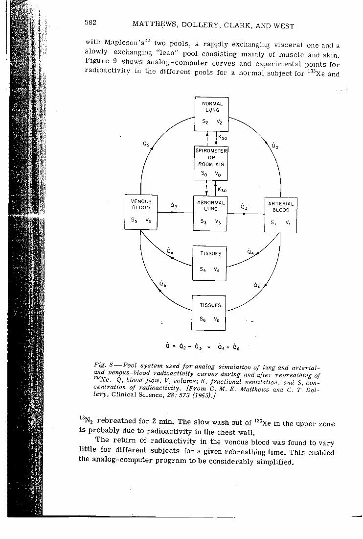
Matthews and DoUery^' used an analog computer to investigate the effect of blood and tissue uptake of • ''Xe on lung wash-in and wash-out rate in normal subjects and in patients rebreathing '' ^Xe in a closed ci rcui t . They also investigated ^'^N2. Radioactivity in ar ter ia l and venous blood as well as counting rate over the lung were measured with external scintillation counters. Results were consistent with a model with two tissue pools (Fig. 8), one of about 30 to 40 l i ters with a blood flow of 2.5 to 4,4 l i t e r s /min and the other of about 0.3 to 2 liters with a blood flow of 2.4 l i t e r s /min . These correspond f a i r l y well
li
582 MATTHEWS, DOLLERY, CLARK, AND WEST
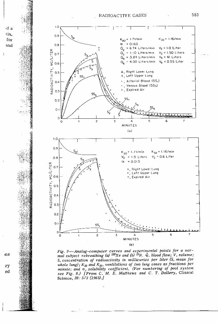
with Mapleson's^^ two pools, a rapidly exchanging visceral one and a slowly exchanging "lean" pool consisting mainly of muscle and skin. Figure 9 shows analog-computer curves and experimental points for radioactivity in the different pools for a normal subject for '^^Xe and
V E N O U S B L O O D
N O R M A L L U N G
S P I R O M E T E R OR
ROOM A I R
Sn V„
A B N O R M A L L U N G
A R T E R I A L B L O O D
T I S S U E S
T I S S U E S
0 = 0 2 + 0 , = 64 + Q«
F-ig. 8—Pool system used for analog simulation of lung ayid arterial-and venous-blood radioactivity curves during and after rebreathing of
Xe. Q, blood flow; V, volume; K, fractional ventilation; and S. concentration of radioactivity. [From C. M. E. Matthews and C T Dol-lery, Clinical Science, 28 : 573 (1965).]
^ N2 rebreathed for 2 min. The slow wash out of ^ ^Xe in the upper zone is probably due to radioactivity in the chest wall .
The return of radioactivity in the venous blood was found to var>' l i t t l e fo r different subjects for a given rebreathing time. This enabled the analog-computer program to be considerably simplified.
RADIOACTIVE GASES 583
id a tin. for and
K j Q = L 7 l / m i n
= 0 1 6 0
0 ^ = 6 . 7 4 L i t e r s / m i n
L l O L i t e r s / m m
= 3 . 8 4 L i t e r s / m i n
- 4 . 0 0 L i t e r s / m i n
o < o Q < 0.3 cc
L 1 6 / m i n
SIz = 1.0 L i t e r
V j = 1 .50 L i t e r s
V 4 = 41 L i t e r s
Vfi = 0 . 5 5 L i t e r
R i g h t Lower L u n g L e f t U p p e r L u n g
A r t e r i a l B l o o d (5S, )
V e n o u s B l o o d (5S5)
E x p i r e d A i r
5 S .
M I N U T E S
( Q )
K 2 o = 1 . 7 1 / m i n K J Q = L I 6 / m i n
V j = 0 . 6 L i t e r Vg = ( .9 L i t e r s
cc = 0 . 0 1 3
R i g h t L o w e r L u n g L e t t U p p e r L u n g
, E x p i r e d A i r
M I N U T E S
( b )
Fig. 9—Analog-computer curves and experimental points for a normal subject rebreathing (a) ^^Xe and (b) " N . Q, blood flow;_V, volume; S, concentration of radioactivity in millicuries per liter (S, mean for whole lung); K20 and K^q, ventilations of two lung zones as fractions per minute; and a, solubility coefficient. (For numbering of pool system see Fig. 8.) [From C. M. E. Matthews and C. T. Dollery, Clinical Science, 28 : 5/3 (1965).]
584 MATTHEWS, DOLLERY, CLARK, AND WEST
METHODS OF USE
ml
Counting-rate Clearance
The use of the external scintillation counters is complicated by the fact that measured counting rate wi l l depend on radioactivity in both alveolar air and blood. Thus counting-rate clearance w i l l , in general, be a complex function of both alveolar clearance and blood clearance. Counting-rate clearance may be defined as the fractional rate of change of counting rate recorded by scintillation counters pointing at the lung during a breath-holding period. The following equation for the counting-rate clearance or the exponential slope, S, of the counting-rate curve a:t time t has been derived'^:
1
A B(e~''' -e" ' ' ' : B e-'^' - Ae-^' ior A ^ B
S = Ah_
1 + At
(6)
for A = B
where A is the alveolar clearance and B is the blood clearance as before. This assumes exponential alveolar and blood clearances.
For '^0-labeled carbon dioxide, A is so much greater than B that the slope S becomes equal to B, and hence the clearance during breath holding of C'^02 can be used as a measure of blood flow. At the other extreme A is negligibly small for '''N2, and S is equal to 0. For ''^''Xe, B is much greater than A, and thus S is equal to A.
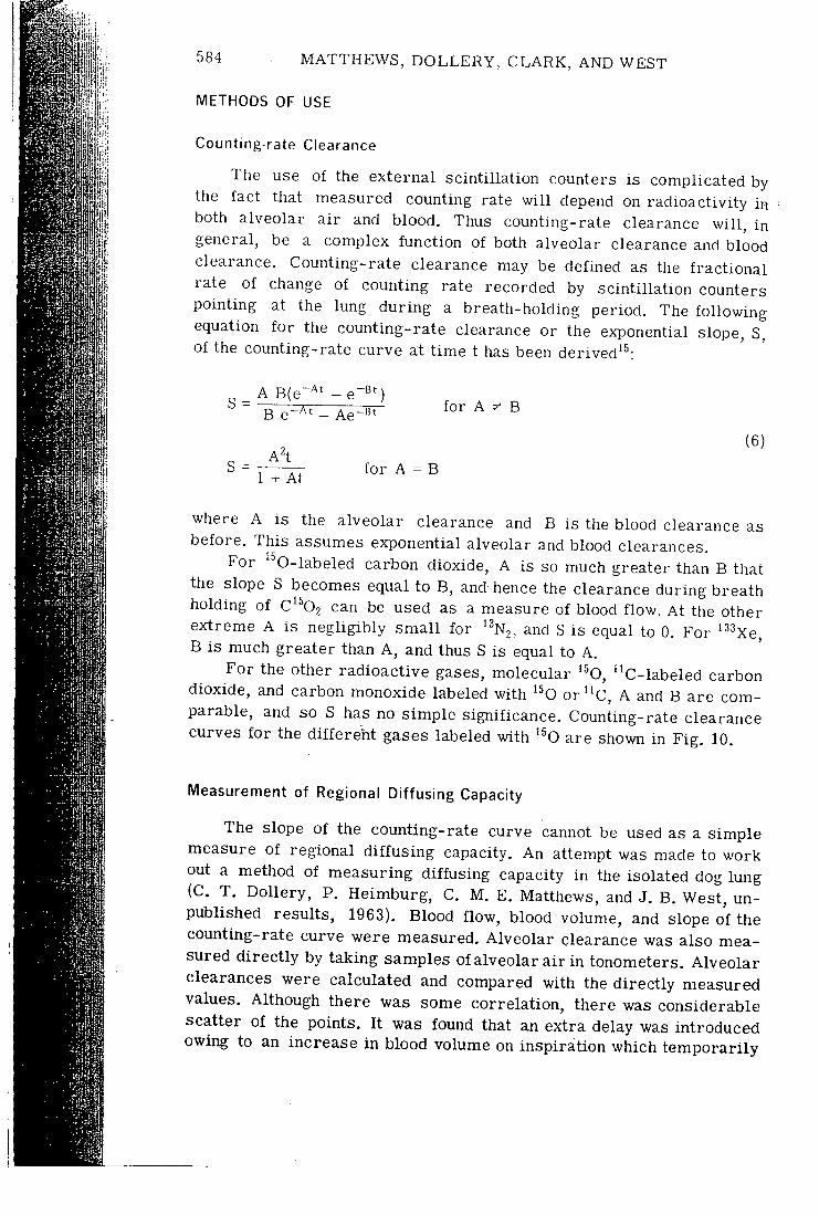
For the other radioactive gases, molecular '^O, "C-labeled carbon dioxide, and carbon monoxide labeled with '^O o r " C , A and B are comparable, and so S has no simple significance. Counting-rate clearance curves for the different gases labeled with '^O are shown in Fig. 10.
Measurement of Regional Diffusing Capacity
The slope of the counting-rate curve cannot be used as a simple measure of regional diffusing capacity. An attempt was made to work out a method of measuring diffusing capacity in the isolated dog lung (C. T. Dollery, P. Heimburg, C, M, E. Matthews, and J. B. West, unpublished results, 1963). Blood flow, blood volume, and slope of the counting-rate curve were measured. Alveolar clearance was also measured directly by taking samples of alveolar air in tonometers. Alveolar clearances were calculated and compared with the directly measured values. Although there was some correlation, there was considerable scatter of the points. I t was found that an extra delay was introduced owing to an increase in blood volume on inspiration which temporarily
RADIOACTIVE GASES 585
by , in
in od lal rs ng. S.
6)
Fig. lO^Fall in counting rate over the lungs for three different gases labeled with '^0. The first arrow shows the time of inspiration, and the second one shoivs the end of the breath-holding period, about 10 sec later.
reduced blood flow. This probably accounts for the erratic results obtained and means that diffusing capacity cannot be accurately measured by this method. A method based on the difference in the clearance curves of radioactive carbon monoxide and a radioactive substance, which was very rapidly absorbed by the blood and which remained in the blood, might be possible if a suitable substance could be found.
Table 3 shows approximate calculated and measured values for counting-rate clearances.
Measurement of Regional Blood Flow
Regional blood flow has been measured by the following methods: 1, ^^Oj counting-rate clearance during breath holding 2. C^^02 counting-rate clearance during breath holding 3. ^"Xe arr ival 4, ^^N2 a r r iva l
Method 1 does not give a true measure of blood flow since S is a complex function of flow.
Method 2 is normally satisfactory. However, i t actually measures blood flow per unit water volume (see . p. 576), and so the result obtained wi l l be affected by the presence of edema.
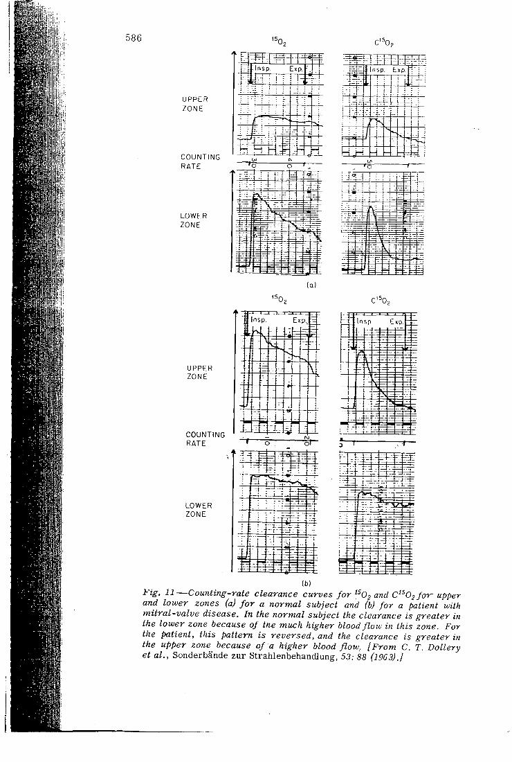
Figure 11 shows ^^02 and C^^02 clearance curves for a normal subject and for a patient with mitral-valve disease.
In method 3, '' ' Xe is dissolved in saline and injected intravenously. The ^ ^Xe is f i r s t mixed with carbon dioxide and then shaken with saline. The carbon dioxide dissolves rapidly and thus increases the partial pressure of ^ ''Xe in the remaining bubble and drives i t into solution. After injection the subject holds his breath, and the counting rate is measured over a region of lung. When the injected '^''Xe reaches
586 15r
U P P E R Z O N E
C O U N T I N G R A T E
L O W E R Z O N E
j r r - l n S p . Exp.
( Q )
<5r
U P P E R Z O N E
C O U N T I N G R A T E o
L O W E R Z O N E
- Mr:.. T - i J —
Insp. E x p
I • ( - ! • I : . L . .
i i i • I • • t
_ . L. -
• i i - • i - - ! ! : * i ;
i i • : ; L i M
f : 1 • 1 'l4 1 '
! -1 T -
- i -4 - i
- 4 ; .- i ; .
-4^
C' Og
In sp . E xp.
( -~ z = -
= 2-E E
1 — 1 "
1 — [' • 1 ^ . 1-, E-
i . --
i .
r -1 - v =
1—t-
-• f - -_l-r -I .J—}-.
(b)
i ^ i ^ , J J—Counting-rate clearance curves for ^^02 and C^O^/or upper and lower zones (a) for a normal subject and (b) for a patient with mitral-valve disease. In the normal subject the clearance is greater in the lower zone because of tne micch higher blood flow in this zone. For the patient, this pattern is reversed, and the clearance is greater in the upper zone because of a higher blood flow, [From C. T. Dollery et al., Sonderbande zur Strahlenbehandlung, 53: 88 (1963).]
RADIOACTIVE GASES 587
the lung capillaries, i t is nearly al l evolved into the alveolar air; thus the radioactivity arr iving m a region of lung is proportional to the blood flow to that region as a fraction of cardiac output. This method gives slightly different information f rom that given by method 2 since i t depends on cardiac output. However, when blood flow in two regions is to be compared, this difference is immaterial .
This method, introduced by Ball et al.,' ' has been used to investigate the distribution of blood flow in tlie isolated lung in relation to vascular and alveolar pressures.^'' It is alsoused routinely in patients 24
Method 4 is exactly the same as method 3 except that ' • 'Nx is used. Since the breath-holding period is so short, very little uptake of '''''Xe by the blood occurs; thus similar results should be obtained with '"' Xe and '^Nj .
Detection of Cardiac Shunts The method for detecting cardiac shunts has already been men
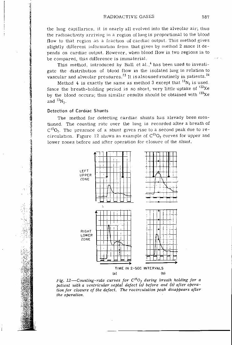
tioned. The counting rate over the lung is recorded after a breath of C'^02. The presence of a shunt gives rise to a second peak due to recirculation. Figure 12 shows an example of C'^02 curves for upper and lower zones before and after operation for closure of the shunt.
L E F T
U P P E R Z O N E
R I G H T L O W E R Z O N E
> i I > I > >
1
>
1
r v f
> > >
\
>
! ,
>
I
>
i
> >
?
>
I
>
>
1 1
r _ • • M r _ • •
• : : i V 1 . :
. . i
1 1
T I M E IN 2 - S E C I N T E R V A L S
(Q) (b)
Fig. 12—Counting-rate curves for C O during breath holding for a patient with a ventricular septal defect (a) before and (b) after operation for closure of the defect. The recirculation peak disappears after the operation.
588 MATTHEWS, DOLLERY, CLARK, AND WEST
Measurement of Regional Ventilation Per Unit Lung Volume
The following methods have been used to study regional ventilation:
1. Single breath of '^02 2. Single breath of ' ^Xe 3. Single breath of '^Nj 4. Wash in and wash out of '"'•'Xe 5. Wash in and wash out of '''N2 Regional ventilation measured by the single-breath method must be
related to regional lung volume, and this has been measured by re-breathing either ^^bie or ' N2 in a closed circuit for a few minutes and taking the counting rate over a region to be proportional to lung volume in that region. In practice, however, equilibration may not occur witJi '•' Xe owing to the uptake in blood which, with an alveolar clearance rate of about 38%/min, is certainly not negligible during 2 min rebreathing. On the other hand, for '' N2 with an alveolar clearance of only about 3%/ min, the uptake w i l l be small even for several minutes of rebreathing. With an analog computer^' it was found that, although the error with '' ''Xe was probably small in normal subjects, i t could be considerable in patients, and volume could be underestimated by about 45%. This is illustrated in Fig. 13 for a patient with bullous emphysema where concentration in the different regions of the lung did not reach the spirometer concentration even after 4 min rebreathing.
Another disadvantage of the single-breath method is that in regions of low ventilation there is l i t t le uptake of the radioactive gas, and so it is diff icul t to obtain a quantitative measure of the reduction in ventilation. The results obtained by the wash-out method are much more sensitive to small proportions of badly ventilated alveoli and also give ventilation per unit volume directly. The wash-in method is less sensitive than the waSlh-out method. Here again ' N2 is much better than '• • Xe because of its lower solubility. The ' ^Xe wash out is distorted by radioactivity in the chest wall (Fig. 9) and by uptake of ' ' Xe in arterial blood and return in venous blood. Nevertheless, '" ' Xe can be used to give a rough indication of reduced ventilation. Figure 14 shows a washout curve after rebreathing of '^^Xe and after injection in a patient with emphysema. The reduced ventilation in the lower zone is clearly shown, although the lower trace shows that this zone is perfused. Scanning Profile scanning has been used with '''"'Xe and the single-breath method to show the distribution of ventilation and with the injection method to show the distribution of perfusion.
Body CO2 Stores and Control of Ventilation
The use of " C O 2 for investigation of body CO2 stores has already been mentioned. This work is now being extended to cover control of
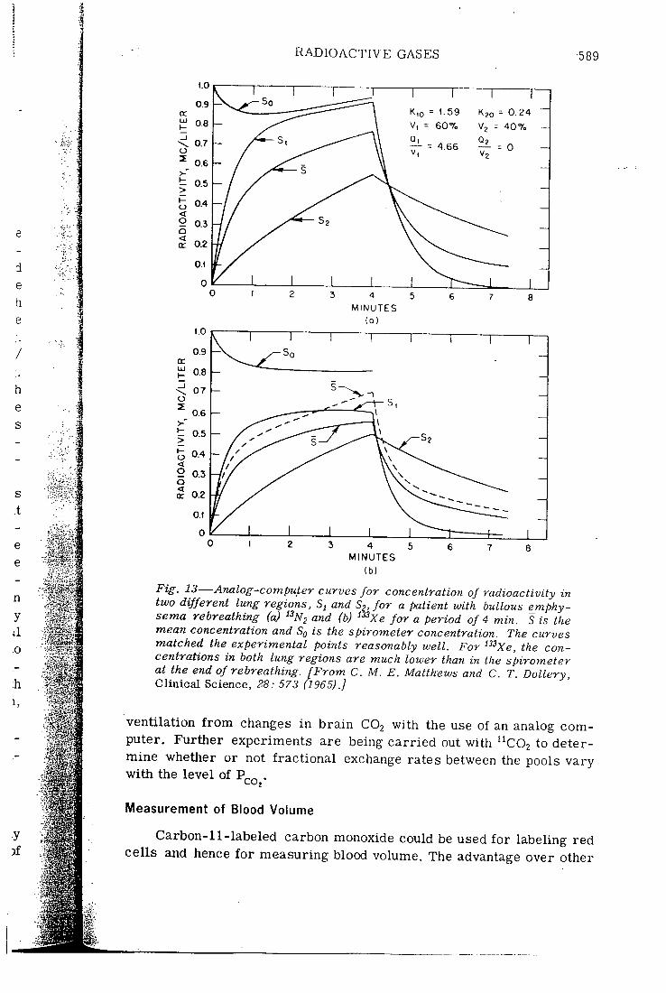
RADIOACTIVE GASES •589
d e h e
0 . 2 4
M I N U T E S
M I N U T E S
1.0
0.9 Q: LU 1- 0.8 _l \ 0.7 o
0.6 >-* 1- 0.5 >
CT
I
0.4 < o 0.3 Q < 0.2 0.2
O.t
0
Fig. 13—Analog-computer curves for concentration of radioactivity in two different lung regions, and So, for a patient with bullous emphysema rebreathing (a) ^^N2 and (b) '^Xe for a period of 4 min. S is the mean concentration and Sg is the spirometer concentration. The curves matched the experimental points reasonably well. For '^^Xe, the concentrations in both lung regions are much lower than iji the spirometer at the end of rebreathing. [From C. M. E. Matthews and C. T: Dollery, C l i n i c a l Sc i ence , 28: 573 (1965).]
ventilation f rom changes in brain C O 2 with the use of an analog computer. Further experiments are being carried out with " C O 2 to determine whether or not fractional exchange rates between the pools vary with the level of ,
Measurement of Blood Volume
Carbon-11-labeled carbon monoxide could be used for labeling red cells and hence for measuring blood volume. The advantage over other
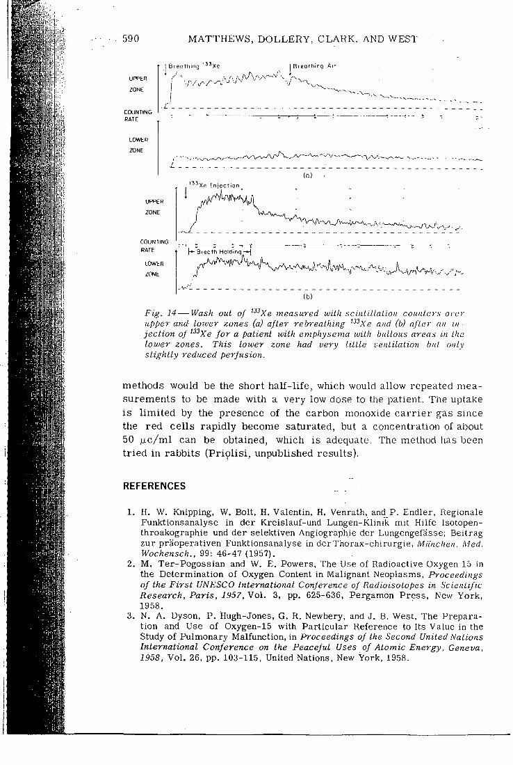
I? ill!
tl
i
Hi4
I i
590 MATTHEWS, DOLLERY, CLARK, AND WEST
U P P E R
ZONE
COUNTING RATE
tOWER
ZONE
I B r e o t h i n g ' ^ ' x e Breo th ing Ai r
U P P E R
ZONE
COUNTING RATE
tOWER
ZONE
Xe I n j e c t i o n
( Q )
•'V A'-../vyv~ wv_ . ,•, .V
h- Breoth Holding
(b)
Fig. 14 — Wash out of ^^^Xe measured with scinLULaLiOH couiUers orcr upper and lower zones (a) after rebreathing "•'Xe a/id (b) after an injection of ^^^Xe for a patient with emphysema with bullous areas in the lower zones. This lower zone had very Little uenlilalion bid. only slightly reduced perfusion.
methods would be the short half - l i fe , which would allow repeated measurements to be made with a very low dose to the patient. The uptake is l imited by the presence of the carbon monoxide carr ier gas since the red cells rapidly become saturated, but a concentration of about 50 pic/ml can be obtained, which is adequate. The method has been tr ied in rabbits (Priol is i , unpublished results).
R E F E R E N C E S
1. H. W. Knipping, W. Bolt, H. Valentin, H. Venrath, and P. Endler, Regionale Funktionsanalyse in der Kreislauf-und Lungen-Klinik mit Hilfe Isotopen-throakographie und der selektiven Angiographie der Lungengefasse; Beitrag zur praoperativen Funktionsanalyse in der Tharax-chirurgie, Miinchen. Med. Wochensch., 99: 46-47 (1957).
2. M . Ter-Pogossian and W. E. Powers, The Use of Radioactive Oxygen 15 in the Determination of Oxygen Content in Malignant Neoplasms, Proceedings of the First UNESCO International Conference of Radioisotopes in Scientific Research, Paris, 1957, Vol . 3, pp. 625-636, Pergamon Press, New York, 1958.
3. N. A, Dyson, P. Hugh-Jones, G. R. Newbery, and J. B. West, The Preparation and Use of Oxygen-15 with Particular Reference to Its Value in the Study of Pulmonary Malfunction, in Proceedings of the Second United Nations International Conference on the Peaceful Uses of Atomic Energy, Geneva, 1958, Vol . 26, pp. 103-115, United Nations, New York, 1958.
RADIOACTIVE GASES 591
9,
10,
11.
12.
14.
4. W. C. Ball, Jr., P. B. Stewart, L. G. S. Newsham, and D. V. Bates, Regional Pulmonary Function Studied with Xenon^^'', J. Clin. Invest., 41: 519-531 (1962).
5. P. D. Buckingham and G. R. Forse, The Preparation and Processing of Radioactive Gases for Clinical Use, hit. J. Appl. Radial., 14: 439-445(1963).
6. L . C. Baker and P. D. Buckingham, Cadmium Sulphide D.C. Conductivity Cells for Gamma Ray Detection, Phys. Med. Biol., 10: 201-208 (1965).
7. C. T. Dollery, J. F. Fowler, P. Hugh-Jones, C. M. E. Matthews, and J. B. West, The Preparation and Use of Radioactive Oxygen, Carbon Monoxide, and Carbon Dioxide for Investigation of Regional Lung Function, and Their Comparison with Xenon-133, Strahlentherapie, Sonderbande, Pt. 5, 53; 88-103 (1963).
8. C. M. E. Matthews, J. F. Fowler, and P. C. R. Turner, Absorbed Doses f rom i33xe M.R.C. Cyclotron Unit, Tech. Mem., No. 84. Rev. (1963). N. A. Dyson, J. D. Sinclair, and J, B. West, A Comparison of the Uptakes of Oxygen-15 and Oxygen-16 in the Lung, J. Physiol., 152: 325-336 (1960). J. B. West and C. T. Dollery, Uptake of Oxygen-15-labeled CO2 Compared with Carbon-ll-labeled COj in the Lung, J. Appl. Physiol., 17: 9-13 (1962). A. S. E. Fowle, C. M. E. Matthews, and E. J. M. Campbell, The Rapid Distribution of •'HjO and ^'COj in the Body in Relation to the Immediate Carbon Dioxide Storage Capacity, Clin. Sci., 27: 51-65 (1964). W. H. Isbister, P. F. Schofield, and H. B. Torrance, Measurement of the Solubility of Xenon-133 in Blood and Human Brain, Phys. Med. Biol., 10: 243-250 (1965). N. Veall and B. L . Mallett, The Partition of Trace Amounts of Xenon Between Human Blood and Brain Tissues at 37°C, Phys. Med. Biol., 10: 375-380 (1965). C. T. Dollery, P. Heimburg, and P. Hugh-Jones, The Relationship Between Blood Flow and Clearance Rate of Radioactive Carbon Dioxide and Oxygen in Normal and Oedematous Lungs, J. Physiol., 162: 93-104 (1962).
15. J. B. West, R. A. B. Holland, C. T. Dollery, and C. M. E. Matthews, Interpretation of Radioactive Gas Clearance Rates in the Lung, J. Appl. Physiol., 17: 14-20 (1962).
16. M. Ter-Pogossian, J, S. Spratt, Jr., S. Rudman, and A. Spencer, Radioactive Oxygen 15 in Study of Kinetics of Oxygen of Respiration, Amer. J. Physiol., 201: 582-586 (1961).
17. C. T. Dollery and J. B. West, Metabolism of Oxygen-15, jVa^wre, 187: 1121 (1960).
18. C. A. Tobias, H. B. Jones, J. H. Lawrence, and J. G. Hamilton, Symposium on Radioactive Isotopes; Uptake and Elimination of Krypton and Other Inert Gases by the Human Body, J. Clin. Invest., 28: 1375-1385 (1949).
19. H. B. Jones, Respiratory System: Nitrogen Elimination, in Medical Physics, Vol. 2, O. Glasser (Ed.), pp. 855-871, Year Book Publishers, Inc., Chicago, 1950.
20. S. S. Kety, The Theory and Applications of the Exchange of Inert Gas at the Lungs and Tissues, Pharmacol. Rev., 3: 1-41 (1951).
21. C. M. E. Matthews and C. T. Dollery, Interpretation o f ' " X e Lung"Wash-in and Wash-out Curves Using an Analogue Computer, Clin. Sci., 28: 573-590 (1965).
22. W. W. Mapleson, An Electric Analogue for Uptake and Exchange of Inert Gases and Other Agents, J. Appl. Physiol.. 18: 197-204 (1963).
23. J. B, West, C. T. Dollery, and A. Naimark, Distribution of Blood Flow in Isolated Lung; Relation to Vascular and Alveolar Pressures, J. Appl. Physiol., 19: 713-724 (1964),
24. C. T. Dollery, P, Hugh-Jones, and C. M. E. Matthews, Use of Radioactive Xenon for Studies of Regional Lung Function, Brit. Med. J., 2: 1006-1016 (Oct. 20. 1962).
1 S'
I; • ^
El
i i
J?
I
I i m
m M
r i i
592 MATTHEWS, DOLLERY, CLARK, AND WEST
25. C. T. Dollery and P. M. Gilliam, The Distribution of Blood and Gas Within the Lungs Measured by Scanning After Administration of ' "Xe, Thorax, IS: 316-325 (1963).
26. J. B. West, C. T. Dollery, and P. Hugh-Jones, The Use of Radioactive Carbon Dioxide to Measure Ilegional Blood Flow in the Lungs of l^atients with Pulmonary Disease, J. Clin. Invest., 40: 1-12 (1961).
27. C. T. IDollery, N. A. Dyson, and J. D. Sinclair, Regional Variations in Uptake of Radioactive CO in the Normal Lung, J. AppL. Physiol., 15: 411-417 (1960).
DISCUSSION
INGHAM: We have been using ' ^Xe at McMaster for pulmonary function studies and wonder if you have any comments on this radionuclide.
MATTHEWS: Xenon-135 would have the advantage of a shorter half- l i fe than '' ^Xe and thus a lower dose but would be less convenient and would have the same disadvantages, owing to blood solubility, as '^^Xe.
••V
J gjj, ,«i
mi
l i P i
1
•1
Radioactive Pharmaceuticals
Proceedings of a symposium held at the Oak Ridge Institute of Nuclear Studies, an operating unit of Oak Ridge Associated Universities, November 1-4, 1965
E d i t o r s
Gould A. Andrews, M.D. Ralph M. Kniseley, M.D. Henry N. Wagner, Jr., M.D.
T e c h n i c a l E d i t o r
Elizabeth B. Anderson, M.S.
A p r i l 1966
U.S. ATOMIC ENERGY COMMISSION/Division of Technical Information
I N V I T R O L A B E L L I N G O F R E D C E L L S W I T H C A K B O N - L I
.I.e. C L . A R K , l l . I . GI..-\.SS (*) and D J . . S I L V E S T K H Medicnl Research Council Cyclolron Unit, and
(*) Department of Medical. Physics, Postgraduate Medical School, Hammersmith Hospital, London, W.12, England
A hsli arl.
T h e ready a%'ailal:)ility of "C-laljcI lecl carlxji i inoiioxicle from tlie Medical Research C o u n c i r s cyclotron has led to the development of a routme technique for lahe l l ing red cells with this isotope.
T h e method of producing the lahelled gas and of lahel l ing the ceU? w'lW he described, and pre l iminary results of a comparison with the conventional * 'Cr lahel l ing procedure wi l l he presented.
I N T R O D U C T I O N
Before the stahle isotope '•'C and the long-lived ratlioisoto{)e '""C became
avai lable as isotopic tracers for car!)on, a remarkable volume of useful work " '
was achieved using short-l ived " C , w h i c h was made without m u c h d i f f i cu l tv—
though in smal l y i e ld—with early cyclotrons. Not surprisingly, in view of
its 20-minute half - l i fe , " C has found few pract ical applications since. I n recent
years it has again proved valuable , however, part icular ly in the form of labelled
carbon dioxide gas, for a n u m b e r of physiological studies T h e purpose of
this paper is to report another useful appl icat ion of this isotope (this Lime one
i n w h i c h it is incorporated iijto a high-molecular weight compound), where
its short hal f - l i fe is a positive advantage.
T h e measurement of c i rcu la t ing blood volumes is important in medicine,
and is conventionally achieved b y labe l l ing red cells with ^^Cr and performing
what is, in effect, a classical isotope di lut ion analysis. However, the long physical
and biological half- l ives of " C r preclude its use in, for example, infants and
expectant mothers, where the radiat ion dose would he too high, and in patient.-:
where serial measurements at short intervals are required. F o r such application.?
a short-l ived label is desirable.
T h e aff ini ty of haemoglobin for carbon monoxide is wel l known, and
indeed non-radioactive carbon monoxide has been used as a red cell l abe l
S ince carbon monoxide labe l l ed w i t h " C is readi ly available f rom the Medica l
R e s e a r c h Counci l 's cyclotron, a routine technique for label l ing red cells w i t h this
isotope has been developed.
60:i
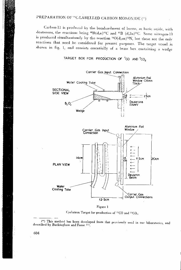
P R E P A R A T I O N O F " C - L A B E L L E D C A R B O N M O N O X I D E ( \)
C a r l ) o n - l l is produced by the bombardment of boron, as boric oxide, witii
(leuterons, tbe reactions being " 'B(d ,n)"C and " B (d,2n)"C. .Some nitrogen-13
is produced simultaneously by the reaction '*0(d,an)'^N, but tliese are the only
reactions that need be considered for present pur^joses. T h e target vessel. a&
shown in fig. 1, and consists essentially of a brass box containing a wedge
T A R G E T BOX F O R PRODUCTION O F "cO AND "CO,
Carrier Gasjnput Connection
Water Cooling Tube
S E C T I O N A L S I D E VIEW
AJuminium Foil Window 0 6 mm Thich
: Deuterons l5MeV
P L A N VIEW
Water Cooling Tube
Carrier Gas Input Connection ~"
Aluminium Foil Window /
ll-5cm
Deuteron
I2-5cm
POcm
Carrier Gas Output Connections
Figure 1
Cyclotron Target for production of " C O and " C 0 >
A ""Ti^ 'leveloped from that previously used in our laboratories, and described by Buclangham and Forse (').
604
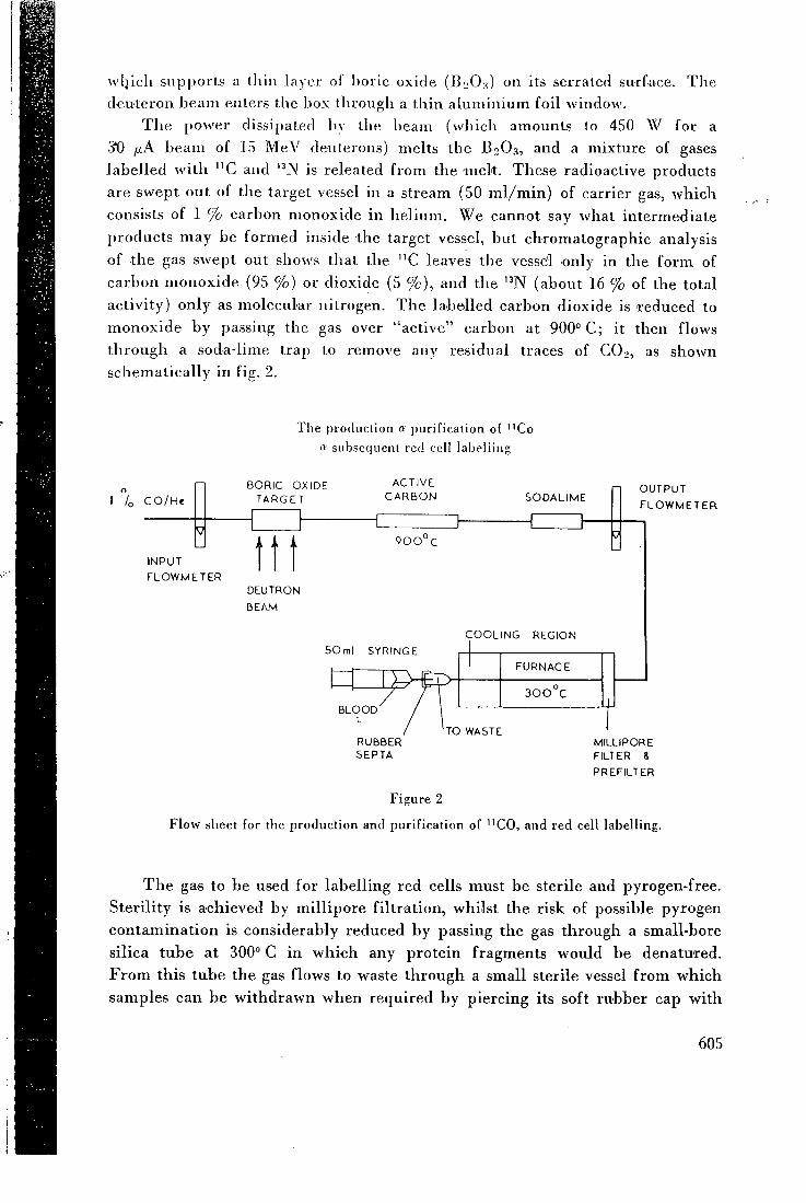
wlj icb supports a ihin layer of l)oric oxide (B:;0.s) on its serrated surface. T h e
deuteron beam enters the hox through a thin a lumin ium foil window.
T h e power dissipated bv the beam (which amounts to 450 W for a
3*0 ^ A beam of 1.5 M e V deiitcrons) melts the B2O3, and a mixture of gases
label led wi th " C and is releated from the mek . These radioactive products
are swept out of the target vessel in a stream (50 m l / m i n ) of carrier gas, wh ich
consists of 1 % carbon monoxide in he l ium. W e cannot say what intermediate
products may be formed inside the target vessel, but chromatographic analysis
of the gas swept out shows that the " C leaves the vessel only in the form of
carbon monoxide (95 % ) or dioxide (5 % ) , and the ^'N (about 16 % of the total
activity) only as molecular nitrogen. T h e label led carbon dioxide is reduced to
monoxide by passing the gas over "active" carbon at 900" C ; it then flows
through a soda-lime trap to remove any residual traces of C O 2 , as shown
schematical ly in fig. 2.
The production a purification of " C o a siibscquenl red cell lahelliiig
I L C O / H «
B O R I C O X I D E T A R G E T
A C T I V E C A R B O N S O D A L I M E
I N P U T
F L O W M E T E R
9 0 0 ° C
O U T P U T
F L O W M E T E R
D E U T R O N
B E A M
S O m I S Y R I N G E
B L O O D
C O O L I N G R E G I O N
F U R N A C E
3 0 0 ° C
R U B B E R S E P T A
TO W A S T E M I L U P O R E F I L T E R & P R E F I L T E R
Figure 2
Flow sheet for the production and purification of " C O , and red cell labelling.
T h e gas to be used for label l ing red cells must be sterile and pyrogen-free.
Steri l i ty is achieved by mi l l ipore f i l tration, whilst the r isk of possible pyrogen
contaminat ion is considerably reduced by passing the gas through a small-bore
s i l ica tube at 300° C in w h i c h any protein fragments would be denatured.
F r o m this tube the gas flows to waste through a smal l sterile vessel from w h i c h
samples can be wi thdrawn when required by piercing its soft rubber cap wi th
605
a i ivpodermic needle. Under typical operating conditions (beam current 30 ;.t,A,
gas flow 50 m l / n i i n ) the specific activity of the C O at the sampling point
(wli ic l i incidenlallv is some 100 feel from the target vessel) is about 200 j u C / m M .
R E D C E L L L A B E L L I N G T E C H N I Q U E
Samples of "whole blood", usually between 5 and 15 ml , are drawn into
a sterile 50 ml syringe to w h i c h a little anticoagulant has previously been
added. T h e syringe is then f i l led to capacity with the radioactive gas extracted
f rom the sampling vessel, and is sealed with a sterile cap. B lood and gas
are next mixed by rotating the syringe on its axis at 10 r.p.m. for 10 minutes.
T h i s method of mixing is suitably eff icient (about 50 % of the C O is absorbed
into the red cells in 10 minutes) and causes very little haemolysis compared
wi th that produced, for example, by bubbling the gas through the blood, or
l:)y more violent agitation.
Af ter mixing, the activity remaining in the gas phase is expelled to waste.
Because of its very low solubility the '^N-labelled nitrogen is completely
removed by this and the subsequent washing steps. L h e red cells are now
centrifuged to separate them from the plasma in which a very small amount
of C O dissolves, and, after washing three times with isotonic saline, they are
ready for c l inical use. A typical preparation would give 50 ,l(.C of " C in 5 m l
of red cells at this time.
F o r one series of experiments, blood samples were labelled simultaneously
wi th both " C and " C r s imply by adding ^'Cr-labelled sodium chromate to the
syringe before drawing in the radioactive gas and mixing. F i g . 3 shows the
result obtained when a sample of such double-labelled red cells was haemo-
lysed, and a fraction of the haemolysate was run through a column of Sephadex
G-75. T h e " C r was distributed between high and low molecular weight frac
tions, where the first corresponded with haemoglobin. T h i s result was in
agreement wi th the observation of Pr ins whose suggestion that the low
molecular weight substance to w h i c h the " C r was botmd was glutathione was
later endorsed by Koutras et al. A s was expected, however, the ^'C elution
corresponded entirely with the haemoglobin.
R E S U L T S A N D D I S C U S S I O N
Carbon-11 label led red cells have been used at H a m m e r s m i t h Hospi ta l in a
number of c l in ica l studies, the results of which w i l l be reported i n detail
elsewhere. I n this paper, however, we wish to draw attention to the result
of a series of blood volume measurements that was made using cells label led
wi th both " C and ^'Cr by the method just described.
606
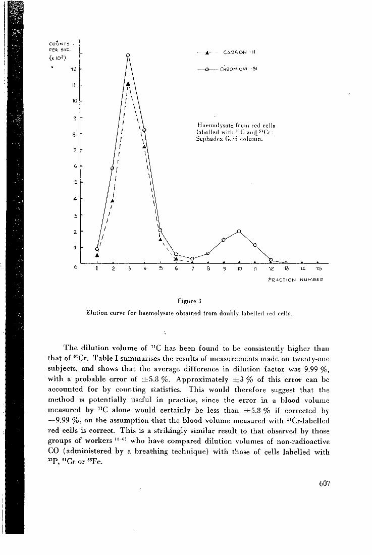
C O U N T S PEE. sec.
12
11
10
8
7
6
5
4
5
2
1
C A C B O N • 11
CHZOMi'Jtvi -51
Hacmolysatc f i o n i red cells labelled with " C aiid_5'Cr: Sephadex G.75 column.
10 12 \i 14- I S
F R A C T I O N N U M B E C
Figure 3
Elution curve for haemolvsale obtained from doublv labelled red cells.
T h e di lut ion volume of " C has been found to be consistently higher than
that of * 'Cr . T a b l e I summarises the results of measurements made on twenty-one
subjects, and shows that the average difference in dilution factor was 9.99 %,
wi th a probable error of ± 5 . 8 %. Approximate ly ± 3 % of this error can be
accounted for by counting statistics. T h i s would therefore suggest that the
method is potentially useful in practice, since the error in a blood volume
measured by " C alone would certainly be less than ± 5 . 8 % if corrected by
—9.99 %, on the assumption that the blood volume measured with *'Cr-labelled
red cells is correct. T h i s is a strikingly s imi lar result to that observed by those
groups of workers who have compared dilution volumes of non-radioactive
C O (administered by a breathing technique) with those of cells labelled with
^^F, ^ 'Cr or " F e .
607
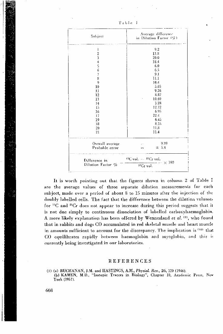
T a l j l c I
Av : , ; ( i i f r e r c i i c c Sill ij c(;t ill I)i liiiioii Factor i'/r]
1 9.2 2 1.3.8 3 20.0 4 18.4 5 6.0 6 6..S 7 9.1 8 11.1 9 10.4
10 5.6S n 9.26 12 8.87 13 10.69 14 3.28 15 12.72 16 6.9:i 17 22.4 19 6.45 18 8.35 20 11.8 21 11.4
Overall avei'agc 9.99 Probable error ± 5.8
Difference in Dilution Factor %
' C v o l . ' C r v o l .
5'Cr vol X 100
I t is worth pointing out that the figures shown in column 2 of T a b l e I
are the average values of three separate dilution measurements for each
subject, made over a period of about 8 to 15 minutes after the injection of the
doubly labelled cells. T h e fact that the difference between the dilution volumes
for " C and ^'Cr does not appear to increase during this period suggests that it
is not due s imply to continuous dissociation of labelled carboxyhaemoglobin.
A more l ike ly explanation has been offered by Wennesland et al. who found
that in rabbits and dogs C O accumulated i n red skeletal muscle and heart muscle
in amounts suff ic ient to account for the discrepancy. T h e implication is that
C O equilibrates raj j id ly between haemoglobin and myoglobin, and this i?
currently being investigated i n our laboratories.
R E F E R E N C E S
(1) (a) B U C H A N A N , J .M. and H A S T I N G S , A . M . , Physiol. Rev., 26, 120 (1946). (b) K A M E N , M.D. , "Isotopic Tracers in Biology", Chapter 10, Academic Press, Ne>r Y o r k (1957).
608
(2) (a) W E S T , J .B . and D O L L E R Y , C . T . , ]. Appl. Physiol, 17, 9 (1962). • (b) F O W L E , A . S . E . , M A T T H E W S , C . M . E . and C A M P B E L L , E . J . M . , Clin. Sci., 27, 51
(1964). <3) R O O T , W.S., A L L E N . T . H . and G R E G E R S E N , M.I. , Am. ]. Physiol., 175, 233 (1953). l i ) N O M O F , N., H O P P E R , J . , B R O W N , E . , S C O T T , K . and W E N N E S L A N D , R., ]. Clin.
Invest.. 33, 1382 (1954). (5) N I C K E R S O N . J . L . , S H A R P E , L . M . , R O O T , W.C. , F L E M I N G , T . C . and G R E G E R S O N ,
M.I. , Fed. Proc, 9, 94 (1950). (6) W E N N E S L A N D , R. , N O M O F , N., B R O W N , E . , H O P P E R , J . and B R A D L E Y , B.
Proc. Soc. Exper. Biol, and Med., 96, 655 (1957). (7) B U C K I N G H A M , P .D. and F O R S E , G.R. , Int. ]. Appl. Rad. Isotopes, 14, 439 (1963). (8) P R I N S , H . K . , Vox. Sang. (Basel), 7, 370 (1962). (9) K O U T R A S , G.A., H A T T O R I , M., S C H N E I D E R , A.S. , E B A U G H , F . G . Jr., and V A L E N
T I N E , W.M. , J. Clin. Invest., 43 , 323 (1964). (10) H A L D A N E , J . and S M I T H , J . L . , ]. Physiol., 25, 331 (1899).
609
I n t e r n a t i o n a l J o u r n a l of A p p l i e d R a d i a t i o n and Iso iopcs , lyGG, V u l . 17. pp. 151-154. r c r g a m o u Press L t d . Printed in Nor i l i crn I re land
A Cyclotron Method for the Production of Fluorine-18
J . C . C L A R K a n d D . J . S I L V E S T E R Medica l Research Counc i l Cyclotron Uni t , Hammersmith Hospital, London
{First received 9 June 1964 and in revised form 18 August 19G5)
Fluorine-18 is produced, with yields of up to 40 mc/hr, by the bombardment of water contained in a suitable target h e l d in the external a-particle b e a m of a 30 M e V cyclotron: a m e t h o d is described w h i c h is in routine use to produce samples for medical research purposes.
U N E M E T H O D E E M P L O Y . \ N T U N C Y C L O T R O N P O U R P R O D U I R E D U F L U O R - 1 8
L e fluor-18 se produit, avec des rendements de j u s q u ' a 40 m c / h r , par le bombardement dc I ' e a u contenue d a n s une cible situee dans le faisceau ext^rieur de particules -x d'un cyclotron de 30 M e V : on decrit une methode q u i est cn usage regulicr p o u r la production d'echantillons destines a la r e c h e r c h e medicale.
L I H K J I G T P O H H L I H M E T O f l flJlH n O J l Y H E H M f l O T O P A - I S <l>Top-18 noJiy^aeTcn c B B I X O H O M A O 40 M K / n a c nocpeflCTBOM oSjij'MCHun B O A U , saK-noMciiHofi
B cnei;Ha:ibHofi M H U I C H H , ycTaHOBJreHHof i c o BueujHeM .TivMe a-Maciim 30 MeraaJieKTpoH-BOJibTHoro i i H K J i o T p o H a . O n n c t i B a e T C f i M G T O B y w e n c n o j i L 3 0 B a H H u r i j\j]n noj iyMeHiiH o 6 p a 3 -U.OB B u e J l H X MeHHUHHCKHX HCCJieHOBaHll f l .
E I N E Z Y K L O T R O N M E T H O D E F U R D I E E R Z E U G U N G V O N F L O U R - I B Fluor-18, m i t Ertragen bis zu 40 mc/Stunde, ist erzeugt bei der Bombadierung von Wasser,
enthalten i n e i n e r Aufpral l f lache und festgehalten in dem ausseren a-Massenteilchenstrahl e i n e s 30 M e V Zyklotrons: c i n e Methode w i r d beschrieben welche im normalen Gebrauch ist,
; u m Proben f i ir Medizinische Versuchszwecke zu erzeugen.
I N T R O D U C T I O N '•'carrier-free" or otherwise, from the irradiated • F L U O R I N E - 1 8 is essentially a puce positron- material and contaminadng activities.
emitting isotope which, with a half-life of 109-7 T h e yields of F ^ ^ obtained by these techniques min,<i' is the longest-lived radioisotope of the are generally substantially higher than those element. I t was first reported as a product of which have been achieved by previously re-various cyclotron-induced nuclear reactions in ported methods using accelerators.'20-2'') I n 1 9 3 7 ( 2 - 5 ) and the first tracer studies with F ^ ^ fact, however, even higher yields can be ob-were carried out with cyclotron produced tained very conveniently with an accelerator material. '^'" under suitable conditions. T h e investigations
T h a t F ^ 8 could also be made in a nuclear reac- for which this isotope is at present produced in tor, by the irradiation of salts containing lithium the Medical Research Council's Cyclotron U n i t and oxygen, was first reported by K N I G H T require that it should be supplied to the clinician
; et a/.<8) Since then numerous papers '^ - i^ ' have concerned in a high specific activity solution, i appeared from reactor centres in many parts of either as fluoride or fluoroborate, suitable for i the world describing a variety of techniques clinical injection: the purpose of this paper is to
which may be adopted to isolate it, either describe the latest developments in a method of
151 1
) s I \ I
152 . / . C . Clark and D. J. Silvesler
TITANIUM TARGET P L A T E (Recessed at rear for cooling water) [t/^
CATALYST V E S S E L PRESSURE GAUGE 5 VALVE
O - O 0 O 5 TITANIUM F O I L
\
MOUNTING P L A T E
• WATER C O O L I N G P L A T E
T A R G E T V E S S E L F O R
FLUORINE-18 PRODUCTION
F I G . 1. Target vessel for F ^ ^ production.
production which ha.s been in routine use since early 1963.
M E T H O D A N D D I S C U S S I O N
Fluorine-18 is made on the M R C Cyclotron by bombardment of water in the external 30 M c V a-particle beam; 10 ml of water are contained in a specially designed titanium
\ L-'GOOCH CRUCIBLE contoininq cololysl
PYREX FUNNEL
0 RINQ
• SCREW CUP 0 RINQ
C=- 0 RINQ
F I G . 2. Target plate and catalyst vessel.
vessel, an exploded view of which can be seen in Fig. 1. T h e a-beam passes through a thin (0-0005 in.) titanium foil on the front of the target, striking the water which is held in a shallow depression (the overall dimension.s of which are 5-25 in. >' 2 in. 0-062 iii.) in ihr main titanium target plate. T h e back ol iliis plate is water-cooled, and the front foil is air-cooled, to carry away the thermal power, aboiu 900 W , dissipated by the beam. T h e water is injected into the vessel, and removed after bombardment, through a hole which is sealed with a screw cap during bon-ibardment, by means of a syringe fitted with a tap and a needle which is long enough to reach to the lowest point inside the vessel.
T h e intensity of the ionising radiation produced by the beam causes partial decomposition of the water, and hydrogen and oxygen are liberated. These radiolytic gases are reconi-bined, within the sealed target system, by means of the catalyst (Deoxo catalyst, Model D , supplied by Engelhard Industries L t d . , Baker Platinum Division) contained in the vessel shown in detail in Fig. 2. Before the water is put into the target, the whole assembly is flushed with oxygen and pressure-tested up to 40 Ib/in^ by connecting an oxygen cylinder to the valve on the catalyst vessel. During bombardment, the pressure inside the target is indicated by means of the gauge on top of the catalyst vessel (which can be viewed from the cyclotron control room by closed-circuit television). Starting from atmospheric pressure, it
A cyclotron method for the production of flu 1 5 3
rapidly increases to 10-15 Ib/in'^ when the beam is brought on, remains at about that value throughout bombardment, fluctuating with beam current, and drops to 3-4 Ib/in^ after bombardment. T h i s excess pressure must be released by opening the valve before the screw cap is opened to withdraw the water.
Fluorine-18 is the product of the nuclear reactions ©^"(a, pn) and 0^''(a, d) for which the threshold energies are 23-2 M e V and 20-4 M e V respectively, and for which the total cross section is about 100 mbarns at 30 MeV.'^^' I n practice, using the target described, a 1-hr bombardment at a beam current of 35 /^A yields about 40 mc of F^^. Without the catalyst in the sealed system, the radiolytic gas pressure builds up rapidly and soon causes the thin titanium foil to burst. When a simple splash trap was used in place of the catalyst vessel, and the target was bombarded at atmospheric pressure by permitting the radiolytic gases to escape, yields were only—at most—a quarter of those which are obtained w i i h catalyst in a sealed vacuum.
Immediately after bombardment, the active solutions are usually left in the target vessel for about 20 min to allow any very short-lived contaminating species to decay. T h e longest-lived of these has been identified as O^^, which has a 2-min half-life and could result from 0^^(a, an) and ©^^(n, 2n) reactions. T h e only other radioactive contaminants to have been found in the samples are traces of V*^ (16-1 days) and Cr^^ (17-8 days), identified by chemical separation and y-spectroscopy after the F^^ had decayed. These contaminants result from recoils following nuclear reactions occurring in the titanium foil, but their activity amounts to not more than about 1 ^c at the end of bombardment.
I n early experiments on this method of production the target vessel used was made of a luminium, in which less long-lived activity accumulates than in titanium. However, it was found that much of the F^^ which was produced remained adsorbed on the aluminium surfaces when the active solution was removed from this vessel. T h i s loss could be prevented by using a sodium bicarbonate buffer solution ( p H = 8, 394 mg N a H C O s / l . ) instead of pure water, but then substantial quantities (about 50 ^g/ml) of
aluminium were found in the solutions after bombardment. Using a titanium vessel and pure water, the F^^ losses through surface adsorption are negligible, and less than 1 ^g titanium/ml (the limit of detection for the ring-oven test we used) has been found in solution after bombardment.
I f the subsequent use of the isotope requires it, all radioactive and inactive contaminants can be readily removed by a simplified version of the method described by S T R A N K S , ' ^ ^ ' whereby concentrated H2SO4 is added to the active target solution in a Polythene apparatus and, on warming, aqueous HF^^ is distilled into a trap in a stream of nitrogen gas. I n the clinical work for which the isotope has so far been required, however, this purification step has not proved necessary.
T h e F^^ present in the target solution after bombardment is, of course, carrier-free. Various paper-electrophoresis and chromatography experiments have indicated that it is not in the form of simple fluoride ions, but is complexed in some as yet undetermined manner. T h e same experiments have also shown, however, that the complexed F^^ very readily exchanges with a few micrograms of inactive fluoride ions, giving solutions of high specific activity ^^F-fluoride.
I n preparing F^^-fluoroborate solutions the method used is based on that described by AsKENASY el a/.< '" T h e target solution (10 ml) is reduced to about 2 ml by evaporation in a small beaker under a radiant heater. T h e solution is transferred to a test-tube containing 4 mg KBF4 and is acidified with 0-1 ml 4 M H C i . T h e tube is placed in a boiling water-bath for 20 min, during which time isotopic exchange takes place, and then the solution is neutralized by addition of a few drops of I M N a H C O j . Fluoride ions are removed by passing the solution through a small column of chromatographic alumina (approx. 1-5 X 0-25 in.) . Us ing this technique, about 70 per cent of the F^® appears as labelled fluoroborate. T h e entire operation can be carried out within about an hour from the end of bombardment, so that samples having activities of up to 20 mc can readily be obtained.
Acknowledgments—It is a pleasure to acknowledge the extensive preliminary work on F '^ production
1 5 4 J . C . Clark and D. J. Siluesler
carried out in this U n i t by D r . F R A N C O I S , D r . M A T I - U E W S and M r . T U R N E R , and the assistance of
M r . S H A R P and M r . F I N D I N G in the design of the
present target. O u r thanks are also due to the Director of the U n i t , M r . D . D . V O N B E R G , for his advice and for permission to publish this paper.
R E F E R E N C E S 1. M A H O N Y J . D . and M A R K O W I T Z S . S . J . iiwrg.
nucl. Chcm. 26, 9 0 7 ( 1 9 6 4 ) . 2 . S N E L L A . H . Phys. Rev. 51, 1 4 3 ( 1 9 3 7 ) .
3 . DuBRiDGE L . A . , B A R N E S S . W . and B U C K J . H .
Phys. Rev. 51, 9 9 5 ( 1 9 3 7 ) . 4 . P O O L M . L . , C O R K J . M . and T H O R N T O N R . L .
Phys. Rev. 52, 2 3 9 ( 1 9 3 7 ) . 5 . Y A S A K I T . and W A T A N A B E S . Nature, Land.
141, 7 8 7 ( 1 9 3 8 ) .
6 . V O L K E R J . F . , H O D G E H . C , W I L S O N H . J . and
VooRHis S . N . V A N J. biol. Chem. 134, 5 4 3 ( 1 9 4 0 ) . 7. V o L K E R J . F . , SocNNAES R . F . and B I B B Y B . G .
Am. J. Physiol. 132, 7 0 7 ( 1 9 4 1 ) . 8 . K N I G H T J . D . , N O V E Y T . B . , G A N N O N G . V . and
T U R K E V T C H A . Radiochemical Studies: The Fission Products, Bk. 3 , pp. 1 9 1 6 - 1 9 2 3 , M c G r a w - H i l l , New Y o r k ( 1 9 5 1 ) .
9 . B E R N S T E I N R . B . and K . \ T Z J . J . Nucleonics 11, ( 1 0 ) 4 6 ( 1 9 5 3 ) .
B A N K S H . O . , J R . Nucleonics 13, ( 1 2 ) 6 2 ( 1 9 5 5 ) .
A N B A R M . Production and Use of Short-Lived Radioisotopes from Reactors, V o l . I I , p. 2 2 7 , I A E A , V i e n n a ( 1 9 6 3 ) . B A L G A R C Z Y K L . and L A N Z E L E . Atomkernenergie
8, 4 0 4 ( 1 9 6 3 ) .
10 .
1 1 .
12 .
1 3 . B E G K . and B R O W N F . Int. J. appl. Radiat. Isotopes 14, 1 3 7 ( 1 9 6 3 ) .
14 . B R E S E S T I M . , D E L T U R C O . \ . M . and O S T I D I C U .A..
Radiochim. Acta 2, 4 9 ( 1 9 6 3 ) . 1 5 . F E L I X F . W . , P A R R W I T Z D . and S Z A B O D E B O L S , E .
Production and Use of Short-Lived Radioisotopes from Reactors,Vo\. I , p. 1 0 5 , I A E A , V i e n n a ( 1 9 6 3 ) .
16 . N A G Y G . A . and B E R E I K . J. inorg. nucl. Chem. 26, 6 5 9 ( 1 9 6 3 ) .
1 7 . S T A N G L . G . , J R . Production and Use of Short-Lived Radioisotopes from Reactors, V o l . I , p. 3 , I A E A , V i e n n a ( 1 9 6 3 ) .
18 . S T R A N K S D . R . Iiwrganic Syntheses, V o l . V I I , p. 1 5 0 , M c G r a w - H i l l , New Y o r k ( 1 9 6 3 ) .
1 9 . T H O M A S C . C . , J R . , S O N D E L J . A . and K E R N S
R . C . Lit. J. appl. Radiat. Isotopes 16, 71 ( 1 9 6 5 ) . 2 0 . G A R R I S O N W . M . and H A M I L T O N J . G . Chem. Rev.
49, 2 5 8 ( 1 9 5 1 ) .
M Y E R S H . M . , H A M I L T O N J . G . and B E C K S H .
J. dent. Res. 31, 7 4 3 ( 1 9 5 2 ) . MuRiN A . N . , N E F E D O V V . D . and Y U T L A N D O V
I . A . Usp. Khim. 24, 5 2 7 ( 1 9 5 5 ) . 2 3 . C A R L S O N G . H . , S I N G E R L . , S E R V I C E D . H . and
A R M S T R O N G \ V . D . /;;/. J. appl. Radiat. Isotopes 4, 2 1 0 ( 1 9 5 9 ) .
2 4 . H A R D W I C K J . L . , F R E M L I N J , H . and M . A T H I E S O N
J . Br. dent. J. 104, 4 7 ( 1 9 5 8 ) . 2 5 . S A I T O K . , N O Z A K I T . , T A N A K A S . , F U R U K A W A M .
and C H E N G H . Int. J. appl. Radial. Isotopes 14, 3 5 7 ( 1 9 6 3 ) .
2 6 . AsKENASY H . M . , A N B A R M . , L A O R Y . , L E W I T U S
Z . , K o s A R Y I . Z . and G U T T M A N N S . Am. J. Roentg. 88, 3 5 0 ( 1 9 6 2 ) .
21
2 2
Using- cyclotron-produced isotopes
at Hammersmith Hospital by J . C . C L A R K , C . M . E . M A T T H E W S , D . J . S I L V E S T E R and D . D . V O N B E R C
Medical Research Council Cyclotron Unit, Hammersmith Hospital, London, England
At Hammersmith Hospital wc have been using the Medical Research Council cyclotron to explore a wide variety of accelerator-produced radionuclides for new applications in nuclear medicine. So far we have studied some 3G radionuclides, and thirteen are currently being produced (see Table 1). In addition, we are using the cyclotron to produce charged-particle beams for radiobiological studies as well as a fast-neutron beam for radiotherapy.
There are varigus reasons for preferring accelerator-produced isotopes to the more usual reactor-produced isotopes of the same clement ( / ) . For example, accelerators open up a broad new range of isotopes to the medical community—increasing the chance of obtaining isotopes with acceptable physical properties for the problem in hand. In addition, the ideal half-life for an isotope for many medical purposes is inconveniently short for transport from producer to remote user. A cyclotron which produces short-lived isotopes directly in a hospital offers an excellent solution.
The Hammersmith cyclotron The cyclotron at Hammeismith (2,
S) has a higher energy (16-Mev deuterons) tlian those now gaining popularity in the United States (6-8 Mev deuterons), but of course it is larger and more costly. The machine forms part of the equipment of the Medical Research Council Cyclotron Unit which provides radiation and radioisotopes for medical research, diagnosis and therapy. Two other accelerators—an 8-Mev dectron linear accel
erator and a 2-Mev Van de Graaff machine—provide additional radiation sources for radiobiological studies but are of course too low in energy for isotope production. The cyclotron runs for about two-thirds of its time on isotope production; the remaining one-third provides radiation for radio-.biological work and for radiotherapy with fast neutrons which has started recently.
Although the staff is mainly nonmedical, there is close collaboration with medical workers at Hammersmith and other centers for developing isotope apphcations. In addition to collaborative projects, the isotope section of the unit is also engaged on an internal program of improving detection instrumentation with camera and scanning devices and interpreting tracer experiments using an analog computer.
CHRISTINE MATTHEWS, who is senior isotope physicist at Hammeremith, joined the Unit in 1959 after spending 5 years at the National Institute for Medical Research. She received her Ph.D. from Trinity College in Dublin as well as a BJSC. in physiology from London University. DAvro J . SmVESTER, senior radio-chemist, joined the Unit in 1959 after a year as research associate at Brookhaven. Both he and JOHN C : CLARK, who came to the group in 1961, previously did postgraduate work in radiochemistry at the Univ. of Durham. DEREK D . VON-
'BERO. Director of the Cyclotron Unit since 1962, graduated in electrical engineering from London Univ., did post graduate work, in radioastronomy st Cambridge after the war and joined the Unit design team in 1949.
Production program. For production purposes radionuclides fall into two categories. The first consists of those materials that have such shon half-lives thai they are processed continuously "on line" during production and then piped from the accelerator to the user. The second group includes radionuclides with longer half-lives which are produced by batch methods.
This latter group can be subdivided into those radionuclides which must be used on the day the>- are made and those which will "keep" fo: several days. How long they will "keep" depends on either the loss of total activity or the rise in the proportion of long-lived contaminants.
At Hammeremith the machine running schedule is largely shaped by half-life considerations. Between S and 10 a.m. the machine usually make; isotopes to be processed and used laltr the same day (F" and Fe"). Ehirinr the day isotopes processed and used "on line" are produced (0", and C") alternating with radiation; research on new production methods 2 also carried out. In the evening, between 6 and 10 pjn., longer-lived nu-terials are made for processmg on a later day ( I " , Y", Ce", etc.).
Short-lived gases "on Une" In the earUest clinical work wrai
radioisotopes from our cyclotron ve used radioactive gases only for luw-function studies. More recently, hov-ever, we have also been using the gases for investigating carbon dioxide pods and for blood-volume and flow tnei-surements. The labeled gases indoie
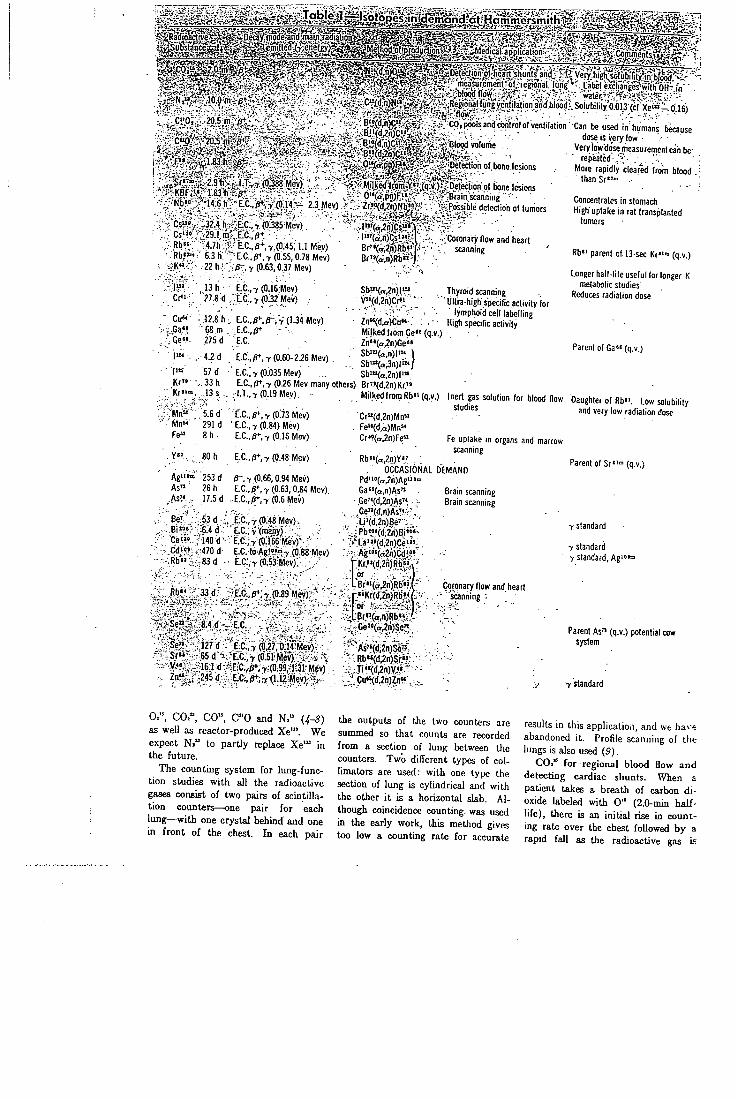
m i
, C " 0 , 20 5 m j}*^
C » 0 '
O^h i f f
Iiloodflow^ , ' water iRegionallungvenfilation anif blood-- Solubility'o 013 (cf Xe<*- 0 16)
Cp.poolsandtxjntrolofventilation Can be used in humane b ^ u ' e dose IS very low
.iffBlopd volume . .
Detection of bone.leslons
J r " » ^ 2 9 h ' KBF, '« ' " l83t i
I T T(0J88Mev)
Kb" I 4 6 h EC p*- Y(014 - 23Mev)
C 324 h ' i t C r (0.385 Mev) C 291m E.C..pt Rb" 4 7h E.C..p-^.r(0.45: l . I Mev)
6 3 h - E.C..0+. 7(0.55. 0.78 Mev) K " 22 h .'/!-. y (0.63.0.37 Mev)
| m 13 h E.C..7 (0.16 Mev) Cr"- 27.8 d .E.C..7(0.32Mev)
Cu" 12.8 h LC..^.p-:y{l.3i Mov) •Ga'» .68 m E.C.,(J<- - • Ge»« 275 d E.C. •|«« . , 4.2d .E.C.,r,T (0.60-2i5 Mev) . | m • 57 d • E.C.;> (0.035 Mev)
Mll l !ed^romy"(qv) Detection of bone lesions 0"(afPn)F>« Brain scanning ••• ^r»°(d 2n)Nb" Possible detection ol tumors
l"'(a^n)C " M l " ' ( a n ) ( ; s ' » 8r"(a.2n)Rb«'( B r ' V . n ) R b " - j
..Coronary flow and heart scanning
K r " K r " " .
;?"lvin»"" - Mn"
.33h .13 s .
'5.6 6' '291 d
8 h .
Sb^Ko^nJI-J" V"(d,2n)Cr^i '•
Zn«(d^)Cu«-,. . . -Milked from Ge" (q.v.) Zn"(a^n)Ge" Sb'"(a,n)|i" Sb'"(a3n)r'" Sb>"(a.2n)l"'
E.C.,p+.7 (0.26 Mev many others) Br'-«(d,2n)Kr." I T , 7 (0.19 Mev)
E.C.,fi+,7(0:73 Mev) •E.C.,7(0.84) Mev)
E.C.,/r-,7(0.16 Mev)
Se"^ 8 4 d E.C
Se" 127 d E0,7 (0^7,014 Mev) S r " 65 d E.C 7 (0 51 Mev) V " 161 d "E-C,^,7(0 99 1 31 Mev) Z n " ^ 245<J EC ,r,7(112Mev)
Milked froinRb«'(q.v.)
Cr«(d,2n)Mna Fe«(d,a)Mn" Cr'?(a.2n)Fe»
Thyroid scanning Ultra high specific activity (or
lymphoid cell labelling High specific activity
Inert gas solution for blood flow studies
Very l0i> dOic measuremeni can be rejieated ,, . 'V
More rapidly cleared from blood than Sr '"» ••
Concentrates in stomach High uptake in rat transplanted
tumors
Rb" parent of 13-sec Kr«'» (q.v.)
Longerhall-lileusefulforlonger K metabolic studies
Reduces radiation dose
Y»' . 80 h E.C..p*.7 (0.48 Mev)
Agiito 253 d r. 7 (0.66.0-94 Mev) As" 26 h E.C.. r, 7 (0.63,0.84 Mev) As" . 17.5 d ..E.C..^.7(0.6Mev)
Be' 53 d •. EX.,V (0.48 Mev): Bi»»,«?'1^6.4 d-. E.C.,7:(many) Ce"< ^'l40 d ' - : E.C.;7(0;i66Mev)-.
: (id'«»;i--470 d- E.C. to.Ag!»J»7 (0;88 Mev) Rb" . -SSd . E.C:.7(0.53Mev), . ,.
Rb" 33d EC p*,7(089 Mev)
Rb"(a.2n)Y" OCCASIONAL DEMAND
Pd"»(a.2n)Ag>'» Ga"(a.n)Asn Ge»(d.2n)As» • Ge"(d.n)As»r-.
.U'(d.2n)Be'--•Pb'."(d:2ii)Bi«"
••Ua>»(d.2n)Ce"' Ag'"(a2n)Cd"'
"Kr."(d.2ii)Rb" or .;
-Br«'(S,2n)RB«," r.«<Kr(d;2n)RB!<;
Brain scanning Brain scanning
-•<Kr(d2i or
, -Br"(a,n «,n)Rb".;; Ge"(a,2n)Se"
As"(d2n)Se'» .Rb"(d,2n)Sr'?: Ti«(d,2n)V««
• Cu»(d.2n)Zn«f
Coronary flow and heart "scanning •
Parenl of Ga"(q.v.)
Daughter of Rb". Low solubility and very low radiation dose
Fe uptake in organs and marrow scanning
Parent of S r " " (q.v.)
Y standard
7 standard 7 standard, A g ' " "
Parent l^^-system
7Vtandard
(q.v.) potential cow
O.", CO.", CO", C"0 and N." U S ) as well as reactor-produced Xe'". We expect Ni" to partly replace Xe'" in the future.
The counting system for lung-function studies with all the radioactive gases consist of two p^rs of scintillation counters—one pair for each lung—with one crystal behind and one in front of the chest. In each pair
the outputs of the two counters are summed so that counts are recorded from a section of lung between the counters. Two different types of collimators are used: with one type the section of lung is cylindrical and with the other it is a horizontal slab. Although coincidence counting, was used in the early work, this method gives too low a counting rate for accurate
results in this application, and we have abandoned it. Profile scanning of the lungs is also used ( 9 ) .
C O . " for-regional blood Bow and detecting cardiac shunts. When £ patient takes a breath of carbon dioxide labeled with 0" (2.0-min half-life), there is an initial rise in counting rate oyer the chest followed by a rapid fall as the radioactive gas is
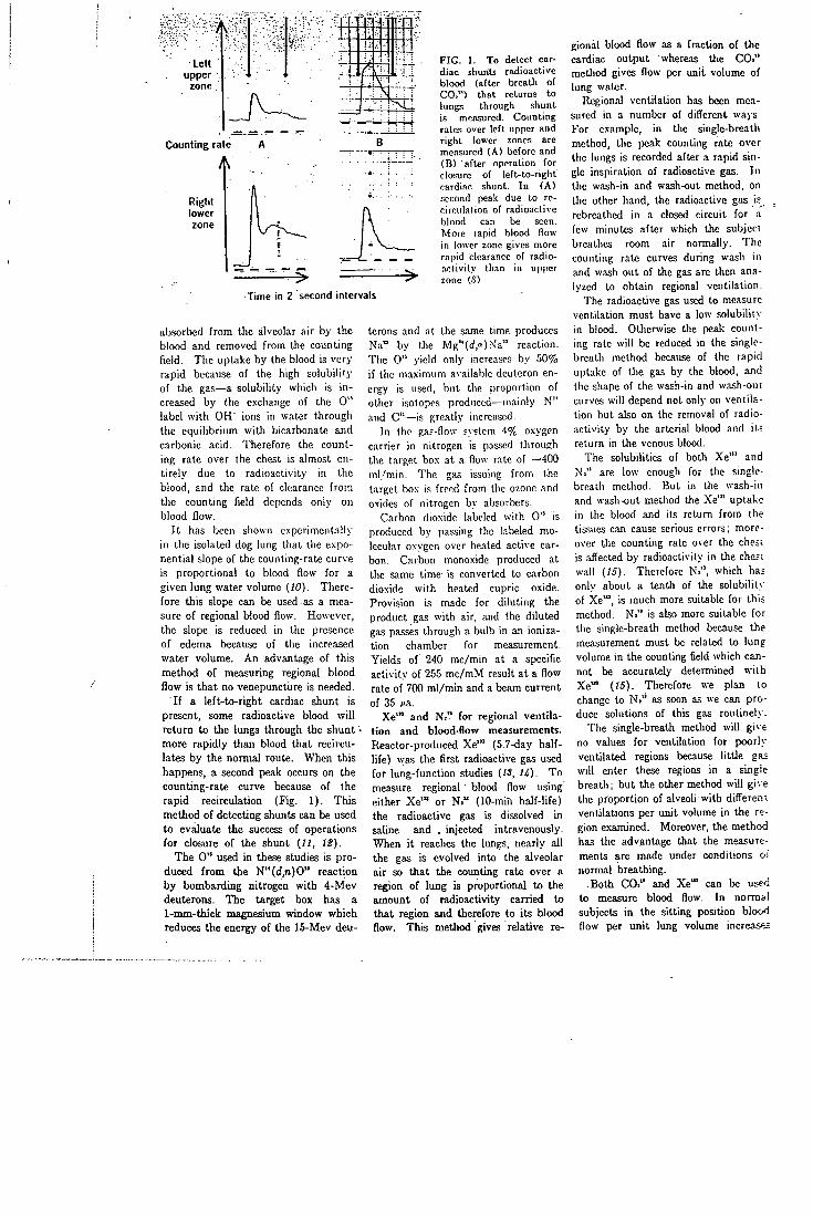
Lett upper
Counting rate
A
Right lower zone
F I G . 1. To detect cardiac shunts radioactive blood (after breath of CO.") that returns to lungs through shunt is measured. Counting rates over lef t upper and r ight lower zones arc mc.isured ( A ) before and (B) after operation for closure of le f t - to- r igh t cardiac shunt. I n ( A ) second peak due to recirculation of radioactive blood can be seen. More rapid blood flow in lower zone gives more rapid clearance of radio-. i c l i v i l y than in upper zone (.S)
Time in 2 second intervals
absorbed from the alveolar air by the blood and removed from the counting field. The uptake by the blood is very rapid because of the high solubility of the gas—a solubility which is increased by the exchange of the 0" label with OH" ions in water through the equilibrium with bicarbonate and carbonic acid. Therefore the counting rate over the chest is almost entirely due to radioactivity in the blood, and the rate of clearance from the counting field depends only on blood flow.
It has been shown experimentally in the isolated dog lung that the exponential slope of the counting-rate curve is proportional to blood flow for a given lung water volume (10). Therefore this slope can be used.as a measure of regional Wood flow. However, the slope is reduced in the presence of edema because of the increased water volume. An advantage of this method of measuring regional blood flow is that no venepuncture is needed.
If a left-to-right cardiac shunt is present, some radioactive blood will return to the lungs through the shunt ' more rapidly than blood that recirculates by the normal route. When this happens, a second peak occurs on the counting-rate curve because of the rapid recirculation (Fig. 1). This method of detecting shunts can be used to evaluate the success of operations for closure of the shunt (//, 12).
The 0" used in these studies is produced from the N"(d,n)0'' reaction by bombarding nitrogen with 4-Mev deuterons. The target box has a 1-nmi-thick magnesium window which reduces the energy of the 15-Mev deu
terons and at the same time produces Na" by the Mg"((i,»)Na" reaction. The 0" yield only increa.ses by 50% if the maximum available deutcron energy is used, but the proportion of other isotopes produced—mainly N" and C"—is greatly increased.
In the gas-flow fvstem 4% oxygen carrier in nitrogen is passed through the target box at a flow rate of ~400 ml/min. The gas issuing from the target box is freed from the ozone and oxides of nitrogen by absorbers.
Carbon dioxide labeled with 0" is produced by pa.ssing the labeled molecular oxygen over heated active carbon. Carbon monoxide produced at the same time is converted to carbon dioxide with heated cupric oxide. Provision is made for diluting the product gas with air, and the diluted gas passes through a bulb in an ionization chamber for measurement. Yields of 240 mc/min at a specific activity of 255 mc/mM result at a flow rate of 700 ml/min and a beam current of 35 M.
Xe'" and Ni" for regional ventilation and blood-flow measurements. Reactor-produced X e " (5.7-day half-life) was the first radioactive gas used for lung-function studies {IS, U). To measure regional blood flow using either X e " or N." (lO-min half-life) the radioactive gas is dissolved in saline and . injected intravenously. When it reaches the lungs, nearly all the gas is evolved into the alveolar air so that the counting rate over a region of lung is proportional to the amount of radioactivity carried to that region and therefore to its blood flow. This method gives relative re
gional blood flow as a fraction of the cardiac output whereas the CO." method gives flow per unit volume of lung water.
Regional ventilation has been measured in a number of different wajs. For example, in the single-breath method, the peak counting rate over the lungs is recorded after a rapid single inspiration of radioactive gas. In the wash-in and wash-out method, on the other hand, the radioactive gas is rebrcathed in a closed circuit for a few minutes after which the subject breathes room air normally. The counting rate curves during wash in and wash out of the gas are then analyzed to obtain regional ventilation.
The radioactive gas used to measure ventilation must have a low solubility in blood. Otherwise the peak counting rate will be reduced in the single-breath method because of the rapid uptake of the gas by the blood, and the shape of the wash-in and wash-out curves will depend not only on ventilation but also on the removal of radioactivity by the arterial blood and it-: return in the venous blood.
The solubilities of both Xe"' and Ni" are low enough for the single-breath method. But in the wash-in and wash-out method tlie Xe'" uptake in the blood and its return from the tissues can cause serious errors; moreover the counting rate over the chest is affected by radioactivity in the che;i wall {15). Therefore N,", which has only about a tenth of the solubility of Xe'", is much more suitable for this method. N." is also more suitable for the single-breath method because the measurement must be related to lung volume in the counting field which cannot be accurately determined with X e " {15). Therefore we plan to change to N." as soon as we can produce solutions of this gas routinel>'.
The single-breath method will give no values for ventilation for poorly ventilated regions because little ga; will enter these regions in a single breath; but the other method will give the proportion of alveoU with difi'eren: ventilations per unit volume in the region examined. Moreover, the method has the advantage that the measurements are made imder conditions oi normal breathing.
Both CO." and X e " can be used to measure blood flow. In normal subjects in the sitting position blood flow per unit lung volume increases
from apex to base of the lung whereas in patients vnth severe mitral stenosis this pattern is reversed (16, 17). The physiology of the distribution of lung blood flow has been worked out in detail using the isolated perfused dog lung [18). The absorbed doses for lungs and gonads are shown in Table 2 for the radioactive gases (-5, 6, S, 19). The dose from a series of tests is normally of the same order of magnitude as from a chest x-ray.
The N" is produced by bombarding carbon with deuterons by the C"(d,n)N" reaction. The target material is a layer of active carbon granules supported in the external deuteron beam by a graphite grid. This replaces the nichrorne grid which was used to produce gaseous N." by Nicholas, Silvester and Fowler (SO) in studying nitrogen fixation in bacterial cells and their extracts.
Carrier gas is passed through the target to sweep out the volatile N" compounds released from the active carbon. Any oxides of nitrogen are reduced to nitrogen with heated copper. Various carrier gases have been used including argon, methane and hydrogen. Argon gives high yields but also contributes the long-lived A" contaminant. Hydrogen, on the other hand, gives lower yields but has the advantage of being readily removed from the gas phase by conversion to water, giving a large increase in specific activity.
When solutions of Ni" are needed, the active carbon is outgased under vacuum before use, and a carrier gas of high-purity hydrogen is used. With a carrier gas flow of 50-ml/min hydrogen and a deuteron beam current of 35 /<a, gas samples with a specific activity of 5 curies/liter are obtained after the hydrogen is oxidized with cupric oxide at 700°C. If this gas is then mixed with water or normkl saline, solutions are produced containing 1(X)-150 fic/ml of N.". A typical sample of 1 mc N>" in 10 ml of saline takes 10 rain to prepare. This solution is sterile and pyrogen free because it is the product of a high-temperature combustion. The output of the furnace has a sterile demountable assembly.
(TO, for studying body C O . pools. We have used C"0. (20-min half-life) to study hoAy CO. in normal volunteers (SI). The radioactive gas was administered during VtrVi min of re-
breathing, and radioactivity in arterial blood samples was measured at intervals up to ~1 hr after injection. The results indicate that there is a rapid exchange between blood and extracellular CO. and a slower exchange with an intracellular pool. This hypothesis explains the rate of rise of Pco, (partial pressure of CO. in blood) during rcbreatliing which could not be explained by models in which the rate of exchange of CO. throughout the body was limited only by blood flow.
We are now extending these experiments using an improved technique in which radioactivity and CX). in expired air are measured continuously. Since ~0.5 mc is given, the doses are one tenth of those shown in Table 2. Results are being analyzed with an analog computer using a model that includes rate limitations due to both blood (low and cxtracellular-intracellular exchange.
The control of ventilation is atso simulated with this model (29), and we find that a number of different experiments can all be matched with the same value for pool sizes and exchange rates in a given subject. This new model includes separate pools for dissolved CO. and bicarbonate and takes into account the interchange with buffer anions. We hope that this model will help us understand tlic control of the acid-base balance in llie body.
The C " is produced by the B"(d,n)C" and B"(d,2n)C" reactions by bombarding boi-on with deuterons. The target material is boric oxide which is kept in the molten slate by the power dissipated by the beam (450
T a b l e 2 — A b s o r b e d tissue doses from isotopes
Absorbed dose (millirads) to:
Isotopes Lungs Gonads
Single breath 5 mc for 20 sec
C'K), 80 (apex) 18 COi" 220 (apex) 55
Single breath or injection 1 mc for 30 sec
Xe"» 14 0.1—OJ N." 35 0.1-0.5
Rebreathing 1 mc/l for 2 min
Xe"» 57 2 - 3 H , " 136 0 . 5 - 2
watts at 30 M ) and is prevented from running off the wedge by surface tension, mixture of carbon monoxide and carbon dioxide labeled with C " together with some N." is released from the molten boric oxide.
When C"-lalieled carbon dioxide is needed, a flow of 100 ml/min of 5% CO. in helium is used as a carrier sweep gas, and the C"-labclcd carbon monoxide is oxidized to carbon dioxide with heated cupric oxide. The' .N"-labelcd nitrogen is removed in batches by trapping the carbon dioxide a l - 1 9 6 ° C .
C"0 for red-cell volume measurements. To measure circulating red-cell volumes one conventionally u.scs nn isotope dilution technique with red cells labeled with Cr". But the long half-life of Cr" precludes its use in infan;; and expectant mothers because the radiation dose would be too high and in patients when serial mcasuremeni; at short intervals are required. For these applications a short-lived la'oc-1 nm.st be used.
The aflinity of hemoglobin for carbon monoxide is well known, and t-?-cause this gas labeled with short-li^'ed C" is readily available from our cyclotron, we have developed a routine method for labeling red cells n-ith this isotope (23).
The target arrangement and bombardment conditions are the same as for C"0. but when we need C"0 we use a flow of 50 ml/min of 1% carbon monoxide in helium as carrier sweep gas. The carbon dioxide is reduced to carbon monoxide using active carbon at 900°C. The gas then fiow; through a soda lime trap to rem';'ve the last traces of CO..
For red-blood-cell labehng high specific activity is needed. This ii achieved by using small carrier concentrations, low flow rates and high beam currents. The gas must also be sterile and pyrogen-free. We therefore -jse Millipore filtration and bake the gas in a small silica tube furnace. Tbt gas is then removed by a sterile septum straight into a 50 ml disfKisablt syringe which contains the "whole-blood" sample. About 15 ml of blood are then niixed with 35 ml of the O'-labeled 1% carbon monoxide/heljucL mixture by gently rotating the syringe on its axis at 20 rpm for 10 min. Because the N"-labeled molecular nitrogen has a very low solubility, it does not interfere with tlie radiochemicai
purity of the red cells. Yields of 7 mc/min are obtained in the gas phase with a specific activity of 250 mc/mM at a beam current of 35ca at - '14 Mev,
B a t c h i so topes : F " and Sr" '" The longer-lived radioisotopes used
at ffammersmith are produced i>_\ batch methods. F" fluoride and Sr""' cjtrate are used for detecting bone lesions—sometimes letting one see the lesions on the scan before they Iwcomc visible on the x-ray (Si-S6). As Monte Blau of Roswcll Park Memorial Institute has pointed out, F " fluoride is more rapidly cleared from the blood than Sr"" and is more snilabic; liow-e%'er, because of its short half-life, i t can only be used close to a cyclotron or reactor. Sr'"", on the other h.-ind, has the advantage that i t can be milked from its longer-lived parent Y " (SO-hr half- l i fe) .
The dose from both F" and Sr""' is considerably smaller than that from tlie longer-lived Ca" and Sr" whioli are more often used. For examijlc, Blau calculates the dose from F" lo the whole body as only 0.23 rads/mc {S4) while we calculate i t as 0.41 rads/mc allowing a factor of five for nonuniform distribution in bone. The doses from Ca" and Sr" are several rads per investigation.
Both osteolytic and osteoblastic lesions show an increased uptake of these isotopes compared with nonnal bone. The uptake is rapid and is probably due to exchange with salts of the bone crystal and not to metabolism; thus increased uptake may be due to a larger available area of crystal. Budy discusses the uptake mechanism of a number of different isotopes in bone (27).
The distribution of F " in the body has also been considered to depend on blood flow. Using the positron camera Van Dyke and his colleagues have visualized the distribution of this uptake for the whole body and have studied the variation in uptake pattern in various diseases {S8).
We make F " in 40-60-mc batches twice a week and dispatch them to several London hospitals that are interested in bone scanning. More distant parts of the country are supplied wi th Y " cows which are also made on a regular schedule.
Twenty-one patients have now been scanned wi th F " by R. Morrison and
J. Davcy at Hammersmith. These patients either have known or suspected neoplastic changes on x-ray or have symptoms that suggest bone involvement although their .x-rays arc normal. <.'.ood scans \mve been ob-laii\ed in all palionl,,, and four positive scans were foimd when the x-r.iys were entirely normal.
For example. Fig. 2 shows a K" scan of a G4-year-old jiaticnt who had a history of carcinoma of the cervical stump in May 1965 and was treated with supcrvoltage irradiation. She was well and free of recurrence for 1 year when she presented with severe pain ui the left arm and some tenderness over the head of the left hunieni.s. The x-rays, as Fig. 2 sli<tws, were normal. We gave 1 mc of F" intravenously and scanned the patient I hr later. As the scan shows, increased uptake resulted compared with the expected upt.ake in normal subjects, and despite the normal x-ray finding the patient was considered to have a secondary depn.sit in the head of the humerus. She was given a course of palliative irradiation to this .secondary- depn.sit and 5 months later was free of pain.
R. McCready of the Royal Marsden Hospital has given 1-2-mc intravenous injections of F" to 47 patients. Of 15 cases with positive x-rays, 14 gave positive scans. In a further 8 positive scans, the x-rays were negative. Of the remaining cases, 19 showed negative scans and x-rays while 5 gave equivocal results. McCrcady has also used Sr"" and finds that the bone uptake is similar to that for F". Moreover he finds that both F " and Sr"" give better scans in a shorter time than Sr". Since a high percentage of the scans are negative, much time is wasted if each scan takes a long time so this is an important advantage of
* these short-lived isotopes.
We make F " by irradiating water held in . a titanium vessel in the external alpha beam of the cyclotron (29). The essential nuclear reaction is 0"(<',pn)F" for which the threshold energy is ~ 2 0 Mev. Recently we have installed an infrared TV camera which lets us observe the distribution of the beam on the target during bombardment. I f the beam is kept well spread, up to 60 mc of F " can be obtained from a 1-hr bombardment at 40 /"a.
For purposes such as bone-lesion
scanning the cyclotron target solution can be administered directly to the patients without further treatment other than sterilization because con-Laminating activities are negligible (only traces of Cr" and V " which recoil from the walls of the target ves-sel) and the .solutions have been shown to 1)0 pyrogen free. Reactor produced F", on the other hand, must be separated from the lithium salt from which it is made and from the large quanti- . .. ties of tritium that are made simultaneously (SO, SI).
The Y " for the Sr"" generator is produced by the Rb"(o,2n)"\'" reaction. Rubidium chloride is used as the target, melted onto a copper plate which is water cooled during bombardment. The separation procedure has been described by Hillman and his group (.??). After bombardment the rubidium chloride is removed from the tArgel plate by dissolution in dilute hydrochloric acid containing a few milligrams of yttrium carrier. Yttrium hydroxide is precipitated and purified and finally applied in citric acid solution to an anion exchange resine column in the carbonate form.
The column consists of a 1-ml I x ^ of amberlite CG.400 resin followed by a 4-ml bed of IRA 400 mounted in a 10-nil dispo.sable syringe. The Sr"" daughter is milked by allowing 5 ml of sterile 0.005% citric acid to flow through the resin column. The milk-ings are then sterilized by autoclaving or MiUipore filtration. The cow system will produce pyrogen-free solutions if the milking procedure is carried out carefully.
Production yields of Y " at 30 Mev are ~340 w A A h at the end of bombardment after chemistry. This gives a Sr"° yield on milking of 240 « A A h at the end of bombardment. We found that the average milking efficiency for seven cows was 72%. Occasionally we find very high milking efficiencies of around 90%. Yttrium contamination of the milkings never amounts to more than 0.05% of the Sr"" content at the time of milking.
KBF)" for brain scanning. Previously two groups have used K B F . " (1.8-hr half-life) for brain scarming (SS, Si). At Hammersmith we use the F" produced by alpha bombardment of water to label fluoroborate using a method similar lo that described by Ashkenasy and his group (SS).
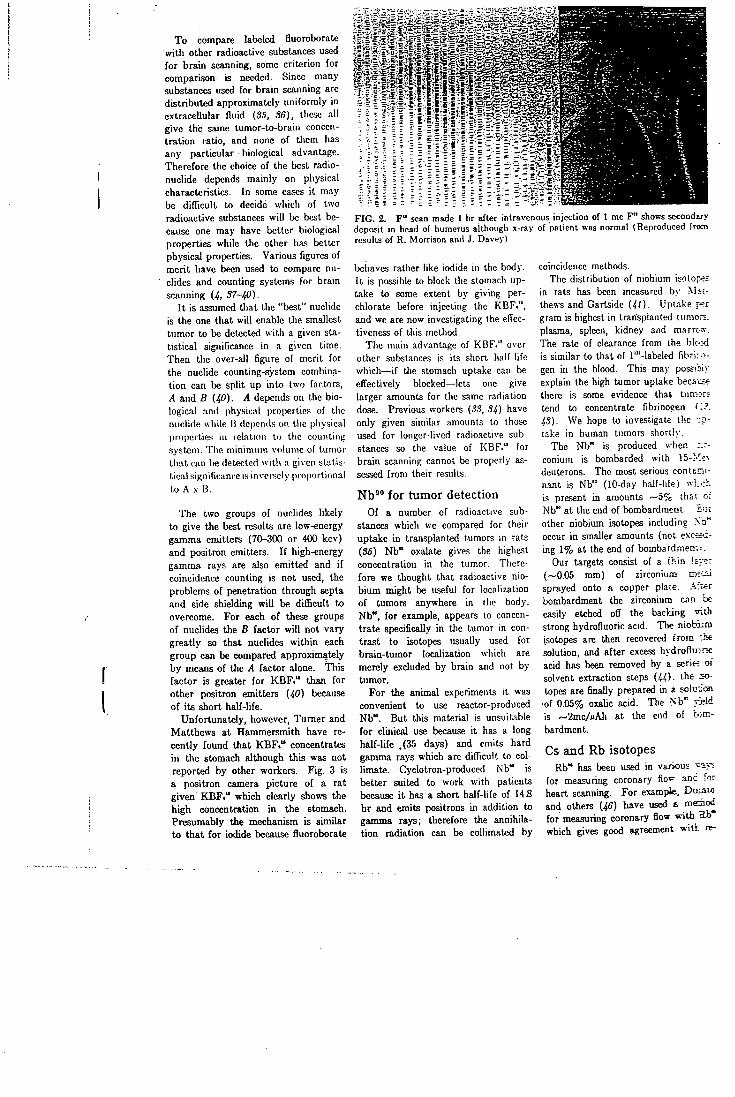
To compare labeled fluoroborate wit l i other radioactive substances used for brain scanning, some criterion for comparison is needed. Since many substances used for brain scanning are distributed approximately uniformly in extracellular fluid (35, 36), these all give the same tumor-to-brain concentration ratio, and none of them has any particular biological advantage. Therefore the choice of the best radionuclide depends mainly on physical characteristics. I n some cases i t may be difficult to decide which of two radioactive substances will be best because one may have better biological properties while the other has better physical properties. Various figures of merit have been used to compare nuclides and counting systems for brain scanning (4, 37-40).
I t is assumed that the "best" nuclide is the one that will enable the smallest tumor to be detected with a given statistical significance in a given time. Then the over-all figure of merit for the nuclide counting-system combination can be split up into two factors, A and B (40). A depends on the biological and phy.sical piopei tie.' of tlie nuclide while B dcpeiuls on the physical properties in relation to the counting system. The minimum volume of tumor that can be detected with a given statistical significance is inversely proportional to A .V B.
The two groups of nuclides likely to give the best results are low-energy gamma emitters (70-300 or 400 kev) and positron emitters. I f high-energy gamma rays are also emitted and i f coincidence counting is not used, the problems of penetration through septa and side shielding wil l be difficult to overcome. For each of these groups of nuclides the B factor will not vary greatly so that nuclides within each group can be compared approximately by means of the A factor alone. This factor is greater for K B F , " than for other positron emitters (40) because of its short half-life.
Unfortunately, however, Turner and Matthews at Hammersmith have recently found that K B F . " concentrates in the stomach although this was not reported by other workers. Fig. 3 is a positron camera picture of a rat given K B F . " which clearly shows the high concentration in the stomach. Presumably the mechanism is similar to that for iodide because fluoroborate
F I G . 2. F " scan made 1 hr after intravenous inject ion of 1 mc P ' shows secondso' deposit in head of humerus although x-ray of patient was normal (Reproduced from results of R. Morrison and J. Davey)
behaves rather like iodide in the body. I t is possible to block the stomach uptake to some extent by giving per-chlorate before injecting the KBF." , and we are now investigating the effectiveness of this method.
The main advantage of KBF." over other substances is its short half-life which—if the stomach uptake can be effectively blocked—lets one give larger amounts for the same radiation dose. Previous workers (3.?, 34) have only given similar amounts to those used for longer-lived radioactive substances so the value of KBF." for brain scanning cannot be properly assessed from their results.
N b ' " f o r tumor detection Of a number of radioactive sub
stances which we compared for their uptake in transplanted tumors in rats (35) Nb" oxalate gives the highest concentration in the tumor. Therefore we thought that radioactive niobium might be useful for localization of tumors anywhere in the body. Nb", for example, appears to concentrate specifically in the tumor in contrast to isotopes usually used for brain-tumor localization which are merely excluded by brain and not by tumor.
For the animal experiments it was convenient to use reactor-produced Nb". But this material is unsuitable for clinical use because i t has a long half-life .(35 days) and emits hard gamma rays which are difficvilt to col-limate. Cyclotron-produced Nb" is better suited to work with patients because i t has a short half-life of 14.8 hr and emits positrons in addition to gamma rays; therefore the annihilation radiation can be collimated by
coincidence methods. The distribution of niobium isoto;:-?;
in rats has been measured by M--:-thews and Gartside (4/). Uptake f.=r gram is highest in transplanted tume.-. plasma, spleen, kidney and marrt-s-. The rate of clearance from the blc':<i is similar to that of r"-labeled fibrir^-gen in the blood. This may possibiv explain the high tumor uptake becaiji? there is some evidence that tumors tend to concentrate fibrinoeen I'.?. 43). We hope to investigate the take in human tumors shorily.
The Nb" is produced when :.r-conium is bombarded with 15-j ' ' rv deuterons. The most serious conttai-nant is Nb" (10-day half-life) w.v_,:h is present in amounts ~ o f c thai of Nb" at the end of bombardmeni. 3iit other niobium isotopes including Vo""' occur in smaller amounts (nov exceeding 1% at the end of bombardmeo'.'.
Our targets consist of a thin \V:~.-(~0.05 mm) of zirconium vn-j^ sprayed onto n copper plate. .Vr'wr bombardment the zirconium can be easily etched off the backing uith strong hydrofluoric acid. The niobiim isotopes are then recovered f rom •.be solution, and after excess hydroflu:ric acid has been removed by a serie; 01 solvent extraction steps (U). the sy-topes are finally prepared in a soluion
•of 0.05% oxahc acid. The >"b" yj?id is '-'2mc//'Ah at the end of b:cn-bardment.
Cs and Rb isotopes Rb" has been used in various v a j ;
for measuring coronary flo=' and for heart scannmg. For examp)le, Daiato and others (46) have used a roemoc for measuring coronary flow with Sb" which gives good agreement wi t l . re-
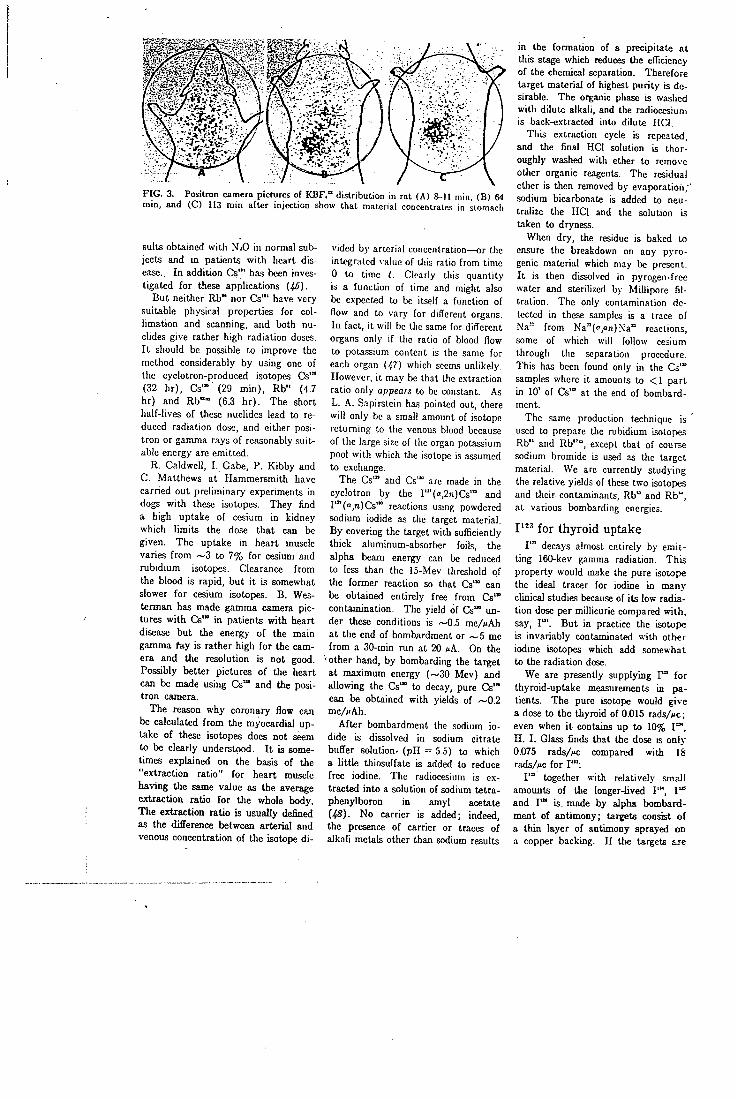
F I G . 3. Positron camera pictures of K B F , " dis t r ibut ion in rat ( A ) 8-11 min, ( B ) 64 min , and ( C ) 113 m i n af ter inject ion show that material concentrates in stomach
suits obtained with N.O in normal subjects and in patients with heart disease. In addition Cs"' has been investigated for these applications (45).
But neither Rb" nor Cs"' have very suitable physical properties for col-limation and scanning, and both nuclides give rather high radiation doses. I t should be possible to improve the method considerably by using one of the cyclotron-produced isotopes Cs"* (32 h r ) , Cs'"' (29 min) , Rb" (4.7 hr) and Rb"" (6.3 h r ) . The short half-lives of these nuclides lead to reduced radiation dose, and either positron or gamma rays of reasonably suitable energy are emitted.
R. Caldwell, I . Gabe, P. Kibby and C. Matthews at Hammersmith have carried out preliminary experiments in dogs with these isotopes. They find a high uptake of cesium in kidney which limits the dose that can be given. The uptake in heart muscle varies from ~3 to 7% for cesium and rubidium isotopes. Clearance from the blood is rapid, but i t is somewhat slower for cesium isotopes. B. Wes-terman has made gamma camera pictures with Cs" in patients with heart disease but the energy of the main gamma fay is rather high for the camera and the resolution is not good. Possibly better pictures of the heart can be made using Cs'" and the positron camera.
The reason why coronary flow can be calculated from the myocardial uptake of these isotopes does not seem to be clearly understood. I t is sometimes explained on the basis of the "extraction ratio" for heart muscle having the same value as the average extraction ratio for the whole body. The extraction ratio is usually defined as the difference between arterial and venous concentration of the isotope di
vided by arterial concentration—or the integrated \ alue of this ratio from time 0 to time 1. Clearly this quantity is a function of time and might also be expected to be itself a function of flow and to vary for different organs. In fact, it will be the same for difl'erent organs only if the ratio of blood flow to pota-ssium content is the same for each organ (47) which seems unlikely. However, i t may be that the extraction ratio only appears to be constant. As L. A.. Sapirstein has pointed out, there will only be a small amount of isotope returning to the venous blood because of the large size of the organ potassium pool with which the isotope is assumed to exchange.
The Cs"* and Cs'" are made in the cyclotron by the r" (» ,2n)Cs" ' and !" '( ' ' , '»)Cs'" reactions using powdered sodium iodide as the target material. By covering the target with sufficiently thick aluminum-absorber foils, the alpha beam energy can be reduced to less than the 15-Mev threshold of the former reaction so that Cs"° can be obtained entirely free from Cs"* contamination. The yield of Cs'" under these conditions is ~0.5 mc//iAh at the end of bombardment or ~ 5 mc from a 30-min run at 20 cA. On the
•other hand, by bombarding the target at maximum energy (~30 Mev) and allowing the Cs'" to decay, pure Cs" can be obtained with yields of ~0.2 m c / M h .
After bombardment the sodium iodide is dissolved in sodium citrate buffer solution- (pH = 5.5) to which a little thiosulfate is added to reduce free iodine. The radiocesium is extracted into a solution of sodium tetra-phenylboron in amyl acetate [48). No carrier is added; indeed, the presence of carrier or traces of alkali metals other than sodium results
in the formation of a precipitate at this stage which reduces the efficiency of the chemical separation. Therefore target material of highest purity is desirable. The organic phase is washed with dilute alkali, and the radiocesiuni is back-extracted into dilute HCI.
This extraction cycle is repealed, and the final HCI solution is thoroughly washed with ether to remove other organic reagents. The residual ether is then removed by evaporation, ' sodium bicarbonate is added to neutralize the HCI and the solution is taken to dryness.
When dry, the residue is baked to ensure the breakdown on any pyro-genic material which may be present. I t is then dissolved in pyrogen-frce water and sterilized by Millipore filtration. The only contamination detected in these samples is a trace of Na° from Na''(<',<'r!).\'a'' reactions, some of which will follow cesium through the separation procedure. This has been found only in the Cs'" samples where it amounts to < 1 part in 10' of CIs"* at the end of bombardment.
The same production technique is used to prepare the rubidium isotopes Rb" and Rb"", except that of course sodium bromide is used as the target material. We are currently stud>Tng the relative yields of these two isotopes and their contaminants, Rb" and Rb", at various bombarding energies.
P " for thyro id uptake P" decays almost entirely by emit
ting 160-kev gamma radiation. This property would make the pure isotope the ideal tracer for iodine in many clinical studies because of its low radiation dose per millicurie compared with, say, I " ' . But in practice the isotope is invariably contaminated with other iodine isotopes which add somewhat to the radiation dose.
We are presently supplying I " for thyroid-uptake measurements in patients. The pure isotope would give a dose to the thyroid of 0.015 rads/^c; even when i t contains up to 10% 1°", H . I . Glass finds that the dose is only 0.075 rads/w compared with IS rads7w; for I ' " ;
P" together with relatively small amounts of the longer-lived I " " , I " and I " is made by alpha bombardment of antimony; targets consist of a thin layer of antimony sprayed on a copfier backing. I f the targets ise
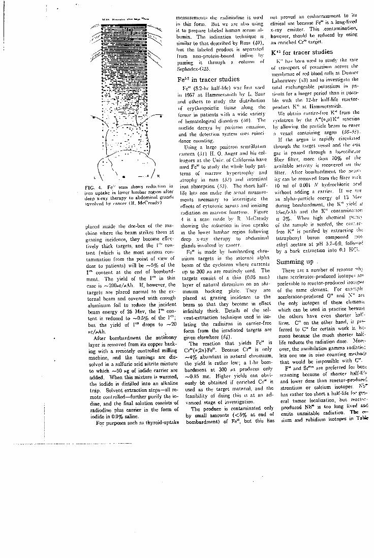
I , . " " . . .
F I G . 4. Fe- scan .shows re i lu i t ion in iron uptake in lower lumbar region after deep x-ray therapy to abdominal grand.s involved l iy rancor (R . McCready)
placed inside (he dee-box of the machine where the beam strikes them at grazing incidence, they become effectively thick targets, and the 1'"' content (which is the most serious contamination from the point of view of dose to patients) will be ~ 5 % of the I ' " content at the end of bombardment. The yield of the I ' " in this case is ~20Qiic/^.\h. I f , however, the targets are placed normal to the external beam and covered with enough aluminum foil to reduce the incident beam energy of 25 Mev, the I ' " content is reduced to ~0.5% of the I ' " ; but the yield of I ' " drops to —20 f c / M h .
After bombardment the antimony layer is removed from its copper backing with a remotely controlled milling machine, and the turnings are dissolved in a sulfuric acid nitrite mixture to which ~50 »ig of iodide carrier are added. When this mixture is warmed, the iodide is distilled into an alkaline trap. Solvent extraction steps—all remote controlled—further purify the iodine, and the final solution consists of radiodine plus carrier in the form of iodide in 0.9% saline.
For purposes such ns thyroid-uptake
measurements the radioiodinc is u.scd in this form. But we are also using it to prepare labeled luim.in .scrum albumin. The iodinatioii technique is .similar to that described by Rosa H9), but the labeled product is separated from non-prolein-bound iodine by passing i t through a column of Sephadex-C'25.
Fe^^ i n tracer studies Fe" (S.2-hr half-life) w.-i.s fir.-l used
in 1957 at Hammersmith by L. Szur and others to study the distribution of erythropoietic tissue along the femur in patients with a wide variety of hematological disorder; (-iO). The iiuchde decays by posiiroii emission, and the detection system u.<e.s coincidence counting.
Using a large positron scintillarion c.unera ( j / ) H . 0. .- nger and his colleagues at the Univ. of Gdifornia have used Fe" to study the wlmle-body patterns of marrow liyiiertrophy and atrophy in man (.5-') and intestinal iron absorption (.-i.J). The .short half-life lets one make the serial measurements necessary to mvesiigatc the cfTecIs of cytotoxic .ngents and ionizing radiation on marrow function. Figure 4 is a scan made by R. .McCready showing the reduction in iron uptake in the lower lumbar region following deep x-ray therapy to nbdominal glands involved by cancer.
Fe" is made by bombarding chromium targets in the internal alpha beam of the cyclotron where currents up to 300 Ma are routinely used. The targets consist of a thin (0.05 mm) lajer of natural chromium on an aluminum backing plate. They are placed at grazing incidence to the beam so that they become in effect infinitely thick. Details of the sol-vent-ex'traction technique used in isolating the radioiron in carrier-free form from the irradiated targets are given elsewhere (5i).
The reaction that yields Fe" is Cr"(o,2>!)Fe'"-. Because Cr" is only —'4% abundant in natural chromium, the yield is rather low; a I-hr bombardment at 300 jia produces only ~0.15 mc. Higher yields can obviously bfc obtained if enriched Cr" is used as the target material, and the feasibility of doing this is at an advanced stage of investigation.
The produce is contaminated only by small amomits ( < 5 % at end of bombardment) of Fe", but this has
not proved an embarrassment to its clinical use because Fe" is a long-lived x-ray emitter. This contamination, however, should be reduced by using an enriched Cr" target.
K ' ^ f o r tracer studies K" li:i.< bwn used to study llie rale
<pf transport of ixiiassium acro?.s ilic membrane of red blood cells at Donncr Laboratory (-v.?) and lo investigate the total o.vchangcabic potassium in p.i-ricnts for a longer period than is possible with the r2-hr half-life reactor-product K " at Hammersmith.
Wc obtain carrier-free K " from the cyclotron by the A"(o ,p)K" reaction by allowing the particle beam lo enter a vessel containing argon (o6—iiS).
If the argon is rapidly circiil.ued through the target vessel and ilic exit gas is pa.ssed through a boro.silic.ae fiber filter, more than 70% of the available activity is recovered on ihe filter. -After bombardment the acav-ity can be removed from the fiber w.rli 10 ml of 0.001 A' livdrorhloric icid without adding a carrier. If we 'i.-t an alpha-particle energy of 13 Mrv during bombardment, the K " yielc i ; l,'swc/c'\h and the K'" contamin:V;on is 3%. When high chemical purii.'. of the sample is needed, the carr.^r-free K " is purified by extracting ih^ tctrapheiiyl boron compound ;nto ethyl acetate at pH 5.7-C.O, follo^e': by a back extraction into 0.1 HCi.
Summing up . There are a number of reasons ~h;.
these accelerator-produced isolopr= ar' preferable to reactor-produced i?o;-jp« of the same element. For exnr.pk accelerator-produced 0 " and X " ar-; the only isotopes of these elemenii which can be used in practice b « a u H the others have even shorter half-lives. C" on the other hand, i= preferred to C" for certain work in h;:-mans because the much shorter half-life reduces the radiation dose. Mor^ over, the annihilation gamma radiatic-z lets one use in vivo counting m<shoc; that would be impossible with C".
F " and Sr"" are preferred for bor-r scanning because of shorter hiif-lifT and lower dose than reactor-prc-iuc^-f; strontium or calcium isotopes. N t * has rather too short a half-life fee % ^ eral tumor localization, but rt-actor-produced Nb" is too long lived emits unsuitable radiation. The « -sium and rubidium isotopes in Tabic
1 are more suitable than reactor-produced ones both because of their more useful radiation and shorter half-lives. K " , however, is more useful because of its longer half-life. The radiation emitted by I ' " has almost ideal properties for in vivo measurements and for some applications the short half-life is also an advantage. Fe" is useful because of its short half-life which lets one make serial measurements with i t .
In practice we do not choose cyclotron-produced isotopes at Hammersmith over reactor-produced because they are positron emitters or have high specific activities. However, in some cases, if, specific activity were too low the material would be unsuitable. We do not think that coincidence counting with positrons is necessarily an advantage since better efficiency can be obtained for the same resolution with a low-energy gamma-emitting nuclide and focusing collimators {60). I f no suitable low-energy gamma emitter is available, however, coincidence counting may certainly be preferable to the use of focusing collimators with high-energy gamma rays.
So far little use has been made of cyclotron-produced isotopes for labeling molecules more complex than simple inorganic salts. For example, C" or F " might be used to label a wide variety of substances of physiological interest if the difficulties of carrying out the labeling process in a short time can be overcome. Some of the most interesting developments of the future may lie in this direction.
References
; . M . M . Ter-PoKOssian, A new look a l the cydotTOD for m&IdnE short-lived isotopes, HDcuamcs t4. No. 10, SO <I96«)
2. J . W . Qaltop et al, A cyclotron for medicat lT>«>aich, Ptoc l.B.E. lOiB. « 2 <19J7)
J . . D . D . Vonberi. J . F . Fowler, The cyclotron unit: Medical Research Council. Nature 111. 837 ( I S U )
f . M . T«r-Po£oeaian, W . E . Power, "Proc. 1st, ( U N E S C O ) Internal. Conf. oo Radioisotopes in Sdentifio Reaearch," vol. I l l (Perga-mon Press, Paris, 1«J7)
<. N . A . Dyrcn. et al, "Proc. 2nd U.N. Cont. on Feaosful Uses of Atomic EnerKy," Paper J78, (Pereamon PrWM. London. 1959)
e. 0 . T . DoUery, et al. The preparation and use of redioactivQ oxyeeo, carbon monoxide, and carbon dioxide for investigation of regional lung function, and their comparison with xenoQ-133, Bonderbandt zur Strahlen-behcndlwv, U, 8S (1063)
r. P . D . B u d d n ^ a m . O. R . Forse. The preparation and processing of radioactive gases for dinical use, Int. J. Appl. Radn. and Itotojia U, <33 (1963)
J . C . M . E . Matthewa e l a l . Radioactive gases in "Radioactive Pharmaceuticals"- ( U . 8 . A E C , Oak Ridge. 1966)
». C . T . Dollery. P. M. s. CiUam. The distribution of • blood and gas within the lungs measured by scanning after administration of xenon-133. Thoraz. IS, 316 (1983)
10. C . T . Dollery, P. Ueimborg. P. Hugh-Joncs. The relationship between blood flow and dcarance rate of radioacUve carbon dioxide and oxygen in normal and edematous lungs. J. PhuticL I6t, «3 (1962)
11. 0 . T . DoUery. P. Hugh-Jones. Distribution ol gas and blood in the lungs in disease. Bril. Hcd. BuU. II, S9 (1963)
II. C . T . Dollery et al . Regional pulmonary blood flow in patients with circulatory sliuntf. flril. Heart Journal tS, 225 (1961)
(J. H. W. Knipping et al. Funktionsanalysc in der Kreistauf- und Lungen-Klinik mit HiUe der lootopenthorakographie und der selec-tiven Angiographic der Lungengefasse; Deitrag lur praopcrativcn Funktionsanalyse in der Thorax-Chirorgic. Miiiich. Afed. Wscltr, 99. « (1957)
H. W. C . Ball et al. Regional pulmonary function studies with xenon-133. J. Ctiii. Inval. 41, 519 (1962)
li. C. M. E . MatUiews. C . T . Dollery. Inter preUtioo of ""Xe lung wash in out curves using an analogue computer. Clin. Sr i . . :8. 573 (1965)
IC. J. B. West. C . T . Dollery. P. Uugh-Jones. The use of radioactive carbon dioxide to measure fcgtonal blood flow in the lungs of patients with pulmonary disease. J. Clin. Inxttt., II. 1006 (Oct. 26, 1962)
17. C . T . Dollery. P. Hugh-Jones. C . M. E Matthews. "Use of radioactive xenon for studies of regional lung function." Brit. Med. J., ii. 1006 (Oct. 20. 1962)
(S. J . B . West. C . T . DoUery. "Distribution of blood flow and the pressure-flow relatioos of the whole lung." / . Appl. Phytiol. tO, 175 (1965)
IS. C. M. E . Matthews. J . F . Fowler. P. C . R. Turner. Absorbed doses from xenon-133 in M . R. C . Cydotron Unit. Tech. Mem. No. 81. (1963)
to. D. }. D . Nicholas. D . J . SOvester. J . V. Fowler, Use of radioactive nitrogen in studying nitrogen fixation in bacterial cells and their extracts. Nature, IS9, 634 (1961)
tl. A. S. E . Fowle. C . M. E . Matthews. E . J . M . C ^ p b e U , The rapid distribution of 'Hp and ^KX), in the body in relation to the immediate carbon dioxide storage capacity. Clin. Sci.. «7, 51 (1960
It. E . J . M. CampbeU. C . M . E . Matthews, D. Head. .Analogue computer simulation of C O , stores and control of \'entilation. J. Phyiiot. ISi, 55p. (196S)
t j . J . C . C3ark, H . I . Glass. D. J . Silvester. "Proc of Second. Intemat. Conf. on Methods of Preparing and Storing Labelled C o m pounds," (Brussels. 1966)
H. M . Blau. W. Nagler. M . A. Bender. Fluor-ine-18. a new isotope for bone scanning. J. NucL Med.. S, 332 (1962)
tS. M . Blau. Y . Laor. M. A. Bender. Clinical experience with F-18 and Sr-87m for bone scanning. J. Nu^L Med.. 4. 193 (1963)
« . W. G . Myers. M . Olejar. RadiostrDntium-87m in studies of healing bone fracture. / . Nucl. Med. 4. 20J {1963)
i7. A. M. Budy. The use of radioisotopes in orthopsedica. Part I I , / . Bone and Jt. Surg.. 45-A. 1073 (1963)
M. D . Van Dyke et al, Bone blood flow shown with F-18 and the positron camera. Amer. J. PhyiioL tOS. 65 (1965)
tS. J. C. O a r k , D . J . SUvester, A cydotron method for the production of fiuorine-18. Int. J. AppL Radn. and Itotopu. 17. 151 (1966)
SO. M . Aobar. "Production aod Use of Short Lived Radioiaotopes from Reactors." Vol. 2/ ( I A E A , Vienna, 1963)
5/. H . H . Dworkin, P. D . LaFleur, Fiuorine-18; production by neutroo activation and pharmacology in "Radioactive Pharmaceu-ticals." ( I h S . A £ C . Oak Ridge, 1066)
St. M . Haiman et al. Production of Y " and a Si*^» generator. Int. J. AppL Radn. and Itoiopa. 17. 9 UflM)
S3. H . M . Aahkeoaiy et al. The localization of intracranial space occupying lesions by fluoroboratA iocs labelled with fluorine-18. Am«r. / . Rowtgcnol. 8$. U (1962)
5<. W. Eotziao et al. Evaluation of fhiorine-
18 as a scanning agent for int/acxanial tumours in "Medical Radioi^tope Scanning," Vol. 2 ( I A E A , Vienna. 106<)_
3j. C . M . E . Matthews, G . Molinoxo, "A study of the relative value of radioactive substances used for brain tumour localtxation and of the mechanism of tumour: brain concentration. Uptake in transplantable ftbrosarcoaia, brain and other orgaru .in the ik l . Bril. y. Szp, Path,. 44, 260 (1963)
•HI. I'. Mundinger, H. Gerhard. Untersuchungcn uber die Verteiluog der sur Himtuniordia-gnoslik verwendeten Radiotsolope in det Dlutbaho in ea penmen tell en Tumoren und iiicnschlichen Himgeftchwulsten. Ada Ncuro-chiniroica. It. 398 (1963)
.17. W. C . Dewey. W. K . Sinclair. Criteria loi evaluating collimatora used m vivo dislti-liution studies with . radioisotopea. Int. J Appl. Radn. and Itotopcs, JO. I (1961)
Jjf. R . N. Beck, A theoretical study of bn'm ' -scanning systems. / . NucL Med. t, J H (I96I)
.19. It. N. Beck, A theory of radioisotope scanning sj-slems, in ""Medical Radioisotope Scanning." Vol. 1 ( I A E A . Vienna. 1954)
i<i. C . M. E . Matthews, Comparison of isotopes for scanning. J. Nucl. Med.. 6. 1*5 (I96S)
il. C . M. E . Matthews. J . M. Gartside. Tumour uptake and distribution of niobium isotope.' in rats. Brit. J. Cancer, 19. i51 (1965)
i*. E . D. Day, J . A- Plannisek, D. Pressman LooUitstion in vivo of radioiodinated a rv i -ral-fibrin antibodies and radioiodinated rs: fibrinogen in the Murphy rat l>-n)phsarcoina and on other transplanuble rat tumours. J. Nat. Cancer Inti. tt. KM (19S9)
il. G . Monasterio, M. F . Becchini, N. Riccioo:. Radioiodinaled (1"' and 1'^) fibrinogen Jo: delection of malignanl tumours in man. i:. "Medical Radioisotope Scanning." Vol. ( I A E A . Vienna. 1961) H. L . Scherff. A. Herrman. Ckimparati'. * examination of the solvent estraction oi Pt T a , Nb and Zr from strong hydrochlori: acid." Zeilicbrifl fur Elcclrochemit. 6i. 102: (1900)
;J. E . A. Carr, Pharmacology of radioaciiv: tiaesium salta. in "Radioactive Phairnaceu;:-cals" (U. S. A E C , Oak Ridge, 1966)
i6. L . Donato et al. Measurement of co.-one.'-blood flow by enteraal counting with rsdic -active rubidium. Critical appraisal and v&;.-d&tion of the method. CiVc. M. 708 (1966) L. A. Sapirstein. Regional blood fk>w b;. fracliooaJ distribution of indicators. Amr-J. PhutioL 193. 161 (195S)
iS. H . L . Finston. M. T . Kinsley. The rac-chemistry of oeaium. .V..A.5.-N.S. iOSi U. S. ABC 60 (1961)
1,9. U. Rosa et al. Labelling of human fibrog*: with " ' I by electrolytic iodination. Biochl-r. Biophya. Ada, S6. 19 (1960
iO. P. E . Francois. L . Siur. Use ol lron-i2 a* t radioactive tracer. Nature. IBt 1665 (19SS)
5/. H. O. Anger, Human bone narrow dislrib'---lion UiQvm in vivo by iron-52 tod positron acintOialion camera. Science. 1587 (1964)
i? . D. Van Dyke., H. O. Anger, Pattemi & marrow hypertrophy and atrophy In mar. J. Nuci. Med., ff, 109 (1965)
•'i3. R. A. Fawwsj et al. Intestinal iron ab^or;-tion studies uung " F e and Anger posiirc/i camera. / . Nvcl. Med. 7, 569 (1966) D. J . Silvester, J . Sugden, Producikio v carrier-free iron-52 for n*edical use. Sotun tlQ. 1282 (1966)
•ij H . K . Awwad, A. W. G . Goolden. Tbe take of radioactive potassium by red oelU n various thyroid states. Cfin. So. tO. (1961)
6C. R, Overstreet, L . Jacobaon, On the cxisteno-of a calcium isotope with an 8.5 day pcrv>c Phy,. Rev.. 7t. M9 (1947) R. Overstreet, L . Jacobson. P. R. Stoct. denoe for a new isotope of potassium. Pf.yt Rev.. 76. 231 (1949)
i«. T . Lindqvirt. A. C . O. Mitchd, DtKiit«cr»-tion of K-i3. Phya. Rev.. S£. 444 (1949)-
59. N. A. Dyson, P. E . Francois, The preparaum of pota3sium-43 using an interrupted cydoirw beam. Int. 7. Appl. Radn. and Itotopet. 150 (1959)
60. C . M. E . Matthews. Compariaon of coioc-denoe counting and focusing coUimators witi various isotopes in brain tumour detectioi. Brit. J. Radiol. 37.^1 (1964)
M E A S U R E M E N T O F B L O O D V O L U M E U S I N G R E D C E L L S
L A B E L E D W I T H R A D I O A C T I V E C A R B O N M O N O X I D E
H. I . Glass, A . Brant, J. C. Clark, A. C. de Garetta and L. G . Day
Hammersmith Hospital, London, England
mil
I n the early 1940's, blood volume was measured by inhaling a gas mixture containing carbon monoxide ( 7 ) . By measuring the concentration of carbon monoxide in the blood and by knowing the amount extracted f rom a closed-circuit breathing system, one can calculate the blood volume. A modification of this method, in which no blood samples are withdrawn, was described by Sjostrand ( 2 ) , but some of the assumptions on which his method is based have been questioned ( 5 ) . I t has been consistently demonstrated, however, that the dilution volume measured by inhaling carbon monoxide averages 12-16% more than the volume measured with cells labeled with radioactive phosphorus, chromium or iron ( 4 - 7 ) .
Root and his co-workers (8) have shown that a similar difference is found with splenectomized dogs intravenously injected with red cells labeled in vitro with stable carbon monoxide. This work can be criticized, however, because the insensitivity of the chemical methods used means the amount of blood removed for labeling must range f rom 50 to 80 ml in dogs with blood volumes of between 200 "and 350 ml . A second possible criticism is that a correction for the disappearance of the carbon monoxide label f rom the blood may not have been applied, although a disappearance rate of 3 6 % / h r is quoted in the paper. Tobias et al ( 9 ) , who used radioactive " C -carbon monoxide-labeled red cells to study the elimination of carbon monoxide f rom the body, have stressed the importance of this disappearance-rate measurement i f reliable estimates of blood volume are to be made. I f radioactive "C-carbon monoxide-labeled red cells are used to measure blood volupie, the amount of labeled blood used is less than 5 ml in subjects wi th blood volumes of about 5,000 ml . The correction factor applied to account for the loss of " C activity f rom the blood can be determined easily;
The purpose of the present investigation was to obtain an accurate estimate of the clearance rate of "C-carbon monoxide f rom the blood and an accurate estimate of the difference in dilution space measured with ^'Cr-labeled red cells and "C-carbon monoxide-labeled red cells. As a further study of the possible usefulness of this method in practice, repeated measurements of blood volume using " C -carbon monoxide-labeled red cells were made in two volunteers.
M E T H O D S
Production of "C-carbon monoxide. " C was produced by bornbarding boron as boric oxide with 15-MeV deuterons f rom the Medical Research Council cyclotron {10). The nuclear reactions that occur are ' " B ( d , n ) " C and " B ( d , 2 n ) " C {11.12). Some '•"N is produced simultaneously by the "• '0(d,an)' ' 'N reaction. These reactions are the only ones that need to be considered for our purposes. The target vessel consisted essentially of a brass box containing a wedge that supports a thin layer of boric oxide (BzO.i) on its serrated surface. The deuteron beam entered the box through a thin aluminum fo i l window. The radioactive products were swept out of the target vessel in a stream (50 m l / m i n ) of carrier gas which consisted of 1% carbon monoxide in helium. The " C left the vessel only in the form of carbon monoxide or carbon dioxide, and the ' ' 'N (about 16% of the total activity) left only as molecular nitrogen. The labeled carbon dioxide was reduced to monoxide by passing the gas over "active" carbon at 9 0 0 ° C ; it then flowed through a soda-lime trap to remove any residual traces of carbon dioxide. The
Received May 22, 1967; revision accepted Jan. 30, 1968. For reprints contact: H . I . Glass, Dept. of Medical
Physics, Hammersmith Hospital, DuCane Rd., Shepherds Bush, London, W. 12, England.
T A B L E 1 . D I S A P P E A R A N C E RATE O F l i e A C T I V I T Y F R O M B L O O D
"CO dilution space Fractional loss rale Patient (ml) (min"')
AH 3,635 ± 1 20 0.00535 ± 0.00115 MF 3,776 ± 94 0.00530 ± 0.00095 EB 3.919 ± 157 0.00369 ± 0.00134 PC 1.917 ± 63 0.00433 ± 0.001 22 AT 6,469 ± 1 1 5 0.00485 ± 0.00078 RE 4,638 r t 71 0.00304 ± 0.00046 JG 3,766 ± 127 0.00304 ± 0 . 0 0 1 4 6 tc 3,045 ± 128 0.00564 ± 0.00158 MP 3,438 ± 1 1 3 0.00355 ± 0.00128
Weigtited mean — 0.00394 ± 0.00114 Average coefficient of
variation (%) 3.03 —
composition of the final gas mixture used for labeling was checked with a gas chromatograph. Sterility was assured by Millipore filtration while the risk of possible pyrogen contamination was virtually eliminated by passing the gas through a small-bore silica tube at 3 0 0 ° C to denature any protein fragments. From this tube the gas flowed to waste through a small sterile vessel f r o m which samples were withdrawn by piercing its soft rubber cap with a hypodermic needle.
Labeling procedures. To label the red cells with "C-carbon monoxide, 5 ml of venous blood were withdrawn into a 50-ml syringe containing 1.5 ml of acid citrate dextrose anticoagulant ( A C D ) . Forty-five milliliters of "C-carbon monoxide plus helium carrier gas mixture were drawn into the syringe which was then rotated at 10 rpm for 10 min. The excess gas was expelled. Because of the high radioactivity per mill i l i ter of blood achieved by this method (75 / i C i / m l ) , there was adequate time to prepare standards and doses. The amount of "C-carbon monoxide contained in the plasma was less than 0 . 0 1 % . Therefore it was not necessary to wash the plasma f rom the red cells after labeling.
To label the red cells with "C-carbon monoxide and ^'Cr, 10 ml of venous blood were withdrawn into a 50-ml syringe containing 1.5 ml of A C D . Less than 100 ^ C i of "'Cr (as sodium chromate) was added to this syringe under sterile conditions. Forty milliliters of the "C-carbon monoxide gas mixture (containing 500 ^Ci of activity) was drawn into the syringe. The syringe was rotated for 10 min at 10 rpm. The excess gas was expelled, and the red cells were washed three times with isotonic saline (four centrifugations at 3,000 rpm) for 5 min each time. The amount of " 'Cr activity contained in the washing was checked. Virtually no activity (less than
0 .01%) remained in the supernatant after the washing procedure. The washed labeled red ceils were resuspended in saline up to the original volume of 10 ml.
R A D I A T I O N D O S I M E T R Y
" C has a physical half-life of 20.34 min and emits positrons with a maximum energy of 0.97 MeV which yield 0.51-MeV annihilation photons. The k-factor (specific gamma-ray emission) is 5.8 R / h r / mCi at 1 cm. Assuming a body weight of 65 kg and a height of 160 cm, the average geometrical factor {13) is 126 and the geometrical factor at the center of the body is 180. Instantaneous mixing and uniform distribution within the body is assumed. The effective half-time clearance from the blood is 18 min since the biological half-life is 161 min (see below). The average beta dose is 0.0059 mrads//<Ci and the gamma dose is 0.0049 mrads/^Ci, giving a total dose of 0.011 mrads//iCi. A n injected dose of 30 nO gives a whole-body dose of 0.33 mrads. The total-body dose f rom an injection of 30 /xCi of -''Cr is 7.3 mrads.
RESULTS
Clearance of "C-carbon monoxide from the blood. One hundred microcuries of "C-carbon monoxide-labeled autologous red cells was administered to nine normal female patients, and five samples were taken at approximately 10-min intervals from 8 min up to 50 min after injection. The blood clearance rate was estimated by fitting a single exponential function to the data using the computer program devised by Marquardt (14). This program also yields estimates of the errors in the calculated parameters. The results are shown in Table 1. The average fractional loss rate is 0.0039 ± 0.0011 m i n " ' which corresponds to a clearance half-time of 176 min; i.e., the rate of disappearance f rom the blood is 2 5 % / h r . This is identical with the value of 0.0039 previously reported by Pace et al (15) in five women following inhalation of stable carbon monoxide. Although these authors report a difference in carbon monoxide clearance rate between men and women, our own results on 11 male athletes {16) do not confirm this difference when a small quantity of "C-carbon monoxide-labeled cells is used.
Comparison of blood volume measured with " C -carbon monoxide- and '•^Cr-Iabeled red cells. Approximately 2 ml of the labeled red cells were taken up into each of two 5-ml syringes. Three syringes were necessary if two standards were to be used for an occasional check. Each syringe contained 30 /xCi
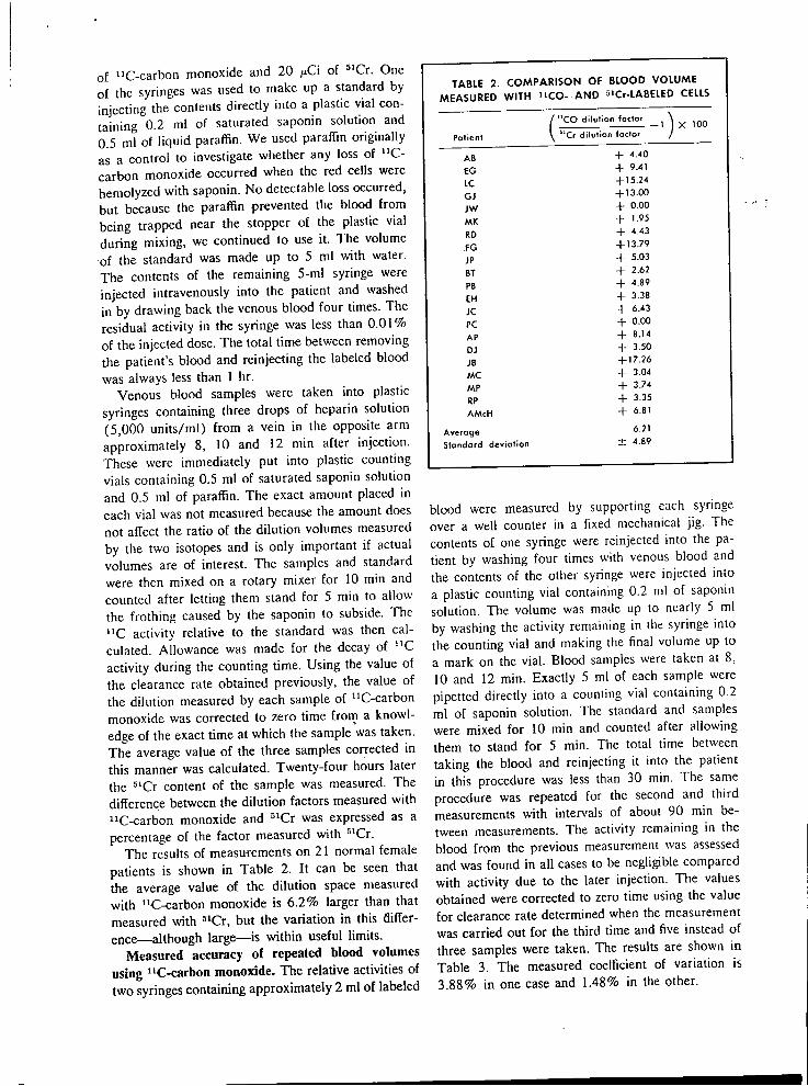
of "C-carbon monoxide and 20 ^iCi of ' 'Cr . One of the syringes was used to make up a standard by injecting the contents directly into a plastic vial containing 0.2 ml of saturated saponin solution and 0.5 ml of liquid paraffin. We used paraffin originally as a control to investigate whether any loss of " C -carbon monoxide occurred when the red cells were hemolyzed with saponin. No detectable loss occurred, but because the paraflSn prevented the blood f rom being trapped near the stopper of the plastic vial during mixing, we continued to use i t . The volume of the standard was made up to 5 ml with water. The contents of the remaining 5-ml syringe were injected intravenously into the patient and washed in by drawing back the venous blood four times. The residual activity in the syringe was less than 0 . 0 1 % of the injected dose. The total time between removing the patient's blood and reinjecting the labeled blood was always less than 1 hr.
Venous blood samples were taken into plastic syringes containing three drops of heparin solution (5,000 uni ts /ml) f rom a vein in the opposite arm approximately 8, 10 and 12 min after injection. These were immediately put into plastic counting vials containing 0.5 ml of saturated saponin solution and 0.5 ml of paraffin. The exact amount placed in each vial was not measured because the amount does not affect the ratio of the dilution volumes measured by the two isotopes and is only important if actual volumes are of interest. The samples and standard were then mixed on a rotary mixer for 10 min and counted after letting them stand for 5 min to allow the frothing caused by the saponin to subside. The " C activity relative to the standard was then calculated. Allowance was made for the decay of " C activity during the counting time. Using the value of the clearance rate obtained previously, the value of the dilution measured by each sample of "C-carbon monoxide was corrected to zero time frorn a knowledge of the exact time at which the sample was taken. The average value of the three samples corrected in this manner was calculated. Twenty-four hours later the " C r content of the sample was measured. The difference between the dilution factors measured with "C-carbon monoxide and ^'Cr was expressed as a percentage of the factor measured with " C r .
The results of measurements on 21 normal female patients is shown in Table 2. I t can be seen that the average value of the dilution space measured with "C-carbon monoxide is 6.2% larger than that measured with ^'Cr, but the variation in this difference—although large—is within useful limits.
Measured accuracy of repeated blood volumes using "C-carbon monoxide. The relative activities of two syringes containing approximately 2 ml of labeled
TABLE 2 . C O M P A R I S O N O F BLOOD V O L U M E M E A S U R E D WITH " C O - A N D s i C r - L A B E L E D CELLS
/ "CO dilution factor
Patient \ "Cr dilution factor
AB - f 4.40
£G -f- 9.41
LC -1-15.24
GJ - f 13.00
JW + 0.00
MK + 1.95
RD -t- 4.43
FG -1-13.79
JP - f 5.03
BT - f 2.62
PB - f 4.89
EH - f 3.38
JC - f 6.43
PC - f 0.00
AP -1- 8.14
DJ -t- 3.50
JB - f 17.26
MC + 3.04
MP + 3.74
RP -1- 3.35
AMcH + 6.81
Average 6.21
Standard deviation ± 4.89
X 100
blood were measured by supporting each syringe over a well counter in a fixed mechanical j ig. The contents of one syringe were reinjected into the patient by washing four times with venous blood and the contents of the other syringe were injected into a plastic counting vial containing 0.2 ml of saponin solution. The volume was made up to nearly 5 ml by washing the activity remaining in the syringe into the counting via! and making the final volume up to a mark on the vial. Blood samples were taken at 8, 10 and 12 min. Exactly 5 ml of each sample were pipetted directly into a counting vial containing 0.2 ml of saponin solution. The standard and samples were mixed for 10 min and counted after allowing them to stand for 5 min. The total time between taking the blood and reinjecting it into the patient in this procedure was less than 30 min. The same procedure was repeated for the second and third measurements with intervals of about 90 min between measurements. The activity remaining in the blood f rom the previous measurement was assessed and was found in all cases to be negligible compared with activity due to the later injection. TTie values obtained were corrected to zero time using the value for clearance rate determined when the measurement was carried out for the third time and five instead of three samples were taken. The results are shown in Table 3. The measured coefficient of variation is 3.88% in one case and 1.48% in the other.
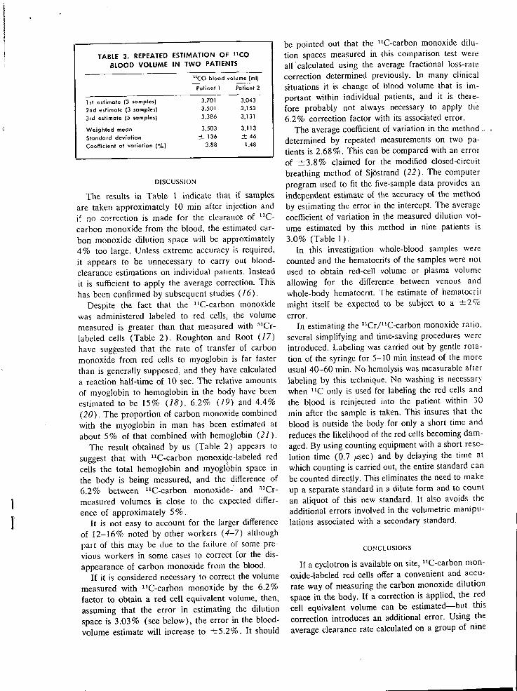
TABLE 3. REPEATED ESTIMATION OF n c o BLOOD VOLUME IN TWO PATIENTS
^'CO blood volume (ml)
Patient 1 Patient 2
1st estimate (3 samples) 3.701 3,043
2nd estimate (3 samples) 3,501 3,153
3rd estimate (5 samples) 3.386 3,131
Weighted mean 3,503 3,113
Standard deviation ± 136 ± 46 Coefficient of variation (%) 3.88 1.48
DISCUSSION
The results in Table 1 indicate that if samples are taken approximately 10 min after injection and if no correction is made for the clearance of " C -carbon monoxide f rom the blood, the estimated carbon monoxide dilution space wil l be approximately 4 % too large. Unless extreme accuracy is required, it appears to be unnecessary to carry out blood-clearance estimations on individual patients. Instead it is sufficient to apply the average correction. This has been confirmed by subsequent studies (16).
Despite the tact that the "C-carbon monoxide was administered labeled to red cells, the volume measured is greater than that measured with "''Cr-labeled cells (Table 2 ) . Roughton and Root (17) have suggested that the rate of transfer of carbon monoxide f rom red cells to myoglobin is far faster than is generally supposed, and they have calculated a reaction half-time of 10 sec. The relative amounts of myoglobin to hemoglobin in the body have been estimated to be 15% (18), 6.2% (19) and 4.4% (20). The proportion of carbon monoxide combined with the myoglobin in man has been estimated at about 5% of that combined with hemoglobin (21).
The result obtained by us (Table 2) appears to suggest that with "C-carbon monoxide-labeled red cells the total hemoglobin and myoglobin space in the body is being measured, and the difference of 6.2% between "C-carbon monoxide- and ^'Cr-measured volumes is close to the expected difference of approximately 5 % .
I t is not easy to account for the larger difference of 12-16% noted by other workers (4-7) although part of this may be due to the failure of some previous workers in some cases to correct for the disappearance of carbon monoxide f rom the blood.
I f i t is considered necessary to correct the volume measured with "C-carbon monoxide by the 6.2% factor lo obtain a red cell equivalent volume, then, assuming that the error in estimating the dilution space is 3.03% (see below), the error in the blood-volume estimate wi l l increase to ± 5 . 2 % . It should
be pointed out that the "C-carbon monoxide dilution spaces measured in this comparison test were all calculated using the average fractional loss-rate correction determined previously. In many clinical situations it is change of blood volume that is important within individual patients, and it is therefore probably not always necessary to apply the 6.2% correction factor with its associated error.
The average coefficient of variation in the method ,. determined by repeated measurements on two patients is 2.68%. This can be compared with an error of ± 3 . 8 % claimed for the modified closed-circuit breathing method of Sjostrand ( 2 2 ) . The computer program used to fit the five-sample data provides an independent estimate of the accuracy of the method by estimating the error in the intercept. The average coefficient of variation in the measured dilution volume estimated by this method in nine patients is 3.0% (Table 1 ) .
In this investigation whole-blood samples were counted and the hematocrits of the samples were not used to obtain red-cell volume or plasma volume allowing for the difference between venous and whole-body hematocrit. The estimate of hematocrit might itself be expected to be subject to a ± 2 % error.
In estimating the ' ' 'Cr/"C-carbon monoxide ratio, several simplifying and time-saving procedures were introduced. Labeling was carried out by gentle rotation of the syringe for 5-10 min instead of the more usual 40-60 min. No hemolysis was measurable after labeling by this technique. No washing is necessan.-when " C only is used for labeling the red cells and the blood is reinjected into the patient within 30 min after the sample is taken. This insures that the blood is outside the body for only a short time and reduces the likelihood of the red cells becoming damaged. By using counting equipment with a short resolution time (0.7 ;isec) and by delaying the time a! which counting is carried out, the entire standard can be counted directly. This eliminates the need to make up a separate standard in a dilute form and to count an aliquot of this new standard. It also avoids the additional errors involved in the volumetric manipulations associated with a secondary standard.
CONCLUSIONS
I f a cyclotron is available on site, "C-carbon monoxide-labeled red cells offer a convenient and accurate way of measuring the carbon monoxide dilution space in the body. I f a correction is applied, the red cell equivalent volume can be estimated—but this correction introduces an additional error. Using the average clearance rate calculated on a group of nine
normal female patients instead of measuring indi
vidual clearance rates does not appear to add sig
nificantly to the error. I t is probably desirable to
measure individual clearances if the greatest possible
accuracy is required. The blood volume can there
fore be estimated by taking two samples at known
times after injection, having allowed approximately
10 min for adequate mixing to take place. The accu
racy of the method determined by repeated estimates
in two patients is ± 2 . 7 % , which compares well with
the computer-derived estimate of ± 3 . 0 % based on
measurements on nine patients. For multiple studies
and in situations in which it is especially desirable
to use minimal radiation doses (such as in pregnancy
and in measurements on children), the "C-carbon
monoxide method is of particular value.
A C K N O W L E D G M E N T S
The authors wish to acknowledge the support and encouragement of J. F. Fowler, W. McGregor and D . D. Vonberg. They also wish to acknowledge the many helpful discussions and critical comments provided by Miss R . N . Arnot during this investigation and the kind cooperation and assistance of Peter Buckingham. This work was supported by a Clinical Research Grant f r o m Hammersmith Hospital.
R E F E R E N C E S
/ . HOPPER, J., JR., TABOR, H . AND W I N K L E R , A. W . :
Simultaneous measurement of the blood volume in man and dog by means of Evans blue dye, T-1824, and by means of carbon monoxide. I . Normal subiects. J. Clin. Invest. 2 3 : 628, 1944.
2. SJOSTRAND, T . : Method for the determination of total haemoglobin in the body. Ada Physiol. Scand. 16:211, 1948.
3. ROOT, W . S. A N D A L L E N , T . H . : Determination of
red cell volume with carbon monoxide. In Methods in Medical Research. Vo l . 8. Year Book Publishers, Chicago, 1960, p. 80.
4. H A L D A N E , J . AND S M I T H , J . L . : The mass and oxygen
capacity of the blood in man. / . Physiol. 25:331,-, 1899. 5. NiCKERSEN, J. L , SHARPE, L . M . , ROOT, W . S., F L E M
ING, T. C. AND GREGERSEN, M . I . : Simultaneous blood volume determination in dogs with dye (T-1824), carbon monoxide and radioactive iron, "Fe. Federation Proc. 9 : 94, 1950.
6. NoMOFF, N . , HOPPER, J. JR., BROWN, E., SCOTT, K . AND
WENNESLAND, R . : Simultaneous determinations of the total volume of red cells by the use of carbon monoxide and chromium-51 in healthy and diseased human subjects. J. Clin. Invest. 33:1,382, 1954.
7. DE HEVESY, G . : Red corpuscle content of circulating blood determined by labelling the erythrocyte with radio-phosphorus. Ada Med. Scand. 116:561, 1944.
8. ROOT, W . S., A L L E N , T . H . AND GREGERSEN, M . I . :
Simultaneous determinations in splenectomised dogs of cell volume with CO and P-32 and plasma volume with T-1824. Am. J. Physiol. 175:233, 1953.
9. TOBIAS, C . A., LAWRENCE, J . H . , ROUCHTON, F . J. W . ,
ROOT, W . S. AND GREGERSEN, M . I . : The elimination of
carbon monoxide f rom the human body with reference to the possible conversion of CO to COi. Am. J. Physiol. 145: 253, 1945.
10. VONBERC, D . D . AND FOWLER, J . F . : The Cyclotron
Unit: Medical Research Council. Nature, 198:827, 1963. / / . C L A R K , J . C , GLASS, H . I . AND SILVESTER, D . J.:
[n-vitro labelling of red cells with carbon-11. In Proc. Second Intern. Conf. Prep. Storage Labelled Compounds. Euralom, Brussels, 1966.
12. BUCKINGHAM, P. D . AND FORSE, G . R . : The prepara
tion and processing of radioactive gases for clinical use. Intern. J. Appl. Radiation Isotopes 14:439, 1963.
13. H I N E , G . J . AND BROWNELL, G . L . : Radiation Do
simetry. Academic Press, New York, 1956. 14. MARQUARDT, D . W . : I B M Share Programme, No.
3094, 1964. 15. PACE, N . , STRAJMAN, E . AND W A L K E R , E . L . : Accel
eration of carbon monoxide elimination in man by high pressure oxygen. Science 111:652, 1950.
16. GLASS, H . I . , EDWARDS, R. H . T . , DE GARRETA, A. C.
AND CLARK, J . C. Blood volume and total haemoglobin in athletes measured with "CO labelled red cells. J. Appl. Physiol. In press.
17. RoUGHTON, F . J. W . AND RooT, W . S.; The fate of CO in the body during recovery f rom mild carbon monoxide poisoning in man. Am. J. Physiol. 145:239, 1945.
18. W H I P P L E , G . H . : The haemoglobin of striated muscle. Am. J. Physiol. 76:693, 1926.
19. B O T H W E L L , T . H . AND FINCH, C . A. : Iron Metabolism. J . A. Churchill Ltd. , London, 1962.
20. D R A B K I N , D . L . : Metabolism of hemin chromopro-teins. Physiol. Rev. 31:245, 1951.
21. K I L L I C K , E . M . : Carbon monoxide anoxaemia. Physiol. Rev. 20 :313 , 1940.
22. SJOSTRAND, T . : Volume and distribution of blood and their significance in regulating the circulation. Physiol. Rev. 33:202, 1953.
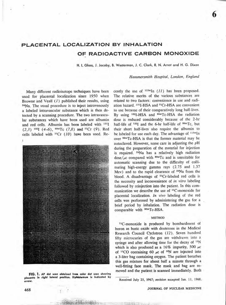
P L A C E N T A L L O C A L I Z A T I O N B Y I N H A L A T I O N
O F R A D I O A C T I V E C A R B O N M O N O X I D E
* H. 1. Glass, J. Jacoby, B. Westerman, J. C. Clark, R. N . Arnot a n d H. G. Dixon
Hammersmith Hospital, London, England
Many different radioisotope techniques have been used for placental localization since 1950 when Browne and Veall ( / ) published their results, using ^••Na. The usual procedure is to inject intravenously a labeled intravascular substance which is then detected by a scanning procedure. The two intravascular substances which have been used are albumin and red cells. Albumin has been labeled with (2.3) (4-6), •s-'Tc (7,8) and " C r ( 9 ) . Red cells labeled with ^'Cr (70) have been used. Re-
F I G . 1. AP dot scan obtained ffoaa color dot scon showing placenta In right lateral position. XipUsternun is indicated by arrow.
468 >u
cently the use of " ' " " I n (11) has been proposed. The relative merits of the various substances are related to two factors: convenience in use and radiation hazard. » " i - H S A and " C r - H S A are convenient to use because of their comparatively long half-lives. By using '^^I-HSA and »<""Tc-HSA the radiation dose is reduced considerably because of the 2-hr half-life of >32i and the 6-hr half-life of '^"'Tc, but their short half-lives also require the albumin to be labeled for use each day. The advantage of " ' ' " ' In over ^'"'Tc-HSA is that the former material may be autoclaved. However, some care in adjusting the pH during the preparation of the material for injection is required. ^^Na has a relatively high radiation dose//ic compared with ^ T c and is unsuitable for automatic scanning due to the difficulty of coUi-mating high-energy gamma rays (2.75 and 1.37 Mev) and to the rapid clearance of ^^Na f rom the blood. A disadvantage of '^Cr-labeled red cells is the necessity and inconvenience of in vitro labeling followed by reinjection into the patient. In this communication we describe the use of "C-monoxide for placental localization. In vivo labeling of the red cells was performed by administering the gas for a brief period by inhalation. The radiation dose is comparable with ' '-"Tc-HSA.
M E T H O D
"C-monoxide is produced by bombardment of boron as boric oxide with deuterons in the Medical Research Council Cyclotron (12). Seven hundred fifty microcuries of the gas are withdrawn into a syringe and after allowing time for the decay of ' ^N which is also produced as a 16% impurity, 500 /ic of " C O containing 60 /ic of ' ' N are injected into a 3-liter bag containing oxygen. The patient breathes this gas mixture for about half a minute through a well-fitting face mask. The mask and bag are removed and the patient is scanned immediately. Both
Received July 25, W e ? ; revision accepted Jan. 1 1 , 1968.
J O U R N A L O F N U C L E A R M E D I C I N E
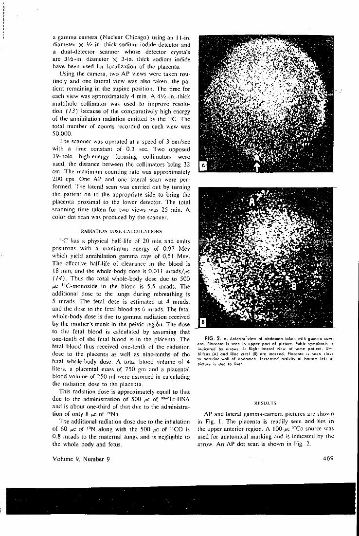
a gamma camera (Nuclear Chicago) using an 11-in. diameter X ' / i - i n . thick sodium iodide detector and a dual-detecior scanner whose detector crystals are 3 ' /2 - in . diameter X 3-in. thick sodium iodide have been used for localization of the placenta.
Using the camera, two A P views were taken routinely and one lateral view was also taken, the patient remaining in the supine position. The time for each view was approximately 4 min. A 4'/z-in.-thick multihole collimator was used to improve resolution {13) because of the comparatively high energy of the annihilation radiation emitted by the " C . The total number of counts recorded on each view was 50,000.
The scanner was operated at a speed of 3 cm/sec with a time constant of 0.3 sec. Two opposed 19-hole high-energy focusing collimators were used, the distance between the collimators being 32 cm. The maximum counting rate was approximately 200 cps. One AP and one lateral scan were performed. The lateral scan was carried out by turning the patient on to the appropriate side to bring the placenta proximal to the lower detector. The total scanning time taken for two views was 25 min. A color dot scan was produced by the scanner.
R A D I A T I O N DOSE C A L C U L A T I O N S
" C has a physical half-life of 20 min and emits positrons with a maximum energy of 0.97 Mev which yield annihilation gamma rays of 0.51 Mev. The effective half-life of clearance in the blood is 18 min, and the whole-body dose is 0.011 mrads//tc {14). Thus the total whole-body dose due to 500 ;iiC "C-monoxide in the blood is 5.5 mrads. The additional dose to the lungs during rebreathing is 5 mrads. The fetal dose is estimated at 4 mrads, and the dose to the fetal blood as 6 mrads. The fetal whole-body dose is due to gamma radiation received by the mother's trunk in the pelvic region. The dose to the fetal blood is calculated by assuming that one-tenth of the fetal blood is in the placenta. The fetal blood thus received one-tenth of the radiation dose to the placenta as well as nine-tenths of the fetal whole-body dose. A total blood volume of 4 liters, a placental mass of 750 gm and a placental blood volume of 250 ml were assumed in calculating the radiation dose to the placenta.
This radiation dose is approximately equal to that due to the administration of 500 /xc of '^-^Tc-HSA and is about one-third of that due to the administration of only 8 ^c of -^Na.
The additional radiation dose due to the inhalation of 60 fjLC of ' ^N along with the 500 ^LC of " C O is 0.8 mrads to the maternal lungs and is negligible to the whole body and fetus.
FIG. 2 . A: Anterior view o l abdomen token wltti gommo com-ero. Plocento is seen in upper port of picture. Pubic symptiosis is indicated by arrows. B: Right lateral view of same patient. U.— -bilicus (A) and iliac crest (B) are marked. Placenta is seen close to anterior wall of abdomen. Increased activity at bottom left cf picture is due to liver.
R E S U L T S
AP and lateral gamma-camera pictures are shovsn in Fig. 1. The placenta is readily seen and lies in the upper anterior region. A 100-/iC "Co source was used for anatomical marking and is indicated by the arrow. A n AP dot scan is shown in Fig. 2.
Volume 9, Number 9 469
GLASS, J A C O B Y , W E S T E R M A N , C L A R K , A R N O T A N D D I X O N
Successful gamma-camera pictures and scans with " C O have now been obtained in 50 patients. The technique, involving a low radiation dose, is simple and is performed both as an in-patient and outpatient procedure. Placentas, whether si.ted anteriorly, posterioriy or laterally were all readily detected by either the camera or the scanner. A n advantage of the scanner is that the size of the picture obtained is the same as the area scanned which makes the identification of the placental position more accurate. Lateral views of the abdomen are more easily obtained with the gamma camera because they do not involve changing the position of the patient. However, both methods are very satisfactory, provide the clinician with a permanent and easily interpretable record and are of value in the diagnosis of placenta previa in cases of antepartum hemorrhage. A significant advantage of this technique over the ^^""Tc-labeled albumin method is the lack of accumulation of activity in the bladder which may confuse the diagnosis of placenta previa.
CONCLUSIONS
With the availability of small isotope-producing cyclotrons, the use of "C-monoxide for the localization of the placenta may well have wide application. The combination of low radiation dose, complete absence of handling and labeling problems and the atraumic administration by 30-sec inhalation makes it an attractive alternative to other substances previously used for placental localization.
ACKNOWLEDGMENTS
The authors wish to acknowledge the support and encouragement of J. F . Fowler, J. C. McClure Browne and D. D. Vonberg. They also wish to thank Peter Buckingham, Miss G. Thompson and Miss M . Gallagher for their assistance and other clinical colleagues for their kind cooperation. This work is supported by a grant f rom the Medical Research Council.
R E F E R E N C E S
/ . BROWNE, J . C . M . AND V E A L L , N . : A method of lo
cating the placenta in the intact human uterus by means of radioactive sodium. J. Ohsici. Gynaecol. Brit. Commonwealth 57:566, 1950.
2. WEINBERG, A., SHAPIRO, C . AND BRUHN, D . F . : ISO-
topic placentography—an evaluation of ils accuracy and safety. Am. J. Obstet. Gynecol. 87:203, 1963.
3. MERCHANT, R . J. AND MORAN, J . P.: Routine use of
isotope localization of ihe placenta using " ' 1 . Obstet. Gynecol. 23:72, 1964.
4. HiBBARD, B . M . : Placental localization using RISA. ]. Obstel. Gynaecol. Brit. Commonwealth 68:481, 1961.
5. POOL, K . R., LEACH, K . G . AND H A W K I N S . L . A. :
Placental localization using ''"I-labelied human serum albumin. J. Obstet. Gynaecol. Brit. Commonwealth 72:706, 1965.
6. HiBBARD, B . M . : Placental localization using radioisotopes. Clin. Obsiei. Gynecol. 9:93, 1966.
7. LARSON, S. M . AND N E L P . W . B . : Visualization of Ihe
placenta by radioisotope photo-scanning using """Tc-albu-min. Am. J. Ohstcr. Gynecol. 93:960. 1965.
8. M C A F E E , J . G . , STERN, H . S., FUEGER, G . F . , BAC-
CISH, M . S., H O L T Z M A N , G . B . AND Z O L L E , I . : '"""Tc-albumin
for scintillation scanning of the placenta. J. Niicl. Med., 5:936, 1964.
9. JOHNSON, P. M . . SCIARRA, J . J. AND STICKLEV. E . E . :
A new radiopharmaceutical for placentography. Radiology 83:346, 1964.
10. VRETTOS. a . S., MEOAPANOS, E., COSTAMIS AND
BINOPOULOS, D . : Isotopic placentography using '•'Cr lagged erythrocytes. Am. J. Obslei. Gynecol. 93:957, 1965.
/ / . STERN, H . S., GOODWIN, D . A. , SCHEFFEL. U . AND
WAGNER, H . N . : " " " I n for blood-pool and brain scanning. Nucleonics 25 :No. 2, 62, 1967.
12. CLARK, J. C , GLASS, H . I . AND SILVESTER. D . J.:
In-vivo labelling of red cells: Proceedings of the Second International Conjerence on Preparing and Storing Labelled Compounds. Euratom, Brussels. 1966.
13. WESTERMAN, B . AND GLASS, H . L : The specification
and performance of an 11-in. gamma camera. J. Nticl. Med. 9:24, 1968.
14. GLASS, H . I . , BRANT, A., C L A R K , J . C , DE GARRETA,
A. C. AND D A V , L . G . : Measurement of blood volume with radioactive carbon monoxide labelled red cells. J. Nucl. Med. In press.
470 J O U R N A L O F N U C L E A R M E D I C I N E
'*CO red cell labeling for blood volume % and total hemodobin in athletes: effect of training
H . I . G L A S S . R. H . T . r ^ D W A R D S . . \ C. DP. C ^ A K R t ' l A.
A N D J . C. C L A R K
Drfiarlincttts of Medical Physics and Mcduinc. and Mrdual Rrscarcft
Council Cyclotron Unit, Royal Postgroduatr Medical .School,
Haininrrsnitlh Hospital, London, lin^laml
G L A S S . H . L , R . H . T . E D W A R D S , A . C . D E G A R K K T A . A N D
J . C . C L A R K . " C O red cell labelint^ for blood volume and total
hemoQlohin in athletes: eject of training. J . AppL Physiol. 2()
( I ) : 131-134. 1969.—Blood volume and lo ia l hemoglobin u c r c measured in 10 male racing cyclisis w i t h a " C O red cell labeling technique. Venous blood (5 m l ) was equilibrated w i d i " C O and subsequcndy reinjocicd. T h e dis t r ibut ion volinne was estimated for each of five, t imed blood samples, and an empir ica l ly selected single exponential func t ion fitted to tlic data and the instantaneous d i lu t i on volume for "CO- labe t ed red cells were calculated. T h e average blood volume of the group was 82.6 rfc 4.2 m l / k g and the average total hemoglobin 10,6 ± 0.7 g / k g . T h e average coefficient of variation in ihe est imation of the " C O d i lu t ion space was ± 3 . 4 % . ns t imai ion f r o m a single l imed sample instead of five samples did not signiticandy increase the error. The error in blood volume was estimated to be ± 5 . 2 % and ± 6 . 2 % for total hemoglobin. I n five subjects after a 2-month intensification of t ra in ing i c M i h i n i ; in improved w o r k i n g capacity there was no si i inif i -cant change in " C O space, total l iemoglobin, or blood volume. T l i i s has been a t t r ibu ted lo ihe high degree of physical funcss of the subjects at the start of the study.
T O T A L H E M O G L O B I N and blood volume are important determinants of cardiorespiratory' fitness ( I I , 12). Most mcastire-ments of b lood volume and total hemoglobin in athletes have been made w i t h Sjoslrand's rcbreathing technique w i t h stable carbon monoxide (14, 19, 20). A method has recently been developed by us in wh ich red cells labeled w i t h radioactive carbon monoxide are reinjected intravenously and the d i l u t ion volume is estimated f r o m subsequent t imed venous blood samples. T h e exposure to radioactivityV is considerably less than the rad ioac t iv i ty dose incurred w i t h other radioactive isotopic methods, making i t suitable fo r studies on normal subjects, especially when repeated measurements are required. I n this paper the technique has been used to measure blood volume and tota l hemoglobin in 10 racing cyclists. I n 5 of these subjects, the measurements were repealed after an i n tensive 2 -mon ih t ra in ing program.
The subjects were 10 male athletes, average age 20.6 years. They were studied in the course o f a t r a in ing program, and
measurements were repealed on 5 ol i l iem fo l lowing a 2 -mondi intensification ol the irainin*^ regime. T l i e physical w o r k i n g capacity at a heart raic of 1 7 ( ) / [ n i n ( P W ' C I T O ) was measiued w i t h cycle crgometcr c.veicise by ihe method described bv Holmgren (14). The average l*VV'Ci:„ of die group ai the start of the study was 1,438 k p i n / m i n .
C a r b o n - I I monoxide-labeied carbon monoxide wa.v produced in the Medical Research C^onneil Cyclotron by bombard ing a boric oxide target w i i l i deuieions (4), Five mil l i l i ters ol venous blood were w i i h d i a w n into a 50-ml syringe con ia in in i j 1.5 m l of acid citrate dexirose aniicoaqulani ( . \ C I ) ) . T h e n 45 ml of hel ium containing 1 % carbon-1 1-labeled carbon monoxide were d r awn into ihe .syringe, which v -as ihen rotated a i r p m for 10 min . Because of ihe l i i i ;h radioactivi ty per m i l l i l i t e i o f blood achieved by this method (75 /^c /ml) . there \v'a> adequate time for preparation of standards and doses despite i l ie 20-min half-l ife o f c a t b o n - I I . The amount o f c a r b o n - / i -labcled carbon monoxide contained in the plasma was measured, and this was found tu be less t l ian 0.01 % (6) . I t was n c M . therelore, necessary to wash the plasma f rom the red ccll.s a tu r labeling.
T w o aliquois were taken, each ol approximately 2 ml , in to two syringes, one of which was used as a standard. Af t e r the count rates of the two syringes were measured, the contents of the one syringe were reinjected into the subject. The syrinec was flushed by d rawing back the venous blood four times The residual act ivi ty in the syringe after such a procedure was found to be less t l ian 0 . 0 1 % of the injected dose (5). Five blood samples were wi thd rawn between 9 and 45 m i n af ic r the inject ion of the labeled red cells. The blood was lysed w i t h saponin and the activity in 5 ml of lyscd whole blood was assayed in a well scinti l lat ion counter. T h e measured ac t iv i ty was counted for a fixed period of 100 sec and was related to the injected ac t iv i ty by comparison w i t h the total contents o f the standard syringe counted under identical conditions. T h e activity in each sample was corrected for decay u p to and d u r ing the count ing periods. This was facilitated by use of a f ixed counting t ime. T h e whole-body dose due to the inject ion o f 30 ^ic of "CO-!abeled cells is estimated to be 0.33 mrad.
The d i l u t i o n volume was estimated for each of the five samples and an empir ica l ly selected single exponential f unc t i on was fitted to the data by a least-squares procedure w i t h the computer p rogram devised by M a r q u a r d i (15). The p roe ram also calculates the errors in the estimated parameters due to scatter o f the data.
Hemoglobin concentration was estimated f r o m dupl icate
131
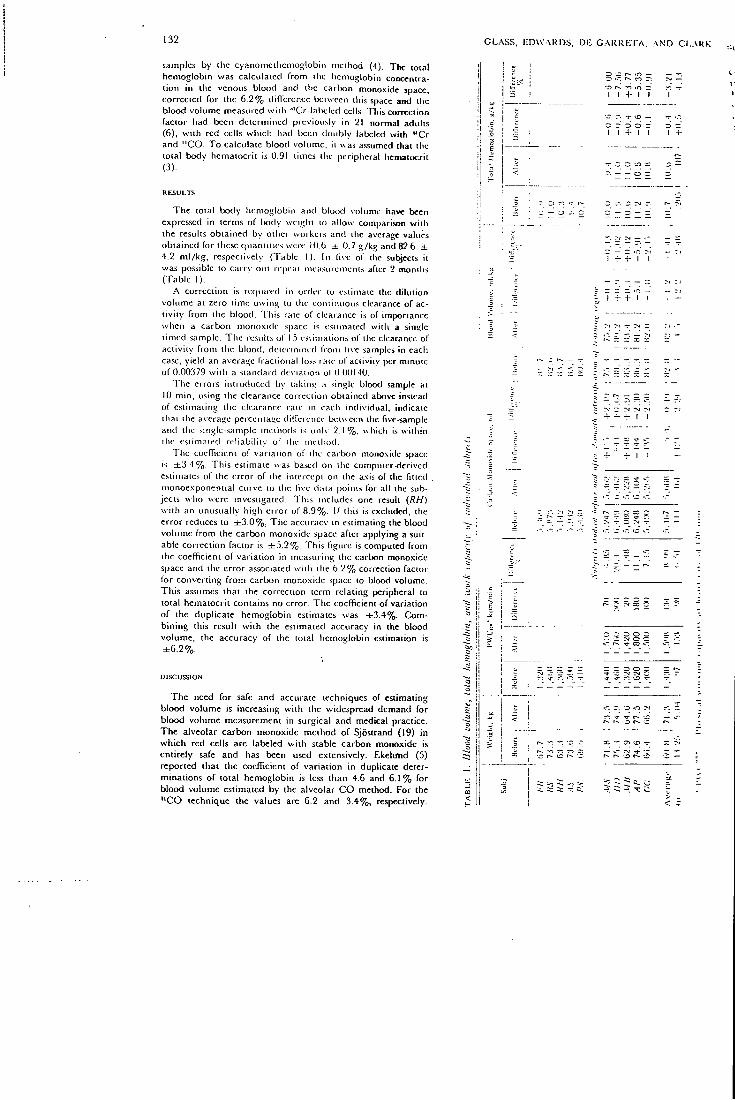
132 GLASS, EDWARDS, DE C A R R E r A , A N D CI-.VKK
samples by i l ic cyanomct l icmoglobin method (4). T l i c total hemoglobin was calculated f rom i l ic hemoglobin concentrat ion in the venous blood and ihc carbon monoxide space, conec icd for the 6 .2% difference beiwccn (his space and ilic blood volume measured w i t h ^ 'Cr- labt lcd cells This correction facior had been determined previously in 21 normal adults (6) , w i t h red cells which had been doubly labeled wi th ' ' C r and " C O . T o calculate blood volume, it was assumed ihat the total body hematocri t is 0.91 times the peripheral hematocrit (3) .
T h e loial body hemoglobin and blood volume have been expressed in terms o f body weight (o allow comparison w i t h the results obtained by other workeis and die average values obtained for these quantit ies were 10.(j ± 0.7 g /kg and 82.6 ± 4.2 m l / k g , respectively {Tabic 1). I n live ol" the subjccis it was possible to carry out repeat mea.-inicnicnis after 2 months (Table 1).
A correction is required in order to estimate ilie d i lu t ion volume at zero time o w i n ^ to the to tu inuous clearance of act iv i ty f r o m the blood. This rate of clearance is of imporiancc when a carbon monoxide space is estimated wi th a single t imed sample. T h e results of 15 estimations of the clearance of act ivi ty f r o m the blood, determined f r o m five samples in each case, yield an average fract ional lo.ss rate of aci ivl iy per minute of 0.00379 w i t h a standard deviat ion of U.OUI-iO,
T h e errors int roduced by l akmn a sin*;le blood sample at 10 m i n , using the clearance correctloti obtained above instead of estimating the clearance rate in each individual , indicate that the average pcrcentatic difference Uuween the five-sample and the single-sample methods i.s only 2 . 1 % . which is w i th in the estimated re l iabi l i ty of the incd iod .
The coefficient of var iat ion of the carbon monoxide space is ± 3 , 4 % . T h i s estimate was based on the computer-derived estimates of the error of tiie intercept on the axis of the fitted monoexponeniial curve to ilu- live data points for all the subjects who were investigated. This includes one result {RH) w i t h an unusually high error of 8.9%. I f tfiis is excluded, the error reduces to ± 3 . 0 % . T l i c acctuacy in estimating the blood volume f r o m the carbon monoxide space after applying a suitable correct ion factor is ± 5 , 2 % . T h i s figure is computed f r o m the coefficient of var ia t ion in measuring the carbon monoxide space and the error associated w i t h the 6 .2% correction facior lor conver t ing f r o m carbon monoxide space to blood volume. Th i s assumes that the correction term relat ing peripheral to total hematocri t contains no error. The coefficient of variat ion of the duplicate hemoglobin estimates was ± 3 . 4 % . Comb in ing this result w i t h the estimated accuracy in the blood volume, the accuracy of the total hemoglobin estimation is ± 6 . 2 % .
DISCUSSION
T h e need for safe and accurate techniques of estimating b lood volume is increasing w i t h the widespread demand for blood volume measurement in surgical and medical practice. The alveolar carbon monoxide method of Sjosirand (19) in w h i c h red cells are labeled w i t h stable carbon monoxide is ent i rely safe and has been used extensively. Ekel imd (5) reported that the coefficient of var ia t ion in duplicate determinations of total hemoglobin is less than 4.6 and 6 . 1 % for b lood volume estimated by the alveolar C O method. For the " C O technique the values are 6.2 and 3.4%, respectively.
3 CO cr. -.D r-.' r i -ri
I I -i- I I
•r r. rr o — c: r 3 d ~ I 1 4 - 1 I
3 c ; — — O :
r I -r- -f- I I
I n T rvi ~
4- -r ^ ' l 'l
; -r 3 -r
. . - i r CO I
ca
r-. o o :
X r-. •£> -r
: 5 0, c
uCO L A B E L I C R B L O O D V O L U M E A N D H E M O G L O I U N
Grcgcrscn (7) , using an inhalat ion method, estimated that the carbon monoxide space exceeded the red cell space by 12-16%. A similar difference was observed by Root and his coworkers (17) in splencctomized dogs by intravenous injection of red cells labeled in vi t ro w i t h stable carbon monoxide. I n a previous study (9) w i t h red cells doub ly labeled w i t h " C O and ^'Cr, we have found the d i l u t i o n space measured w i t h "CO-labc led red cells to be on ly 6 .2% greater than the space measured w i t h ^'Cr-labtded red cells. Roughton and Root (18) have suggested that the rate o f transfer of carbon monoxide f r o m red cells to myoglobin is far faster than is generally .supposed and have calculated a hal f - t ime of reaction of 10 sec. The " C O d i l u t i o n space mca.siued by us has therefore been reduced by 6 .2% in our calculations of blood volume.
The question arises whether q u o i i n i ; the blood volume provides any more useful informat ion ihan the car lxm monoxide .space, which can be estimaied w i t h greater accuracy. When the total hemoglobin is to Ix' estimaied, an addit ional nK :asure-meni , ihe blood hemoglobin concenira i ion, is also required.
The average fract ional lo.ss rate of ac i iv i iy per minute was 0.00379 w i t h a standard deviat ion of 0.00140. This compares w i t h a value o f 0.0039 previously reported by Pace (16) in five women fo l lowing the inhala i ton o f stable carbon monoxide. Since apply ing a correction lor clearance h o m the blood allows the carbon monoxide space lo Ix- calctilaied f r o m one t imed blood .sample wi thout any signil icani loss of accmacy, this greatly enhances the scope of appl icat ion of die method. The sii\gle-sample method requires only 10 min for ihe estimation instead of 45 m i n required lor the stable carbon monoxide method of Sjoslrand, and only three venipunctures.
T l i e blood volumes of 677 males reported in die woi Id literature range f r o m 59.8 to 101.7, w i t h an average of 77.6 m l / k g (9). O u r average of 82.6 ± 4.2 m l / k g i s thus comparable to this and is close to the values 81.9 ± 8.64 m l / k g and 79 m l / k g in 10 (20) and 20 (12) cyclists measured by the alveolar carbon monoxide method of Sjostrand (19).
O u r values for total hemoglobin are sitnilarty comparable to iliose quoted i n the literature. Ho lmgren (12) found a value of 11.02 ± 1.02 g / k g in 19 male cyclisis of an age and physical w o r k i n g capacity similar to ours. Ekelund (5) quotes 10.4 ±
133
1.3 g/kg in 26 normal males. O u r value of 10.6 ± 0.7 g/kw is close to'Ekelund's, but the standard deviat ion o f the average value quoted is smaller. In the present study, no significant change in blood volume and total hemoglobin was observed
riie H c c u r a c v o f the i>i csem metho<( w o u l d be expccied t o demonstrate changes in hlmxl volume and total heino<<lobin of the. order of magnitude noied by oiher workers (13). The apparent absence of change greater than 2 . 9 % is probablv related to the higher ini t ial filne.ss (as reflected by the P W C : , : , , ! of our subjects compared wi th those of l lo lmgren . The percetu-age increase in P W ' C I T O o b s e r v e d in our subjects is comparable
w i t h values C j u o t e d by Holmgren and his co-vvorkers f l 3 i The subjects in the prcs<:nt study would seem t o be most comparable lo their group of skiers in whom con i inuou i u a in-ing of an endurance type led to a smaller increase in bloorl volume in relation t o improvemeni in P W ' C t : , . than iha i (ib-served in the group wi th training of shorter da i ly du ra t i t j u w i t h varying t e m p o up to exhaustion. In this study " G O ^pace blood volume, and total hemoglobin w e r e n o t si t inifieantU changed in five cyclists wi th a high ini t ia l physical wurk iny capacity as a result of a 2-montli intensification o( the t r : i u i i n i : program. I t is possible that these subjects v v e r e appio<ichmi: physiological maxima for " C O space, total hemoglobin, and blood volume when the first measurements weic made .A-sui^gested bv Holmgren {'3)-. die increase in p lusical vvoikim: capacity in the absence of a rorie%pondin'4 increase- m b l o c : volume mig lu be due to a more efiicient peri[ iheial e M T u l : : i ' i [ \ adaptation to muscular work, resulting in a larmier iu i l i / ; i i i . .u i blood oxygen.
The authors wish l o acknowledge ilic cncoiiiatteinrni <inG i-up-port of Profrss*)r J. F. Fowler and M r . D. D. Vonhert;. Drs K. I . I lnilhcs, E. .A. Oppcnhcinicr. K. Kn i l l Jones, and M r P Huc-'mv;-ham t;ave v<iliiabic assistance in the suidies. ; t n d advice ; i n G c n i i -cisin in i h c preparation of the nmnuscripi was eraiefnllv r cc i^oc from Drs. E. J . M . Campbell and N . L. Jones.
This work was supported by die Medical Research CUjiinci; .m-j also by a grant from ihe l lainmersmiih Hospital C-linical l<».-se.;rct. Fund.
Received for publication I I December l % 7 .
R E F E R E N C E S
1. .AsrRAND, P. O. Experirnenlal Studies oj P/iyncal it'ofkin« Capacity in Relation io Sex and Age. Copenhagen: .Munksgaard, 1952.
2. C H A P L I N , H . , P . L . M O L L I S O N , A N D I-l . V E r r e R . The body/ venous hematocrit rat io: its constancy o v e r a wide range. J . Clin. Invest. 32: 1309-1316, 1953.
3. C L A R K , J . C , H . I . GLASS, A N D D . J SILVESTKR. I n v i t ro labelling of red cells wi th c a r b o n - l l . I n : Proceedings of the Second International Conjerence on the Ptepfiration and Storage of Labelled Compounds. Brussels: Euratoni, 19G6.
4. D A C I E , J . v . , A N D S. M . L E W I S . Practical Hematology (3rd cd.). London: Churchil l , 1963, chapt. 2, p. 36.
5. E K E L U N D , L . G . Determination of blood volume. Scand. J. Clin. Lab. Invest. 17. Suppl. 86: 53-66, 196G,
6. GLASS, H . I . , A . BRANT, J . C. C L A R K , A . C . DE GARRETA, A N D L. G . D A V . The mcasurcfucnt of blood volume with radioactive carbon monoxide labelled red cells. J. i\'ucl. Med. 9: 571-575, 1968.
7. GRECERSEN, M . I . , AND R . A. RAWSON. Blood volume. Physiol. Rev. 39: 307-342, 1959.
8. G U L L B R I N G , B . , A . HOLMGREN, T . SJOSTRAND, A N D T . STRAN-D E L L . The effect of blood volume variations o n the pulse rate
i l l supme and upright positions and during exerc iS ' . - .4. Physiol. Scand. 50: 62-71, ly6U.
9. HOBBS, J . T . Total Blood Volume—Its .Measurement and St^ntn-cance. Amcrsham. Buckinghamshire. England: United K i n g dom Atomic Energy Authority, The Radiochemical C r r u r r 1967. Medical Monograph No. 3.
10. HOLMGREN, A . Circulatory changes during muscular work irt man. Scand. J. Clin. Lab. Invest. 8, Suppl. 24: 1-97. I ^ ^ .
11. HOLMGREN, A . Cardiorcspirator\- determinants of cardiovascular fitness. Can. Med. Assoc. J. 96: 967-702. 1 % :
12. HOLMGREN, A . , AND ASTRANO, P . - O . D L and the dimension; and functional capacities of the o.xygcn transport sv-stom ir. humans. J. Appl. Physiol. 2 1 : 1463-1470, 1966.
13. HOLMGREN, A . , F. MOSSFELDT, T . SJOSTRAND. A N D G . S T R O M Effect of training on work capacity, total hemoglobin, blooo volume, heart volume and pulse rate in upright and rccurrtbcn: positions. Acta Physiol. Scand. 50: 72-83, I9G0.
14. LAWSON, H . C . The volume of blood—a critical cxamioiit ion of method for its measurement. I n : Handbooi of PhyiutJogy Circulation. Washington, D . C . : Am. Physiol. See., 1962. s^ t vol. I , chapt. 3, p. 23^9 .
134
M A R Q U A R D T , D . \ V . IM.M. /'rogrammr. No. 3094, White Plains, N.Y., S H A R E organization, 1904. PACE, N . , E. STRAJMAN, ANI> E. L . W A L K K R . .Acceleration of carbon monoxide in man by hit;li pressure oxygen. Scirtue 111; G52-054. 19.50. R O O T , VV. S., T . 11. . \ I , I - I : N . AND . M . I . GRt-iaKRSRN. Simultaneous determinations in splenectomised dogs of cell voltiinc with C O and " P and plasma volume with T - l l l M . .4m. ./. Physiol. 17.1: 2:13-235, 1953.
C L A S S , E D W A R D S . D E C A R R E T A , . A N D C L A R K
18. RouciiTON, F . J . W., A N D VV. S. ROOT . The fate of C O in :Sc body during tiie recovery from mild carbon monoxide poisc.n-ing in man. Am. J. Physiol. 145: 239-252, 1945.
19. SjoSTRANO, T . A method for the determination of total hemoglobin content of the body. Acta Physiol. Scaitd. 10: 211-231, 1948.
20. SjoSTRANU. T . Blood volume. In: Handbook oj Physiology. CVrci/fo/(oii. Washington, D.C. : .Xin. Physiol. Soc, 1902, sect. 2. vol. I , chapt. 4. p. 51-62.
8 I n t e r n a t i o n a l J o u r n a l of Appl i ed R a d i a t i o n a iu! i io iopcs , 19G9, V ol. 20, pp. G77-G31. PerRamon Press. Pr imed in Northern Ire land
A Simplified Method for Simultaneous Electrolyte Studies in Man Utilizing
Potassium-43
F . S K R A B A L , H . I . G L A S S , J . G. C L A R K , K . J E Y A S I N G H and G . F . J O P L L N ' Departments of Medicine, Medical Physics and M . R . C . Cyclotron Unit, R.oyal Postgraduate
Medical School, Hamniersmith Hospital, London VV.12
{R^ceiiedS April 1969)
A method is presented which enables the exchangeable sodium, potassium and bromide space to be estimated entirely by gamma counting using sodium-24, potassium-43 and bromlne-82. The potassium-43 is a cyclotron produced isotope with a 22-hr half-life. The principal gamma radiation is at 370 keV. Duplicate cstinrations on nine patients indicate that the error in the estimation in exchangeable sodium, exchangeable potassium and bromide space is 4-6, 2-78 and 2-14 per cent respectively. By using a dual channel automatic gamma counter the assay may be completed in a single counting run. This technique permits a more w idespread application of these measurements in clinical research and diagnosis.
U N E M E T H O D E S I M P L I F I E E P O U R L E S E T U D E S S I M U L T . M s E E S D ' E L E C T R O L Y T E C H E Z L ' H O M M E UTILIS. -VNT L E POT. \SSIUM-43
On presente une methode qui pcrmet I'estimation du sodium, du potassium et du bromurc echangeables entierement par comptage de gamma en employant le sodium-24, le potassium-43 et le brome-82. Le potassium-43 est un isotope produit du cyclotron ayant une demi-p6riode de 22 heures. Le principal rayonnement gamma est a 370 keV. Des mesures en duplication sur neuf sujets indiquent que les erreurs d'estimation de sodium echangeable, de potassium echangeablc et d'espace de bromme serait de 4,6, 2,78 et 2,14 pour cent. E n employant un compteur gamma automatique a'deux canaux on peut achever le dosage en une seule s^rie dc comptes. Cette methode permet une application plus repandue de ces mesures dans la recherche clinique et dans la diagnostique.
y n P O m E H I - I b l t t M E T O f l O f l H O B P E M E H H b l X 3JIEKTP0J11'IT1'IHECKHX H C C J I E f l O B A H H H B MEJIOBEKE C n P M M E H E H M E M K A J I H f l "
n p H B O H M T C n MBTOA, R a i O m H f i B 0 3 . M 0 H { H 0 C T b nOJIHOrO O n p e a e J i e H l I H O O M e i l H U X i i a T p n H ,
K a j i H H H 6 p o M a n o c p e a c T B O M H S M e p e i i i i H raMMa-aKTiiBiiocTii, npi iMei iHH HaTpml", K a J i i i f i ' " H GpoM' '. KajiHil*^ nDjincTCH H30TonOM c nepHOflOM noJiypacna.-ia B 22 Maca, nojiyMeHHUM 0 noMOmbio miKJioTpoiia; ocnoBHLie raMMa-JiyMH = 370 KiiJio3jieKTponBo.TbT.
flySjiHpoBaHHue o n p e K e n e H M H i i a neBHTi i na t tv i enxax n o K a s H B a i o T , MTO n o r p e m n o c T H o n p e A e j i e n H H oOMenHoro npocTpancTBa AJIH iiaTpiin, KaJiiifi M OpoMa paBHw 4,6%, 2,78% H 2,14% c o o T B e T C T B e H H O . i T p H M e n n e aBTOMaTHMecKiifi raMMa-c'iCTMHK c 2-MH KanajiaMii, MOKHO npoH3BonnTb nojiftoe onpe«GJieHne B OAHH CHeTHbifl nep i iOA. 3TOT Mexofl naer Boa-M o w n o c T b u i H p o K o r o n p H M e n e H d H T a n i i x HaMepeHiiK B K j i n H i m e c K i i x i i c c J i e A O B a n H H x H HHariiocTHKe.
677
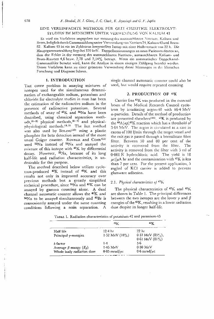
678 F. Skmbal, H. I. Glass, J. C. Clark, K. Jcyasingh ami G. F. JojAin
E I N E V E R E I N F A C H T E M E T H O D S F U R G L E I O H Z E I T I G E ] : L i : K r R O L Y T -S T U D I E N I M M E N S G H E N U N T E R V E R W E N D U X G V O N K.M..IUM 43
Es wird eia Vcrfahren angegcben zur mcssung tdcs austauschbarcn Xnu iuni. Kalium unci Brom,lcdiglichdurchGammazahIunguntcr Verwendung von Natrium 24. Kaiiuin43und Bi om-82. Kaliuin 43 ist cin im Zyklotron hergestelltes Isotop mit einer Halbwertzeit von 22 h. 13ie Hauptgammastralilung liegt bei 370 keV. Doppelbestimmungen an neun Patienten deuten an, dass der Fehlcr in der messung des austauschbaren Natrium-, austauschbaren Kalium- und Brom-Raumes 4,6 bezw. 2,78 und 2,14% betragt. Wenn cin automatischer Doppclkanal-Gammazahlcr benutzt wird, kann die Analyse in einem cinzigen Zahlgang becndet wcrden. Dieses Verfahren kann zu eincr grosseren Verwendung dicscr Mcssungcn in der klinischcn Forschung und Diagnose fuhren.
1. I N T R O D U C T I O N T H E CHIEF problem in assaying mixtures of isotopes used for the simultaneous determination of exchangeable sodium, potassium and chloride for electrolyte studies in man has been the estimation of the radioactive sodium in the presence of radioactive potassium. Several methods of assay of and ^*Na have been described, using chemical separation metho d s , p h y s i c a l methods,'*"'" and physical-physiological methods.'^'^^ The last method was also used by BOLING'^" ' using a plastic phosphor for beta detection instead of the more usual Geiger counter. R O V N E R and C O N N ' ^ I ' used 22Na instead of ^ N a and assayed the mixture of this isotope with by differential decay. However, ^^fyTg^^ because of its long half-life and radiation characteristics, is undesirable for this purpose.
The method described below utilizes cyclotron-produced instead of and this results not only in improved accuracy over previous methods but a greatly simplified technical procedure, since ^*Na and ^^K can be assayed by gamma counting alone. A dual channel automatic counter allows the ^^K and ^ N a to be assayed simultaneously and ^^Bj - is conveniently assayed under the same counting conditions following a resin separation. A
single channel automatic counter could also be used, but would require repeated counting.
2. P R O D U C T I O N OF " K
Carrier free ^•'K was produced in the external beam of the Medical Research Council cyclotron by irradiating argon-40 with 16-4 MeV a-particles. Details of the method of production are presented elsewhere"^'. ''•'K is produced by the *''A(a//)*2K reaction which has a threshold of 3-64 MeV. The argon is circulated at a rate in excess of 100 1/min through the target vessel and the exit gas is passed through a borosilicate fibre filter. Between 50 and 80 per cent of the activity is recovered from the filter. The activity is removed from the filter with 5 ml of 0-001 N hydrochloric acid. The yield is 18 jucj/xA hr and the contamination with ^-K is less than 7 per cent. For the present application, 5 mg/ml of K C l carrier is added to prevent glassware adhesion.
2.1. Physical characteristics o/^-'K
The physical characteristics of and ^•'K are shown in Table 1. The principal differences between the two isotopes are the lower y and fi energies of the *^K, resulting in a lower radiation dose despite its longer half-life.
T A B L E 1. Radiation characteristics of potassium-42 and potassium-43
« K « K
Half life 12-4 hr 22 hr Principal y-energies 1-52 MeV (18%) 0-37 MeV (85%),
0-61 MeV (81%) A:-factor 1-4 5-6 Average fi energy (Ep) 1-45 MeV 0-30 MeV Whole body radiation dose 0-85 mrad//(c 0-6 mrad//ic
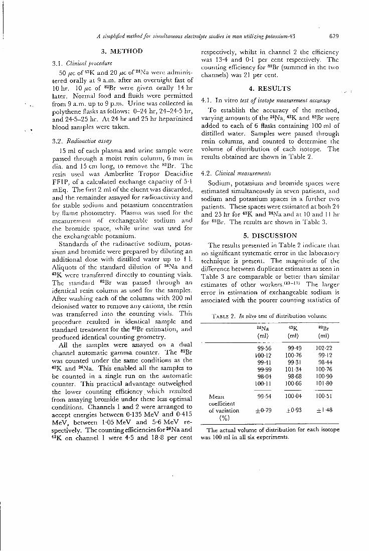
A simplified melliod for simultaneous electrolyte studies in man utilizing potassiumA'i
3. M E T H O D
3.1. Cliriical procedure
50 fxc of and 20 p.c of ^ iN'a were adminis
679
tered orally at 9 a.m. after an overnight fast of 10 hr. 10 j U C of ^^Br were given orally 14 hr later. Normal food and fluids were permitted from 9 a.m. up to 9 p.m. Urine was collected in polythene flasks as follows: 0-24 hr, 24-24-5 hr, and 24-5-25 hr. A t 24 hr and 25 hr heparinised blood samples were taken.
3.2. Radioactive assay
15 m l of each plasma and urine sample were passed through a moist resin column, 6 mm in dia. and 15 cm long, to remove the ^^Br. The resin used was Amberlite Tropor Deacidite FFIP, of a calculated exchange capacity of 5 1 mEq. The first 2 m l of the eluent was discarded, and the remainder assayed for radioactivity and for stable sodium and potassium concentration by flame photometry. Plasma was used for the measurement of exchangeable sodium and the bromide space, while urine was used for the exchangeable potassium.
Standards of the radioactive sodium, potassium and bromide were prepared by diluting an additional dose with distilled water up to 1 1. Aliquots of the standard dilution of ^^Na and
were transferred directly to counting vials. The standard *^Br was passed through an identical resin column as used for the samples. After washing each of the columns wi th 200 ml deionised water to remove any cations, the resin was transferred into the counting vials. This procedure resulted in identical sample and standard treatment for the ^^Br estinnation, and produced identical counting geometry.
A l l the samples were assayed on a dual channel automatic gamma counter. The ^^^r was counted under the same conditions as the *3K and ^^Na. This enabled all the samples to be counted in a single run on the automatic counter. This practical advantage outweighed the lower counting efficiency which resulted from assaying bromide under these less optimal conditions. Channels I and 2 were arranged to accept energies between 0-135 M e V and 0-415 MeV, between 1-05 M e V and 5-6 M e V respectively. The counting efficiencies for ^Na and ^^K on channel 1 were 4-5 and 18-8 per cent
respectively, whilst in channel 2 the eflicicncy was 13-4 and 0-1 per cent respectively. The counting efficiency for ^^Br (summed in the two channels) was 21 per cent.
4 . RESULTS
4.1. I n vitro test of isotope measurement accuracy
To establish the accuracy of the method, varying amounts of the 2*Na, and ^^Br were added to each of 6 flasks containing 100 inl of distilled water. Samples were passed through resin columns, and counted to determine the volume of distribution of each isotope. The results obtained are shown in Table 2.
4.2. Clinical measurements
Sodium, potassium and bromide spaces were estimated simultaneously in seven patients, and sodium and potassium spaces in a further two patients. These spaces were estiinated at both 24 and 25 hr for and ^^Na and at 10 and 11 hr for ^^Br. The results are shown in Table 3.
5. DISCUSSION The results presented in Table 2 indicate that
no significant systematic error in the laboratory technique is present. The magnitude of the difference between duphcate estimates as seen in Table 3 are comparable or better than similar estimates of other w o r k e r s . T l i e larger error in estimation of exchangeable sodium is associated with the poorer counting statistics of
T A B L E 2. In vitro test of distribution volume
24Na 43K. s^Br (ml) (ml) (ml)
99-56 99-49 102-22 10012 100-76 99-12 99-41 99-31 98-44 99-99 101-34 100-76 9804 98-68 100-90
10011 100-66 101-80
Mean 99-54 100-04 100-51 coefficient of variation ±0-79 ±0-93 ±1-48
(%)
The actual volume of distribution for each isotope was 100 ml in all six experiments.
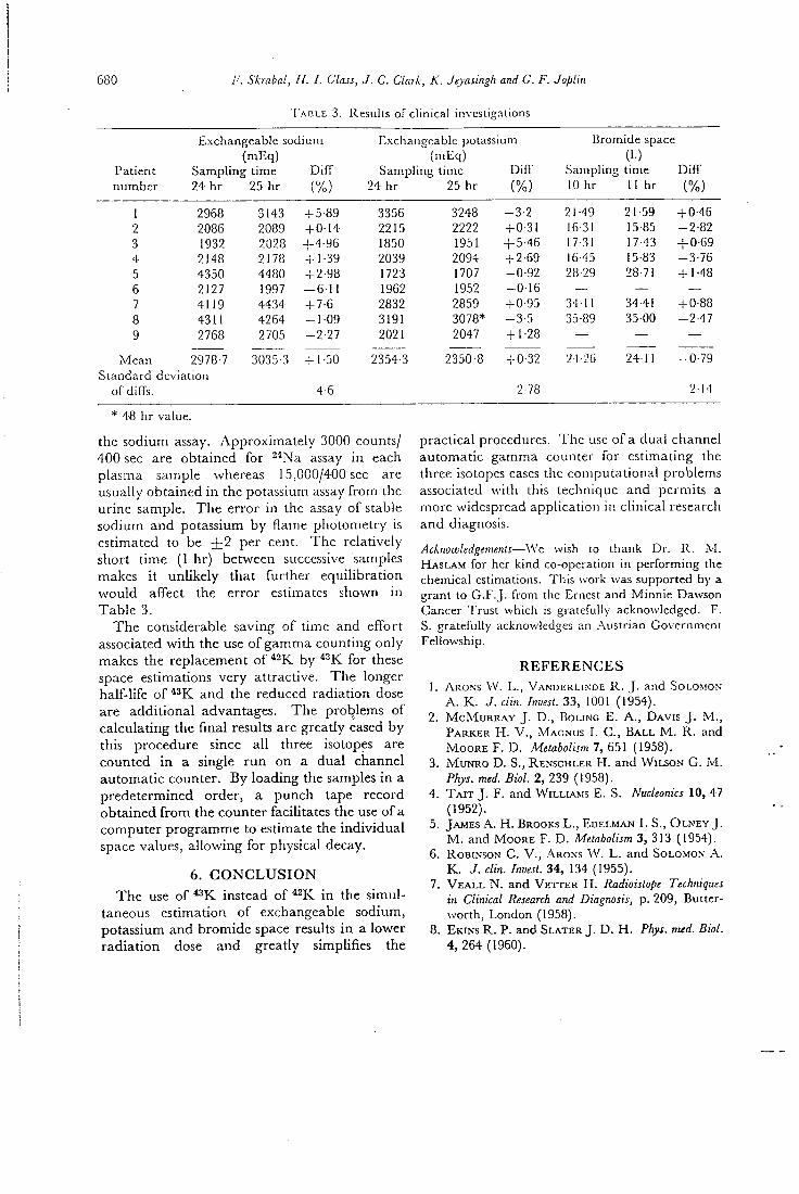
680 Skrabal, H. I. Glass, J. C. Clark, K. Jeyasingh and G. F. Joplin
T A B L E 3. Results of clinical investigations
Exchangeable s.oc iuin Exchangeable potassium Bromide space (mEq) (mEq) (I.)
Patient Sampling time Diff Sampling time Difl' Sampling time Diff number 24 hr 25 hr (%) 24 hr 25 hr (%) 10 hr 11 hr (%)
1 2968 3143 -f 5-89 3356 3248 -3-2 21-49 21-59 + 0-45 2 2086 2089 -fO-14 2215 2222 -fO-31 16-31 15-85 -2-82 3 1932 2028 +4-96 1850 1951 + 5-46 17-31 17-43 + 0-69 4 2148 2178 -rl-39 2039 2094 - -2-69 16-45 15-83 -3-76 5 4350 4480 -i-2-98 1723 1707 -0-92 28-29 28-71 + 1-48 6 2127 1997 -6-11 1962 1952 -0-16 — — — 7 4119 4434 2832 2859 -fO-95 34-11 34-41 + 0-88 8 4311 4264 -1-09 3191 3078* -3-5 35-89 35-00 -2-47 9 2768 2705 -2-27 2021 2047 -H-28 — — —
Mean 2978-7 3035-3 + 1-50 2354-3 2350-8 + 0-32 24-26 24-11 -0-79 Standard dcvia ion
ofdlfTs. 4-6 2-78 2-14
* 48 hr value.
the sodium assay. Approximately 3000 counts/ 400 sec are obtained for ^''Na assay in each plasma sarnple whereas 15,000/400 sec are usually obtained in the potassium assay from the urine sample. The error i n the assay of stable sodium and potassium by flame photometry is estimated to be + 2 per cent. The relatively short time (1 hr) between successive samples makes i t unlikely that further equilibration would affect the error estimates shown in Table 3.
The considerable saving of tirne and effort associated wi th the use of gamma counting only makes the replacement of by ^^K for these space estimations very attractive. The longer half-life of and the reduced radiation dose arc additional advantages. The problems of calculating the final results are greatly eased by this procedure since all three isotopes are counted in a single run on a dual channel automatic counter. By loading the samples in a predetermined order, a punch tape record obtained f rom the counter facihtates the use of a computer programme to estimate the individual space values, allowing for physical decay.
6. C O N C L U S I O N The use of ^^K instead of *2K in the simul
taneous estimation of exchangeable sodium, potassium and bromide space results i n a lower radiation dose and greatly simplifies the
p r a c t i c a l p r o c e d u r e s . T h e u s e o f a d u a l c h a n n e l
a u t o m a t i c g a m m a c o u n t e r f o r e s t i m a t i n g t h e
t h r e e i s o t o p e s e a s e s t h e c o m p u t a t i o n a l p r o b l e m s
a s s o c i a t e d w i t h t h i s t e c h n i q u e a n d p e r m i t s a
m o r e w i d e s p r e a d a p p l i c a t i o n i n c l i n i c a l r e s e a r c h
a n d d i a g n o s i s .
Acknowledgements—We wish to thank Dr. R . M. I-LASLJ\M for her kind co-operation in performing the chemical estimations. This work was supported by a grant to G . F . J , from the Ernest and Minnie Dawson Cancer Trust which is gratefully acknowledged. F. S . gratefully acknowledges an .-Xustrian Government Fellowship.
R E F E R E N C E S
1. ARONS VV. L . , VANDERLI.NDE R . J . and SOLOMON A . K . J. din. Invest. 3 3 , 1001 ( 1 9 5 4 ) .
2 . M c M u R R A Y J . D., BoLiNG E . A . , DAVIS J . M. , PARKER H . V . , MAGNUS I . C . , B A L L M . R . and MOORE F . D . Metabolism 7, 651 (1958) .
3. M u N R O D . S. , RENSCHLER H . a n d WILSO.N G . M . Phys. med. Biol. 2, 2 3 9 (1958) .
4 . T A I T J . F . and WILLIAMS E . S . Nucleonics 10, 4 7 (1952 ) .
5. JAMES A . H . BROOKS L . , EDELMAN L S. , O L N E Y J . M . and MOORE F . D . Metabolism 3 , 313 (1954 ) .
6. ROBINSON C . V . , Ab.oks W . L . and SOLOMON A . K . J . din. Invest. 3 4 , 134 ( 1 9 5 5 ) .
7. V E A L L N . and V E T T E R H . Radioistope Techniques in Clinical Research and Diagnosis, p. 209 , Butter-worth, London (1958) .
8. EKINS R . P. and SLATER J . D. H . Phys. med. Biol. 4, 2 6 4 (1960) .
A simplified method for simultaneous electrolyte studies in man utilizing polassiumA'i 681
9. BELCHER E . H . , FR^VSER R . , JOPLLM G . P . , SLATI.R J . D . H . and TAYLOR R . G . S . Radio-aktwc Isotope in Klinik und Forschung Band I V ' , p. 194, Urban und Schwarzenberg, Miinchen (1960 ) .
10. BoLi.N'G E . A., RossMEissL E . , M C L E A N R . , A L P E R T H . , GARDNER R . , I-IALPIN V I . and LM'KIND J . B. J. appl. Physiol. 18, 1252 ( 1 9 6 4 ) .
11. Rov.N-EP. D . R . and CONN J . W . J . lab. clin. Med. 62, 4 9 2 (1963) .
12. CLARK J . C . and SILVESTER D . To be published ( 1 9 6 9 ) .
13. CoRSA L . J R , O L N E Y J . M . , STEINBURC R . VV., B A L L M . R . and MOORE F . D . J. clin. hwest. 29, 1280 (1950).
14. M I L L E R H . and WMJON G . fvl. Clw. .Sci. 12, 97 (1953).
15. JAMES ,A. H . , BROOKS L . , EDELMAN I . $.. O L N E Y J . M. and MooRE F . D . Metabolism 3 , 313 (1954).-
16. Cox J . R . , PLATTS M . M . , HORN M . E . , .-VDAMS R . and M I L L E R H . E . J. Endocrinol. 36, 103 (1966).
17. - LINDHOLM B . Acta Endocrinol. 55 , 212 (1967).
M . R . C . Cyclotron Unit, Hammersmith Hospital, Department of Radiodiagnosis, Hammersmith Hospital
^'•"Kr an Ultra Short Lived Inert Gas Tracer for Lung Ventilation and Perfusion Studies with the Scintillation Camera
By
J . C . CLARK and T . JONES and
A . MACKINTOSH
Abstract
Krypton-81m, half l i fe 13 sees, is the daughter of cyclotron produced "Rb, halfi ife 4.7 hours. Krypton-81m emits 190 K e V photons in 65 "/o of its transitions and can be readily generated in high radioactiv concentrations both in the gas phase and in sterile water solution. The solution generator is described in detail. T h e photon emission energy is very suitable for use with a gamma camera, and for lung function studies " m K r may have advantages over the widely used '' 'Xe. Prel iminary results of lung ventilation and perfusion studies are presented. Radiation doses are low for high administered acUvlties for example 6 mil l irads lung dose for a 10 mCi intravenous Injection.
Extrait
Le krypton-81m de 13 sec de periode, est le fils du rubidium-81 produil de cyclolron de 4,7 heures de periode. Le krypton-81m emet des photons de 190 K e V qui representent 65 "!« des desintegrations; i l peut etre facilement produit a une actlvlte elevee, aussi blen sous forme gazeuse qu'en solution aqueuse sterile. Le procede d'obtention du *'niKr est decrit en detail. L'energie d'emission des photons est tres favorable a la realisation d'Images a la gamma camera; pour etudier la fonctlon pulmonaire, le * 'mKr peut presenter des avantages pa rapport au '"Xe utilise habituellement. Des resultats preliminaires concernant des etudes de perfusion et de ventilation pulmonaire sont presentes. Les doses de radiation sont faibles, meme apres I 'adminlstratlon d'actlvites elevees; par exemple une dose de 6 milllrads est delivree aux poumons apres I'injection intravelneuse de 10 niCi .
Auszug
Krypton-81m mit einer Halbwertszeit von 13 Sekunden ist das Tochterlsotop des Im Cyclotron erzeugten Rubldium-81 mit einer Halbwertszeit von 4,7 S'.unden. Krypton-Glni sendet mit einer Haufigkeit von 65 Vo 190 KeV Photonen aus. -'mKr kann In einem Generator ohne Schwierigkeit mit hoher radloaktlver Konzentratlon hergestellt werden, und zwar sowohl in der Gasphase als auch in Losung mit sterllem Wasser. Der Generator zur Her-stellung von wassergelostem Krypton-81m wird detailllert beschrleben. Die Gammaenergie der ausgesendeten Photonen ist zur Verwendung mit einer Gammakaniera besonders gut geeignet und dariiberhlnaus diirfte Krypton-81m fur Untersuchungen der Lungenfunktion Vorteile im Verglelch mit dem melstverwendeten Xenon-131 haben. Es werdsn vorla-jfige

SlinKr an Ultra Short Lived Inert Gas Tracer for Lung Ventilation Studies 445
Ergebnisse iiber Untersuchungen der Lungenventilation und Lungenperfusion mitgeteilt. Die Strahlendosis ist aufierst gering, so ergibt sidi z. B. bei der i.v. Verabreichung von 10 mCi Krypton-81m eine Lungendosis von nur 6 millirad.
Introduction
Krypton-81m has a 13 second half life and emits 190 keV photons in 65 "/o of its disintegrations. It is the daughter of ^^Rb which has a half life of 4.7 hours. The decay scheme of *^Rb is shown in figure 1. The nuclear reaction used to produce it is also shown. The external alpha particle beam of the M. R. C. Cyclotron is used to irradiate a sodium bromide target (VONBERG et al., 1970). Figure 2 shows the gamma spectra of the equilibrium n:\ixture and separated ^^'^Kr respectively using a 3 inch by 3 inch Nal (Tl) spectrometer. The krypton can be readily generated in the gas phase, owing to its low solubility in water, by passing a sweep gas through a solution containing ''Rb (JONES et al., in press). Figure 3 shows a schematic diagram of a system capable of delivering 10 litres/min of air labelled with *""Kr. The generator contains a solution of sodium bromide target material with the unseparated *^Rb, and is usually placed in a lead shield about 5 metres away from the }' camera. ^^•"Kr can also be generated
4 -7 Koues
0 0 1 0 1 0
-(o-si 4.%e)
O 0190 t
0-575 (,2 iXf,* ZLlXt)
I OS (Jo i 7 . ^ * J6 fe°/.6.)
2 1 X lO^YEAKb
R b / " " K r
solQted Kr
C H A N N E L N 5
NucLCAe. eeAcnoN Foe THE pe.ooL>cT OF- tb
''&r(c^,?a) "eb « ^ ' - ' 3 7
Fig. 1. Decay scheme for 8iRb/8i"iKr nuclear reaction for the production of siRb.
Fig. 2. Gamma spectrum of 8iRb/8imKr equilibrium mixture on 3 inch by 3 indi Nal (Tl) spectrometer and Gamma spectrum of 8imKr on 3 inch X 3 inch NaT (Tl) spectrometer.
446 J. C. Clark, T. Jones, A. Mackintosh
T W O WAY T A P S ^
L O W M E f E R F I L T E R S
CYLINDER
WAY VALVE
E.N ERATO R I—MOUTKPiet
D.C flvMPUFIcty
Fig. 3. Schematic diagram of the circuit for the generation of gas phase ^'"'Kr.
sterile syrmge •
w a t e r (or injection
Rb in NoBr solution
miil iDO'^c f t l i e r
Zirconium p h o s p h a t e cat ion e x c h a n g e r Imi
polythene s in te rs
lead pot
I w o way IQP
h Ster i le syrtnge
Kr solutton
two w o y lOD
perspex co lumn • milliDOre J't ier
Fig. 4. Schematic diagram of the ^''"Kr solution generator.
in solution by rapidly eluting a bubblefree cation exchange column on which the rubidium is absorbed (JONES et al., in press). A schematic diagram of the solution generator is shown in figure 4. The zirconium phosphate cation exchanger provides a chemical separation of the rubidium from the sodium bromide and has a high distribution co-
,S/mKr an Ultra Short Lived Iriert Gas Tracer for Lung Ventilation Studies 447
efficient for rubidium which leads to low rubidium breakthrough values typically 4 X 10~^ "/o for a 3 ml elution. We use zirconium phosphate prepared in our own laboratory as this gives much higher elution efficiencies than commercial materials (BIO R A D Z P i ) . This is thought to be due to our material having a crystaline lattice which allows the krypton to diffuse out more rapidly. Wi th a 3 ml elution taking 5 seconds the elution efficiency is typically 65 "/o, the krypton activity being corrected for decay back to the start of elution.
Lung Function
A t present ^^^Xe is used extensively for lung function studies (WEST, 1 9 6 7 ; LOKEN and WESTGATE, 1 9 6 8 ; SUPRENANT et al., 1 9 6 7 ; MARKS et al., 1 9 6 8 ) , but it has the disadvantage of emitting low energy photons thus giving relatively poor resolution with the }' camera. *""Kr on the other hand emits photons of more suitable energy for the
Fig. 5. Dynamic lung ventilation study using '">Kr and the gamma camera — "^'n'Kr/air mixture breathed from residual volume up to total lung capacity — right lung anterior view normal.
448 7. C . Clark, T. Jones, A. Mackintosh
y camera. Also the generator systems can produce, more readily than ^ ^Xe dispensing systems, large quantities of isotope with easily controlled radioactive concentrations.
It is possible to study lung ventilation dynamics when high concentrations of ^^'"Kr air mixtures of the order of 40 mCi litre are administered. Investigations have been made in this field by introducing ^I '^Kr during specific parts of inspiration. Figure 5 shows the fate of *^'"Kr inhaled f rom residual volume up to total lung capacity, pictures being taken during both inspiration and the following expiration. The procedure" illustrated was carried out on a 27 year old normal male volunteer and shows that the fi l l ing of the upper zone preceeds the principal fil l ing in the lower zone, a finding which confirms the work of EMILI et al. using ^^^Xe with multiple probes (MILIC-EMILI et al., 1966). The first picture shows an absence of activity due to inactive gas in the dead space of the inspiration equipment. The dose received is 12 milli rads to the lung per inspiration. Investigations of this type have also been carried out in isolated dog lung preparations in order to study patterns of ventilation under controlled conditions.
Lung Perfusion
Lung perfusion can be illustrated by using an intravenous injection of a solution of an inert gas such as ^^'"Kr. The solution passes through the heart to the lungs where once it comes into contact wi th the alveolar spaces, it leaves the blood in which it is very insoluble and accumulates in the air cavities, the air cavities occupying a major part of the lung. Thus the resulting inert gas distribution is dependent on the blood perfusion distribution of the lung.
A comparison made by other workers of the perfusion distributions obtained with an injection of ^^^Xe solution to that wi th an injection of ^ M labelled macroaggregates (CEDERQUIST et al., 1968) indicates that the lung perfusion patterns are similar but that areas of depressed perfusion are not so prominent with - Xe as with ^ • ' ^ I . This, it is thought, is due to relatively non degraded scatter of the low energy "^Xe photons f rom the well perfused areas of the lungs into the low activity regions, thus effectively reducing the detectability for cold areas. This defect would not be so great for the 190 keV y ray of ""^Kr.
Lung perfusion studies were first carried out in dogs. Figure 6 shows serial photographs taken after an intravenous injection of 10 mCi of ^^""Kr into a catheter placed in the femoral vein, the bolus of activity entering the heart through the inferior vena cava. A significant lung perfusion picture can clearly be obtained.
Lung perfusion studies have also been carried out on volunteer patients. The preliminary results are shown in figure 7.
Figure 7 shows an anterior scintigraph taken with a Nuclear Chicago Pho Gamma 11 with a diverging collimator designed for """Tc (LOWE and COTTRALL , 1969). 9 mCi of ^^•"Kr in 3 ml of water were injected into the antecubital vein of the left arm followed by a 10 ml saline flush. The patient held his breath for 10 to 15 seconds after the tracer had left the heart, data being acquired during this period. (A region of reduced perfusion can be seen in the right lung.) Since the radiation dose from such an investigation is only 6 mr the procedure can be repeated several times without significant radiation hazard.
Several patients can be conveniently studied in an afternoon's scintigraphy session wi th a generator dehvering 20 mCi of krypton-81m at 12,00 noon, the limiting factor only being the time required to position the patients.
I t is conceivable that using higher radioactive concentrations, studies of the dynamics of lung perfusion could be carried out thus fully exploiting the y camera, as an instrument for dynamic study.
mm
SlmKr an Ultra Short Lived Inert Gas Tracer for Lung Ventilation Studies 449
INjeCTlON .OF. iOmc's OF t r ^ " ' ' INTC A CATH£T&el" PtAC&D !N A F-EMOeAL .VEIN OF- A DOG .
O O-75 &ecs ; , , .0-75 - V . 1-5,£>EC£.. 1-5-»-2-?5 tecs 2-25 — d-Oi-~C
1
" •• "if.
3 0 g-v-Tf-rc?
Fig. 6. I n j e c t i o n of 10 m C i o f " " ' K r in to a catheter placed i n a f e m o r a l vein of a dog.
Fig . 7. A n t e r i o r v i e w o f l u n g a f t e r the in t ravenous i n j e c t i o n o f 9 m C i o f simjCr s o l u t i o n us ing the gamma camera w i t h a d i v e r g i n g c o l l i m a t o r designed f o r 99m7(-_ s h o w i n g a r eg ion o f d i m i n i s h e d p e r f u s i o n i n the r i g h t l u n g .
Conclusion
Wi th the increasing use of the ;• camera in medicine its ability to carry out both static and dynamic studies of short duration is important.
Thus specific isotopes are required to play a complementary role with the camera. Such isotopes must be available in sufficient activities to make short duration studies statistically feasible and yet they must have the physical characteristics to meet the requirements of high resolution and low absorbed dose to the patient (YANO and ANGER, 1 9 6 8 ) .
29 Radioaklive Isotope IX
450 7. C. Clark, T. Jones, A. Mackintosh
It is believed that in this context ^^'"Kr generators have a useful role to play in medicine, in association with gamma cameras.
Adcnowledgements
We thank Mr. D. D. VONBERG , the Director of our Unit, for his permission to present this paper and his continued encouragement. We thank Mr. SHARP and the cyclor. tron team for preparing the targets and carrying out the irradiations. The cooperation of the medical physics department in the use of the gamma camera was appreciated.
References
C E D E R Q U I S T , E . , Y . N A V E R S T E N , and T . W H I T E : Comparative Study of Regional Lung Function with Intravenous Injection of "'I-Labelled Ma-croaggregated Albumin and '33Xe. SM-108/143 I. A. E. A. Symposium on Medical Scintigraphy, Salzburg 1968.
JONES, T . , J . C . C L A R K , J . M . H U G H E S , and D . Y . RoSENZWEiG: A """K Generator and its uses in Cardiopulmonary Studies with the Scintilation Camera. J. Nucl. Med. Vol. II (1970), 118-124.
L O K E N , M . K . , and H . O . W E S T G A T E : Evaluation of Pulmonary Function Using Xenon-133 and the Scintillation Camera. J . nucl. Med. 9 (1968), 45-50.
L O W E , J. E . , and M . C O T T R A L L : A Diverging Collimator for use with Gamma Camera. Personal Communication (1969).
M A R K S , A., I. C E R V O N Y , R . L A N K F O R D , E . M . S M I T H , A. J. G I L S O N , and W . S M O A K : Venti-lations-Perfusion Relations in Humans Measured by Scintillation Scanning. J. nucl. Med. 9 (1968), 450-456.
M I L I C - E M I L I , J . , J. A. M. H E D E R S O N , M . B . D O L O -vicH, D. T R O P , and K . O . K A N E : Regional Distribution of Inspired Gas in the Lung. J . appl. Physiol. 21 (1966), 749-759.
SUPRENANT, E . L . , L . R . BENNET, M . M . W E B B E R , and A. F. W I L S O N : Measurements of Regional Pulmonary Ventilation with Radio Xenon and the Anger Camera. J. nucl. Med. 8 (1967), 343-344.
V O N B E R G , D . D . , L . C . B A K E R , P. D. B U C K I N G HAM, ]. C. C L A R K , K . FINDING, J . SHARP, and D. J. S I L V E S T E R : Target Systems used for Radioisotope Production on the Medical Researdi Council Cyclotron. Proc. of Int. Conference on the use of cyclotrons in chemistry, metallurgy and biology, Oxford 22/23 Sept. 1969. Butter-worths, London 1970.
W E S T , J. B.: Pulmonary Function Studies with Radioactive Gases. Ann. Rev. Med. 18 (1967), 459-70.
Y A N O , Y . , and H . O. A N G E R : Ultrashort-lived radioisotopes for visualising blood vessels and organs. J. nucl. Med. 9 (1968), 2—6.
Diskussion zum Vortrag Clark
W . A D A M , Ulm:
Das ^""Kr ist zweifellos eine ausgezeichnete Substanz fiir Perfusionsuntersuchun-gen der Lungen, zumal man ja wegen der kurzen Halbwertszeit mehrere Untersuchungen am selben Patienten unter gleichen Bedingungen (zur Gewinnung von Mittelwert und Varianz) oder unter variierten Bedingungen durchfiihren kann.
Die geringe Halbwertszeit diirfte sich aber nachteilig bei Ventilationsuntersuchun-gen bemerkbar machen, es sei denn, man beschrankt sich auf die „single breach"-Methode und verzichtet auf die Informationen, die man mit "^Xe bekommen kann („Wash- in" , „Wash-out '"-Kurven, evtl. Vitalkapazitat und Tiffeneau bezogen auf Lungenteilbereidie, Residualkapazitat). Glauben Sie iiberhaupt, unter Beriicksichti-gung der zeitaufwendigen Vorbereitungen der Ventilationsuntersuchungen, daS nach AbschluG der Inhalation die Aktivi tat in beiden Lungen noch groS genug ist, um eine quantitative Bestimmung der Radioaktivitatsverteilung zu ermoglichen?
10
Department of Medical Physics, Medical Research Council Cyclotron Unit
and Neoratal Research Unit, Institute of Child Hcnilh, Hammersmith Hospital, London
Methods of Measurement of Cerebral Blood Flow in the Newborn Infant using Cyclotron Produced Isotopes
By
R . N . ARNOT, H . I . G L A S S , J . C . C L A R K , J . A . DAVIS , D . SCHIFP
a n d C . G . P I C T O N - W A R L O W
Abstract
Two methods for the measurement of cerebral blood flow in ilie newborn infant are described. The first uses red blood cells labelled with carbon-U monoxide to obtain total cerebral blood flow by measuring mode transit time and cerebral blood volume. The red cells are injected via the umbilical artery. The second method uses Inhalation of krypton-S i m and krypton-85m in succession to obtain perfusion rate of a sec'ion of cerebral tis5i:e. The krypton-81m measurement gives the total blood flow through the section, and the krypton-85m inhalation is required to relate this value to the mass of brain tissue within the field of view of the detector. A method for measuring short term changes In cerebral blood flow is also described. This uses two inhalations. Radiation doses are small. The theory, physical results, and clinical procedures for each method are described.
Extrait
Deux methodes de mesure du debit sanguin cerebral chez le nouveau-ne sont decrites. La premiere utilise des heniaties marquees au carbone-11 monoxyde pour obtenir le debit sanguin cerebral total en mesurant le temps moyen de passage et le volume sanguin cerebral. Les hematies sont injectees dans I'artere ombilicale. La seconde methode utilise I'in-halation successive de ^ ' ^ K r et 85mKr pour mesurer la vitesse de perfusion a travers une partie de tissu cerebral. La mesure du ^'"''Kr donne le debit sanguin total a iravers cette partie de tissu et I'inhalation du ^^^Kr est necessaire pour mettre cette valeur en relation avec la masse du tissu cerebral qui se trouve dans le champ du detecteur. Une mcthode de mesure des variations rapides du debit sanguin cerebral est egalement decrite. Elle utilise deux inhalations de krypton-81m. Les doses de radiation sont faibles. Ln theorie, les rcsulta-.s physiques et I'application clinique sont decrits.
Auszug
Es werden zwei Methoden zur Messung der Gehirndurchblutung belm Neugeborenen angegeben. Bei der ersten werden mit Kohlenstoff-ll-Monoxyd markierte rote Blut-korperchen verwendet, um die totale Gehirndurchblutung aus mittlerer Transltzeit und zerebralem Blutvolumen zu berechnen. Die Injektion der roten Blutkorperchen erfolgt dabei in die Umbilikalarterie. Die zweite Methode bedient sich der Inhalation von Krypton-81m
Measurement of Cerebral Blood Flow in the Newborn 61
und Krypton-85m zu aufeinander folgenden Zeitpun'. ten, um die Perfuslonsrate eines Ab-schnittes von Gehirngewebe zu erhalten. Die Messung der Krypton-81m-Aktivitat erglbt den totalen BlutfluG durch den Gehirnabschnitt, wahrend die Krypton-85m-lnhalation not-wendig ist, um diesen Wert fiir den Blutflufi zu der vom Detektor gesehenen Masse an Gehirn in Bezlehung zu setzen. Es wird noch eine Methode zur Messung kurzzeitiger Ver-anderungen der Gehirndurdiblutung beschrieben. Bei dieser Methode werden zwei Inha-lationen verwendet. Die Strahlenbelastung ist gering. Theorie, physikalische Ergebnisse und der klinische Vorgang fur jede Methode werden beschrieben.
Introduction
In recent years, paediatricians have become increasingly concerned with the problem of hypoglycaemia in the immediate neonatal period, together with the possible sequela to such an insult. The incidence of this problem is of the order of 0.5 % of all live births. Now that the predisposing factors have been catalogued, so that susceptible infants can be monitored f r o m bir th , it has become clear that a large number of infants exhibiting one or two blood glucose recordings of less than 20 mg'/n do not develop any symptoms and that clinical illness as a rule only follows a fair ly prolonged period of severe glucose deprivation albeit that the prognosis in such cases is bad. It would appear therefore that the cerebral activity of newborn infants is in the long, but not in the short term, dependant on supplies of exogenous glucose unless the infant brain possesses, Hke some animals, a specific glucokinase.
In animals and human adults, i t is evident that glucose is the brain's main source of energy. However, EDWARDS ( 1 9 6 4 ) has demonstrated that calves' brains can metabolize lactic acid in the place of glucose and O W E N et al. (1967) have demonstrated that the human adult brain can metabolize beta-hydroxy butyric acid and other ketone bodies. It has been postulated that the newborn infant's brain can similarly make use of substrates other than glucose, such as glycerol, ketones and lactate, especially during the asymptomatic period (SCOPES, 1 9 6 4 ; CORNBLATH and SCHWARTZ, 1 9 6 6 ) , symptoms developing when such substitutes have also been exhausted. Studies related to hypoglycaemia in the newborn period require the knowledge of how much glucose is being utilized by the brain, and what substrates, if any, other than glucose, can the infant 's brain use. Arteriovenous samples of blood across the newborn infant's brain are obtainable f rom an umbilical artery catheter and the posterior sagittal sinus w i t h li t t le stress or danger to the infant, but the interpretation of such arterio-venous differences would be dependent upon a knowledge of the infant 's cerebral blood flow at the time.
I n the measurement of cerebral blood f low in newborn infants, carotid arterial or jugular venous puncture is unacceptable and handling of the infant and radiation dose must be kept to a minimum.
Cerebral clearance has been measured fo l lowing inhalation of a radioactive inert gas using external detectors ( M A L L E T T and V E A L L , 1 9 6 3 ) . This method requires continuous monitoring of the expired air ( V E A L L and M A L L E T T , 1 9 6 6 ; OBRIST et al., 1 9 6 7 ) or a timed sampling of venous cerebral blood ( Z I E R L E R , 1 9 6 5 ) , which are not usually possible i n measurements on babies. Nearly all previous methods using intravenous injections of radioactive tracers (SAPERSTEIN, 1 9 6 2 ; STEINER et al., 1 9 6 2 ; FEDORUK and F E I N D E L , 1 9 6 0 ) are impractical in newborn babies. OLDENDORF ( 1 9 6 2 ) measured the mode transit time of plasma through the adult brain fo l lowing an intravenous injection of ' ^ ^ I hippurari, which together w i t h a measurement of cerebral blood volume, allowed the total cerebral blood flow to be calculated (OLDENDORF, 1 9 6 3 ; KATSUKI et al., 1 9 6 4 ) .
6 2 R . N. Arnoi, H. I. Glass, ]. C. Clark et nl.
We have adapted OLDENDORF'S method, using red blood ceils labelled wi th " C O administered via an umbilical arterial catheter. External monitoring was used and a blood sample was collected f r o m the posterior sagittal sinus.
MATTHEWS (1967) suggested that if krypton-81m (half life 13 seconds) is inhaled continuously unt i l the activity in the brain is constant, then this activity would be proportional to the cerebral blood flow. The flow in the tissue in the field of view of . an external counter may be calculated if the arterial concentration is known. To avoid the necessity of viewing the whole head, a modification to this principle was introduced, which enabled the measured blood flow in a specific quantity of tissue to be estimated using, in addition, krypton-85m (half l i fe 4.4 hours) to estimate the mass of tissue being viewed.
Also, successive measurements wi th krypton-81m alone over the same region were used to measure short term changes in flow. In both methods, inhalation and external monitoring were required for short periods only, and blood sampling to assess the inf low concentration was performed via an umbilical arterial catheter.
The radiation doses to the patient were small (see belov D W .
rom
Physical Characteristics of Isotopes and Radiation Dosage
Carbon-11 has a half l ife of 20 minutes and is a pure positron emitter with annihi ation radiation energy of 511 KeV. The whole body radiation dose to a baby f , 10 aC t "CO labelled red blood cells varies f rom 2.5 to 6.5 mrads (weight varying f rom 2.5 to 0.8 kg).
Krypton-81m has a half l ife of 13 sees and emits a single gamma energy of 190 keV which is 35 "/o internally converted. The radiation dose to the lungs due to breathing 20 mCi/l i tre for 2.5 minutes is 48 mrads. The whole body dose is negligible.
Krypton-85m has a half l i fe of 4.4 hours. It has gamma energies of 150 keV (78 "lo) and 305 keV (13.5 "/o) and a beta radiation of 830 keV. The radiation dose to the lungs due to breathing 0.2 mCi/Iitre for 15 minutes is 42 mrads, which is largely due to the (3 radiation. The whole body dose is 1 mrad.
In comparison, the whole body radiation dose received by a foetus during a singL X-ray f i lm of the maternal pelvis i n late pregnancy is f rom 100 to 300 mrads ( R E E K I E et al., 1967).
Method Using " C O Red Blood Cells
Theory and Assumptions
When a bolus of a non diffusible tracer is introduced into the brain it spreads throughout the vascular system, and a graph of the tracer concentration in the outflowing blood against time gives the spectrum of transit times of the tracer through the system. W i t h external monitoring, the countrate is directly proportional to th activity i n the volume viewed by the detector provided its sensitivity is uniforn throughout that volume. I f Ci(t) and Co(t) are the concentrations of isotope in th inf lowing and outflowing blood respectively, then the countrate R (t) is given by
e
e rm
e
R(t) = SF Ci(t)dt - SF yj^Co(t)dt
where S is the sensitivity of the detecting system and F is the rate at which blood enters and leaves the volume seen by the detector. Since Ci(t) passes through a maximum at the point when inf lowing isotope concentration is greatest, the rate of change
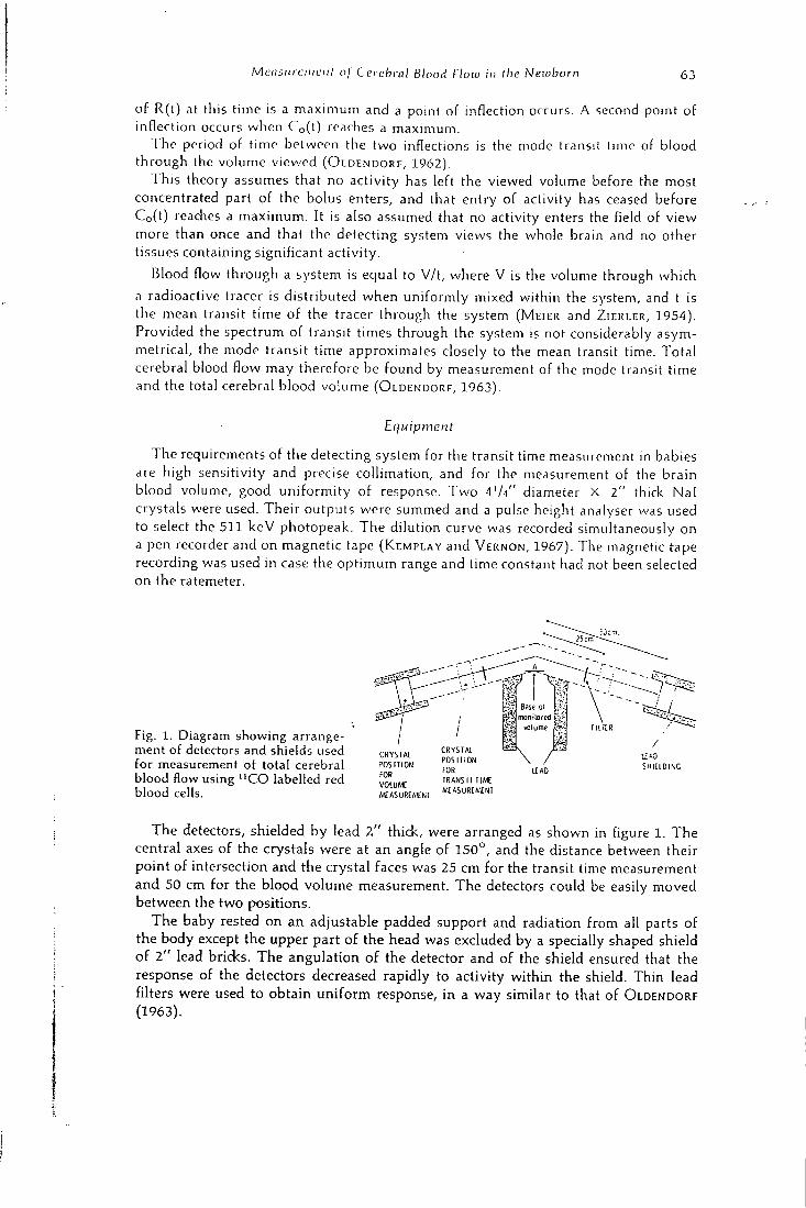
McnsKicniuiit oj Cercbrnt Blood flow in the Newborn 63
of R(t) at this time is a maximum and a point of inflection occurs. A second point of inflection occurs when Co(t) reaches a maximum.
T h e period of time between the two inflections is the mode transit time of blood through the volume viewed ( O L D E N D O R F , 1962) .
T h i s theory assumes that no activity has left the viewed volume before the most concentrated part of the bolus enters, and that entry of activity has ceased before Co(t) reaches a maximum. It is also assumed that no activity enters the field of view more than once and that the detecting system views the whole brain and no other tissues containing significant activity.
Blood flow through a system is equal to V/ t , where V is the volume through which a radioactive tracer is distributed when uniformly mixed within the system, and t is the mean transit time of the tracer through the system ( M E I E R and Z I E R L E R , 1954) . Provided the spectrum of transit times through the system is not considerably asymmetrical, the mode transit time approximates closely to the mean transit time. Total cerebral blood flow may therefore be found by measurement of the mode transit time and the total cerebral blood volume (OLDENDORF, 1 9 6 3 ) .
Equipment
T h e requirements of the detecting system for the transit time measurement in babies are high sensitivity and precise collimation, and for the measurement of the brain blood volume, good uniformity of response. T w o i'h" diameter X 2" thick Nal crystals were used. The ir outputs were summed and a pulse height analyser was used to select the 5 1 1 k e V photopeak. T h e dilution curve was recorded simultaneously on a pen recorder and on magnetic tape (KEMPLAY and VERNON, 1967) . The magnetic tape recording was used in case the optimum range and time constant had not been selected on the ratemeter.
Fig. 1. Diagram showing arrangement of detectors and shields used for measurement of total cerebral blood flow using " C O labelled red blood cells.
Base ol
m o n i t o f e f l t'.:.-
wolume
SHIELDING
C R Y S I A l
P O S I T I O N
FOR
VOLUME
MEASUREMENI
CRYSTAL
P O S I T I O N
FOR
TRANSIT TIME
MEASUREMENT
T h e detectors, shielded by lead 2 " thick, were arranged as shown in figure 1. T h e central axes of the crystals were at an angle of 1 5 0 ° , and the distance between their point of intersection and the crystal faces was 25 cm for the transit time measurement and 5 0 cm for the blood volume measurement. T h e detectors could be easily moved between the two positions.
T h e baby rested on a n adjustable padded support and radiation from all parts of the body except the upper part of the head was excluded by a specially shaped shield of 2 " lead bricks. T h e angulation of the detector and of the shield ensured that the response of the detectors decreased rapidly to activity within the shield. Thin lead filters were used to obtain uni form response, in a way similar to that of OLDENDORF ( 1 9 6 3 ) .
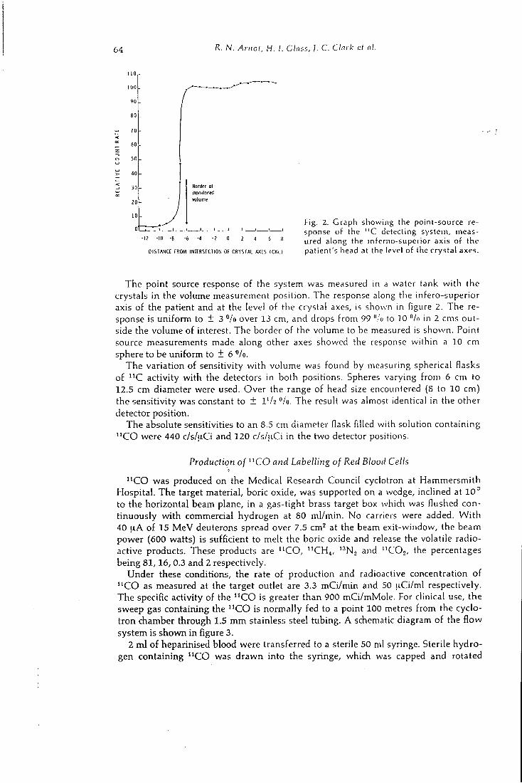
64 R . N. Arnot, H. I. Class, ]. C. CInrk ct nl.
1 1 0
1 0 0
•JO
8 0
7 0
6 0
5 0
4 0
3 0
2 0
1 0
0'
Border o l
moni lore i J
v o l u m e
-12 -10 -8 -6 -4 - 2 0 2 4 6 8
DISTANCE FROM I N I t R S E C I l O N O f CRYSTAL AXES I C M . l
Fig. 2. Graph showing (he point-source response of the " C detecting system, measured along the inferno-superior axis of the patient's head at the level of the crystal axes.
T h e point source response of the system was measured in a water tank with the crystals in the volume measurement position. T h e response along the infero-superior axis of the patient and at the level of the crystal axes, is shown in figure 2. T h e response is uniform to ± 3 */o over 13 cm, and drops from 99 ":u to 10 "/o in 2 cms outside the volume of interest. T h e border of the volume to be measured is shown. Point source measurements made along other axes showed the response within a 10 cm sphere to be uniform to + 6 "/o.
T h e variation of sensitivity with volume was found by measuring spherical flasks of " C activity with the detectors in both positions. Spheres varying from 6 cm to 12.5 cm diameter were used. O v e r the range of head size encountered (8 to 10 cm) the sensitivity was constant to ± IV2 "/o. T h e result was almost identical in the other detector position.
T h e absolute sensitivities to an 8.5 cm diameter flask filled with solution containing " C O were 440 c / s / i iCi and 120 c/s/i^iCi in the two detector positions.
Production of " C O and Labelling of Red Blood Cells
" C O was produced on the Medical Research Counci l cyclotron at Hammersmith Hospital . T h e target material, boric oxide, was supported on a wedge, inclined at 1 0 ° to the horizontal beam plane, in a gas-tight brass target box which was flushed continuously with commercial hydrogen at 80 ml/min. No carriers were added. W i t h 40 [lA of 15 M e V deuterons spread over 7.5 cm^ at the beam exit-window, the beam power (600 watts) is sufficient to melt the boric oxide and release the volatile radioactive products. These products are " C O , " C H 4 , *^N., and " C O 2 , the percentages being 81 ,16 , 0.3 and 2 respectively.
Under these conditions, the rate of production and radioactive concentration of " C O as measured at the target outlet are 3.3 m C i / m i n and 50 uCi /ml respectively. T h e specific activity of the " C O is greater than 900 mCi/mMole . For clinical use, the sweep gas containing the " C O is normally fed to a point 100 metres from the cyclotron chamber through 1.5 m m stainless steel tubing. A schematic diagram of the flow system is shown in figure 3.
2 ml of heparinised blood were transferred to a sterile 50 ml syringe. Sterile hydrogen containing " C O was drawn into the syringe, which was capped and rotated
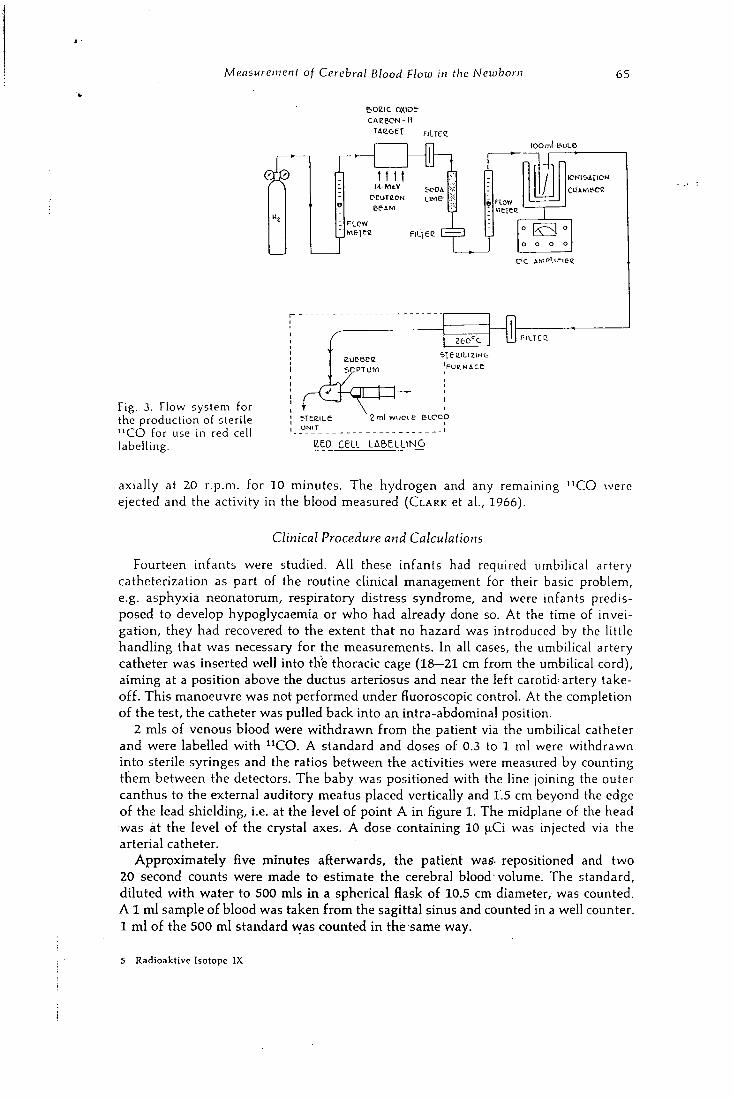
Measurement of Cerebral Blood Flow in the Newborn 65
e-oeic oxioe
TAE-c-ei fiLree lOOml &IJL6
t t 1 l-L MeV
FLOW
HON
I C N l l - i r i O N
o o
o o o o
Fig. 3. Flow system for the production of sterile " C O for use in red cell labelling.
1 — 1 F i u e c
s T e i z i L i z i N G
2 m l w u c L e ELCco . 1
e e o c e u . L A B E L L I N G
5 T E e i L e
ONIT
axially at 20 r.p.m. for 10 minutes. T h e hydrogen and any remaining " C O were ejected and the activity in the blood measured ( C L A R K et al., 1966).
Clinical Procedure and Calculations
Fourteen infants were studied. A l l these infants had required umbilical artery catheterization as part of the routine clinical management for their basic problem, e.g. asphyxia neonatorum, respiratory distress syndrome, and were infants predisposed to develop hypoglycaemia or who had already done so. At the time of invei-gation, they had recovered to the extent that no hazard was introduced by the little handling that was necessary for the measurements. In all cases, the umbilical artery catheter was inserted well into thie thoracic cage (18—21 cm from the umbilical cord), aiming at a position above the ductus arteriosus and near the left carotid^ artery takeoff. T h i s manoeuvre was not performed under fluoroscopic control. At the completion of the test, the catheter was pulled back into an intra-abdominal position.
2 mis of venous blood were wi thdrawn from the patient via the umbilical catheter and were labelled with " C O . A standard and doses of 0.3 to 1 ml were withdrawn into sterile syringes and the ratios between the activities were measured by counting them between the detectors. T h e baby was positioned with the line joining the outer canthus to the external auditory meatus placed vertically and 1.5 cm beyond the edge of the lead shielding, i.e. at the level of point A in figure 1. T h e midplane of the head was at the level of the crystal axes. A dose containing 10 fxCi was injected via the arterial catheter.
Approximate ly five minutes afterwards, the patient wafi. repositioned and two 20 second counts were made to estimate the cerebral blood'volume. T h e standard, diluted with water to 500 mis in a spherical flask of 10.5 cm diameter, was counted. A 1 ml sample of blood was taken from the sagittal sinus and counted in a well counter. 1 ml of the 500 ml standard vvas counted in the same way.
S Radioaktive Isotope I X
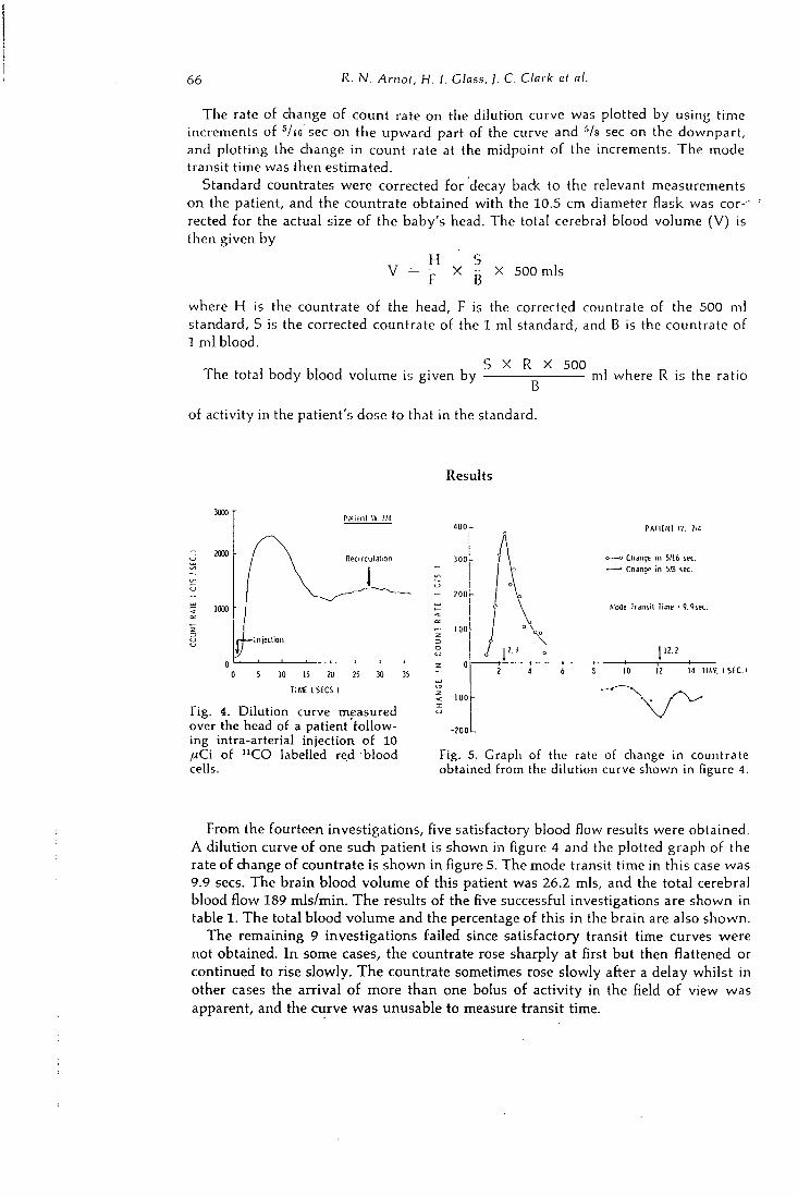
66 R. N. Arnot, H. I. Class, ]. C. Clark et al.
T h e rate of change of count rate on the dilution curve was plotted by using time increments of •''/lo sec on the upward part of the curve and •''/s sec on the downpart, and plotting the change in count rate at the midpoint of the increments. T h e mode transit time was then estimated.
Standard countrates were corrected for decay back to the relevant measurements on the patient, and the countrate obtained with the 10.5 cm diameter flask was cor--rected for the actual size of the baby's head. T h e total cerebral blood volume (V) is then given by
H S V = — X - X 500 mis
t a
where H is the countrate of the head, F is the corrected countrate of the 500 ml standard, S is the corrected countrate of the 1 ml standard, and B is the countrate of 1 ml blood.
n-i > 1 J , , , , , S X R X 500 , , i h e total body blood volume is given by ml where R is the ratio
of activity in the patient's dose to that in the standard.
Results
R e c i r c u l a t i o n
I n j e c l i o n
10 15 20 ?5 30
T I M E ( S E C S I
Fig. 4. Dilution curve measured over the head of a patient following intra-arterial injection of 10 ,uCi of " C O labelled red blood cells.
3 0 0
- 2 0 0
PATIENT W. lit
o — o Change i n S/16 sec.
• — • Change i n 5/B sec.
Mode I r a n s i l Time • 9 . 9 s e c .
14 T I M I SEC.I
Fig. 5. Graph of the rate of change in countrate obtained from the dilution curve shown in figure 4.
From the fourteen investigations, five satisfactory blood flow results were obtained. A dilution curve of one such patient is shown in figure 4 and the plotted graph of the rate of change of countrate is shown in figure 5. T h e mode transit time in this case was 9.9 sees. T h e brain blood volume of this patient was 26.2 mis, and the total cerebral blood flow 189 mls /min. T h e results of the five successful investigations are shown in table 1. T h e total blood volume and the percentage of this in the brain are also shown.
T h e remaining 9 investigations failed since satisfactory transit time curves were not obtained. I n some cases, the countrate rose sharply at first but then flattened or continued to rise slowly. T h e countrate sometimes rose slowly after a delay whilst in other cases the arrival of more than one bolus of activity in the field of view was apparent, and the curve was unusable to measure transit time.
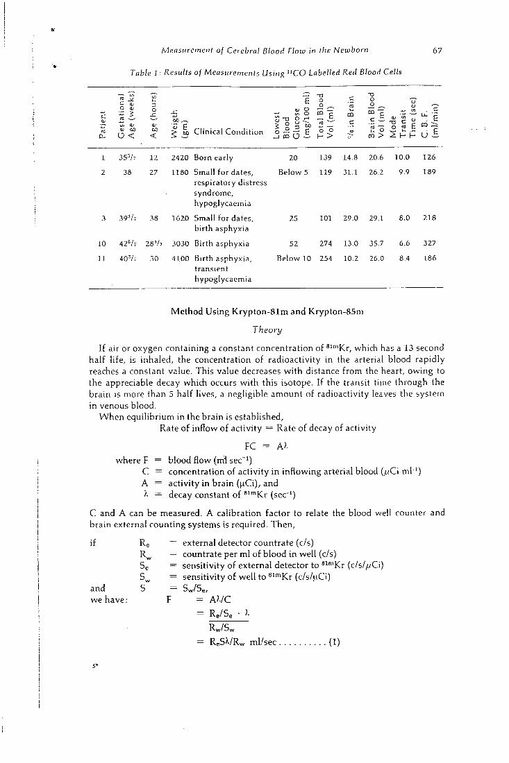
Measurement of Cerebral Blood Flow in the Newborn
Table 1. Results of Measurements Using " C O Labelled Red Blood Cells
67
O QJ
1 5 in HI
U <
o
6 0
<
o
-a o _o ^
^ M Clinical Condition _3 5 ^ J , (° >
CD
c
-a
CD > 2 H H U i .
1 35-V7 12 2420 Born early 20 139 14.8 20.6 10.0 126
2 38 27 1180 Small for dates. Below 5 119 31.1 26.2 9.9 189
respiratory distress syndrome. hypoglycaemia
3 39-V7 38 1620 Small for dates. 25 101 29.0 29.1 8.0 218
birth asphyxia
10 42V7 28V2 3030 Birth asphyxia 52 274 13.0 35.7 6.6 327
U 40-V7 30 4100 Birth asphyxia. Below 10 254 10.2 26.0 8.4 186
transient hypoglycaemia
Method Using Krypton-81m and Krypton-85m
Theory
If air or oxygen containing a constant concentration of ^ " " K r , which has a 13 second half life, is inhaled, the concentration of radioactivity in the arterial blood rapidly reaches a constant value. T h i s value decreases with distance from the heart, owing to the appreciable decay whid i occurs with this isotope. If the transit time through the brain is more than 5 half lives, a negligible amount of radioactivity leaves the system in venous blood.
W h e n equilibrium in the brain is established. Rate of inflow of activity = Rate of decay of activity
P C = A>-
where F = blood flow (ml sec"') C = concentration of activity in inflowing arterial blood (jLiCi ml"') A = activity in brain (|-iCi), and
= decay constant of ^^'"Kr (sec"')
C and A can be measured. A calibration factor to relate the blood well counter and brain external counting systems is required. T h e n ,
if
and we have:
Re = external detector countrate (c/s) R ^ = countrate per ml of blood in well (c/s) Se = sensitivity of external detector to " " " K r (c/s//vCi)
= sensitivity of well to ^ i ^ K r (c/s/^iCi)
S Sw/Se/ F = A ? . / C
= Re/Se •
Rw/S
= R e S V R w ml/sec (1)
68 R. N. Arnot, H. I. Class, j. C. Clark el al.
T h i s technique is useful only if the whole cerebral region can be monitored. H o w ever 190 k e V gamma radiation is scattered through large angels, which makes it difficult to measure the activity in the brain of small babies without interference from the high activity in the mouth, throat and lungs. If only a part of the brain is monitored, it is necessary to estimate the mass of tissue viewed by the detector.
T h e mass of tissue being monitored was estimated by inhalation of ^' '"Kr. A s is seen below, this eliminates the variable detector calibration factor S of equation (1), which varies between patients and between different cerebral regions. After the " " " K r measurement is completed, ^^•"Kr gas is inhaled until the concentration of radioactivity in the cerebral tissue is constant. In this condition, for ''^'"Kr,
if
and we have:
A ' = activity viewed by detector (uCi) M = mass of brain tissue viewed by detector (gm) C = concentration of activity in the blood (uCi ml"') P = equivalent volume of blood contained in 1 gm cerebral tissue
(ml gm"»)
R'g = external detector countrate (c/s) R ' ^ = countrate per ml of blood in well (c/s) S'g = sensitivity of external detector to ^^'"Kr (c/s/uCi) S'^ = sensitivity of well to ssmKr (c /s /uCi) 5 5 w/5 Q,
= A'i'CP
R'w/5'w P
= R ' . 5 7 R ' « . P g m
M
(2)
T h e perfusion rate is obtained by combining (1) and (2):
Perfusion rate = M
Rw — • - • '-V ml/sec/gm (3)
T h e ratio S / S ' was found to be constant owing to the similar energies of 81'"Kr and 85'"Kr (190 k e V and 150 k e V + 17 "/o of 305 k e V respectively). Its value depends only on the counting conditions and is independent of the geometry and composition of the region viewed by the external detector. T h e cerebral perfusion rate can thus be obtained from two external counts and two blood sample counts.
T h e fractional change in flow was measured using S l ' ^ K r only since it is necessary only to measure change in the ratio of A / C under the different physiological conditions being investigated. T h i s technique has the further attraction that it does not depend on the blood sampling being carried out at a point exactly equivalent in distance from the aortic valve to the carotid arteries, since any error is eliminated when the successive results are compared with each other.
Detecting System. Reduction of Scatter Contribution
T h e external detector consisted of a Vz" diameter X V2" thick N a l crystal, and a V2" thidc lead shield and 2" long cylindrical collimator whidi was cut off at an angle of 4 5 ° . A single d ia imel analyser was used to select energies from 170 keV to 256 k e V when counting S l - ^ K r and from 138 keV to 198 keV for 85'"Kr. T h e lower gates corresponded to 85 "/o and 92 *'/o of the photopeak energies of 81'^Kr and SS'^Kr
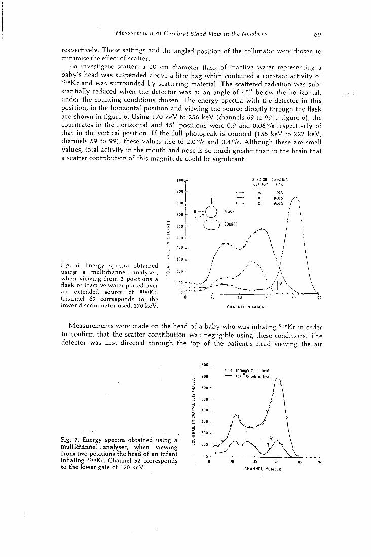
Measurement of Cerebral Blood Flow in the Newborn 69
respectively. These settings and the angled position of the collimator were chosen to minimise the effect of scatter.
T o investigate scatter, a 10 cm diameter flask of inactive water representing a baby's head was suspended above a litre bag which contained a constant activity of *'""Kr and was surrounded by scattering material. T h e scattered radiation was substantially reduced when the detector was at an angle of 4 5 ° below the horizontal, under the counting conditions chosen. T h e energy spectra with the detector in this position, in the horizontal position and viewing the source directly through the flask are shown in figure 6. Us ing 170 k e V to 256 k e V (channels 69 to 99 in figure 6), the countrates in the horizontal and 4 5 ° positions were 0.9 and 0.06 "/o respectively of that in the vertical position. If the full photopeak is counted (155 keV to 227 keV, channels 59 to 99), these values rise to 2.0 "/o and 0.4 "/o. Although these are small values, total activity in the mouth and nose is so much greater than in the brain that a scatter contribution of this magnitude could be significant.
DETECTOR C J U . \ i l N G
Fig . 6. Energy spectra obtained u s i n g a mult ichannel ana lyser , w h e n v i ewing from 3 posit ions a flask of inact ive water placed over a n extended source of 8 imKr. C h a n n e l 69 corresponds to the lower d i scr iminator used, 170 k e V .
OOOr P O S I T I O N
SOURCE
JOO 'r.
to 6 0
C H A N N E L N U M B E R
9 9
Measurements were made on the head of a baby who was inhaling ^ " " K r in order to confirm that the scatter contribution was negligible using these conditions. T h e detector was first directed through the top of the patient's head viewing the air
Fig. 7. Energy spectra obtained using a multichannel . analyser, when viewing from two positions the head of an infant inhaling 8 i m K r . Channel 52 corresponds to the lower gate of 170 keV.
T h r o u g h top o l head
A l 4 5 ° lo side o f head
o ino
40 to C H A N N E L N U M B E R
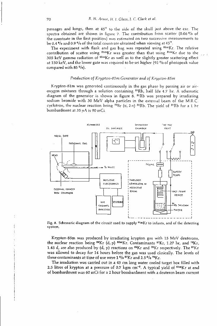
70 R. N. Arnot, H. I. Class, ]. C. Clark et al.
passages and lungs, then at 4 5 ° to the side of the skull just above the ear. T h e spectra obtained are shown in figure 7. T h e contribution from scatter (0.06 */o of the countrate in the first position) was estimated on two successive measurements to be 0.4 "/o and 0.9 *'/o of the total countrate obtained when viewing at 4 5 ° .
T h e experiment with flask and gas bag was repeated using ^^'"Kr. The relative contribution of scatter using 85mj ^ greater than that using ^ " " K r due to the 305 k e V gamma radiation of 85mj<;j. ^gjj (.Q ).j. g slightly greater scattering effect at 150 keV, and the lower gate was required to be set higher (92 °/o of photopeak value compared with 85 "/o).
Production of Krypton-Sim Generator and of Krypton-85m
Krypton-81m was generated continuously in the gas phase by passing air or air-oxygen mixtures through a solution containing ^'Rb, half life 4.7 hr. A schematic diagram of the generator is shown in figure 8. ^'Rb was prepared by irradiating sodium bromide with 30 M e V alpha particles in the external beam of the M . R . C . cyclotron, the nuclear reaction being " B r (a, 2 n) ^'Rb. T h e yield of ^'Rb for a 1 hr bombardment at 35 |.iA is 90 m C i .
plowmeTeE
D C AMPL IFIE-C V i L V t
&l2eATUIWG; W 6
HEiO
l o W i S T C
eKTEENAL COUNJCE
WITU COIUMIJOE
l o o
CUANNeL eECCEDEE
t e N t l A T O e . I N
A D j A C e N T
e o o M
| s A f e T r
T l i P i
- A N f l F O A M
Device
Eb SOLUTION
f.wTEe
L
Fig. 8. Sdiemalic diagram of the circuit used to supply ^'n^Kr to infants, and of the detecting system.
Krypton-85m was produced by irradiating krypton gas with 15 M e V deuterons, the nuclear reaction being 8*Kr (d, p) ssm^r. Contaminants ^ ' K r , 1.27 hr, and '«Kr, 1.45 d, are also produced by (d, p) reactions on ^^Kr and ^^Kr respectively. T h e ^ ' K r was allowed to decay for 14 hours before the gas was used clinically. T h e levels of these contaminants at time of use were 1 "lo ^^Kr and 2.5 "/o ^*Kr.
T h e irradiation was carried out in a 45 c m long water cooled target box filled with 2.5 litres of krypton at a pressure of 0.7 k g m cm"^. A typical yield of ^^^Y^v at end of bombardment was 60 m C i for a 2 hour bombardment with a deuteron beam current
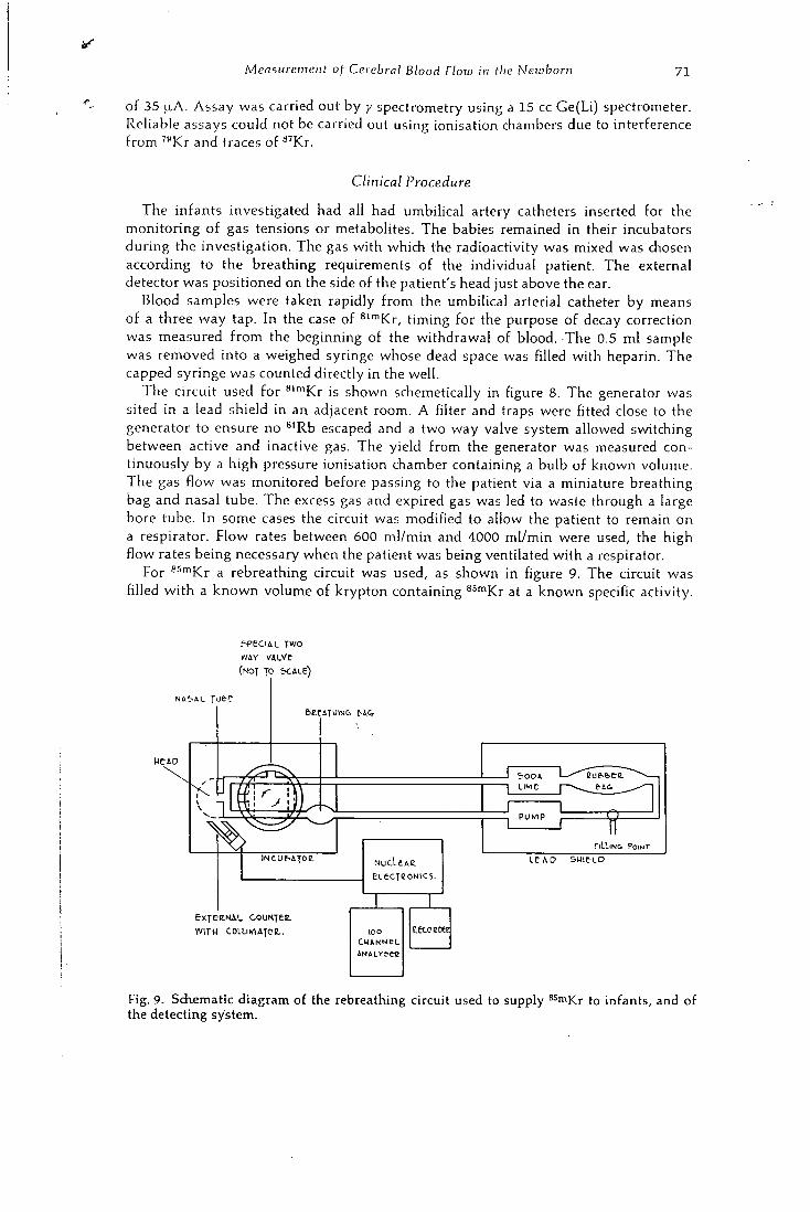
Measurement of Cerebral Blood Flow in the Newborn 71
of 35 LiA. A s s a y was carried out by y spectrometry using a 15 cc G e ( L i ) spectrometer. Reliable assays could not be carried out using ionisation chambers due to interference from " K r and traces of «'Kr.
Clinical Procedure
T h e infants investigated had all had umbilical artery catheters inserted for the monitoring of gas tensions or metabolites. T h e babies remained in their incubators during the investigation. T h e gas with which the radioactivity was mixed was chosen according to the breathing requirements of the individual patient. T h e external detector was positioned on the side of the patient's head just above the ear.
Blood samples were taken rapidly from the umbilical arterial catheter by means of a three way tap. I n the case of ^ " " K r , timing for the purpose of decay correction was measured from the beginning of the withdrawal of blood. T h e 0.5 ml sample was removed into a weighed syringe whose dead space was filled with heparin. T h e capped syringe was counted directly in the well.
T h e circuit used for *'""Kr is shown schemetically in figure 8. T h e generator was sited in a lead shield in an adjacent room. A filter and traps were fitted close to the generator to ensure no ^'Rb escaped and a two way valve system allowed sv^tching between active and inactive gas. T h e yield from the generator was measured continuously by a high pressure ionisation chamber containing a bulb of known volume. T h e gas flow was monitored before passing to the patient via a miniature breathing bag and nasal tube. T h e excess gas and expired gas was led to waste through a large bore tube. In some cases the circuit was modified to allow the patient to remain on a respirator. Flow rates between 600 ml /min and 4000 ml/min were used, the high flow rates being necessary when the patient was being ventilated with a respirator.
For 85mj< ^ 3 rebreathing circuit was used, as shown in figure 9. T h e circuit was filled with a k n o w n volume of krypton containing ^^'^Kr at a known specific activity.
S-PECIAL TWO
WAY VALVe
(NOT TO S C A L e )
Me AO
e x T e n N A L c o u K y e e .
W I T H COLUMAJCE. .
NUcLtAE. eLecreoNics.
? 0 0 A
L I M E
. i=lLl lNG POINT LeAO S H I E L D
l o o CHANNEL A N A L Y ^ r e
Fig. 9. Sdiematic diagram of the rebreathing circuit used to supply ^^^Kr to infants, and of the detecting system.
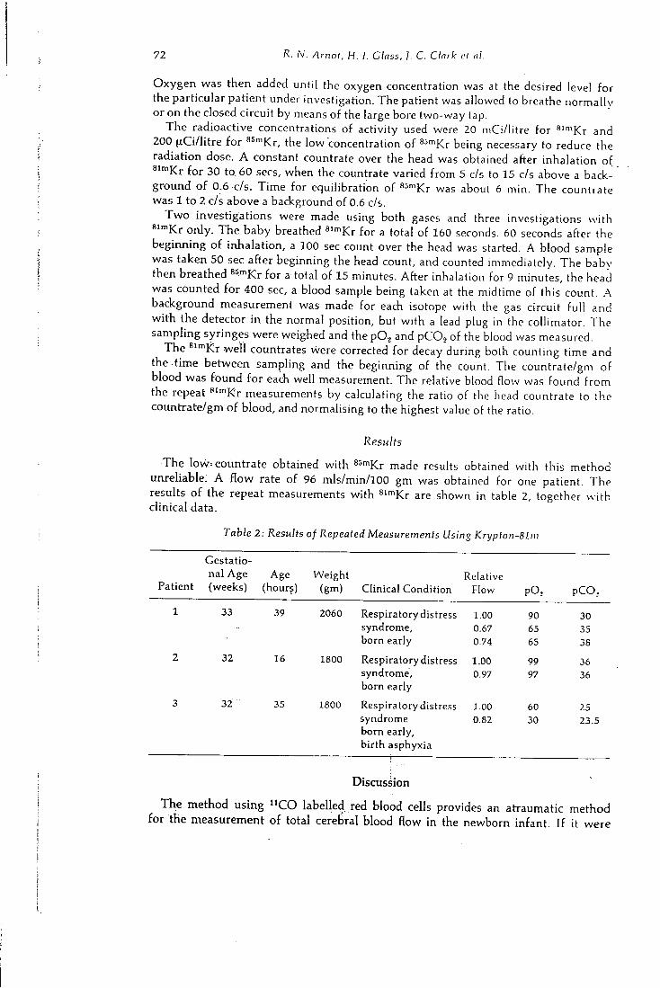
72 R. N. Arnot, H. 1. Class, ]. C. Clark el <;/.
O x y g e n was then added until the oxygen concentration was at the desired level for the particular patient under investigation. T h e patient was allowed to breathe normally or on the closed circuit by means of the large bore two-way tap.
T h e radioactive concentrations of activity used were 20 mCi/litre for ^' '"Kr and 200 (xCi/litre for ^^"^Kr, the low "concentration of ^'^"^Kr being necessary to reduce the radiation dose. A constant countrate over the head was obtained after inhalation of ®""Kr for 30 to. 60 sees, when the countrate varied from 5 c/s to 15 c/s above a background of 0.6 c/s. T ime for equilibration of ^^"^KT was about 6 min. T h e countrate was 1 to 2 c/s above a background of 0.6 c/s.
T w o investigations were made using both gases and three investigations with * " " K r only. T h e baby breathed ^' '^Kr for a total of 160 seconds. 60 seconds after the beginning of inhalation, a 100 sec count over the head was started. A blood sample was taken 50 sec after beginning the head count, and counted immediately. T h e baby then breathed ssmj^j. . total of 15 minutes. After inhalation for 9 minutes, the head was counted for 400 sec, a blood sample being taken at the midtime of this count. .A background measurement was made for each isotope with the gas circuit full and with the detector in the normal position, but with a lead plug in the collimator. T h e sampling syringes were weighed and the p O , and PCO2 of the blood was measured.
T h e ^ ' •"Kr wel l countrates were corrected for decay during both counting time and the time between sampling and the beginning of the count. The countrate/gm of blood was found for each well measurement. T h e relative blood flow was found from the repeat ' '••"Kr measurements by calculating the ratio of the head countrate to the countrate/gm of blood, and normalising to the highest value of the ratio.
Results
T h e l o w countrate obtained with 8 5 m j < ; ; j made results obtained with this method unreliable. A flow rate of 96 mls/min/lOO gm was obtained for one patient. T h e results of the repeat measurements with ^ " " K r are shown in table 2, together with clinical data.
Table 2: Results of Repeated Measurements Usi ng Krypton-Bli
Patient
Gestational Age (weeks)
Age (hour?)
Weight (gm) Clinical Condition
Relative Flow pO, p C O ,
1 33 39 2060 Respiratory distress syndrome, born early
1.00 0.67 0.74
90 65 65
30 35 38
2 32 16 1800 Respiratory distress syndrome, born early
1.00 0.97
99 97
36 36
3 32 35 1800 Respiratory distress syndrome born early, birth asphyxia
i —
1.00 0.82
60 30
25 23.5
Discussion
T h e method us ing " C O labelled red blood cells provides an atraumatic method for the measurement of total cerebral blood flow in the newborn infant. If it were
I
Measurement of Cerebral Blood Flow in the Newborn 7 3
possible to position the catheter tip using an image intensifier, it is likely that successful measurements could a lways be carried out, but there would be a significant increase in radiation dose.
T h e studies were performed at a time when the infant's circulatory system was undergoing a change from a foetal-type circulation to an adult-type circulation, and thus early re-circulation of the bolus of the " C O red blood cells, particularly through the ductus arteriosus could explain the failure of the method in 9 of the infants studied. It would seem that critical placement of the catheter tip above the ductus arteriosus, specifically in the ascending part of the aorta, is essential.
In the 5 successful cases, the cerebral blood flow found was slightly higher than one would have expected. T h e variation in the blood flow from infant to infant may be accounted for by the different clinical conditions of the infants, some having developed a greater degree of acidosis and hypercapnea than others.
T h e method using both ®" Kr and ssmj^j. potentially useful, but was not usable with babies owing to the limitation on 85mj<; ^ concentration due to radiation dosage. It could be used for measurements on adults where a higher radiation dose may be acceptable. T h e positioning of the arterial sampling catheter to give a concentration comparable to that in the carotid arteries would have to be carefully chosen.
Repeat measurements using ^ ' '"Kr proved useful in investigating the effect of change in pOg and p C O j in babies. T h e lowering of blood flow with a decrease in p O j in the patients investigated was unexpected.
T h i s method can only be accurate if successive measurements are made without any movement of the patients or detector. A n y relative movement changes the region monitored and also the ratio of the counter sensitivities. Long term changes in blood flow therefore cannot be investigated in this way.
T h e investigation of the contribution of scatter to countrates mesaured over the heads of patients inhaling radioactive gas are of interest. The results indicate that measurements on adults us ing low energy radiation and large collimators and counting the full photopeak will include a substantial number of counts from scattered radiation.
Conclusion
Three methods are described for the measurement of cerebral blood flow in the newborn infant. T h e method measuring total cerebral blood flow using ' ' C O labelled red blood cells is dependent upon the precise placement of the catheter tip in the aorta above the ductus arteriosus. W i t h this method, 5 successful estimations of cerebral blood flow were made. T h e method employing ^''"Kr and ^'"'Kr was not reliable with the concentrations of ssmj^j. uggj method using repeated measurements with ^'"^Kr alone was useful in assessing the dependence of the brain perfusion rate on PO2.
Acknowledgements
We are grateful to Dr. J. RUNDO of the A . E . R . E . , Harwel l who loaned us the two large crystals to make possible the work with " C , and to the SIR W I L L I A M C O X E N
T r u s t Fund for provision of laboratory facifities. We wish to thank all staff and users of the M.R.C. cyclotron who co-operated in enabling schedule changes to be made at short notice in order to aliov? ihe production of these isotopes as required, shortly after the birth of the patients. We appreciate the encouragement given by Professor J. F . F O W L E R and Professor J. P. M. T I Z A R D .
7 4 Diskussion zum Vortrag Arnot
References
C L A R K , J. C , H. I. G L A S S , and D. J. S Y L V E S T E R - . In vitro Labelling of Red Cells with Carbon-11. Proc. 2nd Int. Conference on Methods of Preparing and Storing Labelled Compounds. Brussels, Nov. 28-Dec. 3, 1966.
C o R N B L A T H , M . , and R. S C H W A R T Z : Disorders in
Carbohydrate Metabolism in Infancy. Major Problems in Clinical Paediatrics, Vol . 3. W. B . Saunders Co. , Philadelphia and London 1966.
E D W A R D S , A. V . : Resistance to Hypoglycaemia in the Newborn Calf. J . Physiol. (Lond.) 171 (1964), 46.
F E D O R U K , S . , and W. F E I N D E L : Measurement of
Brain Circulation by Radioactive lodinated Albumin. Canad. J. Surg. 3 (1960), 312.
K A T S U K I , S . , H . U Z A W A , K . T A N A K A , K . F U K I Y A M A ,
M. Fuj iSHiMA, and 1. O M A E . Brain Circulation Studies by External Counting of Intravenously Injected RISA. Jap. Heart J. 5 (1964), 127.
K E M P L A Y , J. R., and P. V E R N O N : A n Efficient Method of using an Audio Magnetic Tape Recorder for Storing Random Pulses. J. sci. Instrum. 44 (1967), 566.
M A L L E T T , B . L . , and N. V E A L L : Investigation of
Cerebral Blood Flow in Hypertension, using Radioactive Xenon Inhalation and Extracranial Recording. Lancet I (1963), 1081.
M A T T H E W S , C . M . E . : Personal communication, 1967.
M E I E R , P., and K . L. Z I E R L E R : O n the Theory of
the Indicator-Dilution Method for Measurement of Blood Flow and Volume. J. appl. Physiol. 6 (1954), 731.
O B R I S T , W . D . , H . K . T H O M P S O N J R . , C . H . K I N G ,
and H . S. W A N G : Determination of I?egionai Cerebral Blood Flow by Inhalation of Xenon-133. Circular Res. 2 0 ( 1 9 6 7 ) , 124.
O L D E N D O R F , W . H . : Measurement of the Mean Transit Time of Cerebral Circulation by External Detection of an Intravenously Injected Radioisotope. J . nucl. Med. 3 ( 1 9 6 2 ) , 3 8 2 .
O L D E N D O R F , W . H . : Measuring Brain Blood Flow with Radioisotopes. Nucleonics 21 ( 1 9 6 3 ) , 8 7 .
O W E N , O . E . , A. P. M O R G A N , H . G . K E M P , J . M .
S U L L I V A N , M . G . H E R R E R A , and G . P. C A H I L L ;
Brain Metabolism during Fasting. J . clin. Invest. 46 ( 1 9 6 7 ) , 1589 .
R E E K I E , D . , M . D A V I S O N , and ] . K . D A V I D S O N :
The Radiation Hazard in Radiography of the Female Abdomen and Pelvis. Brit. J . Radiol. 40 ( 1 9 6 7 ) , 8 4 9 .
S A P E R S T E I N , L . A.: Measurement of Cephalic and Cerebral Blood Flow Fractions of Cardiac Output in Man. J . clin. Invest. 41 ( 1 9 6 2 ) , 1429 .
S C O P E S , J . W . : Discussion of Hypoglycaemia. Proc. roy. Soc. Med. 5 7 ( 1 9 6 4 ) , 1065 .
S T E I N E R , S . H . , K . H S U , L . O L I N E R , and R. H .
B E H N K E : Measurement of Cerebral Blood Flow by External Isotope Counting. J . clin. Invest. 41 ( 1 9 6 2 ) , 2221 .
V E A L L , N . , and B. L . M A L L E T T : Regional Cerebral
Blood Flow Determination by '"Xe Inhalation and External Recording: the Effect of Arterial Recirculation. Cl in . Sci. 3 0 ( 1 9 6 6 ) , 3 5 3 .
Z I E R L E R , K . L . : Equations for Measuring Blood Flow by External Monitoring of Radioisotopes. Circulat. Res. 16 ( 1 9 6 5 ) , 309 .
Diskussion zum Vortrag Arnot
S. G R E B E , Giefien:
In welcher Grofienordnung liegt die Strahlenbelastung bei diesen Neugeborenen und bedeuten sie kein Ris iko f i ir diese Patienten?
R. ARNOT, London:
T h e radiation dose to the whole body from 10 |xC of " C labelled red blood cells
varies from 2,5 to 6,5 mrads depending on the weight of the patient. T h e administra
tion of Krypton-81m (20 m C / a c t i v e for 2,5 minutes) gives a dose of 48 mrads to
the lungs and a neghgeable whole body dose. For Krypton-85m (0,2 mC/act ive for
15 minutes) the lung dose is 42 mrads and the whole body dose 1 mrad. In comparison
the whole body dose to the foetus from a single x-ray f i lm of the maternal pelvis in
late pregnancy is from 100 to 300 mrads.

11
Department of Medicine, Universitdts Klinik, Innsbruck
Department of Medical Physics, Medical Research Council Cyclotron Unit and Department of Medicine, Hammersmtth Hospital, London
Electrolyte and Body Fluid Investigations in Endocrine Diseases using Cyclotron Produced Isotopes
By
F . SKRABAL, R . N . ARNOT, J . C . C L A R K , F . H E L U S , H . I. GLASS and G . F. joPLir.'
Abstract
The different gamma energies of -''Na, ^'K and "Br allow mixtures of the three isotopes that contain normal quantities of activity to be analyzed with an error of less than 1.5"" by gamma counting of previously untreated samples. A computer programme is used to perform the calculations.
This method was used to investigate the relation between Na space ond Br space in 20 normals and in 46 patients with various endocrine or metabolic diseases.
The mean value for the Na space/Br space ratio in normals was found to be 1.13 ± 0 . 0 7 ( ± 2 S.D.). 39 "/o of the patients were found to have values for the Na space/Br space ratio which were outside the 9 5 ^ / 0 confidence limits for the normal group. The possibility of detecting cellular Na gain or loss on a whole body basis is discussed.
Extrait
Des quantites normales de -''Na, ''''K et "Br contenues dans un meme echantillon peuvent etre separees par spectrometrie y e' mesurees par une methode non destructive avec un pourcentage d'erreur inferieur a 1 ,5%. Un programme de calculateur est utilise pour obtenir les resultats.
Cette technique a ete employee pour etudier le rapport entre I'espace Na ct I'cspace Br chez 20 sujets normaux et chez 46 patients atteints d'affections endocrines ou metaboliques diverses. La valeur moyenne du rapport espace Na/espace Br a ete trouvee egale a 1,13 ± 0,07 ( + 2 S.D.) chez les sujets normaux.
Chez 39 °/o des malades, la valeur de ce rapport se trouve en dehors des limites de con-fiance a 95 "/o etablies pour le groupe normal. La possibilite de detecter par cette methodc une accumulation ou une perte de Na intracellulaire par I'organisme est discutee.
Auszug
Durch die Einfiihrung zweier im Cyclotron hergestellter Isotope, Brom-77 und Ka-Uum-43 wurde es moglich, Mischungen dieser beiden Isotope mit Natrium-24 erstmals ohne vorherige Trennverfahren zu analysieren, indem man die untersdiiedliche Gammastrahlung der drei Isotope unter versdiiedenen Zahlbedingungen miGt. Die Ergebnisse werden mil Hilfe eines Computers erred\net.
Kombiniert mit einer Bestimmung von tritiummarkierten Wassers wurde diese Methode fiir die gleichzeitige Messung des austauschbaren Natriums, austauschbaren Kaliums, extra-
Investigations in Endocrine Diseases using Isotopes 77
zellularen Raumes und des Korperwassers bei 20 Gesunden und 46 Patienten mil endo-krinen Storungen angewendet. Spezielles Interesse lag auf der Bestimmung der Fraktion dcs austauschbaren Natriums, die auGerhalb der extrazellularen Fltissigkeit licgt.
Das Durchschnittsverhaltnis Natriumraum: Bromidraum befrug bei Gesunden 1,13 + 0,07 ( ± 2 5.D.). 39 °/o der Patienten mit endokrinen Storungen lagen auGerhalb der Vertrauens-grenzen fiir gesunde Kontrollobjekte. Die Moglichkeit, Veranderungen des intrazellularen Natriumgehaltes an einer Ganzkorperbasis festzustellen, wird diskutiert.
Introduction
T o investigate electrolytes and body water in humans, several methods are available. T i s sue analysis may be used to investigate the composition of a single tissue, or balance studies may be used to examine net gains or losses from the body. However, neither method gives information about the distribution of electrolytes in the body or the total body stores. T h i s information can be obtained by use of the isotope dilution technique.
T h e simultaneous measurement of exchangeable Na (Nae), exchangeable K (Kg), extra cellular fluid volume ( E C F V ) and total body water ( T B W ) permits the calculation of distribution of sodium, potassium and water between the extra cellular and intra cellular spaces ( V E A L L and V E T T E R , 1958) .
In spite of the obvious advantage of using four isotopes simultaneously, the few reported clinical studies have indicated the inaccuracies and difficulties in separating isotopes in the same sample by chemical or physical separation procedures.
T h e use of two cyclotron-produced isotopes, " K (SKRABAL, G L A S S et al., 1969) and '^Br (SKRABAL, ARNOT et al . , 1 9 6 9 ) made it possible to analyse by gamma counting mixtures of these two isotopes together with '"Na as untreated plasma and urine samples. A computer programme is used to perform the calculations and includes an al lowance for decay of the isotopes during the counting time. Tritiated water may be estimated on the same samples after decay of the other three isotopes.
W h e n it became possible to measure the dilution of -''Na and '"'Br accurately on the same untreated samples (and in the presence of ''^K), the question arose whether it might now be possible to detect smaller differences between these two very similar physiological spaces. Var ious workers ( M C M U R R A Y et al., 1 9 5 8 ; CROOKS et al., 1 9 5 9 ; M O O R E et al . , 1 9 6 3 ) have measured both spaces simultaneously, yet alterations in the ratio between the two spaces in any disease do not appear to have been demonstrated.
W h e have investigated the possibility of detecting an excess or a deficit of exchangeable sodium outside the bromide space and this paper presents our findings in 6 6 cl inical studies.
Material and Methods
Production of «J<C
••^K is produced by the ''"A (a, p) ' " K reaction in the external beam of the Medical Research Counci l cyclotron. T h e argon is circulated at a rate in excess of 100 1/min through the target vessel and bombarded with 16.4 M e V a particles. T h e exit gas is passed through a borosilicate fiber-glass filter which extracts 50 — 8 0 "/o of the activity from the gas phase. T h e activity is removed from the filter using 5 ml of 0 . 0 0 1 N hydrochloric acid. T h i s solution is then evaporated to dryness and baked in the presence of 5 mgm K C l carrier. T h e residue is taken up in 5 ml of isotonic saline and filtered through a millipore filter. T h e yield of is 1 8 ^iCi/n.A hr and the
78 f- Skrabal, R. N. Arnot, ]. C. Clark et al.
contamination at end of bombardment is less than 7 per cent ( C L A R K and W A T S O N , 1 9 6 9 ) .
Production of ''Br
^'Br is produced by the '^As (a, 2 n) " B r reaction in the external beam of the' ' M . R . C . cyclotron. T h e target material , AsgOj , is bombarded with a particles of approximately 2 8 M e V , the yield of ^'Br being about 3 m C i for a 1 hour bombardment at 2 0 [xA. The AsgOj is then dissolved in hot water and after adding concentrated H2SO4 and K2Cr207, the bromine is distilled, without the addition of carrier, into an ice-cooled water trap. T h e p H of this solution is then adjusted to 9 and the bromine is reduced to bromide by adding NH2OH. H C l solution. The solution is evaporated to dryness and the residue is heated to about 4 0 0 ° C to decompose any possible pyrogenic contaminants, then dissolved in water for injection. T h e resulting solution is adjusted to p H 6—7 and filtered ( H E L U S , 1 9 6 9 ) .
•''Na and
T h e -''Na was received in the form of sterile N a C l in isotonic solution from the Radiochemical Centre, A m e r s h a m . Sterile tritiated water was obtained from the same suppliers.
Patients Studied
2 0 normal subjects and 4 6 patients suffering from various endocrine diseases were investigated. T h e patients were classified as follows; untreated acromegaly (8), treated acromegaly (pituitary implantation of y t t r ium-90) ( 9 ) , Cushing's disease ( 5 ) , and 1 0 hypopituitary subjects. There were 1 4 patients with various other endocrine or metabolic diseases.
Clinical Procedures
2 0 (xCi of ^''Na and 5 0 (xCi of ''^K were administered intravenously at 9 a.m. 1 2 hours later 30 |j,Ci "Br and 2 5 0 |.iCi H20 were injected. Doses for injection were prepared in counting vials and after injection, the residue in the syringe was washed into the dose vial and made up to 1 0 mis for subsequent counting. Normal meals were permitted from 9 a.m. to 8 p.m. after which no fluids or food was taken until 1 0 a.m. on the following day. Ur ine was collected in polythene bottles as follows: 0 — 2 4 hours, 2 4 — 2 5 hours, 2 5 — 2 6 hours. Heparinised blood samples were taken at 2 4 hours and 2 5 hours.
T h e concentration of stable sodium and potassium in the plasma was estimated on an auto-analyser and in the urine samples on a flame photometer.
Radioactive Assay and Calculations of Results
A dose of each isotope, identical to that given to the patient was used to prepare three standard solutions in sil iconed glass flasks by diluting each dose with distilled water up to 1 litre. Dupl icate 10 ml samples of each standard solution and 1 0 ml of eacfi plasma and urine sample, together with a bacicground sample of water, were transferred into counting vials. A l l samples were counted twice on a two cfiannel automatic counter, equipped with a puncfied paper tape output. The first time, the
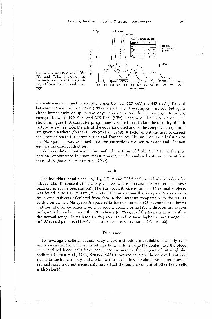
Investigations in Endocrine Diseases using Isotopes 79
Fig. 1. Energy spectra of " B r , ''••'K and -''Na, showing the channels used and the counting efficiencies for each isotope.
0. 373 » * V
COUNTING t f f l C I E N C V DLI
Channel 1 2 3
Isotope
" B r n 4.5 0.4
«K i 16 0.1
2 2 13
Channel 1 Channel 2
• BROMINt - 7 ! - POTASSIUM - « - SODIUM • 24
I 1.37 MeV
0.40 0.50 0.60 0-70
ENERGY I M e V I
0.80 1.00 2.ca
channels were arranged to accept energies between 3 2 0 K e V and 447 K e V (' ' 'K), and between 1.2 M e V and 4.5 M e V ( ""Na) respectively. T h e samples were counted again either immediately or up to two days later using one channel arranged to accept energies between 190 K e V and 2 7 5 K e V ( " B r ) . Spectra of the three isotopes are shown in figure 1. A computer programme was used to calculate the quantity of each isotope in each sample. Details of the equations used and of the computer programme are given elsewhere (SKRABAL, A R N O T et al . , 1 9 6 9 ) . A factor of 0.9 was used to correct the bromide space for serum water and Donnan equilibrium. For the calculation of the N a space it was assumed that the corrections for serum water and Donnan equilibrium cancel each other.
W e have shown that using this method, mixtures of -"Na, "^K, " B r in the proportions encountered in space measurements, can be analysed with an error of less than 1.5 "/o (SKRABAL, A R N O T et al . , 1 9 6 9 ) .
Results
T h e individual results for NaE, K E , E C F V and T B W and the calculated values for intracellular K concentration are given elsewhere (SKRABAL, ARNOT et al., 1 9 6 9 ; SKRABAL et al . , in preparation). T h e N a space/Br space ratio in 20 normal subjects was found to be 1 .13 ± 0 . 0 7 ( ± ' 2 S . D . ) . Figure 2 shows the N a space/Br space ratio for normal subjects calculated from data in the literature compared with the results of this series. T h e N a space/Br space ratio for our normals (95 "/o confidence limits) and the ratio for 46 patients with various endocrine or metabolic diseases are shown in figure 3. It can been seen that 2 8 patients (61 */o) out of the 46 patients are within the normal range. 1 3 patients (28 Vo) were found to have higher values (range 1.2 to 1 .35 ) and 5 patients (11 *'/o) had a ratio closer to unity (range 1.04 to 1.00) .
Discussion
T o investigate ceUular sodium only a few methods are available. T h e only cells easily separated from the extra cellular fluid with its large N a content are the blood cells, and red blood cells have been used to measure the amount of intra cellular sodium (RiECKER et al . , 1 9 6 3 ; B E I L I N , 1 9 6 6 ) . Since red cells are the only cells without nuclei in the human body and are k n o w n to have a low metabolic rate, alterations in red cell sodium do not necessarily imply that the sodium content of other body cells is also altered.
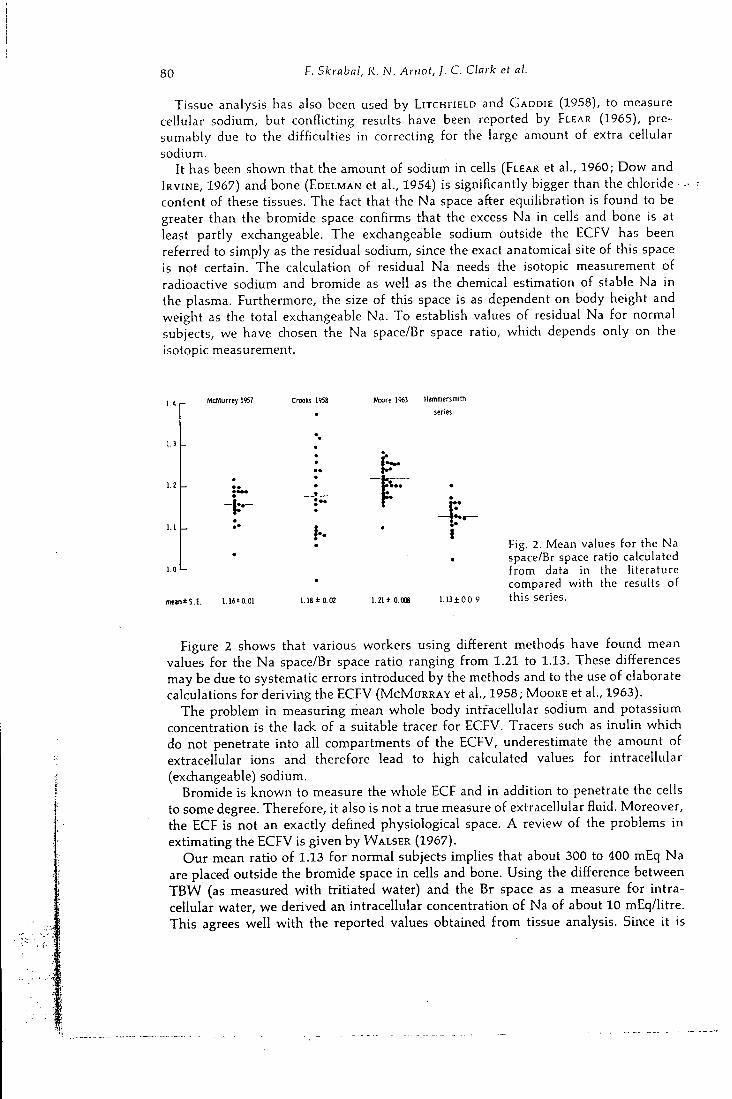
80 F . Skrabal, R . N . Arnot, J . C . Clark et al.
Tissue analysis has also been used by LITCHFIELD and GADDIE (1958), to measure cellular sodium, but conflicting results have been reported by FLEAR (1965), presumably due to the difficulties in correcting for the large amount of extra cellular sodium.
It has been shown that the amount of sodium in cells (FLEAR et al., 1960; Dow and IRVINE, 1967) and bone (EDELMAN et al., 1954) is significantly bigger than the chloride • content of these tissues. The fact that the Na space after equilibration is found to be greater than the bromide space confirms that the excess Na in cells and bone is at least partly exchangeable. The exchangeable sodium outside the ECFV has been referred to simply as the residual sodium, since the exact anatomical site of this space is not certain. The calculation of residual Na needs the isotopic measurement of radioactive sodium and bromide as well as the chemical estimation of stable Na in the plasma. Furthermore, the size of this space is as dependent on body height and weight as the total exchangeable Na. To establish values of residual Na for normal subjects, we have chosen the Na space/Br space ratio, which depends only on the isotopic measurement.
McMurrey 1957 fttoore 1963 Hammersmith
series
meaniS.E. 1.16*0.01
r 4 -
1.21 1 0.006 1.13 ±0.0 9
Fig. 2. Mean values for the Na space/Br space ratio calculated f r o m data in the literature compared w i t h the results of this series.
Figure 2 shows that various workers using different methods have found mean values for the Na space/Br space ratio ranging from 1.21 to 1.13. These differences may be due to systematic errors introduced by the methods and to the use of elaborate calculations for deriving the E C F V ( M C M U R R A Y et al., 1958 ; MOORE et al., 1963) .
The problem in measuring mean whole body intracellular sodium and potassium concentration is the lack of a suitable tracer for E C F V . Tracers such as inulin which do not penetrate into all compartments of the E C F V , underestimate the amount of extracellular ions and therefore lead to high calculated values for intracellular (exchangeable) sodium.
Bromide is known to measure the whole E C F and in addition to penetrate the cells to some degree. Therefore, it also is not a true measure of extracellular fluid. Moreover, the E C F is not an exactly defined physiological space. A review of the problems in extimating the E C F V is given by WALSER (1967) .
Our mean ratio of 1.13 for normal subjects implies that about 300 to 400 mEq Na are placed outside the bromide space in ceils and bone. Using the difference between T B W (as measured with tritiated water) and the Br space as a measure for intracellular water, we derived an intracellular concentration of Na of about 1 0 mEq/litre. This agrees well with the reported values obtained from tissue analysis. Since it is
1
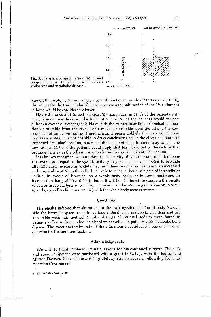
Investigations in Endocrine Diseases using Isotopes 8 1
NORMAL SUBJECTS (201 V A R I O U S ENDOCRINE DISEASES 1461
Fig. 3. Na space/Br space ratio in 20 normal subjects and i n 46 patients w i th various i.o' endocrine and metabolic diseases. mean ± S .E . i.i3±o.om
known that isotopic Na exchanges also with the bone crystals (EDELMAN et al., 1954), the values for the true cellular Na concentration after subtraction of the Na exchanged in bone would be considerably lower.
Figure 3 shows a disturbed Na space/Br space ratio in 39 "/o of the patients with various endocrine diseases. The high ratio in 28 "/o of the patients would indicate either an excess of exchangeable Na outside the extracellular fluid or gradual elimination of bromide from the cells. The removal of bromide from the cells is the consequence of an active transport mechanism. It seems unlikely that this would occur in disease states. It is not possible to draw conclusions about the absolute amount of increased "cellular" sodium, since simultaneous shifts of bromide may occur. The low ratio in 1 1 */o of the patients could imply that Na moves out of the cells or that bromide penetrates the cells in some conditions to a greater extent than sodium.
It is known that after 24 hours the specific activity of Na in tissues other than bone is constant and equal to the specific activity in plasma. The same applies to bromide after 1 2 hours. Increase in "cellular" sodium therefore does not represent an increased exchangeability of Na in the cells. It is likely to reflect either a true gain of intracellular sodium in excess of bromide, on a whole body basis, or in some conditions an increased exchangeability of Na in bone. It will be of interest, to compare the results of cell or tissue analysis in conditions in which cellular sodium gain is known to occur (e.g. the red cell sodium in uraemia)-with the whole body measurements.
Conclusion
The results indicate that alterations in the exchangeable fraction of body Na outside the bromide space occur in various endocrine or metabolic disorders and are detectable with this method. Similar changes of residual sodium were found in patients suffering from endocrine disorders as well as in patients with metabolic bone disease. The exact anatomical site of the alterations in residual Na remains an open question for further investigation.
Acknowledgements
We wish to thank Professor RUSSELL FRASER for his continued support. The -''Na and some equipment were purdiased with a grant to G. F. J. from the ERNEST and M I N N I E D A W S O N Cancer Trust. F. S. gratefully acknowledges a Fellowship-from- the Austrian Government.
6 Radioaktive Isotope IX
8 2 Diskussion zum Vortrag Skrabal
References
B e i l i n , L . J . , G . J . K n i g h t , A. D . M u n r o - F a u r e , and J. A n d e r s o n : The Measurement of Sodium Concentration in Human Red Blood Cells. J. gen. Physiol. 50 (1966), 61.
C l a r k , J. C , and I. A . W a t s o n : The Preperation of ' " K for Medical Use. Radiochem. Radioana-lyt. Letters (Submitted 1970).
C r o o k s , J . , M . M . B l u h m , and F . P. Muldowney: The Interrelation between Total Exdiangeable Sodium, Potassium and Chloride, and Lean Body Mass in Man. Cl in . Sci. 18 (1959), 175.
Dow, J. W. , and R. O . H . I r v i n e : Determination of the Chloride Content of Small Muscle Samples. Cl in . Chim. Acta 15 (1967), 199.
Ede lman, I. S., A . H . James, H . Baden, and F . D . M o o r e : Electrolyte Composition of Bone and the Penetration of Radiosodium and Deute-riumoxide into Dog and Human Bone. J . clin. Invest. 33 (1954), 122.
F l e a r , C . T . G . , R . F . Crompton , and D . M . M a t t h e w s : A n In Vitro Method for the Determination of the Inulin Space of Skeletal Muscle with Observations on the Composition of Human Muscle. Cl in . Sci. 19 (1960), 483.
F l e a r , C . T . G . , R . G . C a r p e n t e r , and I. F l o rence: Variability in the Water, Sodium, Potassium and Chloride Content of Human Skeletal Muscle. J . clin. Path. 18 (1965), 74.
H e l u s , F . : The Production of ' ' B r for Medical Use. Radiochem. Radioanalyt. Letters (In press 1970).
L i t c h f i e l d , J . A . , and R. G a d d i e : The Measurement of the Phase Distribution of Water and Electrolytes in Skeletal Muscle by the Analysis of Small Samples. Cl in . Sci. 17 (1958), 483.
McMuRRAY, J . D . , E. A. Bol ing , J . M. D a v i s , H . V. P a r k e r , I. C . Magnus , M . R . B a l l , and F . D. M o o r e : Body Composition: Simultaneous Determination of Several Aspects by the Dilution Principle. Metabolism 7 (1958), 651.
M o o r e , F . D . , K . H . O l e s e n , J . D. M c M u r r a y , , H. V. P a r k e r , M . R . B a l l , and C . M . Boyden: The Body Cell Mass and its Supporting Environment, p. 532. W. B. Saunders Company, Philadelphia: London 1963.
RiECKER, G . , H. J . GURLAND, H . E d e l , H . D . B a l t e , J . EiCLER, E. Renner , und E. Buchborn: Neue Methoden zur Erkennung von Storungen der intra-extracellularen Wasser- und Elektro-lytverteilung bei Niereninsuffizienz. Verh. dtsch. Ges. inn. Med. 69 (1963), 707.
S k r a b a l , F . , H . 1. G l a s s , J . C . C l a r k , K . Jeya-siNGH, and G . F . Joplin; A Simplified Method for Simultaneous Electrolyte Studies in Man Utilizing Potassium-43. Int. J . appl. Radial. 20 (1969), 677.
S k r a b a l , F . , R . N . A r n o t , F . He lus , H . I. G l a s s , and G . F . Joplin: A Method for Simultaneous Electrolyte Investigations in Man using Bro-mine-77, Potassium-43 and Sodium-24. Int. J . appl. Radiat. (In press 1970).
S k r a b a l , F . , R . N . A r n o t , and G . F . Joplin: Body Composition Studies in Acromegaly, Mb. Gushing, Primary Hyperaldosteronism and Insulinoma. (In preperation).
V e a l l , N . , and H. V e t t e r : In: Radioisotope Techniques in Clinical Research and Diagnosis, p. 209. Butterworth, London 1958.
W a l s e r , M . : Compartments, Pools and Spaces in Medical Physiologic, p. 241. U.S. Atomic Energy Commission 1967. Conf. 661010.
Diskussion zum Vortrag Skrabal
B . FRANCOIS, Lyon: Je voudrais evoquer deux points. En premier lieu, je ne mesestime pas I'interet pour les etudes du metabolisme hydro-
mineral de I'utilisation de ces nouveaux radionuclides produits par un cyclotron. Je signale simplement que chaque semaine, depuis 8 ans, nous utilisons des techniques conventionnelles qui donnent toute satisfaction: separation ^ Na — ^ Br ou -"Na — ^ Br par des equations de decroissance radioactive, separation de I'eau tritiee d'avec le sodium et le brome par distillation par le vide du serum.
Le deuxieme point est une question. L'orateur a fait etat d'une valeur de sodium residuel representant 10 "/o du sodium echangeable et d'une position intra cellulaire de ce sodium residuel. J'avais la notion que le sodium intra cellulaire n'exedait pas 3 "/o du sodium edizmgeable et que le sodium residuel etait essentiellement squelet-tique. L'auteur donne-t-il au sodium residuel une signification veritablement cellulaire?
Diskussion zum Vortrag Skiabal 83
F. SKRABAL , Innsbruck:
Ich selbst halte Natrium-22 auf Grund der langen Halbwertszeit und Strahlungs-charakteristik nicht fiir wunschenswert fiir diese Studien, speziell da Wiederholungen innerhalb kiirzester Zeit von Interesse sind.
Die Halbwertszeiten von 56 Stunden fiir Brom-77, 22 Stunden fiir Kalium-43 und 1 5 Stunden fiir Natrium-24 erlauben eine Wiederholung der Studien innerhalb einer Woche, andererseits sollten sie die Durchfiihrung der Untersuchung auch an Kliniken ermoglichen, die iiber kein eigenes Cyclotron verfiigen.
Zur zweiten Frage: Ich glaube, wir stimmen in dem Punkt iiberein, da2 es prinzipiell nicht moglich ist,
mit dieser Methode zwischen intrazellularem und im Knochen austauschbarem Natrium zu unterscheiden. Wir haben gleichsinnige Veranderungen des Na/Br-Raum-verhaltnisses sowohl bei Patienten mit Nebennierenrindeninsuffizienz gefunden als auch bei Patienten mit Knochenerkrankungen. Bei der ersten Gruppe kam es zum Anstieg des Residualnatriums innerhalb weniger Tage. Die gleichzeitig erfolgende zellulare Hydration wiirde fiir eine intrazellulare Anreicherung des Natrium sprechen Oberdies ist der Austausch von Elektrolyten im Knochen hauptsachlich abhangig von der aktiven Knochenoberflache, d. h. Resorptionsflache. Es erscheint mir unwahr-scheinlich, daS diese sich innerhalb kiirzester Zeit andern wiirde.
N. TELFER, Los Angeles: Did you measure the exchangeable potassium at 24 and 48 hours? Did you find
any abnormalities in the intracellular concentration of potassium?
F. SKRABAL , Innsbruck:
To answer your first question: We have measured the exchangeable K beginning from 24 hours up to 48 hours and we found in agreement with other workers an increase in exchangeable potassium. This increase was fairly constant in all patients and as we were not certain as to how much this increase was due to newly ingested K, we have chosen the 24 hours equilibration period.
To your second question: In the particular group of patients studied we did not find a change in intracellular potassium concentration. Since residual or "intracellular" Na increased the cellular hydration which occurred in some of the patients would be explained by the increasing amount of intracellular ions.
C . CONSTANTiNiDEs, Athen:
When you measure the Na Space/Br Space ratio, do you consider the fraction of Na which goes to bone? Because this may introduce an error into your calculations.
F. SKRABAL , Innsbruck: I think we have covered that point partly already in the discussion with Prof.
FRANCOIS. It is certainly not possible to calculate absolute figures for the intracellu-larily located sodium. Therefore we have chosen the Na/Br ratio.
12
Target systems for radioisotope production on the Medical Research Council cyclotron D. D. Voiibcrg, L. C. Baker, P. D. Buclljiigliani, J. C. Clark, K. Finding, J.Sharp, and D. J. Silvester
Medical Research Council, Cyclotron Unit, Hanvnersmith Hospital. Diicane Road, London, W. 12, England
ABSTRACT
Target systems used for short-lived radioisotope production on the M.R.C. Cyclotron are described in detail and the factors affecting efficient production discussed. Choice of target material, target cooling, radioisotope recovery, suitable beam energy and target thickness for optimum yield of the required isotope, are all considered. The problems of power dissipation in the inicrnai target iisin>j. beam currents up tt) 300iu.A are fully discussed. Reasons ior using the static 'grazing incidence'.target in preference to rotary or vibratory target systems are stated and methods of internal target preparation described.
Beam extraction through a thin vacuum-retaining foil permits a range of external targets to be bombarded using beams of low power density and also allows the use of energy-degrading foils. A method is described of indicating the external beam distribution during bombardment and details are given of external targets using solid, liquid and gaseous target materials. Recently-developed targets for radioactive gas production are described and typical performance figures quoted.
INTRODUCTION
The Medical Research Council cyclotron,''^which was designed and built between 1950 and 1955, operates for 14 h daily on five days a week. About one third of each day is used for experimental radiobiology or fast neutron radiotherapy; the remainder is devoted to producing radioisotopes for medical use, which often but not invariably because of their short half-life cannot be obtained commercially. Much has been published on the uses of these isotopes,^'' and sometimes the methods of producing them have been included, but with very few exceptions the cyclotron target systems have not been described in detail. Tills paper discusses the factors that have to be considered in designing suitable
258
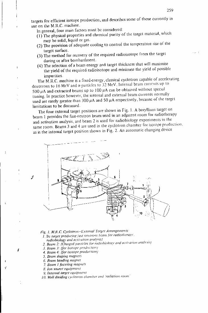
2 5 9
targets for efficient isotope production, and describes some of those currently in use on the M.R.C. machine.
In general, four main factors must be considered: ( 1 ) The physical properties and chemical purity of the target material, which
may be solid, liquid or gas. (2) The provision of adequate cooling to control the temperature rise of the
target surface. ( 3 ) The method for recovery of the required radioisotope from the target
during or after bombardment. (4) The selection of a beam energy and target thickness that will maximise
the yield of the required radioisotope and minimise the yield of possible impurities.
The M.R.C. machine is a fixed-energy, classical cyclotron capable of accelerating deuterons to 1 6 MeV and a-paiticles to 32 MeV. Internal beam currcnls up to 5 0 0 /JA and extracted beams up to 100 iiA can be obtained without special tuning. In practice however, the internal and external beam currents normally used are rarely greater than 3 0 0 fxA and 5 0 M A respectively, because of the target limitations to be discussed.
The four external target positions are shown in Fig. 1 . A beryllium target on beam 1 provides the fast-neutron beam used in an adjacent room for radiotherapy and activation analysis, and beam 2 is used for radiobiology experiments in the same room. Beams 3 and 4 are used in the cyclotron chamber for isotope production, as is the internal target position shown in Fig. 2 . An automatic changing device
Fig. 1. M.R.C. Cyclotron-External Target Arrangements: 1. Be target producing Jast neutrons iieani for radiotlierapy.
radiobiology and activation analysis) 2. Beam 2: (Charged panicles for radiohiology and activation analysis) 3. Beam 3: (for isotope production) 4. Beam 4: (for isotope production) 5. Beam shaping magnets 6. Beam bending magnet 7. Beam J focusing magnets 8. Ion source equipment 9. Internal target equipment
10. Wall dividing cyclotron chamber and radiation room '
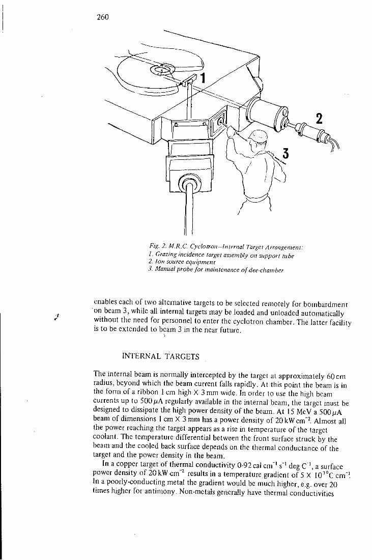
260
1
Fig. 2. M.R.C. Cyclotron-Internal Target Arrangement: 1. Grazing incidence target assembly on support tube 2. Ion source equipment 3. Manual probe for maintenance of dee-chamber
enables each of two alternative targets to be selected remotely for bombardment on beam 3, while all internal targets may be loaded and unloaded automatically without the need for personnel to enter the cyclotron chamber. The latter facility is to be extended to beam 3 in the near future.
INTERNAL TARGETS
The internal beam is normally intercepted by the target at approximately 60 cm radius, beyond which the beam current falls rapidly. At this point the beam is in the form of a ribbon 1 cm high X 3 mm wide. In order to use the high beam currents up to 500/iA regularly available in the internal beam, the target must be designed to dissipate the high power density of the beam. At 15 MeV a 500/uA beam of dimensions 1 cm X 3 mm has a power density of 20 kW cm'^. Almost all the power reaching the target appears as a rise in temperature o f the target coolant. The temperature differential between the front surface struck by the beam and the cooled back surface depends on the thermal conductance of the target and the power density in the beam.
In a copper target of thermal conductivity 0-92 cal cm' ' s'' deg C"', a surface power density of 20kW cm"^ results in a temperature gradient of 5 X 10''°C cm"'. In a poorly-conducting metal the gradient would be much higher, e.g. over 20 times higher for antimony. Non-metals generally have thermal conductivities

261
much lower still, resulting in temperature gradients of the order of 1000 times higher than that in copper.
The hottest surface of the target must be kept below the temperature at which the vapour pressure of the target material rises sufficiently to allow appreciable evaporation either of the target itself, or of the products of the nuclear reaction. I f i t is assumed that the back of the target is kept between 100 and 150°C by cooling water at a pressure of ~7 kg cm"^, then at 20 kW cm"^ melting would occur i f the thickness exceeded 2 mm for copper or 6-1 mm for antimony. Target construction therefore would present serious mechanical problems except for materials of high conductivity like copper. Moreover, the thickness of 0-1 mm is less than the particle range at 15 MeV, and this would affect the production efficiency in some cases.
Perhaps a more serious problem would be that of water cooling. At thicknesses of this order the power density at tbe cooled surface will be similar to that in the beam itself. However, the maximum power which could be removed by water cooling under conditions of surface boiling was found in experimental tests to be only about 4-5 kWcm"^. On several counts, therefore, targets perpendicular to the internal beam of the M.R.C. cyclotron are not practicable.
The two methods commonly employed to reduce the power density at the target surface and at the cooled surface are either to present the target to the beam at grazing incidence, as depicted in Fig. 2, or to move the target or the beam continuously.
Rotating targets have been used at Hammersmith, but have been abandoned because of the complicated mechanism required to provide:
(a) the high rotational speed necessary to avoid mechanical stresses from thermal cycUng, and
(b) sufficient mechanical power to overcome the eddy current drag on the target assembly in the magnetic field.
Since these two requirements are in direct opposition, and since such targets are inherently more expensive to produce than their static counterparts, rotating or vibrating target systems have not been developed. Thus the M.R.C. machine uses
/ a grazing or oblique incidence target in the form of a flat plate arranged tangentially to the beam. From purely thermal considerations, since the power density is reduced by a factor of. 1/cos 6 (Fig. 3), such obhque incidence targets can be thinner than perpendicular incidence targets by a factor of cos 6, where 6
o i c e - c r i o N Ay
NcioE-tvJce-.v
Fig 3. Internal target geometry
262
is the angle between beam direction and the normal to the target face. The effective thickness of the target presented to the beam is increased by 1 /cos Q and the former becomes a tliick target in the nuclear sense in practical cases.
Since the beam has a finite radial width, 0 is not the same for all particles. I f the inner edge of the beam is tangential to the target the average value of 1/cos 0 is about 10 in the M.R.C. machine.
For chemical processing the smaller the amount of target material present the better; in practice target thicknesses between 0-025 and 0 050 mm are found to give good yields on the grazing incidence target.
Clearly a target 0-050 mm in thickness needs mechanical support to retain the coolant; this is provided by a target backing plate which is in intimate thermal contact with both the target inaterial and the coolant. The plate is of high thermal conductivity, copper or aluminium being chosen according to the chemical process to be employed in separation, for some of the backing plate material usually appears as a contaminant.
The target back is smooth for optimum cooling, and a water velocity of 4 m s'' is obtained by narrowing the waterway at the back of the target plate to 0-25 mm. The direction of flow is perpendicular to the long dimension of the beam strike area. Because the effectiveness of the cooling depends on a higii water velocity, it is essential to avoid distortion of the target plate, which would widen the gap and reduce the velocity. In practice copper or aluminium target plates need to be at least 2 mrh thick to avoid distortion under the combined effect of heat and water pressure.
A thin target plate (1 -5 mm) permits a liigh heat transfer rate througli the plate, but affords little cooling due to conduction parallel to its surface. Conversely, a thick plate (6-5 mm) has a low heat transfer rate through the plate but a high cooling capacity due to conduction parallel to its surface. Maximum heat dissipation is therefore achieved at an optimum target plate thickness. Experimental measurements on copper target plates show this to be about 5 m m for applied power densities between 0-3 i^nd 5 kWcm^. However, 3 i n m was
Fig. 4. Internal Target Head: 1. Target plate clamp with carbon blocks 2. Target plate carrying target material 3. Water flow spoiler 4. Target head block with V ring and water wavs 5. Support tube with water connections
263
finally chosen to simplify handling and reduce cost, without serious reduction in effectiveness.
Fig. 4 shows in exploded view the final form of the target assembly incorporating the features described above. Several other features have been added as a result of practical operating experience over a period of seven years. For example, as the machine tuning is adjusted the centre of curvature of the beam moves and the strike area moves along the plate. I f the edge of the beam should approach either end of the target it strikes a carbon block, causing it to glow, giving visible warning on a CCTV monitor. As mentioned above, a remote automatic handling machine removes the target assembly from the vacuum system, detaches it from its water supplies, and transfers it by railway to a distant processing cell where the assembly is dismantled. The target plate is then removed and machined or chemically treated to remove the target layer, from which the required isotope is then extracted. The total bombardment on each target in [lA h is recorded by a device which integrates the rise in water temperature against time.
In preparing targets for bombardment, a layer of target material 0-025-0-050 mm tiiick is applied to the target plate by a method selected to provide the optimum bonding. For example chromium (for "Fe production) is electroplated on aluminium, nickel (for ^^Zn production) is deposited by metal spraying, v/hilst lead (for ^^Bi production) is fused onto copper in a vacuum furnace.
EXTERNAL TARGETS
(a) General. Clearly, internal targets for higlT beam currents can be prepared only from a limited range of materials with suitable physical properties. The M.R.C. cyclotron has the design restnciioii that such targets can only be bombarded at maximum beam energy and, as has been shown, are necessarily
^thick in the nuclear sense. Fortunately these limitations can be overcome by ''using the extracted beam, albeit at the expense of lower maximum currents.
Once extracted from the dee-chamber, the particle beam is defocused by quadrupole magnets and can be rriade to cover a relatively large area (5-10 cm wide and 1 cm high) at the external target positions on beams 3 and 4. Having reduced the power density in this way, targets can be presented normal to the beam instead of at grazing incidence, and it is then possible to vary the tliickness of target material which is bombarded. The energy of the beam incident on tht target surface may also be varied by interposing energy-degrading foils of the appropriate thickness. These two variables can be very important; for example, in ' ^ • ' l production from natural antimony the incident a-particle energy is reduced to 25 MeV, and the energy lost in the antimony is restricted to 2 MeV by limiting its thickness to 0 025 mm. This minimises contamination of the ' ^ " ' l produced by an {a, 2n) reaction by ' ^ " i from (a, n) and (a, 3n) reactions.^
Although it is not always desirable to degrade the beam energy, it is usually necessary to interpose a thin foil between the incoming beam and the target material in order that the latter, i f it be a powder, liquid or gas, should be contained. Similarly, a foi l is needed at the end of the external beam tube to maintain the vacuum. In principle a single foil could combine both these functions, but in practice target foils are often found to be very vulnerable to corrosion by hot target materials, and so separate foils are used. On the M.R.C. macliine, 0-025 mm titanium is used on the beam tube, as tliis has high mechanical strength
2 6 4
and thermal conductivity. Target foils are usually of aluminium or copper, unless there are special reasons for avoiding these materials. Both foUs must be efficiently cooled, or they may be perforated by the beam; this is achieved by circulafing cooled dry air between them and by water-cooling their supporting frames. A grid of 25 0-2 mm diam. tungsten wires having a 5 mm spacing is supported 2-6 cm on the vacuum side of the titanium fo i l , and a CCTV system is used to show the beam distribution indicated by incandescence of the wires intercepting the beam. Since all external targets are mounted in such a way that they are electrically insulated from the cyclotron and its beam tubes, beam currents can be measured using a microammeter and simultaneously integrated.
(bj Solids. The standard target plate used to sujjport solid target materials during bombardment in the external beam is similar to that used for internal bombardment, and they may be prepared in the same way, e.g. anfimony (for ' ^ " ' l production) is electroplated on copper. However, many alternative methods of preparation are
Fig. 5. External Target for Solid Materials: 1. Target friountingplate 2. Target plate carrying target material widi target foil shown cut away 3. Target cooling block with 'O'ring
available; for instance, a thin metal foil (such as indium for "''Sb production) may simply be clamped to the target plate, or a salt (such as NaBr for ^'Rb production) may be pressed firmly or melted into grooves cut in the plate. A typical target of tliis type is shown in Fig. 5 , which also shows the method of applying water cooling to the back of the target plate.
Variations on the standard pattern of solid external targets are used in preparing radioactive gases incorporating "C or '•^N. Carbon-11 is produced by the reactions " ^ ( d , n)"C and "B(d, 2n)"C; the target material is B2O3, wliich is bombarded with deuterons of ~ 1 4 MeV in the target system shown in Fig. 6 . A layer of B 2 O 3 1 - 2 mm thick, is melted on to the wedge. The deuteron beam passes through the 0 - 0 5 0 mm aluminium window and strikes the surface of the B2O3 at grazing incidence. With a defocused beam of ~ 4 0 M A the power density is 1 0 - 2 0 watts cm"^ on the surface of the wedge, which is deliberately in poor thermal contact with the surrounding box.
This power density is sufficient to melt the B2O3, releasing "C-labelled gases
nt.
Fig. 6. External Target for ^Kl Production: 1. Target mounting plate (water cooling not shown) 2. Foil window 3. Brass wedge supporting B2O3 4. Gas-tight target box 5. Wedge locating pins 6. Gas connections
n)
Fig. 7. External Target for ' V' Production: 1. Target mounting plate 2. Foil window 3. Spacer 4. Graphite block 5. Gas-tight target box 6. Gas connections 7. Cooling water connections
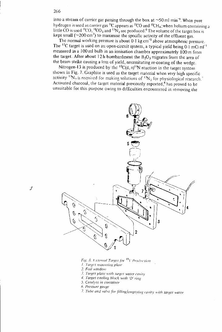
266
into a stream of carrier gas passing through the box at ~ 5 0 m l min"'. When pure hydrogen is used as carrier gas "C appears as "CO and "CH4; when helium containing a little CO is used "CO, "C02and '•'Nzare produced.^The volume of the target box is kept small (~200 cm"') to maximise the specific activity of the effluent gas.
The normal working pressure is about 0-1 kgcm"^ above atmospheric pressure. The "C target is used on an open-circuit system, a typical yield being 0 1 mCi ml"' measured in a 100 ml bulb in an ionisation chamber approximately 100 m from the target. After about 12 h bombardment the B2O3 migrates from the area of the beam strike causing a loss of yield, necessitating re-coating of the wedge.
Nitrogen-13 is produced by the '^C(d, n)'^N reaction in the target system shown in Fig. 7. Graphite is used as the target material when very high specific activity ' ' ' N T is required for making solutions of '"^2 f or physiological research.' Activated charcoal, the target material previously reported,^ has proved to be unsuitable for this purpose owing to difficulties encountered in removing the
Fig. S. External Target for I" I'roihiciion 1. Target nwuiiiitig plaie 2. Foil window 3. I'argei plaie with target water cavity 4. Target cooling block with 'O'ring 5. Catalyst in container 6. Pressure gauge 7. Tube and valve for filUnglenipiying cavity with target watei
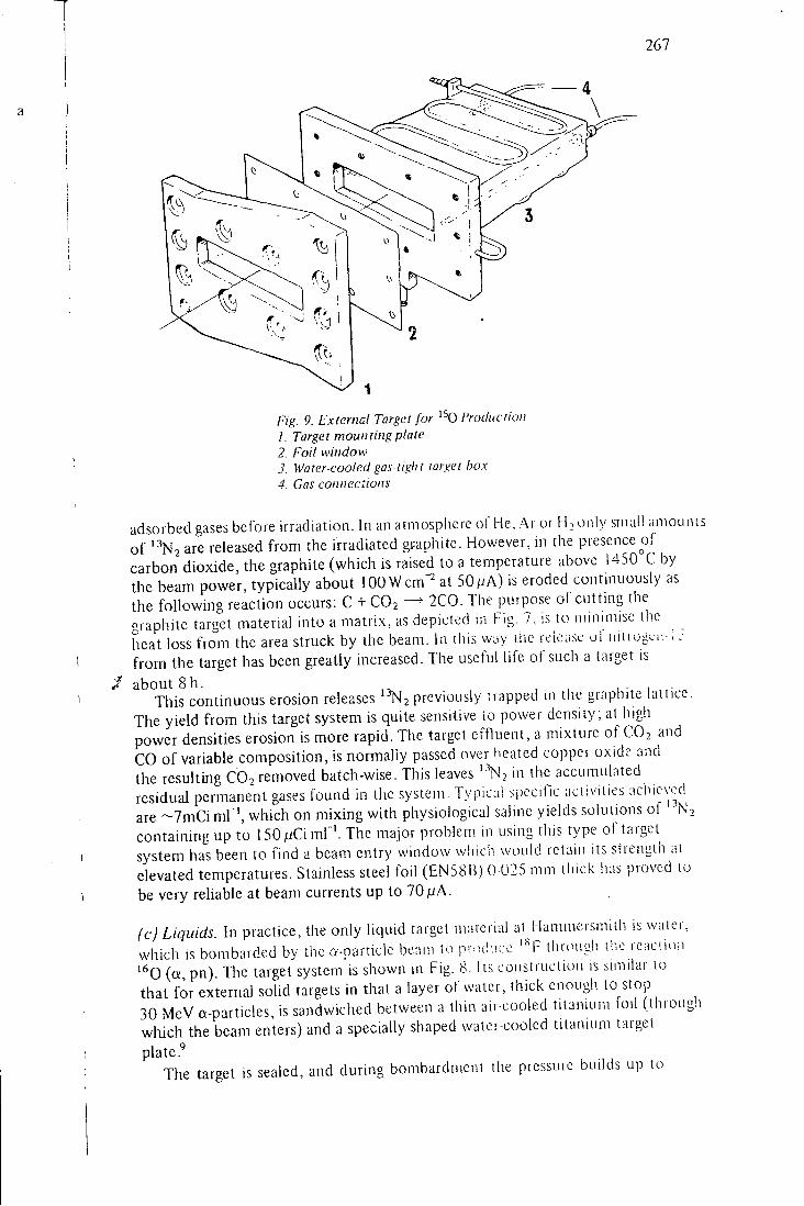
267
\
<0 \
(To
Fig. 9. External Target for ' ^0 I'roduction 1. Target mounting plate 2. Foil window 3. Water-cooled gas-light target box 4. Gas connections
adsorbed gases before irradiation. In an atmosphere of He. Ai or M^oiily small aniounis of are released from the irradiated graphite. However, in the presence of carbon dioxide, the graphite (which is raised to a temperature above 1450°C by the beam power, typically about lOOWcm^ at SOjuA) is eroded continuously as the following reaction occurs: C + CO2 —^ 2C0. The purpose of cutting the graphite target material into a matrix, as depicted in Fig. 7. is to minimise the heat loss from the area struck by the beam. In this WDV the release of niirogcii- i J f rom the target has been greatly increased. The useful life of such a target is about 8h .
This continuous erosion releases ' N2 previously trapped in the graphite lattice. The yield from this target system is quite sensitive to power density; at higli power densities erosion is more rapid. The target effluent, a mixture of CO2 and C O of variable composition, is normally passed over healed copper oxide and the resulting CO2 removed batch-wise. This leaves ''^N, in the accumulated residual permanent gases found m the system. Typical specific activities achieved are ~ 7 m C i ml ', which on mixing with physiological saline yields solutions of '•'NS containing up to 150/jCimr' . The major problem in using this type of target system has been to find a beam entry window whicli would retain its strength at elevated temperatures. Stainless steel foil (EN58B) 0 025 mm tiiick iias proved lu be very reliable at beam currents up to 70/JA.
(c) Liquids. In practice, the only liquid target iviatcrial at Hammersmith is water, which is bombarded by the a-particle beam to prod . ire '^'F through liic rcacuon ' ^ O (a, pn). Tlie target system is shown in Fig. 8. I is construction is similar \o that for external solid targets in that a layer of water, thick enough to stop 30 MeV a-particles, is sandwiched between a thin air-cooled titanium foil (througli wliich the beam enters) and a specially shaped water-cooled titanium target
plate The target is sealed, and during bombardment the pressure builds up to
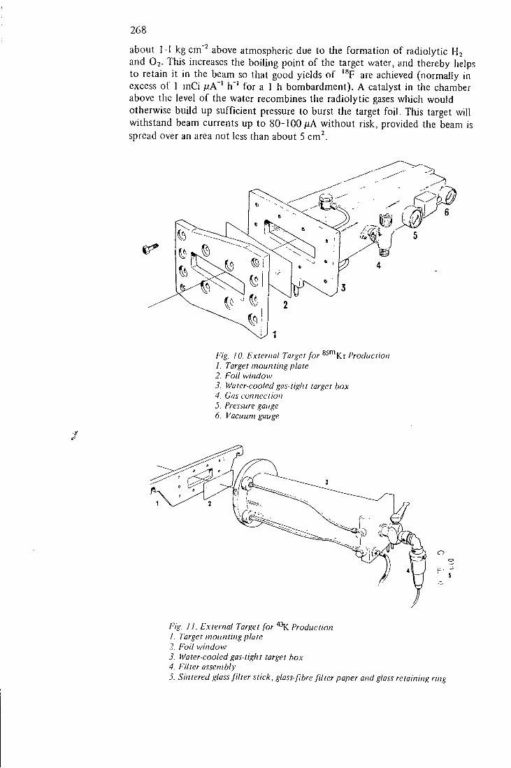
268
about 1 - I kg cm"^ above atmospheric due to the formation of radiolytic H2 and O2. Tliis increases the boiling point of the target water, and thereby helps to retain it in the beam so that good yields of '^F are achieved (normally in excess of 1 mCi ^tA"' h ' ' for a 1 h bombardment). A catalyst in the chamber above the level of the water recombines the radiolytic gases which would otherwise build up sufficient pressure to burst the target foi l . This target will withstand beam currents up to 80-lOOfiA without risk, provided the beam is spread over an area not less than about 5 cm^.
Fig. 10. External Target for ^ " ^ K r Production 1. Target mounting plate 2. Foil window 3. Water-cooled gas-tight target box 4. Gas coniiectioit 5. Pressure gauge 6. Vacuum gauge
Fig. IF External Target for Production 1. Target mounting plate 2. Foil window 3. Water-cooled gas-tight target box 4. Filter assembly 5. Sintered glass filter stick, glass-fibre filter paper and glass retaining ring
269
(d) Gases. Essentially, such a target is shnply a water-cooled metal box filled with gas, and having a thin foi l window for beam entry. The tliickness of this window, its material, the target box dimensions and its operating pressure are all interdependent as discussed below.
Three such targets are in regular use. The first is shown in Fig. 9 and is used to prepare molecular oxygen labelled with '^0. This is produced by the reaction '""N (d, n) '^0, using nitrogen as the target gas, to which 4% O2 is added. The oxygen acts as a carrier for the '^O; if it is absent the yield of '^0 as O2 is very small. The target beam entry window is 1 mm magnesium, which reduces the beam energy from 15 to 5 MeV. This has the effect of reducing the contaminants otherwise produced by the reactions ' ' 'N (d, na) " C and '^0 (d, na) '^N.^
The length of the box is sufficient to stop the 5 MeV deuteron beam with the target gas at about 0-5 kg cm"^ above atmospheric pressure. Using 35 /iA of 5 MeV deuterons and a target gas flow rate of 0-7 1 min"', the yield of '^02 is typically 120 mCi T ' measured in a 500 ml bulb in an ionisation chamber 90 m from the target.
A target similar in design (Fig. 10) is used to produce ** ' ^Kr by the ""Krfd, p) reaction. The 45 cm long target box is filled with krypton to a pressure of 0-7 kg cm^ above atmospheric, ideally sufficient to stop the deuteron beam. This pressure rises to about 1 -75 kg cm"^ during bombardment at 30 /iA. The target box is therefore rigidly constructed, and is fitted with a 0-025 mm titanium foil window which readily withstands the pressure and temperature differential. After bombardemnt, krypton is recovered from the target by sorption on molecular sieve in a trap cooled by liquid nitrogen.
Finally, the ta-iget used for the production of ''^K is shown in Fig. 11. Argon is the target gas, and when this is bombarded in the a-particle beam " • ' K results from the '*'^Ar(Q:, p) reaction. The target box is filled to a pressure of 0-7 kg c m a b o v e atmospheric, and is fitted with a foil window which reduces the incident beam energy to 15 MeV, which is approximately the threshold for the "^^Arfa, pn) " ^K reaction. This foil window ctmsists of 0-025 mm titanium which, as in the krypton target, has the required mechanical strength, backed by a 0-2 mm aluminium fo i l , to degrade the beam energy. '°
During bombardment, the argon is recirculated througli the target box at about 100 1 min ' ' by oU-free diaphragm pumps. On leaving the target box the gas passes through a glass-fibre filter paper supported on a coarse sintered glass disc, which traps more than 70% of the " K produced. At the end ol" bombardment the acUvity is readily recovered by washing the filter with a few ml of dilute hydrochloric acid.
R E F E R E N C E S
1. Gallop, J. W., Vonbers:, D. D.. I'ost, R. .1., Powell, W. B., .Sluirp, .1.. L i i u i \V:itortoii. 1'. .!.. Proc.l.E.E. 104 8, 452 , (1957) .
2. Vonberg, D. D. and Fowler, J. V.,Namre 198 ,827 , (1963) . 3. Clark, J. C , Matthews, C. M. E., Silvester, D. J., and Vonberi;, D. [:>.. Nucleonics 25, 54
(1967). 4. Glass, H . 1. and Silvester, D . J., submitted to Brit. J. Radiol. (19,^91. 5. Silvester, D. J., Sugdcn, J., and Watson, 1. A. , Radiochemical and Rthiicunalviical Letters.
in press (1969). 6. Clark, J. C. and Buckingham, P. D., submitted to Radiochemical and Radioonalvtical
Letters {1969). 1. Clark, J. C. and Buckingham, P. D. , (o be published. 8. Buckingham, P. D. and Forse, D. J.,//?;. / . Appl. Radn. and Isotopes 14, 4 39, (1963). 9. Clark, J. C.and Silvester, D. J., Int. J. Appl. Radn. and Isotopes 17. 151.(1 966).
10. Clark, J. C , submitted to Radiochemical and Radioanalytical Letters {1 969).
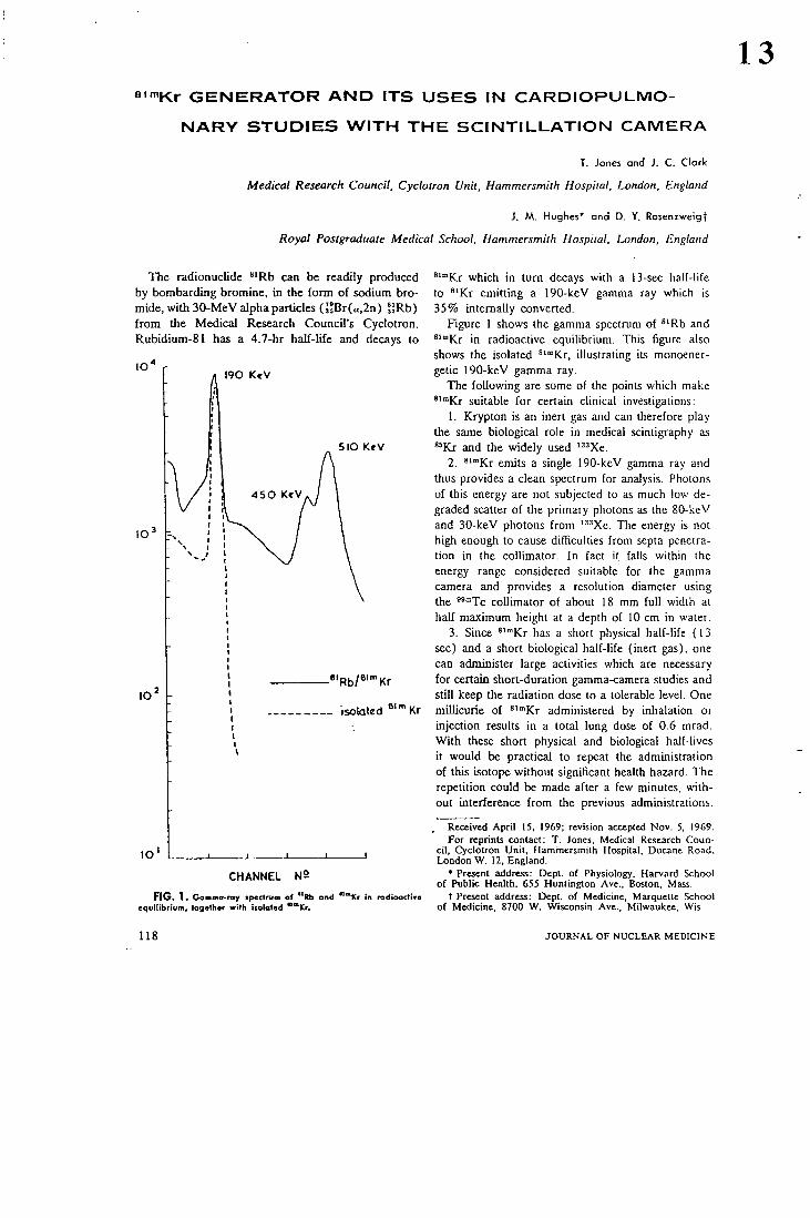
8 " " K r G E N E R A T O R A N D I T S U S E S IN C A R D I O P U L M O
N A R Y S T U D I E S W I T H T H E S C I N T I L L A T I O N C A M E R A
T. Jones and J . C . Clark
Medical Research Council, Cyclotron Unit, Hammersmith Hospital, London, England
i. M. Hughes* and D. Y. Rosenzweigt
Royal Postgraduate Medical School, Hammersmith Hospital, London, England
13
The radionuclide "'Rb can be readily produced by bombarding bromine, in the form of sodium bromide, with 30-MeV alpha particles {nBr(a,2n) l]Rb) from the Medical Research Council's Cyclotron. Rubidium-81 has a 4.7-hr half-life and decays to
I90 K«V
5IO KeV
•'Rb/«"'"Kr
isolated Kr
^'"Kr which in turn decays with a 13-sec half-life to *'Kr emitting a 190-keV gamma ray which is 35% internally converted.
Figure 1 shows the gamma spectrum of *'Rb and *"°Kr in radioactive equilibrium. This figure also shows the isolated *'"Kr, illustrating its monoener-getic 190-keV gamma ray.
The following are some of the points which make " " K r suitable for certain clinical investigations:
1. Krypton is an inert gas and can therefore play the same biological role in medical scintigraphy as " K r and the widely used ' "Xe.
2. ^ '"Kr emits a single 190-kcV gamma ray and thus provides a clean spectrum for analysis. Photons of this energy are not subjected to as much low degraded scatter of the primary photons as the 80-keV and 30-keV photons from •'•''Xe. The energy is not high enough to cause difficulties from septa penetration in the collimator. In fact it falls within the energy range considered suitable for the gamma camera and provides a resolution diameter using the ""Tc collimator of about 18 mm full width at half maximum height at a depth of 10 cm in water.
3. Since «'°Kr has a short physical half-life (13 sec) and a short biological half-life (inert gas), one can administer large activities which are necessary for certain short-duration gamma-camera studies and still keep the radiation dose to a tolerable level. One millicurie of ^ '"Kr administered by inhalation or injection results in a total lung dose of 0.6 mrad. With these short physical and biological half-lives it would be practical to repeat the administration of this isotope without significant health hazard. The repetition could be made after a few minutes, without interference from the previous administrations.
C H A N N E L N 2
FIG. 1. Gamma-ray tp«ctrum of "Rb and """Kr in radioactive equilibrium, together with isolated ""Kr.
, Received April 15, 1969; revision accepted Nov. 5, 1969. For reprints contact: T . Jones, Medical Research Coun
cil, Cyclotron Unit, Hammersmith Hospital, Ducane Road, London W. 12, England.
* Present address: Dept. of Physiology, Harvard School of Public Health, 655 Huntington Ave., Boston, Mass.
t Present address: Dept. of Medicine, Marquette School of Medicine, 8700 W. Wisconsin Ave., Milwaukee, Wis.
118 J O U R N A L O F N U C L E A R M E D I C I N E
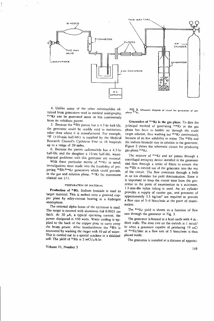
T w o W A Y T A P S ^
B L A D D E R
pLOWMEr F I L T E R S
C Y L I N D E R
w o W A Y V A L V E
M O O T M P I C C t E N E R A T O R
A M P L I F I E R
4. Unlike many of the other radionuclides obtained from generators used in medical scintigraphy, * '"Kr can be generated more or less continuously from its rubidium parent.
5. Because the ^iRb parent has a 4.7-hr half-life, the generator could be readily used in institutions other than where it is manufactured. For example,
(110-min half-life) is supplied by the Medical Research Council's Cyclotron Unit to 16 hospitals up to a range of 20 miles.
6. Because the parent radionuclide has a 4.7-hr half-life and the daughter a 13-sec half-life, waste-disposal problems with this generator are minimal.
With these particular merits of """Kr in mind, investigations were made into the feasibility of preparing ^'Rb-^'^Kr generators which could provide, in the gas and solution phase, *'"'Kr for convenient clinical use (7 ) .
P R E P A R A T I O N O F M A T E R I A L
Production of *'Rb. Sodium bromide is used as target material. This is melted onto a grooved copper plate by eddy-current heating in a hydrogen atmosphere.
The external alpha beam of the cyclotron is used. The target is covered with aluminum foil 0.0025 cm thick. At 30 fiA, a typical operating current, the power dissipated is 450 watts. Water cooling is-applied to the back of the copper plate to carry away the beam power. After bombardment the "Rb is recovered by washing the target with 10 ml of water. This is carried out in a special machine in a shielded ceU. The yield of «'Rb is 2 mCi/,iA-hr.
FIG. 2 . Sthemolic dtogrofn of circuil lor generotion oi 901-phose ""Kr.
Generation of ^'"'Kr In the gas phase. To date the principal method of generating "'""Kr in the gas phase has been to bubble air through the crude target solution, thus washing out *""Kr continuously because of its low solubility in water. The ^'Rb and the sodium bromide stay in solution in the generator. Figure 2 shows the schematic circuit for producing gas-phase *""Kr.
The mixture of *'"'Kr and air passes through a centrifugal antispray device installed in the generator and then through a series of filters to ensure that no *'Rb is carried out of the generator into the rest of the circuit. The flow continues through a bulb in an ion chamber for yield determination. Since it is important to keep the transit time from the generator to the point of examination to a minimum, 1.5-mm-dia nylon tubing is used. An air cylinder provides a supply of carrier gas, and pressures of approximately 3.5 kg/cm^ are required to provide a flow rate of 5-6 liters/min at the point of examination.
The ^'"•Kr yield is shown as a function of flow-rate through the generator in Fig. 3.
The generator is housed in a lead castle with 4-in.-thick walls. TTie dose rate on the outside is 1 mrad/ hr when a generator capable of producing 19 mCi of ^'"'Kr/liter at a flow rate of 5 liters/min is then placed inside.
The generator is installed at a distance of approxi-
Volume 11, Number 3 119
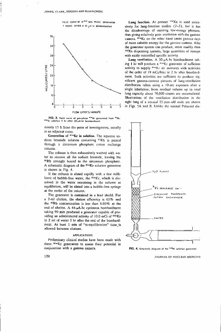
J O N E S , C L A R K , H U G H E S A N D R O S E N Z W E I G
YIELD CURVE OF K r " ' " GAS PHASE GENERATOR
2 HOURS AFTER A 25 hr BOMBARDMENT
36
32
2 8 h
i/i 20
0 1 2 3 4 5 6 7
FLOW LITRES/ MINUTE
FIG. 3. Yield curve of got-phate *""ICr generoted from "Rfc>-"™Kr tolulion 2 hr after 25-^LA-tir bombardment.
mately 15 ft from the point of investigations, usually in an adjacent room.
Generation of ^""Kr in solotion. The aqueous sodium bromide solution containing "'Rb is passed through a zirconium phosphate cation exchange column.
The column is then exhaustively washed with water to remove all the sodium bromide, leaving the *'Rb strongly bound to the zirconium phosphate. A schematic diagram of the " " K r solution generator is shown in Fig. 4.
If the column is eluted rapidly with a few milliliters of bubble-free water, the '""Kr, which is dissolved in the water remaining in the column at equilibrium, will be eluted into a bubble-free syringe at the outlet of the column.
The generator is contained in a lead shield. For a 2-ml elution, the elution efficiency is 65% and the *'Rb contamination is less than 0.03% at the end of elution. A 44-/iA-hr cyclotron bombardment taking 90 min produced a generator capable of providing an administered activity of 10.0 mCi of ^""Kr in 2 ml of water 3 hr after the end of the bombardment. At least 1 min of "re-equilibration" time is allowed between elutions.
A P P L I C A T I O N S
Preliminary clinical studies have been made with these ' ' " K r generators to assess their potential in conjunction with a gamma camera.
Lung function. At present ' "Xe is used extensively for lung-function studies (2-5) , but it has the disadvantage of emitting low-energy photons, thus giving relatively poor resolution with the gamma camera. " " K r on the olhcr hand emits gamma rays of more suitable energy for the gamma camera. Also the generator system can produce, more readily than " 'Xe dispensing systems, large quantities of isotope with easily controlled specific activity.
Lung ventilation. A 30-/iA-hr bombardment taking 1 hr will produce a *'"'Kr generator of sufficient activity to supply *""Kr air mixtures with activities of the order of 19 mCi/liter at 2 hr after bombardment. Such activities are sufficient to produce significant gamma-camera pictures of lung-vcntilaiion distribution taken using a 10-sec exposure after a single inhalation; from residual volume up to total lung capacity about 70,000 counts arc accumulated. Illustrations of the ventilation distribution in the right lung of a normal 25-year-old male are shown in Figs. 5A and B. Unlike the normal Polaroid dis-
- H j O £Ll . ' iNT
Va iB.5oe.Beo ON :-Zl[;.COr.JluM P H O S P H A T t CfrTCOM e)tCMAN&e'B
FIG. 4 . Schematic diogrom of the "°ICr tolulion generator.
120 J O U R N A L O F N U C L E A R M E D I C I N E
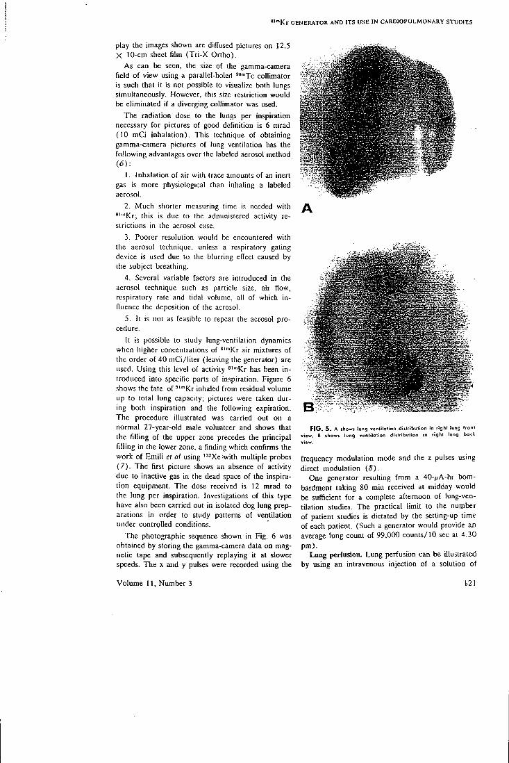
«""Kr G E N E R A T O R A N D I T S U S E I N C A R D I O P U L M O N A R Y S T U D I E S
play the images shown are din;used pictures on 12.5 X 10-cm sheet film (Tri-X Ortho).
As can be seen, the size of the gamma-camera field of view using a parallel-holed '*"'Tc collimator is such that it is not possible to visualize both lungs simultaneously. However, this size restriction would be eliminated if a diverging collimator was used.
The radiation dose to the lungs per inspiration necessary for pictures of good definition is 6 mrad (10 mCi inhalation). This technique of obtaining gamma-camera pictures of lung ventilation has the following advantages over the labeled aerosol method (6) :
1. Inhalation of air with trace amounts of an inert gas is more physiological than inhaling a labeled aerosol.
2. Much shorter measuring time is needed with "'"'Kr; this is due to the administered activity restrictions in the aerosol case.
3. Poorer resolution would be encountered with the aerosol technique, unless a respiratory gating device is used due to the blurring effect caused by the subject breathing.
4. Several variable factors are introduced in the aerosol technique such as particle size, air flow, respiratory rate and tidal volume, all of which influence the deposition of the aerosol.
5. It is not as feasible to repeat the aerosol procedure.
It is possible to study lung-ventilation dynamics when higher concentrations of ^'"'Kr air mixtures of the order of 40 mCi/liter (leaving the generator) are used. Using this level of activity ^'"'Kr has been introduced into specific parts of inspiration. Figure 6 shows the fate of ^'""Kr inhaled from residual volume up to total lung capacity; pictures were taken during both inspiration and the following expiration. The procedure illustrated was carried out on a normal 27-year-old male volunteer and shows that the filling of the upper zone precedes the principal filling in the lower zone, a finding which confirms the work of Emili el al using '"Xe'with multiple probes (7) . The first picture shows an absence of activity due to inactive gas in the dead space of the inspiration equipment. The dose received is 12 mrad to the lung per inspiration. Investigations of this type have also been carried out in isolated dog lung preparations in order to study patterns of ventilation under controlled conditions.
The photographic sequence shown in Fig. 6 was obtained by storing the gamma-camera data on magnetic tape and subsequently replaying it at slower speeds. The x and y pulses were recorded using the
FIG, 5. A showi lung ventilation diitribution in right lung front riew. B showj lung venlitotion distribution in right lung bock
frequency modulation mode and the z pulses using direct modulation (8).
One generator resulting from a 40-/iA-hr bombardment taking 80 min received at midday would be sufficient for a complete afternoon of lung-ventilation studies. The practical limit to the number of patient studies is dictated by the setting-up time of each patient. (Such a generator would provide Em average lung count of 99,000 counts/10 sec al 4.30 pm).
Lung perfusion. Lung perfusion can be illuslraled by using an intravenous injection of a solution of
Volume 11, Number 3 1-21
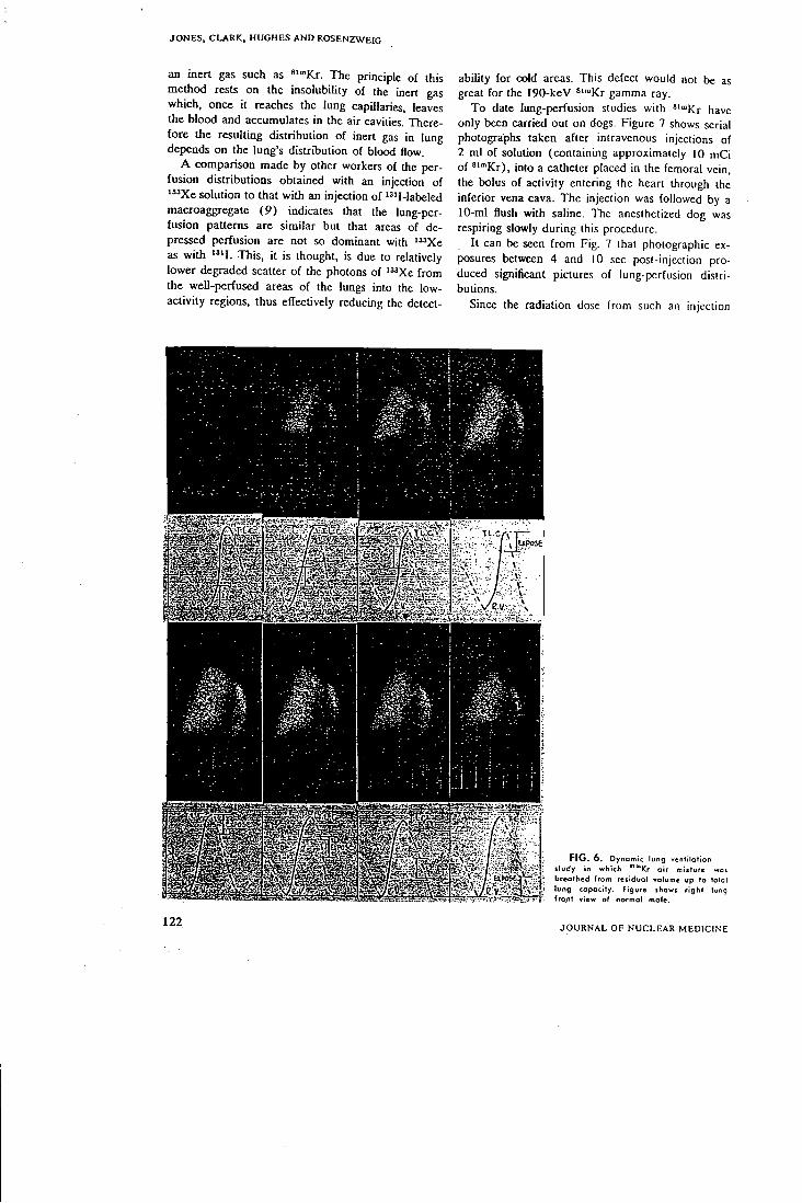
J O N E S , C L A R K , H U G H E S A N D R O S E N Z W E I G
an inert gas such as ^""Kr. The principle of this method rests on the insolubility of the inert gas which, once it reaches the lung capillaries, leaves the blood and accumulates in the air cavities. Therefore the resulting distribution of inert gas in lung depends on the lung's distribution of blood flow.
A comparison made by other workers of the perfusion distributions obtained with an injection of "^Xe solution to that with an injection of '"I-labeled macroaggregate (9) indicates that the lung-per-fusion patterns are similar but that areas of depressed perfusion are not so dominant with " ' X e as with " ' I . This, it is thought, is due to relatively lower degraded scatter of the photons of " 'Xe from the well-perfused areas of the lungs into the low-activity regions, thus effectively reducing the detect-
ability for oold areas. This defect would not be as great for the 190-keV *"°Kr gamma ray.
To date lung-perfusion studies with ^'"'Kr have only been carried out on dogs. Figure 7 shows serial photographs taken after intravenous injections of 2 ml of solution (containing approximately 10 mCi of ' ' " K r ) , into a catheter placed in the femoral vein, the bolus of activity entering the heart through the inferior vena cava. The injection was followed by a 10-ml flush with saline. The anesthetized dog was respiring slowly during this procedure.
It can be seen from Fig. 7 that photographic exposures between 4 and 10 sec post-injection produced significant pictures of lung-perfusion distributions.
Since the radiation dose from such an injection
< tec
FIG. 6 . Dynamic lung ventilation iludy in which """Kr oir mixture woi breathed from reiidual volume up to total lung copocity. Figure ihowj right lung fro.nt view of normol mole.
122 J O U R N A L O F N U C L E A R M E D I C I N E
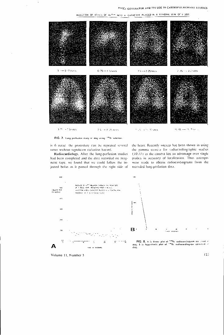
" " " K r G E N E R A T O R A N D I T S U S E IN C A R D I O P U L M O N A R Y S T U D I E S
INJECtlON OF IOrr>cs OF Kr »"" INTO A C A I H E I E R P L A C E D IN A FEt.<0RAL VF.IN OF A QCG
0 —- 0 • "Ssccs 0 -75 — 1 5 s e e s 1 5 — 2 ? 5 s e c s
F I G . 7. lung perfusion itudy in dog uiinq '"'Kr solulion.
is 6 mr;id. ihc procedure c;iii be repealed several limes wiihoul sigiiilicani radiaiion hazard.
Kadiocardiology. Afler the lung-perfusion sludies hiid been conipleled and ihc diiiii recorded on niag-nclic lape, we found lhal we could follow ihc injected bolus as il p;issed through llie riglii side of
ihe hean. Reccnily iiilcresi has been shown in using ihc gamma ciuiicra for radiocardiographic siudies ilO.il) as the camera has an advantage over single probes in accuracy of localization. Thus iiiicmpt'. were made to obtain radiocardiograms from the recorded lung-pcrfusion data.
A
Volume 1 1, Number 3
" ( ix SCCOWDS
F I G . 8. A is lineot plof o( dog. B it logarithmic plot of dog.
"Kf fodtocofdiogram ob . r.-d 'Kt fodlocofdiogrom obia""- d
1 2 ;
J O N E S , C L A R K , H U G H E S A N D R O S E N Z W E I G
It was possible to obtain the quantitative time course of the *""Kr as it passes through the heart by simply masking off on the oscilloscope screen all regions other than the right side of the heart, as depicted by the photographs, then replaying the tape and counting the dots as they appear on the screen with a photomultiplier (8,11-13). Figures 8A and B show a linear and logarithmic plot of a right heart radiocardiogram resulting from an injection through a catheter placed in a femoral vein.
It can be seen that there is a tail on the radio-cardiagrams due, it is thought, to some lung tissue overlying the heart as well as some scatter from the adjacent lung areas into the heart region.
It is thought legitimate to extrapolate the falling portions of the curve to the base line on semilog paper, thus describing the full radiocardiogram. From this a mean transit time can be obtained for the passage through the right heart. The curves shown have been corrected for decay.
The advantages of this radionuclide over others used to date for gamma-camera radiocardiographic studies, e.g. ^"•Tc (10,11), are:
1. The absorbed dose is much lower because the isotope is only present long enough for the examination. A 10-mCi injection of ^'"'Kr results in an absorbed dose of 6 mrad to the lungs (critical organ), whereas a 10-mCi injection of ' ' "Tc results in 960 rarad to the upper large intestine (critical organ) (14).
2. High specific activities are readily available from the "'Rb parent.
3. The test can be repeated without significant hazard.
4. Being an inert gas ^''"Kr does not return from the lungs to the left heart as ^""Tc does. Therefore there is no interference in the radiocardiogram due to activity in the left heart.
This last point, although advantageous in the context of the right heart, is a disadvantage in that a left heart radiocardiogram cannot be obtained as in the ''™Tc case. Even when it is obtained, these composite heart profiles are not easy to analyze.
CONCLUSION
The properties of the gamma camera make it an important instrument for physiological studies of short duration. Thus specific isotopes are required to play a complementary role with the camera in these investigations. Such isotopes must be available in sufficient activities to make short duration studies statistically feasible, and yet they must have the physical characteristics to meet the requirements of high resolution and low absorbed dose to the patient (75).
It is believed that it is in this context that the '"Rb-*'"'Kr generator has a useful role to play in medicine in association with gamma cameras.
ACKNOWLEDGMENT
The authors wish lo acknowledge the support of D . D. Vonljerg and J . B . West and the helpful discussion of D. K. Bewley, D. 5. Silvesler, J . F . Fowler and H. I . Glass Thanks are also expressed for the cooperation of the Medical Physics Departinenl, in particular B . Wcstcrman. and for Ihc lechnical assistance of A . S. O. Ranicar and W . Verney together with the Cyclotron Team for their excellent service
R E F E R E N C E S
/ . J O N E S , T . AND C L A R I C . J . C : A cyclotron prtxluccd
" R b - " " K r generator and its uses in gamma camera sludies. Brii. J. Radiol. 4 2 : 2 3 7 , 1969.
2. W E S T , J . B.: Pulmonary function sludies with radioactive gases. Ann. Rev. Med. 18 :459 , 1967.
3. LoiCEN, M . K . AND W E S T C A T E , H . O : Using xenon-133 and the scintillation camera to evaluate pulmonary function. J. Nucl. Med. 9 : 4 5 , 1968.
4. SuPRENANT, E . L . , B E N N E T T . L . R , W E B B E R . M . M
AND W I L S O N , A. F . : Measurement of regional pulmonar>' ventilation with radioxenon and the Anger camera. / . Nucl. Med. 8 : 3 4 3 , 1967.
5. MARICS. a . , C H E R V O N Y , 1., L A N K F O R C . R . . S M I T H , E .
M., GiLsoN, A. J . AND SMOAIC. W . : Ventilalion perfusion relationship in humans measured by scintillation scanning-J. Nucl. Med. 9-.450. 1968.
6. PiRCHER, F . J . : The aerosol scan in the assessment of lung disease. In Proceedings of the Symposiuni on Nt/clear Activation Techniques in the Life Sciences, vol. 2. I A E A . Vienna. 1969.
7. M I L I C - E M I L I . J . , H E D E R S O N . J . A. M . . D O L O V I C H U .
M . B.. T R O P , D . AND K A N E , K . O . : Regional distribution
of inspired gas in the lung. J. Appl. Physiol. 21:749 . 1966. 8. H O L R O V D , a . M . AND J O N E S , T . : A simple method for
obtaining dynamic quantitative information from the gamma camera. Phys. Med. Biol. 14 :631 , 1969.
9. C E D E R Q U I S T , E . , N A V E R S T E N , Y . AND W H I T E . T . : Com
parative study of regional lung function with intravenous injection of "^I-Iabelled macroaggregated albumin and '"Xe. In Proceedings oj the Symposium on Nuclear Activation Techniques in the Life Sciences, vol. 1, I A E A , Vienna, 1968 .
10. A N G E R , H . O . , G O T T S C H A L K . D . C . Y A N O , Y . AND
SCHAER, L . R . : Scintillation camera in diagnosis and research Nucleonics 23:No. 1, 57 , 1965 .
11. A D A M , W . E . . S C H E N C K , P., K A M P M A N N , H . . L O R E N Z .
W . J . , S C H N E I D E R , W . C . . A M M A N . W . AND B I L A N I U K . L . :
Investigations of cardial dynamics using scintillation camera and computer. In Proceedings of the Symposium on Nuclear Activation Techniques in the Life Sciences, vol. 1. I A E A . Vienna. 1968 .
12. A D A M , W . E . AND L O R E N Z , W . J . : Ein Einfaches Verfahren zur analogen Auswertung funktionssziniigraphi-scher Messungen mil der Szintillation-Kamera. Aiompraris 1 3 : 4 . 1967.
IS. L O R E N Z , W . J . AND A D A M , W . E . : Digitate und analoge Auswertung von Aufnahmen mil de Szintillation Kamera. Nucl. Med. 6 : 3 6 7 , 1967 .
14. S M I T H , E . M . ; Internal dose calculation for ^"'Tc. J. Nucl. Med. 6 : 2 3 1 , 1965 .
15. Y A N O , Y . AND A N G E R , H . O . : Ultrashort lived radioisotopes for visualizing blood vessels and organs. / . Nucl. Med. 9:2, 1968 .
124 J O U R N A L O F N U C L E A R M E D I C I N E
1970, Br. 7 Rodiot., 43. 537-541 14
AUGUST 1970
Measurement of gastric emptying using the scintillation camera and 29Cs B y T . Jones, M.Sc. anA J. C. Clark, B.Sc.
Medical Research Council, Cyclotron Unit, Hammersmith Hospital, London, W.12
N. Kocak, M.D. and A . G. Cox, M.D., F.R.C.S. (E).
Department of Surgery, Royal Postgraduate Medical School, Hammersmith Hospital, London, W.12
and H. I . Glass, M.A. , A. lns t .P . Department of Medical Physics, Royal Postgraduate Medical School, Hammersmith Hospital, London, W.12 {Receixed October, 1939 and in revised form Februtirv, 1970)
Measurement of the rate of gastric emptying is a difficult clinical problem to which the best current solution is c.\ternal detection of a meal labelled with a radioactive tracer substance. This basic method ha.s the advantages that it docs not require nasogastric intubation and the results can be easily quantified.
The main problem in such a technique is accurate localisation of the stomach and continuous detection of all its radioactive contents. .\ radioisotope scanner can be used to outline the stomach and estimate the activity by adding up the counts within a defined area (Griffiths, Owen, Campbell and Shields, 1968; Williams, Glass, Arnot and De Gar-reta, 1968). The main disadvantage of the scanning technique is that detailed changes in stomach emp-tyine may be missed because of the time taken to .•;can the stomacli. Gross inaccuracies are inevitable when stomach emptying is rapid.
We have used the Anger scintillation camera to measure activity in the whole stomach. Quantitation has been achieved by a relatively simple and inexpensive technique, modified from one developed previously for quantifying gamma-camera data (Holroyd and Jones, 1969). In this method a photo-multiplier counts light scintillations within the area of interest on the oscilloscope display of the gamma-camera. 5 'Cr in the form of sodium chromate has been used by workers who have measured gastric emptying by radioisotopic scanning techniques (Griffiths et al., 1968). We have used cyclotron-produced '29Cs absorbed on a suspension of zirconium phosphate.
PHYSICAL CHARACTI-RISTICS OF ' - ^CS
1. Production of i^^Ci and its incorporation into a suitably inert medium for gastric emptying measurements.
•29Cs is produced on the Mcdicaj Research Council's cyclotron by bombarding iodine with 30
.\Ie\' a particles ' - ' I (a,2n) '-'Cs. 'I'lie target is prepared by pressing powdered sodium iodide (analytical grade) into a grooved aluminium plate. The target material is then covered with aluminium foil to prevent contamination and loss of powder during irradiation. The back of the target is water cooled during irradiation to carry awav the beam power, 450 watts al 30 fiA. The yield of '-^Cs is approximately 170 /iCiZ/i.^h. .-Xfter bombardment the sodium iodide is removed from the target plate by washing It with water. To the resulting sodium iodide solution containing '-^Cs is added approximately 1 g of zirconium phosphate cation e.vchanger (BIO-RAD ZP-1 iOO-200 mesh). The i- ^Cs Is absorbed rapidly on to the zirconium phosphate.The slurry of '^gQs-labelled zirconium phosphate is then separated from the sodium iodide by repeated washing and centrifugatlon, discarding the supernate. The high distribution coefficient lor caesium between zirconium phosphate and aqueotis solutions (caesium activity per gram of zirconium phosphate; caesium activity per gram of solution) has been confirmed by various workers (Maeck, Kussy and Rein 1963; Amphlett, .McDonald, Burgess and Maynard 1959). This high afiinlty of zirconium phosphate for caesium together with its insolubility over the pH range of 0-13 (2 ;•; IQ-e M/lOO g in 10 M HCI) (Amphlett, 1964) led us to choose this agent, rather than an ionic solution of 5 'Cr the gastric chemistry of which may be subject to variation. Zirconium phosphate at the mesh sizes used Is thought to be more suitable, due to possible sedimentation in the stomach, for administration with a solid rather than a liquid meal.
2. Physical half-life of '-^Cs This is 321 hours which is relatively short and
therefore prevents unnecessary irradiation of the intestine, but still is convenient for general use. The practical minimum half-life of an isotope for
537

V O L . 43, No. 512
T. Jones, J. C. Clark, N. Kocak, A. G. Cox and H. I. Glass T A B L F , I
R A D I A T I O N C I I A K A C T K R L S T I C S or '^"Cs*
Princip:il y-riiy emission I'niliiiliiiity of y-rav
cnii.ssion per dlsliiti;gration
40 keV 0 02 280 keV 0 03 320 keV 0 0 4 370 kcV 0-4S 410 keV 0-25 545 keV 0 05
.Specific y-ray constant = 2 28 R c m - / m C i . h . Mean 0 energy per disintegration: 21 keV. •Nuc lear data from: L c d c r e r , Hol landerand Pc -Iman(1967).
this S t u d y is dictated by the need to administer the tracer at breakfast for the sake of physiological consistency.
3. Radiation characteristics of '-^Cs These are shown in Tabic I Most of the y rays
emitted from •'-''Cs are distributed between 370 and 410 keV photons. By counting both these y rays a suitable compromise is achieved between collimator septal penetration and depth response, an Important consideration when onlv one detector Is used. The half-value thickness in tissue for '29Cs Is 9-3 cm.
4. Radiation dosimetry The absorbed dose to the lower large intestine,
which is the critical organ, is 3 7 mrads per ^C i of ''29Cs administered. The corresponding dose to the ovaries from activity in the lower large Intestine, considered as a point source 3 / cm away (Macln-tyre, Crespo and Christie, 1963; Smith 1965), is T58 mrads per /uCi administered.
These absorbed dose values were calculated using the ICRP (1959) data for normal gastrointestinal transit (total time spent in the stomacii, small i n testine, upper large intestine, lower large intestine = 1, 4, 8 and 18 hours respectively).-The dose to the critical organ calculated using these data Is half that which would result i f an activity of 5iCr were administered which gave a comparable external count rate. Gastro-lntestlnal transit is frequently much slower than the values given by the ICRP (Davenport, 1966), and this further favours ^'^^C& with its much shorter half-life (32T hours compared with 27-8 days for 51 Cr).
M E T H O D
A solid meal into which has been mixed 100 / iCi of i29Cs-zirconium phosphate suspension is eaten by the subject. The gamma-camera is then man
oeuvred over the supine patient until the whole stomach, as depicted by the tracer, lies centrally in the field of view displayed on the oscilloscope. .\n 11-4 cm thick, 1,090-holc collimator is used (Wes-terman and Glass, 1963). Using a Polaroid camera, a photograph is taken of the display which includes an illuminated graticule to which the stomach can be related. With the aid of the photograph, an outline of the stomach Is cut out of a piece of light-proof paper on which an identical graticule has been ruled. This masking paper is then superimposed on the oscilloscope face and the graticules aligned. This procedure delineates the stomach within the field of view of the gamma-camera. A photonuiltipller tube mounted to view the oscilloscope screen is then used to count light pulses on the exposed part of the oscilloscope screen. The output pulses from the photomultipller are amplified, analysed and counied using standard equiimient. The magnitude of the pulses is adjusted by varying the Intensity control of tiic oscilloscope. As a result of the method used to locate the stomach accurately and to construct the oscilloscope mask it was not normally possible to begin measurements before 15 minutes after completion of the meal. In practice two display osclKo-.scopes are used, one for counting and the other -or serial photography. However, the development o-;' a semi-silvered mirror system could eliminate the need for one of these. The count rate from the stomach resulting from a 100 ;<C1 administration Is about 20 counts per second. C ounts are integrated over 100 second periods to obialn reasonable coiuni.is statistics. This Is an acceptable period since t h e emptying rate of the stomach Is long relative to counting Interval. .Measurements are usually ccn-tinucd for about 90 minutes after tlie mid-tlmc of t h e meal.
The technique described has been used specn-cally in investigations of stomach emptying in patients who have undergone vagotomy. The ptr-pose of this study was to measure gastric motility in patients soon after vagotomy. The full clinical I . T I -plications of these Investigations will be publlsbtd elsewhere (Tinker, Kocak, Jones, Glass and Co.'i. 1970).
Figures 1 and 2 show respectively the mean results In five normal subjects and in five pativTiS within a few wx'eks of vagotomy. Comparison o; these figures suggests that the rate of gastric emptylne is decreased early after vagotomy. Figure 3 shoxs the photographic sequence of stomach emptying in a patient before and after vagotomy. Figure 4 sho^s
538
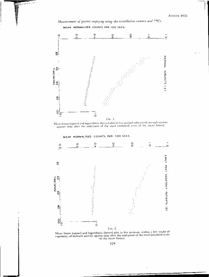
Auci:.>T 1970
Metisiireiiiciil of gastric emplyiiig using I he sciiilt'llatioii coiiic.ta attti '-"C.v'
MEAN NORMALISED COUNTS PER lOO S E C S
or
n o r 2
O:
o L _ O o
l - I G . 1.
Meiiii linear (upper) ;uid logiinthmic flower) plot In live nomial siihieeis nl sliimach aciiviiv against time after the nlid-poiiit of the meal (standard error ol the mean l imns).
MEAN NORMALISED C O U N T S P E R I O O S E C S
8 ^
z
CI o o
Ol
Oo
Mean linear (upper) and logarithmic (lower) plot in five patients within a few weeks of vagotomy, of stomach activity against time after the mid-point of the meal (standard error
•of the mean limits).
539
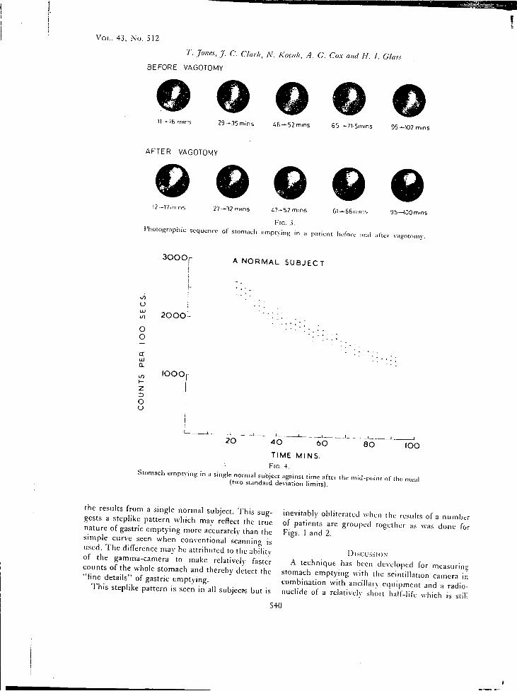
V O L . 43 , No. 5 1 2
T. Jones, J. C. CUuk, N. Kocak, A. C. Co.v a„d II. I. Class
BEFORE VAGOTOMY
11-16 mins 2 9 - 3 5 m i n s 4 6 - 5 2 mins 55-^7I.5min5 95-102 -
AFTER VAGOTOMY
Q Q Q Q Q 12 -17m,ns 2 7 - 3 2 mtns i 7 - 5 2 m i n s
F i c . 3.
61 — 5 6 ^ 1 ^ 5 95—lOOmins
Photographic sequence of stomach e
3 0 0 0
mptying in a patient before and :irier vagotomy.
A N O R M A L S U B J E C T
eO U
O O
a a. •SI I -z
o (J
2 0 0 0 -
l O O O
8 0 lOO 2 0 4 0
T I M E M I N S . F i c . 4 .
•Stomach emptying in a single normal subject agatnst time after the ntid-poin, of the me il (two standard deviation limits).
the results from a single normal subject. This suggests a steplike pattern which may reflect the true nature of gastric emptying more accurately than the siinple curve seen when conventional scanning is used. The difference may be attributed to the ability of the gamma-camera to make relatively faster counts of the whole stomach and thereby detect the "fine details" of gastric emptying.
This steplike pattern is seen in all subjects but is
inevitably obliterated when the results of a number of patients arc grouped together as was done for Figs. 1 and 2.
DlSCU.i.SIO.V
A technique has been developed for measurin'--stomach emptying with the scintillation camera ir. combination with ancillary equipment and a radionuclide of a relatively short half-life which is stiil
540
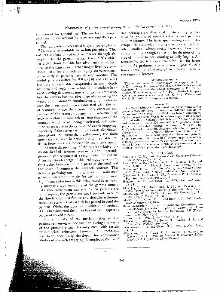
.AUGUST 1970
Measitrement of gastric emptying using the scintillation ca:n°ra and '-^Cs
convenient for general use The method is simple and can he carried out by relatively unskilled personnel.
The radioactive tracer used is cyclotron-produced '-^Cs bound to insoluble zirconium phosphate. This ensures no loss of radioactive marker through absorption by the gastrointestinal tract. '-^Cs which has a 32-1 hour half-life has advantages in reduced dose to the patient over other longer-lived radionuclides used for stomach emptying measurements, particularly in patients with delayed motility. The useful y rays emitted by "z^Cs (370 and 410 keV) facilitate a reasonable compromise between depth response and septal penetration. Other workers have used moving detector scanners for gastric emptying, but the camera has the advantage of measuring the whole of the stomach siinultaneously. This eliminates the main inaccuracies associated with the use of scanners. Since the scanner only observes one portion of the stomach at any time, movement of activity within the stomach or from that part of the stomach which is not being monitored, will introduce inaccuracies in the estimate of gastric emptying, especially if the activity is not uniformly distributed throughout the stomach. Furthermore, the finite time taken to scan in order to obtain suitable statistics increases the time error in the measurement.
The main disadvantage of the cairiera relative to a double-headed scanner system is the inherently [loorer depth response of a single detection system. .\ further disadvantage of this technique rests in the time delay between the mid-point of the meal and the onset of counting the stomach contents. This delav is probably not important when a solid meal is administered but might be with a liquid meal. Significant reduction in this delay could be achieved by magnetic tape recording of the gamma camera data and subsequent analysis. When patients are lying supine, the gastric antrum frequently overlies the duodeno-jejunal flexure and thusvthe technique measures some activity which has passed beyond the pylorus. Whilst this does not invalidate the method, if this has occurred the effect has not been apparent on the observed curves.
The simplicity of the method relics on the patient remaining in one position during the whole of the procedure and this may meet with certain physiological criticisms. However, the technique has been specifically developed for comparative studies of stomach emptying. E.vamples of the use of
this technique are illustrated by the emptying patterns in groups of normal subjects and patients after vagotomy. The basic quaiititatiiig system developed for stomach emptying may also be used for other studies, which must, however, have rate constants long enough to permit localisation of the area of interest before counting actually begins. A l ternatively, the technique could be used for faster studies if a preliminary dose of tracer, possibly of a lower energy, is administered to delineate initially the region of interest.
.-\ (• K N < 1 w 1.1 •: D C .\ 1 i-:.N "1 's T h e authors wish to acknowledge llie support of .Mr.
I ) . \ ) . X'onherg, Director of t'ne Medical Research Counci l Cyclotron Unit , and the iisetui comments of D r . O . K . Bewie\-. 'I'iianks are given to ! \ I r . P. L. l-|orlock for preparing tile isotopic tracer and .Mr. K . S. J . Wilson and .Mr. K , U, Holler for coinpiiler assistafiee.
.AMS I U . \ C T
.•\ simple teehnitjue is presented tor directly measuring gastric emptying tising an .Anger scintillation camera in conjunction vvitli readi)\' available iiucleonic equipment. Cyclotron-prodticed ' --'Cs is the radioisotopic marker u-hich is mi.ved with the patient's nteal. It has a 3 2 1 hour half-lite and principally emits 370 and 410 ke\ ' photons, w-hich provide a hall-value tllickness in tissue of 9-3 cm. The ' -"Cs is bound to insoluble zirconium phosphate lo prevetll absorption from the stomach. IiKisiraiions of tlie use of this metliod are given with normal subjects and patients alter \agotom\-, 'I'he "slep like" emptying p:iitern of the stomach contents is sometimes app;irenl when this technique is used. T h e relati\-e merits of the camera and the scanner for tliis type of study are discussed.
R I - : i - ' I - K K \ C l - S . • \ \ i r i t L K T r , C . B . , I*J64. hmriittttic Ion E.\r!tattj^es (l-.lsevier
Ciblishiag C o . . .Vj»\- \'orii). .-\.\iiMii.i-;i r, C . H.. MriJiiNAi.i). L.. .A., lUnoi^.ss, J . S.. and
.M.^VN.^HD. J . C , ,7. Iitori:. .\ial. Cliem.. Hi. h'l. l.).A\ !:.si'Oi(r. I I . \ \ ' . . r '66. Fhysiulu.siy uf the Digeniie 'Trdii.
216 (Year Hook .Medical Publishers Inc. . Chicago). G K I K H T U S , G . 1-1.. OwHN. G . M . , C . ^ M I ' H K L I . , P. H . , .Siiii-:i.rj.--,
K . , 1968, Ciistroeiilernlo'.;v, j j , 1. HoLHOYD, A M . , and JoNi-s, T . , 1969, Phvi. tited. IStot..
I-I, 031. LF.DERILK. C . W . . H o i . i . . \ N D i - : u , J . M . , and P F . R I . M . \ . V , I . .
1967, Table of Isotopes. 6th edit. (John Wiley. New "t'ork). . M . A C i . N T V H i ; , \ V . J . , (jiitsi'o. G . G . . and CiiHiSTiE, J . H . .
1963. .4m. y. Roenfj.. 9.i, 315. M . ^ E C K , W. J . , K u s s \ . .VI. I-;., and REI.N- . J . E.. 1963, .4nnly-
licol Cliemislry. 35. 20S6. Recommendations of the International Commission on
Radiological Protection. Report of Committee 11 on Permissible Doses lor Internal Radiation, 1959 (Pcrga-mon Press, .N'ew York) .
S . M I T H , E . M . , 1965. J niicl. Med., 6, 231. ' r i . N ' K i ; i i , J . , Koc.^K, N . , J O N E S , T . , G L . A S S , H . I . , and
Co.x, .A. G . . 1970. G"«/(in press). \ V E . S T E R M A N , B . R . , and G L . A S S , H . 1., 1963 ,7 . Sucl. Med.,
9. 24. Wu . i . i .^ .MS, E . D , Gi..\.ss, H . I . . . A R N O T , R . N . , and D E
G . A R R E T A , .A. C , 1968, in Medical Rtdioisulope Scintigraphy .',v, Voi . 1, p'.'665 (I . .A.E. .A. Vienna).
541
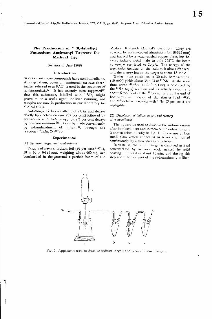
15 International Journal of Applied Radiation and Isotopes, 1970, Vol. 21, pp. 33-36. Pergamon Press. Printed in Northern Ireland
The Production of "'Sb-labelled Potassium Antimonyl Tartrate for
Medical Use
(Received II June 1969)
I n t r o d u c t i o n
SEVER A L antimony compounds have uses in medicine. Amongst them, potassium antimonyl tartrate (hereinafter referred to as PAT) is used in the treatment of schistosomiasis.*^' I t has recently been suggested'^' that this substance, labelled with i "Sb , might prove to be a useful agent for liver scanning, and samples are now in production in our laboratory for clinical trials.
Antimony-117 has a half-life of 2-8 hr and decays chiefly by electron capture (97 per cent) followed by emission of a 158 keV y-ray; only 3 per cent decays by positron emission.'*' I t can be made conveniently by a-bombardment of indium' '" , through the reaction ^^Hn{o^, 2n)"'Sb.
Exper imenta l
(1) Cyclotron targets and bombardment
Targets of natural indium foi l (96 per cent ^^^In), 50 X 10 X 0 125 mm, weighing about 400 mg, are bombarded in the^^xternal a-particle beam of the
Medical Research Council's cyclotron. They are covered by an air-cooled aluminium foil (0 025 mm) and backed by a water-cooled copper plate, but because indium metal melts at only 155°C the beam current is restricted to 20 ^uA. The energy of the a-particles incident on the indium is about 29 M e V , and the energy loss in the target is about 12 M e V .
Under these conditions a 30-min bombardment (10 //Ah) yields about 35 mCi of " 'Sb . A t the same time, some i^^'"Sb (half-life 5 1 hr) is produced by the i i ^ I n (a, n) reaction and its activity amounts to about 6 per cent of the '^'Sb activity at the end of boiTibaidinent. Yields of the sliortcr-llved ^'^Sb and ^ ''Sb from reactions with '^^In (3 per cent) are negligible.
(2) Dissolution of indium targets and recovery of radioantimony
The apparatus used to dissolve the indium targets after bombardment and to recover the radioantimony is shown schematically in Fig. 1. I t consists of four small glass vessels connected in series and flushed continuously by a slow stream of nitrogen.
In vessel A, the indium target is dissolved in 5 mJ concentrated hydrochloric acid, assisted by mild heating. This takes about 10 min, and during this step about 65 per cent of the radioantimony Is liber-
F I G . 1. .Apparatus used to dissolve indium targets and rcc fuc i i .ndioantlnionv.
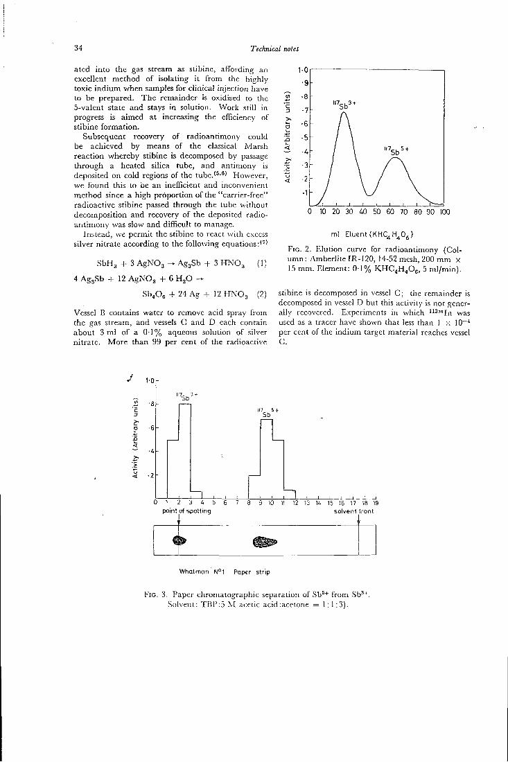
34 Technical notes
ated into the gas stream as stibine, affording an excellent method of isolating it f rom the highly toxic indium when samples for clinical injection have to be prepared. The remainder is oxidised to the 5-valent state and stays in solution. Work still in progress is aimed at increasing the efficiency of stibine formation.
Subsequent recovery of radioantimony could be achieved by means of the classical Marsh reaction whereby stibine is decomposed by passage through a heated silica tube, and antimony is deposited on cold regions of the tube.'^'^' However, we found this to be an inefficient and inconvenient method since a high proportion of the "carrier-free" radioactive stibine passed through the tube without decomposition and recovery of the deposited radioantimony was slow and difficult to manage.
Instead, we permit the stibine to react wi th excess silver nitrate according to the following equations:"'
SbHg + 3 AgNOg ^ AggSb + 3 H N O 3 (1)
4 AggSb -I- 12 AgNOg - I - 6 H j O
Sb.Oe - f 2 4 A g + I 2 H N O 3 (2)
Vessel B contains water to remove acid spray from the gas stream, and vessels C and D each contain about 3 m l of a 0 T % aqueous solution of silver nitrate. More than 99 per cent of the radioactive
c
d
B i-<
0 10 20 30 40 50 60 70 80 90 100
ml Eluent (KHC^H^Oj)
F I G . 2. Elution curve for radioantimony (Column: Amberhte IR-120, 14-52 mesh, 200 m m X 15 mm. Element: 0 1% K H C 4 H 4 O 6 , 5 ml /min) .
stibine is decomposed in vessel C; the remainder is decomposed in vessel D but this activity is not generally recovered. Experiments in which i i3mj | . | ^^^3
used as a tracer have shown that less than 1 x 10~^ per cent of the indium target material reaches vessel C.
1-0
•6h o
<
5b
117 5-1 Sb
0 1 2 3 4 5 point of spotting
9 iO 11 12 13 1i 15 16 17 18 19 solvent 'ront
Whatman N° l Paper strip
FIG. 3. Paper chromatographic separation of Sb*+ from Sb'+ Solvent: TBP:5 M acetic acid:acetone = 1:1:3).
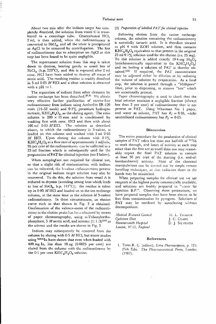
Technical notes 35
About two min after the indium target has completely dissolved, the solution f rom vessel C is transferred to a centrifuge tube. Concentrated H C I , 2 m l , is then added, when the radioantimony is converted to SbCIg, and all the silver is precipitated as AgCl to be removed by centrifugation. The loss of radioantimony due to adsorption on AgCI at this step has been foimd to be quite negligible.
The supernatant solution f rom this step is taken down to dryness, heating gently to avoid loss of SbClg (b.p. 223°C), and this is repeated after 2 ml cone. H C I have been added to destroy all traces of nitr ic acid. The resulting residue is readily dissolved in 5 m l 0-05 M H C I and a clear solution is obtained wi th a p H ~ 1.
The separation of indium f rom other elements by cation exchange has been described.'''•^' Vv'e obtain very effective further purification of carrier-free radioantimony f rom indium using Amberlite IR-120 resin (14-52 mesh) and 0 1 % potassium hydrogen tartrate, K H C 4 H 4 O 6 , as the eluting agent. The resin column is 200 X 15 m m and is conditioned by washing first wi th cone. H C I and then with about 100 m l 0-05 Af H C I . The solution as described above, in which the radioantimony is 3-valent, is loaded on this column and washed with 5 ml 0 05 M H C I . Upon eluting the column with 0 T % KHC4H40g at a flow rate of approximately 5 ml/min, 95 per cent of the radioantimony can be collected in a 25 m l fraction which is subsequently used for the preparation of P A T for clinical injection (see 3 below).
When sampled/are not required for clinical use, so that a slight risk of contamination with indium can be tolerated, the 5-valent radioantimony present in the original indium target solution may also be recovered. T o do this, the solution from vessel A is reduced to dryness (avoiding strong heat which leads to loss of SbCIj, b.p. 141°C); the residue is taken up in 0-05 M H C I and loaded on to the ion exchange column, at the same time as the solution 6f 3-valent radioantimony. I n these circumstances, an elution curve stich as that shown in Fig. 2 is obtained. Confirmation of the valence-states of the radioantimony in the elution peaks has been obtained by means of paper chromatography, using n-Tributylortho-phosphate, 5 M acetic acid, and acetone ( 1 : 1 : 3 ) ' " ' as the solvent and the results are shown in Fig. 3.
I nd ium may subsequently be removed from the column by eluting wi th 0-5 M H C I , but tracer studies using ^^^" In have shown that even when loaded wi th 4 0 0 m g l n , less than 10/^g (0-0025 per cent) are eluted f rom the column wi th the radioantimons' by the 0-1 per cent K H C 4 H 4 O 6 solution.
(3) Preparation of labelled PA T for clinical injection
Following elution from the cation exchange colunan, the solution containing the radioantimony is essentially tartaric acid. I t is therefore titrated to p H 4 with K O H solution, and then contains KHG4H40g equivalent to that present in the original 25 ml 0 1 % solution used for the elution (i.e. 25 mg). To this solution is added exactly 19-4 mg SbjOg (stoichiometrically equivalent to the KHC4H4O6) and on boiling a solution of PAT is thereby obtained."^^' I f required, the P A T concentration may be adjusted either by dilution or by reducing the volume of solution by evaporation. As a final step, the solution is passed through a ".Millipore" filter, prior to dispensing, to remove "bits" which are occasionally present.
Paper chromatography is used to check that the final solution contains a negligible fraction (always less than 3 per cent) of radioantimony that is not present as PAT. Using Whatman No. 1 paper, and water as solvent, PAT has Rf = 0-95, w-hilst imcombined radioantimony has Rf = 0 05.
Discuss ion
The entire procedure lor the production of clinical samples of PAT takes less than one half-life of ' 'Sb to work through, and losses of activity at each step other than the first are so small than one may reasonably expect the final PAT solution to contain at least 30 per cent of the starting (i.e. end-of-bombardment) activity. Most of the chemical manipulations can be carried out by simple remote handling techniques, so iliat radlaiion doses 10 the hands may be miniinised.
When preparing samples for clinical use we use reagents of the highest purity commercially available, and solutions are freshly prepared in "vvater for injection B.P.". Observing these precautions, we have prepared samples that have been shown to be free from contamination by pyrogens. Solutions of PAT may be sterilised by autoclaving without decomposition.
Medical Research Council Cyclotron Unit Hammersmith Hospital London, W.\2, Enoland
M . L . T H A K U R J . C . C L A R K D . J . SILVESTER
References
T O D D R . C . (editor). Extra Pharmacopoeia, p. 125. 25th Edn. The Pharmaceutical Press, Londo:-. (1967).
36 Technical notes
2. C H A U D H R I M . A . and R O W L A N D H . A . K . private communication (1968).
3. L E D E R E R C . M . , H O L L A N D E R J . M . and PERLMAN I . Table of Isotopes, 6th Edn, p. 263. Wiley, New York (1967).
4. T E M M E R G . M . Phys. Rev. 76, 424 (1949).
5. G R E E N D A L E A . E . and L O V E D . L . Analyt. Chem.
35, 632 (1963).
6. TOMILSON L . Analyt. Chim. Acta 31 , 547 (1964).
7. VoGEL A . I . Qualitative Chemical Analysis, 3rd Edn, p. 175. Longmans, Green, London (1947).
8. K L E M E N T R . and SANDMAN H . Z. AncUyl. Chem. 145, 325 (1955).
9. K I M U R A K . , SAITO N . , K A K I H A R A H . and IsHiMORi T . J. chem. Soc. Japan, Pure Chem. Section 74, 305 (1953).
10. T H A K U R M . L . to be published (1969). 11. T A Y L O R F . S. Inorganic Theoretical Chemistry,
9th Edn, p. 589. Wil l iam Heineman, London (1952).
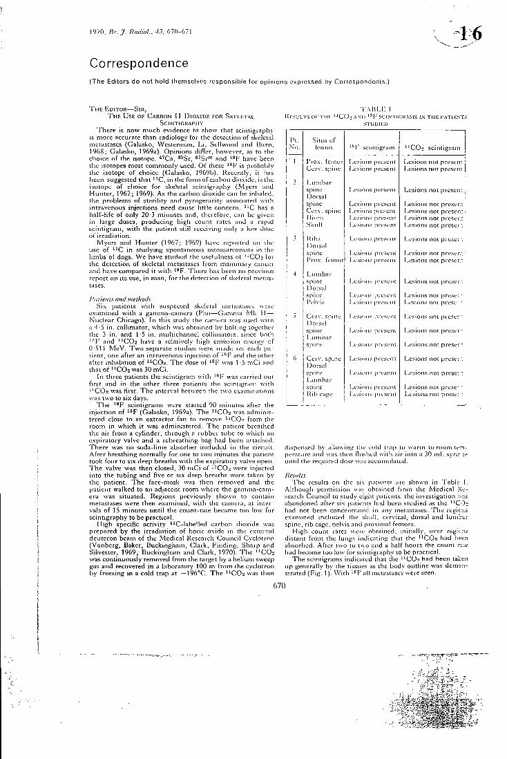
1970, Br. J. Radiol.. 4 J , 670-671
C o r r e s p o n d e n c e ( T h e Edi tors d o no t t io ld themse lves r espons ib l e l o r o p i n i o n s expressed by Co r responden l s . ]
T H I ; E D I T O R — S m , T H E U S E O F C A R B O N 11 D I O X I D E I-OR S K E L E T A L
S C I N T I G R A P H Y T h e r e is now much evidence to show that scintigraphy
is more accurate than radiology for the detection of skeletal metastases (Galasko, Westerman, L i , Sellwood and lUirn, 1968; Galasko, 1969a). Opinions differ, however, as to the choice of the isotope. ' '^Ca, ®^Sr, ^'Sr'" and have been the isotopes most commonly used. O f these ' f" is probably the isotope of choice (Galasko, 1969b). Recently, it Iras been suggested that ' ^ C , in the form of carbon dioxide, is the isotope of choice for skeletal scintigraphy (Myers and Hunter , 1967; 1969). A s the carbon dioxide can be inhaled, the problems of sterility and pyrogenicity associated with intravenous injections need cause little concern. " C has a half-life of only 20 3 minutes and, therefore, can be given in large doses, producing high count rates and a rapid scintigram, with the patient still receiving only a low dose of irradiation.
Myers and Hunter (1967; 1969) ha\'e reported on the use of ^ ' C in studying spontancoLis osteosarcomata in the limbs of dogs. W e have studied the usefulness of "CO2 for the detection of skeletal metastases from mammary cancer and have compared it with ^^F. T i iere has been no previous report on its use, in man, for the detection of skeletal metastases.
Pdlient;; and methods .Six patients with suspected skeletal metastases were
examined with a gamma-camera ( F h o — G a m m a !\'lk II — Nuclear Chicago). In this study the camera was used with a 4-5 in. collimator, which was obtained by bolting together the 3 in. and 1-5 in. multichannel collimators, since botli ' • T and "CO2 have a relatively high emission energy of 0-511 M e V . T w o separate studies were made on each patient, one after an intravenous injection of " * F and the otlier after inhalation of ' ^ C O a . T h e dose of was 1 -5 m C i and thatof i i C 0 2 w a s 30 m C i .
In three patients the scintigram with ' " F was carried out first and in the other three patients the scintigram with ' ^ C O a w'as first. T h e interval between the two examinations was two to six days.
T h e ^^F scintigrams were started 90 minutes after the injection of i ^ F (Galasko, 1969a). T h e "CO2 was administered close to an extractor fan to remove i iC02 from the room in which it was administered. T h e patient breathed the air from a cylinder, through a rubber tube to which an expiratory valve and a rebreathing bag had been attached. T h e r e was no soda-lime absorber included in the circuit. After breathing normally for one to two minutes the patient took four to six deep breaths with the expiratory valve open. T h e valve was then closed, 30 m C i of ^ ' C 0 2 w'ere injected into the tubing and five or six deep breaths were taken by the patient. T h e face-mask was then removed and the patient walked to an adjacent room where the gamma-camera was situated. Regions previously shown to contain metastases were then examined, with the camera, at intervals of 15 minutes until the count-rate became too low for scintigraphy to be practical.
H i g h specific activity "C- labe l l ed carbon dioxide was prepared by the irradiation of boric oxide in the external deuteron beam of the Medical Research Counci l Cyclotron (Vonberg, Baker, Buckingham, Clark , F inding , Sharp and Silvester, 1969; Buckingham and Clark , 1970). T h e "CO2 was continuously removed from the target by a helium sweep gas and recovered in a laboratory 100 m from the cyclotron by freezing in a cold trap at — 1 9 6 ° C . T h e 'ICO2 was then
R l S U L T S 01- T H E " C O :
I ' A B L H 1 AND '•'^F .SCIN riGRA.MS IN THE PATIENT.<
STUDIED
Pi . No.
Sites of lesion '•'^l" scintigram
1 1
"CO2 scintigram j 1 Pro.w femur ! ,esK)ns preseni (.vcsions not present J
Cerv . spine 1 .esions present Lesions not prcsen: j
2 I^Lirnbar spine I.esioiis present Lesions not prescp.i ; I)ors:iI spine I.estons present Lesions not preser.: Cer\". spine I.,esi(,tns jircsent Lesions not presen; i i l ium 1 ,esion< present I ,e.<ions not prescr.: Skuli 1 -eSH^ns presi.-ni Lesions not preser:
Kihs Lesions present I A'sions not preses : l)ors:iI
I A'sions not preses :
spine I .esions present Lesions not prcser.: I'ro.x. lemur 1 .esions pi eseni Lesions not preser.:
4 LLinib;n-spine I .esi'Mis [iresent Lesions not presvr: !)ors:il
Lesions not presvr:
spine ! -esHins preseni 1 .esions not prese: • iVIvis 1 A'SHHis piesenl Lesions nr>t preset *
5 Cer\ ' . spine 1 -csions pi eseni I X'sions noi preser • Dors::!
I X'sions noi preser •
spine 1 A'SHins preseni Lesions not prcsei" i .umlrar spine ! .esions present Lesions not prese: :
6 Cerv . spine Lesions jireseni Lesions not preser: i^orsnl spine I ,esions preseni Lesions not prose: : lAunhar spine i A'SKHis present Lesions not prese- • Ril l cage ! .esiuns present Lesions not prese; :
dispensed by allowing the cold trap to warm to room temperature and was then flushed with air into a 30 ml . syrir^r* until the required dose was accumulated.
Results T h e results on the six patients are shown in Table- L
Although permission was obtained from the Medical .Research Counci l to study eight patients, the investigation v.-.is abandoned after si.K patients had been studied as the • ' C O - : had not been concentrated in any metastases. T'he regicns examined included the skuli. cervical, dorsal and lumbar spine, rib cage, pelvis and pro.xima! femora.
High count rates wore obtained, initially, over regi-i.ns distant from the lungs indicating that the "CO2 had b ~ n absorbed. After two to two and a half hours the count rize had become too low for scintigraphy to be practical.
T h e scintigrams indicated that the "CO2 had been taiien up generally by the tissues as the body outline was demonstrated (F ig . 1). With '^r-"all metastases were seen.
670
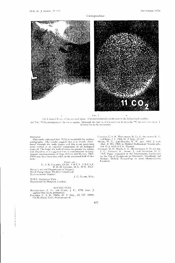
1V70. lii . J . Kinliiil.. •'.)'. I " l
Ciiryespoiulc.iici'
A KM;, i . n
(A) A liilcntt X-r; iv nl* ilic ct rvicnl spine. A lesion (;n io\\ccl) c;in hi: seen in the 3rd cervic.tl vei iehr.i.
(u) 'I'lie ' ' C O > scinii^nun of ihe sanu- rej;ion. Ahiioni^li the outline of the neek c;in he seen the "C" hn^ ri..i e..lu r u n .1: in bone nor in tlie nielasfasis.
Discussion 'Hiis study indiai ied that "CO2 is nnsuitahic for skeletal
scinti^^rapliy. 'I'lie resvdts SLigj^est that it is evenly distn-hulcd througli the hody tissues and this is not surprising since carbon is an essential component ol all biological material. T h e larger the hulk of tissue the greater the uptake and. therefore, it is suggested that its concentration in spontaneous osteosarconiata of tiogs (.VUers and Hunter , 196/; 1069) mav have been due solely to the increased bulk of that limb.
You rs. etc.. C . S. H. G.-M-vsKO, C h . M . . l - . R . C . S . , K . K . C . S . H .
R. K . I I . CoOMiiS, A'l.A., M . B . . l i . C h . Ilreast l.'nit and Department of Surgery, Koval Postgraduate .Medical School and Mainmersmith I lospital.
J . C . Ci..-\RK, B .Sc . M . R . C . Cyclotron Unit , I lammersini th Mospital, London.
C;AI.ASK(). C. S. H.. \VI-:SI iiuMA.v. R.. i-t. I . . .Sn.i.utM n>. R \ . and IJUHN, 1. i . . 1%8. Br. 7. Sun-.. .i:'). M .V
VlvKUs. W. C;., and HUNTKH. W. W.. j u n . . 1967. J. /.••/W. Med., iS', 305; 1969, in Medical Umiiois'MnfH' Sciniiiimf^Uv. Vol. I I , p . 4 3 ( l . A . i : . A . . V i e n n a ) .
Vo.NiiKito, D . n . , BAKRH. L . C . Bi'( K i N f ; i i A . \ i . I*. I ) . . C i . x i t K .
J . C , l - ' i . \ i J L \ ( ; , K . . SHAIU' . J . , and S n . v i - s n - K . i» J . . 1969, Paper presented to the International C o n i e u n c e on the Use of Cyclotrons in Chemistry. Metallurgy and Bioiogv, Oxford. I*r<)ceedmgs in press (Ruiterwttriiis. [,oiKh)n).
K i : i - K R i : . \ c r : s Bl/CKiNciiAM. P. n . , and Cl-.'MiK, j . C , 1970, Inter. J .
applied Uad. (to he published). G A I . . V S K O , C . S. B. , 1969a. Hr. J . Surg., 56, 757; 1969b,
C h . M . thesis. Univ . VVitwatersrand.
671
17 RADlOCHEM.RADIOANAL.LETTERS 6 /5/ 281 - 286 /1971/
THE PREPARATION OF CARBON-11 LABELLED CARBON MONOXIDE AND CARBON DIOXIDE J.C.Clark, P.D.Buckingham
Medical Research Co i i n c i l Cyclotron Unit, Hammersmith H o s p i t a l , London, W.12, U.K.
Received 15 March 1971 Accepted 20 March 1971
A t a r g e t system i s described for the production of ''CO and ^^C02 by the deuteron bombardment of B-O,. The radiochemical composition of the gas at the t a r g e t output i s given for a range of ta r g e t sweep gases using a f i x e d flow rate and beam cur r e n t . Values are a l s o given for the production r a t e and r a d i o a c t i v e concentration of the ^^C l a b e l l e d products, together with values f o r the o v e r a l l l-^C t a r g e t e x t r a c t i o n e f f i c i e n c y . The use of various t a r get sweep gases i s d i s c u s s e d and sweep, qas compositions are given for the d i r e c t production of ^CO and ^C02.
INTRODUCTION
Carbon monoxide and carbon dioxide both l a b e l l e d with '''"C
are i n routine production for c l i n i c a l s t u dies at Hammersmith
H o s p i t a l ^ and elsewhere. The present paper d e s c r i b e s the s y s -
torfis c u r r e n t l y i n use f o r the production of carbon monoxide and
carbon dioxide l a b e l l e d with ^^C. The most notable improvement 2 3
over previous systems ' i s i n the use of hydrogen as a ta r g e t
sweep gas, which makes the production of ^CO v i r t u a l l y c a r r l e r -
- f r e e , w h i l s t i n h i b i t i n g the formation of s i g n i f i c a n t amounts of
TARGET DESIGN
^^C has a 20.3 m'in h a l f - l i f e and decays by the emission
of 0.96 MeV po s i t r o n s . The t a r g e t m a t e r i a l g e n e r a l l y used for
281
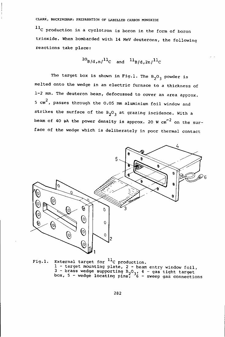
C L A R K , BUCKINGHAM: PREPARATION OF L A B E L L E D CARBON MONOXIDE
• C production i n a c y c l o t r o n i s boron i n the form of boron t r i o x i d e . When bombarded with 14 MeV deuterons, the following r e a c t i o n s take p l a c e :
1 0 B/d,n/^^C and ^^B/d,2n/^^C
The t a r g e t box i s shown i n F i g . l . The 8 2 0 ^ powder i s
melted onto the wedge i n an e l e c t r i c furnace to a t h i c k n e s s of
1-2 mm. The deuteron beam, defocussed to cover an area approx. 2
5 cm , passes through the 0.05 mm aluminium f o i l window and
s t r i k e s the surface of the 6 2 ^ 3 grazing incidence. With a
beam of 40 \iA the power de n s i t y i s approx. 20 W cm'" on the sur
f a c e of the wedge which i s d e l i b e r a t e l y i n poor thermal contact
F i g . l . E x t e r n a l t a r g e t f o r C production. 1 - t a r g e t mounting p l a t e , 2 - beam entry window f o i l , 3 - brass wedge supporting B O , 4 - gas t i g h t t a r g e t box, 5 - wedge l o c a t i n g p i n s , 6 - sweep gas connections
282
C L A R K , BUCKINGHAM: PREPARATION O F L A B E L L E D CARBON MONOXIDE
with the surrounding box. T h i s power density i s s u f f i c i e n t to melt the 8 2 0 ^ /MP = 400 °C/ r e l e a s i n g l a b e l l e d gases i n t o a stream of sweep gas passed through the box at cibout 80 ml min ^ The molten ^2^3 prevented from ra p i d movement down the wedge by the step shape.
The swept volume of the t a r g e t box i s kept small /about
200 ml/ to maximise the r a d i o a c t i v e concentration of the
e f f l u e n t gas. I t s normal working pressure i s approx. 0.007 kg
cm ^ above atmospheric pressure.
TARGET SWEEP GASES AND YIELDS
The nature and composition of the target sweep gas are
of c r i t i c a l importance. The y i e l d i s expressed as the rate of
production of a c t i v i t y i n mCi min continuously monitored by
a 2 0 0 ml container, i n an i o n i s a t i o n chamber approx. 1 0 m from
the t a r g e t . Table 1 shows the e f f e c t of varying the composition
of the sweep gas at a flow rate of 80 ml min ^ and a beam cur
r e n t of 4 0 v jA, the v a l u e s being obtained by radio-gas chroma-
tography and the use of a c a l i b r a t e d i o n i s a t i o n chamber system.
The t a r g e t e f f i c i e n c y v a l u e s were obtained by comparing the
y i e l d of • C induced i n a s i m i l a r wedge under conditions where
there were no l o s s e s of v o l a t i l e products, decay curve a n a l y s i s
being necessary to e x t r a c t the ' C component.
The p r e c i s e nature of the chemical and r a d i o l y t i c reactio.'
which take place i n the t a r g e t during i r r a d i a t i o n i s not f u l l y
known. However, i t i s thought that """CO i s the primary product
of the r e a c t i o n between the ^^C atoms and the oxygen atoms in
the ^2^2' absence of any oxygen r a d i c a l scavenger the
283
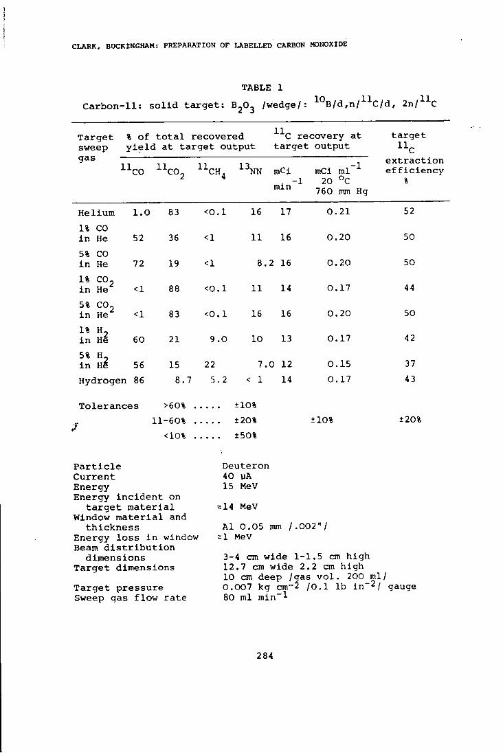
C L A R K , BUCKINGHAM: PREPARATION OF L A B E L L E D CARBON MONOXIDE
Target % of t o t a l recovered
TABLE 1
2^3
11
Carbon-11: s o l i d t a r g e t : B-O, /wedge/: ^°B/d,n/^-^C/d, 2n/^^C
C recovery at ta r g e t sweep y i e l d at t a r g e t output t a r g e t output ^^C gas
l^CO i ^ c o ^ ''CH, 4 l^NN mCi min ^
mCi ml' 20 °C
760 mm
-1
Hg
e x t r a c t i o n e f f i c i e n c y
%
Helium 1.0 83 <0.1 16 17 0.21 52 1% CO i n He 52 36 <1 11 16 0.20 50 5% CO i n He 72 19 <1 8. 2 16 0.20 50 1% CO, i n He <1 88 <0.1 11 14 0.17 44 5% CO, i n He <1 83 <0.1 16 16 0.20 50 1% H, i n He 60 21 9.0 10 13 0.17 42 5% H, i n H6 56 15 22 7. 0 12 0.15 37 Hydrogen 86 8.7 5.2 < 1 14 0.17 43
Tole r a n c e s >60% 11-60%
<10%
+ 10% ±20% ±50%
±10% ±20%
P a r t i c l e Current Energy Energy i n c i d e n t on
t a r g e t m a t e r i a l Window m a t e r i a l and
t h i c k n e s s Energy l o s s i n window Beam d i s t r i b u t i o n
dimensions Target dimensions
Target pressure Sweep gas flow rate
Deuteron 40 MA
15 MeV
=14 MeV
Al 0.05 mm /.002"/ = 1 MeV
3 — 4 cm wide 1-1.5 cm high 12.7 cm wide 2.2 cm high 10 cm deep /gas v o l . 200 ml/ 0.007 kg cm-2 /O.l lb in~2/ gauge 80 ml min"^
284
C L A R K , BUCKINGHAM: PREPARATION OF L A B E L L E D CARBON MONOXIDE
• CO i s l a r g e l y r a d i o l y s e d to ^ ^ 0 0 2 - Thus when helium i s used as the sweep gas roost of the ^C a c t i v i t y i n the targ e t e f f l u e n t i s ^ ^ 0 0 2 ? the add i t i o n of C O 2 c a r r i e r having l i t t l e e f f e c t upon the recovered y i e l d . When s t a b l e CO i s added to the helium l e s s ^ ^ C 0 2 i s made, the CO a c t i n g as a scavenger f o r oxygen r a d i c a l s and p r o t e c t i n g the ^CO. As more CO i s added t h i s protection i s seen to i n c r e a s e .
When a sweep gas of helium containing a small amount of
hydrogen i s used i t a l s o a c t s as a r a d i c a l scavenger giving
some recovery of ^CO without added c a r r i e r . I f however hydrogen
i s used alone, a much higher recovery of ^CO i s achieved, a l s o
without added c a r r i e r .
I t should be noted that there i s a s i g n i f i c a n t recovery
of ^ ^ N 2 with a l l sweep gases except hydrogen. Presumably the
^^N atoms are scavenged by hydrogen to form non v o l a t i l e products,
The absence i n the ta r g e t e f f l u e n t of a p o s s i b l e v o l a t i l e pro
duct, •'"•NH, has been demonstrated both chemically and gas
chromatographically.
The only detected C l a b e l l e d contaminant i s CH^ formed
by the combination of ^C and H r a d i c a l s w i t h i n the target dur
ing i r r a d i a t i o n . T h i s , as would be expected, i s only present i n
s i g n i f i c a n t amounts when H 2 i s used i n the sweep gas.
CONCLUSION
In the production of ^CO and ^ ^ C 0 2 by the deuteron bom
bardment of 32*^3' chemical form of the product n-.^clei re
covered from the targ e t i s l a r g e l y determined by tht target
sweep gas composition. The use of hydrogen i n h i b i t s the forma-
285
C L A R K , BUCKINGHAM: PREPARATION OF L A B E L L E D CARBON MONOXIDE
t i o n of ^ ^ C 0 2 and ^"^N2 r e s u l t i n g i n v i r t u a l l y c a r r i e r f r e e ' CO production. The use of helium or 1% C O 2 i n He r e s u l t s i n the production of a high proportion of ^ ^ C 0 2 , but with no suppress i o n of the • ' • •^N2.
Thus the choice of an appropriate t a r g e t sweep gas r e s u l t s
i n the p r e f e r e n t i a l production of e i t h e r ''"CO or '^•^COj.
We wish to thank Mr.P.L.Horlock for valuable t e c h n i c a l
a s s i s t a n c e .
REFERENCES
1. J.C.Clark, C.M.E.Matthews, D . J . S i l v e s t e r , D.D.Vonberg, Nucleonics, 25 /1967/ 54.
2. P.D.Buckingham, G.R.Forse, Int.J.Appl.Radiation and Isotopes, 14 /1963/ 439.
3. M.J.Welch, M.M.Ter-Pogossian, Rad.Res., 36 /1968/ 580.
286
18 International Journal of Applied Radiation and Isotopes, 1971, Vol. 22, pp. 639-646. Pergamon Press. Primed in Northern Ireland
The Preparation and Storage of Carbon-11 Labelled Gases for Clinical Use
J. G. CLARK and P. D. BUCKINGHAM Medical Research Council Cyclotron Unit, Hammersmith Hospital, London W12, England
{Recewed30 April 1971)
The target systems used for the production of carbon monoxide and carbon dioxide, both labelled with carbon-II, in the Medical Research Council Cyclotron at Hammersmith Hospital are described. The radiochemical composition of the gas at the target output is given for a range of target sweep gases using a fixed flow rate and beam current. Values are also given for the production rate and radioactive concentration of the ' ' C labelled products, together with values for the ^^C target extraction efficiency. Reasons are stated for the present routine use of hydrogen as a target sweep gas and its advantages discussed.
Systems are described for the production of ^iCO and " C O j for routine clinical use and details given of storage devices for these gases.
L A P R E P A R A T I O N E T L E M A G A S I N A G E DBS GAZ M A R Q U E S A U CARBONE-11 P O U R E M P L O I DAN.S L A C L I N I Q . U E
On dccrit les systemes de cibles qui servent a la production du monoxyde de carbone et du bioxyde de carbone, marques tous les deux de carbone-!l, dans le cyclotron du Conseil de Recherche Medical a I'Hopital de Harrersmith. On presentc la composition radiochimique du gaz au d^bit de la cible pour une gamme de gazbalayeurs de cible en employant un taux d'ccoulement et un courant de faisceau fixes. On donne aussi des valeurs pour le taux de production et pour la concentration radioactive dcs produits marques au ^^C, ainsi que des valeur? pour I'efficacite d'extraction du " C de la ciblc. On precise des raisons pour I'emploi couramment habitucl de rhydrogcne commc gaz-balaycur dr clblc cl on discute scs a\ antagcs.
On decrit des syst6mes pour la production du ' ' C O ct du " C O j pour cmploi rcgulier dans la cliniqueet on d^taille des appareils pour I'emmagasinage de ces gaz.
n P H r O T O B J T E l l H E M X P A H E H M E F A S O B , .MEMEIIIIblX y P J l E P O A O M C " , flJIfl n P M M E H E H H f l B K J I l l l l M K E
OnHcaHbi MHiueHHue CHCTeMH n p o H 3 B O A C T B a CO, CO,, MeneHUbix yrjiepoROM C " , B
UHKJioTpoHe CoBeTa MenHiiHHCKHX HCCJienoBamni y So.nbmmLi PaMMepcMiiTa B JToHftOHe. . JXAH paAHOXHMHHecKHH c o c T a B r a s a na BtixoRe Mumenu A J I « oSjiacTn raaos p a s B e p T K i i
MHHienH, HcnojibayH nocxoHHHyio CKopocTb noTOKa ii nocTonnfibiil T O H n y i K i i . T a K w e AaHbi s i i a n e H i i H HHTencuBHOCTH npoHSBOACTBa I I p a j i i i o a K T i i i i H o r i K o i i u e n T p a u H i i n p o a y K T O B . Nie'ieiibix yrjieportOM C " , B.MecTe c aiiaMeuHHMii H J I H DilnjicKTiiiiiiocTii aKCTpaKumi .Miuueiiu C " . nocj ie o6ocHOBaHHH y c T a H O B H B t u e f i C H npaKTHKii npiiMeHemin BOAopoAa B KanecTBe r a s a p a a s e p T K H MHiueHii o6cyH<AaK)Tcs n p e w M y m e c T B a 3 T o r o r a s a .
OnHcaHbi CHCTeMbi RIIH nponaBOACTBa C O " , CO.2" a .nn y c T a n o B i i B i i i e r i C H n p a K T i i K i i B
K j i H H M K e , npeACTaBJieHbi AanHue 0 y c T p o i t C T B a x n.in x p a i i e i u i H 3 T I I X r a a o B .
D I E Z U B E R E I T U N G U N O L A G E R U N G V O N » C M A R K I E R T E N G A S E N
Die TrefTplattensystems fiir die Herstellung von Kohlenmonoxyd und Kohlensaure, beide mit einer I ' C Markierung, im Zyklotron des Medical Research Council im Krankenhaus Hammersmith werden beschrieben. Die radiochemische Zusammensetzung des Gases am Treffplattenausgang wird fiir eine Reihe von Trclfplancnspulgasen bei Benutzung eincr
1 639
640 J. C. Clark and P. D. Buckingham
festen Durchflussmenge und Strahlstromstarkc angegeben. Werte werden auch vorgelcgt fur die Produktionsgeschwindigkeit und die radioaktive Konzentration der ^^C markierten Pro-dukte, zusammen mit Werten fiir die Treffplattenextraktionsausbeute. Es werden Griinde angegeben fiir die derzeitige laufende Verwendung von Wasserstoff als Treffplattenspiilgas, und seine Vorteile werden besprochen.
Einrichtungen fiir die Produktion von ^00 und^COgZuriaufenden klinischen Verwendung werden bexchrieben und Einzelheiten werden angegeben fiir Lagerungsvorrichtungen fUr diese Case.
INTRODUCTION C A R B O N monoxide and carbon dioxide both labelled wi th ^^C are in routine production for clinical studies at Hammersmith Hospital.*" The principal uses are in serial blood volume estimations,'^'^' lung function investigations'*' and organ visualisation using a gamma camera'^' or scintiscanner.'^' Ease of administration and a low radiation dose to the patient make ' ^ C an attractive nuclide for this work. A further advantage is that the 2 0 min half-life is sufficiently long to make i^C storage systems a practical proposition. Such systems allow the use of the nuclide away from the site of production, and make for more efficient use of cyclotron running time.
The present paper describes the systems currently in use for the production and storage of carbon monoxide and carbon dioxide labelled wi th ^^p. The most notable improvement over previous systems"'*' is the use of hydrogen as a target sweep gas, which makes the production of '^^CO virtually carrier-free, whilst inhibi t ing the formation of significant amounts of " N g ( " N N ) .
EXPERIMENTAL Target design
Carbon-11 has a 20.3 min half-life, decays by the emission of 0 .96 M e V positrons and is the product of several nuclear reactions as sliown i n Table. 1.
The target is shown in Fig. 1. The BgOj powder is melted onto the wedge in an electric furnace to a thickness of 1-2 mm. The 14 M e V deuteron beam, defocussed to cover an area approximately 5 cm*, passes through the 0 . 05 m m aluminium foi l window and strikes the surface of the BgOg at grazing incidence. W i t h a beam current of 40 fih. the power density is
T A B L E 1. Some nuclear reactions used for the production of carbon-11
Nuclear reaction Threshold energy
(MeV) Reference
l°B(rf, n)llC 1.70 (7) (8) "B(rf, 2n)l»C 5.89 (7) (8) "N(/), a) l lC 3.13 (9) (10) "B(/), /!)iiC 3.01 (10) l^N((/, an)"C 5.88 (11)
approximately 2 0 W cm"'' on the surface of the wedge which is deliberately in poor thermal contact with the surrounding box. This power density is sufficient to melt the B 2 O 3
(M.P. « i 4 0 0 ° C ) releasing " C labelled gases into a stream of sweep gas passed through the box at about 8 0 ml min~^. The molten B j O j is prevented from rapid movement down the wedge by the step shape.
The volume of the target box is kept small (about 200 cm') to maximise the radioactive concentration of the effluent gas. Its normal working pressure is approximately 0 .007 kg cm~^ (0.1 lb in~2) above atmospheric pressure. Brass is used for al l parts of the target except the front plate and window which are aluminium and water cooled. The gas inlet and outlet connections are made using in vacuum fittings.
During bombardment the molten BjOg slowly migrates from the area of the beam strike causing a loss of yield. This process takes about 12 hr after which the wedge has to be re-coated. Since B 2 O 3 is hygroscopic, wedges are always stored in a desiccator until required for use.
Target sweep gases and yields
The nature and composition of the target sweep gas are of critical importance. The range of gases which we have used is listed in
The preparation and storage of carbon-11 labelled gases for clinical use
4
641
F I G . 1. External target for " C production. (1) Target mounting plate (Beam entry window 2-5 cm X 12-7 cm) (2) Beam entry window foil (3) Brass wedge supporting B j O j (4) Gas tight
target box (5) Wedge locating pins (6) Sweep gas connections.
T A B L E 2. Carbon-11: Solid target: B j O j (wedge): i''B((y, r!)"C "B((/, 2n)>iC
Particle Deuteron Current 40 M Energy 15 MeV Energy incident on target material S^14 MeV Window material and thickness Al 0.05 mm (0.002 in.) Energy loss in window ~1 MeV Beam distribution dimensions 3-4 cm wide 1-1.5 cm high Target dimensions 12.7 cm wide 2.2 cm high 10 cm deep (gas vol. 200 ml) Target pressure 0.007 kg cm-2 (0.1 lb in.-^ gauge)
, Sweep gas flow rate 80 ml min~i
Target Per cent of total recovered ' ' C Recovery at Target " C
sweep gas
" C O " C O 2 " C H , 13NN mCi min ^
mCi ml ' 20°C
760 mm Hg
efficiency per cent
Helium 1.0 83 <0.1 16 17 0.21 52 1% C O in He 52 36 <1 11 16 0.20 50 5% C O in He 72 19 <1 8.2 16 0.20 50 1% C O 2 in He <1 88 <0.1 11 14 0.17 44 5% C O 2 in He <1 83 <0.1 16 16 0.20 50 1% ^ 2 in He 60 21 9.0 10 13 0.17 42 5% H 2 in He 56 15 22 7.0 12 0.15 37
Hydrogen 86 8.7 5.2 <1 14 0.17 43
Tolerances (%)
>60 11-60
<10 •
• • ± 10 • • ± 20 • • ± 50
± 1 0 ± 2 0
Table 2 which also shows the effect on the radiochemical composition of varying the composition of the sweep gas at a fixed beam current of 40 /^A and fixed flow rate of 80 ml min"^. The gas flow system, shown schematically in Fig. 2, is an open circuit design.
The precise nature of the chemical and radiolytic reactions which take place in the target during irradiation is not fully known. However, it is thought that '^CO is the primary product of the reaction between the ^'C atoms and the oxygen atoms in the B^Og. In the
642 J. C. Clark and P. D. Buckingham
Carbon-ll Target NPL lonisotion \Chamber
Pressure Reducing Valve
Gas Sample Filter
200 ml cylinder
UMeV Deuteron Beam
Waste
Row Meter
Septum Row Meter ^ Scavenge
P-jmp ppfN o o o o o Sweep
D.C. Amplifier Air Inlet
Chart Recorder
F I G . 2. ' ' C experimental gas flow system.
absence of any oxygen radical scavenger the listed as follows: " C O is largely radiolysed to "COa-'** Thus when helium is used as the sweep gas most of the " C activity in the target effluent is ' ' G O j , the addition of COg carrier having little effect upon the recovered yield. When stable CO is added to the helium less ^^COg is made, the CO acting as a scavenger for oxygen radicals and protecting the ^^CO. As more CO is added this protection is seen to increase.
When a sweep gas of helium containing a small amount of hydrogen is used it also acts as a radical scavenger giving some recovery of ^^GO without added carrier. I f however hydrogen is used alone, a much higher recovery of " C O is achieved, also without added carrier.
I t should be noted that there is a significant recovery of ^^Ng wi th all sweep gases except hydrogen. Presumably the ^^N atoms are scavenged by hydrogen to form non volatile products. The absence in the target effluent of a possible volatile product, " N H 3 , has been demonstrated both chemically and gas chromatographically.
The only detected ^^C labelled contaminant is ^^CH^ formed by the combination of ^ C and H radicals wi th in the target during irradiation. This, as would be expected, is only present in significant amounts when H j is used in the sweep gas.
The parameters affecting the total yield recovered from the carbon- l l target may be
(a) Target material nuclear cross section (b) Beam current (c) Beam energy (d) Target material thickness (e) Sweep gas flow rate (f) Sweep gas composition (g) Beam distribution (h) Target material water content (i) Target material temperature.
These parameters may be divided into those afl 'ccting the activity produced within the target, and those affecting the recovery of activity from the target. Thus (a-d) largely determine the activity produced, whilst (e-i) tend to determine the amount of recovered activity and its chemical composition. Wi th the recovery of the activity depending upon so many factors, the yield is never constant from day to day or even during a run of say, 3 hr duration. However, i t is possible to determine the relationship between some of the parameters by running for long periods and taking mean values of beam current, yield and analyses of samples.
Activity assay
Table 2 shows the radioactive products that have been detected in ^^G experimental bombardments. The analysis of these products was
The preparation and storage of carbon-11 labelled gases for clinical use 643
carried out using a Pye Series 104 gas chromato-graph, modified so that the effluent gas passed through a /9-counter connected to an amplifier, ratemeter and second recording channel. I t was thus possible to detect both stable and radioactive gases. Three 1.5 m X i=» 4.5 mm bore columns were used: molecular sieve (type 5A) , silica gel and "poropak Q," all of 80-100 mesh. Helium was used as the chro-matograph carrier gas.
The target efficiency values were obtained by comparing the yield of ^ C induced in a similar wedge under conditions where there were no losses of volatile products, decay curve analysis being necessary to extract the ^^C component. The activity of the target effluent was measured using a calibrated ionisation chamber system shown schematically in Fig. 2. The calibration was performed by taking a 1 ml sample f rom the input of the monitored volume and measuring it in a calibrated gamma spectrometer.
P R O D U C T I O N
Routine production of^^CO
For the production of ^^CO for routine clinical use, either Hg (commercial grade) or 1 % C O in He may be used as the sweep gas. Usually H2 is to be preferred since the target effluent gas^rthen contains very little ^^COj, obvialing the need of a reducing furnace. The I ' -CH^ contaminant does not interfere with red cell labelling and one has the advantage of a very low ^'Ng contaminant level.*
W^hen 1 % CO in He is used the " C O 2 is reduced to ^^CO by passing the gas over activated charcoal. A t 800°C the following reaction takes place:
• 12c - f " C O 2 " C O - i - 12CO.
Heated zinc can be used as an alternative reducing agent :'i2'
Zn + " C O 2 ZnO + " C O
We find that for sweep gas flow rates of
* When H 2 containing " C O is administered by rebreathing'*'*' a maximum volume of 15 ml is diluted to 3 1. with air or oxygen thus giving a 0.5% maximum H 2 mixture which is 10 times lower than the explosive limit for air /O, mixtures.*^"
approximately 80 ml min~^ a pyrex glass furnace tube 5 cm long and 2.5 cm dia. filled wi th zinc powder maintained at 400°C ± 10°C to avoid melting, is satisfactory.
Any unconverted ^'COg is absorbed in a soda lime column 60 cm long and 2 cm dia. The use of 1 % CO in He as a sweep gas for clinical production bombardments does have limitations. The presence of CO carrier lowers the specific activity of the target effluent gas, causes inefficient red cell labelling, restricts the volume of gas used when i t is administered by inhalation and causes inefficient trapping of " C O in the storage system to be described. Moreover, the ^^Nj contaminant level is often unacceptably high.
The i^C labelled gases are used at two sites; experimentally, in an area approximately 10 m from the cyclotron target, and clinically in a measuring and dispensing area approximately 100 m from the target. Gas transmission pipes of 1.5 mm bore stainless steel are used between the target and the experimental area. The pipes between the target and the clinical srea are 1.5 mm bore stainless steel and 1.9 mm bore nylon.
At a target pressure of about 0.05 kg err above atmospheric pressure (necessary when using the long gas transmission pipes) and a sweep gas flow rate of 50 ml min~i, the trarisit time from the target to the clinical arei is approximately 8 min.
" C O storage system
The storage systems described in this paper work on the principle of trapping the labelled gas using low temperature techniques. '^'CO may be trapped either on activated charcoal ac — 196°C, or on a molecular sieve at —85^C. ^Ve have found the latter to be more reproducible.
The molecular sieve " C O storage systen i i shown schematically in Fig. 3, and consists of a 27 cm length of 0.6 cm I D copper tubing filled with T V in. pellets of type 13 X molecular sieve (Lindc Ai r Products, US.A). The ga.; is passed through it to waste for 20 min, the trap being cooled to —85°C in an acetone "Driccld ' ba th . j Trapping efficiency is about 85 per Knt-
t"Dricold"; solid C O , supplied by Disiile.'-; Co. Ltd.
644 J. C. Clark and P. D. Buckingham
CO Input Output to Waste
WATER! 100°c ! '
Stainless Steel Tube
Copper Gauze
opper Tube
Molecular Sieve
Acetone/Sol id COj Mixture
F I G . 3. ' ' C O storage system.
W i t h an " C O production rate of about 12 mCi min~ ' , a typical trapped activity is 150 mCi of which approximately 95 per cent is released on heating the trap to 90°C. Since some of the residual oxygen or nitrogen in the system is trapped at —85°C, the volume of the released gas is about 7 ml . This is transferred to the low pressure storage unit shown in Fig. 4 by flushing the trap wi th 40 ml of air or helium. Alternatively, i f about 40 ml of air is introduced into the trap during the first 2 min of trapping, flushing becomes unnecessary since upon heating, the air together wi th about 90 per cent of the " C O is released directly into the low
, Input from CO or CO j Trapping Systems
50 ml Volume ndicator
0 Ring
50 ml. Rubber S a c
Perspex water Container
Lead Shielding
F I G . 4. Low pressure storage unit.
pressure storage unit. A n indication of the volume of stored gas is given by a simple water displacement indicator. When sterile samples are required they are dispensed through a millipore filter. Before use, the molecular sieve is outgassed at 200°C for about 12 hr and wil l retain its adsorbing characteristics providing it remains dry.
Routine production of^^CO^
" C O 2 may be produced continuously without the use of a furnace, by using the B2O3 target with a sweep gas of either pure He or a mixture of 1 % GOg in He. Such a system produces a significant amount of contaminant (Table 2) which for some applications may have to be removed. The ' ^COj storage system to be described does this effectively.
Occasionally " G O and ^^COj are required simultaneously. Under these circumstances " C O 2 is supplied batch-wise using the apparatus depicted in Fig. 5 which is used to oxidise the " C O to " C O j . To prepare a batch of " C O 2 a charge of " G O is first trapped in the storage system described previously. The output of the trap is then connected to the input of the conversion unit and the " G O flushed through the cupric oxide furnace for 3-4 min at about 10 ml min~', to be collected in the tonometer at the furnace output as "CO2. To collect the " C O j a 50 ml syringe is connected to the tonometer output and the gas collected by the displacement of mercury. " C O 2 radioactive concentrations of about 3 mCi m l ~ ' are readily obtained.
Since the trapped " C O contains a little hydrogen, traces of water, which tend to dissolve the "GO2, are formed in the CuO furnace during conversion. To remove this water, and thereby reduce absorption, a drying agent, magnesium perchlorate, is added at the furnace tube output.
"CO2 Storage system
The trap for " C O g shown in Fig. 6 consists of a 3.6 m length of empty copper tube ^ 3 mm O D , 1.7 m m bore, formed into a closely wound spiral approximately 4.5 cm dia. and 7.3 cm long. When used in conjunction with a system using He as the sweep gas it is possible to trap
The preparation and storage of carbon-l 1 labelled gases for clinical use 645
Cupric Oxide rlOOmmx ^rnm dia. COj collection
Syringe
Magnesium Perchlorote
CO Input - from
Irapping System
Meter BOO c
.vlMercury
Mercury
F I G . 5. Apparatus for batch-wise conversion of " C O to ' ' C O ,
COa Input
Needle Valve
Stainless Steet Tube
Wasle
Copper Tube > WATER
1U0"C ;
196"c
Liquid Nitrogen
Ft^i'. 6. " C O g storage system.
" C O 2 of high specific activity. I n use i t is similar to the " C O storage systems already described. The target effluent gas is passed for 20 m i n through the trap which is placed in l iquid nitrogen, and the ^^GOg condensed in the tube. A typical " G O g trapped activity is 220 mCi of which more than 95 per cent is recoverable on heating the trap to approximately 90°C. The released gas is flushed with 20 m l He into a low pressure storage unit as used wi th the " G O molecular sieve trap.
A notable feature is that although the yield may contain as much as 16 per cent ' ' N j , the stored activity contains less than 1 per cent of this nuclide. I t is however essential to ensure the trap is dry before use.
C O N C L U S I O N S " C O and "GO2 are produced for routine
clinical use using well proven cyclotron target
systems. The chemical form of the product nuclei recovered from the target is largely determined by the target sweep gas composition. Hydrogen is now used for " G O production since i t results in a yield which is virtually carrier-free, and has practically no '^Ng contamination. A typical " C O production rate and radioactive concentration at the target output is 12mGi min~' and 0.15 mCi nJ" ' respectively.
When "GOg is required continuously either helium or 1 % CO2 in He is used as the target sweep gas, a typical production rate a n d radioactive concentration at the target output being 17 mCi m i n " ' and 0.2 mCi ml~ ' respectively. Under these conditions however there is significant ' ^ N , contamination. This C v n -laminant is virtually absent i f the " C O , is supplied batch-wise by oxidising " C O which has previously been stored. A charge of " C O , produced batch-wise typically contains about 100 mCi in 35 ml of helium.
Both " C O and "CO2 may be stored in suitable trapping systems with high trapping and release efficiencies. Typical trapped acisvi-ties are 150 mCi of " C O and 220 mCi of " C O , , the radioactive concentrations on rclei£c being approximately 3.5 mCi m l " ' and 10 rr^Ci ml~ ' respectively. The storage systems will supply up to seven 500 ^ C i patient doses at 20-min intervals from a single 100 mCi charge. This results in " G O and "CO2 being available either to a larger number of patients locally or to a more limited number at several miles' radius from the cyclotron.
646 J. C. Clark and P. D. Buckingham
Acknowledgement—We wish to thank Mr. P. L . HoRLOCK for valuable technical assistance.
R E F E R E N C E S 1. C L A R K J . C . and BUCKINGHAM P. D. Short lived
Radioactive Gases for Clinical Use. Butterworth, London (1972).
2. C L A R K J . C , G L A S S H . I . and S I L V E S T E R D . J . Proc. Second International Conference on Methods of Preparing and Storing Labelled Compounds. Euratom, Brussels (1966).
3. G L A S S H . I . , EDWARDS R . H . T . , D E G A R R E T A A. C . and C L A R K J . C . J . appl. Physiol. 26, 131 (1969).
4. W E S T J . B. and D O L L E R Y C . T . J. appl. Physiol. 17, 9 (1962).
5. G L A S S H . I . , J A C O B Y J . , WESTERMAN B., C L A R K J . C . A R N O T T R . N . and D I X O N H . G . J. nitcl. Med. 9, 468 (1968).
6. GLASS H . I . , D E G A R R E T A A. C , L E W I S S. M. , GRAMMATICOS P. and SzuR L . Lancet 1, 669 (1968).
7. BUCKINGHAM P. D . and F O R S E G . R . Int. J. appl. Radiat. Isotopes 14, 439 (1963).
8. W E L C H M . J . and TER-POGOSSIAN M . M . Radiat. Res. 36, 580 (1968).
9. FINN R . D. , CHRISTMAN D . R . , A C H E H . J . and W O L F A. P. To be published in Int. J. appl. Radiat. Isotopes.
10. BARKAS W . H . Phys. Rev. 56, 287 (1939). 11. W A Y K . et al. Nuclear Data Sheets, A2, Nos. 5
and 6, 449. Academic Press, New York (1966). 12. HUSTON H . L . and NORRISH T . H . J. Am. chem.
Soc. 70, 1968 (1948). 13. MACINTOSH R . , MUSHIN W . W . and EPSTEIN H .
G . Physics for the Anaesthetist (including a section on explosions) 2nd E d . , p. 325. Blackwcll, Oxford (1958).
19 International Journal of Applied Radiation and Isotopes, 1972, Vol. 23, pp. 5-8. Pergamon Press. Printed in Northern Ireland
Nitrogen-13 Solutions for Research Studies in Pulmonary Physiology
p . D . B U C K I N G H A M and J . C . C L A R K M R C Cyclotron Unit , Hammersmith Hospital, London W12 OHS, England
{Received 14 June,\971)
Techniques are described in detail for producing saline solutions of nitrogen-13 suitable for clinical use. The target system, cyclotron irradiation conditions and gas handling system are ful ly described.
LES S O L U T I O N S DE NITROGENE-13 P O U R LES ETUDES DE R E C H E R C H E DANS L A P H Y S I O L O G I E P U L M O N A I R E
O n d6crit cn detail les techniques pour la production des solutions salines de nitrog6ne-13 convenables a I'cmploi dans la clinique. O n explique completement le syst^me de cible, Ics conditions dc I'irradiation dans le cyclotron et le systeme pour la manipulation du gaz.
P A C T B O P M A30TbI-13 ,D;JIH HCCJIEf lOBAHMH B J IErOMHOt t OMSMOJIOPl'lM ,n;ano noAeTajTbHoe cnncajiHe npneMOB JS.IIH npoii3BOACTBa coJTeHwx pacTDopoD a30Tbi-13
npiiroHiibix AJin HcnoJibsOBanHH B KJiiimiKe. nojino onHcaiibi MiiiiJcmiaH ciiCTCMa, ycjioBiin oGjiyienun B ^nIUIOTpone, ciiCTeMa AJIH yJiaBJiHBanim Bi.iAe.nnioiniixcn raaoB.
N-13 L O S U N G E N F U R F O R S C H U N G S S T U D I E N LN DER L U N G E N P H Y S I O L O G I E
Arbeitsweisen werden im einzelnen beschrieben zur Herstellung von Salzlosungen mit zur klinischcn Vcrwendung geeignetem Stickstoff-13. Das Treffplattensystem, die Bedingungen der Zyklotronbestrahlung und das System der Gasbehandlung werden eingehand beschrieben.
I N T R O D U C T I O N However , the p roduc t ion o f i ^ N requires the use NiTROGEN-13 (Ti /a = 9.96 m i n ) label led molec- o f a cyc lo t ron i n the v i c i n i t y o f the chnical u l a r n i t rogen i3N2(i2NN) has been produced f a c i l i t y and at present this precludes its use at f o r research studies i n p u l m o n a r y physiology at m a n y centres.
^It^fl^clZ:!'^^ R « t « ; T A R G E T S Y S T E M A N D sterile solutions label led w i t h >»N, have become I R R A D I A T I O N C O N D I T I O N S r o u t i n e l y available.'^>^' M a n y lung , v e n t i l a t i o n T h e M R C cyclo t ron is a fixed energy and per fus ion studies have been carr ied out classical machine capable o f accelerat ing deu-using i^'^Xe ( T j / a = 5.3 days) w h i c h is w i d e l y terons to 1 6 M e V and a lpha pardcles to 32 avai lable commerc ia l ly . However inaccuracies M e V . Ni t rogen-13 is p roduced b y the bom-i n these measurements can arise due to the b a r d m e n t o f carbon w i t h deuterons h a v i n g an so lub ih ty o f xenon.< ' -"> M A T T H E W S and D O L - energy o f f « 1 4 M e V , the nuclear react ion L E R Y < " ' have shown tha t the use o f N g , the be ing ^^C{d, n ) " N . T h e target ma te r i a l used s o l u b i l i t y o f w h i c h is 12 times less t h a n tha t o f f o r the p r o d u c t i o n o f ^^N^ fo r use i n the gas xenon under s imi l a r conditions, should reduce phase is act ivated c h a r c o a l . H o w e v e r , carbon these inaccuracies. A f u r t h e r advantage o f is this physical f o r m is qu i t e unsui table lor i^Ng over ^^^Xe is t ha t the 511 k e V a n n i h i l a d o n m a k i n g " N g fo r labe l l ing solutions, since i t is r a d i a t i o n is no t so readi ly scattered as the ext remely d i f f i c u l t to remove a l l the adsorbed 80 k e V a n d 30 k e V photons emi t t ed b y ^^^Xe. gases before i r r a d i a t i o n . T h e h ighe r energy o f ^^N also a f fords bet ter W h e n m a k i n g " N j fo r use i n so lu t ion , i t is d e p t h response characteristics t h a n ^^^Xe. impera t ive tha t unwan ted gases are excluded
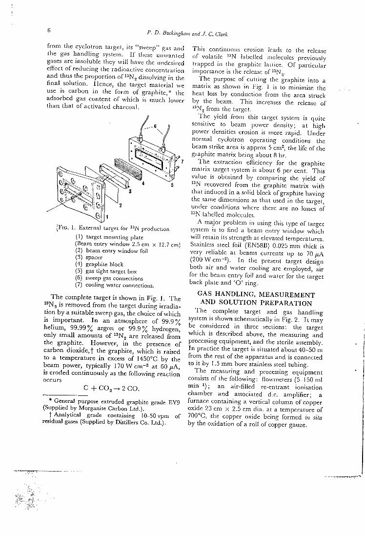
P• D. Buckingham and J. C. Clark
f r o m the cyc lo t ron target, its "sweep" gas and the gas h a n d h n g system. 11 these unwanted gases are insoluble they w i l l have the undesired e f fec t o f r educ ing the radioact ive concentra t ion and thus the p r o p o r t i o n o f ^•^Ng dissolving i n the f i n a l so lu t ion . Hence, the target ma te r i a l we use is c a r b o n i n the f o r m o f g raph i t e ,* the adsorbed gas con ten t o f w h i c h is m u c h t h a n t h a t o f ac t iva ted charcoal .
iower
F I G . 1 . External target for production
(1) target mounting plate (Beam entry window 2.5 cm x 12.7 cm) (2) beam entry window foi l (3) spacer (4) graphite block (5) gas tight target box (6) sweep gas connections (7) cooling water connections.
T h e comple te target is shown i n F i g . I . T h e " N g is r e m o v e d f r o m the target d u r i n g i r r ad i a t i o n b y a sui table sweep gas, the choice o f w h i c h is i m p o r t a n t . I n an atmosphere o f 9 9 . 9 % h e l i u m , 9 9 . 9 9 % argon or 9 9 . 9 % hydrogen, o n l y s m a l l amounts o f ^^Ng are released f r o m the g raph i t e . However , i n the presence o f c a r b o n d iox ide , f the graphi te , w h i c h is raised to a t empera tu re i n excess o f 1450°C by the beam power , t y p i c a l l y 170 W cm~^ at 60 ^ A , is e roded con t inuous ly as the f o l l o w i n g react ion occurs
C + C 0 2 - ^ 2 C O .
* General purpose extruded graphite grade EY9 (Supplied by Morganite Carbon L td . ) .
t Analytical grade containing 10-50 vpm of residual gases (Supplied by Distillers Co. L td . ) .
Th i s continuous erosion leads to the release o f vola t i le ^•'N labelled molecules previously t rapped i n the graphi te la i i ice . O f par t icu lar impor tance is the release o f ^^N^-
T h e purpose o f cu t t ing the graphite in to a m a t r i x as shown i n Fig . 1 is to minimise the heat loss b y conduct ion f r o m the area struck hy the beam. T h i s increases the release o f i ^ N , f r o m the target.
T h e y ie ld f r o m this target system is qu i t e sensitive to beam power density; at h igh power densities erosion is more rap id . U n d e r no rma l cyc lo t ron operat ing conditions the beam strike area is approx 5 cm^, the l ife o f the graphi te m a t r i x being about 8 hr .
T h e ext rac t ion efficiency for the graphi te m a t r i x target system is about 6 per cent. Th i s value is ob ta ined by compar ing the y ie ld o f
recovered f r o m the graphite ma t r ix w i t h tha t induced i n a solid block o f graphi te hav ing the same dimensions as that used i n the target, under condit ions where there are no losses o f ^•'N labelled molecules.
A ma jo r p rob lem in using this type o f target system is to f i n d a beam entry w i n d o w w h i c h w i l l re ta in its strength at elevated temperatures. Stainless steel f o i l (EN58B) 0.025 m m th ick is very reliable at beams currents up to 70 p.A ( 2 0 0 W c m ~ ^ ) . I n the present target design both air and water cool ing are employed, a i r fo r the beam ent ry f o i l and water for the target back plate a n d ' O ' r i n g .
G A S H A N D L I N G , M E A S U R E M E N T A N D S O L U T I O N P R E P A R A T I O N
T h e complete target and gas h a n d l i n g system is shown schematically i n F ig . 2. I t m a y be considered i n three sections: the target w h i c h is described above, the measuring a n d processing equipment , and the sterile assembly. I n practice the target is situated about 40-50 m f r o m the rest o f the apparatus and is connected to i t by 1.5 m m bore stainless steel t ub ing .
T h e measuring and processing equ ipment consists o f the f o l l o w i n g : f lowmeters (5-150 m l m i n ^ ^ ) ; an a i r - f i l l ed re-entrant ionisat ion chamber and associated d.c. a m p l i f i e r ; a furnace con ta in ing a ver t ical co lumn of copper oxide 23 cm x 2.5 cm dia. at a temperature o f 7 0 0 ° C , the copper oxide being f o r m e d in situ b y the ox ida t ion o f a r o l l o f copper gauze.
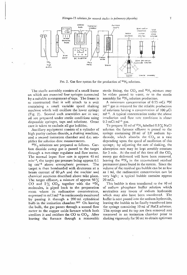
Nitrogen-\3 solutions for research studies in puLmoiiary physiology
F u r n a c e 7 0 0 " C c n a m b e r N i t r o g e n
argei
l 4 M e V d e u t e r o
beam
m e t a r A m p l i f i e r ox id f l
m e t e w a s t e pun ly
10 rn! s o d i u m
p f i c s p f i o : e
Waste oji'.er
s o l u t i o n s sDiuTior i
— - 1 — - J
.- .ofmal
s o l u i t o n
\ 20.T1I 2 N s o d i u m
S h a k e r S i e r i l e a s s e m D I y
F I G . 2. Gas flow system for the production of ^^Nj solutions.
T h e sterile assembly consists o f a smal l f r a m e o n w h i c h are m o u n t e d f o u r syringes connected b y a sui table a r rangement o f taps. T h e f r a m e is so cons t ructed tha t i t w i l l a t t ach to a u n i t c o n t a i n i n g a smal l var iab le speed shaking mach ine w h i c h w i l l oscillate the lower syringe ( F i g . 2 ) . Several such assemblies are i n use; a l l are p repa red under sterile condi t ions using disposable syringes, taps and solutions. Grea t care is taken to exclude a l l gas bubbles .
A n c i l l a r y e q u i p m e n t consists o f a cy l inde r o f h i g h p u r i t y ca rbon d iox ide , a shaking mach ine , a n d a second ionisa t ion chamber a n d d.c. a m p l i f i e r fo r so lu t ion dose measurements.
^^Ng solutions are prepared as fo l lows . Carb o n d i o x i d e sweep gas is passed to the target t h r o u g h a two-stage regula tor a n d f l o w meter . T h e n o r m a l i n p u t f l o w rate is a p p r o x 45 m l m i n ~ ^ , the target gas pressure be ing app rox 0.1 k g cm~^ above atmospheric pressi^re. T h e target is t h e n b o m b a r d e d w i t h deuterons at a b e a m c u r r e n t o f 60 fiA and the nuclear a n d chemica l reactions described above take place. T h e ta rge t e f f luen t , a m i x t u r e o f a p p r o x 9 8 % C O a n d 2 % C O j , together w i t h the ^ ^ N ^ molecules, is p i p e d back to the p r epa ra t i on r o o m where its radioact ive concent ra t ion , expressed i n m C i ml""^ is cont inuously m o n i t o r e d b y passing i t t h r o u g h a 200 m l . c y l i n d r i c a l b u l b i n the ion isa t ion c h a m b e r . O n leav ing the b u l b , the gas passes th rough a second f l o w mete r to the copper oxide fu rnace w h i c h b o t h sterilises i t a n d oxidises the C O to CO2. A f t e r l eav ing the fu rnace t h rough a removable
sterile fitting, the CO., and ^^.N', m ix tu re may be either passed to waste, or to the sterile assembly fo r ^^N^ solution product ion .
A m i n i m u m concentrat ion o f 0.25 m C i m l ~ ^ gas is requi red for the reliable produc t ion o f solutions hav ing a concentration o f 100 wCi m l ~ ^ . A typ ica l concentrat ion under the above i r r ad i a t i on and flow rate conditions is about 0.5 m C i m l ~ i gas.
T o prepare 10 m l o f ^ ^ N , labelled 0.9 % N a C I solution the furnace eff luent is passed to the syringe con ta in ing 20 m l o f 2A'' sodium hydroxide , w h i c h absorbs the CO2 at a rate depending u p o n the speed of oscillation o f this syringe; by ad jus t ing the rate o f shaking, the absorpt ion rate may be kept sensibly constant f o r 5 m i n . A t the end o f this t ime a l l the C O j sweep gas del ivered w i l l have been removed, leaving the ^•'Ng i n the accumulated residual permanent gases f o u n d i n the .system. Since the vo lume o f the residual gas bubble can be as low as 1 m l , the radioact ive concentrat ion can be very h i g h ; a typ ica l bubble contains approx 20 m C i .
Th i s bubb le is then transferred to the 10 m l o f sod ium phosphate bu f f e r solution w h i c h neutralises any traces o f sodium hydroxide w h i c h m a y also have been transferred. T h e bu f f e r is next passed in to the sodium hydroxide , leaving the bubble to be finally transferred in to the syringe con ta in ing 10 m l o f N a C l solut ion. Th i s syringe and its tap are then removed and measured i n an ionisat ion chamber p r i o r to shaking vigorously for 90 sec to obta in o p t i m u m
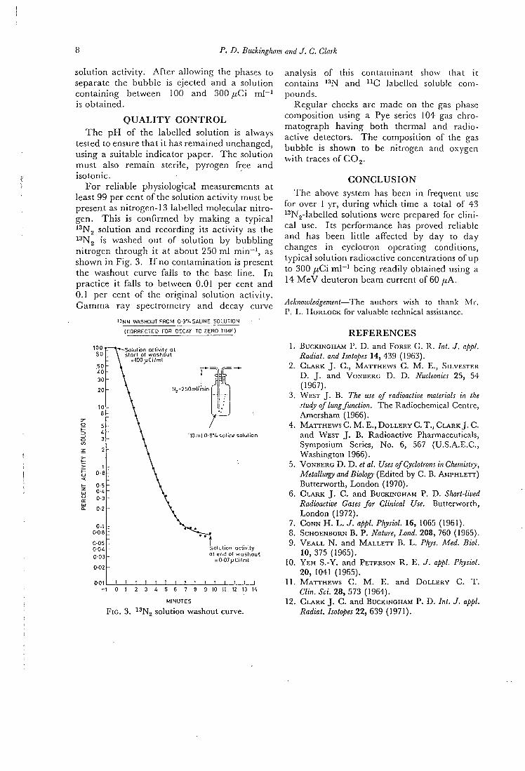
p . D. Buckingham and J. C. Clark
so lu t ion a c t i v i t y . A f t e r a l l o w i n g the phases to separate the bubb le is ejected and a solut ion con t a in ing between 100 and 300 ^ C i m l - ^ is ob t a ined .
Q U A L I T Y C O N T R O L
T h e p H o f the labe l led so lu t ion is always tested to ensure tha t i t has r ema ined unchanged, us ing a suitable i nd ica to r paper. T h e solut ion mus t also r e m a i n sterile, pyrogen free and isotonic .
F o r re l iable physio logica l measurements at least 99 per cent o f the solut ion a c t i v i t y must be present as ni trogen-13 label led molecular n i t r o gen . T h i s is c o n f i r m e d b y m a k i n g a t yp ica l ^^Ng so lu t ion and record ing its a c t i v i t y as the ^ N2 is washed ou t o f solut ion b y b u b b l i n g n i t rogen t h r o u g h i t a t about 250 m l m i n " ^ , as shown i n F i g . 3. I f no c o n t a m i n a t i o n is present the washout curve falls to the base l ine . I n pract ice i t falls to between 0.01 per cent and 0.1 per cent o f the o r i g ina l solut ion ac t iv i ty . G a m m a ray spectrometry and decay curve
13NN WASHOUT FROM 0 9V. SALINE SOLUTION
( C O R R E C T E D FDR DECAY TO Z E R O TIME)
^Solution activity at s tart of washiout
= 100; jCi /ml ,
N, -250ml/min
10ml 0-9"/.saline solution
Solution activity at end of w a s h o u t
= 0 07>iCi/ml
8 9 10 11 12 13 K
F I G . 3 . • N2 solution washout curve.
analysis o f this con taminan t show that i t contains ^^N and ^^C label led soluble compounds.
Regu la r checks are made on the gas phase composi t ion using a Pye series 104 gas chro-ma tograph h a v i n g bo th thermal and radioactive detectors. T h e composi t ion o f the gas bubb le is shown to be ni t rogen and oxygen w i t h traces o f COg-
C O N C L U S I O N
T h e above system has been i n f requent use fo r over 1 y r , d u r i n g w h i c h t ime a total o f 43 ^^Ng-labelled solutions were prepared for c l i n i cal use. I t s performance has proved reliable and has been l i t t l e affected by day to day changes i n cyc lo t ron operat ing condit ions, t yp ica l solut ion radioact ive concentrations o f up to 300 fiCi m l - i being readi ly obtained using a 14 M e V deuteron beam current o f 60 /zA.
Acknowledgement—The authors wish to thank M r . P . L . HoRLOCK for valuable technical assistance.
R E F E R E N C E S
1. BUCKINGHAM P . D . and FORSE G . R . Int. J . ajipt. Radial, and Isotopes 1 4 , 4 3 9 ( 1 9 6 3 ) .
2 . Ci-ARK J . C , M A T T H E W S C . M . E . , SILVESTER D . J . and VONBERG D . D . Nucleonics 2 5 , 5 4 ( 1 9 6 7 ) .
3. W E S T J . B . The use of radioactive materials in the study of lung function. The Radiochemical Centre, Amersham ( 1 9 6 6 ) .
4 . M A T T H E W S C . M . E . , D O L L E R Y C . T . , C I ^ R K J . C . and W E S T J . B . Radioactive Pharmaceuticals, Symposium Series, No. 6 , 5 6 7 ( U . S . A . E . C , Washington 1 9 6 6 ) .
5. VONBERG D . D . et al. Uses of Cyclotrons in Chemistry, Metallurgy and Biology (Edited by C . B . A M P H L E T T ) Butterworth, London ( 1 9 7 0 ) .
6. C L A R K J . C. and BUCKINGHAM P. D . Short-lived Radioactive Gases for Clinical Use. Butterworth, London ( 1 9 7 2 ) .
7. C O N N H . L . J . appl. Physiol. 1 6 , 1065 ( 1 9 6 1 ) . 8. ScHOENBORN B . P . Nature, Land. 2 0 8 , 7 6 0 ( 1 9 6 5 ) . 9. V E A L L N . and M A L L E T T B . L . Phys. Med. Biol.
1 0 , 3 7 5 ( 1 9 6 5 ) . 10. Y E H S . - Y . and PETERSON R . E . J . appl. Physiol.
2 0 , 1041 ( 1 9 6 5 ) . 1 1 . M A T T H E W S C . M . E . and D O L L E R Y C . T .
Clin. Sci. 2 8 , 5 7 3 ( 1 9 6 4 ) . 12. C L A R K J . C. and BUCKINGHAM P. D . Int. J . appl.
Radiat. Isotopes 2 2 , 6 3 9 ( 1 9 7 1 ) .
20 International Journal of Applied Radiation and Isotopes, 1972, Vol. 23, pp. 329-335. Pergamon Press. Printed in Northern Ireland
The Production of Potassium-43 for Medical Use
J . C . C L A R K , M . L . T H A K U R and I . A . W A T S O N
M R C Cyclotron Uni t , Hammersmith Hospiul , Ducane Road, London W12 OHS, England
{Received 3 February 1972)
A dynamic argon gas target system is described for the production of potassium-43 and the method for recovery of the activity is stated. The influence of the incident alpha particle beam energy on the relative yield of potassium-42 and potassium-43 is reported. The effects were investigated of other parameters, such as gas pressure and gas flow rate on the yields. The results are discussed.
L A P R O D U C T I O N D U POTASSIUM-43 P O U R E M P L O I M £ D I C A L O n ddcrit un systeme de cible dynamique a gaz d'argon pour la production du potassium-43
et on precise la mdthode pour le recouvrement de I'activite. On rend c o m p t e de Tinfluence de I'cnergie du faisceau incident de particules alpha sur le rendement rclatif d c potassium-42 et de potassium-43. O n regarda les efTets d'autres parametres tels que la t e n s i o n du gaz et Ic taux d'&oulement du gaz sur les rendements. On pr&ente unc discussion des resultats.
^ P 0 M 3 B 0 ^ ^ C T B 0 KAJIHf l -43 flJIH HCnOJIbSOBAHHH B M E f l H U I ' I H C K H X U E J I H X
Onucatia «KnaMHHecKaH apronoBafl rasoBaa MHUieHHaH ciicTeMa a.nn npousBoacTDa KajiHH-43, flaeTCH i i MeTon RJIH B o c c T a H O B J i e m i H aKTHBHocTii. Cooomeno u j i i i H m i e 3neprii!i naHaiomero nynKa ajibtfia-MacTHit na OTHOCHTeJibHMM BLixoa Ka.niifl-12, Ka.inH-4.3. H HCCJieAonam, ! n j i i i n i i n r i A p y r u x n a p a M e x p o s , iianpiiMcp, nanJieiiiie r a : !a 11 pacxo.t rasa.
D I E H E R S T E L L U N G V O N K A L I U M - 4 3 F U R M E D I Z I N I S C H E Z W E C K E Ein dynamisches Treffplattensystem von Argongas f u r die Herstellung von Kalium-43 wird
beschrieben und ein Verfahren wi rd angegeben fu r die Wiedergewinnung dcr Aktivitat . Der Einfluss der Einfallstrahlenergie eines Alphatcilchens auf die relative Ausbeute an Kalium-42 und Kalium-43 wird festgestclit. Die Wirlcungen von anderen Parameter wie z.B. Gasdruck und Gasgeschwindigkeit auf die Ausbeuten wurden untersucht. Die Ergebnisse werden besprochen.
I N T R O D U C T I O N w i t h that o f r u b i d i u m and caesium has been THERE are several ways i n w h i c h radioact ive carr ied ou t w i t h the a id o f radioact ive potas-potass lum m a y be o f va lue i n med ica l investi- sium*^' and i n pa r t i cu la r the i r a c c u m i i i a l i o n gat ions. For example the presence o f ^ ' ' K i n muscle tissue.*'' T l i e l abe l l ing o f red ceils i n n a t u r a l l y o c c u r r i n g potassium has enabled w i t h radioactive poiassium has been carr ied es t imat ion o f the body content o f potassium ou t successfully and used i n the measurement b y means o f w h o l e body counting.*^' Isotope o f blood vo lume. ' ^ ' I t has also been used f o r d i l u t i o n analysis enables in vivo es t imat ion o f imag ing the heart '" ' and i n myocard ia l per-t o t a l body concentrat ions and measurement o f fusion studies.'^''' O n e f u r t h e r appl ica t ion has the exchangeable potassium.'2~^' Compar ison been f o u n d i n the localisat ion o f b ra in l u -o f the physiological behaviour o f potassium mours.'^^'^^' O f the seven radioact ive isotopes
329
330 J. C. Clark, M. L. Thakur and I. A. Watson
T A B L E 1
Characteristics 42K 43K Ref.
H a l f life 12-4 h 22-6 h 13, 14 y energies 1-524 M e V (18%) 373 keV (87-8%) 13, 14
618 keV (81%) P~ energies 3-52 M e V (max) 830 keV (max) 13 Whole body radiation dose 0-85 m rad / i C i " ! 0-6 m rad /<Ci-i 4 K factor 1-4 5-6 4
o f potassium there are o n l y t w o w h i c h p e r m i t sat isfactory use i n c l i n i c a l investigations. These are a n d ^ ^ K .
has been used most extensively i n medica l w o r k as i t is r ead i ly ava i lab le f r o m a reactor i r r a d i a t i o n . I t is p roduced b y the react ion « K ( n , a )42K, ^^C3.{n, p^^K and « S c ( n , aY^K. T h e h a l f l i f e o f ^ ^ K is r e l a t ive ly short , 12-4 h r , b u t its p r i n c i p a l y - r a y energy a n d ^ ~ end p o i n t energy are h i g h , 1-524 M e V and 3-52 M e V respectively, m a k i n g i t unsui table f o r some m e d i c a l appl ica t ions . O n the other hand ^ ^ K has a b u n d a n t y-rays w i t h energies o f 373 k e V and 618 k e V , a n d /3~ end p o i n t energies o f 830 k e V and 460 k e V , m a k i n g this isotope more sui table f o r med ica l use. These physical characteristics o f a n d are shown i n T a b l e 1.
Potassium-43 can be p repared b y the reactions 4 » A r ( a , />^^3K a n d " G a ( / » , 2 / ) ) " K using accelerators or b y the *''Ca(re, / ' ) ^ ^ K react ion using h i g h l y enr iched ^^Ca'^^' (na tu ra l a b u n d ance 0-145 per cent) f o r a reactor i r r a d i a t i o n w i t h fast neutrons.
I n 1959 D Y S O N a n d F R A N C O I S ' ^ ^ ' described
a n electrostatic m e t h o d o f recover ing f r o m an a rgon gas-phase target i n w h i c h sojiie o f the potassium ions were col lected on a stainless steel r o d he ld at a negat ive po ten t i a l o f abou t 1 k V .
T h e m e t h o d described was inheren t ly i n e f f ic ien t as i t was f o u n d to be necessary to i n t e r r u p t the a lpha pa r t i c l e beam w i t h a 50 per cent d u t y cycle i n o rder to estabhsh a h i g h electr ic field to col lect the ions. A l t h o u g h a more p o w e r f u l source o f h i g h voltage m a y be used to overcome this p r o b l e m , the recovery o f the f r o m a l o n g stainless steel r o d was no t a t t r ac t ive .
I n the first p u b l i c a t i o n describing the discovery o f *^K<^ ' ' i t was f o u n d tha t as the a rgon
was a l lowed to flow out o f the target chamber t h r o u g h a glass woo l filter p l u g some o f the
was carr ied as far as the filter i n the gas phase and could be removed by a w a r m water wash. However , the m a j o r i t y o f the ^• 'K was recovered by washing the walls o f the i r r a d i a t i o n vessel w i t h water . T h e present w o r k describes efforts to increase the p ropo r t i on o f
recovered on a small , easily washed filter a n d other factors i n f l uenc ing the y i e l d and p u r i t y o f the final p roduc t .
T A R G E T S Y S T E M A N D P R E P A R A T I O N F O R C L I N I C A L U S E
T h e target chamber consists o f an a l u m i n i u m tube, 46 c m long, 14 c m d ia . , 3-18 m m w a l l thickness, onto bo th ends o f w h i c h are welded 1-25 c m th ick a l u m i n i u m plates w h i c h are water cooled. T h e f r o n t plate has a rectangular aper ture 12-5 c m X 2-5 c m fo r beam entry-over w h i c h is mounted a beam entry windo\.-consisting o f an 0-025-mm t i t a n i u m f o i l and two 0-10-mm a l u m i n i u m foi ls . Th i s reduces the 3 0 - M e V external a lpha beam of the M e d i c a l Research Counc i l ( M R C ) cyclo t ron to about 17 M e V . As a result about 500 W are dissipated i n the foils. Th i s heat is removed by a comb i n a t i o n o f f r o n t plate water cool ing, a i r coo l ing blast on the f o i l ' ^ ^ ' a n d transfer i n the rec i rcu la t ing argon to the rest o f the system. T h e back plate o f the target chamber has two gas connections, a 6-4-mm gas in le t w i t h de l ivery tube and a 19-2-mm out le t . T h e target is connected to a gas flow system shown in Fig. 1 and schematically i n F ig . 2. I t is o f closed c i r c u i t design and uses oi l-free d i a p h r a g m pumps ( C o m p t o n oi l -free compressors, type 4 D , supplied by Dawson M c D o n a l d and Dawson L t d . , Ashbourne, Derbyshire) to circulate the gas. A flow meter and pressure gauge are p r o v i d e d for m o n i t o r i n g purposes d u r i n g filling
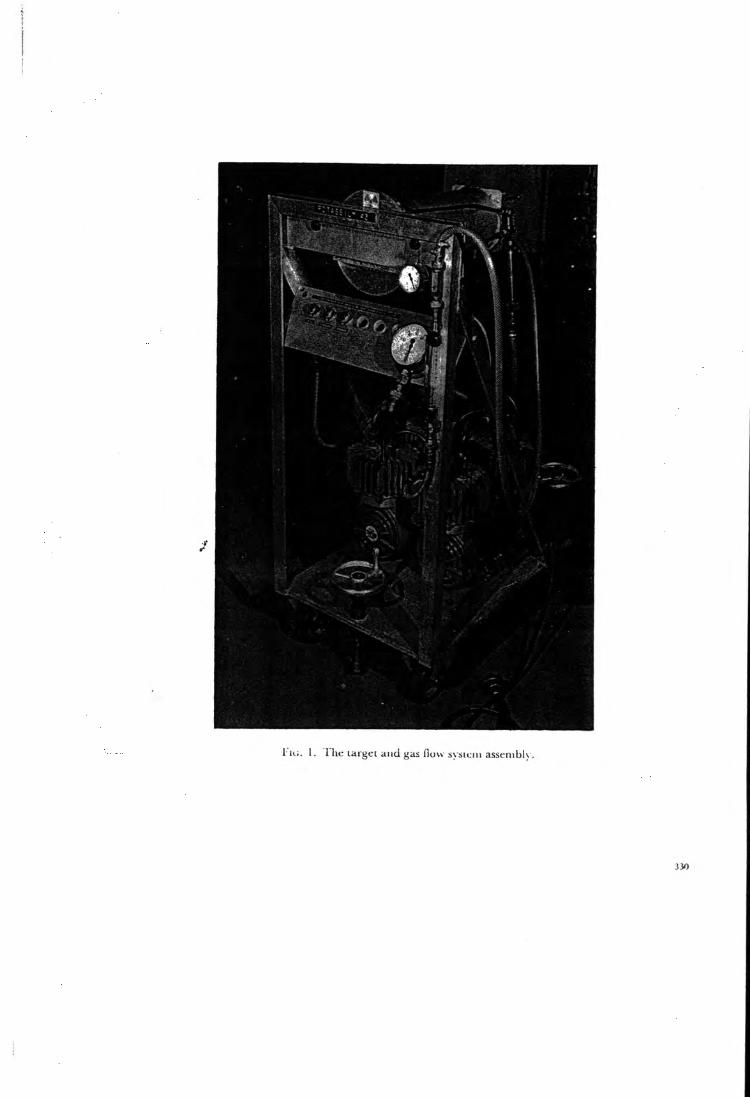
I'lc. 1. The largei and gas flow sysiciu asscmblv .
330
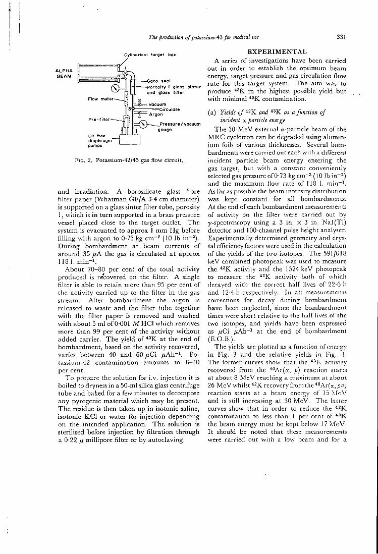
Tlie production of potassiumA3 for medical use 331
Cylindrical forget box / I
A L P H A B E A M
Flow motor
Pre-fi ltor
Oil froo diaphragm pumps
aco sea l •Porosity I glass sintor and glass filtor
Vacuum Circulate
Argon
Pressure / vacuum gauge
F I G . 2. Potassium-42/43 gas flow circuit.
a n d i r r a d i a t i o n . A borosil icate glass fibre filter paper ( W h a t m a n G F / A 3-4 c m d iameter ) is suppor ted on a glass sinter filter tube , porosi ty 1, w h i c h is i n t u r n supported i n a brass pressure vessel placed close to the target ou t le t . T h e system is evacuated to approx 1 m m H g before fining w i t h a rgon to 0-73 kg cm-2 (10 l b i n - ^ ) . D u r i n g b o m b a r d m e n t at beam currents o f a r o u n d 35 /nA the gas is c i rcu la ted at app rox 118 1. m i n - i .
A b o u t 70-80 per cent o f the t o t a l a c t i v i t y p r o d u c e d is r€!covered on the filter. A single filter is able to re ta in more than 95 per cent o f the a c t i v i t y ca r r ied up to the filter i n the gas s t ream. A f t e r b o m b a r d m e n t the a rgon is released to waste a n d the filter tube together w i t h the filter paper is removed a n d washed w i t h abou t 5 m l o f 0-001 M H C l w h i c h removes m o r e t h a n 99 per cent o f the a c t i v i t y w i t h o u t added carr ier . T h e y i e l d o f a t the end o f b o m b a r d m e n t , based on the ac t i v i t y recovered, varies between 40 a n d 60 fiCi fiAh~^. Po-tassium-42 con t amina t i on amounts to 8-10 per cent .
T o prepare the solut ion for i . v . i n j e c t i o n i t is bo i l ed to dryness i n a 50 -ml silica glass cent r i fuge tube and baked fo r a few minutes to decompose a n y pyrogenic ma t e r i a l w h i c h m a y be present. T h e residue is then taken u p i n isotonic saline, i so tonic K C l or wa te r f o r i n j ec t i on depend ing o n the in tended app l i ca t ion . T h e so lu t ion is steril ised before i n j ec t i on b y filtration t h r o u g h a 0-22 fx m i l l i p o r e filter or b y au toc lav ing .
E X P E R I M E N T A L
A series o f investigations have been car r ied out i n order to establish the o p t i m u m beam energy, target pressure and gas c i rcula t ion flow rate for this target system. T h e a i m was to produce i n the highest possible y ie ld b u t w i t h m i n i m a l ^ ^ K con tamina t ion .
(a) Yields o / ^ ^ K and as a function of incident a particle energy
T h e 3 0 - M e V external a-particle beam o f the M R C cyclot ron can be degraded using a l u m i n i u m foils o f various thicknesses. Several b o m bardments were carried out each w i t h a d i f f e ren t inc ident part icle beam energy entering the gas target, b u t w i t h a constant convenient ly selected gas pressure o f 0-73 kg cm"^ (10 lb i n ~ - ) and the m a x i m u m flow rate o f 118 1. m i n ~ ^ . As fa r as possible the beam intensity d i s t r i bu t ion was kept constant fo r a l l bombardments . A t the end o f each bombardmen t measurements o f ac t iv i ty on the filter were carr ied out b y y-spectroscopy using a 3 i n . X 3 i n . N a l ( T l ) detector and 100-channel pulse height analyser. Exper imenta l ly determined geometry and crysta l efficiency factors were used i n the ca lcu la t ion o f the yields o f the two isotopes. T h e 591/618 k e V combined photopcak was used to measure the ac t iv i ty and the 1524keV photopeak to measure the ac t iv i ty both o f w h i c h
decayed w i t h the correct h a l f lives o f 22-6 h and 12-4 h respectively. I n a l l measuremcn;s corrections fo r decay d u r i n g bombardmen t have been neglected, since the b o m b a r d m e n t times were short relat ive to the ha l f lives o f the two isotopes, and yields have been expressed as /j,Ci nAh~^ at the end o f bombardmen t ( E . O . B . ) .
T h e yields are p lo t ted as a func t i on o f energy i n F ig . 3 and the relat ive yields i n Fig . 4 . T h e former curves show that the ^• 'K ac t i v i t y recovered f r o m the ^ ' 'Ar (a , p) reaction starts at about 8 M e V reaching a m a x i m u m at abou t 26 M e V whi ls t ^ ^ K recovery f r o m the '*"'Ar(a, p.'?) reaction starts at a beam energy o f 15 M c \ ' ' and is s t i l l increasing at 30 M e V . T h e la t t e r curves show tha t i n order to reduce the ^ - K contamina t ion to less than 1 per cent o f the beam energy must be kept below 17 M e \ ' . I t should be noted tha t these measurements were carr ied out w i t h a l ow beam and fo r a
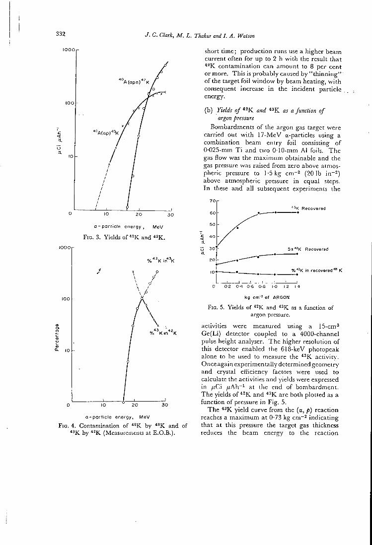
332 J. C. Clark, M. L. Thakur and I. A. Watson
l O O O r
' ° A ( a p n ) ^ = K
a - p a r t i c l e e n e r g y , M e V
F I G . 3. Yields of ^^K and " K .
l O O O r
100
Q- 10
o - p a r t i c l e e n e r g y , M e V
F I G . 4. Contamination of by ^^K and of " K by (Measurements at E.O.B.).
short t i m e ; p r o d u c t i o n runs use a higher beam cur ren t o f t en f o r u p to 2 h w i t h the result t h a t
con tamina t ion can amount to 8 per cent or more. T h i s is p robab ly caused by " t h i n n i n g " o f the target f o i l w i n d o w by beam heating, w i t h consequent increase i n the inc ident pa r t i c l e energy.
(b) Yields o / *2K and ^^K as a function of argon pressure
Bombardments o f the argon gas target were carr ied ou t w i t h 1 7 - M e V a-particles using a combina t ion beam en t ry f o i l consisdng o f 0-025-mm T i and t w o 0-10-mm A l foils. T h e gas f l ow was the m a x i m u m obtainable and the gas pressure was raised f r o m zero above atmospheric pressure to 1-5 kg cm~^ (201b in~^) above atmospheric pressure i n equal steps. I n these and a l l subsequent experiments the
"^K Recovered
< a.
0-6
Sx^^K Recovered
ro^^K in recovered'^ K
kg of ARGON
F I G . 5. Yields of ^^K and as a function of argon pressure.
activities were measured using a IS-cm^ G e ( L i ) detector coupled to a 4000-channel pulse height analyser. T h e higher resolution o f this detector enabled the 618-keV photopeak alone to be used to measure the activirv-. Once again exper imenta l ly determined geometry" and crystal eff iciency factors were used to calculate the activit ies and yields were expressed i n ^ C i / i A h ~ ^ at the end o f bombardmen t . T h e yields o f and are both p lo t ted as a f u n c t i o n o f pressure i n F i g . 5.
T h e y i e l d curve f r o m the (a, p) react ion reaches a m a x i m u m at 0-73 kg cm"^ i n d i c a t i n g tha t a t this pressure the target gas thickness reduces the beam energy to the react ion
The production of potassiumAS for medical use 333
< a. o a.
Recovered
lOx'^^K R e c o v e r e d
% in recovered " K
I I I I I I I I I 0 10 30 50 70 90 n o
Flovx rote , L min '
F I G . 6. Yields o f ^^K and as a function of flow rate of argon.
threshold energy o f about 8 M e V . O n l y a sma l l change i n the y i e l d is noted, the increase w i t h pressure be ing d i f h c u l t to exp la in sat isfactor i ly o n account o f the m a n y o ther variables i n this system. However , f o r rou t ine p r o d u c t i o n purposes i t is clear tha t there is no advantage i n increasing the a rgon pressure above 0-73 k g c m " ^ .
(c) Yields of '^^K and as a function of rate of flow of argon
Since i t i s j t h e gas stream w h i c h carries the p roduced a c t i v i t y to the filter the inf luence o f the f l ow rate o n the recovered ac t iv i ty was s tudied. T h e ro tamete r flow meter was ca l i b r a t ed against a gas meter ca l ib ra ted b y U n i t e d Gas Indust r ies ( L o n d o n ) . T h e flow was con t ro l l ed b y a n adjustable va lve inserted i n the c i r c u l a t i o n system between valve N o . 3 a n d the p u m p ( F i g . 2 ) . Bombard ihents were ca r r ied o u t w i t h 1 7 - M e V a-particles and w i t h a gas jbressure o f 0-73 k g cm~^. Acdvi t i e s were measured us ing the G e ( L i ) detector and the yields, corrected to E . O . B . , were p lo t t ed as a f u n c t i o n o f flow ra te i n F i g . 6. These curves show tha t no increase i n recovery can be obta ined w i t h flow rates i n excess o f 100 1. m i n ~ ^ a n d the re la t ive yields r ema in , as expected, unchanged a t a l l flow rates. A c t i v i t y lost a t the l o w e r flow rates is deposited o n the inside surface o f the target chamber . For rou t ine p roduc t ion purposes therefore the m a x i m u m at ta inable flow rate o f 118 1. m i n ~ ^ is used.
(d ) Activity losses on the target chamber at a fixed energy and pressure
I n order to see the y i e l d d i s t r i bu t i on w i t h i n the target chamber, bombardments were car r ied ou t i n a static system at a pressure o f 0-73 k g cm -2 w i t h I 7 - M e V a-particle beams, o f 35 fjtA.. T h e target was l ined w i t h a l u m i n i u m f o i l w h i c h a f te r b o m b a r d m e n t was removed and cut in to cyl inders 5 c m t a l l . Each piece o f f o i l therefore represents a d i f f e r e n t dep th i n the target. T h e ac t iv i ty was then measured on each cyhnder using the 15-cm^ G e ( L i ) detector. T h e percentage o f the to ta l a c t i v i t y on each s t r ip was calculated and the results are p lo t t ed as a h is togram i n F i g . 7. These measurements were then repeated w i t h the dynamic system w i t h the m a x i m u m gas flow rate. T h e results, expressed as the percentage o f the total a c t i v i t y produced d u r i n g the static r u n , are also p lo t t ed i n F i g . 7. T h e dot ted curves are the to ta l in tegrated ac t i v i t y i n each case. T h e differences between the two histograms represent the a c t i v i t y swept f r o m the target chamber. I n the static system 65-70 per cent o f the ac t i v i t y is produced i n the first 15 c m o f the chamber a n d adheres to the w a l l . I n the dynamic system two thirds o f this ac t iv i ty is removed f r o m the target and collected on the
< a.
u a. 20
S f o t i c
n /
•X—
Dynamic - J
r
1 I
- X X X — , Stotic
Dynamic
100
9 0
SO
70
6 0 ^
5 0 -T;
3 0
20
• 0
0 O 5 10 15 2 0 2 5 3 0 3 5 1 0 « 5
Distance ( c m ) from front of target
F I G . 7. Formation and losses of ^^K activity inside the target chamber.
3 3 4 J. C. Clark, M. L. Thakur and I. A. Watson
glass fibre filter, b u t s t i l l one t h i r d o f the t o t a l a c t i v i t y p roduced is l e f t o n the target wal l s .
A n a t t e m p t was made to reduce these losses o f a c t i v i t y b y chang ing the flow p a t h o f the gas i n the target . T h e flow was a r ranged across the f r o n t section o f the chamber a n d u n d e r these condi t ions the t o t a l recovery increased b y abou t 15-20 per cent r educ ing the t o t a l loss to abou t 10 per cent. cont a m i n a t i o n i n the recovered a c t i v i t y also decreased to abou t 2 per cent. However , as before , the c o n t a m i n a t i o n was f o u n d to be ve ry sensitive to changes i n the i nc iden t par t ic le energy. A 1 - M e V increase raised the i n the recovered a c t i v i t y to abou t 15 per cent. T h i s system has no t ye t been adopted f o r r o u t i n e p r o d u c t i o n as a suitable target has no t ye t been constructed.
the beam. Increasing the pressure to 1-5 kg cm~^ (20 lb i n "^ ) d i d not improve the y ie ld . T h e overal l result o f opera t ing the system at 70 (lA w i t h a T i / g r a p h i t e w i n d o w combina t ion was to raise the rate o f p roduc t ion f r o m 2 m C i h~^ to 3 m C i h~"^ bu t w i t h a decrease i n r e l i a b i l i t y due to f o i l w i n d o w failures. Th i s me thod has not therefore been used for regular p r o d u c t i o n o f this isotope.
T h e measurements described i n this paper were made w h o l l y to ob ta in prac t ica l data for the target system described. Factors such as the shape and size o f target, geometry o f the gas in le t and out le t a l l make the target system unique . V a r i a t i o n i n beam d i s t r i bu t ion also results i n considerable va r i a t ion i n yields and con tamina t ion levels. However the system, a l though perhaps no t ideal , can be operated re l i ab ly and reasonably reproduc ib ly under the condit ions described.
D I S C U S S I O N
T h e m a x i m u m at ta inable external a par t ic le b e a m cur ren t f r o m the M R C cyc lo t ron is 100 [JLA. T h e target f o i l system described i n this paper permi ts a m a x i m u m cont inuous beam c u r r e n t o f o n l y 35 jxA. Even at this beam c u r r e n t the loss o f energy i n the f o i l is equivalent to abou t 500 W . I f beam currents greater t h a n this are employed , pa r t i cu l a r l y i f the beam defocusing is inadequate , then the present coo l ing system o f a i r b l o w n on to the f o i l face and wate r coo l ing a r o u n d the edges o f the f o i l w i n d o w becomes insuff ic ien t , resul t ing ve ry q u i c k l y i n punc tu re o f the f o i l .
Several d i f f e r e n t f o i l materials haye been t r i e d i n c l u d i n g t a n t a l u m , t i t a n i u m a n d stainless steel ( E N 5 8 B ) o f a thickness w h i c h reduces the b e a m energy to 1 7 - M e V , b u t none proved bet ter t h a n the present t i t a n i u m / a l u m i n i u m c o m b i n a t i o n . A graph i te w i n d o w , uncooled, o f 0 -25 -mfn was t r i ed , protected f r o m pressure a n d o x i d a t i o n b y a 0-025-mm t i t a n i u m f o i l . T h i s p roved satisfactory for bombardments at b e a m currents u p to 70 ^uA. However , the y i e l d o f per jxAh. d ropped b y about 30 per cent be low the y i e l d a t 35 fiA. T h i s is p r o b a b l y exp la ined by local density reduc t ion o f the gas i n the target chamber a long the pa th o f the beam as a result o f the heat produced b y
Acknowledgement—The authors wish to thank Dr. D. J . SILVESTER for his comments.
R E F E R E N C E S
Science 1. ANDERSON E . C . and L A N G H A M W . H .
130, 7 1 3 ( 1 9 5 9 ) .
2 . JAMES J . A . and ROBERTSON J . S. Am. J . Dis.
Child. 93, 217 ( 1 9 5 3 ) . 3. CORSA L . J. Clin. Invest. 29, 1280 ( 1 9 5 0 ) . 4 . SKRABAL F. , GLASS H . L , C L A R K J . C , JEVASI.NGH
K . and J O P L I N G . F . Int. J. appl. Radiat. Isotopes 20, 6 7 7 ( 1 9 6 9 ) .
5. JOHNSON J . E . , HARTSUCK J . M . , ZOLLINGER R .
M . and M O O R E F . D . Metabolism 18, 6 6 3 ( 1 9 6 9 ) .
6 . R E L M A N A . S. J . Biol. Med. 29, 2 4 8 ( 1 9 5 6 ) .
7 . R E L M A N A . S., L A M B I C A . T . and BURROW B . A .
J. din. Invest. 36, 8 ( 1 9 5 7 ) . 8. HEVESY G . and H Y L I N G . Acta Physiol. Scand.
24, 2 8 5 ( 1 9 5 2 ) .
9 . H U R L E Y P . J . , COOPER M . , R E B A R . C , POGGEN-
BURG K . J . and W A N G E R H . N . J . nucl. Med. 12 ,
5 1 8 ( 1 9 7 1 ) .
10. H o L M A N B . L . , POGGENBURG K . J . , ADAMS D . P . , E R D H P . and ADELSTEIN S. J . J . nucl. .Med.
Abstract 12 , 3 6 6 ( 1 9 7 1 ) . 11. SusEN A . F . , S M A L L W . T . and M O O R E F . D .
Surg. Forum of Am. Coll. Surgeons, 36th Congress, p. 3 6 2 ( 1 9 5 2 ) .
12 . SILVERSTONE B . , SWEET W . H . and IRETON R . S.
Surg. Forum of Am. Coll. Surgeons, 36th Congress, p. 3 7 1 ( 1 9 5 2 ) .
The production of potassiumAS for medical use 335
13. LEDERER C . M . , H O L L A N D E R J . M . and PERLMAN 17 . OVERSTREET R . , JOCOBSON L . and Srotn- P . L . I . Table of Isotopes {ettiedn), p. lA. Johnsonand Phys. Rev. 75, 231 ( 1 9 4 9 ) . Wiley, New York. 18 . VONBERG D . D . , BAKER L . C , BUCKINGHAM
14. WATERS S. L . Radiochimica Acta, in press. P . B., C L A R K J . C , FINDING K . , SHARP J . and 15 . O ' B R I A N H . A . and H U P F H . B . J. nucl. Med. SILVESTER D . J . Uses of Cyclotrons in Chemistry,
Abstract 9, 3 4 0 ( 1 9 6 8 ) . Metallurgy and Biology, p. 2 5 8 . Butterworths, 16 . DYSON N . A . and FRANCOIS P . E . Int. J . appl. London ( 1 9 7 0 ) .
Radiat. Isotopes 7, 150 ( 1 9 5 9 ) .
21 l A E A - S M - n i / 8
PREPARATION OF i S p - L A B E L L E D DL-3-FLUOROTYROSINE
A . J . PALMER. J. C, C L A R K . R . W . G O U L D I N G . M . R O M A N *
M R C C y c l o t r o n U n i t , H a m m e r s m i t h H o s p i t a l ,
L o n d o n ,
U n i t e d K i n g d o m
Abstract
A method of preparing '*F-labelled DL-3-fluorot^'rosine is described, is produced by the cyclotron bombardment of neon via the ' °Ne (d, a) reaction using a glass-lined target vessel. The ' * F is recovered in aqueous solution by remote washing of the glass liner, by rotation of the liner with 20 ml of water in it. Detailed operation of the system is described. The " F activity is introduced into a diazonium tetrafluoroborate derivative of the amino-acidby an exchange reaction in aqueous acetone/acetonitrile solution. The preparation of this diazonium salt, 4-methoxy-3-(2', 2'-dicarbethoxy-2'-acetamidoethyl)-phenyl diazonium tetrafluoroborate (!) starting from p-methoxybenzyl chloride is fully described. Pyrolysis of the labelled diazonium salt (1) yields "p-diethyl 3-fluoro-4-metliox)'benzylacetamidomalonate (Schiemann Reaction) which is then hydrolysed to "'F-3-fluorot)'rosine by red phosphorus and hydriodic acid. A similar synthesis, starting from L-phenylalanine, has been investigated with a view to preparing '^F-L-phenylanine. Experimental details of the labelling procedure are described and quality control methods for "F-labelled amino-acids are also discussed.
I N T R O D U C T I O N
F o l l o w i n g the s u c c e s s o f the o r g a n o m e t a l l i c c o m p o u n d s c h l o r m e r o d r i n a n d B M H P ( ^ ^ ' ' H g ) , a n d t h e p u r e l y o r g a n i c c o m p o u n d s s e l e n o m e t h i o n i n e C^sse), r o s e b e n g a l a n d s o d i u m i o d o h i p p u r a t e {'^^^l, ^ ^ ^ I , ^^^l) a s s c a n n i n g a g e n t s , i t has b e e n g e n e r a l l y t h o u g h t d e s i r a b l e to t r y to p r e p a r e o r g a n i c c o m p o u n d s c o n t a i n i n g s h o r t - l i v e d g a m m a - e m i t t i n g r a d i o n u c l i d e s i n w h i c h s o m e o f t h e l i m i t a t i o n s o f t he s c a n n i n g a g e n t s i n u se a t the p r e s e n t t i m e , n a m e l y r e l i a b i l i t y o f d i s t r i b u t i o n , l o s s o f l a b e l f r o m t h e f o r m i n w h i c h i t i s a d m i n i s t e r e d , a n d r a d i a t i o n d o s e , a r e m i n i m i z e d . I n t he p a s t f e w y e a r s s e v e r a l o r g a n i c c o m p o u n d s c o n t a i n i n g t h e f i r m l y b o u n d ^^F l a b e l ( T i 110 m i a . P" ) h a v e b e e n p r e p a r e d , p r o m i n e n t a m o n g w h i c h a r e t he m o n o f l u o r o -d e r i v a t i v e s o f tv /o n a t u r a l a r o m a t i c a m i n o a c i d s , p h e n y l a l a n i n e [ 1 , 2 ] a n d t r y p t o p h a n [ 2 ] . A d e s c r i p t i o n w i l l h e r e be g i v e n o f t h e p r e p a r a t i o n o f 18 F - D L - 3 - f l u o r o t y r o s i n e , a p r e v i o u s l y u n r e p o r t e d ^ ^ F - l a b e l l e d a r o m a t i c a m i n o - a c i d .
T h e m o n o f l u o r o a r o m a t i c a m i n o - a c i d s h a v e a t t r a c t e d i n t e r e s t b e c a u s e o f t h e i r w e l l - d o c u m e n t e d p h a r m a c o l o g i c a l p r o p e r t i e s . T h e y a r e i n c o r p o r a t e d i n t o p r o t e i n s [ 3 ] , s h o w a n t i - t u m o u r p r o p e r t i e s [ 4 ] a n d a r e a n t i - m e t a b o l i t e s o f t h e n a t u r a l a m i n o - a c i d s [ 5 ] . T h e s e p r o p e r t i e s a n d t h e u t i l i t y o f ' '^Se-s e l e n o m e t h i o n i n e i n d i c a t e a p o t e n t i a l f o r t h e s e m a t e r i a l s w h e n l a b e l l e d
On leave from Laboratory of Labelled Compounds, Institute of Atomic Physics, Bucharest, Romania.
2 9 1
292 PALMER e t a l .
w i t h I8 j r^ as o r g a n s c a n n i n g a g e n t s , e s p e c i a l l y f o r t h e p a n c r e a s [ 6 ] . T h e s u c c e s s o f a c o m p o u n d i n t h i s a p p l i c a t i o n d e p e n d s c r i t i c a l l y u p o n the p a n c r e a s - t o - l i v e r r a t i o , w h i c h m u s t be as h i g h a s p o s s i b l e i f g o o d p a n c r e a s i m a g e s a r e to be o b t a i n e d . T h e r a t i o v a r i e s w i t h the s t r u c t u r e o f t he c o m p o u n d c o n c e r n e d [ 7 ] , so t h a t i t i s e s s e n t i a l t o t e s t as m a n y d i f f e r e n t c o m p o u n d s a s p o s s i b l e i n o r d e r to m a x i m i z e t h i s r a t i o .
T w o o t h e r i m p o r t a n t p o i n t s m u s t a l s o be c o n s i d e r e d s i n c e t h e y a r e d i r e c t l y r e l a t e d to t he s y n t h e t i c m e t h o d u s e d a t p r e s e n t f o r m a k i n g i ^ p . l a b e l l e d a m i n o - a c i d s . T h e s e c o m p o u n d s s h o w v a r y i n g t o x i c i t i e s . T h e m o s t t o x i c , i n c l u d i n g 3 - f l u o r o t y r o s i n e [ 8 ] , h a v e a n LD50 i n t he r a n g e 5 - 1 0 m g k g " i a n d c o n s e q u e n t l y m u s t be p r e p a r e d w i t h the h i g h e s t p o s s i b l e s p e c i f i c a c t i v i t i e s . A l s o , a l l n a t u r a l a m i n o - a c i d s b e l o n g to t he L - s e r i e s a n d i t i s o n l y p o s s i b l e , a t t he p r e s e n t s t a t e o f k n o w l e d g e , t o p r e p a r e the D L - r a c e m a t e s o f c o m p o u n d s l a b e l l e d w i t h ^^F.
G E N E R A L C O N S I D E R A T I O N S F O R I ^ f - L A B E L L I N G
T h e c a r b o n - f l u o r i n e b o n d h a s a v e r y h i g h d i s s o c i a t i o n e n e r g y ( ~ 110 k c a l m o l e " ! ) , a n d c o n s e q u e n t l y s t a b i l i t y p r o b l e m s a r e l e s s l i k e l y t o be e n c o u n t e r e d w i t h i ^ p - i a b e l l e d c o m p o u n d s t h a n , f o r e x a m p l e , i o d i n e -l a b e l l e d c o m p o u n d s . H o w e v e r , t h i s f a c t m e a n s t h a t s o m e o f t h e s e c o m p o u n d s a r e d i f f i c u l t t o p r e p a r e i n the s h o r t p e r i o d s o f t i m e a v a i l a b l e , the h a l f - l i f e o f t he r a d i o n u c l i d e b e i n g 110 m i n . L o n g r e a c t i o n t i m e s a n d l a r g e m o l a r e x c e s s e s o f t h e f l u o r i n a t i n g a g e n t a r e u s e d i n m a n y f l u o r i n a t i o n r e a c t i o n s , c o n s e q u e n t l y r a d i o c h e m i c a l y i e l d s w o u l d be e x t r e m e l y l o w i f s u c h r e a c t i o n s w e r e a d o p t e d as l a b e l l i n g p r o c e d u r e s . B e c a u s e o f t h e h i g h s p e c i f i c a c t i v i t i e s o f t e n r e q u i r e d , d i f f i c u l t i e s i n h a n d l i n g s m a l l a m o u n t s o f m a t e r i a l e f f i c i e n t l y a r e a l s o e n c o u n t e r e d .
T o p r e p a r e i ^ p - l a b e l l e d 3 - f l u o r o t y r o s i n e i t w a s n e c e s s a r y t o d e v i s e a s y n t h e s i s i n w h i c h t h e ^ ^ F a c t i v i t y c o u l d be i n t r o d u c e d i n t o a p r e c u r s o r w h i c h w o u l d t h e n be r a p i d l y t r a n s f o r m e d i n t o t he p r o d u c t . U n l a b e l l e d 3 - f l u o r o t y r o s i n e h a s b e e n p r e v i o u s l y s y n t h e s i z e d b y c o n d e n s a t i o n o f 3 - f l u o r o - 4 - m e t h o x y - o r 3 - f l u o r o - 4 - e t h o x y b e n z a l d e h y d e , w i t h h i p p u r i c a c i d a n d s u b s e q u e n t h y d r o l y s i s a n d r e d u c t i o n [ 9 ] . I t has a l s o b e e n p r e p a r e d f r o m m - f l u o r o p h e n y l a l a n i n e b y n i t r a t i o n , r e d u c t i o n a n d f i n a l l y d e c o m p o s i t i o n o f t h e c o r r e s p o n d i n g d i a z o n i u m s a l t o n r e f l u x i n g t he a q u e o u s s o l u t i o n [ 9 ] . N e i t h e r o f t h e s e t w o m e t h o d s i s f e a s i b l e as a r o u t e t o t h e I 8 j r - i a b e l l e d m a t e r i a l b e c a u s e o f t h e e x c e s s i v e l y l o n g t i m e s r e q u i r e d .
I n t h e p r e v i o u s l y r e p o r t e d p r e p a r a t i o n o f ^ ^ F - l a b e l l e d p - f l u o r o p h e n y l -a l a n i n e , t h e i S p w a s i n t r o d u c e d i n t o 4 - ( 2 ' ,2' - d i c a r b e t h o x y - 2 ' - a c e t a m i d o e t h y l ) -p h e n y l d i a z o n i u m t e t r a f l u o r o b o r a t e [ 1 ] . T h e r m a l d e c o m p o s i t i o n o f t h i s d i a z o n i u m s a l t g a v e t h e c o r r e s p o n d i n g f l u o r o - e s t e r , a n d t h e n t h e a m i n o - a c i d f u n c t i o n w a s g e n e r a t e d f r o m t h e d i e t h y l a c e t a m i d o m a l o n a t e s i d e c h a i n b y a c i d h y d r o l y s i s . l 8 p _ 3 _ f i u o r o t y r o s i n e h a s n o w b e e n p r e p a r e d i n a n a n a l o g o u s m a n n e r f r o m i 8 j r _ 2 - m e t h o x y - 5 - ( 2 ' , 2 ' - d i c a r b e t h o x y - 2 ' - a c e t a m i d o e t h y l ) -p h e n y l d i a z o n i u m t e t r a f l u o r o b o r a t e , t h e r i n g h y d r o x y l g r o u p b e i n g p r o t e c t e d a s t h e m e t h y l e t h e r . I n a l l s y n t h e s e s o f t h i s t y p e c e r t a i n f u n c t i o n a l g r o u p s ( f o r e x a m p l e O H , N H 2 , a l i p h a t i c C O O H ) m u s t be p r o t e c t e d i n o r d e r n o t t o i n t e r f e r e w i t h t h e p r o d u c t i o n o f t h e d i a z o n i u m s a l t a n d / o r t h e s u b s e q u e n t S c h i e m a n n r e a c t i o n .
L A E A - S M - n i / 8 293
S Y N T H E T I C C H E M I S T R Y
T h e s y n t h e t i c r o u t e a d o p t e d f o r t he p r e p a r a t i o n o f the d i a z o n i u m t e t r a f l u o r o b o r a t e p r e c u r s o r i s s h o w n i n F i g . l . D i e t h y l p - m e t h o x y b e n z y l -a c e t a m i d o m a l o n a t e ( I ) i s p r e p a r e d b y c o n d e n s a t i o n o f p - m e t h o x y b e n z y l c h l o r i d e w i t h d i e t h y l a c e t a m i d o m a l o n a t e . C a r e f u l n i t r a t i o n o f (1) w i t h a o n e m o l a r e q u i v a l e n t o f f u m i n g n i t r i c a c i d i n a c e t i c a n h y d r i d e a t 0 ° C y i e l d s d i e t h y l 3 - n i t r o - 4 - m e t h o x y b e n z y l a c e t a m i d o m a l o n a t e ( I I ) , t he s t r u c t u r e o f w h i c h w a s c o n f i r m e d b y i t s I H N M R s p e c t r u m ( a r o m a t i c r e g i o n H A 6 = 7 .05 ( s i n g l e t ) H B 6 = 7 . 1 5 ( d o u b l e t J = 2 H z ) Hc6 = 7.7 ( d o u b l e t J = 2 H z ) . T h i s c o m p o u n d h a s b e e n p r e v i o u s l y p r e p a r e d b y r e a c t i o n o f 3 - n i t r o - 4 -m e t h o x y b e n z y l c h l o r i d e w i t h d i e t h y l a c e t a m i d o m a l o n a t e [ 1 0 ] . T h e 3 - n i t r o d e r i v a t i v e i s r e d u c e d t o d i e t h y l 3 - a m i n o - 4 - m e t h o x y b e n z y l a c e t a m i d o m a l o n a t e ( I V ) u s i n g 10% P d / C c a t a l y s t i n g l a c i a l a c e t i c a c i d a t a h y d r o g e n p r e s s u r e o f 1 a t m . T h e a m i n e ( I V ) m a y b e d i a z o t i z e d , and t he d i a z o n i u m t e t r a f l u o r o b o r a t e ( V a ) i s o l a t e d i n t h e n o r m a l w a y . T h i s m a t e r i a l i s s t o r e d and u s e d i n t h e r a d i o a c t i v e s y n t h e s i s a s r e q u i r e d . F o r t he r e d u c t i o n , a n d the s u b s e q u e n t d i a z o t i z a t i o n s t e p t o p r o c e e d s a t i s f a c t o r i l y , i t i s e s s e n t i a l t h a t t he d i e t h y l p - m e t h o x y b e n z y l a c e t a m i d o m a l o n a t e i s p u r i f i e d b y v a c u u m d i s t i l l a t i o n a s d e s c r i b e d i n t h e e x p e r i m e n t a l s e c t i o n b e l o w .
T h e r m a l d e c o m p o s i t i o n o f t h e d i a z o n i u m s a l t ( V a o r b) g i v e s d i e t h y l 3 - f l u o r o - 4 - m e t h o x y b e n z y l a c e t a m i d o m a l o n a t e ( V I ) , and t h i s i s h y d r o l y s e d t o 3 - f l u o r o t y r o s i n e ( V I I ) b y h y d r i o d i c a c i d and r e d p h o s p h o r u s ( F i g . 2 ) . T h i s i s t h e s t a n d a r d r e a g e n t f o r t h e r e d u c t i v e d e m e t h y l a t i o n o f m e t h o x y g r o u p s , a n d i t i s a l s o v e r y e f f e c t i v e f o r h y d r o l y s i s o f t h e d i e t h y l a c e t a m i d o m a l o n a t e g r o u p t o g i v e t h e a m i n o - a c i d f u n c t i o n . P a s s a g e o f t h e h y d r o l y s e d
CH
/COOCjHj / = \ /COOC^Hj
I 3 O — < ^ ^ C H f \ -{- N a — ^ N H C ( X : H 3 > C H j O - i ^ ^CH^—(^mCOCH^
HNO3 ( excess I in
H2SO4 at 20°C
C H 3 O — < \ / ) — C H 2 — e - N H C ( X H 3
COOC2H5
XCX)C,H 2"5
( I )
HNO3 I 1 mole I in
(CH3COI2O at 0°C
.COOCjHj
C H 3 O — A - C H j — ^ N H C 0 C H 3
NO2'
COOC2H5
( H )
(al H2 I 1 atm 1,10% Pd/C
in CH3COOH I Gives W
(b) HNO2 , NaBF^ at 0°C
/ = \ / C O O C 2 H 5
C H 3 O — ^ ^^CHj—e -NHC0CH3
BFjNj
( l a )
COOC2H5
F I G . 1, Preparation of 2-methoxy-5-(2', 2'-dicarbethoxy-2'-acetamidoethyl)-phenyl diazonium
tetrafluoroborate.
294 PALMER et al.
C H 3 O — ( f ^ ^ C H j — ^ N H C O C H j
B F 4 N / 2 5
( Y a I
HO —^~^—ZH.^-C»^
1 8 r /
cm)
COOH
NH,
tat)elling - > C H , 0 - / ~ ~ \ — C
/ H O /
CHjCN / CHjCOCHj
Hydrolysis
HI / P / C H 3 C O O H
(135°C , 40 mins )
/COOC^H^
, , -CH^—f-NHCOCHj
\ = y ^COOC^H^ B % N /
( ¥ b I
Decomposition (155°C in vacuo I
/ r \ /C00C,H5
CHjO—<^ V—CH^—f-NHCOCH^
, 8 p / = ^ \ 0 0 C ^ H 5
( H )
FIG. 2. Preparation of '*F-labelled DL-3-fluorotyrosine.
s o l u t i o n d o w n a c o l u m n o f I R 4 B ( O H ' ) r e s i n r e m o v e s h y d r i o d i c a c i d a n d p h o s p h o r i c a c i d , b u t t h e p r o d u c t m a y c o n t a i n t r a c e s o f a c e t i c a c i d w h i c h m a y be n e u t r a l i z e d w i t h a f e w d r o p s o f d i l u t e s o d i u m h y d r o x i d e s o l u t i o n .
T h e p r e p a r a t i o n o f o t h e r f l u o r i n e s u b s t i t u t e d t y r o s i n e d e r i v a t i v e s w a s a l s o a t t e m p t e d . N i t r a t i o n o f (1) w i t h e x c e s s n i t r i c a c i d p r o d u c e d d i e t h y l 3 , 4 - d i n i t r o - 4 - m e t h o x y b e n z y l a c e t a m i d o m a l o n a t e (111), the s t r u c t u r e o f w h i c h w a s a l s o c o n f i r m e d b y ^ H N M R s p e c t r o s c o p y ( a r o m a t i c r e g i o n 2 H A 6 = 7 . 7 1 ( s i n g l e t ) ) . T h i s c a n n o t be u s e d t o p r e p a r e 3 , 5 - d i f l u o r o t y r o s i n e s i n c e t h e e q u i v a l e n t d i a z o n i u m t e t r a f l u o r o b o r a t e d e r i v a t i v e ( w h i c h m a y be e a s i l y p r e p a r e d i n t he n o r m a l w a y ) y i e l d s < 0 . 2 % d i e t h y l 3 , 4 - d i f l u o r o - 4 - m e t h o x y b e n z y l a c e t a m i d o m a l o n a t e o n p y r o l y s i s . I t m i g h t be p o s s i b l e to p r e p a r e t h i s c o m p o u n d i n l a b e l l e d f o r m i f t h e s y n t h e t i c r o u t e f o r 3 - f l u o r o t y r o s i n e i s f o l l o w e d , b u t u s i n g 3 - f l u o r o -4 - m e t h o x y b e n z y l c h l o r i d e as s t a r t i n g m a t e r i a l .
D i e t h y l 2 - n i t r o - 4 - m e t h o x y b e n z y l a c e t a m i d o m a l o n a t e h a s b e e n p r e p a r e d f r o m t h e c o r r e s p o n d i n g b e n z y l c h l o r i d e [ 1 1 ] , b u t t h i s c a r m o t be r e d u c e d t o t h e 2 - a m i n o c o m p o u n d b e c a u s e a n a l t e r n a t i v e r e a c t i o n t a k e s p l a c e [ 1 2 ] . T h u s i t i s n o t p o s s i b l e to p r e p a r e 2 - f l u o r o t y r o s i n e b y t h i s r o u t e .
E X P E R I M E N T A L
D i e t h y l p - m e t h o x y b e n z y l a c e t a m i d o m a l o n a t e (1)
p - M e t h o x y b e n z y l c h l o r i d e w a s p r e p a r e d b y t h e r e a c t i o n o f t h i o n y l c h l o r i d e i n c h l o r o f o r m o n t h e c o r r e s p o n d i n g a l c o h o l , t h e c r u d e p r o d u c t b e i n g p u r i f i e d b y v a c u u m d i s t i l l a t i o n ( b . p . 8 5 ° C / 5 m m H g ) . S o d i u m m e t a l ( 1 . 7 0 g , 0 . 0 7 7 g a t o m ) w a s d i s s o l v e d i n a n h y d r o u s e t h a n o l ( 1 0 5 m l ) a n d d i e t h y l a c e t a m i d o m a l o n a t e ( 1 6 . 7 g , 0 . 0 7 7 m o l e ) w a s a d d e d . T h i s s o l u t i o n w a s s t i r r e d u n d e r r e f l u x a n d a s o l u t i o n o f p - m e t h o x y b e n z y l c h l o r i d e ( 1 2 . 2 g , 0 . 0 8 3 m o l e ) i n e t h a n o l (30 m l ) w a s a d d e d o v e r 10 m i n u n d e r a n h y d r o u s c o n d i t i o n s . T h e m i x t u r e w a s r e f l u x e d f o r a f u r t h e r 2 h , c o o l e d , a n d s t i r r e d i n t o c r u s h e d i c e a n d w a t e r (500 m l ) . T h e w h i t e s o l i d so p r o d u c e d w a s f i l t e r e d o f f a n d p u r i f i e d b y v a c u u m d i s t i l l a t i o n ( b . p . 1 8 0 ° C / 5 m m H g )
I A E A - S M - n i / 8 295
fol lowed by crys ta l l iza t ion f r o m 707o ethanol/water to produce a colourless crystal l ine solid (10.0 g, 38%) m.p. 98° - 99°C ( L i t . 101°C [13]) X (EtOH)n,ax = 277 nm, i /NH(KBr) = 3270 cm"! , i /co = 1750, 1650 cm- i .
Diethyl-3-nitro-4-methoxybenzylacetamidomalonate (II)
The compound (I) (2.95 g, 8.8 mmole) was dissolved in acetic anhydride (10 ml) cooled to b e t w e e n - 5 ° and 0°C, and a solution of fuming n i t r i c acid (0.40 m l , 9.0 mmole) in acetic anhydride (10 ml) (prepared at 0°C) was added over 5 min with eff icient s t i r r i n g . The resultant yellow solution was s t i r r ed for a fu r the r 10 min , then poured on to crushed ice and s t i r r e d , a yellow sticky solid being produced. This was extracted into ch lo ro fo rm, the organic phase was dried over calcium chloride, and the solvent removed in vacuo. Several recrystal l izat ions f r o m aqueous ethanol gave a pale yellow crysta l l ine solid (1.95 g, 58%) identified as die thyl 3-nitro-4-methoxybenzyl-acetamidomalonate, m.p. 165°-168°C, by i H NMR spectroscopy, X max (EtOH) = 260, 330 nm, (e 3240, 2270), v^Q = 1510, 1370 c m - i .
Diethyl 3-amino-4-methoxybenzylacetamidomalonate (IV)
The compound (II) (0.50 g, 1.3 mmole) was hydrogenated at 1 atm in glacia l acetic acid (20 ml) in the presence of 10% Pd/C (0.1 g). Af te r absorption of hydrogen had ceased (about 30 min) , the solution was f i l t e red off f r o m the catalyst and evaporated in vacuo leaving a grey crystal l ine sol id , diethyl 3-amino-4-methoxybenzylacetamidomalonate (0.44 g, 96%), m.p. 185° -189°C(Li t . l91 .5° -93°C[10] ) , i /NH(KBr) = 3450. 3370, 1620 c m - i .
2-Methoxy-5(2' , 2' -dicarbethoxy-2' acetamidoethyl)-phenyl diazonium tet raf luoroborate (Va)
The amine (IV) (0.44 g, r-.25 mmole) was dissolved in a mixture of concentrated hydrochloric acid (0.35 m l , 3.7 mmole) and water (1 ml ) . The resul t ing suspension of the hydrochloride was cooled to 0°C, and a solution of sodium ni t r i te (0.085 g, 1.25 mmole) in water (0.7 ml) added dropwise wi th s t i r r i n g . When the addition was complete a solution of sodium fluoroborate (0.3 g, 2.7 mmole), in water (0.7 m l , previously cooled to 0°C), was added. The product was obtained as a dark o i l which s lowly sol id i f ied to a yel low-grey crystal l ine sol id, 2-methoxy-5-(2' , 2' -dicarbethoxy-2' acetamidoethyl)-phenyl diazonium tetrafluoroborate (0.40 g, 71%) m.p. 125°C-130°C (dec) , (found: C, 45.5; H , 4.9; N , 9.15; C17H22N3 B F 4 O 6 requires: C, 45.25; H , 4.9; N , 9.3%), !^N=N(KBr) •= 2250 cm-i
Diethyl 3-fluoro-4-methoxybenzylacetamidomalonate (VI)
This compound was prepared as described below (m.p. 117°-119°C); (found: C. 57.8; H , 6.5; N , 4.3; C17H22NFO6 requires: C, 57.5; H , 6.2; N , 3.9%). Xn a (EtoH) = 274, 320 nm (e 1600, 220).
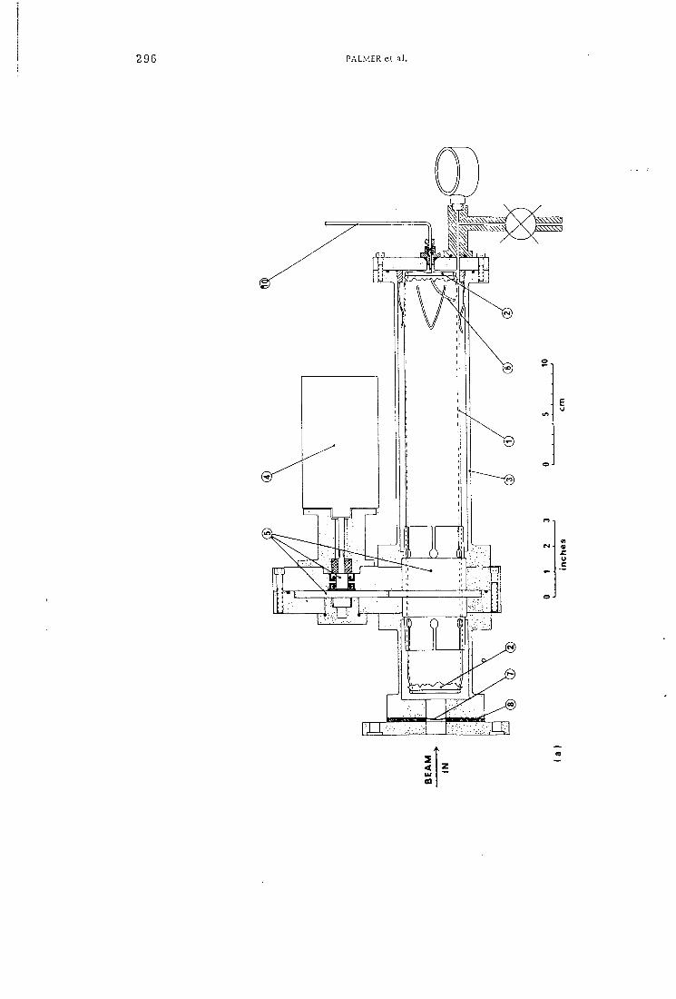
296 PALMER et al .
<
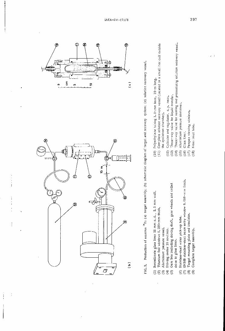
l A E A - S M - n i / 8 297
o u m o
S 5
O
3 '5 <-> S — — <u
O
5 ~ o oj c is ~-3 ra !2 t- P < U 2- :
(N CO l O ^ C - CO
0) = i i Q. (U
o ^ • DO
5 o
o h
a, o o
B "2
.2 ^ S 00
< Q
OO
C 0)
O -o CO uj re o
<-( CM CO l O CD C - CO 0 5
29 8 PALMER et aL
DL-3-F luoro ty ros ine (VII)
This compound was also prepared as described below and was shown to be identical wi th an authentic sample by i . r . and u.v. spectroscopy, > m a x {H2O, pH 7) 274 nm.
THE PREPARATION OF i^p
Operational pr inciples of the target system
I 8 F is produced by the 20]\fe ( d , f f ) i 8 F reaction. Neon (CP grade, supplied by B r i t i s h Oxygen Co., L td . ) , at 7 kgf /cm^ gauge is i rradiated wi th 13-14 MeV deuterons in a 45-cm long target vessel shown schematical ly in Fig.3 . The outer aluminium pressure vessel (Fig.3.3), houses a 56-mm o.d., 2 -mm w a l l , borosil icate glass l iner (Fig.3.1). The glass l i ne r , which has thin t i tanium windows (Fig.3.2) crimped on to each end, may be rotated by the motor (Fig.3.4) and the gear box (Fig.3.5) at 10 r e v / m i n . I r radiat ions are current ly carr ied out at up to 30 nA when the neon pressure r ises to 10 k:gf/cm2 gauge. Most of the i^p produced is caught by the glass l i ne r . Of the i^jr caught on the l iner , 60-65% may be recovered by washing its inner surface with 15-20 m l of water. This is achieved remotely by t r ans fe r r ing the water f r o m the recovery vessel (Fig.3.11) to the target under pressure, rotat ing the glass l iner for 5 min, then, a f ter a draining period of 2 min allowing the solution to re turn by reducing the pressure with the three-way valve (Fig.3.14), in the recovery vessel. The solution is f ina l ly t ransfer red to a suitable container, e.g. a 50 m l syringe or 50 m l boil ing tube via the three-way valve for l iquid t ransfer (Fig.3.13).
The fol lowing features are important fo r the successful operation of this system. A strong target window and a wel l -dis t r ibuted deuteron beam are essential f o r high mechanical r e l i a b i l i t y . For a 4 cm X 2 cm beam aperture a 0.050-mm EN58B stainless-steel f o i l (supplied by Goodfellows Metals L t d , Esher, Surrey, England) has proved to be very rel iable at beam currents and pressures of up to 30 / iA and 10 kg f / cm^ respectively. The 0 .025-mm-thick t i tanium windows on the glass l iner are necessary to reduce "d i f fus ion" losses to the walls of the aluminium pressure vessel. These windows are cr imped on to a r i m formed on the l ine r ends so that no adhesives are required. Adhesives may be degraded during i r radiat ion and give r ise to organic chemical contamination of the wash solut ion . A smal l hole, 3-4 m m in diameter, is l e f t in the rear window to allow entry of the stainless-steel water pick-up tube (Fig.3.6) and for pressure equalization. The target must, of course, be quite level to avoid spillage of solution f r o m these unsealed ends and to ensure eff ic ient pickup of the solution. The glass l iner must be easily wetted so that washing and recovery of the wash solution is e f f ic ient . This simply means care fu l cleaning of the l i ne r w i t h , f o r example, "Pyroneg" (Diversy (UK) L t d , Barnet, Her ts , England) followed by an exhaustive rinse wi th dis t i l led water . The choice of ma te r i a l and diameter of the l iquid transport tube (Fig.3.10) is also important i f ef f ic ient t ransfers are to be achieved. It is best i f the tubing is not wetted and that the solution forms into columns
l A E A - S M - n i / 8 299
rather than droplets in the tube bore. Polyethylene tubing, 1.5 mm bore, 2.7 mm o.d., 15 m long, has proved to be very satisfactory in this respect.
Target system performance
Many i r radia t ions have been ca r r ied out under various conditions up to maximum f i l l i n g pressures and beam currents of 7 kg f / cm^ and 45 /uA. A p r e l im ina ry i r r ad ia t ion of 2 Ah at 20 luA and 3.5 k g f / c m 2 yielded 28 m C i / p A h at the end of bombardment, of on the glass l iner . The l ine r was assayed using a large re-entrant ionization chamber. By washing on the bench with 20 m l of water, 65% of this ^^F was recovered in 19 m l , thus giving a recovered yie ld of 17.8 mCi/nAh. Two fu r the r i r radia t ions under s i m i l a r conditions but with remote washing and re covery yielded 17.5 mCi/nAh and 17.0 mCi/ijAh f o r a dry and a "wet" l ine r respectively. Thus i t is possible to car ry out repetitive i r r ad ia t ion / recovery cycles without drying the target or significant loss of the neon f i l l i n g . To maintain these yield f igures at 30/u A it was necessary to increase the f i l l i n g pressure to 7 k g f / c m ^ , the design l i m i t for the present system. Above 35 fuA at this pressure the yield begins to f a l l through thinning of the target gas by the beam power. The longest i r radiat ion ca r r i ed out to date was 20 pAh at 30 /nA and a pressure of 7 k g f / c m 2 . 318 m C i of ^^F, at end of bombardment, in 19 m l of water were recovered. This corresponded to a yie ld of 15.9 m C i / ^ A h . The only radiochemical impur i t i es detected in the ^^F solutions f r o m this system are 22Na 3.0 X 10⁻⁴%, 4 8 v 6.7 X 10⁻⁵% and ''Be 2.3 X 10⁻⁵%, a l l values being the maximum observed and corrected to end of bombardment.
Preparat ion of ^^F-DL-3- f luoro ty ros ine (VII)
The 18F solution is f i r s t reduced in volume to approximately 0.5 m l by boil ing in a 50 m l centrifuge tube while nitrogen is slowly bubbled in through
.a fine glass capi l la ry . The diazonium tetrafluoroborate (Va) (130-150 mg) is then added to the solution (after cooling) followed by the minimum amount of 50% acetone/acetonitri le solution necessary to dissolve the salt completely (~ 1 m l ) . The solution is then le f t to exchange at room temperature fo r about 1 h . A f t e r this t ime suff ic ient anhydrous sodium sulphate is added to take up the water present and the solution is le f t for a fu r the r 5 m i n .
Par t of the solution is then removed using a Pasteur pipette and added to dry diethyl ether (45 ml) in a 50 -ml centrifuge tube causing precipi tation of the labelled diazonium salt (Vb). The precipitate is separated by centr i fugat ion, and more die thyl ether (45 ml) is added followed by the remaining acetone/acetonitri le solution and fur ther acetone (~ 1 ml) to wash the sodium sulphate. The combined precipitate is then centrifuged of f .
The labelled diazonium salt (Vb) is dried in vacuo at room temperature and then decomposed to the f luoro ester by heating under vacuum in an o i l bath at 155°C. The dark brown t a r r y residue is extracted with hot toluene ( 2 X 2 ml) and this solution is chromatographed on a column of basic alumina (grade 1) (4 X 1.3 cm) eluting with 10% chloroform in toluene.
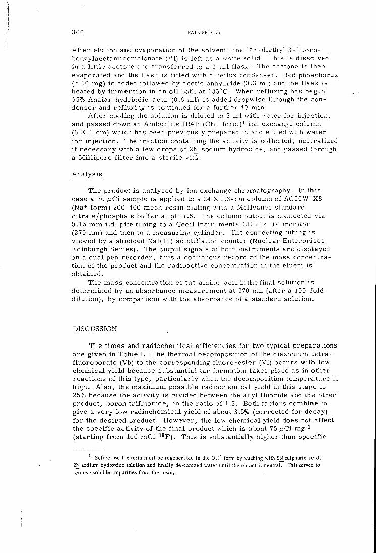
3 00 PALMER e t a L
A f t e r elution and evaporation of the solvent, the i^ j r -d ie thyl 3 - f luoro-benzylacetamidomalonate (VI) is l e f t as a white solid. This is dissolved in a l i t t l e acetone and t ransfe r red to a 2 - m l flask. The acetone is then evaporated and the flask is f i t ted with a ref lux condenser. Red phosphorus (~ 10 mg) is added followed by acetic anhydride (0.3 ml) and the flask is heated by immers ion in an o i l bath at 135°C. When ref luxing has begun 55% Analar hydriodic acid (0.6 ml) is added dropwise through the condenser and ref luxing is continued for a fur ther 40 min .
A f t e r cooling the solution is diluted to 3 m l with water f o r inject ion, and passed down an Amber l i t e IR4B (OH' f o r m ) i ion exchange column ( 6 X 1 cm) which has been previously prepared in and eluted with water fo r in jec t ion . The f r ac t i on containing the act ivi ty is collected, neutralized i f necessary with a few drops of 2N sodium hydroxide, and passed through a M i l l i p o r e f i l t e r into a s ter i le v i a l .
Analysis
The product is analysed by ion exchange chromatography. In this case a 30 p C i sample is applied to a 24 X 1.3-cm column of AG50W-X8 (Na+ f o r m ) 200-400 mesh res in eluting with a Mcllvanes standard citrate/phosphate buffer at pH 7.6. The column output is connected via 0.15 m m i . d . ptfe tubing to a Cecil instruments CE 212 UV monitor (270 nm) and then to a measuring cylinder. The connecting tubing is viewed by a shielded Nal(Tl) sc int i l la t ion counter (Nuclear Enterprises Edinburgh Series). The output signals of both instruments are displayed on a dual pen recorder, thus a continuous record of the mass concentration of the product and the radioactive concentration in the eluent is obtained.
The mass concentration of the amino-acid in the f inal solution is determined by an absorbance measurement at 270 nm (after a 100-fold di lu t ion) , by comparison with the absorbance of a standard solution.
DISCUSSION
The t imes and radiochemical eff iciencies for two typical preparations are given in Table I . The thermal decomposition of the diazonium tetra-f luoroborate (Vb) to the corresponding f luoro-ester (VI) occurs with low chemical yield because substantial tar format ion takes place as in other reactions of this type, pa r t i cu l a r ly when the decomposition temperature is high. Also , the maximum possible radiochemical yield in this stage is 25% because the ac t iv i ty is divided between the a ry l f luoride and the other product, boron t r i f l u o r i d e , in the ra t io of 1:3. Both factors combine to give a ve ry low radiochemical yield of about 3.5% (corrected fo r decay) f o r the desired product. However, the low chemical yield does not affect the specif ic act ivi ty of the f i n a l product which is about 75 ^ C i mg'^ (s tar t ing f r o m ICQ m C i ^^F). This is substantially higher than specific
' Before use the resin must be regenerated in the O H ' form by washing with 2N_ sulphuric acid, 2N sodium hydroxide solution and f ina l ly de-ionized water unt i l the eluant is neutral. This serves to remove soluble impurities from the resin.
I A E A - S M - n i / 8 3 0 1
T A B L E 1. R A D I O C H E M I C A L Y I E L D S F O R
I S F - 3 - F L U O R O T Y R O S I N E P R E P A R A T I O N
Starting 18 Radiochemical yields C o) a Specific activity
Weight (Va) F Radiochemical yields C o)
of ( V l l ) (mg) act ivi ty
Hydrolysis (pCi m g - ' ) b
(mCi) Exchange Decomposition Hydrolysis (pCi m g - ' ) b
150 58 26. 5 3. 1 83 19. 4
130 102 48. 5 3. 6 86 76. 8
Corrected for decay. At t ime of despatch.
Time f r o m Sample Appl ica t ion (mins )
l O r
? 0-8
o 0-6 n
I—
a o c o
o m n <
0-4
0-2
10 20 30 AO 50 60
Recorded Activity (511 keV)
Absorbance of Solut ion (270 n m )
20
AO
60
100
c 8 u c o
c o u
80 B a g
o
Volume o f E l u a n t ( m l )
FIG. 4. Elution behaviour of DL-3-fluorotyroslne on a 24 x 1, 3-cm column of AG50W-X8 (Na'^-form) 200-400 mesh resin.
act iv i t ies hitherto reported fo r other iSp-iabelled amino-acids, and there is probably room f o r f u r t he r improvement . The proportion of act ivi ty incorporated in the diazonium salt (Vb) appears to vary between 25 and 507o when a period of 1 h and 130-150 mg of ma te r i a l are used. The total t ime required fo r the preparat ion (after de l ivery of the ^^F solution) is approximate ly 210 m i n .
Amino-acids may be conveniently analysed on cationic [14] or anionic [ I 5 j ion exchange columns, and this method is valuable when working with shor t - l ived radionuclides. A typica l chromatogram obtained by the technique described in the experimental section is given in Fig.4. This shows a m a j o r peak in both the ac t iv i ty and mass traces at an elution volume of 22 m l corresponding to 3-f luorotyros ine . A smaller multiple peak ~ 5% is also present i n each trace at 7 m l f r o m the par t ia l ly hydro-lysed intermediates. The u.v. spectrum of the product is identical with
3 02 PALMER e t a l .
that of an authentic sample. If the time for hydrolysis is reduced to 20 min the peaks of low retention time account for about 507o of the act ivi ty present.
The method described produces a D L - m i x t u r e of the amino-acid since the asymmetr ic centre is generated in the hydrolysis stage. Resolution of the isomers in a suff ic ient ly short t ime is not at present practicable, but advances in l iquid chromatography might change this in future , for example by the use of suitable opt ical ly active stationary phases [ 1 6 , 1 7 ] .
A C K N O W L E D G E M E N T S
The authors wish to thank D.J. Silvester for his interest in this work . One of us (M.R.) also thanks the Royal Society, London, fo r the award of a Fellowship held over the period A p r i l to December 1 9 7 2 .
R E F E R E N C E S
[ 1 ] GOULDING, R .W. , PALMER, A . J . , The preparation of f luorine-18 labelled p-fluorophenylalanine for c l i n i c a l use, Int. J. Appl . Radiat. Isotopes 23 (1972) 133.
[ 2 ] HOYTE, R. M. et al . , Organic radiopharmaceuticals labelled with short-lived nuclides: 111. '*F-labelled phenylalanines, J. Nucl . Med. 12 (1971) 280; V. '*F-labelled 5- and 6-nuorotryptophan, J. Nuc l . Med. 13 (1972) 713.
[ 3 ] PREVIC, E.P. , BINKLEY, S. B. , Slow exponential growth of E. co l i in presence of p-nuorophenylalanine; effect of analogue on aromatic biosynthesis, Biochim. Biophys. Acta S7 (1964) 277.
[ 4 ] MAY, H. , L I T Z K A , G. , Inhibit ion of tumour growth by 3-fIuorotyrosine, Z. Krebsforsch. 48 (1939) 376.
[ 5 ] MEISTER, A. , Ch. 3, Biochemistry of the Amino Acids 1_, Academic Press, New York (1965). [ 6 ] FOWDEN, L. , "Fluoroamino acids and protein synthesis", Carbon-Fluorine Compounds (A Ciba
Foundation Symposium), Associated Scientific Publishers, Amsterdam (1972) 141. [ 7 ] BLAU, M . , MANSKE, R .F . , The pancreas specificity of ' ^Se-Selenomethionine, J. Nucl. Med. 2
(1961) 102, [ 8 ] NIEMANN, C . , RAPPORT, M . M . , The toxicity of S-f luoro-D- and L-tyrosine, J. Amer. Chem.
Soc. 68 (1946) 1671. [ 9 ] SCHIEMANN, G . , WINKELMULLER, W . , Fluorinated amino acids and their derivatives, J. Prakt.
Chem. 135 (1932) 101. [ 1 0 ] DEGUTIS, J., STRAUKAS, I . , Synthesis of m-{bis(2-chloroethyl)amino]-o-methyl-DL-tyrosine,
Zh.Obshch. K h i m . 32 (1962) 1255. [ 1 1 ] DAVIS, A . L. e t a l . , 2-Aminotyrosine, an analogue of tyrosine, J. Med. Chem. 9 (1966) 828. [ 1 2 ] DAVIS, A . L. e t a l . . The synthesis and biological activities of o-aminophenylalamine and related
compounds. Arch . Biochem. Biophys. 102 (1963) 48. [ 1 3 ] BERLINGUET, L . , Synthesis of 2-amino alcohols, Can. J. Chem. 32 (1954) 31 . [ 1 4 ] MOORE, S., STEIN, W . H . , Chromatography of amino acids on sulphonated polystyrene resins,
J. Biol . Chem. 1 ^ (1951) 663. [ 1 5 ] HIRS, C . H . W . , MOORE, S., STEIN, W. H. , The chromatography of amino acids on ion exchange
resins, J. Amer. Chem. Soc. 76 (1954) 6063. [ 1 6 ] DAVANKOV, V. A . , ROGOZHIN, S. V . , Stereoselective effects in a-amino acid copper (11) complexes,
J. Chromatogr. 60 (1971) 28. [ 1 7 ] BACZUK, R.J . , LANDRAM, C. K . , DUBOIS, R.J., DEHM, B . C . , Liquid chromatographic resolution
of racemic 6-3, 4-dihydroxyphenylalanine, J. Chromatogr. 60 (1971) 351.
D I S C U S S I O N
C. MANTESCU: Did you t r y to use the f luor ina t ion of the diazonium salt in l iquid hydrogen f luor ide?
A . J . PALMER: No. Hydrogen f luor ide is an inconvenient compound to use because of its corrosive nature.
2 2 l A E A - S M - n i / 9
PREPARATION OF i ^ p , L A B E L L E D FLUOROCARBONS FOR USE IN PHARMACODYNAMIC STUDIES
J.C. CLARK, R. W. GOULDING, A.J. PALMER MRC Cylotron Unit, Hammersmith Hospital, London,
United Kingdom
Abstract PREPARATION OF '*F-LABELLED FLUOROCARBONS FOR USE IN PHARK-lACODYNAMIC STUDIES.
For many years fluorocarbons, widely used components of aerosol propellents both for domestic and pharmaceutical use and known by a number of proprietary names including "Freon" and "Arcton", were considered lo be inert. Recently doubts were cast on this belief and work is being carried out in many centres to investigate the possible causes of the reported toxici ty , it was fe l t that labelled fluorocarbons would be of help in some i n vestigations, and F was chosen as having physical properties most suited to the investigations envisaged , namely in -v ivo distribution studies using a scinticamera and retention studies using a whole-body counter.
F-labelled trichlorofluoromethane ("Freon-H") and dichlorodifluoromethane {"Freon-12") were prepared using both silver fluoride Ag^F and silver dif luoride A g ' ^ F j - A g ^ F and Ag'^F.^ were both labelled by l in ing a neon-f i l led target vessel with carrier AgF or AgFz. After irradiation with 14 MeV deuterons the neon was removed and the appropriate reactants added to the target vessel by vacuum transfer, the reaction then being carried out at an elevated temperature. The reaction products were removed by vacuum transfer and purif ied by radio-gas chromatography. The f ina l product was then either diluted with air for rebreathing or transferred by vacuum dis t i l la t ion to aerosol dispensers for placebo administration.
INTRODUCTION
Several chlorofluorocarbons are used as propellents in a wide variety of aerosol generators both fo r domestic and pharmaceutical use. These propellents are known by a number of propr ie tary generic names including Freon ® and Arc ton ® . For example, the compounds CCl4-n Fn are named in this system as F r e o n - I n of A r c t o n - I n . For many years these compounds have been considered to be pharmacologically inert [ 1 , 2] , but doubts were recently cast on this belief and work is being carr ied out in many centres to investigate the pharmacodynamics of these fluorocarbons in man and animals [3-5] . It was fe l t that labelled fluorocarbons would be of help in some investigations. Fluorine-18 was chosen as having physical properties most suited to the investigations envisaged in man, namely in vivo d i s t r i bution studies using a sc int i l la t ion camera and simultaneous retention studies using a whole-body counter. Some work has been reported where •'^Cl was used as the label [5] but its high gamma-ray energies of 1. 60 and 2. 17 MeV make it unsuitable fo r use wi th most imaging devices, in par t icula r an Anger-type sc int i l la t ion camera.
In the present work the method of administrat ion to man was by means of a placebo aerosol drug dispenser of the Riker Medihaler © t y p e , in order to f u l l y simulate certain c l in ica l situations. Animal experiments were to be ca r r i ed out using dilute mixtures of labelled fluorocarbons in a i r .
A direct synthetic route was chosen fo r the. preparation of labelled Freon-11 and - 12, CCI3F and CClgFa, using fluorides of s i lver [6, 7] as
4 1 1
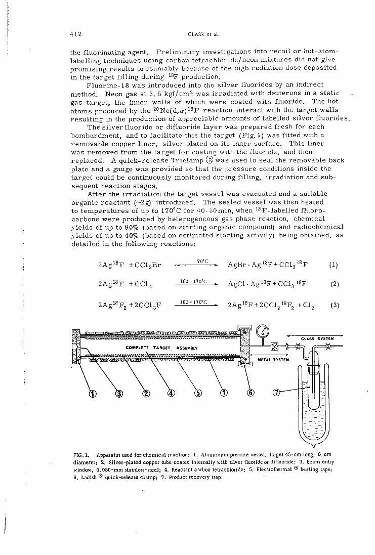
4 1 2 CLARK et al.
the f luor ina t ing agent. P re l imina ry investigations into recoi l or hot-atom-label l ing techniques using carbon tetrachloride/neon mixtures did not give p romis ing results presumably because of the high radiation dose deposited in the target f i l l i n g during ^^F production.
Fluorine-18 was introduced into the s i lver f luorides by an indirect method. Neon gas at 3. 5 kgf /cm2 was i r radiated with deuterons in a static gas target, the inner walls of which were coated with f luoride. The hot atoms produced by the ^'^Ne(d,a)^^F reaction interact with the target walls resul t ing in the production of appreciable amounts of labelled si lver f luor ides .
The s i lver f luor ide or dif luoride layer was prepared f resh fo r each bombardment, and to fac i l i ta te this the target (Fig . 1) was f i t ted with a removable copper l ine r , s i lver plated on its inner surface. This l iner was removed f r o m the target f o r coating with the f luoride, and then replaced. A quick-release Tr i c l amp Cb) was used to seal the removable back plate and a gauge was provided so that the pressure conditions inside the target could be continuously monitored during f i l l i n g , i r radia t ion and subsequent react ion stages.
A f t e r the i r r ad ia t ion the target vessel was evacuated and a suitable organic reactant (~2g) introduced. The sealed vessel was then heated to temperatures of up to 170°C fo r 40-50min, when p . label led f l u o r o -carbons were produced by heterogeneous gas phase reaction, chemical yields of up to 90% (based on start ing organic compound) and radiochemical yields of up to 40% (based on estimated start ing activity) being obtained, as detailed in the fo l lowing reactions:
2Ag^^F +CCl3Br
2Agi8F +CC1^
A g B r - A g i ^ F + C C l g ^ ^ F (1)
. AgCl -AgiSp+CClg 18F (2)
2 A g i ^ F 2 + 2 C C I 3 F 160 - 170°C 2Agl^F + 2CCl2^^F„ +C1. (3)
G L A S S S Y S T E M
C O M P L E T E T A R G E T A S S E M B L Y
METAL S Y S T E M
F I G . l . Apparatus used for chemical reaction: 1. Aluminium pressure vessel, target 45-cm long, 6-cm diameter; 2. Silver-plated copper tube coated internally with silver fluoride or difluoride; 3. Beam entry window, 0.050-mm stainless-steel; 4. Reactant carbon tetrachloride; 5. Electrothermal ® heating tape; 6. Ladish ® quick-release clamp; 7. Product recovery trap.

l A E A - S M - n i / 9 413
For the production of Freon-11 , reaction (1) has certain disadvantages. Bromotrichlormethane readi ly decomposes on heating, and significant amounts of the highly toxic compound carbonyl chloride (phosgene) are produced in the reaction. Reaction (2) does not suffer f r o m these disadvantages and so is used rout inely fo r the preparation of Freon-11. Freon-11 does not react under the same conditions at a significant rate with s i lver f luor ide to give Freon-12; therefore the more reactive dif luoride, react ion (3), is necessary.
In p re l imina ry studies of these reactions, samples were withdrawn during the course of the react ion and analysed by radio-gas chromatography, so that the optimum react ion t imes could be determined. For routine production, af ter al lowing the reaction to proceed fo r 40-50 min, the volat i le products were d i s t i l l ed out of the reaction vessel into a cold t rap. The desired product was isolated using preparative scale glc, when i t was recovered f r o m the hel ium effluent in a glass t rap at -196°C. If necessary it was diluted wi th c a r r i e r on a glass and Teflon © vacuum line, and a smal l sample t r ans fe r red to a tared ampoule for specific act ivi ty determination. A f t e r th i s the requi red mix ture of labelled and unlabelled Freons was made up in a modif ied Riker aerosol dispenser. Final ly the dispenser was tested before c l in ica l use.
E X P E R I M E N T A L
Target system fo r ^^F production
Fluorine-18 was produced f r o m neon by the 20Ne(d, a) ^^F reaction, C. P. Neon being contained in the target vessel shown schematically in Fig. 1. P re l imina ry i r radiat ions of the target wi th a glass l iner with 0. 2 5 mm aluminium end cover f o i l s and a neon- f i l l ing pressure of 3. 5 kgf / cm^ yielded 28 m C i / p A h fo r a 2 / j A h i r r ad ia t ion wi th 14 MeV deuterons at a beam current of 20-25 M A [ 8J.
A copper l iner (Fig . 1.2), electroplated wi th s i lver internally to a thickness of 0.1 mm, was used to support the inorganic f luorinat ing agents which were to be labelled. This l iner was introduced into the target f r o m the back and the back plate secured by a quick-release clamp (Fig . 1.6). The target was also used as the reaction vessel, i t being heated e lec t r i cally at this stage.
Si lver f luor ide preparation
Two methods were used to coat the inside of the s i lver-plated tube wi th s i lve r f luor ide .
Evaporation of aqueous s i lve r f luor ide solutions
This method was used f o r most of the work when preparing Freon-11. 25% aqueous si lver f luor ide solution (25 ml) was evaporated under vacuum on to the l ine r while i t was rotated and gradually heated with hot air to 50°-70°C, A schematic layout of this system is shown in Fig. 2. The apparatus consisted of a ro t a ry evaporator wi th Tef lon vapour duct and l iquid ni t rogen cold trap. The target l ine r was attached to this apparatus sind
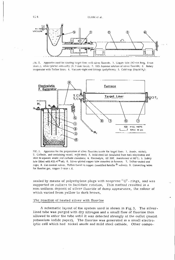
4 1 4 CLARK el al.
V A C U U M
FIG. 2. Apparatus used for coaling target liner with silver fluoride: 1. Copper lube (45-cm long. 5-cm d i a m . ) , silver-plated internally (0. 1-mm thick); 2. 25"/o Aqueous solution of silver fluoride; 3. Rotary evaporator with Tef lon l iner; 4. Vacuum-tight end fit t ings (polythene); 5. Cold trap ( l iquid N2)-
Electrolytic Furnace F, Generator
Target Liner
EZa P.TCE. PARTS SCALE IN cm
FIG. 3. Apparatus for the preparation of silver fluorides inside the target liner: 1. Anode, nickel ; 2. Cathode, and containing vessel, m i l d steel; 3. M i l d steel l id (insulated f rom both electrodes) and skirt to separate anode and cathode chambers; 4. Electrolyte, 1CF.2HF, maintained at 80°C; 5. Safety lute f i l l ed wi th KEL-F® o i l ; 6. Silver-plated copper tube mounted in furnace; 7. Teflon-sealed end caps; 8. Gas control valves, Te f lon barrel in copper (modif ied Rotoflo ® valves); 9. Connecting tubes for fluorine gas, copper 5 - m m i . d .
sealed by means of polyethylene plugs wi th neoprene " O " - r ings, and was supported on r o l l e r s to faci l i ta te rotation. This method resulted in a non-uniform deposit of s i lver f luor ide of damp appearance, the colour of which varied f r o m yel low to dark brown.
The reaction of heated s i lver with f luor ine
A schematic layout of the system used is shown in Fig . 3. The s i lver -l ined tube was purged wi th dry nitrogen and a smal l f low of f luor ine then allowed to enter the tube un t i l i t was detected strongly at the outlet (moist potassium iodide paper). The f luor ine was generated in a smal l electroly t ic cel l which had n ickel anode and m i l d steel cathode. Other compo-
i A E A - S M - 1 7 1 / 9 4 15
nents were steel and Teflon and the electrolyte was the salt KF. 2HF maintained in a molten state at 80°C [9, 10) . The f luorine was conducted through 5 mm-bore copper tubing and copper taps with Teflon plugs. Af t e r f i l l i n g the tube i t was heated to 150°C, and the contents were sampled occasionally using K I paper by allowing a smal l volume to be purged by the nitrogen supply. When only traces of f luorine were detectable at the outlet the heating was stopped. The tube was then purged with nitrogen to waste unt i l cool. The resul t ing s i lver f luoride appeared as a quite uni form greenish yel low layer. The quantity of the f luoride present, however, was unknown. A f t e r preparation the l ined tube was handled under nitrogen.
Si lver d i f luor ide preparation
The s i lver-p la ted target l iner was coated with s i lver dif luoride using f luor ine in a s i m i l a r way to the preparation of the monofluoride described above, except that the temperature of the s i lver was raised to 3 50 C and the f luor ine was passed through slowly unt i l excess reagent was detected strongly at the waste outlet. A f t e r preparation the coated liner was handled under nitrogen as above.
Label l ing of the s i lver f luor ides with ^^F
The coated l iner , prepared by any of the above methods, was introduced into the target vessel. In the case of fluorides derived f r o m fluorine this procedure was car r ied out under dry nitrogen. The target was then evacuated and f i l l e d to a pressure of 3. 5 kg f / cm^ with CP Neon. Irradiations of the neon were ca r r ied out wi th 14 MeV deuterons at beam currents of up to 25MA. No attempt was made to measure the total activity of s i lver halide produced.
Synthesis of ^^F-labelled Freon-11
A f t e r i r r ad ia t ion the neon was released, the target evacuated, and heated to 160°-170°C. Spectroscopic grade carbon tetrachloride (1. 5 ml) was then t r ans fe r red v ia the target valve into the target / react ion vessel. The temperature was maintained fo r 40 min , and then the reaction vessel was connected to an evacuated t rap (Fig. 1. 7) which was cooled to -196°C, When most of the target contents had dis t i l led into the trap (1 min) it was opened to vacuum and the trapping-out procedure continued for a fur ther 5 m i n . The t rap was then isolated and the contents careful ly melted and r e f rozen to obtain a concentration at the bottom of the t rap. A f t e r allowing the pressure to r i se in the t rap i t was dismantled and toluene (0. 5 ml) added to s i m p l i f y f u r t he r handling procedures.
Synthesis of F- label led Freon -12
A procedure analogous to that adopted fo r Freon-11 was used, the main differences being the use of labelled si lver d i f luor ide and inactive Freon-11 as the s tar t ing mater ia l s . Although a detailed study of the temperature/ t ime course of the reaction has not been undertaken, labelled Freon-12 was observed as the m a j o r condensable reaction product af ter 30 min at 160°-170°C.
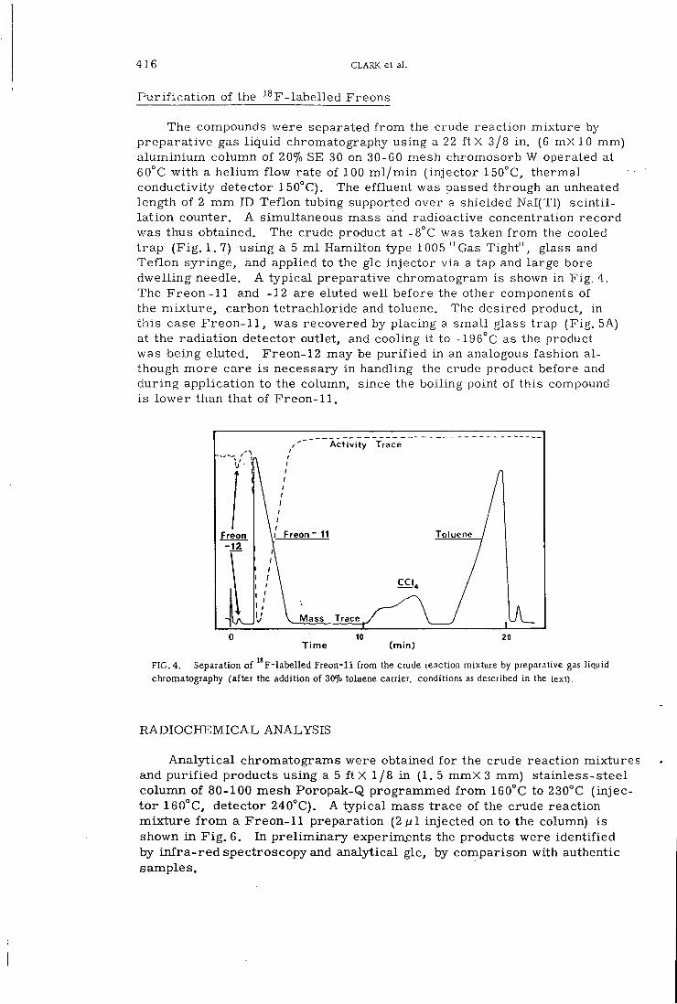
416 CLARK et al .
Pur i f i c a t i on of the ^^F-labelled Freons
The compounds were separated f r o m the crude reaction mixture by preparat ive gas l iqu id chromatography using a 22 f t X 3/8 in. (6 mX 10 mm) a lumin ium column of 20% SE 30 on 30-60 mesh chromosorb W operated at 60°C wi th a hel ium f low rate of 100 m l / m i n ( injector 150°C, thermal conductivity detector 150°C). The effluent was passed through an unheated length of 2 m m ID Teflon tubing supported over a shielded Nal(Tl) s c in t i l la t ion counter. A simultaneous mass and radioactive concentration record was thus obtained. The crude product at -8°C was taken f r o m the cooled t rap (F ig . 1. 7) using a 5 m l Hamilton type 1005 "Gas Tight", glass and Tef lon syringe, and applied to the glc injector via a tap and large bore dwel l ing needle. A typica l preparative chromatogram is shown in Fig. 4. The F reon-11 and -12 are eluted wel l before the other components of the mix tu re , carbon tetrachloride and toluene. The desired product, in this case Freon-11 , was recovered by placing a small glass trap (Fig. 5A) at the radiat ion detector outlet, and cooling it to -196°C as the product was being eluted. Freon-12 may be pur i f i ed in an analogous fashion a l though more care is necessary in handling the crude product before and dur ing application to the column, since the boiling point of this compound is lower than that of Freon-11.
Activity Trace
T o l u e n e / F r e o n F r e o n
Mass Trace
T i m e ( m i n )
F I G . 4. Separation of F-labelled Freon-11 from the crude reaction mixture by preparative gas liquid chromatography (after the addition of 30% toluene carrier, conditions as described in the text).
RADIOCHEMICAL ANALYSIS
Ana ly t i ca l chromatograms were obtained fo r the crude reaction mixtures and p u r i f i e d products using a 5 f t X l / 8 i n ( 1 . 5 mmX 3 mm) stainless-steel column of 80-100 mesh Poropak-Q programmed f r o m 160°C to 230°C ( injecto r 160°C, detector 240°C). A typical mass trace of the crude reaction mix tu re f r o m a Freon-11 preparation (2^/1 injected on to the column) is shown in F i g . 6. In p re l imina ry experiments the products were identif ied by i n f r a - r e d spectroscopy and analytical glc, by comparison with authentic samples.
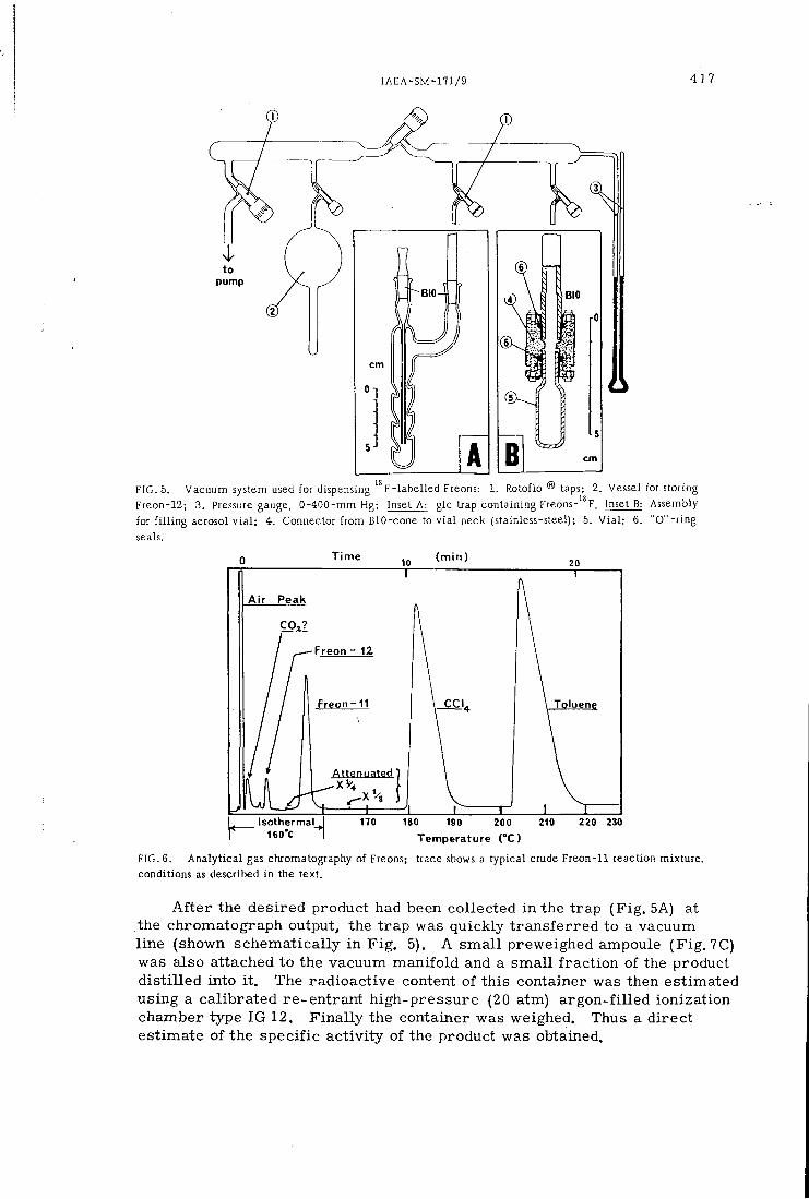
IAEA-SM-171/9 4 1 7
pump
FIG. 5. Vacuum system used for dispensing F-labelled Freons: 1. Rotoflo ® taps; 2. Vessel for storing Freon-12; 3. Pressure gauge, 0-400-mm Hg; Inset A: glc trap containing Freons-"F. Inset B: Assembly for f i l l i n g aerosol v i a l ; 4. Connector f rom BIO-cone to v i a l neck (stainless-steel); 5. V i a l ; 6. " 0 " - r i n g seals.
T i m e 10 ( m i n )
A i r P e a k
F r e o n - 12
F r e o n - 1 1
Attenuated '
lsothermal_^ 160"C
±
T o l u e n e
170 210 220 230 180 190 200
T e m p e r a t u r e ( ° C )
FIG. 6. Ana ly t i ca l gas chromatography of Freons; trace shows a typical crude Freon-11 reaction mixture, conditions as described in the text.
A f t e r the desired product had been collected in the t rap (F ig . 5A) at the chromatograph output, the t rap was quickly t r ans fe r red to a vacuum line (shown schematically i n F ig . 5). A smal l preweighed ampoule (F ig . 7C) was also attached to the vacuum manifold and a smal l f r ac t ion of the product d i s t i l l ed into i t . The radioactive content of this container was then estimated using a cal ibrated re-entrant high-pressure (20 atm) argon-f i l led ionization chamber type IG 12. F ina l ly the container was weighed. Thus a direct estimate of the specific ac t iv i ty of the product was obtained.
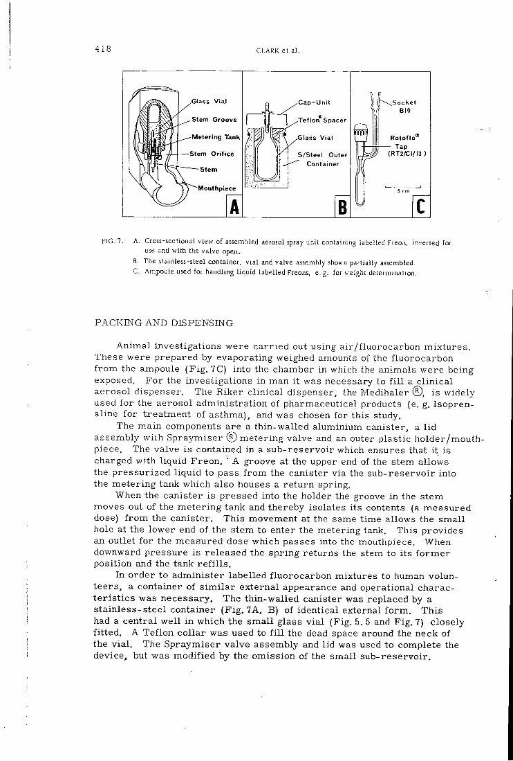
4 1 8 CLARK et al .
G l a s s V i a
S tem G r o o v e
Meter ing Tank
S tem O r i f i c e
S t e m
Mouthpiece
A
C a p - U n i t
•Tef l o n ^ S p a c e r
G l a s s V ia l
S / S t e e l Outer Conta iner
B
( \ S o c k e t B I O
Rotof lo® T a p
( R T 2 A ; I / I 3 )
FIG. 7. A. Cross-sectional view of assembled aerosol spray unit containing iabeUed Freo.i. inverted for use and with the valve open.
B. The stainless-steel container, v i a l and valve assembly shown partially assembled. C. Ampoule used for handling l iquid labelled Freons, e. g. for weight determination.
PACKING AND DISPENSING
Animal investigations were carr ied out using a i r / f luorocarbon mixtures . These were prepared by evaporating weighed amounts of the f luorocarbon f r o m the ampoule (F ig . 7C) into the chamber in which the animals were being exposed. For the investigations in man i t was necessary to f i l l a c l in i ca l aerosol dispenser. The Riker c l inical dispenser, the Medihaler ® , is widely used fo r the aerosol administrat ion of pharmaceutical products (e. g. Isopren-aline fo r treatment of asthma), and was chosen for this study.
The main components are a thin-wal led aluminium canister, a l i d assembly with Spraymiser © m e t e r i n g valve and an outer plastic holder/mouthpiece. The valve is contained in a sub-reservoir which ensures that i t is charged wi th l iquid Freon, ' A groove at the upper end of the stem allows the pressurized l iqu id to pass f r o m the canister via the sub-reservoir into the meter ing tank which also" houses a re turn spring.
When the canister is pressed into the holder the groove in the stem moves out of the meter ing tank and thereby isolates its contents (a measured dose) f r o m the canister. This movement at the same time allows the smal l hole at the lower end of the stem to enter the metering tank. This provides an outlet fo r the measured dose which passes into the mouthpiece. When downward pressure is released the spring returns the stem to its f o r m e r position and the tank r e f i l l s .
In order to administer labelled f luorocarbon mixtures to human volunteers, a container of s i m i l a r external appearance and operational charact e r i s t i c s was necessary. The thin-walled canister was replaced by a stainless-steel container (F ig . 7A, B) of identical external f o r m . This had a central we l l in which the smal l glass v ia l (Fig. 5. 5 and F ig . 7) closely f i t t ed . A Tef lon col lar was used to f i l l the dead space around the neck of the v i a l . The Spraymiser valve assembly and l i d was used to complete the device, but was modif ied by the omission of the smal l sub-reservoir .
IAEA-SM-171/9 4 19
A r e c o m m e n d e d f i l l i n g m i x t u r e f o r such a d ispenser is F r e o n - 1 2 (2 pa r t s ) F r e o n - 1 1 (1 p a r t by gas v o l u m e ) . The f i l l i n g was c a r r i e d out on the vacuum l i n e shown i n F i g . 5. The m a n i f o l d sec t ion was f i l l e d to a su i t ab le p r e s s u r e w i t h the l a b e l l e d p roduc t w h i c h was then condensed in to the v i a l . The amount i n t r o d u c e d in to the v i a l was d e t e r m i n e d f r o m manom e t e r r ead ings . The i n a c t i v e cons t i tuen t was in t roduced by the same p r o cess . The v i a l was then r a p i d l y t r a n s f e r r e d to the s t a i n l e s s - s t e e l cont a i n e r w h i l e the contents w e r e s t i l l f r o z e n , .and the va lve as sembly c r i m p e d on toge the r w i t h i t s s e a l i n g " O " r i n g . A leak tes t was then c a r r i e d out by i m m e r s i n g the a s s e m b l y i n w a t e r , f o l l o w e d by d i scha rge tes ts in to 1 - l i t r e f l a s k s w i t h r u b b e r septa. A needle adaptor was t e m p o r a r i l y f i t t e d to the S p r a y m i s e r v a l v e t o achieve t h i s . The l abe l l ed f l u o r o c a r b o n content of the f l a s k was m e a s u r e d u s i n g a r e - e n t r a n t i o n i z a t i o n chamber thus g i v i n g a c lose e s t ima te of the a c t i v i t y . T h i s f i g u r e together w i t h that f o r the spec i f i c a c t i v i t y enables the m a s s of f l u o r o c a r b o n a d m i n i s t e r e d to the sub jec t to be d e t e r m i n e d . I n a d d i t i o n , the c h e m i c a l c o m p o s i t i o n of the m i x t u r e i n side the a e r o s o l d i spense r m a y be checked by gas c h r o m a t o g r a p h y of a s ample taken out of the f l a s k .
DISCUSSION
The p r o d u c t i o n r a t e of ^ ^ F f r o m neon by the 20Ne(d, o) ^^F r e a c t i o n w i t h 15 M e V deuterons is f a r g r e a t e r than that us ing 3 0 M e V alpha p a r t i c l e s i n the ^^0(a,pn) ^^F r e a c t i o n . The r e s u l t s obtained suggest that a v e r y l a r g e p r o p o r t i o n of the ^^F p roduc t n u c l e i a re caught i n the s i l v e r f l u o r i d e l i n i n g and are ava i l ab le f o r syn the t i c i n c o r p o r a t i o n .
The v o l u m e of 2 57o aqueous s i l v e r f l u o r i d e so lu t ion used was chosen i n o r d e r t o be s u f f i c i e n t t o cove r c o m p l e t e l y the i n t e r i o r of the l i n e r on evapo ra t i on , w h i l e at the same t i m e p r o v i d i n g a reagent of s u f f i c i e n t l y h igh s p e c i f i c a c t i v i t y when i t was l a b e l l e d . Reac t ion of f l u o r i n e w i t h s i l v e r m e t a l produces a m u c h s m a l l e r amount of u n i f o r m l y d i s t r i b u t e d s i l v e r f l u o r i d e and consequen t ly coyld l be used to produce l a b e l l e d F r e o n s w i t h h i g h e r s p e c i f i c a c t i v i t i e s a l though t h i s was not necessa ry f o r the p r e sen t a p p l i c a t i o n .
Reac t ion at IVO^C of c a r b o n t e t r a c h l o r i d e and s i l v e r f l u o r i d e p roduced by e v a p o r a t i o n a lways r e s u l t e d i n the p r o d u c t i o n of s m a l l amounts of F r e o n - 1 2 (< 5%). However i t was n e c e s s a r y t o use s i l v e r d i f l u o r i d e and F r e o n - 1 1 i n o r d e r t o p roduce F F r e o n - 1 2 at a s a t i s f a c t o r i l y h igh r a t e at t h i s t e m p e r a t u r e (near the m a x i m u m a t ta inab le u s ing the present t a r g e t / r e a c t i o n v e s s e l ) . F o r r epea t ed use a s i l v e r m e t a l tube wou ld be d e s i r a b l e s ince the s i l v e r l a y e r on a p l a t e d tube i s soon exhausted. In both types of r e a c t i o n the a l u m i n i u m v e s s e l s e r v e d adequately a l though i t was not i d e a l because of c o r r o s i o n p r o b l e m s . A n i c k e l t a r g e t wou ld p robab ly be m o r e s a t i s f a c t o r y .
I t should be no ted tha t the m a x i m u m at ta inable r a d i o c h e m i c a l y i e l d i n these r e a c t i o n s w i t h s i l v e r f l u o r i d e i s 50%. S i l v e r f l u o r i d e f o r m s m o l e c u l a r compounds w i t h o the r s i l v e r ha l ides p roduced i n the r e a c t i o n w i t h o r g a n i c ha l ides . These c o n t a i n and i m m o b i l i z e one equiva lent of s i l v e r f l u o r i d e ( r e a c t i o n s 1-3) [7] . The t i m e s and r a d i o c h e m i c a l e f f i c i e n c i e s f o r s e v e r a l p r e p a r a t i o n s of ^ ^ F - l a b e l l e d F r e o n - 1 1 are g iven i n T a b l e 1. A l l the p r o d u c t i o n r u n s l i s t e d w e r e c a r r i e d out u s i n g s i l v e r f l u o r i d e p roduced
420 CLARK e t a l .
T A B L E I . R A D I O C H E M I C A L E F F I C I E N C I E S F O R I ^ f - F R E O N - H
P R E P A R A T I O N
Int. beam current
Reaction times (min)
Activity ^ ' F - C F C l 3 (mCi) ^
Radiochem. yield
Specific activity'' " F - C F C I 3
(fjCi mg-1)
Total time for preparation
and purification (min)
5 40 n (67) 18 • 10. 0 121
10 50 50 (81) 30 20.7 129
10 50 45 (89) 41 - 117
15 50 55 (98) 24 23.4 122
20 45 76 (73) 21 35.4 115
The figures in brackets are the time after E. O. B. (min). After glc purification.
by e v a p o r a t i o n of aqueous s o l u t i o n . T h i s r e s u l t e d in the p roduc t i on of up to 76 m C i of i m p u r e ^ ^ j r F r e o n - 1 1 ( f r o m a 2 0 ^ j A n bombardmen t ) a p p r o x i m a t e l y 70 m i n a f t e r E . O . B . P u r i f i c a t i o n by p r e p a r a t i v e scale glc took a f u r t h e r 40 m i n , p r o d u c i n g a f i n a l p roduc t of s p e c i f i c a c t i v i t y ~ 3b ;jCi m g r e a d y f o r p a c k a g i n g f o r c l i n i c a l use .
The c h e m i c a l c o n v e r s i o n of c a r b o n t e t r a c h l o r i d e (2. 2g) was usua l ly ~ 8 0 % . D i l u t i o n of the p r o d u c t t o a v o l u m e of ~3 m l w i t h toluene enabled p u r i f i c a t i o n t o be a c c o m p l i s h e d by a s ing le p r e p a r a t i v e glc i n j e c t i o n . The t r a p p i n g e f f i c i e n c y at the g l c ou t l e t w a s ~ 90%, hand l ing losses w h i c h d id o c c u r b e i n g m a i n l y d u r i n g t r a n s f e r of the so lu t i on f r o m the t r a p to the s y r i n g e . A s y s t e m of d i r e c t t r a n s f e r f r o m the t r a p to the glc in le t po r t w o u l d be d e s i r a b l e .
C O N C L U S I O N
T h e p r e p a r a t i v e m e t h o d d e s c r i b e d above y i e l d e d gas c h r o m a t o g r a p h i c a l l y p u r e ^ ^ F - l a b e l l e d F r e o n - 1 1 of s p e c i f i c a c t i v i t y of 20-30 ^Ci m g ' ^ at t i m e of d i s p a t c h f o r c l i n i c a l use . T h i s was s u f f i c i e n t to p r o v i d e s a t i s f a c t o r y counti n g s t a t i s t i c s and s c i n t i l l a t i o n c a m e r a p i c t u r e s when m e a s u r e d doses w e r e a d m i n i s t e r e d to h u m a n v o l u n t e e r s f r o m the m o d i f i e d R i k e r M e d i h a l e r d i s pense r . T h e r e s u l t s o f i n v e s t i g a t i o n s i n m a n and r a t s w i l l be pub l i shed e l s e w h e r e .
The f l u o r i n a t i o n m e t h o d d e s c r i b e d i s of genera l a p p l i c a t i o n and could be used to p r e p a r e o the r v o l a t i l e ^^F- l abe l l ed compounds. I t s use i n the p r e p a r a t i o n of p o l a r i n v o l a t i l e ^^F compounds , e. g. a m i n o - a c i d s o r s t e r o i d s , w o u l d be m o r e d i f f i c u l t . I n p a r t i c u l a r i t wou ld be necessa ry to produce the s i l v e r f l u o r i d e s on a s m a l l e r sca le and w i t h m u c h h ighe r s p e c i f i c a c t i v i t i e s .
A C K N O W L E D G E M E N T S
The a u t h o r s w i s h t o t h a n k P r o f e s s o r C. T . D o U e r y of the Depa r tmen t of C l i n i c a l P h a r m a c o l o g y , R. P . M . S. , H a m m e r s m i t h H o s p i t a l , f o r h i s c o n t i n u ous e n c o u r a g e m e n t t h r o u g h o u t t h i s work , , and the c y c l o t r o n t e a m who c a r r i e d
IAEA-SM-171/9 421
out the i r r a d i a t i o n s . The guidance of W . M a s s i n g h a m of B i r m i n g h a m U n i v e r s i t y C h e m i s t r y D e p a r t m e n t was g r e a t l y app rec i a t ed when des ign ing , c o n s t r u c t i n g and c o m m i s s i o n i n g the f l u o r i n e c e l l . We are a lso indebted to R i k e r L a b o r a t o r i e s f o r the loan of s p e c i a l t oo l s and supply of the components neces sa ry f o r the a s s e m b l y of the m o d i f i e d M e d i h a l e r , and to L . W . B r o w n f o r the c o n s t r u c t i o n of equ ipmen t used i n t h i s w o r k . S i l v e r p l a t i n g of the t a r g e t l i n e r s was k i n d l y c a r r i e d out by S o m e r v i l l e L a b o r a t o r i e s L t d , L o n d o n .
SOURCES
2 5% Aqueous A g F s o l u t i o n
Neon CP grade
Q u i c k f i t R o t a f l o ® v a l v e s
E l e c t r o t h e r m a l h e a t i n g tape
O F M A T E R I A L S
H o p k i n & W i l l i a m s L t d
B r i t i s h Oxygen Co. L t d
J . A . J o b l i n g & Co. L t d , Stone, S t a f f o r d s h i r e
E l e c t r o t h e r m a l E n g i n e e r i n g L t d , L o n d o n
T r i c l a m p & type 4
H i g h - p r e s s u r e i o n i z a t i o n c h a m b e r Type IG12
S p r a y m i s e r ® m e t e r i n g va lve and M e d i h a l e r ®
L a d i s h C o . , T r i c l o v e r D i v i s i o n
20th C e n t u r y E l e c t r o n i c s L t d , C r o y d o n , S u r r e y
R i k e r L a b o r a t o r i e s
P o r o p a k Q W a t e r s A s s o c i a t e s L t d
R E F E R E N C E S
[1] UNDERWRITERS' LABORATORIES, Underwriters' Laboratories Bulletin, M. H. 2375, Chicago, 111. (1933).
[2] SAYERS. R . R . , YANT, W . P . , CHORNYAK, J . , SHOAF, H. W., Toxicity of Dichlorodifluoromethane: a New Refrigerant, US Bureau of Mines. Report of Investigations, 3013 (May 1930).
[3] DOLLERY, C . T . , DRAFFAN, G . H . . DAVIES, D. S. , WILLIAMS, F . M . , CONNOLLY, M. E . , Blood concentrations in man of fluorinated hydrocarbons after inhalation of pressurised aerosols. Lancet U (1970) 1164.
[4] PATERSON, J . W . , SUDLOW, M . F . , WALKER, S .R . , Blood levels of nuorinated hydrocarbons in asthmatic patients after inhalation of pressurised aerosob, Lancet i i (1971) 565.
[5] MORGAN. A . , BLACK, A . , WALSH, M . , BELCHER, D R . , The absorpUon and retention of inhaled fluororinated hydrocarbon vapours, Int. J . Appl. Radiat. Isotopes 23 (1972) 285.
[6] LOVELACE, A . M . , POSTELNEK. W., RAUSH, D. A . , Aliphatic Fluorine Compounds, A. C . S. Monograph No. 138, Reinhold, New York (1958) 3.
[7] HUDLICKY, M . , Chemistry of Organic Fluorine Compounds, Pergamon Press, Oxford (1961) 51. [8] PALMER. A. J . , CLARK, J . C . , GOULDING, R. W. , ROMAN, M . . "The preparaUon of " F
labeUed DL-3-fluorotyroslne", these Proceedings, lAEA-SM-171/8 . [9] H U D U C K Y , M . , Chemistry of Organic Fluorine Compounds, Pergamon Press, Oxford (1961) 45.
[10] LEECH, H. R. , Laboratory and technical production of fluorine and its compounds. Quart. Rev. Chem. Soc. 3 (1949) 22.
RADIOCHEM.RADIOANAL.LETTERS 1 4 / 2 / 1 0 1 - 1 0 8 / 1 9 7 3 / 23
A' If.;
s r
I t i i .
i l l
THE PREPARATION OF FLUORINE-18 LABELLED COMPOUNDS USING A RECIRCULATORY NEON TARGET
J . C . C l a r k , R .W. G o u l d i n g , M a r i a Roman^, A . J . P a l m e r
M . R . C . C y c l o t r o n U n i t , Hammersmith H o s p i t a l , Ducane R oad , London W . 1 2 , OHS, U . K .
R e c e i v e d 19 A p r i l 1973 A c c e p t e d 27 A p r i l 197 3
A method f o r t h e p r e p a r a t i o n o f f l u r i n e - 1 8 l a b e l l e d met a l f l u o r i d e s and o r g a n i c d i a z o n i u m t e t r a f l u o r o b o r a t e s i s d e s c r i b e d . F i s produced u s i n g a dynamic neon gas t a r g e t s y s t e m . The compound t o be l a b e l l e d i s s u p p o r t e d on a g l a s s f i b r e f i l t e r p a p e r w h i c h i s mounted i n the gas s t r e a m e x t e r n a l l y t o t h e t a r g e t v e s s e l . The g r e a t l y improved s p e c i f i c a c t i v i t i e s o f t h e p r o d u c t amino a c i d s w h i c h may be o b t a i n e d u s i n g t h i s sys tem a r e due t o two f a c t o r s . F i r s t l y v e r y much s m a l l e r s t a r t i n g amounts o f t h e d i a z o n i u m t e t r a f l u o r o b o r a t e may be used compared w i t h p r e v i o u s l y r e p o r t e d methods . S e c o n d l y , the t i m e s r e q u i r e d f o r c h e m i s t r y f o l l o w i n g i r r a d i a t i o n have been s u b s t a n t i a l l y r e d u c e d »
INTRODUCTION
I n r e c e n t y e a r s c o n s i d e r a b l e e f f o r t has been d i r e c t e d t o -
18
wards the s y n t h e s i s o f F l a b e l l e d o r g a n i c compounds, w i t h a
v i e w t o t h e i r p o s s i b l e u s e a s o r g a n s c a n n i n g a g e n t s . However,
no compound h a s y e t been p r e p a r e d t h a t has y i e l d e d e n c o u r a g i n g
r e s u l t s i n c l i n i c a l t r i a l s ^ Amongst the r e a s o n s f o r t h i s a r e
p o s s i b l y t h e c h o i c e o f compound, o r i n s u f f i c i e n t l y h i g h s p e
c i f i c a c t i v i t y . I n a d d i t i o n t o o t h e r t y p e s of compounds, t h e r e
*0n t emporary l e a v e from t h e L a b o r a t o r y o f L a b e l l e d Compounds, I n s t i t u t e of Atomic P h y s i c s , B u c h a r e s t , Romania
101
CLARK e t a l . : THE PREPARATION OF FLUORI.NE-IS LABELLED COMPOUNDS
a r e s e v e r a l r e f e r e n c e s to the p r e p a r a t i o n of ''"^F l a b e l l e d amino a c i d s i n the l i t e r a t u r e ^ ^. P r e v i o u s l y r e p o r t e d work has p r o -
1 ft
duced F l a b e l l e d amino a c i d s w i t h s p e c i f i c a c t i v i t i e s of
10-100 p C i rag / a f t e r c h e m i s t r y / u s i n g aqueous s o l u t i o n s of
•^^F produced from e i t h e r w a t e r ^ ' ^ or neon gas t a r g e t s ^ .
We now w i s h to r e p o r t some p r e l i m i n a r y r e s u l t s o b t a i n e d
from a r e c i r c u l a t o r y neon gas t a r g e t s y s t e m , s i m i l a r to t h a t
used f o r ^"^K p r o d u c t i o n ^ , i n w h i c h s e v e r a l n o n - v o l a t i l e
compounds c o n t a i n i n g i o n i c o r p o t e n t i a l l y i o n i c f l u o r i n e atoms
have been l a b e l l e d a t h i g h s p e c i f i c a c t i v i t i e s / 5 - 1 0 mCi mg
a t end of bombardement, e o b / . P o t a s s i u m f l u o r i d e , antimony
t r i f l u d r i d e and s i x a r o m a t i c d i a z o n i u m t e t r a f l u r o b o r a t e s have
been s u c c e s s f u l l y l a b e l l e d .
The i n o r g a n i c f l u o r i d e s may be used f o r i n t r o d u c i n g F
i n t o n o n - a r o m a t i c compounds by the n u c l e o p h i l i c d i s p l a c e m e n t
o f a n o t h e r h a l o g e n or a good l e a v i n g group / e . g . t o s y l a t e / .
The r e a c t i o n s a r e c a r r i e d out i n anhydrous a p r o t i c s o l v e n t s
e . g . DMSO, DMF, a c e t o n i t r i l e and hexamethy l phosphoramide
/HMPA/. A f t e r about 1 hour r e a c t i o n a t r e f l u x t e m p e r a t u r e , the
m i x t u r e i s c o o l e d , d i l u t e d w i t h water and the o r g a n i c compounds
e x t r a c t e d i n t o e t h e r or c h l o r o f o r m . Model compounds used have
i n c l u d e d bromocyc lohexane and 1-bromoadmantane.
The d i a z o n i u m s a l t s l a b e l l e d by t h i s method may be p r e
c u r s o r s o f u s e f u l s y n t h e t i c i n t e r m e d i a t e s e . g . / f l u o r o b e n z o y l
c h l o r i d e / , or t h e f l u o r o - a n a l o g u e s o f u s e f u l b i o - o r g a n i c s u b -18
s t r a t e s / e . g . amino a c i d s , T a b l e 1 / . We have p r e p a r e d F 18
l a b e l l e d p - and m- f l u o r o p h e n y l a l a n i n e / I I I a , b / , F - 3 -18
f l u o r o t y r o s i n e / I I I c / , F - 5 - and 6 - f l u o r o t y p t o p h a n / I I I d , e /
and ^ ® F - p - f l u o r o b e n z o i c a c i d u s i n g t h i s t a r g e t s y s t e m . 102
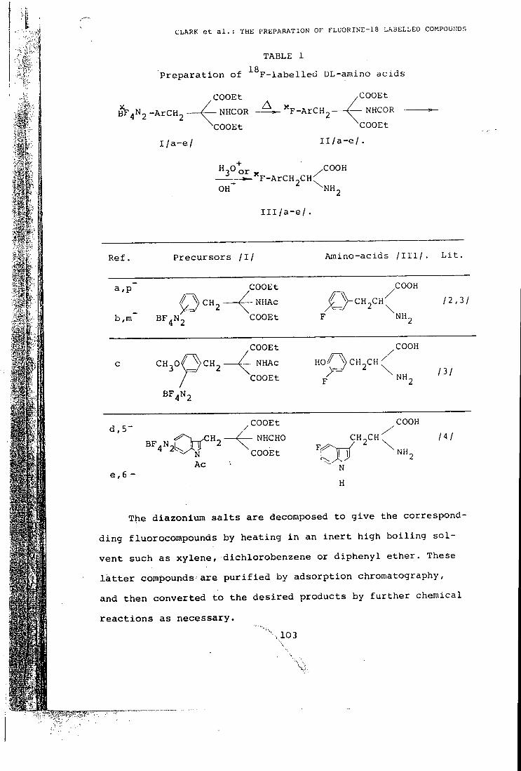
CLARK e t a l . : THE PREPARATION OF FLUORINE-18 LABELLED COMPOUNDS
TABLE 1 1 A
P r e p a r a t i o n o f F - l a b e l l e d DL-amino a c i d s
^COOEt yCOOEt
^F^n^ -ArCH2 4 ~ NHCOR ^F-ArCH2 (— NHCOR
COOEt
I / a - e /
'COOEt
I l / a - e / .
— • - ' ' F - A r C H - C H ^ ' O H " ^ N H
COOH
2 I l l / a - e /
R e f . P r e c u r s o r s / I / A m i n o - a c i d s / I I I / . L i t .
a , p ~ COOEt / ~ V c H „ C H ^ 2 \
COOH
- A - NHAc / ~ V c H „ C H ^ 2 \ 12,21
b,m BF,N2 COOEt F NH2
c CH-,0 3 p C H , - ^ .
COOEt
NHAc
COOEt
HO<i^VCH_CH
COOH
NH,
BF4N2
d , 5 -
e , 6 -
BF ."Of" .COOEt
NHCHO
COOH
CH2CH N Ac
COOEt NH,
N
H
/ 3 /
/ 4 /
The d i a z o n i u m s a l t s a r e decomposed to g i v e the c o r r e s p o n d -
d i n g f luorocompounds by h e a t i n g i n an i n e r t h i g h b o i l i n g s o l
v e n t s u c h a s x y l e n e , d i c h l o r o b e n z e n e or d i p h e n y l e t h e r . These
l a t t e r compounds a r e p u r i f i e d by a d s o r p t i o n chromatography ,
and t h e n c o n v e r t e d t o t h e d e s i r e d p r o d u c t s by f u r t h e r c h e m i c a l
r e a c t i o n s a s n e c e s s a r y .
"' ' .103
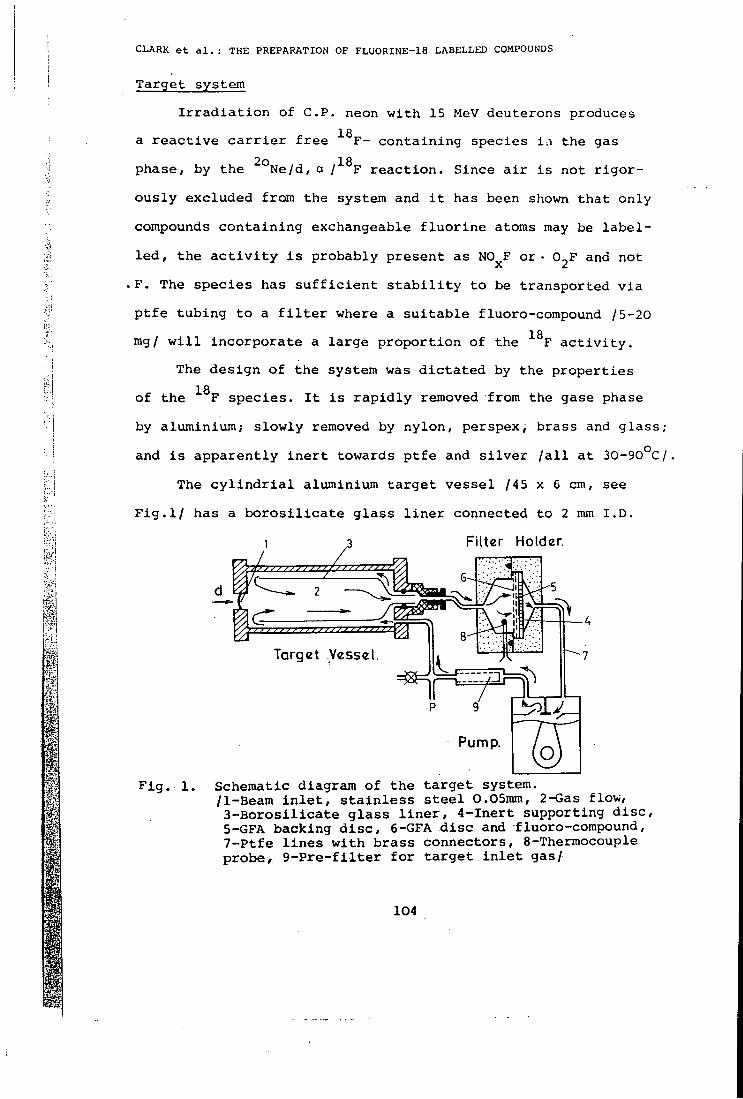
PI
i I
I
i f f :
CLARK e t a l . : THE PREPARATION OF FLUORINE-18 LABELLED COMPOUNDS
T a r g e t s y s t e m
I r r a d i a t i o n of C P . neon w i t h 15 MeV d e u t e r o n s produces 1 A
a r e a c t i v e c a r r i e r f r e e F - c o n t a i n i n g s p e c i e s i n the gas
p h a s e , by t h e ^ ° N e / d , a I^^F r e a c t i o n . S i n c e a i r i s not r i g o r
o u s l y e x c l u d e d from the s y s t e m and i t has been shown t h a t o n l y
compounds c o n t a i n i n g e x c h a n g e a b l e f l u o r i n e atoms may be l a b e l
l e d , t h e a c t i v i t y i s p r o b a b l y p r e s e n t a s NO F o r • 0 _ F and not
F . The s p e c i e s h a s s u f f i c i e n t s t a b i l i t y to be t r a n s p o r t e d v i a
p t f e t u b i n g t o a f i l t e r where a s u i t a b l e f luoro-compound 15-20 1 Q
rag/ w i l l i n c o r p o r a t e a l a r g e p r o p o r t i o n o f the F a c t i v i t y . The d e s i g n o f t h e s y s t e m was d i c t a t e d by the p r o p e r t i e s
18
o f t h e F s p e c i e s . I t i s r a p i d l y removed frora the gase phase
by a l u m i n i v m ; s l o w l y removed by n y l o n , p e r s p e x , b r a s s and g l a s s ;
and i s a p p a r e n t l y i n e r t towards p t f e and s i l v e r / a l l a t 3 0 - 9 0 ° C /
The c y l i n d r i a l a lumin ium t a r g e t v e s s e l /45 x 6 cm, see
F i g . l / h a s a b o r o s i l i c a t e g l a s s l i n e r c o n n e c t e d to 2 mm I . D ,
1 ,3 Fi l ter Holder.
Target Vesse
F i g . 1.
Pump.
S c h e m a t i c d i a g r a m o f t h e t a r g e t s y s t e m . /1 -Beam i n l e t , s t a i n l e s s s t e e l 0.05mm, 2 -Gas f l o w ,
3 - B o r o s i l i c a t e g l a s s l i n e r , 4 - I n e r t s u p p o r t i n g d i s c , 5-GFA b a c k i n g d i s c , 6-GFA d i s c and f l u o r o - c o m p o u n d , 7 - P t f e l i n e s w i t h b r a s s c o n n e c t o r s , 8 -Thermocouple p r o b e , 9 - P r e - f l i t e r f o r t a r g e t i n l e t g a s /
104
CLARK e t a l . : THE PREPARATION OF FLU0RINE-18LABELLED COMPOUNDS
p t f e t u b i n g a s shown. The s y s t e m i s e v a c u a t e d t o ' 1 mm Hg and
t h e n f i l l e d w i t h C P - g r a d e neon to a p r e s s u r e of 4 k g f . c m
and t i ie gas i s c i r c u l a t e d a t a f l o w r a t e o f " 10 l i t r e / m i n _2
X / a t 4 k g f . cm / by means of a Compton o i l - f r e e d iaphram
pump, t y p e 415 l E ^ w i t h 0 . 2 5 HP motor .
Most i r r a d i a t i o n s to d a t e have been c a r r i e d out a t beam
c u r r e n t s o f 5 - l o pA. The maximum becim c u r r e n t i s d e t e r m i n e d
by two f a c t o r s , t h e f i r s t o f w h i c h i s t h e p r e s s u r e of the neon
i n t h e s y s t e m . I t i s d e s i r a b l e t h a t t h e beam i s c o m p l e t e l y a t
t e n u a t e d b e f o r e i t r e a c h e s t h e back o f the t a r g e t , f a v o u r i n g
a h i g h p r e s s u r e , but the maximum o p e r a t i n g p r e s s u r e i s d e t e r
mined by t h e p r e s s u r e r a t i n g of the pump. The second i s t h e
t e m p e r a t u r e of t h e gas e n t e r i n g the f i l t e r h o l d e r w h i c h must
be k e p t be low 1 0 0 ° C s i n c e d i a z o n i u m t e t r a f l u o r o b o r a t e s a r e
t e m p e r a t u r e s e n s i t i v e compounds.
The compound to be l a b e l l e d / 5 - 2 0 mg/ i s c o a t e d onto a
t y p e GFA g l a s s f i b r e f i l t e r paper /What:man L t d . / , 3-5 cm i n
d i a m e t e r . To a c h i e v e t h i s t h e compourd d i s s o l v e d i n a s u i t a b l e
s o l v e n t / "^-l m l / , namely w a t e r f o r p o t a s s i u m f l u o r i d e , e t h a n o l f
f o r ant imony t r i f l u o r i d e and a c e t o n i t r i l e f o r the d i a z o n i u m
t e t r a f l u o r o b o r a t e s , i s a p p l i e d to t h e f i l t e r e l ement and t h e
s o l v e n t a l l o w e d to e v a p o r a t e . I n t h e c a s e o f p o t a s s i u m f l u o r i
de t h e f i l t e r i s d r i e d a t 1 2 0 ° C f o r 1 day b e f o r e u s e . A f t e r the
bombardement t h e GFA f i l t e r paper i s removed from the f i l t e r
u n i t and u s e d w i t h o u t e l u t i o n o f t h e l a b e l l e d f luoro -compound
^ D a w s o n , Mc. Dona ld and Dawson L t d . A s h b o u r n e , D e r b y s h i r e , E n g l a n d
105
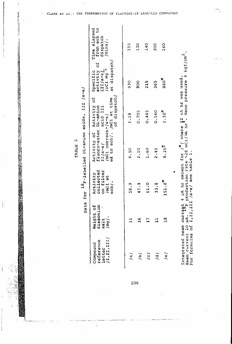
CLARK e t a l . : THE PREPARATION OF FLUORINE-18 LABELLED COMPOUNDS
I
to 13 o <a o c -H e nl
Q
T3 0)
0) XI flj .-(
I Cm
00
O in
10 +J nJ Q
rs O 0) 4J HI
It) o o • •M m cn
(U O W -H
<M T3 ~ -
s: u
M-l O
-.H 4-1 0) Cr> >*-l -H I g W
o a u M 3 . 10 (0 H
M-l o > i 0 H • P C M •H 1-t > g
+J I -H U i-J O < Q nj
u O -P
tn >i 0) 4J I
O Q) > M I •H O (0 •P 3 ^ U r - l M
o
m a to •P
- ~ <S (U I (0 U
-p o •
u o o 0) u
0
> , <U <y 4J +J J J 4J •H O .H > Q) -H -H ^ »W •p ^ o o c < o o
IW g o 3 JC O
-H m
(0
g o ~ - 0)
4J
rt) g 01 ^
0) H
U " 0) H
(X <U 4J M g >n +J » O O 0) M
LA LTI
o o o n -tr O rH . H <N
O
r-t 00
O T)
g O
o O in in O 0) 0) n O iH VD (0 M a oo CM n CO 3 D 0)
(0 V) ITS a)
u
in in O K < <u 00 O CO a z .-t in in
CM • •-\ O o O OJ r-W
Vl 0) x: u x: 3
- ~ u K l >; e
O O o in in — in .-1 n 00 •
(N >-l (N CO o e ip (U
0) .H +J 4J XI Oi rj (0 0) k -P
' 0 X C 0) Q) 0 0)
K 01 o 00 00 M -P
x: u -~~ oo r i 3 0) u^ n in < -a 1
r - l 3 0 nJ
a H
•P Pn H flO H OH > U M M • M 3 < -O 3. M
g O *P m H o Q) XI -P <u
G <0
U 3 H g 3 U U O
>P
01 4J (0 M
Q) g •P (0 M C (U o
H m t ,
106
CLARK e t a l . : THE PREPARATION OF FLURINE-18 LABELLED COMPOUNDS
from i t . The f i l t e r p a p e r i s m e r e l y crumpled-up and i n t r o d u c e d i n t o the r e a c t i o n m i x t u r e .
18
P r e p a r a t i o n of F - l a b e l l e d DL amino a c i d s :
The d i a z o n i u m t e t r a f l u o r o b o r a t e 110-20 mg/ i s l a b e l l e d
a s d e s c r i b e d above . A f t e r bombardement the l a b e l l e d p r o d u c t ,
I / a - e / , s t i l l on t h e GFA f i l t e r p a p e r , i s decomposed i n d i p h e n y l
e t h e r / b i p h e n y l /Dowtherm A,'\'3 m l / a t 1 6 0 - 2 0 0 ° C , Decompos i t ion
i s comple te w i t h i n 10 min and the r e s u l t i n g f l u o r o - e s t e r , I l / a - e /
now i n s o l u t i o n , i s p u r i f i e d by chromatography on b a s i c a l u m i n a ,
e l u t i n g w i t h 10-50% c h l o r o f o r m i n t o l u e n e . The c o n d i t i o n s f o r
t h e f i n a l s t a g e , namely h y d r o l y s i s t o the f r e e amino a c i d , I I I /
/ a - e / , depend upon t l ie p a r t i c u l a r compound. Compounds I I / a , b / a r e
h e a t e d i n 13N s u l p h u r i c a c i d a t 1 7 0 ° C f o r 30 mins^^ and I I / c / i s
r e f l u x e d i n h y d r i o d i c a c i d / r e d p h o s p h o r u s / a c e t i c a c i d f o r 40
m i n s . Compounds I I / d , e / a r e r e f l u x e d i n 2 .5N sodium h y d r o x i d e
f o r 50 m i n s , t r e a t e d w i t h c o n e , h y d r o c h l o r i c a c i d to pH 1, and
r e f l u x e d a f u r t h e r 10 mins."^
The f i n a l p r o d u c t i s d e a c i d i f i e d u s i n g I R 4 B / 0 H / r e s i n , or
a l t e r n a t i v e l y i n c a s e s / a , b , d and e / d e s a l t e d u s i n g Poropak
, R a d i o c h e m i c a l a n a l y s i s o f the p r o d u c t i s by means o f the t i c
on s i l i c a g e l ^ , o r by e i t h e r h i g h ^ o r l o w ^ ' ' ' p r e s s u r e i o n e x
change c h r o m a t o g r a p h y , and t h e s p e c i f i c a c t i v i t y d e t e r m i n e d 2 4
by i o n i s a t i o n chamber a s s a y and U . V . s p e c t r o p h o t o m e t r y ' . D a t a
f o r s e v e r a l t y p i c a l p r e p a r a t i o n s a r e g i v e n i n T a b l e 2 .
107
C L A R K e t a l . : T H E P R E P A R A T I O N O F F L U O R I N E - 1 8 L A B E L L E D C O M P O U N D S
Many of t h e i d e a s c o n c e r n i n g t h i s t a r g e t s y s t e m were based
on i n f o r m a t i o n s u p p l i e d by D r . Nozaki o f t h e I n s t i t u t e o f
P h y s i c a l and C h e m i c a l R e s e a r c h , Wako S h i , J a p a n , who has used
18
a s i m i l a r a p p r o a c h t o F l a b e l l i n g p r o c e d u r e s .
REFERENCES
1. M . F . C o t t r a l l , D . M . T a y l o r , T . J . M c E l w a i n , B r i t . J . R a d i o l . , 46 / 1 9 7 J / 277 .
2 . R . W . G o u l d i n g , A . J . P a l m e r , I n t . J . A p p l . R a d i a t i o n I s o t o p e s , 23 / 1 9 / 2 / 1 3 3 .
3 . H . L . A t k i n s , D . R . C h r i s t m a n , J . S . F o w l e r , W.Hauser , R . M . H o y t e , J . F . K l o p p e r , S . S . L i n , A . P . W o l f , J . N u c l . M e d . , 1 2 / 1 9 7 1 / 28o, i b i d , 1 3 / 1 9 7 2 / 7 1 3 .
4. A . J . P a l m j g , J . C . C l a r k , R . W . G o u l d i n g , M.Roman, "The p r e p a r a t i o n of F - l a b e l l e d D L - 3 - f l u o r o t y r o s i n e " , p r e s e n t e d a t the Symposium on New Developments i n R a d i o p h a r m a c e u t i c a l s and L a b e l l e d Compounds, Copenhagen, Denmark, 26-30 M a r c h , 1973 . P r o c e e d i n g s to be p u b l i s h e d by The I n t e r n a t i o n a l Atomic E n e r g y A g e n c y , V i e n n a , A u s t r i a .
5 . G . F i r n a u , C . N a h r a i a s , S . G a r n e t t , I n t . J . A p p l . R a d i a t i o n I s o t o p e s , 24 / 1 9 7 3 / 182 .
6 . J . C . C l a r k , M . L . T h a k u r , I . A . Watson, I n t . . J . A p p l . R a d i a t i o n I s o t o p e s , 23 / 1 9 7 2 / 329 .
7 . A . N i e d e r w i e s s e r , P . G i l i b e r t i , J . C h r o m a t o g . , 6 1 / 1 9 7 1 / 81 , i b i d 9 5 .
8. C . H a w o r t h , T . A . W a l m s l e y , J . C h r o m a t o g . , 66 / 1 9 7 2 / 311 .
9. A . Mondino, G . B o n g i o v a n n i , V .Noe , I . R a f f a e l e , J . C h r o m a t o g . , 63 / 1 9 7 1 / 411 . ,
108
Thorax (1974), 29, 99.
Use of ' F labelled fluorocarbon-11 to investigate the fate of inhaled fluorocarbons in man and in the rat
F A I T H M . W I L L I A M S , G . H . D R A F F A N , a n d C . T . D O L L E R Y
MRC Clinical Pharmacology Research Group, Royal Postgraduate Medical School, Du Cane Road, London W12
i . C . C L A R K a n d A . J . P A L M E R
MRC Cyclotron Unit
P . V E R N O N i
Department of Medical Physics, Hammersmith Hospital, London W12
Williams, Faith M., DrafFan, G. H., Dollery, C. T., Clark, J . C , Palmer, A. J . , and Vernon, P. (1974). Thorax, 29, 99-103. Use of " F labelled fluorocarbon-11 to investigate the fate of inhaled fluorocarbons in man and in the rat. The distribution and elimination of ^*F labelled fluorocarbon-11 has been followed in a group of rats killed after air breathing following six minutes' exposure to " F fluorocarbon-11. Whole body and individual organ count rates were measured. In four volunteers the fate of ' ' F labelled fluorocarbon-11 was followed by both whole body counting and gamma camera measurement of the activity in the lung and mouth region after inhalation from a specially loaded aerosol dispenser.
In the rat there was a high initial level in high blood flow organs and in the adrenals and fat: the level in blood and high blood flow organs fell rapidly. Elimination from fat was slow but the adrenal level had fallen within one hour. The fall in whole body count rate was similar to that in fat.
In manjht fall in lung concentration was consistent with rapid uptake into tissues followed by slow elimination; the whole body count rate curve also indicated slow elimination. There was no evidence of deposition of droplets of fluorocarbon in the mouth region after use of the aerosol.
The present investigation was undertaken as part diately after use of the inhaler the concentration of of a series (Draffan, Dollery, Williams, and Clare, fluorocarbon in arterial blood rose to a peak and 1974) designed to evaluate the hazards that might then fell again, initially rapidly and then more arise from the fluorocarbon propellants'used in slowly. The slow phase probably reflected elimina-pressurized aerosol products for the treatment of tion of fluorocarbon concentrated in the tissues and asthma. The aim was to determine the absorption, fat deposits. Morgan, Black, Walsh, and Belcher distribution, and elimination of fluorocarbons in (1972) investigated the retention of ''CI fluoro-man and rat. carbon after inhalation of the vapour, by counting
The levels of fluorocarbons-11 and -12 (F-11, the expired gas, and drew a similar conclusion. CCI3F; F-12, CC1,F,) in arterial and venous blood - hese studies were made with cyclotron pro-and alveolar air have been measured m man ^^^^^ isp fluorocarbon-11. ' T has a radiation foUowing the use of an aerosol mhaler (DoUery ^eV) suitable for external detection and a et al. 1970; Paterson, Sudlow, and Walker, 1971; Dollery et al., 1974; Draffan et al, 1974). Imme-
24
et al.. 1970; Paterson, Sudlow, and Walker, 1971; T ' T ' ^ ^ ' ^ l " " ' Dollerv ^ / « / 1974- nraffpn 1074 T half-life of 110 mmutes. The count rate/time
curves of the whole body and specific organs in iPresent address: Department of Nuclear Medicine. Charing Cross ^^^^ expOSed tO " F fluorOCarbon VapOUr Were R^nJ^i- J;^"t°nH„,= rir r- T r. „ u , d . measured. In man, distribution and elimination Kequcsis for reprints to: Dr. C . T . Dollery. Royal Postgraduate r 11 j L • 1 1 . Medical School. Du Cane Road. London W12 Were toUowed by whole body counting and USe
99
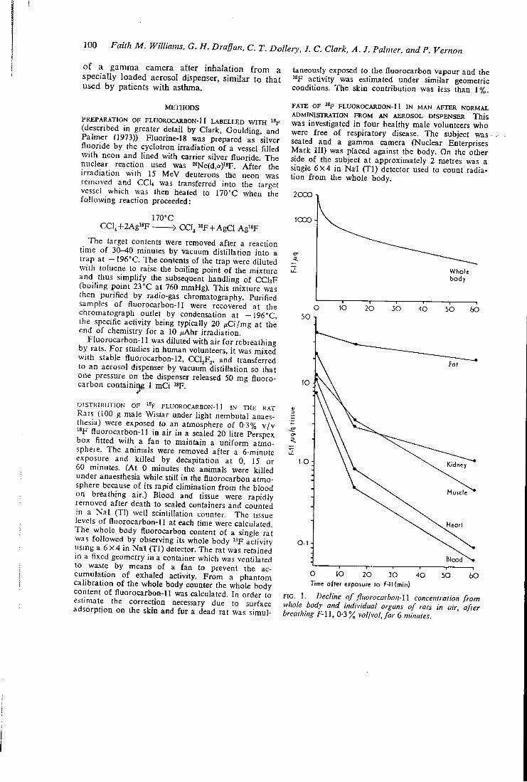
ICQ Faith M. Williams. G. H. Draffan, C. T. Dollery, J. C. Clark. A. J. Palmer, and P. Vernon
of a gamma camera after inhalation from a specially loaded aerosol dispenser, similar to that used by patients with asthma.
METHODS
PREPARATION OF FLUOROCARBON-11 L A B E L L E D W I T H (described in greater detail by Clark, Goulding, and Palmer (1973)) Fluorine-18 was prepared as silver fluoride by the cyclotron irradiation of a vessel filled with neon and lined with carrier silver fluoride. The nuclear reaction used was 20Ne(d,a)i'F. After the irradiation with 15 MeV deuterons the neon was removed and CCU was transferred into the target vessel which was then heated to 170°C when the following reaction proceeded:
170°C CCl4+2Ag"F > CCI3 «F +AgCl Ag«F
The target contents were removed after a reaction time of 30-40 minutes by vacuum distillation into a trap at — 196°C. The contents of the trap were diluted with toluene to raise the boiling point of the mixture and thus simplify the subsequent handling of CCI3F (boiling point 23°C at 760 mmHg). This mixture was then purified by radio-gas chromatography. Purified samples of fluorocarbon-11 were recovered at the chromatograph outlet by condensation at — 196°C, the specific activity being typically 20 /iCi/mg at the end of chemistry for a 10 /lAhr irradiation.
Fluorocarbon-11 was diluted with air for rebreathing by rats. For studies in human volunteers, it was mixed with stable fluorocarbon-I2, CCljFj, and transferred to an aerosol dispenser by vacuum distillation so that one pressure on the dispenser released 50 mg fluorocarbon containi^ 1 mCi "F.
D I S T K I B U T I O N O F % F L U O R O C A R B O N - 1 1 I N T H E R A T
Rats (100 g male Wistar under light nembutal anaesthesia) were exposed to an atmosphere of 03% v/v
fluorocarbon-11 in air in a sealed 20 litre Perspex box fitted with a fan to maintain a uniform atmosphere. The animals were removed after a 6-minute exposure and killed by decapitation at 0, 15 or 60 minutes. (At 0 minutes the animals were killed under anaesthesia while still in the fluorocarbon atmosphere because of its rapid elimination from the blood on breathing air.) Blood and tissue were rapidly removed after death to sealed containers and counted in a Nal (Tl) well scintillation counter. The (issue levels of fluorocarbon-11 at each time were calculated. The whole body fluorocarbon content of a single rat was followed by observing its whole body ^ F activity using a 6 X 4 in Nal (Tl) detector. The rat was retained in a fixed geometry in a container which was ventilated to waste by means of a fan to prevent the accumulation of exhaled activity. From a phantom calibration of the whole body counter the whole body content of fluorocarbon-11 was calculated. In order to estimate the correction necessary due to surface adsorption on the skin and fur a dead rat was simul
taneously exposed to the fluorocarbon vapour and the '*F activity was estimated under similar geometric conditions. The skin contribution was less than 1%.
FATE OF " F FLUOROCARBON-!! IN MAN AFTER NORMAL ADMINISTRATION FROM AN AEROSOL DISPENSER This was investigated in four healthy male volunteers who were free of respiratory disease. The subject was • seated and a gamma camera (Nuclear Enterprises Mark III) was placed against the body. On the other side of the subject at approximately 2 metres was a single 6X4 in Na'I (Tl) detector used to count radiation from the whole body.
2 0 0 0 ^
1 0 2 0 3 0 40 5 0 6 0
Blood
O 1 0 2 0 30 40 5 0 60 Time after exposure to F-I1(min}
F I G . 1. Decline of fliiorocarbon-\\ concentralion from whole body and individual organs of rats in air after breathing F-U, 0-3% vol/vol, for 6 minutes.
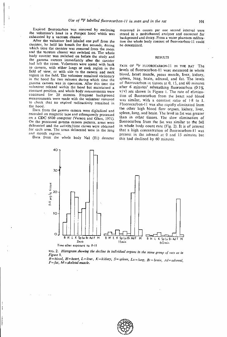
Use of "F labelled fluorocarbon-] 1 in man and in the rat 101
Expired fluorocarboa was removed by enclosing the volunteer's head in a Perspex hood which was exhausted by a vacuum cleaner.
After the volunteer had inhaled one puff from the canister, he held his breath for five seconds, during which time the canister was removed from the room and the vacuum cleaner was switched on. The whole body counter was switched on before the study and the gamma camera immediately after the canister had left the room. Volunteers were seated with back to camera, with either lungs or neck region in the field of view, or with side to the camera and neck region in the field. The volunteer remained stationary in the hood for two minutes during which time the gamma camera was in operation. After this lime the volunteer relaxed within the hood but maintained a constant position, and whole body measurements were continued for 20 minutes. Frequent background measurements were made with the volunteer removed to check that no expired radioactivity remained in the hood.
Data from the gamma camera were digitalized and recorded on magnetic tape and subsequently processed on a CDC 6600 computer (Vernon and Glass, 1971). On the processed gamma camera pictures, areas were delineated and the activity/time curves were obtained for each area. The areas delineated were in the lung and mouth regions.
Data from the whole body Nal (Tl) detector
measured in counts per one second interval were stored in a multichannel analyser and corrected for background and decay. From a water phantom calibration the whole body content of fluorocarbon-11 could be determined.
RESULTS
FATE OF % FLUOROCARBON-11 IN THE RAT The levels of fluorocarbon-11 were measured in whole blood, heart muscle, psoas muscle, liver, kidney, spleen, lung, brain, adrenal, and fat. The levels of fluorocarbon in tissues at 0, 15, and 60 minutes after 6 minutes' rebreathing fluorocarbon (03% v/v) are shown in Figure 1. The rate of elimination of fluorocarbon from the heart and blood was similar, with a constant ratio of 1-8 to 1. Fluorocarbon-11 was also rapidly eliminated from the other high blood flow organs, kidney, liver, spleen, lung, and brain. The level in fat was greater than in other tissues. The slow elimination of fluorocarbon from the fat was similar to the fall in whole body count rate (Fig. 2). It is of interest that a high concentration of fluorocarbon-11 was present in the adrenal at 0 and 15 minutes but this had declined by 60 minutes.
40
30 4
,20-^
O J B H L K SpLuBrAdF M B H L K Sp Lu Br AdF M B H L K Sp Lu Br Ad F M
Time after exposure to F-11
F I G . 2. Histogram showing the decline in individual organs in the same group of rats as in Figure 1. B=blood H= heart L=liver, K=kidney, S=spleen, Lu^lung, Br = brain, Ad^adrenal F=fat, M= skeletal muscle.
Use of "F labelled fluorocarbon-11 in man and in the rat 103
carbon concentration fell more slowly but had reached a low value by 60 minutes. Elimination from fat was very slow and had not reached half the initial value by one hour. The slow rate of elimination from fat must reflect a high tissue: blood partition coefficient and a low blood flow per unit mass of tissue. The blood:heart muscle ratio, 1:1-8, agrees well with that found in vitro using dog heart muscle (DoUery et al., 1974). Cox, King, and Parke (1972) found that 97% of a single dose of fluorocarbon-11 given to a rat was expired unchanged within six hours, indicating that the radioactivity measured in our studies represents unchanged fluorocarbon.
The whole body count rate in man initially fell quickly. The final phase in our studies (ty range 12 to 20 minutes) was shorter than that observed by Morgan et al. (1972), who measured expired ^Cl fluorocarbon-11 (ty approximately 100 minutes).
The profile of fluorocarbon elimination from the lung indicated an initial transfer of the dose to the blood stream and uptake by tissues and later pulmonary excretion of fluorocarbon released from the tissues and fat. The time course of elimination from the lung observed using the gamma camera was similar to the elimination from alveolar gas measured by mass spectrometry (Draffan et al.. 191 A). The absence of an early peak and initial fall in count rate in the mouth region suggests that fluorocafbon-l 1 was not deposited in the mouth as droplets. If droplets had formed one wojild expect to see a rapid fall in count rate witfl the first breath as the droplets were volatilized and washed directly out of the mouth.
The main factor influencing the elimination of fluorocarbon-11 from the body is concentration in fat depots. The slow release of fluorocarbon from fat into blood should not prove any hazard to the heart of man. The high concentration in the adrenal gland must also reflect its lipid content. Perfluro-n-hexane (C5F12) can bind to microsomes
and in vitro can act as an inhibitor of oxidation (Ullrich and Diehl, 1971). Fluorocarbon-11 (CCI3F) will probably act in a similar manner. However, it is improbable that a high concentration would persist for long enough in man, after normal use of an aerosol inhaler, significantly to inhibit the formation of adrenal steroids.
R E F E R E N C E S Clark, J . C , Goulding, R. \V., and Palmer, L . A. J . (1973).
The preparation of " F labelled fluorocarbons for use in pharmacodynamic studies. In New Developments in Radiopharmaceuticals and Labelled Compounds. I A E A , Vienna STI/PUB/344.
Cox, P. J . , King, L . J . , and Parke, D. V. (1972). A study of the possible metabolism of trichlorofiuoromethane. Biochemical Journal, 130, 13 P.
Dollery, C . T . , Draffan, G . H., Davies, D. S., Williams, Faith M., and Conolly, M. E. (1970). Blood concentrations in man of fluorinaied hydrocarbons after inhalation of pressurised aerosols. Lancet, 2, 1164. Williams, F . M., DralTan, G. H., Wise, G . , Sahyoun, H. , Paterson, J . W., and Walker. S. R. (1974). Arterial blood levels of fluorocarbons in asthmatics following use of pressurised aerosols. Clinical Pharmacology and Tliera-peuatics, in press.
Draffan, G . H. , Dollery, C. T., Williams, Faith M., and Clare, R. A. (1974). Alveolar gas concentrations of fluorocarbons-11 and -12 in man after use of pressurised aerosols. Thorax, 29, 95.
Morgan, A., Black, A., Walsh, M., and Belcher, D. R. (1972). The absorption and retention of inhaled fluori-nated hydrocarbon vapours. International Journal of Applied Radiation and Isotopes, 23, 285.
Paterson, J. W., Sudlow, M. F . . and Walker. S. R. (1971). Blood-levels of HuoriiKik-d ludrocarbons in asihmaiic patients after inhalation of pressurised aerosols. Lancet, 2, 565.
Ullrich, V. and Diehl, H. (1971). Uncoupling of mono-oxygenation and electron transport by fluorocarbons in liver microsomes. Etnupcan Journal of Biochemistry, 20, 509.
Vernon, P. and Glass, H. I. (1971). An ofl'-line digital system for use with a gamma camera. Physics in Medicine and Biology, 16, 405.
Progress in Respiration Research Editor: H. Herzog, Basel Publishers: S. KARGER, Basel SEPARATUM (Printed in Switzerland)
Free Communications
25
Prog. Resp. Res., vol. 9, pp. 249-253 (Karger, Basel 1975)
An External Counting Method for Regional Measurements of Extravascular Lung Water'
F. FAZ[o^ J . C . C L A R K , P. D . B U C K I N G H A M , C . G . RHODES, F . R. H U D
SON, H A Z E L A. JONES, T . JONES and J . M . B . HUGHES
Departments of Medicine, Medical Physics and M R C Cyclotron Unit, Hammersmith Hospital, London
Introduction
A n estimation of extravascular lung water volume ( E L W V ) can be made in vivo by the standard double indicator technique of C H I N A R D and ENNS [1]. This technique requires an injection in the venous side of the circulation of a mixture of tracers, one non-diffusible indicator ( N D I ) or intravascular reference tracer and the other freely diffusible in the interstitial spaces (DI) , and arterial sampling of the downstream dilution curves.
The downslope of each curve is then extrapolated in order to eliminate recirculation, and mean transit time ( M T T ) is calculated.
The E L W V is calculated as:
/ E L W V = (tDi-tNm)F,
where F is the water flow through the lungs (i.e. the water content of the cardiac output) and t o i and tNo i are, respectively, the M T T for the D I and for the N D I .
Indicators labelled with y-emitting radioisotopes have already been used as intravascular tracers. On the other hand, the water tracer which is commonly used is T H O (tritiated water), a/3-emitter which cannot be detected externally. For this reason, it requires arterial sampling and time-consuming laboratory procedures for liquid scintillation counting. Our aim was to
1 Supported in part by grant 73.00490.04 of the Italian National Research Council. 2 On leave from CNR Laboratory of Clinical Physiology, Pisa, Italy. Recipient of a Wellcome Trust Fellowship.
F A Z I O et al. 2 5 0
develop a non-invasive method for the estimation of lung water by external recording of dilution curves over the chest.
Therefore, we decided to substitute a y-emitting radioisotope for THO. The y-emitting water tracer with the longest half-life Hg^ O (2.05 min half-life, 511 keV energy) was chosen as the DI . Red blood cells labelled with monoxide (20.3 min half-life, 511 keV energy) were used as intravascular tracers. Both isotopes were prepared in the MRC Cyclotron at Hammersmith Hospital.
Methods and Results
Dogs anaesthetized with chioialose were studied in the supine and erect postures. Injections of Hj^'O and C O red cells were made via a catheter placed in the superior vena cava, with an interval of approximately 5 min between injections. Blood was withdrawn by a Harvard pump via two identical catheters simultaneously from the aorta (Ao) and the pulmonary artery (PA) and allowed to flow through a coil in front of a scintillation crystal. Time-activity curves, representing the input (PA) and the output (Ao) of the pulmonary circulation, were recorded on a multi-channel analyser. Time-activity curves for two regions of the right lung (apex and base) were also recorded externally by means of probes collimated over the chest. To assess regional distribution of blood flow, ^ " I -labelled albumin microspheres were injected. The animal was killed, the lungs excised, dried, and sliced. Slices were counted and blood flow distribution from base to apex expressed in counts per gram of dry lung.
Figure 1 shows regional time-activity curves for H^^^O and "CO-RBC in a supine dog. The count rates and definition of these curves are sufficiently good for regional lung water to be measured not only with the double extrapolation technique, but als'owith the more sophisticated deconvolution procedures [2] required for the determination of the true frequency function of transit times (and consequently the true MTT) for water in the extravascular spaces of the lung.
Figure 2 shows curves from similar regions in a dog tilted head up for about 3 h. Whereas the curves obtained from the bottom of the lung show a steep downslope followed by a recirculation phase, the shape of the curves recorded from the top are very shallow with recirculation poorly defined. The latter resemble arterial dilution curves obtained from heart patients with very low cardiac output [3]. In fact, the erect anaesthetized dog presents a very large gradient of perfusion from apex to base of the lung. Figure 3 shows the distribution of blood flow per gram of dry lung in one erect and one supine dog. Whereas the horizontal dog presents a reasonably uniform
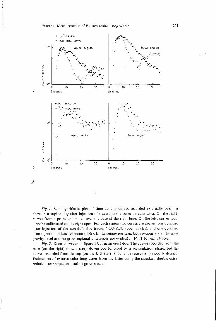
External Measurements of Extravascular Lung Water 251
10
10
• H 2 ' 0 curve » "CO-RBG curve
Apicdl region Basal region
0 10 Seconds
20 30 0 10 Seconds
20 30
10
10
• H 2 ' 0 curve
° "CO-RBC curve
Apical region
8 o
Basal region
0 10 Seconds
20 30 0 10 Seconds
20 30
Fig. 1. Semilogarithmic plot of time activity curves recorded externally over the chest in a supine dog after injection of tracers in the superior vena cava. On the right: curves from a probe collimated over the base of the right lung. On the left: curves from a probe collimated on the right apex. For each region two curves are shown: one obtained after injection of the non-diffusible tracer, ' ^ C O - R B C (open circles), and one obtained after injection of labelled water (dots). In the supine position, both regions are at the same gravity level and no gross regional differences are evident in M T T for each tracer.
Fig. 2. Same curves as in figure 1 but in an erect dog. The curves recorded from the base (on the right) show a steep downslope followed by a recirculation phase, but the curves recorded from the top (on the left) are shallow with recirculation poorly defined. Estimation of extravascular lung water from the latter using the standard double extrapolation technique can lead to gross errors.
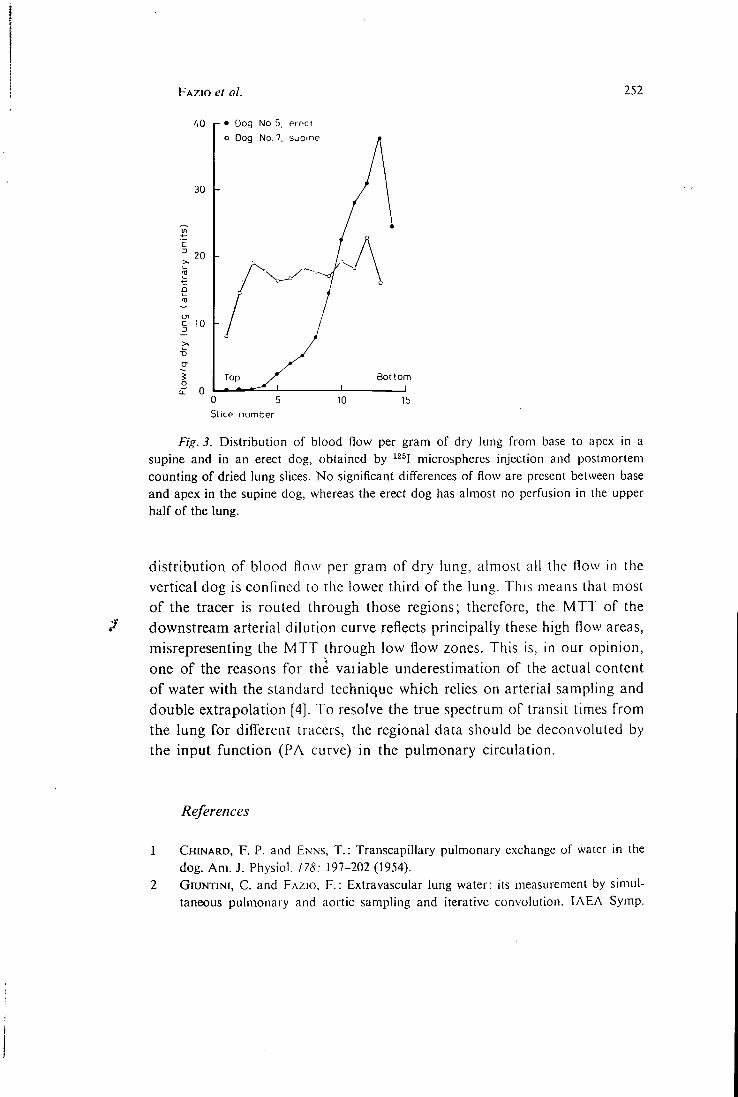
F A Z I O et al. 252
40
30
20
p • Dog No. 5, erect o Dog No. 7, supine
Bottom
10
•D cn S o
C 0 0 5 Slice number
Fig. 3. Distribution of blood flow per gram of dry lung from base to apex in a supine and in an erect dog, obtained by ^^^I microspheres injection and postmortem counting of dried lung slices. No significant differences of flow are present between base and apex in the supine dog, whereas the erect dog has almost no perfusion in the upper half of the lung.
distribution of blood f low per gram of dry lung, almost al l the flow in the vertical dog is confined to the lower third of the lung. This means that most of the tracer is routed through those regions; therefore, the MTT of the
/ downstream arterial dilution curve reflects principally these high flow areas, misrepresenting the MTT through low flow zones. This is, in our opinion, one of the reasons for the vaiiable underestimation of the actual content of water with the standard technique which relies on arterial sampling and double extrapolation [4]. To resolve the true spectrum of transit times from the lung for diff'erent tracers, the regional data should be deconvoluled by the input function (PA curve) in the pulmonary circulation.
References
1 C H I N A R D , F . P . and E N N S , T . : Transcapillary pulmonary exchange of water in the dog. Am. J . Physiol. 178: 197-202 (1954).
2 GiUNTiNi, C . and F A Z I O , F . : Extravascular lung water: its measurement by simultaneous pulmonary and aortic sampling and iterative convolution. I A E A Symp.
External Measurements of Extravascular Lung Water 253
Dynamic Studies with Radioisotopes in Clinical Medicine and Research, Knoxville 1974.
GiUNTiNi, C ; F A Z I O , F . , and M A N C I N I , P . : Determination of transit time distribution of extravascular lung water; in Central hemodynamics and gas exchange, edited by C . GiUNTiNi, pp. 459^72 (Minerva Medica, Torino 1971).
P E A R C E , M . L . ; Y A M A S H I T A , J . , and B E A Z E L L , J . : Measurement of pulmonary edema.
Circulation Res. 17: 414-A26 (1965).
Dr . F . F A Z I O , Departments of Medicine, Hammersmith Hospital, London WI2 OHS
(England)
26
hort-lived Radioactive Gases for
Clinical Use
J. C. CLARK, BSC and
P. D. BUCKINGHAM, A I S T
Medical Research Council.. Cyclotron Unit., Hammersmith Hospital, London
BUTTERWORTHS London and Boston
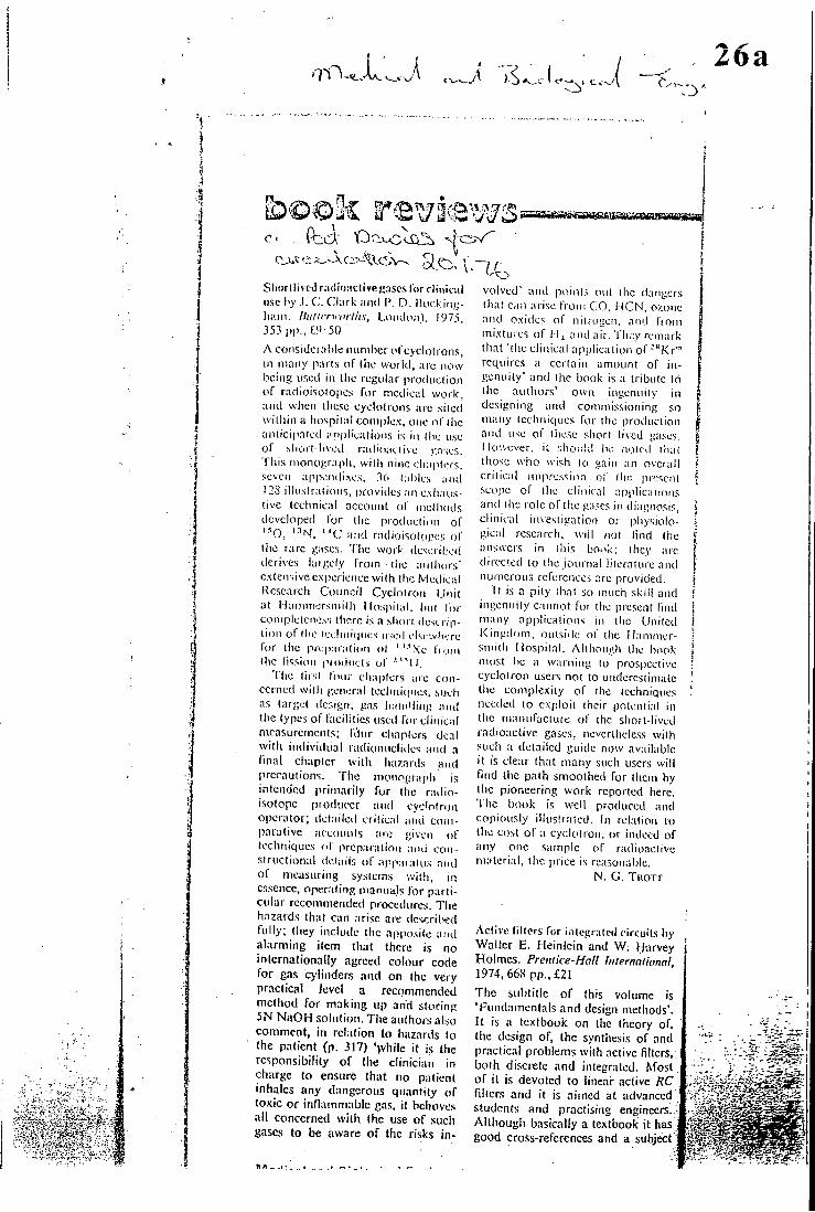
\J > tv-26a
^oc)ic reviews c . feci- Or-uolcc:.
Shortlived rn<Iii);iclive c-iscsrorcltninii use by .1. C. Clark aiul P. O. liucking-ham. lUilicrwoiths, LoiKlon), I97.S, 35.1 PI'-, t''-50 A con5i(Jcral)le luinibcr ofcyclolron.s, in many parts of liic world, arc now being uscil in (ho regular produtlion of radioisotopes for medical work, and when lliese cyclotrons arc .siled within a liospilal coniplcv, one ol (lie anlici|ialed ariplieations is in ll\e use of slioi l-lived railioaciive r.oses. This monograph, willi nine chapters, seven appendixes. 36 tables and 128 illuslralions, provides an cxhatis-live technical account ol' mclliods developed lor the produciion of '^O, '^N, " C and radioisotopes ol' the rare gases, '('lie work descril'ed derives laiyely from liie aiilliors' extensive experience with tbe Medical Research Council Cyclotron Unit at llammersmilh liospilal, but lor completeness there is a slim i description of the icchnifpies used ei-icwbere for the prcp iralion (it ''''Xe Imm the lisslon pioilucts ul ' " I k
'Che lirsl I'lnir chaplers aic concerned with general tecliinc|ues, .such as target design, gas li.indling and the types of facilities used forcliincal measurements; lOur chapters deal with individual radionucliiles and a fmal chapter with hazartls and precautions. Tlic monograpli is intended primarily for the radioisotope producer and cyclotron operator; detailed critical and comparative accoiu>ts arc given of techniques ol preparation and constructional details of app.aratus and of measuring systems with, in essence, operating manuals for particular rccoinmended procedures. The hazards that can arise are described fully; they include the apposite and alarming item that there is no internationally agreed colour code for gas c>linders and on the very practical Jcvcl a recommended method for making up and storing 5N NaOH solution. The authors also comment, in relation to hazards to the patient (p. 317) '>vhile it is the responsibility of the clinician in charge to ensure that no patient inhales any dangerous quantity of toxic or inflammable gas, it behoves all concerned with the use of such gases to be aware of the risks in
volved' and points out the dangers that can arise from C^O, i ICN, ozone and oxides of niiroijon, aiu.l from mixtures of M) and air. They remark that "the clinical application of-"Kr" requires a certain amoLint of ingenuity' and the book is a tribute 16 the authors' own ingenuity in designing and commissioning so many techniques for llie production and use of llie.^e short lived gases. Ilo-.vcver, it should W. noted that those who wish to gain an overall critical impression of tin.', present scope of the clinical applications and (lie role of the gases in diagnosis, clinical investigation or physiological research, will not lind (he ansucrs in this booi;: they arc directed to the journal literature and numerous references are provided.
Tt is a pity that so much skill and ingenuity cannot for ihc present find many applications in the tinited Kingdom, oufsirle of the Hammersmith Hospital. Allhoiri'.h the book nuist be a warning to prospective cyclotron users not to underestimate the complexity of the techniques needed to exploit their iiotential in the manufacture of the short-lived radioactive gases, nevertheless with such a detailed guide now available it is clear that many such users will llnd the path smoothed for them by the pioneering work reported here, 'the book is well produced and copiously iUusUaied. In relation to the cost of a cyckitron, or indeed of any one sample of radioactive material, the price is reasonable.
N. G. TuoTT
Aclive filters for intogrntcd circuits by Walter E . Heinlein and W; Harvey Holmes. Prentice-Hall Internalionnl, 1974, 668 pp., £21
The subtitle of this volume is 'Fundamentals and design methods'. It is a textbook on the theory of, the design of, the synthesis of and practical problems with active filters, both discrete and integrated. Most of it is devoted to linear active RC fillers and it is aimed at advanced students and practising engineers.. Although basically a textbook it has good cross-references and a subject
It
F pi ie ) . U y V
Short-li'i t'd Ihuliddcd^ e ( l a s r s f o r (7//f/rc// U.^r. Hy j . (' Chirk ;ind 1\ I ) . lUickiii^liiiin, pp. xii i 353, \97^ (Loruio i i , IhittcM-worth), /•/^5().
T h i s oNccIk'iit monograph sets out in a i lear ;iiul o idrrly lor/nat (1K" ilcfailcd technology for prochu in/ ' / , ';IS(MMIS radioiuichdfs suitalilc for piihuonary (unction ami other chnical work. Inc \ i tahly it is a spcciahsl's hook, since most doctors and pliysicists usin^ radioacti\ c ,'ascs ai e more concernetl n i th the clinical aspects of the sul-)ject than with details of manufactme, yield, and desiiiti of piocessin/j systems for radiopharmaceuticals. I' l irthermorr a la:[:e j")roportion of the hook is de\ oted to the reallv "shortdixxni" ^ases, '•'iN2. ^H)j . " C O and " C O i - , which are of only a-:.-demic interest to those not in close proximity to a cycini ion. T h e authors de\'ote little space to '•'•'Xe, prcsumahlv he-cause its technolou>- is well known, hut do /.,'i\e usefui .sec-tioris on ^'-''Xe anti '-' ' X c , and the radionuclides of kr\ pton, of w li ich"M\r'" has been siiown to be of promise for rt'),Monal ventilation studies. A section on dispensing' and t hnicai facilities brink's tlie suliject closer to the hospital deimrtmenf, but if a multiprobc detector system is to be described in sucii detail, is not a scintillation camera worthy of mention also.' h'or such a specialist book it seems unnecessary to hi! e\'cn a sniall chapter with the elements of nticlcar physics, but this ha{)pens too often in books concerned prim.uily witli the application of radioactive substances to medicine.
W i t h its attentiofi to detail, clear illustrations, and incki -sion of useful appendices on matliematical analysis this book is of direct benefit to those working in a cyclotron unit dedicated to clinical investigation. T o tliose of us less fortunate it provides a comprehensive background to the subject, and leads to a better understanding of the problems of supplying hospitals with clinical radioactive gases.
D» M . ACKERY.
26b
160
26c
Short-lived Radioactive Gases for Clinical Use. By J . C . C L A R K nnd P. D . . B U C K I N O I I A M
(London: Buttcrwortlis, 1975) [Pp. xi i - f 353]" £9-50
\\'licii tlip decision wn.s taken some 25 ycftrs ago to .' of n medical oyclol.rnn at Hnninicrsinil.li Hospital it was cnv-sagcd that one of its useful fuiictioii.'< would bo tlin prodnclioii of radioaclixo nuclides for medical rosenrcli wliicli could not bo produced in a reactor. Tins |iroinisc was n m | i l y fulfilled as cvicloncod by tlio construction of siiiiilur ryclotroiis and ac-cdcraloi.-^ in medical iiislitulos in many other coimtrios. The short-lived gases ( " C . " N , '^0 and \ ariniis isotopes of K'r and Xe) prox idod the tools for Roino K])f'Cfncular advances in clinical physiology, and it is \vi( h tlio liroduction of these imclidcs that this monograph i.s concorTicd.
The authors have condensed thoir considerable cx|icitisc ami cxpcricnco info a single volumo ^vllich covers in niintito detail every .stop of the tarpet preparation, l)ornl)ar(liiieiit, ec|iar'al ion, j jiurification. assay and dispensing of the various pn-ses listed and some of I lu'lr snnple compounds. The handling techniques, the cquipnient (much of it fa iginal and higlily iiigi'iiious) and aliovo all the phiinbing. are desei ibnd in ofiiial detail. Thore can be no floid)( that f la; pioud owner of a new I cNclodon or other partiolo aecolorntor will find this book i i i \ aluable. Unfortunalely. the paucity of i .such individuals means that the book will ne\ er achii'\ e a mass market, ihoupli woi kers in relateil fields may bo able to jirofit by a spin-off process il t ho\' arc picpared lo w<u k llirough a lot of material which ma>' noli bo immediately leknant lo llieu' problem, for cvaniple, the 20-minulc " C , a( present a\ ailable as C O j or C O , eouid in (hcoi \' be u.scd in cenlics a f w kllomclrcs away from the eyclotron for the synthesis of a range of impoilant organic com|)(nmds, and thero m a y well be seoiio for a separate inonogrft])h on this subject in the near future. Jf so, it will draw to a largo e.xtent 0"i the toehniqncs described in the |iresent volume.
Although the authors are concerned primarily with (he production of slujrt i i \cd radioacti\o gases, the increasing nvmiber of off-si to uscr.s of some of these materials will find the voluine to bo a useful reference .source, paiticularly when it comes lo quality control and measurement. Tho I standard of production of the book i.s high, p.irl ic\ilarl}' the illustration.s, and the price is modest by ! today's .standards. |
N . V E A I . F - I
Atomic Power Industry in Japan Vol 21 N o l l Nov 1975 p8
26d
Short-lived Radioactive Gases for Clinical Use
. 1"
,. ' . J , . - J . C . C l a r k and p. D . Buckingham
B5, .353 p, Butterwoirth, ' 1975 : { i 9 * ' K 5 0 D v^''
- K ffi O E S : W f 1] ffl 11 ft a » 3 4 , L V,-
c: O^ff iX- 10 a ^ p * '
A's ^m<0 MRC. Cyclotron Unit,
Hammersmith • Hospital 5 .
2 AotS:f;t-^£2>'b ox-, 4 ; $ t c i
i>--;5LJ; 5. •
c.< KS13^^Xl-;^)A>fe^:•fc2./
is -5 i 5 At - i --ii-Cti-i *>s&® C'.
y .= - t>^i /<0
* . i-; 5 n m« -c ttvr f;
o ?f V > A -C- % o i io--0 ^ 2>
• ei;' a e t t s i l . i l . U X . - I A ' O A t - § J g : .
z.i\-\,z.<o^j\---:f<o 10
• ± t c i j i .;s;gE^ .#-.-twv-c rfc-T- d
y ^ i)- -C t- <!5 Clark y
X - T «p Buckingham <D.®: I«
5 , *):t:-«. f W . M ^ r i i i S L t S i f S
.-C-fcS. ^ l * t t » « f f i , 7 < y r
- T-oifSttfj; i : o S * t t ; f i « S 5 x - ,
i o:&ffitc/i: t^iD'>-/ i:V-A^ (OX
l . '^AX-fc?>. ^ 2 * l i - ^ ' f ^ D f
B v t t J: 7 -f y r - y ^ S i - o i - -
•cx-fco, ^ - y-y h €>o < I; :)3-^
f - © S g ,
< 0 « a o L7l>fc/£i:', -t-in^nm
lCA>75>-a-.)i;\,>^iS«*l^ i 6 V - ^
o j g - t t Pfl H ^ J E T I-- .
- o ^ l ^ X i f 3 » X l t i i | 4 f S K 8 i i ^
T" ^ i : - ^ < ti :&i t t $ t C E # t
v>x.J; 5.- ® f c o ' f f i v - ^ K i t 4 t-cs
e i x x jo-1),. ^-C1-; 4
ic 11, c icSJiii^fc^.Do,&9>:^
• d i a H A - b ^ ' i i f o n ? ) . » . 4 * l t K
- .;:r:i.-e^'ff<D^^,4ii|Aioi,>J5=3
ir.T*.-:!), g « © S s - t r t < E S ^ «
.ffl<oA^t-lSf>-f, • i ^ t c - i f - i ' if p
f iJS 1r *.lRl -f 5 © A o t *3 * X
. - • X i - i a i - . / i A P ^ « i L X j f § l ^ ( l - r
: - 5 ; : . t ^ ; K I 2 L X * > - t f c V v ;
. •,r:>ro4i5(iSigp>'Daa*.i:x
'SN, " C tern 4~6 W-fi'-h X h it-X V - 5 . l -^iLife , j - f Ci i - ibO Rl
- V y I - o f n i a i *'^ft i f t i - : r i s ? .
A - . | ¥ L < l 8 K 3 i t X i . - 5 > . L,-i-i ,c;
© R ' ^ ^ l l "0 <oi5l--i-fe;f o i .
1=00, C"00, m-V.'?'0 '^00 (A%),
a « 4 ^ c o C"00(2.5%), ll,"0, jo:
(fiiRd "00 «f=gii!3rii.'i 13ft-;.ifc
- , / c l 7 c i : S . M ? J : ' ' 9 - - V A
; t x f c ¥ A > ^ 0 8 o o S t p t i a A ' A - o X
i - ? ) f i t ' x f e ? > . i •.p.ife-- - y ; :-
A. p, 7 = -t •> V * -.-. ,i 'i jx
^ i f^Xfeo .
?g!)<0 2ffliifl!l'5 7.- -
•«Xe IC-QV-X± i ."1 'C X -J 'i -!.-?S
x-a-'-^bn.xi.-s. lo
. | S i - ± < o f f i g ^ 7 L . x r - o .
— ^ L X g E ' C / c - i t l . i r S . i S
45©*-f J: 5 ic^eE^Sgi'O-^.-i-.'Krfe
f n 1- c V, A
i c i o X j 5 l c 3 J A A ^ o 5 f K ' ' t
X ? | ± ! f X < 7 l 2 , . f f ^ i - . . 3 C > ' ^
i c i i : < i < x - : i '
X f c 5 . i ^ i i i -^c-f e>'«2r3 L
x S ^ ^ / i t t O - ^ - i J ^ r S i ^ t X A o ii
^ t i ! ) 8 i f e i ' * ; ! ) , : £ ^ - i > o i S S « -
tX#-fci>d> J; < 0. CCnS
t x f c ^ > 5 . ^ V ^ > -If < :7 c j- c > i- io
AVHK.4 2 o , $ f> IC " C , 1 Jt
t t^fc-^ f - - i f -f = i ;
c i x - i . ^ ) 5 - .
27
RADIOCHEM. RADIOANAL. LETTERS 25(4) 245-254 (1976)
KRYPT0N-81m GENERATORS
J. C. Clark, P. L . Horlock, 1. A. Watson
Medical Research Council, Cyclotron Unit, Hammersmith Hospital, DucaneRoad, London W12 OHS, U. K.
Received 5 March 1976 Accepted 11 March 1976
Methods fo r the production of rubidium-81 and subsequent preparation of Krypton 81m gas and solution phase generator systems are br ief ly reviewed. Some aspects of zirconium phosphate generator operation and clinical application are discussed.
INTRODUCTION
Krypton-81m decays with a 13 sec ha l f - l i fe and emits 190 keV photons 81
in 65% of its disintegrations. I t is the daughter of Rb which has a half-
l i f e of 4. 58 hours.
Recent clinical interests have led to the exploitation of the very short
half l i f e of ^^"^Kr to obtain functional images of various organs directly f r o m the scintillation camera during the continuous administration of 81m,, 1-3 K r at constant concentrations
Generators have been developed fo r producing ^^"^Kr in solution fo r
intravenous and intraarter ial infusion as well as for producing gas for use
in lung ventilation studies.
The main factors to be considered in the preparation of these genera
tors are: 81
1) Cyclotron production of useful quantities qf the Rb parent 2) Generator design
245
CLARJK at a l . : K R Y P T 0 N - 8 1 m G E N E R A T O R S '
81 3) Chemical separation of Rb f rom the target material
81 4) Loading the generator with Rb 5) Procedures for testing the generator
In this paper these points w i l l be considered, together with some
practical aspects of generator operation.
THE PRODUCTION OF RUBIDIUM-81 81
There are several reported methods fo r producing Rb and these are shown in Table 1.
79 81 81 81 Both the Br( o<: , 2n) Rb and the Be( ot , 4n) Rb reactions are
seen to proceed in good yield for the alpha particle energies shown. So
dium bromide and cuprous bromide have been used as target materials.
The yield for the 21 MeV helium-3 reaction with bromine is not high enough 3 .
for useful generator production. The use of He at higher energies is be
ing investigated'^. By using an enriched ^*^Kr(37%) target both 20 MeV ^He
and 8 MeV deuterons have been used to produce practical quantities of '' Rb the latter particle providing samples particularly low in ^^"^Rb.
82 81 The K r ( p , 2n) Rb reaction does not appear to have been investigated.
81
A l l these methods of producing Rb simultaneously produce other
rubidium radionuclides which are of importance only when considering
the whole body radiation dose due to rubidium breakthrough f rom solution
generators. GENERATOR DESIGN
The chemical aspects of generator design only are considered here.
Two types of "^Kr generators have been reported and some of their
characteristics are shown in Table 2. 81
They both use a cation exchange material to retain the Rb whilst
allowing the "^Kr to be recovered in either the solution or gas phase.
However, they have quite different rubidium loading characteristics.
246
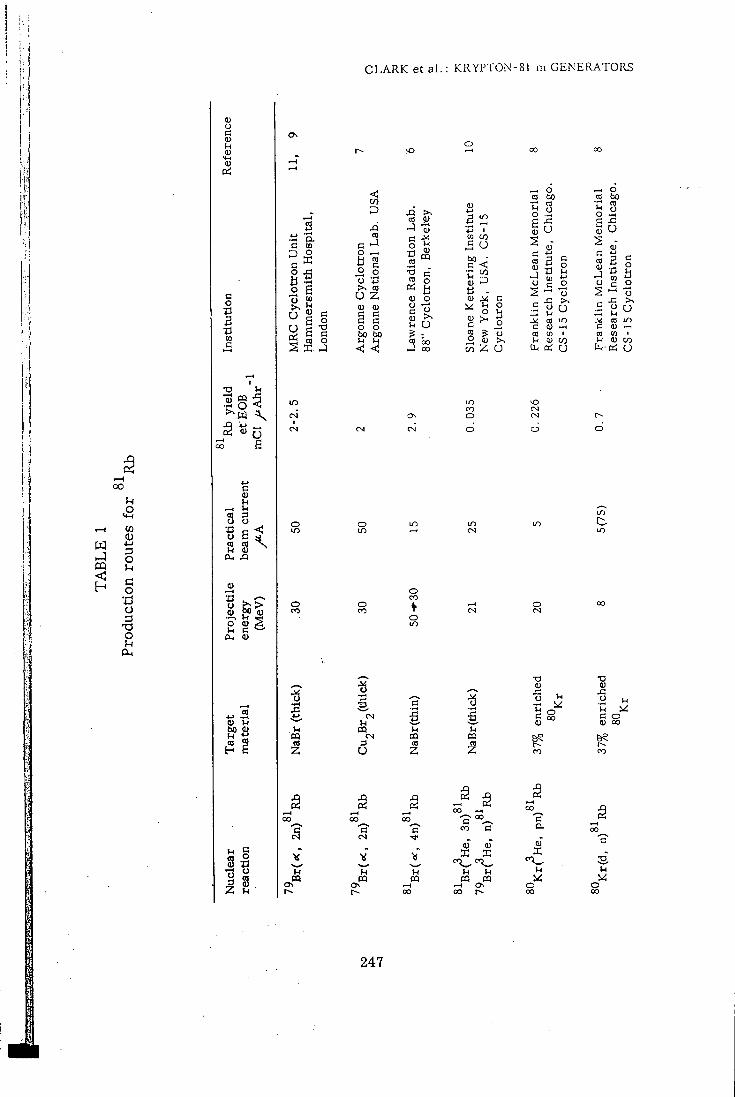
C L A R K e t a l . : KRYPTON-81 m G E N E R A T O R S
W
OQ
to <u 4-1 3 O 1 c o •C! o
3 s
o
u
C3
o •a a tn c
o ^
3 3
2 S a. XI
(U
tan lU
(U n
s i 1 ^
• a g -
o 5 c 6 o 6 C
^ CO o
2 I . J
O 0^
o
o CO
•3
u m OS
.6 CO
§ s .2 o
CO
O Z (U <u S c o o bo bo u u
< <
o
o
m 3 u
J3 CO J C o H CO •3 CO ai <u o c Q)
Si (D
V-l
<S a o b o
TJ >.
O ? : 5 g
o CO
l - l 03 CO
Z
a) *-> 3 12
c o b o o >
U
•a I . CQ
»—4 oo
c 00
^—^ c
—H 0^ OO .-1
- c CO 13
•>*<
"IT IH l - l CQ CQ CQ CQ
o rH Os 00 00 r»
o o CO bo .2 bo 'u CO
o u ca
o o
em
Ch
em
Ch
<u c *-» c
Lea
i :i
stit
u tr
on
Lea
3 •o 03 tr
on
(J :ist
itu
_o o c o — 7J Si J: >>
c rc
U rc
O CO lU
in ea
l O
CO CO 1 CO (0 1 l-l H) 0 0 u 0) CO
U a, cd O
o
O
CO
c &
QT I
00
l - l
d
in
a o aj 00
CO
3
o 00
247
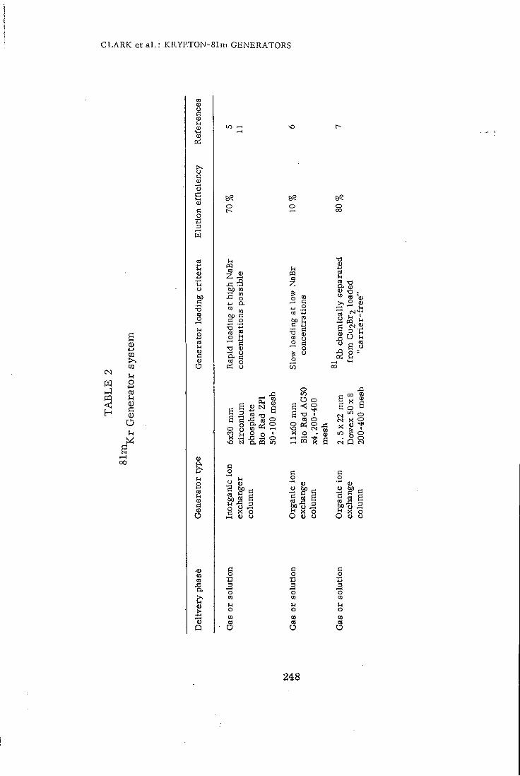
C L A R K et a l . : KRYPTON-81m G E N E R A T O R S
CN m u 2 S <u a o O u
00
CO (U o d 0) l-l (U (U
Pi
lU
o 3
l-l
"H o
•a § l-l S 2
O
l-l S 2 qi
(5
CO
•a
o
CQ
bo CO •p CO o D.
™ CO
^1 s 2
c X! 1)
5 8
x; CO
2 -o
S o ^ •r! :=! C>
CO O
N Cl. CQ in
o
CO
Ep5 i B «
c o 3
O [0
6? o
u CQ CO
Z
I s ^ .2
6 S o
o o I
o o CM
o 00
•a B 2 §-•§ <u -a -» M "a,
rg«-i l-l ' CQ l-l
•• 3
S
XI -
S 2
J3 CO
^ i
C^) C S
X in
<2 S X d o ^
o
O
o Q
c o
bOo 2 O (U o
c o n 3
l-l o CO
c o •£3 3
u o 01 CO
o
248
C L A R K e t a l . : KRYPT0N-81m G E N E R A T O R S
The f i r s t type developed in our laboratory^ incorporates a column of the inorganic ion exchange materfal zirconium phosphate while the second type of generator developed in the Donner laboratory^ and subsequently improved in the Veterans Administration Hospital, Hines Illinois^ uses a cation exchanger of the organic type, a sulphonated polystyrene-divinyl benzene copolymer eg. D0wex5Ox8.
81
CHEMICAL SEPARATION OF Rb AND GENERATOR LOADING
Zirconium phosphate is an insoluble crystalline material with a high
affinity for rubidium even in the presence of high concentrations of sodium 81
ions. This property enables one to load Rb onto the zirconium phosphate
generator rapidly at high efficiency using the irradiated 2. 5 gram sodium
bromide target dissolved in 20 ml water. A wash with 100 ml water to re
move the unwanted sodium bromide is a l l that remains to complete the ge
nerator preparation. The whole procedure is accomplished in approxima
tely 10 min and readily carried out by remote control.
The organic cation exchanger used in the second type of generator
w i l l only retain rubidium when i t is applied to the column in solutions of
low ionic strength. Thus, i f a sodium bromide target is used, the target
solution must be diluted to give a concentration of less than 0. 5% and the
resulting volume of 200-500 ml must be loaded slowly i f large losses of
Rb are to be avoided . Alternatively the ^^Rb must be chemically se
parated f r o m the target material and loaded on to the column essentially
, 7
carr ier free . Both of these procedures are extremely time consuming
and the resulting generators appear to offer no advantages over the z i r
conium phosphate type, both types having elution efficiencies of 70-80%
and similar rubidium breakthrough values.
249
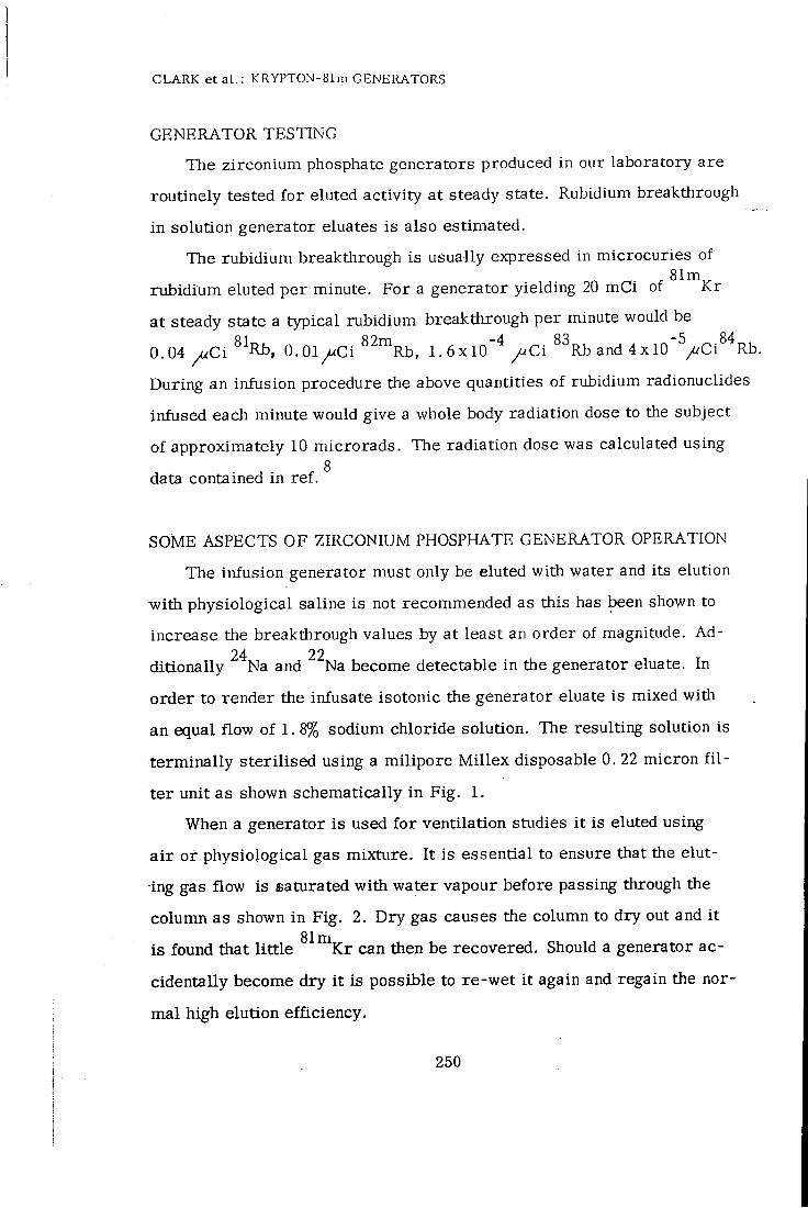
C L A R K et a L : K R Y P T 0 N - 8 1 m G E N E R A T O R S
GENERATOR TESTING
The zirconium phosphate generators produced in our laboratory are
routinely tested for eluted activity at steady state. Rubidium breakthrough
in solution generator eluates is also estimated.
The rubidium breakthrough is usually expressed in microcuries of
rubidium eluted per minute. For a generator yielding 20 mCi of ^^"^Kr
at steady state a typical rubidium breakthrough per minute would be
0.04 ^ C i 0.01/<Ci ^^™Rb, 1 .6x10"^/<Ci ^^Rb and 4xlO'^/^Ci^^Rb.
During an infusion procedure the above quantities of rubidium radionuclides
infused each minute would give a whole body radiation dose to the subject
of approximately 10 microrads. The radiation dose was calculated using
data contained in ref. ^
SOME ASPECTS OF ZIRCONIUM PHOSPHATE GENERATOR OPERATION
The infusion generator must only be eluted with water and its elution
with physiological saline is not recommended as this has been shown to
increase the breakthrough values by at least an order of magnitude. Ad-24 22
ditionally Na and Na become detectable in the generator eluate. In
order to render the infusate isotonic the generator eluate is mixed with
an equal flow of 1. 8% sodium chloride solution. The resulting solution is
terminally sterilised using a milipore Mil lex disposable 0. 22 micron f i l
ter unit as shown schematically in Fig. 1.
When a generator is used for ventilation studies i t is eluted using
a i r or physiological gas mixture. I t is essential to ensure that the elut-
ing gas flow is saturated with water vapour before passing through the
column as shown in Fig. 2. Dry gas causes the column to dry out and i t
is found that l i t t le ^'^"^Kr can then be recovered. Should a generator ac
cidentally become dry i t is possible to re-wet i t again and regain the nor
mal high elution efficiency.
250
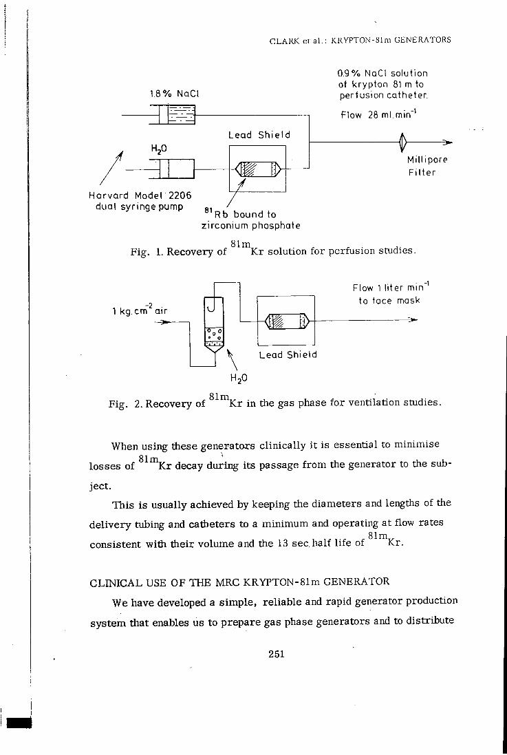
C L A R K e t a b : KRYPT0N-81m G E N E R A T O R S
1.8% NaCl
Lead Sh ie ld
Harvard Model 2206 dual syringe pump
0.9% NaCI solution of krypton 81 m to perfusion catheter.
Flow 28 ml.min -1
Mini pore Fi l ter
Rb bound to zirconium phosphate
Fig. 1. Recovery of ^''""^Kr solution for perfusion studies.
1 kg.cm air U
Flow 1 liter min to face mask
•1
Lead Shield
H2O
Fig. 2. Recovery of ^ ^ " \ r in the gas phase for ventilation studies.
When using these generators clinically i t is essential to minimise
losses of ^^"^Kr decay during its passage f rom the generator to the sub
ject.
This is usually achieved by keeping the diameters and lengths of the
delivery tubing and catheters to a minimum and operating at flow rates
consistent with their volume and the 13 sec half l i fe of ^^"^Kr.
CLINICAL USE OF THE MRC KRYPTON-81m GENERATOR
We have developed a simple, reliable and rapid generator production
system that enables us to prepare gas phase generators and to distribute
251
CLARK e t a l . : KRYPT0N-81m GENERATORS
t±iem to many centres in the United Kingdom at distances of up to 600 kilometres by road, rai l and air within a few hours.
Solution generators have only been prepared for on site use up to the
present time as the present design requires a high standard of technical
expertise to ensure its safe assembly and final testing before use in human
infusion investigations. An integrated generator system is under construc
tion which has been designed to deliver both sterile solutions for infusion
and ^"^"^Kr in the gas phase for ventilation. This development should make
lung ventilation and perfusion investigations with ^^"^Kr a practical pro
position as well as making a safe infusion available for off site use.
R E F E R E N C E S
1. F . Fazio, T. Jones, British Medical Journal, 265 (1975) 673.
2. E . Kaplan, L . W. Mayron, W. E . Barnes, L . G. Colombetti, A. M. Friedman, J. E . Gindler, J. Nucl. Med. , 15 (1974) 874.
3. J . H. Turner, A. P. Selwyn, T. Jones, T. R. Evans, M. J. Raphael, J, P. Lavender. Cardiovascular Research (1976) (in press).
4. J. H. Fremlin, Dept. of Physics, University of Birmingham. U. K. (Personal communication).
5. J . C . Clark, T. Jones, A. Mackintosh. ^^"^Kr an Ultra Short Lived Inert Gas Trac'er for Lung Ventilation and Perfusion studies with the Scintilation Camera. Radioaktive Isotope in Klinik und Furschung, 444 (Urban and Schwarzenberg- MUnchen - Berlin, Wien, 1970)
6. Y. Yano, J . McRae, H. O. Anger, J. Nucl. Med. , 11 (1970) 674.
7. L . G. Colombetti, L . W. Mayron, E . Kaplan, A. M. Friedman, J. E . Gindler, J . Nucl. Med., 15 (1975) 868.
8. B. Rich, N. Lembares, P. V. Harper, K. A. Lathrop, F. Atkins. Radiopharmaceuticals, 174 (Society of Nuclear Medicine Inc. New York, 1975)
252
CLARK e t a l . : KRYPT0N-81m GENERATORS
9. S. L . Waters, D. J. Silvester, I. W. Goodier, Phys. Rev. , C. 2. (1970) 2441.
10, I. A. Watson. Radiochem. Radioanal. Lett. , 4 (1970) 7.
11. J . C . Clark, P. D. Buckingham, Capt. 8, Radionuclides of the Rare Gases in "Short Lived Radioactive gases for Clinical Use". (Butter-worths, London, 1975).
253
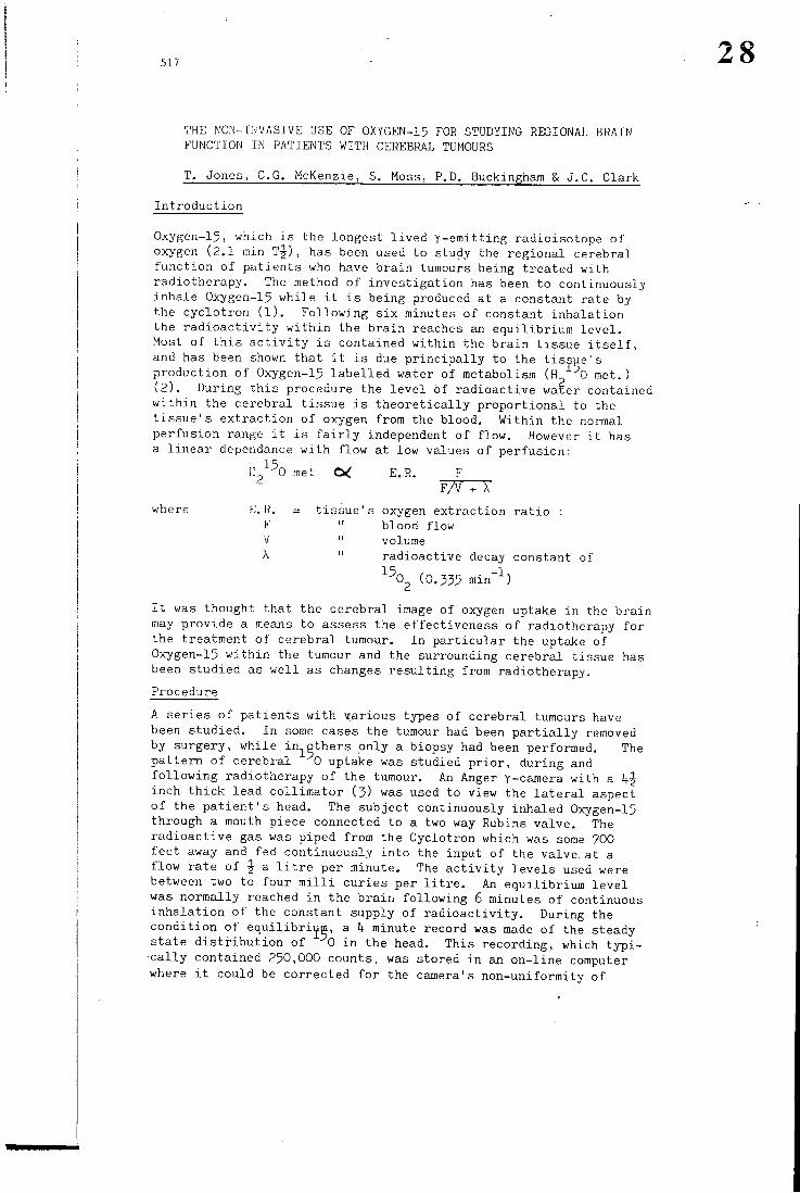
517
THE NON-INVASIVE USE OF OXYGEN-I5 FOR STUDYING REGIONAL BRAIN FUNCTION IN PATIENTS WITH CEREBRAL TUMOURS
T. Jones, C.G. McKenzie, S. Moss, P.P. Buckingham & J.C. Clark
In t roduc t ion
Oxygen-15, which i s the longest l i v e d y-era i t t ing radioisotope of oxygen (2 .1 min T J ) , has been used to study the regional cerebral f u n c t i o n of pa t ien ts who have bra in tumours being t reated wi th radiotherapy. The method of i nves t iga t ion has been to continuously inhale Oxygen-15 whi le i t i s being produced at a constant rate by the cyc lo t ron ( l ) . Fol lowing s ix minutes of constant i nha l a t ion the r a d i o a c t i v i t y w i t h i n the b ra in reaches an equ i l ib r ium l e v e l . Most of t h i s a c t i v i t y i s contained w i t h i n the b ra in t issue i t s e l f , and has been shown that i t i s due p r i n c i p a l l y to the t i s sue ' s production of Oxygen-15 l a b e l l e d water of metabolism (H 0 met. ) ( 2 ) . During t h i s procedure the l e v e l of rad ioac t ive water contained w i t h i n the cerebral t issue i s t h e o r e t i c a l l y p ropor t iona l to the t i s s u e ' s ex t r ac t i on of oxygen from the blood. Within the normal pe r fus ion range i t i s f a i r l y independent of f l ow . However i t has a l i n e a r dependance w i t h f low at low values of pe r fus ion :
HJ'-^O met E.R. F F/V + X
\ where E.R. = t i s sue ' s oxygen ex t rac t ion r a t i o : F " blood flow V " volume
\ " radioact ive decay constant of
•"" O (0 .535 rain"-'-)
I t was thought that the cerebral image of oxygen uptake i n the bra in may provide a means to assess the ef fec t iveness of radiotherapy f o r the treatment of cerebral tumour. I n p a r t i c u l a r the uptake of Oxygen-15 w i t h i n the tumour and the surrounding cerebral tissue has been s tudied as w e l l as changes r e s u l t i n g from radiotherapy.
Procedure
A ser ies o f pa t ien t s w i t h var ious types o f cerebral tumours have been s tudied. I n some cases the tumour had been p a r t i a l l y removed by surgery, while in-j^pthers only a biopsy had been performed. The pa t t e rn of cerebral 0 uptake was studied p r i o r , during and f o l l o w i n g radiotherapy of the tumour. An Anger y-camera wi th a IfJ inch t h i c k lead c o l l i m a t o r ( 3 ) was used to view the l a t e r a l aspect of the p a t i e n t ' s head. The subject continuously inhaled Oxygen-15 through a mouth piece connected to a two way Rubins valve. The rad ioac t ive gas was piped from the Cyclotron which was some 700 fee t away and fed continuously i n to the input of the valve.a t a f low ra te of a l i t r e per minute. The a c t i v i t y l eve l s used were between two to four m i l l i cur ies per l i t r e . An equ i l ib r ium l e v e l was normally reached i n the b ra in f o l l o w i n g 6 minutes of continuous i n h a l a t i o n of the constant supply of r a d i o a c t i v i t y . During the cond i t ion of equi l ib r i i^m, a h minute record was made of the steady s ta te d i s t r i b u t i o n of 0 i n the head. This recording, which t y p i c a l l y contained 250,000 counts, was stored i n an on- l ine computer where i t could be corrected f o r the camera's non-uniformi ty of
28
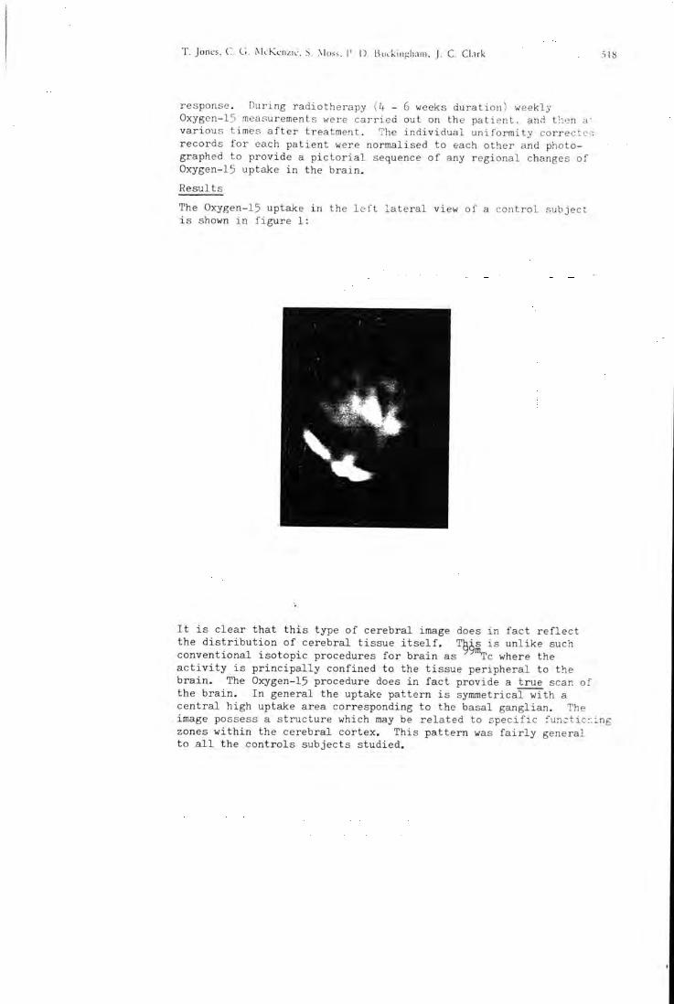
T. Jones, C. G. McKc-nzi<.-. S. Moss. P. D BiKkinsiham, C. Cl.uk 518
response. During radiotherapy ik - 6 weeks durat ion) weekly Oxygen-15 measurements were ca r r ied out on the pa t ien t , and then a', various times a f t e r treatment. The i n d i v i d u a l u n i f o r m i t y correctc-.i records f o r each pat ient were normalised to each other and photographed to provide a p i c t o r i a l sequence o f any regional changes of Oxygen-15 uptake i n the b r a in .
Results
The Oxygen-15 uptake i n the l e f t l a t e r a l view of a con t ro l subject i s shown i n f i g u r e 1:
I t i s clear that t h i s type of cerebral image does i n f ac t r e f l e c t the d i s t r i b u t i o n of cerebral t issue i t s e l f . This i s un l ike such conventional i so top ic procedures f o r b ra in as "Vc where the a c t i v i t y i s p r i n c i p a l l y confined to the t issue per iphera l to the b r a i n . The Oxygen-15 procedure does i n f a c t provide a t rue scan of the b r a in . I n general the uptake pa t te rn i s symmetrical wi th a cen t r a l high uptake area corresponding to the basal ganglian. The image possess a s t ruc ture which may be r e l a t ed to s p e c i f i c func t ic r . ing zones w i t h i n the cerebral cortex. This pa t t e rn was f a i r l y general to a l l the cont ro ls subjects s tudied.
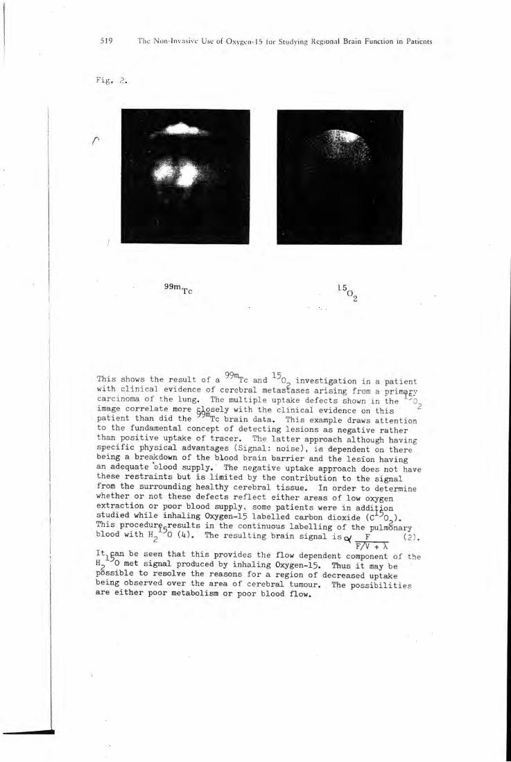
519 The Non-lnvasivc Use of Oxygcn-15 for Studying Regional Brain Function in Patients
F ig . 2.
99m Tc 15
This shows the r e su l t of a c and -"- 0 i n v e s t i g a t i o n i n a pa t ient w i t h c l i n i c a l evidence of cerebral metastases a r i s i n g from a prira^gy carcinoma o f the lung. The m u l t i p l e uptake defects shown i n the image co r r e l a t e more c lose ly w i t h the c l i n i c a l evidence on t h i s pa t ien t than did the ""TC b r a i n data. This exajnple draws a t t e n t i o n to the fundamental concept of de tec t ing les ions as negative ra ther than p o s i t i v e uptake of t racer . The l a t t e r approach although having s p e c i f i c phys ica l advantages (S igna l : noise) , i-s dependent on there being a breakdown o f the blood b ra in b a r r i e r and the l e s ion having an adequate "blood supply. The negative uptake approach does not have these r e s t r a i n t s but i s l i m i t e d by the c o n t r i b u t i o n to the s igna l from the surrounding healthy cerebral t i s sue . I n order to determine whether or not these defects r e f l e c t e i t he r areas o f low oxygen e x t r a c t i o n or poor blood supply, some pa t ien t s were i n add i t i on studied whi le i n h a l i n g Oxygen-15 l a b e l l e d carbon dioxide (C 0 ) . This procedure-resul ts i n the continuous l a b e l l i n g of the pulmonary blood w i t h H 0 ( 4 ) . The r e s u l t i n g b r a i n s ignal i s Q/ F ( 2 ) ,
F/V + X I t^can be seen that t h i s provides the f low dependent component of the
0 met s igna l produced by i n h a l i n g Oxygen-15. Thus i t may be possible to resolve the reasons f o r a region o f decreased uptake being observed over the area o f cerebral tumour. The p o s s i b i l i t i e s are e i t h e r poor metabolism or poor blood f l ow .
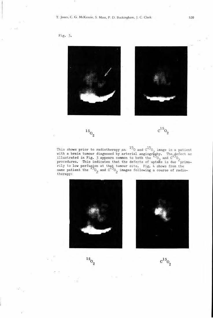
T. Jones, C. G. McKenzic, S. Moss, P. D. Buckingham, J. C. Clark 520
F i g . 3 .
15 o„
image i n a pa t ient This shows p r i o r to radiotherapy an. -"" 0 and C-'- O, w i t h a b r a in tumour diagnosed by a r t e r i a l angiography. The-j^defect as i l l u s t r a t e d i n F i g , 3 appears common to both the 0 and C 0^ procedures. This indica tes that the defects of uptaRe - - -r i l y to low per fus ion at t h j t tumour s i t e . F i g . same pa t ien t the 0 and C 0 images f o l l o w i n g a course of r ad io therapy:
e i s due priraa-k shows from the
15,
521 The Non-Invasivc Use of Oxygen-15 for Studying Regional Brain Function in Patients
15 ^he image produced by C 0 ^ i nha l a t i on i s f a i r l y normal whereas the
0 ^ uptake i n the l es ion although somewhat improved s t i l l shows some decrease i n uptake. This indica tes that there i s s t i l l a metabolic defect at the s i t e . This pa t ient showed a clear improvement c l i n i c a l l y as a r e s u l t of the radiotherapy. Discussion
Prel iminary r e su l t s of using Oxygen-15 to produce a pos i t i ve image of the ac t ive cerebral t issue have shown tha t tumours tend to be exhib i ted as areas w i t h low uptake of a c t i v i t y . I n some cases such defects have been re l a t ed to poor per fus ion of that region which i s a f f e c t e d by the l e s i o n . This zone of low uptake has o f t en appeared l a r g e r than would have been predic ted from the angiographic study, and t h i s i s due presumably to l o c a l hydrosta t ic pressure caused by edema formation per iphera l to the tumour. Defects located at the s i t e of a tumour have been shown to improve wi th radiotherapy. In c e r t a i n cases a regrowth o f the tumour has had corre la ted wi th i t a r e tu rn of the defect i n the Oxygen-15 bra in image. This work i s s t i l l at the p re l iminary stage, however i t does appear that we have a method to monitor l o c a l f u n c t i o n a l changes i n the b r a in . This could provide an ob jec t ive phys io log ica l feed-back to the therapis t who may i n tu rn be able to adjus t the therapeutic regime.
REFERENCES
1) CLARK. J .C., BUCKINGHAM, P.D. (1975); Short Lived Radioactive Gases f o r C l i n i c a l Use: pp I36-I7O. Butterworths (London).
2) JONES, T . , CHESLER, D.A. , TER-POGOSSIAN, M.M. (1976): The continuous i n h a l a t i o n of oxygen-15 f o r assessing regional oxygen ex t r ac t i on i n the Bra in of Man. Accepted f o r p u b l i c a t i o n : B r i t i s h J. Radiology.
3 ) WESTEHMANN, B.R. , GLASS, H . I . ( I 9 6 8 ) : Physical Spec i f i ca t ions of a Gamma Camera. J . Nucl Med. 9, 1 .24.
k) WEST, J.B. , and DOLLERY C.T. ( I 9 6 2 ) : Uptake of Oxygen-15 l abe l l ed Co^ compared w i t h carbon-11 l abe l l ed Co^ i n the lung. Journal o f appl ied physiology 17: 9.
Address
Medical Research Counci l , Cyclotron Unit and Departments of Radiotherapy & Medical Physics, Hammersmith Hosp i ta l , Ducane Road, London W12.
T. Jones, C. G. McKenzie, S. Moss, P. D. Buckingham, J. C. Clark 522
Abstract
The continuous i n h a l a t i o n of Oxygen-15 which has a h a l f l i f e of 2.1 minute r e su l t s i n the r a d i o a c t i v i t y w i t h i n the t issue reaching a state of "dynamic e q u i l i b r i u m " . The d i s t r i b u t i o n of t racer i n the b ra in dur ing such a steady state procedure r e f l e c t s d i r e c t l y the f u n c t i o n i n g cerebral t i ssue un l ike the conventional bra in scan. The i n h a l a t i o n o f molecular Oxygen-15 produces a cerebral d i s t r i but ion o f r a d i o a c t i v i t y which i s re la ted to oxygen u t i l i s a t i o n and blood f l o w . This can be compared to the d i s t r i b u t i o n obtained whi le i n h a l i n g Oxygen-15 l abe l l ed carbon dioxide which has a sole dependance on blood f l o w . These techniques have been applied to pa t ien t s w i t h cerebral tumours who are undergoing treatment wi th r a d i a t i o n . Inves t iga t ions have been ca r r i ed out w i th the object of ob ta in ing in fo rma t ion on blood f low and oxygen u t i l i s a t i o n rates to the l e s ions r e l a t i v e to the normal t i ssue . Se r i a l measurements have helped to understand how these re la t ionships change dur ing and f o l l o w i n g treatment.
Extrait
L ' i n n a i a t i o n c o n t i n u e d ' o x y g f e n e - 1 5 , q u i a une d e m i - v i e de 2 , 1 m i n u t e s , c o n d u i t h une r a d i o a c t i v i t e t i s s u l a i r e qui a t t a i n t un e t a t " d ' e q u i l i b r e d y n a m i q u e " . A l a d i f f e r e n c e du s can c e r e b r a l c o n v e n t i o n n e l l a d i s t r i b u t i o n du t r a c e u r dans l e c e r v e a u d u r a n t un t e l s t e a d y s t a t e r e f l a t e d i r e c t e -ment l e t i s s u c e r e b r a l f o n c t i o n n e l . L ' i n - h a l a t i o n d ' o x y g ^ n e -15 m o l e c u l a i r e p r o d u i t une d i s t r i b u t i o n c e r e b r a l e de l a r a d i o a c t i v i t e q u i e s t r e l i e e a 1 ' u t i l i s a t i o n . d e I ' o x y g f e n e e t au d e b i t s a n g u i n . Ces t e c h n i q u e s o n t e t e employees chez des m a l a d e s p o r t e u r s de t u m e u r s c e r e b r a l e s soumis a un t r a i t e m e n t p a r i r r a d i a t i o n . Des e t u d e s o n t e t e c o n d u i t e s v i s a n t a o b t e n i r des i n f o r m a t i o n s s u r l e d e b i t s a n g u i n e t 1 ' u t i l i s a t i o n d ' o x y g ^ n e des l e s i o n s p a r r a p p o r t au t i s s u n o r m a l . Des mesures en s e r i e s o n t p e r m i s de comprendre comment ces r e l a t i o n s se m o d i f i e n t d u r a n t e t anrhs l e t r a i t e m e n t .
Auszug
1 5 Die K o n t i n u i e r l i c h e I n h a l a t i o n von 0 m i t e i n e r H a l b w e r t s -z s i t von 2 , 1 min f u h r t im Gewebe zu G i n e r A K t i v i t a t s a n -r e i c h e r u n g m i t dy.namischem G l e i c h g e w i c h t . Die V e r t e i l u n g des T r a c e r s im G e h i r n u n t e r s o l c h e n Study S t a t e V e r h a l t n i s -sen e n t s p r i c h t . anders a l s b e i de r K o n v e n t i o n e 1 l e n G e h i r n -s z i n t i g r a p h i e , d i r s K t der f u n K t i o n i e r e n d e n G e h i r n s u b s t a n z . Die I n h a l a t i o n m o l e k u l a r e n ' '^0 f u h r t zu e i n e r R a d i o a K t i v i -t a t s v e r t e i l u n g im G e h i r n , d i e zum S a u e r s t o f f v e r b r a u c h und z u r D u r c h b l u t u n g des G e h i r n s i n B e z i e h u n g s t e h t . S o l c h s E r g e b n i s s e Konnen zu V e r t e i 1 u n g s s t u d i e n i n Bez i ehung ge-s e t z t w e r d e n , d i e d u r c h I n h a l a t i o n von ' '^0 m a r k i e r t e n K o h l e n d i o x y d gewonnen werden und d i e a u s s c h 1 i e B 1 i c h von d e r D u r c h b l u t u n g abhangen . D l e b e s c h r i e b e n e n Techn iKen wurden b e i P a t i e n t e n angewendet , d i e wegen e ines H i r n t u m o r s e i n e r S t r a h 1 e n t h e r a p i e u n t e r z o g e n w u r d e n . Z i e l der U n t s p -
523 The Non-Invasive Use of Oxygen-15 for Studying Regional Brain Function in Patients
suchungen war I n f o r m a t i o n e n uber D u r c h b l u t u n g und Sauer-s t o f f u t i 1 i s a t i o n i n den L a s i o n e n im V e r g l e i c h zum Normal gewebe zu e r h a l t e n . Ser ienmessungen wurden zum besse ren V e r s t a n d n i s von D u r c h b l u t u n g und S a u e r s t o f f v e r b r a u c h wahrend und nach d e r S t r a h 1enbeh and 1ung d u r c h g s f u h r t .
2 9 HI. Clinical Applications of Cyclotron Produced Products
Prog. luicl. Med., vol. 4, pp. 140-14,5 (Karger, Basel 1978)
The Medical Research Council Cyclotron at Hammersmith Hospital
A Status Report, March 1976
J O H N C . C L A R K
Medical Research Council, Cyclouon Unit, Hammersmith Hospital, London
Introductory Overview
The Facility
The Hammersmith cyclotron is of classical design and accelerates a-particles to 32 MeV, deuterons to 16 MeV and protons (via H:..*) to 8 MeV. Irradiations of targets may be carried out either internally or externally. Four external beam lines are installed at the present time, two of which are available for neutron therapy and radiobiology the remaining two being dedicated to radionuclide production. Both internal and external targets may be removed or replaced remotely without interrupting the cyclotron's running schedule. Additionally a remote-controlled actuator, installed on an external beam line, is able to present any one of eight preloaded targets to the charged particle beam thus facilitating rapid changes between on-line products during a busy clinical program 11,2]. One of the radionuclide production beam lines is scheduled to be withdrawn from service (October 1976)' to be replaced by three beam lines that will be capable of effectively simultaneous operation using a beam-sharing technique [3]. Ancillary facilities around the cyclotron include a shielded room where neutron radiotherapy and radiobiology takes place and a radiochemistry laboratory equipped with hot cells where radionuclides are processed to radiopharmaceutical standards. The cyclotron building also houses an animal investigation and a clinical application facility. Other clinical areas on the hospital site are supplied with most of the short half-lived gaseous products by pipeline.
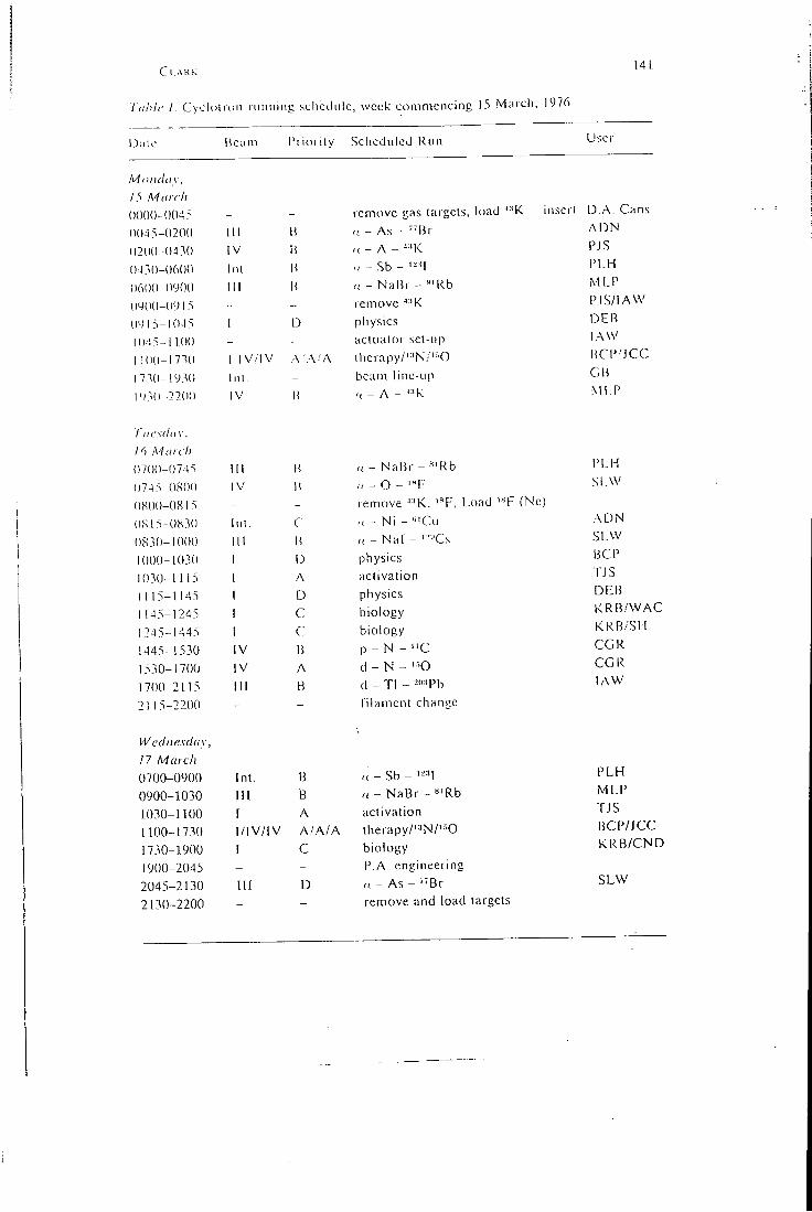
Cl.ARK
Tiihlc I. CycliHroii running schcthilc, week commencing 15 Maich, 1976
141.
Dale Beam I'ritirily Scheduled Run User
M on (lay, 15 M(licit
()()()()- ()04> _ - remove gas targets, load •'•'K inscrl D.A. Cans
(K)45-()20() 111 H <( - As - "'Br ADN
(12(1(1-0430 IV H „ - A - ^ 'K PJS
(143(1-0600 Inl. H - Sb - ' - ' I I 'LH
(16(1(1-0900 I l l li r< - NaBr - "'Rb MLP
0400 (1913 „ _ remove ^-'K PJS/IAW
(1415-1043 1 physics DEB
1043-1 1,00 _ actuator set-up lAVV
1 100-173(1 1 IV/IV \ A-A therapy/'^'N/'^O BCPMCC
1730-1930 Inl. - beam line-Lip GB
!')".(! 2200 IV H r! - A - '-'K Ml.P
I'licsdiiy.
16 A'liirch
0700-0745 III H n - NaBr - «'Rb PLH
0743-080(1 IV H '( - 0 - '«F SI.W
0X00-0813 _ _ remove '•'K. '^F, Load "<F (Nc.)
(1815-0830 Inl. C „ _ Ni - "'Cu ADN
(1830-1000 I l l H r, - Nal - '--'Cs SLVV
1000-1030 1 0 physics BCP
1030-1115 I A activation -ns 1 1 15-1145 1 D physics DEB
1 145-1245 1 C biology KRB./\VAC
1245-1445 1 C biology KRB.'SH
1445-1530 IV 13 p - N - "C CGR
1530-1700 IV A d - N - ' 'O CGR
1700-2115 I I I B d _ Tl - --" 'Pb lAW
2115-2200 - - filament change
W cdnesday, 17 March
PLH 0700-0900 Int. B a - Sb - ' -'1 PLH
0900-1030 i l l B a - NaBr - s'Rb iMLP
1030-1100 [ A activation rjs 1100-1730 1/IV/IV A./A/A therapy/'-'N/'-'^O BCP/JCC
1730-1900 I c biology KRB/CND
1900-2045 - P A. engineering 2045-2130 IK D ,( - As - ' 'Br SLW
2130-2200 remove and load targets
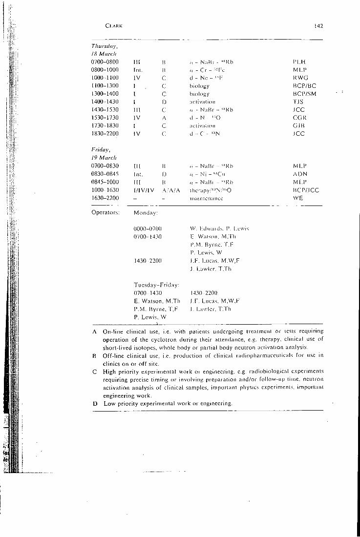
, I , - ' ' pi •
I '• II-:
CLARK 142
Thursday, 18 Marc/1 0700-0800 I I I B ,1 - NaKr - "'Kb I ' l .H 0800-1000 Inl. li r, - Cr - -Fc MLP 1000-1100 IV c d - Nc - ' - I - RWG 1100-1300 I c biology BCP/BC 1300-1400 I c biology BCP/SM 1400-1430 [ D .iclival ion TJS 1430-1530 111 c n - NaBr - '^'Kb ICC 1530-1730 IV A d - N '•••0 CGR 1730-1830 1 C aclivalion GJB 1830-2200 IV C d - C - ' 'N JCC
Friday, 19 March 0700-0830 I I I B „ - Nalir - - 'Rb MLP 0830-0845 Int. D n - Ni - '''Cu ADN 0845-1000 I l l B /( - NaBr - -"Rb MI.P 1000-1630 I / IV/ IV A.'A! A lhcrapy.''-'N./'"'0 BCP/ICC 1630-2200 - - maintcnanco WE
Operators: Monday:
1:1:1
0000-0700 0700-1430
1430-2200
Tuesday-Friday: 0700-1430 E. Watson, M,Th P.M. Byrne, T,F P. Lewis, W
VV. Edwards. P. Lewis E. Watson. M.Th P.M. Byrne. T.F P. Lewis. W LF. Lucas, M,W,F L Lawler. T.Th
1430-2200 LF. Lucas, M,W,F L Lawlcr, T.Th
A On-line clinical use, i.e. with patients undergoing treatment or tests requiring operation of the cyclotron during their attendance, e.g. therapy, clinical use of short-lived isotopes, whole body or partial body neutron activation analysis.
B Off-line clinical use, i.e. production of clinical radiopharmaceuticals for use in clinics on or off site.
C High priority experimental work or engineering, e.g. radiobiological experiments requiring precise timing or involving preparation and/or follow-up time, neutron activation analysis of clinical samples, important physics e.xperimcnis. imporiani engineering work.
D Low priority experimental work or engineering.
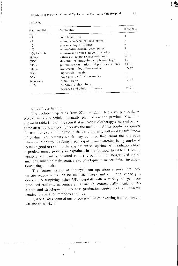
The Medical Rcscarc h Council Cyclotron at Hammersmith Hospital
lahlc II.
Radionuclide Application Kc eroncc
hone blood flow 4
radiopharmaceutical development .S
"C pliai niacological studies 6
"C radiopharmaceutical development 7
i: .0,-i-C'- '0.. noninvasive brain metabolism studies 8
HJ'-'O oxtravascular lung water estimation <•). I I
C ' O detection of intrapulmonary hemorrhage 1 1
Kr"i pidmonary ventilation and perfusion studies 12- -14
Kr"' myocardial blood flow studies L=i
i-'Cs myocardial imaging —
•T-c bone marrow function studies -
Neutrons radiotherapy 17 I S
icspiratory physiology research and clinical diagnosis p; -21
()j)crailII>^ Schedules The cyclotron operates from 07.00 to 22.00 h 5 days per week. A
typical weekly schedule, normally planned on the previous Friday. i< shown in table 1. it will be seen that neutron radiotherapy is carried otn on three afternoons a week. Generally the medium half-life products rcqtiircti for use that day arc prepared in the early morning followed by fulfillmem of on-line requirements which may continue throughout the day even when radiotherapy is taking place, rapid beam switching being employed to make good use of intcrtherapy patient set-up time. A l l irradiations have a predetermined priority as explained in the footnote to table I . Evening sessions are usually devoted to the production of longer-lived radionuclides, machine maintenance and development or preclinical investigations using animals.
The routine nature of the cyclotron operation ensures that mosi on-site requirements can be met each week and additional capacity is devoted to supplying other UK hospitals with a variety of cyclotron-produced radiopharmaceuticals that arc not commercially available. Research and development into new production routes and radiopharmaceutical preparation methods continue.
Table II lists some of our ongoing activities involving both on-site and
off-site co-workers.
Ci..\RK 144
Rejcrcnccs
Sil,vri,s-ri;K, D.J.: liiomedical cyclotion in a medical center: capabilities and problems; in Radiopharmaceuticals, p. 157 (Society of Nuclear Medicine, New York 1975).
VoNisERG, D.D.; BAKER, L . C ; BUCKINGHAM, P.D.; CLARK, J . C ; FINIJINCI, K . . SHART, }., and SILVESTER, D.J.; Target systems for radioisotope production on the Medical Research Council cyclotron; in Uses of cyclotrons in chemistry, metallurgy and biology, pp. 258-269 (Butlcrworths, London 1970). BURTON, G.: Cyclotron beam sharing for multiple target irradiations. I'roc. 5th Int. Cyclotron Conf. 1969, pp. 250-253 (Butterworths. London 1970). WooroN, R.; REEVE, J., and VEAL, N . : The clinical measurement of skeletal blood flow. J. Clin. Sci. molcc. Med. 50: 261-268 (1976).
GOULDINO, R . W . : Fluorinc-18 labelled L-/)-fluorophenylalaninc. I i i i . .1. appl. Radiat. Isotopes 26: 561-564 (1975). PALMER. A.J . : The preparation of "C-meihyl labelled 1.r-dimethyl-4,4'-di-pyridinium diiodidc. J. Lab. Cpcls. RaditiphainT (in press. I477i. GOULDING, R . W . : Liquid chromatography of sugars and related polyhydric alcohols on cation exchangers. J. Chromat. 103: 229-239 (1975). JONES, T . ; MCKENZIE, C .G. ; Moss. S.; BUCKINGHAM. P.D.. and Ci AKK. l.C.; The assessment of regional cerebral function in brain tumom palicnls by continuous inhalation of o.vygen-15. Br. .1. Radiol. 50: 158 (197/1, FAZIO, F.; JONES, T.; MACARTHUR. C . G . C ; STEINER, R.E.. and HUGHES. J.M.B.: Measurement of regional pulmonary oedema in man using radioactive water Hi'^'O. Br. J. Radiol. 49: 393-397 (1976).
JONES, T . ; JONES, H.A.; RHODES, C . G . ; BUCKINGHAM, P.D.. and HuciiiEs. J.M.B.: Distribution of extravascular fki id volumes in isolated perfused lungs measured with Hi'SQ. J. Clin. Invest. 57: 706-713 (1976).
11 EwAN, P .W. ; JONES, H.A.; RI-IODES, C.G. , and HUGHES. J .M B.: Dcieciion of intrapulmonary haemorrhage in man with carbon monoxide uptake: application in Goodpastures' syndrome. New Engl. J. Med. 295: 1391-1396 (1976). FAZIO, F . and JONES, T . ; Assessment of regional ventilation by continuous inhalation of radioactive krypton-81m. Br. med. J. ///.• 673-676 (1975). HARF, A.; PRATT, T . , and HUGHES, J .M.B.: Regional ventilation perfusion ratios at rest and on exercise using krypton 81m. Bull. Eur. Physiophath. Resp. I2!6: 1-195 (1976).
CLARK, J.C.; HORLOCK, P.L., and WATSON, LA.: Krypton-8lm generators. Radiochem. radioanal. Lett. 25: 245-254 (1976).
15 TURNER, J.H.; SELWYN, A.P.; JONES, T . ; EVANS, T.R.; RAPHAEL. M.J.. and LAVENDER, J.P.; Continuous imaging of regional myocardial blood fknv in dogs using krypton-81m. Cardiovasc. Res. 10: 398-404 (1976).
16 SELV^'YN, A.P. and SHILLINGEORD, J.P.: Continuous assessment of regional myocardial perfusion using krypton-81m in dogs and man; effects of nitroglycerine and stress in acute ischaemia. Circulation 54: suppl. (1976).
17 CATTERALL, M . ; SUTHERLAND, L , and BEWLEY, D.K.: First results of a
randomised clinical trial of fast neutrons compared with X-rays or gamma
10
12
13
14
1 he MctlKiil R e s L M i v l i (. ( u i n c i l ( \ c l o i i < . n , i i I lainmoi sinilh Hospii:,! l-)s
rays in liealnK'iil ol adxanccil lummiis of ihc licai.1 aiul neck. Hr. moil .1. //.• 6,S3-6.S6 (i97.si.
18 S.B.: .An hiNhM ic i l sui vc\ of i ailmhioliiuy and radiollieraphy s'.illi fast iiculrons. Curi. lop. Radial. Res. (J II: \ -.S (lV7dl.
19 Ei.SfiK. N.M.: Local perfusion and vciililalion in chronic broiichilis and emphysema. Thorax Ml: .s96 (1975).
20 G o D f R f i Y . S.; R'lNC iu. I ri . R,: S idC KS. L. and H,\i i.inih-SMi i i i . K.: Generalised pulmonary hypcnnlhiiuin aiul l allols leiiaioyv in a neonalc iiuesligaietl hy pulmonary physiolouy aiul railuilsolope mclhods. I horax .^(1: 4.S2 (PJ/S).
21 SYKILS, M.K.; .AKNUI. R.N.: CI.-\KK. I .C.. and Hl KiNt;. A.N.: Suidies of \ciilila-lion at tiilfcrenl m N p i i a l o i v flow rales iisiny kiAplon-.SIm and niiroucn-i.^. Bull. Eur. Physiopalli Rcsp. 12: 1-2(1'. (1 W7fM.
Dr. .I.e. Ci .•\KK. MRC: Cyclotron Unit. Hammersmith Hospital. London (Encia nd)
PHYSIOLOGICAL SOCIETY, JULY 1977 SIP
nasal mucosa wi th clock-like regularit}'. This area may be associated with the respirator}' centre in the medulla as changes in arterial PQQ^ cause changes in nasal resistance (Principato & Ozenberger, 1 9 7 0 ; Dallimore & Eccles, 1977), although there is some evidence that the hypothalamus regulates the sympathetic outflow to the nasal mucosa of the cat (Mal-comson, 1953).
The work was carried out at the V.P. Chest; Institute, University of Delhi, and I would like to thank Professor A. S. Paintal, Dr S. K . Jain and Mr A. K v i i n a r for their help and encouragement.
R E F E R E N C E S
D A I L I M O K E , N . S . & E C C L E S , R . (in press). Acta oto-lar. H E E T D E R K S , D . R . ( 1 9 2 7 ) . Am J . Med. Sci. 174, 2 3 1 - 2 4 4 .
M A i c O M S O N , K . G . ( 1 9 5 9 ) . J . Lar. Otol. 73, 7 3 - 9 8 .
P a i N C i P A T O , J . J . & O Z E N B E R G E R , J . M. ( 1 9 7 0 ) . Arclis Otolar. 91, 7 1 - 7 7 .
S T O K S T E D , P . ( 1 9 5 3 ) . Acta oto-lar. Suppl. 109, 1 5 9 - 1 7 5 .
C . 61 Effect of posture on distribution of pulmonary ventilation and perfusion studied with simKr and ssmKr
B Y C . AMJS, G . C I G F E T T A , J . C . C L A R K , J . M . B . H U G H E S , H A Z E L A.
J O N E S and T . A . P R A T T . Departments of Medicine and Diagnostic Radiology, Royal Postgraduate Medical School, and M.R.C. Cyclotron Unit, Hammersmith Hospital, London, W12 OHS
Using radioactive gases, West & Dollery (1960) and Kaneko, Milic-Emi l i , Dolovich, Dawson & Bates (1966) have suggested that the effect of posture on the distribution of ventilation and perfusion in the lung is mediated by gravity.
W i t h the introduction of the short half-life (13 sec) isotope s i^Kr {Ey 190 keV), distributions of ventilation and perfusion can be measured in the steady state during spontaneous breathing. Using a gamma camera, continuous inhalation of an air-^^"^Kr mixture, gives regional counts which reflect regional ventilation (Fazio & Jones, 1975). Similarly, continuous intravenous infusion of s i m j ^ produces regional counts reflecting local pulmonary perfusion. Division of images constructed from the accumulated counts produces a distribution of ventilation-perfusion ratios (VAjQ). W i t h this technique Harf , Pratt & Hughes (1976) found that tA/Q in seated subjects decreased f rom high values at the apex (mean 1-8) to lower values at the base (0-8-0-9).
We have extended these measurements to the supine, r ight and left lateral decubitus and prone suspended (on all fours) postures and corrected ventilation and perfusion images for local alveolar volume by rebreathing
82P PROCEEDINGS OF THE
a l o n g e r h a l f - l i f e (4-4 h r ) i s o t o p e , ^^^Kr {Ey 150 k e V ) . T h e VAjQ r a t i o
d e c r e a s e d f r o m s u p e r i o r t o i n f e r i o r i n t h e p r o n e s u s ] 3 e n d e d p o s t u r e a n d i n
t h e u p j ) e r m o s t l u n g i n t h e l a t e r a l d e c u b i t u s p o s i t i o n . T h i s g r a d i e n t w a s
r e v e r s e d i n t h e s u p e r i o r - i n f e r i o r a x i s i n t h e s u p i n e p o s t u r e a n d i n t h e
l o w e r m o s t l u n g i n t h e l a t e r a l d e c u b i t u s p o s t u r e . R e v e r s a l o f t h e VAfQ
g r a d i e n t m i g h t b e r e l a t e d t o s m a l l e r r e s t i n g l u n g v o l u m e s s e e n i n t h e
l a t t e r p o s t u r e s ( K a n e k o et al. 1966) a n d t h e i r e f f e c t o n b l o o d flow d i s t r i
b u t i o n ( H u g h e s , G l a z i e r , M a l o n e y & W e s t , 1968). V o l u m e c o r r e c t i o n s h o w e d t h a t t h e d e c r e a s i n g VAfQ g r a d i e n t i n l a r g e r
v o l u m e s i t u a t i o n s w a s d u e t o t h e g r a d i e n t o f i n c r e a s i n g b l o o d flow b e i n g
g r e a t e r t h a n t h a t f o r v e n t i l a t i o n . I n s m a l l e r v o l u m e s i t u a t i o n s b l o o d flow
t e n d e d t o b e u n i f o r m i n t h e v e r t i c a l d i r e c t i o n b u t v e n t i l a t i o n p e r u n i t
a l v e o l a r v o l u m e c o n t i n u e d t o i n c r e a s e , s o r e v e r s i n g t h e VAjQ g r a d i e n t .
I n h o r i z o n t a l p o s t u r e s VAjQ t e n d e d t o i n c r e a s e , a p p r o a c h i n g t h e
d i a p h r a g m a l o n g a c r a n i o - c a u d a l i s o g r a v i t y l i n e . T h i s w a s m o r e m a r k e d
i n l a r g e r r e s t i n g v o l u m e s i t u a t i o n s a n d a p p e a r e d t o b e r e l a t e d m o r e t o
d e c r e a s e i n b l o o d flow p e r u n i t a l v e o l a r v o l u m e t h a n i n c r e a s e i n v e n t i l a t i o n .
W e c o n c l u d e t h a t c h a n g e s i n p o s t u r e i n f l u e n c e l o c a l l u n g e x p a n s i o n
w h i c h i n t u r n m o d i f i e s t h e g r a v i t y d e t e r m i n e d d i s t r i b u t i o n o f b l o o d flow,
a n d c o n s e q u e n t l y t h e d i s t r i b u t i o n o f g a s e x c h a n g e w i t h i n t h e l u n g .
R E F E R E N C E S
F A Z I O , F . & J O N E S , T . ( 1 9 7 5 ) . Brit. Med. J . ii, 6 7 3 - 6 7 5 .
H A R F , A., P R A T T , T . & H U G H E S , J . M . B . ( 1 9 7 6 ) . Bull europ. Physio-palh. Re-sp. 12. 1 9 5 p.
H U G H E S , J . M . B . , G L A Z I E R , B . , M A L O N E Y , J . E . & W E S T , J . B . (1968) . Respir. Physiol. 4, 5 8 - 7 2 .
K A N E K O , K . , M I L I C - E M I L I , j . , D O L O V I C H , M . B . , D A W S O N , A. & B.A.TES, D . V . (1966) .
J . appl. Physiol. 2 1 , 7 6 7 - 7 7 7 . W E S T , J . B . & D O L L E R Y , C . T . ( 1 9 6 0 ) . J . appl. Physiol. 15, 4 0 5 - 4 1 0 .
C . 6 2
Some properties of the non-chemical drive to breathe at the breaking-point of breath-holding B Y a . J . G U Y a n d J . M . P A T R I C K . Department of Physiology mid Pharmacology, University Hospital and Medical School, Nottingham, NG7 2UH
T h e d r i v e t o b r e a t h e a t t h e b r e a k i n g - p o i n t o f b r e a t h - h o l d i n g c a n b e
d e s c r i b e d i n t e r m s o f t h e i n t e r a c t i o n b e t w e e n a c h e m i c a l c o m p o n e n t a n d a
t i m e - d e p e n d e n t n o n - c h e m i c a l c o m p o n e n t t h a t i s r e l a t e d i n v e r s e l y t o l u n g
v o l u m e ( P a t r i c k & R e e d , 1 9 6 9 ) . I n five y o u n g a d u l t s u b j e c t s w e h a v e
i n v e s t i g a t e d t h e d i s s i p a t i o n o f t h e n o n - c h e m i c a l d r i v e f o l l o w i n g t h e
b r e a k i n g - p o i n t , a n d i t s e f f e c t o n t h e s u b s e q u e n t b r e a t h i n g p a t t e r n .
A b s l r a c l H c p r o d u c t i o n I ' o r m ^ ,
Society of Nuclear Medicine 24(h Annual Meeting ^^^Widi the ( linical Uadioassay Society
(Hi I VIM A l l s I I'. .\( I III 1(1
-I K i : l O S I AV H 7 / / / / , V I tOKDI l i ,
< I I U I I. <i/i/i <!'!<• preferred •>nl'l' - I I Id^^itit (llinil
C oMipulri,' tiiita analysis ( iini|>HlL(l tomography i /(i-^unciiy rncl inr i i i ' . - , inciabolisni ( IMN! 1' u-nicTtilogy I I I iii.itiiliijiy
| H I l - . - l l M l l l l I!
III V I I I !• Nliidics Ntnidloj^v Oiiti'iogy ljvUuiIni;s
C ( ' i i lni(iii ; ir)^ ^ir.vjniiiss.u
K ;i(llopli;ir Iii^KTUlii.nl '.ciciicc
Alislt .K l'. imisl l.c :, ,nv;;i p , , M . tn,iil.((l| in I fliriiari 15. 1^77.
I M r O K l A M 1 his Mjai al l abstiacts a i c p C i l lor
Ihc pioL ' iaiii of Ihc Soi i cu o l Nm Uar Mci.liciiic Annual Mcc l i i i i ; HMI |V-primed i l i rcc l ly f r o m (he lypci l copy o i l this sliccl. T o insure pririCini; qual-i(y, (hr inslrucHons on (lie reverse side niusl 1)0 followed coni/.'Irfely for nil alislmets. Ahslracis (hat do no l coi i -l o r n i r i l h r i w i l l he rclvpcd hv l l ic pub lishcr ai a C(>st of %25 lo ilu- a i i lhor o i w i l l be icjciMcd.
n.KASi; ( K ARSTRAC: ( ARi:H'|. | .Y
I CR APrF.ARAN< i; BKFORK MAILING
: PULMONARY VENTILATION AND PERFUSION MEASUREMENTS USING ; .Kr-81ra; BASIC METHODOLOGY. Peter J . Kenny, Penny P. Wats on. .Jol'n C. C l a r k , Ronald D. Finn and Albert -J. G i l s o n . M-mnt S i n a i Medical Center, Miami Beach, F L .
Kr-81in from a R h - e i - > Kr-81ni pencrntor has liocii u.scd to measure changes in reg ional v e n t i l a t i o n ( V / V ) , regional perlusloi i ( Q / V ) and regional v e n t i l a t i o n perfusion r a t i o ( V / ' Q ) in 20 dogs in the contro l s t a t e and fol lowing lol-.i.'-occ lus ion and/or drug i n t e r v e n t i o n . Data from tbl.? vork were analyzed to provide a b a s i s for the ose of Kr-81m for s i m i l a r measureioents in puliaonary function s tud ies li; bunans. Ser ia i . quant i ta t i ve images were chtaluod durine the equ i l ibr ium and washout phases for both v e n t i l n t i o n and perfus ion s tudies us ing a s c i n t i l l a t i o n rjrera-compu'.-e r system. A method was developed for measuring Q / V an-i V / Q by constant intravenous I n f u s i o n of Kr-81ra in a dextrose so lu t ion . In t h i s c a s e , the e q u i l i b r i u m rcncc-ntie-t ion of Kr-81m in any region (lepciiHs on thc v,i.hi;>s of Q/V and V / V in that region. V/V was measiircd froi:; vi.ni. i 1 a t washout s t u d i e s . From t h i s , separate q u a n t i t a t i v e deter minations of (],'V and V/Q, as w e l l as V/V, for the i-'ho^^ lung and se l ec ted regions were made. [>cc.;o. 'j of t iu- shor:: h a l f - l i f e and short e q u i l i b r a t i o n t imt's for ^•r-81m d i s t r i bution in tV'.c lungs , i t was p o s s i b l e to nake re!:eated determinations of these q u a n t i t i e s at fntervaTs of a few minutes to assess the e f f e c t s of i n t e r v e n t i o n . l or example, wi th in minutes of lobar o c c l u s i o n , perfusion reduction averaging 50Z was shown to have occurred in the hypoxic region, as expected, presumably due to refJ ex v a s o c o n s t r i c t i o n . The techniques developed provide a b a s i s for the use of Kr-81m i n combined v e n t i l a t i o n and per fus ion s tudies for the measurement of V /V , Q/V ar.d V / ' j i n huraaiis.
T Y I ' I K i n . I \ \ M \ Oi- A i m i O R I'KP.SHNI INC TAI' I K:
Peter J . Kenny '
r .XAMIM r
TECllNl";TIUM-99m POLYPHOSPHATE BONE I M A G I N G IN L J : G G
P E R T H E S D l S E A S i : . J a m e s A . D a n i g e l i s , R o b e r t \ ^ F i s h e r , a n d Mac-r B . O z o n o f f . N e w i n g t o n C l i l l d r c n ' s H o s p i t a l , Newin<j ton , C o n n .
T h i s i n v e s t i r j a t i o n w a s u n d e r t a k e n t o c o m p a r e t h e d i a g n o s t i c u s e f u l n e s s o f r a d i o n u c l i d e b o n e i m a g i n g t e c h n i < i u e s t o s t a n d a r d r a d i o g r a p h i c
1
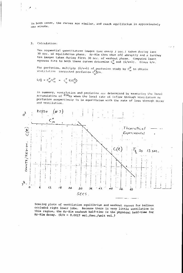
I n b o th c a s e s one minute.
, t h e c u r v e s a r e s i m i l a r , and r e a c h e q u i l i b r i u m i n a p p r o x i m a t e l y
C a l c u l a t i o n :
Ten s e q u e n t i a l q u a n t i t a t i v e images (one e v e r y 3 s e c . ) t a k e n d u r i n g l a s t 30 s e c . o f e q u i l i b r i u m p h a s e . Kr-81m then s h u t o f f a b r u p t l y and a f u r t h e r t e n images t a k e n d u r i n g f i r s t 30 s e c . o f washout p h a s e . Computed l e a s t s q u a r e s f i t s t o both t h e s e c u r v e s d e t e r m i n e C^ and (v/v+ A ). G i v e s v/v.
Fo r p e r f u s i o n , m u l t i p l y (O/v+A) of p e r f u s i o n s t u d y by C^ to o b t a i n vent i l a t; j on c o r r e c t e d p e r f u s i o n C^O/v.
v /0 ^/C^O o o^
I n sumjnary, v e n t i l a t i o n and perfu.sion a r e d e t e r m i n e d by me a s u r i n g the l o c a l a c c u m u l a t i o n o f ^ ^ ^ K r when t h e l o c a l r a t e o f i n f l o w thrbugh v e n t i l a t i o n o r p e r f u s i o n r e s p e c t i v e l y i s i n e q u i l i b r i u m w i t h the r a t e o f l o s s through decay and v e n t i l a t i o n .
PHYDo ( # ? )
c
o
-! I -
12 s j ? c .
r
I.. I
Semilog p l o t s of v e n t i l a t i o n equilibrium and washout curves for balloon occluded r i g h t lower lobe. Because there i s very l i t t l e v e n t i l a t i o n i n t h i s region, the Kr-81m washout half-time is the p h y s i c a l half-time for Kr-81m decay, (v/v = 0.0017 v o l . / s e c . / u n i t v o l . )
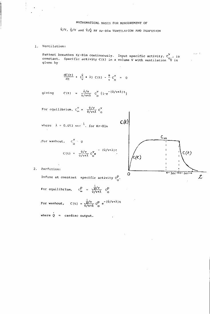
M A T H E M A T I C A L B A S I S F O R M E A S U R E M E N T O F
v / v , Q / v and V / Q BY Kr-81m V E N T I L A T I O N AND P E R F U S I O N
1. V e n t i l a t i o n :
P a t i e n t b r e a t h e s Kr-81m c o n t i n u o u s l y . I n p u t s p e c i f i c a c t i v i t y , c " , i s c o n s t a n t . S p e c i f i c a c t i v i t y C ( t ) i n a volume V w i t h v e n t i l a t i o n °V i s g i v e n by
d C ( t ) V , V - ~ - + ( - + A) C ( t ) - - c " = 0 a t V V o
g i v i n g C ( t ) .•^/^ pV , - ( v / v + A ) t .
F o r e q u i l i b r i u m , C^ = c' v/v+A o
where A = 0.053 s e c f o r Kr-81n
.For washout, C = 0 o
C ( t ) _v/v ^ v^ ( O / v + A ) t
v /v+A o
2. P e r f u s i o n :
I n f u s e a t c o n s t a n t s p e c i f i c a c t i v i t y C^.
F o r e q u i l i b r i u m , = °° v /v+A o
F o r washout, C ( t ) = ( v / v + A ) t v /v+A o
where Q = c a r d i a c o u t p u t .
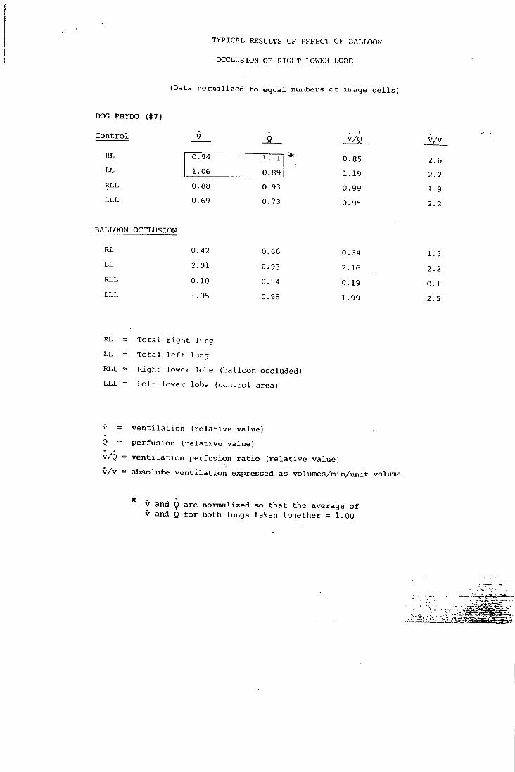
TYPICAL RESULTS OF EFFECT OF BALLOON
OCCLUSION OF RIGHT LOWER LOBE
(Data n o r m a l i z e d t o e q u a l numbers o f image c e l l s )
DOG PHYDO (#7)
C o n t r o l v / v
RL 0 94 1.11 * 0 .85 2 .6 L L 1 06 0.89 1 .19 2 2 RLL 0 88 0.93 0 .99 1 9 L L L 0 69 0.73 0 95 2 2
BALLOON OCCLUSION
RL 0. 42 0.66 0 64 1 3 L L 2. 01 0.93 2. 16 2 2 RLL 0. 10 0.54 0. 19 0. 1 L L L 1. 95 0.98 1. 99 2. 5
RL = T o t a l r i g h t lung
L L = T o t a l l e f t l u n g
RLL = R i g h t l o w e r lobe ( b a l l o o n o c c l u d e d )
L L L = L e f t l o w e r lobe ( c o n t r o l a r e a )
V = v e n t i l a t i o n ( r e l a t i v e value) Q = perfusion ( r e l a t i v e value) v/Q = v e n t i l a t i o n perfusion r a t i o ( r e l a t i v e value) v/v = absolute v e n t i l a t i o n expressed as volumes/min/unit volume
* V and Q are normalized so that the average of V and Q for both lungs taken together = 1.00
International Journal of Applied Radiation and Isotopes, 1977, Vol. 28, pp. 53 - 65 . Pergamon Press. Prioled in Northern Ireland 32
The Preparation of Fluorine-18 Labelled Radiopharmaceuticals
A. J . P A L M E R , J . C . C L A R K and R. W. G O U L D I N G M R C Cyclotron Unit, Hammersmith Hospital, Ducane Road, London WI2 OHS, U .K.
{Received 3 April 1976)
Progress in the preparation of fluorine-18 labelled radiopharmaceuticals is reviewed. The pharmacology and design of '*F-labelled organic compounds of biomedical interest, production of the radionuclide and methods oflabelling which have been used to date are discussed. Emphasis has been placed on the practical chemical problems encountered, together with the important fields of product purification and quality control.
I N T R O D U C T I O N I N T H E past three decades the study of fluorine compounds has become a major branch of organic chemistry, concerning both highly fluorinated systems and those in which fluorine may replace only one or two hydrogen atoms in an organic molecule. Many fluorine-containing compounds have found use in medicine and in recent years some research has been directed towardsthepreparation of radiopharmaceuticals incorporating the only radionuclide of fluorme with a useful half life, {Ty2 llOmin/3"'). This article is concerned with the preparative aspects of incorporating '^F into organic molecules of potential biomedical interest, together with tlie design of such compounds. R O B I N S O N has recently reviewed their potential applications''' while the use of '^F as fluoride ion for bone scanning and dental studies is well known.
For labelling organic molecules ' * F is usually considered in conjunction with three other common cyclotron produced jS" emitters, ' ^ C , '^N and '^O. The in vivo detection of these nuchdes requires speciahzed detectors and electronics if their full potential is to be realized, but using modern positron imaging devices'-'"''' it is possible to obtain more detailed positional information than is obtainable from a y-emitter. From the biochemical point of view ^ ' C , ^N and ' are ideal for labelling an organic radiopharmaceutical since no "foreign" atom is introduced. However, they all have very short half-lives, and if this factor proves unacceptable then ^*F may offer an alternative. Since, with minor exceptions fluorocarhon compounds do
53
not occur in nature, they are wholly artificial and consequently the electronic properties and size of fluorine relative to hydrogen (which it usually replaces in an organic molecule) must be considered together with the related pharmacological effects.
Superficially ' * F would appear to be an ideal label from the radiochemical point of view. It may be produced in a reactor or on a cyclotron using a variety of nuclear reactions. Times of up to 4 hr (2 X Tm) have been considered acceptable for chemical syntheses. As stated previously fluorine usually replaces hydrogen, consequendy a large number of possible positions of labelhng are frequently available, and the very high dissociation energy of the C - F bond means that these compounds may be expected to have improved in vivo stability when compared for example with similar iodine compounds.
Many fluorination reactions give a low yield based on fluorine, and classical methods of introducing fluorine use extreme reaction conditions and very reactive or corrosive reagents. Often simple compounds have been fluorinated and then transformed into more biologically interesting compounds by a series of lime consuming reactions. The aim in ' ^F chemistry is to avoid such reactions and procedures and where possible adopt the most direct and rapid high yield method. It is always desirable to optimize the yield with respect to the ' * F in the starting material. Biosynthetic techniques,'^'*' which are frequently used for preparing other radiopharmaceuticals and have the advantage that they yield high specific activity products, cannot
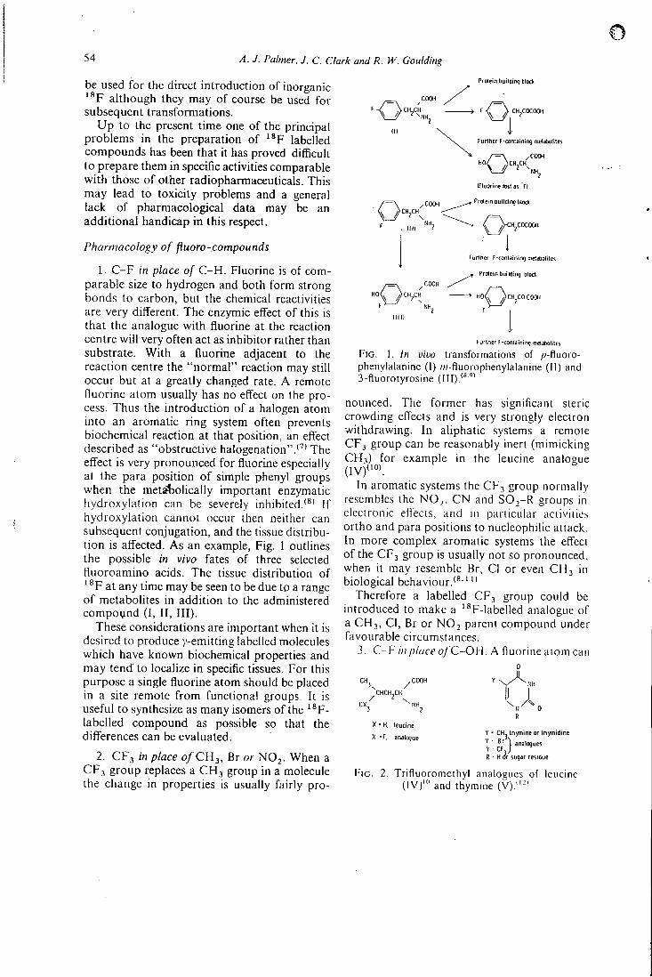
54 A. J. Palmer. J. C. Clark and R. W. Goulding
be used for the direct introduction of inorganic ' ^ F although they may of course be used for subsequent transformations.
Up to the present time one of the principal problems in the preparation of ' ^ F labelled compounds has been that it has proved difficult to prepare them in specific activities comparable with those of other radiopharmaceuticals. This may lead to toxicity problems and a general lack of pharmacological data may be an additional handicap in this respect.
Pharmacology of jiuoro-compounds
1. C - F in place of C - H . Fluorine is of comparable size to hydrogen and both form strong bonds to carbon, but the chemical reactivities are very different. The enzymic effect of this is that the analogue with fluorine at the reaction centre will very often act as inhibitor rather than substrate. With a fluorine adjacent to the reaction centre the "normal" reaction may still occur but at a greatly changed rate. A remote fluorine atom usually has no effect on the process. Thus the introduction of a halogen atom into an aromatic ring system often prevents biochemical reaction at that position, an effect described as "obstructive halogenation".'"" The effect is very pronounced for fluorine especially at the para position of simple phenyl groups when the meta-tolically important enzymatic hydroxylation can be severely inhibited.*®' If hydroxylation cannot occur then neither can subsequent conjugation, and the tissue distribution is affected. As an example, Fig. 1 outlines the possible in vivo fates of three selected fluoroamino acids. The tissue distribution of '®F at any time may be seen to be due to a range of metabolites in addition to the administered compound (I, I I , I I I ) .
These considerations are important when it is desired to produce y-emitting labelled molecules which have known biochemical properties and may tend to localize in specific tissues. For this purpose a single fluorine atom should be placed in a site remote from functional groups. It is useful to synthesize as many isomers of the '®F-labelled compound as possible so that the differences can be evaluated.
2. CF3 in place 0 / C H 3 , Br or NOj . When a C F j group replaces a CH3 group in a molecule the change in properties is usually fairly pro-
Prrtein building block
OCHXH
,COCIH CH CH
Furtner F-containing mrtabolites
C\ ^COOH
iriuorine lost as F)
Protein building block
,COCOOH
Furlher F-containirtq metabolites
^ Protein building block
H o Q c
Fgrlner F-containinq metabolites
F I G . 1. In vivo transformations of /)-fiuoro-phenylalanine (I) w-fiuorophenylalanine (II) and 3-f1uorotyrosine (III ) . '""
nounced. The former has significant sieric crowding effects and is very strongly electron withdrawing. In aliphatic systems a remote CF3 group can be reasonably inert (mimicking C H i ) for example in the leucine analogue (IVf°> .
In aromatic systems the CF3 group normally resembles the N O , , C N and S O j - R groups in eiecironic effects, and in particular activities ortho and para positions to nucleophilic attack. In more complex aromatic systems the effect of the CF3 group is usually not so pronounced, when it may resemble Br, CI or even CH3 in biological behaviour.'® ' "
Therefore a labelled CF3 group could be introduced to make a '®F-labelled analogue of a CH3, CI, Br or NO2 parent compound under favourable circumstances.
3. C - F in place o/ 'C-OH. A fluorine atom can 0
3 . CHCH,CH
/ m 3 2
X • H. leucine
X -F . analogue ; ; B r j 3 „ 3 l c u e s
R ' H i5r sugar restilue
F I G . 2. Trifiuoromethvl analoeues of leucine (IV)'" and thymine (V).""'
The preparation of fluorine-lS labelled radiopharmaceuticals 55
also be introduced as the analogue of a hydroxyl group. Pharmacological application has been concentrated on polyhydroxy compounds such as carbohydrates. The fluorine atom can only act as a hydrogen bond acceptor, whereas the hydroxyl group can act as donor and acceptor. It is best to introduce a single fluorine atom because polyfluoro compounds have a different pharmacology. It has been shown that one mono-fluoro analogue of glucose out of all the possible isomers is transported in the same manner as glucose Itself"^' This is because only in this isomer can the same H-bonding occur in the receptor site of the transport molecule.
4. - C 6 H 4 ' ^ F as a protein label. Various hapten groups have been used for tagging proteins and enzymes for non-radioactive tracer work. Proteins have been labelled directly with radioiodine, or with ' ' In via covalently bound E D T A groups'**' (these are often not very stable in vivo). It would appear that a perfect radio-label would have a half-life of 2-24 hr and would be covalently bound to the protein via a spacer group. Proteins have been usefully labelled with "C-methyl groups despite the short half l i f e . ' ' 5 ' ' ^ F labelled fluorophenol, fluoroanihne and fluorobenzoic acid are readily prepared (see under reaction scheme 1) and could be used to label proteins by established procedures.
Up to the present time only '*F-labelled compounds of the type described in section (1) have been reported.
Stereochemical effects
Biological activity is closely related to the stereochemistry of the substrate molecule, and when such a molecule exists in two or more isomeric forms (geometrical or optical) the biological activity of each of these forrhs can be very different. This isomerism may already be present in the molecule or it may be generated when a foreign atom (the label) is introduced.
In those cases where a single stereo-isomer of a labelled compound is required it may be possible to use a synthetic sequence in which the precursor to be labelled is available as a single stereo-isomeric form and can be converted to the final product via stages which proceed with effectively 100% stereospecificity. Alternatively an epimeric or racemic mixture may be generated
during or after the labelling stage (cf the amino acids below) and this may be followed by a procedure for resolution. The consequent loss of time and material must be offset against the fact that this may be an easier technique than a stereospecific synthesis.
Strategy o / ' ^ F labelled synthesis Because of the short half life it is essential to
introduce the ' ^ F label at the latest stage possible. Usually a suitable precursor is prepared in bulk and stored for future use. Most reactions used for ' * F labelling have to be completed in less than the optimum times and using other than the optimum relative amounts of reactants. Consequently mixtures of labelled and/or unlabelled products are often obtained. Preparative gas (gc) or liquid chromatography (Ic) are especially useful separation techniques since they can be carried out rapidly. Solvent extraction and sublimation may also be useful. Conversely, preparative thin layer chromatography is not so useful because spotting, zone identification and elution are relatively lengthy processes.
' * F labelled compounds for in vivo use are preferably administered in approximately isotonic solutions. Because of the short half-life, lengthy de-salting or solvent removal stages are not practicable. The best approach is to either be able to isolate a non-polar compound from a volatile organic solvent such as ether, or to carry out reactions in aqueous solutions of low-ionic strength. Immobilized reagents are especially useful (e.g. H'^-form ion exchange resin in place of acids, or F " -form resin for fluorin-ation"^'). If any final liquid chromatographic purification is required the preferred eluant is water or an approximately isotonic, non-toxic buff"er.
Analytical considerations It is necessary lo establish the ideniiiy and
homogeneity of any starting materials one may synthesize for subsequent '^F-labelling. Infrared and nuclear magnetic resonance (nmr) are useful for the former, and thin layer (tic) or gas (gc) chromatography for the latter.
Since the range of compounds that have been labelled with ' ^ F is not very extensive new reactions attempted in future will present new-problems to be solved. Development work
56 A. J. Palmer. J. C. Clark and R. W. Goulding
normally requires a lengthy series of trial reactions which are followed by various standard radio-chromatographic techniques e.g. tic/ autoradiography. The technique of coupled glc-mass spectrometry is valuable for the analysis and identification of mixtures that are volatile or can be made volatile by derivatization.
When a complete synthesis of the required '®F-compound has been achieved it is often not possible to perform an extensive chromatographic analysis before the in vivo use because of the short half-life of the label. Therefore, in a series of preliminary experiments employing stable or active fluorine and using the projected procedure, the identity and homogeneity of the product should be ascertained. Then a standardized experimental procedure is established and the final product may be analysed by a shortened procedure concurrently with its utilization. In order to determine the specific activity of the solution to be administered, measurement of the u.v. absorbance at a selected wavelength is often a valuable and rapid method.
The points mentioned in this section may appear to be self evident, but failure to appreciate them will invariably give misleading results. Much information in the literature concerning short-lived labelled compounds is unsatisfactory because adequate product analysis is not described. Certaiijfclaimed preparations do not quote any method of analysis and the phrase "analysed by tic" has appeared without a description of the stationary or mobile phase used. Even when exchange processes are used compound degradation may occur and so the identity of the labelled "product" can never be assumed without proof.
P R O D U C T I O N O F T H E R A D I O N U C L I D E Some nliclear reactions which lead to the pro
duction of fluorine-18 are listed in Table 1. The
T A B L E 1. Some nuclear reactions for the production of Fluorine-18
No. Target Proiedile Reaction 1 0
2 0 "o6le.pnl'«F 3 0 'He %i\A 4 Ne d
5 Ne 'He ^°Nel'Hc«p)"F
excitation curves for these nuclear reactions together with the thick target saturation activities for deuterons and tritons of up to 24 MeV, and ^He and *He up to 42 MeV have been measured by NozAKi"*'.
Reaction 1 has been in use for many years using the 2.73 MeV (max) tritons generated in a nuclear reactor by the ^Li(n, a)^H reaction."^' The target material is usually Li2C03 with a ^Li enrichment of at least 90%. Fluorine-18 is recovered as ' * F ~ without the addition of carrier by H F distillation into aqueous alkah,'^°"^'' or by ion exchange.'"-'"' Partial removal of the undesirable impuritv tritium (typically 750mCi^H to 2.5 mCi'^F'"*') as water and is achieved by repeated evaporations to dryness. The traces of tritium that remain after this treatment have been identified as ^H-acetate and ^H-formate.<"'
The recovery of anhydrous H ' * F from reactor irradiated L 1 2 C O 3 by distillation of H F onto ion exchange resin which is subsequently dried and eluted with H F has been described.''^' However, as up to 1.5 g of H F was used to achieve a 90% recovery from the resin, the resulting H ' ^ F was of low specific activity.
Improvements relating to the L 1 2 C O 3 target design have been described which enable high neutron fluxes to be used more effectively and batches of 65-75 mCi of fluorine-18 to be prepared.'''^'
Reactions 2 and 3 employing oxygen'-^' or vvater''^'^°' as target materials have been extensively used for ' * F production. Aqueous solutions of ' ^ F from water targets have been used to label aromatic diazonium fluoroborates by exchange.'^' '*'' Fluoride has been recovered from aqueous solutions using a fluoride form anion exchange resin column which after careful, drying has been used in anhydrous interhalogen exchange reactions to label a variety of aliphatic compounds with fluorine-18."^' The use of an oxygen gas target to produce anhydrous H F has been demonstrated.''*' Here the ' * F trapped heterogenously by the silver plated target walls during irradiation is subsequently recovered by exchange with ~ 1 g of anhydrous H F carrier.
Reactions 4 and 5 have received a great deal of attention in recent years, particularly under anhydrous conditions. Here the aim has been to produce a variety of high specific activity
The preparation of fluorine-lS labelled radiopharmaceuticals 57
fluorinating agents. The investigations have followed three main approaches.
1. Homogeneous scavenging o/ ' * F with recovery during irradiation
The mixing of nitric oxide, hydrogen or chlorine with neon before irradiation has yielded N O ' V " ' , H'«F<^*' and C l ' V ^ * ' reportedly carrier-free from nickel targets preconditioned with F2. A recovery of 60% is reported for NO'^F but the data for H'^F and CI * F suggest large losses of . '^F to the target walls. The addition of F2 to the neon resulted in the recovery of '^Fz with 30% efficiency.*^"' However, these preliminary results provide encouragement for further development of the technique.
2. Heterogeneous scavenging with post irradiation recovery
The losses of ' ^ F in the homogeneous scavenging approach are largely to the walls of the target vessel. There are several reports of the recovery of carrier-free ' ^ F by the washing of glass-lined target vessels with normal saline'^^' or water'-'*'^^' with recovery efficiencies of around 60-90%. The washing of a glass-lined target with dilute solutions of AgF, BF3 and aromatic fluoroborates in anhydrous organic solvents with 50-80% recoveries described by I D O (1974) appears to have some attractive features although the specific activity of the fluorinating agent is reduced by the introduction of carrier.'^^'
A further heterogeneous approach to ' * F recovery has been to coat the target walls with a thin layer of the organic compound to be labelled. After irradiation the organic inaterial is recovered in solution by washing the target
walls. Inorganic ' * F is removed by ion exchange and the labelled product separated from the starting material by ion exchange chromatography.'^^'
The fluorinating agents AgF, K F and SbFj ( lOOmg-lg) have been labelled by a similar technique."^' A variation of the technique has been described where a target lined with AgF or AgFj was used. After irradiation the neon was removed and the vessel used as a reactor for the synthesis of CCI3F and CCl2F2.'*°' This technique is in principle applicable to many fluorina-tions involving volatile organic substrates.
3. Heterogeneous scavenging during irradiation remote from the target
The recovery of ' *F by exchange labelling of a smafl quandty of a simple or complex fluoride placed remote from the target has been demonstrated.''"' The neon is circulated during irradiation through a glass-lined target vessel, and the effluent gas is led through P T F E tubing to a filter holder where a glass fibre element, previously coated with a 5-50 mg of an ionic fluoro-compound, e.g. K F , NaBF4, SbFs, or an aromatic diazonium fluoroborate is supported. A reactor containing dry fluoride-form ion exchange resin (Dowex 1, ~2gm) may also be used.
The volatile '*F-labelled intermediate which is continuously swept from the target during irradiation is removed almost quantitatively by the coated filter. The filter element or ion exchange resin can often be used directly in the chemical syntheses that follow. This technique is useful for fluorinations involving non-volatile organic substrates.
Methods of ^^F Labelling The Balz-Schiemann reaction has been used
extensively for making aryl fluoro-substituted compounds. Nucleophilic displacement reactions have been used for the preparation of aliphatic fluoro-compounds. Additionally various other methods have been employed in special cases.
P R E P A R A T I O N O F A R O M A T I C F L U O R O - C A R B O N S VIA T H E
B A L Z - S C f f l E M A N N R E A C T I O N Compounds which are monofluorinated in an
aromatic ring system often show biological activity related to the corresponding unfluorin-ated or phenolic compound. Hence methods of
58 A. J. Palmer, J. C. Clark and R. W. Goulding
preparing such compounds in labelled form are of immediate interest. Direct mono-fluorination of aromatic rings is only possible in very special circumstances, but the Balz-Schiemann reaction'*^' allows the ready conversion of an aryl nitro compound, via the amine and diazonium salt, to the corresponding fluoro-compound (Fig. 3, general reaction scheme).
The tretrafluoroborate anion of the diazonium salt may be labelled by exchange and the C-'®F bond is formed when the diazonium fluoro-borate is decomposed thermally. Many simple '^F-aryl fluoro-compounds have been prepared by this method'*^' and of these, amines, phenols and carboxylic acids could be used as starting materials for the preparation of other labelled compounds. The chemical yield in the Balz-Schiemann reaction is often low, and a large amount of side-products may be produced. Also, since the fluorine is introduced as the labelled fluoroborate anion, the maximum possible radiochemical yield is 25% and in practical examples it is ~ 2-15%.
A further complication is introduced because other reactive groups present in the molecule can prevent any of stages I, II or III from pro-ceding. In such cases (in fact with almost all the compounds of biological interest) it is necessary to use "protected" derivatives of such groups, prepared eithewby a direct protection reaction or by synthesis from simpler compounds. After stage III the protecting group(s) are removed by suitable deprotection reactions. The reported chemistry of protecting groups is very extensive especially in the peptide, steroid and carbohydrate fields,'""' but so far only the simplest protecting groups have been employed during '^F-syntheses.
The diazonium fluoroborates may be labelled by exchange with carrier-free ' ^ F in water'^^' or water/acetone'^" or by heterogenous exchange between the solid supported in an inert matrix and carrier-free fluorine-18 extracted from a recirculatory neon gas target.^"" The last method is far superior sirice exchange is instantaneous and much higher specific activities may be achieved. The labelled diazonium fluoroborate may be decomposed dry or in an inert solvent. A mixture of products is always obtained as shown by tic (silica gel, 10% ethyl acetate in chloroform) and the ratio of various
components varies with the decomposition solvent used. Biphenyl, tetrahydronaphthalene and phenanthrene give good yields, whereas dichlorobenzene and malononitrile give poor, yields of the desired protected fluoro-compound.''* '•"^' Purification of the mixture may be effected by preparative tic or Ic when the diazonium salt is decomposed dry, but Ic is the best method when a solvent is used.
1. Aromatic amino acids and catecholamines
Fluorine-18 labelled analogues of four naturally occurring aromatic amino acids have been prepared. These are phenylalanine, tyrosine, tryptophan and 3,4-dihydroxyphenylalanine (DOPA). The first three are utilized in the production of enzymes and proteins and the ' ^ F -labelled analogues are being investigated as potential pancreas scanning agents'"*' since this organ is a site of rapid utilization of amino acids for protein production. D O P A is associated with melanin-formation and Parkinsonism. All of the '*F-amino acids have been evaluated as potential melanoma localising agents'" ' and fluoro-DOPA.
Abnormalities in the quantities and metabolism of catecholamines are associated with hypertension, Parkinsonism, chromaffin tissue tumours and other pathological conditions.*""* '*F-6-Fluoro-dopamine is being evaluated as an adrenal scanning agent.'^°' Fluorine-18 labelled derivatives of amino acids and catecholamines which have been prepared are listed in Tables 2-4.
Labelled p- and m-fluorophenylalanine may be prepared by a published method'^" used for the inactive compounds essentially without modification. A diethyl acetomidomalonate derivative of the corresponding diazonium tetrafluoroborate (Table 2) is first prepared and labelled. After the decomposition the protecting groups (ester and amide; may be removed by vigorous acid hydrolysis when the dl-amiao acid is obtained.' '•^'' Alternatively it is possible to prepare the d/-acylamino acid which may be stereoselectively deacetylated using the fungal enzyme amino acylase to give the /-amino acid.'"'"'
A mixture of the enzyme, d-acylamino acid and /-amino acid is obtained and the required /-amino acid can be recovered efficiently by Ic
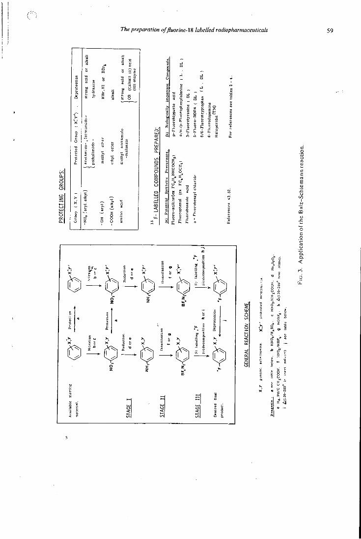
The preparation of fluorine-lS labelled radiopharmaceuticals 59
§1
Q LU or < a . on Q_ m
t-0 t / i n o L. 2 : =) a» 0 IX O-
0 0 .c
c
1 1 "5
CO c
< 1 0 a
Q 01
CO I
o c o
D . Q .
<
6
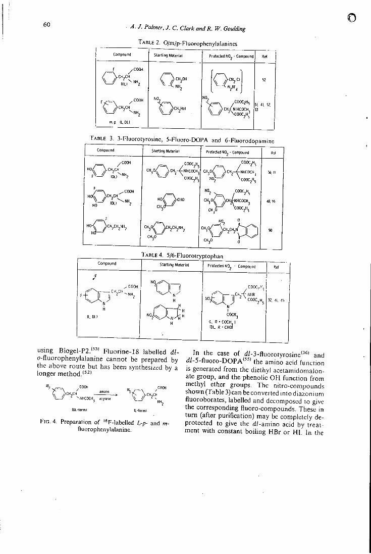
6 0 A. J. Palmer, J. C. Clark andR. W. Goulding
T A B L E 2. O/m/p-Fluorophenylalanines
Compound Starling Material Protected NO^ - Compound Ret
f y C O O H
A A c H CH Q>CH,OH ^ NH^
Q^CH^C, 52
P ^ C O O H NO^
' ^ C H ^ H a l
NO, < ^ = N -C00C?H5
C , ) C H / N H C O C H
V — ' / ' \COOC^H^ '
31. 'li. 52. 53
m. p (L, DL)
T A B L E 3. 3-Fluorotyrosine, 5-Fluoro-DOPA and 6-Fluorodopamin
Compound Starting Material Protected NO^ - Compound Re(
/ C O O H
^ \ / V c H j C H , ^ , ^ N H ^
}CH2-< ;NHC0CH3
COOCjHj
COOC^H^
/ C H ^ - V - N H C O C H j
NO^ \ c O O C j H 5
36.41
/ = \ -COOH H0(\ / )CH CH
HO ?
H o / \ c H O
CHjO '
' ° 2 V = , l^'^i^ C H 3 O / \ cH iNHCOCH3
CH3O COOCjH^
48.55
CH3O
^°2 °,
CH3O
50
T A B L E 4. 5/6-Fluorotryptophan Compound
•s
Starling Material
^ COOH
' ^ " 2 ^ " - N H ,
I t DLI
Protected NO^ - Compound
COOC,H^
CH -eWHR ^ ^ ' ' ° = 2 " 5
N I COCH3
I t R • COCH ) (DL R • CHOi
Ref
32. 41. 45
using Biogel-P2.'"' Fluorine-18 labelled dl-o-fluorophenylalanine cannot be prepared by the above route but has been synthesized by a longer method.'*^'
COOH / amino
^NHC0CH3 acylase
(OL-formI (L-form)
F I G . 4. Preparation of '*F-Iabelled L-p- and m-fluorophenylalanine.
In the case of d/-3-fluorotyrosine'^*' and (iJ-5-fluoro-DOPA''^' the amino acid function is generated from the diethyl aceiamidomalon-ate group, and the phenolic OH function from methyl ether groups. The nitro-compounds shown (Table 3) can be converted into diazonium fluoroborates, labelled and decomposed to give the corresponding fluoro-compounds. These in turn (after purification) may be completely de-protected to give the d/-amino acid by treatment with constant boihng HBr or HI. In the
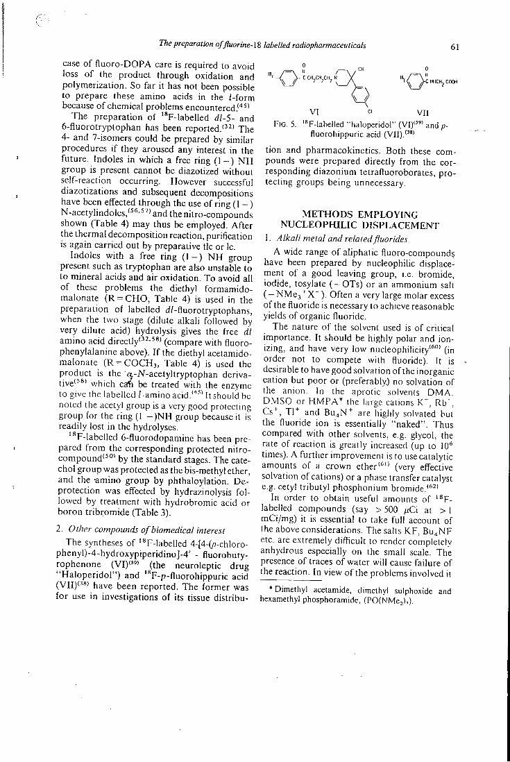
The preparation offluorine-lS labelled radiopharmaceuticals 61
case of fluoro-DOPA care is required to avoid loss of the product through oxidation and polymerization. So far it has not been possible to prepare these amino acids in the /-form because of chemical problems encountered.'"^'
The preparation of '*F-labelled dl-5- and 6-fluorotryptophan has been reported.'^'' The 4- and 7-isomers could be prepared by similar procedures if they aroused any interest in the future. Indoles in which a free ring (1 - ) N H group is present cannot be diazotized without self-reaction occurring. However successful diazotizations and subsequent decompositions have been effected through the use of ring (1 —) N-acetylindoles,'^*'^^'and the nitro-compounds shown (Table 4) may thus be employed. After the thermal decomposition reaction, purification is again carried out by preparative tic or Ic.
Indoles with a free ring ( I - ) N H group present such as tryptophan are also unstable to to mineral acids and air oxidation. To avoid all of these problems the diethyl formamido-malonate (R = C H O , Table 4) is used in the preparation of labelled d/-fluorotryptophans, when the two stage (dilute alkali followed by very dilute acid) hydrolysis gives the free dl amino acid directly'^'-^** (compare with fluoro-phenylalanine above). If the diethyl acetamido-malonale (R = C 0 C H 3 , Table 4) is used the product is the a-iV-acetyltryptophan derivative'**' which ca^ be treated with the enzyme to give the labelled /-amino acid.'""'' It should be noted the acetyl group is a very good protecting group for the ring (1 - ) N H group because it is readily lost in the hydrolyses.
'*F-labelled 6-fluorodopamine has been prepared from the corresponding protected nitrocompound'^'*' by the standard stages. The catechol group was protected as the bis-methyl ether, and the •aminO group by phthaloylation. De-protection was effected by hydrazinolysis followed by treatment with hydrobromic acid or boron tribromide (Table 3).
2. Other compounds of biomedical interest
The syntheses of '*F-labelled 4-[4-(/7-chloro-phenyl)-4-hydroxypiperidino]-4' - fluorobuty-rophenone (VI) ' '" (the neuroleptic drug "Haloperidol") and **F-p-fluorohippuric acid ( V I I ) < 3 8 ) have been reported. The former was for use in investigations of its tissue distribu-
0
V I CI V I I
F I G . 5. 'sp-iabelied "haloperidol" ( V I ) " " and p-fluorohippuric acid ( V I I ) . ' " "
tion and pharmacokinetics. Both these compounds were prepared directly from the corresponding diazonium tetrafluoroborates, protecting groups being unnecessary.
M E T H O D S E M P L O Y I N G N U C L E O P H I L I C D I S P L A C E M E N T
1. Alkali metal and related fluorides
A wide range of aliphatic fluoro-compounds have been prepared by nucleophilic displacement of a good leaving group, i.e. bromide, iodide, tosylate ( - O T s ) or an ammonium salt ( - N M c j ' ^ X " ) . Often a very large molar excess of the fluoride is necessary to achieve reasonable yields of organic fluoride.
The nature of the solvent used is of critical importance. It should be highly polar and ionizing, and have very low nucleophilicity'*"' (in order not to compete with fluoride). It is desirable to have good solvation of the inorganic cation but poor or (preferably.) no solvation of the anion. In the aprotic solvents DMA. D M S O or H M P A * the large cations , Rb' , Cs^, Tl"^ and Bu4N'^ are highly solvated but the fluoride ion is essentially "naked". Thus compared with other solvents, e.g. glycol, the rate of reaction is greatly increased (up to 10*' times). A further improvement is to use catalytic amounts of a crown ether'*" (very effective solvation of cations) or a phase transfer catalyst e.g. cetyl tributyl phosphonium bromide.'*^'
In order to obtain useful amounts of ' ^ F -labelled compounds (say > 500 pC\ at > I mCi/mg) it is essential to take full account of the above considerations. The salts K F , Bu^NF etc. are extremely difficult to render completely anhydrous especially on the small scale. The presence of traces of water will cause failure of the reaction. In view of the problems involved it
* Dimethyl acetamide, dimethyl sulphoxide and hexamethyl phosphoramide, (PO(NMe2)i)
6 2 A. J. Palmer, J. C. Clark and R. W. Goulding
o
is not surprising that relatively little use has been made of these reagents for ^^F- labelling.
ROBINSON'^^' has developed a novel alternative technique in which fluoride-form ion exchange resin (Dowex-1) is used as the fluorinating agent
Resin [CH2NMe3 + F"]„,
which is labelled by exchange with carrier-free fluorine-18 in neon'"" or water."^' The success of this particular reagent may be due to the ease with which it can be rendered anhydrous (1 hr/150°C in vacuo). It has a certain formal similarity to the phase transfer catalysts, and, as an immobilized reagent, facilitates very rapid work up after the reaction.'*^* The mixture of alkyi bromide and fluoride obtained can be separated by preparative gas chromatography. Various ^^F-2-flluorocarboxylic acids (protected as ethyl esters) have been prepared by the method, and from these, by reduction, the corresponding 2-fluoroalkanols. These compounds have been used for brain and heart studies and also investigation of ethanol metabolism.'*"'
2. Silver fluorides
Silver (I) fluoride is much more versatile than the alkali metal fluorides in that it can be used to prepare pri-, sec- and /er/-alkyl fluorides'**' and heteroarqftiatic compounds. The reactions are relatively rapid and the reagent is often used without solvent with both liquid and gaseous substrates (at 25-200°C). High yields of the organic fluoro-compound are obtained even with '^F-labelled material (up to 50% radiochemical yield, X = Br or I):
R X + 2 AgF R F -t- AgF • AgX However silver fluoride is awkward to handle because it is extremely hygroscopic and corrosive (it normally cannot be used in glass). It has been employed to prepare labelled freon-ll (CCI3F) , '"" 6-fluoro-9-benzyl purine'^*' and 3-fluoro-cholestene'^®' (Table i).
Silver difluoride, A g F j , can occasionally act as nucleophilic fluorinating agent and is more reactive than the monofluoride. It is instantly hydrolysed by water. See also .electrophilic fluorinations below. Mercury (I) and (II) fluorides have been used in similar way.***'*'' One fluorine-18 reaction has been reported (Table 5)
T A B L E 5. Preparation of "F-labelled compounds by halogen displacement
Reagent Substrate Product Re(
F Ion exchange BrCH^CH^OH ' FCH^CH^OH
IS
74
1 8 - ' " ' " F Ion exchange
' FCH^CH^OH
IS 1 8 - ' " ' " F Ion exchange Ethyl halocarboxylates Ethyl F -f 1 uorocartwrytates, 17. 74. 75 resin acids and alcohols Qiy resin
subsequent transformation)
Ag' F CCI^ c ' ^ c i . 40 18
c ' ^ c i .
Ag F CfCI^ 40
Ag'^F. 3-lodocholestene F-3-Fluorocholestene 38
Ag'«F
18
6-Chtoro-9-benzyl purine
F-6-Fluoro-9-ben:yl purine 38
K F /COOH 2, < Oi-iodoestrone T-? . 4-Oifluoroeslrone 76
K ' ^ /COOH 3. S-DI-iodotyrosine F-3. 5-0i(luoro(yrosine 76 K ' ^ /COOH
(odopregnolone - 3 -acetate
F-?l-Fluor opreqnolone-3-acetate
77
with Ag'^Fj . Pratical details concerned with the small scale handling of fluorine gas and silver fluorides have been published.'"'"
3. Antimony and bismuth fluorides SbFj , B i F j , S b F j C l j , SbF; and BiFj , have
been used to affect the transformation R C C I 3 R C F j . The group R may be an alkyl, aryl or heteroaromatic group, but very few compounds of type R C C I 3 have been prepared and hence are available to be used as substrates. This is unfortunate since good yields of product are obtained even with 10-100 mg amounts of ''F-labelled SbFj ( R = C H 3 , CeHs)."" Antimony trifluoride may be labelled by exposuretocarrier-freefluorine-18in neon."^"" It undergoes partial hydrolysis b\ exposure to moisture during handling but may afterwards be completely reactivated (as SbFjCU) by treatment with SbCls .'**'
Some compounds which have been reported to have been prepared by methods employing nucleophilic displacement are listed in Table 5.
P R E P A R A T I O N BY D I R E C T F L U O R I N A T I O N AND O T H E R
E L E C T R O P H I L I C F L U O R I N A T I N G A G E N T S
The past few years has seen the introduction of some novel methods of fluorination which seem likely to lend themselves to the preparation of ^*F labelled radiopharmaceuticals. Perchloryl fluoride (FCIO3) and fluoroxytri-fluoromethane (CF3OF) have been used extensively as a source of electrophilic fluorine to fluorinate activated alkenes. C F 3 O F and
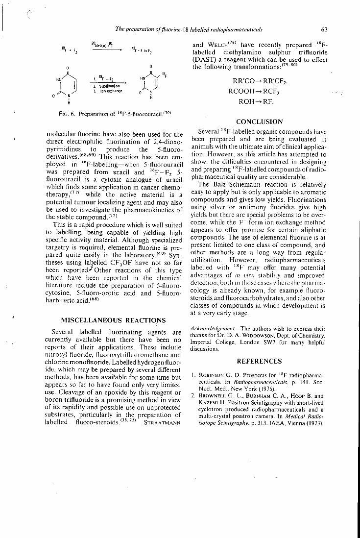
The preparation offluorine-M labelled radiopharmaceuticals 63
'"F - F in F ,
0 H
2. Sublimation 3. Ion exctiange
H
F I G . 6. Preparation of '*F-5-f1uorouracil.'^''*
molecular fluorine have also been used for the direct electrophilic fluorination of 2,4-dioxo-pyrimidines to produce the 5-fluoro-derivatives.'*^'^^' This reaction has been employed in "*F-labelling—when 5-fluorouracil was prepared from uracil and ' ^ F - F z 5-fluorouracil is a cytoxic analogue of uracil which finds some application in cancer chemotherapy,'^'"' while the active material is a potential tumour localizing agent and may also be used to investigate the pharmacokinetics of the stable compound.
This is a rapid procedure which is well suited to labelling, being capable of yielding high specific activity material. Although specialized targetry is required, elemental fluorine is prepared quite easily in the laboratory.''*"' Syntheses using labelled C F 3 O F have not so far been reported.*^ Other reactions of this type which have been reported in the chemical literature include the preparation of 5-fluoro-cytosine, 5-fluoro-orotic acid and 5-fluoro-barbituric acid.'*^'
M I S C E L L A N E O U S R E A C T I O N S
Several labelled fluorinating agents are currently available but there have been no reports of their applications. These include nitrosyl fluoride, fluoroxytrifluoromethane and chlorine monofluoride. Labelled hydrogen fluoride, which may be prepared by several different methods, has been available for some time but appears so far to have found only very limited use. Cleavage of an epoxide by this reagent or boron trifluoride is a promising method in view of its rapidity and possible use on unprotected substrates, particularly in the preparation of labelled fluoro-steroids.'^^''^' S T R A A T M A N N
and W E L C H ' ' * ' have recently prepared ' * F -labelled diethylamino sulphur trifluoride (DAST) a reagent which can be used to effect the following transformations:'^''*'"
R R ' C O ^ R R ' C F z .
R C O O H ^ R C F j
R O H - ^ R F .
C O N C L U S I O N
Several '^F-labelled organic compounds have been prepared and are being evaluated in animals with the ultimate aim of clinical application. However, as this article has attempted to show, the difficulties encountered in designing and preparing '*F-labelled compounds of radio-pharmaceuticai quality are considerable.
The Balz-Schiemann reaction is relatively easy to apply but is only applicable to aromatic compounds and gives low yields. Fluorinations using silver or antimony fluorides give high yields but there are special problems to be overcome, while the F~ form ion exchange method appears to offer promise for certain aliphatic compounds. The use of elemental fluorine is at present limited to one class of compound, and other methods are a long way from regular utilization. However, radiopharmaceuticals labelled with ' ^ F may offer many potential advantages of in vivu stability and improved detection, both in those cases where the pharmacology is already known, for example fluoro-steroids and fluorocarbohydrates, and also other classes of compounds in which development is at a very early stage.
Acknowledgement—The authors wish to express their thanks for Dr. D. A. WIDDOWSON, Dept. of Chemistry, Imperial College, London SW7 for many helpful discussions.
R E F E R E N C E S
1. ROBINSON G . D . Prospects for ' * F radiopharmaceuticals. In Radiopharmaceuticals, p. 141. Soc. Nucl. Med., New York (1975).
2. B R O W N E L L G . L . , BURNHAM C . A., HOOP B . and K A Z E M I H . Positron Scintigraphy with short-lived cyclotron produced radiopharmaceuticals and a multi-crystal positron camera. In Medical Radioisotope Scintigraphy, p. 313. I A E A , Vienna (1973).
64 A. J. Palmer. J. C. Clark and R. W. Goulding
o
)0
12
15.
16.
20.
21.
22.
23.
24.
. TER-POGOSSIAN M . M . , PHELPS M . E., HOFFMAN E . J . and M U L L A N I N . A . Radiology 114, 89 (1975).
. MuEHLLEHNER G . J. nucl. Med. 16, 653 (1975).
. LiFTON J. F . and W E L C H M . J . Radial. Res. 45, 35 (1971).
. COHEN M . B . , SPOLTER L . , C H A N G C . C , M A C -D O N A L D N . S., T A K A H A S H I J . and BOBINET D . D . J. nucl. Med. 15, 1192(1974).
. CHENOWETH M , D . and M C C A R T Y L . P . Pharmacol. Rev. 15, 673 (1963).
. SMITH F . A . Chem. Tech. U . S . 3, 422 (1973). F O W D E N L . Fluoroamino acids and protein synthesis. In Carbon-Fluorine Compounds (A Ciba Foundation Symposium), p. 141. Associated Scientific Publishers, Amsterdam (1972).
. RENNERT O . M . and ANKER H . S. Biochemistry 2 , 471 (1963). SMITH F . A. Handbook of Experimental Pharmacology, (Edited by SMITH F . A . ) , Vol. 20, Part 2, p. 351. Springer-Verlag, New York (1970). HEIDELBERGER C . Fluorinated pyridine nucleotides, [n Carbon-Fluorine Compounds. (A Ciba Foundation Symposium), p. 125. Associated Scientific Publishers. Amsterdam (1972). BARNETT J . E . G . Fluorine as a substitute for oxygen in biological systems: examples in mammalian membrane transport and giycosidase action. ibid p. 95. G O O D W I N D . A., SUNDBERG M . W . , D I A M A N T I C . I . and MEARES C . F . '"In-labelled radiopharmaceuticals and their clinical use. In Radiopharmaceuticals, p. 80. Soc. Nucl . Med., New York (1975). STRAATMANN M . G . and W E L C H M . J. J. nucl. Med. 16, 425 (1975)^ M A R C H E P., M A R A Z A N O C , MAZIERE M . , M O R G A T J. L . , DE LA LOSA P., COMAR D . and FROMAGEOT P. Radiochem. Radioanal. Lett. 21, 53 (1975). ROBINSON G . D . Biologically active '*F-fluoro-organic compounds: Rapid high activity synthesis of '*F-fluorocarboxylates and derivatives. In Radiopharmaceuticals and Labelled Compounds. Vol. I , 423. I A E A . Vienna (1973). N o z A K i T . , IwAMOTO M . and l o o T . Int. J. appl: Radial. Isotopes 25, 393 (1974). Radioisotope Production and Quality Control. IAEA Technical Report Series No. 128 p. 589. IAEA. Vienna (1971). THOMAS C . C , SONDEL J. A. and KERNS R . C . //;/. J. appl. Radial. Isotopes 16, 71 (1965). H E I . U S F. , K R A U S S D . and M A I E R - B O R S T W . Radiochem. Radioanal. Lett. 15, 225 (1973). C H A N P. K.. H. , F I R N A U G . and GARNETT E. S. /W^/19, 237 (1974). BEG K . and BROWN F . Int. J. appl. Radial. Isotopes 14, 137 (1963). SCHOLZ K . L. and SODD V. J. ibidlZ, 465 (1972).
25
26
27 28,
30
32.
33
34.
35.
36.
37,
38.
39.
40.
42.
. COLLINS C . H . , COLLINS K . E., .ACKERHALT R . E . and BLAU M . ibid 26, 571 (1975).
. NozAKi T . , T A N A K A Y . , SHIMAMURA A . and KARASAWA T . ibid 19, 27 (1968). CLARK J. C . and SILVESTER D . J. ^iW 17, 151 (1966). TILBURY R . S. and D A H L R. , MAMACOS J. P. and L A U G H L I N J. S. * ( a ' 21 , 277 (1970). HiNN G . M . , NELI> W . B . and WEITKAMP W . G . ibid 12, 699 (1971). LINDNER L . , SUER T . H . G . A. . BRINKMAN G . A.
and VEENBOER J. T. ihidlA, 124 (1973).
G O U L D I N G R . W . and PALMER A. J. ibid 23, 137 (1972) . A T K I N S H . L . , CHRIST.M.^N D . R. , F O W L E R J . S.
HAUSER W . , HOYTE R. .M. , KLOPI'ER J. F., L I N N
S. S. and W O L F A. P. J. nucl. Med. 13, 713 (1972). W E L C H M. J., LIFTON J. F. and GASPER P. P. ibid
12, 405 (1971) Abstract. LAMBRECHT R . M . and WOLF A. P. Cyclotron and short lived halogen isotopes for radiopharmaceutical applications. In Radiopharmaceuticals and Labelled Compounds. Vol. 1, p. 275. IAEA, Vienna (1973) . HARPER P. V.. LEMBARF.S N . and KRIZEK H . J. nucl. Med. 12, 362 (1971) Abstract. PALMER A. J., CLARK J. C . G O U L D I N G R. W . and ROMAN M . The preparation of '*F-labelled D L - 3 -fiuorotyrosine. In Radiopharmaceuticals and Labelled Compounds. Vol. 1. p. 291. IAEA, Vienna (1973). WOLBER G. , HARTMANN G. , HELUS F. , HOVER K . H. and LORENZ W . J. Applications of the compact cyclotron of the German Cancer Research Centre in nuclear medicine, neutron therapy and radiation biophysics. 7ili Int. Conf. Cyclotrons and their Applications, p. 444. Birkhauser Verlag, Basel (1975).
iDO T . Synthesis and in vivo distribution patterns of'*F-organic compounds. Pruc. Isi World Cong. Nucl. Med., p. 901. World Federation of Nuclear Medicine and Biology (1974). LEBOWITZ E.. RICHARDS P. and BARANOWSKY J. M. J. appl. Radial. Isotopes 23, 392 (1972). CLARK J. C , G O U L D I N G R. W . and PALMER A. J. Preparation of '"F-hibelled lluorocarbons for use in pharmacodynamic studies. In Radiopharmaceuticals and Labelled Compounds. Vol. I . p. 411. IAEA, Vienna (1973). CLARK J. C . GOULDINC R. W . . RO.MAN M . and PALMER A. J. Radiochem. Radioanal. Lett. 14, 101 (1973). SuscHtrzKY H. The Balz-Schiemann reaction In Advances in Fluorine Cheiiu.siry (Edited by STACEV M . , T A T L O W J. C. and SHARPE A. G ). Butterworths. Washington (1965).
The preparation offluorine-lS labelled radiopharmaceuticals 65
4 3 . NozAKi T. and T A N A K A Y . Int. J. appl. Radial. Isotope.': IS, 1 1 1 (1967) .
4 4 . M C O M I E (Editor) Protective Groups in Organic Chemistry. Plenum, London (1973) .
4 5 . GouLDiNG R . W . P h . D . Thesis, University of London ( 1 9 7 6 ) .
4 6 . T A Y L O R D . M . and COTTRALL M . F . The evaluation of amino acids labelled with ' * F for pancreas scanning. In Radiopharmaceuticals and Labelled Compounds. Vol . 1, p. 405 . I A E A , Vienna ( 1 9 7 3 ) .
4 7 . D A M P U R E H . J., OsMAN S. and S O M A I A S. To be published.
4 8 . G A R N E T E . S. and F IRNAU G . '^F-5-Fluoro-DOPA as a new brain scanning agent. In Radio
pharmaceuticals and Labelled Compounds, Vol. 1, p. 405 . I A E A , Vienna ( 1 9 7 3 ) .
4 9 . AxELROD J. and WEINSHILBOUM R . A^. Engl. J. Med. 2 8 7 , 2 3 7 ( 1 9 7 2 ) .
50. M A C G R E G O R R . R . , ANSARI A. N . , A T K I N S H . L . , CHRISTMAN D . R . , FOWLER J. S. and W O L F A. P. J. nucl. Med. 15, 5 1 3 ( 1 9 7 4 ) Abstract.
5 1 . O K U D A T . and TATSUMI S. J. Biochem. 44, 6 3 1 ( 1 9 5 7 ) .
52. HoYTE R . M . , L I N S. S., CHRISTMAN D . R . , A T K I N S H . L . , HAUSER W . and W O L F A. P. J. nucl. Med. 12, 2 8 0 ( 1 9 7 1 ) .
53. GouLDiNG R . W . and GUNASEKERA S. W . Int. J. appl. Radiat. Isotopes 26, 561 (1975) .
54. TosA T., M O R I T . , FUSE N . and CHIBATA 1. Enzymologia 31, 2 1 4 ( 1 9 6 8 ) .
55. F I R N A U G . , NAHMIAS C . and GARNETT S. Int. J. appl. Radiat. Isotopes 24,182 ( 1 9 7 3 ) ; 7. Med. Chem. 1 6 , 4 1 6 ( 1 9 ^ 3 ) .
56. H Y D O R N a . E . J. org. Chem. 32, 4 1 0 0 (1967) . 57. I K A N R. , HOFFMANN E. , BERGMAN E. D . and
G A H I N a . IsraelJ. Chem. 2, 32 ( 1 9 6 4 ) . 58. BERGMAN E . D . and HOFFMAN E . J. Chem. Soc.
2 8 2 7 (1962) . 59. KooK C . S., REED M . F . and DIGENIS G . A . J. med.
Chem. 18, 533 ( 1 9 7 5 ) . 60 . SCFIADT F . L . and SCHLEYER P. R> Tetrahedron
Lett. 2335 (1974) .
6 1 . LiOTTA C . L . , HARRIS H . P., M C D E R M O T T M . ,
GONZALES T . and SMITH K . Tetrahedron Leii.
71 (1975). 62. L A N D I N I D . , Q u i d S. and D O L L A F . Synthesis
430(1975).
63. C A I N E L L I G . and M A N E S C A L C H I F . ibid. 472 (1976).
64. ROBINSON G . D . , USZLER J. M . and BENNETT L . R . J. nucl. Med 16, 561 (1975) Abstract.
65. B H A N D A R I K . S. and PINCOCK R . E . Synthesis, 655 (1975).
66. H U D L I C K Y M . Chemistry of Organic Fluorine Compounds, pp. 51; 59. Pergamon Press, Oxford (1961) .
67. CHAMBERS R. D . Fluorine in Organic Chemistry. p. 39. Wiley, New York (1973).
68. BARTON D . H . R . , BUBB W . A., HESSE R . H . and PECHET M . M . J. Chem. Soc. Perkin I. 2095 (1974) .
69. BARTON D . H . R . , HESSE R . H . , T O H H . T . and PECHET M . M . J. org. Chem. 37 , 329 (1972).
70. FOWLER J. S., F I N N R. D . , LAMBRECHT R . M . and W O L F A. P. J. nucl. Med 14, 63 (1973).
71. BUSCH H . An Introduction to the Biochemistry of the Cancer Cell p. 139. Academic Press, New York (1962) .
72. W O L F W . , BERMON J. A. and SHANI J. J. nucl. Med. 16, 582 (1975) Abstract.
73. PALMER-A. J. Unpublished results. 74. ROBINSON G . D . J. nucl. Med. 14, 446 (1973)
Abstract; ibid 16, 561 (1975) Abstract. 75. DE K L E I J N J. P., MEEUWISSEN H . J. and V A N
ZANTEN B . Radiochem. Radioanal. Lett. 23, 139 (1975) .
76. MANTESCU C , GENUNCHE A. and SIMONESCU L . '*F-labelling of bioactive molecules using K ' ^ F -C H J C O O H solutions as fiuorinating agent. In Radiopharmaceuticals and Labelled Compound';. Vol. 1, p. 395. IAEA, Vienna (1973).
77. E N G R . J. nucl. Med. 16, 526 (1975) Abstraci. 78. S T R A A T M A N N M . G . M . S C . Thesis, Washington
University, St. Louis (1976). 79. M A R K O V S K I J L . N . , P A S H I N N I K V. E. and K I R
SANOV A . V . Synthesis 787 (1973). 80. M I D D L E T O N W . J. / . org. Chem. 40, 574
(1975).
Journal of Neurology, Neurosurgery, and Psychiatry, 1978,41, I-10 3 3
Study of regional cerebral metabolism and blood flow relationships in man using the method of continuously inhaling oxygen-15 and oxygen-15 labelled carbon dioxide
G . L . L E N Z [ ' , T . J O N E S - , C . G . M C K E N Z I E . P. D . B U C K I N G H A M . . I . e . n . A R K , A N D S . M O S S
From the Medical Research Council Cyclotron Unit. Departments of Radiotherapy and Medical Physics, Hammersmith Hospital, London
S U M M A R Y A new technique for assessing regional oxygen use and blood How has been applied to a wide range of neurological patients. The method couples the brain's high metabolic demand for oxygen with a shortlived radioactive form of this metabolite, namely oxygen-15 (half life; 2.1 min). This combination produces, during the continuous inhalation of either molecular oxygen-15 or labelled carbon dioxide, steady state functional images of the brain which are relatively free of contribution from extracerebral tissues. These are complementary images in that they relate to regional oxygen uptake and blood flow and hence offer a direct insight to the regional demand-to-supply relationships within the brain in physiological and pathological conditions. In the clinical groups studied, metabolic and circulatory defects were observed and instances of cerebrovascular insufficiency and relative luxury perfusion were defined which hitherto have been deduced from indirect methods. The clinical acceptability of this non-invasive approach allowed us to study those categories of patients which normally do not warrant invasive examination.
The funct ion o f tlie cerebral tissue depends critically on the use ofoxygen, its energy turnover being mainly based on glycolytic aerobic metabolism. A n impairment in the rate o f consumption, because of a failure of either supply or use, often constitutes a pathological condit ion. I t follows that a study of the cerebral blood f low ( C B F ) and the cerebral oxygen consumption ( C M R O 2 ) at a regional level is a direct and rational method o f investigating cerebral physiology and pathology. The first clinical study o f C M R O 2 and C B F was presented by Kety and Schmidt (1948). Their technique required continuous sampling o f arterial and jugular bulb blood, and provided an average measurement o f C M R O 2 and C B F for the hemisphere investigated. This method has been used extensively in clinical research but has a limited resolution for investigating focal disease.
While a large number o f regional C B F studies have
' Present address: Department or Neuropsychiatry, Institute of Neurology. University ofSiena, Italy. • Address for reprint requests: Dr T. Jones. M R C Cyclotron Unit. Hammersmith Hospital, Ducane Road, London W120HS. England. Accepted 14 July 1977
1
been presented over the last 20 years, ihe l irsi regional assessment of C M R O j in man uas published hy Ter-Pogossian ei ul. in 1970. This meiliod involves a carotid artery injection o f o.vygen-IS (2.1 min T-U labelled red cells to provide the single passage extraction ofoxygen, followed by a similar injection of labelled water for the regional C B F determination. This technique requires the use of heavily shielded single probes for external recording and so limils the spatial resolution that can be achieved. These methods, although they provide absolute values of C M i R O a , have limited application because of their invasiveness.
A non-invasive approach to suidying regional metabolism and flow has recently been published by Jones et al. (1976a). The steady siaic disiribution of radioactivity within the brain achieved during ihc continuous inhalation of oxygen-15 is recorded with a gamma ray imaging device. This distribution is related to the regional oxygen upiake, while a repeat procedure using oxygen-15 labelled carbon dioxide produces a distribution related to regional cerebral circulation. The ratio between the oxygen
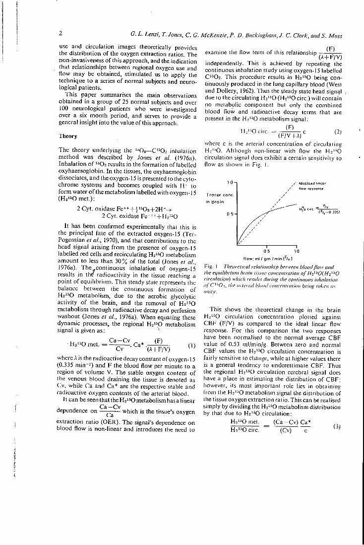
G. L. Lenzi, T. Jones, C. G. McKenzie, P. D. Buckingham, J. C. Clark, and S. Moss
use and circulation images theoretically provides the distr ibution o f the oxygen extraction ratios. The non-invasiveness o f this approach, and the indication that relationships between regional oxygen use and flow may be obtained, stimulated us to apply the technique to a series o f normal subjects and neurological patients.
This paper summarises the main observations obtained in a group o f 25 normal subjects and over 100 neurological patients who were investigated over a six month period, and serves to provide a general insight into the value of this approach.
Theory
The theory underlying the ' 02—C' 02 inhalation method was described by Jones et al. (1976a). Inhalation o f ^^Oz results in the formation of labelled oxyhaemoglobin. In the tissues, the oxyhaemoglobin dissociates, and the oxygen-15 is presented to the cytochrome systems and becomes coupled with H+ to f o r m water o f the metabolism labelled with oxygen-15 (H2 '50met.):
2 Cyt. oxidase Fe^-'-^i^^Oz+lH*^ 2 Cyt. oxidase Fe+ + + -1-H2' 0
I t has been confirmed experimentally that this is the principal fate o f the extracted oxygen-15 (Ter-Pogossian et al., 1970), and that contributions to the head signal arising f r o m the presence of oxygen-15 labelled red cells and recirculating H2' 0 metabolism amount to less than 30% of the total (Jones el al., 1976a). The,,continuous inhalation of oxygen-15 results in the radioactivity in the tissue reaching a point o f equilibrium. This steady state represents the balance between the continuous formation o f Ha'^O metabolism, due to the aerobic glycolytic activity o f the brain, and the removal of Hz^'^O metabolism through radioactive decay and perfusion washout (Jones et al., 1976a). When equating these dynamic processes, the regional Ha'^O metabolism signal is given as:
• Hz^^O met. C a - C v
. Ca^ (F)
(1) Cv ' ^ ^ X-^+F/V)
where A is the radioactive decay constant of oxygen-15 (0.335 m i n - ' ) and F the blood flow per minute to a region o f volume V. The stable oxygen content o f the venous blood draining the tissue is denoted as Cv, while Ca and Ca* are the respective stable and radioactive oxygen contents of the arterial blood.
11 can be seen that the Hz'^^O metabolism has a linear
dependence on — which is the tissue's oxygen Ca
extraction rat io (OER). The signal's dependence on blood flow is non-linear and introduces the need to
(F) examine the flow term of this relationship ^^^p^y^
independently. This is achieved by repeating the continuous inhalation study using oxygen-15 labelled C'=02. This procedure results in Hz'^O being continuously produced in the lung capillary blood (West and Dollery, 1962). Thus the steady state head signal due to the circulating H2' 0 (Hs'^O circ.) wi l l contain no metabolic component but only the combined blood flow and radioactive decay terms that are present in the H j ' ^O metabolism signal:
^ ^ ' • • ° ^ ' - - ( F ^ , ^ where c is the arterial concentration of circulating Hi^-O. Although non-linear with flow the Hj ' - 'O circulation signal does exhibit a certain sensitivity to flow as shown in Fig. 1.
1 0 - ,
Tracer cone,
in Bra in
0-5-
Idealised linear How response
H , 0 c i r c . ( f / v - O 335)
0-5 10
flow, ml / gm / min )
Fig. 1 Theoretical relationship between blood flow and the equilibrium hrain lissue concentration of H^'-^OiHi'^O circulation) which results during the continuous inhalation oj C'"0-j, the arterial tilaod ciincejiliaiion being taken as unity.
This shows the theoretical change in the brain Hs'^O circulation concentration plotted against CBF (F /V) as compared to the ideal linear flow response. For this comparison the two responses have been normalised to the normal average CBF value of 0.53 ml/min/g. Between zero and normal CBF values the H2'^0 circulation concentration is fairly sensitive to change, while at higher values there is a general tendency to underestimate CBF. Thus the regional H2'^0 circulation cerebral signal does have a place in estimating the distribution of C B F ; however, its most important role lies in obtaining f rom the H-i'^O metabolism signal the distribution of the tissue oxygen extraction ratio. This can be realised simply by dividing the H2' 0 metabolism distribution by that due to Hs'^O circulation:
H2 '^0met. _ ( C a - C v ) Ca* Ho'^O circ. (Cv)
Study of regional cerebral metabolism and blood flow relationships in man
Ca*/c wi l l be a constant for all cerebral regions and hence this normalisation procedure wil l produce a distribution which represents the regional oxygen extraction ratio. I t should be stated that OER is an expression o f the balance between the metabolic demand o f the cerebral tissue ( C M R O 2 ) and nutr i tional supply (CBF). In physiological and pathological terms this balance may be o f more interest than regional C M R O 2 or CBF.
Methods
The corresponding absorbed doses to the blood are 70 millirads and 110 millirads, and to the gonads 15 and 25 millirads. The computer-stored images were then corrected for the non-uniformity of the Anger camera, smoothed, and displayed for photography. In addition the regional counts were normalised to the counts contained within the area o f maximal uptake and printed out in matrix f o r m for subsequent digital analysis.
Results
The Medical Research Council's cyclotron was used as a source o f oxygen-15. A constant 25 /iA deuteron beam was used to produce ' 02 (target gas: N2 - f 1 %02) and C^^Oz (target gas - M %C02) (Clark and Buckingham, 1975). The radioactive gases were piped some 210 m f rom the cyclotron to the clinical investigation area where the gamma camera was situated. A t this location an operator monitored the specific activity o f the gas and made dilut ion adjustments to ensure a constant administration rate to the patient o f 1.5 millicuries per minute of oxygen-15 and 0.75 millicuries per minute of oxygen-15 labelled carbon dioxide. In both cases the total flow rate approximated to 0.51/min. The radioactive gases were in turn administered into a standard oxygen mask worn by the patient who breathed mainly room air through the side holes in the mask. Radioactivity exhaled through the side holes was disposed to waste using an exhaust fan withdrawing air over and around the face mask. Cotton w ^ l was inserted into the patient's nostrils to encourage inhalation through the mouth and minimise the contribution to the radioactivity in the antero-inferior parts o f the brain f rom radioactive gas in the upper airways. Dur ing the study the patient lay supine with eyes closed and in a quiet room. The distributions o f radioactivity contained within the head were recorded using an Anger camera collima-ted w i t h a 99 m m (4^ inch) thick leadvmultihole collimator. The performance o f this collimator for detecting 511 keV annihilation radiation has been reported previously by Westerman and Glass (1968). The radioactivity within the brain usually attained equil ibrium after six minutes o f constant inhalation, (Jones et at., 1974) whereupon a four minute steady state image containing between 250.10^ and 350.10^ counts was recorded on-line with a computer (HP2100A). The typical time delay between recording the H2^50 metabolism image and the H2'*0 circulat ion image was 15 minutes. A total o f 15 millicuries o f 1=02 and 7.5 millicuries o f C"02 were administered for each paired study. This represents a radiation absorbed dose o f 190 millirads and 120 millirads respectively to the lung tissue (critical organ).
The study was performed on 127 subjects, 25 normal volunteers and 102 patients. The Table summarises the two groups, with respect to mean age and sex
Table Summary of wean age and se.x ratio of subjects studied
Number Mean age (yr)
Mate.-fenialc ratio
Normal volunteers 25 39.9 2:1 Patients Cerebrovascular disease 33 57.7 1.6:1 Brain tumours 38 48.7 1.6:1 Extrapyramidal disorders 18 58.0 1.8:1 Other 14 47.4 0.6:1
ratio. Neurological patients were further divided into three main subgroups: tumours, cerebrovascular diseases, and extrapyramidal diseases. A few other miscellaneous conditions, including some psychiatric disorders, were investigated.
N O R M A L VOLUNTEERS
The distributions of radioactivity recorded in two normal subjects during continuous inhalation of "O2 and C"02 are shown in Fig. 2. In both subjects the left dominant hemisphere was nearest to the gamma-camera. The pictures clearly show positive images o f the brain, due to H^'^O metabolism when the subjects inhale ' 'O2, and to H2'^0 circulation when C'502 is inhaled. It can be seen that the extracerebral structures contribute little to the head image. This could be predicted f rom the theory (see Fig. 1), when considering the relaiivels low blood flow and volumes o f these tissues. Some degree of contamination was observed in the antero-inferior regions of the brain, because of the gaseous '''O2 and C'^02 contained in the upper airways. This interference was greatest with '^02, because of its lower extraction in the lungs. The distributions of H 2 ' ^ 0 metabolism and H2'*0 circulation are not uniform throughout the brain. The geometrical shape of the brain has to be taken into account, the maximal activity being recorded in the inferior parts, where in transverse
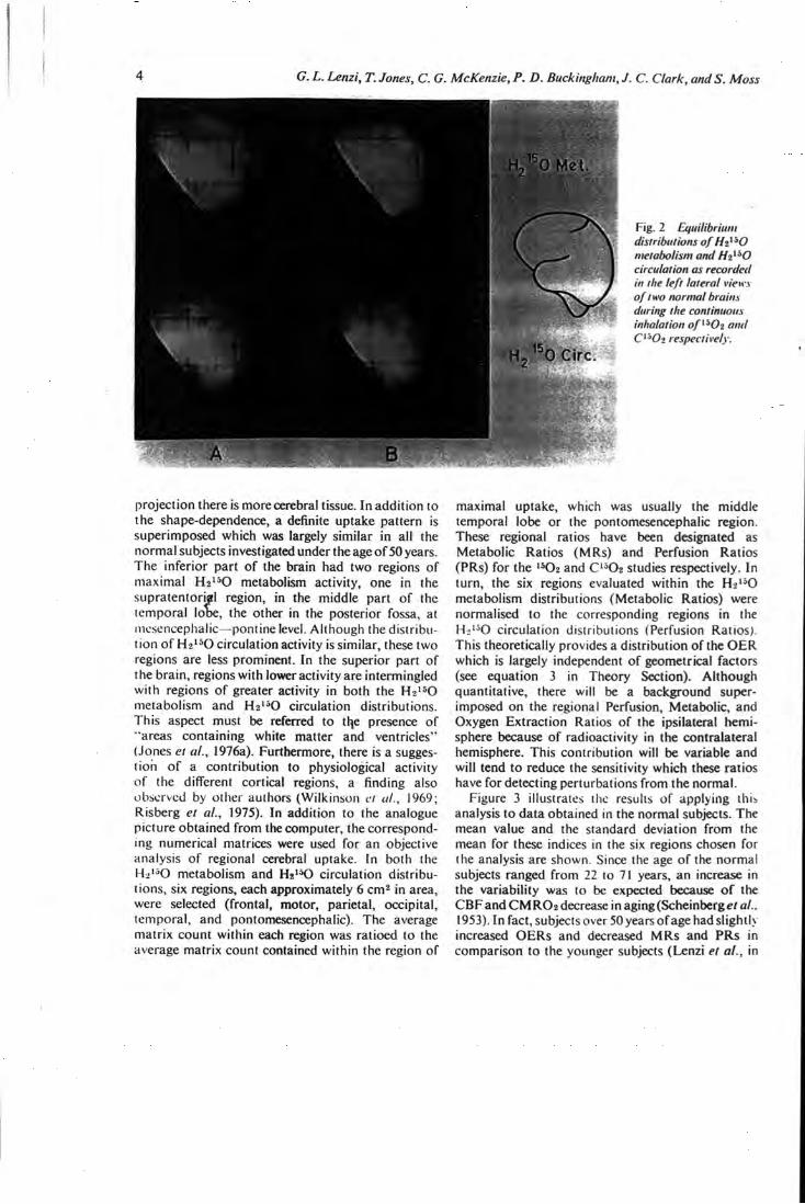
G. L. Lenzi, T. Jones, C. G. McKenzie, P. D. Buckingliam, J. C. Clark, and S. Moss
0 M e t .
0 Circ
Fig. 2 Equilibrium distributions of Hi^'O metabolism and Ht^'O circulation as recorded in the left lateral vie»:\ of two normal brains during the continuous inhalation o /"Oj and C'Oz respectively.
projection there is more cerebral tissue. In addition to the shape-dependence, a definite uptake pattern is superimposed which was largely similar in all the normal subjects investigated under the age of 50 years. The inferior part of the brain had two regions of maximal H2 '*0 metabolism activity, one in the supratentorial region, in the middle part of the temporal lobe, the other in the posterior fossa, at mesencephalic—pontine level. Although the distribution of Hz^^O circulation activity is similar, these two regions are less prominent. In the superior part of the brain, regions with lower activity are intermingled with regions of greater activity in both the Hj '^O metabolism and H2'*0 circulation distributions. This aspect must be referred to tl e presence of "areas containing white matter and ventricles" (Jones el al., 1976a). Furthermore, there is a suggestion of a contribution to physiological activity of the different cortical regions, a finding also observed by other authors (Wilkinson et ul., 1969; Risberg et al., 1975). In addition to the analogue picture obtained from the computer, the corresponding numerical matrices were used for an objective analysis of regional cerebral uptake. In both the H2'^0 metabolism and H j ' K ) circulation distributions, six regions, each approximately 6 cm^ in area, were selected (frontal, motor, parietal, occipital, temporal, and pontomesencephalic). The average matrix count within each region was ratioed to the average matrix count contained within the region of
maximal uptake, which was usually the middle temporal lobe or the pontomesencephalic region. These regional ratios have been designated as Metabolic Ratios (MRs) and Perfusion Ratios (PRs) for the " O 2 and C'^Oa studies respectively. In turn, the six regions evaluated within the H>'*0 metabolism distributions (Metabolic Ratios) were normalised to the corresponding regions in the Hi'^O circulation distributions (Perfusion Ratios). This theoretically provides a distribution of the O E R which is largely independent of geometrical factors (see equation 3 in Theory Section). Although quantitative, there will be a background superimposed on the regional Perfusion, Metabolic, and Oxygen Extraction Ratios of the ipsilateral hemisphere because of radioactivity in the contralateral hemisphere. This contribution will be variable and will tend to reduce the sensitivity which these ratios have for detecting perturbations from the normal.
Figure 3 illustrates the results of applying this analysis to data obtained in the normal subjects. The mean value and the standard deviation from the mean for these indices in the six regions chosen for the analysis are shown. Since the age of the normal subjects ranged from 22 to 71 years, an increase in the variability was to be expected because of the C B F and C M R O a decrease in aging(Scheinberg^/ al., 1953). In fact, subjects over 50 years of age had slightly increased O E R s and decreased MRs and PRs in comparison to the younger subjects (Lenzi et al., in
Study of regional cerebral metabolism and bloodflow relationships in man
N O R M A L
SUBJECTS GROUP n = 25
O E R
preparation). The O E R shown in this figure demonstrates the close relationship between the distribution of oxygen uptake and flow, as observed by Raichle et ul. (1976) with the intracarotid injection method.
NEUROLOGICAL DISORDERS
Over 100 neurological patients were investigated during a six month period. The difi'erent neurological groups studied are listed in the Table. A more detailed evaluation of the three major groups—cerebrovascular disease, brain tumours, and extrapyramidal disorders—will be presented in subsequent publications. In this report we will confine ourselves to describing the main perturbations in the distributions of the H2'*0 metabolism and H^^Ki circulation observed within the neurological group.
In general we Jkpect that the present technique would detect a regional decrease in metabolism and
Fig. 3 Quantitative distribution of the Metabolic Ratios (MR) and Perfusion Ratios (PR) in 25 normal subjects as determined for si.x representative cerebral regions. The distribution of the Cxygen Extraction Ratios obtained by norntolising the Metabolic Ratios to the corresponding Perfusion Ratios is also shown.
circulation, a regional increase in metabolism and circulation, and an imbalance between the regional metabolism and circulation.
Regional decrease in metabolism and circulation The most common instances of a parallel decrease in the H 2 " 0 metabolism and H2'*0 circulation uptakes were seen in stroke patients who had a permanent occlusion of the aflected cerebral arteries, and in patients with primary malignant and secondary brain tumours. All these pathological processes produce gross variations in both the neuronal activity and cerebral function. The parallel circulatory decrease observed was either the primary cause of the disease, as in the strokes, or signified a lower metabolic demand from the tumour's cells relative to the neurones. Figure 4 shows the results obtained in a patient in whom computerised axial tomography
Fig. 4 Distribution of / / >' ' 0 meiaholism and Hi'-'O circulation in the right lateral vieif of a patient with diagnosed cerebral tilrophv and niuliiple l ereltral infarctions. Although both metabolism and perfusion w e regionally reduced ihc relative balance between the two is near normal.
G. L. Lenzi, T. Jones, C. G. McKenzie, P. D. Buckingham, J. C. Clark, and S. Moss
demonstrated "a severe cerebral atrophy, with moderate dilatation of ventricles, and numerous cortical translucencies suggesting multiple infarcts". Both Ha'^o metabolism and Hz'^O circulation images show an impairment of the radioactivity uptake in the superior region of the brain. Some degree of circulation is observed in the proximity of the middle cerebral artery, as if the process were alTecting mainly the smaller arterial branches. Analogue images of the radioactive distribution in the brain are subject to contrast enhancement in the display and photographic process. In all examples the corresponding perfusion and metabolic images were normalised quantitatively to each other in the computer so that the same number of counts were present in the regions of maximal uptake. Thus photographic recordings of the two images were always made under the same conditions of contrast and intensity.
Regional increase in metabolism and circulation In our series no paralled increases in the Ha'^O metabolism and Hz^^O circulation uptakes were observed, such as might be expected within an active epileptic focus as reported by Hougaard et al. (1976). We must underline that we have not had the opportunity of studying epileptic patients (only a few
patients had had fits, and no fits occurred during an investigation).
Increases of H j ' ^ O circulation activity in a lesion were found to parallel the angiographic and neurosurgical findings of increased blood supply. An example of this is shown in Fig. 5 which presents the images obtained in a patient with a highly vascularised meningioma. The position and rich filling of the tumour vessels is shown in the carotid angiogram. The Hzisp circulation image endorses these findings while the Hz^K) metabolism image is within the normal limits. The correlation of the two distributions shows a decreased O E R in the region of the meningioma.
Imbalance between regional metabolism and circulation Examples of imbalance between the metabolic and circulating distributions were also seen in patients without tumours. This uncoupling between flow and metabolism could not have been predicted, and is thus of greater interest with respect to the relevance of these measurements in the interpretation of neurological disorders. The mismatching of the two distributions may reveal a pathological condition that is not actually producing symptoms or anatomically definite lesions. For instance, we were able to
H^'-'OCIrc
O.E.R
Fig. 5 Distribution of Hz^'O metabolism and Hz^'O circulation in the left lateral view of a patient with a highly vascularised meningioma. The observed area of lu.yury perfusion correlates anatomically with the tumour'.s angiogrciplnc blush.
Study of regional cerebral metabolism and bloodflo w relationships in man
"visualise" areas of clinically silent cerebrovascular insufficiency, where a reduction in circulatory activity was not accompanied by, or was greater than the reduction in metabolic activity. This was observed in asymptomatic cases who had previously presented with transient ischaemic attacks, an example of which is shown in Fig. 6. In this case, the impairment of Ha^^O circulation activity was larger than that of the H2150 metabolism activity. The slight decrease in metabolic activity in the parietal region indicated that no definite lesion had occurred and this was confirmed by the patient's neurological history. However, an ischaemic region, where there is preserved neuronal activity coupled with a decrease in the blood supply, indicated that the cerebrovascular insufficiency had not caused neuronal damage. Corresponding to the decrease in the P R there was an increase in the O E R .
The reverse pattern—that is, unaffected flow but reduced metabolism—was observed in "stroke" patients with a normal angiography. Figure 7 shows the distributions obtained in a patient in whom computerised axial tomography had demonstrated an infarct in the left frontotemporal region. The H2'^0 metabolism shows a definite triangular area of decrease in the frontal region extending downward to the temporal pole. In contrast, the Hz'^O circulation image shows no defect, and provides evidence of a relative luxury perfusion situation where there is an excess of blood flow with respect to the metabolic needs of the tissue. In such a situation the reduced use of oxygen points to permanent neuronal damage because of the trapsient reduction of blood flow. The clear-cut defect in the Ha^O metabolism picture also
demonstrates that the arterial recirculation of the metabolically produced labelled water does not appear to affect significantly the resolution that this technique has for demonstrating metabolic defects.
The general comparison between the present technique and the other neuroradiological examinations proved it satisfactory in detecting and locating lesions and in assessing their size and shape. However, the important aspect of these studies is not detection of lesions but investigation of the effect of lesions on cerebral metabolism and blood flow. A preliminary report on the use of this method to assess the effect of radiotherapy on cerebral tumours has already been published by Jones et al. (1976b). The non-invasiveness of the method allows examination of patients such as those with Parkinson's disease in whom such investigations as carotid catheterisation are usually unjustified. This particular aspect is exemplified in Fig. 8. This patient had a two year history of a very mild right extrapyramidal disease which required only anticholinergic treatment to obtain a good control. Surprisingly, a large impairment particularly in the H2'^0 metabolism uptake was detected in the left hemisphere which was contralateral to the side presenting the extrapyramidal signs. In this case the right hemisphere study showed fairly normal distributions with no mismatching of metabolism to perfusion (Fig. 8 ) . The frequency of such aspects in extrapyramidal disorders as seen when using this technique is of interest and justifies a more intensive investigation of this particular group of patients.
There was concern that the superimposition of the
H2 0 Met H2'"'0Circ
Fig. 6 Distribution of Hi''^0 metabolism and Hi'-^O circulation in the right lateral view of a patient presenting with symptoms of transient ischaemic attacks. A zone of ischaemia is seen in the parietal region
G. L. Lenzi, T. Jones, C. G. McKenzie, P. D. Buckingham, J. C. Clark, andS. Moss
H^'^OCiri OMel
Fig. 7 Distribution of metabolism and
Hz^^O circulation in the left lateral view of a stroke patient with a lesion diagnosed in the frontotemporal region from the CAT scan. The isotope study indicates a relative luxury perfusion at this site (which had been found to he normal on angiography).
Right
Hg^fVle t . , \
Fig. 8 Distribution of Hz'^0 metabolism and Hz^'^O circtdalion in the left and right lateral views of a patient with a two year history of very mild e.xirapyramidal disease. A decreased metabolic activity is seen in the left parieio-occipital region while the right hemisphere e.shibits a normal metabolism; perfusion balance.
radioactivity in the two hemispheres would significantly impair the focal resolution of this technique. In practice this turned out to be less of a problem than expected as illustrated in Fig. 8 which shows that the defect in the left hemisphere is not detected in the right view. This suggests that the contribution from the contralateral hemisphere does not impair the ability to detect an ipsilateral defect, and can be explained by the fact that most of the radioactivity
recorded over the brain arises from the metabolically active cortical grey matter adjacent to the gamma camera. The fall-off of spatial resolution with distance from the camera face and the tissue attenuation of gamma rays emitted from the contralateral hemisphere, calculated as being between 45% and 65% (mean 57%), results in the contralateral signal behaving as a defocused attenuated background superimposed upon the ipsilateral image.
Study of regional cerebral metabolism and bloodflo w relationships in man
Discussion
The rationale o f the present approach rests in coupling the high aerobic metabolic demand of the cerebral tissues wi th a short half life radionuclide o f oxygen. This in effect produces a high differential between the concentration o f radioactivity in the brain and extracerebral tissues. Thus it is possible to produce a direct functional image o f the brain wi th a negligible extracerebral contribution, and still administer the labelled oxygen through a general and physiological pathway—namely, inhalation. In addi t ion, repeated investigations showed that these images were reproducible. The study was performed by recording the lateral projection and hence a superimposition o f activity f rom both hemispheres would be expected. However, in detection of focal pathology in the ipsilateral hemisphere, we observed a close agreement with other neuroradiological investigations. This can be explained by the interference f r o m the contralateral hemisphere behaving as a diffuse background, a consequence of the sharp decrease in spatial resolution with distance f rom the camera and some attenuation o f photons (57%) in the interposing tissue. For display purposes the intensity and contrast is adjusted to illustrate any focal mismatching between flow and metabolism. This is legitimate provided both images have been normalised to the counts in the region o f maximal uptake, an adjustment which helps to supress the effect o f the background due to activity in the contralateral hemisphere. Hence mismatching is more dramaticallj^llustrated in the analogue images than f r o m quantitative ratios which are directly subjected to the background effect.
The interference with the H2i^O metabolic cerebral signal due to recirculating H 2 ' ^ 0 was demonstrated in animals to be less than 30% (Jones et al., 1976a). This low interference was substantiated in those cases in our series which presented a metabolic lesion wi th preserved blood flow (Fig. ^7). The analogue and digital distributions o f Vi^^^O metabolism arid H 2 ' ^ 0 circulation obtained are in addition to regional and structural physiological differences dependent on the brain's geometry. In our normal .series the regional variations were found to be sufficiently consistent to enable the identification of pathological features. The ratio between the H 2 ' ^ 0 metabolism distribution and H2 ' ^ 0 circulation produces the distribution of the OER which is largely independent o f the brain's geometry (equation 3). This ratio represents the balance between the metabolic demand ( C M R O 2 ) and the blood supply (CBF), and is, therefore, a direct expression o f the physiological condition o f the nervous tissue. A striking outcome of the application o f this approach
to a neurological population has been the positive demonstration o f instances of true cerebrovascular insufficiency—that is, a situation of low flow wiih preserved metabolic activity, and of relative luxury perfusion where there is normal flow but reduced regional metabolism. These situations until now have been only indirectly inferred. In addition, unsuspected impairments have been observed in neurological patients such as those with extrapyramidal disorders which, to date, not being ethically suitable for invasive investigation, were only evaluated with clinical examination. No evident increase in regional metabolism was observed, in conlrasi 10 recent experimental observations (Reivich, 1976 unpublished), but it should be emphasised that the present technique delects only aerobic glycolyiic metabolism.
The main limitation of this work has been the lack of absolute quantitative measurement of regional C M R O 2 or CBF. The theory indicates that such parameters may be extracted from the steady state uptake o f H2i=0 metabolism and H2 '^0 circulation provided quantitative regional uptake measuremenis are performed and related to the corresponding arterial blood concentrations (Jones ei al., 1976a). This is technically difficult when using a conventional Anger camera, and is best performed with imaging devices which employ positron-coincidence detection. Tomographic emission studies of cerebral uptake would eliminate the superimposed position o f tissue signals, and hence greatly advance the method by virtue o f improved detection contrast. The most practical technical approaches 10 boih quantitation and tomography would appear 10 be either the PETT system developed by Tcr-Pogossian ci al. (1975) or the Positron camera of Brownell (Brownell and Burnham, 1972; Hoop ei al.. 1976). It should be emphasised, however, that, even when these sophisticated devices are used, aiierial sampling wil l be necessary to obtain absokiie regional C M R O 2 or CBF values.
The acceptability of this non-invasive method is underlined by the fact thai oui of more than 100 patients studied, only one refused 10 cooperate. Therefore, it seems logical 10 six-ciilaic upon a larger application to the complete spectrum of neurological and psychiatric disorders. The modifications of the distributions observed in old normal subjecls indicate that this approach could be rclc\ani in ihe investigations of aging processes. A logical progression of otir approach would be the evaluation of therapeuiic agents in neurological disorders (Jones el al., 1976b). In particular, our denionsiraiion of ischaemia is a positive example where an objective assessmenl of therapy could be obtained.
10 G. L. Lenzi, T. Jones, C. G. McKenzie, P. D. Buckingham, J. C. Clark, and S. Moss
The continuous support o f this work by M r D . D . Vonberg and D r R. Morrison is acknowledged. The success o f this study has rested closely on the cooperation o f those neurologists who referred patients to us f r o m outside our institutes. I n particular our colleagues at the following London centres— Hammersmith, Central Middlesex, Royal Free, and National Hospital for Nervous Diseases—are acknowledged for the interest they have shown in these investigations. We are deeply indebted to Professor J. Marshall and to Professor C. Fieschi for their invaluable suggestions and criticisms in the preparation o f this manuscript. The technical support o f M r P. J. Sleight and the cyclotron operating team is recognised as having been of utmost importance. D r Lenzi was supported by N A T O and the National Research Council o f Italy Grant Number 215.7 (1974).
References
Brownell, G. L. , and Burnham, C. A. (1972). M G H positron camera. In Tomographic Imaging in Nuclear Medicine. Edited by F. S. Freedman, pp. 154-164. Society of Nuclear Medicine: New York.
Clark, J. C , and Buckingham, P. D. (1975). Short lived Radioactive Gases for Clinical Use. Butterworths; London.
Hoop, B., Hnatowich, D. J., Brownell, G. L., Jones, T., McKusick, K. A., Ojemann, R. G., Parker, J. A., Subramanyam, R., and Taveras, J. M . (1976). Techniques for positron scintigraphy of the brain. Journal of Nuclear Medicine, 17, 473-479.
Hougaard, 1 ^ Oikawa, T., Sveinsdottir, E., Skinhoj, E., Ingvar, D. H., and Lassen, N . A. (1976). Regional cerebral blood flow in focal cortical epilepsy. Archives of Neurology {Chicago), 33, 527-535.
Jones, T., Brownell, G. L., and Ter-Pogossian, M . M . (1974). Equilibrium images of short-lived radiopharmaceuticals for dynamic observations. Journal of Nuclear Medicine, 15, 505.
Jones, T., Chesler, D. A., and Ter-Pogossian, M. M. (1976a). The continuous inhalation of oxygen-15 for
assessing regional oxygen extraction in the brain of man. British Journal of Radiology, 49, 339-343.
Jones, T., McKenzie, C. G., Moss, S., Buckingham, P. D., and Clark, J. C. (1976b). The non-invasive use of oxygen-15 for studying regional brain function in patients with cerebral tumours. Proceedings of the I2th Bad Gastein Symposium on Radioactive Isotopes in Clinical Medicine and Research.
Kety, S. S., and Schmidt, C. F. (1948). The nitrous oxide method for the quantitative determination of cerebral blood flow in man: theory, procedures and normal values. Journal of Clinical Investigation, 27,476-483.
Raichle, M . E., Grubb, R. L., Gado, M. H. , Eichling, J. C , and Ter-Pogossian, M. M. (1976). Correlation between regional cerebral blood flow and oxidative metabolism. In vivo studies in man. Archives of Neurology (Chicago), 33, 523-526.
Risberg, J., Halsey, J. H., Wills, E. L., and Wilson, E. M. (1975). Hemispheric specialization in normal man studied by bilateral measurements of the regional cerebral blood flow. A study with the 133-Xe inhalation technique. Brain, 98, 511-524.
Scheinberg, P., Blackburn, I . , Rich, M., and Saslaw, M . (1953). Effects of ageing on cerebral circulation and metabolism. Archives of Neurology and Psychiatry (Chicago), 70, 77-85.
Ter-Pogossian, M. M. , Eichling, J. O., Davis, D. O., and Welch, M . J. (1970). The measure in vivo of regional oxygen utilization by means of oxyhemoglobin labelled with radioactive oxygen-15. Journal of Clinical Investigation, 49, 381-391.
Ter-Pogossian, M. M. , Phelps, M. E., Hoffman, E. J., and Mullani, N . A. (1975). A positron emission transaxial tomograph for nuclear medicine imaging (PETT). Radiology, 114, 89-98.
West, J. B., and Dollery, C. T. (1962). Uptake of oxygen-15 labelled CO2 compared with carbon-11 labelled C O 2 in the lung. Journal of Applied Physiology, 17, 9-13.
Westerman, B. R., and Glass, H. 1. (1968). Physical specification of a gamma camera, Journal of Nuclear Medicine, 9, 24-30.
Wilkinson, I. M . S., Bull, J. W. D., Du Boulay, G. H. , Marshall, J., Ross Russell, R. W., and Symon, L. (1969). Regional blood flow in the normal cerebral hemisphere. Journal of Neurology, Neurosurgery, and Psychiatry., 32, 367-378.
1^84 K A D I O C H H . M I S T K V I N M1£UIC1N1; iVoc. A>udyL Div. Ch<:i1^^.
Raciiochemistry m Medicine The following arc summaries of tlie seven papers presented at a Meeting of the Radiochemical Methods Group held on May I7t l i , 1978, in Sutton, Surrey.
Vafue of / n Vivo Radionuclide Methods in Clinical Diagnosis D. O. Cdsgrove Department of Nuclear Medicine, Royal Marsdsn Hospital, Sutton, Surrey. SM2 5PT _ .
Central to the u.sc of radionuclides in diagnostics is the well tried tracer concej'jt wi th its implication that the nuclide used is not discriminated by the body mechanisms. The choice of radionuclides for in vivo use is greatly influenced by dosimetric considerations, especially the biological 'I\ and, for external counting, the degree to wliicii the disintegration pattern approximates to the ideal of a pure y emitter of medium energy (too low and tlic tissue dose rises, too high and capture by the sodium iodide crystal falls off). For imaging, the inetastable nuclides, especially*'Tc™, have proved pliysically ideal (although not without difficulty for the radiochemist) and have allowed the u.se of larger doses than were acceptable with, for example, " 4 , w i th great improvements in image clarity and with the potential for dynamic imaging.
D^^namic iinaging, well established and invaluable in renal work, has become increasingly more important in cardiology. I t is much .simpler than X-ray angiocardiography and therefore more readily repeatable. Information on the passage of a blood pool agent is coilectcd through several cardiac cycles and displayed in slow motion in order to give a good visuai impression of the heart's contractions. Numerical information can be extracted and prom.ises to be useful in providing cardiac output measurements, etc.
Static imaging has improved considerably o\'cr the past decade, owing to a combination of bettery-ray cameras and more suitable radio))hannaceuticals. The images are two-dimcnsionai representations of the distribution of the nuclide in the bod}', but it must be emphasised tliat they are not simple anatomical images, but show anatomy as revealed by selective organ function. Some specific clinically valuable applications are mentioned and a classification by tracer type is informative.
Chemical Ident i ty The classic imaging nuclide is " ^ I for the thyroid, and i t is still one of the most valuable. Its
action depends upon selective concentration of iodide by functioning thyroid tissue and may be thought of as giving a positive image. In the brain an intact blood - brain barrier excludi= most solutes but becomes permeable when damaged. Many tracers are available but the most useful are anionic foiins of ^^Tc'" pertechnetate. The normal brain scan thus shows no brain at a l l ; regions of increased activity are abnormalities. Many other examples of this group are less commonly used, e.g., *'Fe for the bone marrow and tagged damaged red cells for the spleen.
Chemical Similarity l a this application the tracer is sufficiently siinilar to a chemical present in the tissue under
examination, although the lack of identity can lead to difficulties in interpretation. The use of as a substitute for chloride as a bone scanning agent is a now superseded example, and one of current interest is the substitution of for potassium as a carcUac muscle imaging agent. Selenium-75 can be substituted for sulphur in amino acids used to reveal sites of pr-> tein S3Tithesis. Selenomethionine is a useful agent for imaging the pancreas.
T r a c e r s Linked to Metabol i tes I n this application the nuclide is bonded to a metabolite with a minimum of disturbance of
behaviour. lodoalbumin is the classic example, used for blood pool imaging or volunie estimation and iodo- or selenocholesterol are similar substances that are useful in imaging t i e adrenals. The most valuable example is the use of phosphate-like substances, tagged wiih *'Tc™, for bone scanning. A number of similar compounds arc available, and these rely the fact that increased laying down of calcium phosphate occurs whenever bone is diseased so tha t a hot spot is produced. Thus the scans are non-specific for pathology but they aie
1 Oclober, 1978 K A D I . O C H E M I S T R Y I N M E D I C I N E 287
j i T A B L E I
i -j S O M E T E C H N E T I U M - O O H I K A D I O P H . ' V R M A C E U T I C A L S
a
Vi
Preparation Organ visualised Pcrtcchnetate Brain, thyroid
- siilphni" colloid \ L iver "To"' - hydrolyscd tin colloid J
Liver
" T o " - bromomKrcinihydroxypropanc J Kidney - dimcrcaptosuccinatc f Kidney
- D T P A J - glucohcptonato Kidney, brain - methylenedipliosphonate \ Bone - imidodiphosplioiiate / Heart - diethylacctanilidoiminodiacctatc j
99-['Qm - diraethvlacctanilidoiminodiacclate V Gal l bladder
- pyridoxylidineglutamate J "To™ - human scrum albumin—native Blood pool
microspheres Blood pool macroaggregates Lungs
90*] (.m - filiriiiogcn ~1 e9'|*^oi
- plasmin I Blood clot "'To" - streptokinase [ detection 99'|"(-in - urokinase J 99X0"! - labelled cells—
J red blood colls (heat damaged) Spleen .3 leucocytes I platelets k % The oxidation states of S^TG'" in some of tlie commonly used radiopharmaceuticals iiave been 5 studied and tlie evidence suggests^ that the diethylenetriamine-A'A'A''A'"A'"-pentaacetic acid I (DTPA) complex contains T c ( I I I ) and the human serum albumin complex Tc(V). S In the development of new ^^Tc'" radiopharmaceuticals not only is the cliemistryof technetium I important but so is the choice of the complexing ligand, which must be based on known or I predicted biological behaviour. I n relation to radiopharmaceuticals based on complex and I clielate formation a point that is often unrecognised, or forgotten, is that the formation of the J complex can itself change the biological properties of the ligand. This change can result from I complexing wi t l i groups essential for biological activity, or f rom changes in lipophilicity or
I molecular charge. These changes usually lead to radiopharmaceuticals which do not fulfi l the :| intended aim. Occasionally however, alteration in properties may be advantageous. For I example, dimethylacetanilidoiminodiacetic acid is a derivative of the drug lidocaine. The i parent compound is excreted mainly in the urine whereas the '^Tc"" complex is excreted i J, almost exclusively in the bile, making i t a useful agent for imaging the gall bladder.'' I To conclude, '^Tc™ is not only the most widely used radionuclide in contemporary nuclear I medicine, but i t retains great potential for further exploitation. To the chemist the develop-I ment of new s'Tc"" radiophannaceuticals provides many fascinating challenges, from the basic
chemistry of technetium at picomolar concentrations, through tlie chemistrj-, biochemistry and pharmacology of ^^Tc™ complexes, to the problems of developing rapid and reliable methods of preparation and of quality control.
References 1. "Guidelines for the Preparation of Radiopharmaceuticals in Hospitals," Brit ish Institute of Radiology
Special Report No. U , Brit ish Institute of Radiology, London. 1975. 2. Owunwannc, A. , Marinsky. J . , and Blau. M. , / . Nud. Med., 1977, 18, 1099. 3. Richards, P. , and Steigman, J . , in Subramanian, G . , Rhodes. B . A . , Cooper, J . F . , and Sodd, V . J . ,
Editors, "Radiopharmaceuticals," Society of Nuclear Medicine, New York , 1975, p. 23. 4 . R y a n . J . , Cooper, M . , Loberg, M. , Harvey . E . , and Sikorski, S. . J . Nucl. Med., 1977, 1 8 , 997.
Radioactive Gases of Short Half-life for Brain, Heart and Lung Studies John C. Clark Medical Research Council, Cyclotron Unit, Hammersmith Hospital, Ducane Road, London, W\2 OHS
The application of radioisotopes as in vivo probes in order to study organ morphology and function and the measurement of body spaces by radioisotope dilution analysis has been sum-
288 K A D I O C I I ] ; M I S T I ? Y I N M E D I C I N E Proc. Analyt. Div. Chem. Soc.
marised in the prececding papers. This paper discusses the clinical applications of gaseous species containing radionuclides with short half-lives.
Of the radioisotopes in current practice ^^vr'" at 13 s has the shortest half-life. I t is obtained from a radionuclide generator, the parent radionuclide ^'Rb having a half-life of 4.58 h. The chemical separation is simple, the daughter being an inert gas. " 'Kr" ' decays to ^'Kr», which has a half-life of 2.1 x 10 year and thus poses no further hazard. The krypton generator can be coupled on-line to the patient and after the study a very rapid decay occurs when the generator is switched off. In this respect the almost instant disappearance of radiation resembles the conventional X-ray technique. The use of a short half-life radio= isotope provides a new approach to dynamic studies in nuclear medicine. The krypton generators can be shipped readil)' by air and by using C-oncordc can reach America, where un t i l recently they were unobtainable, in a usable state. An ultra-safe generator design is essential as the direct coupling to patients precludes intermediate testing.
^iRb is prepared by either the (p,2n) or (d,3n) reactions on ^^Kr or, as at Hammersmith, the alternative reaction ''Br(a,2n)8iRb using sodium or copper bromides as targets. The gas-phase reaction with a krypton target is the preferred production method. The bombardment of natural krypton wi th 30 MeV protons yields approximately 10 mCi of *'Rb yxA"' fi-^ The *^Rb collects on the walls of the gas target pressure vessel. The best ^^Rb recovery method reported is the use of a target vessel whose cylindrical stainless-steel walls can be rotated. When water is introduced into the rotating liner an epicyclic rod acts as a squeegee, ensuring good contact wi th the walls and an 80-90% recovery of **Rb, which can be absorbed on to a very^ small cation-exchange column. W i t h sodium broinide targets i t is found that a zirconium phosphate inorganic ion exchanger has a high aff ini ty for rubidium even in the presence of a gross excess of sodium ions. At Hammersmith Hospital there is a hot cell where six generators can be made and tested in parallel from one irradiation, all chemistry and target handling being carried out by remote control. Irradiations are carried out between 05.00 and 07.00 hrs, shipment througliout the U K and continental Europe being bv road, rail or air.
I n operation the column is eluted with humidified air for use in pulmonary ventilation studies. When *>Kr™ is required in solution a specially prepared column is eluted with 5% dextrose in water. The eluate is passed through a small column of AG50 resin to ensure low *'Rb contamination. Terminal sterilisation is carried out on-hne using a Millipore 0.22 /xm Swinnex disposable filter unit.
"Tc^" microspheres (Ey 140 keV) can be monitored along with ^^Kr"" (Ey 190 keV) as a differential test of lung function. The "STc™ microspheres test the blood supply and *'Kr°> tests the ventilation as this isotope decays so rapidly that a significant decay occurs before a volume distribution can be achieved. Gross mismatching of the two isotopes (monitored at 140 and 190 keV, respectively) indicates abnormal lung function.
. (Continuous infusion of s^Kr™ solution by a specially designed aortic root catheter whose tip is placed just above the aortic valve (the region from where the coronary arteries draw their blood supply) allows studies of the blood flow to the myocardium. This technique can give a semi-quantitative estimate .of flow on a minute to minute basis. I n contrast only one flow estimation can be carried out using ^'Tc^" microspheres. Similarly, arterial infusion of ^^Kr™ enables studies of brain blood flow diuing drug stimulation.
Radioactive oxygen, ^^0, has been available for many years. I t has a 2 min half-life and is available as C^^O, C^ Oa, " O j and Hj^^O. Nitrogen is irradiated with deuterons at about 7 MeV. Oxygen-15 atoms are produced by the i''ls^(d,n)*^0 reaction and react wi th selected substrates in radiolytic reactions in the target to form, for example, i^Ojand C^^Oj at radiochemical purities of around 99%. One recent appHcation of ' ^0 , and C^'O, is in the study of regional brain oxygen consumption and blood flow.
I f a subject breathes ^^Oj unti l a steady-state equilibrium is obtained the body tissues become labelled wi th Hj^^O generated by metabolism of ^^Oj. Some background is inevitably present due t o the ^K)^ bound to haemoglobin in the blood but i n practice this effect is small. I f on the other hand C^^Oj is administered under similar conditions the body tissues become labelled wi th Hg^^O by the exchange process
(catalysed by carbonic anhydrase) and in this instance the labelled water content of the
October. 1078 R A D I O C H E M I S T R Y I N M E D I C I N E 289
tissue can be related to blood flow. Tims, by use of a suitable imaging device distributions of water of metabolism and flow can be studied and compared.
In i t ia l studies are being carried out in the brain where mismatching between metabolism and flow can be seen clearly in a \'ariety of disorders. During radiotlicrajjy of brain tumours, for
•example, brain metabolism and blood flow can be monitored non-invasivcly, the only cooperation required from the patient is to breath the labelled gas whilst being located in front of the imaging device (y-ray camera). Small cyclotrons (7-8 MeV deutcrons) suitable for dedicated ''O productioi\ may at ;f250000 become a useful addition to the technological arsenal of the clinical scientist.
Radioactive nitrogen ^'N with its 10 min half-life can be produced by the i-C(d,n)'^N or "0(p,a)^^N reactions. I f carbon dioxide or methane are irradiated with deuterons ' ^N, and '^N^Tg are the major products, respectively. I f water is irradiated with protons of 10 MeV or more "N-labelled nitrate and nitrite are formed which can be reduced readily to '^NH3.
I ' N j is used in the solution and gas phase for pulmonary function studies whereas has found application as a cerebral blood flow marker and in myocardial metabolism studies.
Radioactive carbon-Il is commonly produced by the "N(p,a)"C reaction to yield "CO,, which can be reduced to "CO by zinc at 400 °C. "CO can be used to label red blood cells as "C carboxyhaemoglobin and used to define blood pools, e.g. the heart chamber volumes in vivo. I f approximately 5% H2 is present during the proton irradiation of nitrogen " C H 4 is produced; this can be ciiemically converted into the useful synthetic precursor H " C N with ammonia by passing over platinum catalyst at 1 000 °C. Several natural and synthetic amino acids have been prepared using H^'CN and a modified Strecker synthesis for use in in vivo protein metabolism studies, e.g., in the pancreas and tumours.
The radionuchdes " 0 , ^^N and "C all decay by the emission of positrons. These positrons, on annihilation, give rise to a 180° correlated coincidence pair of .511 keV photons. These photons can, wi th the aid of recently developed multi-crystal ring detectors using 66 or more sodium iodide (thallium) crystals, be used to generate a quantitative image similar to an i)i vivo autoradiograph using computer assisted reconstructed emission tomography, a technique akin to that used by the now familiar E M I scanner. The positron and EMI studies can be considered as complementary. Wi th suitable labelled radiopharmaceuticals the former provides functional information whereas the latter provides morphological information.
I n conclusion, the application of radioactive gases of short half-hfe is now providing the cUnician wi th useful information previously unobtainable both in the routine and research areas. The transition to the preparation and use of more complex molecules, based on simple gases as precursors has begun, and a challenging but interesting future seems to lie ahead.
Radiopharmaceuticals Containing Fluorine-18 A. J . Palmer Medical Research Council, Cyclotron Unit. Hammersmith Hospital, Duoane Road, London, W12 OHS
Fluorine-18 (half-life 110 min jB+) found a place in nuclear medicine in the late sixties and early seventies when i t was extensively used (in the form of a simple aqueous solution of fluoride) for bone scanning.^ Since that time i t has been largely superseded for this purpose by ^^Tc"' labelled polyphosphate complexes but its use on a smaller scale for dental studies continues.-Interest has also continued in the preparation and applications of organic radiopharmaceuticals labelled wi th this radionuclide.
Fluorine-18 labelled organic radiopharmaceuticals would appear to have several potential advantages. I n fluorinated analogues the fluorine can replace either a hydrogen atom or a hydroxyl group in a normal organic compound, while a third possibility is the introduction of a trifluoromethyl group. In analogues of the first type the Van der Waals radii of the two elements are very similar (rp = approximately 0.135 nm, = approximately 0.110 nm) and fluorine is the only element that can replace hydrogen withont notable steric consequences.' The carbon - fluorine bond has a very high dissociation energy and consequently improved in vivo stability would be expected for these compounds when compared with, for example, the equivalent iodine-labelled material. Fluorine and hydrogen are however very different in their reactivities. Owing to the very strong electron vrithdrawing inductive effect of
K i n T ' I O N - N i ,11 A N D N H X >( "A K i )I A i >( ) I ) j | ( ) \ \
iht hvpi rli nsi^f pt'-vvs--in ifu- iH! C ir< Hi s .'X'J: 4;I.A-44 1 . | w7(, \1ithiMi|: .r Hipli aruii.il [Tis'-un. vfrsus huiiii'ral faciors in ihi-paih<ii:i. iuM> I 'f the \'aw.iiUir I L N U M I S i\i nialipiiani h^iH rt^Tisitui' VUc L-asf t i " fni;ihT;il l;H-i;irs as '•M.-H as prcssuic C I M I Sn 52 : i !3- I H
gUvtini !)S. Sum MCi. KttMiulorH ( : liMtits it' hvpiTicnsivi- plasmii i,n i b i ' T(-spiMWf t i i an L S v i l H U d ancr\ picparaiimi in n t i r a d i L - n . i t i i i t:ardiov:is ' R , s 10: : f>K- :7J . I'J7(, (_'(ilcni;iii IXi . Ciraiigi. HJ, Clu\i(in At" N'.'hiik-hotlv l inulni iwA aui<ui-piii:iii'i> Miul hypt rk PSion Ciri.- Res (suppi M): 7(''-S7. ]<•}"/]
("oU-nian TCV (. uv-ky AW Jr. Guyuui AC": ii»;pv:rinii.nUi! hvpcrun-^\on amt lon^ icrm ct>nlrnl nf arifrial prcssuri.-. / / j Cardioviiscutai Phvsii'lo : . ctlilcil hv AC Cu\ion. CI: Jont s Baltiniorc, Uni \ 'tTSii\ Park P c . 1^74. pp 2y^-2^^ KalsriLT S: Suroid p^iicriiiaiuni of rcspt)nsi"- lo svmpalhumimL- i i i amines in auriu" strips, Br ! Pharmacol 36; ^H^I-^-Jl. 1^6'/ Ahhoud FM: i.\fft cis of sodium iingioicnsion. and sicroids on vascuUii
r>..- .VV 14.- 14-/. 1 "pin i.l; ( i n i r a ! lopiiu-ni of d i - . ' \ \ .
p i t i p l K K t ! adUEurjru n-aciivMK* Ml mail l-f,: i' k i i d n . . / . i M i i } - \ . K
^ c •hanl :n in itu ii-nsioii lit ra! ( I K .H: >f.*.'-5?» 1 Hat usk i C-. f uKh I . riioi iu n M' t t-nl miliai ion aru! . I i M iopmin i of 28: I i U v M s P.I. i)ari:k' Mi. Dolkf) Ci R,.i, p n ^ f n l i o n oi dtovwonin'slcr-tiu- fi l u t l i o w d o p a m i r u ( iiii Sii 4H. . . . . Bi.v;iii Ri)' I;.lfL..i 1.1 vynipailii-iK- dcnvrvauon prolifi-raiioti m ill-, pro^ifie rabhil t a i aru r\
KlL-ming v't'U \ ;iiiahk .scnsiii'.M* of cxiiiah;-.-nisnis anti hiolo^ual sii:mfKanii.- Rc Ni-iir..si' KlL-mini: U U . .W l'hilhps .1.1. U-.-sifall DP Po^:jun-iiona! supcrv. ns, in iiv ant! suhs(. !isi:i\ o{ i. vciiat'k- iissuf Jrut;\ l-.r^i h Ptusi.'i
t-xpfnnii-nial h
y[\-ni.T
vii.-reii ni iir, . i PtfUl iMol l L x p i t K i l l M
liitv i o n i u m pi loll 111 ilu-oi! in rais in i.-i n:;dl i-
on sni.'oih niu.sv'k Ciri- Ri-v 37: U-
u-IK Pussibli- liuvha : 43-"(I
Continuous Assessment of Regional Myocardial Perfusion in Dogs Using Krypton-81m
A N D R F A V P . S t i . w v N . T E R K V J O N E S . J . H A K V F Y T U K N K K . T I M P K A H .
J O H N C L A R K , A N D P E T B R L A V E N D E R
S L ' M M A R Y Kr\pt()n-81m has been conIinu(»usl\ eluled in S^c devtrtise fr(»m a c>cl<tir(in-madf njbidium-81 generalor. The unique phyiiical properties of this inert freely diffusible gas (half-lire 1. secondsl have allowed the developmeni of a teehnique for the constant infusion of (his tracer into the aortic sinuses of 2? dogs. Theoretical considerations suggest thai an equilibrium o f ' " " K r activity in the myocardium is principal!) dependent on blood flow. Experiments have tested the delivery of this indicator and have recorded quanlilalise high spatial resolution images of the heart with a gamma camera and digital computer. The systematic error was determined b\ comparing changes in regional blood flow (in ml/g per min, u.sing an electromagnetic flow probet and changes in calculated flow (ml/g per min) using the regional activity of " ' " K r (/• = < 0.001; r = 0.97; y = V O K \ + 0 .105;n = 60) The random error and uncertainties concerning mixing and streaming of the indicator were tested by repealing measurements with alterations in heart rate, blood pressure, coronary flow, and total myocardial " ' " K r acti>it> using different intenentions (reproducibility, P = 0 .001 . r = 0 .982; y = 0.982x + - 0 . 2 5 7 . n = 100 observations). .A.n> quantiTication of changes in the myocardial activity of " ' " K r must consider the stability of the arterial concentration of this indicator and washout of """Kr at high values of myocardial blood flow. This ultra-short lived radionuclide will , however, provide an assessment of changes in the distribution of regional myocardial perfusion.
IN 1968, Yano and Anger first suggested that ultrashorl-ed radionuclides such as k ryp lon -81m could be used l o
*isualize blood vessels and organs. ' K ryp ton -81m genera-lors were designed to allow the intermit tent e lu l ion o f this (2S indicator f r o m its parent compound , rubidium-81 f'Rb).--3 This was used ini t ia l ly fo r vent i la t ion and per-lision studies of the lungs and later fo r studies of cerebral ' ' loodflow.-'- '
I^rypion-Slm (half- l i fe 13 seconds) allowed the intro-''uction of a technique for continuous observation and '^sessmenl o f the distr ibution o f regional myocardial
f'om ihc Cardiovascular Research Unit. M R C Cyclotron Unit and "togy Deparlmcn!. Royal Postgraduate Medical School, Hammer-
,?^nospiial, London, England, V / U J * " ^ " ' supported in part by the British Heart Foundation.
reprints: Dr. A . P . Selwyn, Cardiovascular Research Unit, ' N 2 d ' "ospiial, Du Cane Road, London W 1 2 OHS,
i«ived June 2 0 , 1977; accepted for publication February 8 , 1978 .
perfusion in dogs.'' Al though the iheorelical considerations and ini t ial experiments are of interesi. a practical validation of the results bv an independent method has not been provided.
It is the purpose of this paper to present an investigation of and validation for the use of -'"'"Kr for conlinu ' ius assessment of changes in regional myocardial perfusion ( R M P ) in 2.S dogs.
M e t h o d s
Theoretical Considerations
I f " " " K r is infused constantly into the aortic sinuses, the arterial concentration of this indicator wi l l fluctuate ^ii ih pulsatile b lood flow. I f the pattern of mixing and streaming of this freely diffusible gas is stable, then the effect, over minutes, wi l l be that a constant quantity of " " K r will reach the coronary circulation per unit of b lood f low. Accumula t ion of *""Kr in the myocardial water space will
C I R C U l . A ' I I O N K l S l - . A R C I I
i i M i l i m ;in cqu i l ib r iu i i i oi atuv i u w i t h i n l l u iK i i n . D u r i n i : a i n i i su in i infusion the nnu'Uni nf "-'"K r. [ K r | . in ;i viiUinu 111 nn i i c i i rd i i i n i ( V ) u i l l Jcpcntl on: { I ) hloiul f l o u in ln i h f nn i icard iuni ( F ) : (2) ar icr i id ciMucniraliiMi of ihv i in l ica io i (C'a); bknul f l o w out of the mvoeanl iuni (T )• (4 t venous eontentra i io i i of i h i nnheaior | K r ] ; and (5) disappearanee of llie i n j i e a l o i b\ ileea\ (A)
1 I K i J V
\
F(ea)— Mvocanhuni
- A l K r l .
T h i ^ model assumes that the venous eoneeniration is in ev.juiiibiii im u i i h the tissue e o n e e i i t l a l K U I / P represents ihe par in ion toef f ie ien! eif " " ' K r taken lo he I .d-'"
It IS probable thai the part i t ion eocffieieni of " ""Kr wi l l ehaiitii. ^viih di f ferent pathuphvsioloiiiel cireunistanees. I t one uses this ullrashort-l ived iraeer. as shown in Hqualions 1 a n d 4. the part i t ion eoeffieieiil is relaied lo the washout
of the isotope | j and l l ierefore lakes a seeondar\ role
in the mvoeardial signal of """Kr a e l iMt \ . The nivueardial
equi l ibr ium of ' " " K r in the heart wi l l be influeneed b\
arriv al of '^""kr, bv blood f lo iv . and bv rapid deeay Small
ehanges in the par t i t ion eoeffieicni VKIII not be Important .
This IS quite unlike the circumstanees that exist when
eonsideriin; the washout of a long-lived uidicatoi sueh as •xeiHin-1 ri3- ' this State o l dvnamie equi l ibr ium eaii be expresse'd as:
F(Ca) : [ K r ] P [ K r ] A
iherel 'ore
[ K r ] = F(Ca i
V
The deca\ constant for " ""Kr is 3 .2 /min . and this means that the time constant for observing a change in the equi l ib r ium of " ' " 'Kr activiiv in the myocardium must be more than 13 seconds and preferably 30 seconds. The magnitude of f luctuat ion of " " " K r in the arterial b lood wi th each heart beat wi l l be small in comparison lo the quantity of the active indicator m the myocardial water space. The denominator in Equat ion 1 is influenced b\ the high value f o r the decay constant. Hence, the steady state m\ocardiaI " ' " 'Kr aclivitv is principal ly dependent on the arrival of the tracer, i .e. . myocardial blood f low (F) rather than its removal ."
I f the delivery of " " " K r is such that the mean concentration of the tracer in the arterial b lood is constant (when measured at 30-second intervals), it is possible to examine theoretically the change in the myocardial signal. ( K r ) which results f r o m changes in myocardial b lood f low ( F ) . For any change in myocardial blood f l o w f r o m F, to F., we have the fo l lowing expressions f o r the corresponding myocardial counts/min o f " " "Kr , | K r ] .
(Ca) = g [ K r , ] / ^ P + 3.2 (2)
is the relat ive detee l ion e f l ' u i e n c K ^ betu « he re
c o u n t i n g the mvoeard ia l K, a . n l e m . ' l K i i ai-d ,h
e o n e e n i r a i i o n of — K r in the a r i e n a l blo , .d (C a ) K :
a s s u m e d that in anv single exper imei i i C a does noi ehv' "
s ign i f i ea iu lv and g remains c o n s i a n i . „ p o s ^ b l e i , ,
for ['., b\ d iv idini ; l u j u a l H u i 2 h\ 3 "
.2 I-
I K
( K i , o l /PI- ,
I., I I \ -P I
I I l l s can be written
V ~ ! K r , ]
| K r , ) P I -
I - ,
l l l i i l lows that i iuen a k i i o u i i \ . i l i i e ol ^ . . mej>ured
b \ a relerenee leehiiiqut. . aiui ihe corresponding mv.iear-
dial ^ ' " ' K r signal. | K r | . il should be pos'-ible lo cnnvcri
subsequent tissue e{)unts i ,e . . j K i .[ into v alues ol ^. simpU
b\ mseri ion in io Hquaiion -). In eflecl ii shouki be po-MhIe
lo calibrate each preparation in <»rde!' t > e o n \ e n changes
in myocardial " '" 'Kr acti\ i i \ inUi absolute changes in ^ .
B \ calibrating the ^ '" 'Kr counts m this \va\ it is posiihis to lest; ( I ) the the<iretical considerations. (2) the staHliu of the arterial concentration, mixing and streaming of [he indicator, and (3) whether this technique can dfi<;ci changes in regional myocardial perlusion.
Experimental Protocol
Mcmgrel dogs weighing 3ll-.^5 kg were anestheiizec! wi th intravenous sodium thiopental (12 m g ' k g ) . W-n-ila-l i o n was provided via a cuffed endotracheal tube anJ a mechanical ventilator. Anesthesia was maintained b\ the intermit tent intravenous injection of pentobarbital ( i - " ' mg/kg) . Care was taken that with each administration the eyelash reflex was not abolished and the heart rale ;nd blood pressure did not change by more than ^'"t . A iefi thoracotomy was performed and the heart supported !fi 3 l iericardial cradle. A reversible snare was placed arf«:nd the lef t anterior descending coronary arier\ ( L . A D ) or a ma jor branch of this v essel (Fig. I ) . This a l loued occlusion a n d release to be effected without interfer ing w i th im2i-ing .
A modi f i ed Paulin ring cardiac calheier (Cooks Cai^i^- ^ ter C o . ) . introduced through a femoral a r ler io tomy. seated b e l o w the coronary ostia in the aortic sinusw- • " '""Kr w a s eluted continuously in normal saline froJB ^ I " 'Rb generator and infused in 5% dextrose at Ht ml/na" ( bv a constant rate infusion p u m p (Walson-Marlo*' j M H R E 2 0 0 ) . ' ' ;
The dog's heart was positioned under a wide-firf^ [ gamma camera (Toshiba G C A 202) . and images v.-^^ | recorded on Polaroid or 35-mm f i l m w-ith the dog in i"^ left lateral posi t ion. The resolution of this camera p ! ^
-12. . \ o . 6, J , NI-. I t
; : P ^ - 3 . 2 ) K R Y P T O N - S l m A N D M Y O C A R D I A L B L O O D FLOV./ ,Sr/» wi cl o/.
• >n efficiencies bctw content, ( K r | . and crial blood (Ca) . I f j | ncni Ca does not ch nl- It is possible lo sol
V
PF, "
ilue o l ~; . as measur
corresponding myoca, be possible to convej
into \alues of ^, simp!
-•ct it should be possibli der to convert change
solute changes in
1 this wa\" i l is possiblf . i l ions. ( 2 ) the stabilil) g and streaming of ttifl technique can detea usion.
kg \\ere anesthelizeil, (12 m g / k g ) . Ventila-
lotracheal tube and a • as mainta ined by the M pentobarbi ta l (l3, ich adminis t ra t ion the id the heart rate and lore than 5%^ A left heart supported in a;
e was placed arounil: y artery ( L A D ) or a:!, his a l lowed occlusion/ i t e r f e r ing wi th imag-;
heter (Cooks CathefJ ral a r te r io tomy, the aort ic sinuses-^.
>rmal saline front.J^jj: xtrose at 10 t n ' / n w ^ p (Watson M a r l ^ J s
under a w i d e ^ ^ ^ l ) . and i m a g e s ^ g % | wi th the d o g a f l P l p i
Krypton-Sim St, rtc<trose
Rubidium-81 generator
Constant inlusion pump
Coronary snare
Electron! agnelic flow probe
FIGURE 1 The expcrimcnial diagram shows the "Rb generator,
infusion pump, and the aortic sinus catheter. Regional coronary
fiov was measured using an electromagnetic flow probe. The LAD
snare is shown.
collimeter fo r " " " K r was 7 m m , usihg a l ine spread function. Quanti tat ive high spatial resolution images o f total and regional myocardial counts/min of *™Kr were recorded wi th the gamma camera and a digi tal computer (Deltron-Nova 1220). These quantitative images were recorded on a magnetic disc at 30-second intervals throughout the experiments. The digital computer was programmed to recall and display images recorded on an oscilloscope screen within a 64 x 64 matr ix of squares. An electronic light pen was available to enclose up to seven areas of interest wi thin the matrix,, and the computer *as capable of displaying the counts recorded in each area of interest f o r each 30-second interval of the experiments . Corrections were made fo r the decay of " ' R b , A cannula *as inserted into the second femoral ar tery, and thoracic 3ortic pressure was measured wi th Statham P 2 3 D b pressure transducer. The E G G (standard lead 2) was continuously moni tored , and these variables were re-Wrded on a multichannel instrument (Hewlet t -Packard , 7788A).
A suitably sized standard error electromagnetic f l o w probe was f i t t e d to the coronary vessel immediate ly prox-
."nal to the snare. The probe diameter var ied between 3 *nd 5 m m . Pulsatile regional coronary f l o w was recorded
connecting the probe to a standard error f lowmete r JSEM 275) , The f lowmeter system has a carrier f requency
285 H z and the output is nominally f la t ( down less than dB) to 80 H z . Numerous mechanical occlusions of the
L A D were made bv a distally placed snare to ensure an accurate zero level throughout the studies. Recordimrs were selected for analysis only i f , in adjacent zero determinations, there was a change in zero level of less than ;-. ^ % of peak f l o « . The f l o w probes were calibrated in s i tu at the end of the experiments by cannulaling the main Ictt coronary artery, lying o f f all the branches except the onc of interest, and perfusing that artery with the dog s o w n blood f r o m a continuous infusion pump. The areas unde-; the systolic and diastolic portions of the phasic f low tracings were analyzed by planimetry and were calculaied by using the calibration data f low in mill i l i ters per minut-,.-( m l / m i n ) . The mean of six measurements of flow in m! min fo r each control period and each intervention w,;> used. The dicrotic notch of the arterial pressure wa\e wa:> taken as the beginning of diastolic coronary f l o w .
Each experiment was started by positioning the dog's heart under the gamma camera. "'""Kr in S'w- dextrose w;;> delivered by constant infusion into the aortic sinuse-. Th-. total and regional myocardial activities of the indicatv •: were recorded for 20 minutes while heart rate, aort:,-pressure, and phasic regional coronary f low were stabl;-. The L A D was occluded for 30 seconds and a region o n the heart on the visual display showing diminished " ' " " K i activity was ident i f ied. The camera was rotated in reiatio-n to the heart so that the area of interest was positioned c«n the edge of the image. This minimized the effects o f counts f r o m the opposite side of the heart when invesL--gating regional counts/min (CPM) in the area under stud^..
A variety of intenent ions was used to change total o r regional myocardial blood f low.
1. The atria were placed at rates of 150 to 225 bezi^.: min (« = 25 dogs).
2. Intravenous isoproterenol was given at betweea ( i . L 5 and 0.4 ^ g / k g per min {n = 25 dogs).
3. Temporary L A D occlusion was used to produce 2-0 and 5.0 minutes of regional ischemia fo l lowed by reper-fusion {n = 25 dogs).
4. Pentobarbital (3.0 mg/kg, iv ) was used to pioduce decreases in coronary f l o w (n = 20 dogs).
Regional coronary f low (electromagnetic f l o w probe) and total and regional myocardial CPM of " ' "K. - weirc recorded before , dur ing, and after each in t e r \ ; n t i o in , when heart rate, blood pressure, and phasic rrgio[:;al coronary f l o w were stable.
The reproducibi l i ty of changes in * ' " K r myccarc&al C P M was tested in three ways,
1, To ta l C P M of " " " K r was recorded fo r 20 lainuti is before the interventions were init iated and while heart rate, b lood pressure, and regional coronary flow (eleano-magnetic f l o w probe) were stable (n = 25 dogs) ,
2 . I n each experiment, the total and regional myocardial C P M of *""Kr were recorded before, dur ing , and aftier interventions affecting the whole heart ( i .e . , a t r ia l pacinig,
. pentobarbi ta l and isoproterenol) and in those producnng local changes ( i .e . , L A D occlusion). The dogs « e r e a3-lowed to recover so that heart rate, blood pressure, amd coronary f l o w returned to control- A t least f o u r inEerven- i tions were used in each experiment cuid at least two ^of these were repeated. The ini t ial control myocardi i i ' " ' " K r act ivi ty and the controls after the interventioni were compared. The effects of the samejntervent ion defiverred
1 i l \ \Kt . (tn 1(11.il .UKI rt.guMi.it nu i>t ;ut.ii.il ^ " " K i ( I ' M \>,i."rL coii ip.n cd (/; ; 2 ^ di\i:s)
^ In cn]i dog. ihc qu ; in l i l . i t i \ c im;igi.s til m \ i K ; n d i . i l ^ " " K i tLCoidcd hclore . during and . l i l c i ihnse interxLii tions l l i . i l ; i lfected Ihe whole l K ; i r t . were divided in lo si \ areiis wi th an electrimic light pen. using the visual disjihiv The act ivi ty in each area was expressed as a ratio of the
tut . i l myocardial activi ty. Six ratios f r o m each hetirt were c ; ikul ; i ted before , dur ing , and alter each intervention (n --- 2> dogs; ]2> observations). A Iwivway analysis ol v ;iri,ince was used to test whether the control or intervent ion ratios changed dur ing the experiments.
• M u l l u in le rvc i i l i ihi. he;in u i n . i i i u d m P ' '--iliiif, uiulci l lu g.mini.i e;imi.r,i .iiul I I K L A I ) i . i i i v i n g th^ f|^,^ probe t i . i - , oct l iulcd loi -1. seconds I he regional decrease in . u l iv i lv ol ~'"'Ki vkiis Ki ivrded with the g;intni;i camera ;ind digi t . i l computer ,ind was (Uillined on ihc visua) disphiv wi th an electronic lighl pen. T he position of each heart and region of Interest were checked bv comparing this area to the one outlined at the beginning (vf |[^^. experiments The b;ickground activiiv w.is estimated by the simulKincous recordini: <if C'PM l ion i ,in a r c i around the image in the f ie ld ol the gamma camera This area v a the s;imc si/e as the heart image Total myocardial C P K ^
F I G U R E 2 The four images show the myocardial distribution o f ' K r during the aortic sinus infuaon. The distribution did not change when heart rate, aortic pressure, and coronary flow were altered by interventions affecting the whole heart. This suggested stabSity rf catheter position, also mixing and streaming of the indicator, a = control heart rate ISOjminule; b = IVI isoproterenol given, heart rate ^^.'^ minute; c = heart rate 220lminute; d = control, ISO/minute, after infusion.
' I ' l l (l^-cridjjV " ' " • ' earner, ' visual' t ion of cac[,% ^ romparin ] n>ng of tf,j5 stimatcd by'V area around Jus area cardial CP^^
K R Y P T O N - S l m A N D S n O C A R D I A l B L O O D M O W v,•/ . . ,„ , / ,;/
were analyzed by ctmstruettng an area of interest including the whole heart and excluding the aortic root . The background activity was typically less than . 1% of the mvocai-dial act ivi ty . The edge of the image of myocardial activnv was therefore clearly delineated when constructing the areas of interest with an electronic l ighl pen
A t the end of each experiment, patent blue dye was selectively injected into the vessel carrying the flow probe The heart was stopped within .3 seconds wi th intra-arierial potassium chloride. '" This out l ined the area of suppiv f o i dissectitm. The heart was removed and the myocardium supplied by the coronary vessel carrying the probe was dissected out and weighed. Paired t-tests and linear regression analysis were used to examine the systematic and random errors using krypton-81m.
Results
The arterial tension of oxygen and carbon dioxide did not vary beyond the normal ranges (Po^. 9.S-125 mm U g ; and P C O 2 . 36-42 mm Hg) in all the dogs. The arterial pH was maintained between 7.41 and 7.48. The measured hackground activity did not exceed 2 .5% of the total myocardial activity of """Kr in any of the experiments There was no detectable activity in the lung fields (i.e . no more than 2% of the total myocardial " " " K r C P M counting areas of equivalent size.
f i g u r e 2 shows loui inuigcs of the myocardi.il d is i i ibu-lu in ot ' " " K r Lhi-, i vpu . i l <. \ , iniple f r o m one experiment shows images reeotded b e i n i e . during, and ;dter intr . ive-nous istiprolereiK'i w;i^ . idniimstered. Each tmaiic was composed bv recording 100.0(1(1 counts. The regional activity in six selected areas of interest was expressed as a ratio of the total " " "Kr myocardial C P M . These ratios d i d not change hv more than - -1^; in any of the experiment^ (a iwo -wav aniilvsis ol var iance was used lo show that the changes vvere insignifie.ini in i 25 observations). The mvo-cardial distr ibution ivl " ""Kr was tested in this wa\ b \ using interventions ih;ii altered regional coronarv flow heart rate, and a o u i t blood pressure. The myocardial distr ibution was eli.inged if the ring catheter was wi thd r a w n into the iiseendtng a(ui;i
Ouanl i ta i ive images recorded at 30-secimd intervaL shtiwed no evidence ol beai-io-beat fluctuations in mvi"^ cardial " " " K r activnv that may have been present due t<> the pulsatile nature ol the coronary arterial circulation O c c l u s i o n of a ni;. i"r hr.jneh o! the L .AD resulted i r
images showing regional defects in activity (F ig , 3c i . O n release of the coronarv snare , a regional reactive increase in activiiv was seen (f ig, 3 . d and e) . and after 20 minute-the images returned to eonirol (Fig. 3 f ) .
Areas of interesi were constructed around the r tgior . supplied by the snared coronary vessel and the rest of the
F I G U R E 3 A typical example of the myocardial distribution of'^'Kr activity during a coruinuous aortic sinus infusion. Panels a and t shem the comrol period; c shows the distribution of'"'Kr 2 minutes after LAD occlusion; d and e show ihe myocardial distribution at 30 ttcon^ and 3 minutes after l^D release. The regional reactive increase in activity is shown. Panel f shows recovery at 20 minutes after LAD occlusion.
E
50V.
• ) 0 C -
i s c h e m i c
T I M E - M I N U T E S 10
F I G U R E 4 r>p,ra/ mdiv.dual data recorded durmg the tmugtng shown in Figure The control period show, a uuble ha-eline ,.freg. ^ coronary flow lelectromagnelic flow (e.m.f.j probe/ and regional activity of '"Kr. Regional myocardial dimmuiior „
-Kr activity and zero coronary flow follows occlusion of the LAD. Reactive hyperemia and recovery of both parameter. shown cier LAD release
myocardium. Figure 4 shows a typical example of the time course for the changes in regional phasic coronary flow (electromagnetic flow probe) and the regional changes in myocardial """Kr activity.
Reproducibility
The total myocardial activity of """Kr varied by no more than ±49f in each dog during the 20-minute control period while heart rate, blood pressure, and regional coronary flow (electromagnetic flow probe) were stable (n = 25 dogs).
In each dog. control values for regional and total """Kr C P M before and after interventions were compared. The effects of identical interventions given twice on "'""Kr C P M were also compared (total observations = 100). The return to control and the changes in regional and total myocardial activity produced by repeating interventions were highly reproducible (P = < 0.001; r = 0,982; y = 0 ,982X + - 0 , 2 5 7 ; linear regression).
In each dog. the resting reference control regional coronary flow in ml/g per min was calculated using the flowmeter data. The specific gravity of myocardium (1.05) was used to convert this to flow per unit volume ( F , / V ) . This was related to the resting control regional C P M of *""Kr in that area [ K , ] . During each intervention, a new value for flow per unit volume ( F j / V ) was calculated using the new regional C P M of " " K r , [Ki], and Equation 4. This result was converted to ml/g per min and compared to the new regional coronary flow measured using the electromagnetic flow probe and weight of myocardium
<.0Ol
908i+,105
0 1 2 '
Measured myocardial flow using e m r. probe mUgmlmn.
F I G U R E 5 The relationship between measured regional ntyear-dial blood flow (electromagnetic flow probe and weig/ii o f f f -ment) and calculated regional myocardial blood flow uant changes in the regional activity of'Kr in the same sepnenl. I * ' open circles represent the initial values of regional blood fair measured with Ihe probe and used to calibrate subsequent chi^p in myocardial ""Kr activity.
>9?; i K R ~ i r i O N - K I m A N D N n O C A R H I A L B L O O D Fl.OW/.SV / i .wi , / , / /
nulion
• 105
m l / g n «
supplied. 1 he relationship shown in l-'igiire w;is st ; i l is i i-i-;illv s igni f ic ; i i i l
Discuss ion
Kryp ton- ,S lm emits a moiuienergelic IVO ke\'' g; imm;i i--iv. I h e g;is is biologicalK and chemically inert and tiifluses Ireelv- 'I'his indiciilor can be eluled continuouslv f i o n i the cvcloiron-produced radioactive parent, rub i i l -jum-81 . contained in a portable generator , '
Kaplan and M a y r o n " have shcvwn lhat when " " " K r was infused in lo the cortmarv circulation ihere was no signif i -(•;inl detectable ac i iv i iy in ihe lung fields.
The n i a j i i r disadv anlage of using " " " K r is the dcl iverv o f ihe indicator which requires an invasive technique. l~he use of radiopoiassium and related radionuclides is much less invasive but makes certain assumptions about myocardial cell membrane meiabolism , '- The energies emitted by "K. - ' " T L . ""Rh. and '-"Cs can impair image resolution seriously,'-'
The clearance of '''-'Xc f rom the heart is a funct ion of nivocardial b lood flow ." The parti t ion cocfficieni must be considered because o f the accumulation of this gas in f a t . Also, these techniques generally do not provide images of high spatial resolut ion. Radioactive microspheres must be injected in lo the left a t r ium to assess myocardial b lood flow and can provide detailed quantitative measurements separating endocardial and epicardial events. This cannot be done in man; the measurements are not dynamic and problems of uneven vascular distribution musi be considered,'^
In these experiments the systematic error was tested within a segment of ventricular myocardium, A d i f fe ren t experimental design wil l be necessary if this technique is required to assess changes in overall or total myocardial perfusion.
The gamma camera used in this way cannot record the regional myocardia l dis tr ibution of "'""Kr in three dimensions. Th is can be overcome partially by observing the region of interest on the edge of the image of myocardial activity. Th is minimizes interference f r o m " " K r C P M from the opposite side of the heart. In its present f o r m , this technique cannot separate endocardial and epicardial CPM of * ' " ' K r . Pinhole coll imation of transaxial emission scanning may be of help in the fu ture .
Changes in the regional distribution of activity could occur if the de l ivery , mixing, or streaming of the indicator were unstable. These experiments showed that when the position o f the catheter was stable and a constant infusion was delivered into the aortic sinuses, a stable baseline o f lotal myocardial C P M of " ' " 'Kr could be recorded. Six Selected areas of myocardium in each dog received the same proportion o f ' " " K r activity when heart rate, blood pressure, and coronary flow were altered by interventions "lat affect the whole heart. This suggested that, in these experiments, mixing and streaming of the indicator in the
..aortic sinuses and coronary circulation were stable. T h e Myocardial distribution of radioactively labeled micro-
^ h e r e s and * " " K r were identical when both were admin-•stered in the same way using the ring catheter.^
It is necessary to consider that as blood flow increases effects of washout of *""Kr on the relationship between
regional (,'PM of the tracer and blood flow become more inipvirtani. In this studv. a reference technique was useil lo dclcrni ine this relationship. Using theoretical considerations, i l was possible to calibrate Ihe regional niyocardi;il aciivitv of " " " K r , Figure .5 domonsirales the systematic error ami shows a sigiiificani relationship belwecn measured and calculated mvocardi;il blood f low.
I f regional C P M of """Kr are used in this way to calculate changes in myocardial perfusion, the conccnira-l ion ot ihc indicator in blood deliveied to the coronarv circulati(in must be stable or measured and incorporated in the calculations. In these experimenis. the relationship between calculated and measured regional myocardial perfusion suggested that ihe arterial conceniralion d id nvl change enough lo invalidale Ihe theorelical considerations or the comparison with a reference technique. If arterial concentration o f " " " K r altered, bul mixing and streaming were stable, then regional activiiv could be used to assess relative changes in the regional perfusion of one area in relation lo the surrounding mvocardiuni. In conclusion, investigations of the systematic and random errors hav-e shown that the indicator must be delivered inlo the aortic sinuses and thai the mvocardial distribution of " " " K r during a constant infusion is stable. Absolute quani i ia t ion of f low is d i f f icu l t because the geometrical relationship bciween a heart and the gamma camera is complex, the arterial concentration of the indicator must be stable, and washout of " ' " 'K r at high values for flow unit volume must be considered. The theoretical considerations and these experiments have shown thai ihe unique physical properties of " ' " 'K r can be used to assess changes in regional myocardial perfusion.
Acknowledgments
W'c lhank Aviva Peine (consuhani sialistician) for advice. T . Pran fo: technical assistance, and P, Horlock for preparation ol generators.
References
1, Yano Y . Anger HO; Ultrashon lived radioisotopes for visualizing blood vessels and organs. J NucI Med 9: 2-6. 1968
2, Yano Y'. McRae J . Anger HO; Lung function studies using shon lived radionuclides and the scintillation camera. J NucI Med 11: 674-67V*. 1970
3, Clark J C . Horlock PL. Watson lA: Kryplon-81m genctaiors. Radj-ochem Radioanalyt Liners 25: 2-15-248, 1976
4 Anroi RN, Glass HI, Clark JC: Radioaktivc Isotope. In Klinik ur>d Forshung, vol 9, pp 76-78, 1970
5. Jones T. Clark JC: A cyclotron produced "Rb-*""Kr generator and ns uses in gamma camera studies (abslr). Br J Radiol 4 2 : 237. 1969
6. Turner J H , Sclwyn AP. Jones T, Evans T R . Raphael MJ, Lavender JP: Continuous imaging of regional myocardial blood flow in doe-using Krypton-81m, Cjrdiovasc Res 10: 398-404. 1976
7. Kery SS: The theory and applications of the exchange of incn gas at the lungs and tissues. Pharmacol Rev 3: 1-41. 1951
8. Ross RS. Ueda K, Litchtlen P. Recs R: Measurement of myocardial blood flow in animals and man by selective injection of radioactrs'e inert gas into the coronary arteries. Circ Res 15: 28-41. 1963
9. PauUn S: Coronary Angiography Acta Radiol [Diagn] (Stockh) 233: 1-180, 1964
10. Eckcnhoff J E , Hafkcnshiel J H , Landmcsser CM; The coronary circulation in the dog. Am J Physiol 148: 582-596, 1947
11. Kaplan E , Mayron LW; Evaluation of perfusion with the "Kb-•"°Kr generator, Semin NucI Med 6: 163-190, 1976
12. Foe ND: Comparative myocardial uptake and clearance characteristis of potassium and cesium. J NucI Med 13: 551-560, 1974
13. Martin ND, Zaret B L . Strauss W H , Wells HP, AJbes J ; Myocarcfizl imaging using " K and the gamma camera. Radiology 112: 4 4 6 - 4 ^ , 1974
14. Buckberg G D , Luclt J C , Payne B , Hoffman J I E , Archie JP, Fizfcr D E : Some sources of error in measuring regional blood flow wxib radioactive microspheres. J Appl Physiol 31: 598-604. 1971
Chapter 1
K R Y P T O N 81m G E N E R A T O R S F O R V E N T I L A T I O N A N D P E R F U S I O N
J . C . C l a r k , P . L . H o r l o c k a n d I . A . W a t s o n
Krypton 81m decays with a 13 second half-life and emits 190 keV photons in 65% of its disintegrations. It is the daughter of rubidium 81 which has a half-life of 4-58 hours.
Recent clinical interests have led to the exploitation of the very short half-life of ^ 'Kr"" to obtain functional images of various organs directly from the scintillation camera during the continuous administration of ^'Kr"" at constant concentrations ( K a p l a n ei al., 1974a; Faz io and Jones, 1975; Turner el al., 1976b; Kaplan et al., 1976), Generators have been developed for delivering constant concentrations of ^ ' K r " " solution for intravenous and intra-arterial infusion as well as in the gas phase for use i n lung ventilation studies.
The main factors to be considered in the preparation of these generators are:
(1) Cyclotron production of useful quantities of the ^ ' R b parent, (2) Generator design,
(3) Chemical separation of ^ ' R b from the target material and generator loading.
(4) Procedures for generator testing.
In this paper these points will be considered, together with some practical aspects of generator operation, with particular reference to generators produced and distributed by^the Medical Research Counc i l , Cyclotron Unit .
T H E P R O D U C T I O N O F R U B I D I U M 81
There are several reported methods for producing ^ ' R b and these are shown in Table l - I . Both the '^Br{a,2nf'Rb and the ^ ' B r ( a , 4/))^'Rb reactions proceed in good yield with 30 and 50 M e V alpha particles respectively. Sodium bromide and cuprous bromide have been used as target materials. T h e yield for the 21 M e V helium 3 reaction with bromine is not high enough for useful generator production. T h e use of ^He at 29 M e V has recently been investigated (Guillaume, 1978) using sodium bromide and cupric bromide targets and ^^Rb yields of practical proportions achieved.
3 6
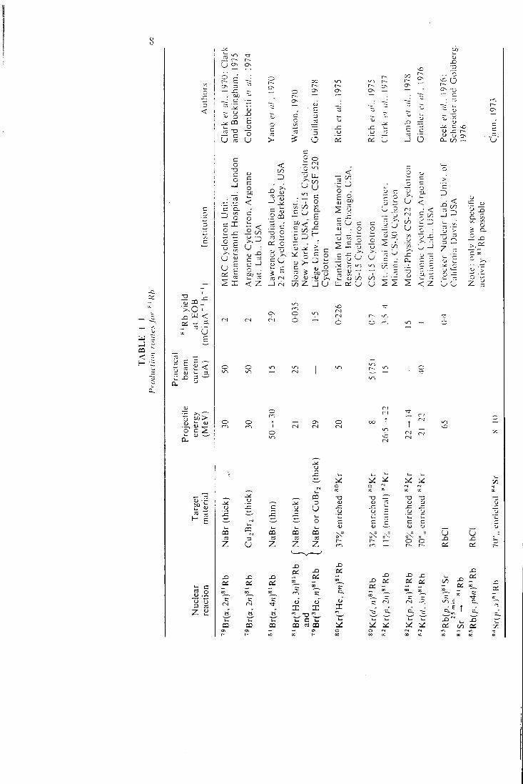
!2 j= _c UJ < a: - =-
< =1.
o c 2
c o
Z
c
u an
o
a:
i l
u
z
o
C C O
„ n < C CL
o O
?"
be
• - C
— CD :
X I -
ich
ich E c:
a: - J Q a.
< 3
(U - —
< 00
U : <" -e 51) —
< Z
o cn -
.2 c -g o
c£ o
5 E = —1 to
c
iro o
r-l o u .
o CS
c O
00 U CL
£ Ml
US
A.
5
:cri
n U
SA
.
Ml '
>
<
dj . o o
^ l l I 00 U
5 s u O
O N O r-1 O —
t o
o .—, c
'— •—' 03 B
r
Br
u Z Z
X3
X I X ) x > D i
XI 00 C C
-a-
t-i CD CD CQ ON 1 CO
CD
u
oa z
X )
Zlj <
• <; S 00
in
2 s s
o o CC 00 00 X
•o "O "O
dj r; JZ *^
tj o nr
nr
nr
-—- (i> ^ = = r - O
• < -5 00
J =3
Z -
< z u o z
u X )
u X
X CC
t. C CD M CO
X „
X 00 OS - — X
X CO X IT" : —
^ .E
X> 1^ a: 00 CC O oo X X X X X Xl c
I 'KOmc IION Ol KK^'PION S i m ( i I! N'P. R .Al() U .S 9
By using a 37",, enriched krypton 80 target both 20 MeV 'He and 8 iVleV deuterons have been u.sed lo produce useful quantities of rubidium 81, Natural and 70",, enriched ^•^Kr* have been irradiated with protons and deuterons to produce clinically useful generators (Clark cl <;/., 1977: Gindler et ai, 1976; and L a m b ei al.. 1978). In all target systems using krypton the *^'Rb product must be recovered from a large area of target wall and no particularly attractive method of achieving this has yet been reported, A remotely controlled target with a rotating glass or stainless steel liner may offer a practicable solution if suitable conditions can be found to effect an efficient recovery of *^'Rb from the walls (Palmer ei al.. 1973; Guillaume,-1976). Alternatively, a target whose walls can be remotely steam treated may be considered (McDona ld , 1977; Fr iedman. 1976), If enriched krypton is lo be used a highly reliable containment system should be employed.
The irradiation of rubidium targets with about 65 MeV protons has been 81 Sr for //; \ i\ a described primarily as a source of high purity ^ ' R b via
generator studies, but higher production rates may be possible if lower purities and specific activities are acceptable for in vitro generator applications. The proton irradiation of enriched ^'^Sr was proposed by C a n n (1973) but no practicable applications have been described.
Most methods of producing ^' Rb simultaneously produce other rubidium radionuclides which are of importance primarily when considering the whole body radiation dose due to rubidium breakthrough from solution generators. O f secondary concern is the provision of lead shielding for the generator assembly as - Rb"" emits particularly hard gamma-rays.
G E N E R A T O R D E S I G N
T w o types of ^ ' K r " ' generators have been reported and some of their characteristics are shown in Table l - I I . Both use a cation exchange material to retain the ^ 'Rb whilst allowing the ^ 'Kr"" to be recovered in either the solution or gas phase. However, they have quite different rubidium loading characteristics (see next section).
The first type developed in our laboratory (Clark et al., 1970) incorporates a column of the inorganic ion exchange material zirconium phosphate. The second type of generator developed in the Donner laboratory (Yano et al., 1970) and subsequently improved in the Veterans Administration Hospital, Hines, in collaboration with the Argonne National Laboratory (Colombetti et al., 1974) uses a cation exchanger of the organic type, e.g. Dowex 50 x 8.
Neither generator system may be eluted with isotonic (0-9%) N a C l
*70% enriched *^Kr, $7300 per litre (Nov., 1976), Mound Laboratorv. Miamisbure. Ohio 45342, USA.
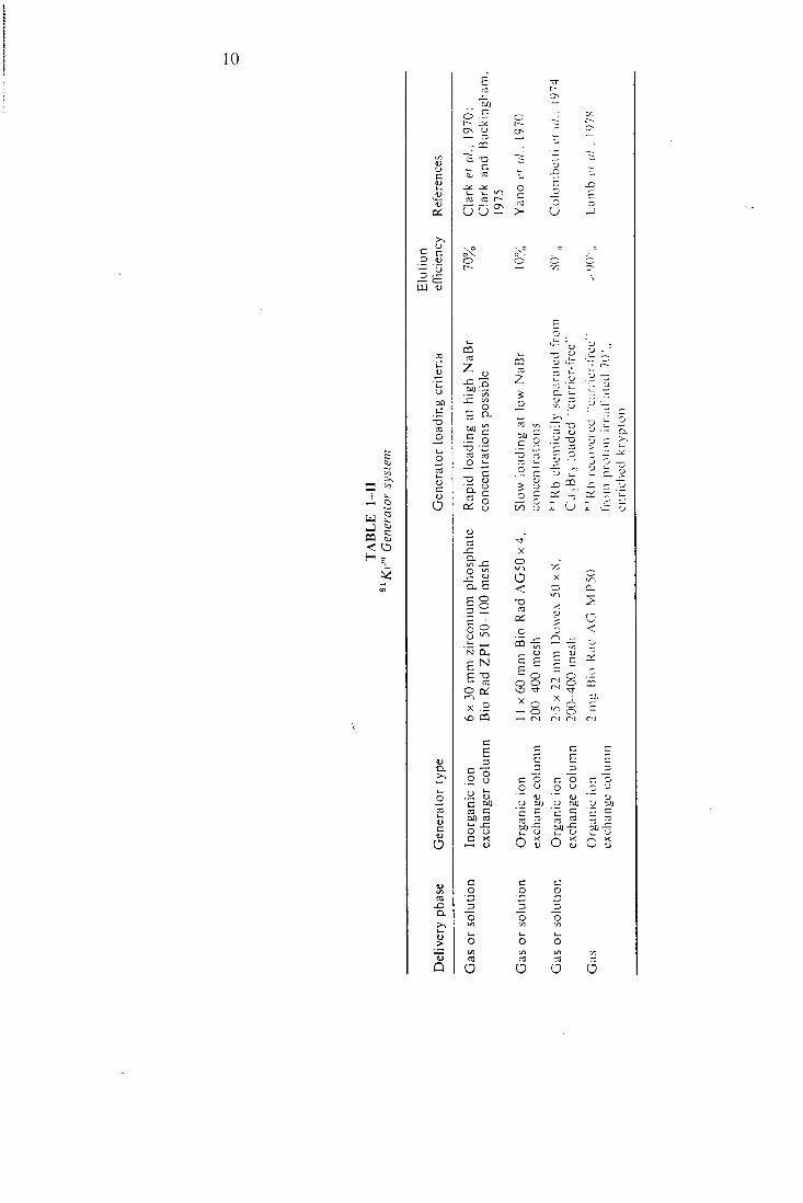
o a s u
CO c: t-^
u u 2 >
_5 o
U
> ,
O (U
CQ < O
c
a o
C
o
en
z 00
' D .
- E Q.
m —
so o V.
^ — — -a o
on Wl on
ica _ ?-
rati -o - E o rati
c _o ' J 1) GJ CO
nc
o nc X ) CO
5 CO
CO CO
6 '_»
o "5. p o
I
'Fo E
X . 2
X 1 .
2
X
o
< •a ^ 'i
E 1 ^
X - > ^
E ^
• O '^' O = - r^l f--l Ol
C
o
D
C 00 a c OX) 03
o-g C X
— (U
o _2 o
03
O
C E E -c "5 c o o
o CJ o o c •J to
CJ 00 u 00 "c c c c
03 "c —
00 - C 00 u
o ex
o ex
c cx
c C o o rj O o t/: u«
O O o3 03
o o a
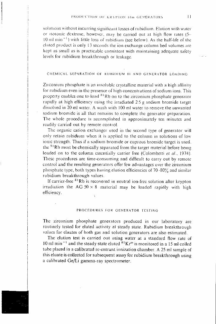
P R O D U C T I O N O l - R K ^ p • r O N f i l m G K Nl^ K A l O R S i i
solutions without incurring significant losses of rubidium. Elution with water or isotonic dextrose, however, may be carried out at high flow rates (5 -10 ml in in" ' ) with little loss of rubidium (see below). As the half-life of the eluied product is only 13 seconds the ion exchange column bed volumes are kept as small as is practicable consistent with maintaining adequate safety levels for rubidium breakthrough or leakage.
C H E M I C A L S E P A R A T I O N O F R U B I D I U M 81 A N D G E N E R A T O R L O A D I N G
Zirconium phosphate is an insoluble crystalline material with a high affinity for rubidium even in the presence of high concentrations of sodium ions. This property enables one to load Rb on to the zirconium phosphate generator rapidly at high efficiency using the irradiated 2 5 g sodium bromide target dissolved in 20 ml water. A wash with 100 ml water lo remove the unwanted sodium bromide is all that remains to complete the generator preparation. The whole procedure is accomplished in approximately ten minutes and readily carried out by remote control
The organic cation exchanger used in the second type of generator will only retain rubidium when it is applied to the column in solutions of low ionic strength. Thus if a sodium bromide or cuprous bromide target is used, the Rb must be chemically separated from the target material before being loaded on to the column essentially carrier free (Colombetti et al.. 1974), These procedures are time-consuming and difficult to carry out by remote control and the resulting generators offer few advantages over the zirconium phosphate type, both types having elution efficiencies of 70-80% and similar rubidium breakthrough values.
If carrier-free ^ ' R b is recovered in neutral ion-free solution after krypton irradiation the A G 50 x 8 material may be loaded rapidly with high efficiency.
P R O C E D U R E S F O R G E N E R A T O R T E S T I N G
The zirconium phosphate generators produced in our laboratory are routinely tested for eluted activity at steady state. Rubidium breakthrough values for eluates of both gas and solution generators are also estimated.
The elution test is carried out using water at a standard flow rate of 10 ml min " ' and the steady state eluted ^' Kr"" is monitored in a 15 ml coiled tube placed in a calibrated re-entrant ionization chamber. A 25 ml sample of this eluate is collected for subsequent assay for rubidium breakthrough using a calibrated G e / L i gamma-ray spectrometer.
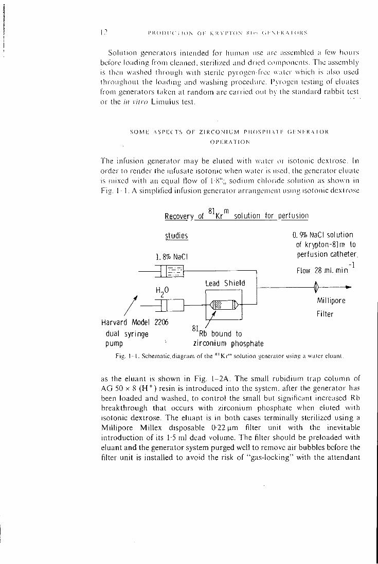
1 I ' K O D l i C 1 I O N O l " K K ^ L T O . N ,S I m < i I \ I K A I () K .S
SoluliiMi generators intentded for human use are assembled a few hours before loading from cleaned, slerihzed and dried ct^mponents. The assembly-is then washed through with sterile pyrogen-free water which is also used throughout the loading and washing procedure. I^yiogen testing of eluates from generators taken at random are carried out by the standard rabbit test or the //; vitro Limulus test.
S O M L A S P L C T S O F Z I R C O N I U M P 11 O S I '1 l A T I - ( 1 1 ; N i: K-X ro K
O P I : R A T I O N
The infusion generator may be eluted with water or isotonic dextrose. In order to render the infusate isotonic when water is u.sed. the generator eiuale is mixed with an equal flow of I H"; , sodium chloride solution as shown in Fig. 1 -1 . A simplified infusion generator arrangement using isotonic dextro.se
Recovery of ^^Kr^ solution for perfusion
studies
1.8% NaCI
Z'.'_!r.
H Lead Shield
0, 9?o NaCI solution of krypton-81m to perfusion catheter.
Flow 28 ml. min ^
Millipore
Filter Harvard Model 2206
dual syringe ""Rb bound to pump • zirconium phosphate
Fit : . 1 -1 . Schema t i c . ( j i a a r am o f the ' * ' K r ' " .solution "encra lor us in i ; ; i wa te r e k i t i n l .
as the eluant is shown in Fig . 1-2A. The small rubidium trap column of A G 50 X 8 (H"^) resin is introduced into the system, after the generator has been loaded and washed, to control the small but significant increased Rb breakthrough that occurs with zirconium phosphate when eluted with isotonic dextrose. The eluant is in both cases terminally sterilized using a Millipore Millex disposable 0 22 |im filter unit with the inevitable introduction of its 1-5 ml dead volume. The filter should be preloaded with eluant and the generator system purged well to remove air bubbles before the filter unit is installed to avoid the risk of "gas-locking" with the attendant
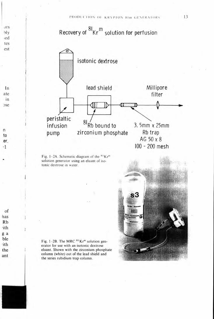
P R O D I ( I I O N o r K R - > P I ( ) N XI m C • I : N F R A I ( ) R ^ 13
j r s bly ;ed tes est
In ate
in Dse
n to er. -1
81 m Recovery of Kr solution for perfusion
isotonic dextrose
leatj shield
peristaltic gj infusion Rb bound to pump zirconium pfiosphate
Millipore filter
3.5mm x 25mm Rb trap
AG 50 X 8 100 - 200 mesfi
Fig, 1-2A. Schematic diagram of the "'Kr'^ solution generator using an eluanl of isotonic dextrose in water.
F i g , 1-2B, T h e M R C ^ ' K r " solution generator for use with an isotonic dextrose eluant. Shown with the zirconium phosphate co lumn (white) out of the lead shield and the series rubidium trap co lumn.
14 P R O D L i C T I O N O l - K R Y i ' l O N S i m (11: \ I-R A I'O R S
-2 - k ^
IKgcm air
Recovery of ^^Kr"^ in the gas phase
for ventilation studies
U A
Lead Shield
Flow 1 liter min
to face mask
•
F i g . I -. 'IA. Schematic d i t t i i r am o f the " ' K i ' " i;;is i ;cnci ; iU ' r
©
©
©
®
I ' ly , I }H. l i x p l o d c i l \ K'u o f ' i h e .\1RC " ' K i ' " u;is >-icnor;ilor.
risk of membrane fracture. A new filter unit should be used for each subject studied. Figure 1 -2B shows the M R C solution generator with its column exposed.
When a generator is used for ventilation studies it is eluted using air or j physiological gas mixture. It is essential to ensure that the eluting gas flow -.s saturated with water vapour before passing through the column as shown in Fig . 1-3A. Dry gas causes the column to dry out and little ^' Kr"" can then be recovered. Should a generator accidentally become dry it is possible to re-wet it and regain the normal high elution efficiency. Figure 1-3B shows the salient features of the M R C gas generator as currently distributed.
P R O D U C r i O N O F K R Y P r O N S i m G E N E R A T O R S 15
When using Kr'" generators it is essential to minimize losses of Kr"' by decay during its passage from the generator to the subject. This is achieved by keeping the diameters and lengths of the delivery tubing and catheters to a minimum and operating at flow rates consistent with their volume and the 13 second half-life of'^'Kr'".
Delivery of'^'Kr'" in the gas phase is normally accomplished using a disposable face mask as for oxygen administration but there may be occasions when the inhoinogeneous inhaled mixture delivered by this method has disadvantages. In these cases it may be necessary to provide some suitable preinixing system to cope with breath by breath administration. Occasionally small droplets of liquid may be found in the gas delivery tube and they may be found to contain small amounts of ^ ' R b . They are due to operating the generator with too inuch water in the saturator or at too high a flow rate. They may readily be prevented from reaching the face mask by the insertion of a small trap in the delivery tube close to the generator.
C O N C L U S I O N
Krypton 81m generators for infusion and ventilation studies have been developed for use in routine clinical investigations. Their widespread application will depend largely on a highly organized production and distribution system.
New applications continue to be found particularly for the infusion generator and the results reported so far seem to match the ingenuity of the investigators in tackling the problems of working with a 13 second half-life radionuclide in clinical research.
Chapter 6 37 C O M B I N E D U S E O F K R Y P T O N 81m A N D 85m I N
V E N T I L A T I O N S T U D I E S
T. Jones, J . C . Clark, C . G . Rhodes. J. Heather and P. Tofts
The continuous inhalation of krypton 81m gas prox idcs a sensitive means for detecting focal defects m lung ventilation (Fazio and Jones. 1975). Although of clinical value the lung ventilation unages obtamed have a hmiled functional interpretation. This is due to the regional lung counts being representative of the total ventilation V to that area and not ventilatory turnover K/vol which is the basic quantitative measure of ventilation and of which absolute values are required. Furthermore, the relationship between the content of ^' Kr'" in the lung and veniilation is nonlinear with a tendency to underestimate V as ventilatory turnover F 'vol increases. This is a particular problem when assessing ventilation images obtained in young children and certain disease states where high values of V/vo\ are experienced. The goal then is to be able to analyse the semiquantitative ^ ' K r ' " lung ventilation images to obtain absolute values of regional V/vol. In turn, this could be used to obtain the true distributions of both V. on the ^ 'Kr"" inhalation image, and regional lung blood How from the image obtained during the continuous intravenous infusion of ^ ' K r ' " solution (Harf at.. 1978; Ciofetta et «/., 1977). As discussed in the earlier paper on the theoretical use of ^ 'Kr"" , one approach to achieving quantitation would be to repeat the inhalation procedure using a krypton isotope with a different radioactive half-life to that of ^ 'Kr ' " . For this purpose we have elected to use cyclotron produced ^^^Kr"" (Clark and Buckingham, 1975) which has a half-life of 4-4 hours. This follows the initial use of this tracer as an internal standard in brain blood-flow studies with ^ ' K r " ' by Arnot et al. (1970). Figure 6-1 shows the time-activity curve one would theoretically expect to record over the lung during the continuous inhalation of (a) ^ 'Kr"" and (b) a long-lived krypton isotope such as "^Kr"". These curves have been calculated for a K/vol of 1 7 litre min " ' l i t re" ' (typical values in erect normal man) with the lung concentration of tracer being expressed as a percentage of that in the inspired gas. With the long-lived krypton the lung concentration wil l build up to reach that of the concentration in the inspired gas. The 13 second half-life of ^ 'Kr"" causes the equilibrium concentration in the lung to be appreciably less than that in the
''>- l M O N A R Y P H Y S I O L O G V
IS
)• a ts n
le 1-ol ss of l i -of of
i!£D2_concentration Inspired concentration
47
hdf life isotope
17 l/l/m<n
'Krtl3 sec txilf life
Time • sees
Fig. 6 -1 , Theoretical lime aciiviiy cuives that would be recorded over the lung, wiih a ventilatory turnover rate of 1 -7 I T ' min ^ ' . during the continuous inhalation of (a I a lone-lived
insoluble radioactive gas and (b) krypton 81m.
inspired gas. Thus by comparing the equihbrium level reached by *' Kr"* with that of ^^Kr"" it is possible to determine how nearly the short half-life isotope has reached equilibrium with the inspired concentration. In effect one is providing the lung with a challenge (radioactive decay) in an attempt to reach true equilibrium. From the measured response to this challenge it is possible to determine the rate of ventilatory turnover V/vo\. This process of challenge and observation of the consequence of introducing a tracer is not unlike that of challenging the lung wi th a delta function of activity, a procedure commonly used in the difTerentiation approach to measuring tissue flow rates. W i t h continuous administration of ^ 'Kr"", the challenge (half-life of the tracer) is known precisely. In wri t ing the basic formulae for the equilibration levels of ^'Kr*" and ^^Kr"" in the lung, (^ 'Kr"") and (^^Kr""), we have:
^ K/vol + 3-2 (1)
and ( 85
^7vor+a0026
48 I H I l . M O N . X K V I ' l l V S I C ) L C H ; \ -
For ^-'^Kr'" the emitted y ray used in detection has an energy of 150 keV which IS sufficiently close to the 190 keV photons from Kr'" for (/ tc be ihc same for both tracers. The radioactive decay rate of '''•''Kr'" {0 0026 mm ' ' ) is negligible relative to the typical value of K/vol . Hence by dividing the two lung signals we have;
3-2
8 1 K r " 7 a/(85m;
This expression takes the same form as that of equation (3) derived in the earlier theoretical paper. The relationship between the •--"'Kr"' lung signal and the concentration of this tracer in the inspired air prov ides the transposition between the detection efficiencies for monitoring the inspired concentration and the lung concentration.
r X p 11RIM i: N T . A L p K D C " I - : D i : K i-
The experimental procedure is to administer sequentially '"^'Kr'" and ^^Kr"' from a well-mixed reservoir. The resulting pulmonary distributions at equilibrium measured by the gamma camera are recorded in turn as numerical matrices, so providing values of regional ^ 'Kr ' " and "-""Kr'". On both occasions the tracers are pumped from the reservoir, which is at atmospheric pressure, through a thin bore tube which delivers the isotopes to a disposable face mask. At equilibrium the content of tracer in the tube is recorded with a scintillation counter viewing a short section of tube to give the ratio: Ca(81m)/C<3(85m) necessary for equation (3). This is used to operate on the regional (^^Kr" ' ) / (^ 'Kr" ' ) ratios determined for each of the matrix points and'-the computer is set to calculate expression (3) and so obtain a matrix of V/vo\ values. Before this is done it is necessary to correct for possible differences in photon energy window setting on the camera and scintillation probe. This is done by piping in turn the two tracers to a short tube placed in front of the gamma camera. In addition, the appropriate amount of absorber, equivalent to the chest wall, is placed between the tube and the camera face in order to simulate the differential absorption of the two tracers. Figure 6-2 shows how important this is, due to the spill-over in the 150 keV photo peak (74% abundant) f rom the 315 keV photons also emitted by ^^Kr"" (13% abundant). This energy spectrum was recorded with the gamma camera placed over the chest of a normal subject during the continuous inhalation of ^^Kr"". This calibration procedure allows for the additional counts falling into the 150 keV windows of the camera and probe.
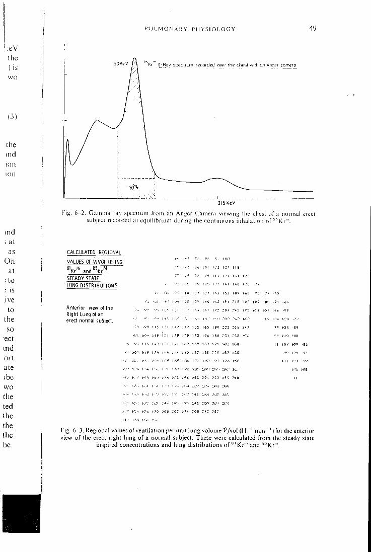
P U L M O N A K Y P H Y SI O L O G Y 49
;eV the ) is wo
(3)
the ind ion ion
ISOKeV l \ *^Kr 5-Roy spect rum recorded over the chest wi th on Arqer comero
315 KeV
Fig. 6 -2. Gamma ray spectrum from an Anger Camera viewing the chest of a normal erect subject recorded at equilibrium during the continuous inhalation of ^ 'Kr" ' .
CALCULATED REGIONAL
VALUES OF V / v a USING £ \ ' . c . 1 (?n
m , 85 M 96 l O t 103 1 ?7 l i s Kr and Kr
STEADY STATE 7 9 93 99 11 A 1-: 7 1-7.1 1-3;
LUNG DISTRIBUTIONS •^•I- 100 •99 100 1-3? 1-41 1 AO 1-3 6 77
i i o -•?•> 1-10 1-: 7 10 7 113 10 3 1-8 9 1-6 8 90 7A •60
-v.: . c i i . y - 1-:? 140 1-4 3 1 9 1 M 8 L'O? 109 88 •91 •6A
An te r i o r view of the ?; .9:- -v.' 10'. I-,'. 1 1-H 7
1-1 it 1 7; 20t r-AO 1 90 1-1 1 1-0 7 !•! 6 -99 Right Lung of an erect normal subject. V 1-r, 1 •-. •. 1 - • 1- •.: •. -:>••• •(:••' lOA 1-20
•?? iif- 1 ii 1-4? 1A7 1 1.6 0 1 80 :-2 2 338 I'A 7 99 1-3 3 -89
•fU 1-0" Mt"! i-'.M yj.p I S 8 I 73 1-7 6 1-8 0 20 1 :-3 8 l-"6 9? II 0 1-08
v.- M i v- to 1- 1 1-62 1-6 9 1-5 7 1-9 1 1.8 3 1 8 1 1 107 1-09 •83
K'''. 1-1 i i l.vH 1 1-.A 1-60 1-6 7 |.SB :• 9 1-8 3 108 99 I-2A -93
••• • J - : ; , i-f. i'f 11.'V )-c:.9 18.', 1 l-'?.' 1-7/. IF.ll t-1 I \C2 -yo
• V.'*' i : ' . . - . ]•/."'- I'Vtt > :i :• :v<p- -ft;. :v.r H:.-. W. 1 >08
• V . I - . - / l-Wi 1-60 1-"//-, l-i.'.. 1-7 6 1-8 s 30 3 1-9 0 M 8 \ 1
.... )•.":-• ]-'.-• 1 t-'-l 1- 1 -'i.i :;i..'-t -. :.vfifi
.... - i - i v i-.-.o I - V y.-.: ]•••• :!.vr> :v'-o •.•.?>r.
1-7'.' :'--J.\ :'-u': J.'-'V IV- : ;H !•! r&7
!-.•• i-':*! iv'o 170 ::'08 :-o:- 1-7-i :.0B r 4 - ?^ ?
Fig. 6-3. Regional values of ventilation per unit lung volume K/vol (11" ' m i n " ' ) for the anterior view of the erect right lung of a normal subject. These were calculated from the steady state
inspired concentrations and lung distributions of ^ ' K r " and '^Kr"".
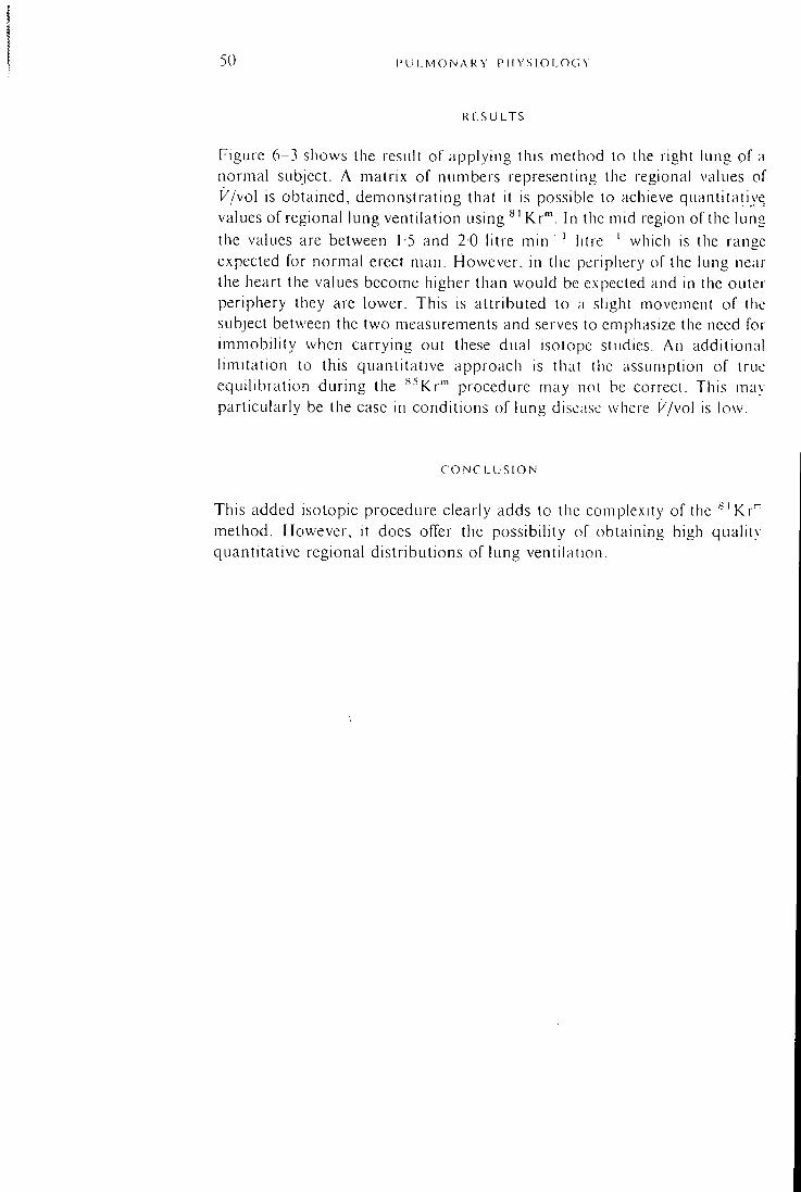
50 l ' U I . M O N / \ R Y P H V S I O L O G V
R E S U L T S
Figure 6-3 shows the result of applying this method to the right lung of a normal subject. A matrix of numbers representing the regional values of K/vol is obtained, demonstrating that it is possible to achieve quantitative values of regional lung ventilation using ^' Kr"' . In the mid region of the lung the values are between 15 and 20 litre m i n ' ' l i t r e " ' which is the range expected for normal erect man. However, in the periphery of the lung near the heart the values become higher than would be expected and in the outer periphery they are lower. This is attributed to a slight movement of the subject between the two measurements and serves to emphasize the need for immobili ty when carrying out these dual isotope studies. An additional limitation to this quantitative approach is that the assumption of true equilibration during the ^^Kr'" procedure may not be correct. This may particularly be the case in conditions of lung disease where k'/vol is low.
C O N C L U S I O N
This added isotopic procedure clearly adds to the complexity of the ' - 'Kr ' " method. However, it does offer the possibility of obtaining high quality quantitative regional distributions of lung ventilation.
D I S C U S S I O N III
! a of ve ng 'ge ,'ar ter he :br lal ue ay
Chairman: Phillip Hugh-Jones
PHILLIP !iUGH-.iONLS: 1 would like a chance of asking you about the changing ratio between " ' K r " ' and ^^Kr"' being solely due to movement of the patient.
T E R R Y JONLS: We did identify movement by looking at the qualitative profile and also we were able to superimpose the images by moving one of them. There was then a uniform gradient of values across the lung in the way that you might have predicted.
MiciiALL HUGiii;s: I think normalizing to the inspired concentrations of two gases is technically a little bit messy. It means introducing an extra counting channel and may not be a very useful tool for respiratory physicians because you are now going to end up with the distribution of total ventilation. The trouble is that the dead space of the lungs, that is the trachea and bronchi, have a volume which is only about one-twentieth of the alveolar compartment, and therefore when you compute ventilation per unit volume you will get very high values in the centre of the lung. This would not be so bad, but m the centre of the lung you would have the major bronchi with a high V/vo\ and the surrounding lung with a very low K/ \o l and the result is a rather confusing mixture.
TERRY JONES: Ycs. I am sure that is right but it is a matter of which way the signal is weighted. Where are the counts coming from"^ Are they coming mainly f rom the bronchi or the alveoli? There is a big weighting factor for the high volume areas.
Chapter 7 38 U S E O F K R Y P T O N 81m A N D 85m F O R
M E A S U R E M E N T O F V E N T I L A T I O N A N D P E R F U S I O N
D I S T R I B U T I O N W I T H I N T H E L U N G S
T . C . Amis, G . Ciofetta, J . C. Clark, J. M. B. Hughes, Hazel A. Jones and T. A. Pratt
The use of ^ 'Kr"" for measurement of pulmonary \entilalion and perfusion distributions (Fazio and Jones, 1975; Harf ci a\.. 1978) is subject to certain limitations because of the inlltience of regional lung volume and ventilatory washout of the radioactive gas.
As part of a general study into the efTecl of posture on regional disiribtil ion of ventilation and perfusion within the lung we have used information obtained during equilibration and clearance of the longer half-life (4-4 hours I isotope krypton 85m (^^Kr'") to correct that obtained with ''^'Kr'" for local alveolar volume and ventilatory washotit. In (Mder to demonstrate the application of this technique, results for two normal subjects in a lateral decubitus posture wil l be presented.
T H E O R Y
During steady state inhalation of ^ ' K r ' " counts are obtained from particular regions which reflect the balance between arrival of the radioactive gas. representing ventilation, and its removal due to ventilatory washout and radioactive decay (Fazio and Jones. 1975). Removal, except at high ventilatory rates, tends to be dominated by the high \ alue (3 2 min " ' ) for the decay constant (A) of ^'Kr™. Thus;
''N^^V^C,k'J[_{ViVA), + .^ (1)
where ^ ' N ^ is the regional counts obtained with ^ 'Kr"", is regional ventilation, Cj is inspired concentration of ^ 'Kr"". VA is alveolar volume and k'f^ the regional geometric factor. As the inspired concentration is not measured, regional counts are expressed as a fraction of the total counts for the lung field. That is :
81 T
\V/VA)r + X
iV/VA)^ + ;. kr
The subscript T refers to values for the total lung field.
IM i I, M O N A R Y P H Y S I O L O G Y 1 1
) N After equilibration with the longer half-life isotope "-'Kr"'. regional count
rates reflect local hmn volume.
(3
where " ^ V ^ refers to the regional count rate for ^^^Kr"', Cj the concentration of "- 'Kr'" throughout the lung and /c the regional geometric factor for «^Kr"' equilibration.
Local counts are expressed as a fraction of those for the total field: 85
8 5 Nr {VA)rkr
(4)
An arbitrary index for ventilation per unit alveolar volume is obtained by division of equations (2) and (4):
R/ i n h a l e d ,S I
{ y / v A ) R
(V/VA)r
\VIVA)r + }-
( l / / l / / l ) , + ;. (5)
Similarly, an intravenous infusion oC^'Kr" ' combined with equilibration and clearance of ^^Y.x"^ gives:
\VIVA)r + ).
{VIVA)^ + ). (6)
where QI^A refers to perfusion per unit alveolar volume. In equations (5) and (6) it has been assumed that the ratio of regional and
total geometric factors (kjkj-) is the same for both isotopes. This assumption is based on the similar principal gamma-ray energies of the two isotopes (190 keV for ^ 'Kr"" and 150 keV for ^^Kr" ' ) . In addition, errors associated wi th the relative counting efficiency of the detector for both isotopes are minimized by dealing with ratios of counts in this manner.
In the case of regional ventilation it is possible to solve equation (5) for regional ventilation per unit alveolar volume in litre m i n " ' l i t r e " ' .
{1+1/{V/VA) (7)
{V/VA)y is obtained f rom an initial slope analysis (Lassen, 1967) of the clearance of ^^i^^m f^^^^ tl^e total lung field.
In addition, {V/VA)^ has been estimated f rom the local clearance of ^^Kr"" for comparison with the results obtained f rom equation (7). Calculated { ^ / y ) R values are used to correct the Q/VA index obtained with equation (6).
54 P U L i V 1 0 N . - \ k Y I ' l l Y S I O I . O f . Y
M i r r i - lODS
For the '^ 'Kr"' ventilation and perfusion sltidy krypton is continuously eluted from a generator containing its parent " 'Rb (Jones ci al., 1970) and administered as described by Harf et al. (1978). except that the subject breathes through a mouth piece.
Immediately fol lowing the perfusion study the subject is switched into a rebreathing circuit containing a spirometer filled with oxygen and suflicienl ^^Kr"" to provide a concentration at equilibration of 1 mCi per litre. **^Kr'" is prepared in the Medical Research Council cyclotron by 16 MeV deuteron irradiation of krypton (Clark and Buckingham. 1975).
The subject takes a few deep breaths to speed equilibration and then breathes quietly until a satisfactory plateau of counts is obtained and an image representing alveolar volume is accumulated. The subject then breathes room air while clearance of the radioactive gas from the lungs is monitored.
Count rates are recorded with a large field gamma camera (Jumbo. Toshiba Corp.) using a low energy, high resolution collimator. The camera is linked to a computer (Nova 1220, Medical Data System) which displays the information as an image on a 64 x 64 matrix.
Areas of interest are selected and normalization and division undertaken as described previously. Washout curves are plotted for the total lung field and for regions within the lung. Rate constants are calculated from the initial slopes of these curves.
The total radiation dose to the lungs for a Kr"" ventilation and perfusion study lasting approximately four minutes is 40 mrad, while rebrealhing for three minutes at a lung concentration of 1 mCi ^^Kr"' per litre involves a dose to the lungs of 100 mrad.
R E S U L T S
Ventilation, perfusion and alveolar volume linages obtained for a normal subject in the left lateral decubitus posture are shown in Fig. 7-1. Ventilation, as measured by count density, appears evenly distributed between the upper and lower lungs. The volume distribution obtained with ^^Kr"" is heavily weighted to the upper lung.
Despite the uniform ventilation image, ventilation per unit alveolar volume is greater in the lower lung (2-23 litre m i n " ' l i t r e " ' ) than in the upper lung (094 litre m i n " ' l i t r e " ' ) as demonstrated by the clearance of ^^Kr-" (Fig. 7-2).
Ventilation per unit alveolar volume was found to increase f rom superior to inferior for a vertical slice taken across both lungs in a subject in right
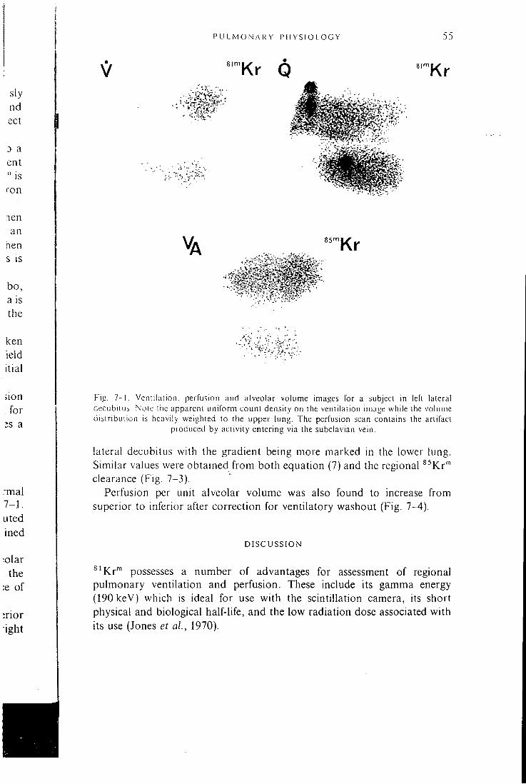
P LI 1. M O N A R Y P H Y S I O L O G Y
8|ni Kr Q
' • I T '
8|m Kr
Va 85^1 Kr
Fig. 7 -1 . Ventilation, perfusion and alveolar volume images for a subject in lefi lateral decubitus. Note tiie apparent uniform count density on the ventilation image while the volume distribution is heavily weighted to the upper lung. The perfusion scan contains the artifact
produced by activity entering via the subclavian vein.
lateral decubitus with the gradient being more marked in the lower lung. Similar values were obtained f rom both equation (7) and the regional ^^Kr"" clearance (Fig. 7-3).
Perfusion per unit alveolar volume was also found to increase from superior to inferior after correction for ventilatory washout (Fig. 7-4).
D I S C U S S I O N
^ 'Kr"" possesses a number of advantages for assessment of regional pulmonary ventilation and perfusion. These include its gamma energy (190ke'V) which is ideal for use with the scintillation camera, its short physical and biological half-life, and the low radiation dose associated with its use (Jones et al., 1970).
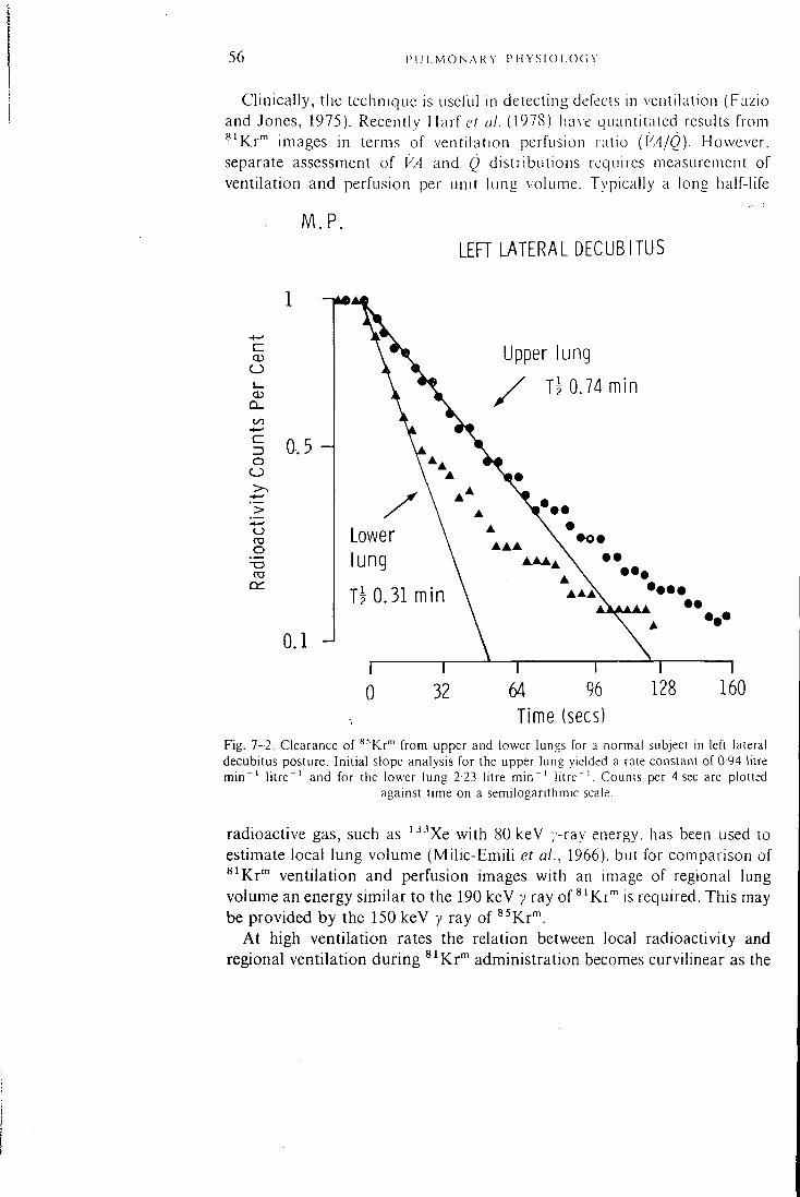
56 P U L M O N A R Y IM-I Y S I O l .OC. \
Clinically, the technique is useful in detecting defects in ventilation (Fazio and Jones, 1975). Recently Harf et al. (1978) ha\e qiiantitaled results from ^ ' K r " ' images in terms of ventilation-perfiision ratio (K4/0) . However, separate assessment of VA and Q distributions requires measurement of ventilation and perfusion per unit hing volume. Typically a long half-life
O
CD D _
Z3 O
o TO O
*-o cm
M . P . LEFT LATERAL DECUBITUS
0.5
0.1 -J
Upper lung
Ti 0.74 min
Lower lung
T l 0.31 min •••• •••
32 1
64 96 Time (sees)
160
Fig. 7-2. Clearance of ^^Kr"" from upper and lower lungs for a normal subject in left lateral decubitus posture. Initial slope analysis for the upper lung yielded a rate constant of 0 94 litre m i n " ' l i t r e " ' and for the lower lung 2-23 litre m i n " ' l i t r e " ' . Counts per 4 sec are plotted
against time on a semilogarithmic scale.
radioactive gas, such as '^•'Xe with 80 keV y-ray energy, has been used to estimate local lung volume (Mil ic-Emil i et al., 1966), but for comparison of 8ij(^j.m ventilation and perfusion images with an image of regional lung volume an energy similar to the 190 keV y ray of ^'Kr"" is required. This may be provided by the 150 keV y ray of ^^Kr'".
At high ventilation rates the relation between local radioactivity and regional ventilation during ^^Kr"" administration becomes curvilinear as the
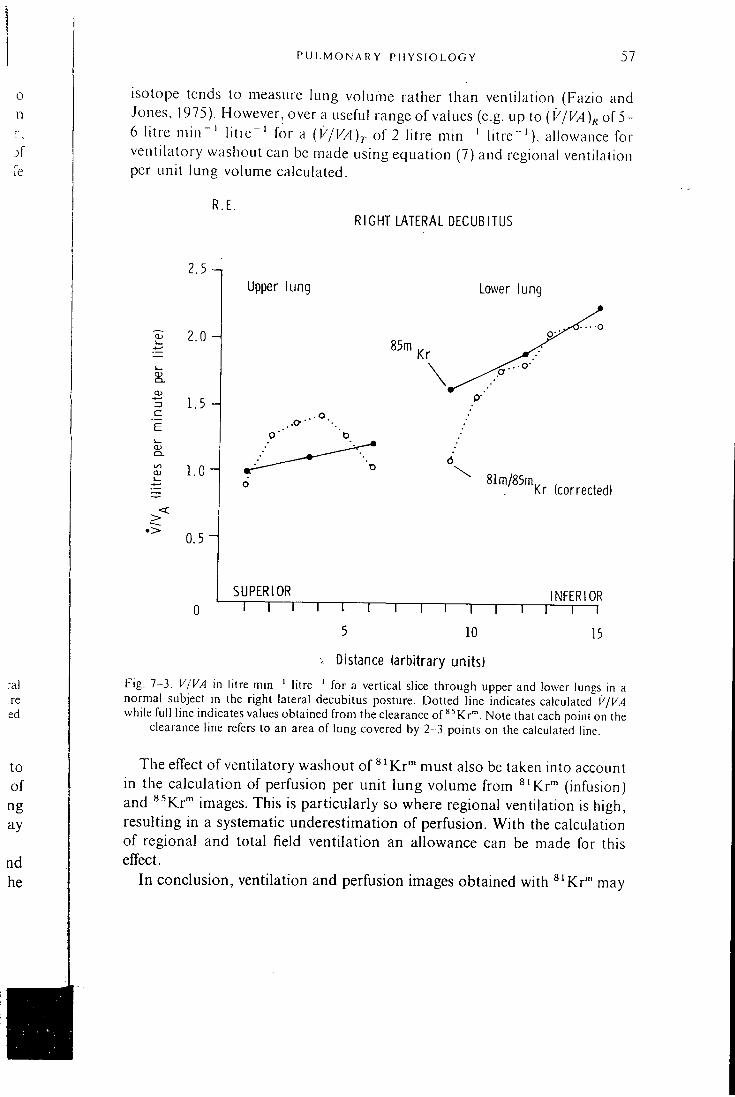
P U L M O N A R Y P H Y S I O L O G Y 57
0 n r ,
j f fe
isotope tends to measure lung volume rather than ventilation (Fazio and Jones, 1975). However, over a useful range of values (e.g. up to (K/K/ l )^ of 5-6 litre m i n " ' l i t r e^ ' for a {V/VA)r of 2 litre m i n " ' l i t r e " ' ) , allowance for ventilatory washout can be made using equation (7) and regional ventilation per unit lung volume calculated.
R.E. RIGHT LATERAL DECUBITUS
:al re ed
< >
2 . 5 - ,
2.0
3 1.5
Upper lung Lower lung
l . o H
0.5 i
8Im/85m Kr (corrected)
SUPERIOR
"I I — r T — i — 1 — r INFERIOR
"T"
10
1 T "1 1
15
V Distance (arbitrary units)
Fig. 7-3. V/VA in litre m i n " ' l i t r e " ' for a vertical slice through upper and lower lungs in a normal subject in the right lateral decubitus posture. Dotted line indicates calculated K/K,4 while ful l line indicates values obtained f rom the clearance of ^ ^ K r " . Note that each point on the
clearance line refers to an area of lung covered by 2-3 points on the calculated line.
to of ng ay
nd he
The effect of ventilatory washout of'^'Kr'" must also be taken into account in the calculation of perfusion per unit lung volume from ^'Kr"" (infusion) and ^^Kr"^ images. This is particularly so where regional ventilation is high, resulting in a systematic underestimation of perfusion. With the calculation of regional and total field ventilation an allowance can be made for this effect.
In conclusion, ventilation and perfusion images obtained with ^'Kr™ may
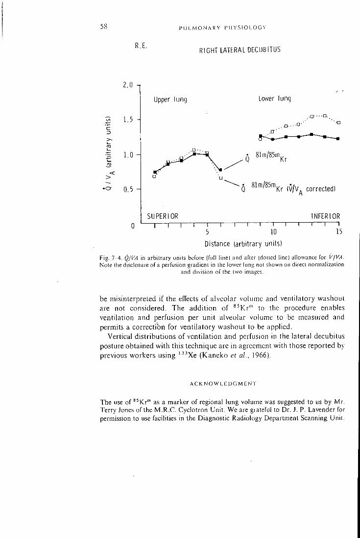
58 P U L M O N A R Y P H Y S I O L O G ^ •
R.E. RIGHT LATERAL DECUBITUS
c Z3 >-\— ra
2.0 n
1.5 A
1.0 H
0.5
Upper lung Lower lung
SUPERIOR 1 r
• 81m/85m|^^
Q 81m/85m^^ ^^^^^^^^^1
NFERIOR 1 1
15 5 10
Distance (arbitrary units)
Fig. 7-4. Q/VA m arbitrary units before (full line) and after (dotted line) allowance for V/VA. Note the disclosure of a perfusion gradient in the lower lung not shown on direct normalization
and division of the two imaees.
be misinterpreted if the effects of alveolar volume and ventilatory washout are not considered. The addition of ^^Kr"" to the procedure enables ventilation and perfusion per unit alveolar volume to be measured and permits a correction for ventilatory washout to be applied.
Vertical distributions of ventilation and perfusion in the lateral decubitus posture obtained with this technique are in agreement with those reported by previous workers using ^•'•'Xe (Kaneko et al., 1966).
A C K N O W L E D G M E N T
The use of ^^Kr" as a marker of regional lung volume was suggested to us by Mr. Terry Jones of the M . R . C . Cyclotron Unit. We are grateful to Dr. J. P. Lavender for permission to use facilities in the Diagnostic Radiology Department Scanning Unit.
D I S C U S S I O N IV
Chairman: Phillip Hugh-Jones
P H I L L I P H U G H - J O N E s : Thank y o u very m u c h Dr. Amis. It is interest ing that ,
y e a r s ago , people s h o w e d by b r o n c h o s p i r o m e t r y that the lower l u n g was the better vent i lated but I d id not expect to see s u c h a beaut i fu l d e m o n s t r a t i o n of
it as y o u h a v e s h o w n .
TERRY. lONEs : You were us ing the w a s h o u t to give y o u a c a l i b r a t i o n , btit the
w a s h o u t t echn iques are not very sa t i s fac tory . I w o n d e r whether we c o u l d use
the same a p p r o a c h , us ing ^ ' K r " ' or ^^Kr"", but m e a s u r i n g overal l vent i lat ion
per unit v o l u m e by m e a s u r e m e n t s t a k e n at the m o u t h . This c o u l d result in very a c c u r a t e values .
TERRY A M I S : It w o u l d be very h a r d to relate these va lues to the w a s h o u t you see w i th a g a m m a c a m e r a . It is poss ib le to o b t a i n a figure for the total
w a s h o u t of the w h o l e field but t h a t c a n be ha l f of the vent i lat ion as m e a s u r e d
at the m o u t h .
TERRY jONES: What I w a s sugges t ing w a s l o o k i n g at the end exp ired * 'Kr" ' and therefore getting a m e a s u r e of a c t u a l a l v e o l a r turnover , that is t idal
v o l u m e versus total l u n g c a p a c i t y .
Id
a ck ity
IS
: O f
;an his
Chapter 9
M E A S U R E M E N T O F T O T A L L U N G V E N T I L A T I O N
I N A N A E S T H E T I Z E D D O G S U S I N G K R Y P T O N 81m
A N D N I T R O G E N 13
R. N. Arnot, M. K . Sykes, J . C . Clark, A. N. Herring, M. K. Chakrabarti and P. Buranapong
The use of the radionuclide ^ 'Kr"" for the demonstration of regional underventilation in pulmonary disease by means of gamma camera imaging of the lungs during continuous inhalation of the gas was described by Fazio and Jones in 1975. ^ 'Kr"" has a half-life of 13 seconds, radioactive equilibrium is obtained rapidly, and the regional count rate depends on the relationship between the ventilation and the rates of wash-out and radioactive decay. In abnormal regions, the rate of wash-out is much less than the rate of radioactive decay (3 2 m i n ^ ' ) ; hence the regional count rate is highly dependent on the regional ventilation.
An alternative technique for assessing regional ventilation is based on the analysis of regional wash-out curves of an inert gas such as '-^^Xe or ^^'Nj-Although the use of ' ^ N j is restricted to locations where there is a cyclotron or other particle accelerator producing radionuclides, it has the advantage over '^^Xe of a lower solubility in blood and tissue which reduces errors due to radioactivity washing out of tissue into the lungs and to radioactivity in the chest wall . ^ N2 has been used in studies of both normal and abnormal lungs using collimated scintillation counters (Rosenzweig ei al., 1970) and gamma cameras (Ronchetti et al., 1975; Ewan et at.. 1975; Nosil et al., 1976a).
I t is of interest to assess whether the variation of ^^Kr'" count rate with change in ventilation is in accordance with the theoretical response, and whether the use of ' N2 in vivo wash-out curves to measure ventilation is valid. Rosenzweig et al. (1970) compared ^^Nj regional wash-out measurements with results derived f rom curves of the ' -^Nj concentration in expired air during wash-out, but, since the latter provide a measure of total lung ventilation, it was not possible to draw conclusions on the degree of correlation between the two techniques. No quantitative studies of the ^'Kr"" technique have been described. Using a gamma camera, we have obtained ^^Kr"" count rates and ' ^ N j wash-out curves in the whole lung field at varied rates of ventilation. Also, curves of ^ N2 concentration in expired air were obtained by end-tidal sampling, and alveolar ventilation and functional
39
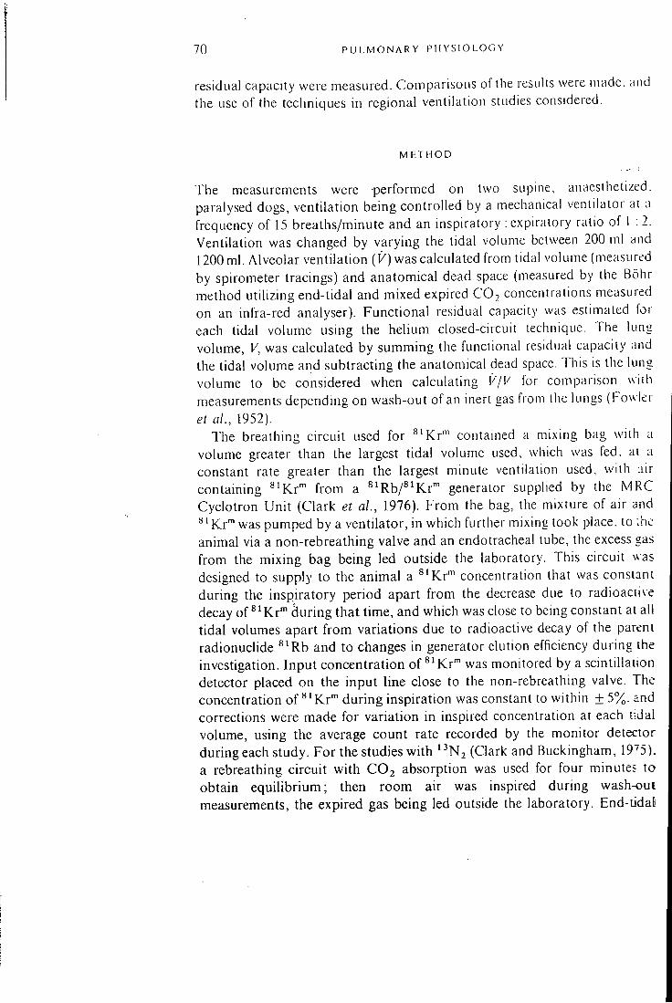
70 P U L M O N A R Y P H Y S I O L O G Y
residual capacity were measured. Comparisons of the results were made, and the use of the techniques in regional ventilation studies considered.
M E T H O D
The ineasurements were performed on two supine, anaesthetized, paralysed dogs, ventilation being controlled by a mechanical ventilator at a frequency of 15 breaths/minute and an inspiratory : expiratory ratio of 1 :2. Ventilation was changed by varying the tidal volume between 200 ml and 1200 ml. Alveolar ventilation (K) was calculated from tidal volume (measured by spirometer tracings) and anatomical dead space (measured by the Bohr method utilizing end-tidal and mixed expired C O , concentrations measured on an infra-red analyser). Functional residual capacity was estimated for each tidal volume using the helium closed-circuit technique. The lung volume, V, was calculated by summing the functional residual capacity and the tidal volume and subtracting the anatomical dead space. This is the lung volume to be considered when calculating V/V for comparison with measurements depending on wash-out of an inert gas from the lungs (Fowler et fl/., 1952).
The breathing circuit used for ^'Kr"Vcontained a mixing bag with a volume greater than the largest tidal volume used, which was fed. at a constant rate greater than the largest minute ventilation used, with air containing ^ 'Kr"" f rom a ^^Rb/^'Kr"" generator supplied by the MRC Cyclotron Unit (Clark et al., 1976). From the bag, the mixture of air and ^ 'Kr"" was pumped by a ventilator, in which further mixing took place, to ihe animal via a non-rebreathing valve and an endotracheal tube, the excess gas f rom the inixing bag being led outside the laboratory. This circuit was designed to supply to the animal a ^'Kr™ concentration that was constant during the inspiratory period apart f rom the decrease due to radioactive decay of ^ 'Kr"" during that time, and which was close to being constant at all tidal volumes apart f rom variations due to radioactive decay of the parent radionuclide ^^Rb and to changes in generator elution efficiency during the investigation. Input concentration of ^ 'Kr"" was monitored by a scintillation detector placed on the input line close to the non-rebreathing valve. The concentration of ^ 'Kr"" during inspiration was constant to within ± 5%. and corrections were made for variation in inspired concentration at each tidal volume, using the average count rate recorded by the monitor detector during each study. For the studies wi th ^• 'Nj (Clark and Buckingham, 19"5). a rebreathing circuit wi th CO2 absorption was used for four minutes to obtain equil ibr ium; then room air was inspired during wash-oui measurements, the expired gas being led outside the laboratory. End-tida^
P U L M O N A R Y P H Y S I O L O G Y 71
, and samples of expired ' • ' N j were passed through a coil in a well-type scintillation counter whose output was recorded on paper tape for computer processing.
Right lateral gamma camera views of the supine dogs were obtained using a high energy collimator, and data f rom both ^^Kr" ' and '^N2 studies were recorded on magnetic tape for computer processing. Maximum count rates of less than 7000counts/s were used, avoidmg the need to correct for gamma camera dead-time. Non-uniformity corrections were made, and an area covering the whole lung field was chosen (Fig. 9-1), thus minimizing effects
1.1.1.1 i . i . r I I t I.( M I M X U - I . I . I
I . [ . I . - N X . • • • • • • • ' M l I . I
t I I . I . t l . l ' X — ' • • • • • • • • • ' H I t I
• • • • • • • • • • « M > t V « V K H V V V S « » > ' - » l I
Fig. 9 - L Computer output of the right lateral gamma camera view of the lungs of a dog ventilated with ^ ' K r . The anterior of the animal is in the upper part of the view and its diaphragm to the right. The shadow of the heart is visible anteriorly. The area used for
quantitation is outlined.
due to different detector sensitivities to the 190 keV and 511 keV radiations of ^ 'Kr"" and '^N2. A modification of the computer program of Vernon and Glass (1971) was used to obtain the total ^ ' K r ' " counts accumulated and the 13 Nj wash-out curves corrected for background and radioactive decay. ^ 'Kr"" counts accumulated during the whole breathing cycle were obtained, and ' • ' N j counts accumulated during alternate time intervals of 2 seconds that included the inspiratory phase of the breathing cycle were used to obtain the ^ ^ N j wash-out curves. The ^ ^ N j wash-out curves were analysed into two exponential components using the S A A M compartment analysis program of Herman et al. (1962), and average fractional ventilation [V/V) was calculated. The expired air concentration curves were analysed similarly to obtain V/V.
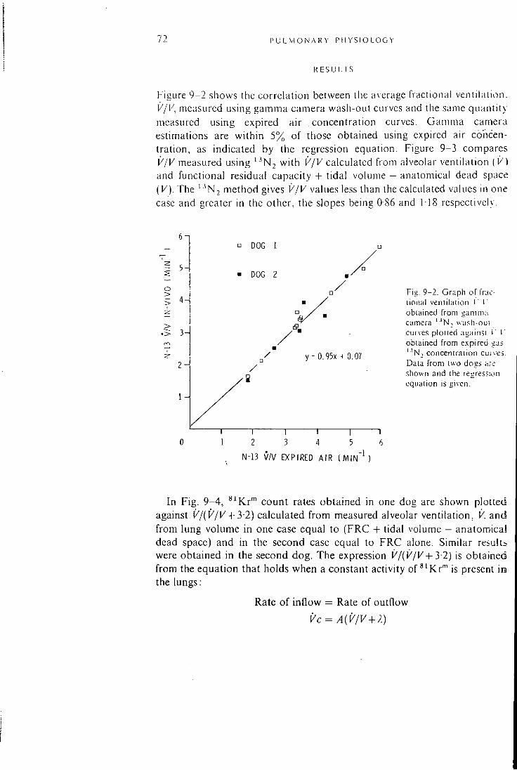
72 1' U I . M O N A K Y P i I Y SI ( ) L O G Y
R E S U L T S
Figure 9-2 shows the correlation between tlie average fractional ventilation. V/V, measured using gamma camera wash-out curves and the same quantity measured using expired air concentration curves. Gamma camera estimations are within 5% of those obtained using expired air concentration, as indicated by the regression equation. Figure 9-3 compares V/V measured using ' ^ N , with V/V calculated from alveolar ventilation ( V) and functional residual capacity - f tidal volume — anatomical dead space (V). The '^N2 method gives V/V values less than the calculated values in one case and greater in the other, the slopes being 0 86 and 118 respectively.
o ->
2 H
• DOG 1
DOG 2
y = 0.95x + 0.07
3 5 N - 1 3 V /V EXPIRED A I R ( M I N " ' )
Fig. 9-2. Graph of fiac-lional venlilaiion I T obtained from gamma camera '^N, u-a.-ih-oui curves pioued againsi > I ' obtained from expired gas '•'N, concentration curves. Data from two dogs a.'e shown and the regression equation is given.
In Fig. 9-4, ' '^Kr '" count rates obtained in one dog are shown plotted against V/(V/V + 3-2) calculated f rom measured alveolar ventilation, V. and from lung volume in one case equal to (FRC - I - tidal volume - anatomicaS dead space) and in the second case equal to FRC alone. Similar results were obtained in the second dog. The expression V/{V/V + 3-2) is obtained from the equation that holds when a constant activity of ^^Kr"" is present in the lungs:
Rate of inflow
Vc
Rate of outflow
AiV/V + A)
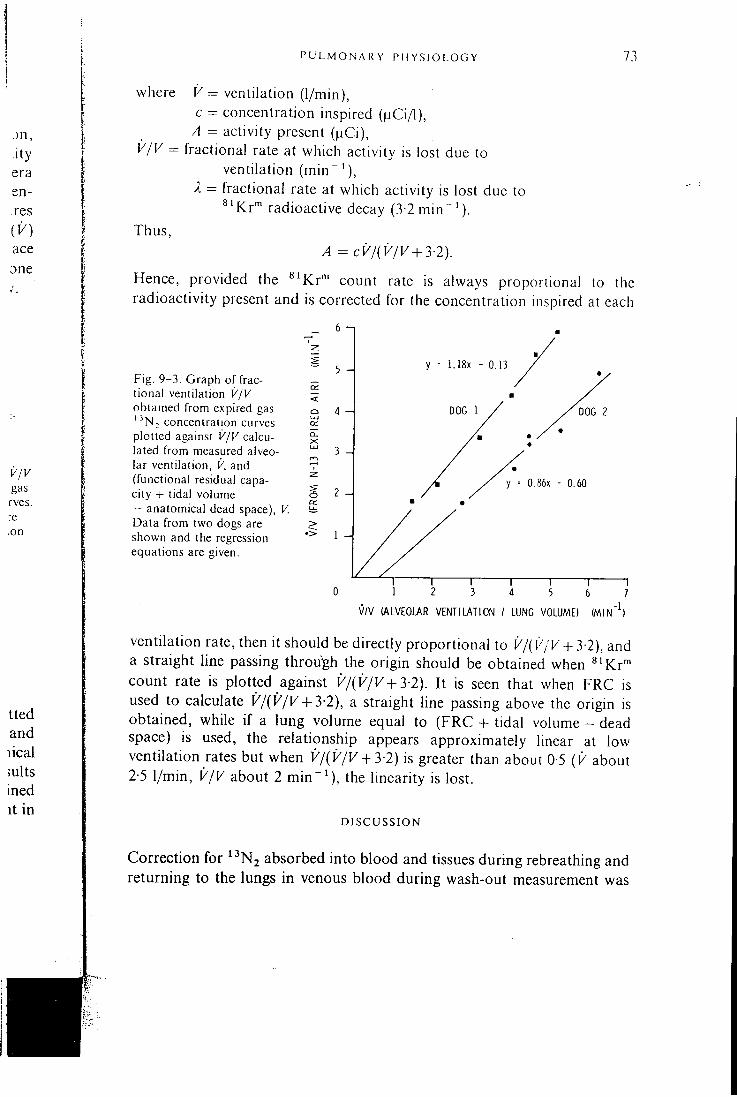
P U L M O N A R Y P H Y S I O L O G Y 73
where = ventilation (1/min), c = concentration inspired ( | iCi/ l ) ,
A = activity present ( j iCi) , V/V = fractional rate at which activity is lost due to
ventilation (min~ X = fractional rate at which activity is lost due to
^ 'Kr"" radioactive decay (3-2 m i n " ' ) . Thus,
A = cV/{V/V + 3-2).
Hence, provided the ^^Kr"" count rate is always proportional to the radioactivity present and is corrected for the concentration inspired at each
_ 6 - ,
Fig. 9-3. Graph of fractional ventilation VJV obtained from expired gas '^Nj concentration curves plotted against V/V calculated from measured alveolar ventilation, V, and (functional residual capacity + tidal volume - anatomical dead space), V. Data from two dogs are shown and the regression equations are given.
a.
5 H
4 H
3 -4
S 2
> •> 1 -A
y = 1.18X - 0 . 1 3
DOG 2
D.86X - 0 . 6 0
V / V ( A L V E O W R V E N T I L A T I O N / LUNG VOLUME) W I N )
ventilation rate, then it should be directly proportional to V/iV/V + 3-2), and a straight line passing through the origin should be obtained when ^^Kr"" count rate is plotted against V/{V/V +3-2). I t is seen that when FRC is used to calculate V/{V/V+3-2), a straight line passing above the origin is obtained, while i f a lung volume equal to (FRC + tidal volume - dead space) is used, the relationship appears approximately linear at low ventilation rates but when V/{V/V + 3-2) is greater than about 0-5 {V about 2-5 1/min, V/V about 2 m i n " ^ ) , the linearity is lost.
D I S C U S S I O N
Correction for ^^N2 absorbed into blood and tissues during rebreathing and returning to the lungs in venous blood during wash-out measurement was
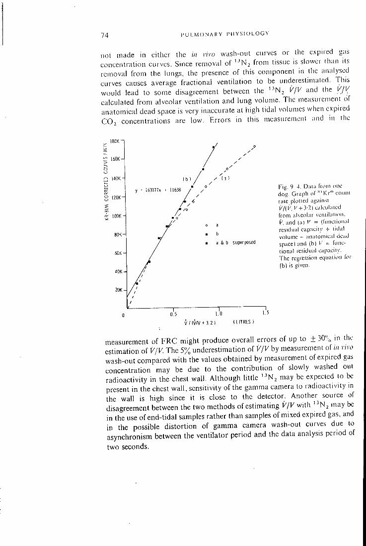
74 P U L M O N A R Y P H Y S I O L O G Y
not made m either the in vivo wash-out curves or the expned gas concentration curves. Since removal of '"^Nj from tissue is slower than its removal from the lungs, the presence of this component in the analysed curves causes average fractional ventilation to be underestimated. This would lead to some disagreement between the ' - ' N j V/V and the V/V calculated from alveolar ventilation and lung volume. The measurement of anatomical dead space is very inaccurate at high tidal volumes when expired CO2 concentrations are low. Errors in this measurement and in ihc
18OK
R 140KH
° 120Ki
a IOOKH
163177X
a & b superposed
V / (V/V + 3.2 (LITRES )
Fig. 9 4. D;ii;i fio(n one dog. Graph of Kr"' coiini rate plotied ag;iinsi 17(1' l' + .' -2l c.ilculaicd from alveolar vcniihinon. k and (a) K = (funciional residual capaciiy + lidal volume - anatomical dead space) and (b) = functional residual capaciiy. The regression equation for (b) is given.
measurement of FRC might produce overall errors of up to ± 30",, in the estimation of V/V. The 5% underestimation of V/V by measurement of in vivo wash-out compared with the values obtained by measurement of expired gas concentration may be due to the contribution of slowly washed out radioactivity in the chest wall. Although little ' ^ N j may be expected to be present in the chest wall , sensitivity of the gamma camera to radioactivity in the wall is high since it is close to the detector. Another source of disagreement between the two methods of estimating V/V with ' ^ N j may be in the use of end-tidal samples rather than samples of mixed expired gas, and in the possible distortion of gamma camera wash-out curves due to asynchronism between the ventilator period and the data analysis period of two seconds.
1' U L M O N A R Y P I I Y S I O L O G Y 75
Although agieement between the calculated values of V/V and the values measured with ' - ' N j was not obtained, Fig. 9-3 shows that the relationship is close to linear. Thus, measurements of V/V with ' • ' N j under different physiological conditions may be expected to give the correct relative values. This would also apply to measurements of relative regional V/V made with a gamma camera since the shapes of wash-out curves should not be affected by the different camera sensitivities to radioactivity in the regions under consideration. Measurements of relative regional ventilation, V, are also possible using '"^Nj if measurement of the relative volumes under consideration is made. This may be achieved in multiple studies by obtaining uniform ' ^ N j concentration throughout the lungs by rebreathing and obtaining images of the radioactivity distributions. The count density in any region is dependent on the camera sensitivity to radioactivity in that region, so the relative volumes of regions in any one image cannot be obtained; but when multiple studies are carried out, for example under different physiological conditions, without movement of the subject, then changes in relative regional volume and hence in relative regional ventilation can be obtained since sensitivity factors are then reduced.
The results given in Fig. 9-4 show that the ^^Kr"' response was in accordance with the predicted response only at the lower ventilation rates used if lung volume was calculated as (FRC + tidal volume - dead space), but that when FRC alone was used as the lung volume, a linear relationship between ^ 'Kr"" count rate and V/{V/V + 3-2) held over the whole range of ventilation rates used. This indicates that the lung volume V in the steady state equation for ^ 'Kr"" may be close to the FRC alone if counts are accumulated during the entire breathing period when expiration time is longer than inspiration time, as was the case in these experiments. At the lower ventilation rates, the increase in lung volume due to the addition of the tidal volume — dead space was small, resulting in little difference in the values of V/{V/V+3-2) calculated by the two methods and in little difference between the count rates recorded during inspiration and expiration. At higher ventilation rates, tidal volume - dead space becomes an increasingly higher fraction of lung volume, and the difference between the count rates recorded during inspiration and expiration increases. Thus, using an inspiratory : expiratory ratio of 1 : 2, the number of counts obtained f rom ^^Kr"" in the lungs during continuous accumulation was biased towards the measurement of the smaller volume. This finding may be of importance in techniques that involve measurement of high ventilation rates using both ^^Kr" ' images and estimations of V/V f rom wash-out curves (Amis et ai, 1978; Spaventi et al, 1978), since V/V obtained f rom wash-out curves refers to a lung volume equal to (FRC - f tidal volume — dead space) and the V/V in the steady state equation for ^ 'Kr"" may not.
76 P U L M O N A R Y P H Y S I O L O G Y
The use of ^^Kv"' images alone for investigating ventilation is limited owing to the non-linear relationship of count rate or count density with either V or V/V. The *^'Kr"' response is proportional to V/(V/V + }-2). Thus, except at very low values of V/V ( < 1) such as may be found in diseased lung, the response never approaches proportionality with V. In the human, the normal value of V/V is approximately half of that in the dog, hence the non-linearity of the relationship between ^ ' K r " ' count rate and ventilation V up to the normal range of ventilation is probably less pronounced in the human than in the dog. Nevertheless, even in this range, measurements of relative ventilation of the whole lung or of changes in relative regional ventilation under several different physiological conditions can be considered to indicate only the direction of change and not its magnitude. Values of relative regional ventilation in a single image cannot be obtained owing both to the non-linearity of the response of the technique and to the dependence of the count densities in different regions on different camera sensitivities to radioactivity in those regions. Additional measurements, such as those described by Spaventi et al. (1978), Amis et al. (1978) or Jones (1978) are necessary to obtain more precise data on ventilation from ^ ' K r ' " images. As outlined above, care in the use of such techniques may be necessary under certain ventilatory conditions.
A final point should be considered in assessing the usefulness of "^'Kr'" images in studies of regional ventilation. The distribution of count density in the lung field may also depend on the variation of ^'Kr"" concentration in the gas taken into the alveoli during inspiration. It has been found in the conscious human that the first part of the breath inhaled from residual volume is distributed preferentially to the upper (non-dependent) part of the lungs (Mil ic-Emil i et ai, 1966; Jones et ai, 1970; Grant et al., 1974). Therefore, if the inhaled ^ 'Kr"" is not of constant concentration, artefacts in count density distribution may occur. Some radioactive decay during inspiration is inevitable but may be of less consequence than the inhalation during the first part of the breath of dead-space gas in which ^ 'Kr"" concentration may be only 50% of that in the remainder of the tidal volume. In the dog the dead-space constitutes about 40% of the normal tidal volume. I t is less in the human, but its possible effect requires consideration.
C O N C L U S I O N
Using the described breathing circuits and data analysis techniques on whole immobilized lungs, the predicted relative responses of the ^ 'Kr"" and ' • ' N , techniques to changes in alveolar ventilation are obtainable, over a wide range in the case of ' ^ N j and over a range of ventilation rates from below
P U L M O N A R Y P 11 Y S I O L O G •> 77
normal to normal in the case of *"Kr" ' . With ' ' ^ N , , measurements of V/V in repeated studies of the whole lung and in either a single image or repeated studies of regions of the lung can be expected to give the correct relative values. Measurements of changes in relative V i n repealed studies can be made with ' -^Nj if the relative volumes being investigated are also measured. Using s 'Kr"' imaging alone, values of relative regional ventilation in a single image cannot be found, but in repeated studies of the whole lung or of regions, the direction of change in relative ventilation should be obtainable, provided artefacts due to non-constant *"Kr'" concentration during inspiration are not introduced. To obtain the magnitude of relative ventilation, measurements in addition to count densities are required. When repeated studies are carried out, whether with ^ ' K r ' " or ' - ^N, . it is important that there be no movement of the subject between studies.
Measurements of V/V using ' ^ N j gamma camera wash-out curves correlated well with those made using ' -^Nj expired gas concentration curves. Good agreement between these ' ^ N j measurements and the values of V/V calculated from measured alveolar ventilation, V. and lung volume (functional residual capacity -F tidal volume - anatomical dead space). K was not obtained; this is considered to be due to errors m the measurements of dead space and functional residual capacity and to non-correction o f ' ^ N , data for return of absorbed ' -^Nj f rom blood to the lungs.
A C K N O W L E D G M E N T S
We wish to thank Mr. P. Horlock and Mr. I . A. Watson for supplying the ^' Rb/^' Kr"" generators and Mr. S. C. Choong of the Computer Unit of the Royal Postgraduate Medical School for his assistance in computer programming.
D I S C U S S I O N V I
Chainnan: Phillip Hugh-Jones
P H I L L I P H U G H - j O N F ^ s : I didn't quite gather how you measure the dead space and functional residual capacity (FRC) on the animals.
KF.ITH S Y K R s : We measured FRC by the helium dilution technique. This increased with tidal volume. We also measured the dead space first by the Bohr and then by the Fowler technique; there were large c'.iscrepancies between the two which we are now trying to sort out.
P H I L L I P H U G H - j o N i E s : Why I questioned this is that you are using a dead space corrected ventilation per unit volume as your standard and so it is fairly critical.
ROSEMARY ARNOT: YCS indeed.
P H I L L I P HUGH-JONES: I think to me the great interest is the curvilinear relation of ventilation (K) to ^^Kr"" counts. This does seem linear over a useful range but it raises a problem with exercise.
M I C H A E L H U G H E S : I would like to ask Miss Arnot how she kept the inspired concentration of ^^Kr"" constant unless the bag was emptied with each breath? About the dead space errors: first the gas i n the dead space from the previous breath goes preferentially to the apex. That is physiological. The only additional error that you have with ^ 'Kr"" is the decay that has occurred between one cycle and the next. Normally the oxygen we exhale is re-inhaled with the next breath but nothing happens to it while it is sitting in the dead space. This is not quite the same with ^^Kr"".
ROSEMARY A R N O T : The aim was to achieve constant concentration of ^ ' K r " during each breath. The breaths might vary f rom each other but during each inspiratory period we think we got constant concentration simply by mixing all ages of gas wi thin the bag and again within the ventilator. When we monitored the concentration there was only about 5% variation during ihe inspiratory period.
MICHAEL H U G H E S : You monitored it over the trachea?
ROSEMARY ARNOT: NO , we monitored it just before entering the mouth. We
P U L M O N A R Y P H Y S I O L O G Y 79
ace
'his the cies
ace irly
ion nge
;red ach the
The red lied ead
confirmed your work (with B. J. B. Grant) on the regional distribution of the first part of the breath by looking at the regional distribution of ^' Kr"' using the circuit that 1 have described and comparing it with the regional distribution of a bolus of ^ ' K r " ' put into the endotracheal tube at the beginning of each breath. We found a preferential distribution to the upper regions with the bolus.
T I : R R Y JONES: Your relationship of ^ 'Kr"" counts to theoretical values (Fig. 9-4) does not go through the origin. Do you think that the theory of ^ ' K r " ' breaks down at very low ventilation rates?
ROSEMARY A R N O T ! The rcasou for its not going through the origin may be a technical error. We plotted the calculated response against the actual counts that we obtained and these had to be processed, corrected for concentration and for the decay of the generator. The calculated expression we plotted it against had to contain calculated measurements including alveolar ventilation and FRC and somewhere between the two there may be room for error. I would expect this to be the case rather than a breakdown in the theoretical equation.
E R v i N K A P L A N : You w c T C usiug normal animals, but in a clinical situation where, for instance, you are dealing with obstructive pulmonary disease and-you have emphysematous areas and bullae in these regions, you may have virtually total decay of the ^^Kr"" at the time of equilibration. Then all you can say about these areas is that the ventilation is so low that the counts which you are getting may be almost undetectable. So this refers back to Terry Jones' comment that the equation may not work at very low ventilatory rates. This wil l not cause any problems in a clinical situation.
ROSEMARY A R N O T : Of couTse in the poorly ventilated areas of the lung, because the count rates are so low, statistics are very bad.
ICr-" ach (ing
we the
We
Chapter 21 40
y y y t
n
y le
C L I N I C A L A P P L I C A T I O N S O F K R Y P T O N 8 l m
I N T H E A S S E S S M E N T O F R E G I O N A L M Y O C A R D I A L
P E R F U S I O N
A. P. Selwyn, R. E. Steiner, J. Clark, M . Raphael and G. Fo rse
n le
i l •y
d 3 )f
•y n le IS
•y •g IS It
IS
is Df ;h
is le le le iS
Angina pectoris and acute myocardial infarction are thought to be due to functional disturbances of regional myocardial perfusion in patients with segmental rigid coronary artery stenosis. A dynamic assessment of the distribution of blood flow particularly in relation to the stenotic lesions seen at arteriography may help to rationalize the treatment of angina pectoris and the application of coronary venous bypass graft surgery (CABGS).
The purpose of this paper is to introduce a technique using '*'Kr"' for the dynamic assessment of regional myocardial perfusion m man. The preliminary results are shown, demonstrating the potential of this ultra-short-lived, inert, freely-diffusing indicator.
M E T H O D S
Eighteen patients (all male, aged between 41 and 64 years) were selected for coronary arteriography and left ventricular angiography. Al l suffered typical anginal chest pain and all had ECG evidence of ischaemic heart disease. These symptoms were disabling and resistent to medical therapy in each patient. Informed consent wa$ obtained in each case.
Ten mg of Diazepam were given intramuscularly 30 minutes before the procedure. A pacing electrode was introduced percutaneously into the right femoral vein and positioned in the right atrium. Selective coronary arteriography and left ventricular angiography were performed via a Seldinger puncture of the right femoral artery and using the Judkins technique.
A number 7 French sheath was introduced into the femoral artery using a guide wire and introducer. This was used to introduce a modified ring catheter (Paulin, 1964). The catheter was heparinized and had a ring diameter approximately equal to the diameter of the aortic root in each patient. The patients were then moved in order to position their chests within the field of the gamma camera (Toshiba GCA 202).
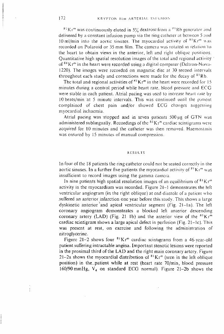
172 K R Y P T O N S i m A R T E R I A L I N I l i S I O N
^' Kr'" was continuously eluted in 5% dextrose from a Rb generator and delivered by a constant infusion pump via the ring catheter at between 5 and lOml/min into the aortic sinuses. The myocardial activity of ''^'Kr'" was recorded on Polaroid or 35 mm f i lm. The camera was rotated in relation to the heart to obtain views in the anterior, left and right oblique positions. Quantitative high spatial resolution images of the total and regional activity of ^ 'Kr"" in the heart were recorded using a digital computer (Dellron-Nova-1220). The images were recorded on magnetic disc at 30 second intervals throughout each study and corrections were made for the decay of ' "Rb .
The total and regional activities of ^ 'Kr"" in the heart were recorded for 15 minutes during a control period while heart rate, blood pressure and ECG were stable in each patient. Atr ia l pacing was used to increase heart rale by 10 beats/min at 5 minute intervals. This was continued until the patient complained of chest pain and/or showed ECG changes suggesting myocardial ischaemia.
Atrial pacing was stopped and in seven patients 500 |ig of G T N was administered sublingually. Recordings of the ^ ' K r " ' cardiac scintigrams were acquired for 10 minutes and the catheter was then removed. Haemosiasis was ensured by 15 minutes of manual compression.
R E S U L T S
In four of the 18 patients the ring catheter could not be seated correctly in the aortic sinuses. In a further five patients the myocardial activity of ^ ' K r ' " was insufficient to record images using the gamma camera.
In nine patients high spatial resolution images of an equilibrium o f ' ^ ' K r ' " activity in the myocardium was recorded. Figure 21-1 demonstrates the left ventricular angiogram (in the right oblique) at end diastole of a patient who suffered an anterior infarction one year before this study. This shows a large dyskinetic anterior 'and apical ventricular segment (Fig. 21-la) . The left coronary angiogram demonstrates a blocked left anterior descending coronary artery ( L A D ) (Fig. 2 1 - l b ) and the anterior view of the ^ 'Kr"" cardiac scintigram shows a large apical defect in perfusion (Fig. 21- lc) . This was present at rest, on exercise and following the administration of nitroglycerine.
Figure 21-2 shows four ^^Kr"" cardiac scintigrams from a 46-year-old patient suffering intractable angina. Important stenotic lesions were reported in the proximal third of the L A D and the right main coronary artery. Figure 21-2a shows the myocardial distribution of ^ 'Kr"" (seen in the left oblique position) in the._patient while at rest (heart rate 70/min, blood pressure 160/90 mmHg, V4 on standard ECG normal). Figure 21-2b shows the
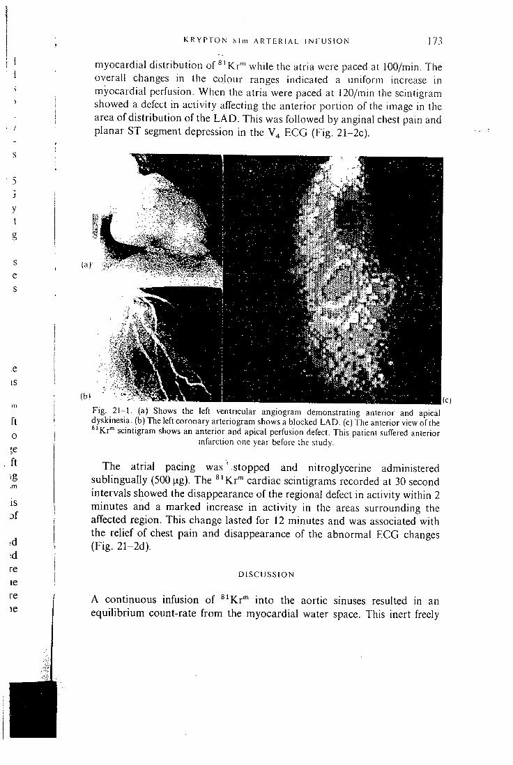
K R Y P T O N c S l m A R T E R I A L I N l U S I O N 173
myocardial distribution of ^ ' K r " ' while the atria were paced at 100/min. The overall changes in the colour ranges indicated a uniform increase in myocardial perfusion. When the atria were paced at 120/min the scintigram showed a defect in activity alTecting the anterior portion of the image in the area of distribution of the L A D . This was followed by anginal chest pain and planar ST segment depression in the V4 ECG (Fig. 21-2c).
.e IS
ft o ;e f t
3S ft
Fig. 2 1 - 1 . (a) Shows the left ventricular angiogram demonstrating anterior and apical dyskinesia, (b) The left coronary arteriogram shows a blocked L A D . (c)The anterior view of the ^'Kr"" scintigram shows an anterior and apical perfusion defect. This patient suffered anterior
infarction one year before the study.
The atrial pacing was ' stopped and nitroglycerine administered sublingually (500 )ag). The ^^Kr"* cardiac scintigrams recorded at 30 second intervals showed the disappearance of the regional defect in activity within 2 minutes and a marked increase in activity in the areas surrounding the affected region. This change lasted for 12 minutes and was associated with the relief of chest pain and disappearance of the abnormal ECG changes (Fig. 21-2d).
DISCUSSION
A continuous infusion of ^^Kr"" into the aortic sinuses resulted in an equilibrium count-rate f rom the myocardial water space. This inert freely
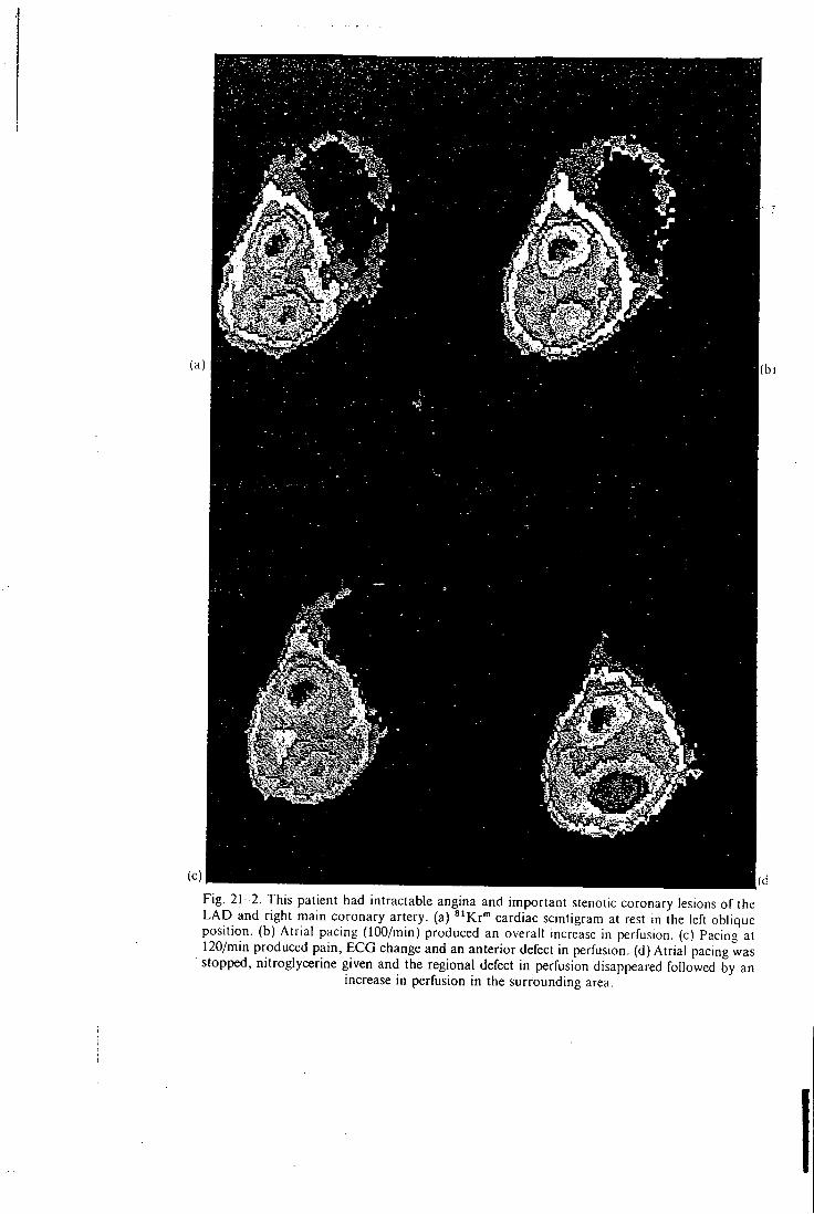
Fig. 2 1 - 2 . This patient had intractable angina and important stenotic coronary lesions of the L A D and right main coronary artery, (a) ^'Kr"" cardiac scintigram at rest in the left oblique position, (b) Atrial pacing (lOO/min) produced an overall increase in perfusion, (c) Pacing at 120/min produced pain, E C G change and an anterior defect in perfusion, (d) Atrial pacing was stopped, nitroglycerine given and the regional defect in perfusion disappeared followed by an
increase in perfusion in the surrounding area.
(b)
• K R Y P T O N S h n A R T E R I A L I N F U S I O N 175
diffusing radionuclide emits a single 190 keV gamma ray, ideally suited to high spatial resolution imaging using a gamma camera. The myocardial equilibrium of **'Kr'" in the heart depends on the arrival of the indicator by blood flow and radioactive decay. Animal studies have shown that moment to moment changes in the regional activity of ^ 'Kr"" can be used to assess changes in regional inyocardial perfusion.
Segmental rigid stenosis of a coronary artery in man interferes with the adaptation of myocardial perfusion necessary to meet the metabolic requirements of working ventricular tissue. Selective coronary arteriography wil l outline the luminal anatomy of affected vessels. This procedure cannot assess the relative importance of each lesion wi th respect to the disturbances of blood flow. Techniques using long-lived radionuclides provide information of the situation only at the moment of administration. The singular advantage of ^ 'Kr"" is that the short half life (13 seconds) allows dynamic studies of right and left coronary circulations and of each coronary venous bypass graft.
This short-lived tracer must, however, be delivered by invasive techniques and is therefore only applicable during angiography or during bypass graft surgery. The geometrical factors relating the heart to a gamma camera are complex and limit the separation of endocardial and epicardial events. Absolute quantitation of myocardial blood flow must assume a stable arterial concentration of the tracer. The ring catheters used in this study were not easily seated in the aortic sinuses, nor did they provide consistently adequate images. Clearly more work is required in order to design a catheter that is easily introduced and reliable when in position.
In conclusion, this preliminary study has shown that a continuous infusion of ^"Kr"" into the aortic sinuses of patients wi th intractable angina pectoris can provide an assessment of moment-to-moment changes in regional myocardial perfusion. The functional disturbances in myocardial blood flow can be related to the stenotic lesions seen on the angiogram. These dynamic techniques may provide a rational method o f investigating the effects of therapies and the application of CABG surgery.
(d)


