Embed Size (px)
Citation preview

Cytokeratin 18 Fragment Levels as a Noninvasive Biomarker forNonalcoholic Steatohepatitis in Bariatric Surgery Patients
Dima L. Diab1, Lisa Yerian6, Philip Schauer3, Sangeeta R. Kashyap1, Rocio Lopez7, StanleyL. Hazen4,5, and Ariel E. Feldstein2,4,5
1 Department of Endocrinology, Cleveland Clinic, Cleveland, OH, United States, 44195
2 Department of Pediatric Gastroenterology, Cleveland Clinic, Cleveland, OH, United States, 44195
3 Department of General Surgery, Cleveland Clinic, Cleveland, OH, United States, 44195
4 Department of Cell Biology, Cleveland Clinic, Cleveland, OH, United States, 44195
5 Center for Cardiovascular Diagnostics and Prevention, Cleveland Clinic, Cleveland, OH, United States,44195
6 Department of Anatomic Pathology, Cleveland Clinic, Cleveland, OH, United States, 44195
7 Quantitative Health Sciences, Cleveland Clinic, Cleveland, OH, United States, 44195
AbstractBackground & Aims—Nonalcoholic fatty liver disease (NAFLD) is extremely common amongmorbidly obese patients. We assessed the usefulness of plasma caspase-generated cytokeratin 18(CK-18) fragments as a novel marker for NAFLD in a bariatric cohort.
Methods—The cohort consisted of 99 consecutive patients who underwent liver biopsy at the timeof bariatric surgery. CK-18 levels were measured using an enzyme-linked immunosorbant assaybefore and 6 months after surgery. Patients were subdivided into four histological groups: notNAFLD (normal liver biopsy), NAFL, borderline diagnosis, and definitive nonalcoholicsteatohepatitis (NASH).
Results—CK-18 levels were significantly higher in subjects with NASH compared to those withnot NAFLD, NAFL, or borderline diagnosis (median [Q25, Q75]: 389 U/L [275, 839] vs. 196 U/L[158, 245], vs. 217 U/L [154, 228], or vs. 200 U/L [176, 274], respectively; P<0.0001). CK-18 levelswere significantly higher in subjects with moderate to severe fibrosis versus those with no or mildfibrosis (334.5 U/L [240.5, 896] vs. 207 U/L [175, 275], respectively; P=0.007). A significantdecrease in CK-18 levels was observed in most patients 6 months postoperatively. The area underthe ROC curve for NASH diagnosis was estimated to be 0.88 (95% CI: 0.77, 0.99). The values withthe best combination of sensitivity and specificity were 252 U/L (sensitivity = 82%, specificity =77%) and 275 U/L (sensitivity = 77%, specificity = 100%).
Conclusion—These results support the potential utility of this test for diagnosis and staging ofNAFLD before bariatric surgery.
Corresponding author: Dr. Ariel E. Feldstein, Departments of Pediatric Gastroenterology and Cell Biology, Cleveland Clinic Foundation,9500 Euclid Avenue, Cleveland, Ohio 44195, Tel: 216-444-5348, Fax: 216-444-2974, Email: [email protected] Disclosures: None; no conflict of interest existPublisher's Disclaimer: This is a PDF file of an unedited manuscript that has been accepted for publication. As a service to our customerswe are providing this early version of the manuscript. The manuscript will undergo copyediting, typesetting, and review of the resultingproof before it is published in its final citable form. Please note that during the production process errors may be discovered which couldaffect the content, and all legal disclaimers that apply to the journal pertain.
NIH Public AccessAuthor ManuscriptClin Gastroenterol Hepatol. Author manuscript; available in PMC 2009 November 1.
Published in final edited form as:Clin Gastroenterol Hepatol. 2008 November ; 6(11): 1249–1254. doi:10.1016/j.cgh.2008.07.016.
NIH
-PA Author Manuscript
NIH
-PA Author Manuscript
NIH
-PA Author Manuscript

KeywordsNonalcoholic fatty liver disease; NASH; apoptosis; biomarker; cytokeratin 18; bariatric surgery;morbid obese
INTRODUCTIONNonalcoholic fatty liver disease (NAFLD) is the most common cause of chronic liver diseasein the United States and occurs mainly in overweight or obese individuals (1,2). It is extremelycommon among patients undergoing bariatric surgery (3,4). NAFLD encompasses a widespectrum of conditions ranging from nonalcoholic fatty liver (NAFL) or steatosis tononalcoholic steatohepatitis (NASH) to cirrhosis (5). NASH is a potentially serious conditionsince as many as 25% of patients may progress to cirrhosis and experience its complications(6,7). A liver biopsy remains the only reliable way to differentiate steatosis from NASH anddetermine the stage and grade of the disease (8). Morbidly obese patients undergoing bariatricsurgery are a group at particular risk for NAFLD and for development of the more seriousforms of this condition (9,10). Development of reliable noninvasive biomarkers to diagnoseand determine disease severity prior to bariatric surgery and to monitor disease statuspostoperatively would be of significant clinical utility.
Increased hepatocyte death by apoptosis may play an important role in liver injury and diseaseprogression in NAFLD (11). During the process of apoptosis, effector caspases (mainly caspase3) are activated and cleave a number of substrates inside the cell including cytokeratin 18(CK-18), the major intermediate filament protein in the liver, resulting in the characteristicmorphologic changes of apoptosis (11). Previously, we demonstrated that the plasmaconcentration of caspase-generated CK-18 fragments accurately differentiate NASH fromNAFL and predicts stage of fibrosis in patients with NAFLD (12,13). The aim of this studywas to assess the utility of this novel biomarker in determining NASH, assessing diseaseseverity and monitoring disease status following bariatric surgery in morbidly obese patients.
PATIENTS AND METHODSPatient characteristics
The study was approved by the Cleveland Clinic Institutional Review Board. Our cohortconsisted of 99 consecutive patients who underwent liver biopsy at the time of bariatric surgeryas part of a standard clinical procedure. ’’The diagnosis of NAFLD was based on liver biopsyfeatures as assessed by an experienced hepatopathologist (L.Y.). Patients were subdivided intofour histological groups: not NAFLD (normal liver biopsy), NAFL, borderline diagnosis anddefinitive NASH. The NAFLD NIDDK activity score (14) was applied to each patient (seebelow). Demographic, clinical and laboratory data were obtained from clinic visits (2–4 wks)prior to surgery. The absence of current excessive alcohol use was defined by an average dailyconsumption of alcohol of ≤20 grams/day for men and <10 grams/day for women. Prevalenceof diabetes, hypertension and hyperlipidemia was assessed by review of past medical history.Prevalence of diabetes was based on past medical history and/or fasting plasma glucose of126mg/dl or greater.
Liver histology—The histological diagnosis of NAFLD was established by the studypathologist according to her expertise and following the NAS in a blinded manner regardingthe CK-18 fragment measurements and patient’s clinical and laboratory data (14). In thisscoring system, the degree of steatosis, liver injury and inflammatory activity is measured usingan 8-point scale (steatosis 0–3; lobular inflammation 0–3; ballooning degeneration ofhepatocytes 0–2). The NAS is the unweighted sum of steatosis, lobular inflammation and
Diab et al. Page 2
Clin Gastroenterol Hepatol. Author manuscript; available in PMC 2009 November 1.
NIH
-PA Author Manuscript
NIH
-PA Author Manuscript
NIH
-PA Author Manuscript

hepatocellular ballooning scores. The stage of fibrosis was similarly measured using a 6-pointscale (1a, b = mild (1a)/moderate (1b) zone 3 perisinusoidal fibrosis; 1c = portal fibrosis only;2 = zone 3 and portal/periportal fibrosis; 3 = bridging fibrosis; 4 = cirrhosis).
Measurement of caspase-generated CK-18 fragments in the blood—CK-18 levelswere measured in 86 patients who had plasma available within one week prior to surgery usinga sandwich immunoELISA specific for CK-18 fragments. Additionally, CK-18 levels weremeasured 6 months after bariatric surgery in those patients with available plasma (n = 34). Allsamples were initially processed to plasma and stored frozen at −80°C. The plasma wassubsequently used for quantitative measurement of the apoptosis-associated neo-epitope in theC-terminal domain of CK-18 by the M30-Apoptosense ELISA kit (PEVIVA, Alexis,Grünwald, Germany). All assays were performed in duplicate and the absorbance wasdetermined using a microplate reader (Molecular Devices M2, Sunnyvale, California, US).
Statistical Analysis—Descriptive statistics were computed for all variables. These includemeans and standard deviations or medians, as well as 25th and 75th percentiles for continuousfactors. For categorical variables, frequencies and percentages were estimated. Kruskal-Wallisand Dunn’s tests were used to assess whether CK-18 levels were significantly different betweenthe three subject groups. In addition, Wilcoxon rank sum tests were used to compare CK-18levels between subjects with moderate to severe fibrosis and those with mild fibrosis.Spearman’s correlation coefficients were used to assess associations between CK-18 levelsand histological characteristics. Logistic regression analysis was used to assess the associationbetween plasma levels of CK-18 fragments and the likelihood of having definitive NASH asopposed to simple steatosis. To predict the presence of NASH with optimal sensitivity andspecificity, receiver operating characteristic curve analysis was used to estimate potentialcutoff values of plasma CK-18 fragments. The same was done to assess the utility of CK-18levels in the prediction of fibrosis. A P value of 0.05 was considered statistically significant.SAS version 9.1 software (SAS Institute, Cary, NC) and R 2.0.1 software (The R Foundationfor Statistical Computing) were used to perform all analyses.
RESULTSCharacteristics of the patient population
The main clinical and laboratory characteristics of the patients are described in Table 1 whilethe histological characteristics of the liver biopsies are summarized in Table 2. The patients’age (median 51 years), gender (68% females), and BMI (median 48 kg/m2) did not statisticallydiffer among the four histological groups. There was no difference in the prevalence of diabetes,hypertension or hyperlipidemia among the groups. Serum AST and ALT were within thenormal range in most patients, although subjects with NASH tended to have significantly higherAST and ALT levels than both subjects without NAFLD and those with NAFL. In addition,borderline subjects had higher ALT levels than those without NAFLD.
CK-18 fragments are markedly increased in patients with definitive NASHPlasma levels of CK-18 fragments ranged from 103 to 1000 U/L (Median (Q25, Q75): 226 U/L (177, 298)). Compared to either subjects with not NAFLD, NAFL or borderline diagnosis,CK-18 levels were significantly higher in subjects with NASH (median (Q25, Q75): 196 (158,245) vs. 217 (154, 228) vs. 200 (176, 274) vs. 389 (275, 839), respectively; P<0.0001) (Fig.1). On the other hand, there was no evidence to suggest a difference in CK-18 levels betweensubjects without NAFLD, and those with NAFL or borderline diagnosis (P>0.40).
CK-18 fragment levels showed a significant positive correlation with NAS, the individual NAScomponents, as well as with fibrosis (Table 3). The majority of patients had no or mild fibrosis;
Diab et al. Page 3
Clin Gastroenterol Hepatol. Author manuscript; available in PMC 2009 November 1.
NIH
-PA Author Manuscript
NIH
-PA Author Manuscript
NIH
-PA Author Manuscript

nevertheless, CK-18 levels were significantly higher in subjects with moderate to severefibrosis (stage 2–3) than in those with no or mild fibrosis (stage 0 -1) (median (Q25, Q75):334.5 (240.5, 896) vs. 207 (175, 275), respectively; P=0.007) (Fig. 2, and Fig. 3). Moreover,we further performed a restricted analysis looking only at patients with NASH or borderlinediagnosis and found similar results: CK-18 levels were significantly higher in those patientswith borderline diagnosis or NASH with moderate to severe fibrosis than in those with no ormild fibrosis (334.5 (240.5, 896) vs. 234.5 (181.5, 346), respectively; P=0.047).
CK-18 fragments as an independent predictor of NASHThe risk of having definitive NASH on liver biopsy increased with increasing CK-18 fragmentlevels. For every 50 U/L increase in the plasma level of CK-18, the likelihood of having NASHincreased 2.45 times (OR (95% CI): 2.45 (1.20, 5.00)). Using the area under the receiveroperating characteristic (ROC) curve approach we next calculated potential cutoff values toseparate patients with “definitive NASH” from those with simple steatosis or borderlinediagnosis (Fig. 4). The area under the ROC curve was estimated to be 0.88 (95% CI: 0.77,0.99) and was found to be significantly higher than 0.5 (i.e. better than chance assignment).The values with the best combination of sensitivity and specificity were 252 U/L(sensitivity=82% and specificity=77%) and 275 U/L (sensitivity=77% and specificity=100%).The positive and negative predictive values with a CK-18 level of 252 U/L were 85.7% and71.4%, respectively, and with a CK-18 level of 275 U/L were 100% and 72.2%, respectively.
Changes in CK-18 fragment levels following bariatric surgeryCK-18 fragment levels were measured at 6 months after bariatric surgery in 34 patients (8 withnot NAFLD, 5 with NAFL, 11 with borderline diagnosis and 10 with NASH). The baselineand 6-month laboratory and clinical features of these patients are summarized in Table 4. Ofthe 34 patients, 3 (8.8%) had an increase in CK-18 levels and 31 (91.2%) had a decrease. CK-18decreases ranged between 13% and 88% of the original value with a median value of 44%.Initial CK-18 fragment concentration was found to be significantly correlated to the percentchange in CK-18 fragment levels (rho (95% CI): 0.59 (0.30, 0.88)). Subjects with NASH hada significantly greater decrease in CK-18 values than those without NASH (Median (Q25,Q75): 70 (50, 80) vs. (40 (20, 50); p=0.003). In addition, the percent change in CK-18 fragmentlevels was positively correlated to changes in both ALT and AST levels (Fig. 5).
DISCUSSIONObesity is major public health problem worldwide (15) and it is strongly associated withNAFLD, an increasingly recognized form of chronic liver disease that can progress to cirrhosisand end-stage liver disease (16,17). Morbidly obese patients are a population at particular riskfor developing NAFLD (18,19) and recent studies assessing the histological characteristics ofliver biopsies from these patients at the time of bariatric surgery have demonstrated thatNAFLD is almost universally present (3,20–22). Thirty to 50% of these patients may haveNASH and close to 50% some degree of fibrosis, while about 10% may have severe fibrosis(3). Moreover, increasing evidence suggests that bariatric surgery is an effective weight losstreatment that rapidly corrects many of the metabolic complications of obesity includingNAFLD (10,23). At the present time, an invasive liver biopsy is the only reliable way todiagnose NASH and assess the severity of liver damage (8). A reliable non-invasive test to notonly assess for NASH and disease severity prior to surgery but also allow for frequentmonitoring of disease status after bariatric surgery would be of great clinical utility.
Emerging data suggest that hepatocyte apoptosis may be a key component of the “second hit”involved in the progression of NAFLD to the more severe forms of this disease (11,24–27). Acentral consequence of the apoptotic process is the activation of the effector caspases (mainly
Diab et al. Page 4
Clin Gastroenterol Hepatol. Author manuscript; available in PMC 2009 November 1.
NIH
-PA Author Manuscript
NIH
-PA Author Manuscript
NIH
-PA Author Manuscript
caspase 3) which cleave a number of different substrates inside the cell including cytokeratin18 (CK-18), the major intermediate filament protein in the liver, resulting in the characteristicmorphologic changes of apoptosis. We have demonstrated in a small pilot study using a specificimmunoELISA assay that these fragments are strikingly increased in the serum of patients withNASH as compared to both patients with NAFL and normal liver biopsies (13). Using thisnovel approach in a recent study, we were able to demonstrate that determination of CK-18fragments in the blood accurately identifies the presence of NASH and the severity of fibrosison liver biopsy in adult patients with well-characterized NAFLD (12). Our current data extendthese observations by demonstrating that determination of CK-18 fragment levels in the bloodaccurate identifies the presence of NASH and disease severity in morbidly obese patientsundergoing bariatric surgery. Using the AUC approach, two cutoff values were identified: thefirst one to minimize the rate of false positive results (275 U/L) with a specificity of 100% anda sensitivity of 77%, and a second one to minimize the false negative rates (252 U/L) with aspecificity of 77% and a sensitivity of 82%. Finally, bariatric surgery resulted in a dramaticdecrease in CK-18 levels 6 months following surgery. These changes were greater in thosesubjects with NASH and positively correlated with changes in transaminases, suggesting thatmeasuring CK-18 fragment levels in the blood may be a useful test to monitor disease statuspostoperatively. A limitation of the current study in this regard is that we do not have followup liver biopsies to assess histological changes after surgery. However, previous data (10,23)clearly demonstrate a dramatic effect of bariatric surgery to achieve profound weight loss,normalize hyperlipidemia, resolve hyperglycemia and improve NAFLD. Therefore, to furtherassess the utility of this biomarker to monitor disease status following surgery and as aprognostic marker in this population, we are in the process of planning a larger longitudinalprospective study with baseline and 1-year follow up liver biopsies. Thus, the CK-18 testappears to have several unique features that fulfill many of the requirements for an idealbiomarker for NAFLD including that the test is simple, easy to measure and handle, and isreproducible. It not only identifies the presence of NASH but also the risk of associated fibrosis,and it allows for monitoring disease progression over time.
In summary, our findings support the usefulness of this test as a noninvasive NASH biomarkerin the care of morbidly obese patients undergoing bariatric surgery.
AcknowledgementsThis work was supported by the Cleveland Clinic General Clinical Research Center (M01 RR-018390) and by NIHgrant (DK076852) and the AGA Research Scholar Award (RSA) to AEF.
References1. Angulo P. Nonalcoholic fatty liver disease. N Engl J Med 2002;346:1221–1231. [PubMed: 11961152]2. Wieckowska A, Feldstein AE. Nonalcoholic fatty liver disease in the pediatric population: a review.
Curr Opin Pediatr 2005;17:636–641. [PubMed: 16160540]3. Machado M, Marques-Vidal P, Cortez-Pinto H. Hepatic histology in obese patients undergoing
bariatric surgery. J Hepatol 2006;45:600–606. [PubMed: 16899321]4. Kroh M, Liu R, Chand B. Laparoscopic bariatric surgery: what else are we uncovering? Liver pathology
and preoperative indicators of advanced liver disease in morbidly obese patients. Surg Endosc. 20075. Brunt EM, Tiniakos DG. Pathological features of NASH. Front Biosci 2005;10:1475–1484. [PubMed:
15769638]6. Adams LA, Lymp JF, St Sauver J, Sanderson SO, Lindor KD, Feldstein A, Angulo P. The natural
history of nonalcoholic fatty liver disease: a population-based cohort study. Gastroenterology2005;129:113–121. [PubMed: 16012941]
7. Ekstedt M, Franzen LE, Mathiesen UL, Thorelius L, Holmqvist M, Bodemar G, Kechagias S. Long-term follow-up of patients with NAFLD and elevated liver enzymes. Hepatology 2006;44:865–873.[PubMed: 17006923]
Diab et al. Page 5
Clin Gastroenterol Hepatol. Author manuscript; available in PMC 2009 November 1.
NIH
-PA Author Manuscript
NIH
-PA Author Manuscript
NIH
-PA Author Manuscript
8. Wieckowska A, McCullough AJ, Feldstein AE. Noninvasive diagnosis and monitoring of nonalcoholicsteatohepatitis: Present and Future. Hepatology 2007;46:582–589. [PubMed: 17661414]
9. Angulo P. NAFLD, obesity, and bariatric surgery. Gastroenterology 2006;130:1848–1852. [PubMed:16697746]
10. Mathurin P, Gonzalez F, Kerdraon O, Leteurtre E, Arnalsteen L, Hollebecque A, Louvet A, et al. Theevolution of severe steatosis after bariatric surgery is related to insulin resistance. Gastroenterology2006;130:1617–1624. [PubMed: 16697725]
11. Feldstein AE, Gores GJ. Apoptosis in alcoholic and nonalcoholic steatohepatitis. Front Biosci2005;10:3093–3099. [PubMed: 15970563]
12. Wieckowska A, Lopez AR, Zein NN, McCullough AJ, Feldstein AE. Noninvasive assessment ofhepatocyte apoptosis in nonalcoholic fatty liver disease: A multi-center validation study.Gastroenterology 2007;132:A-729.
13. Wieckowska A, Zein NN, Yerian LM, Lopez AR, McCullough AJ, Feldstein AE. In vivo assessmentof liver cell apoptosis as a novel biomarker of disease severity in nonalcoholic fatty liver disease.Hepatology 2006;44:27–33. [PubMed: 16799979]
14. Kleiner DE, Brunt EM, Van Natta M, Behling C, Contos MJ, Cummings OW, Ferrell LD, et al. Designand validation of a histological scoring system for nonalcoholic fatty liver disease. Hepatology2005;41:1313–1321. [PubMed: 15915461]
15. Friedman JM. Obesity in the new millennium. Nature 2000;404:632–634. [PubMed: 10766249]16. Machado M, Cortez-Pinto H. Non-alcoholic steatohepatitis and metabolic syndrome. Curr Opin Clin
Nutr Metab Care 2006;9:637–642. [PubMed: 16912563]17. Marchesini G, Bugianesi E, Forlani G, Cerrelli F, Lenzi M, Manini R, Natale S, et al. Nonalcoholic
fatty liver, steatohepatitis, and the metabolic syndrome. Hepatology 2003;37:917–923. [PubMed:12668987]
18. Haynes P, Liangpunsakul S, Chalasani N. Nonalcoholic fatty liver disease in individuals with severeobesity. Clin Liver Dis 2004;8:535–547. viii. [PubMed: 15331062]
19. Collantes R, Ong JP, Younossi ZM. Nonalcoholic fatty liver disease and the epidemic of obesity.Cleve Clin J Med 2004;71:657–664. [PubMed: 15449761]
20. Moretto M, Kupski C, Mottin CC, Repetto G, Garcia Toneto M, Rizzolli J, Berleze D, et al. Hepaticsteatosis in patients undergoing bariatric surgery and its relationship to body mass index and co-morbidities. Obes Surg 2003;13:622–624. [PubMed: 12940291]
21. Harnois F, Msika S, Sabate JM, Mechler C, Jouet P, Barge J, Coffin B. Prevalence and predictivefactors of non-alcoholic steatohepatitis (NASH) in morbidly obese patients undergoing bariatricsurgery. Obes Surg 2006;16:183–188. [PubMed: 16469221]
22. Dallal RM, Mattar SG, Lord JL, Watson AR, Cottam DR, Eid GM, Hamad G, et al. Results oflaparoscopic gastric bypass in patients with cirrhosis. Obes Surg 2004;14:47–53. [PubMed:14980033]
23. Mattar SG, Velcu LM, Rabinovitz M, Demetris AJ, Krasinskas AM, Barinas-Mitchell E, Eid GM, etal. Surgically-induced weight loss significantly improves nonalcoholic fatty liver disease and themetabolic syndrome. Ann Surg 2005;242:610–617. 618–620. [PubMed: 16192822]
24. Feldstein AE, Canbay A, Angulo P, Taniai M, Burgart LJ, Lindor KD, Gores GJ. Hepatocyte apoptosisand fas expression are prominent features of human nonalcoholic steatohepatitis. Gastroenterology2003;125:437–443. [PubMed: 12891546]
25. Feldstein AE, Canbay A, Guicciardi ME, Higuchi H, Bronk SF, Gores GJ. Diet associated hepaticsteatosis sensitizes to Fas mediated liver injury in mice. J Hepatol 2003;39:978–983. [PubMed:14642615]
26. Feldstein AE, Werneburg NW, Canbay A, Guicciardi ME, Bronk SF, Rydzewski R, Burgart LJ, etal. Free fatty acids promote hepatic lipotoxicity by stimulating TNF-alpha expression via a lysosomalpathway. Hepatology 2004;40:185–194. [PubMed: 15239102]
27. Feldstein AE, Werneburg NW, Li Z, Bronk SF, Gores GJ. Bax inhibition protects against free fattyacid-induced lysosomal permeabilization. Am J Physiol Gastrointest Liver Physiol2006;290:G1339–1346. [PubMed: 16484678]
Diab et al. Page 6
Clin Gastroenterol Hepatol. Author manuscript; available in PMC 2009 November 1.
NIH
-PA Author Manuscript
NIH
-PA Author Manuscript
NIH
-PA Author Manuscript
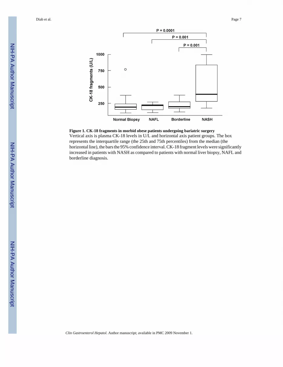
Figure 1. CK-18 fragments in morbid obese patients undergoing bariatric surgeryVertical axis is plasma CK-18 levels in U/L and horizontal axis patient groups. The boxrepresents the interquartile range (the 25th and 75th percentiles) from the median (thehorizontal line), the bars the 95% confidence interval. CK-18 fragment levels were significantlyincreased in patients with NASH as compared to patients with normal liver biopsy, NAFL andborderline diagnosis.
Diab et al. Page 7
Clin Gastroenterol Hepatol. Author manuscript; available in PMC 2009 November 1.
NIH
-PA Author Manuscript
NIH
-PA Author Manuscript
NIH
-PA Author Manuscript

Figure 2. CK-18 fragments are increased with the severity of fibrosis on liver biopsyVertical axis is plasma CK-18 levels in U/L and horizontal axis is the grade of fibrosis. Thebox represents the interquartile range (the 25th and 75th percentiles) from the median (thehorizontal line), the bars the 95% confidence interval. CK-18 fragment levels were significantlyhigher in patients with moderate to severe fibrosis (stage 2–3) compared to those patients withno or mild fibrosis (stage 0–1).
Diab et al. Page 8
Clin Gastroenterol Hepatol. Author manuscript; available in PMC 2009 November 1.
NIH
-PA Author Manuscript
NIH
-PA Author Manuscript
NIH
-PA Author Manuscript

Figure 3. CK-18 fragments positively correlates with stage of fibrosis on liver biopsyVertical axis is plasma CK-18 levels in U/L and horizontal axis is the stage of fibrosis. Thebox represents the interquartile range (the 25th and 75th percentiles) from the median (thehorizontal line), the bars the 95% confidence interval.
Diab et al. Page 9
Clin Gastroenterol Hepatol. Author manuscript; available in PMC 2009 November 1.
NIH
-PA Author Manuscript
NIH
-PA Author Manuscript
NIH
-PA Author Manuscript

Figure 4. CK-18 fragment levels accurately diagnose NASH in morbid obese patients undergoingbariatric surgeryThe area under the ROC curve for NASH diagnosis was estimated to be 0.88 (95% CI: 0.77,0.99) and was found to be significantly higher than 0.5 (i.e. better than chance assignment).The values with the best combination of sensitivity and specificity were 252 U/L(sensitivity=82% and specificity=77%) and 275 U/L (sensitivity=77% and specificity=100%).
Diab et al. Page 10
Clin Gastroenterol Hepatol. Author manuscript; available in PMC 2009 November 1.
NIH
-PA Author Manuscript
NIH
-PA Author Manuscript
NIH
-PA Author Manuscript

Figure 5. CK-18 fragment levels significantly decrease following bariatric surgery and the percentchange positively correlates with changes in aminotransferases levelsA positive correlation between changes in CK-18 fragment levels and changes in activities ofALT and AST is shown. ALT, alanine aminotransferase; AST, aspartate aminotransferase.
Diab et al. Page 11
Clin Gastroenterol Hepatol. Author manuscript; available in PMC 2009 November 1.
NIH
-PA Author Manuscript
NIH
-PA Author Manuscript
NIH
-PA Author Manuscript

NIH
-PA Author Manuscript
NIH
-PA Author Manuscript
NIH
-PA Author Manuscript
Diab et al. Page 12Ta
ble
1D
emog
rahi
c an
d C
linic
al C
hara
cter
istic
s of S
ubje
cts w
ho U
nder
wen
t Bar
iatri
c Su
rger
y
Fact
orA
ll (N
= 8
6)N
ot N
AFL
D (N
= 2
1)N
AFL
(N =
13)
Bor
derl
ine
(N =
30)
NA
SH (N
= 2
2)P
valu
e
Age
(yr)
51.0
(41.
0, 5
6.0)
51.0
(41.
0, 5
7.0)
46.0
(42.
0, 5
1.0)
52.5
(42.
0, 5
6.0)
50.0
(40.
0, 5
6.0)
0.68
BM
I (kg
/m2 )
48.0
(43.
0, 5
4.0)
48.0
(46.
0, 5
4.0)
48.0
(45.
0, 5
1.0)
48.0
(42.
0, 5
4.0)
47.5
(42.
0, 5
5.0)
0.93
AST
(U/L
)23
.0 (1
8.0,
29.
0)19
.0 (1
6.0,
24.
0)20
.0 (1
7.0,
23.
0)23
.5 (1
9.0,
29.
0)28
.5 (2
3.5,
50.
5)0.
01A
LT (U
/L)
21.5
(16.
0, 3
3.0)
17.0
(11.
0, 1
9.0)
17.0
(15.
0, 2
0.0)
26.0
(19.
0, 3
4.0)
33.5
(22.
0, 6
1.5)
0.00
1G
ende
r0.
98
Mal
e (%
)18
(20.
9)4
(19.
1)3
(23.
1)7
(23.
3)4
(18.
2)
Fem
ale
(%)
68 (7
9.1)
17 (8
1.0)
10 (7
6.9)
23 (7
6.7)
18 (8
1.8)
Ethn
icity
0.04
C
auca
sian
(%)
70 (8
1.4)
15 (7
1.4)
8 (6
1.5)
26 (8
6.7)
21 (9
5.5)
O
ther
(%)
16 (1
8.6)
6 (2
8.6)
5 (3
8.5)
4 (1
3.3)
1 (4
.6)
Dia
bete
s0.
92
No
(%)
51 (5
9.3)
12 (5
7.1)
8 (6
1.5)
19 (6
3.3)
12 (5
4.6)
Y
es (%
)35
(40.
7)9
(42.
9)5
(38.
5)11
(36.
7)10
(45.
5)H
TN0.
7
No
(%)
28 (3
2.6)
9 (4
2.9)
4 (3
0.8)
9 (3
0.0)
6 (2
7.3)
Y
es (%
)58
(67.
4)12
(57.
1)9
(69.
2)21
(70.
0)16
(72.
7)D
yslip
idem
ia0.
96
No
(%)
37 (4
3.0)
9 (4
2.9)
5 (3
8.5)
14 (4
6.7)
9 (4
0.9)
Y
es (%
)49
(57.
0)12
(57.
1)8
(61.
5)16
(53.
3)13
(59.
1)
Stat
istic
s inc
lude
num
ber (
%) o
r med
ian
(25t
h an
d 75
th p
erce
ntile
s)
Clin Gastroenterol Hepatol. Author manuscript; available in PMC 2009 November 1.

NIH
-PA Author Manuscript
NIH
-PA Author Manuscript
NIH
-PA Author Manuscript
Diab et al. Page 13
Table 2Histological Characteristics of the patient population (n = 86)
Factor Number (%)
Steatosis <5% 31 (36) 5–33% 30 (35) 34–65% 14 (16) >=66% 11 (13)Lobular Inflammation None 34 (39) <2 under 20x 36 (42) 2–4 under 20x 16 (19)Ballooning None 28 (33) Few 22 (26) Many 36 (42)Fibrosis 0 51 (61) 1 21 (25) 2 9 (11) 3 3 (4)NAS 0 21 (24) 1 10 (12) 2 3 (3.5) 3 14 (16) 4 16 (19) 5 9 (10.5) 6 5 (6) 7 8 (9)
Clin Gastroenterol Hepatol. Author manuscript; available in PMC 2009 November 1.

NIH
-PA Author Manuscript
NIH
-PA Author Manuscript
NIH
-PA Author Manuscript
Diab et al. Page 14
Table 3Correlation Between CK-18 Levels and Histological Characteristics
Factor rho 95% CI P value
NAS 0.44 (0.24,0.63) <0.001Steatosis 0.4 (0.20,0.60) <0.001Lobular Inflammation 0.45 (0.25,0.64) <0.001Ballooning 0.33 (0.12,0.53) 0.002Fibrosis 0.27 (0.06,0.48) 0.013
Clin Gastroenterol Hepatol. Author manuscript; available in PMC 2009 November 1.

NIH
-PA Author Manuscript
NIH
-PA Author Manuscript
NIH
-PA Author Manuscript
Diab et al. Page 15
Table 4CK-18 fragment levels, BMI, AST and ALT values at baseline and 6 month post-surgery
Baseline 6 months
CK-18 fragment levels 248 (183, 338) 133.8 (102.9, 157.5)BMI 50 (44, 54) 38.5 (33, 43)ALT 22 (14, 34) 13 (10, 24)AST 21 (16, 30) 17 (14, 21)
Statistics include median (25th and 75th percentiles)
Clin Gastroenterol Hepatol. Author manuscript; available in PMC 2009 November 1.


![[Results of bariatric surgery. Experience over 18 years]](https://img.pdfslide.net/doc/110x75/633733dd1c5ab7fce205968a/results-of-bariatric-surgery-experience-over-18-years.jpg)


