Embed Size (px)
Citation preview
493
Several different cytokinetic mechanisms operate in floweringplants. During ‘conventional’ somatic cytokinesis, the mitoticspindle remnants give rise to a phragmoplast that serves as aframework for the assembly of the cell plate. Cell plates fusewith the parental plasma membrane at specific cortical sitespreviously defined by the preprophase band of microtubules.In nuclear endosperms, meiocytes, and gametophytic cells,cytokinesis occurs without preprophase bands. The positionof the new cell walls is determined instead by interactingarrays of microtubules that radiate from the nuclear envelopesurfaces. The nuclear cytoplasmic domains defined by thesemicrotubule arrays demarcate the boundaries of the futurecells. Recent studies have provided new insights into theultrastructural similarities and dissimilarities betweenconventional and non-conventional cytokinesis. Numerousproteins have also been localized to cytokinesis-relatedcytoskeletal arrays and cell plates but the functions of most ofthem have yet to be elucidated.
AddressesDepartment of Molecular, Cellular, and Developmental Biology,University of Colorado, Boulder, Colorado 80309-0347, USA*[email protected]†[email protected]
Current Opinion in Plant Biology 2000, 3:493–502
1369-5266/00/$ — see front matter© 2000 Elsevier Science Ltd. All rights reserved.
AbbreviationsATM1 Arabidopsis thaliana myosin 1CaM calmodulinCDC2 cell division cycle protein 2CRP1 CYSTEINE-RICH PROTEIN 1dcd discordiagem1 gemini pollen1GFP green fluorescent proteinKatAp kinesin Arabidopsis thaliana A proteinKCBP kinesin-like calmodulin binding proteinKOR1 KORRIGAN1MAPK mitogen-activated protein kinaseMT microtubuleNACK1 NPK1-activating kinesin-like proteinNCD nuclear cytoplasmic domainNPK1 Nicotiana protein kinase 1PPB preprophase bandtan tangled
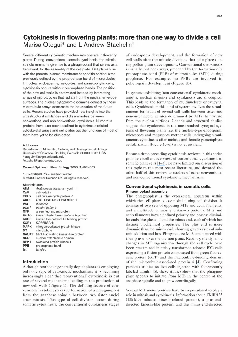
IntroductionAlthough textbooks generally depict plants as employingonly one type of cytokinetic mechanism, it is becomingincreasingly clear that ‘conventional’ cytokinesis is butone of several mechanisms leading to the production ofnew cell walls (Figure 1). The defining feature of con-ventional cytokinesis is the formation of a phragmoplastfrom the anaphase spindle between two sister nucleiafter mitosis. This type of cell division occurs duringsomatic cytokinesis, the conventional cytokinesis stages
of endosperm development, and the formation of newcell walls after the mitotic divisions that take place dur-ing pollen grain development. Conventional cytokinesisis usually, but not always, preceded by the formation of apreprophase band (PPB) of microtubules (MTs) duringprophase. For example, no PPBs are involved in pollen-grain development (Figure 1b).
In systems exhibiting ‘non-conventional’ cytokinetic mech-anisms, nuclear division and cytokinesis are uncoupled.This leads to the formation of multinucleate or syncytialcells. Cytokinesis in this kind of system involves the simul-taneous formation of several cell walls between sister andnon-sister nuclei at sites determined by MTs that radiatefrom the nuclear surfaces. Genetic and structural studiessuggest that cytokinesis in the most studied syncytial sys-tems of flowering plants (i.e. the nuclear-type endosperm,microspore and megaspore mother cells undergoing simul-taneous cytokinesis after meiosis and female gametophytecellularization [Figure 1c–e]) is not equivalent.
Because three preceding cytokinesis reviews in this seriesprovide excellent overviews of conventional cytokinesis insomatic plant cells [1–3], we have limited our discussion ofthis topic to the most recent literature, and devoted theother half of this review to studies of other conventionaland non-conventional cytokinetic mechanisms.
Conventional cytokinesis in somatic cellsPhragmoplast assemblyThe phragmoplast is the cytoskeletal apparatus withinwhich the cell plate is assembled during cell division. Itconsists of two sets of opposing MTs and actin filaments,and a multitude of mostly unknown proteins. MTs andactin filaments have a defined polarity and possess dissimi-lar ends, the plus end and the minus end, each of which hasdistinct biochemical properties. The plus end is moredynamic than the minus end, showing greater rates of sub-unit addition and loss. Phragmoplast MTs are oriented withtheir plus ends at the division plane. Recently, the dynamicchanges in MT organization through the cell cycle havebeen reexamined in stably transformed tobacco BY2 cellsexpressing a fusion protein constructed from green fluores-cent protein (GFP) and the microtubule-binding domainof the microtubule-associated protein 4 [4]. Confirmingprevious studies on live cells injected with fluorescentlylabeled tubulin [5], these studies show that the phragmo-plast appears to initiate from MTs in the center of theanaphase spindle and to grow centrifugally.
Several MT motor proteins have been postulated to play arole in mitosis and cytokinesis. Information about TKRP125(125 kDa tobacco kinesin-related protein), a plus-end-directed kinesin-like protein, and the minus-end-directed
Cytokinesis in flowering plants: more than one way to divide a cellMarisa Otegui* and L Andrew Staehelin†

MT motor proteins KatAp (kinesin Arabidopsis thaliana Aprotein), KatB and KatC, all of which show high levels ofexpression during mitosis and cytokinesis, has been sum-marized in previous reviews [1–3]. The plus-end-directedMT motor protein responsible for vesicle transport has yetto be positively identified.
Another minus-end-directed motor protein, the kinesin-like calmodulin binding protein (KCBP), has also beenimplicated in cell division. KCBP is expressed during mito-sis and cytokinesis, and localizes to the PPB, mitoticspindle and phragmoplast of tobacco BY-2 cells [6]. Morerecently, the motor and tail domains of KCBP have beenreported to possess MT-bundling activity [7•]. The interac-tion of KCBP with MTs is negatively regulated by Ca2+ andcalmodulin (CaM). When KCBP is incubated with antibod-ies raised against the CaM-binding region, this inhibition isabolished, suggesting that the antibodies keep KCBP in aconstitutively active form [8••]. Recently, the role(s) ofKCBP in cytokinesis have been tested by injecting theseantibodies into Tradescantia stamen hair cells [8••].Injection of the antibodies at late prophase induced theprecocious breakdown of the nuclear envelope and transi-tion into prometaphase. The cells did not, however,progress into anaphase. Injection of antibodies at late-metaphase/early-anaphase did not interfere with anaphasetransition, but caused aberrant phragmoplast formation anddelayed cytokinesis. These results suggest that KCBP is
involved in the organization of spindle and phragmoplastMTs. As CaM inhibits KCBP–MT interaction in vitroonly in the presence of Ca2+, the different effects of theanti-KCBP antibodies during the cell cycle may be relatedto changes in Ca2+ concentrations in the vicinity of thespindle and phragmoplast.
The localization of γ-tubulin, a marker for MT organizingcenters in most eukaryotes, has been reexamined in anumber of plant cells by Panteris et al. [9]. This report con-firms the absence of γ-tubulin containing discretecentrosome-like structures, and instead shows the associa-tion of γ-tubulin with the PPB and phragmoplast MTsalong their length. In contrast to an earlier report [10], thispaper also demonstrates the presence of γ-tubulin at theplus end as well as at the minus end of MTs during theearly stages of phragmoplast assembly.
Actin-filament arrays are prominent components of thecytokinetic apparatus of plants, but their functions arestill poorly understood. Within the phragmoplast, theactin filaments are organized parallel to the MTs, forminga bipolar array with their non-overlapping plus ends ori-ented towards the equatorial plane. These actin filamentsare most probably involved in phragmoplast assemblyand turnover. Their role in vesicle trafficking to the cellplate is less clear. A second set of actin filaments connectsthe edge of the expanding cell plate to the cortical actin
494 Cell biology
Figure 1
Comparison of the cytokinetic mechanismsoperating in flowering plants, and thestructures, PPB, spindles and/or NCDs, thatappear to regulate the positioning of newcell walls. Conventional cytokinesis takesplace (a) in somatic cells and (b) in pollengrains, although the division site isestablished by different mechanisms. Duringcellularization of (c) syncytial endospermcells, (d) meiocytes and (e) femalegametophytes, cell walls are formed betweensister and non-sister nucleii (non-conventional cytokinesis). (e) During femalegametophyte cellularization studies of themicropylar pole have shown thatconventional cell-plate formation occursbetween sister nuclei soon after the lastcycle of nuclear cell division (those indicatedwith the numbers 1 and 2). These studiesalso show that additional non-conventionalcell plates arise between non-sister nuclei(those indicated with the number 3). Nodefinitive information about the events at thechalazal pole is available.
Con
vent
iona
l
(c) Endosperm cellularization
(a) Somatic cytokinesis
Male Female
Non
-con
vent
iona
l
PPB Spindle/NCD? Spindle/NCD?
Spindle/NCDSpindle/NCDNCDNCD
(b) Cytokinesis after pollen mitosisI II
(d) Meiocyte cytokinesis
Micropylar pole
Chalazal pole
?
(e) Female gametophyte cellularization
3'
Spatialregulation
1
33
Spindle/NCDor
Spatialregulation
1'
2'3'
2
Current Opinion in Plant Biology

network in the vicinity of the former PPB zone. Ascytochalasin B interferes with cell-plate guidance to thecortical division site, these latter actin filaments mostprobably participate in cell-plate alignment and lateralexpansion [11]. Recent support for the involvement of anacto-myosin-based mechanism in cell-plate guidance hascome from a study of the effects of 2,3-butanedione2-monoxime (BDM), a myosin ATPase inhibitor, on cell-plate formation in Tradescantia stamen hairs [12]. InBDM-treated cells, the cell plates failed to expand cen-trifugally and the phragmoplast became misaligned.Thus, the driving force that pulls the phragmoplast-cellplate to the previously defined cortical division siteappears to be generated by myosin.
Cytokinesis-related actin dynamics have been investigatedin Clivia endosperm cells microinjected with fluorescentphalloidin [13]. During the early stages of cytokinesis,microfilaments are distributed uniformly in the phragmo-plast. Short actin filaments are seen to assemble within thephragmoplast in conjunction with the incorporation of vesi-cles into the cell-plate margins. Furthermore, parallelimmunolocalization experiments with anti-vinculin anti-bodies suggest that the actin filaments may be connected tothe cell plate via vinculin linkers. Such anchoring may bothstabilize the cell-plate assembly intermediates and anchorthe cell plate to the orienting filaments.
Another protein that appears to play a role in actin-cytoskeleton–membrane interaction during cytokinesis isWLIM1, a LIM protein from sunflower [14]. WLIM1 isstructurally related to the muscle proteins CYSTEINE-RICH PROTEIN 1 (CRP1), CRP2 and CRP3/MUSCLELIM PROTEIN (MLP), which anchor the actin cytoskele-ton to sites of membrane adhesion. The discovery ofWLIM1 in phragmoplasts provides further support for aclose functional association between the actin cytoskeletonand the membrane components of the cell plate.
There are also reports of other proteins that co-localizewith cytokinesis-related cytoskeletal arrays but their func-tions have not been determined yet. CDC2 (cell divisioncycle protein 2) is a cyclin-dependent kinase and its injec-tion into Tradescantia stamen hair cells accelerates PPBdisassembly. Thus, CDC2 has been postulated to regulatethe disassembly of the PPB [15]. In tobacco BY-2 cellsexpressing a CDC2::GFP fusion protein, the GFP fluores-cence is seen to localize to the late PPB transiently, then tothe mitotic spindle and the early phragmoplast, and finallyto become diffuse at the later stages of cell-plate formation[16]. TSO1 encodes a protein with two cystein-rich regionsbearing similarity to the Drosophila enhancer of zeste andits plant homologues [17,18]. tso1 mutants of Arabidopsisexhibit defects in cell division, specifically in floral meris-tems. It has been postulated that the TSO1 protein couldserve as a transcriptional regulator that modulates expres-sion of genes encoding cytoskeleton elements or factorsregulating cytoskeleton activity [18].
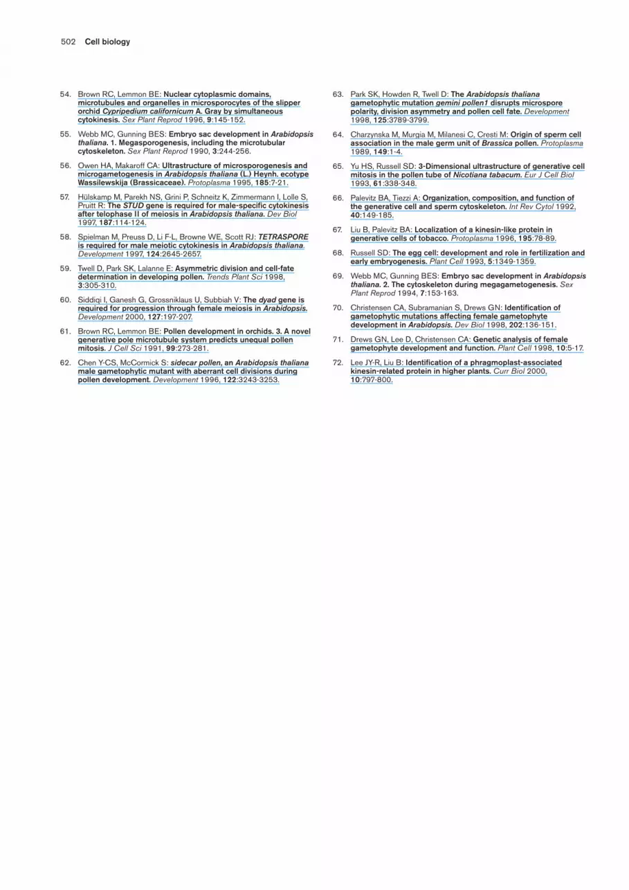
Cell-plate formationThe cell plate grows by fusion of Golgi-derived vesiclesthat start to be produced during mitosis [19]. Studiesemploying a tobacco BY-2 cell line expressing aGFP::mannosidase fusion protein that localizes to cis- andmedial-Golgi cisternae, have shown that duringmetaphase, 20% of all Golgi stacks concentrate in thevicinity of the mitotic spindle while a similar percentageare found in an equatorial region under the plasma mem-brane [20]. This so-called ‘Golgi belt’ accurately predictsthe future site of cell division, and thus forms a novelmarker of the region previously defined by the PPB.During telophase and cytokinesis, many Golgi stacksredistribute around the phragmoplast. These findingsindicate that the Golgi stacks are redistributed duringcytokinesis to ensure both equal partitioning betweendaughter cells and rapid cell-plate assembly.
The actual process of cell-plate formation involves ahighly complex set of carefully controlled membranetransformation and maturation events (Figure 2). Theelucidation of the complex nature of these architecturalchanges by Samuels et al. [21] set the stage for identify-ing and functionally characterizing the molecules thatgive rise to these structures. Although several moleculeshave been reported to be related to cell-plate formation,we are still far from understanding the function of eventhe best investigated proteins. Owing to space con-straints, this discussion is limited to those protein andcytokinesis-deficient mutants that have been studied inthe past two years.
Dynamin proteins are large GTPases that have beenshown in animal cells to serve as mechanoenzymes func-tioning in the scission of nascent vesicles from the plasmamembrane and the trans-Golgi network [22]. In plants,dynamin-like proteins such as soybean phragmoplastinand its Arabidopsis homologue, ADL1, have been local-ized to the growing cell plate [23,24]. As the maturationof both somatic-type [21] and syncytial-type cell plates[25••] involves the removal of excess membrane via bud-ding, clathrin-coated vesicles, phragmoplastin and ADL1may participate in the scission of these vesicles.Nevertheless, because purified phragmoplastin can beinduced to polymerize into spiral structures via two self-assembly domains [26•], Verma and coworkers havepostulated that phragmoplastin could be involved in theformation of the membrane fusion tubes that mediatevesicle fusion by squeezing the cell-plate-forming vesicles [23,26•]. A third possible function for cell-plate-associated dynamin-like molecules is suggested by thediscovery that syncytial-type cell plates possess transientring-like structures of about 30 nm in diameter, whichappear to constrict the tubules of the wide tubular net-work at random intervals [27] (Figure 2b[ii]; see alsobelow). The function of these rings is unknown but theymay help to maintain the tubular architecture of the earlystages of cell-plate formation.
Cytokinesis in flowering plants Otegui and Staehelin 495

Many protein functions, including dynamin assembly, areregulated by phosphorylation. Two mitogen-activated pro-tein kinases (MAPKs) have been associated with plant cellcytokinesis. Both p43Ntf6 from tobacco [28] and MMK3(Medicago MAP kinase 3) [29•] from alfalfa show activityonly in dividing cells and appear to localize to the phragmo-plast and the cell plate. Additionally, NPK1 (Nicotianaprotein kinase 1), which is related to the MAPK kinasekinases, has been identified in dividing cells and could actupstream of p43Ntf6 in a kinase cascade mechanism [30•].Recently, a putative activator for NPK1, NPK1-activatingkinesin-like protein (NACK1), has been identified. NACK1has been shown to activate NPK1 when both genes are co-expressed in yeast cells [31]. Immuno-histochemical studieshave localized NPK1 and NACK1 to the equatorial region ofphragmoplasts. Expression of a kinase-negative construct ofthe NPK1 cDNA containing the NACK1-interacting regionin tobacco BY-2 cells affects cell-plate formation, resulting inmultinucleate cells. These results show that kinase cascadepathways play important roles in regulating cytokinesis.
Although the Ca2+ binding protein centrin is considered to bea marker protein for MT organizing centers in many eukary-otes, in plants, centrin-like proteins have been also localized
to other structures such as cell plates [32]. This localizationhas recently been confirmed for tobacco BY-2 cells and gym-nosperms [33,34]. The function of cell-plate-associatedcentrin molecules has yet to be elucidated.
Annexins bind certain types of membrane phospholipids ina Ca2+-dependent manner. They have been postulated toparticipate in the process of exocytosis of secretory vesiclesin both plant and animal cells [35]. The Sp32 annexin fromtobacco, which contains multiple actin-binding motifs, hasbeen shown recently both to become more abundant at theend of mitosis and to localize to the cortical division site[36]. Whether Sp32 annexin mediates the localized secre-tion of specific types of molecules or the fusion of the cellplate with the plasma membrane is not known.
ATM1 (Arabidopsis thaliana myosin 1) is a class VIIImyosin that binds to late-stage cell plates and the plasmamembrane of post-cytokinesis cell walls [37]. In electronmicrographs that are immunolabelled with anti-ATM1antibody, the labeling appears mostly over young plamod-esmata. This suggests that ATM1 could be involved in theestablishment of cytoplasmic actin cables at sites ofintercellular communication.
496 Cell biology
Figure 2
Schematic comparison of the main stages ofsomatic-type and syncytial-type cell-platedevelopment. (a) (i) and (ii) show the differentmechanisms of vesicle (V) fusion and thelower number of MTs in the mini-phragmoplasts (MP) that give rise to thesyncytial-type cell plates. (b) In both types ofcell-plate development, the fused, Golgi-derived vesicles give rise to tubular networkscovered by a ‘fuzzy coat’ (FC). As thesenetworks mature, the excess membrane isremoved by clathrin-coated budding vesicles(CB). In the wide tubular network of syncytial-type cell plates depicted in (b) (ii), ring-likestructures (RS) appear to constrict thetubules at random intervals. (c) (ii) During thelater stages of syncytial-type cell-plateformation, the wide tubular networksproduced by individual mini-phragmoplastsmerge into larger coherent networks beforethey differentiate into thin tubular networksand, eventually, into cell walls. FT, fusiontubules; HGI, hourglass-shapedintermediates; M, matrix; TN, tubular network;TTN, thin TN; TVN, tubulo-vesicular network;and WTN, wide TN.
Somatic-type cell plate
(a) (i) Vesicle fusion via 20 nm fusion tubes (ii) Vesicle fusion via hourglass-shaped intermediates
(b) (i) Tubulo-vesicular network (ii) Wide tubular networks
(c) (i) Tubular network (ii) Thin tubular network Network fusion
Syncytial-type cell plate
V
V
FT
WTN
FC
M
M
MT
CB
FC
MP MP
RS
CB
WTN WTN
Current Opinion in Plant Biology
WTNTTNTVNTN TTN
MP
MT
HGI
MP MP MP

The mechanism of interaction between cell-plate forma-tion and phragmoplast turnover is not clear. A recentpaper on brefeldin-A-treated cells has shed some newinsights on this interaction [19]. When dividing cells aretreated with brefeldin A, a drug that disrupts trafficking toand through the Golgi apparatus, both cell-plate formationand the depolymerization of MTs in the central region ofthe phragmoplast are inhibited. This strongly suggeststhat the processes of cell-plate assembly and phragmoplastMT turnover are coupled.
Modification of cell-wall forming moleculesThe KORRIGAN1 (KOR1) protein of Arabidopsis is oneof the most unusual cell-plate-associated molecules dis-covered to date. The KOR1 gene encodes anendo-1,4-β-D-glucanase [38]. What makes KOR1 uniqueis that its enzymatic domain is linked to a transmembranedomain, which limits its activity to the membrane–cell-wall interface. kor1-1 mutant plants have a dwarfphenotype caused by a cell-expansion defect [38]. Themore severe kor1-2 mutant is also defective in cytokinesisas evidenced by its severely deformed cell plates, incom-plete cell walls, and multinucleate cells [39••]. Whenexpressed as a GFP fusion protein in tobacco BY-2 cells,KOR1 localizes to the cell plate and lateral walls. Themost likely function of KOR1 during cell-plate formationis to relieve strains in the cellulose-xyloglucan network.Such strains arise as the nascent cellulose fibrils are ran-domly exuded into the mass of xyloglucan and pecticpolysaccharide cell-wall-matrix mol-ecules within the cellplate. During elongative growth, KOR1 most likely pro-motes wall expansion by cleaving taught xyloglucansmolecules that crosslink newly deposited cellulose fibrilsadjacent to the plasma membrane.
Spatial regulation of the division siteThe importance of the PPB in defining the site at which anew cell wall will link up with the parental wall is now wellestablished [40]. Current work is focused on identifying andcharacterizing the function of the molecules that define thelocation of PPB assembly, and that bring about the localizedchanges in the cortical cytoskeleton and the plasma mem-brane that promote the proper positioning of the cell plateand its fusion with the parental plasma membrane [2].
The most interesting new insights into this process comefrom studies of the tangled (tan), discordia (dcd) and warty-1mutants of maize. The tan1 mutant exhibits leaves withabnormally oriented cell walls [41]: the division planes areestablished by PPBs but mainly in transverse or obliqueorientation. Thus, TAN1 appears to be required for theestablishment of longitudinal division planes. In addition,as the cell plates of tan1 leaf cells frequently fail to reachthe division site, TAN1 may also be required for phragmo-plast guidance. TAN1 has been recently cloned andencodes a highly basic protein that is related to the basicdomains of the adenomatous polyposis coli proteins. Itbinds MTs in vitro, localizes to the PPB and the spindle,
and could play a role in orienting cytokinesis-relatedcytoskeletal arrays (L Smith, personal communication).
The dcd mutants, dcd1 and dcd2, also display incorrectly ori-ented cell walls in the leaf epidermis [42•]. Themisoriented walls of these mutants are, however, associatedexclusively with cell types that arise through asymmetricdivision, for example, stomata-complex and silica-cork cellpairs. In these mutants, polarization of the cytoplasm priorto mitosis and PPB formation are not affected in the cellsundergoing asymmetric division, but the cell plates oftenfail to reach the division site. Because cytochalasin D treat-ment produces phenotypes that mimic those of dcdmutants, and dcd1 is hypersensitive to the effects ofcytochalasin D, the DCD gene products are thought to berequired for an actin-dependent process that is related tophragmoplast guidance in asymmetric divisions. AlthoughPPB formation is not affected in either dcd1 or dcd2 singlemutants, it is greatly reduced in the dcd1 ; dcd2 doublemutant, revealing redundant functions for these genes inPPB formation (L Smith, personal comunication).
In the warty-1 mutant, abnormally enlarged cells appear inthe leaf blade [43]. When these cells exceed a 200%increase in length and width compared with non-mutantcells, they show misorientated cell plates and incompletecell walls. These results suggest a correlation betweenexpansion and cytokinesis regulation that becomes alteredwhen a particular size threshold is surpassed.
Conventional and non-conventional cytokinesisin other systemsEndosperm cellularizationCell-wall formation during endosperm development canoccur in three different ways: first, in the cellularendosperm, cytokinesis occurs after all mitotic divisions;second, in helobial endosperms, division of the primaryendosperm nucleus is followed by the formation of a wallthat unequally partitions the endosperm in two cells,each of which undergoes free nuclear division cycles and,later on, becomes cellularized; and third in nuclearendosperms, the initial developmental stages involveseveral cycles of nuclear division without cytokinesis andthe repositioning of the nuclei to the cortical cytoplasm.Radial systems of MTs associated with MT-organizingcenters on the nuclear surfaces then define the futureboundaries of the cell by organizing the cytoplasm intonuclear cytoplasmic domains (NCDs) (Figure 3;[44•,45]). Shortly thereafter, the cellularization processbegins with the assembly of cell walls between sister andnon-sister nuclei.
The interpretation of the process by which cell wallsdevelop has been controversial for more than 90 years.One of the most widely accepted theories postulatedthat the first endosperm cell walls are initiated asingrowths from the syncytial cell wall in the absence oftypical phragmoplasts (reviewed in [1]). By employing
Cytokinesis in flowering plants Otegui and Staehelin 497

high-pressure-freezing/freeze-substitution techniquesinstead of conventional chemical fixation to preserve devel-oping Arabidopsis seeds for ultrastructural analysis, it hasbeen demonstrated that the first cell walls formed duringthe cellularization process involve groups of coordinated‘mini-phragmoplasts’ [25••]. Mini-phragmoplasts consist oftwo opposing sets of approximately 10 MTs, which arisefrom overlapping clusters of MTs that radiate from neigh-boring nuclei (Figure 3). Each mini-phragmoplast gives riseto a tubular network that fuses with other contiguous net-works to produce a peculiar kind of cell plate, thesyncytial-type cell-plate. The transient membrane configu-rations observed during syncytial-type cell-plate formationdiffer in some aspects from those studied during somatic-typecell-plate formation [25••] (Figure 3).
One of the most striking differences pertains to the mech-anism of Golgi-derived vesicle fusion. In somatic-type cellplates, vesicles fuse by means of 20 nm fusion tubes,whereas in syncytial-type cell plates, fusion occursbetween close vesicles, which give rise to transient hour-glass-shaped intermediates (Figure 2). Despite thisdifference, the KNOLLE protein, which is involved inmembrane fusion events, has been detected during cytoki-nesis in both somatic and endosperm tissues [24]. Anotherinteresting feature of syncytial-type cell-plate formation isthe development of independent tubular networks associ-ated with different mini-phragmoplasts between a givenpair of endosperm nuclei. These networks eventually fusewith each other, and then with networks formed betweendifferent pairs of nuclei to produce large, honey-comb-shaped cell-plate complexes that are eventually convertedinto the new cell walls. Owing to the irregular distributionof mini-phragmoplasts around the cell plate and theconsolidation of larger cell-plate complexes via the fusionof different networks, syncytial-type cell plates mature ina patchwork manner.
As mentioned above, it has been shown that theKNOLLE protein localizes to growing endosperm cellwalls. The Arabidopsis mutants titan [46] and fist [47], andpilz group mutants [48] which show deficiencies duringcell division in embryo cells, also show alterations duringendosperm cellularization. More studies are needed toidentify the signals that trigger endosperm cellularizationand to characterize the molecular differences betweensomatic- and syncytial-type cell plates.
From an evolutionary point of view, nuclear types ofendosperm appear to have evolved several times in differentlineages from cellular endosperm [49]. Thus, the evolution-ary transitions from the cellular type to nuclear types wouldhave involved disruptions of some aspects of the normalcytokinetic process [50]. This hypothesis is supported bythe finding that in the nuclear endosperm of some cereals,transient post-mitotic phragmoplasts are formed but fail toproduce cell plates, whereas in the nuclear endosperm ofthe basal eudicot genus Platanus, an incipient cell plate hasbeen observed between the two sister nuclei derived fromthe primary endosperm division [50].
Simultaneous cell-wall formation after meiosisThere are two mechanisms of intersporal cell-wall formationafter meiosis, the successive type and the simultaneous type.In the successive type, cell walls are assembled after bothtelophase I and telophase II, whereas in the simultaneoustype, cell walls form between all four resulting haploid nucleiat the same time, that is, after telophase II. Neither of thesesystems involves the formation of PPBs. Instead, as innuclear endosperms, the multinucleate cells employ radialMT arrays emanating from the nuclear envelope surfaces todefine NCDs, and thereby, the sites of the future cell walls.
Successive cytokinesis is accomplished by formation ofphragmoplast-associated cell plates after each nuclear
498 Cell biology
Figure 3
Spatial regulation of new cell wall positioningby NCDs in the nuclear endosperm.(a) Confocal laser scanning micrograph of thenuclear endosperm of Cornopus didymous(Brassicaceae). MTs wereimmunofluorescence-labeled with anti-tubulinantibodies and the nuclei (N) were stainedwith propidium iodide. (b) Explanatorydiagram showing how the MTs depicted in(a) arise from MT-organizing centers (MTOCs)on the nuclear envelope surface and howmini-phragmoplasts, in which syncytial-typecell plates form, are produced in the MToverlapping regions. (Micrograph shownin [a] is courtesy of Roy Brown.)

division. The mechanism of cell-wall formation duringsimultaneous cytokinesis is more varied. Thus, simultane-ous cytokinesis after male meiosis has been reported toinvolve cleavage furrows in most dicots [51]; phragmoplastsand cell plates in some orchids [52] and Carex (Cyperaceae)[53]; and cell plates without a typical phragmoplast in theorchid Cypripedium [54]. In Arabidopsis, both male andfemale meioses are followed by simultaneous cytokinesis.Cytokinesis during megasporogenesis involves the forma-tion of phragmoplasts and cell plates [55], whereascytokinesis during microsporogenesis appears to depend onthe formation of cleavage furrows from the parental cellwall [56]. Formation of these latter cell walls does notappear to involve the KNOLLE protein [24].
The Arabidopsis mutants stud and tes fail to produce cellwalls after male meiosis, resulting in tetra-nucleatemicrospores [57,58]. Other cytokinetic processes, however,such as cell-wall formation following female meiosis ormitosis during pollen development, are not affected.Thus, up to four sperm-cell pairs have been observed inmature stud and tetraspore pollen grains, showing that allfour haploid nuclei can undergo nuclear migration andasymmetric division sharing a common cytoplasm. BothSTUD and TETRASPORE map to similar locations andmight be allelic to each other [59].
Little is known about the molecular regulation of cytokine-sis after female meiosis. In dyad mutants of Arabidopsis,which are arrested at the end of meiosis 1, a cell wall isdeposited between the two resulting nuclei [60]. This indi-cates that the mechanism that prevents cytokinesis aftermeiosis 1 in the wild type is also affected by this mutation.
Conventional cytokinesis during pollen development The first mitotic division during pollen-grain development(Figure 1) is the asymmetric division that gives rise to twocells with distinct developmental fates, the large vegeta-tive cell and the small generative cell. This asymmetricdivision plays a critical role in the control of pollen cellfate, and involves a striking reorganization of cytoplasmand migration of the microspore nucleus to the peripheryassociated with a unique generative pole MT system [61].After mitosis, a phragmoplast arises from the mitotic spin-dle remnants and a cell plate forms in association with it.The mechanism by which the phragmoplast and cell plateare guided to the cortical fusion site is unknown, but it isprobably related to the polarization of the cytoplasm andthe establishment of NCDs before cytokinesis
In the Arabidopsis mutant sidecar pollen, microspores under-go a premature symmetric division. From the two resultingcells, one is able to undergo asymmetric division and togive rise to a generative and a vegetative cell, whereas theother develops vegetative cell characteristics [62]. Thegemini pollen1 (gem1) mutation also affects the first mitoticdivision, giving rise to a range of pollen phenotypescharacterized by equal, unequal, or incomplete cell
divisions [63]. These mutants also show incompletenuclear migration, which could account for their altereddivision symmetry. In contrast to sidecar pollen daughtercells, neither of the daughter cells of gem1 pollen is capableof polarization and undergoing further asymmetric divi-sion. As gem1 also shows division phenotypes consistentwith spatial uncoupling of karyokinesis and cytokinesis, ithas been suggested that GEM1 may be required for thelocalization of phragmoplast activity.
The second mitotic division during pollen developmenttakes place in the generative cell and gives rise to twosperm cells (Figure 1). This division can occur inside thepollen grain (as in Arabidopsis and Brassica) or inside thepollen tube (as in tobacco and tomato). Several reportshave documented that this division involves a regular spin-dle and the formation of a normal-type phragmoplast andcell plate [64,65]. In Tradescantia, however, an unconven-tional mitotic spindle and cytokinesis by means of acleavage furrow have been described (reviewed in [66]). Akinesin-like protein related to KatAp has been found tolocalize in the mitotic spindle and phragmoplast duringpollen mitosis II [67]. One of the most striking features ofthe generative cell is its lack of F-actin throughout divi-sion. Moreover, cytokinesis is unaffected by cytochalasin Dat concentrations that perturb vegetative microfilaments [66],indicating that the cytoskeleton arrays involved incytokinesis do not involve actin filaments.
Female gametophyte cellularizationIn most flowering plants, including Arabidopsis, fourmegaspores are formed after meiosis but only one is func-tional. This megaspore undergoes three cycles of nucleardivision giving rise to an eight-nucleate embryo sac, which,after cellularization, is transformed into a seven-celledembryo sac (Figure 1). After two cycles of nuclear divisionthe female gametophyte contains four nuclei. Two of themare located in the pole facing the micropyle, the micropy-lar pole, and the other two at the opposing chalazal pole.The cellularization process has been most thoroughlyinvestigated for the micropylar pole. There, after the lastcycle of nuclear division, one conventional vertical cellplate (cell plate 1 in Figure 1) separates the cytoplasm ofthe two sister synergid nuclei, and a second conventionaltransverse cell plate (cell plate 2 in Figure 1) formsbetween the egg nucleus and one of the polar nuclei(reviewed in [68]). The phragmoplasts and cell platesmarked as ‘3’ in Figure 1, arise between non-sister nuclei[69]. There is, however, some disagreement about the totalnumber of cell plates that are formed in this final stage ofcellularization [68]. No PPBs have been detected duringfemale gametophyte cellularization in Arabidopsis, butnuclear-based MT arrays have been observed during thecoenocytic phases, suggesting that the location of the newcell walls is determined by means of the MT-dependentNCDs. This latter process appears to be perturbed in thefem4 (female gametophyte4) mutant of Arabidopsis, whichexhibits an embryo sac that has altered cellularization
Cytokinesis in flowering plants Otegui and Staehelin 499

patterns [70,71]. Most likely, the mutation affectscytoskeleton organization at the time of NCD formation.
Conclusion and perspectivesSomatic cytokinesis research is a rapidly-expanding fieldthat has produced a number of important new findings inrecent years. These include the identification of anincreasing number of molecules involved in cell-plate for-mation and in the regulation of cytokinesis-relatedcytoskeletal arrays in somatic cells. Nevertheless, the listof these molecules identified so far is still short and littlefunctional information is available on them. Even less isknown about the molecules and mechanisms involved incell-wall formation in the nuclear endosperms, meiocytesand gametophytic cells. Nevertheless, comparative studiesof conventional and non-conventional cytokinetic systemscould aid research in both fields by defining which mol-ecules are common to both systems and which are unique.Within the next decade we will hopefully gain answers toquestions such as: What is the nature of the fuzzy-coatmaterial that stabilizes the tubular networks of early cellplates during conventional and non-conventional cytoki-nesis? What is the nature of the molecules that mediatecell-plate maturation? How are cell plates guided to thecortical division sites in the absence of PPB formation?Does male meiosis-associated cytokinesis really involvecleavage furrows or, as in the remaining syncytial cells, docell plates give rise to new cell walls? What is the signalthat triggers cellularization in syncytial systems? Plant cellcytokinesis will clearly remain a challenging area ofresearch for many years to come.
UpdateRecently, a new kinesin-related protein, AtPAKRP1(Arabidopsis thaliana phragmoplast-associated kinesin-relatedprotein 1), with putative plus-end-directed motor activity,has been shown to localize to the phragmoplast MTs at ornear their plus ends [72]. Some preliminary results withinhibitors and truncated fusion proteins suggest that thisamino-terminal motor protein could play a role in positioningthe two opposite sets of phragmoplast MTs [72].
AcknowledgementsWe would like to thank William Friedman for critical reading of themanuscript, and members of the Staehelin laboratory for comments andsuggestions. We thank also various colleagues who provided unpublishedmanuscripts and information. This work was supported by a National Instituteof Health grant (18639) to LA Staehelin; MS Otegui’s work is supported by theFullbright–Antorchas Foundation and a Consejo Nacional de InvestigacionesCientíficas y Téchnicas (CONICET) posdoctoral fellowship.
References and recommended readingPapers of particular interest, published within the annual period of review,have been highlighted as:
• of special interest•• of outstanding interest
1. Heese M, Ulrike M, Jürgens G: Cytokinesis in flowering plants:cellular process and developmental integration. Curr Opin PlantBiol 1998, 1:486-491.
2. Smith LG: Divide and conquer: cytokinesis in plant cells. Curr OpinPlant Biol 1999, 2:447-453.
3. Sylvester AW: Division decisions and the spatial regulation ofcytokinesis. Curr Opin Plant Biol 2000, 3:58-66.
4. Granger CL, Cyr RJ: Microtubule reorganization in tobacco BY-2cells stably expressing GFP-MBD. Planta 2000, 210:502-509.
5. Zhang D, Wadsworth P, Hepler PK: Dynamics of microfilaments aresimilar, but distinct from microtubules during cytokinesis in living,dividing plant cells. Cell Motil Cytoskeleton 1993, 24:151-155.
6. Bowser J, Reddy ASN: Localization of a kinesin-like calmodulin-binding protein in dividing cells of Arabidopsis and tobacco.Plant J 1997, 12:1429-1437.
7. Kao Y-L, Deavours BE, Phelps KK, Walker RA, Reddy ASN: Bundling • of microtubules by motor and tail domains of a kinesin-like
calmodulin-binding protein form Arabidopsis: regulation byCa2+/calmodulin. Biochem Biophys Res Comm 2000, 267:201-207.
KCBP is shown to have MT-bundling activity within both the carboxy-terminalmotor domain and the amino-terminal tail region. The MT-bundling activitywithin the carboxy-terminal motor domain is shown to be negatively regulatedby Ca2+/calmodulin.
8. Vos JW, Safadi F, Reddy ASN, Hepler PK: The kinesin-like •• calmodulin binding protein is differentially involved in cell
division. Plant Cell 2000, 12:979-990.The function of KCBP, a kinesin-like calmodulin binding protein, during thecell cycle was tested by injecting antibodies against the CaM-bindingregion of KCBP into plant cells. These antibodies keep KCBP in a consti-tutively active form. Antibody injection during prophase induces breakdownof the nuclear envelope, and cells are arrested at metaphase. Injection ofantibodies later in cell division, does not affect anaphase transition butcauses aberrant phragmoplast formation.
9. Panteris E, Apostolakos P, Galatis B: Gamma-tubulin colocalizeswith microtubule arrays and tubulin paracrystals in dividingvegetative cells of higher plants. Protoplasma 2000,210:179-187.
10. Joshi HC, Palevitz BA: γγ-tubulin and microtubule organization inplants. Trends Cell Biol 1996, 6:41-44.
11. Mineyuki Y, Gunning BES: A role for preprophase bands ofmicrotubules in maturation of new cell walls, and a generalproposal on the function of preprophase band sites in celldivision in higher plants. J Cell Sci 1990, 97:527-537.
12. Molchan TM, Valster AH, Hepler PK: Actomyosin promotes cellplate alignment and lateral expansion in plant cells. Mol Biol Cell1999, 10:85.
13. Endlé M-C, Stoppin V, Lambert A-M, Schmit A-C: The growing cellplate of higher plants is a site of both actin assembly andvinculin-like antigen recruitment. Eur J Cell Biol 1998, 77:10-18.
14. Mundel C, Baltz R, Eliasson Å, Bronner R, Grass N, Kräuter R,Evrard J-L, Steinmetz A: A LIM-domain protein from sunflower islocalized to the cytoplasm and/or nucleus in a wide variety oftissues and is associated with the phragmoplast in dividing cells.Plant Mol Biol 2000, 42:291-302.
15. Hush J, Wu L, John PC, Hepler LH, Hepler PK: Plant mitosispromoting factor disassembles the microtubule preprophaseband and accelerates prophase progression in Tradescantia. CellBiol Int 1996, 20:275-287.
16. Weinganner M, Schweighofer A, Meskiene I, Heberle-Bors E,Doonan J, Bögre L: Visualization of microtubule movements inliving cells expressing Cdc2–GFP. Abstract S32-10 of the 6thInternational Congress of Plant Molecular Biology, 2000 June 18–24,Quebec, Canada.
17. Song J-Y, Leung T, Ehler LK, Wang C, Liu Z: Regulation ofmeristem organization and cell division by TSO1, an Arabidopsisgene with cystein-rich repeats. Development 2000,127:2207-2217.
18. Hauser BA, He JQ, Park SO, Gasser CS: TSO1 is a novel proteinthat modulates cytokinesis and cell expansion in Arabidopsis.Development 2000, 127:2219-2226.
19. Yasuhara H, Shibaoka H: Inhibition of cell-plate formation bybrefeldin A inhibited the depolymerization of microtubules in thecentral region of the phragmoplast. Plant Cell Physiol 2000,41:300-310.
20. Nebenführ A, Frohlick JA, Staehelin LA: Redistribution of Golgistacks and other organelles during mitosis and cytokinesis inplant cells. Plant Physiol 2000, 124:135-151.
500 Cell biology

21. Samuels AL, Giddings TH, Staehelin LA: Cytokinesis in tobaccoBY-2 and root tip cells: a new model of cell plate formation inhigher plants. J Cell Biol 1995, 130:1-13.
22. Cao H, Thompson HM, Krueger EW, McNiven MA: Disruption ofGolgi structure and function in mammalian cell expressing amutant dynamin. J Cell Sci 2000, 113:1993-2002.
23. Gu X, Verma DPS: Dynamics of phragmoplastin in living cellsduring cell plate formation and uncoupling of cell elongation fromthe plane of cell division. Plant Cell 1997, 9:157-169.
24. Lauber MH, Waizenegger I, Steinmann T, Schwarz H, Mayer U,Hwang I, Lukowitz W, Jürgens G: The Arabidopsis KNOLLE proteinis a cytokinesis-specific syntaxin. J Cell Biol 1997,139:1485-1493.
25. Otegui M, Staehelin LA: Syncytial-type cell plates: a novel kind of •• cell plate involved in endosperm cellularization of Arabidopsis.
Plant Cell 2000, 12:933-947.The endosperm cellularization process in Arabidopsis is re-evaluated inhigh-pressure-frozen, freeze-substituted developing seeds. A new kind ofcell plate, the syncytial-type cell plate, was found to arise between sisterand non-sister nuclei. At the onset of cellularization, groups of MTs orga-nized in mini-phragmoplasts arise in the overlapping region of the con-tiguous, nuclear-based radial MT systems that also define NCDs.Similarities and dissimilarities between syncytial-type and somatic-typecell plates are discussed.
26. Zhang Z, Hong Z, Verma DPS: Phragmoplastin polymerizes into • spiral coiled structures via intermolecular interaction of two self-
assembly domains. J Biol Chem 2000, 275:8779-8774.Using a yeast two hybrid system and an in vitro protein–protein interactionassay, the authors show that phragmoplastin polymerizes into spiral struc-tures by two self-assembly domains. Electron microscopy observationsrevealed that helical arrays of phragmoplastin can be induced by reducingsalt concentration.
27. Otegui M, Staehelin LA: A novel model of endospermcellularization in Arabidopsis thaliana based on high-pressurefrozen-freeze-substituted samples and electron microscopictomography. Abstract 5 of the16th International Congress on SexualPlant Reproduction, 2000 April 1–5, Banff, Canada.
28. Calderini O, Bögre L, Vicente O, Binarova P, Heberle-Bors E,Wilson C: A cell cycle regulated MAP kinase with a possible rolein cytokinesis in tobacco cells. J Cell Sci 1998, 111:3091-3100.
29. Bögre L, Calderini O, Binarova P, Mattauch M, Till S, Kiegerl S, • Jonak C, Pollaschek C, Barker P, Huskisson N et al.: A MAP kinase is
activated late in plant mitosis and becomes localized to the planeof cell division. Plant Cell 1999, 11:101-113.
A novel alfalfa MAPK, MMK3, is demonstrated to be active only in dividingcells. During cytokinesis, MMK3 localizes to the equatorial plane associatedwith the phragmoplast and cell plate. Treatments with drugs that disrupt MTsindicate that intact MTs are required for MMK3 activation.
30. Nishihama R, Machida Y: The MAP kinase cascade that includes • MAKKK-related protein kinase NPK1 controls a mitotic process in
plant cells. In MAP Kinases in Plants Signal Transduction, vol 27.Edited by Hirt H. Berlin: Springer-Verlag; 2000:119-130.
A review discussing the kinases and activator factors, including NPK1, thatare found to participate in the MAP kinase cascade pathway and appear toplay a role during cytokinesis.
31. Machida Y, Nishihama R, Soyano T, Ishikawa M, Asada T, Araki T:NPK1 MAPKKK-mediated signal pathway controls plantcytokinesis. Abstract S09-51 of the 6th International Congress ofPlant Molecular Biology, 2000 June 18–24, Quebec, Canada.
32. Del Vecchio AJ, Harper JDI, Vaughn KC, Baron AT, Salisbury JL,Overall RL: Centrin homologues in higher plants are prominentlyassociated with the developing cell plate. Protoplasma 1997,196:224-234.
33. Stoppin-Mellet V, Canaday J, Lambert A-M: Characterization ofmicrosome-associated tobacco BY-2 centrins. Eur J Cell Biol1999, 78:842-848.
34. Harper JDI, Fowke LC, Gilmer S, Overall RL, Marc J: A centrinhomologue is localised across the developing cell plate ingymnosperms and angiosperms. Protoplasma 2000, 211:207-216.
35. Thiel G, Battey N: Exocytosis in plants. Plant Mol Biol 1998,38:111-125.
36. Proust J, Houlné G, Schantz M-L, Shen W-H, Schantz R: Regulationof biosynthesis and cellular localization of Sp32 annexins intobacco BY2 cells. Plant Mol Biol 1999, 39:361-372.
37. Reichelt S, Knight AE, Hodge TP, Baluska F, Samaj J, Volkmann D,Kendrick-Jones J: Characterization of the unconventional myosinVIII in plant cells and its localization at the post-cytokinetic cellwall. Plant J 1999, 19:555-567.
38. Nicol F, His I, Jauneau A, Vernhettes S, Canut H, Höfte H: A plasmamembrane-bound putative endo-1,4-ββ-D-glucanase is requiredfor normal wall assembly and cell elongation in Arabidopsis.EMBO J 1998, 17:5563-5576.
39. Zuo J, Niu Q-W, Nishizawa N, Wu Y, Kost B, Chua N-H: KORRIGAN, •• an Arabidopsis endo-1,4-ββ-glucanase, localizes to the cell plate
by polarized targeting and is essential for cytokinesis. Plant Cell2000, 12:1137-1152.
Different approaches are taken to analyze the function, localization, and tar-geting-to-the-cell-plate sequence of KOR1, a putative membrane-anchoredendo-β-1,4-endoglucanase from Arabidopsis. The Arabidopsis kor1-2mutant shows altered cell division and expansion. When a GFP::KOR1fusion protein is expressed in BY2 cells, it localizes to the cell plate.Substitution mutations in each of the two targeting motifs of KOR1 causethe fusion proteins to localize to the plasma membrane as well.
40. Mineyuki Y: The preprophase band of microtubules: its function asa cytokinetic apparatus in higher plants. Int Rev Cytol 1999,187:1-49.
41. Cleary AL, Smith LG: The Tangled1 gene is required for spatialcontrol of cytoskeletal arrays associated with cell division duringmaize leaf development. Plant Cell 1998, 10:1875-1888.
42. Gallagher K, Smith LG: discordia mutations specifically misorient • asymmetric cell division during development of the maize leaf
epidermis. Development 1999, 126:4623-4633.Analysis of the dcd1 and dcd2 mutant phenotypes shows that the corre-sponding genes are involved in phragmoplast guidance to the cortical divi-sion sites of asymmetrically dividing maize leaf epidermal cells. Interestingly,cell fate is not affected in dcd mutants showing that cell-fate specificationfollowing asymmetric cell division is not related to cell-wall position.
43. Reynolds JO, Eisses JF, Sylvester AW: Balancing division andexpansion during maize leaf morphogenesis: analysis of themutant warty-1. Development 1998, 125:259-268.
44. Pickett-Heaps JD, Gunning BES, Brown RC, Lemmon BE, Cleary AL: • The cytoplast concept in dividing plant cells: cytoplasmic domains
and the evolution of spatially organized cell division. Am J Bot1999, 86:153-172.
This is a review about cytokinetic mechanisms in algal cells and terrestrialplants. The authors give an evolutionary interpretation of the differentmechanisms involved in the spatial positioning of new cell walls.
45. Brown RC, Lemmon BE, Nguyen H, Olsen O-A: Development ofendosperm in Arabidopsis thaliana. Sex Plant Reprod 1999,12:32-42.
46. Liu C-M, Meinke D: The titan mutants of Arabidopsis are disruptedin mitosis and cell cycle control during seed development. Plant J1998, 16:21-31.
47. Dunn SM, Drews GN, Fischer RL, Harada JJ, Goldberg RB,Koltunow A: fist: an Arabidopsis mutant with altered cell divisionplanes and radial pattern disruption during embryogenesis. SexPlant Reprod 1997, 10:358-367.
48. Mayer U, Herzog U, Berger F, Inzé D, Jürgens G: Mutations in thePILZ group genes disrupt the microtubule cytoskeleton anduncouple cell cycle progression form cell division inArabidopsis embryo and endosperm. Eur J Cell Biol 1999,78:100-108.
49. Floyd SK, Friedman WE: Evolution of endosperm developmentalpatterns among basal flowering plants. Int J Plant Sci 2000,in press.
50. Floyd SK, Lerner VT, Friedman WE: A developmental andevolutionary analysis of embryology in Platanus (Platanaceae), abasal eudicot. Am J Bot 1999, 86:1523-1537.
51. Brown RC, Lemmon BE: Microtubules associated withsimultaneous cytokinesis of coenocytic microsporocytes. Am JBot 1988, 75:1848-1856.
52. Brown RC, Lemmon BE: Pollen development in orchids. I.Cytoskeletal control of division plane in irregular patterns ofmeiotic cytokinesis. Protoplasma 1991, 163:9-18.
53. Brown RC, Lemmon BE: The cytoskeleton and polarization duringpollen development in Carex blanda (Cyperaceae). Am J Bot2000, 87:1-11.
Cytokinesis in flowering plants Otegui and Staehelin 501
54. Brown RC, Lemmon BE: Nuclear cytoplasmic domains,microtubules and organelles in microsporocytes of the slipperorchid Cypripedium californicum A. Gray by simultaneouscytokinesis. Sex Plant Reprod 1996, 9:145-152.
55. Webb MC, Gunning BES: Embryo sac development in Arabidopsisthaliana. 1. Megasporogenesis, including the microtubularcytoskeleton. Sex Plant Reprod 1990, 3:244-256.
56. Owen HA, Makaroff CA: Ultrastructure of microsporogenesis andmicrogametogenesis in Arabidopsis thaliana (L.) Heynh. ecotypeWassilewskija (Brassicaceae). Protoplasma 1995, 185:7-21.
57. Hülskamp M, Parekh NS, Grini P, Schneitz K, Zimmermann I, Lolle S,Pruitt R: The STUD gene is required for male-specific cytokinesisafter telophase II of meiosis in Arabidopsis thaliana. Dev Biol1997, 187:114-124.
58. Spielman M, Preuss D, Li F-L, Browne WE, Scott RJ: TETRASPOREis required for male meiotic cytokinesis in Arabidopsis thaliana.Development 1997, 124:2645-2657.
59. Twell D, Park SK, Lalanne E: Asymmetric division and cell-fatedetermination in developing pollen. Trends Plant Sci 1998,3:305-310.
60. Siddiqi I, Ganesh G, Grossniklaus U, Subbiah V: The dyad gene isrequired for progression through female meiosis in Arabidopsis.Development 2000, 127:197-207.
61. Brown RC, Lemmon BE: Pollen development in orchids. 3. A novelgenerative pole microtubule system predicts unequal pollenmitosis. J Cell Sci 1991, 99:273-281.
62. Chen Y-CS, McCormick S: sidecar pollen, an Arabidopsis thalianamale gametophytic mutant with aberrant cell divisions duringpollen development. Development 1996, 122:3243-3253.
63. Park SK, Howden R, Twell D: The Arabidopsis thalianagametophytic mutation gemini pollen1 disrupts microsporepolarity, division asymmetry and pollen cell fate. Development1998, 125:3789-3799.
64. Charzynska M, Murgia M, Milanesi C, Cresti M: Origin of sperm cellassociation in the male germ unit of Brassica pollen. Protoplasma1989, 149:1-4.
65. Yu HS, Russell SD: 3-Dimensional ultrastructure of generative cellmitosis in the pollen tube of Nicotiana tabacum. Eur J Cell Biol1993, 61:338-348.
66. Palevitz BA, Tiezzi A: Organization, composition, and function ofthe generative cell and sperm cytoskeleton. Int Rev Cytol 1992,40:149-185.
67. Liu B, Palevitz BA: Localization of a kinesin-like protein ingenerative cells of tobacco. Protoplasma 1996, 195:78-89.
68. Russell SD: The egg cell: development and role in fertilization andearly embryogenesis. Plant Cell 1993, 5:1349-1359.
69. Webb MC, Gunning BES: Embryo sac development in Arabidopsisthaliana. 2. The cytoskeleton during megagametogenesis. SexPlant Reprod 1994, 7:153-163.
70. Christensen CA, Subramanian S, Drews GN: Identification ofgametophytic mutations affecting female gametophytedevelopment in Arabidopsis. Dev Biol 1998, 202:136-151.
71. Drews GN, Lee D, Christensen CA: Genetic analysis of femalegametophyte development and function. Plant Cell 1998, 10:5-17.
72. Lee JY-R, Liu B: Identification of a phragmoplast-associatedkinesin-related protein in higher plants. Curr Biol 2000,10:797-800.
502 Cell biology


