Embed Size (px)
Citation preview
pharmaceutics
Article
Deaggregation and Crystallization Inhibition by Small Amountof Polymer Addition for a Co-AmorphousCurcumin-Magnolol System
Jiawei Han †, Luyuan Li †, Meiling Su, Weili Heng, Yuanfeng Wei, Yuan Gao * and Shuai Qian *
�����������������
Citation: Han, J.; Li, L.; Su, M.;
Heng, W.; Wei, Y.; Gao, Y.; Qian, S.
Deaggregation and Crystallization
Inhibition by Small Amount of
Polymer Addition for a
Co-Amorphous Curcumin-Magnolol
System. Pharmaceutics 2021, 13, 1725.
https://doi.org/10.3390/
pharmaceutics13101725
Academic Editors: Leena Peltonen
and Arvind K. Bansal
Received: 19 August 2021
Accepted: 13 October 2021
Published: 18 October 2021
Publisher’s Note: MDPI stays neutral
with regard to jurisdictional claims in
published maps and institutional affil-
iations.
Copyright: © 2021 by the authors.
Licensee MDPI, Basel, Switzerland.
This article is an open access article
distributed under the terms and
conditions of the Creative Commons
Attribution (CC BY) license (https://
creativecommons.org/licenses/by/
4.0/).
School of Traditional Chinese Pharmacy, China Pharmaceutical University, Nanjing 211198, China;[email protected] (J.H.); [email protected] (L.L.); [email protected] (M.S.);[email protected] (W.H.); [email protected] (Y.W.)* Correspondence: [email protected] (Y.G.); [email protected] (S.Q.);
Tel.: +86-25-83379418 (Y.G.); +86-139-1595-7175 (S.Q.)† These authors made equal contributions to this work.
Abstract: Different from previously reported co-amorphous systems, a co-amorphous curcumin-magnolol (CUR-MAG CM) system, as compared with its crystalline counterparts, exhibited decreaseddissolution due to its aggregation during dissolution. The main purpose of the present study is todeaggregate CUR-MAG CM to optimize drug dissolution and explore the deaggregation mechanisminvolved. Herein, a small amount of polymer (HPMC, HPC, and PVP K30) was co-formulated at5% (w/w) with CUR-MAG CM as ternary co-amorphous systems. The polymer addition changedthe surface properties of CUR-MAG CM including improved water wettability enhanced surfacefree energy, and hence exerted a deaggregating effect. As a result, the ternary co-amorphous systemsshowed faster and higher dissolution as compared with crystalline CUR/MAG and CUR-MAGCM. In addition, the nucleation and crystal growth of dissolved CUR and MAG molecules weresignificantly inhibited by the added polymer, maintaining a supersaturated concentration for a longtime. Furthermore, polymer addition increased the Tg of CUR-MAG CM, potentially involvingmolecular interactions and inhibiting molecular mobility, resulting in enhanced physical stabilityunder 25 ◦C/60% RH and 40 ◦C/75% RH conditions. Therefore, this study provides a promisingstrategy to optimize the dissolution and physical stability of co-amorphous systems by deaggregationand crystallization inhibition via adding small amounts of polymers.
Keywords: curcumin; magnolol; co-amorphous; deaggregation; dissolution; physical stability
1. Introduction
Most drug candidates under development are poorly soluble in water, which isrelated to various pharmaceutical performance problems [1,2]. Enhancing the solubil-ity/dissolution of these drugs has become a vital issue for pharmaceutical enterprises todevelop effective medicines with suitable dosage forms for patients [3]. Amorphization bydisordering crystal lattice is a well-known approach to overcome the solubility/dissolutiondefects of poorly soluble drugs [4–6]. However, the application of amorphous systemsis severely limited due to the inherent thermodynamic instability and devitrification riskduring processing, storage, and dissolution [7–9], resulting in the loss of amorphous ad-vantages. Therefore, it is crucial to seek effective approaches to stabilize amorphous drugs.
In recent years, co-amorphous drug delivery systems have aroused wide attention forstabilizing neat amorphous drugs and providing solubility/dissolution advantages (suchas the faster dissolution rate and enhanced supersaturation ability) over correspondingcrystalline and amorphous drugs [10]. A co-amorphous system is prepared by amor-phizing a drug and a small molecule excipient (e.g., glibenclamide-arginine [11] andgriseofulvin-tryptophan [12]) or a drug and another drug (e.g., docetaxel-bicalutamide [13]
Pharmaceutics 2021, 13, 1725. https://doi.org/10.3390/pharmaceutics13101725 https://www.mdpi.com/journal/pharmaceutics
Pharmaceutics 2021, 13, 1725 2 of 23
and ezetimibe-simvastatin [14]), to form a single amorphous phase in a certain stoichio-metric ratio. In addition, a co-amorphous drug delivery system has become an effectivealternative strategy to overcome the limitations of amorphous solid dispersion (ASD).Different from polymer-based ASD, a co-amorphous system possesses lower volume/massof dosage with superior physical stability [6,15]. Furthermore, drug combinations arealso intended to form co-amorphous systems in order to enhance solubility/dissolutionof poorly soluble drugs, and also achieve potential combination therapy [16], such asketoprofen-ethenzamide [17] and famotidine and ibuprofen [18].
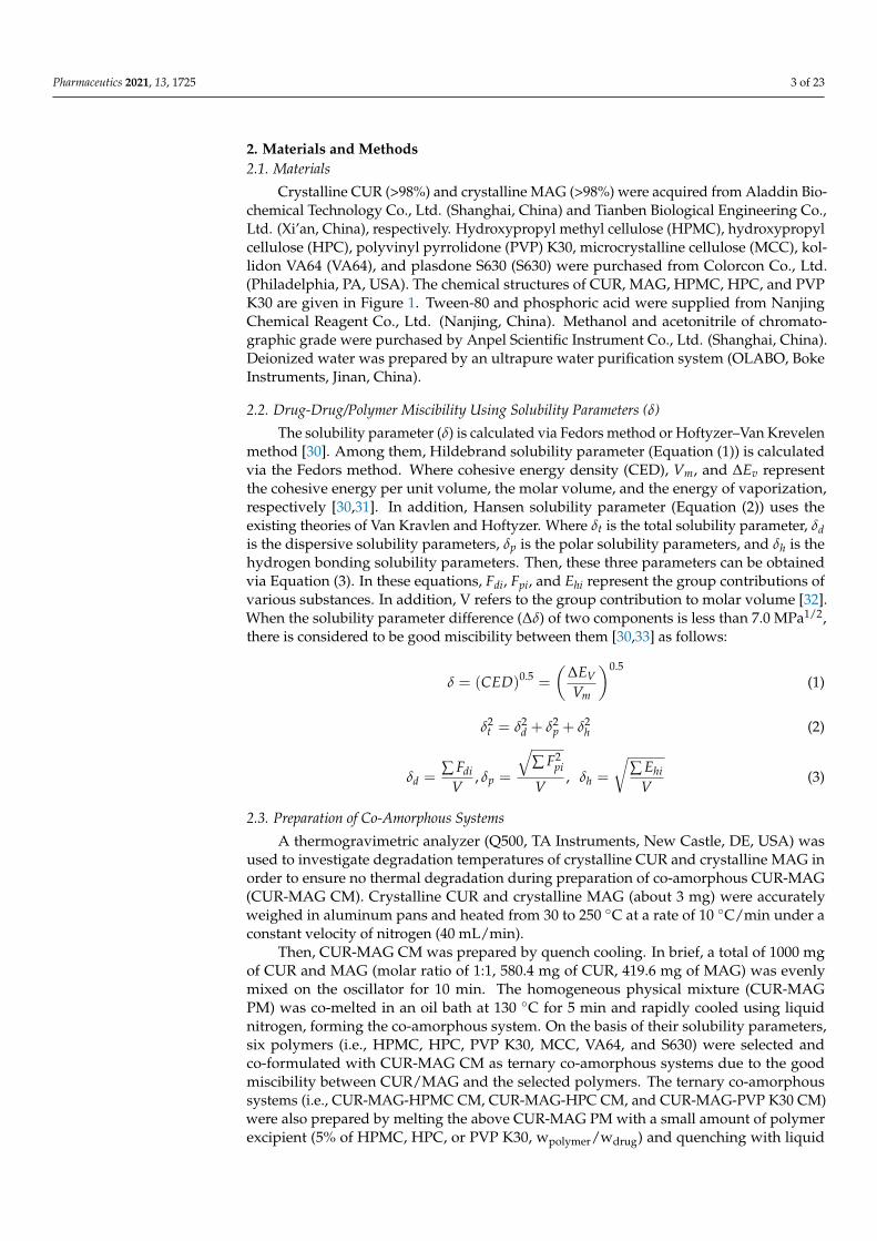
In the present study, co-amorphous curcumin-magnolol was designed based on theirsolubility/dissolution defects and potential drug combination advantages. Curcumin(CUR, Figure 1a) and magnolol (MAG, Figure 1b) have gained increasing interest becauseof their multiple biological activities (such as antioxidant, anticancer, antimicrobial, andanti-inflammatory) [19–23]. Moreover, the two drugs have generated combined thera-peutic effects in cardiovascular diseases, Alzheimer’s disease, and neurodegenerativediseases [24–26]. However, CUR shows poor oral absorption mainly because of low aque-ous solubility/dissolution (0.96 µg/mL in simulated gastric fluid) [8]. In addition, it isprone to phase II metabolism by β-glucuronidase in the gastrointestinal tract [27], whileMAG also belongs to poorly soluble drugs and has been proven to be an inhibitor ofβ-glucuronidation with high inhibitory activity [28,29]. Thus, such a co-amorphous systemwas designed and prepared.
Figure 1. Chemical structures of the model drugs: (a) CUR; (b) MAG and commonly used polymericstabilizers; (c) HPMC; (d) HPC; and (e) PVP K30.
Whereas, unlike high solubility/dissolution and physical stability of co-amorphoussystems previously reported, co-amorphous CUR-MAG (CUR-MAG CM) unexpectedlyexhibited much lower dissolution as compared with their crystalline counterparts due to itsaggregation during dissolution. Since polymeric excipients have potential to enhance thesolubility/dissolution of poorly soluble drugs and stabilize amorphous drugs, the questionis whether co-formulating with only a small amount of polymer could deaggregate CUR-MAG CM to optimize the dissolution, and improve its physical stability? The aim of thisstudy was to answer this question and explore the internal mechanism for deaggregationeffect using small amounts of polymers.
Pharmaceutics 2021, 13, 1725 3 of 23
2. Materials and Methods2.1. Materials
Crystalline CUR (>98%) and crystalline MAG (>98%) were acquired from Aladdin Bio-chemical Technology Co., Ltd. (Shanghai, China) and Tianben Biological Engineering Co.,Ltd. (Xi’an, China), respectively. Hydroxypropyl methyl cellulose (HPMC), hydroxypropylcellulose (HPC), polyvinyl pyrrolidone (PVP) K30, microcrystalline cellulose (MCC), kol-lidon VA64 (VA64), and plasdone S630 (S630) were purchased from Colorcon Co., Ltd.(Philadelphia, PA, USA). The chemical structures of CUR, MAG, HPMC, HPC, and PVPK30 are given in Figure 1. Tween-80 and phosphoric acid were supplied from NanjingChemical Reagent Co., Ltd. (Nanjing, China). Methanol and acetonitrile of chromato-graphic grade were purchased by Anpel Scientific Instrument Co., Ltd. (Shanghai, China).Deionized water was prepared by an ultrapure water purification system (OLABO, BokeInstruments, Jinan, China).
2.2. Drug-Drug/Polymer Miscibility Using Solubility Parameters (δ)
The solubility parameter (δ) is calculated via Fedors method or Hoftyzer–Van Krevelenmethod [30]. Among them, Hildebrand solubility parameter (Equation (1)) is calculatedvia the Fedors method. Where cohesive energy density (CED), Vm, and ∆Ev representthe cohesive energy per unit volume, the molar volume, and the energy of vaporization,respectively [30,31]. In addition, Hansen solubility parameter (Equation (2)) uses theexisting theories of Van Kravlen and Hoftyzer. Where δt is the total solubility parameter, δdis the dispersive solubility parameters, δp is the polar solubility parameters, and δh is thehydrogen bonding solubility parameters. Then, these three parameters can be obtainedvia Equation (3). In these equations, Fdi, Fpi, and Ehi represent the group contributions ofvarious substances. In addition, V refers to the group contribution to molar volume [32].When the solubility parameter difference (∆δ) of two components is less than 7.0 MPa1/2,there is considered to be good miscibility between them [30,33] as follows:
δ = (CED)0.5 =
(∆EVVm
)0.5(1)
δ2t = δ2
d + δ2p + δ2
h (2)
δd =∑ Fdi
V, δp =
√∑ F2
pi
V, δh =
√∑ Ehi
V(3)
2.3. Preparation of Co-Amorphous Systems
A thermogravimetric analyzer (Q500, TA Instruments, New Castle, DE, USA) wasused to investigate degradation temperatures of crystalline CUR and crystalline MAG inorder to ensure no thermal degradation during preparation of co-amorphous CUR-MAG(CUR-MAG CM). Crystalline CUR and crystalline MAG (about 3 mg) were accuratelyweighed in aluminum pans and heated from 30 to 250 ◦C at a rate of 10 ◦C/min under aconstant velocity of nitrogen (40 mL/min).
Then, CUR-MAG CM was prepared by quench cooling. In brief, a total of 1000 mgof CUR and MAG (molar ratio of 1:1, 580.4 mg of CUR, 419.6 mg of MAG) was evenlymixed on the oscillator for 10 min. The homogeneous physical mixture (CUR-MAGPM) was co-melted in an oil bath at 130 ◦C for 5 min and rapidly cooled using liquidnitrogen, forming the co-amorphous system. On the basis of their solubility parameters,six polymers (i.e., HPMC, HPC, PVP K30, MCC, VA64, and S630) were selected andco-formulated with CUR-MAG CM as ternary co-amorphous systems due to the goodmiscibility between CUR/MAG and the selected polymers. The ternary co-amorphoussystems (i.e., CUR-MAG-HPMC CM, CUR-MAG-HPC CM, and CUR-MAG-PVP K30 CM)were also prepared by melting the above CUR-MAG PM with a small amount of polymerexcipient (5% of HPMC, HPC, or PVP K30, wpolymer/wdrug) and quenching with liquid
Pharmaceutics 2021, 13, 1725 4 of 23
nitrogen. Meanwhile, other ternary co-amorphous systems were also prepared with MCC,VA64, and S630 in the same way (i.e., CUR-MAG-MCC CM, CUR-MAG-VA64 CM, andCUR-MAG-S630 CM). The obtained samples were ground with an agate mortar, sievedthrough 80 meshes, and stored in a desiccator above silica gel and anhydrous calciumchloride before analysis.
2.4. X-ray Powder Diffraction (XRPD)
XRPD diffractograms of the samples were acquired using an X-ray diffractometer(D/max 2500, Rigaku Co., Tokyo, Japan) with Cu-Kα radiation of 1.5406 Å. The diffrac-tometer was operated with a fixed tube current (100 mA) and a voltage (40 kV). A graphitemonochromator was employed to monochromate the X-ray beam, and a standard scintilla-tion counter simultaneously served as the detector. The sample was scanned at a step sizeof 0.02◦ in the reflectance mode range of 5~40◦ (2θ).
2.5. Differential Scanning Calorimetry (DSC)
The samples were performed on a thermal analyzer system (DSC 250, TA Instruments,New Castle, DE, USA) for thermal analysis. Approximately 3 mg of the sample powderswere sealed in an aluminum pan and heated to 250 ◦C at 10 ◦C/min under nitrogen flowwith a rate of 50 mL/min. Data including the glass transition temperature (Tg), recrys-tallization temperature (Trc), and melting temperature (Tm) were analyzed using TRIOSsoftware (version 5.0.0.44608). To obtain Tgs of amorphous CUR and amorphous MAG,the thermal analyzer was used to prepare amorphous CUR and amorphous MAG, and tosimultaneously determine their Tgs. Crystalline CUR or crystalline MAG (approximately3 mg) was sealed in an aluminum plate, melted at 5 ◦C above the melting point, and keptisothermal for 3 min in the DSC analyzer. Then, the sample plate was cooled to −25 ◦Cand maintained for 3 min, and the sample was heated to 250 ◦C at a rate of 10 ◦C/min. Tgsof amorphous CUR and amorphous MAG were analyzed using TRIOS software.
The Tg of the co-amorphous system assuming free volume additivity can be predictedusing the Fox equation (Equation (4)) [15,34] as follows:
1Tg12
=w1
Tg1+
w2
Tg2(4)
where Tg12 is the theoretical Tg of the co-amorphous system, Tg1 and Tg2 are the Tg of eachcomponent, w1 and w2 are their weight fractions. A positive or negative deviation from thetheoretically calculated Tg value reflects strong or weak intermolecular interactions betweenthe components. Furthermore, a theoretical Tg could also be calculated for q ternary co-amorphous systems using the Fox equation [10]. For example, in CUR-MAG-HPMC CM,the CUR-MAG CM was assumed as the first single “component”, and the added HPMCwas treated as the second “component”, as has been previously reported [10,35].
2.6. Raman Spectroscopy
Raman spectra of the samples were monitored by an Invia Raman spectrometer(Renishaw Co., Ltd., Gloucestershire, UK) at a 785 nm excitation laser, and were scanned ina measurement range of 800~1800 cm−1 at a resolution of 4 cm−1. OMNIC software wasused to analyze the measured spectral data.
2.7. Fourier Transform Infrared Spectroscopy (FTIR)
FTIR spectra of the samples were recorded on a FTIR spectrometer (IRAffinity-1S,Shimadzu Co., Ltd., Tokyo, Japan) equipped with LabSolutions IR software. Each sample(2 mg) was mixed with KBr (100 mg) for tablet compaction, and the tablet was scanned45 times in the range of 400~4000 cm−1 at a resolution of 4 cm−1.
Pharmaceutics 2021, 13, 1725 5 of 23
2.8. Solid-State 13C Nuclear Magnetic Resonance Spectroscopy (ss 13C NMR)
The ss 13C NMR spectra of the samples were determined using a Bruker AVANCEIII 400 MHz wide-bore spectrometer (Bruker Analytik GmbH, Rheinstetten, Germany)equipped with a double-resonance CP-MAS probe head. The sample was placed in 4 mmZrO2 rotors sealed with Kel-F1 to record the 13C NMR spectra with a sweep width of50 kHz. The ss 13C NMR spectra were obtained using the magic-angle spinning (MASfrequency 14 kHz) technique and samples were collected 480 times. All spectra werereferenced to an external sample of a-glycine at 176.03 ppm.
2.9. Dissolution under Supersaturated Conditions
Dissolution tests of crystalline CUR, crystalline MAG, CUR-MAG PM, CUR-MAG CM,CUR-MAG-HPMC CM, CUR-MAG-HPC CM, CUR-MAG-PVP K30 CM, CUR-MAG-MCCCM, CUR-MAG-VA64 CM, and CUR-MAG-S630 CM (equivalent to 400 mg of CUR or289.2 mg of MAG, molar ratio of 1:1) were conducted in triplicates by a small-volumedissolution apparatus (RC806D, Tianda Tianfa Technology Co., Ltd., Tianjin, China) witha constant stirring of 100 rpm at 37 ◦C. The dissolution tests were carried out in 200 mL0.1 M HCl buffer (pH 1.2) and 0.2 M phosphate buffer (pH 6.8) with 0.5% Tween-80. Atpredetermined time points (10, 20, 30, 45, 60, 90, 120, 240, 360, and 480 min), 2 mL aliquotswere collected and an equal volume of fresh medium was immediately added to themedium. After filtration using a 0.45 µm membrane and dilution with an equal volumeof methanol, the concentration of CUR and MAG was analyzed at 35 ◦C by the HPLCsystem (LC-2010A, Shimadzu Co., Ltd., Tokyo, Japan) with a Kromasil® C18 column (5 µm,250 × 4.6 mm). The mobile phase (60:40, acetonitrile to 0.3% phosphoric acid solution) waspumped up at the speed of 1 mL/min for 18 min. The wavelengths for detecting CUR andMAG were set at 430 and 294 nm, respectively.
2.10. Contact Angle Measurements and Surface Free Energy Calculation
The contact angle measurements of the co-amorphous samples were conducted bysessile drop method using a contact angle apparatus (OCA15EC, Dataphysics Ltd., Filder-stadt, Germany) with different test liquids (i.e., water, glycerol, and diiodomethane) [36,37].The contact angles for both sides of each drop were measured between the liquid andtablet surface. Furthermore, the contact angle measurement is the most common methodto calculate surface free energy.
The surface free energy parameters were calculated from the contact angle data of thetest liquids, which were analyzed by van Oss’s acid-base method. According to van Ossand co-workers to interfacial tensions, the solid surface free energy is composed of nonpolarLifshitz-van der Waals (γLW
SV ) and polar acid-base (γABSV ) interactions. The γAB
SV interaction isattributed to the electron-donor (γ−SV) and the electron-acceptor (γ+
SV) interactions. Thus,the total surface free energy (γtot
SV) of the solid can be expressed as Equation (5) [36,38,39]as follows:
γtotSV = γLW
SV + γABSV = γLW
SV + 2√
γ+SV γ−SV (5)
The relationship between the solid-liquid interfacial tension (γSL) as a function of thecomponents and parameters of the surface tension of the solid and liquid can be expressedas Equation (6) [38,40]:
γSL = γSV + γLV − 2√
γLWLV γLW
SV − 2√
γ+LV γ−SV − 2
√γ−LV γ+
SV (6)
where γLW is the Lifshitz-van der Waals component of the surface free energy of solidsor liquids, γ− and γ+ are the electron-donor and electron-acceptor parameters of theacid-base components of the solid (SV) or liquid (LV) surface free energy, respectively.
Combining with the Young equation (Equation (7)):
γLV cos θ = γSV − γSL (7)
Pharmaceutics 2021, 13, 1725 6 of 23
and the work of the liquid adhesion to solid surface (Adhesion work, WA) equation(Equation (8)):
WA = γLV(cos θ + 1) (8)
The WA can be further expressed by Equation (9) [36,38–40]:
WA = γLV(cos θ + 1) = 2√
γLWLV γLW
SV + 2√
γ+LV γ−SV + 2
√γ−LV γ+
SV (9)
2.11. Nucleation Inhibition of CUR and MAG by Polymer
Nucleation induction time (tn) was usually used to assess the effect of polymericexcipients on nucleation and crystallization of drug solutions [41]. In this study, supersat-urated solutions of CUR-MAG CM, CUR-MAG-HPMC CM, CUR-MAG-HPC CM, andCUR-MAG-PVP K30 CM were prepared by adding 1 mL DMSO solution (i.e., dissolvingthe samples in DMSO solvent at the same CUR concentration equal to 2 mg/mL CUR) into99 mL water with a constant stirring of 200 rpm at 25 ◦C. The turbidity-time profiles ofthe supersaturated solutions were determined in triplicate using a UV spectrophotometer(TU-1901, PGeneral Ltd., Beijing, China) at a wavelength of 600 nm (no UV absorption forboth drugs and polymers).
2.12. Broadband Dielectric Spectroscopy (BDS)
The dielectric permittivity (ε*(ω) = ε′(ω) − iε′′(ω)) measurements were conductedusing the dielectric spectrometer (Novocontrol concept 40, Novocontrol TechnologiesGmbH & Co. KG, Montabaur, Germany). The spectrometer operated at the frequencyrange from 10−2 to 106 Hz, and a Quatro Cryosystem equipped with a nitrogen gas cryostatcontrolled the temperature stability (better than 0.1 ◦C). The samples were placed betweentwo stainless-steel flat electrodes of the capacitor with a 0.1 mm gap and mounted in acryostat. Dielectric measurements on CUR-MAG CM, CUR-MAG-HPMC CM, CUR-MAG-HPC CM, and CUR-MAG-PVP K30 CM (5% of HPMC, HPC, or PVP K30 to drugs in theternary co-amorphous systems, wpolymer/wdrug), were performed from 16 to 40 ◦C underdry nitrogen gas flow.
2.13. Physical Stability Evaluation
CUR-MAG CM, CUR-MAG-HPMC CM, CUR-MAG-HPC CM, and CUR-MAG-PVPK30 CM were stored under long-term condition (25 ◦C, 60% RH) and accelerated condition(40 ◦C, 75% RH) in a stability chamber (YSEI Experimental Instrument Co., Ltd., Chongqing,China) for 3 months. The samples were evaluated by XRPD on 7, 14, 21, 30, 60, and 90 days.The appearance of recrystallization peaks from the amorphous halo in the XRPD patternsreflected recrystallization.
3. Results and Discussion3.1. Drug-Drug/Polymer Miscibility Using Solubility Parameters (δ)
Compounds are considered to be miscible when they have similar δ values, since en-ergy requirements for mixing are compensated by the energy released in the interactionsbetween the components (exothermic energy of mixing) [30]. For the theoretical miscibilityassessment, if the solubility parameter difference (∆δ) is less than 7.0 MPa1/2, the componentsare considered to have good miscibility. If ∆δ is less than 2.0 MPa1/2, the components maypossess excellent miscibility and easily form a solid solution, while ∆δ greater than 10 MPa1/2
indicates immiscibility between them [30,42]. Theoretical predicted δ using the Fedors andHoftyzer–Van Krevelen methods are given in Supplementary Materials Table S1. The ∆δ of0.41 MPa1/2 between CUR and MAG suggested their excellent miscibility, and the ∆δ val-ues between CUR/MAG and each polymer were all less than 7.0 MPa1/2, indicating goodmiscibility between CUR/MAG and the selected polymers.
Pharmaceutics 2021, 13, 1725 7 of 23
3.2. Preparation of Co-Amorphous Systems
The purpose of this section was to conduct safe preparation (without thermal decom-position) of CUR-MAG CM during melting and quench cooling. A thermogravimetricanalysis (TGA) was used to investigate whether crystalline CUR and crystalline MAGunderwent thermal degradation during the preparation process. As depicted in FigureS1, crystalline CUR showed no weight loss in the range from 30 to 198.1 ◦C, and crys-talline MAG showed a steep descent in the TGA profile until 162.6 ◦C. The ranges ofthermodynamic degradation were much higher than their melting points, as describedin Section 3.3.2 (Tm = 181.9 ◦C for CUR and Tm = 101.4 ◦C for MAG, respectively). Theresults suggested that CUR-MAG CM could be safely prepared by quench cooling. Due tothe good miscibility between CUR and MAG or between CUR/MAG and the polymers,the components easily formed a clarified solid solution. A polarizing light microscopy(PLM) observation demonstrated the complete amorphization of CUR-MAG CM and itsternary co-amorphous systems (Figure S2). At last, a higher yield rate of 98.15 ± 0.74% wasobtained in CUR-MAG CM, and the content of CUR and MAG determined by the HPLCmethod was 0.573± 0.004 g/g for CUR and 0.408± 0.003 g/g for MAG in the final productof CUR-MAG CM (n = 3). In addition, all ternary co-amorphous systems also maintainedhigh yield rates (Table S2).
3.3. Solid-State Characterization3.3.1. XRPD
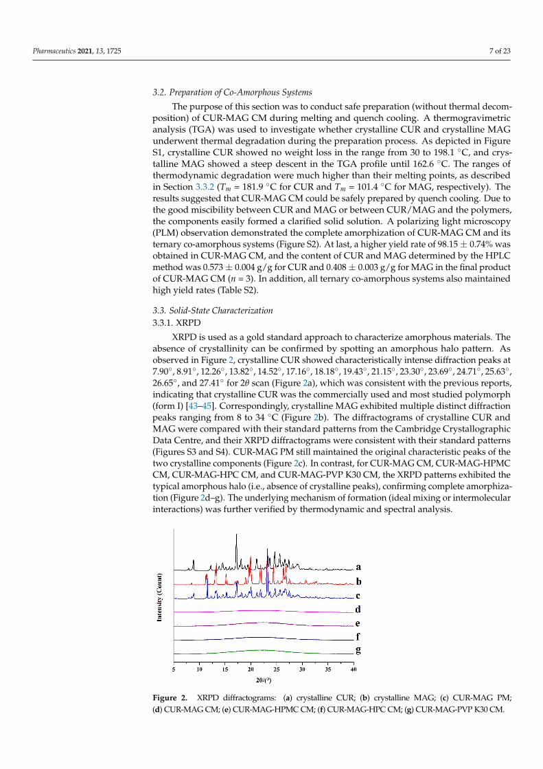
XRPD is used as a gold standard approach to characterize amorphous materials. Theabsence of crystallinity can be confirmed by spotting an amorphous halo pattern. Asobserved in Figure 2, crystalline CUR showed characteristically intense diffraction peaks at7.90◦, 8.91◦, 12.26◦, 13.82◦, 14.52◦, 17.16◦, 18.18◦, 19.43◦, 21.15◦, 23.30◦, 23.69◦, 24.71◦, 25.63◦,26.65◦, and 27.41◦ for 2θ scan (Figure 2a), which was consistent with the previous reports,indicating that crystalline CUR was the commercially used and most studied polymorph(form I) [43–45]. Correspondingly, crystalline MAG exhibited multiple distinct diffractionpeaks ranging from 8 to 34 ◦C (Figure 2b). The diffractograms of crystalline CUR andMAG were compared with their standard patterns from the Cambridge CrystallographicData Centre, and their XRPD diffractograms were consistent with their standard patterns(Figures S3 and S4). CUR-MAG PM still maintained the original characteristic peaks of thetwo crystalline components (Figure 2c). In contrast, for CUR-MAG CM, CUR-MAG-HPMCCM, CUR-MAG-HPC CM, and CUR-MAG-PVP K30 CM, the XRPD patterns exhibited thetypical amorphous halo (i.e., absence of crystalline peaks), confirming complete amorphiza-tion (Figure 2d–g). The underlying mechanism of formation (ideal mixing or intermolecularinteractions) was further verified by thermodynamic and spectral analysis.
Figure 2. XRPD diffractograms: (a) crystalline CUR; (b) crystalline MAG; (c) CUR-MAG PM;(d) CUR-MAG CM; (e) CUR-MAG-HPMC CM; (f) CUR-MAG-HPC CM; (g) CUR-MAG-PVP K30 CM.
Pharmaceutics 2021, 13, 1725 8 of 23
3.3.2. DSC
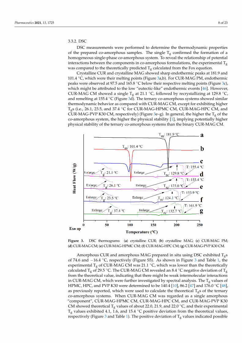
DSC measurements were performed to determine the thermodynamic propertiesof the prepared co-amorphous samples. The single Tg confirmed the formation of ahomogeneous single-phase co-amorphous system. To reveal the relationship of potentialinteractions between the components in co-amorphous formulations, the experimental Tgwas compared to the theoretically predicted Tg calculated from the Fox equation.
Crystalline CUR and crystalline MAG showed sharp endothermic peaks at 181.9 and101.4 ◦C, which were their melting points (Figure 3a,b). For CUR-MAG PM, endothermicpeaks were observed at 97.5 and 165.8 ◦C below their respective melting points (Figure 3c),which might be attributed to the low “eutectic-like” endothermic events [46]. However,CUR-MAG CM showed a single Tg at 21.1 ◦C, followed by recrystallizing at 129.8 ◦C,and remelting at 155.4 ◦C (Figure 3d). The ternary co-amorphous systems showed similarthermodynamic behavior as compared with CUR-MAG CM, except for exhibiting higherTgs (i.e., 26.1, 23.5, and 37.4 ◦C for CUR-MAG-HPMC CM, CUR-MAG-HPC CM, andCUR-MAG-PVP K30 CM, respectively) (Figure 3e–g). In general, the higher the Tg of theco-amorphous system, the higher the physical stability [1], implying potentially higherphysical stability of the ternary co-amorphous systems than the binary CUR-MAG CM.
Figure 3. DSC thermograms: (a) crystalline CUR; (b) crystalline MAG; (c) CUR-MAG PM;(d) CUR-MAG CM; (e) CUR-MAG-HPMC CM; (f) CUR-MAG-HPC CM; (g) CUR-MAG-PVP K30 CM.
Amorphous CUR and amorphous MAG prepared in situ using DSC exhibited Tgsof 74.6 and −16.4 ◦C, respectively (Figure S5). As shown in Figure 3 and Table 1, theexperimental Tg of CUR-MAG CM was 21.1 ◦C, which was lower than the theoreticallycalculated Tg of 29.5 ◦C. The CUR-MAG CM revealed an 8.4 ◦C negative deviation of Tgfrom the theoretical value, indicating that there might be weak intermolecular interactionsin CUR-MAG CM, which were further investigated by spectral analysis. The Tg values ofHPMC, HPC, and PVP K30 were determined to be 140.4 [10], 86.2 [47] and 176.0 ◦C [48],as previously reported, which were used to calculate the theoretical Tgs of the ternaryco-amorphous systems. When CUR-MAG CM was regarded as a single amorphous“component”, CUR-MAG-HPMC CM, CUR-MAG-HPC CM, and CUR-MAG-PVP K30CM showed theoretical Tg values of about 22.0, 21.9, and 22.0 ◦C, and their experimentalTg values exhibited 4.1, 1.6, and 15.4 ◦C positive deviation from the theoretical values,respectively (Figure 3 and Table 1). The positive deviation of Tg values indicated possible
Pharmaceutics 2021, 13, 1725 9 of 23
molecular interactions between CUR-MAG CM and the added polymers in ternary co-amorphous systems.
Table 1. The experimental and theoretical Tg values of CUR-MAG CM and its ternaryco-amorphous systems.
Sample Experimental Tg, ◦C Calculated Tg, ◦C ∆Tg, ◦C
Amorphous CUR 74.6Amorphous MAG −16.4
CUR-MAG CM 21.1 29.5 −8.4CUR-MAG-HPMC CM 26.1 22.0 4.1CUR-MAG-HPC CM 23.5 21.9 1.6
CUR-MAG-PVP K30 CM 37.4 22.0 15.4
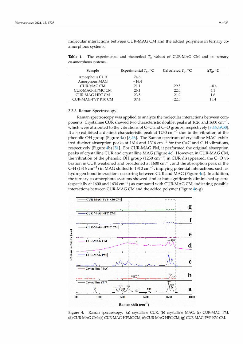
3.3.3. Raman Spectroscopy
Raman spectroscopy was applied to analyze the molecular interactions between com-ponents. Crystalline CUR showed two characteristic doublet peaks at 1626 and 1600 cm−1,which were attributed to the vibrations of C=C and C=O groups, respectively [8,46,49,50].It also exhibited a distinct characteristic peak at 1250 cm−1 due to the vibration of thephenolic OH group (Figure 4a) [8,46]. The Raman spectrum of crystalline MAG exhib-ited distinct absorption peaks at 1614 and 1316 cm−1 for the C=C and C-H vibrations,respectively (Figure 4b) [51]. For CUR-MAG PM, it performed the original absorptionpeaks of crystalline CUR and crystalline MAG (Figure 4c). However, in CUR-MAG CM,the vibration of the phenolic OH group (1250 cm−1) in CUR disappeared, the C=O vi-bration in CUR weakened and broadened at 1600 cm−1, and the absorption peak of theC-H (1316 cm−1) in MAG shifted to 1310 cm−1, implying potential interactions, such ashydrogen bond interactions occurring between CUR and MAG (Figure 4d). In addition,the ternary co-amorphous systems showed similar but significantly diminished spectra(especially at 1600 and 1634 cm−1) as compared with CUR-MAG CM, indicating possibleinteractions between CUR-MAG CM and the added polymer (Figure 4e–g).
Figure 4. Raman spectroscopy: (a) crystalline CUR; (b) crystalline MAG; (c) CUR-MAG PM;(d) CUR-MAG CM; (e) CUR-MAG-HPMC CM; (f) CUR-MAG-HPC CM; (g) CUR-MAG-PVP K30 CM.
Pharmaceutics 2021, 13, 1725 10 of 23
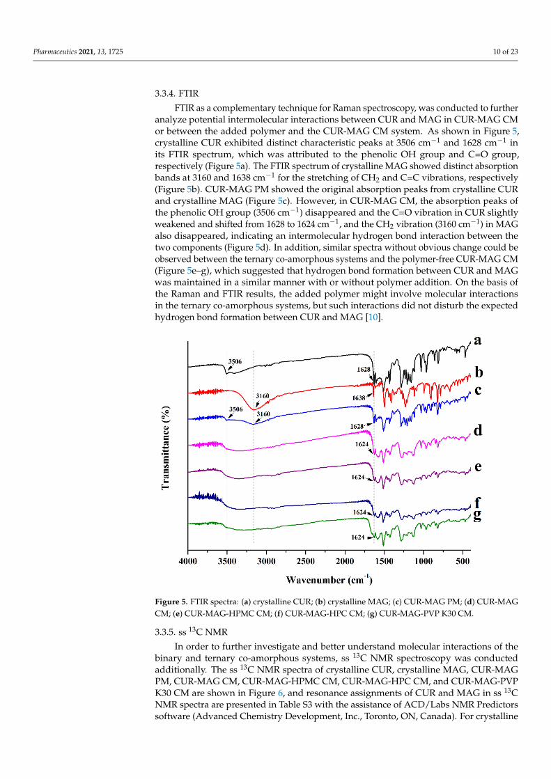
3.3.4. FTIR
FTIR as a complementary technique for Raman spectroscopy, was conducted to furtheranalyze potential intermolecular interactions between CUR and MAG in CUR-MAG CMor between the added polymer and the CUR-MAG CM system. As shown in Figure 5,crystalline CUR exhibited distinct characteristic peaks at 3506 cm−1 and 1628 cm−1 inits FTIR spectrum, which was attributed to the phenolic OH group and C=O group,respectively (Figure 5a). The FTIR spectrum of crystalline MAG showed distinct absorptionbands at 3160 and 1638 cm−1 for the stretching of CH2 and C=C vibrations, respectively(Figure 5b). CUR-MAG PM showed the original absorption peaks from crystalline CURand crystalline MAG (Figure 5c). However, in CUR-MAG CM, the absorption peaks ofthe phenolic OH group (3506 cm−1) disappeared and the C=O vibration in CUR slightlyweakened and shifted from 1628 to 1624 cm−1, and the CH2 vibration (3160 cm−1) in MAGalso disappeared, indicating an intermolecular hydrogen bond interaction between thetwo components (Figure 5d). In addition, similar spectra without obvious change could beobserved between the ternary co-amorphous systems and the polymer-free CUR-MAG CM(Figure 5e–g), which suggested that hydrogen bond formation between CUR and MAGwas maintained in a similar manner with or without polymer addition. On the basis ofthe Raman and FTIR results, the added polymer might involve molecular interactionsin the ternary co-amorphous systems, but such interactions did not disturb the expectedhydrogen bond formation between CUR and MAG [10].
Figure 5. FTIR spectra: (a) crystalline CUR; (b) crystalline MAG; (c) CUR-MAG PM; (d) CUR-MAGCM; (e) CUR-MAG-HPMC CM; (f) CUR-MAG-HPC CM; (g) CUR-MAG-PVP K30 CM.
3.3.5. ss 13C NMR
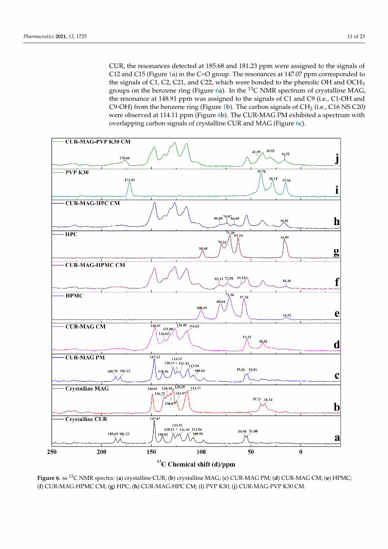
In order to further investigate and better understand molecular interactions of thebinary and ternary co-amorphous systems, ss 13C NMR spectroscopy was conductedadditionally. The ss 13C NMR spectra of crystalline CUR, crystalline MAG, CUR-MAGPM, CUR-MAG CM, CUR-MAG-HPMC CM, CUR-MAG-HPC CM, and CUR-MAG-PVPK30 CM are shown in Figure 6, and resonance assignments of CUR and MAG in ss 13CNMR spectra are presented in Table S3 with the assistance of ACD/Labs NMR Predictorssoftware (Advanced Chemistry Development, Inc., Toronto, ON, Canada). For crystalline
Pharmaceutics 2021, 13, 1725 11 of 23
CUR, the resonances detected at 185.68 and 181.23 ppm were assigned to the signals ofC12 and C15 (Figure 1a) in the C=O group. The resonances at 147.07 ppm corresponded tothe signals of C1, C2, C21, and C22, which were bonded to the phenolic OH and OCH3groups on the benzene ring (Figure 6a). In the 13C NMR spectrum of crystalline MAG,the resonance at 148.91 ppm was assigned to the signals of C1 and C9 (i.e., C1-OH andC9-OH) from the benzene ring (Figure 1b). The carbon signals of CH2 (i.e., C16 NS C20)were observed at 114.11 ppm (Figure 6b). The CUR-MAG PM exhibited a spectrum withoverlapping carbon signals of crystalline CUR and MAG (Figure 6c).
Figure 6. ss 13C NMR spectra: (a) crystalline CUR; (b) crystalline MAG; (c) CUR-MAG PM; (d) CUR-MAG CM; (e) HPMC;(f) CUR-MAG-HPMC CM; (g) HPC; (h) CUR-MAG-HPC CM; (i) PVP K30; (j) CUR-MAG-PVP K30 CM.
Pharmaceutics 2021, 13, 1725 12 of 23
However, CUR-MAG CM showed some shifted or disappeared signals with signifi-cantly broadening lines, which was distinguishable from the physical mixture (Figure 6d).On the one hand, the carbon signals (i.e., C12 and C15) from the C=O group disappearedand the carbon signals of C2 and C22 (i.e., C2-OH and C22-OH) shifted from 147.07 to146.61 ppm in CUR. On the other hand, the carbon signals (i.e., C16 and C20) from theCH2 group shifted from 114.11 to 114.62 ppm in MAG. According to the results of spectralanalysis (Raman, FTIR, and 13C NMR spectra), the intermolecular hydrogen bond inter-actions in CUR-MAG CM might occur between the phenolic OH and/or C=O groups inCUR and the CH2 group in MAG. In addition, the ternary co-amorphous systems exhibitedsimilar spectra with CUR-MAG CM except for some new and weak signals (Figure 6f,h,j).In the ternary co-amorphous systems, the original signals of the binary CUR-MAG CMsystem did not change, indicating that the added polymers (HPMC, HPC, and PVP K30)could not break the molecular interaction between CUR and MAG in CUR-MAG CM. Thegeneration of new signals in the ss 13C NMR spectra was due to the polymer introduction.As compared with the signals of polymers (Figure 6e,g,i), the new signals from polymersin ternary co-amorphous systems shifted to varying degrees. For example, the signals ofPVP K30 at 171.93, 39.78, 28.15, and 15.16 ppm (Figure 6i) shifted to 176.66, 41.59, 30.92,and 16.32 ppm, respectively (Figure 6j). These results indicated that the added polymersinvolved molecular interactions in the ternary co-amorphous systems, but such interactionsdid not disturb the expected hydrogen bond formation between CUR and MAG.
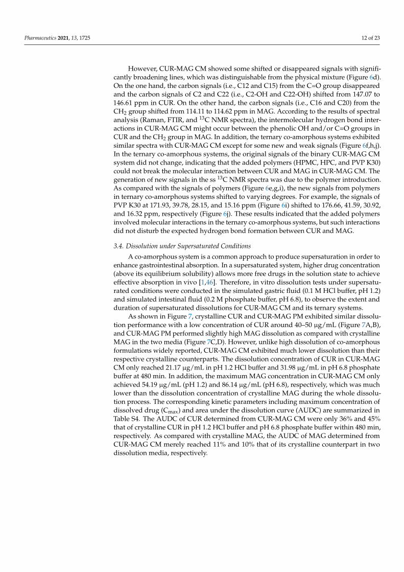
3.4. Dissolution under Supersaturated Conditions
A co-amorphous system is a common approach to produce supersaturation in order toenhance gastrointestinal absorption. In a supersaturated system, higher drug concentration(above its equilibrium solubility) allows more free drugs in the solution state to achieveeffective absorption in vivo [1,46]. Therefore, in vitro dissolution tests under supersatu-rated conditions were conducted in the simulated gastric fluid (0.1 M HCl buffer, pH 1.2)and simulated intestinal fluid (0.2 M phosphate buffer, pH 6.8), to observe the extent andduration of supersaturated dissolutions for CUR-MAG CM and its ternary systems.
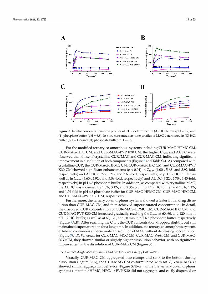
As shown in Figure 7, crystalline CUR and CUR-MAG PM exhibited similar dissolu-tion performance with a low concentration of CUR around 40–50 µg/mL (Figure 7A,B),and CUR-MAG PM performed slightly high MAG dissolution as compared with crystallineMAG in the two media (Figure 7C,D). However, unlike high dissolution of co-amorphousformulations widely reported, CUR-MAG CM exhibited much lower dissolution than theirrespective crystalline counterparts. The dissolution concentration of CUR in CUR-MAGCM only reached 21.17 µg/mL in pH 1.2 HCl buffer and 31.98 µg/mL in pH 6.8 phosphatebuffer at 480 min. In addition, the maximum MAG concentration in CUR-MAG CM onlyachieved 54.19 µg/mL (pH 1.2) and 86.14 µg/mL (pH 6.8), respectively, which was muchlower than the dissolution concentration of crystalline MAG during the whole dissolu-tion process. The corresponding kinetic parameters including maximum concentration ofdissolved drug (Cmax) and area under the dissolution curve (AUDC) are summarized inTable S4. The AUDC of CUR determined from CUR-MAG CM were only 36% and 45%that of crystalline CUR in pH 1.2 HCl buffer and pH 6.8 phosphate buffer within 480 min,respectively. As compared with crystalline MAG, the AUDC of MAG determined fromCUR-MAG CM merely reached 11% and 10% that of its crystalline counterpart in twodissolution media, respectively.
Pharmaceutics 2021, 13, 1725 13 of 23
Figure 7. In vitro concentration–time profiles of CUR determined in (A) HCl buffer (pH = 1.2) and(B) phosphate buffer (pH = 6.8). In vitro concentration–time profiles of MAG determined in (C) HClbuffer (pH = 1.2) and (D) phosphate buffer (pH = 6.8).
For the modified ternary co-amorphous systems including CUR-MAG-HPMC CM,CUR-MAG-HPC CM, and CUR-MAG-PVP K30 CM, the higher Cmax and AUDC wereobserved than those of crystalline CUR/MAG and CUR-MAG CM, indicating significantimprovement in dissolution of both components (Figure 7 and Table S4). As compared withcrystalline CUR, the CUR-MAG-HPMC CM, CUR-MAG-HPC CM, and CUR-MAG-PVPK30 CM showed significant enhancements (p < 0.01) in Cmax (4.00-, 5.68- and 3.92-fold,respectively) and AUDC (3.72-, 5.21-, and 3.68-fold, respectively) in pH 1.2 HCl buffer, aswell as in Cmax (3.60-, 2.92-, and 5.08-fold, respectively) and AUDC (3.22-, 2.70-, 4.45-fold,respectively) in pH 6.8 phosphate buffer. In addition, as compared with crystalline MAG,the AUDC was increased by 1.82-, 3.12-, and 2.36-fold in pH 1.2 HCl buffer and 1.31-, 1.42-,and 1.79-fold in pH 6.8 phosphate buffer for CUR-MAG-HPMC CM, CUR-MAG-HPC CM,and CUR-MAG-PVP K30 CM, respectively.
Furthermore, the ternary co-amorphous systems showed a faster initial drug disso-lution than CUR-MAG CM, and then achieved supersaturated concentration. In detail,the dissolved CUR concentration of CUR-MAG-HPMC CM, CUR-MAG-HPC CM, andCUR-MAG-PVP K30 CM increased gradually, reaching the Cmax at 60, 60, and 120 min inpH 1.2 HCl buffer, as well as at 60, 120, and 60 min in pH 6.8 phosphate buffer, respectively(Figure 7A,B). After reaching the Cmax, the CUR concentration dropped slightly, but stillmaintained supersaturation for a long time. In addition, the ternary co-amorphous systemsexhibited continuous supersaturated dissolution of MAG without decreasing concentration(Figure 7C,D). Whereas, for CUR-MAG-MCC CM, CUR-MAG-VA64 CM, and CUR-MAG-S630 CM, they showed similar or slightly higher dissolution behavior, with no significantimprovement in the dissolution of CUR-MAG CM (Figure S6).
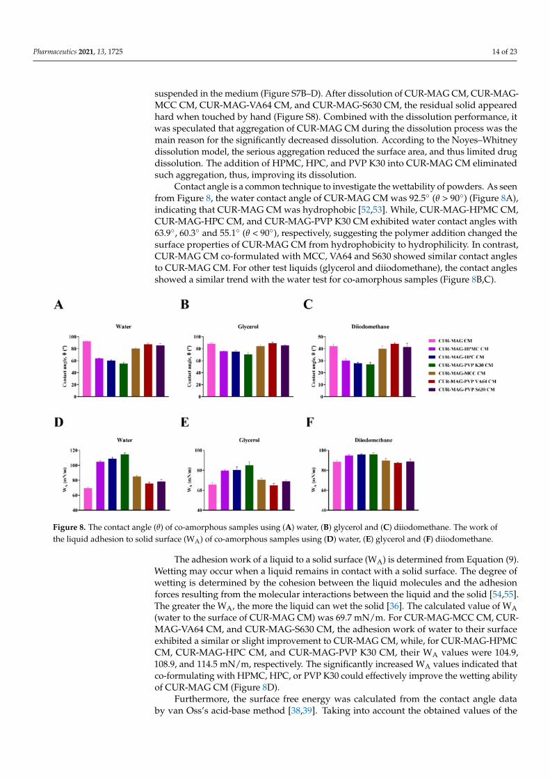
3.5. Contact Angle Measurements and Surface Free Energy Calculation
Visually, CUR-MAG CM aggregated into clumps and sank to the bottom duringdissolution (Figure S7A), the CUR-MAG CM co-formulated with MCC, VA64, or S630showed similar aggregation behavior (Figure S7E–G), while the ternary co-amorphoussystems containing HPMC, HPC, or PVP K30 did not aggregate and easily dispersed or
Pharmaceutics 2021, 13, 1725 14 of 23
suspended in the medium (Figure S7B–D). After dissolution of CUR-MAG CM, CUR-MAG-MCC CM, CUR-MAG-VA64 CM, and CUR-MAG-S630 CM, the residual solid appearedhard when touched by hand (Figure S8). Combined with the dissolution performance, itwas speculated that aggregation of CUR-MAG CM during the dissolution process was themain reason for the significantly decreased dissolution. According to the Noyes–Whitneydissolution model, the serious aggregation reduced the surface area, and thus limited drugdissolution. The addition of HPMC, HPC, and PVP K30 into CUR-MAG CM eliminatedsuch aggregation, thus, improving its dissolution.
Contact angle is a common technique to investigate the wettability of powders. As seenfrom Figure 8, the water contact angle of CUR-MAG CM was 92.5◦ (θ > 90◦) (Figure 8A),indicating that CUR-MAG CM was hydrophobic [52,53]. While, CUR-MAG-HPMC CM,CUR-MAG-HPC CM, and CUR-MAG-PVP K30 CM exhibited water contact angles with63.9◦, 60.3◦ and 55.1◦ (θ < 90◦), respectively, suggesting the polymer addition changed thesurface properties of CUR-MAG CM from hydrophobicity to hydrophilicity. In contrast,CUR-MAG CM co-formulated with MCC, VA64 and S630 showed similar contact anglesto CUR-MAG CM. For other test liquids (glycerol and diiodomethane), the contact anglesshowed a similar trend with the water test for co-amorphous samples (Figure 8B,C).
Figure 8. The contact angle (θ) of co-amorphous samples using (A) water, (B) glycerol and (C) diiodomethane. The work ofthe liquid adhesion to solid surface (WA) of co-amorphous samples using (D) water, (E) glycerol and (F) diiodomethane.
The adhesion work of a liquid to a solid surface (WA) is determined from Equation (9).Wetting may occur when a liquid remains in contact with a solid surface. The degree ofwetting is determined by the cohesion between the liquid molecules and the adhesionforces resulting from the molecular interactions between the liquid and the solid [54,55].The greater the WA, the more the liquid can wet the solid [36]. The calculated value of WA(water to the surface of CUR-MAG CM) was 69.7 mN/m. For CUR-MAG-MCC CM, CUR-MAG-VA64 CM, and CUR-MAG-S630 CM, the adhesion work of water to their surfaceexhibited a similar or slight improvement to CUR-MAG CM, while, for CUR-MAG-HPMCCM, CUR-MAG-HPC CM, and CUR-MAG-PVP K30 CM, their WA values were 104.9,108.9, and 114.5 mN/m, respectively. The significantly increased WA values indicated thatco-formulating with HPMC, HPC, or PVP K30 could effectively improve the wetting abilityof CUR-MAG CM (Figure 8D).
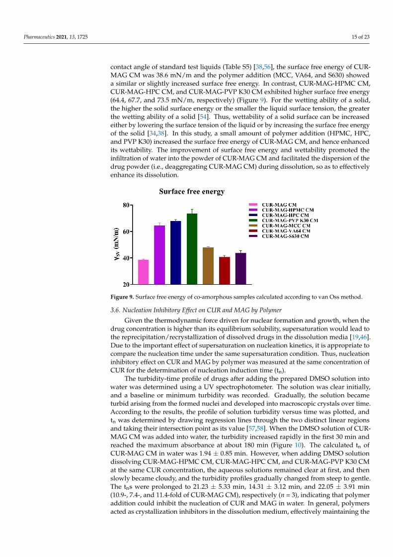
Furthermore, the surface free energy was calculated from the contact angle databy van Oss’s acid-base method [38,39]. Taking into account the obtained values of the
Pharmaceutics 2021, 13, 1725 15 of 23
contact angle of standard test liquids (Table S5) [38,56], the surface free energy of CUR-MAG CM was 38.6 mN/m and the polymer addition (MCC, VA64, and S630) showeda similar or slightly increased surface free energy. In contrast, CUR-MAG-HPMC CM,CUR-MAG-HPC CM, and CUR-MAG-PVP K30 CM exhibited higher surface free energy(64.4, 67.7, and 73.5 mN/m, respectively) (Figure 9). For the wetting ability of a solid,the higher the solid surface energy or the smaller the liquid surface tension, the greaterthe wetting ability of a solid [54]. Thus, wettability of a solid surface can be increasedeither by lowering the surface tension of the liquid or by increasing the surface free energyof the solid [34,38]. In this study, a small amount of polymer addition (HPMC, HPC,and PVP K30) increased the surface free energy of CUR-MAG CM, and hence enhancedits wettability. The improvement of surface free energy and wettability promoted theinfiltration of water into the powder of CUR-MAG CM and facilitated the dispersion of thedrug powder (i.e., deaggregating CUR-MAG CM) during dissolution, so as to effectivelyenhance its dissolution.
Figure 9. Surface free energy of co-amorphous samples calculated according to van Oss method.
3.6. Nucleation Inhibitory Effect on CUR and MAG by Polymer
Given the thermodynamic force driven for nuclear formation and growth, when thedrug concentration is higher than its equilibrium solubility, supersaturation would lead tothe reprecipitation/recrystallization of dissolved drugs in the dissolution media [19,46].Due to the important effect of supersaturation on nucleation kinetics, it is appropriate tocompare the nucleation time under the same supersaturation condition. Thus, nucleationinhibitory effect on CUR and MAG by polymer was measured at the same concentration ofCUR for the determination of nucleation induction time (tn).
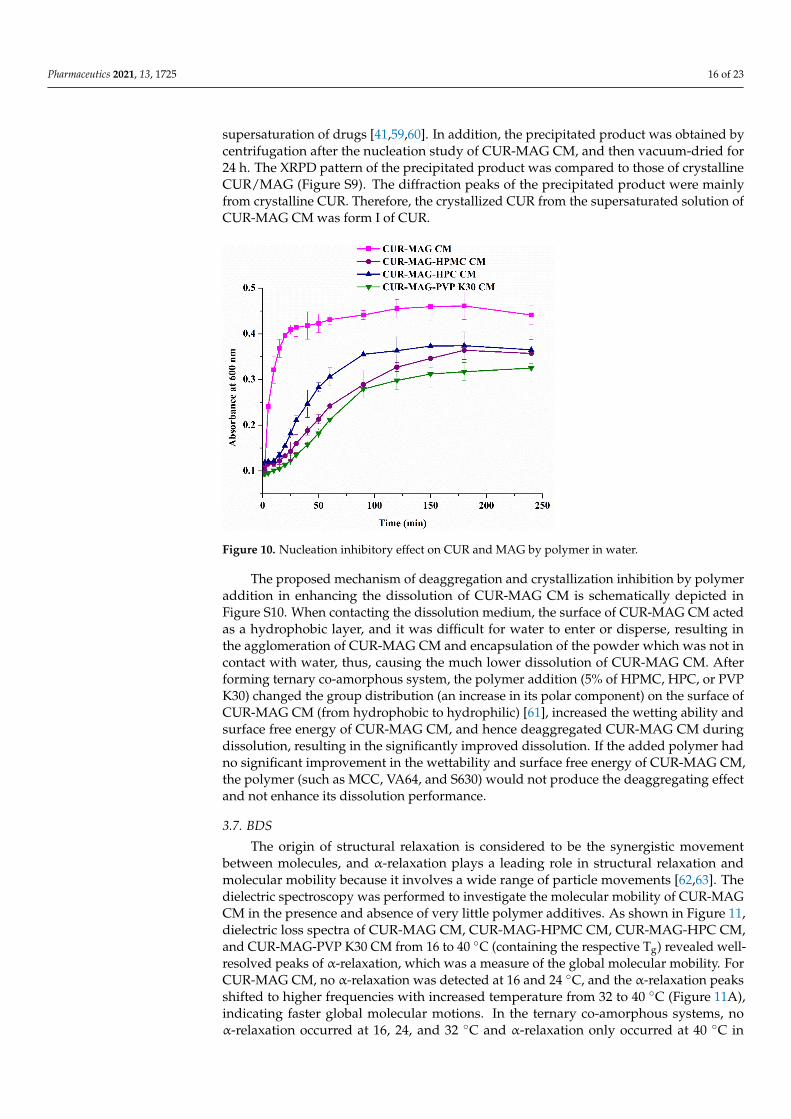
The turbidity-time profile of drugs after adding the prepared DMSO solution intowater was determined using a UV spectrophotometer. The solution was clear initially,and a baseline or minimum turbidity was recorded. Gradually, the solution becameturbid arising from the formed nuclei and developed into macroscopic crystals over time.According to the results, the profile of solution turbidity versus time was plotted, andtn was determined by drawing regression lines through the two distinct linear regionsand taking their intersection point as its value [57,58]. When the DMSO solution of CUR-MAG CM was added into water, the turbidity increased rapidly in the first 30 min andreached the maximum absorbance at about 180 min (Figure 10). The calculated tn ofCUR-MAG CM in water was 1.94 ± 0.85 min. However, when adding DMSO solutiondissolving CUR-MAG-HPMC CM, CUR-MAG-HPC CM, and CUR-MAG-PVP K30 CMat the same CUR concentration, the aqueous solutions remained clear at first, and thenslowly became cloudy, and the turbidity profiles gradually changed from steep to gentle.The tns were prolonged to 21.23 ± 5.33 min, 14.31 ± 3.12 min, and 22.05 ± 3.91 min(10.9-, 7.4-, and 11.4-fold of CUR-MAG CM), respectively (n = 3), indicating that polymeraddition could inhibit the nucleation of CUR and MAG in water. In general, polymersacted as crystallization inhibitors in the dissolution medium, effectively maintaining the
Pharmaceutics 2021, 13, 1725 16 of 23
supersaturation of drugs [41,59,60]. In addition, the precipitated product was obtained bycentrifugation after the nucleation study of CUR-MAG CM, and then vacuum-dried for24 h. The XRPD pattern of the precipitated product was compared to those of crystallineCUR/MAG (Figure S9). The diffraction peaks of the precipitated product were mainlyfrom crystalline CUR. Therefore, the crystallized CUR from the supersaturated solution ofCUR-MAG CM was form I of CUR.
Figure 10. Nucleation inhibitory effect on CUR and MAG by polymer in water.
The proposed mechanism of deaggregation and crystallization inhibition by polymeraddition in enhancing the dissolution of CUR-MAG CM is schematically depicted inFigure S10. When contacting the dissolution medium, the surface of CUR-MAG CM actedas a hydrophobic layer, and it was difficult for water to enter or disperse, resulting inthe agglomeration of CUR-MAG CM and encapsulation of the powder which was not incontact with water, thus, causing the much lower dissolution of CUR-MAG CM. Afterforming ternary co-amorphous system, the polymer addition (5% of HPMC, HPC, or PVPK30) changed the group distribution (an increase in its polar component) on the surface ofCUR-MAG CM (from hydrophobic to hydrophilic) [61], increased the wetting ability andsurface free energy of CUR-MAG CM, and hence deaggregated CUR-MAG CM duringdissolution, resulting in the significantly improved dissolution. If the added polymer hadno significant improvement in the wettability and surface free energy of CUR-MAG CM,the polymer (such as MCC, VA64, and S630) would not produce the deaggregating effectand not enhance its dissolution performance.
3.7. BDS
The origin of structural relaxation is considered to be the synergistic movementbetween molecules, and α-relaxation plays a leading role in structural relaxation andmolecular mobility because it involves a wide range of particle movements [62,63]. Thedielectric spectroscopy was performed to investigate the molecular mobility of CUR-MAGCM in the presence and absence of very little polymer additives. As shown in Figure 11,dielectric loss spectra of CUR-MAG CM, CUR-MAG-HPMC CM, CUR-MAG-HPC CM,and CUR-MAG-PVP K30 CM from 16 to 40 ◦C (containing the respective Tg) revealed well-resolved peaks of α-relaxation, which was a measure of the global molecular mobility. ForCUR-MAG CM, no α-relaxation was detected at 16 and 24 ◦C, and the α-relaxation peaksshifted to higher frequencies with increased temperature from 32 to 40 ◦C (Figure 11A),indicating faster global molecular motions. In the ternary co-amorphous systems, noα-relaxation occurred at 16, 24, and 32 ◦C and α-relaxation only occurred at 40 ◦C in
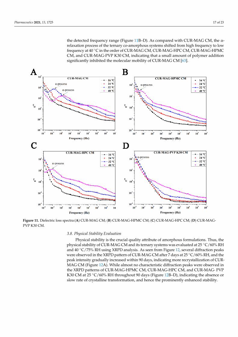
Pharmaceutics 2021, 13, 1725 17 of 23
the detected frequency range (Figure 11B–D). As compared with CUR-MAG CM, the α-relaxation process of the ternary co-amorphous systems shifted from high frequency to lowfrequency at 40 ◦C in the order of CUR-MAG CM, CUR-MAG-HPC CM, CUR-MAG-HPMCCM, and CUR-MAG-PVP K30 CM, indicating that a small amount of polymer additionsignificantly inhibited the molecular mobility of CUR-MAG CM [63].
Figure 11. Dielectric loss spectra:(A) CUR-MAG CM; (B) CUR-MAG-HPMC CM; (C) CUR-MAG-HPC CM; (D) CUR-MAG-PVP K30 CM.
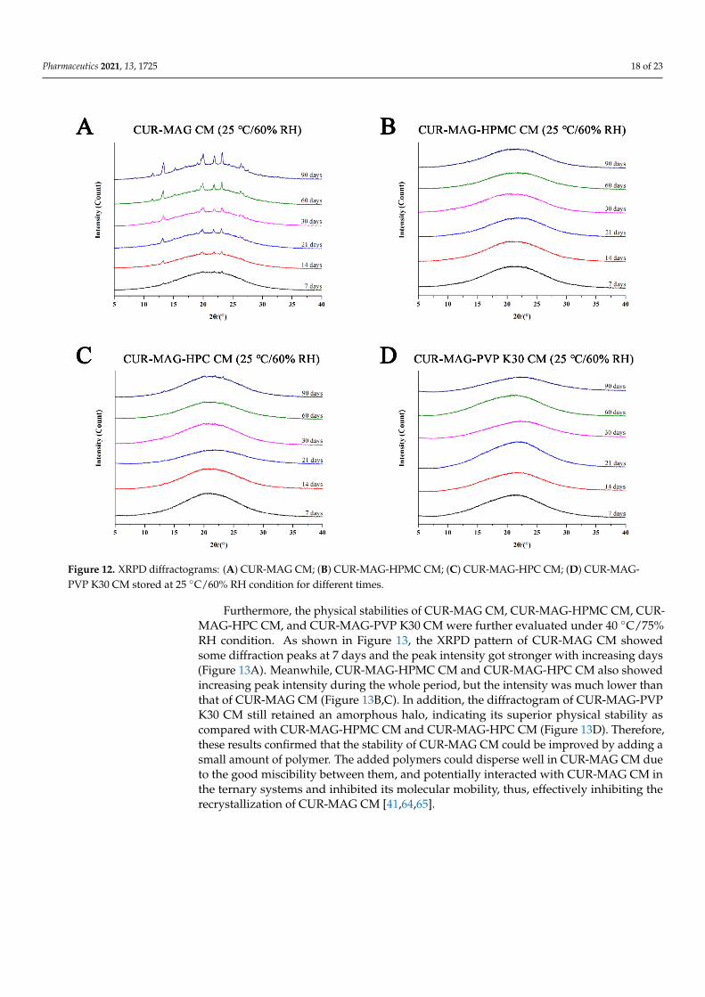
3.8. Physical Stability Evaluation
Physical stability is the crucial quality attribute of amorphous formulations. Thus, thephysical stability of CUR-MAG CM and its ternary systems was evaluated at 25 ◦C/60% RHand 40 ◦C/75% RH using XRPD analysis. As seen from Figure 12, several diffraction peakswere observed in the XRPD pattern of CUR-MAG CM after 7 days at 25 ◦C/60% RH, and thepeak intensity gradually increased within 90 days, indicating more recrystallization of CUR-MAG CM (Figure 12A). While almost no characteristic diffraction peaks were observed inthe XRPD patterns of CUR-MAG-HPMC CM, CUR-MAG-HPC CM, and CUR-MAG- PVPK30 CM at 25 ◦C/60% RH throughout 90 days (Figure 12B–D), indicating the absence orslow rate of crystalline transformation, and hence the prominently enhanced stability.
Pharmaceutics 2021, 13, 1725 18 of 23
Figure 12. XRPD diffractograms: (A) CUR-MAG CM; (B) CUR-MAG-HPMC CM; (C) CUR-MAG-HPC CM; (D) CUR-MAG-PVP K30 CM stored at 25 ◦C/60% RH condition for different times.
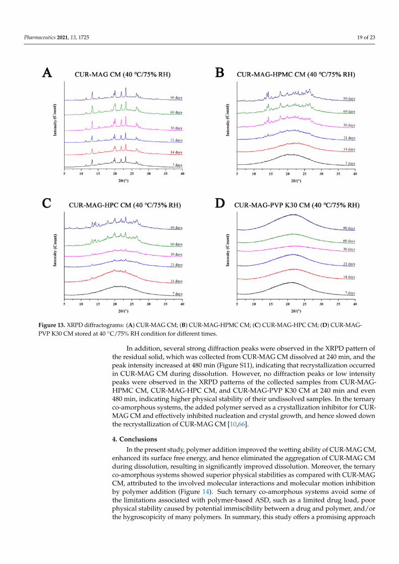
Furthermore, the physical stabilities of CUR-MAG CM, CUR-MAG-HPMC CM, CUR-MAG-HPC CM, and CUR-MAG-PVP K30 CM were further evaluated under 40 ◦C/75%RH condition. As shown in Figure 13, the XRPD pattern of CUR-MAG CM showedsome diffraction peaks at 7 days and the peak intensity got stronger with increasing days(Figure 13A). Meanwhile, CUR-MAG-HPMC CM and CUR-MAG-HPC CM also showedincreasing peak intensity during the whole period, but the intensity was much lower thanthat of CUR-MAG CM (Figure 13B,C). In addition, the diffractogram of CUR-MAG-PVPK30 CM still retained an amorphous halo, indicating its superior physical stability ascompared with CUR-MAG-HPMC CM and CUR-MAG-HPC CM (Figure 13D). Therefore,these results confirmed that the stability of CUR-MAG CM could be improved by adding asmall amount of polymer. The added polymers could disperse well in CUR-MAG CM dueto the good miscibility between them, and potentially interacted with CUR-MAG CM inthe ternary systems and inhibited its molecular mobility, thus, effectively inhibiting therecrystallization of CUR-MAG CM [41,64,65].
Pharmaceutics 2021, 13, 1725 19 of 23
Figure 13. XRPD diffractograms: (A) CUR-MAG CM; (B) CUR-MAG-HPMC CM; (C) CUR-MAG-HPC CM; (D) CUR-MAG-PVP K30 CM stored at 40 ◦C/75% RH condition for different times.
In addition, several strong diffraction peaks were observed in the XRPD pattern ofthe residual solid, which was collected from CUR-MAG CM dissolved at 240 min, and thepeak intensity increased at 480 min (Figure S11), indicating that recrystallization occurredin CUR-MAG CM during dissolution. However, no diffraction peaks or low intensitypeaks were observed in the XRPD patterns of the collected samples from CUR-MAG-HPMC CM, CUR-MAG-HPC CM, and CUR-MAG-PVP K30 CM at 240 min and even480 min, indicating higher physical stability of their undissolved samples. In the ternaryco-amorphous systems, the added polymer served as a crystallization inhibitor for CUR-MAG CM and effectively inhibited nucleation and crystal growth, and hence slowed downthe recrystallization of CUR-MAG CM [10,66].
4. Conclusions
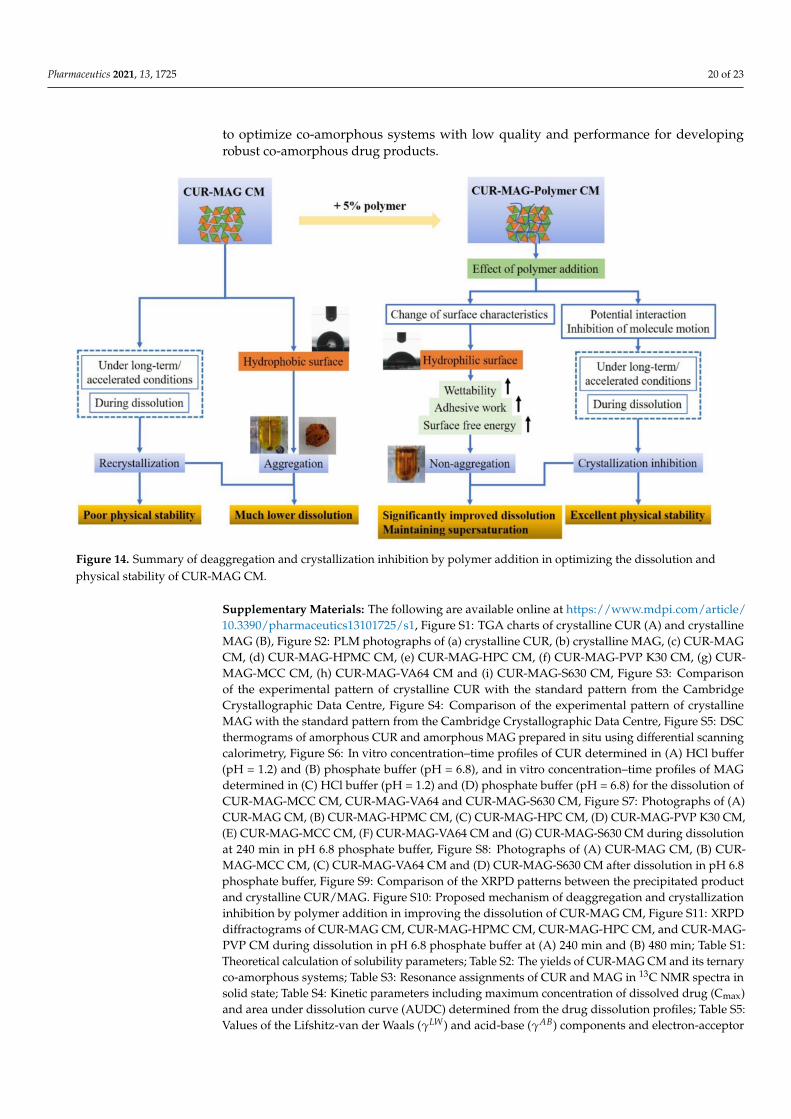
In the present study, polymer addition improved the wetting ability of CUR-MAG CM,enhanced its surface free energy, and hence eliminated the aggregation of CUR-MAG CMduring dissolution, resulting in significantly improved dissolution. Moreover, the ternaryco-amorphous systems showed superior physical stabilities as compared with CUR-MAGCM, attributed to the involved molecular interactions and molecular motion inhibitionby polymer addition (Figure 14). Such ternary co-amorphous systems avoid some ofthe limitations associated with polymer-based ASD, such as a limited drug load, poorphysical stability caused by potential immiscibility between a drug and polymer, and/orthe hygroscopicity of many polymers. In summary, this study offers a promising approach
Pharmaceutics 2021, 13, 1725 20 of 23
to optimize co-amorphous systems with low quality and performance for developingrobust co-amorphous drug products.
Figure 14. Summary of deaggregation and crystallization inhibition by polymer addition in optimizing the dissolution andphysical stability of CUR-MAG CM.
Supplementary Materials: The following are available online at https://www.mdpi.com/article/10.3390/pharmaceutics13101725/s1, Figure S1: TGA charts of crystalline CUR (A) and crystallineMAG (B), Figure S2: PLM photographs of (a) crystalline CUR, (b) crystalline MAG, (c) CUR-MAGCM, (d) CUR-MAG-HPMC CM, (e) CUR-MAG-HPC CM, (f) CUR-MAG-PVP K30 CM, (g) CUR-MAG-MCC CM, (h) CUR-MAG-VA64 CM and (i) CUR-MAG-S630 CM, Figure S3: Comparisonof the experimental pattern of crystalline CUR with the standard pattern from the CambridgeCrystallographic Data Centre, Figure S4: Comparison of the experimental pattern of crystallineMAG with the standard pattern from the Cambridge Crystallographic Data Centre, Figure S5: DSCthermograms of amorphous CUR and amorphous MAG prepared in situ using differential scanningcalorimetry, Figure S6: In vitro concentration–time profiles of CUR determined in (A) HCl buffer(pH = 1.2) and (B) phosphate buffer (pH = 6.8), and in vitro concentration–time profiles of MAGdetermined in (C) HCl buffer (pH = 1.2) and (D) phosphate buffer (pH = 6.8) for the dissolution ofCUR-MAG-MCC CM, CUR-MAG-VA64 and CUR-MAG-S630 CM, Figure S7: Photographs of (A)CUR-MAG CM, (B) CUR-MAG-HPMC CM, (C) CUR-MAG-HPC CM, (D) CUR-MAG-PVP K30 CM,(E) CUR-MAG-MCC CM, (F) CUR-MAG-VA64 CM and (G) CUR-MAG-S630 CM during dissolutionat 240 min in pH 6.8 phosphate buffer, Figure S8: Photographs of (A) CUR-MAG CM, (B) CUR-MAG-MCC CM, (C) CUR-MAG-VA64 CM and (D) CUR-MAG-S630 CM after dissolution in pH 6.8phosphate buffer, Figure S9: Comparison of the XRPD patterns between the precipitated productand crystalline CUR/MAG. Figure S10: Proposed mechanism of deaggregation and crystallizationinhibition by polymer addition in improving the dissolution of CUR-MAG CM, Figure S11: XRPDdiffractograms of CUR-MAG CM, CUR-MAG-HPMC CM, CUR-MAG-HPC CM, and CUR-MAG-PVP CM during dissolution in pH 6.8 phosphate buffer at (A) 240 min and (B) 480 min; Table S1:Theoretical calculation of solubility parameters; Table S2: The yields of CUR-MAG CM and its ternaryco-amorphous systems; Table S3: Resonance assignments of CUR and MAG in 13C NMR spectra insolid state; Table S4: Kinetic parameters including maximum concentration of dissolved drug (Cmax)and area under dissolution curve (AUDC) determined from the drug dissolution profiles; Table S5:Values of the Lifshitz-van der Waals (γLW) and acid-base (γAB) components and electron-acceptor
Pharmaceutics 2021, 13, 1725 21 of 23
(γ+) and electron-donor (γ−) parameters of the acid-base component of the surface tension (γtot) ofthe model liquids, used for the surface free energy determination of co-amorphous samples.
Author Contributions: Conceptualization, J.H. and L.L.; methodology, L.L. and M.S.; formal analysis,J.H. and Y.W.; investigation, Y.W. and J.H.; resources, Y.W. and S.Q.; data curation, W.H. and Y.G.;writing—review and editing, J.H. and S.Q.; supervision, M.S.; project administration, W.H.; fundingacquisition, Y.G. and S.Q. All authors have read and agreed to the published version of the manuscript.
Funding: This research was supported by the National Natural Science Foundation of China(82074029, 81773675, and 81873012), the “Double First-Class” University Project (CPU2018GY11and CPU2018GY27), the Postgraduate Research & Practice Innovation Program of Jiangsu Province(KYCX20_0675), the Natural Science Foundation of Jiangsu Province (SBK2020042291), and a ChinaPostdoctoral Science Foundation Funded Project (2020M671665).
Institutional Review Board Statement: Not applicable.
Informed Consent Statement: Not applicable.
Data Availability Statement: Not applicable.
Acknowledgments: We thank Zunting Pang, Daoyi Zheng, and Jianjun Zhang form China Pharma-ceutical University for their participation in this study and their insightful comments during therevision process.
Conflicts of Interest: The authors declare no conflict of interest.
References1. Han, J.; Wei, Y.; Lu, Y.; Wang, R.; Zhang, J.; Gao, Y.; Qian, S. Co-amorphous systems for the delivery of poorly water-soluble
drugs: Recent advances and an update. Expert Opin. Drug Deliv. 2020, 17, 1411–1435. [CrossRef]2. Shi, Q.; Moinuddin, S.M.; Cai, T. Advances in coamorphous drug delivery systems. Acta Pharm. Sin. B 2019, 9, 19–35. [CrossRef]3. Qian, S.; Heng, W.L.; Wei, Y.F.; Zhang, J.J.; Gao, Y. Coamorphous lurasidone hydrochloride-saccharin with charge-assisted
hydrogen bonding interaction shows improved physical stability and enhanced dissolution with pH-independent solubilitybehavior. Cryst. Growth Des. 2015, 15, 2920–2928. [CrossRef]
4. Wang, Z.; Sun, M.; Liu, T.; Gao, Z.; Ye, Q.; Tan, X.; Hou, Y.; Sun, J.; Wang, D.; He, Z. Co-amorphous solid dispersion systems oflacidipine-spironolactone with improved dissolution rate and enhanced physical stability. Asian J. Pharm. Sci. 2019, 14, 95–103.[CrossRef] [PubMed]
5. Kasten, G.; Lobmann, K.; Grohganz, H.; Rades, T. Co-former selection for co-amorphous drug-amino acid formulations. Int. J.Pharm. 2019, 557, 366–373. [CrossRef] [PubMed]
6. Shi, X.; Song, S.; Ding, Z.; Fan, B.; Huang, W.; Xu, T. Improving the solubility, dissolution, and bioavailability of ibrutinib bypreparing it in a coamorphous state with saccharin. J. Pharm. Sci. 2019, 108, 3020–3028. [CrossRef] [PubMed]
7. Wu, W.; Lobmann, K.; Rades, T.; Grohganz, H. On the role of salt formation and structural similarity of co-formers in co-amorphous drug delivery systems. Int. J. Pharm. 2018, 535, 86–94. [CrossRef]
8. Fan, N.; He, Z.; Ma, P.; Wang, X.; Li, C.; Sun, J.; Sun, Y.; Li, J. Impact of HPMC on inhibiting crystallization and improvingpermeability of curcumin amorphous solid dispersions. Carbohydr. Polym. 2018, 181, 543–550. [CrossRef]
9. Fan, N.; Ma, P.; Wang, X.; Li, C.; Zhang, X.; Zhang, K.; Li, J.; He, Z. Storage stability and solubilization ability of HPMC incurcumin amorphous solid dispersions formulated by Eudragit E100. Carbohydr. Polym. 2018, 199, 492–498. [CrossRef]
10. Liu, J.; Grohganz, H.; Rades, T. Influence of polymer addition on the amorphization, dissolution and physical stability ofco-amorphous systems. Int. J. Pharm. 2020, 588, 119768. [CrossRef]
11. Ruponen, M.; Visti, M.; Ojarinta, R.; Laitinen, R. Permeability of glibenclamide through a PAMPA membrane: The effect ofco-amorphization. Eur. J. Pharm. Biopharm. 2018, 129, 247–256. [CrossRef]
12. Franca, M.T.; Marcos, T.M.; Pereira, R.N.; Stulzer, H.K. Could the small molecules such as amino acids improve aqueous solubilityand stabilize amorphous systems containing Griseofulvin? Eur. J. Pharm. Sci. 2019, 143, 105178. [CrossRef]
13. Bohr, A.; Nascimento, T.L.; Harmankaya, N.; Weisser, J.J.; Wang, Y.; Grohganz, H.; Rades, T.; Lobmann, K. Efflux inhibitor bicalu-tamide increases oral bioavailability of the poorly soluble efflux substrate docetaxel in co-amorphous anti-cancer combinationtherapy. Molecules 2019, 24, 266. [CrossRef] [PubMed]
14. Knapik-Kowalczuk, J.; Chmiel, K.; Jurkiewicz, K.; Correia, N.T.; Sawicki, W.; Paluch, M. Physical stability and viscoelasticproperties of co-amorphous ezetimibe/simvastatin system. Pharmaceuticals 2019, 12, 40. [CrossRef] [PubMed]
15. Fung, M.H.; DeVault, M.; Kuwata, K.T.; Suryanarayanan, R. Drug-excipient interactions: Effect on molecular mobility andphysical stability of ketoconazole-organic acid coamorphous systems. Mol. Pharm. 2018, 15, 1052–1061. [CrossRef]
16. Ueda, H.; Kadota, K.; Imono, M.; Ito, T.; Kunita, A.; Tozuka, Y. Co-amorphous formation induced by combination of tranilast anddiphenhydramine hydrochloride. J. Pharm. Sci. 2017, 106, 123–128. [CrossRef] [PubMed]
Pharmaceutics 2021, 13, 1725 22 of 23
17. Hirakawa, Y.; Ueda, H.; Wakabayashi, R.; Kamiya, N.; Goto, M. A novel binary supercooled liquid formulation for transdermaldrug delivery. Biol. Pharm. Bull. 2019, 43, 393–398. [CrossRef] [PubMed]
18. Russo, M.G.; Baldoni, H.A.; Davila, Y.A.; Brusau, E.V.; Ellena, J.A.; Narda, G.E. Rational design of a famotidine-ibuprofencoamorphous system: An experimental and theoretical study. J. Phys. Chem. B 2018, 122, 8772–8782. [CrossRef] [PubMed]
19. Huang, R.; Han, J.; Wang, R.; Zhao, X.; Qiao, H.; Chen, L.; Li, W.; Di, L.; Zhang, W.; Li, J. Surfactant-free solid dispersion of BCSclass IV drug in an amorphous chitosan oligosaccharide matrix for concomitant dissolution in vitro—Permeability increase. Eur.J. Pharm. Sci. 2019, 130, 147–155. [CrossRef]
20. Mirzaei, H.; Shakeri, A.; Rashidi, B.; Jalili, A.; Banikazemi, Z.; Sahebkar, A. Phytosomal curcumin: A review of pharmacokinetic,experimental and clinical studies. Biomed. Pharmacother. 2017, 85, 102–112. [CrossRef]
21. Chen, C.H.; Hsu, F.T.; Chen, W.L.; Chen, J.H. Induction of apoptosis, inhibition of MCL-1, and VEGF-A expression are associatedwith the anti-cancer efficacy of magnolol combined with regorafenib in hepatocellular carcinoma. Cancers 2021, 13, 2066.[CrossRef] [PubMed]
22. Lin, Y.; Li, Y.; Zeng, Y.; Tian, B.; Qu, X.; Yuan, Q.; Song, Y. Pharmacology, toxicity, bioavailability, and formulation of magnolol:An update. Front. Pharmacol. 2021, 12, 632767. [CrossRef] [PubMed]
23. Zhu, X.; Yu, Z.; Feng, L.; Deng, L.; Fang, Z.; Liu, Z.; Li, Y.; Wu, X.; Qin, L.; Guo, R.; et al. Chitosan-based nanoparticle co-deliveryof docetaxel and curcumin ameliorates anti-tumor chemoimmunotherapy in lung cancer. Carbohydr. Polym. 2021, 268, 118237.[CrossRef] [PubMed]
24. Ghafoor, B.; Ali, M.N.; Riaz, Z. Synthesis and appraisal of natural drug-polymer-based matrices relevant to the application ofdrug-eluting coronary stent coatings. Cardiol. Res. Pract. 2020, 2020, 4073091. [CrossRef] [PubMed]
25. Guerra-Araiza, C.; Alvarez-Mejia, A.L.; Sanchez-Torres, S.; Farfan-Garcia, E.; Mondragon-Lozano, R.; Pinto-Almazan, R.; Salgado-Ceballos, H. Effect of natural exogenous antioxidants on aging and on neurodegenerative diseases. Free Radic. Res. 2013, 47,451–462. [CrossRef] [PubMed]
26. Sahoo, A.K.; Dandapat, J.; Dash, U.C.; Kanhar, S. Features and outcomes of drugs for combination therapy as multi-targetsstrategy to combat Alzheimer’s disease. J. Ethnopharmacol. 2018, 215, 42–73. [CrossRef]
27. Tsuda, T. Curcumin as a functional food-derived factor: Degradation products, metabolites, bioactivity, and future perspectives.Food Funct. 2018, 9, 705–714. [CrossRef]
28. Xiao, L.; Zhu, L.; Li, W.; Li, C.; Cao, Y.; Ge, G.; Sun, X. New insights into SN-38 glucuronidation: Evidence for the important roleof UDP glucuronosyltransferase 1A9. Basic Clin. Pharmacol. Toxicol. 2018, 122, 424–428. [CrossRef]
29. Zhu, L.; Ge, G.; Zhang, H.; Liu, H.; He, G.; Liang, S.; Zhang, Y.; Fang, Z.; Dong, P.; Finel, M.; et al. Characterization of hepaticand intestinal glucuronidation of magnolol: Application of the relative activity factor approach to decipher the contributions ofmultiple UDP-glucuronosyltransferase isoforms. Drug Metab. Dispos. 2012, 40, 529–538. [CrossRef]
30. Metre, S.; Mukesh, S.; Samal, S.K.; Chand, M.; Sangamwar, A.T. Enhanced biopharmaceutical performance of rivaroxaban throughpolymeric amorphous solid dispersion. Mol. Pharm. 2018, 15, 652–668. [CrossRef]
31. DeBoyace, K.; Wildfong, P.L.D. The application of modeling and prediction to the formation and stability of amorphous soliddispersions. J. Pharm. Sci. 2018, 107, 57–74. [CrossRef] [PubMed]
32. Korhonen, O.; Pajula, K.; Laitinen, R. Rational excipient selection for co-amorphous formulations. Expert Opin. Drug Deliv. 2017,14, 551–569. [CrossRef] [PubMed]
33. Chavan, R.B.; Thipparaboina, R.; Kumar, D.; Shastri, N.R. Co amorphous systems: A product development perspective. Int. J.Pharm. 2016, 515, 403–415. [CrossRef]
34. Zhu, S.; Gao, H.; Babu, S.; Garad, S. Co-amorphous formation of high-dose zwitterionic compounds with amino acids to improvesolubility and enable parenteral delivery. Mol. Pharm. 2018, 15, 97–107. [CrossRef]
35. Pacult, J.; Rams-Baron, M.; Chmiel, K.; Jurkiewicz, K.; Antosik, A.; Szafraniec, J.; Kurek, M.; Jachowicz, R.; Paluch, M. Howcan we improve the physical stability of co-amorphous system containing flutamide and bicalutamide? The case of ternaryamorphous solid dispersions. Eur. J. Pharm. Sci. 2019, 136, 104947. [CrossRef] [PubMed]
36. Zdziennicka, A.; Szymczyk, K.; Janczuk, B. Correlation between surface free energy of quartz and its wettability by aqueoussolutions of nonionic, anionic and cationic surfactants. J. Colloid Interface Sci. 2009, 340, 243–248. [CrossRef]
37. Lewandowska, K.; Sionkowska, A.; Grabska, S.; Kaczmarek, B. Surface and thermal properties of collagen/hyaluronic acid blendscontaining chitosan. Int. J. Biol. Macromol. 2016, 92, 371–376. [CrossRef]
38. Krawczyk, J. Surface free energy of the human skin and its critical surface tension of wetting in the skin/surfactant aqueoussolution/air system. Skin Res. Technol. 2015, 21, 214–223. [CrossRef]
39. Rojewska, M.; Bartkowiak, A.; Strzemiecka, B.; Jamrozik, A.; Voelkel, A.; Prochaska, K. Surface properties and surface free energyof cellulosic etc mucoadhesive polymers. Carbohydr. Polym. 2017, 171, 152–162. [CrossRef]
40. Bayramoglu, G.; Yakup Arica, M. Surface energy components of a dye-ligand immobilized pHEMA membranes: Effects of theirmolecular attracting forces for non-covalent interactions with IgG and HSA in aqueous media. Int. J. Biol. Macromol. 2005, 37,249–256. [CrossRef]
41. Edueng, K.; Mahlin, D.; Larsson, P.; Bergstrom, C.A.S. Mechanism-based selection of stabilization strategy for amorphousformulations: Insights into crystallization pathways. J. Control. Release 2017, 256, 193–202. [CrossRef] [PubMed]
42. Salem, A.; Nagy, S.; Pal, S.; Szechenyi, A. Reliability of the Hansen solubility parameters as co-crystal formation prediction tool.Int. J. Pharm. 2019, 558, 319–327. [CrossRef]
Pharmaceutics 2021, 13, 1725 23 of 23
43. Sanphui, P.; Goud, N.R.; Khandavilli, U.B.; Bhanoth, S.; Nangia, A. New polymorphs of curcumin. Chem. Commun. 2011, 47,5013–5015. [CrossRef] [PubMed]
44. Matlinska, M.A.; Wasylishen, R.E.; Bernard, G.M.; Terskikh, V.V.; Brinkmann, A.; Michaelis, V.K. Capturing elusive polymorphsof curcumin: A structural characterization and computational study. Cryst. Growth Des. 2018, 18, 5556–5563. [CrossRef]
45. Sanphui, P.; Bolla, G. Curcumin, a biological wonder molecule: A crystal engineering point of view. Cryst. Growth Des. 2018, 18,5690–5711. [CrossRef]
46. Wang, R.; Han, J.; Jiang, A.; Huang, R.; Fu, T.; Wang, L.; Zheng, Q.; Li, W.; Li, J. Involvement of metabolism-permeability inenhancing the oral bioavailability of curcumin in excipient-free solid dispersions co-formed with piperine. Int. J. Pharm. 2019,561, 9–18. [CrossRef]
47. Luebbert, C.; Stoyanov, E.; Sadowski, G. Phase behavior of ASDs based on hydroxypropyl cellulose. Int. J. Pharm. 2021, 3, 100070.48. Safna Hussan, K.P.; Thayyil, M.S.; Deshpande, S.K.; Jinitha, T.V.; Manoj, K.; Ngai, K.L. Molecular dynamics, physical and thermal
stability of neat amorphous amlodipine besylate and in binary mixture. Eur. J. Pharm. Sci. 2018, 119, 268–278. [CrossRef]49. Fan, N.; Lu, T.; Li, J. Surface tracking of curcumin amorphous solid dispersions formulated by binary polymers. J. Pharm. Sci.
2020, 109, 1068–1078. [CrossRef]50. Minecka, A.; Kaminska, E.; Heczko, D.; Jurkiewicz, K.; Wolnica, K.; Dulski, M.; Hachula, B.; Pisarski, W.; Tarnacka, M.; Talik, A.;
et al. Studying structural and local dynamics in model H-bonded active ingredient—Curcumin in the supercooled and glassystates at various thermodynamic conditions. Eur. J. Pharm. Sci. 2019, 135, 38–50. [CrossRef]
51. Yu, D.Q.; Han, X.J.; Shan, T.Y.; Xu, R.; Hu, J.; Cheng, W.X.; Zha, L.P.; Peng, H.S. Microscopic characteristic and chemicalcomposition analysis of three medicinal plants and surface frosts. Molecules 2019, 24, 4548. [CrossRef]
52. Kozbial, A.; Trouba, C.; Liu, H.T.; Li, L. Characterization of the intrinsic water wettability of graphite using contact anglemeasurements: Effect of defects on static and dynamic contact angles. Langmuir 2017, 33, 959–967. [CrossRef]
53. Zhu, C.; Gao, Y.; Li, H.; Meng, S.; Li, L.; Francisco, J.S.; Zeng, X.C. Characterizing hydrophobicity of amino acid side chains in aprotein environment via measuring contact angle of a water nanodroplet on planar peptide network. Proc. Natl. Acad. Sci. USA2016, 113, 12946–12951. [CrossRef] [PubMed]
54. Javaid, M.A.; Zia, K.M.; Ilyas, H.N.; Yaqub, N.; Bhatti, I.A.; Rehan, M.; Shoaib, M.; Bahadur, A. Influence of chitosan/1,4-butanediol blends on the thermal and surface behavior of polycaprolactone diol-based polyurethanes. Int. J. Biol. Macromol. 2019,141, 1022–1034. [CrossRef] [PubMed]
55. Yarce, C.J.; Pineda, D.; Correa, C.E.; Salamanca, C.H. Relationship between surface properties and in vitro drug release from acompressed matrix containing an amphiphilic polymer material. Pharmaceuticals 2016, 9, 34. [CrossRef] [PubMed]
56. Wu, Q.; Tiraferri, A.; Li, T.; Xie, W.; Chang, H.; Bai, Y.; Liu, B. Superwettable PVDF/PVDF-g-PEGMA ultrafiltration membranes.ACS Omega 2020, 5, 23450–23459. [CrossRef]
57. Rabizadeh, T.; Stawski, T.M.; Morgan, D.J.; Peacock, C.L.; Benning, L.G. The effects of inorganic additives on the nucleation andgrowth kinetics of calcium sulfate dihydrate crystals. Cryst. Growth Des. 2017, 17, 582–589. [CrossRef]
58. Pang, Z.; Weng, X.; Wei, Y.; Gao, Y.; Zhang, J.; Qian, S. Modification of hygroscopicity and tabletability of l-carnitine by acocrystallization technique. CrystEngComm 2021, 23, 2138–2149. [CrossRef]
59. Jackson, M.J.; Kestur, U.S.; Hussain, M.A.; Taylor, L.S. Characterization of supersaturated danazol solutions—Impact of polymerson solution properties and phase transitions. Pharm. Res. 2016, 33, 1276–1288. [CrossRef]
60. Sarode, A.L.; Wang, P.; Obara, S.; Worthen, D.R. Supersaturation, nucleation, and crystal growth during single- and biphasicdissolution of amorphous solid dispersions: Polymer effects and implications for oral bioavailability enhancement of poorlywater soluble drugs. Eur. J. Pharm. Biopharm. 2014, 86, 351–360. [CrossRef]
61. Syromotina, D.S.; Surmenev, R.A.; Surmeneva, M.A.; Boyandin, A.N.; Nikolaeva, E.D.; Prymak, O.; Epple, M.; Ulbricht, M.;Oehr, C.; Volova, T.G. Surface wettability and energy effects on the biological performance of poly-3-hydroxybutyrate filmstreated with RF plasma. Mater. Sci. Eng. C Mater. Biol. Appl. 2016, 62, 450–457. [CrossRef] [PubMed]
62. Kaminska, E.; Madejczyk, O.; Tarnacka, M.; Jurkiewicz, K.; Kaminski, K.; Paluch, M. Studying of crystal growth and overallcrystallization of naproxen from binary mixtures. Eur. J. Pharm. Biopharm. 2017, 113, 75–87. [CrossRef] [PubMed]
63. Minecka, A.; Kaminska, E.; Tarnacka, M.; Jurkiewicz, K.; Talik, A.; Wolnica, K.; Dulski, M.; Kasprzycka, A.; Spychalska, P.;Garbacz, G.; et al. Does the molecular mobility and flexibility of the saccharide ring affect the glass-forming ability of naproxen inbinary mixtures? Eur. J. Pharm. Sci. 2020, 141, 105091. [CrossRef] [PubMed]
64. Zhang, M.; Suo, Z.; Peng, X.; Gan, N.; Zhao, L.; Tang, P.; Wei, X.; Li, H. Microcrystalline cellulose as an effective crystal growthinhibitor for the ternary Ibrutinib formulation. Carbohydr. Polym. 2020, 229, 115476. [CrossRef]
65. Li, N.; Taylor, L.S. Tailoring supersaturation from amorphous solid dispersions. J. Control. Release 2018, 279, 114–125. [CrossRef][PubMed]
66. Zhang, J.; Liu, Z.; Wu, H.; Cai, T. Effect of polymeric excipients on nucleation and crystal growth kinetics of amorphousfluconazole. Biomater. Sci. 2021, 9, 4308–4316. [CrossRef] [PubMed]


