Embed Size (px)
Citation preview

of February 28, 2013.This information is current as
Thymic Hypofunction in Down SyndromeDecreased AIRE Expression and Global
Duarte, Antonio Coutinho and Magda Carneiro-SampaioC. Bento-de-Souza, Luciana Bento-de-Souza, Maria I.Helena Brentani, Leandro de A. Lima, Magaly Arrais, Luiz Flavia A. Lima, Carlos A. Moreira-Filho, Patrícia L. Ramos,
http://www.jimmunol.org/content/187/6/3422doi: 10.4049/jimmunol.1003053August 2011;
2011; 187:3422-3430; Prepublished online 19J Immunol
MaterialSupplementary
3.DC1.htmlhttp://www.jimmunol.org/content/suppl/2011/08/19/jimmunol.100305
Referenceshttp://www.jimmunol.org/content/187/6/3422.full#ref-list-1
, 19 of which you can access for free at: cites 55 articlesThis article
Subscriptionshttp://jimmunol.org/subscriptions
is online at: The Journal of ImmunologyInformation about subscribing to
Permissionshttp://www.aai.org/ji/copyright.htmlSubmit copyright permission requests at:
Email Alertshttp://jimmunol.org/cgi/alerts/etocReceive free email-alerts when new articles cite this article. Sign up at:
Print ISSN: 0022-1767 Online ISSN: 1550-6606. Immunologists, Inc. All rights reserved.Copyright © 2011 by The American Association of9650 Rockville Pike, Bethesda, MD 20814-3994.The American Association of Immunologists, Inc.,
is published twice each month byThe Journal of Immunology
by guest on February 28, 2013http://jim
munol.org/
Dow
nloaded from

The Journal of Immunology
Decreased AIRE Expression and Global ThymicHypofunction in Down Syndrome
Flavia A. Lima,*,1 Carlos A. Moreira-Filho,*,1 Patrıcia L. Ramos,* Helena Brentani,†
Leandro de A. Lima,‡ Magaly Arrais,x Luiz C. Bento-de-Souza,x Luciana Bento-de-Souza,x
Maria I. Duarte,{ Antonio Coutinho,‖ and Magda Carneiro-Sampaio*
The Down syndrome (DS) immune phenotype is characterized by thymus hypotrophy, higher propensity to organ-specific auto-
immune disorders, and higher susceptibility to infections, among other features. Considering that AIRE (autoimmune regulator) is
located on 21q22.3, we analyzed protein and gene expression in surgically removed thymuses from 14 DS patients with congenital
heart defects, who were compared with 42 age-matched controls with heart anomaly as an isolated malformation. Immunohis-
tochemistry revealed 70.48 6 49.59 AIRE-positive cells/mm2 in DS versus 154.70 6 61.16 AIRE-positive cells/mm2 in controls
(p, 0.0001), and quantitative PCR as well as DNA microarray data confirmed those results. The number of FOXP3-positive cells/
mm2 was equivalent in both groups. Thymus transcriptome analysis showed 407 genes significantly hypoexpressed in DS, most of
which were related, according to network transcriptional analysis (FunNet), to cell division and to immunity. Immune response-
related genes included those involved in 1) Ag processing and presentation (HLA-DQB1, HLA-DRB3, CD1A, CD1B, CD1C, ERAP)
and 2) thymic T cell differentiation (IL2RG, RAG2, CD3D, CD3E, PRDX2, CDK6) and selection (SH2D1A, CD74). It is noteworthy
that relevant AIRE-partner genes, such as TOP2A, LAMNB1, and NUP93, were found hypoexpressed in DNA microarrays and
quantitative real-time PCR analyses. These findings on global thymic hypofunction in DS revealed molecular mechanisms un-
derlying DS immune phenotype and strongly suggest that DS immune abnormalities are present since early development, rather
than being a consequence of precocious aging, as widely hypothesized. Thus, DS should be considered as a non-monogenic primary
immunodeficiency. The Journal of Immunology, 2011, 187: 3422–3430.
Down syndrome (DS) represents the most common chro-mosomal disorder (∼1 in 700 live births) and resultsfrom total or partial trisomy of chromosome 21 (1–3).
DS is associated with several complex clinical features includingimmunological abnormalities (4–8), and it has long been noticedthat patients present abnormal thymuses, characterized by lym-phocyte depletion, cortical atrophy, and loss of corticomedullarydemarcation (9, 10). In addition to higher susceptibility to infec-tions—as evidenced during the H1N1 2009 pandemic where thelikelihood of death was 300 times greater for DS patients (11)—
DS is characterized by increased frequency of organ-specific au-toimmune disorders and of lymphoid and myeloid leukemias,contrasting with a decreased risk for allergic diseases, particularlyasthma (12, 13). Compared with the general population, the in-cidence of celiac disease, insulin-dependent diabetes mellitus, andhypothyroidism is, respectively 10–40, 6, and 4 times higher inDS patients, but Addison’s disease, pernicious anemia, alopeciaareata, vitiligo, and chronic hepatitis have also been reported (12,14–16). This range of autoimmune disorders is reminiscent ofautoimmune polyendocrinopathy–candidiasis–ectodermal dystro-phy (APECED), as are autoantibody reactivity patterns (17), andthere is increased susceptibility to oral candidiasis (18) inAPECED and DS. APECED is a monogenic disorder caused byloss-of-function mutations in AIRE (autoimmune regulator) (19,20), which is located on 21q22.3.In the search for the molecular basis for clinical features in DS
patients, we have studied AIRE expression in DS thymus comparedwith that in age-matched individuals with heart defects as an iso-lated congenital malformation. We also studied global gene ex-pression and transcriptional networks in thymuses of DS patientsand controls, and we now report the finding that AIRE expressionin DS thymic medullary epithelial cells is significantly reduced,data that were confirmed by quantitative PCR (qPCR). Addition-ally, the current analysis showed hypoexpression of genes relatedto immune response and to cell proliferation in DS thymus.
Materials and MethodsSpecimens
Thymic tissues (corticomedullary sections) were collected between 2007and 2010 from 14DS (all with simple trisomy 21) infants and children (4moto 12 y old), all with congenital heart defects. These DS patients are beingfollowed up: one died a few weeks after the surgery because of infectious
*Department of Pediatrics, Faculty of Medicine, University of Sao Paulo, 05403-900Sao Paulo, Brazil; †Department of Psychiatry, Faculty of Medicine, University of SaoPaulo, 05403-900 Sao Paulo, Brazil; ‡Hospital do Cancer AC Camargo, 01509-010Sao Paulo, Brazil; xHospital do Coracao, Sanatorio Sırio, 04004-030 Sao Paulo,Brazil; {Department of Pathology, Faculty of Medicine, University of Sao Paulo,05403-900 Sao Paulo, Brazil; and ‖Instituto Gulbenkian de Ciencia, P2781-901Oeiras, Portugal
1F.A.L. and C.A.M.-F. contributed equally to this work.
Received for publication September 14, 2010. Accepted for publication July 18,2011.
This work was supported by Fundacao de Amparo a Pesquisa do Estado de Sao Paulothrough grants 2008/58238-4 (to M.C.-S.) and 2005/56446-0 (to C.A.M.-F.). F.A.L.was a Fundacao de Amparo a Pesquisa do Estado de Sao Paulo doctoral fellow (2005/60069-8).
Address correspondence and reprint requests to Prof. Magda Carneiro-Sampaio,Department of Pediatrics, Faculty of Medicine, University of Sao Paulo, AvenidaDr. Eneas Carvalho Aguiar 647, 05403-900, Sao Paulo, SP, Brazil. E-mail address:[email protected]
The online version of this article contains supplemental material.
Abbreviations used in this article: APECED, autoimmune polyendocrinopathy–can-didiasis–ectodermal dystrophy; DS, Down syndrome; GO, gene ontology; PID, pri-mary immunodeficiency; qPCR, quantitative PCR; SAM, significance analysis ofmicroarrays.
Copyright� 2011 by TheAmericanAssociation of Immunologists, Inc. 0022-1767/11/$16.00
www.jimmunol.org/cgi/doi/10.4049/jimmunol.1003053
by guest on February 28, 2013http://jim
munol.org/
Dow
nloaded from

complications; another developed Hashimoto thyroiditis at 3 y old. Controlsamples (42 thymuses) were from age-matched individuals with heartanomalies but no clinical signs of any other congenital malformation. Allsamples were collected during corrective cardiac surgery at the Hospital doCoracao–Associacao do Sanatorio Sırio (Sao Paulo, Brazil). No tissue wasremoved for research purposes only. This study was approved by thehospital’s ethics committee, and informed consent from the parents of allparticipating individuals was obtained.
Immunohistochemistry
Small pieces of each thymus were fixed in 10% neutral buffered formalinand processed into paraffin blocks. Thymus sections (4 mm) were depar-affinized and rehydrated through graded alcohols to water. For antigenicretrieval, sections were microwave-treated at 800 W in 10 mM Tris–EDTAbuffer at pH 8 for 15 min and allowed to cool for 15 min. After washed inrunning tap water for 5 min, tissue endogenous peroxidase was inhibitedwith a solution of 3% H2O2 for 10 min at room temperature. Unspecificbinding sites were blocked by incubation with 5% BSA (Sigma) diluted in10 mM PBS pH 7.4 for 10 min. Slides were incubated overnight at 4˚Cwith a polyclonal rabbit anti-AIRE serum (sc-33188; Santa Cruz Bio-technology, Santa Cruz, CA) and with an affinity purified rat anti-humananti-FOXP3 14-4776-82 (eBioscience, San Diego, CA). Primary Ab wasblown off, and the slides were then incubated with the Universal LSAB+Kit/HRP (DakoCytomation, Carpinteria, CA). The reaction was developedwith the substrate in 3,39 diaminobenzidine chromogen. Tissue samplesfrom DS patients and controls were processed simultaneously. Doubleimmunohistochemistry staining was performed after completion of thesingle staining protocol. Sections were sequentially incubated with 5%BSA for 10 min, monoclonal mouse anti-cytokeratin (clone AE1/AE3;DakoCytomation) diluted in PBS containing 0.1% BSA overnight at4˚C, and then with the Universal LSAB+ Kit/AP (DakoCytomation).Revelation was performed using the alkaline phosphatase permanent redchromogen. After the single or double protocol, the tissues were coun-terstained with Mayer’s hematoxylin.
Quantification of stained cells and statistical analysis
Epithelial cells expressing nuclear AIRE or thymocytes expressing FOXP3were identified and quantified using a light microscope. For 15 random areasof the medullary region of each thymus, the number of positive cellsper square millimeter was determined using an integration graticule (CarlZeiss 4740680000000 Netzmikrometer 12.53) under 3400 magnifica-tion. Counting of positive cells was performed in a blinded fashionindependently by two pathologists. Statistical analysis was initially per-formed with the Shapiro–Wilk normality test to determine whether the datawere consistent with a normal distribution. Groups were compared usingthe two-tailed Student t test for unpaired data. Correlation coefficientswere calculated using the Pearson correlation (r) test. The p values ,0.05were considered statistically significant. All statistical tests were per-formed using GraphPad Prism software.
Functional genomic studies
Obtention of thymic RNA. Fresh ex vivo explants from the thymuses of fourDS and four control patients, all of them ,2 y old, were collected at theoperating room and immediately immersed into RNAlater RNA Stabili-zation Reagent (cat. no. 76104; Qiagen, Valencia, CA). RNAwas extractedfrom tissue fragments using the RNeasy Lipid Tissue Mini Kit accordingto the manufacturer’s instructions (cat. no. 74804; Qiagen).
Microarray hybridization and gene expression analysis. To determine geneexpression profiles, 44 K DNA microarrays (cat. no. G4845A; AgilentTechnologies, Santa Clara, CA) were used. The procedures for hybrid-ization followed the protocols provided by the manufacturer’s instructions(One-Color Microarray-Based Gene Quick Amp Labeling).
Expression analysis. The images were captured by the reader AgilentBundle according to the parameters recommended for bioarrays and were
extracted by Agilent Feature Extraction software version 9.5.3. Among the45,015 spots present in each array, only those with none or only one flag(i.e., low intensity, saturation, controls, etc.) were selected for analysis usingthe R software version 2.9.2 (R Development Core Team). By means of theTMEV software version 4.4.1 (21), we selected as differentially expressedtranscripts those presenting a p value #0.05 (Student t test and adjustedBonferroni correction) and fold variation of 62. Hierarchical clusteringwas based on Pearson correlation and complete linkage. The significanceanalysis of microarrays (SAM) procedure (22) was used with a false dis-covery ratio of zero.
Transcriptional interaction analyses (gene ontology and network analysis).We used the FunNet software—based on the Gene Ontology Consortium(http://www.geneontology.org) and on the Kyoto Encyclopedia of Genesand Genomes (http://www.genome.jp/kegg) genomic annotations—forperforming the functional profiling of gene expression data and identify-ing the biological themes in which the differentially expressed genes areinvolved. Themes with significant relationship in the transcriptional ex-pression space were associated to build transcriptional modules in a prox-imity network. A transcriptional interaction network, corresponding withthe theme proximity network, was then obtained (http://www.funnet.info).
qPCR. Differential gene expression data were validated through qPCR.Specific primers for AIRE and five other selected genes (Table I) weredesigned using Primer-BLAST (Primer3 Input, version 0.4.0, and BLAST,available at http://www.ncbi.nlm.nih.gov/tools/primer-blast/). All thymusRNA samples were amplified in triplicate. Amplification reactions wereperformed in a 25-ml final volume containing 13 SYBR Green mix(Quantitec SYBR Green PCR kit; Qiagen, Hilden, DE), 10 pmol of eachprimer, and 2 ml cDNA (1:10 dilution, synthesized from 1 mg of totalRNA). Real-time PCR amplifications were performed in an Applied Bio-systems StepOne Plus Real Time PCR System with StepOne software(Applied Biosystems, Foster City, CA) with the following cycling para-meters: an initial hot start of 95˚C for 15 min followed by 50 cycles of95˚C for 15 s and 60˚C for 30 s. To normalize qPCR reactions, GAPDHwas included as reference gene after checking that amplification curves forRNA samples obtained from five DS thymuses (four from our patients andone additional DS thymus sample kindly provided by Instituto de MedicinaIntegral de Pernambuco, Recife, PE, Brazil) and six control thymuses (fourfrom our controls and two additional samples provided by Instituto deMedicina Integral de Pernambuco) yielded essentially the same results.Relative expression was determined by the relative standard curve methodand presented as fold-change comparing DS versus control mean values.
ResultsQuantification of AIRE-positive cells in the thymus
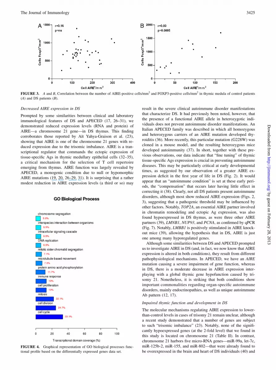
AIRE immunoreactivity was observed in nuclei of epithelial cellslocated in the medullary region, some of them around Hassall’scorpuscles (Fig. 1A–D). AIRE-positive cells were marked as ep-ithelial by costaining with a cytokeratin-specific Ab (Fig. 1E, 1F).The number of AIRE protein-expressing epithelial cells in thymicmedulla of DS patients (70.48 6 49.6 cells/mm2; n = 14) wassignificantly lower (p , 0.0001) than that in the thymic medullaof the control group (154.70 6 61.2 cells/mm2; n = 42), thesedifferences being more marked in infants (Fig. 2, SupplementalFig. 1).
FOXP3-positive cells and correlation with AIRE expression
There was no significant difference in the numbers of FOXP3-positive thymocytes in thymic medulla of both groups: 824.4 6483.5 cells/mm2 in DS (n = 14) and 617.0 6 245.4 cells/mm2 incontrols (n = 35). Whereas the numbers of AIRE- and FOXP3-
Table I. Primers used in the qPCR assays with their sequences and respective product length
Gene Forward Primer (59 to 39) Reverse Primer (59 to 39) Product Length (bp)
AIRE ACCGGGTTTTCTTCCCAATA AGAGACGCCCATGCAGACT 224CDK6 TGGAGTGTTGGCTGCATATT ACAGGGCACTGTAGGCAGAT 260LMNB1 CTGGCGAAGATGTGAAGGTT TCTGAGAAGGCTCTGCACTG 268NUP93 TCAGGCACAACCTCTCAGAA CCACAAAGCATGGCACTTAAT 254PCNA GAATTCCAGAACAGGAGTACAGC TTCAGGTACCTCAGTGCAAAAG 258TOP2A GCTGCCCCAAAAGGAACTA TAGGTTTCTTTGCCCGTACA 251
The Journal of Immunology 3423
by guest on February 28, 2013http://jim
munol.org/
Dow
nloaded from

positive cells were not correlated in the control group (r = 0.16, p= 0.35, Fig. 3A), a significant positive correlation was found in DS(r = 0.80, p = 0.0005, Fig. 3B). Finally, no significant correlationwas found between age and the numbers of either AIRE- orFOXP3-positive cells in both DS and control groups (data notshown).
Transcriptome profile analysis
We identified 21,940 valid thymic transcripts using the R program.Microarray data were deposited in the Gene Expression Omnibuspublic database (http://www.ncbi.nlm.nih.gov/geo/) under acces-sion number GSE23910. In the comparison of DS versus controls,a total of 1,238 differentially expressed transcripts were selectedusing permuted t test, adjusted p value #0.05 (Bonferroni), and
a fold of 62. The SAM procedure (22) revealed 407 significantlyhypoexpressed genes in the DS group (false discovery ratio = 0),whereas no hyperexpressed gene was observed. Hierarchicalclustering showed complete separation between DS and controls(Supplemental Fig. 2). Overlap analysis (SAM and t test data),performed by means of the TMEV 4.4.1 program, yielded 156hypoexpressed genes. The AIRE gene was not found significantlyhypoexpressed in this analysis (fold of 21.87).
Network transcriptional analysis
This analysis was accomplished using SAM-selected differentiallyexpressed genes and the FunNet software. The strength of the linksbetween each pair of genes is given by Pearson’s correlation co-efficient of expression profiles. From a total of 13,041 links, weselected the 654 links with a value above the third quartile (0.949).A graphical representation of the functional gene profile ac-
cording to gene ontology (GO) biological processes is shown inFig. 4. The transcriptional domain coverage (GO categories)shows that the majority of the hypoexpressed genes belong tocategories linked to cell cycle/cell proliferation or to immuneresponse. Theme proximity network analysis (Fig. 5) confirms thepicture revealed by GO categorization, as essentially two modulesappear in this analysis, the first linked to cell division/proliferationand the second encompassing themes relevant to the immune re-sponse, such as Ag processing and presentation via MHC class II,T cell differentiation, selection, and activation, among others. Thethemes “male sex differentiation” and “male somatic sex de-termination” only encompass the X-linked androgen receptorgene (AR).The picture showed in the theme proximity network was con-
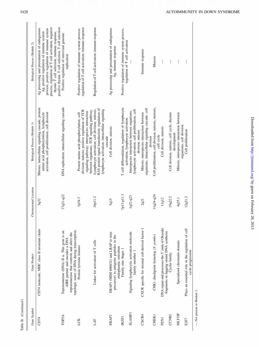
firmed by the transcriptional interaction network analysis (Fig. 6),which depicts genes involved in immune function and in celldivision/proliferation as the most frequent gene interactions. TableII shows selected relevant genes per category/function.
qPCR validation of DNA microarray data
Fig. 7 shows qPCR expression fold-changes comparing DS versuscontrol samples for the genes (Table 1) AIRE, 2) TOP2A, LMNB1,and NUP93 (AIRE-target genes), and 3) CDK6 and PCNA (notmodulated by AIRE). The results demonstrate downregulation ofall these genes in DS thymus and corroborate DNA microarrayexpression values.
DiscussionThis study revealed for the first time, to our knowledge, reducedthymic expression of a large set of genes that may constitute thebasis for the molecular mechanisms underlying the immune dis-turbances characteristically seen in DS patients: thymus hypo-trophy, higher propensity to develop organ-specific autoimmunedisorders, and higher susceptibility to infection. Furthermore, DSthymus presents a pattern of widespread gene hypoexpression,which may well result in a hypofunctional thymic environ-ment that characterizes the disease. Our data are in agreementwith those of Aıt Yahya-Graison et al. (23), who studied gene-expression variation of 136 genes located on chromosome 21 inDS lymphoblastoid cell lines and found that only 29% are ex-pressed proportionally to the genomic-dosage imbalance, the other71% having their expression “compensated” to normal or below-normal levels. Similar results were found by Prandini et al. (24)studying the gene-expression variation of DS lymphoblastoid andfibroblast cells. Notably, Sommer et al. (25) used serial analysisof gene expression methodology to study dysregulated genes inblood lymphocytes of DS children and found that none of the 30mostly expressed tags was located on chromosome 21.
FIGURE 1. Representative illustrations of AIRE immunoreactivity
(brown color) in thymic specimens from a control patient (A) and a DS
patient (B). Nuclear AIRE is strongly stained in several cells of the thymic
medullary region of control patients. C and D, Double-staining AIRE
immunofluorescence (green color) in thymic specimens from a control
patient (C) and a DS patient (D). A–D, Original magnification3200 (inset,
original magnification 3400). E and F, Double-staining immunohisto-
chemistry for AIRE (brown color) and cytokeratin (reddish) showing an
AIRE-positive (E) and an AIRE-negative (F) epithelial cell (original
magnification 31,000).
FIGURE 2. Numbers of AIRE-positive cells/mm2 in the thymic med-
ullary region of DS and control groups. A, Infants (,1 y old). B, Children.
DS patients from both age groups present significantly lower numbers of
AIRE-positive cells/mm2.
3424 AUTOIMMUNITY IN DOWN SYNDROME
by guest on February 28, 2013http://jim
munol.org/
Dow
nloaded from
Decreased AIRE expression in DS
Prompted by some similarities between clinical and laboratoryimmunological features of DS and APECED (17, 26–31), wedemonstrated reduced expression levels (RNA and protein) ofAIRE—a chromosome 21 gene—in DS thymus. This findingcorroborates those reported by Aıt Yahya-Graison et al. (23),showing that AIRE is one of the chromosome 21 genes with re-duced expression due to the trisomic imbalance. AIRE is a tran-scriptional regulator that commands the ectopic expression oftissue-specific Ags in thymic medullary epithelial cells (32–35),a critical mechanism for the selection of T cell repertoireemerging from thymus. AIRE function was largely revealed byAPECED, a monogenic condition due to null or hypomorphicAIRE mutations (19, 20, 26–29, 31). It is surprising that a rathermodest reduction in AIRE expression levels (a third or so) may
result in the severe clinical autoimmune disorder manifestationsthat characterize DS. It had previously been noted, however, thatthe presence of a functional AIRE allele in heterozygotic indi-viduals does not prevent autoimmune disorder manifestations. AnItalian APECED family was described in which all homozygousand heterozygous carriers of an AIRE mutation developed thy-roiditis (36). More recently, this particular mutation (G228W) wascloned in a mouse model, and the resulting heterozygous micedeveloped autoimmunity (37). In short, together with these pre-vious observations, our data indicate that “fine tuning” of thymictissue-specific Ags expression is crucial in preventing autoimmunediseases. This may be particularly critical at early developmentaltimes, as suggested by our observation of a greater AIRE ex-pression deficit in the first year of life in DS (Fig. 2). It wouldappear that an “autoimmune condition” is set at these early peri-ods, the “compensation” that occurs later having little effect incorrecting it (38). Clearly, not all DS patients present autoimmunedisorders, although most show reduced AIRE expression (Figs. 2,3), suggesting that a pathogenic threshold may be influenced byother factors. Notably, TOP2A, an essential AIRE partner involvedin chromatin remodeling and ectopic Ag expression, was alsofound hypoexpressed in DS thymus, as were three other AIREpartners (39), LMNB1, NUP93, and PCNA, as confirmed by qPCR(Fig. 7). Notably, LMBB1 is positively stimulated in AIRE knock-out mice (39), allowing the hypothesis that in DS, AIRE is justone among many hyporegulated genes.Although some similarities between DS and APECED prompted
us to investigate AIRE in DS (and, in fact, we now know that AIREexpression is altered in both conditions), they result from differentpathophysiological mechanisms. In APECED, we have an AIREmutation causing a severe impairment of gene function, whereasin DS, there is a moderate decrease in AIRE expression inter-playing with a global thymic gene hypofunction caused by tri-somy 21. Nonetheless, it is striking that both conditions showimportant commonalities regarding organ-specific autoimmunedisorders, mainly endocrinopathies, as well as unique autoimmuneAb pattern (12, 17).
Impaired thymic function and development in DS
The molecular mechanisms regulating AIRE expression to lower-than-control levels in cases of trisomy 21 remain unclear, althougha recent study demonstrated that a number of genes are subjectto such “trisomic imbalance” (23). Notably, none of the signifi-cantly hypoexpressed genes (at the 2-fold level) that we found inthis study is located on chromosome 21 (Table II). In contrast,chromosome 21 harbors five micro-RNA genes—miR-99a, let-7c,miR-125b-2, miR-155, and miR-802—that were already found tobe overexpressed in the brain and heart of DS individuals (40) and
FIGURE 3. A and B, Correlation between the number of AIRE-positive cells/mm2 and FOXP3-positive cells/mm2 in thymic medula of control patients
(A) and DS patients (B).
FIGURE 4. Graphical representation of GO biological processes func-
tional profile based on the differentially expressed genes data set.
The Journal of Immunology 3425
by guest on February 28, 2013http://jim
munol.org/
Dow
nloaded from

may well exert regulatory effects on many of the genes describedin this study. This is the case of miR-155, which regulates thedevelopment of regulatory T cells and the innate immune responsethrough downregulation of SOCS1 (41), and of miR-125-b andlet-7c, which regulate macrophage responses to various stimuli(42, 43).Yet, our global gene expression and transcriptional network
analyses demonstrated deficient expression of many other genes inDS thymus. Notably, a number of these genes are known to reg-
ulate biological processes related to the development/activationof T cells and to the establishment of central tolerance (Figs. 4–6 and Table II): 1) Ag processing and presentation of Ag via MHCclass II (ERAP2, CD1D, HLA-DQB1, HLA-DRB3, CD1A, CD1B,CD1C); 2) thymic T cell selection (CD3D, CD74, CD1D, CD3E),3) T cell activation (LAT). It follows that susceptibility to organ-specific autoimmune disorder in DS may not be the consequenceof deficient AIRE expression only, but owe as well to the reducedexpression of other genes involved in critical thymic functions.
FIGURE 6. Transcriptional interaction network cor-
responding with the theme proximity network related to
GO biological process. Colored circles indicate pre-
dominant gene function.
FIGURE 5. Theme proximity network built for GO biological process. Module 1 is represented by triangles and module 2 by squares.
3426 AUTOIMMUNITY IN DOWN SYNDROME
by guest on February 28, 2013http://jim
munol.org/
Dow
nloaded from

Table
II.
Selectedrelevantgenes
hypoexpressed
inDSthymus
displayed
bycategory
andfunction
GeneSymbol
GeneProduct
Chromosomal
Location
Biological
Process
(Module
1)
Biological
Process
(Module
2)
KIF23
Kinesin-likeprotein
family
15q23
DNA
metabolicprocess;mitosis;celldivision;
cellcycle,
DNA
repair
Microtubule-based
process
ZWIN
TA
protein
involved
inkinetochore
function
10q21-q22
Chromosomeorganization;spindleorganization,
mitosis;celldivision;cellcycle;
chromosome
segregation;etc.
Microtubule-based
process
GRAP2
Thisgeneencodes
amem
beroftheGRB2/Sem
5/
Drk
family.
Thismem
ber
isan
adapter-like
protein
involved
inleukocyte-specificprotein-
tyrosinekinasesignaling.
22q13.2
Celldivision,
mitosis,Ras
protein
signal
transduction,intracellularsignalingcascade
—
PRDX2
Peroxiredoxin
familyofantioxidantenzymes
19p13.2
Lymphocyteactivation;cellproliferation;cell
division;regulationofhydrogen
peroxide
metabolicprocess;thymus
developm
ent;Tcell
differentiation;
regulationoflymphocyte
activation;homeostasisofnumber
ofcells
RegulationofTcellactivation;im
mune
response;negativeregulationofTcell
differentiation
PRSS16
Serineproteaseexpressed
exclusively
inthe
thym
us
6p21
Nucleotidebinding
—
IL2RG
IL-2R,g
Xq13.1
Lymphocyte
activation;celldivision;mitosis;
interspeciesinteractionbetweenorganism;Tcell
activation
RegulationofTcellactivation;positive
regulationofim
munesystem
process;im
mune
response
RAG2
Thisgeneencodes
aproteinthatisinvolved
inthe
initiationofV(D
)Jrecombinationduring
Band
Tcelldevelopm
ent
11p3
DNA
metabolicprocess,cellproliferation,cell
division,mitosis,Tcelldifferentiation,DNA
recombination
Bcellhomeostatic
proliferation,im
mune
response
SH2D1A
Role
inthebidirectional
stim
ulationofTand
Bcells
Xq25-q26
Mitosis
Positive
regulationofim
munesystem
process
CD3D
PartoftheTCR–CD3complex
11q23
Lymphocyteactivation,celldivision,
mitosis,
Tcelldifferentiation
Positive
thym
icTcellselection,Tcellselection
CD3E
CD3-ε
polypeptide
11q23
Cellproliferation;protein
aminoacid
phosphorylation;lymphocyte
activation;Tcell
differentiation
NegativethymicTcellselection;Tcellselection
HLA-D
RB3
HLA-D
RB3belongsto
theHLAclassIIb-chain
paralogs.ThisclassIImoleculeisaheterodimer
consistingofan
a(D
RA)andab(D
RB)chain,
both
anchoredin
themem
brane.
Itplays
acentral
role
intheim
munesystem
by
presentingpeptides
derived
from
extracellular
proteins.
6p21.3
Mitosis
Immuneresponse,Agprocessingand
presentation
HLA-D
QB1
HLA-D
QB1belongsto
theHLAclassIIb-chain
paralogs
6p21.3
Celldivision,
mitosis
Agprocessingandpresentationofpeptideor
polysaccharideAgvia
MHC
classII,Ag
processingandpresentation
CD1A
CD1familyoftransm
embraneglycoproteins
1q22-q23
Celldivision,
mitosis
Agprocessingandpresentation
CD1B
CD1familyoftransm
embraneglycoproteins
1q22-q23
Celldivision
Immune
response;Agprocessingand
presentation
CD1C
CD1familyoftransm
embraneglycoproteins
1q22-q23
Mitosis;celldivision
Immune
response;Agprocessingand
presentation
CD1D
CD1familyoftransm
embraneglycoproteins
1q22-q23
Lymphocyte
activation;mitosis;interspecies
interactionbetweenorganisms;Tcell
differentiation
Agprocessingandpresentationofendogenous
Ag;im
muneresponse;Tcellselection;Ag
processingandpresentation
(Table
continues)
The Journal of Immunology 3427
by guest on February 28, 2013http://jim
munol.org/
Dow
nloaded from
Table
II.(C
ontinued
)
GeneSymbol
GeneProduct
Chromosomal
Location
Biological
Process
(Module
1)
Biological
Process
(Module
2)
CD74
CD74
molecule,MHC
classIIinvariantchain
5q32
Mitosis,intracellularsignalingcascade,
protein
aminoacid
phosphorylation,lymphocyte
activation,cellproliferation,celldivision
Agprocessingandpresentationofendogenous
Ag,positive
regulationofim
munesystem
process,positive
regulationofim
munesystem
process,regulationofTcellactivation,negative
thym
icTcellselection,im
muneresponse,
positive
thymic
Tcellselection,Tcellselection
TOP2A
Topoisomerase(D
NA)II
a.Thisgeneisan
AIREpartner
andencodes
aDNA
topoisomerasethat
controlsandalters
the
topologic
states
ofDNA
duringtranscription.
17q21-q22
DNA
replication,intracellularsignalingcascade
Positive
regulationofretroviral
genome
replication
LCK
Protein
tyrosinekinases
1p34.3
Protein
aminoacid
phosphorylation;cell
division;
mitosis;positive
regulationofTCR
signalingpathway;interspeciesinteraction
betweenorganisms;TCR
signalingpathw
ay
Positive
regulationofim
munesystem
process;
regulationofTcellactivation;im
muneresponse
LAT
Linker
foractivationofTcells
16p11.2
Lymphocyte
activation;celldivision;mitosis;
RASprotein
signal
transduction;regulationof
lymphocyte
activation;intracellularsignaling
cascade
RegulationofTcellactivation;immuneresponse
ERAP2
ERAP1(M
IM606832)andLRAPto
trim
precursors
toantigenic
peptides
inthe
endoplasmic
reticulum
5q15
Celldivision,
mitosis
Agprocessingandpresentationofendogenous
Ag,im
muneresponse
IKZF1
Fam
ilyzincfinger
17p13-p11.1
Tcelldifferentiation,regulationoflymphocyte
activation,lymphocyte
activation
Positive
regulationofim
munesystem
process,
regulationofTcellactivation
SLAMF1
Signalinglymphocyticactivationmolecule
familymem
ber
11q22-q23
Interspeciesinteractionbetweenorganisms,
lymphocyte
activation,cellproliferation,cell
division,
mitosis
—
CXCR4
CXCRspecificforstromal
cell-derived
factor-1
2q21
Mitosis,interspeciesinteractionbetween
organisms,intracellularsignalingcascade,
cell
division
Immune
response
CHEK1
CHK1checkpointhomolog(S.pombe)
11q24-q24
Cellproliferation,celldivision,mitosis,meiosis,
cellcycle
Meiosis
FEN1
DNArepairandprocesses
the59endsofOkazaki
fragments
inlaggingstrandDNA
synthesis
11q12
Celldivision,
mitosis
—
CCNB2
Cyclin
family
15q22.2
Celldivision,mitosis,cellcycle,
thymus
developm
ent
—
MLF1IP
Specializedchromatin
domain
4q35.1
Mitosis,interspeciesinteractionbetween
organisms,celldivision
—
E2F7
Playsan
essential
role
intheregulationofcell
cycleprogression
12q21.2
Cellproliferation
—
—,Notpresentin
Module
2.
3428 AUTOIMMUNITY IN DOWN SYNDROME
by guest on February 28, 2013http://jim
munol.org/
Dow
nloaded from

Altogether, our data reinforce the pivotal role of defective centraltolerance in the pathogenesis of DS autoimmune disorders. Thus,the thymus in DS individuals had been reported to be smaller andhypocellular, even in infants, containing a decreased proportion ofphenotypically mature TCR-ab+ thymocytes (6). The number ofTRECs and the size of T cell subpopulations (CD4+, CD8+, CD4+
CD45+RA cells) in the peripheral blood of DS individuals havebeen described as reduced at various age groups (4, 6, 7). It wasalso demonstrated that DS patients present low naive T cellnumbers (44). Additionally, DS individuals fail to show the nor-mal, notorious expansion of circulating lymphocyte numbers inthe first months of life (45). Our finding that several genes relatedto cell proliferation (Figs. 4–6 and Table II) are hypofunctional inDS (1) cell cycle regulation [E2F7]; 2) cell proliferation [ZWINT,KIF23, CHEK1]; 3) DNA replication [FEN1]; 4) homeostasis ofnumber of cells [PRDX2], and 5) thymus development [PRDX2,IKZF1, CCNB2, CDK6]) may be causally related to DS immunephenotype (i.e., thymus hypotrophy and hypocellularity). It is in-teresting to note that CDK6 was recently found to be essentialfor thymocyte development (46).In contrast with the scenario described above, augmented num-
bers of FOXP3+CD25+ natural regulatory T cells were observedin DS peripheral blood (7), which is in accordance with our datashowing normal numbers of FOXP3-positive cells in DS thymus(Fig. 3).Global gene profiles and transcriptional network analyses pre-
sented in this study may also help to understand the higher sus-ceptibility to infections systematically described in DS (5, 6, 12).Thus, mutations of genes that were found hypoexpressed inDS thymus have been associated with severe primary immuno-deficiencies (47): IL2RG (X-linked SCID), RAG2, CD3D, CD3E(SCID), and SH2D1A (X-linked lymphoproliferative syndrome).Notably, milder immunodeficiency forms, in which autoimmunemanifestations are frequently part of the clinical picture, were
also associated with mutations in some of these genes, includingIL2RG and RAG (48). Furthermore, other hypoexpressed genesin DS thymus are involved in other biological processes that arealso relevant for resistance to infections: 1) positive regulationof neutrophil differentiation (IKZF1), 2) NK cell differentiation(IKZF1), 3) respiratory burst during acute inflammatory response(PRDX2), 4) negative regulation of oxygen and reactive oxygenspecies metabolic process (PRDX2), among others. Althoughmany such processes are unlikely to occur inside the thymus,hypoexpression due to trisomic imbalance may well result indeficient immune functions in the periphery. This may be the caseof the genes categorized as “interspecies interaction betweenorganisms” (i.e., IL2RG, KRT19, LCK, RAN, SGTA, SLAMF1,CXCR4, MLF1IP, CD1D).It should also be noted that the genes with more interactions
(hubs) in the transcriptional network analysis (Fig. 6) were 1)PRDX2, which codes for a member of the peroxiredoxin family ofantioxidant enzymes involved in T cell antiviral activity (49) andin thymus development, and was described as hypoexpressed infetal DS brain (50); 2) GRAP2, which codes for an adapter-likeprotein involved in leukocyte-specific protein-tyrosine kinase sig-naling (51); 3) ZWINT, involved in kinetochore function (52); and4) KIF23, which codes for a kinesin-like protein family involvedin chromosome movement during cell division (53).
DS: a primary immunodeficiency?
The current study 1) contributes to the understanding of thymichypotrophy in DS patients, 2) demonstrates its association withreduced expression of critical genes, probably derived from tri-somic imbalance, and 3) strongly suggests that DS typical immunemalfunction is owed to impaired central tolerance, possibly due toboth decreased AIRE expression and global thymic hypofunction.Thus, our results are in general agreement with the recent proposalby Kusters et al. (6) that “the immune system in DS is intrinsicallydeficient from the very beginning, and not simply another victimof a generalized process of precocious aging,” as hypothesized byothers (7, 8, 54, 55). Altogether, our data indicate that DS is in-deed a primary, rather than a secondary, immunodeficiency, con-trary to what is largely accepted (47). It would seem, therefore,that DS should well be considered as a non-monogenic primaryimmunodeficiency (PID). It has largely been recognized that mostPIDs are monogenic disorders (47); however, there exist goodexamples of polygenic PIDs (such as DiGeorge syndrome) that arecaused by a deletion encompassing several loci on chromosome22q11.2 (56).
AcknowledgmentsWe are grateful to Prof. Alberto Duarte (Hospital do Coracao, Associacao
Sanatorio Sırio de Sao Paulo) and to Dr. Letıcia Watanabe (Hospital das
Clınicas da Faculdade de Medicina da Universidade de Sao Paulo) for
valuable help in selection of patients and to Drs. Joao Guilherme Bezerra
Alves, Fernando Moraes, and Pollyanna Buregio Frota (Instituto de Medic-
ina Integral de Pernambuco) for kindly providing additional thymus sam-
ples used for qPCR validation. We thank Dr. Silvia Yumi Bando for
valuable technical assistance.
DisclosuresThe authors have no financial conflicts of interest.
References1. Gorlin, R. J., M. M. Cohen, Jr., and R. C. M. Hennekam. 2001. Chromosomal
syndromes: common and/or well-known syndromes. In Syndromes of the Headand Neck, 4th Ed. Oxford University Press, Oxford, p. 35–44.
2. Megarbane, A., A. Ravel, C. Mircher, F. Sturtz, Y. Grattau, M. O. Rethore,J. M. Delabar, and W. C. Mobley. 2009. The 50th anniversary of the discovery of
FIGURE 7. qPCR validation of DNA microarray data. A, Boxplots
comparing the DNA microarray expression values of six selected genes in
DS (gray) and control (white) samples. B, qPCR expression fold-changes
comparing DS with control samples for the same genes showing down-
regulation in DS. qPCR was performed using RNA samples from five DS
patients and six control patients.
The Journal of Immunology 3429
by guest on February 28, 2013http://jim
munol.org/
Dow
nloaded from

trisomy 21: the past, present, and future of research and treatment of Downsyndrome. Genet. Med. 11: 611–616.
3. Korbel, J. O., T. Tirosh-Wagner, A. E. Urban, X. N. Chen, M. Kasowski, L. Dai,F. Grubert, C. Erdman, M. C. Gao, K. Lange, et al. 2009. The genetic archi-tecture of Down syndrome phenotypes revealed by high-resolution analysis ofhuman segmental trisomies. Proc. Natl. Acad. Sci. USA 106: 12031–12036.
4. de Hingh, Y. C., P. W. van der Vossen, E. F. Gemen, A. B. Mulder, W. C. Hop,F. Brus, and E. de Vries. 2005. Intrinsic abnormalities of lymphocyte counts inchildren with down syndrome. J. Pediatr. 147: 744–747.
5. Garrison, M. M., H. Jeffries, and D. A. Christakis. 2005. Risk of death forchildren with down syndrome and sepsis. J. Pediatr. 147: 748–752.
6. Kusters, M. A., R. H. Verstegen, E. F. Gemen, and E. de Vries. 2009. Intrinsicdefect of the immune system in children with Down syndrome: a review. Clin.Exp. Immunol. 156: 189–193.
7. Roat, E., N. Prada, E. Lugli, M. Nasi, R. Ferraresi, L. Troiano, C. Giovenzana,M. Pinti, O. Biagioni, M. Mariotti, et al. 2008. Homeostatic cytokines and ex-pansion of regulatory T cells accompany thymic impairment in children withDown syndrome. Rejuvenation Res. 11: 573–583.
8. da Rosa Utiyama, S. R., R. M. Nisihara, F. R. Nass, N. P. Oliveira, P. T. Fiedler,and I. T. de Messias-Reason. 2008. Autoantibodies in patients with Down syn-drome: early senescence of the immune system or precocious markers for im-munological diseases? J. Paediatr. Child Health 44: 182–186.
9. Levin, S., M. Schlesinger, Z. Handzel, T. Hahn, Y. Altman, B. Czernobilsky, andJ. Boss. 1979. Thymic deficiency in Down’s syndrome. Pediatrics 63: 80–87.
10. Larocca, L. M., L. Lauriola, F. O. Ranelletti, M. Piantelli, N. Maggiano,R. Ricci, and A. Capelli. 1990. Morphological and immunohistochemical studyof Down syndrome thymus. Am. J. Med. Genet. Suppl. 7(Suppl.): 225–230.
11. Perez-Padilla, R., R. Fernandez, C. Garcıa-Sancho, F. Franco-Marina, O. Aburto,H. Lopez-Gatell, and I. Bojorquez. 2010. Pandemic (H1N1) 2009 virus andDown syndrome patients. Emerg. Infect. Dis. 16: 1312–1314.
12. Goldacre, M. J., C. J. Wotton, V. Seagroatt, and D. Yeates. 2004. Cancers andimmune related diseases associated with Down’s syndrome: a record linkagestudy. Arch. Dis. Child. 89: 1014–1017.
13. Hitzler, J. K., and A. Zipursky. 2005. Origins of leukaemia in children withDown syndrome. Nat. Rev. Cancer 5: 11–20.
14. Burch, P. R., and A. Milunsky. 1969. Early-onset diabetes mellitus in the generaland Down’s syndrome populations. Genetics, aetiology, and pathogenesis.Lancet 1: 554–558.
15. Shield, J. P., E. J. Wadsworth, T. J. Hassold, L. A. Judis, and P. A. Jacobs. 1999.Is disomic homozygosity at the APECED locus the cause of increased autoim-munity in Down’s syndrome? Arch. Dis. Child. 81: 147–150.
16. Gillespie, K. M., R. J. Dix, A. J. Williams, R. Newton, Z. F. Robinson,P. J. Bingley, E. A. Gale, and J. P. Shield. 2006. Islet autoimmunity in childrenwith Down’s syndrome. Diabetes 55: 3185–3188.
17. Soderbergh, A., J. Gustafsson, O. Ekwall, A. Hallgren, T. Nilsson, O. Kampe,F. Rorsman, and G. Anneren. 2006. Autoantibodies linked to autoimmune pol-yendocrine syndrome type I are prevalent in Down syndrome. Acta Paediatr. 95:1657–1660.
18. Carlstedt, K., L. Krekmanova, G. Dahllof, B. Ericsson, G. Braathen, andT. Modeer. 1996. Oral carriage of Candida species in children and adolescentswith Down’s syndrome. Int. J. Paediatr. Dent. 6: 95–100.
19. Betterle, C., N. A. Greggio, and M. Volpato. 1998. Clinical review 93: auto-immune polyglandular syndrome type 1. J. Clin. Endocrinol. Metab. 83: 1049–1055.
20. Perheentupa, J. 2006. Autoimmune polyendocrinopathy-candidiasis-ectodermaldystrophy. J. Clin. Endocrinol. Metab. 91: 2843–2850.
21. Saeed, A. I., V. Sharov, J. White, J. Li, W. Liang, N. Bhagabati, J. Braisted,M. Klapa, T. Currier, M. Thiagarajan, et al. 2003. TM4: a free, open-sourcesystem for microarray data management and analysis. Biotechniques 34: 374–378.
22. Tusher, V. G., R. Tibshirani, and G. Chu. 2001. Significance analysis ofmicroarrays applied to the ionizing radiation response. Proc. Natl. Acad. Sci.USA 98: 5116–5121.
23. Aıt Yahya-Graison, E. A., J. Aubert, L. Dauphinot, I. Rivals, M. Prieur,G. Golfier, J. Rossier, L. Personnaz, N. Creau, H. Blehaut, et al. 2007. Classi-fication of human chromosome 21 gene-expression variations in Down syn-drome: impact on disease phenotypes. Am. J. Hum. Genet. 81: 475–491.
24. Prandini, P., S. Deutsch, R. Lyle, M. Gagnebin, C. Delucinge Vivier,M. Delorenzi, C. Gehrig, P. Descombes, S. Sherman, F. Dagna Bricarelli, et al.2007. Natural gene-expression variation in Down syndrome modulates the out-come of gene-dosage imbalance. Am. J. Hum. Genet. 81: 252–263.
25. Sommer, C. A., E. C. Pavarino-Bertelli, E. M. Goloni-Bertollo, and F. Henrique-Silva. 2008. Identification of dysregulated genes in lymphocytes from childrenwith Down syndrome. Genome 51: 19–29.
26. Finnish-German APECED Consortium. 1997. An autoimmune disease,APECED, caused by mutations in a novel gene featuring two PHD-type zinc-finger domains. Nat. Genet. 17: 399–403.
27. Nagamine, K., P. Peterson, H. S. Scott, J. Kudoh, S. Minoshima, M. Heino,K. J. Krohn, M. D. Lalioti, P. E. Mullis, S. E. Antonarakis, et al. 1997. Positionalcloning of the APECED gene. Nat. Genet. 17: 393–398.
28. Anderson, M. S., E. S. Venanzi, L. Klein, Z. Chen, S. P. Berzins, S. J. Turley,H. von Boehmer, R. Bronson, A. Dierich, C. Benoist, and D. Mathis. 2002.Projection of an immunological self shadow within the thymus by the aireprotein. Science 298: 1395–1401.
29. Villasenor, J., C. Benoist, and D. Mathis. 2005. AIRE and APECED: molecularinsights into an autoimmune disease. Immunol. Rev. 204: 156–164.
30. Carneiro-Sampaio, M., and A. Coutinho. 2007. Tolerance and autoimmunity:lessons at the bedside of primary immunodeficiencies. Adv. Immunol. 95: 51–82.
31. DeVoss, J. J., and M. S. Anderson. 2007. Lessons on immune tolerance from themonogenic disease APS1. Curr. Opin. Genet. Dev. 17: 193–200.
32. Taubert, R., J. Schwendemann, and B. Kyewski. 2007. Highly variable expres-sion of tissue-restricted self-antigens in human thymus: implications for self-tolerance and autoimmunity. Eur. J. Immunol. 37: 838–848.
33. Peterson, P., T. Org, and A. Rebane. 2008. Transcriptional regulation by AIRE:molecular mechanisms of central tolerance. Nat. Rev. Immunol. 8: 948–957.
34. Mathis, D., and C. Benoist. 2009. Aire. Annu. Rev. Immunol. 27: 287–312.35. Org, T., A. Rebane, K. Kisand, M. Laan, U. Haljasorg, R. Andreson, and
P. Peterson. 2009. AIRE activated tissue specific genes have histone mod-ifications associated with inactive chromatin. Hum. Mol. Genet. 18: 4699–4710.
36. Cetani, F., G. Barbesino, S. Borsari, E. Pardi, L. Cianferotti, A. Pinchera, andC. Marcocci. 2001. A novel mutation of the autoimmune regulator gene in anItalian kindred with autoimmune polyendocrinopathy-candidiasis-ectodermaldystrophy, acting in a dominant fashion and strongly cosegregating with hypo-thyroid autoimmune thyroiditis. J. Clin. Endocrinol. Metab. 86: 4747–4752.
37. Su, M. A., K. Giang, K. Zumer, H. Jiang, I. Oven, J. L. Rinn, J. J. Devoss,K. P. Johannes, W. Lu, J. Gardner, et al. 2008. Mechanisms of an autoimmunitysyndrome in mice caused by a dominant mutation in Aire. J. Clin. Invest. 118:1712–1726.
38. Guerau-de-Arellano, M., M. Martinic, C. Benoist, and D. Mathis. 2009. Neonataltolerance revisited: a perinatal window for Aire control of autoimmunity. J. Exp.Med. 206: 1245–1252.
39. Abramson, J., M. Giraud, C. Benoist, and D. Mathis. 2010. Aire’s partners in themolecular control of immunological tolerance. Cell 140: 123–135.
40. Kuhn, D. E., G. J. Nuovo, M. M. Martin, G. E. Malana, A. P. Pleister, J. Jiang,T. D. Schmittgen, A. V. Terry, Jr., K. Gardiner, E. Head, et al. 2008. Humanchromosome 21-derived miRNAs are overexpressed in Down syndrome brainsand hearts. Biochem. Biophys. Res. Commun. 370: 473–477.
41. Tsitsiou, E., and M. A. Lindsay. 2009. microRNAs and the immune response.Curr. Opin. Pharmacol. 9: 514–520.
42. Okada, H., G. Kohanbash, and M. T. Lotze. 2010. MicroRNAs in immuneregulation—opportunities for cancer immunotherapy. Int. J. Biochem. Cell Biol.42: 1256–1261.
43. Murphy, A. J., P. M. Guyre, and P. A. Pioli. 2010. Estradiol suppresses NF-kappaB activation through coordinated regulation of let-7a and miR-125b in primaryhuman macrophages. J. Immunol. 184: 5029–5037.
44. Bloemers, B. L., L. Bont, R. A. de Weger, S. A. Otto, J. A. Borghans, andK. Tesselaar. 2011. Decreased thymic output accounts for decreased naive T cellnumbers in children with Down syndrome. J. Immunol. 186: 4500–4507.
45. Kusters, M. A. A., E. F. A. Gemen, R. H. J. Verstegen, P. C. Wever, and E. DEVries. 2010. Both normal memory counts and decreased naive cells favor in-trinsic defect over early senescence of Down syndrome T lymphocytes. Pediatr.Res. 67: 557–562.
46. Hu, M. G., A. Deshpande, N. Schlichting, E. A. Hinds, C. Mao, M. Dose,G. F. Hu, R. A. Van Etten, F. Gounari, and P. W. Hinds. 2011. CDK6 kinaseactivity is required for thymocyte development. Blood 117: 6120–6131.
47. Notarangelo, L. D., A. Fischer, R. S. Geha, J. L. Casanova, H. Chapel,M. E. Conley, C. Cunningham-Rundles, A. Etzioni, L. Hammartrom,S. Nonoyama, et al; International Union of Immunological Societies ExpertCommittee on Primary Immunodeficiencies. 2009. Primary immunodeficiencies:2009 update. J. Allergy Clin. Immunol. 124: 1161–1178.
48. Liston, A., A. Enders, and O. M. Siggs. 2008. Unravelling the association ofpartial T-cell immunodeficiency and immune dysregulation. Nat. Rev. Immunol.8: 545–558.
49. Geiben-Lynn, R., M. Kursar, N. V. Brown, M. M. Addo, H. Shau, J. Lieberman,A. D. Luster, and B. D. Walker. 2003. HIV-1 antiviral activity of recombinantnatural killer cell enhancing factors, NKEF-A and NKEF-B, members of theperoxiredoxin family. J. Biol. Chem. 278: 1569–1574.
50. Sanchez-Font, M. F., J. Sebastia, C. Sanfeliu, R. Cristofol, G. Marfany, andR. Gonzalez-Duarte. 2003. Peroxiredoxin 2 (PRDX2), an antioxidant enzyme, isunder-expressed in Down syndrome fetal brains. Cell. Mol. Life Sci. 60: 1513–1523.
51. Shen, R., Y. B. Ouyang, C. K. Qu, A. Alonso, L. Sperzel, T. Mustelin,M. H. Kaplan, and G. S. Feng. 2002. Grap negatively regulates T-cell receptor-elicited lymphocyte proliferation and interleukin-2 induction. Mol. Cell. Biol.22: 3230–3236.
52. Famulski, J. K., L. Vos, X. Sun, and G. Chan. 2008. Stable hZW10 kinetochoreresidency, mediated by hZwint-1 interaction, is essential for the mitotic check-point. J. Cell Biol. 180: 507–520.
53. Seguin, L., C. Liot, R. Mzali, R. Harada, A. Siret, A. Nepveu, and J. Bertoglio.2009. CUX1 and E2F1 regulate coordinated expression of the mitotic complexgenes Ect2, MgcRacGAP, and MKLP1 in S phase. Mol. Cell. Biol. 29: 570–581.
54. Cossarizza, A., D. Monti, G. Montagnani, C. Ortolani, M. Masi, M. Zannotti,and C. Franceschi. 1990. Precocious aging of the immune system in Downsyndrome: alteration of B lymphocytes, T-lymphocyte subsets, and cells withnatural killer markers. Am. J. Med. Genet. Suppl. 7(Suppl.): 213–218.
55. Cuadrado, E., and M. J. Barrena. 1996. Immune dysfunction in Down’s syn-drome: primary immune deficiency or early senescence of the immune system?Clin. Immunol. Immunopathol. 78: 209–214.
56. Kobrynski, L. J., and K. E. Sullivan. 2007. Velocardiofacial syndrome, DiGeorgesyndrome: the chromosome 22q11.2 deletion syndromes. Lancet 370: 1443–1452.
3430 AUTOIMMUNITY IN DOWN SYNDROME
by guest on February 28, 2013http://jim
munol.org/
Dow
nloaded from


