Embed Size (px)
Citation preview

7. Penn, A. A., Riquelme, P. A., Feller, M. B. & Shatz, C. J. Competition in retinogeniculate patterning
driven by spontaneous activity. Science 279, 2108–2112 (1998).
8. Cynader, M., Berman, N. & Hein, A. Cats reared in stroboscopic illumination: effects on receptive
fields in visual cortex. Proc. Natl Acad. Sci. USA 70, 1353–1354 (1973).
9. Schmidt, J. T. & Eisele, L. E. Stroboscopic illumination and dark rearing block the sharpening of the
regenerated retinotectal map in goldfish. Neuroscience 14, 535–546 (1985).
10. Weliky, M. & Katz, L. C. Disruption of orientation tuning in visual cortex by artificially correlated
neuronal activity. Nature 386, 680–685 (1997).
11. Sharma, J., Angelucci, A. & Sur, M. Induction of visual orientation modules in auditory cortex. Nature
404, 841–847 (2000).
12. von Melchner, L., Pallas, S. L. & Sur, M. Visual behaviour mediated by retinal projections directed to
the auditory pathway. Nature 404, 871–876 (2000).
13. Fregnac, Y., Shulz, D., Thorpe, S. & Bienenstock, E. A cellular analogue of visual cortical plasticity.
Nature 333, 367–370 (1988).
14. Schuett, S., Bonhoeffer, T. & Hubener, M. Pairing-induced changes of orientation maps in cat visual
cortex. Neuron 32, 325–337 (2001).
15. Gaze, R. M., Keating, M. J. & Chung, S. H. The evolution of the retinotectal map during development
in Xenopus. Proc. R. Soc. London B 185, 301–330 (1974).
16. Holt, C. E. & Harris, W. A. Order in the initial retinotectal map in Xenopus: a new technique for
labelling growing nerve fibres. Nature 301, 150–152 (1983).
17. Markram, H., Lubke, J., Frotscher, M. & Sakmann, B. Regulation of synaptic efficacy by coincidence of
postsynaptic APs and EPSPs. Science 275, 213–215 (1997).
18. Zhang, L. I., Tao, H. W., Holt, C. E., Harris, W. A. & Poo, M. M. A critical window for cooperation and
competition among developing retinotectal synapses. Nature 395, 37–44 (1998).
19. Feldman, D. E. Timing-based LTP and LTD at vertical inputs to layer II/III pyramidal cells in rat barrel
cortex. Neuron 27, 45–56 (2000).
20. Boettiger, C. A. & Doupe, A. J. Developmentally restricted synaptic plasticity in a songbird nucleus
required for song learning. Neuron 31, 809–818 (2001).
21. Abbott, L. F. & Blum, K. I. Functional significance of long-term potentiation for sequence learning and
prediction. Cereb. Cortex 6, 406–416 (1996).
22. Zanker, J. M. & Zeil, J. Motion Vision—Computational, Neural, and Ecological Constraints (Springer,
New York, 2000).
23. Rao, R. P. & Sejnowski, T. J. Predictive learning of temporal sequences in recurrent neocortical circuits.
Novartis Found. Symp. 239, 208–229 (2001).
24. Roberts, P. D. Computational consequences of temporally asymmetric learning rules. I. Differential
hebbian learning. J. Comput. Neurosci. 7, 235–246 (1999).
25. Mehta, M. R., Barnes, C. A. & McNaughton, B. L. Experience-dependent, asymmetric expansion of
hippocampal place fields. Proc. Natl Acad. Sci. USA 94, 8918–8921 (1997).
26. Mehta, M. R., Quirk, M. C. & Wilson, M. A. Experience-dependent asymmetric shape of hippocampal
receptive fields. Neuron 25, 707–715 (2000).
27. Bliss, T. V. P. & Collingridge, G. L. A synaptic model of memory: long-term potentiation in the
hippocampus. Nature 361, 31–39 (1993).
28. Malenka, R. C. & Nicoll, R. A. Long-term potentiation—a decade of progress? Science 285, 1870–1874
(1999).
29. Zhang, L. I., Tao, H. & Poo, M. Visual input induces long-term potentiation of developing retinotectal
synapses. Nature Neurosci. 3, 708–715 (2000).
30. Rae, J., Cooper, K., Gates, P. & Watsky, M. Low access resistance perforated patch recordings using
amphotericin B. J. Neurosci. Methods 37, 15–26 (1991).
AcknowledgementsThis work was supported by grants from NSF and NIH. F.E. was supported in part by along-term fellowship from the Human Frontier Science Program.
Competing interests statementThe authors declare that they have no competing financial interests.
Correspondence and requests for materials should be addressed to M.-m.P.
(e-mail: [email protected]).
..............................................................
Dendrite growth increasedby visual activity requiresNMDA receptor and Rho GTPasesWun Chey Sin, Kurt Haas, Edward S. Ruthazer & Hollis T. Cline
Cold Spring Harbor Laboratory, Cold Spring Harbor, New York 11724, USA.............................................................................................................................................................................
Previous studies suggest that neuronal activity may guide thedevelopment of synaptic connections in the central nervoussystem through mechanisms involving glutamate receptors andGTPase-dependent modulation of the actin cytoskeleton1–7. Herewe demonstrate by in vivo time-lapse imaging of optic tectal cells
in Xenopus laevis tadpoles that enhanced visual activity driven bya light stimulus promotes dendritic arbor growth. The stimulus-induced dendritic arbor growth requires glutamate-receptor-mediated synaptic transmission, decreased RhoA activity andincreased Rac and Cdc42 activity. The results delineate a role forRho GTPases in the structural plasticity driven by visual stimu-lation in vivo.
We used in vivo time-lapse imaging of single optic tectal neuronsin Xenopus tadpoles to test the function of visual activity in neuronaldevelopment. We compared the dendritic arbor growth rates ofindividual tectal neurons during a 4-h period with a visual stimulusto a preceding 4-h period in the absence of light. This imagingprotocol allows the comparison of dendritic arbor structures of thesame neurons over time and therefore provides a sensitive measureof structural plasticity. Visual stimulation significantly enhanceddendritic arbor elaboration compared with growth rates in thepreceding 4-h period in the dark (Fig. 1a, b; see also SupplementaryTable 1). Neurons from animals exposed to visual stimulus through-out the 8-h protocol maintained a constant rate of dendritic arborelaboration (Fig. 1b; see also Supplementary Fig. 1 and Table 1).This indicates that growth rates do not change with longer periodsof stimulation. Another set of animals was exposed to visualstimulus within the first 4-h period and then returned to the dark
Figure 1 Visual stimulation in vivo promotes dendritic arbor growth by a glutamate-
receptor-dependent mechanism. a, Drawings of neurons imaged three times at 4-h
intervals. Two examples are shown for each treatment. Animals were placed in a dark
chamber for 4 h (dark) or a chamber with a light stimulus for 4 h (light) in the presence
or absence of APV as depicted by the bar over the neurons. Arrowheads identify
efferent axons in this and subsequent figures. Scale bar, 100 mm. b, Quantification of
dendritic arbor growth rates during 4 h in the dark (D) or with visual stimulation (L).
c, Dendritic arbor growth rates normalized to the growth rate in the 0–4 h period in
the dark (D). d, Quantification of branch-tip additions. e, Contribution of new
branches or branch extension (hatched, top region) to increased branch length. Asterisk,
P , 0.05.
letters to nature
NATURE | VOL 419 | 3 OCTOBER 2002 | www.nature.com/nature 475
for the second 4-h period. Notably, the enhanced growth rates ofthese neurons in response to light stimulation were maintainedduring the 4-h dark period (Fig. 1b; see also Supplementary Fig. 1).This suggests that exposure to visual stimulation triggers mecha-nisms that enhance dendritic arbor growth over a period followingtermination of the stimulus.
Although the experimental protocol was designed to detect
structural changes within individual neurons, we also detect asignificant increase in the growth rate of neurons from animalsexposed to visual stimulation during the first 4 h of the experiment(68.3 ^ 12.1 mm, n ¼ 16) compared with the growth rate ofneurons from animals placed in the dark for the initial 4 h(35.4 ^ 6.0 mm, n ¼ 84, P , 0.002). This indicates that theincreased growth rate is due to the visual stimulus rather than
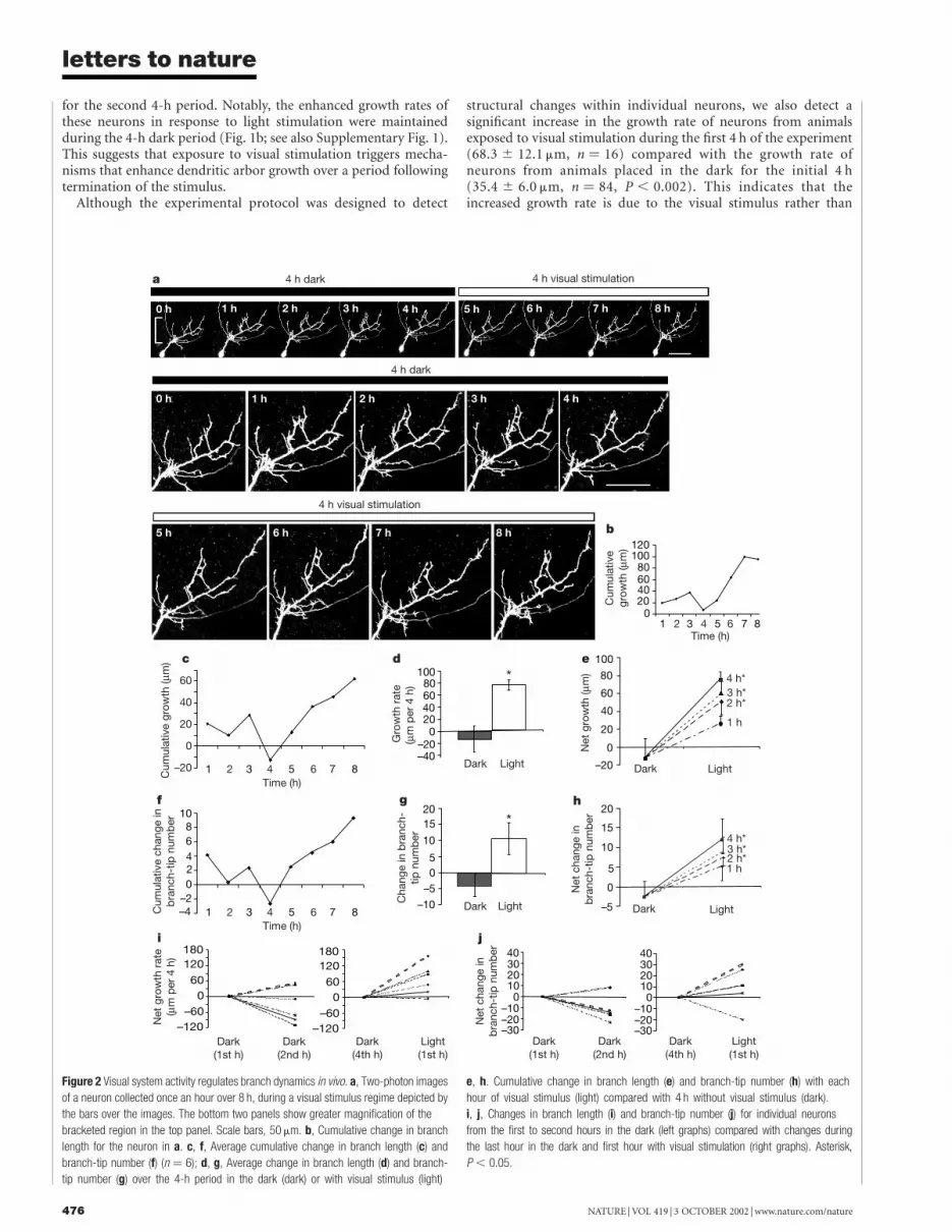
Figure 2 Visual system activity regulates branch dynamics in vivo. a, Two-photon images
of a neuron collected once an hour over 8 h, during a visual stimulus regime depicted by
the bars over the images. The bottom two panels show greater magnification of the
bracketed region in the top panel. Scale bars, 50 mm. b, Cumulative change in branch
length for the neuron in a. c, f, Average cumulative change in branch length (c) and
branch-tip number (f) (n ¼ 6); d, g, Average change in branch length (d) and branch-
tip number (g) over the 4-h period in the dark (dark) or with visual stimulus (light)
e, h. Cumulative change in branch length (e) and branch-tip number (h) with each
hour of visual stimulus (light) compared with 4 h without visual stimulus (dark).
i, j, Changes in branch length (i) and branch-tip number (j) for individual neurons
from the first to second hours in the dark (left graphs) compared with changes during
the last hour in the dark and first hour with visual stimulation (right graphs). Asterisk,
P , 0.05.
letters to nature
NATURE | VOL 419 | 3 OCTOBER 2002 | www.nature.com/nature476

a reaction to the 4 h in the dark.The light-induced increase in dendritic arbor growth rate was
blocked by exposure to APV (3-amino-phosphonovaleric acid;Fig. 1a, c) and CNQX (6-cyano-nitroquinoxaline-2,3-dione;Supplementary Fig. 1) during visual stimulation. These antago-nists block NMDAR (N-methyl-D-aspartate receptors) andAMPAR (a-amino-3-hydroxy-5-methyl-4-isoxazolepropionatereceptors), respectively. Therefore glutamate-receptor-dependentsignalling is necessary for light-induced arbor elaboration.
Dendritic arbors, grow through the selective stabilization of afraction of newly added branches and the extension of thosebranches7–9. Visual stimulation significantly increased the numberof new branches detected after 4 h (Fig. 1d, e; see also Supple-mentary Table 2). APV blocked the light-induced increase inbranch additions and increased the rate of branch retractions(Fig. 1d; see also Supplementary Table 2). Newly added branchesaccount for the increased branch length seen with light stimu-lation (Fig. 1e). Total branch length from branch additions andextensions is significantly greater when animals are exposed tolight, compared with dark or compared with animals exposed tolight in the presence of APV. The data indicate that visualstimulation enhances arbor growth by stabilizing newly addedbranches and suggest a role of NMDA receptors in activity-induced branch initiation and stabilization.
The time course of the effect of visual stimulation on dendriticarbor dynamics was analysed by collecting two-photon microscopeimages of individual green fluorescent protein (GFP)-expressingtectal neurons at 1-h intervals over a period of 8 h (Fig. 2a). Thisstimulation and imaging protocol led to a significant light-inducedincrease in dendritic arbor growth rate and branch-tip number(Fig. 2d, g), as seen when animals were imaged three times over 8 h(Fig. 1). Despite the continuous addition and retraction of branchesthroughout 8 h (Fig. 2c, f), arbors showed a net increase in growthrate and branch-tip number during light stimulation, but not over
Figure 3 Decreased RhoA activity mediates light-induced dendritic arbor growth.
a, Time-lapse in vivo confocal images collected at 4-h intervals over 8 h of tectal neurons
expressing EGFP, dominant-negative RhoA (RhoAN19) or constitutively active (CA) RhoA
(RhoAV14). Scale bar, 100 mm. b, c, Change in branch-tip number (b) and branch length
(c) over 4 h in the dark (dark) or with visual stimulation (light). Asterisk, P , 0.05,
Wilcoxon signed-rank test; double asterisk, P , 0.05, Mann–Whitney U-test.
Figure 4 Rac and Cdc42 mediate the light-induced increase in dendrite branch number.
a,Time-lapse in vivo confocal images of tectal neurons expressing EGFP, dominant-
negative Rac (RacN17) or dominant-negative Cdc42 (Cdc42N17) over 8 h. Scale bar,
100 mm. b, c, Change in branch-tip number (b) and dendritic branch length (c) over 4 h in
the dark (dark) or with visual stimulation (light). d, Change in secondary dendritic branches
(expressed as per cent of initial total branch number) added to the primary dendrite during
4 h in the dark (D) or with visual stimulation (L) in neurons expressing the designated
constructs. e, Summary of the role of visual stimulation and the Rho GTPases in dendritic
arbor development (see text for details). Asterisk, P , 0.05, Wilcoxon signed-rank test;
double asterisk, P , 0.05, Mann–Whitney U test.
letters to nature
NATURE | VOL 419 | 3 OCTOBER 2002 | www.nature.com/nature 477

4 h in the dark (Fig. 2e, h). The change in branch length and branch-tip number is significantly greater by the second hour of visualstimulation than the change seen over 4 h in the dark. Five out of sixneurons increased branch-length growth rates and branch-tipnumber in the first hour of visual stimulation compared with theprevious hour in the dark (Fig. 2i, j). By contrast, most of theneurons decreased growth rates and branch dynamics during thefirst and second time windows in the dark (Fig. 2i, j). These dataindicate that dendritic arbors respond within the first hour to visualstimulation with an increase in branch length and branch-tip number. The continued increase in branch-tip number andbranch length with each hour of visual stimulation indicates that thesystem does not adapt to the visual stimulus over 4 h of visualstimulation.
Neuronal arbor elaboration requires the reorganization of thecytoskeleton. The Rho family of small GTPases regulates actindynamics10 and has been implicated in the control of neuronalmorphogenesis11. Regulators and effectors of small GTPases such askalirin-7, SynGAP and citron associate with NMDA receptors,providing potential biochemical links between NMDAR activityand GTPase-mediated regulation of cytoskeletal dynamics12–16.However, no study has yet demonstrated a role for Rho GTPasesunder physiological conditions that control dendritic arbor devel-opment. Therefore we investigated the contribution of the Rhofamily of GTPases to the signalling mechanisms underlying activity-induced dendritic growth.
We have used recently an in situ binding assay to demonstrate thatstimulation of the optic nerve significantly decreases endogenousRhoA activity and increases endogenous Rac activity in the optictectum3. These changes require glutamate receptor activity. Here,we tested whether increased Rac and Cdc42 and decreased RhoAactivity are required for light-induced dendritic arbor elaborationby collecting time-lapse images in vivo of optic tectal neurons co-expressing GFP and either constitutively active or dominant-nega-tive forms of Rho GTPases. Constitutively active RhoAV14 blockedthe light-induced dendritic arbor development that is seen in GFP-expressing neurons (Fig. 3a–c). These data indicate that RhoAactivity inhibits activity-dependent dendritic arbor growth. Theyalso suggest that visual stimulation promotes arbor growth byinhibiting RhoA, consistent with previous studies showing thatdecreased RhoA activity increases dendritic arbor elaboration4. Iflight stimulation activated a parallel pathway promoting dendriticarbor elaboration that is independent of RhoA activity, one wouldpredict a synergistic effect of visual stimulation and expression ofdominant-negative RhoAN19; however, light stimulation did notincrease further dendritic arbor elaboration induced by dominant-negative RhoAN19 (Fig. 3a–c). This occlusion of light-induceddendritic arbor growth by dominant-negative RhoAN19 suggeststhat endogenous RhoA activity is relatively high in dendrites withslow growth rates and that decreasing RhoA activity increasesdendritic arbor development. Therefore, in combination with ourfindings that optic nerve stimulation decreases endogenous RhoAactivity3, these data suggest that RhoA is a downstream componentof the light-induced pathway controlling dendritic arborelaboration.
RhoA has been reported to negatively regulate dendrite out-growth through ROK (also known as ROCK), a RhoA-bindingkinase11. Other potential RhoA effectors are mDia, protein kinase N,citron kinase, Lim kinase-1 and PRK2 (refs 17, 18). Expression ofconstitutively active ROK inhibited light-induced dendritic arborgrowth (Fig. 3b, c). This suggests that the inhibition of activity-dependent dendritic arbor growth by RhoAV14 is at least partiallymediated by enhanced ROK activity, consistent with the idea thatlight-induced arbor elaboration occurs when both RhoA and ROKactivities are low.
Previous experiments4 and experiments included here indicatethat Rac activity controls rates of branch additions and retractions.
Our in situ GTPase assays3 indicate that decreasing Rho activityincreases Rac activity. This suggests that the increase in branch-tipnumber seen with inhibition of RhoA activity is probably due toactivation of Rac. These data also suggest that regulation of branchdynamics is affected primarily by Rac activity, and that RhoA canalter Rac-mediated branch dynamics in vivo.
Tectal neurons expressing dominant-negative RacN17 andCdc42N17 failed to respond to light stimulus with the increase inbranch-tip number and branch length seen in GFP-expressing(EGFP) neurons (Fig. 4a–c; see also Supplementary Table 3).These data indicate that activation of Rac and Cdc42 is requiredfor increased arbor elaboration in response to visual stimulation.Neurons expressing constitutively active RacV12 failed to show thesignificant light-induced increase in branch-tip number seen withEGFP controls (Fig. 4b, c). Although this is consistent with ourobservations that expression of constitutively active RacV12 increasesRhoA activity3, which inhibits dendritic arbor growth, we cannotexclude the possibility that cycling of Rac from the GTP-bound toGDP-bound form may be required for its optimum activity19.
GTPase expression also affects arbor development in the absenceof visual stimulation. Constitutively active RhoAV14 inhibited ratesof branch additions and dendritic arbor growth in the darkcompared with GFP-expressing control neurons (P , 0.05,Mann–Whitney U-test), whereas constitutively active ROK didnot (Fig. 3b, c). Neurons expressing dominant-negativeCdc42N17 or dominant-negative RacN17 had significantlydecreased rates of arbor growth and changes in branch-tip numbercompared with GFP-expressing control neurons (P , 0.05; Fig. 4a–c). These neurons could be imaged and appeared healthy for up to10 days, indicating that expression of these mutants was not toxic.These observations are consistent with previous results4,11 andsupport the idea that the Rho GTPases are positioned at a bottleneckof multiple signal transduction pathways affecting neuronal struc-ture11.
Dendritic arbor development may proceed through repeatedbranching of dendritic growth cones or through a mechanism called‘interstitial/back branching’ in which new branches emerge frommore stable branches within the arbor20. Tectal projection neuronsimaged in this study have a single stable primary dendrite, anaverage of 12 secondary branches originating from the primarydendrite, and complex arborizations with up to fifth-orderbranches. Light stimulus significantly increased the number ofsecondary branches (Fig. 4d), indicating that interstitial branchingis a prominent mechanism controlling dendritic arbor elaborationin response to visual stimulation in vivo. Expression of dominant-negative RacN17, Cdc42N17, RhoAN19 or constitutively activeRhoAV14 prevented the light-induced increase in secondarybranches. This indicates that the GTPases regulate the formationof interstitial branches in response to visual stimulation.
Our data indicate that visual system activity affects dendriticarbor elaboration through a mechanism involving Rho GTPases(Fig. 4e). Expression of mutant GTPases that interfere withendogenous GTPases blocks light-induced dendritic arbor devel-opment. We suggest that enhanced Rac and Cdc42 activity pro-motes branch dynamics. Decreased Rho activity promotes branchelongation mediated by several downstream effectors, includingROK. Rac- and Cdc42-mediated branch addition and stabilizationand RhoA-mediated branch elongation4 cooperate to result indendritic arbor growth.
Our data demonstrating that NMDAR activity and GTPases areboth required for dendritic arbor development suggest that calciuminflux through NMDAR affects regulators of Rho GTPase activity.Such regulation may occur through GTPase regulators or effectorssuch as SynGAP, kalarin or trio12,13,16,21. The Rho GTPases may alsoaffect intracellular calcium levels by regulating calcium influx intocells22 or calcium-induced calcium release from intracellularstores23. Calcium-dependent events, including calcium- and calmo-
letters to nature
NATURE | VOL 419 | 3 OCTOBER 2002 | www.nature.com/nature478

dulin-dependent kinase type II (CaMKII)-mediated branch stabili-zation24, may cooperate with GTPase-mediated branch initiationand extension to control dendritic arbor development.
Glutamate-mediated synaptic activity is probably one of severalregulators that affect dendritic arbor elaboration of tectal cells byactivating the Rho GTPases. Several other extracellular signallingmolecules that affect Rho GTPase activity include growth factors,adhesion or guidance molecules, cytokines, neurotransmitters andother neuroactive molecules25. Indeed, the decreases in growth ratesseen with expression of mutant GTPase constructs in the absence ofvisual stimulation probably represent the effects of these othersignalling pathways on GTPase-mediated regulation of cytoskeletalstructure.
Little is known about the mechanisms that link synaptic activityto structural changes in dendrities. Previous studies have indicatedthat actin-based spine dynamics are regulated by glutamate receptoractivity6 and that afferent activity promotes the formation ofdendritic structures2,4,5,7 and the stabilization of synaptic con-tacts2,26. Here, we show a direct relation between sensory input,glutamate-mediated synaptic activity and GTPase-mediated den-dritic structural plasticity in the intact animal (Fig. 4e). Ourobservations demonstrate that the initiation and selective stabiliz-ation of new dendritic branches occur in response to a visualstimulus. Our results indicate that visual stimulation affects struc-tural plasticity in vivo, through NMDA receptor-induced intracellu-lar signalling events mediated by the Rho GTPases. A
MethodsImage acquisitionTadpoles were anaesthetized with 0.02% 3-aminobenzoic acid ethyl ester (MS222, Sigma)in Steinberg’s solution for all procedures. Single tectal neurons were labelled byionophoresis of DiI3 or by electroporation with pEGFP–C1 (Clontech), or with pEGFP–C1 and recombinant EGFP–GTPase plasmids in a 4:1 (GTPase:GFP) ratio27. Confocalimages were collected through a £ 40 Nikon oil immersion lens (1.3 NA) at 2-mm stepsthrough the entire z-dimension of labelled neurons3. Two-photon images were collected at1.5-mm steps with a custom-built microscope based on a modified Olympus Fluoviewconfocal scan box mounted on an Olympus BX50WI microscope with a Tsunamifemtosecond-pulsed Ti:Sapphire laser. We used an Olympus LUM Plan F1/IR £ 40 waterimmersion objective (0.8 NA). Two-photon optical sections were an average of threeframes. For the experiment in which neurons were imaged every hour over 8 h, animalswere returned to a dark chamber in between image collection during the first 4-h period.During the second 4-h period animals were returned to the chamber with the visualstimulus in between imaging sessions. Tadpoles recovered from anaesthetic betweenimaging sessions.
Visual stimulusAlbino Xenopus laevis tadpoles were reared in ambient light until stage 46–47. Tadpoleshave a functional visual system from stage 39/40 onward, in which retinal ganglion cellaxons make glutamate-receptor-mediated synaptic connections with dendrites of tectalcells28. Retinal ganglion cells respond well to a light on/light off stimulus. A 2-s step of lighton or off evokes synaptic currents in tectal cells of Xenopus tadpoles28. On the basis of theseresponse properties, freely swimming tadpoles were placed individually in wells of a 12-well plate in a black Plexiglas chamber with a 3 £ 4 panel of green light-emitting diodes(LEDs; lmax 567 nm, AND191GCP; Allied Electronics) on the top of the chamber. Eachrow of LEDs turned on and remained on for 1 s at a frequency of 0.2 Hz. The rows turnedon and off sequentially to create a simulated motion stimulus, with 1 s of darkness betweeneach cycle. Xenopus tadpole tectal cells can respond to repeated stimulation of the sameregion of retina without apparent adaptation28. In our experiments, adaptation of tectalcells to the stimulus is not likely to be significant because the animals are swimming freelyduring the stimulation period, so the apparent location and direction of the stimulus varyconstantly. Light exposure during the 4-h period in the absence of visual stimulation waskept to a minimum, but animals were exposed to some light during imaging. Someanimals were exposed to 100 mM DL-APV or 20 mM CNQX during visual stimulation.MK801, another NMDA receptor antagonist, had a similar inhibitory effect on light-induced growth to that seen with APV (relative growth rate: 0.94 ^ 0.73, n ¼ 9).Treatment of tectal cells with APV while the animals were in the dark for 4 hdid not significantly affect dendritic growth (relative growth rate: 1.34 ^ 0.5, n ¼ 9,P ¼ 0.7).
Image analysisWe used a 4-h imaging protocol on the basis of our previous studies which showed that 4 his sufficiently long to detect quantifiable increases in dendritic arbor branch length andbranch-tip number, but short enough so that images of arbors from sequential time pointscan be reliably superimposed9. We measured the total dendritic branch length (TDBL) andbranch-tip number of the dendritic arbors by reconstructing the whole dendritic arbor in
three dimensions using Object Image (http://simon.bio.uva.nl/object-image.html). Thesoftware computed the TDBL and branch-tip number using custom macros writtenby E.S.R.29. Values are expressed as mean and standard error. Statistical analysis wasdone with Mann–Whitney U nonparametric statistical analysis and Wilcoxon signed-rank test. Analysis was performed blind to the type of treatment. Between 9 and 24neurons per treatment were analysed, unless stated otherwise.
Construction of expression plasmids of GTPase mutantsFragments (600 base pairs) containing the full coding sequence of RacN17, Cdc42N17,RhoAN19 and RhoV14 were cloned in-frame into pEGFP–C1 (Clontech Laboratories)and expressed as GFP-fusion proteins30.
Received 10 April; accepted 25 June 2002; doi:10.1038/nature00987.
1. Builluart, P. et al. Oligophrenin-1 encodes a rhoGAP protein involved in X-linked mental retardation.
Nature 392, 823–826 (1998).
2. Engert, F. & Bonhoeffer, T. Dendritic spine changes associated with hippocampal long-term synaptic
plasticity. Nature 399, 66–70 (1999).
3. Li, Z., Aizenman, C. D. & Cline, H. T. Regulation of Rho GTPases by crosstalk and neuronal activity in
vivo. Neuron 33, 741–750 (2002).
4. Li, Z., Van Aelst, L. & Cline, H. T. Rho GTPases regulate distinct aspects of dendritic arbor growth in
Xenopus central neurons in vivo. Nature Neurosci. 3, 217–225 (2000).
5. Maletic-Savatic, M., Malinow, R. & Svoboda, K. Rapid dendritic morphogenesis in CA1 hippocampal
dendrites induced by synaptic activity. Science 283, 1923–1927 (1999).
6. Fischer, M., Kaech, S., Wagner, U., Brinkhaus, H. & Matus, A. Glutamate receptors regulate actin-
based plasticity in dendritic spines. Nature Neurosci. 3, 887–894 (2000).
7. Wong, W. T., Faulkner-Jones, B. E., Sanes, J. R. & Wong, R. O. Rapid dendritic remodeling in the
developing retina: dependence on neurotransmission and reciprocal regulation by Rac and Rho.
J. Neurosci. 20, 5024–5036 (2000).
8. Wu, G.-Y., Zou, D. J., Rajan, I. & Cline, H. T. Dendritic dynamics in vivo change during neuronal
maturation. J. Neurosci. 19, 4472–4483 (1999).
9. Rajan, I. & Cline, H. T. Glutamate receptor activity is required for normal development of tectal cell
dendrites in vivo. J. Neurosci. 18, 7836–7846 (1998).
10. Van Aelst, L. & D’Souza-Schorey, C. Rho GTPases and signaling networks. Genes Dev. 11, 2295–2322
(1997).
11. Luo, L. Rho GTPases in neuronal morphogenesis. Nature Rev. Neurosci. 1, 173–180 (2000).
12. Penzes, P. et al. The neuronal Rho-GEF Kalirin-7 interacts with PDZ domain-containing proteins and
regulates dendritic morphogenesis. Neuron 29, 229–242 (2001).
13. Kim, J. H., Liao, D., Lau, L.-F. & Huganir, R. L. SynGAP: a synaptic RasGAP that associates with the
PSD-95/SAP90 protein family. Neuron 20, 683–691 (1998).
14. Zhang, W., Vazquez, L., Apperson, M. & Kennedy, M. B. Citron binds to PSD-95 at glutamatergic
synapses on inhibitory neurons in the hippocampus. J. Neurosci. 19, 96–108 (1999).
15. Furuyashiki, T. et al. Citron, a Rho-target, interacts with PSD-95/SAP-90 at glutamatergic synapses in
the thalamus. J. Neurosci. 19, 109–118 (1999).
16. Chen, H. J., Rojas, S. M., Oguni, A. & Kennedy, M. B. A synaptic Ras-GTPase activating protein (p135
SynGAP) inhibited by CaM kinase II. Neuron 20, 895–904 (1998).
17. Lawler, S. Regulation of actin dynamics: the LIM kinase connection. Curr. Biol. 9, R800–R802
(1999).
18. Aspenstrom, P. Effectors for the Rho GTPases. Curr. Opin. Cell Biol. 11, 95–102 (1999).
19. Albertinazzi, C., Gilardelli, D., Paris, S., Longhi, R. & de Curtis, I. Overexpression of a neural-specific
rho family GTPase, cRac1B, selectively induces enhanced neuritogenesis and neurite branching in
primary neurons. J. Cell Biol. 142, 815–825 (1998).
20. Acebes, A. & Ferrus, A. Cellular and molecular features of axon collaterals and dendrites. Trends
Neurosci. 23, 557–565 (2000).
21. Debant, A. et al. The multidomain protein Trio binds the LAR transmembrane tyrosine phosphatase,
contains a protein kinase domain, and has separate rac-specific and rho-specific guanine nucleotide
exchange factor domains. Proc. Natl Acad. Sci. USA 93, 5466–5471 (1996).
22. Wilk-Blaszczak, M. et al. The monomeric G-proteins Rac1 and/or Cdc42 are required for the
inhibition of voltage-dependent calcium current by bradykinin. J. Neurosci. 17, 4094–4100 (1997).
23. Djouder, N., Prepens, U., Aktories, K. & Cavalie, A. Inhibition of calcium release-activated calcium
current by Rac/Cdc42-inactivating clostridial cytotoxins in RBL cells. J. Biol. Chem. 275, 18732–18738
(2000).
24. Wu, G.-Y. & Cline, H. T. Stabilization of dendritic arbor structure in vivo by CaMKII. Science 279,
222–226 (1998).
25. Kjoller, L. & Hall, A. Signaling to Rho GTPases. Exp. Cell Res. 253, 166–179 (1999).
26. Lendvai, B., Stern, E. A., Chen, B. & Svoboda, K. Experience-dependent plasticity of dendritic spines
in the developing rat barrel cortex in vivo. Nature 404, 876–881 (2000).
27. Haas, K., Sin, W.-C., Javaherian, A., Li, Z. & Cline, H. T. Single-cell electroporation for in vivo
neuronal gene expression. Neuron 29, 1–9 (2001).
28. Zhang, L. I., Tao, H. W. & Poo, M. Visual input induces long-term potentiation of developing
retinotectal synapses. Nature Neurosci. 3, 708–715 (2000).
29. Ruthazer, E. S. & Cline, H. T. Multiphoton imaging of neurons in living tissue: acquisition and analysis
of time-lapse morphological data. J. Real-Time Imag. 8, 175–188 (2002).
30. Leung, T., Chen, X. Q., Manser, E. & Lim, L. The p160 RhoA-binding kinase ROK alpha is a member of
a kinase family and is involved in the reorganization of the cytoskeleton. Mol. Cell. Biol. 16, 5313–5327
(1996).
Supplementary information accompanies the paper on Nature’s website
(http://www.nature.com/nature).
AcknowledgementsWe thank K. Svoboda, P. O’Brien and B. Burbach for help constructing the two-photonmicroscope and P. O’Brien for constructing the electronic circuit for visual stimulation.We also thank K. Bronson for technical assistance, J. Duffy for assistance with the figures,and T. Leung and J. Dong for cDNA. We are grateful to L. Van Aelst for discussions, and
letters to nature
NATURE | VOL 419 | 3 OCTOBER 2002 | www.nature.com/nature 479

R. Malinow, J. Dubnau and members of the Cline laboratory for comments on themanuscript. This work was supported by the NIH (H.T.C., K.H., E.S.R) and anendowment from the Charles Robertson Foundation to H.T.C..
Competing interests statementThe authors declare that they have no competing financial interests.
Correspondence and requests for materials should be addressed to H.C.
(e-mail: [email protected]).
..............................................................
ERAAP customizes peptides forMHC class I molecules in theendoplasmic reticulumThomas Serwold, Federico Gonzalez, Jennifer Kim, Richard Jacob& Nilabh Shastri
Division of Immunology, Department of Molecular and Cell Biology, University ofCalifornia, Berkeley, California 94720-3200, USA.............................................................................................................................................................................
The ability of killer T cells carrying the CD8 antigen to detecttumours or intracellular pathogens requires an extensive displayof antigenic peptides by major histocompatibility complex(MHC) class I molecules on the surface of potential targetcells1. These peptides are derived from almost all intracellularproteins and reveal the presence of foreign pathogens andmutations. How cells produce thousands of distinct peptidescleaved to the precise lengths required for binding different MHCclass I molecules remains unknown2,3. The peptides are cleavedfrom endogenously synthesized proteins by the proteasome inthe cytoplasm4,5 and then trimmed by an unknown aminopepti-dase in the endoplasmic reticulum (ER)6–8. Here we identifyERAAP, the aminopeptidase associated with antigen processingin the ER. ERAAP has a broad substrate specificity, and itsexpression is strongly upregulated by interferon-g. Reducingthe expression of ERAAP through RNA interference preventsthe trimming of peptides for MHC class I molecules in the ER andgreatly reduces the expression of MHC class I molecules on thecell surface. Thus, ERAAP is the missing link between theproducts of cytosolic processing and the final peptides presentedby MHC class I molecules on the cell surface.
To identify the ER aminopeptidase, we used detergent to solu-bilize microsomes from mouse liver and spleens. Ion exchangechromatography of solubilized microsomes showed a predominantpeak of activity peak that was blocked by the aminopeptidaseinhibitor leucinethiol (Fig. 1a). This activity was purified by severalchromatographic steps (Methods). The highly enriched fractionsfrom the final purification step contained one main band with arelative molecular mass (M r) of roughly 100,000 (100K) on a SDSpolyacrylamide gel electrophoresis (SDS–PAGE) gel stained withCoomassie blue (Fig. 1b). We subjected this band to in-gel trypsindigestion followed by matrix-assisted laser-desorption ionizationtime-of-flight mass spectrometry and used the resulting trypticpeptide fingerprint to search the National Center for BiotechnologyInformation database9–11.
In two independent experiments, 25 masses matched the pre-dicted output of trypsin-digested murine adipocyte-derived leucineaminopeptidase (accession code AF227511)12. This assignment wasverified with the amino acid sequences of four peptides determinedby high-energy collision-induced dissociation tandem mass spec-
trometry analysis (Fig. 1c)13. This murine aminopeptidase of 930amino acids and its probable human and rat orthologues are alsoknown as puromycin-insensitive leucyl-specific aminopepti-dase12,14,15. On the basis of our results, we hereafter refer to thisenzyme as ERAAP (for ER aminopeptidase associated with antigenprocessing) to designate its intracellular location and function intrimming peptides in the MHC class I antigen processing pathway.
ERAAP is a member of the M1 family of zinc metalloproteasesthat are defined by a highly conserved His-Glu-X-X-His-X18-Glumotif in the core peptidase unit (ref. 16 and Fig. 1c). The aminoterminus contains a hydrophobic leader sequence, which indicatedthat it might be cotranslationally translocated into the ER. Hydro-phobicity plots did not predict any other transmembrane domains,and there were no other obvious motifs to predict its intracellularlocation. To determine its location, therefore, we first transfectedCOS cells with the murine ERAAP complementary DNA andcarried out western blot analysis with the whole-cell lysates andthe culture supernatants. A single band close to the predicted
Figure 1 Purification and identification of ERAAP. a, Microsomes from mouse livers and
spleens were lysed in detergent and separated by ion exchange chromatography.
Fractions were tested for aminopeptidase activity by incubating with leucine p nitroanilide
without (filled circles) or with (open circles) the aminopeptidase inhibitor leucinethiol.
b, SDS–PAGE gel stained with Coomassie blue of three consecutive fractions containing
aminopeptidase activity from the final hydrophobic interaction chromatography
purification step. c, The sequence of murine ERAAP. Underlined peptides matched the
masses identified by mass spectrometry. Peptides identified by sequencing are
underlined in bold. The ER translocation signal at the N terminus is in italics, active-site
residues are boxed, and putative N-linked glycosylation sites are in bold.
letters to nature
NATURE | VOL 419 | 3 OCTOBER 2002 | www.nature.com/nature480


