Embed Size (px)
Citation preview
The Rockefeller University Press, 0021-9525/2001/10/41/11 $5.00The Journal of Cell Biology, Volume 155, Number 1, October 1, 2001 41–51http://www.jcb.org/cgi/doi/10.1083/jcb.200103145
JCB
Article
41
Dense core secretory vesicles revealed as a dynamic
Ca
2
�
store in neuroendocrine cells with a vesicle-associated membrane protein aequorin chimaera
Kathryn J. Mitchell,
1
Paolo Pinton,
2,3
Aniko Varadi,
1
Carlo Tacchetti,
4
Edward K. Ainscow,
1
Tullio Pozzan,
3
Rosario Rizzuto,
2
and Guy A. Rutter
1
1
Departments of Biochemistry, University of Bristol, BS8 1TD Bristol, United Kingdom
2
Experimental and Diagnostic Medicine Section of General Pathology, University of Ferrara, 44100 Ferrara, Italy
3
Biomedical Sciences and CNR Center for Study of Biological Membranes, University of Padova, 35121 Padova 17, Italy
4
Experimental Medicine, University of Genova Medical School, 16132 Genova, Italy
he role of dense core secretory vesicles in the control
of cytosolic-free Ca
2
�
concentrations ([Ca
2
�
]
c
) inneuronal and neuroendocrine cells is enigmatic.
By constructing a vesicle-associated membrane protein2–synaptobrevin.aequorin chimera, we show that in clonal
pancreatic islet
β
-cells: (a) increases in [Ca
2
�
]
c
cause aprompt increase in intravesicular-free Ca
2
�
concentration
([Ca
2
�
]
SV
), which is mediated by a P-type Ca
2
�
-ATPase distinctfrom the sarco(endo) plasmic reticulum Ca
2
�
-ATPase, butwhich may be related to the PMR1/ATP2C1 family of Ca
2
�
pumps; (b) steady state Ca
2
�
concentrations are 3–5-foldlower in secretory vesicles than in the endoplasmic reticulum(ER) or Golgi apparatus, suggesting the existence of tightly
T
bound and more rapidly exchanging pools of Ca
2
�
; (c)inositol (1,4,5) trisphosphate has no impact on [Ca
2
�
]
SV
inintact or permeabilized cells; and (d) ryanodine receptor(RyR) activation with caffeine or 4-chloro-3-ethylphenol inintact cells, or cyclic ADPribose in permeabilized cells,causes a dramatic fall in [Ca
2
�
]
SV
. Thus, secretory vesiclesrepresent a dynamic Ca
2
�
store in neuroendocrine cells,whose characteristics are in part distinct from the ER/Golgiapparatus. The presence of RyRs on secretory vesiclessuggests that local Ca
2
�
-induced Ca
2
�
release from vesicles
docked at the plasma membrane could participate intriggering exocytosis.
Introduction
In most mammalian cells, the ER (Streb et al., 1984; Rizzutoet al., 1993; Brini et al., 1993) and Golgi complex (Pinton etal., 1998) are believed to represent the major mobilizable
intracellular Ca
2
�
stores (Rutter et al., 1998). Uptake of Ca
2
�
into these stores is mediated largely by sarco(endo)plasmicreticulum Ca
2
�
–ATPases (SERCAs)* (Moller et al., 1996)and helps to maintain resting cytosolic Ca
2
�
concentrations
([Ca
2
�
]
c
) at levels (
�
10
�
7
M) some four orders of magnitudelower than in the extracellular space (
�
10
�
3
M). Release of
Ca
2
�
from the ER and Golgi complex is provoked by hor-mones and other agonists which generate inositol 1,4,5trisphosphate (IP
3
) (Berridge, 1993) to open intracellularreceptors (Mikoshiba, 1997). In certain cell types, such asskeletal muscle, these stores are also accessed by “Ca
2
�
-inducedCa
2
�
release”, via receptors for the insecticide, ryanodine (Laiet al., 1988; Takeshima et al., 1989).
Secretory vesicles of both the dense core (Hellman et al.,1976; Hutton et al., 1983) and small synaptic types (Andrewsand Reese, 1986) possess a substantial proportion of the to-tal Ca
2
�
in cells specialized for peptide secretion. However,despite many studies, the role of secretory vesicles as Ca
2
�
buffers or stores remains controversial (Pozzan et al., 1994;Rutter et al., 1998; Yoo, 2000).
Mobilization of dense core vesicle Ca
2
�
by IP
3
was origi-nally suggested as a possibility in chromaffin cells (Yoo and
Albanesi, 1990). Later data suggested that IP
3
receptorsmay be present on the vesicle membrane in insulin-secretingcells (Blondel et al., 1994), though subsequent experiments
Address correspondence to Guy Rutter, Department of Biochemistry,University of Bristol, BS8 1TD Bristol, UK. Tel.: (44) 117-954-6401.Fax: (44) 117-928-8274. E-mail: [email protected]
*Abbreviations used in this paper: cADPr, cyclic ADP ribose; CPA, cyclo-piazonic acid; DHPG, dihydroxyphenylglycine; GFP, green fluorescent
protein; HA, hemagglutinin; IB, intracellular buffer; IP
3
, inositol 1,4,5trisphosphate; KRB, Krebs-Ringer bicarbonate buffer;
RyR, ryanodinereceptor; 4-CEP, 4-chloro-3-ethylphenol; SERCA, sarco(endo)plasmic
reticulum Ca
2
�
–ATPase; VAMP, vesicle-associated membrane protein.
Key words: calcium; secretory vesicle; insulin; ryanodine receptor; aequorin
on February 6, 2016
jcb.rupress.orgD
ownloaded from
Published September 24, 2001
42 The Journal of Cell Biology
|
Volume 155, Number 1, 2001
showed the antibody used cross-reacted with insulin (Ravaz-zola et al., 1996). IP
3
, as well as the receptor (RyR) agonistcyclic ADP ribose (cADPr) (Galione, 1994) have been re-ported to release Ca
2
�
from individual acinar cell zymogengranules (Gerasimenko et al., 1996), although contaminationof these preparations (e.g., with ER or Golgi apparatus–derived vesicles) is a potential problem (Yule et al., 1997). Fi-nally, evidence for the direct participation of secretory gran-ules in the control of cytoplasmic Ca
2
�
concentration wasrecently provided in intact goblet cells (Nguyen et al., 1998).
At present, the molecular mechanisms responsible forCa
2
�
uptake into secretory vesicles are also a matter of con-troversy. At high Ca
2
�
concentrations (
�
50
�
M), uptakeinto dense core vesicles in permeabilized hypophysial cells(Troadec et al., 1998; Thirion et al., 1999) and into isolatedchromaffin cell granules (Krieger-Brauer and Gratzl, 1982)can occur via Na
�
/Ca
2
�
exchange, while synaptic vesiclesalso accumulate Ca
2
�
via a Ca
2
�
-H
�
antiport system (Gon-calves et al., 1998). However, the pump/exchange mecha-nisms active at physiological [Ca
2
�
]
c
are uncertain.To examine the pathways by which Ca
2
�
crosses the limit-ing membrane of the dense core secretory vesicle of living is-let
�
-cells, we have developed a new approach to monitorthe free Ca
2
�
concentration within the secretory vesicle ma-trix ([Ca
2
�
]
SV
) using recombinant targeted aequorin (Riz-zuto et al., 1995). Cloned from the jellyfish
Aequorea victo-ria
(Inouye et al., 1985), aequorin is a calcium-sensitivebioluminescent protein (Cobbold and Rink, 1987), previ-ously used to measure free Ca
2
�
concentrations in a varietyof subcellular organelles (Rutter et al., 1998). Importantly,aequorin activity is less severely inhibited at low pH values(
�
6.5; Blinks, 1989) than Ca
2
�
probes based on green fluo-rescent protein (GFP) (Miyawaki et al., 1997; Baird et al.,1999; Emmanouilidou et al., 1999). If appropriately tar-geted, this probe should allow Ca
2
�
concentrations to bemeasured in the acidic environment of the secretory granuleinterior (Orci et al., 1985).
Vesicle-associated membrane protein (VAMP)2/synapto-brevin (Sudhof et al., 1989) is a vesicle-specific SNARE witha single transmembrane-spanning region. Expression of chi-maeric cDNA encoding a fusion protein between VAMP2and aequorin (VAMP.Aq) has therefore allowed the intrave-sicular-free Ca
2
�
concentration to be monitored dynamicallyin live MIN6
�
-cells. With this approach, we show that Ca
2
�
is actively pumped into dense core vesicles when [Ca
2
�
]
c
in-creases, and may be released via RyR, but not IP
3
, receptors.This release may be important at sites of high intracellularCa
2
�
, including sites of exocytosis at the plasma membrane.
Results
Subcellular targeting of recombinant VAMP.aequorin
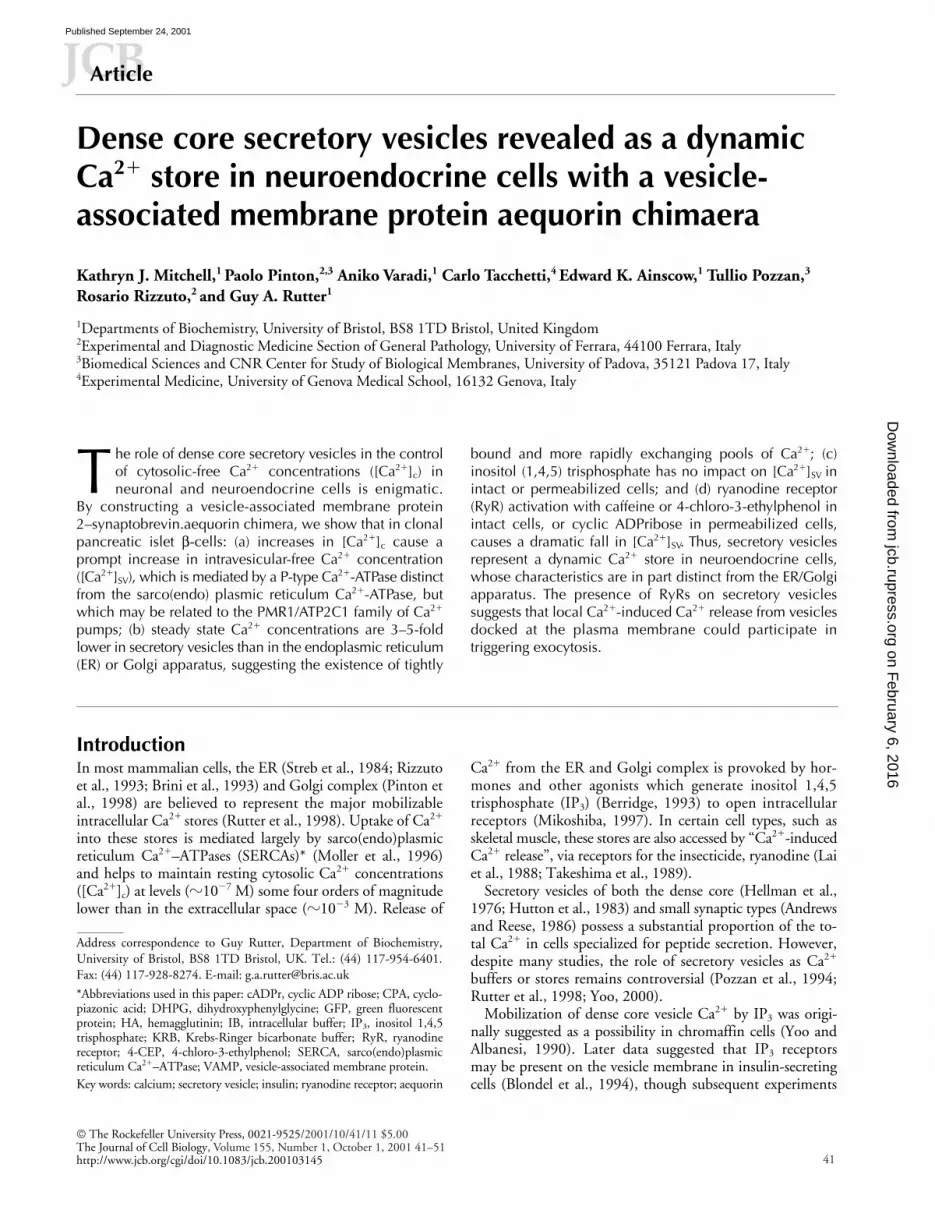
Chimeric cDNA encoding hemagglutinin (HA)1-tagged ae-quorin, fused to VAMP2 (Sudhof et al., 1989), was gener-ated as shown in Fig. 1 A.
Immunocytochemical analysis of MIN6 cells transfectedwith VAMP.Aq cDNA revealed close overlap with insulinstaining (Fig. 1 B). Explored at a higher resolution by immuno-electron microscopy (Fig. 1 C), VAMP.Aq immunoreactivitywas highly enriched in 61 of 148 (41.2%;
n
�
11 cells) vesicles
colabeled for insulin (Fig. 1 C). Analyzed by single labeling forVAMP.Aq, staining of the ER, Golgi apparatus, and small syn-aptic-like microvesicles (Reetz et al., 1991) was very low, whilereactivity was also present on the plasma membrane and in en-dosomes (see the legend to Fig. 1 and Discussion).
Reconstitution and calibration of secretory vesicle and ER-targeted aequorins
Given the high total Ca
2
�
content of secretory vesicles (Hut-ton et al., 1983), we used the approach adopted previouslyto measure Ca
2
�
in the ER lumen (Montero et al., 1995).Apoaequorin was reconstituted at a low free Ca
2
�
concentra-tion (Montero et al., 1995), achieved by depleting cells ofCa
2
�
(Materials and methods). Depletion of vesicle Ca
2
�
had no marked effect on glucose or K
�
-stimulated insulinsecretion, or on vesicle motility (Pouli et al., 1998b; Tsuboiet al., 2000; unpublished data).
To determine the response of the expressed aequorins toCa
2
�
in situ, permeabilized cells were incubated at buffered
Figure 1. Localization of VAMP.Aq. (A) Schematic map of VAMP.Aq. VAMP2 and aequorin cDNAs were fused via an HA1 epitope tag linker (Materials and methods) in order to localize mutated aequorin to the secretory vesicle lumen. (B) Confocal immunolocalization of VAMP.Aq. MIN6 cells were transfected with VAMP.Aq and stained with (a) mouse anti-HA1 monoclonal anti-body (1:200) and (b) guinea pig antiinsulin antibody (1:150). (c) Extent of colocalization. (C) Immunoelectron microscopic local-ization of insulin (15-nm gold) or VAMP.Aq (anti-HA tag, 10-nm gold). Morphometric analysis of separate sections from 10 singly labeled cells revealed the following distribution of anti-HA gold particles: dense core vesicles, 36; ER, 2; Golgi apparatus, 0; plasma membrane, 16; endosomes, 19.
on February 6, 2016
jcb.rupress.orgD
ownloaded from
Published September 24, 2001

Free [Ca2�] in insulin secretory vesicles | Mitchell et al. 43
Ca2� concentrations in the presence of ionomycin and mo-nensin (Fig. 2, C and E). The sensitivity to Ca2� (at pH 7.0)of the VAMP.Aq chimaera was similar to that reported previ-ously for mutant (D119A) aequorin (Montero et al., 1995).Intravesicular pH in intact cells was determined using a fu-sion construct between VAMP2 and a mutated, pH-sensitiveGFP (pH.fluorin(e); Miesenbock et al., 1998), and gave a pHvalue of 6.3 0.02 (n � 85 cells; Fig. 2 A). Confirming thatthis low intravesicular pH was unlikely to significantly affectVAMP.Aq, near identical calibration data were obtained at
pH 5.7 (unpublished data). Therefore, we used the constantsobtained in vitro (Montero et al., 1995) to calculate [Ca2�]from the fractional rate of aequorin consumption (F) accord-ing to: [Ca2�] � 1.44�10(LOG[F]�3.4) (Rutter et al., 1993).
Dynamic measurement of secretory vesicle and ER Ca2� concentrationsTo monitor uptake of Ca2� into vesicles in living cells, weprovoked rapid Ca2� influx into cells through store-operatedchannels (Parekh and Penner, 1997). During reintroduction
Figure 2. Measurement of intravesicular pH (A) and Ca2� with cytosolic aequorin (B), VAMP.Aq (C and D), and ER.Aq (E and F). (A) Confocal images were acquired from cells transfected with pH.fluorin(e) (Miesenbock et al., 1998), maintained initially in KRB, pH 7.4, before digitonin-permeabilization and exchange into IB at the specified pH values, and buffered with morpholinosulphonic acid, Hepes, or TRIS (10 mM), plus 10 �M ionomycin, 10 �M mo-nensin, and 1 �M FCCP. Normalized fluorescence ratios before permeabilization were in the range of 0.16–0.18 (arrow); data were fitted using the Graph Pad PrismTM. (C and E) After Ca2� depletion and aequorin reconstitution, transfected cells were permeabilized with digitonin and perifused in the presence of ionomycin, mon-ensin, and CPA (10 �M each) with N-(2-hydroxyethyl)ethylene di-amine triacetate (HEDTA)-buffered Ca2� solutions at 0.5 mM calcu-lated free Mg2�. Cells expressing either (B) cytosolic aequorin, (D) VAMP.Aq, or (F) ER.Aq were perifused with KRB, supplemented with 1 mM EGTA (KRB/EGTA) and, where indicated, EGTA was replaced with 1.5 mM CaCl2. Cells were finally lysed in Ca2�-rich hypotonic medium (10 mM CaCl2, 0.1 mM digitonin) for calibration (see Materials and methods and Results). The background count rate (�1,200 cps; D and F) was identical during perifusion of untransfected cells and is due to autooxidation of coelenterazine n.
Figure 3. Ca2� uptake into secretory vesicles (A–C) or ER (D and E). After Ca2� depletion and aequorin reconstitution, cells were permeabilized with 20 �M digitonin in IB; (A and D; see Materials and methods). Cells were perifused initially at �10�9 M free [Ca2�] (buffered with 0.2 mM EGTA, 1 mM HEDTA; free [Mg2�] � 0.5 mM) and then at 400 nM free [Ca2�], as indicated. Where present (closed symbols) ATP was 1 mM. Note that accumulation of Ca2� at zero ATP is likely to be due to synthesis of small amounts of ATP by mito-chondria. (B) Dose–response for the inhibition of vesicular [Ca2�] in-creases by orthovanadate. Cells expressing VAMP.Aq were perme-abilized and perifused at 400 nM Ca2� in the presence of 1 mM ATP, plus the indicated concentrations of NaVO4. The initial rates of [Ca2�]SV increase upon the stepped increase in perifusate-free [Ca2�] from �1 to 400 nM were calculated by fitting time course data to a simple first order rate equation by nonlinear regression analysis (Microsoft ExcelTM). (C and E) After Ca2� depletion and aequorinreconstitution, Ca2� uptake into intact cells was initiated by replac-ing KRB containing 1 mM EGTA with EGTA-free KRB at 1.5 mM CaCl2, as indicated. Closed symbols, cells were incubated with thapsigargin (TG; 1 �M) during the final 10 min of reconstitution, and the perifusion medium was supplemented with CPA (10 �M). Data are representative of three separate experiments in each case.
on February 6, 2016
jcb.rupress.orgD
ownloaded from
Published September 24, 2001
44 The Journal of Cell Biology | Volume 155, Number 1, 2001
of CaCl2 to cells previously perifused in EGTA, [Ca2�]c ap-proached 500 nM, then fell back to 150–200 nM (Fig. 2 B).By contrast, steady state free [Ca2�] in the secretory vesiclematrix was 30–90 �M (51; 7.5 �M; n � 5 separate cultures;Fig. 2 D), whereas free [Ca2�] in the ER lumen was fivefoldhigher (249; 12.9 �M; n � 3 preparations; Fig. 2 F).
Pathways of Ca2� uptake into dense core secretory vesiclesBoth the rate and extent of the [Ca2�] increases in the secre-tory vesicles and the ER were strongly dependent on thepresence of added ATP in permeabilized cells (Fig. 3, A andD). The increase in [Ca2�]SV upon reintroduction of Ca2�
ions was completely inhibited by the P-type Ca2� pump in-hibitor, orthovanadatate, at �100 �M (Fig. 3 B), but wasinsensitive to 10 �M orthovanadatate, a concentrationwhich strongly inhibits the plasma membrane Ca2�-ATPase
(Carafoli, 1991). Preincubation with the specific SERCA in-hibitor, thapsigargin (Thastrup, 1990), and perifusion withcyclopiazonic acid (CPA) (Mason et al., 1991) had no effecton the changes in [Ca2�]SV (Fig. 3 C and Table I), but mark-edly (�85%) inhibited ER [Ca2�] increases (Fig. 3 E).
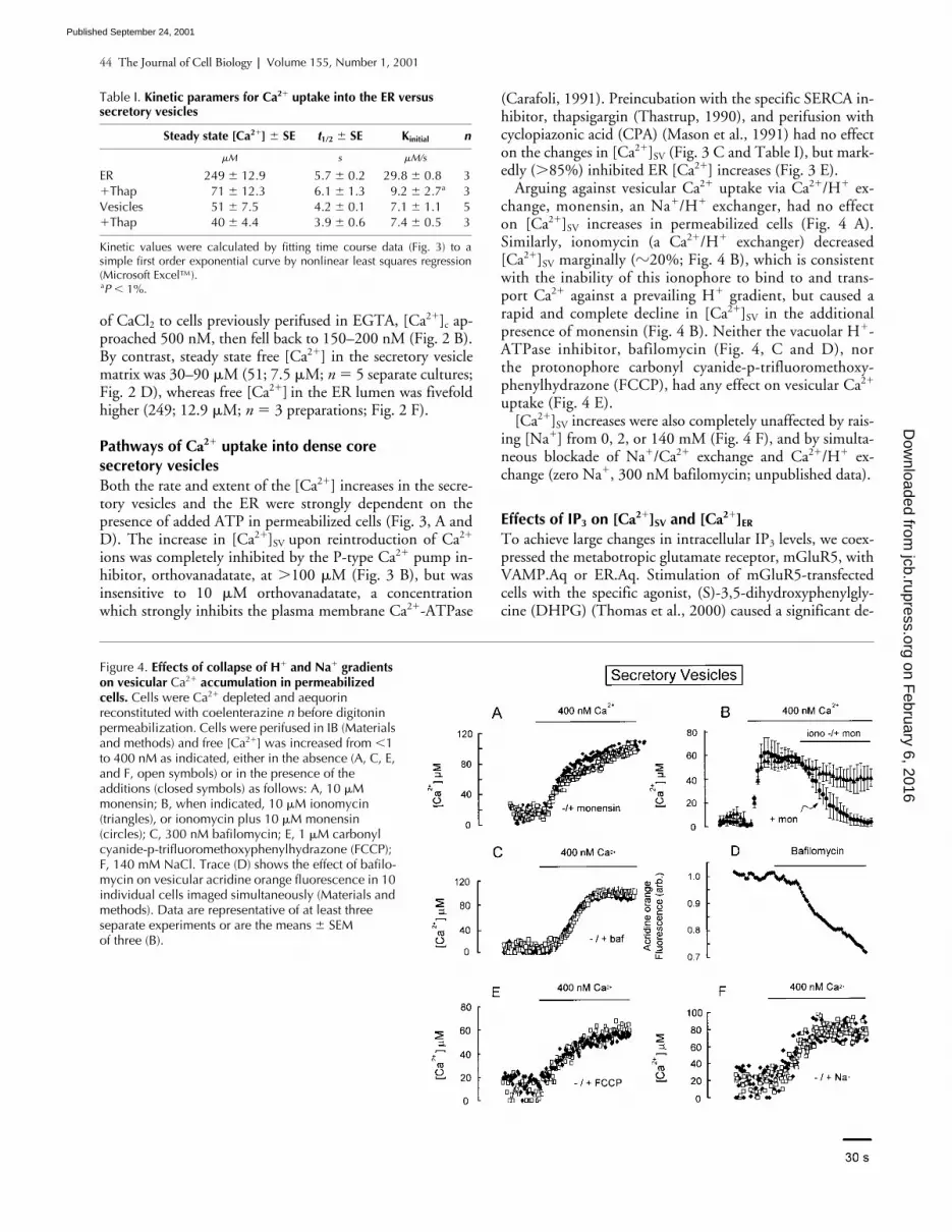
Arguing against vesicular Ca2� uptake via Ca2�/H� ex-change, monensin, an Na�/H� exchanger, had no effecton [Ca2�]SV increases in permeabilized cells (Fig. 4 A).Similarly, ionomycin (a Ca2�/H� exchanger) decreased[Ca2�]SV marginally (�20%; Fig. 4 B), which is consistentwith the inability of this ionophore to bind to and trans-port Ca2� against a prevailing H� gradient, but caused arapid and complete decline in [Ca2�]SV in the additionalpresence of monensin (Fig. 4 B). Neither the vacuolar H�-ATPase inhibitor, bafilomycin (Fig. 4, C and D), northe protonophore carbonyl cyanide-p-trifluoromethoxy-phenylhydrazone (FCCP), had any effect on vesicular Ca2�
uptake (Fig. 4 E).[Ca2�]SV increases were also completely unaffected by rais-
ing [Na�] from 0, 2, or 140 mM (Fig. 4 F), and by simulta-neous blockade of Na�/Ca2� exchange and Ca2�/H� ex-change (zero Na�, 300 nM bafilomycin; unpublished data).
Effects of IP3 on [Ca2�]SV and [Ca2�]ER
To achieve large changes in intracellular IP3 levels, we coex-pressed the metabotropic glutamate receptor, mGluR5, withVAMP.Aq or ER.Aq. Stimulation of mGluR5-transfectedcells with the specific agonist, (S)-3,5-dihydroxyphenylgly-cine (DHPG) (Thomas et al., 2000) caused a significant de-
Table I. Kinetic paramers for Ca2� uptake into the ER versus secretory vesicles
Steady state [Ca2�] SE t1/2 SE Kinitial n
�M s �M/s
ER 249 12.9 5.7 0.2 29.8 0.8 3�Thap 71 12.3 6.1 1.3 9.2 2.7a 3Vesicles 51 7.5 4.2 0.1 7.1 1.1 5�Thap 40 4.4 3.9 0.6 7.4 0.5 3
Kinetic values were calculated by fitting time course data (Fig. 3) to asimple first order exponential curve by nonlinear least squares regression(Microsoft Excel™).aP � 1%.
Figure 4. Effects of collapse of H� and Na� gradients on vesicular Ca2� accumulation in permeabilized cells. Cells were Ca2� depleted and aequorin reconstituted with coelenterazine n before digitonin permeabilization. Cells were perifused in IB (Materials and methods) and free [Ca2�] was increased from �1 to 400 nM as indicated, either in the absence (A, C, E, and F, open symbols) or in the presence of the additions (closed symbols) as follows: A, 10 �M monensin; B, when indicated, 10 �M ionomycin (triangles), or ionomycin plus 10 �M monensin (circles); C, 300 nM bafilomycin; E, 1 �M carbonyl cyanide-p-trifluoromethoxyphenylhydrazone (FCCP); F, 140 mM NaCl. Trace (D) shows the effect of bafilo-mycin on vesicular acridine orange fluorescence in 10 individual cells imaged simultaneously (Materials and methods). Data are representative of at least three separate experiments or are the means SEM of three (B).
on February 6, 2016
jcb.rupress.orgD
ownloaded from
Published September 24, 2001

Free [Ca2�] in insulin secretory vesicles | Mitchell et al. 45
crease in [Ca2�]ER (Fig. 5 E), but an increase in [Ca2�]SV
(Fig. 5 A). DHPG had no significant effect on the distribu-tion of vesicles (imaged after expression of VAMP.GFP/pH-fluorin (r); Fig. 5 D), indicating that enhanced exocytosisand exposure of the VAMP.Aq to the extracellular mediumwas unlikely to contribute to the observed increases in[Ca2�]SV (Fig. 5 A, and see Discussion).
Similarly, in permeabilized cells, IP3 had no effect on[Ca2�]SV (Fig. 5, B and C), while causing a large decrease in[Ca2�]ER (Fig. 5, F and G).
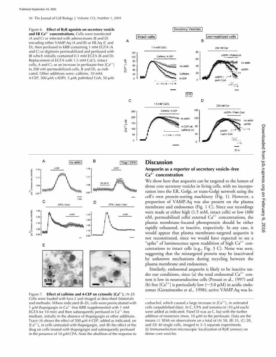
RyR activation decreases [Ca2�]SV and [Ca2�]ER
The RyR agonists caffeine or 4-chloro-3-ethylphenol (4-CEP) (Zorzato et al., 1993) provoked rapid decreases in[Ca2�]SV and [Ca2�]ER (Fig. 6, A and C) in intact cells. Simi-larly, cADPr (Galione, 1994) caused clear decreases in bothparameters in permeabilized cells (Fig. 6, B and D), and theeffects of cADPr were strongly potentiated by palmitoylCoA (Fig. 6, B and D), a coagonist of RyRs (Chini andDousa, 1996).
4-CEP mobilizes Ca2� from an acidic, CPA-insensitive store in fura-2–loaded MIN6 cellsThe above results suggested that activation of RyR on vesi-cles should cause an increase in cytoplasmic [Ca2�]c, even af-ter the depletion of the ER Ca2� pool. Added to fura-2–loaded MIN6 cells, 4-CEP caused a substantial increase in
[Ca2�]c in the absence of external Ca2� (Fig. 7 A). This[Ca2�]c increase was partly retained after depletion ofSERCA-dependent stores, giving a peak [Ca2�]c increase of30–40% of that in control cells (Fig. 7 B versus A). The[Ca2�]c increase elicited by 4-CEP was also partially retainedafter treatment of cells with ionomycin (Fig. 7 C), but com-pletely abolished after treatment with ionomycin plus mon-ensin, to deplete acidic Ca2� stores (Fig. 7 D). Demonstrat-ing that the effects of 4-CEP on [Ca2�]SV were likelymediated by RyR, immunoreactivity to these channels wasrevealed on vesicle membranes by direct immunoelectronmicroscopy (Fig. 7 E).
Effects of nutrient secretagogues on [Ca2�]ER and [Ca2�]SV
Exposure of islet �- or MIN6 cells to glucose or other nutri-ents causes an increase in intracellular-free [ATP] (Kennedyet al., 1999) closure of ATP-sensitive K� (KATP) channels(Bryan and AguilarBryan, 1997) and Ca2� entry via voltage-sensitive (L-type) Ca2� channels (Safayhi et al., 1997).
High (20 mM) glucose, or a combination of nutrientsecretagogues (Ashcroft and Ashcroft, 1992), caused a largeincrease in steady state [Ca2�] both in the ER and in vesi-cles (Fig. 8, A and C), with no significant change in vesicu-lar distribution (Fig. 8 B), which is consistent with theincreases in [Ca2�]c seen under these conditions (Grapen-giesser et al., 1988).
Figure 5. Effect of IP3 on [Ca2�]SV and [Ca2�]ER in intact (A and E) and permeabilized (B, C, F, and G) cells. MIN6 cells were cotransfected with (A) VAMP.Aq or (E) ER.Aq plus a plasmid bearing mGluR5 cDNA. After the depletion of intracellular Ca2� stores and aequorin reconstitution, cells were perifused in KRB, which initially contained 1 mM EGTA. Where indicated, EGTA was replaced with 1.5 mM CaCl2. After the achievement of steady state [Ca2�], cells were challenged with the mGluR5 recep-tor agonist DHPG (30 �M) as indicated. Data are means of five independent experiments. (B, C, F, and G) Cells transfected with plasmids encoding VAMP.Aq (B and C) or ER.Aq (F and G) were Ca2� depleted and aequorin was reconstituted before permeabilization and perifusion in IB buffer which initially contained 0.1 mM EGTA (free [Ca2�] �1 nM). HEDTA and CaCl2 were added to give a calculated free [Ca2�] of 200 nM, as indicated. After the achievement of steady state [Ca2�]SV or [Ca2�]ER, IP3 (5 �M) was introduced (filled symbols) as shown (B and F). Mean steady state [Ca2�]SV and [Ca2�]ER in three separate experiments in the presence or absence of IP3 are shown in C and G, respectively. Double asterisk indicates P � 1% for the effect of IP3 on [Ca2�]ER. D shows cells cotransfected with VAMP.GFP (pH.fluorin(r); Miesenbock et al., 1998) and mGluR5 cDNAs and depleted of Ca2�, as in A. Localization of vesicles (a) after 1 h of Ca2�-depletion, (b) 120 s after readdition of CaCl2, and (c) 60 s after DHPG addition.
on February 6, 2016
jcb.rupress.orgD
ownloaded from
Published September 24, 2001
46 The Journal of Cell Biology | Volume 155, Number 1, 2001
DiscussionAequorin as a reporter of secretory vesicle–free Ca2� concentrationWe show here that aequorin can be targeted to the lumen ofdense core secretory vesicles in living cells, with no incorpo-ration into the ER, Golgi, or trans-Golgi network using thecell’s own protein-sorting machinery (Fig. 1). However, aproportion of VAMP.Aq was also present on the plasmamembrane and endosomes (Fig. 1 C). Since our recordingswere made at either high (1.5 mM, intact cells) or low (400nM, permeabilized cells) external Ca2� concentrations, theplasma membrane–located photoprotein should be eitherrapidly exhausted, or inactive, respectively. In any case, itwould appear that plasma membrane–targeted aequorin isnot reconstituted, since we would have expected to see a“spike” of luminescence upon readdition of high Ca2� con-centrations to intact cells (e.g., Fig. 3 C). None was seen,suggesting that the mistargeted protein may be inactivatedby unknown mechanisms during recycling between theplasma membrane and endosomes.
Similarly, endosomal aequorin is likely to be inactive un-der our conditions, since (a) the total endosomal Ca2� con-tent is low in neuroendocrine cells (Pezzati et al., 1997) and(b) free [Ca2�] is particularly low (�3.0 �M) in acidic endo-somes (Gerasimenko et al., 1998); active VAMP.Aq was lo-
Figure 6. Effect of RyR agonists on secretory vesicle and ER Ca2� concentrations. Cells were transfected(A and C) or infected with adenoviruses (B and D) encoding either VAMP.Aq (A and B) or ER.Aq (C and D), then perifused in KRB containing 1 mM EGTA (A and C) or digitonin permeabilized and perifused with IB which initially contained 0.1 mM EGTA (B and D). Replacement of EGTA with 1.5 mM CaCl2 (intact cells, A and C), or an increase in perifusate-free [Ca2�] to 200 nM (permeabilized cells, B and D), as indi-cated. Other additions were: caffeine, 10 mM; 4-CEP, 500 �M; cADPr, 5 �M; palmitoyl CoA, 50 �M.
carbachol, which caused a large increase in [Ca2�]c in untreated cells (unpublished data). In C, CPA and ionomycin (10 �M each) were added as indicated. Panel D was as C, but with the further addition of monensin (mon, 10 �M) to the perifusate. Data are the means ( SEM) on observations on a total of (A) 58, (B) 35, (C) 28, and (D) 40 single cells, imaged in 3–5 separate experiments.(E) Immunoelectron microscopic localization of RyR (arrows) on dense core vesicles.
Figure 7. Effect of caffeine and 4-CEP on cytosolic [Ca2�]. (A–D) Cells were loaded with fura-2 and imaged as described (Materials and methods). Where indicated (B–D), cells were preincubated with 1 �M thapsigargin in Ca2�-free KRB (supplemented with 1 mM EGTA for 10 min) and then subsequently perifused in Ca2�-free medium, initially in the absence of thapsigargin or other additions. Trace (A) shows the effect of 500 �M 4-CEP, added as indicated, on [Ca2�]c in cells untreated with thapsigargin, and (B) the effect of the drug on cells treated with thapsigargin and subsequently perifused in the presence of 10 �M CPA. Note the abolition of the response to
on February 6, 2016
jcb.rupress.orgD
ownloaded from
Published September 24, 2001

Free [Ca2�] in insulin secretory vesicles | Mitchell et al. 47
cated exclusively in an acidic compartment (Fig. 4 B). Simu-lation of the contribution of endosomal VAMP.Aq revealsthat our measurements may slightly underestimate [Ca2�]SV
(by �10%; unpublished data).Mutated aequorins respond well to Ca2� concentrations
over the range (30–90 �M) which pertains within the secre-tory vesicle, as well as the rest of the secretory pathway
(200–300 �M) (Montero et al., 1997; Pinton et al., 1998),without significant interference from the low pH environ-ment. This feature of aequorin contrasts with GFP-basedCa2� probes (Miyawaki et al., 1997; Baird et al., 1999; Em-manouilidou et al., 1999), whose fluorescence is strongly re-duced at acidic pHs (Miesenbock et al., 1998; Fig. 2 E).Thus, aequorin may represent the most suitable molecularlytargetable Ca2� reporter presently available for the secretoryvesicle interior.
Free Ca2� concentration in the secretory vesicle lumenDespite possessing a larger total Ca2� content (Andersson etal., 1982; Hutton et al., 1983; Nicaise et al., 1992), se-cretory vesicles displayed a significantly lower free [Ca2�]than the ER or Golgi apparatus. Our measured values for[Ca2�]SV (�50 �M) correspond fairly well to measurementsusing other techniques in isolated chromaffin granules(Krieger-Brauer and Gratzl, 1982; 24 �M; null point titra-tion), respiratory tract goblet cells (24 �M; calcium orange 5N fluorescence; Nguyen et al., 1998) and platelet -granules(12 �M; null point titration; Grinstein et al., 1983). Thus,free Ca2� represents �0.05% of the total vesicular calciumcontent of �-cell secretory vesicles (assuming a total Ca2�
concentration of 50–100 mM; Hutton et al., 1983). Secre-tory vesicles appear to have the highest Ca2�-buffering ca-pacity of all subcellular organelles so far examined, with thepercentage of free Ca2� being much higher both in the cyto-sol (�2%) and in the ER (�10%; Pozzan et al., 1994). Im-portantly, resting [Ca2�]SV was well below the KM for Ca2�
of proinsulin-processing enzymes (Davidson et al., 1988).The identity of the Ca2� binding proteins (or other mole-
cules) responsible for chelating free Ca2� in these vesicles isunknown. Chromogranins (Yoo and Albanesi, 1990), or themammalian homologue of Tetrahymena thermophila granulelattice protein 1 (Grlp1) (Chilcoat et al., 1996), are eachstrong possibilities. In addition, Ca2� chelation by smallmolecules, such as ATP (Hutton et al., 1983), may also beinvolved. Finally, it is likely that in islet �-cells the insulincrystal itself also participates in chelating vesicular Ca2�
(Palmieri et al., 1988; see below).
Uptake of Ca2� into secretory vesiclesThe dense core secretory vesicle pool of islet �-cells has pre-viously been considered relatively inert (Howell et al., 1975;Prentki et al., 1984). We provide evidence here that net up-take of Ca2� into the dense core secretory vesicle populationoccurs during activated Ca2� influx (Figs. 2 and 3), and inresponse to a receptor agonist (Fig. 5) or to nutrients (Fig.8). However, and as discussed below (see also Fig. 9), thesemeasurements do not exclude the possibility that discretevesicle pools may experience different [Ca2�]SV changes.
We considered the possibility that the increases in[Ca2�]SV observed upon challenge of cells with agonists (Fig.5) or nutrients (Fig. 8) may be due in part to the activationof exocytosis, and thus the exposure of aequorin within thevesicle matrix to the extracellular Ca2� concentrations (1.5mM). However, this phenomenon is likely to contributenegligibly to the observed signals, since only a tiny fractionof the total vesicle population (the “primed” pool) in �-cells(5–20/13,000 per min; Rorsman, 1997) undergoes fusion,
Figure 8. Effect of nutrient secretagogues on steady state [Ca2�] in secretory vesicles and the ER. Ca2� depletion, and aequorin reconstitution, were carried out as described in Fig. 3 before expo-sure of cells expressing VAMP.Aq (A) or ER.Aq (C) to the concentra-tions of nutrients shown for 2 min in complete KRB medium containing 1.5 mM CaCl2. Mixed nutrients, 20 mM glucose, 10 mM glutamine, 10 mM leucine. In all cases, cells were preperifused for 5 min and maintained in the presence of 1 mM 3-isobutyl-1-methylxanthine (IBMX). Asterisk indicates P � 5%; double asterisk indicates P � 1% for the effect of 20 mM glucose or mixed nutri-ents, respectively. In B, cells expressing VAMP.GFP (pH.fluorin(e)) were Ca2� depleted as in A, before (a and c) or after (b and d) reintroduction of CaCl2 in the presence of 3 mM (a and b) or 30 mM (c and d) glucose. Bar, 5 �m.
on February 6, 2016
jcb.rupress.orgD
ownloaded from
Published September 24, 2001

48 The Journal of Cell Biology | Volume 155, Number 1, 2001
even during the maximal stimulation of exocytosis. Consis-tent with this, we could detect no significant net movementof VAMP.GFP fluorescence to the plasma membrane afterstimulation with DHPG (Fig. 5 D) or nutrients (Fig. 8 B).
Changes in intravesicular Ca2� concentration detectedwith vesicle-targeted aequorin seem likely to result largelyfrom the flux of Ca2� ions across the vesicle membrane.However, two caveats are important: (a) Ca2� release from/association with binding sites (or a “polyanionic matrix”)(Nguyen et al., 1998) within the vesicle may also occur, es-pecially if the intravesicular concentrations of other ions(e.g., K�) change; (b) the number of Ca2� ions that aretransported is unknown, since this depends on the bindingto intraorganellar sites. As discussed above, the present stud-ies demonstrate that the ratio of free to bound Ca2� withinthe secretory vesicles is much lower than that in the ER orGolgi lumen. This would appear at first to suggest thatchanges in intravesicular Ca2� concentration must involvethe flux of a very large number of Ca2� ions. However, an al-ternative possibility is that there are two (or more) pools ofbound Ca2� in the secretory vesicle, one (the larger) com-prising Ca2� which is tightly bound (perhaps to a structural
component such as the insulin crystal) and the other a moreloosely bound and readily exchangeable pool. Depletion ofthis latter, “labile” pool, may thus lead to a substantial de-crease in the free vesicular [Ca2�], while involving the releaseinto the cytosol of relatively few Ca2� ions.
The present data suggest that Ca2� accumulation into ves-icles is catalyzed at physiological [Ca2�]c chiefly by a P-typeCa2�-ATPase. Previous studies have indicated that transportof Ca2� into the Golgi apparatus is mediated partly by aSERCA pump, and partly by another, unidentified thapsi-gargin-insensitive system (Pinton et al., 1998). This secondATP-dependent Ca2� uptake system may be closely relatedto ATP2C1 (Hu et al., 2000), the mammalian homologueof the yeast Golgi Ca2� transport ATPase, PMR1 (Sorin etal., 1997). Supporting this view, mRNA-encoding ATP2C1is abundant in MIN6 cells, and polyclonal antibodies raisedto ATP2C1 reveal a punctate pattern of intracellular stain-ing (unpublished data). Intriguingly, although patients de-fective in the ATP2C1 gene develop skin lesions (“Hailey-Hailey” disease; Hu et al., 2000), it is unclear whether theseindividuals also tend to suffer from neuroendocrine or otherdisorders (e.g., diabetes mellitus).
Secretory vesicles are an IP3-insensitive, but caffeine/cADPr-sensitive Ca2� storeThe present results suggest that IP3 is unlikely to stimulatethe release of Ca2� from dense core secretory vesicles directly(Fig. 5).
By contrast, we now show (Figs. 6 and 7) that functionalRyR channels are present on the dense core vesicle mem-brane of MIN6 cells. Type II RyR mRNA is present in ob/obmouse islets, mouse �-TC3 cells (Islam et al., 1998), and inrat islets (Holz et al., 1999). RyR II protein has been de-tected in INS1 �-cells (Gamberucci et al., 1999), and ryano-dine binding sites revealed in human islets (Holz et al.,1999), mouse islets and MIN6 cells (Varadi and Rutter,2001). Evidence for functional RyRs on the ER INS-1�-cells (Maechler et al., 1999) has also recently been pro-vided. The present data indicate that dense core vesicles mayrepresent a large fraction (�30%) of the total RyR–accessi-ble Ca2� pool in MIN6 �-cells (Fig. 7 B). However, the pro-portion of the total vesicular Ca2� pool which is mobilizablevia RyR is small. Thus, the [Ca2�]c increases observed whenthe total acidic Ca2� pool was emptied with CPA, ionomy-cin, and monensin (Fig. 7 D) were much larger than thoseapparent when cells were treated with 4-CEP after CPA andionomycin (Fig. 7 C).
Role of cADPr in �-cellsIn the present studies, cADPr caused an apparent release ofCa2� from both the ER and from the secretory vesicles inpermeabilized MIN6 �-cells. Moreover, we have recentlyobserved (Varadi and Rutter, 2001) that photolysis of cagedcADPr increases [Ca2�]c in MIN6 cells. These findings weresurprising in light of earlier results using ob/ob mouse islets(Islam et al., 1993) or INS-1 �-cells (Rutter et al., 1994),where cADPr was ineffective in permeabilized cells. How-ever, it should be noted that the present studies may providegreater sensitivity to small Ca2� fluxes by measuring changesin [Ca2�] inside the secretory vesicle or ER.
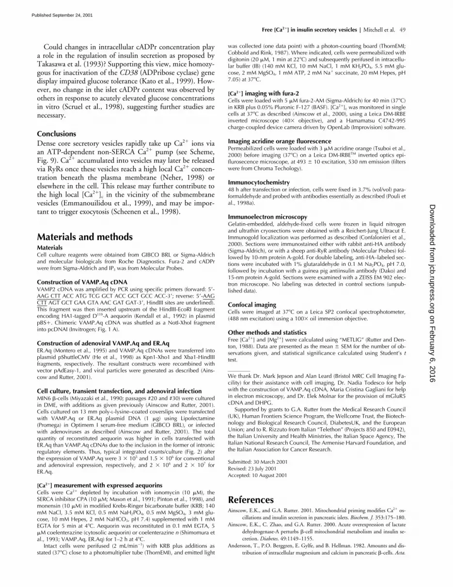
Figure 9. Scheme: redistribution of organellar Ca2� in secretory cells in response to G protein–coupled receptors (e.g., acetyl choline, AcCh) or glucose. IP3 generated in response to AcCh releases Ca2� from the endoplasmic reticulum and Golgi apparatus, leading to an increase in cytosolic [Ca2�] and uptake of Ca2� into dense core secretory vesicles distant from the plasma membrane (deep vesicles). Vesicular Ca2� uptake is catalyzed by an undefined Ca2�-ATPase, with properties similar to PMR1/ATP2C1 (see Discus-sion). Increases in blood glucose lead to (1) the uptake of the sugar via glucose transporters, (2) enhanced ATP synthesis and closure of ATP-sensitive K� channels, and (3) Ca2� influx through L-type Ca2� channels. The resultant increases in [Ca2�]c are likely to promote net Ca2� uptake (reflecting the balance of uptake versus release) into vesicles distant from the cell surface (deep vesicles). For those vesi-cles (�0.5% of total; Rorsman, 1997) located close to the plasma membrane and primed for exocytosis (primed vesicles), larger local [Ca2�]c increases (e.g., at the mouth of activated plasma membrane Ca2� channels; Neher, 1998) may activate vesicular RyRs and pro-voke net Ca2� release.
on February 6, 2016
jcb.rupress.orgD
ownloaded from
Published September 24, 2001
Free [Ca2�] in insulin secretory vesicles | Mitchell et al. 49
Could changes in intracellular cADPr concentration playa role in the regulation of insulin secretion as proposed byTakasawa et al. (1993)? Supporting this view, mice homozy-gous for inactivation of the CD38 (ADPribose cyclase) genedisplay impaired glucose tolerance (Kato et al., 1999). How-ever, no change in the islet cADPr content was observed byothers in response to acutely elevated glucose concentrationsin vitro (Scruel et al., 1998), suggesting further studies arenecessary.
ConclusionsDense core secretory vesicles rapidly take up Ca2� ions viaan ATP-dependent non-SERCA Ca2� pump (see Scheme,Fig. 9). Ca2� accumulated into vesicles may later be releasedvia RyRs once these vesicles reach a high local Ca2� concen-tration beneath the plasma membrane (Neher, 1998) orelsewhere in the cell. This release may further contribute tothe high local [Ca2�]c in the vicinity of the submembranevesicles (Emmanouilidou et al., 1999), and may be impor-tant to trigger exocytosis (Scheenen et al., 1998).
Materials and methodsMaterialsCell culture reagents were obtained from GIBCO BRL or Sigma-Aldrichand molecular biologicals from Roche Diagnostics. Fura-2 and cADPrwere from Sigma-Aldrich and IP3 was from Molecular Probes.
Construction of VAMP.Aq cDNAVAMP2 cDNA was amplified by PCR using specific primers (forward: 5�-AAG CTT ACC ATG TCG GCT ACC GCT GCC ACC-3�; reverse: 5�-AAGCTT AGT GCT GAA GTA AAC GAT GAT-3�, HindIII sites are underlined).This fragment was then inserted upstream of the HindIII-EcoRI fragmentencoding HA1-tagged D119-A aequorin (Kendall et al., 1992) in plasmidpBS�. Chimeric VAMP.Aq cDNA was shuttled as a NotI-XhoI fragmentinto pcDNAI (Invitrogen; Fig. 1 A).
Construction of adenoviral VAMP.Aq and ER.AqER.Aq (Montero et al., 1995) and VAMP.Aq cDNAs were transferred intoplasmid pShuttleCMV (He et al., 1998) as Kpn1-Xho1 and Xba1-HindIIIfragments, respectively. The resultant constructs were recombined withvector pAdEasy-1, and viral particles were generated as described (Ains-cow and Rutter, 2001).
Cell culture, transient transfection, and adenoviral infectionMIN6 �-cells (Miyazaki et al., 1990; passages #20 and #30) were culturedin DME, with additions as given previously (Ainscow and Rutter, 2001).Cells cultured on 13 mm poly-L-lysine–coated coverslips were transfectedwith VAMP.Aq or ER.Aq plasmid DNA (1 �g) using Lipofectamine(Promega) in Optimem I serum-free medium (GIBCO BRL), or infectedwith adenoviruses as described (Ainscow and Rutter, 2001). The totalquantity of reconstituted aequorin was higher in cells transfected withER.Aq than VAMP.Aq cDNAs due to the inclusion in the former of intronicregulatory elements. Thus, typical integrated counts/culture (Fig. 2) afterthe expression of VAMP.Aq were 3 � 105 and 1.5 � 106 for conventionaland adenoviral expression, respectively, and 2 � 106 and 2 � 107 forER.Aq.
[Ca2�] measurement with expressed aequorinsCells were Ca2� depleted by incubation with ionomycin (10 �M), theSERCA inhibitor CPA (10 �M; Mason et al., 1991; Pinton et al., 1998), andmonensin (10 �M) in modified Krebs-Ringer bicarbonate buffer (KRB; 140mM NaCl, 3.5 mM KCl, 0.5 mM NaH2PO4, 0.5 mM MgSO4, 3 mM glu-cose, 10 mM Hepes, 2 mM NaHCO3, pH 7.4) supplemented with 1 mMEGTA for 5 min at 4 C. Aequorin was reconstituted in 0.1 mM EGTA, 5�M coelenterazine (cytosolic aequorin) or coelenterazine n (Shimomura etal., 1993; VAMP.Aq, ER.Aq) for 1–2 h at 4 C.
Intact cells were perifused (2 mL/min�1) with KRB plus additions asstated (37 C) close to a photomultiplier tube (ThornEMI), and emitted light
was collected (one data point) with a photon-counting board (ThornEMI;Cobbold and Rink, 1987). Where indicated, cells were permeabilized withdigitonin (20 �M, 1 min at 22 C) and subsequently perifused in intracellu-lar buffer (IB) (140 mM KCl, 10 mM NaCl, 1 mM KH2PO4, 5.5 mM glu-cose, 2 mM MgSO4, 1 mM ATP, 2 mM Na� succinate, 20 mM Hepes, pH7.05) at 37 C.
[Ca2�] imaging with fura-2Cells were loaded with 5 �M fura-2-AM (Sigma-Aldrich) for 40 min (37 C)in KRB plus 0.05% Pluronic F-127 (BASF). [Ca2�]c was monitored in singlecells at 37 C as described (Ainscow et al., 2000), using a Leica DM-IRBEinverted microscope (40� objective), and a Hamamatsu C4742-995charge-coupled device camera driven by OpenLab (Improvision) software.
Imaging acridine orange fluorescencePermeabilized cells were loaded with 3 �M acridine orange (Tsuboi et al.,2000) before imaging (37 C) on a Leica DM-IRBETM inverted optics epi-flurosecence microscope, at 493 10 excitation, 530 nm emission (filterswere from Chroma Techology).
Immunocytochemistry48 h after transfection or infection, cells were fixed in 3.7% (vol/vol) para-formaldehyde and probed with antibodies essentially as described (Pouli etal., 1998a).
Immunoelectron microscopyGelatin-embedded, aldehyde-fixed cells were frozen in liquid nitrogenand ultrathin cryosections were obtained with a Reichert-Jung Ultracut E.Immunogold localization was performed as described (Confalonieri et al.,2000). Sections were immunostained either with rabbit anti-HA antibody(Sigma-Aldrich), or with a sheep anti-RyR antibody (Molecular Probes) fol-lowed by 10-nm protein A-gold. For double labeling, anti-HA–labeled sec-tions were incubated with 1% glutaraldehyde in 0.1 M Na2PO4, pH 7.0,followed by incubation with a guinea pig antiinsulin antibody (Dako) and15-nm protein A-gold. Sections were examined with a ZEISS EM 902 elec-tron microscope. No labeling was detected in control sections (unpub-lished data).
Confocal imagingCells were imaged at 37 C on a Leica SP2 confocal spectrophotometer,(488 nm excitation) using a 100� oil immersion objective.
Other methods and statisticsFree [Ca2�] and [Mg2�] were calculated using “METLIG” (Rutter and Den-ton, 1988). Data are presented as the mean SEM for the number of ob-servations given, and statistical significance calculated using Student’s ttest.
We thank Dr. Mark Jepson and Alan Leard (Bristol MRC Cell Imaging Fa-cility) for their assistance with cell imaging, Dr. Nadia Todesco for helpwith the construction of VAMP.Aq cDNA, Maria Cristina Gagliani for helpin electron microscopy, and Dr. Elek Molnar for the provision of mGluR5cDNA and DHPG.
Supported by grants to G.A. Rutter from the Medical Research Council(UK), Human Frontiers Science Program, the Wellcome Trust, the Biotech-nology and Biological Research Council, DiabetesUK, and the EuropeanUnion; and to R. Rizzuto from Italian “Telethon” (Projects 850 and E0942),the Italian University and Health Ministries, the Italian Space Agency, TheItalian National Research Council, The Armenise Harvard Foundation, andthe Italian Association for Cancer Research.
Submitted: 30 March 2001Revised: 23 July 2001Accepted: 10 August 2001
ReferencesAinscow, E.K., and G.A. Rutter. 2001. Mitochondrial priming modifies Ca2� os-
cillations and insulin secretion in pancreatic islets. Biochem. J. 353:175–180.Ainscow, E.K., C. Zhao, and G.A. Rutter. 2000. Acute overexpression of lactate
dehydrogenase-A perturbs �-cell mitochondrial metabolism and insulin se-cretion. Diabetes. 49:1149–1155.
Andersson, T., P.O. Berggren, E. Gylfe, and B. Hellman. 1982. Amounts and dis-tribution of intracellular magnesium and calcium in pancreatic �-cells. Acta.
on February 6, 2016
jcb.rupress.orgD
ownloaded from
Published September 24, 2001

50 The Journal of Cell Biology | Volume 155, Number 1, 2001
Physiol. Scand. 114:235–241.Andrews, S.B., and T.S. Reese. 1986. Intracellular structure and elemental analysis
in rapid-frozen neurons. Ann. NY Acad. Sci. 483:284–294.Ashcroft, F.M., and S.J.M. Ashcroft. 1992. Mechanisms of insulin secretion. In In-
sulin, Molecular Biology to Pathology. F.M. Ashcroft and S.J.H. Ashcroft,editors. Oxford University Press, Oxford, NY. 97–150.
Baird, G.S., D.A. Zacharias, and R.Y. Tsien. 1999. Circular permutation and re-ceptor insertion within green fluorescent proteins. Proc. Natl. Acad. Sci.USA. 96:11241–11246.
Berridge, M.J. 1993. Inositol trisphosphate and calcium signalling. Nature. 361:315–325.
Blinks, J.R. 1989. Use of calcium-regulated photoproteins as intracellular Ca2� in-dicators. Methods Enzymol. 172:164–203.
Blondel, O., M.M. Moody, A.M. Depaoli, A.H. Sharp, C.A. Ross, H. Swift, andG.I. Bell. 1994. Localization of inositol trisphosphate receptor subtype 3 toinsulin and somatostatin secretory granules and regulation of expression inislets and insulinoma cells. Proc. Natl. Acad. Sci. USA. 91:7777–7781.
Brini, M., M. Murgia, L. Pasti, D. Picard, T. Pozzan, and R. Rizzuto. 1993. Nu-clear Ca2� concentration measured with specifically targeted recombinantaequorin. EMBO J. 12:4813–4819.
Bryan, J., and L. AguilarBryan. 1997. The ABCs of ATP-sensitive potassium chan-nels: more pieces of the puzzle. Curr. Opin. Cell Biol. 9:553–559.
Carafoli, E. 1991. Calcium pump of the plasma membrane. Physiol. Rev. 71:129–153.
Chilcoat, N.D., S.M. Melia, A. Haddad, and A.P. Turkewitz. 1996. Granule lat-tice protein 1 (Grl1p), an acidic, calcium-binding protein in Tetrahymenathermophila dense-core secretory granules, influences granule size, shape,content organization, and release but not protein sorting or condensation. J.Cell Biol. 135:1775–1787.
Chini, E.N., and T.P. Dousa. 1996. Palmitoyl-CoA potentiates the Ca2� releaseelicited by cyclic ADP-ribose. Am. J. Physiol. 270:C530–C537.
Cobbold, P.H., and T.J. Rink. 1987. Fluorescence and bioluminescence measur-ment of cytoplasmic free calcium. Biochem. J. 248:313–328.
Confalonieri, S., A.E. Salcini, C. Puri, C. Tacchetti, and P.P. Di Fiore. 2000. Ty-rosine phosphorylation of Eps15 is required for ligand-regulated, but notconstitutive, endocytosis. J. Cell Biol. 150:905–912.
Davidson, H.W., C.J. Rhodes, and J.C. Hutton. 1988. Intraorganellar calcium andpH control proinsulin cleavage in the pancreatic � cell via two distinct site-specific endopeptidases. Nature. 333:93–96.
Emmanouilidou, E., A. Teschemacher, A.E. Pouli, L.I. Nicholls, E.P. Seward, andG.A. Rutter. 1999. Imaging [Ca2�] changes at the secretory vesicle surfacewith a recombinant targeted cameleon. Curr. Biol. 9:918.
Galione, A. 1994. Cyclic ADP-ribose, the ADP-ribosyl cyclase pathway and cal-cium signalling. Mol. Cell. Endocrinol. 98:125–131.
Gamberucci, A., R. Fulceri, W. Pralong, G. Banhegyi, P. Marcolongo, S.L. Wat-kins, and A. Benedetti. 1999. Caffeine releases a glucose-primed endoplas-mic reticulum Ca2� pool in the insulin secreting cell line INS-1. FEBS Lett.446:309–312.
Gerasimenko, O.V., J.V. Gerasimenko, P.V. Belan, and O.H. Petersen. 1996.Inositol trisphosphate and cyclic ADP-ribose-mediated release of Ca2� fromsingle isolated pancreatic zymogen granules. Cell. 84:473–480.
Gerasimenko, J.V., A.V. Tepikin, O.H. Petersen, and O.V. Gerasimenko. 1998.Calcium uptake via endocytosis with rapid release from acidifying endo-somes. Curr. Biol. 8:1335–1338.
Goncalves, P.P., S.M. Meireles, C. Gravato, and M.G. Vale. 1998. Ca2�-H� anti-port activity in synaptic vesicles isolated from sheep brain cortex. Neurosci.Lett. 247:87–90.
Grapengiesser, E., E. Gylfe, and B. Hellman. 1988. Glucose-induced oscillations ofcytoplasmic Ca2� in the pancreatic �-cell. Biochem. Biophys. Res. Commun.151:1299–1304.
Grinstein, S., W. Furuya, M.J. Vander, and R.G. Hancock. 1983. The total andfree concentrations of Ca2� and Mg2� inside platelet secretory granules.Measurements employing a novel double null point technique. J. Biol.Chem. 258:14774–14777.
He, T.C., S. Zhou, L.T. da Costa, J. Yu, K.W. Kinzler, and B. Vogelstein. 1998. Asimplified system for generating recombinant adenoviruses. Proc. Natl. Acad.Sci. USA. 95:2509–2514.
Hellman, B., J. Sehlin, and I.B. Taljedal. 1976. Calcium and secretion: distinctionbetween two pools of glucose-sensitive calcium in pancreatic islets. Science.194:1421–1423.
Holz, G.G., C.A. Leech, R.S. Heller, M. Castonguay, and J.F. Habener. 1999.cAMP-dependent mobilization of intracellular Ca2� stores by activation of
ryanodine receptors in pancreatic �-cells. A Ca2� signaling system stimu-lated by the insulinotropic hormone glucagon-like peptide-1-(7-37). J. Biol.Chem. 274:14147–14156.
Howell, S.L., W. Montague, and M. Tyhurst. 1975. Calcium distribution in isletsof Langerhans: a study of calcium concentrations and of calcium accumula-tion in B cell organelles. J. Cell Sci. 19:395–409.
Hu, Z., J.M. Bonifas, J. Beech, G. Bench, T. Shigihara, H. Ogawa, S. Ikeda, T.Mauro, and E.H. Epstein, Jr. 2000. Mutations in ATP2C1, encoding a cal-cium pump, cause Hailey-Hailey disease. Nat. Genet. 24:61–65.
Hutton, J.C., E.J. Penn, and M. Peshavaria. 1983. Low-molecular-weight constitu-ents of isolated insulin-secretory vesicles. Bivalent cations, adenine nucle-otides and inorganic phosphate. Biochem. J. 210:297–305.
Inouye, S., M. Noguchi, Y. Sakaki, Y. Takagi, T. Miyata, S. Iwanaga, T. Miyata,and F.I. Tsuji. 1985. Cloning and sequence analysis of cDNA for the lumi-nescent protein aequorin. Proc. Natl. Acad. Sci. USA. 82:3154–3158.
Islam, M.S., O. Larsson, and P.O. Berggren. 1993. Cyclic ADP-ribose and pancre-atic � cells. Science. 262:1499.
Islam, M.S., I. Leibiger, B. Leibiger, D. Rossi, V. Sorrentino, T.J. Ekstrom, H.Westerblad, F.H. Andrade, and P.O. Berggren. 1998. In situ activation ofthe type 2 ryanodine receptor in pancreatic � cells requires cAMP-depen-dent phosphorylation. Proc. Natl. Acad. Sci. USA. 95:6145–6150.
Kato, I., Y. Yamamoto, M. Fujimura, N. Noguchi, S. Takasawa, and H. Okamoto.1999. CD38 disruption impairs glucose-induced increases in cyclic ADP-ribose, [Ca2�]i, and insulin secretion. J. Biol. Chem. 274:1869–1872.
Kendall, J.M., G. SalaNewby, V. Ghalaut, R.L. Dormer, and A.K. Campbell.1992. Engineering the Ca2� activated photoprotein aequorin with reducedaffinity for calcium. Biochem. Biophys. Res. Commun. 187:1091–1097.
Kennedy, H.J., A.E. Pouli, L.S. Jouaville, R. Rizzuto, and G.A. Rutter. 1999. Glu-cose-induced ATP microdomains in single islet �-cells. J. Biol. Chem. 274:13281–13291.
Krieger-Brauer, H., and M. Gratzl. 1982. Uptake of Ca2� by isolated secretory ves-icles from adrenal medulla. Biochim. Biophys. Acta. 691:61–70.
Lai, F.A., H.P. Erickson, E. Rousseau, Q.Y. Liu, and G. Meissner. 1988. Purifica-tion and reconstitution of the calcium release channel from skeletal muscle.Nature. 331:315–319.
Maechler, P., E.D. Kennedy, E. Sebo, A. Valeva, T. Pozzan, and C.B. Wollheim.1999. Secretagogues modulate the calcium concentration in the endoplasmicreticulum of insulin-secreting cells. Studies in aequorin-expressing intact andpermeabilized INS-1 cells. J. Biol. Chem. 274:12583–12592.
Mason, M.J., C. Garcia-Rodriguez, and S. Grinstein. 1991. Coupling between in-tracellular Ca2� stores and the Ca2� permeability of the plasma membrane.Comparison of the effects of thapsigargin, 2,5-di-(tert-butyl)-1,4-hydro-quinone, and cyclopiazonic acid in rat thymic lymphocytes. J. Biol. Chem.266:20856–20862.
Miesenbock, G., D.A. De Angelis, and J.E. Rothman. 1998. Visualizing secretionand synaptic transmission with pH-sensitive green fluorescent proteins. Na-ture. 394:192–195.
Mikoshiba, K. 1997. The InsP3 receptor and intracellular Ca2� signaling. Curr.Opin. Neurobiol. 7:339–345.
Miyawaki, A., J. Llopis, R. Heim, J.M. McCaffery, J.A. Adams, M. Ikura, and R.Y.Tsien. 1997. Fluorescent indicators for Ca2� based on green fluorescent pro-teins and calmodulin. Nature. 388:882–887.
Miyazaki, J., K. Araki, E. Yamato, H. Ikegami, T. Asano, Y. Shibasaki, Y. Oka, andK. Yamamura. 1990. Establishment of a pancreatic � cell line that retainsglucose inducible insulin secretion: special reference to expression of glucosetransporter isoforms. Endocrinology. 127:126–132.
Moller, J.V., B. Juul, and M. le Maire. 1996. Structural organization, ion trans-port, and energy transduction of P-type ATPases. Biochim. Biophys. Acta.1286:1–51.
Montero, M., M. Brini, R. Marsault, J. Alvarez, R. Sitia, T. Pozzan, and R. Riz-zuto. 1995. Monitoring dynamic changes in free Ca2� concentration in theendoplasmic reticulum of intact cells. EMBO J. 14:5467–5475.
Montero, M., J. Alvarez, W.J. Scheenen, R. Rizzuto, J. Meldolesi, and T. Pozzan.1997. Ca2� homeostasis in the endoplasmic reticulum: coexistence of highand low [Ca2�] subcompartments in intact HeLa cells. J. Cell Biol. 139:601–611.
Neher, E. 1998. Vesicle pools and Ca2� microdomains: new tools for understand-ing their roles in neurotransmitter release. Neuron. 20:389–399.
Nguyen, T., W.C. Chin, and P. Verdugo. 1998. Role of Ca2�/K� ion exchange inintracellular storage and release of Ca2�. Nature. 395:908–912.
Nicaise, G., K. Maggio, S. Thirion, M. Horoyan, and E. Keicher. 1992. The cal-cium loading of secretory granules. A possible key event in stimulus-secre-
on February 6, 2016
jcb.rupress.orgD
ownloaded from
Published September 24, 2001

Free [Ca2�] in insulin secretory vesicles | Mitchell et al. 51
tion coupling. Biol. Cell. 75:89–99.Orci, L., M. Ravazzola, M. Amherdt, O. Madsen, J.D. Vassalli, and A. Perrelet.
1985. Direct identification of prohormone conversion site in insulin-secret-ing cells. Cell. 42:671–681.
Palmieri, R., R.W. Lee, and M.F. Dunn. 1988. 1H Fourier transform NMR stud-ies of insulin: coordination of Ca2� to the Glu(B13) site drives hexamer as-sembly and induces a conformation change. Biochemistry. 27:3387–3397.
Parekh, A.B., and R. Penner. 1997. Store depletion and calcium influx. Physiol.Rev. 77:901–930.
Pezzati, R., M. Bossi, P. Podini, J. Meldolesi, and F. Grohovaz. 1997. High-resolu-tion calcium mapping of the endoplasmic reticulum-Golgi-exocytic mem-brane system. Electron energy loss imaging analysis of quick frozen-freezedried PC12 cells. Mol. Biol. Cell. 8:1501–1512.
Pinton, P., T. Pozzan, and R. Rizzuto. 1998. The Golgi apparatus is an inositol1,4,5-trisphosphate-sensitive Ca2� store, with functional properties distinctfrom those of the endoplasmic reticulum. EMBO J. 17:5298–5308.
Pouli, A.E., H.J. Kennedy, J.G. Schofield, and G.A. Rutter. 1998a. Insulin target-ing to the regulated secretory pathway after fusion with green fluorescentprotein and firefly luciferase. Biochem. J. 331:669–675.
Pouli, A.E., E. Emmanouilidou, C. Zhao, C. Wasmeier, J.C. Hutton, and G.A.Rutter. 1998b. Secretory-granule dynamics visualized in vivo with a phogringreen fluorescent protein chimaera. Biochem. J. 333:193–199.
Pozzan, T., R. Rizzuto, P. Volpe, and J. Meldolesi. 1994. Molecular and cellularphysiology of intracellular calcium stores. Physiol. Rev. 74:595–636.
Prentki, M., D. Janjic, T.J. Biden, B. Blondel, and C.B. Wollheim. 1984. Regula-tion of Ca2� transport by isolated organelles of a rat insulinoma. Studieswith endoplasmic reticulum and secretory granules. J. Biol. Chem. 259:10118–10123.
Ravazzola, M., P.A. Halban, and L. Orci. 1996. Inositol 1,4,5-trisphosphate recep-tor subtype 3 in pancreatic islet cell secretory granules revisited. Proc. Natl.Acad. Sci. USA. 93:2745–2748.
Reetz, A., M. Solimena, M. Matteoli, F. Folli, K. Takei, and P. De Camilli. 1991.GABA and pancreatic B-cells: colocalization of glutamic acid decarboxylase(GAD) and GABA with synaptic-like microvesicles suggests their role inGABA storage and secretion. EMBO J. 10:1275–1284.
Rizzuto, R., M. Brini, M. Murgia, and T. Pozzan. 1993. Microdomains with highCa2� close to IP3-sensitive channels that are sensed by neighbouring mito-chondria. Science. 262:744–747.
Rizzuto, R., M. Brini, C. Bastianutto, R. Marsault, and T. Pozzan. 1995. Photo-protein-mediated measurement of calcium ion concentration in mitochon-dria of living cells. Methods Enzymol. 260:417–428.
Rorsman, P. 1997. The pancreatic �-cell as a fuel sensor: an electrophysiologist’sviewpoint. Diabetologia. 40:487–495.
Rutter, G.A., and R.M. Denton. 1988. Regulation of NAD�-linked isocitratedehydrogenase and 2-oxoglutarate dehydrogenase by Ca2� ions within tolu-ene-permeabilized rat heart mitochondria. Interactions with regulation byadenine nucleotides and NADH/NAD� ratios. Biochem. J. 252:181–189(erratum published 253:935).
Rutter, G.A., J.-M. Theler, M. Murghia, C.B. Wollheim, T. Pozzan, and R. Riz-zuto. 1993. Stimulated Ca2� influx raises mitochondrial free Ca2� to su-pramicromolar levels in a pancreatic �-cell line: possible role in glucose andagonist-induced insulin secretion. J. Biol. Chem. 268:22385–22390.
Rutter, G.A., J.-M. Theler, G. Li, and C.B. Wollheim. 1994. Ca2� stores in insu-lin-secreting cells: lack of effect of cADP ribose. Cell Calcium. 16:71–80.
Rutter, G.A., C. Fasolato, and R. Rizzuto. 1998. Calcium and organelles: a two-sided story. Biochem Biophys. Res. Commun. 253:549–557.
Safayhi, H., H. Haase, U. Kramer, A. Bihlmayer, M. Roenfeldt, H.P. Ammon, M.Froschmayr, T.N. Cassidy, I. Morano, M.K. Ahlijanian, and J. Striessnig.1997. L-type calcium channels in insulin-secreting cells: biochemical charac-terization and phosphorylation in RINm5F cells. Mol. Endocrinol. 11:619–
629.Scheenen, W.M., C.B. Wollheim, T. Pozzan, and C. Fasolato. 1998. Ca2� deple-
tion from granules inhibits exocytosis. A study with insulin-secreting cells. J.Biol. Chem. 273:19002–19008.
Scruel, O., T. Wada, K. Kontani, A. Sener, T. Katada, and W.J. Malaisse. 1998.Effects of D-glucose and starvation upon the cyclic ADP-ribose content ofrat pancreatic islets. Biochem. Mol. Biol. Int. 45:783–790.
Shimomura, O., B. Musicki, Y. Kishi, and S. Inouye. 1993. Light-emitting proper-ties of recombinant semi-synthetic aequorins and recombinant fluorescein-conjugated aequorin for measuring cellular calcium. Cell Calcium. 14:373–378.
Sorin, A., G. Rosas, and R. Rao. 1997. PMR1, a Ca2�-ATPase in yeast Golgi, hasproperties distinct from sarco/endoplasmic reticulum and plasma membranecalcium pumps. J. Biol. Chem. 272:9895–9901.
Streb, H., E. Bayerdorffer, W. Haase, R.F. Irvine, and I. Schulz. 1984. Effect ofinositol-1,4,5-trisphosphate on isolated subcellular fractions of rat pancreas.J. Membr. Biol. 81:241–253.
Sudhof, T.C., M. Baumert, M.S. Perin, and R. Jahn. 1989. A synaptic vesiclemembrane protein is conserved from mammals to Drosophila. Neuron.2:1475–1481.
Takasawa, S., K. Nata, H. Yonekura, and H. Okamoto. 1993. Cyclic ADP-ribosein insulin secretion from pancreatic B-cells. Science. 259:370–373.
Takeshima, H., S. Nishimura, T. Matsumoto, H. Ishida, K. Kangawa, N. Mi-namino, H. Matsuo, M. Ueda, M. Hanaoka, T. Hirose, et al. 1989. Primarystructure and expression from complementary DNA of skeletal muscle ryan-odine receptor. Nature. 339:439–445.
Thastrup, O. 1990. Role of Ca2�-ATPases in regulation of cellular Ca2� signalling,as studied with selective microsomal Ca2�-ATPase inhibitor, thapsigargin.Agents Actions. 29:8–15.
Thirion, S., J.D. Troadec, N.B. Pivovarova, S. Pagnotta, S.B. Andrews, R.D. Leap-man, and G. Nicaise. 1999. Stimulus-secretion coupling in neurohypophys-ial nerve endings: a role for intravesicular sodium? Proc. Natl. Acad. Sci.USA. 96:3206–3210.
Thomas, L.S., D.E. Jane, R. Harris, and M.J. Croucher. 2000. Metabotropicglutamate autoreceptors of the mGlu(5) subtype positively modulate neu-ronal glutamate release in the rat forebrain in vitro. Neuropharmacology. 39:1554–1566.
Troadec, J.D., S. Thirion, J.P. Laugier, and G. Nicaise. 1998. Calcium-inducedcalcium increase in secretory vesicles of permeabilized rat neurohypophysialnerve terminals. Biol. Cell. 90:339–347.
Tsuboi, T., C. Zhao, S. Terakawa, and G.A. Rutter. 2000. Simultaneous evanes-cent wave imaging of insulin vesicle membrane and cargo during a singleexocytotic event. Curr. Biol. 10:1307–1310.
Varadi, A., and G.A. Rutter. 2001. Dynamic imaging of endoplasmic reticulum[Ca2�] in MIN6 cells using recombinant cameleons: roles of SERCA2 andryanodine receptors. Diabetes. In press.
Yoo, S.H. 2000. Coupling of the IP3 receptor/Ca2� channel with Ca2� storage pro-teins chromogranins A and B in secretory granules. Trends Neurosci. 23:424–428.
Yoo, S.H., and J.P. Albanesi. 1990. Inositol 1,4,5-trisphosphate-triggered Ca2� re-lease from bovine adrenal medullary secretory vesicles. J. Biol. Chem. 265:13446–13448.
Yule, D.I., S.A. Ernst, H. Ohnishi, and R.J.H. Wojcikiewicz. 1997. Evidence thatzymogen granules are not a physiologically relevant calcium pool. Definingthe distribution of inositol 1,4,5-trisphosphate receptors in pancreatic acinarcells. J. Biol. Chem. 272:9093–9098.
Zorzato, F., E. Scutari, V. Tegazzin, E. Clementi, and S. Treves. 1993. Chlorocre-sol: an activator of ryanodine receptor-mediated Ca2� release. Mol. Pharma-col. 44:1192–1201.
on February 6, 2016
jcb.rupress.orgD
ownloaded from
Published September 24, 2001


