Embed Size (px)
Citation preview
DENTAL AND MEDICAL JOURNAL - REVIEW
e-ISSN 2667-7288 Vol 3, Issue 2, (2021) Review Article / Derleme Makale
Dent & Med J - R http://www.dergipark.org.tr/dmj
66
IMPACTED THIRD MOLAR: TO EXTRACT OR NOT TO EXTRACT, THAT IS THE QUESTION…
GÖMÜLÜ ÜÇÜNCÜ MOLAR: ÇEKMEK YA DA ÇEKMEMEK, İŞTE BÜTÜN MESELE…
Melda Pelin AKKITAP1, Birsay GUMRU2
1 Res. Assist. Dt., Department of Oral and Maxillofacial Radiology, Marmara University Faculty of Dentistry, Istanbul/TURKEY
ORCID ID: 0000-0001-7744-6615
2 Assoc. Prof. (DDS, PhD), Department of Oral and Maxillofacial Radiology, Marmara University Faculty of Dentistry, Istanbul/TURKEY
ORCID ID: 0000-0002-7734-4755
Corresponding Author:
Assoc. Prof. Birsay Gumru (DDS, PhD)
Marmara Üniversitesi, Diş Hekimliği Fakültesi, Ağız, Diş ve Çene Radyolojisi Anabilim Dalı, Başıbüyük Mah. Başıbüyük Yolu
Sok. 9/3 34854 Başıbüyük, Maltepe/İstanbul
E-mail: [email protected], Tel: +90 (216) 421 1621 Mobile: +90 (506) 535 8732
Article Info / Makale Bilgisi
Received / Teslim: May 20, 2021
Accepted / Kabul: May 31, 2021
Online Published / Yayınlanma: June 30, 2021
DOI:
Akkıtap MP, Gumru B. Impacted Third Molar: To Extract or Not To Extract, That Is The Question…. Dent & Med J - R. 2021;3(2):66-82.
DENTAL AND MEDICAL JOURNAL - REVIEW
e-ISSN 2667-7288 Vol 3, Issue 2, (2021) Review Article / Derleme Makale
Dent & Med J - R http://www.dergipark.org.tr/dmj
67
Abstract
The third molars are the last teeth that erupt in the oral cavity and are also the most commonly impacted
teeth in the jaws. Management of the impacted third molars by dental professionals is based on individual
assessment of the clinical symptoms and the radiological findings indicative of oral pathology. Although
extraction is definitely indicated in case of a pathological condition due to impacted third molars, no consensus
has yet been reached on the management of asymptomatic third molars. Many studies revealed that the
decision of whether or not to extract the asymptomatic impacted third molars was a challenging and
controversial issue among professionals, and that accompanying pathological conditions as well as
postoperative complications associated with the extraction of the impacted third molars played a critical role
in this decision.
In this review, the clinical and radiological evaluation, classification, associated pathologies, and
management of the impacted third molars are discussed comprehensively, and it is aimed to assist dental
professionals in the decision of prophylactic extraction of the third molars as well as to provide guidance in the
treatment planning of the associated pathologies.
Keywords: impacted third molar, asymptomatic, extraction, pathology.
Özet
Üçüncü molar dişler; oral kavitede en son süren ve aynı zamanda çenelerde en sık gömülü kalan
dişlerdir. Gömülü üçüncü molar dişler için diş hekimlerinin yaklaşımı oral patolojinin göstergesi olan klinik
semptomlar ve radyolojik bulguların değerlendirilmesine dayanmaktadır. Gömülü üçüncü molar dişlere bağlı
patoloji mevcut olması durumunda çekim kesinlikle endike iken, asemptomatik üçüncü molar dişlerin tedavisi
konusunda henüz bir fikir birliğine varılamamıştır. Birçok çalışma asemptomatik gömülü üçüncü molar dişlerin
çekilip çekilmemesi kararının diş hekimleri için zorlayıcı ve tartışmalı bir konu olduğunu ve eşlik eden patolojik
durumların yanı sıra gömülü üçüncü molar dişlerin çekimi ile ilişkili postoperatif komplikasyonların bu kararda
kritik bir rol oynadığını ortaya koymuştur.
Bu derlemede, gömülü üçüncü molar dişlerin klinik ve radyolojik değerlendirmesi, sınıflandırması, ilişkili
patolojileri ve tedavi yaklaşımları kapsamlı bir şekilde tartışılmakta, profilaktik çekim kararı konusunda
hekimlere yardımcı olmak ve ilişkili patolojilerin tedavi planlamasında rehberlik sağlamak amaçlanmaktadır.
Anahtar Kelimeler: gömülü üçüncü molar diş, asemptomatik, çekim, patoloji.
DENTAL AND MEDICAL JOURNAL - REVIEW
e-ISSN 2667-7288 Vol 3, Issue 2, (2021) Review Article / Derleme Makale
Dent & Med J - R http://www.dergipark.org.tr/dmj
68
OVERVIEW / GENEL BAKIŞ
Surgical removal of the third molars, which are the most commonly impacted teeth (16.7-68.6% of all
impacted teeth), was cited as one of the most frequent surgical procedures performed by oral and maxillofacial
surgeons worldwide (1,2).
Third molars may appear on radiographs at the earliest at 5 and at the latest at 16 years of age, and are
expected to erupt between the ages of 18 and 24 (3,4). However, they may fail to erupt within the expected
time due to local factors (lack of space, retained/ankylosed deciduous teeth, premature loss of deciduous teeth,
supernumerary teeth, ectopic positioning of tooth buds, obstruction of eruption path, infection, trauma,
cyst/tumour, dilaceration), systemic factors (malnutrition, radiation, anaemia, rickets, vitamin D deficiency,
cretinism, endocrine diseases, cleft palate, specific infections such as syphilis and tuberculosis, specific
syndromes such as cleidocranial dysostosis, oxycephaly, progeria, and achondroplasia) and also genetic factors
(1,4).
Mandibular third molars are presented to have a higher impaction incidence in comparison to their
maxillary counterparts, and the tendency of third molar impaction is reported to be higher in females compared
to males (1,5).
Most impacted third molars may remain asymptomatic throughout life, whereas some may cause various
pathological conditions including pericoronitis, dental caries, pulpitis, periodontitis, marginal bone loss, external
root resorption (ERR), neuropathic pain, odontogenic cyst/tumour, mandibular fracture, and late anterior
crowding depending on factors such as eruption status and position.
While the indication for both “disease-positive and symptom-positive” or “disease-positive and symptom-
free” impacted third molars is therapeutic removal, the necessity of prophylactic removal of “disease-free and
symptom-free” impacted third molars is still controversial (2,6). A large number of asymptomatic third molars
are extracted without any pathological signs. Some researchers argued that prophylactic removal was not
suitable for the asymptomatic impacted third molars (2,6). In this context, the decision of extraction should
not be generalized and the necessity should be assessed individually for each case.
In this review, the clinical and radiographic assessment, classification, associated pathologies, and
management of the impacted third molars are discussed and it is aimed to assist dental professionals in the
decision of prophylactic extraction and to guide the treatment planning of associated pathologies.
CLINICAL AND RADIOGRAPHIC ASSESSMENT
Assessment of impacted third molars is performed by clinical and radiographic evaluations. The clinical
evaluation includes inspection and palpation of the temporomandibular joint (TMJ) and movement of the
mandible, determination of mobility characteristics of lips and cheeks, size and contours of the tongue, and
appearance of soft tissue overlying the impacted teeth (4,7). Radiographic assessment of the impacted third
molars includes relationship with the adjacent teeth, size of the follicular sac, morphology of the root, density
DENTAL AND MEDICAL JOURNAL - REVIEW
e-ISSN 2667-7288 Vol 3, Issue 2, (2021) Review Article / Derleme Makale
Dent & Med J - R http://www.dergipark.org.tr/dmj
69
of the surrounding bone, relation to the inferior alveolar nerve/vessels, the bucco-lingual position, relationship
with the mandibular corpus/ramus, and possible presence of pathologies (4,7,8).
Panoramic radiography (OPTG) is preferred as the primary imaging modality in assessment of the
impacted third molars and associated pathologies. Although OPTG helps diagnosis, follow-up of tooth eruption,
and evaluation of treatment results, it has the potential to be misinterpreted due to reasons such as
magnification, distortion, and blurring and may not be sufficient in most cases with pathologies including caries,
marginal bone loss, and ERR due to the superimposition of anatomical structures and adjacent teeth to the
relevant area (9). On the other hand, cone beam computed tomography (CBCT) supplies more accurate
diagnostic data in terms of relationship of the impacted tooth with the adjacent tooth, the nasal floor, the
maxillary sinus, and the mandibular canal without overlapping of the anatomical structures in three dimensions
(10).
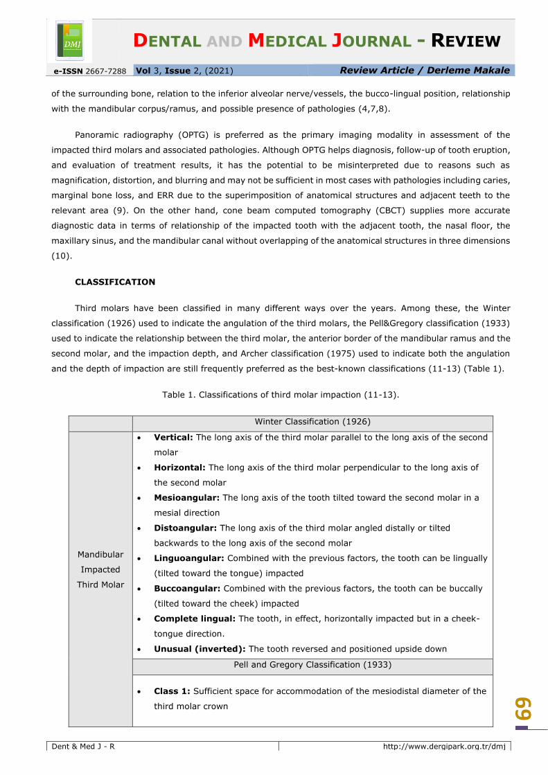
CLASSIFICATION
Third molars have been classified in many different ways over the years. Among these, the Winter
classification (1926) used to indicate the angulation of the third molars, the Pell&Gregory classification (1933)
used to indicate the relationship between the third molar, the anterior border of the mandibular ramus and the
second molar, and the impaction depth, and Archer classification (1975) used to indicate both the angulation
and the depth of impaction are still frequently preferred as the best-known classifications (11-13) (Table 1).
Table 1. Classifications of third molar impaction (11-13).
Winter Classification (1926)
Mandibular
Impacted
Third Molar
• Vertical: The long axis of the third molar parallel to the long axis of the second
molar
• Horizontal: The long axis of the third molar perpendicular to the long axis of
the second molar
• Mesioangular: The long axis of the tooth tilted toward the second molar in a
mesial direction
• Distoangular: The long axis of the third molar angled distally or tilted
backwards to the long axis of the second molar
• Linguoangular: Combined with the previous factors, the tooth can be lingually
(tilted toward the tongue) impacted
• Buccoangular: Combined with the previous factors, the tooth can be buccally
(tilted toward the cheek) impacted
• Complete lingual: The tooth, in effect, horizontally impacted but in a cheek-
tongue direction.
• Unusual (inverted): The tooth reversed and positioned upside down
Pell and Gregory Classification (1933)
• Class 1: Sufficient space for accommodation of the mesiodistal diameter of the
third molar crown
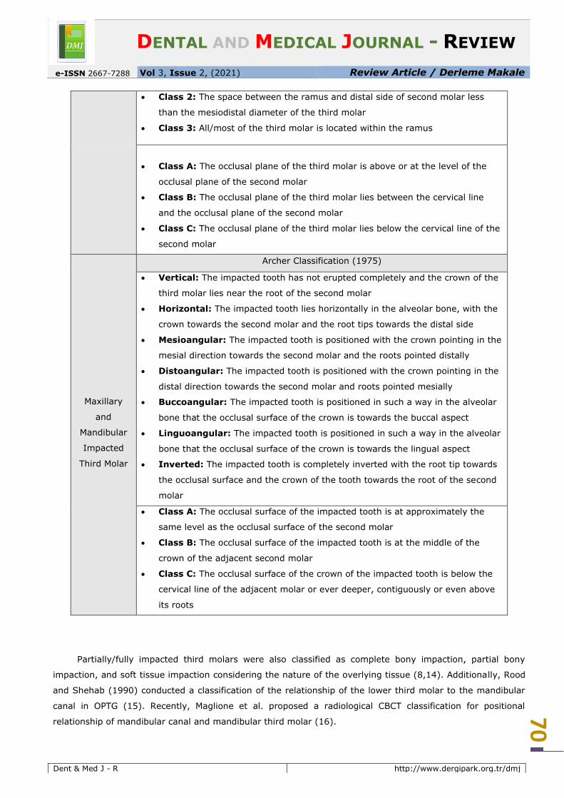
DENTAL AND MEDICAL JOURNAL - REVIEW
e-ISSN 2667-7288 Vol 3, Issue 2, (2021) Review Article / Derleme Makale
Dent & Med J - R http://www.dergipark.org.tr/dmj
70
• Class 2: The space between the ramus and distal side of second molar less
than the mesiodistal diameter of the third molar
• Class 3: All/most of the third molar is located within the ramus
• Class A: The occlusal plane of the third molar is above or at the level of the
occlusal plane of the second molar
• Class B: The occlusal plane of the third molar lies between the cervical line
and the occlusal plane of the second molar
• Class C: The occlusal plane of the third molar lies below the cervical line of the
second molar
Maxillary
and
Mandibular
Impacted
Third Molar
Archer Classification (1975)
• Vertical: The impacted tooth has not erupted completely and the crown of the
third molar lies near the root of the second molar
• Horizontal: The impacted tooth lies horizontally in the alveolar bone, with the
crown towards the second molar and the root tips towards the distal side
• Mesioangular: The impacted tooth is positioned with the crown pointing in the
mesial direction towards the second molar and the roots pointed distally
• Distoangular: The impacted tooth is positioned with the crown pointing in the
distal direction towards the second molar and roots pointed mesially
• Buccoangular: The impacted tooth is positioned in such a way in the alveolar
bone that the occlusal surface of the crown is towards the buccal aspect
• Linguoangular: The impacted tooth is positioned in such a way in the alveolar
bone that the occlusal surface of the crown is towards the lingual aspect
• Inverted: The impacted tooth is completely inverted with the root tip towards
the occlusal surface and the crown of the tooth towards the root of the second
molar
• Class A: The occlusal surface of the impacted tooth is at approximately the
same level as the occlusal surface of the second molar
• Class B: The occlusal surface of the impacted tooth is at the middle of the
crown of the adjacent second molar
• Class C: The occlusal surface of the crown of the impacted tooth is below the
cervical line of the adjacent molar or ever deeper, contiguously or even above
its roots
Partially/fully impacted third molars were also classified as complete bony impaction, partial bony
impaction, and soft tissue impaction considering the nature of the overlying tissue (8,14). Additionally, Rood
and Shehab (1990) conducted a classification of the relationship of the lower third molar to the mandibular
canal in OPTG (15). Recently, Maglione et al. proposed a radiological CBCT classification for positional
relationship of mandibular canal and mandibular third molar (16).
DENTAL AND MEDICAL JOURNAL - REVIEW
e-ISSN 2667-7288 Vol 3, Issue 2, (2021) Review Article / Derleme Makale
Dent & Med J - R http://www.dergipark.org.tr/dmj
71
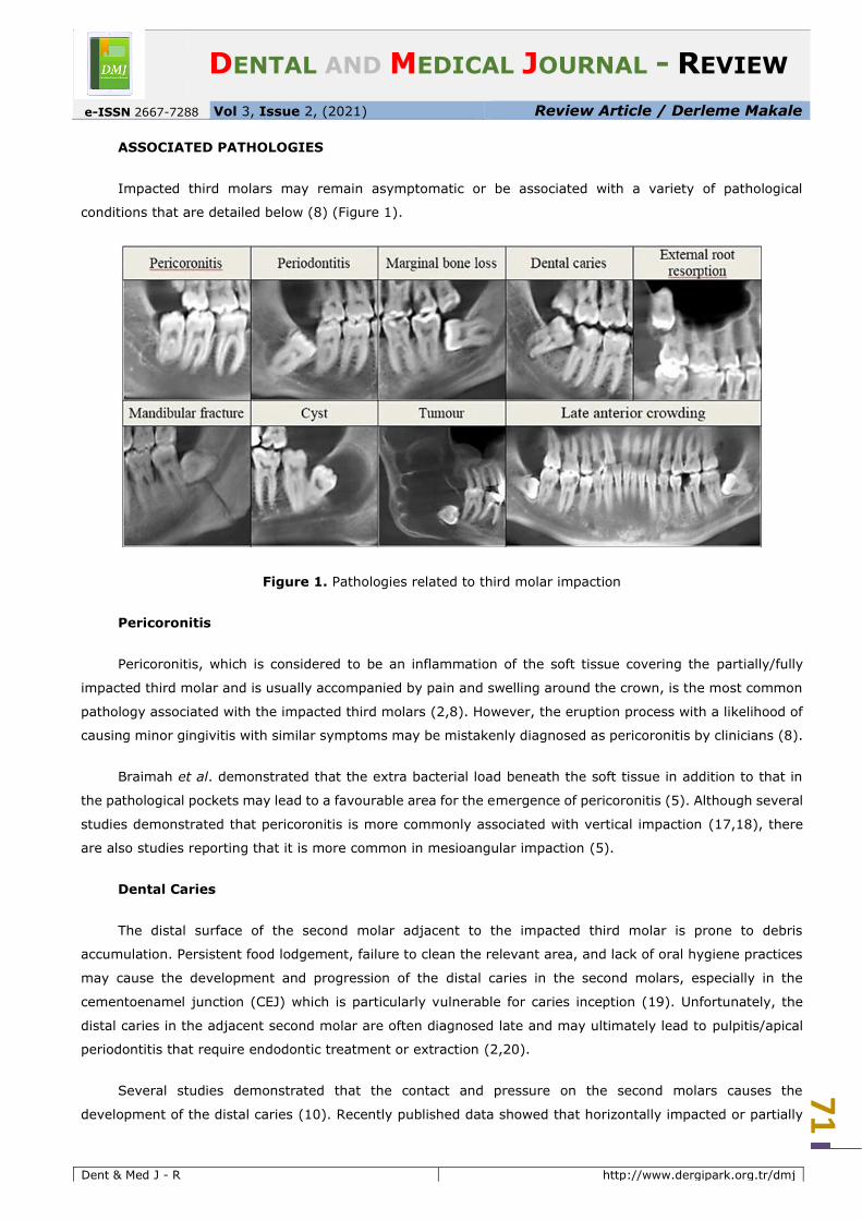
ASSOCIATED PATHOLOGIES
Impacted third molars may remain asymptomatic or be associated with a variety of pathological
conditions that are detailed below (8) (Figure 1).
Figure 1. Pathologies related to third molar impaction
Pericoronitis
Pericoronitis, which is considered to be an inflammation of the soft tissue covering the partially/fully
impacted third molar and is usually accompanied by pain and swelling around the crown, is the most common
pathology associated with the impacted third molars (2,8). However, the eruption process with a likelihood of
causing minor gingivitis with similar symptoms may be mistakenly diagnosed as pericoronitis by clinicians (8).
Braimah et al. demonstrated that the extra bacterial load beneath the soft tissue in addition to that in
the pathological pockets may lead to a favourable area for the emergence of pericoronitis (5). Although several
studies demonstrated that pericoronitis is more commonly associated with vertical impaction (17,18), there
are also studies reporting that it is more common in mesioangular impaction (5).
Dental Caries
The distal surface of the second molar adjacent to the impacted third molar is prone to debris
accumulation. Persistent food lodgement, failure to clean the relevant area, and lack of oral hygiene practices
may cause the development and progression of the distal caries in the second molars, especially in the
cementoenamel junction (CEJ) which is particularly vulnerable for caries inception (19). Unfortunately, the
distal caries in the adjacent second molar are often diagnosed late and may ultimately lead to pulpitis/apical
periodontitis that require endodontic treatment or extraction (2,20).
Several studies demonstrated that the contact and pressure on the second molars causes the
development of the distal caries (10). Recently published data showed that horizontally impacted or partially
DENTAL AND MEDICAL JOURNAL - REVIEW
e-ISSN 2667-7288 Vol 3, Issue 2, (2021) Review Article / Derleme Makale
Dent & Med J - R http://www.dergipark.org.tr/dmj
72
erupted mesioangular third molars having contact points at/below the CEJ of the second molars pose a higher
risk of causing distal caries compared to those with contact points above the CEJ (20). Besides, Chen et al.
suggested that food impaction between the second and third molars may occur more commonly and cleaning
may be more demanding when the third molar is either buccally or lingually dispositioned (21).
Periodontitis
The impacted third molars have been recognized as a high risk for periodontal pathology affecting the
distal aspect of the adjacent second molars (22). Blakey et al. reported significant improvement in the
periodontal status of the distal aspect of the second molars as well as in the overall periodontal health with
removal of the impacted third molars (23). On the contrary, some studies suggested that the impacted third
molar removal has a negative impact on the periodontal status of the second molar (24).
Partially impacted third molars are reported to constitute a serious periodontal risk factor (25). It was
suggested that the surgical removal of a partially impacted tooth usually results in greater weakening of the
periodontal tissues distal to the second molar compared to a fully impacted tooth (26). Furthermore,
mesioangular impaction, which creates an area difficult to clean properly and more suitable for food entrapment
and bacterial colonization, was reported to be mostly associated with periodontitis (17).
Marginal bone loss
Marginal bone loss on the distal aspect of the adjacent second molar plays an important part in predicting
the prognosis of the tooth and deciding on surgical intervention. More specifically, regarding the prognosis of
the second molars, both horizontal and mesioangular positions of the impacted third molars were reported to
increase the risk of adjacent bone loss. Moreover, a study evaluating the effect of age and gender on bone loss
in the second molars adjacent to the impacted third molars revealed that age was an important consideration
in patients with severe bone loss, but gender was not reported to play a statistically significant role (22).
External root resorption
ERR in the contact area of the second molar with the adjacent impacted third molar results from the
mechanical pressure (27). Several studies showed that the eruption movement of the tooth does not end with
the termination of root formation and closure of the apex and the mechanical pressure exerted on the adjacent
tooth that stimulates the formation and progression of ERR continues, so the probability of ERR may increase
with age (10,28).
Most studies revealed that the horizontal and mesioangular positions of the impacted third molars may
create a large contact area with the second molar and a greater pressure for resorption. As well as these
angulations, the deep position of the impacted third molar, such as Class B or C impaction, was associated
with a higher ERR frequency in the second molars (10,28,29).
The most common locations of ERR are reported to be the cervical and middle part (28), cervical and
apical part (29), and apical part of the root of the second molar (27). Furthermore, in a study on the association
DENTAL AND MEDICAL JOURNAL - REVIEW
e-ISSN 2667-7288 Vol 3, Issue 2, (2021) Review Article / Derleme Makale
Dent & Med J - R http://www.dergipark.org.tr/dmj
73
between the location and the severity of ERR, severe ERRs were commonly observed in the apical region of
the root, while mild ERRs were found in the cervical part (28).
Neuropathic pain
Occasionally, patients may present with the complaint of jaw pain without signs of clinical or radiographic
pathology. In such cases, the impacted third molar removal usually relieves this pain, although the mechanism
has not yet been explained. Approximately 1-2% of the mandibular third molars are removed to relieve this
pain. In a patient presenting with such complaints, the surgeon should ensure that all other possible pain
sources have been eliminated before recommending surgical extraction of the third molar, and inform the
patient that removing the third molar may not completely relieve this pain (30).
Cyst and tumour development
Most studies reported the development of cysts/tumours related with impacted third molars with a very
low incidence ranging from 2% to 6.2% (8,31). In the third molar region, the most frequently reported
odontogenic cyst is dentigerous cyst and the most common tumour is ameloblastoma (32). The probability of
malignancies including epidermoid and odontogenic carcinoma was also reported. The incidence was reported
to be 2.31% for cysts and 0.79% for tumours of which only 0.02% were malignant (33).
Mandibular fracture
The impacted third molars create an area of low resistance and increase the fracture risk at the
mandibular angle because the impacted tooth covers most of the osseous portion reducing the amount of bone
in the angle area (4,34). Reitzik et al. demonstrated that the mandible with impacted third molars was fractured
at about 60% of the force required to fracture the mandible with fully erupted third molars (35). Several
studies recommended the extraction of impacted third molars in order to reduce the angle fracture risk (36).
On the contrary, Zhu et al. suggested that the condylar fracture frequency was lower in patients with impacted
third molars (34). Similarly, Bodner et al. reported that leaving partially impacted mandibular third molars in
place without extraction contributed to the prevention of condylar fractures (37).
Late crowding in the lower incisors
Third molars have been shown among the causes of late crowding in the lower incisors (8). However, it
remains controversial whether the third molars support the development of malocclusion or relapse following
orthodontic movements and whether they affect post-orthodontic stability (4,8). While some studies suggested
a cause and effect relationship between the presence of impacted third molars and late crowding in the lower
incisors (38,39), others could not show such a relationship (40).
Friedman et al. argued that the lower third molars cannot cause crowding in the lower incisors because
they develop in the cancellous bone without any firm support and cannot possess sufficient force to push the
other teeth (6). In contrast, Vego and Kaplan reported that individuals with congenital absence of third molars
showed greater dental stability and less anterior crowding compared to those with third molars (41,42).
DENTAL AND MEDICAL JOURNAL - REVIEW
e-ISSN 2667-7288 Vol 3, Issue 2, (2021) Review Article / Derleme Makale
Dent & Med J - R http://www.dergipark.org.tr/dmj
74
MANAGEMENT
In the treatment planning of the impacted third molars, the patient’s complaint, history, physical
evaluation, radiological assessment, diagnosis, and prognosis should be considered. The management includes
observation, exposure, transplantation, and extraction (8).
Observation
Long-term observation is appropriate when the patient is over 40 years of age, has an impacted third
molar fully covered with bone, and has no history or symptoms of a pathological condition such as an enlarged
follicular sac (8,30). Most impacted teeth retain the eruption potential and, if no indication for a direct surgical
management arises, annual/biannual evaluation may be recommended (8).
Exposure
Exposure is preferred for the impacted third molars that may erupt with appropriate occlusion but could
not erupt due to the obstruction caused by conditions such as follicle, sclerotic bone, hypertrophic soft tissue,
and odontoma. This management approach may also be considered in the absence of the second molar (8).
Transplantation
Transplantation of the impacted third molar is a conservative alternative approach for oral rehabilitation
of young patients with compromised or absent first molars and may be preferred instead of the insertion of
dental prostheses or implants (43,44).
The impacted third molar transplant procedure is indicated when adolescent patients have extensively
carious first permanent molar with compromised tooth structure or have excellent chance of developing third
molar with incomplete root formation or when missing first molar tooth in cases of severe juvenile periodontitis
(44).
Extraction
The impacted third molar extraction is performed therapeutically to eliminate associated pathology or
prophylactically to prevent the anticipated pathological development and minimize the risks during and after
surgery (8). Additionally, extraction may be indicated for restorative and orthodontic purposes.
a) Indications for the third molar extraction
Therapeutic extraction of the impacted third molar is an appropriate indication for relieving pain and
discomfort in cases with concomitant pathologies and clinical manifestations such as recurrent pericoronitis,
cyst/tumour development, non-restorable caries, and marginal bone loss distal to the second molar (10,29).
Prophylactic removal of asymptomatic impacted third molars, which is defined as the surgical removal of
the teeth in the absence of local disease, is recommended for reducing the morbidity due to tooth retention in
young patients (31). Many researchers emphasized that the term “asymptomatic” does not mean “disease
DENTAL AND MEDICAL JOURNAL - REVIEW
e-ISSN 2667-7288 Vol 3, Issue 2, (2021) Review Article / Derleme Makale
Dent & Med J - R http://www.dergipark.org.tr/dmj
75
free” or “unlikely to develop pathological changes” and that asymptomatic impacted third molars have the
potential to trigger pathological changes (2).
Although the necessity of prophylactic removal of asymptomatic impacted third molars is still a matter
of debate, possible indications have been put forward by Stordeur and Eyssen as follows: i) To reduce the
development of future pathologies ii) For orthodontic reasons to prevent future crowding iii) If surgery at an
older age will increase the complication risk iv) In specific medical/surgical situations (45).
b) Contraindications for the third molar extraction
The decision of extraction should be based on comprehensive assessment of the potential benefits versus
risks. Extraction is contraindicated in cases where the surgical complication and sequelae risks overweigh the
potential benefits. Common contraindications are associated with three main conditions including advanced
age, poor health, and surgical damage to adjacent structures (30).
With advancing age, the possibility of fracture increases as the flexibility of the bone decreases, and the
incidence of cardiovascular/lung diseases and other health problems increases. Extraction of impacted third
molars without pathological symptoms is considered a contraindication, as the response to surgical trauma is
less tolerant and the recovery period is prolonged. Certain diseases such as congenital coagulopathies, asthma,
and epilepsy may compromise the medical status of younger people and the impacted third molars may need
to be removed before the incipient pathological process becomes fulminant. The compromised medical status
of not only elderly but also young patients remains a relative contraindication to surgical removal of the
impacted third molars and requires consultation with the patient’s physician (30).
Retention of the impacted third molar located close to the nerves, teeth, and other vital structures such
as sinuses may be a more appropriate approach. Moreover, in elderly patients, the impacted third molar
surgery may result in considerable bone defects that may lead to the eventual loss of adjacent teeth rather
than improving or maintaining periodontal health, and this may also be considered a contraindication (30).
c) International guidelines
Various clinical practice guidelines are available that have been developed to help clinicians make
decisions about the appropriate management of the impacted third molars. In 1979, a number of indications
for impacted third molar extraction were proposed by the National Institutes of Health Consensus Development
Conference, including infection, non-restorable carious lesions, destruction in adjacent bone, and
cysts/tumours (46). In 2000, the National Institute of Health and Clinical Excellence (NICE) presented a
guideline which supported no prophylactic impacted teeth removal and proposed recurrent pericoronitis, non-
restorable caries, irreversible pulpitis and periapical pathologies, ERR of adjacent teeth, cellulitis, abscesses,
osteomyelitis, follicular diseases such as cysts/tumours, tooth fractures, possible surgical interventions or
reconstructive jaw surgery, and tumour resection procedures as indications for the therapeutic impacted third
molar removal (47). In the years following implementation of these guidelines, there was a slight decrease in
the impacted third molar removal (48).
DENTAL AND MEDICAL JOURNAL - REVIEW
e-ISSN 2667-7288 Vol 3, Issue 2, (2021) Review Article / Derleme Makale
Dent & Med J - R http://www.dergipark.org.tr/dmj
76
In 2007, the American Association of Oral and Maxillofacial Surgeons (AAOMS) updated the basic aspects
regarding the third molars and their extraction and asserted that all third molars, whether impacted or not,
pose a significant risk to the patients’ well-being and recommended extraction in all cases, including those that
are asymptomatic (49).
d) Complications and risks following surgery
The extraction of third molars may result in a variety of postoperative complications with reported rates
ranging from 6.9 to 30.6% which may be related to age/medical status of the patient, position of the tooth,
knowledge/experience of the dental surgeon, surgical equipment/technique, and improper irrigation during
surgical procedure (38). The most frequently reported postoperative complications of third molar removal
include pain, bleeding, oedema and dysphagia, secondary infection and abscess formation, alveolar osteitis,
TMJ symptoms (muscle and/or jaw pain, trismus, TMJ noise), trauma to nervous tissue, oroantral perforation
and fistula (38,50). Although very rarely, iatrogenic mandibular fracture, iatrogenic damage to the adjacent
second molar or buccal fat herniation may also occur (8).
Pain
Pain emerges within 6-24 hours following the third molar surgery and reaches its maximum intensity in
the first 12 hours (28,30). The presence of acute inflammation such as pericoronitis, periodontitis,
submucous/pericoronal abscesses during the surgical removal of the impacted tooth may lead to increased
postoperative pain. The large amount of bone covering the distal portion of the tooth crown and the necessity
of mucoperiosteal flap incision may also cause the pain to be more severe due to the prolonged operation time
and more soft tissue handling (50). Also, women may be more susceptible to postoperative pain compared to
men (30).
Bleeding
The possibility of postoperative bleeding can be minimized by using a good surgical technique.
Comprehensive preparation and preoperative planning (e.g. determination of international normalized ratio,
factor replacement, haematology consultation) are required prior to surgery as it may be difficult to achieve
immediate postoperative haemostasis in patients with acquired/congenital coagulopathies (30).
Oedema and dysphagia
Oedema is an expected sequela of the third molar surgery and usually peaks at the end of the second
postoperative day and resolves within 5-7 days (30). Postoperative oedema directly results from prolonged
operation time and damage to the periosteum, and may be frequently accompanied by dysphagia in cases with
mucoperiosteal incision and flap reflection manipulation (50).
Secondary infection and abscess formation
Infection subsequent to the impacted third molar removal is almost always a minor complication with an
incidence of 1.7% to 2.7% and occurs in the first postoperative week. Approximately 50% of infections are
reported to be localized subperiosteal abscess type infections occurring 2 to 4 weeks after an uneventful
DENTAL AND MEDICAL JOURNAL - REVIEW
e-ISSN 2667-7288 Vol 3, Issue 2, (2021) Review Article / Derleme Makale
Dent & Med J - R http://www.dergipark.org.tr/dmj
77
postoperative period, while the remaining 50% include important postoperative infections that warrant
surgery, antibiotic use, and hospitalization (30,50).
Alveolar osteitis
Following the third molar surgery, alveolar osteitis incidence ranges from 3% to 25% (30). The risk of
the alveolar osteitis, which increases with traumatic and difficult extraction, the lack of surgical experience,
and tobacco use, is not considered a reason for the prophylactic third molar removal (8). Alveolar osteitis was
reported not to be associated with incision type, flap design, suture, presence of acute inflammation, pre-
operative antibiotic therapy, skills of the surgeon, quantity of the local vasoconstrictor used, filling of alveolus
with blood during suturing, and oral hygiene, however the use of elevator with exaggerated force was indicated
to increase the possibility of alveolar osteitis due to damage to the alveolar walls (50).
Temporomandibular joint symptoms
One of the risk factors associated with the third molar removal is the probability of TMJ damage and signs
and symptoms of temporomandibular disorders (TMDs) including persistent joint/muscle pain, reduced jaw
opening (trismus), and TMJ noise (click or crunching). Location and eruption/impaction degree of the tooth,
increased surgical difficulty, and demographic characteristics such as age and gender were considered to be
associated with the increase in TMD signs and symptoms (51-53).
Results of most studies assessing the third molar removal as a risk factor in the development of TMDs
vary widely (65,66). While some studies demonstrated a statistical relationship between the third molar
removal and the development of TMD signs and symptoms (52,53), others failed to provide reliable results
due to the low quality detected in bias assessment risk (51).
Oroantral perforation and fistula
Surgical maxillary third molar removal is usually considered to be easier due to the more flexible and
less dense bone and the absence of large vessels or nerves, but the spatial proximity of the floor of the
maxillary sinus increases the risk of oroantral perforation with subsequent fistula formation (54).
Oroantral perforation is considered a frequent complication of maxillary third molar surgery like fracture
of the tuberosity, fracture of the roots, and displacement of the tooth/roots into the sinus (54). In case the
oroantral communication fails to close spontaneously, epithelialization occurs usually after at least 48-72 hours
and an oroantral fistula, which is a pathological communication between the oral cavity and the maxillary sinus,
develops (55).
Trauma to nervous tissue
Mandibular third molar surgery places both the lingual nerve and the inferior alveolar nerve at risk of
injury which is usually experienced due to the orientation of the third molar, the depth of impaction, and the
anatomical relationship between the third molar and the inferior alveolar nerve canal (30,50). While the lingual
nerve is mostly injured during the course of soft tissue flap reflection, the inferior alveolar nerve is damaged
during manipulation of roots and elevation from the socket (30). The sensory nerve damage rate following
DENTAL AND MEDICAL JOURNAL - REVIEW
e-ISSN 2667-7288 Vol 3, Issue 2, (2021) Review Article / Derleme Makale
Dent & Med J - R http://www.dergipark.org.tr/dmj
78
third molar surgery ranged from 0.5% to 20% (2,8,28) with only a small proportion remaining permanent (up
to 1% of cases) (8).
SUMMARY / SONUÇ
The management of third molars requires a comprehensive evaluation and decision course for both the
clinician and the patient. The clinician should investigate about whether the tooth require removal or will cause
acquired disease/symptoms in the future, weigh the risks and benefits of both the sequelae of its retention in
the mouth and the proposed surgical procedure, and then revise his/her plans to account for the patient’s
medical status along with the tolerance of risk associated with the surgical procedure. The patient should be
enlightened about all possible options and be included in the decision process.
Acknowledgements / Teşekkür
References / Referanslar
1. Kaczor-Urbanowicz K, Zadurska M, Czochrowska E. Impacted teeth: An interdisciplinary perspective. Adv
Clin Exp Med 2016;25:575-85.
2. Kim JY, Jee HG, Song HC, Kim SJ, Kim MR. Clinical and pathologic features related to the impacted third
molars in patients of different ages: A retrospective study in the Korean population. J Dent Sci
2017;12:354-9.
3. Elsey MJ, Rock WP. Influence of orthodontic treatment on development of third molars. Br J Oral Maxillofac
Surg 2000;38:350-3.
4. Shoshani-Dror D, Shilo D, Ginini JG, Emodi O, Rachmiel A. Controversy regarding the need for prophylactic
removal of impacted third molars: An overview. Quintessence Int 2018;49:653-62.
5. Braimah RO, Ibikunle AA, Taiwo AO, Ndukwe KC, Owotade JF, Aregbesola SB. Pathologies associated with
impacted mandibular third molars in Sub-Saharan Africans. Dent Med Res 2018;6:2-6.
6. Friedman JW. The prophylactic extraction of third molars: a public health hazard. Am J Public Health
2007;97:1554-9.
7. Khan I, Halli R, Gadre P, Gadre KS. Correlation of panoramic radiographs and spiral CT scan in the
preoperative assessment of intimacy of the inferior alveolar canal to impacted mandibular third molars. J
Craniofac Surg 2011;22:566-70.
8. Santosh P. Impacted mandibular third molars: Review of literature and a proposal of a combined clinical
and radiological classification. Ann Med Health Sci Res 2015;5:229-34.
DENTAL AND MEDICAL JOURNAL - REVIEW
e-ISSN 2667-7288 Vol 3, Issue 2, (2021) Review Article / Derleme Makale
Dent & Med J - R http://www.dergipark.org.tr/dmj
79
9. Hermann L, Wenzel A, Schropp L, Matzen LH. Impact of CBCT on treatment decision related to surgical
removal of impacted maxillary third molars: does CBCT change the surgical approach? Dentomaxillofac
Radiol 2019;48:20190209.
10. Oenning AC, Melo SL, Groppo FC, Haiter-Neto F. Mesial inclination of impacted third molars and its
propensity to stimulate external root resorption in second molars - a cone-beam computed tomographic
evaluation. J Oral Maxillofac Surg 2015;73:379-86.
11. Winter GB. Principles of exodontia as applied to the impacted mandibular third molar: a complete treatise
on the operative technic with clinical diagnoses and radiographic interpretations. 4th ed. St Louis:
American Medical Book Company, 1926.
12. Pell GJ, Gregory GT. Impacted mandibular third molars: Classification and modified techniques for
removal. Dent Digest 1933;39:330-8.
13. Archer WH. Oral and Maxillofacial Surgery. 5th ed. Philadelphia: Saunders, 1975.
14. Gbotolorun OM, Olojede AC, Arotiba GT, Ladeinde AL, Akinwande JA, Bamgbose BO. Impacted mandibular
third molars: presentation and postoperative complications at the Lagos University Teaching Hospital. Nig
Q J Hosp Med 2007;17:26-9.
15. Rood JP, Shehab BA. The radiological prediction of inferior alveolar nerve injury during third molar surgery.
Br J Oral Maxillofac Surg 1990;28:20-5.
16. Maglione M, Costantinides F, Bazzocchi G. Classification of impacted mandibular third molars on cone-
beam CT images. J Clin Exp Dent 2015;7:224-31.
17. Ishfaq M, Wahid A, Rahim AU, Munim A. Patterns and presentations of impacted mandibular third molars
subjected to removal at Khyber College of Dentistry Peshawar. Pak Oral Dent J 2006;26:221-6.
18. Hazza'a AM, Bataineh AB, Odat AA. Angulation of mandibular third molars as a predictive factor for
pericoronitis. J Contemp Dent Pract 2009;10:51-8.
19. Sasano T, Kuribara N, Iikubo M, Yoshida A, Satoh-Kuiriwada S, Shoji N, et al. Influence of angular position
and degree of impaction of third molars on development of symptoms: Long-term follow-up under good
oral hygiene conditions. Tohoku J Exp Med 2003;200:75-83.
20. Kang F, Huang C, Sah MK, Jiang B. Effect of eruption status of the mandibular third molar on distal caries
in the adjacent second molar. J Oral Maxillofac Surg 2016;74:684-92.
21. Chen Y, Zheng J, Li D, Huang Z, Huang Z, Wang X, et al. Three-dimensional position of mandibular third
molars and its association with distal caries in mandibular second molars: a cone beam computed
tomographic study. Clin Oral Investig 2020;24:3265-73.
DENTAL AND MEDICAL JOURNAL - REVIEW
e-ISSN 2667-7288 Vol 3, Issue 2, (2021) Review Article / Derleme Makale
Dent & Med J - R http://www.dergipark.org.tr/dmj
80
22. Dias MJ, Franco A, Junqueira JL, Fayad FT, Pereira PH, Oenning AC. Marginal bone loss in the second molar
related to impacted mandibular third molars: comparison between panoramic images and cone beam
computed tomography. Med Oral Patol Oral Cir Bucal 2020;25:395-402.
23. Blakey GH, Parker DW, Hull DJ, White RP Jr, Offenbacher S, Phillips C, et al. Impact of removal of
asymptomatic third molars on periodontal pathology. J Oral Maxillofac Surg 2009;67:245-50.
24. Richardson DT, Dodson TB. Risk of periodontal defects after third molar surgery: An exercise in evidence-
based clinical decision-making. Oral Surg Oral Med Oral Pathol Oral Radiol Endod 2005;100:133-7.
25. Offenbacher S, Beck JD, Moss KL, Barros S, Mendoza L, White RP Jr. What are the local and systemic
implications of third molar retention? J Oral Maxillofac Surg 2012;70:58-65.
26. Petsos H, Korte J, Eickholz P, Hoffmann T, Borchard R. Surgical removal of third molars and periodontal
tissues of adjacent second molars. J Clin Periodontol 2016;43:453-60.
27. Li D, Tao Y, Cui M, Zhang X, Hu X. External root resorption in maxillary and mandibular second molars
associated with impacted third molars: a cone-beam computed tomographic study. Clin Oral Investig
2019;23:4195-203.
28. Smailienė D, Trakinienė G, Beinorienė A, Tutlienė U. Relationship between the position of impacted third
molars and external root resorption of adjacent second molars: A retrospective CBCT study. Medicina
(Kaunas) 2019;55:305.
29. Wang D, He X, Wang Y, Li Z, Zhu Y, Sun C, et al. External root resorption of the second molar associated
with mesially and horizontally impacted mandibular third molar: evidence from cone beam computed
tomography. Clin Oral Investig 2017;21:1335-42.
30. Miloro M, Peterson LJ. Peterson's principles of oral and maxillofacial surgery. 4th ed. Shelton: People's
Medical Publishing House-USA, 2012.
31. Stathopoulos P, Mezitis M, Kappatos C, Titsinides S, Stylogianni E. Cysts and tumors associated with
impacted third molars: is prophylactic removal justified? J Oral Maxillofac Surg 2011;69:405-8.
32. Mello FW, Melo G, Kammer PV, Speight PM, Rivero ERC. Prevalence of odontogenic cysts and tumours
associated with impacted third molars: A systematic review and meta-analysis. J Craniomaxillofac Surg
2019;47:996-1002.
33. Güven O, Keskin A, Akal UK. The incidence of cysts and tumors around impacted third molars. Int J Oral
Maxillofac Surg 2000;29:131-5.
34. Zhu SJ, Choi BH, Kim HJ, Park WS, Huh JY, Jung JH, et al. Relationship between the presence of unerupted
mandibular third molars and fractures of the mandibular condyle. Int J Oral Maxillofac Surg 2005;34:382-
5.
DENTAL AND MEDICAL JOURNAL - REVIEW
e-ISSN 2667-7288 Vol 3, Issue 2, (2021) Review Article / Derleme Makale
Dent & Med J - R http://www.dergipark.org.tr/dmj
81
35. Reitzik M, Lownie JF, Cleaton-jones P, Austin J. Experimental fractures of monkey mandibles. Int J Oral
Surg 1978;7:100-3.
36. Meisami T, Sojat A, Sàndor GK, Lawrence HP, Clokie CM. Impacted third molars and risk of angle fracture.
Int J Oral Maxillofac Surg 2002;31:140-4.
37. Bodner L, Brennan PA, McLeod NM. Characteristics of iatrogenic mandibular fractures associated with tooth
removal: review and analysis of 189 cases. Br J Oral Maxillofac Surg 2011;49:567-72.
38. de Boer MP, Raghoebar GM, Stegenga B, Schoen PJ, Boering G. Complications after mandibular third molar
extraction. Quintessence Int 1995;26:779-84.
39. Zawawi KH, Melis M. The role of mandibular third molars on lower anterior teeth crowding and relapse
after orthodontic treatment: a systematic review. Scientific World Journal 2014;2014:615429.
40. Okazaki K. Relationship between initial crowding and interproximal force during retention phase. J Oral
Sci 2010;52:197-201.
41. Vego L. A longitudinal study of mandibular arch perimeter. Angle Orthod 1962;32:187-92.
42. Kaplan RG. Mandibular third molars and postretention crowding. Am J Orthod 1974;66:411-30.
43. Pinto Júnior AAC, Costa SMA, Cunha JFD, Palmier AC. Two-stage technique in third molar
autotransplantation: case report. Rev Gaúch Odontol 2018;66:96-100.
44. Ali A, Saraf P, Dandagi S, Satyrajit D, Vishwanath MN. Autotransplantation of an unerupted wisdom tooth
immediately after removal of grossly destroyed permanent mandibular first molar. A case report.
International Journal of Medical Dentistry 2017;7:210-6.
45. Stordeur S, Eyssen M. Prophylactic removal of pathology-free wisdom teeth: Rapid assessment. Brussels:
Belgian Health Care Knowledge Centre, 2012.
46. Removal of third molars. Sponsored by the National Institute of Dental Research, November 28-30, 1979.
Natl Inst Health Consens Dev Conf Summ 1979;2:65-8.
47. National Institute for Clinical Excellence. Guidance on the extraction of wisdoms teeth. London, UK: NICE,
2000.
48. McArdle LW, Renton T. The effects of NICE guidelines on the management of third molar teeth. Br Dent J
2012;213:8.
49. Haug RH, Abdul-Majid J, Blakey GH, White RP. Evidenced-based decision making: the third molar. Dent
Clin North Am 2009;53:77-96.
50. Stevao ELL, Bath MS. Are impacted third molars always necessary to be removed? Part I - A literature
review. Adv Dent & Oral Health 2016;2:555593.
DENTAL AND MEDICAL JOURNAL - REVIEW
e-ISSN 2667-7288 Vol 3, Issue 2, (2021) Review Article / Derleme Makale
Dent & Med J - R http://www.dergipark.org.tr/dmj
82
51. Huang GJ, Drangsholt MT, Rue TC, Cruikshank DC, Hobson KA. Age and third molar extraction as risk
factors for temporomandibular disorder. J Dent Res 2008;87:283-7.
52. Duval F, Leroux A, Bertaud V, Meary F, Le Padellec C, Refuveille L, et al. Relations between extraction of
wisdom teeth and temporomandibular disorders: A case/control study. Orthod Fr 2015;86:209-19.
53. Munawar NK, Abd Sattar SS, Hariri F. The incidence of signs and symptoms of temporomandibular
disorders following third molar surgery. Ann Dent 2016;23:29-37.
54. Hasegawa T, Tachibana A, Takeda D, Iwata E, Arimoto S, Sakakibara A, et al. Risk factors associated with
oroantral perforation during surgical removal of maxillary third molar teeth. Oral Maxillofac Surg
2016;20:369-75.
55. Pauly G, Kashyap RR, Shetty R, Kini R, Rao PR, Girish YR. Oroantral fistula: Radiodiagnostic lessons from
a rare case. A J Diagn Imaging 2017;2:21-4.


