Embed Size (px)
Citation preview
AMERICAN JOURNAL OF PHYSICAL ANTHROPOLOGY 62363-375 (1983)
Dental Formulae and Dental Eruption Patterns in Parapithecidae (Primates, Anthropoidea)
RICHARD F. KAY AND ELWYN L. SIMONS Duke University Medical Center (R.EK.), Durham, North Carolina 27710 and Duke University Center for the Study of Primate Biology and History (E.L.S.), Durham, North Carolina 27710
KEY WORDS development
Apidium, Parapithecus, Anthropoidea, Dental
ABSTRACT The eruption sequence for the lower teeth of Apidium phi+ mense based on 18 juvenile specimens is dP3, dP4, MI, Ma, Pa, P4, (P3, M3), C . Only five specimens of Parapithecus grangeri show developing lower teeth. P2, MI, and M2 all erupted before P3 and P4; C and M3 were the last cheek teeth to erupt. Late eruption of the lower canines in parapithecids is a possible shared derived resemblance linking these species with Anthropoidea and Adapidae and distinguishing both from Omomyidae, Tarsiidae, and tooth- combed lemurs. Late eruption of M3 in parapithecids is a shared derived resemblance with Anthropoidea alone.
The lower dental formula of Apidium phiomense is confirmed as 2.1 .3 .3 by additional specimens which show the incisors. Based in part on tooth socket counts, the deciduous lower dental formula was 2.1-3. New specimens of Parapithecus grangeri now demonstrate an adult mandibular dental formula of 0 .1 .3 .3 (not 2 .1 .3 .3 as previously thought) and a juvenile formula of 1.1.3. The number of incisors possessed by Parapithecus fraasi is again open to debate. Material is insufficient to judge whether this species had a pair of incisors in each lower jaw quadrant, by analogy with Apidium, or had under- gone reduction to just one incisor. In any event, the presence of two incisors in another parapithecid Apidium shows anterior tooth reduction of Parapithecus grangeri occurred independent of, and should not be considered a shared derived similarity with, Tarsiidae, as was once thought.
The Parapithecidae is an extinct family of the Anthropoidea documented thus far only from the Oligocene of Africa. The phyloge- netic position of parapithecids has long been a subject of lively debate. For many years emphasis was placed on Parapithecus fraasi being a linking form between Tarsius and living monkeys and apes, based on a speci- men which appeared to have a V-shaped mandible, a mobile mandibular symphysis, and the loss of one lower incisor from each jaw quadrant giving a lower dental formula of 1.1 .3 .3 , as in tarsiers. Although addi- tional specimens of €? fraasi have now been recovered from quarry M in the Upper Fossil Wood zone of the Fayum Oligocene, none gives additional information about the ante- rior teeth of this species. However, recovery
of additional specimens of Apidium phi+ mense, a close relative of Parapithecus fraasi, showed it to have had a fused mandibular symphysis and two incisors (Simons, 1967, 1971, 1972). Simons concluded on this basis and from reexamination of the relevant spec- imen that the supposed lack of fusion of the symphysis of €? fraasi was an artifact of breakage and that the hypothesized loss of incisors was only the result of postmortem loss of lateral incisors and retention of the central incisors. He concluded that the inci- sor alveolar borders have been entirely bro- ken away when the left and right rami were collected.
Received March 21, 1983; revised June 8, 1983 accepted June 16,1983.
0 1983 ALAN R. LISS, INC.

364 R.F. KAY AND E.L. SIMONS
On a related front, interest has been re- newed recently in the sequence of dental de- velopment and eruption of the teeth and its relevance for establishing tooth homology on one hand, and phylogeny on the other. Conroy et al. (1975) reported briefly on the dental eruption in parapithecids and some of its phy- logenetic implicaions. Since then, many addi- tional specimens of Apidium phiomense and Parapithecus grangeri have been collected which provide evidence about eruption se- quence. Also, a clearer picture of dental erup- tion sequences in living primates has emerged. The purpose of this paper is to give new evidence about the dental formulae and the sequences of dental eruption in parapithe- cids and to consider its implication for current views of their phylogenetic relationships.
APIDIUM PHIOMENSE (FIGS. 1,2,3) Development of lower teeth
Twenty specimens of A. phiomense have deciduous teeth andor incompletely erupted
adult teeth. Of these, 18 are mandibles and two are maxillary fragments. The state of eruption is shown for 16 of the mandibles in Table 1 (based on radiographs made at 56KV, 200 MA for 1 second). Two juvenile mandi- bles belonging to the Cairo Geological Mu- seum (CGM 18646 and CGM 40153) have not been fully studied. Eleven mandibles pre- serve teeth of the left side only and must belong to different individuals. Of the re- maining seven right jaws, all except one have no left-side counterparts of comparable age. Thus, a minimum of 17 individuals is represented.
The earliest represented developmental stage is seen in YPM 23959, a broken left mandible preserving dP3, mesial to which are respectively two broken tooth roots of slightly smaller caliber than those of dP3, a single larger broken root socket and two very small sockets (Fig. 1A). These may be inter- preted as roots and sockets for a two-rooted dPz, a single-rooted dC and two deciduous
TABLE 1. Develovmental data forADidium uhiomense mandibular dentitions
Tooth locus Specimen No. dCIC dP2/P2 dP3P3 dP4IP4 Mi M2 M3 YPM 23959l D21A D2/A DI/A - - - - YPM 209343 -B4 D2/C1 D2B d 2 B Dl c2 A DPC 1043 -B5 D21C1 D2B D2B Dl c2 B2 DPC 2942 -iB5 D2/C1 D2B D2iB Di C2-s2 - YPM 23960 -/B4 D2/C1 D2B D2B D1
YPM 23958 -iB4 D'IC, D3n3 DziB D1 D1 B YPM 24595 - - -n3 DzD DI DI -
DPC 1050 -n3 EID' D'ICz D6/Cz D1 D1 C YPM 23991 -iB EID' D ~ / c ~ ElCz Dl D1 C2
EIC3 Dl D1 c2 AMNH 11370 - - -
YPM 23952 - - - - D1 D1 C YPM 23948 - - - ElC3 D2 D6 C DPC 1102 EICS ElDl EIDl E D 1 D1 D1 D1
- -
YPM 23996 - - - D2D D1 C-D2 B2 YPM 23997 - - - - n3 Dl C-D2 B2
YPM 24597 - - -n3 -n3 DI Di B-C2
Developmental stages: A, tooth not developed; B, tooth in crypt; C, tooth partly erupted: C,, slight migration toward alveolar margin; C Z . cusp tips a t or near alveolar margin; C3, some cusp-tip wear; D, tooth fully erupted; D,, enamel worn, not perforated; Dz, enamel perforated in several places; DS, much of tooth surface shows wear; E, tooth reabsorbed and/or shed. 'Three broken sockets are visible mesial to broken dP' roots: interpreted as two small sockets for dI1, dIz and a larger one for dC. 'Portion of root or root socket visible but crown hroken away. 3YPM 20934 is misidentified as YPM 20234 in Conroy et al. (1975). 4Mesially and ventrally part of a bony crypt for the adult canine is preserved. 'Adult canine in crypt exhibiting partially formed crown; no root formation. 'Tooth has been lost postmortem, but root socket or sockets give indications of its developmental status. 7Small pieces of dP3 mesiobuccal and distolingual roots are preserved. 'Specimen preserves right perminent canine in crypt; crypt for developing right canine partly preserved. Small sockets for right and left (?I deciduous incisors are present; no space for permanent incisors.

PARAPITHECID DENTAL ERUPTION 365
In YPM 20934, one of the roots of dP2 and both roots of dP3 and dP4 are visible, MI is fully erupted, and Mz is above the alveolar margin but not in occlusion; M3 has not de- veloped to the bell stage (Fig. IB). A broken crypt for a developing C is visible as are, on
incisors. No bony crypts are visible radi- ographically, implying that no permanent teeth have developed to a recognizable de- gree at this stage. Nothing can be learned from this specimen about the state of the teeth distal to dP3.
Fig. 1. Apidium phiomense. Radiographs of mandibles of juvenile specimens, all from Quarry I, Upper Fossil Wood zone, Jebel Qatrani Formation, Fayum Province Egypt. A, YPM 23959; B, YPM 20934; C, YPM 23958; D, DPC 1050; E, YPM 23991. Abbreviations: Standard dental terminology identifies each tooth or tooth germ; r.s., root socket. Apporximately x2.5.

366 R.F. KAY AND E.L. SIMONS
Fig. 2. Apidium phiomense (A and B) and A. moustafai (C). Radiographs of juvenile speci- mens. A, AMNH 13370; B, DPC 1102; C, YPM 20911. Abbreviations as in Figure 1. Approxi- mately x2.5.
radiographic examination, those for Pa, P3, and P4, each containing a tooth-bud. P2 is further developed than either P3 or P4: its root is partially formed, whereas P3 and P4 are crowns without roots. YPM 23960,23996, and 23997, and DPC 1043 and 2942 show a comparable degree of development in pre- served parts to YPM 20934 except in having an M3 in its crypt. Three of these specimens show P2, with its root partly formed, close to the alveolar margin whereas roots of P3 and P4 are not yet formed and they are further from the alveolar margin. Two specimens
show C in its crypt with a partially formed crown.
YPM 23958 has advanced further in devel- opment (Fig. 1C). MI and M2 are fully devel- oped and erupted, while dC-dP4 remained in occlusion. The crown of dP3 is much more heavily worn than that of dP4, suggesting dP3 erupted before dP4. YPM 24595 and 24507 show a similar stage of development to that of YPM 23958 in preserved parts.
In DPC 1050 and YPM 23991 dp2 has been shed and P2 is fully erupted. In each, the root structure of the erupting P4 is more fully

PARAPITHECID DENTAL ERUPTION 367
Fig. 3. Apidium phiomense. Scanning electron micrograph (SEMI of anterior dentition of DPC 1089 (Quarry I) viewed from a medial and slightly posterior perspective. Approximately x 10. SEM is of original specimen.
developed than that of P3 (Figs. lD, E). In YPM 23991, P4 is more fully erupted than P3 whereas in DPC 1050 the two appear equally b u t incompletely) erupted. AMNH 11370 has advanced somewhat further (Fig. 2A); dP4 is shed and P4 root formation is far advanced and the tooth is nearly in occlusion, while M3 has less well-developed roots and is less fully erupted. YPM 23948 and 23952 show a com- parable state of devleopment to AMNH 11370.
DPC 1102 is a virtually dentally adult spec- imen with P2-M3 fully erupted and in occlu- sion (Fig. 2B); however, in this presumed fe- male (Fleagle et al., 19801, C is not fully in occlusion.
From these specimens of Apidium phi@ mense, the following pattern of eruption of the canine and postcanine teeth may be de- duced (the sequence of eruption is from left to right; specimens within parentheses have an uncertain eruption sequence):
dP3, dP4, M1, pz, p4, (P3, M3)1 c. From the available specimens, it appears that the time of eruption of P4, P3, and M3 are so similar as to suggest the possibility that there is variability in this part of the erup- tive sequence.
As to the sequence of calcification of the tooth buds in Apidium phiomense, little can be said with certainty. However, if it is as- sumed that the rate of crown calcification is similar or nearly constant for all tooth buds (a dubious assumption, a t best), projecting backward from early developmental stages, it appears the sequence of initial calcification of the adult teeth was:
Mi, Ma, P2, (P3, P4), C, M3.
This is more or less the same as (although less certain than) the eruption sequence with the exception that M3 apparently began to calcify after C (as shown by YPM 209341, but erupted before it (as in DPC 1102).
On the basis of the mixed evidence of the amount of calcification of the tooth crown, the state of root formation and the eruption of the tooth, Conroy et al. (1975) give a devel- opmental (equals eruption) in Apidium phi@ mense of M1, Mz, P2, M3, Pq, P3, C. This is substantially in agreement with the present findings based on a larger sample except in the timing of eruption of M3. The presently available series suggest M3 erupts ufcer P4, not before it. For example, AMNH 11370 has P4 almost fully erupted with very slight wear on the tip of its protoconid, while M3 has

368 R.F. KAY AND E.L. SIMONS
barely erupted above the level of the alveolar bone. Furthermore, the roots of P4 are almost completely developed whereas those of M3 are much less completely formed. Whether M3 erupts before or after P3 is uncertain on the basis of the available material. Data re- viewed above as to the developmental (calci- fication) sequence imply that M3 may develop later in the sequence than it erupts. In fact, M3 may be the last tooth in the lower jaw to begin calcification, not fourth from the last, as Conroy et al. (1975) state.
Development of upper teeth Two subadult maxillae of Apidium phi@
mense are known. DPC 3060 from quarry M has dP2-dp in occlusion and exhibiting very little wear. No wear facet is visible on dP4 suggesting M' was not in occlusion. A grove mesial to dP2 oints to the presence of dC. C,
Based on their positions relative to the alveo- lar margin, it may be inferred that P2 would have been first to erupt, then p. C and P3 would have erupted later. YPM 23962 is a ri ht maxillary fragment with dP3, d p , and M in occlusion. is in the bell stage; also a partial crypt for P3 is preserved. Based on these two specimens a sequence of eruption of the permanent maxillary teeth may have been (P2, MI), p, (P3, C).
Dental formula A permanent dental formula of 2 .1 .3 * 3/
2-1.3.3 has been proposed for A p i d i u m p h i e mense (Simons, 1971, 1972). More specimens (especially DPC 1089 from Quarry I) (Fig. 3) confirm the lower incisor formula: Apidium has two incisors in each lower jaw quadrant with 11 having a much smaller caliber than 12. No specimen has yet been recovered which preserves upper incisors in combination with other teeth to confirm the upper anterior dental formula. A number of isolated upper incisors appear to be attributable to this spe- cies, however, on the basis of size and general structural similarity to those of other anthropoids.
On the basis of material described above, the deciduous dental formula of Apidium phiomense is ?.1-3/2.1.3.
P2, P3, and Pf crowns are partially formed.
8 .
APZDIUM MOUSTAFAI (FIG. 2C)
YPM 20911 is the only mandible of A. moustafai not a full adult dentally. Left Pa- M2 are fully erupted. Broken root sockets for M3 remain, but do not clarify its state of
eruption. The canine is present, but une- rupted. Thus C was either the last or next to the last (before M3) of the cheek teeth to erupt, as in A. phiomense.
A maxillary fragment YPM 20922 pre- serves dP3, d p and M' in occlusion. Devel- oping tooth buds for the permanent teeth were not observed radiographically.
PARAPITHECUS GRANGER1 (FIGS. 4 , 5 , 6 ) Development of the lower teeth
Five specimens (all from quarry I) have deciduous lower teeth and/or partially erupted permanent ones. The state of devel- opment is detailed in Table 2 based on visual and radiographic examination, as outlined above. Based on the state of development pattern of wear and communality of pre- served parts, each specimen came from a dif- ferent individual.
The youngest individual represented is YPM 23796, a right mandible showing the state of development a t all tooth loci on the right side and that of the anterior dentition of the left side. In this specimen, dP, had presumably been shed and P2 was fully erupted, as judged from the state of the P2 root socket. dP3 and dP4 are in occlusion, each being moderately worn. Radiographs reveal that P3 and P4 are in their crypts but have begun neither root formation nor erup- tion (Fig. 4A). MI and M2 are in occlusion; M3 is in its crypt. A groove for the buccal edge of the root of right dC is visible with right C beneath it with crown formation es- sentially complete. The complete mesial sur- face of the crypt for left C is preserved. This structure is closely pressed against the crypt for right C, the tooth germ of which may be seen through a narrow window in the bone (Fig. 5A). This window appears to be a real feature of the bony conformation, not a prod- uct of postmortem breakage; a similar win- dow is clearly visible also in DPC 2399 as noted below. The alveolar margin of the sym- physis of YPM 23796 is preserved lingually but broken away labially. Visible on this lin- gual alveolar region are portions of two small incisor crypts disposed symmetrically about the midline. Thus, at this stage of develop- ment there are indications of only a single incisor in each lower jaw quadrant.
DPC 2399 confirms the interpretations made about YPM 23796. This specimen shows a slightly later stage of development in which P4 was partially erupted and has slight tip wear; neither P3 nor P2 are pre-
PARAPITHECID DENTAL ERUPTION 369
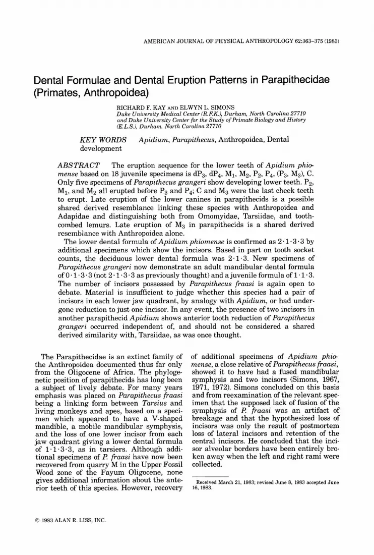
Fig. 4. Parapithecus grangeri. Radiographs of juvenile specimens. A, YPM 23796; B, DPC 2399. Abbreviations as in Figure 1. Approximately ~ 2 . 5 .
TABLE 2. Deuelopmental data for Parapithecus grangeri mandibular dentitions
Specimen Tooth locus No. dClC dPzlPz dP3IP3 dP4IP4 M1 pz M3
YPM 23796' D 2 B EID' DZB DZB D2 Di B DPC 1114 d6B4 EID' DG/B D6/B D2 D6 DPC 1118 D6B4 - D6/B4 D2B D1 D1 B DPC 2399 D'IB EID' -1c26 EIC3 D1 D6 B
- D2 D1 cz
-
DPC 3110 - - -
Developmental stages: A, tooth not developed; B, tooth in crypt; C , tooth partly erupted; C,, some movement; C2, cusp tips at or near alveolar margin: Cs, some tip wear; D, tooth fully erupted D,, little wear; DZ, enamel perforated in several places; D3, much of tooth surface shows wear; E, tooth reabsorbed and/or shed. For an explanation of superscript notation, see footnotes to Table 1.
served, but their sockets suggest they were fully or almost fully erupted (Fig. 4B). Left C remains in its crypt without detectable root formation; dC was presumably in occlusion, but has been broken away. Grooves in lin- gual alveolar bone of the symphysis may be remnants of sockets for single incisors in each lower jaw quadrant. The mesial part of the socket for right C is intact a window in the
alveolar bone links the sockets of left and right C.
In summary, available evidence indicates that eruption sequence for the lower cheek teeth of Parapithecus grangeri may have been the same as that in Apidium phiomense: Pa, MI, and M2 all erupted before P3 and Pq; C and M 3 were the last of the cheek teeth to erupt.

370 R.F. KAY AND E.L. SIMONS
Fig. 5. Purupithecus grungeri, YPM 23796. A) Stereo sockets for right and left (?) deciduous incisors; n.f., nu- SEM of symphyseal region of the right side viewed from trient foramen for canine; Pz, socket for second lower a medial and anterosuperior perspective. Approximately premolar; s., midline (symphysis) of mandible; s.k., sym- X 5 . B) Same specimen viewed superiorly. Approxi- physeal keel in midline; w, window in bone between mately x 10. Abbreviations: C, right canine in crypt and crypts for right and left canines. SEMs are of original crypt for left canine; dP,, deciduous third premolar; I, specimen.
Development of the upper teeth DPC 1123, a maxilla of Parapithecus gran-
geri, contains a deciduous p. In this speci- men M1 is fully erupted and M2 erupting with roots incompletely formed. P3 and P are still in their crypts; the state of dP3 can- not be determined.
Dental formula From juvenile specimens described above,
teeth or tooth sockets confirm the presence in l? grangeri of one (presumably) deciduous lower incisor, as well as a deciduous canine and at least two (probably three) deciduous premolars. A number of mandibles, DPC

PARAPITHECID DENTAL ERUPTION 371
Fig. 6. Purupithecus grangeri, DPC 3135. A) Stereo SEM of symphyseal region viewed from a n anterior and lateral perspective. Approximately x5. B) Same speci- men viewed occlusally. Approximately x 10. Note break
which runs through the socket for right C. Abbrevia- tions: C, canine sockets; Pz, sockets for right and left canines; Ps, lower third premolar; s, symphysis of man- dible. SEMs are of original specimen.
2376, DPC 2807, and DPC 3135 show that this species had a permanent canine, three permanent premolars and three molars. However, newly recognized specimens show that adult Parapithecus grangeri had no lower incisors. The configuration of this part of the jaw is especially clear from DPC 3135, from quarry I (Fig. 6) a nearly complete man- dible with right and left P3-M3 and all sock- ets for teeth anterior to P3. The enamel is heavily eroded on all teeth but the identifi-
cation of DPC 3135 as I? grangeri is definite judging from the overall size of the cheek teeth, the relatively small size of the M3, and the shallowness of the jaw beneath the mo- lars. These and other proportional similari- ties are nearly identical with undoubted I? grangeri specimens such as DPC 2376 and DPC 2807 which between them retain C-M3 in good condition. In DPC 3135, anterior to each P3 is a smaller socket for Pz and a large canine socket. The right canine socket is bro-
372 R.F. KAY AND E.L. SIMONS
ken but the irregular edges of the two parts fit together precisely without any lost bone; each canine socket is missing some alveolar bone labially, lingually and distally. How- ever, the mesial margins are extremely well preserved and jointly form a midline of the mandible. As Figure 6 clearly shows, there is no space for lower incisor sockets in the symphysis of this adult specimen. Based on the currently available specimens it is likely that either: 1) a single deciduous incisor erupted and persisted in the jaw until the eruption of C and then was lost; or 2) a single deciduous incisor erupted, and was replaced by a single permanent one; then this perma- nent incisor is lost with eruption of C. If we opt for the first choice, the lower dental for- mula was:
deciduous: 1 * 1 * 3 adult: 0.1.3.3.
In the event the second option proves corect, the permanent formula would be 1 . 1 * 3 3. In either case, lower incisors are not retained in the adult.
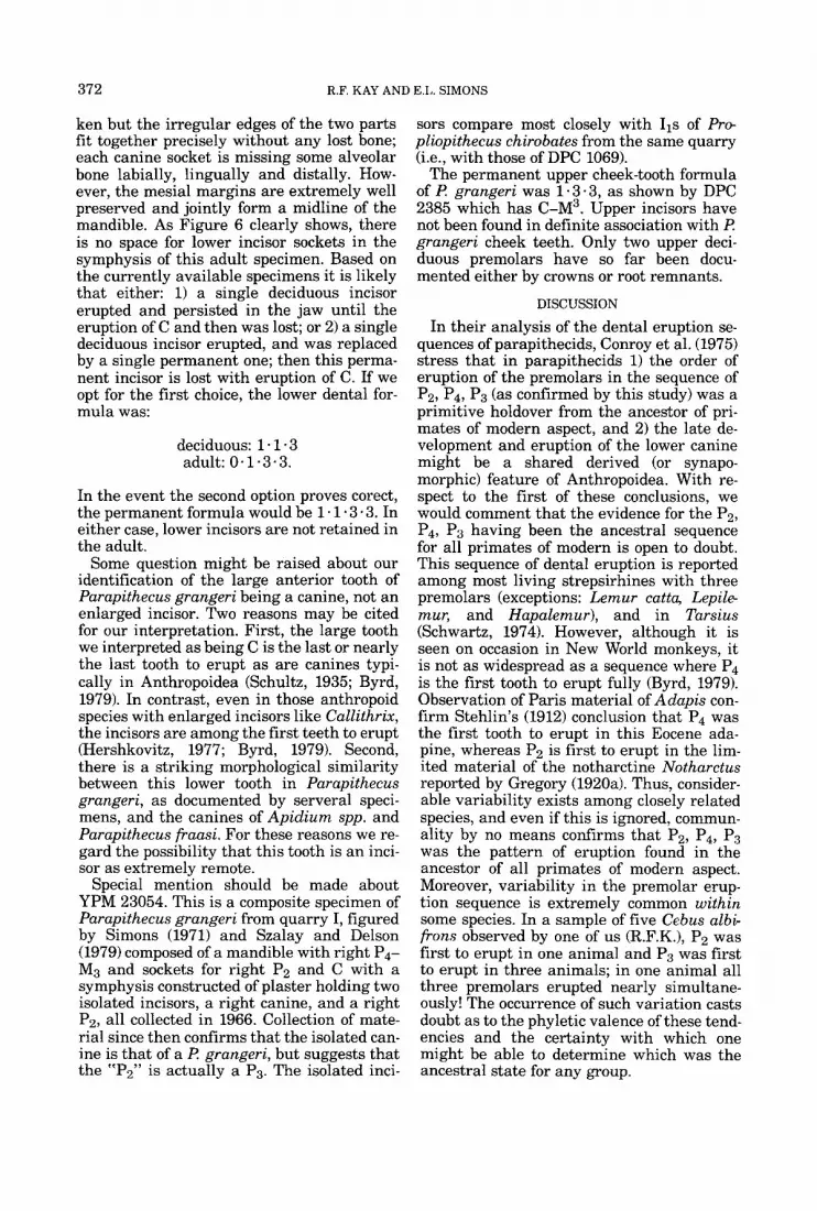
Some question might be raised about our identification of the large anterior tooth of Parapithecus grangeri being a canine, not an enlarged incisor. Two reasons may be cited for our interpretation. First, the large tooth we interpreted as being C is the last or nearly the last tooth to erupt as are canines typi- cally in Anthropoidea (Schultz, 1935; Byrd, 1979). In contrast, even in those anthropoid species with enlarged incisors like Callithrix, the incisors are among the first teeth to erupt (Hershkovitz, 1977; Byrd, 1979). Second, there is a striking morphological similarity between this lower tooth in Parapithecus grangeri, as documented by serveral speci- mens, and the canines of Apidium spp. and Parapithecus fi-aasi. For these reasons we re- gard the possibility that this tooth is an inci- sor as extremely remote.
Special mention should be made about YPM 23054. This is a composite specimen of Parapithecus grangeri from quarry I, figured by Simons (1971) and Szalay and Delson (1979) composed of a mandible with right P4- M3 and sockets for right P2 and C with a symphysis constructed of plaster holding two isolated incisors, a right canine, and a right Pa, all collected in 1966. Collection of mate- rial since then confirms that the isolated can- ine is that of a €? grangeri, but suggests that the “Pz” is actually a P3. The isolated inci-
sors compare most closely with 11s of P r e pliopithecus chirobates from the same quarry Ce., with those of DPC 1069).
The permanent upper cheek-tooth formula of €? grangeri was 1.3.3, as shown by DPC 2385 which has C-M3. Upper incisors have not been found in definite association with €? grangeri cheek teeth. Only two upper deci- duous premolars have so far been docu- mented either by crowns or root remnants.
DISCUSSION
In their analysis of the dental eruption se- quences of parapithecids, Conroy et al. (1975) stress that in parapithecids 1) the order of eruption of the premolars in the sequence of Pz, Pq, P3 (as confirmed by this study) was a primitive holdover from the ancestor of pri- mates of modern aspect, and 2) the late de- velopment and eruption of the lower canine might be a shared derived (or synapo- morphic) feature of Anthropoidea. With re- spect to the first of these conclusions, we would comment that the evidence for the Pz, P4, P3 having been the ancestral sequence for all primates of modern is open to doubt. This sequence of dental eruption is reported among most living strepsirhines with three premolars (exceptions: Lemur catta, Lepile- muq and Hapalemur), and in Tarsius (Schwartz, 1974). However, although it is seen on occasion in New World monkeys, it is not as widespread as a sequence where P4 is the first tooth to erupt fully (Byrd, 1979). Observation of Paris material of Adapis con- firm Stehlin’s (1912) conclusion that P4 was the first tooth to erupt in this Eocene ada- pine, whereas P2 is first to erupt in the lim- ited material of the notharctine Notharctus reported by Gregory (1920a). Thus, consider- able variability exists among closely related species, and even if this is ignored, commun- ality by no means confirms that Pz, P4, P3 was the pattern of eruption found in the ancestor of all primates of modern aspect. Moreover, variability in the premolar erup- tion sequence is extremely common within some species. In a sample of five Ccbus albi- f rom observed by one of us (R.F.K.), P2 was first to erupt in one animal and P3 was first to erupt in three animals; in one animal all three premolars erupted nearly simultane- ously! The occurrence of such variation casts doubt as to the phyletic valence of these tend- encies and the certainty with which one might be able to determine which was the ancestral state for any group.

PARAPITHECID DENTAL ERUPTION 373
The late eruption of the lower canines of parapithecids, the second point emphasized by Conroy et al. (1975), is confirmed by addi- tional material; the lower canine of Apidium is either the last tooth (or the penultimate one before M3) to erupt. This is a resem- blance to extant anthropoids as Conroy et al. noted. Recent studies by Byrd (1979) confirm that late canine eruption is ubiquitous among New World monkeys; it is found in most Old World monkeys and hominoids (except Homo, and Nasalis; Schultz, 1935). It is not seen among nonanthropoids: tarsiers or living strepsirhines (Schwartz, 1974). However, the late eruption of the lower canines occurs in the Eocene adapids Notharctus and Adapis (Gregory, 1920a; Stehlin, 1912) although not in the omomyid Absarokius (Gazin, 1958). This raises the possibility that late eruption of the lower canines is a shared-derived re- semblance between adapines and anthro- poids.
The new material also shows that (contra Conry et al., 1975) M3 was very late to erupt in Apidium phiomense, being the next-to-last (before the canine) and virtually simultane- ous with P3. The late eruption of M3 may be another derived feature parapithecids share with extant anthropoids. In virtually all ex- tant Old World and most New World mon- keys with three molars M3 comes in after all the deciduous premolars have been replaced (exceptions: Aotus, Pithecia) whereas it erupts before all deciduous premolars are replaced in Tarsius, all extant strepsirhines (except indriids), as well as Eocene adapines NCF tharctus and Adapis and the omomyid A bsa- rokius (vide Byrd, 1979; Gazin, 1958; Gregory, 1920a; Schultz, 1935; Stehlin, 1912).
Perhaps the most surprising finding of this study is the demonstration that Pczrczpithecus grangeri adults had no lower incisors, and juveniles had just one incisor in each lower quadrant. This reopens the question of the dental formula of Parapithecus fraasi. I? fraasi, described by Schlosser (1911), is based on a mandible collected by Richard Markgraf in 1907 (Gingerich, 1978), separated into two parts at the midline and damaged postmor- tem by the loss of one cheek-tooth (now known to have been the right Pz). It was generally agreed by early workers that the damage at the symphysis did not include the loss of any teeth, and it was stated that there were eight teeth in each jaw quadrant. Schlosser (1911) could not decide whether the second tooth from the symphysis was an in-
cisor or a canine and so whether the dental formula was 1.1 - 3 - 3 as in living tarsiers or 2 * 1 -2.3 as in present-day catarrhines. Schwalbe (1915), Gregory (1916, 1920b) and Werth (1918) opted for the latter interpreta- tion, as did Kalin (1961), after a restudy of this specimen. He also concluded that the mandibular symphysis was unfused during life as in primitive primates, not fused as in anthropoids.
With recovery of new specimens of Api- dium, Simons (1963, 1971, 1972) was able to show that some parapithecids had a dental formula 2 .1 * 3 * 3. By analogy, he suggested that Parapithecus fraasi and I? grangeri probably also had a formula of 2-1.3.3. He interpreted Schlosser’s specimens of P fraasi as having lost an incisor from each lower jaw quadrant when the mandibular symphysis, fused in life, was damaged postmortem.
The new specimens of Parapithecus gran- geri described here show its dental formula to be 0.1.3.3 in the adult. A single lower incisor in each jaw ramus was present in juveniles before the permanent canines erupted. As those are known only from their alveoli, whether they represent deciduous in- cisors or their replacement teeth cannot yet be determined. The incisor reduction in Par- apithecus grangeri makes more plausible Schlosser’s (1911) suggested dental formula of 1.1.3.3 for I? fraasi. There is no longer any reason to believe that teeth were lost from this specimen postmortem, either by natural processes or when collected. If so, I? fraasi, with a single pair of lower incisors might be viewed as an “intermediate” form between Parapithecus grangeri, with no inci- sors in the adult, and a primitive parapithe- cid with a pair of incisors in each quadrant of the lower jaw, as in Apidium.
One aspect of the incisors of Parapithecus grangeri derserves additional mention. These teeth have much lighter colored enamel than the other teeth in the jaw. Such a situation is frequently, although not always, a charac- teristic of deciduous teeth, raising the possi- bility that the incisors of Parapithecus fraasi are of the deciduous complement persisting into the adult. Only the recovery of more anterior teeth of Parapithecus fraasi will re- solve this possibility.
In view of the difference in dental formulae between adults of Parapithecus fraasi, in which at least one incisor is retained, and those of Parapithecus grangeri which have no incisors, Gingerich’s (1978) suggestion

374 R.F. KAY AND E.L. SIMONS
that the latter be removed to its own genus Simonsius now deserves additional consider- ation. We reserve our opinion pending a reas- sessment now in preparation of other structural details of the two species. On the other hand, we disagree with his proposed synonomy of Parapithecus fraasi with Api- dium phiomense. Gingerich dismisses three obvious differences between the two as being of the sort found as common variants within living species and claims that the type speci- men of the former lies well within the range of variation to be expected in a sample of the latter. The three differences he dismisses are that 1) the third molar of the type specimen of A. phiomense is relatively much larger than that of l? fraasi, 2) the type of A. phi@ mense has a well-developed centroconid on the molars whereas I? fraasi lacks it, and 3) the teeth of the type of A. phiomense are unworn whereas those of I? fraasi are mod- erately worn. Now that many specimens of A . phiomense are known, it becomes possible to test and reject this claim as well as to add other differences between the two species.
Through the 1981 field season, 33 speci- mens of Apidium phiomense were collected for which both M 2 and M3 lengths can be estimated. The mean of the ratio of M2:M3 is 0.87 with a standard deviation of 0.035. The same ratio for the type of l? fraasi is 0.95, outside the 95% confidence interval for A. phiomense.
In available samples of unworn 26 Mzs and 22 unworn M3s of A. phiomense collected through the 1981 field season, all possess centroconids. Therefore, the type of l? fraasi differs uniquely from A. phiomense in having no M2-3 centroconids.
Of course tooth wear, by itself, as Ginger- ich rightly states, is of no relevance to a taxonomic determination. However, interest- ingly Apidium phiomense molars wear flat and because of extremely thick enamel most crown features are obliterated before the enamel is performated. In ?? fraasi the en- amel is much thinner and performated at an early stage in the wear. Failure to recognize this difference might lead to an eroneous con- clusion that some A. phiomense lack centro- conids when this feature was simply removed owing to an advanced state of wear.
Another distinct difference is that Parapi- thecus fraasi seems to have possessed a re- duced number of incisors whereas Apidium phiomense certainly had two in each low jaw quadrant.
The conclusion reached on examination of these many new specimens of A . phiomense is that this species should continue to be re- garded as distinct from Parapithecus fraasi at both the specific and generic levels.
It is important to recognize that although the reduced anterior dental formula of Par- apithecus is Tarsier-like, as noted originally by Schlosser (1911), incisor loss was clearly evolved in parallel in the two groups because Apidium, a close relative of Parapithecus, still preserves its full complement of incisors. Also, it becomes clear that Parapithecus grangeri can not have been directly ancestral to living Old World monkeys, a view advo- cated by each of us in the past.
ACKNOWLEDGMENTS
This research was supported by NSF grant BNS-81-14925 and BNS-80-16206 and Smith- sonian foreign Currency grants 70869600 and 809479. P. Chatrath prepared some of the specimens. We also thank John Fleagle and Ross MacPhee for helpful comments. Eric Effmann helped with the radiography.
LITERATURE CITED
Byrd, KE (1979) Sequences and asymmetries of dental development and eruption in the Ceboidea. Ph.D. Dis- sertation, University of Washington.
Conroy, G, Schwartz, JH, and Simons, EL (1975) Dental eruption patterns in Parapithecidae (Primates, An- thropoidea). Folia primatol. 24:275-281.
Fleagle, JG, Kay, RF, and Simons, EL (1980) Sexual dimorphism in early anthropoids. Nature 287:328-330.
Gazin, CL (1958) A review of the Middle and Upper Eocene Primates of North America. Smithsonian Misc. Coll. (Washington) 136:l-112.
Gingerich, PD (1978) The Stuttgart collection of Oligo- cene primates from the Fayum Province of Egypt. Pa- laont. Z. 52:82-92.
Gregory, WK (1916) Studies on the evolution of the Pri- mates. Bull. AM. Mus. Nat. Hist. 35:239-355.
Gregory, WK (1920a) On the structure and relations of Notharctus, an American Eocene primate. Mem. Am. Mus. Nat. Hist., New York 3:49-243.
Gregory, WK (1920b) The origin and evolution of the human dentition. Part IV. The dentition of the higher primates and their relationships with man. J. Dent. Res. 2:607-717.
Hershkovitz, P (1977) Living New World Monkeys (Pla- tyrrhini), Volume 1. Chicago: University of Chicago Press.
Kali,n, J (1961) Sur les primates de I’Oligocene inferieur d’Egypte. Ann. de Paleont. 47:3-48.
Schlosser, M (1911) Beitrage zur Kenntnis der oligoza- nen Landsaugetiere aus dem Fayum: Aegypten. Beitr. Palaont. Geol. Osterreich-Ungarns 245-167.
Schultz, A (1935) Eruption and decay of the permanent teeth in primates. Am. J. Phys. Anthropol. 1:489-581.
Schwalbe, G (1915) Ueber den fossilen Affen Oreopithe- cus barnbolii. Zeit. Morphol. Anthropol. 19:234-235.

PARAPITHECID DENTAL ERUPTION 375
Schwartz, J (1974) Dental development and eruption in the prosimians and its bearing on their evolution. Ph.D. Dissertation, Columbia University.
Simons, EL (1963) A critical reappraisal of Tertiary pri- mates. In: J Buettner-Janusch (ed) Evolutionary and Genetic Biology of Primates, Volume 1. New York: Academic Press, pp. 66-130.
Simons EL (1967) The earliest apes. Sci. Amer. 217:28- 35.
Simons EL (1971) A current review of the interrelation- ships of Oligocene and Miocene Catarrhini. In: A.A. Dahlberg, (ed) Dental Morphology and Evolution. Chi-
cago: University of Chicago Press, pp. 193-210. Simons EL (1972) Primate Evolution. New York: Mac-
Millan and Company. Stehlin HG (1912) Die Saugetiere des schweizerischen
Eocaens. Abh. Schweiz. Palaont. Ges. (Zurich) 38:1165- 1298.
Szalay, FS, and Delson, E (1979) Evolutionary History of the Primates. New York: Academic Press.
Werth, E (1918) Purapzthecus, ein primitiver Menschen- affe. Sitzungsber. D. Ges. Naturforsch. Freunde zu Berlin 1918:327-345.