Embed Size (px)
Citation preview
eISSN 2036-7406
Publisher's Disclaimer. E-publishing ahead of print is increasingly important for the rapid dissemination of science. Dermatology Reports is, therefore, E-publishing PDF files of an early version of manuscripts that undergone a regular peer review and have been accepted for publication, but have not been through the copyediting, typesetting, pagination and proofreading processes, which may lead to differences between this version and the final one. The final version of the manuscript will then appear on a regular issue of the journal. E-publishing of this PDF file has been approved by the authors.
Please cite this article as [Epub Ahead of Print] with its assigned doi: 10.4081/dr.2022.9512
Note: The publisher is not responsible for the content or functionality of any supporting information supplied by the authors. Any queries should be directed to the corresponding author for the article.
Dermatology Reportshttps://www.pagepress.org/journals/index.php/dr/index
Possible relationship between poor skin disorders prognosis and serum zinc level: A narrative review Mohammed Al Abadie, Zinah Sharara, Miriam Al Abadie, Patrick A Ball, Hana Morrissey
1. PhD, Royal Wolverhampton NHS Trust, Wolverhampton, United Kingdom, [email protected]
2. MSc Clin Derm, National Health Service (NHS), Community Dermatology Clinics (Health Harmonie), United Kingdom, [email protected]
3. Student, Medway School of Pharmacy, University of Greenwich, United Kingdom, [email protected]
4. PhD, University of Wolverhampton, School of Pharmacy, United Kingdom, [email protected]
5. PhD, University of Wolverhampton, School of Pharmacy, United Kingdom, [email protected]
Acknowledgement: None Corresponding Author: Dr Hana Morrissey, University of Wolverhampton, School of Pharmacy, United Kingdom, WV11LY, +447961755705, [email protected] Key words: Serum Zinc, dermatosis, chronic inflammation, autoimmune, atopic dermatitis, Zinc supplementation.
Contribution Details:
Contributor 1
Contributor 2
Contributor 3
Contributor 4
Contributor 5
Concepts √ √ √ - -
Design √ √ √ √ √
Definition of intellectual content
√ √ √ √ √
Literature search √ √ √ √ √
Clinical studies - - - - -
Experimental studies - - - - -
Data acquisition - - - - -
Data analysis - - - - -
Statistical analysis - - - - -
Manuscript preparation √ √ √ √ √
Manuscript editing - - - √ √
Manuscript review √ √ √ √ √
Guarantor - - - - -
Conflicting Interest: None Funding: no external funding
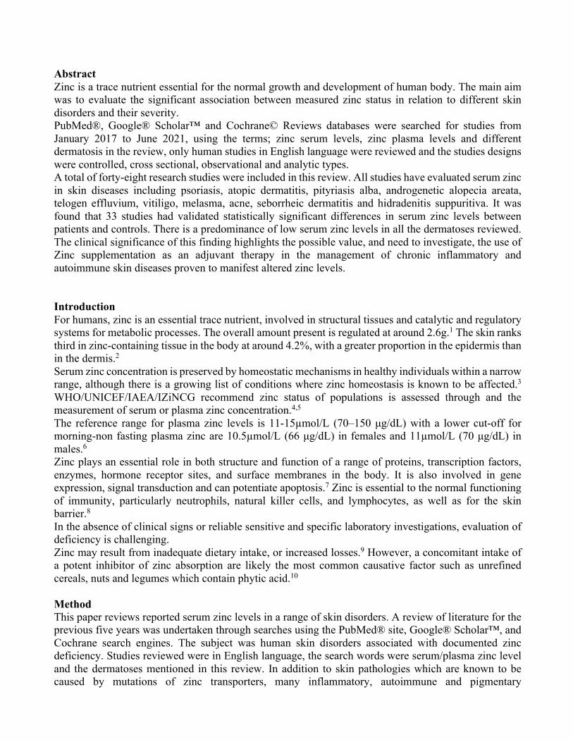
Abstract Zinc is a trace nutrient essential for the normal growth and development of human body. The main aim was to evaluate the significant association between measured zinc status in relation to different skin disorders and their severity. PubMed®, Google® Scholar™ and Cochrane© Reviews databases were searched for studies from January 2017 to June 2021, using the terms; zinc serum levels, zinc plasma levels and different dermatosis in the review, only human studies in English language were reviewed and the studies designs were controlled, cross sectional, observational and analytic types. A total of forty-eight research studies were included in this review. All studies have evaluated serum zinc in skin diseases including psoriasis, atopic dermatitis, pityriasis alba, androgenetic alopecia areata, telogen effluvium, vitiligo, melasma, acne, seborrheic dermatitis and hidradenitis suppuritiva. It was found that 33 studies had validated statistically significant differences in serum zinc levels between patients and controls. There is a predominance of low serum zinc levels in all the dermatoses reviewed. The clinical significance of this finding highlights the possible value, and need to investigate, the use of Zinc supplementation as an adjuvant therapy in the management of chronic inflammatory and autoimmune skin diseases proven to manifest altered zinc levels. Introduction For humans, zinc is an essential trace nutrient, involved in structural tissues and catalytic and regulatory systems for metabolic processes. The overall amount present is regulated at around 2.6g.1 The skin ranks third in zinc-containing tissue in the body at around 4.2%, with a greater proportion in the epidermis than in the dermis.2 Serum zinc concentration is preserved by homeostatic mechanisms in healthy individuals within a narrow range, although there is a growing list of conditions where zinc homeostasis is known to be affected.3 WHO/UNICEF/IAEA/IZiNCG recommend zinc status of populations is assessed through and the measurement of serum or plasma zinc concentration.4,5 The reference range for plasma zinc levels is 11-15µmol/L (70–150 μg/dL) with a lower cut-off for morning-non fasting plasma zinc are 10.5µmol/L (66 μg/dL) in females and 11µmol/L (70 μg/dL) in males.6 Zinc plays an essential role in both structure and function of a range of proteins, transcription factors, enzymes, hormone receptor sites, and surface membranes in the body. It is also involved in gene expression, signal transduction and can potentiate apoptosis.7 Zinc is essential to the normal functioning of immunity, particularly neutrophils, natural killer cells, and lymphocytes, as well as for the skin barrier.8 In the absence of clinical signs or reliable sensitive and specific laboratory investigations, evaluation of deficiency is challenging. Zinc may result from inadequate dietary intake, or increased losses.9 However, a concomitant intake of a potent inhibitor of zinc absorption are likely the most common causative factor such as unrefined cereals, nuts and legumes which contain phytic acid.10 Method This paper reviews reported serum zinc levels in a range of skin disorders. A review of literature for the previous five years was undertaken through searches using the PubMed® site, Google® Scholar™, and Cochrane search engines. The subject was human skin disorders associated with documented zinc deficiency. Studies reviewed were in English language, the search words were serum/plasma zinc level and the dermatoses mentioned in this review. In addition to skin pathologies which are known to be caused by mutations of zinc transporters, many inflammatory, autoimmune and pigmentary
dermatological conditions have been suggested to be associated low serum zinc. The study designs included controlled, cross sectional, analytic and observational, that mainly address serum zinc concentration as an index of zinc status. Studies that evaluated other trace elements and micronutrients also included, however, only the outcome of zinc level result in relation to dermatoses was interpretated in this review. The outcome of zinc level in the hair or tissue were not interpreted. Results Psoriasis Psoriasis is characterized externally by plaques and scaly papules. The aetiology is complex, multifactorial, and poorly understood. In addition to skin and joints, psoriasis is associated with metabolic syndrome, cardiovascular disease, obesity, and mental diseases.11-13 Five studies were included in this review, that investigated the correlation between serum zinc level and psoriasis with different levels of severity. Three studies reported a significant decrease in the mean serum level of zinc in patients exhibiting mild, moderate and severe disease compared to controls.14-16 Two studies examined the copper/zinc ratio and both showed a higher copper/zinc ratio in patients with psoriasis compared to controls.17,18
Atopic Dermatitis Atopic dermatitis is a chronic, relapsing inflammatory disorder of the skin presenting as eczematous skin plaques with lichenification, xerosis and severe pruritus.19,20 Again, the aetiology of the condition is poorly understood, but both genetic and environmental factors are recognized.21 Studies have linked the defective skin barrier and altered immune responses with a filaggrin gene mutation.22 The role of zinc as a trace element in Atopic Dermatitis has been the subject of a limited number of studies. Three studies, one controlled and two observational, tested serum zinc level in children with varying severity of atopic dermatitis and concluded a strong correlation between zinc levels and the severity of atopic dermatitis which was statistically significant.23-25 However, Esenboga et al. in their controlled study, concluded that serum zinc levels did not change with the severity of the disease, nor did the presence of atopy influence serum zinc levels among other trace element tested.26
Pityriasis alba Pityriasis alba is asymptomatic, scaly ill-defined hypopigmented skin patches, mainly affecting children aged >15 years. The pathogenesis of PA is not yet understood; it often is found with atopic dermatitis and is perceived as a milder form. It occurs more commonly in individuals with darker skin complexion. Other potential risk factors are xerosis, and mineral deficiencies.27,28
Two recent controlled studies evaluated the zinc level in children with pityriasis alba, and demonstrated serum Zinc was lower in patients with PA than controls. Further it correlated significantly with both the number and size of lesions.29 Zinc deficiency was found to increase the risk of pityriasis alba by more than 15 folds.30
Androgenic alopecia Androgenic alopecia, (male pattern baldness), is the most common cause of progressive hair loss. Around 30% of Caucasian men will experience it around age 30 years, 50% by age 50 and 80% by the age of 70 years.31 There are variable pathogenetic pathways leading to hair loss, including genetic and non-genetic factors.32 Five studies were found. All tested serum zinc levels in relation to androgenetic alopecia. One study involved females with androgenetic alopecia serum zinc levels in the patient group were
significantly lower than that in the control.33 Another study showed that females with the Ludwig clinical pattern had lower plasma zinc levels.34 Kondrakhina et al.35 in his case-controlled study that enrolled males with androgenetic alopecia, studied many nutrient parameters, however, differences were shown in zinc content only. El Esway et al. similarly tested males and demonstrated a similar outcome. However, a non-significant relationship was found between the grade of alopecia and zinc level.36 Gowda involved other non-scarring hair loss problem like telogen effluvium and alopecia areata and detect for other micronutrients, nevertheless, zinc deficiency was seen in 11.76% of participants with MPHL, and no significant difference in other micronutrients was observed between micronutrient levels and androgenetic alopecia.37 Telogen Effluvium Telogen effluvium is diffuse non-scarring hair loss, is caused by any disruption of hair cycle occurring as a reaction pattern to various physical or mental stressors and resulting in increased, synchronized telogen hair shedding.38,39 Data from studies are conflicting. Four studies in females with telogen effluvium were located, three of which showed no statistical difference between patients and controls.40-42 However, in one study from Bangladesh, the authors concluded that zinc level was significantly lower in telogen effluvium patients.43
Alopecia Areata Alopecia areata is a non-scarring, autoimmune, inflammatory hair loss occurring most commonly on the scalp and/or the body. Etiology and pathogenesis are not understood but a complex interplay between environmental and genetic factors is currently thought to lead to the development of alopecia areata. Research has focussed on whether serum levels of micronutrients are different in these patients.44 Histopathology is reveals an increase in catagen and telogen follicles, with inflammatory lymphocytic infiltration in the peribulbar region considered characteristic.45 Four studies were identified for this review; three were controlled studies that evaluated serum zinc in alopecia areata patients.46,47 Zinc level was significantly lower in patients than controls with a significant correlation both between occurrence and duration of alopecia and serum.48 Conversely, in a cross-sectional study for alopecia areata where zinc level was documented in 22 patients, only one patient was reported to be zinc deficient.49 Vitiligo Vitiligo is a common depigmenting disorder affecting the skin and hair with 0.1%-2% incidence rate.50,51 The cause appears to be a loss of functional melanocytes. It is generally agreed that there is an absence of functional melanocytes as result of autoimmune destruction. The etiology is multifactorial and has polygenic inheritance.52,53 The seven studies included in this review documented that patients with vitiligo had serum zinc levels significantly lower than in healthy controls.54-58 There was also a negative correlation between vitiligo extent and severity and serum zinc levels.59 Mirnezami and Rahimi60 observed significantly different zinc levels in patients with generalized vitiligo and the controls, and correlated lower serum zinc levels with disease duration. They further demonstrated a negative relationship between serum zinc level and the age of the patient with vitiligo. Acne vulgaris Acne vulgaris is a common skin disorder characterized by inflammation of the sebaceous glands with follicular unit keratin obstruction which leads to the formation of acne lesions primarily on the face, chest and back.61-63 Four involved mechanisms include follicular proliferation with ruptures, sebum production, inflammation, and the presence of Cutibacterium acnes which induces inflammation.50,64
In the last five years eight studies were identified that measured serum d zinc level in patients with acne. Data are conflicting as five of these studies concluded significant differences in serum zinc levels between acne and control groups.65-68 Otami69 stated that serum zinc levels significantly varied between mild and the moderate–severe acne group, as the deficiency was more evident in higher grade acne. Conversely, a controlled study with 200 patients tested, found no correlation between zinc level and presentation or severity of acne, except that in females, acne severity was affected by zinc level.70 Two other studies have also failed to identify a relationship between serum zinc level and acne.71,72
Hidradenitis Suppurativa Hidradenitis suppurativa, is an inflammatory disorder affecting around 1% of the population.73 It is characterized by inflamed nodules and abscesses, complicated with scarring and sinuses particularly in intertriginous areas. The severity varies between patients and is categorized into 3 stages based on the presence of sinus tracts and scarring in a classification proposed by Hurly in 1989.74 Due to the associated pain, mobility restriction, purulence and systemic involvement, the disease can profoundly affect the quality of life.75,76 In a large controlled study that tested serum zinc level in 122 patients with different stages of Hs and 122 controls, Low serum zinc levels were found in patients graded Hurley stage III.74
Seborrheic dermatitis Seborrheic dermatitis is a common inflammatory skin disease with unknown etiology and a chronic relapsing course. Colonisation with Malassezia furfur, active sebaceous gland, hormones, dysregulation of the immune system and external factors contribute to the pathogenesis of seborrheic dermatitis.77,78 It was postulated that zinc is known to be involved in many biological processes that appear to contribute to the development of seborrheic dermatitis, so zinc level was investigated in controlled studies of patients. Three studies were found, two of which found no significant difference between patients and controls,79 despite the lower concentrations measured in the patients group.80 However, Karabay and Cerman81 in their controlled study concluded that significantly lower serum zinc levels were found in patients than in controls, although no correlation with severity was detected. Melasma Melasma is a is an acquired hyperpigmentation skin disorder that is caused by dysfunction of melanogenesis. Zinc has a role in anti-oxidative and anti-inflammatory processes including melanogenesis. Four studies were included, two studies validated a significant correlation between melasma and zinc deficiency and high negative correlation between serum zinc level and both modified Melasma Area Severity Index (mMASI) and duration of melasma was also proven,82,83 while other controlled study found the difference was not statistically significant between patients and controls.84 An observational analytic study by Rambie85 also stated that there was no significant relationship between serum zinc levels and the severity of disease in 30 melasma patients. Discussion Psoriasis The studies in this review were consistent with the data from a 2019 metanalysis that analysed studies from 1989 to 2015, and confirmed that serum zinc levels were generally decreased in patients with psoriasis.86 However, the results in this review contradict with an earlier study that showed that serum zinc levels were increased in patients with psoriasis.87 Other earlier studies proved no significant difference of zinc level between patients and controls.88-91 Psoriasis is characterised by hyperproliferation of keratinocytes with failure of differentiation. Zinc is an essential component of processes in protein synthesis, DNA and RNA synthesis and repair, and
regulation of enzymatic processes including scavenging of free radicals.92 In human keratinocyte culture, zinc and its transporter ZIP2 are found in the epidermis and closely associated with the differentiation of keratinocytes and cell turnover of epidermal layers.93 Zinc can impact psoriasis by means of regulating keratinization in the epithelium, enzymatic and immunological functioning.94
Atopic dermatitis The limited number of studies in this review, demonstrated a strong correlation between low zinc levels and atopic dermatitis severity. Low serum zinc levels increase the risk of severe atopic dermatitis. This was in line with recent review of earlier studies that also concluded that low serum, hair and erythrocyte zinc levels are associated with atopic dermatitis.95 A recent in vitro study has tested the effect of zinc supplementation on the Th1-driven reaction in mixed lymphocyte cultures. In physiological dose, zinc supplementation was associated with a significant reduction in cell proliferation and pro-inflammatory cytokine production following reactivation, compared to untreated controls.96 This is consistent with many studies, some in murine samples, that proved zinc deficiency can cause inhibition of the anti-inflammatory processes, and how can that affect Th2 cytokines, which has significant role in the pathogenesis of AD, and thus suggests a correlation.97,98 Low zinc levels can also disrupt the integrity of the membrane barrier, which can further affect atopic dermatitis progression.96-99
Hair loss The relationship between zinc and hair loss has been researched extensively in literature, with results broadly consistent with current findings. A meta-analysis that searched published data up to 30 April 2016 looking at trace elements and alopecia areata, suggested that low serum levels of zinc were risk factors for alopecia areata.100 A review article on zinc in patients with alopecia areata showed in 4/6 4 out of 6 case–control studies, that zinc levels were lower in patients than controls.101 In another study of four types of hair loss, all groups with hair loss had statistically lower zinc concentration.102 However, a study in males with androgenic alopecia did not find a difference in zinc levels between patients and controls.103 Zinc is an element of finger motifs for variant transcription factors that use hedgehog signaling to control hair growth. Its inhibitory role on apoptosis-related endonucleases also inhibits catagen.104,105 Paus et al.106 described zinc to be is a potent immunomodulator of hair follicles and also, an inhibitor of hair follicle regression, accelerating follicle recovery. This suggests zinc is essential to the hair growth cycle rather than as a component of the hair per se. Vitiligo The role of zinc deficiency in vitiligo has been validated in many studies. All are consistent and support two earlier studies, the first which was a review published in 2014 in Chinese literature that included data from 16 studies including 891 patients with vitiligo and 1682 healthy controls.107 Another metanalysis involved 11 studies including 570 vitiligo cases and 580 controls also confirmed that lower zinc level can increase the risk of vitiligo.108 The proposed mechanism through which zinc deficiency develops is that inflammatory cytokines promote the expression of zinc transporters, which mobilises zinc into cells resulting in hypozincemia and augmentation of the inflammatory process. The cytokines IL-6 and IL-17 promote the expression of the zinc transporters ZIP14 and ZIP8.109,110
This correlation between interleukins and zinc metabolism was further supported in another recent study in vitiligo patients.57 .Zinc, among other micronutrients, has a key role in the process of melanogenesis
and specifically the final stage of eumelanin formation. It is involved in catalysis of 5, 6-dihydroxy indole-2-carboxylic acid production and increased polimerisation to eumelanin.111,112
Pilosebaceous Disorders Zinc levels in the sera of patients with acne are significantly lower, and this was validated in the majority of studies identified from the last five years,68-72 this also was confirmed by a recent review in 2020 that concluded the correlation of low serum zinc level and suggested that zinc is effective in the treatment of acne as an adjuvant or as monotherapy.113 Three studies did not find this association,73-75 nor did an earlier study.114 The relation between zinc and acne can be explained through its inhibitory role on tumor necrosis factor- (TNF-)α and IL-6.115,116 Furthermore, in keratinocyte cell culture studies zinc gluconate decreased IL-8 secretion through inhibition of TLR2 and TLR4 expression, which have all proven to have a significant role in acne pathogenesis.117 Zinc also has a regulatory role in vitamin A metabolism through protein synthesis and zinc dependent retinol dehydrogenase enzyme.118 Low serum zinc levels may increase androgen production,119 which in turn affects sebaceous gland activity. Thus, zinc was considered a contributory factor in the pathogenesis of several inflammatory skin diseases associated with dysregulation of innate immunity, such as inflammatory acne and hidradenitis suppurativa. In this update, the correlation between low zinc level and hidradenitis suppurativa was evident in patients at Hurly stage III, primarily in one study in 2018,79 which was consistent with the data from a recent systematic review that revealed the association between low serum zinc levels and increased lesion count in hidradenitis suppurativa.120 In regards to seborrheic dermatitis, 2 studies concluded no significant relationship to zinc level,82,83 whilst one correlated disease activity to serum zinc level.84
Zinc deficiency Zinc deficiency may be either inherited or acquired, with similar clinical cutaneous manifestation. Acrodermatitis enteropathica (Figure 1), the inherited form, classically presents during infancy and characterized by diarrhea, dermatitis and alopecia.121 The acquired form is caused by reduced zinc intake or absorption due to other causes, like restricted or deficient diets and malabsorption states.122 Bel-Serrat et al., is a useful detailed summary of factors affecting zinc absorption and quantification of all routes of zinc loss.3 When depletion is sufficient the rash develops over a few days and is mainly acral and periorificial in location. It presents as eczematous scaly plaques that progress into vesicles, bullae, or pustules, with underlying scald like erythema or fissuring, often with symmetrical distribution around the mid-line.123 Similarly as proven in this review, many inflammatory and autoimmune skin diseases can drastically affect the skin and produce changes in zinc losses and absorption. The treatment of acrodermatitis enteropathica is supplementation of oral zinc, the recommended dose is between 1 and 3 mg/kg/day of elemental zinc.124,125 Conclusion It will be valuable to apply knowledge about zinc levels in skin disorders, and the known roles of zinc in cell biology and body functions toward future treatment strategies in skin disorders. Zinc status is frequently overlooked and not routinely tested; thus, it is recommended to identify skin disorders that are associated with zinc deficiency, and test serum zinc to define zinc status while initiating the treatment plan. Zinc therapy as an adjuvant is an attractive choice due to its availability, cost-effectiveness and low side effect profile.
Researchers need to integrate existing data into further large scale interventional studies further investigating the role of zinc in diverse cutaneous disorders, in order to consolidate the role of zinc supplement as a future, hopefully evidence-based therapeutic intervention. References 1. Jackson M.J. Physiology of zinc: General aspects. In: Mills C.F., editor. Zinc in Human Biology.
Springer; London, UK: 1989. pp. 1–14. 2. Michaelsson G., Ljunghall K., Danielson B.G. Zinc in epidermis and dermis in healthy subjects.
Acta Derm Venereol. 1980;60:295–299. 3. Bel-Serrat S, Stammers AL, Warthon-Medina M, et al. EURRECA Network. Factors that affect
zinc bioavailability and losses in adult and elderly populations. Nutr Rev. 2014;72:334-52. 4. Gibson RS, Hess SY, Hotz C, Brown KH. Indicators of zinc status at the population level: a review
of the evidence. Brit J Nutr, 2008;99:S14-S23. 5. Hess SY, Peerson JM, King JC, Brown KH. Use of Serum Zinc Concentration as an Indicator of
Population Zinc Status. Food and Nutrition Bulletin, 2007;28: S403–S429. 6. Gibson RS, Hess SY, Hotz C, Brown KH. Indicators of zinc status at the population level: A review
of the evidence. Br J Nutr 2008;99:S14-23. 7. Richardson NJ. UK consumer perceptions of meat. Proc Nutr Soc. 1994;53:281-287. 8. Hambidge KM, Krebs NF. Zinc deficiency: a special challenge. J Nutr. 2007;137:1101–1105. 9. Shankar AH, Prasad AS. Zinc and immune function: the biological basis of altered resistance to
infection. Am J Clin Nutr. 1998;68:447S-463S. 10. Lonnerdal B. Dietary factors influencing zinc absorption. J Nutr. 2000;130:1378S-1383S 11. Brito-Luna MJ, Villanueva-Quintero DG, Sandoval-Talamantes AK, et al. Correlation of IL-12,
IL-22, and IL-23 in patients with psoriasis and metabolic syndrome. Preliminary report. Cytokine. 2016;85:130–136.
12. Ergun T, Seckin Gencosmanoglu D, Karakoc-Aydiner E, et al. Prevalence of obesity in paediatric psoriasis and its impact on disease severity and progression. Australas J Dermatol. 2017;58:e182–187.
13. Ku SH, Kwon WJ, Cho EB, et al. The Association between psoriasis area and severity index and cardiovascular risk factor in Korean psoriasis patients. Ann Dermatol. 2016;28:360–363.
14. Al-Hasan AS. Correlation between Zinc and Iron with Psoriatic Patients in Al-Anbar Governorate. Annals of the Romanian Society for Cell Biology. 2021;5:3589-3597.
15. Rahayu T, Putra E, Diana R, Dharmawan N. Kadar Zinc Serum Penderita Psoriasis. Cermin Dunia Kedokteran, 2021;48:138-141.
16. Rao VM, Deepthi M, Ramalingam K, et al. Study on serum copper, zinc and selenium trace element levels in Psoriasis. Indian J Clin Exper Dermatol. 2019;5:239-242.
17. Gajjar M, Sirajwala HB. Serum Copper and Zinc ratio in Psoriasis. Int J Res Med. 2015; 4(3);55-59.
18. Aggarwal J, Singh A, Gupta S, Prasad R. Copper and zinc status in psoriasis: correlation with severity. Indian J Clin Biochem. 2021;36:120-123.
19. Khan F, Naeem SM, Ahmad Z, Zuberi NA. Association of serum levels of zinc and copper with degree of severity in patients with psoriasis. Journal of Saidu Medical College, 2018;7:72-75.
20. Solomon LM, Beerman H. Atopic dermatitis. Am J Med Sci. 1966;252:478–496. 21. De Benedetto A, Agnihothri R, McGirt LY, et al. Atopic dermatitis: a disease caused by innate
immune defects? J Invest Dermatol. 2009;129:14–30. 22. Cookson W. Genetics and genomics of asthma and allergic diseases. Immunol rev. 2002;190:195-
206
23. van den Oord RA, Sheikh A. Filaggrin gene defects and risk of developing allergic sensitisation and allergic disorders: systematic review and meta‐analysis. Brit Med J. 2009;339:2433.
24. Farhood IG, Ahmed MH, Al-Bandar RT, Farhood RG. Assessment of Serum Zinc Level in Patients with Atopic Dermatitis. Iraqi Journal of Medical Sciences, 2019;17:103-107.
25. Ehlayel MS, Bener A. Risk factors of zinc deficiency in children with atopic dermatitis. Eur Ann Allergy Clin Immunol. 2020;52:18-22.
26. Landiasari DA, Kawuryan DL, Hidayah D. Correlation between Serum Zinc Levels and Severity of Atopic Dermatitis. Asia Pacific J Peds Child Hlth 2020;3:114-118.
27. Esenboga S, Cetinkaya PG, Sahiner N, et al. Infantile atopic dermatitis: Serum vitamin D, zinc and TARC levels and their relationship with disease phenotype and severity. Allergologia et Immunopathologia. 2021;49:162-168.
28. Jadotte YT, Janniger CK. Pityriasis alba revisited: perspectives on an enigmatic disorder of childhood. Cutis. 2011;87:66-72.
29. Vinod S, Singh G, Dash K, Grover S. Clinico epidemiological study of pityriasis alba. Indian J Dermatol, Venereol Leprol. 2002;68:338-340.
30. Elesawy FM, Akl EM, Halim WAA. Zinc has a role in pathogenesis of pityriasis alba. Indian J Paed Dermatol. 2020;21:178-183.
31. Khafagy GM, Nada HR, Rashid LA, et al. Role of trace elements in pityriasis alba. J Trace Elem Med Biol. 2020;59:126422.
32. Severi G, Sinclair R, Hopper JL, et al. Androgenetic alopecia in men aged 40–69 years: prevalence and risk factors. Brit J Dermatol. 2003;149:1207-1213.
33. Marcińska M; Pośpiech E; Abidi S; et al. Evaluation of DNA Variants Associated with Androgenetic Alopecia and Their Potential to Predict Male Pattern Baldness. PLoS ONE 2015;10:e0127852.
34. Dhaher SA, Yacoub AA, Jacob AA. Estimation of zinc and iron levels in the serum and hair of women with androgenetic alopecia: Case–control study. Indian J Dermatol, 2018;63:369-374.
35. Aiempanakit K, Jandee S, Chiratikarnwong K, et al. Low plasma zinc levels in androgenetic alopecia. Indian J Dermatol, Venereol Leprol. 2017;83:741.
36. Kondrakhina IN, Verbenko DA, Zatevalov AM, et al. Plasma Zinc Levels in Males with Androgenetic Alopecia as Possible Predictors of the Subsequent Conservative Therapy’s Effectiveness. Diagnostics Internet 2020;10:336.
37. El‐Esawy FM, Hussein MS, Ibrahim Mansour A. Serum biotin and zinc in male androgenetic alopecia. Journal Cosmet Dermatol. 2019;18:1546-1549.
38. Gowda D, Premalatha V, Imtiyaz DB. Prevalence of Nutritional Deficiencies in Hair Loss among Indian Participants: Results of a Cross-sectional Study. Int J Trichol. 2017;9:101–104.
39. Kligman AM. Pathologic dynamics of human hair loss: I. Telogen effluvium. Arch Dermatol. 1961;83:175-198.
40. Harrison S, Sinclair R. Telogen effluvium. Clin Experiment Dermatol: Clin Dermatol. 2002;27:389-395.
41. Yavuz IH, Yavuz GO, Bilgili SG, et al. Assessment of heavy metal and trace element levels in patients with telogen effluvium. Indian J Dermatol. 2018;63:246–250.
42. Farah HS, Hajleh MNA, Shalan N, et al. The association between the levels of Ferritin, TSH, Zinc, Hb, vitamin B12, vitamin D and the hair loss among different age groups of women: A retrospective study. International J Pharmaceut Res. 2021;13:143-148.
43. Mohammad AP, Baba AT, Ghassemi MO. Comparison between serum levels of vitamin D and zinc in women with diffuse non-scarring hair loss (telogen effluvium) and healthy women. Pakistan J Med Hlth Sci. 2020;14:1400-1404.
44. Rahman F, Akhter QS. Serum zinc and copper levels in alopecia. Journal of Bangladesh Society of Physiologist. 2019;14:21-25.
45. Thompson JM, Mirza MA, Park MK, et al. The role of micronutrients in alopecia areata: a review. Am J Clin Dermatol. 2017;18:663-679.
46. Wasserman D, Guzman‐Sanchez DA, Scott K, McMichael A. Alopecia areata. Int J Dermatol. 2007;46:121-31.
47. Mikhael NW, Hussein MS, Mansour AI, Abdalamer RS. Evaluation of Serum Level of Zinc and Biotin in Patients with Alopecia Areata. Benha J Appl Sci. 2020;5:1-6.
48. Ozaydin-Yavuz G, Yavuz IH, Demir H, et al. Alopecia Areata Different View; Heavy Metals. Indian J Dermatol, 2019;64:7–11.
49. Sara S, Armaghan Ghareaghaji Z, Afsaneh R. Evaluating the serum zinc and vitamin D levels in alopecia areata. Iranian J Dermatol. 2018;21:77-80.
50. Alamoudi SM, Marghalani SM, Alajmi RS, et al. Association Between Vitamin D and Zinc Levels With Alopecia Areata Phenotypes at a Tertiary Care Center. Cureus, 2021;13:e14738.
51. Alkhateeb A, Fain PR, Thody A, et al. Epidemiology of vitiligo and associated autoimmune diseases in Caucasian probands and their families. Pigment Cell Research. 2003;16:208-214.
52. Jin Y, Riccardi SL, Gowan K, et al. Fine-mapping of vitiligo susceptibility loci on chromosomes 7 and 9 and interactions with NLRP1 (NALP1). J Invest Dermatol. 2010;130:774-783
53. Castanet J, Ortonne JP. Pathophysiology of vitiligo. Clinics Dermatol. 1997;15:845-851. 54. Le Poole IC, van den Wijngaard RM, Westerhof W, Dutrieux RP, Das PK. Presence or absence of
melanocytes in vitiligo lesions: an immunohistochemical investigation. J Invest Dermatol. 1993;100(6):816-822.
55. Saniee SS, Zare AG, RadmehrEe A, Radmehr A. Zinc, Vitamin D, and TSH Levels in Patients with Vitiligo. Erciyes Med J. 2019;41(2):148-153.
56. Singh CD, Gera CD. Evaluation of Zinc Levels in Patients with Vitiligo-A Clinical Study. J Adv Med Dent Sci Res. 2019;7:156-158.
57. Mogaddam MR, Ardabili NS, Maleki N, et al. Evaluation of the serum zinc level in patients with vitiligo. Advances in Dermatology and Allergology/Postȩpy Dermatologii i Alergologii. 2017;34:116-119.
58. Salem MA, El-Raheem TA, Aboraia NM. Serum Copper and zinc levels in vitiligo patients. Egyptian J Hosp Med. 2018;70:273-281.
59. Sanad EM, El-Fallah AA, Al-Doori AR, Salem RM. Serum Zinc and Inflammatory Cytokines in Vitiligo. J Clin Aesthet Dermatol. 2020;13:S29–S33.
60. Zaki AM, Nada AS, Elshahed AR, et al. Therapeutic implications of assessment of serum zinc levels in patients with vitiligo: A patient controlled prospective study. Dermatol Ther. 2020;33(6):e13998.
61. Mirnezami, M., Rahimi, H. Serum zinc level in vitiligo: A case-control study. Indian J Dermatol. 2018;63:227-230.
62. Dawson AL, Dellavalle RP. Acne vulgaris. Brit Med J. 2013;346:f2634. 63. Holick MF, MacLaughlin JA, Doppelt SH. Regulation of cutaneous previtamin D3 photosynthesis
in man: skin pigment is not an essential regulator. Science. 1981;211:590-593. 64. Thiboutot D, Gollnick H, Bettoli V, et al. New insights into the management of acne: an update
from the global alliance to improve outcomes in acne group. J Am Acad Dermatol. 2009;60:S1-50 65. Kim J, Park T, Kim HJ, et al. Inferences in microbial structural signatures of acne microbiome and
mycobiome. J Microbiol. 2021;59:369-375. 66. Butool F, Mohammed A, Syed PA, Mohammed RA. Role of serum Zinc and Copper levels in
patients with acne vulgaris. J Orofacial Res. 2019;8:71-75.
67. Kahssay M, Assefa B, Daba F, et al. Magnitude and associated factors of zinc deficiency among patients with acne vulgaris: a cross-sectional study. Medico Res Chron. 2017;4:481-494.
68. Gaber HAA, Abozied AAH, Abd-Elkareem IM, El-Shazly YNY. Serum Zinc Levels in Patients with Acne Vulgaris and Its Relation to The Severity of Disease. Egyptian J Hosp Med, 2019;75:2845-2848.
69. Awad SM, Morsy H, Sayed AA, et al. Oxidative stress and psychiatric morbidity in patients with facial acne. J Cosmet Dermatol. 2018;17:203–208.
70. Otami C, Chandra DN, Sitohang IB. Association of zinc intake & serum zinc levels with acne severity. Journal of General-Procedural Dermatology and Venereology Indonesia, 2020;4:52-57.
71. Atefi NS, Bazargan MD. Determination of serum levels of zinc in acne vulgaris patients: a case control study. Iranian J Dermatol. 2020;23:28-31.
72. Kumar GP, Priscilla T. Correlation of serum zinc levels with severity of acne vulgaris. Journal of Evolution of Medical and Dental Sciences. 2018;7:560-564.
73. Ikaraoha CI, Mbadiwe NC, Anyanwu CJ, et al. The Role of Blood Lead, Cadmium, Zinc and Copper in Development and Severity of Acne Vulgaris in a Nigerian Population. Biol Trace Elem Res. 2017;176:251-257.
74. Hurley HJ. Axillary hyperhidrosis, apocrine bromhidrosis, hidradenitis suppurativa, and familial benign pemphigus: surgical approach. In: Roenigk RK, Roenigk HH Jr, eds. Roenigk and Roenigk’s Dermatologic Surgery: Principles and Practice. 2nd ed. New York, NY: Marcel Dekker; 1996:623-645.
75. Wolk K, Join‐Lambert O, Sabat R. Aetiology and pathogenesis of hidradenitis suppurativa. Brit J Dermatol. 2020;183:999-1010.
76. Sabat R, Chanwangpong A, Schneider-Burrus S, et al., Increased prevalence of metabolic syndrome in patients with acne inversa. PloS One. 2012;7:e31810.
77. Poveda I, Vilarrasa E, Martorell A, et al., Serum zinc levels in hidradenitis suppurativa: A case–control study. Am J Clin Dermatol. 2018;19:771-777.
78. Harada K, Saito M, Sugita T, Tsuboi R. Malassezia species and their associated skin diseases. J Dermatol. 2015;42:250–257.
79. Bakardzhiev I, Argirov A. New insights into the etiopathogenesis of seborrheic dermatitis. Clin Res Dermatol Open Access. 2017;4:1-5.
80. Zohreh H, Majid S, Mohammad H. The relationship of serum selenium, zinc, and copper levels with seborrheic dermatitis: a case-control study. Iranian J Dermatol. 2019;22:7-12.
81. Nazik H, Bengü AŞ, Gül FÇ, et al. Evaluation of the levels of trace elements in the blood and hair of patients with seborrheic dermatitis. Trace Elements and Electrolytes. 2018;DOI 10.5414/TEX01555
82. Karabay EA, Cerman AA. Serum zinc levels in seborrheic dermatitis: a case-control study. Turkish J Med Sci. 2019;49:1503-1508.
83. Rostami Mogaddam M, Safavi Ardabili N, Iranparvar Alamdari M, et al. Evaluation of the serum zinc level in adult patients with melasma: Is there a relationship with serum zinc deficiency and melasma? J Cosmet Dermatol. 2018;17:417-422.
84. Abdelaziz MI, Attwa EM, Esawy AM, Khalifa N. Evaluation of the Serum Zinc Level in Adult Egyptian Patients with Melasma. Zagazig University Medical Journal. 2022;in press DOI: 10.21608/ZUMJ.2020.17857.1580
85. Sekarnesia IS, Sitohang IBS, Agustin T, et al. A comparison of serum zinc levels in melasma and non-melasma patients: a preliminary study of thyroid dysfunction. Acta Dermatovenerologica Alpina, Pannonica et Adriatica, 2020;29:59
86. Rambe PS, Simanungkalit R, Yosi A. The Relationship between Serum Zinc Level and Severity of Melasma. International Journal of Scientific and Research Publications, 2020;10:317-321.
87. Lei L, Su J, Chen J, et al. Abnormal serum copper and zinc levels in patients with psoriasis: A meta-analysis. Indian J Dermatol. 2019;64:224-230.
88. Butnaru C, Pascu M, Mircea C, et al. Serum zinc and copper levels in some dermatological diseases. Rev Med Chir Soc Med Nat Iasi. 2008;112:253–257.
89. Halevy S, Giryes H, Friger M, et al. The role of trace elements in psoriatic patients undergoing balneotherapy with Dead Sea bath salt. Isr Med Assoc J. 2001;3:828–832.
90. Cao JQ, Cui R, Zhang ZX, et al. Determination of serum trace elements in patients with psoriasis. Zhongguo Mafen Pifubing Zazhi. 2005;21:617–619.
91. Tasaki M, Hanada K, Hashimoto I. Analyses of serum copper and zinc levels and copper/zinc ratios in skin diseases. J Dermatol. 1993;20:21–24.
92. Ala S, Shokrzadeh M, Golpour M, et al. Zinc and copper levels in Iranian patients with psoriasis: A case control study. Biol Trace Elem Res. 2013;153:22–27.
93. Zeng Q, Yin J, Fan F, et al. Decreased copper and zinc in sera of Chinese vitiligo patients: A meta-analysis. J Dermatol. 2014;41:245–251.
94. Inoue Y, Hasegawa S, Ban S, et al. ZIP2 protein, a zinc transporter, is associated with keratinocyte differentiation. J Biol Chem. 2014;289:21451-21462.
95. Ogawa Y, Kawamura T, Shimada S. Zinc and skin biology. Arch Biochem Biophys, 2016;611:113-119.
96. Gray NA, Dhana A, Stein DJ, Khumalo NP. Zinc and atopic dermatitis: a systematic review and meta‐analysis. J Europ Acad Dermatol Venereol. 2019;33:1042-1050.
97. Maywald M, Rink L. Zinc supplementation induces CD4+ CD25+ Foxp3+ antigen-specific regulatory T cells and suppresses IFN-γ production by upregulation of Foxp3 and KLF-10 and downregulation of IRF-1. Europ J Nutr. 2017;56:1859-1869.
98. Takahashi H, Nakazawa M, Takahashi K, et al. Effects of zinc deficient diet on development of atopic dermatitis-like eruptions in DS-Nh mice. J Dermatol Sci. 2008;50:31–39
99. Hextall B, Jenkinson C, Humphreys R. Dietary supplements for established atopic eczema. Cochrane Database Syst Rev. 2012;2:CD005205. doi: 10.1002/14651858.CD005205.pub3.
100. Shankar AH, Prasad AS. Zinc and immune function: the biological basis of altered resistance to infection. Am J Clin Nutr 1998;68:447s–463s.
101. Jin W, Zheng H, Shan B, Wu Y. Changes of serum trace elements level in patients with alopecia areata: a meta‐analysis. J Dermatol. 2017;44:588-591.
102. Thompson JM, Mirza MA, Park MK, et al. The role of micronutrients in alopecia areata: a review. Am J Clin Dermatol. 2017;18:663–679.
103. Kil MS, Kim CW, Kim SS. Analysis of serum zinc and copper concentrations in hair loss. Ann Dermatol. 2013;25:405–409.
104. Ozturk P, Kurutas E, Ataseven A, et al., BMI and levels of zinc, copper in hair, serum and urine of Turkish male patients with androgenetic alopecia. J Trace Element Med Biol. 2014;28:266–270.
105. Ruiz I, Altaba A. Gli proteins and Hedgehog signaling: development and cancer. Trends Genet. 1999; 15:418–425.
106. Karashima T, Tsuruta D, Hamada T, et al. Oral zinc therapy for zinc deficiency-related telogen effluvium. Dermatol Ther. 2012;25:210–213.
107. Plonka PM, Handjiski B, Popik M, et al. Zinc as an ambivalent but potent modulator of murine hair growth in vivo–preliminary observations. Experiment Dermatol. 2005;14:844-853.
108. Zeng Q, Yin J, Fan F, et al. Decreased copper and zinc in sera of Chinese vitiligo patients: A meta‐analysis. J Dermatol. 2014;41:245-251.
109. Huo J, Liu T, Huan Y, Li F, Wang R. Serum level of antioxidant vitamins and minerals in patients with vitiligo, a systematic review and meta-analysis. J Trace Element Med Biol. 2020;62:126570.
110. Liuzzi JP, Lichten LA, Rivera S, et al. Interleukin-6 regulates the zinc transporter Zip14 in liver and contributes to the hypozincemia of the acute-phase response. Proc Nat Acad Sci. 2005;102:6843-6848.
111. Wong CP, Rinaldi NA, Ho E. Zinc deficiency enhanced inflammatory response by increasing immune cell activation and inducing IL6 promoter demethylation. Molecular Nutr Food Res. 2015;59:991-999.
112. Shameer P, Prasad PV, Kaviarasan PK. Serum zinc level in vitiligo: A case control study. Indian J Dermatol Venereol Leprol. 2005;71:206–207
113. Zhu B, Wang J, Zhou F, et al. Zinc depletion by TPEN induces apoptosis in human acute promyelocytic NB4 cells. Cell Physiol Biochem. 2017;42:1822-1836.
114. Yee BE, Richards P, Sui JY, Marsch AF. Serum zinc levels and efficacy of zinc treatment in acne vulgaris: A systematic review and meta‐analysis. Dermatol Ther. 2020;33:e14252.
115. Rostami Mogaddam M, Safavi Ardabili N, Maleki N, Soflaee M. Correlation between the severity and type of acne lesions with serum zinc levels in patients with acne vulgaris. BioMed Res Internat. 2014:474108.doi: 10.1155/2014/474108.
116. Tenaud I, Sainte-Marie I, Jumbou O, et al. In vitro modulation of keratinocyte wound healing integrins by zinc, copper and manganese. Brit J Dermatol. 1999;140:26-34.
117. Khammari A, Brocard A, Moyse D, et al. Hidradenitis suppurativa: the role of deficient cutaneous innate immunity. Arch Dermatol. 2012;148:182-186.
118. Jugeau S, Tenaud I, Knol AC, et al. Induction of toll‐like receptors by Propionibacterium acnes. Brit J Dermatol. 2005;153:1105-1113.
119. Christian P, West Jr KP. Interactions between zinc and vitamin A: an update. Am J Clin Nutr. 1998;68:435S-41S.
120. Brocard A, Knol AC, Khammari A, Dréno B. Hidradenitis suppurativa and zinc: a new therapeutic approach. Dermatology. 2007;214:325-327.
121. Hoi F, Lehmer L, Ekelem C, Mesinkovska NA. Dietary and metabolic factors in the pathogenesis of hidradenitis suppurativa: a systematic review. Internat J Dermatol. 2020;59:143-153.
122. Kury S, Dréno B, Bézieau S, et al. Identification of SLC39A4, a gene involved in acrodermatitis enteropathica. Nat Genet 2002;31:239–240
123. Maxfield L, Shukla S, Crane JS. Zinc Deficiency. Updated 2021 Aug 13. In: StatPearls Internet. Treasure Island (FL): StatPearls Publishing; 2021 Jan-.Available from: https://www.ncbi.nlm.nih.gov/books/NBK493231/
124. Seneviratne JKK. Cutaneous manifestations of zinc deficiency in children. Sri Lanka J Child Hlth 2002;31:106-108.
125. Ciampo IRLD, Sawamura R, Ciampo LAD, Fernandes MIM, Acrodermatitis enteropathica: clinical manifestations and pediatric diagnosis. Revista Paulista de Pediatria, 2018;36:238-241
126. Van Wouwe JP. Clinical and laboratory assessment of zinc deficiency in Dutch children: a review. Biol Trace Elem Res 1995;49:211–225.
127. Leung AK, Leong KF, Lam JM. Acrodermatitis enteropathica in a 3-month-old boy. Canada Med Ass J 2021;193:E243.
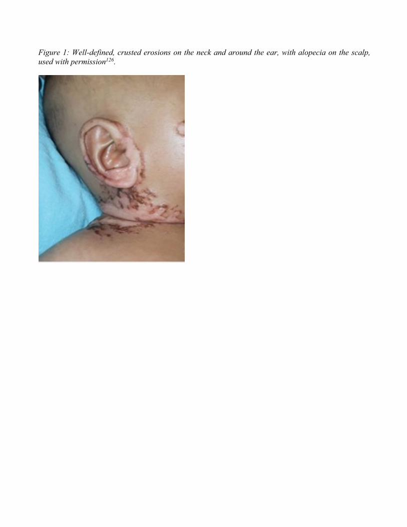
Figure 1: Well-defined, crusted erosions on the neck and around the ear, with alopecia on the scalp, used with permission126.


