Embed Size (px)
Citation preview
Downloaded from www.microbiologyresearch.org by
IP: 54.162.190.106
On: Sat, 06 Feb 2016 03:38:29
INTERNATIONAL JOURNAL OF SYSTEMATIC BACTERIOLOGY, Jan. 1997, p. 144-149 0020-7713/97/$04.00+ 0 Copyright 0 1997, International Union of Microbiological Societies
Vol. 47, No. 1
Desulfonatronovibrio hydrogenovorans gen. ~ o v . , sp. nov., an Alkaliphilic, Sulfate-Reducing Bacterium
T. N. ZHILINA,' G. A. ZAVARZIN,l F. A. RAINEY,2* E. N. PIKUTA,l G. A. OSIPOV,3 AND N. A. KOSTRIKINA'
Institute of Microbiology, Russian Academy of Sciences, 11 7811 MOSCOW,~ and State Institute of Biological Equipment
0-381 24 Braunschweig, Germany2 Manufacturing, Moscow, Russia, and Deutsche Sammlung von Mikroorganismen und Zellkulturen GmbH,
A new alkaliphilic, sulfate-reducing bacterium, strain Z-7935T (T = type strain), was isolated from a soda-depositing lake, Lake Magadi in Kenya. This organism is a motile vibrio which utilizes only hydrogen and formate as electron donors and sulfate, sulfite, and thiosulfate, but not sulfur, as electron acceptors. Thiosul- fate is dismutated. Strain Z-7935T is an obligately sodium-dependent alkaliphile which grows in sodium carbonate medium and does not grow at pH 7; the maximum pH for growth is more than pH 10, and the optimum pH is 9.5 to 9.7. The optimum NaCl concentration for growth is 3% (wt/vol). The optimum temper- ature for growth is 37°C. The G+C content of the DNA is 48.6 mol%. 16s ribosomal DNA sequence analysis revealed that strain Z-7935T represents a new lineage with genus status in the delta subclass of the Proteobac- teria. The name Desulfonatronovibrio hydrogenovorans gen. nov., sp. nov. is proposed for this organism; the type strain of D. hydrogenovorans is strain 2-7935 (= DSM 9292).
The development of alkalinity is often ascribed to sulfate- reducing bacteria, as suggested by Abd-el-Malek and Rizk for the Wadi-el-Natrun (1). Sulfate-reducing bacteria are thought to be responsible for the disappearance of sulfate in Lake Magadi. Direct evidence of sulfate reduction is found in the black muds interbedded with trona (Na,CO, * NaHCO, - 2H20), which indicate that H,S was formed and sulfate was depleted in the groundwater and that subsequent binding of sulfide by iron occurred in trachitic lava rocks in Lake Magadi (2). However, as pointed out by Tindall (17), this theory has not been supported by the successful isolation of alkaliphilic sulfate reducers which can grow at pH values greater than 9 from soda lakes, in which the pH is often greater than 10. Nevertheless, it has been shown for a group of Central Asian soda lakes with p H values greater than 9 that in these alkali- philic environments, in which the salinity is as high as 15%, sulfidogenesis constitutes a dominant hydrogen sink, compet- ing with acetogenesis but not with methanogenesis, which oc- curs via the methylotrophic pathway with the utilization of methanol and/or methylamines (26). Thus, the search for and isolation of alkaliphilic, sulfate-reducing bacteria is important for our understanding of (i) the bacterial diversity which em- ploys bioenergetics that are currently unknown or considered impossible, (ii) the role of these bacteria in community metab- olism as the main electron sink for hydrogen-producing pri- mary anaerobes, and (iii) the geochemical processes that are influenced by producers of extreme alkalinity in such environ- ments. The processes for which causative agent(s) have yet to be isolated remain unelucidated from a microbiological point of view.
Isolation of alkaliphilic, hydrogen-consuming, sulfate-reduc- ing bacteria from the alkaliphilic anaerobic community in Lake Magadi was first reported in 1994 (27). Here, we describe the isolation from Lake Magadi of an alkaliphilic, sulfate-reducing bacterium which grows in an extremely alkaline environment. The phylogenetic affiliation of this organism with representa- tives of the sulfate-reducing bacteria is described.
~ ~-
* Corresponding au thor . Present address: Depa r tmen t of Microbi- ology, Louisiana S ta te University, Baton Rouge, LA 70803-1715.
MATERIALS AND METHODS
Sampling. A sample was collected from sediments of an alkaline lake, Lake Magadi (East African Rift, Kenya). The sample was taken during the dry season of 1991 from a trench dug into the trona; it was assumed that the liquor at the sample site came from a depth of 8 to 10 ft under the surface of the trona and was enriched with NaC1. Subsamples of the mud were taken anaerobically, and these subsamples were transported to Moscow at the ambient temperature; in the laboratory they were stored at 4°C.
Media and growth conditions. Selective alkaline medium I1 (27) containing (per liter) 1.5 g of NaHCO,, 10 g of Na,CO,, 10 g of NaCI, 1 g of NH,CI, 0.2 g of KCI, 0.2 g of K,HPO,, 3 g of Na,SO,, 0.5 g of Na,S * 9H,O, 10 ml of a vitamin solution (24), 1 ml of a trace element solution (21), 0.5 g of yeast extract, and H, or formate as the substrate was used for enrichment and isolation. A pure culture was maintained in an optimized medium formulated after the study of the physiology of the new isolate was completed and was cultivated under strictly anaerobic conditions in rubber membrane-sealed glass vessels with H, in the gas phase. The optimized medium contained (per liter) 3.5 g of Na2C0,, 24 g of NaHCO, (added after cooling), 20 g of NaCI, 5 g of Na2S0,, 0.2 g of K,HPO,, 0.1 g of MgCI, - 7H,O, 0.5 g of NH,CI, 0.2 g of KCI, 0.12 g of sodium acetate or 0.5 g of yeast extract, 10 ml of a vitamin solution (24), 1 ml of a trace element solution (21), and 0.5 g of Na,S * 9H,O or thioglycolate (final pH 9.5). Cultures were incubated at 37°C. The substrates (electron donors) used were H, in the gas phase and sodium formate (5 @liter, with N, in gas phase). Electron acceptors were added at the following concentrations: Na,SO,*-, 30 mM; Na,S0,2-, 5 mM; Na,S,O,'-, 10 mM; NaNO,-, 10 mM; sodium fumarate, 10 mM; So, 2 giliter; and dimethyl sulfoxide, 2 ml/liter. Twenty-milliliter portions of the me- dium were distributed into 100-ml rubber-stoppered screw-cap flasks by using a standard strictly anaerobic technique. In the case of roll tube cultivation, 2% (wtivol) agar (Difco Laboratories, Detroit, Mich.) was added to 4-ml portions of carbonate-free medium. The carbonate solution was injected into Hungate tubes after sterilization. NaCl requirements were studied with optimized medium in which the Na,CO, and NaHCO, were replaced by 5 g of K,CO, per liter and the Na,S was replaced by K2S. Growth, detected as visible turbidity, was measured by determining the optical density at 600 nm in Hungate tubes with a spectro- photometer (Spekol, Jena, Germany) equipped with a type ER tube adapter. Microscopic counts were also used to determine growth. The pH limits for growth and sulfidogenesis were determined in the carbonate medium containing 10 g of Na,CO, per liter and 15 g of NaHCO, per liter; the pH was adjusted to the appropriate value by adding 6 N NaOH or 6 N HCl to an anaerobic flask with a built-in pH electrode under a stream of N,. After the dispensed medium had been autoclaved and the pH had been verified, the flasks were inoculated with a 1% (vol/vol) inoculum and incubated for 9 days at 37°C. The growth response was measured by determining sulfide formation and turbidity. Sulfidogenesis at different pH values was measured by monitoring H,S formation from radiola- belled sulfate at 37°C during incubation for 24 h (5,20). All of the chemicals used in this study were obtained from Russian suppliers.
Analytical procedures. The presence of desulfoviridin was checked fluoro- metrically by using a spectrofluorimeter (Hitachi, Tokyo, Japan) (10). H,S was quantified by the methylene blue reaction and colorimetric detection (18). Sul- fate consumption (or formation of sulfate from Na,S,O,) was measured by a
144

Downloaded from www.microbiologyresearch.org by
IP: 54.162.190.106
On: Sat, 06 Feb 2016 03:38:29
VOL. 47, 1997 DESULFONATRONOWBRIO GEN. NOV. 145
nephelometric reaction with BaCl, (4). Hydrogen consumption was monitored by measuring pressure and/or by gas chromatography with a type 5A molecular sieve column. Lipids were extracted from cell biomass that was dried in a stream of helium and then under a vacuum. To 30 mg of dry biomass, 200 p1 of a 5.4 N solution of anhydrous HCI in methanol was added, and the mixture was heated at 70°C for 2 h. The methyl esters of fatty acids and aldehyde derivatives obtained were extracted twice with 100 p1 of hexane. The extract was dried and silylated in 20 pl of N,O-bis(trimethylsi1yl)trifluoroacetamide for 15 min at 65°C. A 1-pl portion of the reaction mixture was analyzed with a model HP-5985B gas chro- matography-mass spectrometry system (Hewlett-Packard, Palo Alto, Calif.) equipped with a capillary column (25 by 0.25 mm) consisting of fused quartz containing an Ultra-1 nonpolar methylsilicone phase. The temperature profile included a 2-min isotherm at 150°C and subsequent programmed temperature increases (the temperature was increased at a rate of 5"C/min to 250°C and then at a rate of 10"C/min to 300°C). Data processing was carried out with an HP-1000 computer by using the standard programs of the gas chromatography-mass spec- trometry system (Hewlett-Packard).
Microscopy. The morphology of cultures was observed with an anoptral Ze- topan microscope (Reicherdt, Vienna, Austria). Negative staining of whole cells by phosphotungstic acid and fixation for preparation of thin sections were carried out as described previously (28). Microscopy was performed with a model JEM- 1OOC electron microscope (JEOL, Tokyo, Japan ).
G+C content. The guanine-plus-cytosine (G+C') content of the genomic DNA was determined by a thermal denaturation method (9). Escherichiu cob K-12 DNA was used as the standard.
16s rDNA sequence determination and data analysis. Genomic DNA was extracted and the gene coding for 16s rRNA (16s rDNA) was amplified as described previously (12). Purified PCR products were directly sequenced by using a Tuq DyeDeoxy terminator cycle sequencing kit (Applied Biosystems, Foster City, Calif.). Sequence reaction mixtures were electrophoresed with an Applied Biosystems model 373A DNA sequencer. The 16s rDNA sequence of strain Z-7935T (T = type strain) was manually aligned with sequences of repre- sentatives of the delta subclass of the Proteobucteriu. Painvise evolutionary dis- tances were computed by using the correction of Jukes and Cantor (6). The neighbor-joining method was used to reconstruct a phylogenetic tree from the distance matrices (14).
Nucleotide sequence accession numbers. Thc 16s rDNA sequences deter- mined in this study are available from EMBL under accession numbers X99234 to X99237.
RESULTS
Enrichment and isolation. The sediment sample used in this study had an oily black appearance, and a white mineral film formed on the surface. The purple bacteria usually found in the lagoons of Lake Magadi were absent at the sampling site. At the moment of sampling, the pH of the water was 10.2 and the temperature was 50°C. Samples of the sediment from Lake Magadi were incubated in 100-ml screw-cap bottles containing selective alkaline medium I1 (pH 9.7) with H, as the substrate; after several weeks of incubation, dominant vibrioid bacteria were observed. A pure culture was isolated by the serial dilu- tion method in liquid selective alkaline medium I1 containing 0.5 g of yeast extract per liter and H, as the substrates. Colo- nies of the sulfate reducer were obtained in roll tubes with sodium formate as the substrate. The colonies were yellowish, translucent, lens shaped, and less than 0.2 mm in diameter. The purity of the culture was indicated by the absence of growth on glucose-peptone sulfate-free medium containing ex- cessive quantities of yeast extract. The absence of growth in media containing various substrates utilized by sulfate-reduc- ing bacteria provided additional proof of purity. Several single colonies were isolated. One of these colonies, designated strain Z-7935T, was chosen for further characterization.
Morphology. Strain Z-7935T is a highly motile vibrio with a polar flagellum and filamentous appendages (Fig. 1). Motility was observed in young cultures but drastically diminished dur- ing 1 year of subcultivation. At pH 10 short spirilla were ob- served. The cells, which occurred singly or in pairs, were 0.5 km in diameter and 1.5 to 2 km long. Multiplication was by binary fission with the formation of two sister cells (Fig. lb). The cell wall had a typical gram-negative structure in ultrathin sections, and the periplasmic space was well-developed (Fig. 2). Spores were never observed.
b
FIG. 1. Morphology of strain Z-7935T. (A) Cells as viewed with the anoptral microscope. Bar = 10 pm. (B) Negatively stained dividing cells. Note the surface and appendages. Bar = 1 pm. (C) Negatively stained cell with monopolar flagellum. Bar = 1 pm.
Metabolic properties. Strain Z-7935T is very restricted with respect to the range of electron donors utilized. A total of 28 substrates were tested as electron donors, and only H, and formate were utilized. No growth occurred on the following compounds (each at a concentration of 5 @liter): acetate, pro- pionate, butyrate, pyruvate, lactate, malate, fumarate, succi- nate, methanol, ethanol, glycerol, glycine, cysteine, cystine, serine, alanine, glutamate, aspartate, Casamino Acids, yeast extract, choline, betaine, glucose, fructose, rhamnose, and mannose. Sulfate, sulfite, and thosulfate were utilized as elec-

Downloaded from www.microbiologyresearch.org by
IP: 54.162.190.106
On: Sat, 06 Feb 2016 03:38:29
146 ZHILINA ET AL. INT. J. SYST. BACTERIOL.
FIG. 2. Ultrastructure of strain 2-7935''. Bar = 0.5 pm
tron acceptors. Formation of sulfide was observed in the pres- ence of dimethyl sulfoxide; however, there was no growth in subcultures. Elemental sulfur inhibited growth. Fumarate and nitrate did not support growth. Strain Z-7935T was strictly anaerobic, and its growth was inhibited by air. Thiosulfate was decomposed; equimolar quantities of sulfate and H,S were formed from 10 mmol of thiosulfate, indicating that the organ- ism had the ability to dismutate.
Growth conditions. Like most hydrogen-consuming, sulfate- reducing bacteria, strain Z-7935IT needs organic compounds for anabolic reactions, and so its type of nutrition can be described as lithoheterotrophic. Growth dependence on yeast extract was observed (Fig. 3). However, yeast extract could be partially replaced by vitamins and acetate as a carbon source. A sodium acetate concentration of 0.16 g/liter was sufficient for optimal growth. The limits for growth and sulfidogenesis were determined with various pH values and salt contents. In growth experiments sulfidogenesis occurred without a lag phase at pH values between 9.5 and 9.7; this range of pH values was opti- mal, based on the rate of sulfide formation, and growth oc- curred at p H 8.0 to 10.2. In short-time experiments with ra- dioactive sulfate, sulfidogenesis increased linearly to a maximal value at pH 9.5 and rapidly decreased to one-tenth the maxi- mal value at pH 10. The pH of the medium did not change
1 6
0 4
0 2
0 ~ +- - ~ _ - ~~
0 025 0 5 0 75 1 125 1 5
Yeast Extract [gA]
FIG. 3. Efiect of yeast extract concentration o n the specific growth rate (k) of strain Z-793ST.
TABLE 1. Fatty acids in the cells of strain Z-7935T and Desulfohalobiurn retbaense HR ,oo
Fatty acid 96 of total fatty acids in:
Desulfohalobium retbaense HRlo{ Strain Z-793ST
C14:0 iso-C,,,O anteiso-C,,:, C15:I 0-6 CI5:O iS0-C I h:O
c,,:, w-9 CI6:l 0-7 Cl6:O iso-C,,,, w-7c iso-C I 7:o anteiso-C, 7:o
CI,:, w-6 C17:O
Cl,:, 0-9 CI8:1 w-7c
c17:1 w-8
Branched CIS:, w-6
w-5c
0 1.9 5.6 3.7 3.9 0 2.6 4.4 0 2.7
32.8 0 1.1 2.1 0 0 5.9 0 3.4 0 0 5.9
18.2 0 1 .o 1.6
0.11 1.68 i 0.1 0.95 15.28 -+ 2.3 3.6 i 0.3 0.23 3.93 t 0.4 5.34 i 0.5 0.58 4.10 t 0.7 13.33 t 2.8 3.88 t 0.1 1.9 i 0.4 2.24 t 1.1 8.05 2 1.5 0.71 2 0.6 3.33 -+ 0.8 0.36 0 6.53 t 2.2 0.23 3.86 i 1.4 15.97 t 5.2 0.43 0 0
Data from reference 8.
during growth. At pH values higher than 10 there was a de- crease in the rate of sulfidogenesis, while at pH 11.5 to 12 formation of H,S did not occur. Strain Z-7935T is an obligate alkaliphile which does not grow at p H 7; carbonate ions are required for growth. When Na,CO,-NaHCO, was replaced by NaCl and the pH values were maintained with 50 mM serine buffer (pK,, 9.4), no growth occurred. Dependence on sodium was studied in media containing 5 g of K2C03 per liter and increasing concentrations of NaC1. There was no growth or sulfidogenesis without sodium ions. Maximal sulfidogenesis was observed in the presence of 3% (wt/vol) NaCI, and at higher NaCl concentrations sulfidogenesis decreased. Strain Z-7935T is a weak halophile that is obligately dependent on sodium ions. Dependence on chloride ions was not detected. Strain Z-7935T is a mesophile. The optimal temperature for growth was 37"C, while at 40°C growth stopped after a fast start. At 45 and 15°C there was no growth, and at 22 or 26°C growth began after a lag phase. Desulfoviridin, a pigment com- mon to Desulfovibn'o species, was not found in strain Z-7935T.
Lipid analysis. The fatty acid distribution in the cells of strain Z-7935T is shown in Table 1. Saturated fatty acids ac- counted for 66.7% of the total fatty acids, with C16:o and acids predominating. Branched saturated fatty acids accounted for only 11% of the total fatty acids. The following monoun- saturated fatty acids, which accounted for 12% of the total fatty acids, were also present: CIGI1, C18:1 0-9, 0-11, and C19:1 fatty acids. The absence in strain Z-7935T of isoheptadecenoic acid (is0 CI7:,) and the presence of cis-vaccenic acid 0-11) are notable.
strain Z-7935T is 48.6 mol%. DNA base composition. The G + C content of the DNA of

Downloaded from www.microbiologyresearch.org by
IP: 54.162.190.106
On: Sat, 06 Feb 2016 03:38:29
VOL. 47, 1997
-
DESULFONATRONOVIBRIO GEN. NOV. 147
Desulfohalobiurn retbaense
-
L
strain TD3
Deulfovibrio desulfuricans -z Desulfovibrio vulgaris
Desulfovibno africanus
- ,- Desulfovibrio halophilus
Llesulfovibrio salexigens - -
Desulfovibrio longus
-I Desulfuromonas acetoxidans
Des ulfobulbus propionicus
Desulfococcus multivorans
Desulfomonile tiedjei
Desulfacinum infernurn
- Desulfobacter postgatei
5%
FIG. 4. Phylogenetic dendrogram showing the position of alkaliphilic, sul- fate-reducing bacterial strain Z-7935T within the radiation of sulfate-reducing bacteria belonging to the delta subclass of the Proteobacteria. The position of the root was determined by including Eschen'chia coli as an outgroup organism. Scale bar = 5 inferred substitutions per 100 nucleotides.
Phylogenetic analysis. An almost complete 16s rDNA se- quence comprising 1,503 nucleotides was determined for strain Z-7935T. A comparative analysis of this 16s rDNA sequence with the sequences available for representatives of the major bacterial groups indicated that strain Z-7935T is a member of the delta subclass of the Proteobacteria. In order to complete the data set for the halophilic members of the delta subclass of the Proteobacteria, the 16s rDNA sequences of Desulfohalo-
bium retbaense DSM 5692= and Desulfovibrio halophilus DSM 5663T were determined and included in the phylogenetic anal- ysis. The phylogenetic dendrogram in Fig. 4 shows the position of strain Z-7935T within the radiation of the members of the delta subclass of the Proteobacteria for which 16s rDNA se- quence data are available. Strain Z-7935T represents a distinct lineage, clustering with Desulfohalobium retbaense and strain TD3 (13). The highest levels of 16s rDNA sequence similarity were the levels of similarity between strain Z-7935T and Des- ulfohalobium retbaense (88.7%) and between strains Z-7935T and TD3 (86.6%) (Table 2).
DISCUSSION
The role of hydrogen-consuming, sulfate-reducing bacteria in the alkaliphilic community investigated in this study is in- teresting from an ecological and physiological viewpoint. The type of nutrition displayed by strain Z-7935~~ is typical of litho- heterotrophic, sulfate-reducing bacteria that utilize hydrogen or formate for the reduction of oxidized sulfur compounds in the catabolic reaction and acetate in the anabolic pathway (16). Strain Z-7935T differs from other sulfate-reducing bacteria by its unusually narrow range of electron donors utilized. Strain Z-7935T is a lithoheterotrophic, sodium-dependent alkaliphile which may be responsible for the consumption of hydrogen in the alkaliphilic community from which it was isolated. This organism is clearly a member of an alkaliphilic anaerobic com- munity in which primary anaerobes (e.g., alkaliphilic spiro- chetes [27, 281) produce hydrogen and acetate.
The geographical distribution of alkaliphilic sulfate reducers in continental soda lakes was revealed by the study of anaer- obic communities in the lakes of Central Asia. Physiologically similar pure cultures were isolated from four alkaline lakes in Tuva, and these cultures were found to belong to the same species and to have DNA-DNA homology values of more than 90% (7). It was demonstrated that in Asian lakes, which rep- resent a range of pH and salinity combinations and harbor different microbial communities, as in "natural elective cul-
TABLE 2. 16s rDNA similarity values for strain Z-7935T and related taxa
% 16s rDNA similarity to:
Desulfohalobium retbaense Strain TD3 Desulfomicrobium baculatus Desulfovibrio vulgaris Desulfovibrio desulfuricans Desulfovibno halophilus Desulfovibrio salexigens Desulfovibrio ajhcanus Desulfovibrio gigas Desulfovibrio longus Desulfomonile tiedjei Desulfacimum infernurn Desulfococcus multivorans Desulfobulbus propionicus Desulfobacter postgatei Desulfuromonas acetoxidans Escherichia coli
88.7 86.6 88.0 86.5 85.7 85.1 86.0 85.9 85.8 85.9 86.3 85.9 85.0 87.4 91.9 86.5 86.7 85.8 86.5 88.3 87.8 87.1 87.1 85.9 85.5 88.4 87.4 91.0 86.5 85.6 84.7 87.1 90.3 89.0 88.6 87.8 87.1 88.1 86.5 85.5 87.8 89.0 87.6 87.0 89.1 86.3 86.6 84.9 86.1 89.1 89.2 90.2 89.2 89.5 88.7 82.9 83.3 82.5 81.7 81.4 82.4 83.7 81.5 82.7 82.6 84.1 83.2 84.4 84.1 82.5 82.4 82.7 82.3 82.0 83.5 83.5 82.9 87.2 83.9 84.3 83.2 84.4 83.5 84.1 85.1 83.0 83.4 83.4 84.0 87.9 86.5 84.0 84.0 84.2 83.0 83.9 83.5 82.9 82.1 82.5 83.4 83.5 85.8 86.4 85.2 83.0 82.8 80.7 83.4 81.5 83.0 81.7 82.0 82.3 83.1 82.7 84.0 83.5 87.0 85.3 85.3 85.3 83.4 83.6 84.1 84.5 84.6 84.4 84.4 85.3 84.5 87.6 86.4 86.1 87.1 86.4 80.1 80.3 80.0 79.5 79.8 79.8 80.6 79.5 80.5 79.4 79.2 80.0 78.1 81.2 80.8 78.2 80.9
Downloaded from www.microbiologyresearch.org by
IP: 54.162.190.106
On: Sat, 06 Feb 2016 03:38:29
148 ZHILINA ET AL. INT. J. SYST. BACTERIOL.
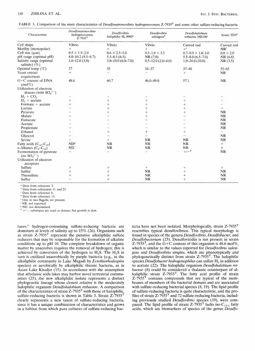
TABLE 3. Comparison of the main characteristics of Desulfonatronovibn'o hydrogenovorans Z-7935T and some other sulfate-reducing bacteria
Characteristic Desulfovibrio halophilus SL 8903"
Desulfonatronovibrio hydrogenovorans
Z-793S Desulfovibrio
salexigensb Desulfohalobium Strain TD3d retbaense HR100"
Cell shape Motility (monopolar) Cell size (pm) pH range (optimal pH) Salinity range (optimal
Optimal temp ("C) Yeast extract
G+C content of DNA
Utilization of electron
salinity) (%)
requirement
(mol%)
donors (with SO,,-) H2 + CO, H, + acetate Formate + acetate Lactate Pyruvat e Malate Fumarate Acetate Propionate Ethanol Glycerol Serine
Fatty acids (C14-C18) n-Alkanes (C6-C16) Fermentation of pyruvate
Utilization of electron (no SO,,-)
acceptors Sulfate Sulfite Thiosulfate Sulfur
Vibrio Vibrio + + 0.5 X 1.5-2.0 8.0-10.2 (9.5-9.7) 5.5-8.5 (6.5) 1.0-12.0 (3.0)
0.6 X 2.5-5.0
3 .O-18 .O (6.0-7.0)
37 +
35 -
48.6 60.7
+ + + -
+ + + +
Vibrio Curved rod + +e
0.5-1.0 X 3.5 NR (7.0) 5.5-8.0 (6.5-7.0) 0.5-12.0 (2.0-4.0) 1.0-24.0 (10.0)
0.7-0.9 X 1.0-3.0
34-37 -
37-40 +
46.0-49.0 57.1
-
+ + + + + - - -
+ NR NR NR
-
-
+ NR NR NR
-
+ + + + - -
-
-
+ NR NR NR
-
+ / - h
+ + + +
Curved rod
0.8 X 2.0 NR(6.8) NR (3.5)
N R ~
55-65 NR
NR
-
-
-
-
NR NR NR NR NR
NR NR + + NR
-
+ NR NR NR
'' Data from reference 3. ' Data from references 11 and 23.
Data from reference 8. '' Data from reference 13. (' One or two flagella are present. ' NR, not reported. S ND, not determined. I' +I-, substrates are used as donors, but growth is slow
tures," hydrogen-consuming sulfate-reducing bacteria are dominant at levels of salinity up to 15% (26). Organisms such as strain Z-7935T represent the putative alkaliphilic sulfate reducers that may be responsible for the formation of alkaline conditions up to pH 10. The complete breakdown of organic matter by anaerobes requires the removal of hydrogen; this is achieved by conversion of the hydrogen to H,S. The H,S in turn is oxidized anaerobically by purple bacteria (e.g., in the alkaliphilic community in Lake Magadi by Ectothiorhodospira species) or aerobically by alkaliphilic thionic bacteria, as in Asian Lake Khadyn (15). In accordance with the assumption that athalassic soda lakes may harbor novel terrestrial commu- nities (25), the new alkaliphilic isolate represents a distinct phylogenetic lineage whose closest relative is the moderately halophilic organism Desulfohalobium retbaense. A comparison of the characteristics of strain Z-7935T with those of halophilic, sulfate-reducing bacteria is shown in Table 3. Strain Z-7935T clearly represents a new taxon of sulfate-reducing bacteria, since it has a unique combination of characteristics and grows in a habitat from which pure cultures of sulfate-reducing bac-
teria have not been isolated. Morphologically, strain Z-7935T resembles typical desulfovibrios. This typical morphology is found in species of the genera Desulfovibrio, Desulfobacter, and Desulfobacterium (23). Desulfoviridin is not present in strain Z-7935T, and the G + C content of this organism is 48.6 mol%, which is similar to the values reported for Desulfovibiio salexi- gens and Desulfovibrio simplex, which are phenotypically and phylogenetically distinct from strain Z-7935T. The halophilic species Desulfobacter hydrogenophilus can utilize H, in addition to acetate (22). The halophilic organism Desulfohalobium ret- baense (8) could be considered a thalassic counterpart of al- kaliphilic strain Z-7935T. The fatty acid profile of strain Z-7935T contains compounds that are typical of the mem- branes of members of the bacterial domain and are associated with sulfate-reducing bacterial species (8, 19). The lipid profile of sulfate-reducing bacteria is quite characteristic, and the pro- files of strain Z-7935T and 72 sulfate-reducing bacteria, includ- ing previously studied Desulfovibrio species (19), were com- pared. The lipid profile of strain Z-7935T lacks iso-C17,1 fatty acids, which are biomarkers of species of the genus Desulfo-

Downloaded from www.microbiologyresearch.org by
IP: 54.162.190.106
On: Sat, 06 Feb 2016 03:38:29
VOL. 47, 1997 DESULFONATRONOI.?BRIO GEN. NOV. 149
vibrio. On the basis of its fatty acid profile, strain Z-7935T cannot be affiliated with any species except Desulfohalobium retbaense and Desulfomicrobium baculatus [sic] (correlation co- efficient, 0.23). On the basis of the phylogenetic analysis it can be concluded that strain Z-7935T is a member of a recently discovered new lineage within the delta branch of the Pro- teobacteria that comprises hydrocarbon-utilizing strain TD3 (13) and Desulfohalobium retbaense (Fig. 4 and Table 2). With its distinct branch point and 16s rDNA sequence difference of >11% compared with its nearest relative, as well as its distinct phenotypic characteristics (Table 3), strain Z-7935T clearly represents a new genus. We propose that the alkaliphilic, sul- fate-reducing strain Z-7935T should be placed in a new genus and species, Desulfonatronovibrio hydrogenovorans; the name of this organism describes its ecophysiology.
Description of Desulfonatronovibrio gen. nov. Desulfona- tronovibrio (De.sul.fo.nat.ro.no.vi’ b.rio. M. L. pref. de, nega- tive; M.L.n. sulfo, sulfate; M.L.n. natron, soda; M.L.n. vibrio, curved rod; M.L.n. Desulfonatronovibrio, sulfate-reducing curved rod from a soda environment). Alkaliphilic sulfate- reducing eubacterium. Cells are motile, asporogenous, gram- negative vibrios with polar flagella. Lithoheterotrophic, utiliz- ing hydrogen for the reduction of sulfur compounds. Strictly anaerobic. Obligately dependent on sodium ions. Vitamins and acetate as a carbon source are necessary for growth on hydro- gen or formate and can be replaced by yeast extract. Phyloge- netically a member of the delta subclass of the Proteobacteria. The type species is Desulfonatronovibrio hydrogenovorans.
Description of Desulfonatronovibrio hydrogenovorans sp. nov. Desulfonatronovibrio hydrogenovorans (hy . dro .ge. no .vo ’ rans. M.L.n. hydrogen, hydrogen; M.L. part. vorans, utilizing; M.L. part. hydrogenovorans, hydrogen utilizing). Motile vibrio with one polar flagellum. Cells are 0.5 by 1.5 to 2 km, occur singly or in pairs, and develop short spirilla under suboptimal condi- tions. Multiplication is by binary fission. Gram-negative cell wall structure. Strictly anaerobic and lithoheterotrophic. Uti- lizes only hydrogen and formate as electron donors and sulfate, sulfite, and thiosulfate as electron acceptors. Sulfur is not re- duced. Sulfide is the only product of catabolism. Yeast extract and acetate are utilized for anabolism. Obligate alkaliphile which does not grow at pH 7; the maximum pH for growth is about 10.2, and the optimal pH for growth and sulfidogenesis in sodium carbonate medium is 9.5 to 9.7. Sodium ions are required for growth; no growth occurs in the presence of NaCl concentrations less than 1% (wt/vol) or more than 12% (wt/ vol). Growth occurs if NaCl is replaced by equimolar amounts of Na,CO, and NaHCO,. Requires carbonate anion. The op- timum temperature for growth is 37”C, and the temperature range for growth is 15 to 43°C. Slow growth occurs at 22 to 26°C after a long lag phase. The G+C content of the DNA is 48.6 mol% (as determined by the thermal denaturation meth- od). Habitat: bottom deposits of alkaline athalassic soda lakes. The type strain is strain 2-7935, which was isolated from the sediments of an equatorial soda lake, Lake Magadi. This strain has been deposited in the Deutsche Sammlung von Mikroor- ganismen as strain DSM 9292T.
ACKNOWLEDGMENTS
We are grateful to A. M. Lysenko for determining the DNA G + C content and to Marcus Schmidt for assistance in preparing graphics. We thank B. Tindall for providing rare old literature on Lake Magadi.
This work was supported in part by grants to G.A.Z. from ISF 68300 and from the Russian State “Biodiversity” program.
REFERENCES
1. Abd-el-Malek, Y., and S. G. Rizk. 1963. Bacterial sulphate reduction and the development of alkalinity. 111. Experiments under natural conditions. J. Appl. Bacteriol. 2620-26.
2. Baker, B. H. 1958. The geology of the Magadi area. Report no. 42, p. 81. Geological Survey of Kenya, Nairobi, Kenya.
3. Caumette, P., Y. Cohen, and R. Matheron. 1991. Isolation and characteriza- tion of Desulfovibrio halophilus sp. nov., a halophilic sulfate-reducing bacte- rium isolated from Solar Lake (Sinai). Syst. Appl. Microbiol. 14:33-38.
4. Cypionka, H., and N. Pfennig. 1986. Growth yields of Desulfotornaculurn orientis with hydrogen in chemostat culture. Arch. Microbiol. 143:396-399.
5. Ivanov, M. V. 1956. Application of isotopes for the studying sulfate reduction in Lake Belovod. Mikrobiologiya 29305-309.
6. Jukes, T. H., and C. R. Cantor. 1969. Evolution of protein niolecules, p. 21-132. In H. N. Munro (ed.), Mammalian protein metabolism. Academic Press, New York, N.Y.
7. Lysenko, A., and E. Pikuta. Unpublished data. 8. Ollivier, B., C. E. Hatchikian, G. Premier, J. Guezennec, and J. L. Garcia.
1991. Desulfohalobiurn retbaense gen. nov., sp. nov., a halophilic sulfate- reducing bacterium from sediments of a hypersaline lake in Senegal. Int. J. Syst. Bacteriol. 41:74-81.
9. Owen, R. J., R. Hill, and S. P. Lapage. 1969. Determination of DNA base composition from melting profiles in dilute buffers. Biopolymers 7503-516.
10. Postgate, J. R. 1984. The sulphate-reducing bacteria, 2nd ed. Cambridge University Press, Cambridge, United Kingdom.
11. Postgate, J. R., and L. L. Campbell. 1966. Classification of Desulfovibrio species, the nonsporulating sulfate-reducing bacteria. Bacteriol. Rev. 30:
12. Rainey, F. A., N. Ward-Rainey, R. M. Kroppenstedt, and E. Stackebrandt. 1996. The genus Nocardiopsis represents a phylogenetically coherent taxon and a distinct actinomycete lineage: proposal of Nocardiopsaceae fam. nov. Int. J. Syst. Bacteriol. 461088-1092.
13. Rueter, P., R. Rabus, H. Wilkes, F. Aeckersberg, F. A. Rainey, H. W. Jann- asch, and F. Widdel. 1994. Anaerobic oxidation of hydrocarbons in crude oil by new types of sulphate-reducing bacteria. Nature 372:455-458.
14. Saitou, N., and M. Nei. 1987. The neighbor-joining method: a new method for reconstructing phylogenetic trees. Mol. Biol. Evol. 4:406-425.
15. Sorokin, D. Y., A. M. Lysenko, and L. L. Mitushina. 1996. Isolation and characterization of alkaliphilic heterotrophic bacteria, which oxidize reduced sulfur compounds to tetrationate. Microbiology 65326-338.
16. Sorokin, Y. 1966. Role of carbon dioxide and acetate in biosynthesis by sulfate-reducing bacteria. Nature 210551-552.
17. Tindall, B. J. 1986. Procaryotic life in the alkaline, saline, athalassic envi- ronment, p. 31-67. In F. Rodriguez-Valera (ed.), Halophilic bacteria. CRC Press, Boca Raton, Fla.
18. Truper, H. G., and H. G. Schlegel. 1964. Sulphur methabolism in Thiorho- dacaae. I. Quantitative measurements on growing cells of Chrornatium oke- nii. Antonie van Leeuwenhoek. J. Microbiol. Serol. 30:225-238.
19. Vainshtein, M., H. Hippe, and R. M. Kroppenstedt. 1992. Cellular fatty acid composition of Desulfovibrio species and its use in classification of sulfate- reducing bacteria. Syst. Appl. Microbiol. 15554-566.
20. Vainshtein, M. B., and C. S. Laurinavichus. 1988. Enumeration and culti- vation of anaerobic bacteria. Institute of Biochemistry and Physiology of Microorganisms, Russian Academy of Sciences, Pushchino, Russia.
21. Whitman, W. B., E. Ankwanda, and M. L. Wolfe. 1982. Nutrition and carbon metabolism of Methanococcus voltue. J. Bacteriol. 149852-863.
22. Widdel, F. 1987. New types of acetate-oxidizing, sulfate-reducing Desul- fobacter species, D. hydrogenophilus sp. nov., D. latus sp. nov., and D. curvatus sp. nov. Arch. Microbiol. 148:286-291.
23. Widdel, F. 1988. Microbiology and ecology of sulfate- and sulfur-reducing bacteria, p. 469-585. In A. J. B. Zehnder (ed.), Biology of anaerobic micro- organisms. John Wiley & Sons, New York, N.Y.
24. Wolin, E. A., M. J. Wolin, and R. S. Wolfe. 1963. Formation of methane by bacterial extracts. J. Biol. Chem. 2382882-2886.
25. Zavarzin, G. A. 1993. Epicontinental alkaline water bodies as relict biotopes for the development of terrestrial biota. Microbiology 62:789-800.
26. Zavarzin, G. A., T. N. Zhilina, and E. V. Pikuta. 1996. Secondary anaerobes in the haloalkaliphilic communities in Tuva Lakes. Microbiologia 65480- 486.
27. Zhilina, T. N., and G. A. Zavarzin. 1994. Alkaliphilic anaerobic community at pH 10. Curr. Microbiol. 29109-112.
28. Zhilina, T. N., G. A. Zavarzin, F. Rainey, V. V. Kevbrin, N. A. Kostrikina, and A. M. Lysenko. 1996. Spirochaeta alkalica sp. nov., Spirochaeta afncana sp. nov., and Spirochaeta asiatica sp. nov., alkaliphilic anaerobes from the continental soda lakes in Central Asia and the East African Rift. Int. J. Syst. Bacteriol. 46:305-312.
732-738.