Embed Size (px)
Citation preview
ASSOCIATE EDITOR: EMILY E. SCOTT
Diabetic Peripheral Neuropathy: Should a ChaperoneAccompany Our Therapeutic Approach?
Kevin L. Farmer, Chengyuan Li, and Rick T. Dobrowsky
Department of Pharmacology and Toxicology, University of Kansas, Lawrence, Kansas
Abstract . . . . . . . . . . . . . . . . . . . . . . . . . . . . . . . . . . . . . . . . . . . . . . . . . . . . . . . . . . . . . . . . . . . . . . . . . . . . . . . . 881I. Introduction . . . . . . . . . . . . . . . . . . . . . . . . . . . . . . . . . . . . . . . . . . . . . . . . . . . . . . . . . . . . . . . . . . . . . . . . . . . . . 881
II. Pathogenesis of diabetic peripheral neuropathy . . . . . . . . . . . . . . . . . . . . . . . . . . . . . . . . . . . . . . . . . . . . . 882A. Polyol pathway . . . . . . . . . . . . . . . . . . . . . . . . . . . . . . . . . . . . . . . . . . . . . . . . . . . . . . . . . . . . . . . . . . . . . . . 882B. Advanced glycation end-products . . . . . . . . . . . . . . . . . . . . . . . . . . . . . . . . . . . . . . . . . . . . . . . . . . . . . . . 882C. Protein kinase C. . . . . . . . . . . . . . . . . . . . . . . . . . . . . . . . . . . . . . . . . . . . . . . . . . . . . . . . . . . . . . . . . . . . . . 883D. Poly(ADP-ribose) polymerase pathway . . . . . . . . . . . . . . . . . . . . . . . . . . . . . . . . . . . . . . . . . . . . . . . . . . 883E. Hexosamine pathway . . . . . . . . . . . . . . . . . . . . . . . . . . . . . . . . . . . . . . . . . . . . . . . . . . . . . . . . . . . . . . . . . 883F. Oxidative/nitrosative stress and mitochondrial dysfunction . . . . . . . . . . . . . . . . . . . . . . . . . . . . . . . 883G. Inflammation, lipid mediators, and dyslipidemia. . . . . . . . . . . . . . . . . . . . . . . . . . . . . . . . . . . . . . . . . 884H. Growth factors . . . . . . . . . . . . . . . . . . . . . . . . . . . . . . . . . . . . . . . . . . . . . . . . . . . . . . . . . . . . . . . . . . . . . . . 885
III. Management of diabetic peripheral neuropathy . . . . . . . . . . . . . . . . . . . . . . . . . . . . . . . . . . . . . . . . . . . . . 886A. Targeting hyperglycemia . . . . . . . . . . . . . . . . . . . . . . . . . . . . . . . . . . . . . . . . . . . . . . . . . . . . . . . . . . . . . . 886B. Targeting casual mechanisms. . . . . . . . . . . . . . . . . . . . . . . . . . . . . . . . . . . . . . . . . . . . . . . . . . . . . . . . . . 886
1. Aldose reductase inhibitors . . . . . . . . . . . . . . . . . . . . . . . . . . . . . . . . . . . . . . . . . . . . . . . . . . . . . . . . . 8862. Blocking advanced glycation end-product formation and activation of the receptor for
advanced glycation end-products . . . . . . . . . . . . . . . . . . . . . . . . . . . . . . . . . . . . . . . . . . . . . . . . . . . . 8873. Protein kinase C inhibitors . . . . . . . . . . . . . . . . . . . . . . . . . . . . . . . . . . . . . . . . . . . . . . . . . . . . . . . . . 8874. Hexosamine pathway . . . . . . . . . . . . . . . . . . . . . . . . . . . . . . . . . . . . . . . . . . . . . . . . . . . . . . . . . . . . . . 8875. Antioxidants, �-lipoic acid (Thiocytic acid). . . . . . . . . . . . . . . . . . . . . . . . . . . . . . . . . . . . . . . . . . . . 8886. Growth factors . . . . . . . . . . . . . . . . . . . . . . . . . . . . . . . . . . . . . . . . . . . . . . . . . . . . . . . . . . . . . . . . . . . . 888
C. Targeting painful symptoms . . . . . . . . . . . . . . . . . . . . . . . . . . . . . . . . . . . . . . . . . . . . . . . . . . . . . . . . . . . 8881. Tricyclic antidepressants . . . . . . . . . . . . . . . . . . . . . . . . . . . . . . . . . . . . . . . . . . . . . . . . . . . . . . . . . . . 8882. Anticonvulsants . . . . . . . . . . . . . . . . . . . . . . . . . . . . . . . . . . . . . . . . . . . . . . . . . . . . . . . . . . . . . . . . . . . 8883. Opioids and topical analgesics . . . . . . . . . . . . . . . . . . . . . . . . . . . . . . . . . . . . . . . . . . . . . . . . . . . . . . 888
D. Chaperoning diabetic stress—targeting tolerance? . . . . . . . . . . . . . . . . . . . . . . . . . . . . . . . . . . . . . . . 8891. Controlling the biological outcome of inhibiting the 90-kDa heat-shock protein:
dissociating cytotoxicity from cytoprotection is paramount to attain specifictherapeutic efficacies . . . . . . . . . . . . . . . . . . . . . . . . . . . . . . . . . . . . . . . . . . . . . . . . . . . . . . . . . . . . . . . 889
2. Heat-shock protein expression and DPN—an impaired defense against stress? . . . . . . . . . . . 8903. 70-kDa heat-shock protein and neuroprotection in DPN . . . . . . . . . . . . . . . . . . . . . . . . . . . . . . . 8914. 70-kDa heat-shock protein family members and mitochondrial function . . . . . . . . . . . . . . . . . 891
E. Exercise as a nonpharmacological therapy for DPN . . . . . . . . . . . . . . . . . . . . . . . . . . . . . . . . . . . . . . 894IV. Conclusions . . . . . . . . . . . . . . . . . . . . . . . . . . . . . . . . . . . . . . . . . . . . . . . . . . . . . . . . . . . . . . . . . . . . . . . . . . . . . 895
Acknowledgments . . . . . . . . . . . . . . . . . . . . . . . . . . . . . . . . . . . . . . . . . . . . . . . . . . . . . . . . . . . . . . . . . . . . . . . . 895References . . . . . . . . . . . . . . . . . . . . . . . . . . . . . . . . . . . . . . . . . . . . . . . . . . . . . . . . . . . . . . . . . . . . . . . . . . . . . . 896
Address correspondence to: Rick T. Dobrowsky, Department of Pharmacology and Toxicology, The University of Kansas, 5064 Malott Hall,1251 Wescoe Hall Dr., Lawrence, KS 66045. E-mail [email protected]
This article is available online at http://pharmrev.aspetjournals.org.http://dx.doi.org/10.1124/pr.111.005314.
1521-0081/12/6404-880–900$25.00PHARMACOLOGICAL REVIEWS Vol. 64, No. 4Copyright © 2012 by The American Society for Pharmacology and Experimental Therapeutics 5314/3787657Pharmacol Rev 64:880–900, 2012
880
Abstract——Diabetic peripheral neuropathy (DPN)is a common complication of diabetes that is associ-ated with axonal atrophy, demyelination, blunted re-generative potential, and loss of peripheral nerve fi-bers. The development and progression of DPN is duein large part to hyperglycemia but is also affected byinsulin deficiency and dyslipidemia. Although numer-ous biochemical mechanisms contribute to DPN, in-creased oxidative/nitrosative stress and mitochon-drial dysfunction seem intimately associated withnerve dysfunction and diminished regenerative ca-pacity. Despite advances in understanding the etiol-ogy of DPN, few approved therapies exist for the phar-macological management of painful or insensate DPN.Therefore, identifying novel therapeutic strategies re-mains paramount. Because DPN does not develop witheither temporal or biochemical uniformity, its thera-peutic management may benefit from a multifaceted
approach that inhibits pathogenic mechanisms, man-ages inflammation, and increases cytoprotective re-sponses. Finally, exercise has long been recognized asa part of the therapeutic management of diabetes, andexercise can delay and/or prevent the development ofpainful DPN. This review presents an overview of ex-isting therapies that target both causal and symptom-atic features of DPN and discusses the role of up-reg-ulating cytoprotective pathways via modulatingmolecular chaperones. Overall, it may be unrealisticto expect that a single pharmacologic entity will suf-fice to ameliorate the multiple symptoms of humanDPN. Thus, combinatorial therapies that target causalmechanisms and enhance endogenous reparative ca-pacity may enhance nerve function and improve re-generation in DPN if they converge to decrease oxida-tive stress, improve mitochondrial bioenergetics, andincrease response to trophic factors.
I. Introduction
DPN1 is the most prevalent complication of diabetesand often manifests as a distal, symmetric, sensorimotorneuropathy. In the United States, 26.8 million peopleare affected by diabetes; by the year 2030, that numberis predicted to increase to approximately 35.9 millionpeople (Shaw et al., 2010). A recent population-basedstudy reported that more than half of patients who havetype 1 or 2 diabetes develop DPN (Harati, 2007). Ofthese patients with DPN, 15 to 30% suffer from painfuldiabetic neuropathy, whereas the remainder experiencea loss of sensation and numbness (Ramos et al., 2007).Clinical symptoms associated with DPN involve poorgait and balance associated with large sensory fibersand abnormal cold and/or heat sensation associated withsmall sensory fibers. Chronic pain associated with dia-betes is represented by hyperalgesia, allodynia, pares-thesias, and spontaneous pain (Gooch and Podwall,2004; Edwards et al., 2008). Symptoms are described astingling, “pins and needles,” burning, itching, and anabnormal sensation to pain and temperature. Over time,these symptoms may advance from the toes to the foot
and up the leg, and these symptoms may occur in thefingers and hands (Zochodne, 2007).
DPN arises as a result of the degeneration of small,unmyelinated C fibers or thinly myelinated A� sensoryfibers that mediate pain/temperature sensation. Diabe-tes-induced changes in C fibers lead to the developmentof small-fiber neuropathy, which often produces positive(painful) symptoms: allodynia and hyperesthesias. Pro-gressive neurodegeneration may spontaneously resolvethe neuropathic pain, but decreased response thresholds(Lennertz et al., 2011) and loss of epidermal innervationof C fibers in the feet (Beiswenger et al., 2008) cancontribute to negative neuropathic symptoms such asthermal hypoalgesia. Likewise, degeneration of A� fi-bers leads to a loss of vibration and tactile sensation(Lennertz et al., 2011) with axon-myelin separation(Powell and Myers, 1983; Myers and Powell, 1984; Loveet al., 1986) and eventual segmental demyelination inlong-term DPN (Zochodne, 2007). Thus, deficits in bothC and A� fiber function greatly affects the detection ofnoxious and non-noxious stimuli. Unfortunately, thecomplexity of the multiple and temporally nonuniformbiochemical insults within endothelium, neurons, andSchwann cells that contribute to the sensory phenotypesassociated with DPN has rendered the development ofeffective therapeutics difficult.
Many excellent reviews have been published that 1)describe the various biochemical insults that contributeto the pathogenesis of DPN and 2) outline treatmentstrategies directed at blocking either causal mecha-nisms or treating neuropathic pain (Leinninger et al.,2004; Vincent et al., 2004, 2009b; Pop-Busui et al., 2006;Calcutt and Backonja, 2007; Zochodne, 2007, 2008; Cal-cutt et al., 2008, 2009; Edwards et al., 2008; Tavakoliand Malik, 2008; Veves et al., 2008; Obrosova, 2009a;Fernyhough et al., 2010; Sivitz and Yorek, 2010; Maliket al., 2011). Thus, the intent of the current review is tobriefly highlight and update many of these features andadd to the discussion by proposing that both pharmaco-
1Abbreviations: AGE, advanced glycation end product; AR, aldosereductase; BDNF, brain-derived neurotrophic factor; COX, cyclooxygen-ase; DCCT, Diabetes Control and Complication Trial; DPN, diabeticperipheral neuropathy; DRG, dorsal root ganglia; F-6-P, fructose-6phosphate; FDA, U.S. Food and Drug Administration; GDNF, glialcell-derived neurotrophic factor; GlcNAc, N-acetyl glucosamine; HETE,hydroxyeicosatetraenoic; HSF, heat-shock factor; Hsp, heat-shock pro-tein; HSR, heat-shock response; KU-32, N-{7-[(2R,3R,4S,5R)-3,4-dihydroxy-5-methoxy-6,6-dimethyl-tetrahydro-2H-pyran-2-yloxy]-8-methyl-2-oxo-2H-chromen-3-yl}acetamide; LA, �-lipoic acid; MAPK,mitogen-activated protein kinase; MNCV, motor nerve conduction ve-locity; MnSOD, manganese superoxide dismutase; mtHsp70, mitochon-drial paralog of Hsp70; NF-�B, nuclear factor �B; NGF, nerve growthfactor; NT, neurotrophin; PARP, poly(ADP-ribose) polymerase; PKC,protein kinase C; PKI-166, 4-phenethylamino-6-(yderoxyl)phenyl-7H-pyrrolo(2,3-d)pyrimidine; RAGE, receptor for AGE; RBX, ruboxistau-rin; ROS, reactive oxygen species; SNCV, sensory nerve conductionvelocity; sRAGE, soluble RAGE; STZ, streptozotocin; TCA, tricyclicantidepressant; TNF, tumor necrosis factor.
MECHANISMS AND THERAPIES IN DIABETIC PERIPHERAL NEUROPATHY 881
logic and nonpharmacologic approaches that help mod-ulate the expression and activity of cytoprotective mo-lecular chaperones may afford a complementary tactictoward improving the management of DPN.
II. Pathogenesis of DiabeticPeripheral Neuropathy
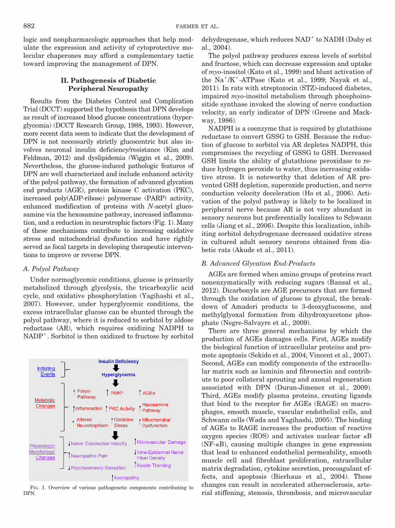
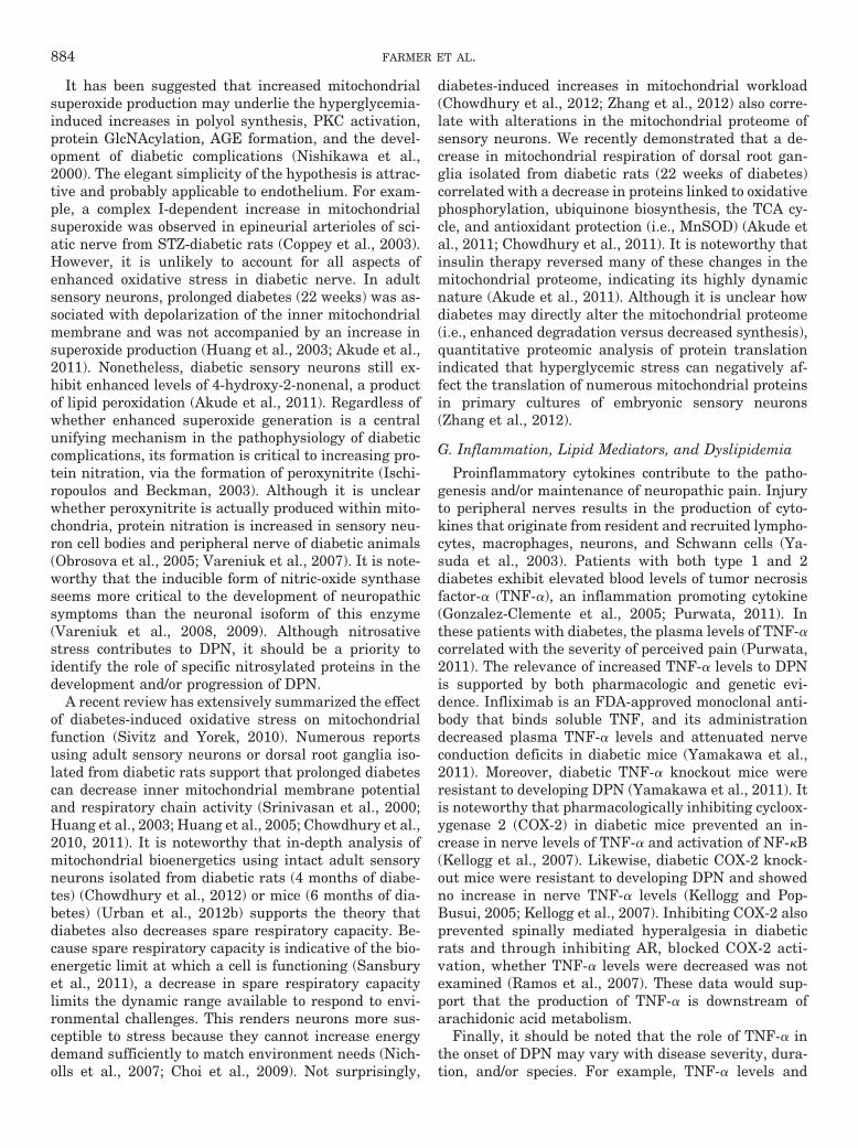
Results from the Diabetes Control and ComplicationTrial (DCCT) supported the hypothesis that DPN developsas result of increased blood glucose concentrations (hyper-glycemia) (DCCT Research Group, 1988, 1993). However,more recent data seem to indicate that the development ofDPN is not necessarily strictly glucocentric but also in-volves neuronal insulin deficiency/resistance (Kim andFeldman, 2012) and dyslipidemia (Wiggin et al., 2009).Nevertheless, the glucose-induced pathologic features ofDPN are well characterized and include enhanced activityof the polyol pathway, the formation of advanced glycationend products (AGE), protein kinase C activation (PKC),increased poly(ADP-ribose) polymerase (PARP) activity,enhanced modification of proteins with N-acetyl gluco-samine via the hexosamine pathway, increased inflamma-tion, and a reduction in neurotrophic factors (Fig. 1). Manyof these mechanisms contribute to increasing oxidativestress and mitochondrial dysfunction and have rightlyserved as focal targets in developing therapeutic interven-tions to improve or reverse DPN.
A. Polyol Pathway
Under normoglycemic conditions, glucose is primarilymetabolized through glycolysis, the tricarboxylic acidcycle, and oxidative phosphorylation (Yagihashi et al.,2007). However, under hyperglycemic conditions, theexcess intracellular glucose can be shunted through thepolyol pathway, where it is reduced to sorbitol by aldosereductase (AR), which requires oxidizing NADPH toNADP�. Sorbitol is then oxidized to fructose by sorbitol
dehydrogenase, which reduces NAD� to NADH (Duby etal., 2004).
The polyol pathway produces excess levels of sorbitoland fructose, which can decrease expression and uptakeof myo-inositol (Kato et al., 1999) and blunt activation ofthe Na�/K�-ATPase (Kato et al., 1999; Nayak et al.,2011). In rats with streptozocin (STZ)-induced diabetes,impaired myo-inositol metabolism through phosphoino-sitide synthase invoked the slowing of nerve conductionvelocity, an early indicator of DPN (Greene and Mack-way, 1986).
NADPH is a coenzyme that is required by glutathionereductase to convert GSSG to GSH. Because the reduc-tion of glucose to sorbitol via AR depletes NADPH, thiscompromises the recycling of GSSG to GSH. DecreasedGSH limits the ability of glutathione peroxidase to re-duce hydrogen peroxide to water, thus increasing oxida-tive stress. It is noteworthy that deletion of AR pre-vented GSH depletion, superoxide production, and nerveconduction velocity deceleration (Ho et al., 2006). Acti-vation of the polyol pathway is likely to be localized inperipheral nerve because AR is not very abundant insensory neurons but preferentially localizes to Schwanncells (Jiang et al., 2006). Despite this localization, inhib-iting sorbitol dehydrogenase decreased oxidative stressin cultured adult sensory neurons obtained from dia-betic rats (Akude et al., 2011).
B. Advanced Glycation End-Products
AGEs are formed when amino groups of proteins reactnonenzymatically with reducing sugars (Bansal et al.,2012). Dicarbonyls are AGE precursors that are formedthrough the oxidation of glucose to glyoxal, the break-down of Amadori products to 3-deoxyglucosone, andmethylglyoxal formation from dihydroxyacetone phos-phate (Negre-Salvayre et al., 2009).
There are three general mechanisms by which theproduction of AGEs damages cells. First, AGEs modifythe biological function of intracellular proteins and pro-mote apoptosis (Sekido et al., 2004; Vincent et al., 2007).Second, AGEs can modify components of the extracellu-lar matrix such as laminin and fibronectin and contrib-ute to poor collateral sprouting and axonal regenerationassociated with DPN (Duran-Jimenez et al., 2009).Third, AGEs modify plasma proteins, creating ligandsthat bind to the receptor for AGEs (RAGE) on macro-phages, smooth muscle, vascular endothelial cells, andSchwann cells (Wada and Yagihashi, 2005). The bindingof AGEs to RAGE increases the production of reactiveoxygen species (ROS) and activates nuclear factor �B(NF-�B), causing multiple changes in gene expressionthat lead to enhanced endothelial permeability, smoothmuscle cell and fibroblast proliferation, extracellularmatrix degradation, cytokine secretion, procoagulant ef-fects, and apoptosis (Bierhaus et al., 2004). Thesechanges can result in accelerated atherosclerosis, arte-rial stiffening, stenosis, thrombosis, and microvascular
FIG. 1. Overview of various pathogenetic components contributing toDPN.
882 FARMER ET AL.

complications (Negre-Salvayre et al., 2009). The impor-tance of RAGE activation to DPN is underscored byobservations that functional and structural abnormali-ties of experimental DPN, such as pain perception (Bier-haus et al., 2004), nerve conduction deficits, and axonalatrophy, are diminished in RAGE-null mice (Toth et al.,2008).
C. Protein Kinase C
PKC comprises a family of enzymes that phosphory-late numerous target proteins. The activity of manyPKC family members depends on Ca2� ions, phosphati-dylserine, and diacylglycerol; chronic hyperglycemia el-evates diacylglycerol levels. In general, the contribu-tions of aberrant PKC activation to DPN seem to centeron affecting nerve blood flow and improving nerve con-duction velocity deficits (Obrosova, 2009a). In vitro, PKCactivation can lead to NF-�B activation, overexpressionof plasminogen activator inhibitor-1, the expression ofvascular endothelial growth factor (Madonna and DeCaterina, 2011), along with reductions in nitric oxideproduction in Zucker fatty rats (Bohlen, 2004). Thesepathological changes can alter vasoconstriction and cap-illary permeability (Edwards et al., 2008). PKC� is alsoinvolved in the mechanism, leading to reduced Na�/K�-ATPase activity, resulting in decreased nerve conduc-tion velocities and nerve regeneration (Lehning et al.,1994). Treatment with an inhibitor of PKC� improvedMNCV, normalized nerve blood flow, and restored Na�/K�-ATPase activity in STZ-induced diabetic rats (Cam-eron and Cotter, 2002).
D. Poly(ADP-ribose) Polymerase Pathway
PARP is a ubiquitous nuclear DNA repair enzymethat cleaves NAD� to produce ADP-ribose residues thatcan attach to proteins. PARP has been shown to causeand to be activated by oxidative stress (Obrosova et al.,2005). Enhanced PARP activity depletes NAD�, leadingto energy failure, increased oxidative stress, and aninhibition of glyceraldehyde 3-phosphate dehydrogenase(Obrosova et al., 2005; Pacher and Szabo, 2008; Negi etal., 2010a,b). PARP has been implicated in nerve con-duction velocity deficits, neurovascular dysfunction,thermal and mechanical hyper- and hypoalgesia, me-chanical allodynia, and myelinated fiber loss (Obrosovaet al., 2005, 2009a; Pacher and Szabo, 2008; Homs et al.,2011; Stavniichuk et al., 2011; Dieckmann et al., 2012).Moreover, an increase in poly(ADP-ribosylated) proteinshas been reported after 4 weeks of diabetes in rat sciaticnerve and in cultured human Schwann cells (Obrosovaet al., 2005). Although the identification of specific ADP-ribosylated proteins and their contribution to the pro-gression of DPN remains unknown, the fact thatPARP-1 knockout mice do not develop small-fiber neu-ropathy supports an important role of PARP activationin the development of DPN (Obrosova et al., 2004, 2008).
E. Hexosamine Pathway
During normal glucose metabolism, approximately 3%of total glucose is diverted into the hexosamine pathwayvia fructose-6 phosphate (F-6-P) (Marshall et al., 1991),and the magnitude of this shunting can increase in hyper-glycemically stressed cells. Glutamine:F-6-P amidotrans-ferase converts F-6-P to glucosamine-6 phosphate, which isa precursor for the formation of UDP-N-acetyl glucosamine(UDP-GlcNAc). O-GlcNAc transferase uses UDP-GlcNAcas the substrate to modify serine and threonine residueswith an O-linked N-acetylglucosamine moiety.
Increased protein modification by O-GlcNAc can leadto diabetic vascular complications by inhibiting the tran-scription factor Sp1 (Yang et al., 2001, 2002). Sp1 medi-ates activation of many glucose-induced housekeepinggenes, plasminogen activator inhibitor-1, and trans-forming growth factor-�1 (Du et al., 2000). The activa-tion of these particular genes plays a role in the devel-opment of atherosclerosis by promoting vascular smoothmuscle cell mitosis and endothelial fibrosis, increasingcollagen matrix production, and decreasing proliferationin mesangial cells (Sayeski and Kudlow, 1996). More-over, the cross-talk that occurs between O-GlcNAcylationand phosphorylation provides an additional level of reg-ulation (Hart et al., 2011). For example, O-GlcNAc mod-ification of eNOS can decrease eNOS serine phosphory-lation at the Akt activation site, impairing vasodilationand accelerating atherosclerosis (Du et al., 2001). Thus,hyperglycemia-induced activation of the hexosamine path-way contributes to the pathogenesis of many diabetes-related complications as a result of changes in gene expres-sion and protein function. However, any direct contributionof protein O-GlcNAcylation to the onset or progression ofDPN has not been reported.
F. Oxidative/Nitrosative Stress andMitochondrial Dysfunction
Chronic or acute hyperglycemia can induce oxidativestress by overloading the flow of reducing equivalentsfrom glycolysis and the TCA cycle into oxidative phos-phorylation. In hyperglycemically stressed endothelialcells, the shunting of excess glucose into glycolysis andthe TCA pathway increases NADH and FADH2. Thiscontributes to producing a hyperpolarized membranepotential as a result of the pumping of excess proteinsacross the inner mitochondrial membrane (Nishikawa etal., 2000). This slows electron transport and can increasethe production of superoxide anion produced at complexI (Treberg et al., 2011) and complex III of the electrontransport chain. Superoxide produced at complex I isdirected primarily to the mitochondrial matrix, whereasthat generated at complex III is more equally distrib-uted between the mitochondrial matrix and intermem-brane space (Sivitz and Yorek, 2010). Thus, both sites ofsuperoxide production may contribute to oxidative dam-age of mitochondrial proteins.
MECHANISMS AND THERAPIES IN DIABETIC PERIPHERAL NEUROPATHY 883
It has been suggested that increased mitochondrialsuperoxide production may underlie the hyperglycemia-induced increases in polyol synthesis, PKC activation,protein GlcNAcylation, AGE formation, and the devel-opment of diabetic complications (Nishikawa et al.,2000). The elegant simplicity of the hypothesis is attrac-tive and probably applicable to endothelium. For exam-ple, a complex I-dependent increase in mitochondrialsuperoxide was observed in epineurial arterioles of sci-atic nerve from STZ-diabetic rats (Coppey et al., 2003).However, it is unlikely to account for all aspects ofenhanced oxidative stress in diabetic nerve. In adultsensory neurons, prolonged diabetes (22 weeks) was as-sociated with depolarization of the inner mitochondrialmembrane and was not accompanied by an increase insuperoxide production (Huang et al., 2003; Akude et al.,2011). Nonetheless, diabetic sensory neurons still ex-hibit enhanced levels of 4-hydroxy-2-nonenal, a productof lipid peroxidation (Akude et al., 2011). Regardless ofwhether enhanced superoxide generation is a centralunifying mechanism in the pathophysiology of diabeticcomplications, its formation is critical to increasing pro-tein nitration, via the formation of peroxynitrite (Ischi-ropoulos and Beckman, 2003). Although it is unclearwhether peroxynitrite is actually produced within mito-chondria, protein nitration is increased in sensory neu-ron cell bodies and peripheral nerve of diabetic animals(Obrosova et al., 2005; Vareniuk et al., 2007). It is note-worthy that the inducible form of nitric-oxide synthaseseems more critical to the development of neuropathicsymptoms than the neuronal isoform of this enzyme(Vareniuk et al., 2008, 2009). Although nitrosativestress contributes to DPN, it should be a priority toidentify the role of specific nitrosylated proteins in thedevelopment and/or progression of DPN.
A recent review has extensively summarized the effectof diabetes-induced oxidative stress on mitochondrialfunction (Sivitz and Yorek, 2010). Numerous reportsusing adult sensory neurons or dorsal root ganglia iso-lated from diabetic rats support that prolonged diabetescan decrease inner mitochondrial membrane potentialand respiratory chain activity (Srinivasan et al., 2000;Huang et al., 2003; Huang et al., 2005; Chowdhury et al.,2010, 2011). It is noteworthy that in-depth analysis ofmitochondrial bioenergetics using intact adult sensoryneurons isolated from diabetic rats (4 months of diabe-tes) (Chowdhury et al., 2012) or mice (6 months of dia-betes) (Urban et al., 2012b) supports the theory thatdiabetes also decreases spare respiratory capacity. Be-cause spare respiratory capacity is indicative of the bio-energetic limit at which a cell is functioning (Sansburyet al., 2011), a decrease in spare respiratory capacitylimits the dynamic range available to respond to envi-ronmental challenges. This renders neurons more sus-ceptible to stress because they cannot increase energydemand sufficiently to match environment needs (Nich-olls et al., 2007; Choi et al., 2009). Not surprisingly,
diabetes-induced increases in mitochondrial workload(Chowdhury et al., 2012; Zhang et al., 2012) also corre-late with alterations in the mitochondrial proteome ofsensory neurons. We recently demonstrated that a de-crease in mitochondrial respiration of dorsal root gan-glia isolated from diabetic rats (22 weeks of diabetes)correlated with a decrease in proteins linked to oxidativephosphorylation, ubiquinone biosynthesis, the TCA cy-cle, and antioxidant protection (i.e., MnSOD) (Akude etal., 2011; Chowdhury et al., 2011). It is noteworthy thatinsulin therapy reversed many of these changes in themitochondrial proteome, indicating its highly dynamicnature (Akude et al., 2011). Although it is unclear howdiabetes may directly alter the mitochondrial proteome(i.e., enhanced degradation versus decreased synthesis),quantitative proteomic analysis of protein translationindicated that hyperglycemic stress can negatively af-fect the translation of numerous mitochondrial proteinsin primary cultures of embryonic sensory neurons(Zhang et al., 2012).
G. Inflammation, Lipid Mediators, and Dyslipidemia
Proinflammatory cytokines contribute to the patho-genesis and/or maintenance of neuropathic pain. Injuryto peripheral nerves results in the production of cyto-kines that originate from resident and recruited lympho-cytes, macrophages, neurons, and Schwann cells (Ya-suda et al., 2003). Patients with both type 1 and 2diabetes exhibit elevated blood levels of tumor necrosisfactor-� (TNF-�), an inflammation promoting cytokine(Gonzalez-Clemente et al., 2005; Purwata, 2011). Inthese patients with diabetes, the plasma levels of TNF-�correlated with the severity of perceived pain (Purwata,2011). The relevance of increased TNF-� levels to DPNis supported by both pharmacologic and genetic evi-dence. Infliximab is an FDA-approved monoclonal anti-body that binds soluble TNF, and its administrationdecreased plasma TNF-� levels and attenuated nerveconduction deficits in diabetic mice (Yamakawa et al.,2011). Moreover, diabetic TNF-� knockout mice wereresistant to developing DPN (Yamakawa et al., 2011). Itis noteworthy that pharmacologically inhibiting cycloox-ygenase 2 (COX-2) in diabetic mice prevented an in-crease in nerve levels of TNF-� and activation of NF-�B(Kellogg et al., 2007). Likewise, diabetic COX-2 knock-out mice were resistant to developing DPN and showedno increase in nerve TNF-� levels (Kellogg and Pop-Busui, 2005; Kellogg et al., 2007). Inhibiting COX-2 alsoprevented spinally mediated hyperalgesia in diabeticrats and through inhibiting AR, blocked COX-2 acti-vation, whether TNF-� levels were decreased was notexamined (Ramos et al., 2007). These data would sup-port that the production of TNF-� is downstream ofarachidonic acid metabolism.
Finally, it should be noted that the role of TNF-� inthe onset of DPN may vary with disease severity, dura-tion, and/or species. For example, TNF-� levels and
884 FARMER ET AL.
NF-�B signaling were decreased in dorsal root ganglia(DRG) obtained from rats that had a relatively moderatelevel of diabetes for 5 months (Saleh et al., 2011). Like-wise, despite an increase in the expression of COX-2,TNF-� levels were decreased in the sciatic nerve of ratsthat manifested a tactile allodynia after 4 weeks of dia-betes (Jolivalt et al., 2009). These data suggest that atleast in the diabetic rat model, early sensory neuropathyis not associated with the enhanced production of TNF-�that is apparent in longer term and more severely dia-betic animals. Indeed, numerous genes associated withthe gene ontology category of inflammatory responsewere up-regulated in sural nerve biopsies from patientsclassified as having progressive DPN (Hur et al., 2011).How these mediators may contribute to ultrastructuralchanges in humans with minimal but progressive neu-ropathy remains unclear (Malik et al., 2005).
12/15-Lipoxygenases convert arachidonic acid to 12/15-hydroxyeicosatetraenoic (HETE) acids. These inflam-matory lipid mediators are increased in diabetic nerveand spinal cord and enhance oxidative/nitrosative stress(Stavniichuk et al., 2010). It is noteworthy that inhibit-ing AR decreased formation of 12-HETE in sciatic nervebut not spinal cord (Stavniichuk et al., 2012), suggestingthat the lipid metabolite may be produced preferentiallyin Schwann cells. Furthermore, neither p42/44 nor p38MAPKs were activated in sciatic nerve of diabetic 12/15-lipoxygenase knockout mice. Given the localization ofAR to Schwann cells and the role of aberrant p42/p44MAPK in promoting demyelination (Harrisingh et al.,2004; Syed et al., 2010; Napoli et al., 2012), these datasuggest that the coordinated actions of the polyol path-way and 12-HETE production may increase the degra-dation of myelin in Schwann cells via enhanced activa-tion of p42/p44 MAPK. Consistent with this possibility,larger myelinated fibers in tibial nerve are spared frommyelin thinning in diabetic 12/15-lipoxygenase-deficientmice, but the gene deletion did not prevent the loss ofsmall unmyelinated fibers innervating the epidermis(Obrosova et al., 2010).
A growing body of evidence supports expanding the fo-cus on glucocentric mechanisms in the development andprogression of DPN to also encompass dyslipidemia (Vin-cent et al., 2009b). Evidence supporting this premise hasbeen gathered from human cohorts (Wiggin et al., 2009)and by feeding mice a high-fat diet, which promoted DPNeven in the absence of elevating blood glucose level (Ob-rosova et al., 2007b; Vincent et al., 2009a). Remarkably, arecent report indicates that superimposing dyslipidemiaon a diabetic background can differentially affect sensorymodalities associated with DPN. Diabetic C57BL/6 micegiven a standard chow exhibited a mechanical hypoalgesia(an insensate neuropathy), whereas diabetic mice placedon a high-fat diet exhibited a mechanical allodynia (a pain-ful neuropathy) (Guilford et al., 2011). On the other hand,the high-fat diet did not significantly worsen the thermalhypoalgesia that was manifested in the diabetic mice. The
effect of the high-fat diet on promoting a mechanical allo-dynia may be related to an increase in 12/15-lipoxygenaseactivity because an ethanolic extract from the plant Arte-misia dracunculus L. helped normalize these measures(Watcho et al., 2010). These results suggest that a high-fatdiet may differentially affect metabolic functions in my-elinated versus unmyelinated fibers. Given the complexinteractions between glucose and lipid metabolism, as wellas nerve function and psychosensory behaviors, systemsbiology approaches will undoubtedly prove useful in dis-secting how the up-/down-regulation of gene networks bydiabetes and dyslipidemia may differentially contribute tothe progression and severity of DPN (Wiggin et al., 2008;Hur et al., 2011; Pande et al., 2011).
H. Growth Factors
Neurotrophic factors are molecules that develop andmaintain the nervous system by promoting the growthand/or survival of neurons. The neurotrophin family ofgrowth factors includes nerve growth factor (NGF),brain-derived neurotrophic factor (BDNF), neurotrophin(NT)-3, and NT-4/5. Glial cell-derived neurotrophic fac-tors (GDNF) form a second family and include GDNF,neurturin, artemin, and persephin. Each neurotrophicfactor regulates neuronal function and supports thegrowth and survival of distinct groups of neurons(Boucher and McMahon, 2001). Likewise, members ofthe neuregulin family of growth factors are importantfor the survival of Schwann cells (Syroid et al., 1996) andregulate both the formation (Taveggia et al., 2005; Naveand Salzer, 2006; Syed et al., 2010) and degeneration(Zanazzi et al., 2001; Syed et al., 2010) of the myelinsheath.
A deficiency in NGF and NT-3 has long been charac-terized in both tissue and serum in DPN (Faradji andSotelo, 1990; Hellweg and Hartung, 1990; Fernyhoughet al., 1998). Diabetes also reduces anterograde andretrograde axonal transport of BDNF, NGF, and NT-3 inperipheral nerves (Hellweg et al., 1994; Fernyhough etal., 1998; Mizisin et al., 1999). Loss of neurotrophicsupport can affect fiber morphology because intrathecaldelivery of NGF or NT-3 improved myelinated fiber inner-vation in the dermal footpad of diabetic mice (Christiansonet al., 2007). Likewise, unmyelinated, nonpeptidergic fi-bers were increased after intrathecal administration ofGDNF (Akkina et al., 2001), whereas intramuscularGDNF gene therapy improved myelination and neuropep-tide levels in the sciatic nerve of diabetic rats (Liu et al.,2009). In addition, GDNF may be useful in amelioratingautonomic neuropathy associated with poor gastrointes-tinal motility (Anitha et al., 2006).
Although chronic hyperglycemia is considered the ma-jor trigger in the pathogenesis of DPN (DCCT ResearchGroup, 1993), patients with impaired glucose tolerance(prediabetes) also develop DPN (Smith and Singleton,2008). In an STZ rat model, altered mechanical sensi-tivity occurred before the development of hyperglycemia,
MECHANISMS AND THERAPIES IN DIABETIC PERIPHERAL NEUROPATHY 885
when blood insulin levels fell below 2 ng/ml; the onset ofhyperglycemic-induced DPN occurred when insulin lev-els decreased to less than 0.3 ng/ml (Romanovsky et al.,2006). Thus, impaired insulin signaling or insulin defi-ciency in peripheral neurons may also contribute toDPN. Insulin is important for general neuronal function(Kim and Feldman, 2012; Urban et al., 2012a), andinsulin receptors are abundantly expressed in neuronalcell bodies in the DRG and peripheral axons innervatingthe epidermis (Sugimoto et al., 2000, 2002; Toth et al.,2006; Guo et al., 2011). It is noteworthy that neuronalinsulin receptors are increased after physical injury ofperipheral nerves (Toth et al., 2006) and in diabetes(Guo et al., 2011). At doses insufficient to alter hyper-glycemia, local injections of insulin into the hindpawfootpad of diabetic mice improved nerve fiber densityand mechanical sensation (Guo et al., 2011). Likewise,intranasal insulin administration reduced several phys-iological indices of DPN and increased sensory nervefibers in the plantar footpads in the absence of correct-ing the systemic hyperglycemia (Francis et al., 2009).
Insulin deficiency may alter neuronal function in dia-betic sensory neurons by decreasing mitochondrial res-piration because insulin enhanced respiratory activityand inner mitochondrial membrane potential in a phos-phatidylinositol-3-kinase-dependent manner (Huang etal., 2003, 2005; Chowdhury et al., 2010). It is also nota-ble that insulin-dependent mitochondrial production ofH2O2 can enhance insulin receptor phosphorylation incerebellar granule neurons (Storozhevykh et al., 2007).However, diabetic sensory neurons are less responsive toproducing ROS, even when using the potent superoxideinducer antimycin A (Zherebitskaya et al., 2009; Chow-dhury et al., 2010). Although it remains unclear whetherthe increase in insulin receptor phosphorylation byH2O2 is critical for the efficacy of insulin, these resultssuggest that a point may exist at which insulin therapymay no longer improve mitochondrial function if insulin-induced H2O2 production is compromised (Cheng et al.,2010). Along this line, the electrophysiological deficits ofDPN were less responsive to insulin as the disease pro-gressed (Szilvassy et al., 2012).
The response of Schwann cells to growth factors mayalso be affected in diabetic nerve. Neuregulin-1 forms afamily of several gliotrophic factors (Esper et al., 2006) thatbind to Erb B2 receptors, which preferentially localize toSchwann cells (Grinspan et al., 1996). Neuregulins play acomplex role in the regulation of myelination because theymay both promote myelination and induce demyelinationin a concentration-dependent manner (Syed et al., 2010).After axotomy, increases in neuregulin levels may contrib-ute to Wallerian degeneration by activating Erb B2 recep-tors (Guertin et al., 2005). In cultured neonatal ratSchwann cells, hyperglycemia stimulated neuregulin-induced Erb B2 activation and increased thymidine uptake(Tan et al., 2003). On the other hand, a separate reportfound no effect of hyperglycemia on Erb B2 signaling and a
decrease in neuregulin-induced mitogenesis (Gumy et al.,2008). Although the underlying reason for these discrepantfindings is unclear, changes in Erb B2 activity may indeedcontribute to altered nerve function in DPN. In diabeticmice, Erb B2 phosphorylation increased in sciatic nerve,and this was correlated with a decrease in MNCV and theinduction of a sensory hypoalgesia (McGuire et al., 2009).It is noteworthy that treating diabetic C57BL/6 mice withan Erb B2 inhibitor, 4-phenethylamino-6-(yderoxyl)phe-nyl-7H-pyrrolo(2,3-d)pyrimidine (PKI-166) or erlotinib, at-tenuated the electrophysiological deficits and improvedmechanical but not thermal hypoalgesia. Because thermalsensitivity is mediated primarily via unmyelinated C fi-bers (Julius and Basbaum, 2001), these data indicate thatmyelinated fibers may be more sensitive to pathologicalactivation of Erb B2. In this regard, hyperglycemia in-creased the extent of neuregulin-induced demyelination inmyelinated Schwann cell-sensory neuron cocultures (Yu etal., 2008). However, the effect of diabetes on the expressionof various neuregulin-1 isoforms and whether they contrib-ute to myelin thinning in peripheral nerve remains to bedetermined.
III. Management of DiabeticPeripheral Neuropathy
A. Targeting Hyperglycemia
Strict glycemic control is the only proven methodavailable that prevents the development of DPN and/orslows the progression of this disease (Tesfaye et al.,2005, 2011). This is mainly achieved with the use of oralantidiabetics and insulin in conjunction with lifestylechanges in diet and exercise. Although modulating in-cretin expression is a new approach toward managingblood glucose levels, increasing incretin levels may di-rectly improve DPN in type 1 diabetes, even withoutaffecting blood glucose levels. Receptors for the incretin,glucagon-like peptide 1, localize to axons and Schwanncells of sciatic nerve (Jolivalt et al., 2011). Treatingdiabetic rats with exenatide, a glucagon-like peptide-1mimetic, improved MNCV and intraepidermal nerve fi-ber density without improving plasma insulin and bloodglucose (Jolivalt et al., 2011). Likewise, dipeptidyl pep-tidase IV inhibitors are a new class of antidiabeticagents that increase the half-life of endogenous incre-tins. Because a vildagliptin analog also improved DPNin diabetic rats without affecting blood glucose levels(Bianchi et al., 2012), incretins are likely to have extra-pancreatic effects that are directly neurotrophic (Perryet al., 2002).
B. Targeting Casual Mechanisms
1. Aldose Reductase Inhibitors. Due to the numerousnegative effects of increased AR activity during chronichyperglycemia, the inhibition of this enzyme remains anattractive strategy for treating DPN (Oates, 2008). Al-dose reductase inhibitors inhibit tissue accumulation of
886 FARMER ET AL.
sorbitol and fructose by reducing the flux of glucosethrough the polyol pathway and are represented bythree chemical classes; acetic acid compounds, spirohy-dantoins, and a succinimide (Schemmel et al., 2010),although new agents also continue to be evaluated (Mac-cari et al., 2011; Rapposelli et al., 2011).
Alrestatin, epalrestat, ponalrestat, tolrestat, zenares-tat, and zopolrestat represent the acetic acid com-pounds. Sorbinil and fiderestat are spirohydantoins, andranirestat is a succinimide. Although all these agentshave shown efficacy in animal models of DPN, epalrestatis the only AR inhibitor that has been approved to treatDPN and has been available in Japan since 1992(Schemmel et al., 2010). In patients with diabetestreated with ranirestat for 12 weeks, the drug dose de-pendently inhibited sorbitol and fructose levels in thesural nerve (Bril and Buchanan, 2004), and a 48-weekextension of this study revealed an improvement inSNCV by �1 m/s relative to baseline and compared withplacebo (Bril and Buchanan, 2006). Unfortunately, inphase 3 testing, 52 weeks of ranirestat treatment (20and 40 mg/day) failed to improve summed SNCV, possi-bly related to an unexpected improvement in SNCV inthe placebo group (Bril et al., 2009). However significantimprovements in MNCV were observed and the drugwas well tolerated, suggesting that a more refined trialdesign may demonstrate efficacy in the primary end-point of summed SNCV (Bril et al., 2009).
2. Blocking Advanced Glycation End-Product Forma-tion and Activation of the Receptor for Advanced Glyca-tion End-Products. Several experimental and/or clini-cal studies have examined agents that reduce/inhibitAGE formation, cleave AGEs, or block RAGE. Amino-guanidine was the first AGE inhibitor studied and pre-vents AGE formation by reacting with carbonyl groupsof reduced sugars (Sugimoto et al., 2008). Double-blind,multidose, placebo-controlled, randomized clinical trialsin patients with type 1 and 2 diabetes with diabeticnephropathy showed no reduction in the nephropathyprogression (Freedman et al., 1999; Bolton et al., 2004).Although aminoguanidine did slow the development ofretinopathy in these patients, clinical trials have beendiscontinued because of adverse effects, such as gastro-intestinal disturbances and liver function test abnor-malities (Bolton et al., 2004).
An alternative strategy for decreasing AGEs is to de-crease methylglyoxal, a major dicarbonyl glucose metab-olite that modifies arginine residues of proteins. In thisregard, diabetic rats treated with thiamine or benfo-tiamine (also see section B.4) showed decreased levels ofprotein glycation (Karachalias et al., 2010). Likewise,Xue et al. (2011) suggest that up-regulating glyoxalase1, which metabolizes methylglyoxal, can decrease meth-ylglyoxal-derived protein adducts. Glyoxalase activitymay also be a potential biomarker for susceptibility toDPN. BALB/cByJ mouse express a 10-fold greater levelof glyoxalase 1 than BALB/cJ mice and were resistant to
developing an insensate neuropathy and loss of intra-epidermal nerve fibers compared with the diabeticBALB/cJ mice (Jack et al., 2012).
Another approach to modulate the effects of AGEs isto prevent their binding to RAGE. This is achieved witha soluble form of the extracellular ligand-binding do-main of RAGE (sRAGE) and/or anti-RAGE antibodies.Similar to diabetic RAGE knockout mice, wild-type micetreated with sRAGE show a diminished extent of diabe-tes-related complications (Sakaguchi et al., 2003; Wendtet al., 2003). In diabetic rodents, sRAGE treatment nor-malized thermo-nociception and corrected neuronal def-icits (Bierhaus et al., 2004). The presence of endogenoussRAGE has also been identified in the blood. In patientswith diabetes who have DPN, plasma levels of sRAGEwere decreased by 59 and 77% compared with patientswith diabetes who did not have DPN and healthy pa-tients without diabetes, respectively (El-Mesallamy etal., 2011). Although sRAGE is in clinical trials for otherdisorders, no current trials were listed for its use in DPNin early 2012 (http://www.clinicaltrials.gov).
3. Protein Kinase C Inhibitors. Ruboxistaurin (RBX,also known as LY333531 or Arxxant) is a bisindolylma-leimide that selectively inhibits PKC-�1 and PKC-�2(Geraldes and King, 2010). RBX has successfully re-duced the development of diabetic retinopathy and ne-phropathy (Beckman et al., 2002; Tuttle et al., 2005;Aiello et al., 2006), but it has not been as successful inthe setting of DPN (Vinik et al., 2005). However, in asubset of patients with less severe DPN, RBX did signif-icantly improve neuropathic symptoms and nerve func-tion compared with placebo. Nonetheless, the status foradvancing the approval of RBX for use in DPN is un-clear.
4. Hexosamine Pathway. Currently, pharmacologicmodulation of the hexosamine pathway is primarily in-direct. Thiamine (vitamin B1) is a water-soluble vitaminthat serves, after phosphorylation, as a coenzyme fortransketolase. Transketolase plays a fundamental rolein intracellular glucose metabolism by shifting excessF-6-P from glycolysis into the pentose phosphate path-way, thereby preventing activation of the hexosaminepathway and decreasing methylglyoxal formation as de-scribed above (Beltramo et al., 2008). Patients with di-abetes show a high prevalence of thiamine and transke-tolase deficiency (Thornalley et al., 2007; Alam et al.,2011), and high doses of thiamine in combination withpyridoxine (vitamin B6) improved neuropathic symp-toms in patients with diabetes who have DPN (Abbasand Swai, 1997).
Benfotiamine is a lipid-soluble thiamine derivativewith improved bioavailability (Beltramo et al., 2008;Balakumar et al., 2010). In the Benfotiamine in DiabeticPolyneuropathy pilot study (Haupt et al., 2005), benfo-tiamine (400 mg/day for 3 weeks) improved painful neu-ropathic symptoms; similar results were seen in a fol-low-up phase 3 clinical trial, (600 mg/day benfotiamine
MECHANISMS AND THERAPIES IN DIABETIC PERIPHERAL NEUROPATHY 887
for 6 weeks) (Stracke et al., 2008). Benfotiamine waswell tolerated with no side effects in both clinical trialsand is available as a dietary supplement in the UnitedStates. As mentioned above, any benefits of benfo-tiamine are likely to include modulation of AGEs aswell.
5. Antioxidants, �-Lipoic Acid (Thiocytic Acid). In-creasing antioxidant defense is a logical therapeutic ap-proach for treating DPN and �-lipoic acid (LA) is amongthe better studied antioxidants in humans. Oral admin-istration of LA (minimum of 600 mg/day) reduced neu-ropathic symptoms associated with DPN by 50% frombaseline in the SYDNEY 2 trial (Ziegler et al., 2006).However, compared with results from subjects in thecontrol group, this reduction in symptoms falls below theclinically relevant threshold of 30% (Mijnhout et al.,2010). The recent Neurological Assessment of ThiocticAcid in Neuropathy (NATHAN) 1 trial was a 4-yeartreatment (600 mg/day) of patients with diabetes andmild-to-moderate DPN (Ziegler et al., 2011). LA did notsignificantly improve nerve conduction attributes butdid improve some scores of small-fiber neuropathy andmuscular function. It is important to note that the lackof efficacy in the NATHAN 1 trial seems to arise fromthe lack of further decline of nerve conduction deficits inthe placebo-treated group during the study (Ziegler etal., 2011), an inherent challenge to trial design in DPN(Dyck et al., 2007). LA is approved for treating DPN inGermany (Gooch and Podwall, 2004) and is commer-cially available as a dietary supplement in the UnitedStates.
6. Growth Factors. The use of growth factors in treat-ing DPN has been extensively explored (Leinninger et al.,2004; Calcutt et al., 2008). NGF, BDNF, and NT-3 havebeen assessed in various levels of clinical trials of DPNwith limited success. NGF therapy has advanced the fur-thest but showed no efficacy in a large, multicenter phaseIII trial (Apfel, 2002). In addition, a recent proof-of-concepttrial examining the analgesic effects of tanezumab, amonoclonal antibody against NGF, was halted in 2010 forpotential safety issues (http://www.clinicaltrials.gov/ct2/show/NCT01087203).
C. Targeting Painful Symptoms
Controlling pain is one of the most difficult manage-ment issues encountered when treating patients whohave DPN. A number of drugs have shown efficacy atmanaging neuropathic pain in randomized clinical tri-als. Unfortunately, there are few FDA-approved treat-ments and no standardized algorithms for treating neu-ropathic pain associated with DPN.
1. Tricyclic Antidepressants. Tricyclic antidepres-sants (TCAs) inhibit the reuptake of noradrenalineand/or serotonin, which modulates pain transmissionwithin the central nervous system. Amitriptyline, imip-ramine, and desipramine are considered the first-linetreatment for DPN, although large, controlled trials
seem to be lacking (Veves et al., 2008). Nonetheless, ameta-analysis revealed that one in three patients withdiabetes who have DPN that is treated with TCAsachieved a 50% reduction in neuropathic pain (Mendelland Sahenk, 2003). On the other hand, duloxetine (Cym-balta) is a serotonin-norepinephrine reuptake inhibitorthat relieves pain by increasing synaptic availability ofserotonin and norepinephrine in the descending inhibi-tory pathway (Smith and Nicholson, 2007). Duloxetine isthe only antidepressant approved by the FDA for thetreatment of DPN (Raskin et al., 2005).
2. Anticonvulsants. Anticonvulsants were originallydeveloped to prevent seizures, and because neuronal hy-perexcitability is associated with neuropathic pain, it isbelieved they may be used for treating this debilitatingsymptom of DPN. Anticonvulsants regulate neuronal hy-perexcitability by blocking calcium and/or sodium chan-nels, through the enhancement of inhibitory GABAergicneurotransmission and inhibition of glutamatergic neu-rotransmission (Sullivan and Robinson, 2006).
Gabapentin (Neurontin) and pregabalin (Lyrica) arethe two anticonvulsants used most frequently to treatneuropathic pain. These agents are structurally relatedcompounds and are derivatives of the inhibitory neu-rotransmitter GABA; they do not have a direct pharma-cological effect on GABA uptake or metabolism (Spruceet al., 2003). The �2-� site of the voltage-gated calciumchannel is the target of these agents. They reduce syn-aptic neurotransmitter release into hyperexcited neu-rons by diminishing calcium intake at nerve terminals(Finnerup et al., 2010).
Gabapentin at a dose of 1800 mg/day is effective intreating DPN (Backonja et al., 1998). Pregabalin is con-sidered a successor to gabapentin and is more rapidlyabsorbed (1 versus 3–4 h) and has a higher bioavailabil-ity (90 versus 33–66%) than gabapentin (Piyapolrungrojet al., 2001). In the clinical setting, pregabalin (300–600mg/day) effectively alleviates pain associated with DPN(Rosenstock et al., 2004; Tolle et al., 2008), and it is theonly anticonvulsant approved for the treatment of DPNin the United States and Europe (Tzellos et al., 2010).
3. Opioids and Topical Analgesics. Opioids are effec-tive analgesics used for treating both acute and chronicpain. Several opiates, such as controlled and sustained-released oxycodone, tapentadol, and tramadol, haveshown clinical efficacy in relieving painful DPN (Haratiet al., 1998; Gimbel et al., 2003; Watson et al., 2003;Schwartz et al., 2011; Yao et al., 2012). The most com-mon side effects that occur with opioid use are constipa-tion, dizziness or vertigo, dry itchy skin, nausea, somno-lence or drowsiness, and vomiting (Furlan et al., 2006).Although opioids may be effective in treating DPN, thedevelopment of tolerance with a potential for depen-dence may occur with repeated and prolonged exposure.Consequently, other therapeutic agents are prescribedfirst; however, when these therapies fail to provide suf-ficient pain relief, opioids may be the only choice.
888 FARMER ET AL.
Topical treatments offer several therapeutic advan-tages, including lack of drug interactions, minimal sideeffects, and no need for dose titration (Tesfaye et al.,2011). However, few randomized control trials have beendesigned that explore the use of topical treatments as atherapy for treating neuropathic pain in DPN. Topicallidocaine is a voltage-gated sodium channel blocker thathas been recommended as a first line treatment forlocalized neuropathic pain. A systematic review re-ported that 5% lidocaine medicated patches are effectivein reducing painful DPN, and these patients experiencea greater improvement in quality of life (Wolff et al.,2010). Side effects include burning sensation, elevatedaspartate aminotransferase levels and blood pressure,headache, muscle spasms, and tingling sensation. How-ever, compared with other agents used to reduce symp-toms associated with painful DPN, 5% lidocaine medi-cated patches are associated with fewer and lessclinically significant adverse events (Wolff et al., 2010).The FDA has approved these patches.
Capsaicin is a natural product isolated from red chilipeppers. Its topical application depletes substance Pfrom sensory nerves and has shown promising effects onpainful DPN (Zhang and Li Wan Po, 1994). The Capsa-icin Study Group (1992) evaluated the efficacy of capsa-icin for treating DPN in an 8-week, double-blind, place-bo-controlled study. They concluded that topicalcapsaicin is effective for treating painful symptoms as-sociated with DPN. In addition, patients exhibited im-provements in daily activities and an overall enhance-ment in their quality of life (Capsaicin Study Group,1992). Although a patch containing 8% capsaicin hasyielded encouraging results in treating peripheral neu-ropathic pain (Peppin et al., 2011), a medical concern isthat its application can result in denervation of theepidermis, which can be exacerbated in patients withdiabetes and neuropathy (Polydefkis et al., 2004).
D. Chaperoning Diabetic Stress—Targeting Tolerance?
It is clear from the discussion above that the efficacyof many agents in preventing or reversing experimentalneuropathy in rodents has not readily translated to aid-ing the management of human DPN. It is likely that thisrepresents limitations of the animal models in servingas reliable surrogates for both functional and morpho-logic deficits seen in human DPN, but this is not neces-sarily the sole problem (Calcutt et al., 2009). Anotherconfounding factor in identifying effective treatments isthat the contribution of these targets/pathways to theprogression of DPN differs between patients and doesnot occur with temporal and/or biochemical uniformity.Thus, with necessary attention to good glycemic control,combinatorial therapies targeting multiple glucose-me-diated insults may prove the most beneficial. However, aparallel and/or complementary approach to help coun-teract glucotoxicity is up-regulating endogenous cyto-
protective responses via pharmacologic modulation ofmolecular chaperones.
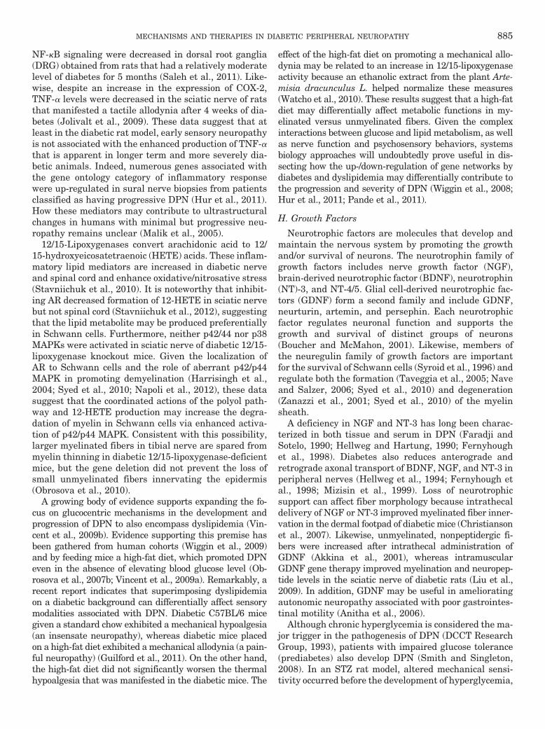
1. Controlling the Biological Outcome of Inhibiting the90-kDa Heat-Shock Protein: Dissociating Cytotoxicityfrom Cytoprotection Is Paramount to Attain SpecificTherapeutic Efficacies. Molecular chaperones, such asthe 70- and 90-kDA heat-shock proteins (Hsp70 andHsp90), are essential for the folding of nascent polypep-tides into their biologically active structures and forrefolding aggregated and denatured proteins that mayoccur upon cell stress (Mayer and Bukau, 2005; Petersonand Blagg, 2009). Energy derived from hydrolysis ofATP by the intrinsic N-terminal ATPase activity ofHsp90 is important in the conformational maturation of“client proteins” that form a stabilized complex withhomodimerized Hsp90. Inhibiting Hsp90 interferes withprotein maturation, which leads to degradation of Hsp90client proteins (Powers and Workman, 2007). Becausemany oncoproteins are Hsp90 clients, N-terminal Hsp90inhibitors are attractive as potential chemotherapeuticagents because they can promote cytotoxicity via onco-protein degradation (Bishop et al., 2007; Taldone et al.,2009). An important aspect of N-terminal Hsp90 inhib-itors in cancer therapy is that the drugs selectively in-hibit Hsp90 and induce client protein degradation inmalignant versus normal cells (Chiosis et al., 2003; Ka-mal et al., 2003; Luo et al., 2008). Although this selec-tivity aids the clinical efficacy of N-terminal Hsp90 in-hibitors, their use has been hampered because inductionof client protein degradation and cytotoxicity can occurat drug concentrations that also activate an antagonisticaspect of Hsp90 biology, induction of the cytoprotectiveheat-shock response (HSR) (Fig. 2).
Hsp90 binds the transcription factor heat-shock factor1 (HSF1) and inhibits its transactivating capacity (Be-narroch, 2011). Upon exposure to stress or Hsp90 inhib-itors, HSF1 dissociates, trimerizes, undergoes phosphor-ylation, and translocates to the nucleus to up-regulateantioxidant genes and a variety of chaperones: the heat-shock response. Unfortunately, because chaperones canfunction as prosurvival factors that facilitate refolding ofthe oncoproteins, HSR induction antagonizes the de-sired cytotoxicity when treating malignancies. On theother hand, although induction of the HSR interfereswith their chemotherapeutic potential, stimulating thisaspect of Hsp90 biology has potential utility for treatingneurodegenerative diseases associated with protein mis-folding: HSR induction decreases protein aggregation.
N-terminal Hsp90 inhibitors have been shown to de-crease � protein aggregation in Alzheimer disease models(Dickey et al., 2007; Luo et al., 2007) and improve motorfunction in spinal and bulbar muscular atrophy (Waza etal., 2005). Although a similar selectivity exists for the useof N-terminal inhibitors in treating neurodegenerative dis-eases (Dickey et al., 2007), this selectivity does not circum-vent the issue related to dissociating client protein degra-dation from induction of the HSR. Now, the inverse caveat
MECHANISMS AND THERAPIES IN DIABETIC PERIPHERAL NEUROPATHY 889
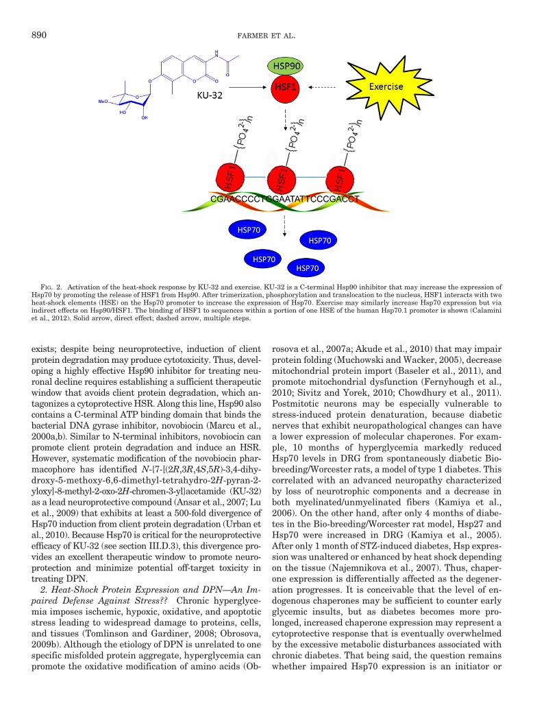
exists; despite being neuroprotective, induction of clientprotein degradation may produce cytotoxicity. Thus, devel-oping a highly effective Hsp90 inhibitor for treating neu-ronal decline requires establishing a sufficient therapeuticwindow that avoids client protein degradation, which an-tagonizes a cytoprotective HSR. Along this line, Hsp90 alsocontains a C-terminal ATP binding domain that binds thebacterial DNA gyrase inhibitor, novobiocin (Marcu et al.,2000a,b). Similar to N-terminal inhibitors, novobiocin canpromote client protein degradation and induce an HSR.However, systematic modification of the novobiocin phar-macophore has identified N-{7-[(2R,3R,4S,5R)-3,4-dihy-droxy-5-methoxy-6,6-dimethyl-tetrahydro-2H-pyran-2-yloxy]-8-methyl-2-oxo-2H-chromen-3-yl}acetamide (KU-32)as a lead neuroprotective compound (Ansar et al., 2007; Luet al., 2009) that exhibits at least a 500-fold divergence ofHsp70 induction from client protein degradation (Urban etal., 2010). Because Hsp70 is critical for the neuroprotectiveefficacy of KU-32 (see section III.D.3), this divergence pro-vides an excellent therapeutic window to promote neuro-protection and minimize potential off-target toxicity intreating DPN.
2. Heat-Shock Protein Expression and DPN—An Im-paired Defense Against Stress?? Chronic hyperglyce-mia imposes ischemic, hypoxic, oxidative, and apoptoticstress leading to widespread damage to proteins, cells,and tissues (Tomlinson and Gardiner, 2008; Obrosova,2009b). Although the etiology of DPN is unrelated to onespecific misfolded protein aggregate, hyperglycemia canpromote the oxidative modification of amino acids (Ob-
rosova et al., 2007a; Akude et al., 2010) that may impairprotein folding (Muchowski and Wacker, 2005), decreasemitochondrial protein import (Baseler et al., 2011), andpromote mitochondrial dysfunction (Fernyhough et al.,2010; Sivitz and Yorek, 2010; Chowdhury et al., 2011).Postmitotic neurons may be especially vulnerable tostress-induced protein denaturation, because diabeticnerves that exhibit neuropathological changes can havea lower expression of molecular chaperones. For exam-ple, 10 months of hyperglycemia markedly reducedHsp70 levels in DRG from spontaneously diabetic Bio-breeding/Worcester rats, a model of type 1 diabetes. Thiscorrelated with an advanced neuropathy characterizedby loss of neurotrophic components and a decrease inboth myelinated/unmyelinated fibers (Kamiya et al.,2006). On the other hand, after only 4 months of diabe-tes in the Bio-breeding/Worcester rat model, Hsp27 andHsp70 were increased in DRG (Kamiya et al., 2005).After only 1 month of STZ-induced diabetes, Hsp expres-sion was unaltered or enhanced by heat shock dependingon the tissue (Najemnikova et al., 2007). Thus, chaper-one expression is differentially affected as the degener-ation progresses. It is conceivable that the level of en-dogenous chaperones may be sufficient to counter earlyglycemic insults, but as diabetes becomes more pro-longed, increased chaperone expression may represent acytoprotective response that is eventually overwhelmedby the excessive metabolic disturbances associated withchronic diabetes. That being said, the question remainswhether impaired Hsp70 expression is an initiator or
FIG. 2. Activation of the heat-shock response by KU-32 and exercise. KU-32 is a C-terminal Hsp90 inhibitor that may increase the expression ofHsp70 by promoting the release of HSF1 from Hsp90. After trimerization, phosphorylation and translocation to the nucleus, HSF1 interacts with twoheat-shock elements (HSE) on the Hsp70 promoter to increase the expression of Hsp70. Exercise may similarly increase Hsp70 expression but viaindirect effects on Hsp90/HSF1. The binding of HSF1 to sequences within a portion of one HSE of the human Hsp70.1 promoter is shown (Calaminiet al., 2012). Solid arrow, direct effect; dashed arrow, multiple steps.
890 FARMER ET AL.
outcome of DPN. However, genetic knockout of thestress-inducible isoforms of Hsp70 (Hsp70.1 andHsp70.3) caused no significant difference in the rate ofonset or severity of a sensory hypoalgesia that developedin diabetic Hsp70 knockout mice (Urban et al., 2010).These data suggest that decreased Hsp70 expression isnot an essential pathogenic component in DPN. Al-though there can be considerable functional redundancybetween inducible (Hsp70) and constitutive (Hsc70)paralogs, we have noted that Hsc70 does not change indiabetic nerve, suggesting that its decline is also not acritical feature in the etiology of DPN. It is noteworthythat high anti-Hsp70 antibody levels, but not those ofanti-Hsp60 antibody, were detected in an analysis of 531patients with type 1 diabetes from the EURODIABStudy (Gruden et al., 2009). Independent of other riskfactors and inflammation, a 50% lower likelihood of mi-cro/macrovascular complications was associated withhigh anti-Hsp70 antibody levels (Gruden et al., 2009).Although the underlying origin of the antigen was notidentified, these data suggest that elevated Hsp70 anti-body levels may serve as a positive biomarker for pa-tients less prone to developing diabetic complications(Gruden et al., 2009). It may be worthwhile to determinewhether a relationship exists between Hsp70 antibodylevels and the level of glyoxalase 1 expression (Jack etal., 2012) as a predictive biomarker pair for the devel-opment of DPN in humans (Rabbani and Thornalley,2011).
3. 70-kDa Heat-Shock Protein and Neuroprotection inDPN. Although most other Hsps are abundantly dis-tributed in the nerve, the inducible form of Hsp70 isweakly expressed, and its induction typically representsa cellular protective program adopted by neurons andglia during stress response (Pavlik et al., 2003; Pavlikand Aneja, 2007). Thus, physically or pharmacologicallyincreasing Hsp expression may confer protection againstneuropathic changes associated with diabetes. For ex-ample, patients with type 2 diabetes experienced reduc-tions in blood glucose concentration and symptomaticneuropathy after receiving regular hot-tub treatment(Hooper, 1999, 2003). In diabetic rats, the HSF1 activa-tors bimoclomol and arimoclomol (BRX-220) improveddiabetic wound healing and nerve conduction deficits,respectively (Vígh et al., 1997; Kurthy et al., 2002);direct HSF1 activators may provide another pharmaco-logic route to modulating genes that express chaperonesand antioxidant proteins (Neef et al., 2011). The poten-tial effectiveness of �-lipoic acid in treating DPN hasalready been noted and �-lipoic acid therapy has beennormalized. Hsp70 levels were decreased in a cohort ofpatients with type 1 diabetes and DPN (Strokov et al.,2000). Overexpression of Hsp70 in muscle is sufficient toimprove insulin resistance and glucose utilization(Chung et al., 2008), and this metabolic correction wouldbe anticipated to also improve DPN. However, the ther-apeutic benefits of Hsp70 induction in DPN do not seem
to hinge on a metabolic correction and may be relativelynerve-specific (Urban et al., 2010, 2012b). On the otherhand, modulating chaperones may improve the trophiceffects of impaired insulin signaling in DPN, becauseseveral components of the insulin signaling cascade relyon interactions with heat shock proteins (Urban et al.,2012a).
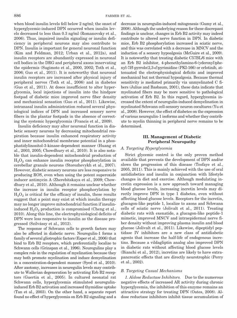
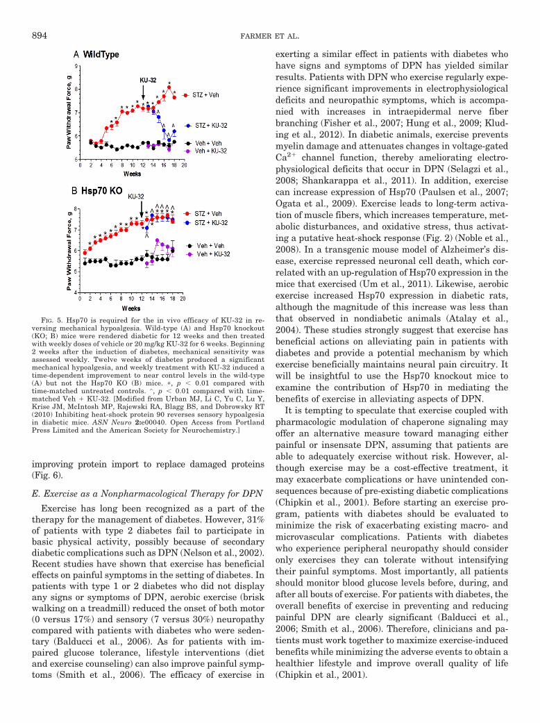
We have been exploring the potential of a novel classof Hsp90 inhibitors as a pharmacologic tool to modulateHsp70 in DPN. KU-32 is a C-terminal, novobiocin-basedHsp90 inhibitor (Fig. 2) that protected against neuronalcell death (Ansar et al., 2007). Despite being only a weakactivator of the Hsp70 promoter compared with the pro-totypical N-terminal Hsp90 inhibitor geldanamycin(Fig. 3A), KU-32 increased Hsp70 expression in explantcultures of neonatal mouse sensory neurons (Fig. 3B)without affecting the expression of Hsc70 (Fig. 3C). It isnoteworthy that weekly treatment of diabetic mice withKU-32 reversed a pre-existing mechanical and thermalhypoalgesia and improved deficits in both sensory andmotor nerve conduction velocities (Urban et al., 2010).Moreover, after 16 weeks of diabetes in Swiss-Webstermice, intraepidermal nerve fiber density was decreasedby 30%, and this loss of fibers was reversed by 10 weeksof KU-32 therapy (Urban et al., 2012b). Because noobvious improvement in plasma glucose or insulin levelswere observed in mice administered KU-32, the protec-tion did not require a metabolic correction of glucoseutilization and probably resulted from a direct effect onchaperone expression in peripheral nerve. Indeed, de-spite the modest induction of Hsp70 induced by KU-32,it is central to drug efficacy. For example, KU-32 pro-tected against neuregulin-induced demyelination in my-elinated Schwann cell-sensory neuron cocultures pre-pared from wild-type mice (Figs. 4, A and B), but thiseffect was lost in neurons isolated from Hsp70 knockoutmice (Figs. 4, C and D). Likewise, although KU-32 re-versed multiple indices of DPN in wild-type mice, thedrug was ineffective at reversing an insensate neuropa-thy in diabetic Hsp70 KO mice (Figs. 5, A and B) (Urbanet al., 2010). These data agree with other reports sup-porting Hsp70 as a key neuroprotective chaperone pre-venting neuronal apoptosis (Bienemann et al., 2008) andimproving neurodegenerative diseases associated withprotein misfolding (Dickey et al., 2007; Luo et al., 2007).Thus, modulating Hsp70 may increase the tolerance ofneurons and glia to the fluxing metabolic stresses asso-ciated with diabetes (Calcutt, 2010).
4. 70-kDa Heat-Shock Protein Family Members andMitochondrial Function. Although it remains unclearhow Hsp70 may antagonize the proneuropathic action ofthe established biochemical mediators of DPN, Hsp70 canimprove oxidative stress and mitochondrial function.Hsp70 overexpression significantly potentiated the activi-ties of glutathione peroxidase and glutathione reductase,leading to a higher GSH/GSSG ratio, attenuated ROS pro-duction, and improved cell survival under conditions of
MECHANISMS AND THERAPIES IN DIABETIC PERIPHERAL NEUROPATHY 891
hypoxia and glucose deprivation (Guo et al., 2007). Like-wise, overexpression of Hsp70 in astrocytes increased mi-tochondrial function and decreased neuronal injury afterbasal forebrain ischemia (Xu et al., 2010). Consistent withthis result, KU-32 increased the translation of Hsp70, aswell as other chaperones and MnSOD in hyperglycemi-cally stressed sensory neurons. Increased expression of themolecular chaperones and MnSOD correlated with a de-crease in mitochondrial superoxide levels and improvedmitochondrial bioenergetics (Zhang et al., 2012). However,it remains to be determined whether chaperone inductionimproves mitochondrial bioenergetics to decrease oxidativestress or if enhanced expression of MnSOD and decreasedsuperoxide production precedes improved respiratory func-tion (Fig. 6). It is also worth noting that NADPH oxidasescan serve as a potential source of superoxide in DPN (Coppeyet al., 2003). It is noteworthy that down-regulation of Hsp70with small interfering RNA increased NADPH-dependentROS production in vascular smooth muscle cells (Madrigal-Matute et al., 2012), raising the possibility that increasingHsp70 may also decrease extra-mitochondrial sources of su-peroxide via effects on NADPH oxidase.
Several lines of evidence indicate that cytosolic Hsp70 ispositively involved in affecting mitochondrial bioenerget-ics. Rat hearts transfected with Hsp70 through viral infu-sion showed improved mitochondrial respiration and at-tenuated mitochondrial damage during ischemia/reperfusion injury (Suzuki et al., 2002). Transgenicoverexpression of Hsp70 in mouse skeletal muscles in-creased citrate synthase and �-hydroxyacyl-CoA-dehydro-genase activities, suggesting an enhanced oxidative capac-ity (Chung et al., 2008). In contrast, lower Hsp70 mRNA
expression correlated with reduced mitochondrial enzy-matic activity in skeletal muscle of diabetic humans com-pared with healthy control subjects (Bruce et al., 2003).Overexpression of Hsp70 in glucose-deprived astrocytesalso inhibited proton leak and ROS accumulation (Ouyanget al., 2006; Xu et al., 2010).
How Hsp70 may improve mitochondrial function isunresolved. Drp1 is a mitochondrial fission protein thatmay contribute to mitochondrial dysfunction in DPN(Vincent et al., 2010). A recent report indicates thatinhibiting Drp1 translocation and phosphorylation inpodocytes decreased oxidative stress and improved fea-tures of diabetic nephropathy (Wang et al., 2012). Giventhe ability of KU-32 to decrease mitochondrial superox-ide levels in hyperglycemically stressed sensory neurons(Zhang et al., 2012), it will be important to determinewhether this effect may be mediated via Hsp70-depen-dent interactions with Drp1 or upstream kinases in di-abetic sensory neurons. Alternatively, oxidative modifi-cation of Drp1 leads to formation of tetramers and largeraggregates. Because modified Drp1 has enhanced GT-Pase activity, which can promote mitochondrial fission(Nakamura and Lipton, 2010), Hsp70 may decrease or-ganellar fission by aiding Drp1 refolding. However, afolding-incompetent, ATPase-deficient Hsp70 mutantthat maintained the ability to bind and aid the solubilityof denatured proteins still improved mitochondrial func-tion in cultured astrocytes (Ouyang et al., 2006). Thesedata imply that the folding competency of Hsp70 maynot be a key feature for protection. It is noteworthy thatcombining Hsp70 overexpression with inhibition of itsATPase activity by methylene blue increased the protea-
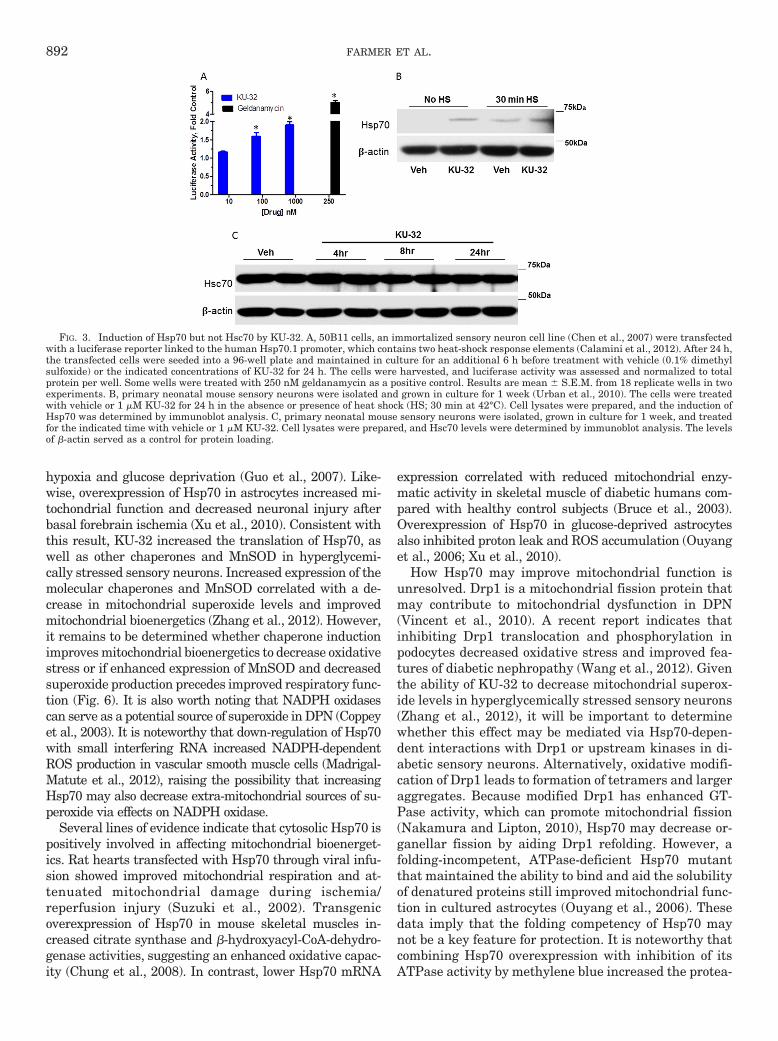
FIG. 3. Induction of Hsp70 but not Hsc70 by KU-32. A, 50B11 cells, an immortalized sensory neuron cell line (Chen et al., 2007) were transfectedwith a luciferase reporter linked to the human Hsp70.1 promoter, which contains two heat-shock response elements (Calamini et al., 2012). After 24 h,the transfected cells were seeded into a 96-well plate and maintained in culture for an additional 6 h before treatment with vehicle (0.1% dimethylsulfoxide) or the indicated concentrations of KU-32 for 24 h. The cells were harvested, and luciferase activity was assessed and normalized to totalprotein per well. Some wells were treated with 250 nM geldanamycin as a positive control. Results are mean � S.E.M. from 18 replicate wells in twoexperiments. B, primary neonatal mouse sensory neurons were isolated and grown in culture for 1 week (Urban et al., 2010). The cells were treatedwith vehicle or 1 �M KU-32 for 24 h in the absence or presence of heat shock (HS; 30 min at 42°C). Cell lysates were prepared, and the induction ofHsp70 was determined by immunoblot analysis. C, primary neonatal mouse sensory neurons were isolated, grown in culture for 1 week, and treatedfor the indicated time with vehicle or 1 �M KU-32. Cell lysates were prepared, and Hsc70 levels were determined by immunoblot analysis. The levelsof �-actin served as a control for protein loading.
892 FARMER ET AL.
somal degradation of � (Jinwal et al., 2009). Althoughmitochondrial dysfunction in DPN is not necessarilyassociated with a single protein aggregate, it will beinteresting to determine whether increasing proteinclearance by combining an Hsp70 inducer with a selec-tive ATPase inhibitor may prove beneficial in treatingDPN, as has been suggested for cancer therapy (Koren etal., 2010). Finally, cytosolic Hsp70 and Hsp90 interactwith TOMM70, a mitochondrial outer membrane trans-locase, to mediate import of nuclear-encoded proteins(Young et al., 2003). Because 99% of the mitochondrialproteins arise from nuclear genes and need to be im-ported into the organelle (Schmidt et al., 2010), Hsp70may facilitate the replacement of oxidatively damagedmitochondrial proteins (Fig. 6).
Cytosolic Hsp70 may also have help from other Hsp70family members in decreasing oxidative stress. Geneticoverexpression of the mitochondrial paralog of Hsp70(mortalin/Grp75/mtHsp70) is sufficient to protect cultured
primary astrocytes from ischemia-induced oxidative dam-age and death (Voloboueva et al., 2008). Likewise,mtHsp70 overexpression in brain reduced oxidative stressmarkers and improved mitochondrial function induced byischemia/reperfusion (Xu et al., 2009). Overexpression ofmtHsp70 in cardiac myocytes also potentiated the activityof respiratory chain complexes III and IV and abrogatedROS generation and lipid peroxidation after hypoxia/reoxygenation (Williamson et al., 2008). Using an unbiasedproteomic screen, we recently identified that KU-32 alsoincreased translation of MnSOD and mtHsp70 in hyper-glycemically stressed neurons (Zhang et al., 2012). Unfor-tunately, it is unclear how KU-32 may increase mtHsp70expression, because it is not induced as part of the classicheat-shock response (Deocaris et al., 2006). Nevertheless,approaches that can increase both cytosolic and mitochon-drial Hsp70 family members are likely to improve mito-chondrial function in DPN via effects on decreasing oxida-tive stress, aiding the refolding of oxidatively damaged and
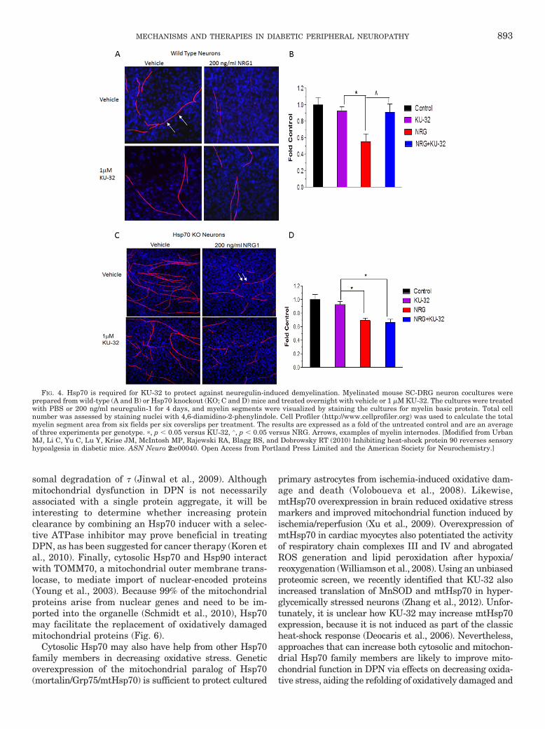
FIG. 4. Hsp70 is required for KU-32 to protect against neuregulin-induced demyelination. Myelinated mouse SC-DRG neuron cocultures wereprepared from wild-type (A and B) or Hsp70 knockout (KO; C and D) mice and treated overnight with vehicle or 1 �M KU-32. The cultures were treatedwith PBS or 200 ng/ml neuregulin-1 for 4 days, and myelin segments were visualized by staining the cultures for myelin basic protein. Total cellnumber was assessed by staining nuclei with 4,6-diamidino-2-phenylindole. Cell Profiler (http://www.cellprofiler.org) was used to calculate the totalmyelin segment area from six fields per six coverslips per treatment. The results are expressed as a fold of the untreated control and are an averageof three experiments per genotype. �, p � 0.05 versus KU-32, ∧, p � 0.05 versus NRG. Arrows, examples of myelin internodes. [Modified from UrbanMJ, Li C, Yu C, Lu Y, Krise JM, McIntosh MP, Rajewski RA, Blagg BS, and Dobrowsky RT (2010) Inhibiting heat-shock protein 90 reverses sensoryhypoalgesia in diabetic mice. ASN Neuro 2:e00040. Open Access from Portland Press Limited and the American Society for Neurochemistry.]
MECHANISMS AND THERAPIES IN DIABETIC PERIPHERAL NEUROPATHY 893
improving protein import to replace damaged proteins(Fig. 6).
E. Exercise as a Nonpharmacological Therapy for DPN
Exercise has long been recognized as a part of thetherapy for the management of diabetes. However, 31%of patients with type 2 diabetes fail to participate inbasic physical activity, possibly because of secondarydiabetic complications such as DPN (Nelson et al., 2002).Recent studies have shown that exercise has beneficialeffects on painful symptoms in the setting of diabetes. Inpatients with type 1 or 2 diabetes who did not displayany signs or symptoms of DPN, aerobic exercise (briskwalking on a treadmill) reduced the onset of both motor(0 versus 17%) and sensory (7 versus 30%) neuropathycompared with patients with diabetes who were seden-tary (Balducci et al., 2006). As for patients with im-paired glucose tolerance, lifestyle interventions (dietand exercise counseling) can also improve painful symp-toms (Smith et al., 2006). The efficacy of exercise in
exerting a similar effect in patients with diabetes whohave signs and symptoms of DPN has yielded similarresults. Patients with DPN who exercise regularly expe-rience significant improvements in electrophysiologicaldeficits and neuropathic symptoms, which is accompa-nied with increases in intraepidermal nerve fiberbranching (Fisher et al., 2007; Hung et al., 2009; Klud-ing et al., 2012). In diabetic animals, exercise preventsmyelin damage and attenuates changes in voltage-gatedCa2� channel function, thereby ameliorating electro-physiological deficits that occur in DPN (Selagzi et al.,2008; Shankarappa et al., 2011). In addition, exercisecan increase expression of Hsp70 (Paulsen et al., 2007;Ogata et al., 2009). Exercise leads to long-term activa-tion of muscle fibers, which increases temperature, met-abolic disturbances, and oxidative stress, thus activat-ing a putative heat-shock response (Fig. 2) (Noble et al.,2008). In a transgenic mouse model of Alzheimer’s dis-ease, exercise repressed neuronal cell death, which cor-related with an up-regulation of Hsp70 expression in themice that exercised (Um et al., 2011). Likewise, aerobicexercise increased Hsp70 expression in diabetic rats,although the magnitude of this increase was less thanthat observed in nondiabetic animals (Atalay et al.,2004). These studies strongly suggest that exercise hasbeneficial actions on alleviating pain in patients withdiabetes and provide a potential mechanism by whichexercise beneficially maintains neural pain circuitry. Itwill be insightful to use the Hsp70 knockout mice toexamine the contribution of Hsp70 in mediating thebenefits of exercise in alleviating aspects of DPN.
It is tempting to speculate that exercise coupled withpharmacologic modulation of chaperone signaling mayoffer an alternative measure toward managing eitherpainful or insensate DPN, assuming that patients areable to adequately exercise without risk. However, al-though exercise may be a cost-effective treatment, itmay exacerbate complications or have unintended con-sequences because of pre-existing diabetic complications(Chipkin et al., 2001). Before starting an exercise pro-gram, patients with diabetes should be evaluated tominimize the risk of exacerbating existing macro- andmicrovascular complications. Patients with diabeteswho experience peripheral neuropathy should consideronly exercises they can tolerate without intensifyingtheir painful symptoms. Most importantly, all patientsshould monitor blood glucose levels before, during, andafter all bouts of exercise. For patients with diabetes, theoverall benefits of exercise in preventing and reducingpainful DPN are clearly significant (Balducci et al.,2006; Smith et al., 2006). Therefore, clinicians and pa-tients must work together to maximize exercise-inducedbenefits while minimizing the adverse events to obtain ahealthier lifestyle and improve overall quality of life(Chipkin et al., 2001).
FIG. 5. Hsp70 is required for the in vivo efficacy of KU-32 in re-versing mechanical hypoalgesia. Wild-type (A) and Hsp70 knockout(KO; B) mice were rendered diabetic for 12 weeks and then treatedwith weekly doses of vehicle or 20 mg/kg KU-32 for 6 weeks. Beginning2 weeks after the induction of diabetes, mechanical sensitivity wasassessed weekly. Twelve weeks of diabetes produced a significantmechanical hypoalgesia, and weekly treatment with KU-32 induced atime-dependent improvement to near control levels in the wild-type(A) but not the Hsp70 KO (B) mice. �, p � 0.01 compared withtime-matched untreated controls. ∧, p � 0.01 compared with time-matched Veh � KU-32. [Modified from Urban MJ, Li C, Yu C, Lu Y,Krise JM, McIntosh MP, Rajewski RA, Blagg BS, and Dobrowsky RT(2010) Inhibiting heat-shock protein 90 reverses sensory hypoalgesiain diabetic mice. ASN Neuro 2:e00040. Open Access from PortlandPress Limited and the American Society for Neurochemistry.]
894 FARMER ET AL.
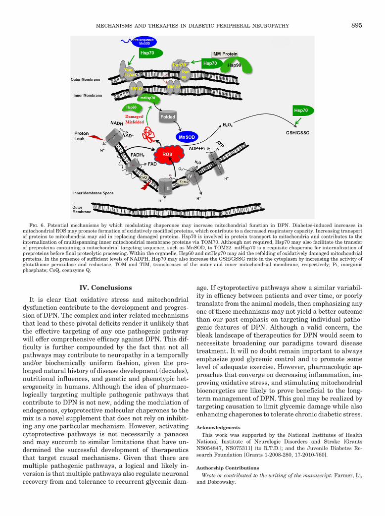
IV. Conclusions
It is clear that oxidative stress and mitochondrialdysfunction contribute to the development and progres-sion of DPN. The complex and inter-related mechanismsthat lead to these pivotal deficits render it unlikely thatthe effective targeting of any one pathogenic pathwaywill offer comprehensive efficacy against DPN. This dif-ficulty is further compounded by the fact that not allpathways may contribute to neuropathy in a temporallyand/or biochemically uniform fashion, given the pro-longed natural history of disease development (decades),nutritional influences, and genetic and phenotypic het-erogeneity in humans. Although the idea of pharmaco-logically targeting multiple pathogenic pathways thatcontribute to DPN is not new, adding the modulation ofendogenous, cytoprotective molecular chaperones to themix is a novel supplement that does not rely on inhibit-ing any one particular mechanism. However, activatingcytoprotective pathways is not necessarily a panaceaand may succumb to similar limitations that have un-dermined the successful development of therapeuticsthat target causal mechanisms. Given that there aremultiple pathogenic pathways, a logical and likely in-version is that multiple pathways also regulate neuronalrecovery from and tolerance to recurrent glycemic dam-
age. If cytoprotective pathways show a similar variabil-ity in efficacy between patients and over time, or poorlytranslate from the animal models, then emphasizing anyone of these mechanisms may not yield a better outcomethan our past emphasis on targeting individual patho-genic features of DPN. Although a valid concern, thebleak landscape of therapeutics for DPN would seem tonecessitate broadening our paradigms toward diseasetreatment. It will no doubt remain important to alwaysemphasize good glycemic control and to promote somelevel of adequate exercise. However, pharmacologic ap-proaches that converge on decreasing inflammation, im-proving oxidative stress, and stimulating mitochondrialbioenergetics are likely to prove beneficial to the long-term management of DPN. This goal may be realized bytargeting causation to limit glycemic damage while alsoenhancing chaperones to tolerate chronic diabetic stress.
Acknowledgments
This work was supported by the National Institutes of HealthNational Institute of Neurologic Disorders and Stroke [GrantsNS054847, NS075311] (to R.T.D.); and the Juvenile Diabetes Re-search Foundation [Grants 1-2008-280, 17-2010-760].
Authorship Contributions
Wrote or contributed to the writing of the manuscript: Farmer, Li,and Dobrowsky.
FIG. 6. Potential mechanisms by which modulating chaperones may increase mitochondrial function in DPN. Diabetes-induced increases inmitochondrial ROS may promote formation of oxidatively modified proteins, which contribute to a decreased respiratory capacity. Increasing transportof proteins to mitochondria may aid in replacing damaged proteins. Hsp70 is involved in protein transport to mitochondria and contributes to theinternalization of multispanning inner mitochondrial membrane proteins via TOM70. Although not required, Hsp70 may also facilitate the transferof preproteins containing a mitochondrial targeting sequence, such as MnSOD, to TOM22. mtHsp70 is a requisite chaperone for internalization ofpreproteins before final proteolytic processing. Within the organelle, Hsp60 and mtHsp70 may aid the refolding of oxidatively damaged mitochondrialproteins. In the presence of sufficient levels of NADPH, Hsp70 may also increase the GSH/GSSG ratio in the cytoplasm by increasing the activity ofglutathione peroxidase and reductase. TOM and TIM, translocases of the outer and inner mitochondrial membrane, respectively; Pi, inorganicphosphate; CoQ, coenzyme Q.
MECHANISMS AND THERAPIES IN DIABETIC PERIPHERAL NEUROPATHY 895
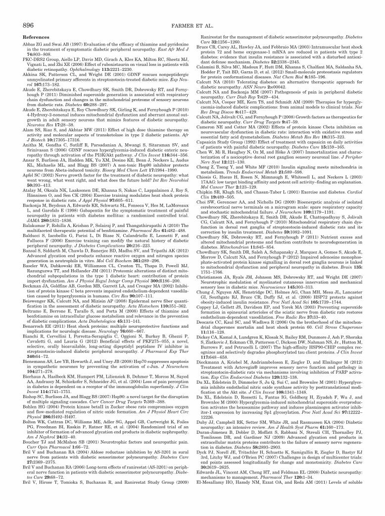
ReferencesAbbas ZG and Swai AB (1997) Evaluation of the efficacy of thiamine and pyridoxine
in the treatment of symptomatic diabetic peripheral neuropathy. East Afr Med J74:803–808.
PKC-DRS2 Group, Aiello LP, Davis MD, Girach A, Kles KA, Milton RC, Sheetz MJ,Vignati L, and Zhi XE (2006) Effect of ruboxistaurin on visual loss in patients withdiabetic retinopathy. Ophthalmology 113:2221–2230.
Akkina SK, Patterson CL, and Wright DE (2001) GDNF rescues nonpeptidergicunmyelinated primary afferents in streptozotocin-treated diabetic mice. Exp Neu-rol 167:173–182.
Akude E, Zherebitskaya E, Chowdhury SK, Smith DR, Dobrowsky RT, and Ferny-hough P (2011) Diminished superoxide generation is associated with respiratorychain dysfunction and changes in the mitochondrial proteome of sensory neuronsfrom diabetic rats. Diabetes 60:288–297.
Akude E, Zherebitskaya E, Roy Chowdhury SK, Girling K, and Fernyhough P (2010)4-Hydroxy-2-nonenal induces mitochondrial dysfunction and aberrant axonal out-growth in adult sensory neurons that mimics features of diabetic neuropathy.Neurotox Res 17:28–38.
Alam SS, Riaz S, and Akhtar MW (2011) Effect of high dose thiamine therapy onactivity and molecular aspects of transketolase in type 2 diabetic patients. AfrJ Biotech 10:17305–17316.
Anitha M, Gondha C, Sutliff R, Parsadanian A, Mwangi S, Sitaraman SV, andSrinivasan S (2006) GDNF rescues hyperglycemia-induced diabetic enteric neu-ropathy through activation of the PI3K/Akt pathway. J Clin Invest 116:344–356.
Ansar S, Burlison JA, Hadden MK, Yu XM, Desino KE, Bean J, Neckers L, AudusKL, Michaelis ML, and Blagg BS (2007) A non-toxic Hsp90 inhibitor protectsneurons from Abeta-induced toxicity. Bioorg Med Chem Lett 17:1984–1990.
Apfel SC (2002) Nerve growth factor for the treatment of diabetic neuropathy: whatwent wrong, what went right, and what does the future hold? Int Rev Neurobiol50:393–413.
Atalay M, Oksala NK, Laaksonen DE, Khanna S, Nakao C, Lappalainen J, Roy S,Hanninen O, and Sen CK (2004) Exercise training modulates heat shock proteinresponse in diabetic rats. J Appl Physiol 97:605–611.
Backonja M, Beydoun A, Edwards KR, Schwartz SL, Fonseca V, Hes M, LaMoreauxL, and Garofalo E (1998) Gabapentin for the symptomatic treatment of painfulneuropathy in patients with diabetes mellitus: a randomized controlled trial.JAMA 280:1831–1836.
Balakumar P, Rohilla A, Krishan P, Solairaj P, and Thangathirupathi A (2010) Themultifaceted therapeutic potential of benfotiamine. Pharmacol Res 61:482–488.
Balducci S, Iacobellis G, Parisi L, Di Biase N, Calandriello E, Leonetti F, andFallucca F (2006) Exercise training can modify the natural history of diabeticperipheral neuropathy. J Diabetes Complications 20:216–223.
Bansal S, Siddarth M, Chawla D, Banerjee BD, Madhu SV, and Tripathi AK (2012)Advanced glycation end products enhance reactive oxygen and nitrogen speciesgeneration in neutrophils in vitro. Mol Cell Biochem 361:289–296.
Baseler WA, Dabkowski ER, Williamson CL, Croston TL, Thapa D, Powell MJ,Razunguzwa TT, and Hollander JM (2011) Proteomic alterations of distinct mito-chondrial subpopulations in the type 1 diabetic heart: contribution of proteinimport dysfunction. Am J Physiol Regul Integr Comp Physiol 300:R186–200.
Beckman JA, Goldfine AB, Gordon MB, Garrett LA, and Creager MA (2002) Inhibi-tion of protein kinase C beta prevents impaired endothelium-dependent vasodila-tion caused by hyperglycemia in humans. Circ Res 90:107–111.
Beiswenger KK, Calcutt NA, and Mizisin AP (2008) Epidermal nerve fiber quanti-fication in the assessment of diabetic neuropathy. Acta Histochem 110:351–362.
Beltramo E, Berrone E, Tarallo S, and Porta M (2008) Effects of thiamine andbenfotiamine on intracellular glucose metabolism and relevance in the preventionof diabetic complications. Acta Diabetologica 45:131–141.
Benarroch EE (2011) Heat shock proteins: multiple neuroprotective functions andimplications for neurologic disease. Neurology 76:660–667.
Bianchi R, Cervellini I, Porretta-Serapiglia C, Oggioni N, Burkey B, Ghezzi P,Cavaletti G, and Lauria G (2012) Beneficial effects of PKF275–055, a novel,selective, orally bioavailable, long-acting dipeptidyl peptidase IV inhibitor instreptozotocin-induced diabetic peripheral neuropathy. J Pharmacol Exp Ther340:64–72.
Bienemann AS, Lee YB, Howarth J, and Uney JB (2008) Hsp70 suppresses apoptosisin sympathetic neurones by preventing the activation of c-Jun. J Neurochem104:271–278.
Bierhaus A, Haslbeck KM, Humpert PM, Liliensiek B, Dehmer T, Morcos M, SayedAA, Andrassy M, Schiekofer S, Schneider JG, et al. (2004) Loss of pain perceptionin diabetes is dependent on a receptor of the immunoglobulin superfamily. J ClinInvest 114:1741–1751.
Bishop SC, Burlison JA, and Blagg BS (2007) Hsp90: a novel target for the disruptionof multiple signaling cascades. Curr Cancer Drug Targets 7:369–388.
Bohlen HG (2004) Protein kinase betaII in Zucker obese rats compromises oxygenand flow-mediated regulation of nitric oxide formation. Am J Physiol Heart CircPhysiol 286:H492–H497.
Bolton WK, Cattran DC, Williams ME, Adler SG, Appel GB, Cartwright K, FoilesPG, Freedman BI, Raskin P, Ratner RE, et al. (2004) Randomized trial of aninhibitor of formation of advanced glycation end products in diabetic nephropathy.Am J Nephrol 24:32–40.
Boucher TJ and McMahon SB (2001) Neurotrophic factors and neuropathic pain.Curr Opin Pharmacol 1:66–72.
Bril V and Buchanan RA (2004) Aldose reductase inhibition by AS-3201 in suralnerve from patients with diabetic sensorimotor polyneuropathy. Diabetes Care27:2369–2375.
Bril V and Buchanan RA (2006) Long-term effects of ranirestat (AS-3201) on periph-eral nerve function in patients with diabetic sensorimotor polyneuropathy. Diabe-tes Care 29:68–72.
Bril V, Hirose T, Tomioka S, Buchanan R, and Ranirestat Study Group (2009)
Ranirestat for the management of diabetic sensorimotor polyneuropathy. DiabetesCare 32:1256–1260.
Bruce CR, Carey AL, Hawley JA, and Febbraio MA (2003) Intramuscular heat shockprotein 72 and heme oxygenase-1 mRNA are reduced in patients with type 2diabetes: evidence that insulin resistance is associated with a disturbed antioxi-dant defense mechanism. Diabetes 52:2338–2345.
Calamini B, Silva MC, Madoux F, Hutt DM, Khanna S, Chalfant MA, Saldanha SA,Hodder P, Tait BD, Garza D, et al. (2012) Small-molecule proteostasis regulatorsfor protein conformational diseases. Nat Chem Biol 8:185–196.
Calcutt NA (2010) Tolerating diabetes: an alternative therapeutic approach fordiabetic neuropathy. ASN Neuro 2:e00042.
Calcutt NA and Backonja MM (2007) Pathogenesis of pain in peripheral diabeticneuropathy. Curr Diab Rep 7:429–434.
Calcutt NA, Cooper ME, Kern TS, and Schmidt AM (2009) Therapies for hypergly-caemia-induced diabetic complications: from animal models to clinical trials. NatRev Drug Discov 8:417–429.
Calcutt NA, Jolivalt CG, and Fernyhough P (2008) Growth factors as therapeutics fordiabetic neuropathy. Curr Drug Targets 9:47–59.
Cameron NE and Cotter MA (2002) Effects of protein kinase Cbeta inhibition onneurovascular dysfunction in diabetic rats: interaction with oxidative stress andessential fatty acid dysmetabolism. Diabetes Metab Res Rev 18:315–323.
Capsaicin Study Group (1992) Effect of treatment with capsaicin on daily activitiesof patients with painful diabetic neuropathy. Diabetes Care 15:159–165.
Chen W, Mi R, Haughey N, Oz M, and Hoke A (2007) Immortalization and charac-terization of a nociceptive dorsal root ganglion sensory neuronal line. J PeripherNerv Syst 12:121–130.
Cheng Z, Tseng Y, and White MF (2010) Insulin signaling meets mitochondria inmetabolism. Trends Endocrinol Metab 21:589–598.
Chiosis G, Huezo H, Rosen N, Mimnaugh E, Whitesell L, and Neckers L (2003)17AAG: low target binding affinity and potent cell activity–finding an explanation.Mol Cancer Ther 2:123–129.
Chipkin SR, Klugh SA, and Chasan-Taber L (2001) Exercise and diabetes. CardiolClin 19:489–505.
Choi SW, Gerencser AA, and Nicholls DG (2009) Bioenergetic analysis of isolatedcerebrocortical nerve terminals on a microgram scale: spare respiratory capacityand stochastic mitochondrial failure. J Neurochem 109:1179–1191.
Chowdhury SK, Zherebitskaya E, Smith DR, Akude E, Chattopadhyay S, JolivaltCG, Calcutt NA, and Fernyhough P (2010) Mitochondrial respiratory chain dys-function in dorsal root ganglia of streptozotocin-induced diabetic rats and itscorrection by insulin treatment. Diabetes 59:1082–1091.
Chowdhury SK, Dobrowsky RT, and Fernyhough P (2011) Nutrient excess andaltered mitochondrial proteome and function contribute to neurodegeneration indiabetes. Mitochondrion 11:845–854.
Chowdhury SK, Smith DR, Saleh A, Schapansky J, Marquez A, Gomes S, Akude E,Morrow D, Calcutt NA, and Fernyhough P (2012) Impaired adenosine monophos-phate-activated protein kinase signalling in dorsal root ganglia neurons is linkedto mitochondrial dysfunction and peripheral neuropathy in diabetes. Brain 135:1751–1766.
Christianson JA, Ryals JM, Johnson MS, Dobrowsky RT, and Wright DE (2007)Neurotrophic modulation of myelinated cutaneous innervation and mechanicalsensory loss in diabetic mice. Neuroscience 145:303–313.
Chung J, Nguyen AK, Henstridge DC, Holmes AG, Chan MH, Mesa JL, LancasterGI, Southgate RJ, Bruce CR, Duffy SJ, et al. (2008) HSP72 protects againstobesity-induced insulin resistance. Proc Natl Acad Sci 105:1739–1744.
Coppey LJ, Gellett JS, Davidson EP, and Yorek MA (2003) Preventing superoxideformation in epineurial arterioles of the sciatic nerve from diabetic rats restoresendothelium-dependent vasodilation. Free Radic Res 37:33–40.
Deocaris CC, Kaul SC, and Wadhwa R (2006) On the brotherhood of the mitochon-drial chaperones mortalin and heat shock protein 60. Cell Stress Chaperones11:116–128.
Dickey CA, Kamal A, Lundgren K, Klosak N, Bailey RM, Dunmore J, Ash P, ShorakaS, Zlatkovic J, Eckman CB, Patterson C, Dickson DW, Nahman NS, Jr., Hutton M,Burrows F, and Petrucelli L (2007) The high-affinity HSP90-CHIP complex rec-ognizes and selectively degrades phosphorylated tau client proteins. J Clin Invest117:648–658.
Dieckmann A, Kriebel M, Andriambeloson E, Ziegler D, and Elmlinger M (2012)Treatment with Actovegin® improves sensory nerve function and pathology instreptozotocin-diabetic rats via mechanisms involving inhibition of PARP activa-tion. Exp Clin Endocrinol Diabetes 120:132–138.
Du XL, Edelstein D, Dimmeler S, Ju Q, Sui C, and Brownlee M (2001) Hyperglyce-mia inhibits endothelial nitric oxide synthase activity by posttranslational modi-fication at the Akt site. J Clin Invest 108:1341–1348.
Du XL, Edelstein D, Rossetti L, Fantus IG, Goldberg H, Ziyadeh F, Wu J, andBrownlee M (2000) Hyperglycemia-induced mitochondrial superoxide overproduc-tion activates the hexosamine pathway and induces plasminogen activator inhib-itor-1 expression by increasing Sp1 glycosylation. Proc Natl Acad Sci 97:12222–12226.
Duby JJ, Campbell RK, Setter SM, White JR, and Rasmussen KA (2004) Diabeticneuropathy: an intensive review. Am J Health Syst Pharm 61:160–173.
Duran-Jimenez B, Dobler D, Moffatt S, Rabbani N, Streuli CH, Thornalley PJ,Tomlinson DR, and Gardiner NJ (2009) Advanced glycation end products inextracellular matrix proteins contribute to the failure of sensory nerve regenera-tion in diabetes. Diabetes 58:2893–2903.
Dyck PJ, Norell JE, Tritschler H, Schuette K, Samigullin R, Ziegler D, Bastyr EJ3rd, Litchy WJ, and O’Brien PC (2007) Challenges in design of multicenter trials:end points assessed longitudinally for change and monotonicity. Diabetes Care30:2619–2625.
Edwards JL, Vincent AM, Cheng HT, and Feldman EL (2008) Diabetic neuropathy:mechanisms to management. Pharmacol Ther 120:1–34.
El-Mesallamy HO, Hamdy NM, Ezzat OA, and Reda AM (2011) Levels of soluble
896 FARMER ET AL.
advanced glycation end product-receptors and other soluble serum markers asindicators of diabetic neuropathy in the foot. J Investig Med 59:1233–1238.
Esper RM, Pankonin MS, and Loeb JA (2006) Neuregulins: versatile growth anddifferentiation factors in nervous system development and human disease. BrainRes Rev 51:161–175.
Faradji V and Sotelo J (1990) Low serum levels of nerve growth factor in diabeticneuropathy. Acta Neurol Scand 81:402–406.
Fernyhough P, Diemel LT, and Tomlinson DR (1998) Target tissue production andaxonal transport of neurotrophin-3 are reduced in streptozotocin-diabetic rats.Diabetologia 41:300–306.
Fernyhough P, Roy Chowdhury SK, and Schmidt RE (2010) Mitochondrial stress andthe pathogenesis of diabetic neuropathy. Expert Rev Endocrinol Metab 5:39–49.
Finnerup NB, Sindrup SH, and Jensen TS (2010) The evidence for pharmacologicaltreatment of neuropathic pain. Pain 150:573–581.
Fisher MA, Langbein WE, Collins EG, Williams K, and Corzine L (2007) Physiolog-ical improvement with moderate exercise in type II diabetic neuropathy. Electro-myogr Clin Neurophysiol 47:23–28.
Francis G, Martinez J, Liu W, Nguyen T, Ayer A, Fine J, Zochodne D, Hanson LR,Frey WH 2nd, and Toth C (2009) Intranasal insulin ameliorates experimentaldiabetic neuropathy. Diabetes 58:934–945.
Freedman BI, Wuerth JP, Cartwright K, Bain RP, Dippe S, Hershon K, MooradianAD, and Spinowitz BS (1999) Design and baseline characteristics for the amino-guanidine Clinical Trial in Overt Type 2 Diabetic Nephropathy (ACTION II).Control Clin Trials 20:493–510.
Furlan AD, Sandoval JA, Mailis-Gagnon A, and Tunks E (2006) Opioids for chronicnoncancer pain: a meta-analysis of effectiveness and side effects. CMAJ 174:1589–1594.
Geraldes P and King GL (2010) Activation of protein kinase C isoforms and itsimpact on diabetic complications. Circ Res 106:1319–1331.
Gimbel JS, Richards P, and Portenoy RK (2003) Controlled-release oxycodone forpain in diabetic neuropathy: a randomized controlled trial. Neurology 60:927–934.
Gonzalez-Clemente JM, Mauricio D, Richart C, Broch M, Caixas A, Megia A, Gime-nez-Palop O, Simon I, Martinez-Riquelme A, Gimenez-Perez G, et al. (2005)Diabetic neuropathy is associated with activation of the TNF-alpha system insubjects with Type 1 diabetes mellitus. Clin Endocrinol (Oxf) 63:525–529.
Gooch C and Podwall D (2004) The diabetic neuropathies. Neurologist 10:311–322.Greene DA and Mackway AM (1986) Decreased myo-inositol content and Na�-K�-
ATPase activity in superior cervical ganglion of STZ-diabetic rat and prevention byaldose reductase inhibition. Diabetes 35:1106–1108.
Grinspan JB, Marchionni MA, Reeves M, Coulaloglou M, and Scherer SS (1996)Axonal interactions regulate Schwann cell apoptosis in developing peripheralnerve: neuregulin receptors and the role of neuregulins. J Neurosci 16:6107–6118.
DCCT Research Group (1988) Factors in development of diabetic neuropathy. Base-line analysis of neuropathy in feasibility phase of Diabetes Control and Compli-cations Trial (DCCT). Diabetes 37:476–481.
DCCT Research Group (1993) The effect of intensive treatment of diabetes on thedevelopment and progression of long-term complications in insulin-dependentdiabetes mellitus. N Engl J Med 329:977–986.
Gruden G, Bruno G, Chaturvedi N, Burt D, Pinach S, Schalkwijk C, Stehouwer CD,Witte DR, Fuller JH, Cavallo-Perin P, et al. (2009) ANTI-HSP60 and ANTI-HSP70antibody levels and micro/macrovascular complications in type 1 diabetes: theEURODIAB Study. J Intern Med 266:527–536.
Guertin AD, Zhang DP, Mak KS, Alberta JA, and Kim HA (2005) Microanatomy ofaxon/glial signaling during Wallerian degeneration. J Neurosci 25:3478–3487.
Guilford BL, Ryals JM and Wright DE (2011) Phenotypic changes in diabetic neu-ropathy induced by a high-fat diet in diabetic C57Bl/6 mice. Exp Diabetes Reshttp://dx.doi.org/10.1155/2011/848307.
Gumy LF, Bampton ET, and Tolkovsky AM (2008) Hyperglycaemia inhibitsSchwann cell proliferation and migration and restricts regeneration of axons andSchwann cells from adult murine DRG. Mol Cell Neurosci 37:298–311.
Guo G, Kan M, Martinez JA, and Zochodne DW (2011) Local insulin and the rapidregrowth of diabetic epidermal axons. Neurobiol Dis 43:414–421.
Guo S, Wharton W, Moseley P, and Shi H (2007) Heat shock protein 70 regulatescellular redox status by modulating glutathione-related enzyme activities. CellStress Chaperones 12:245–254.
Harati Y (2007) Diabetic neuropathies: unanswered questions. Neurol Clin 25:303–317.
Harati Y, Gooch C, Swenson M, Edelman S, Greene D, Raskin P, Donofrio P,Cornblath D, Sachdeo R, Siu CO, et al. (1998) Double-blind randomized trial oftramadol for the treatment of the pain of diabetic neuropathy. Neurology 50:1842–1846.
Harrisingh MC, Perez-Nadales E, Parkinson DB, Malcolm DS, Mudge AW, andLloyd AC (2004) The Ras/Raf/ERK signalling pathway drives Schwann cell dedi-fferentiation. EMBO J 23:3061–3071.
Hart GW, Slawson C, Ramirez-Correa G, and Lagerlof O (2011) Cross talk betweenO-GlcNAcylation and phosphorylation: roles in signaling, transcription, andchronic disease. Ann Rev Biochem 80:825–858.
Haupt E, Ledermann H, and Kopcke W (2005) Benfotiamine in the treatment ofdiabetic polyneuropathy–a three-week randomized, controlled pilot study (BEDIPstudy). Int J Clin Pharmacol Ther 43:71–77.
Hellweg R and Hartung HD (1990) Endogenous levels of nerve growth factor (NGF)are altered in experimental diabetes mellitus: a possible role for NGF in thepathogenesis of diabetic neuropathy. J Neurosci Res 26:258–267.
Hellweg R, Raivich G, Hartung HD, Hock C, and Kreutzberg GW (1994) Axonaltransport of endogenous nerve growth factor (NGF) and NGF receptor in experi-mental diabetic neuropathy. Exp Neurol 130:24–30.
Ho EC, Lam KS, Chen YS, Yip JC, Arvindakshan M, Yamagishi S, Yagihashi S,Oates PJ, Ellery CA, Chung SS, et al. (2006) Aldose reductase-deficient mice areprotected from delayed motor nerve conduction velocity, increased c-Jun NH2-terminal kinase activation, depletion of reduced glutathione, increased superoxideaccumulation, and DNA damage. Diabetes 55:1946–1953.
Homs J, Ariza L, Pages G, Verdu E, Casals L, Udina E, Chillon M, Bosch A, andNavarro X (2011) Comparative study of peripheral neuropathy and nerve regen-eration in NOD and ICR diabetic mice. J Peripher Nerv Syst 16:213–227.
Hooper PL (1999) Hot-tub therapy for type 2 diabetes mellitus. N Engl J Med341:924–925.
Hooper PL (2003) Diabetes, nitric oxide, and heat shock proteins. Diabetes Care26:951–952.
Huang TJ, Price SA, Chilton L, Calcutt NA, Tomlinson DR, Verkhratsky A, andFernyhough P (2003) Insulin prevents depolarization of the mitochondrial innermembrane in sensory neurons of type 1 diabetic rats in the presence of sustainedhyperglycemia. Diabetes 52:2129–2136.
Huang TJ, Verkhratsky A, and Fernyhough P (2005) Insulin enhances mitochondrialinner membrane potential and increases ATP levels through phosphoinositide3-kinase in adult sensory neurons. Mol Cell Neurosci 28:42–54.
Hung JW, Liou CW, Wang PW, Yeh SH, Lin LW, Lo SK, and Tsai FM (2009) Effectof 12-week tai chi chuan exercise on peripheral nerve modulation in patients withtype 2 diabetes mellitus. J Rehabil Med 41:924–929.
Hur J, Sullivan KA, Pande M, Hong Y, Sima AA, Jagadish HV, Kretzler M, andFeldman EL (2011) The identification of gene expression profiles associated withprogression of human diabetic neuropathy. Brain 134:3222–3235.
Ischiropoulos H and Beckman JS (2003) Oxidative stress and nitration in neurode-generation: cause, effect, or association? J Clin Invest 111:163–169.
Jack MM, Ryals JM, and Wright DE (2012) Protection from diabetes-inducedperipheral sensory neuropathy–a role for elevated glyoxalase I? Exp Neurol234:62– 69.
Jiang Y, Calcutt NA, Ramos KM, Rames KM, and Mizisin AP (2006) Novel sites ofaldose reductase immunolocalization in normal and streptozotocin-diabetic rats. JPeripher Nerv Syst 11:274–285.
Jinwal UK, Miyata Y, Koren J 3rd, Jones JR, Trotter JH, Chang L, O’Leary J,Morgan D, Lee DC, Shults CL, et al. (2009) Chemical manipulation of hsp70ATPase activity regulates tau stability. J Neurosci 29:12079–12088.
Jolivalt CG, Fineman M, Deacon CF, Carr RD, and Calcutt NA (2011) GLP-1 signalsvia ERK in peripheral nerve and prevents nerve dysfunction in diabetic mice.Diabetes Obes Metab 13:990–1000.
Jolivalt CG, Mizisin LM, Nelson A, Cunha JM, Ramos KM, Bonke D, and Calcutt NA(2009) B vitamins alleviate indices of neuropathic pain in diabetic rats. EurJ Pharmacol 612:41–47.
Julius D and Basbaum AI (2001) Molecular mechanisms of nociception. Nature413:203–210.
Kamal A, Thao L, Sensintaffar J, Zhang L, Boehm MF, Fritz LC, and Burrows FJ(2003) A high-affinity conformation of Hsp90 confers tumour selectivity on Hsp90inhibitors. Nature 425:407–410.
Kamiya H, Zhang W, and Sima AA (2006) Degeneration of the Golgi and neuronalloss in dorsal root ganglia in diabetic BioBreeding/Worcester rats. Diabetologia49:2763–2774.
Kamiya H, Zhangm W, and Sima AA (2005) Apoptotic stress is counterbalanced bysurvival elements preventing programmed cell death of dorsal root ganglions insubacute type 1 diabetic BB/Wor rats. Diabetes 54:3288–3295.
Karachalias N, Babaei-Jadidi R, Rabbani N, and Thornalley PJ (2010) Increasedprotein damage in renal glomeruli, retina, nerve, plasma and urine and its pre-vention by thiamine and benfotiamine therapy in a rat model of diabetes. Diabe-tologia 53:1506–1516.
Kato K, Thomas TP, Stevens MJ, Greene DA, and Nakamura J (1999) 2-Chloroad-enosine reverses hyperglycemia-induced inhibition of phosphoinositide synthesisin cultured human retinal pigment epithelial cells and prevents reduced nerveconduction velocity in diabetic rats. Metabolism 48:827–833.
Kellogg AP and Pop-Busui R (2005) Peripheral nerve dysfunction in experimentaldiabetes is mediated by cyclooxygenase-2 and oxidative stress. Antioxid RedoxSignal 7:1521–1529.
Kellogg AP, Wiggin TD, Larkin DD, Hayes JM, Stevens MJ, and Pop-Busui R (2007)Protective effects of cyclooxygenase-2 gene inactivation against peripheral nervedysfunction and intraepidermal nerve fiber loss in experimental diabetes. Diabetes56:2997–3005.
Kim B and Feldman EL (2012) Insulin resistance in the nervous system. TrendsEndocrinol Metab 23:133–141.
Kluding P, Pasnoor M, Singh R, Jernigan S, Farmer K, Rucker J, Sharma N, andWright DE (2012) The effect of exercise on neuropathic symptoms, nerve function,and cutaneous innervation in people with diabetic peripheral neuropathy. J Dia-betes Complications http://dx.doi.org/10.1016/j.jdiacomp.2012.05.007.
Koren J 3rd, Jinwal UK, Jin Y, O’Leary J, Jones JR, Johnson AG, Blair LJ,Abisambra JF, Chang L, Miyata Y, et al. (2010) Facilitating Akt clearance viamanipulation of Hsp70 activity and levels. J Biol Chem 285:2498–2505.
Kurthy M, Mogyorosi T, Nagy K, Kukorelli T, Jednakovits A, Talosi L, and Bíro K(2002) Effect of BRX-220 against peripheral neuropathy and insulin resistance indiabetic rat models. Ann NY Acad Sci 967:482–489.
Lehning EJ, LoPachin RM, Mathew J, and Eichberg J (1994) Changes in Na-KATPase and protein kinase C activities in peripheral nerve of acrylamide-treatedrats. J Toxicol Environ Health 42:331–342.
Leinninger GM, Vincent AM, and Feldman EL (2004) The role of growth factors indiabetic peripheral neuropathy. J Peripher Nerv Syst 9:26–53.
Lennertz RC, Medler KA, Bain JL, Wright DE, and Stucky CL (2011) Impairedsensory nerve function and axon morphology in mice with diabetic neuropathy.J Neurophysiol 106:905–914.
Liu GS, Shi JY, Lai CL, Hong YR, Shin SJ, Huang HT, Lam HC, Wen ZH, Hsu KS,Chen CH, et al. (2009) Peripheral gene transfer of glial cell-derived neurotrophicfactor ameliorates neuropathic deficits in diabetic rats. Hum Gene Ther 20:715–727.
Love S, Cruz-Hofling MA, and Duchen LW (1986) Morphological abnormalities inmyelinated nerve fibres caused by Leiurus, Centruroides and Phoneutria venomsand their prevention by tetrodotoxin. Q J Exp Physiol 71:115–122.
Lu Y, Ansar S, Michaelis ML, and Blagg BS (2009) Neuroprotective activity and
MECHANISMS AND THERAPIES IN DIABETIC PERIPHERAL NEUROPATHY 897
evaluation of Hsp90 inhibitors in an immortalized neuronal cell line. Bioorg MedChem 17:1709–1715.
Luo W, Dou F, Rodina A, Chip S, Kim J, Zhao Q, Moulick K, Aguirre J, Wu N,Greengard P, et al. (2007) Roles of heat-shock protein 90 in maintaining andfacilitating the neurodegenerative phenotype in tauopathies. Proc Natl Acad Sci104:9511–9516.
Luo W, Rodina A, and Chiosis G (2008) Heat shock protein 90: Translation fromcancer to Alzheimer’s disease treatment? BMC Neurosci 9 (Suppl 2):S7.
Maccari R, Del Corso A, Giglio M, Moschini R, Mura U, and Ottana R (2011) In vitroevaluation of 5-arylidene-2-thioxo-4-thiazolidinones active as aldose reductaseinhibitors. Bioorg Med Chem Lett 21:200–203.
Madonna R and De Caterina R (2011) Cellular and molecular mechanisms of vas-cular injury in diabetes–part I: pathways of vascular disease in diabetes. VasculPharmacol 54:68–74.
Madrigal-Matute J, Fernandez-Garcia CE, Gomez-Guerrero C, Lopez-Franco O,Munoz-Garcia B, Egido J, Blanco-Colio LM, and Martin-Ventura JL (2012) Hsp90inhibition by 17-DMAG attenuates oxidative stress in experimental atherosclero-sis. Cardiovasc Res 95:116–123.
Malik RA, Tesfaye S, Newrick PG, Walker D, Rajbhandari SM, Siddique I, SharmaAK, Boulton AJ, King RH, Thomas PK, et al. (2005) Sural nerve pathology indiabetic patients with minimal but progressive neuropathy. Diabetologia 48:578–585.
Malik RA, Veves A, Tesfaye S, Smith G, Cameron N, Zochodne D, Lauria G, and theToronto Consensus Panel on Diabetic Neuropathy (2011) Small fibre neuropathy:Role in the diagnosis of diabetic sensorimotor polyneuropathy. Diabetes Metab ResRev 27:678–684.
Marcu MG, Chadli A, Bouhouche I, Catelli M, and Neckers LM (2000a) The heatshock protein 90 antagonist novobiocin interacts with a previously unrecognizedATP-binding domain in the carboxyl terminus of the chaperone. J Biol Chem275:37181–37186.
Marcu MG, Schulte TW, and Neckers L (2000b) Novobiocin and related coumarinsand depletion of heat shock protein 90-dependent signaling proteins. J NatlCancer Inst 92:242–248.
Marshall S, Bacote V, and Traxinger RR (1991) Discovery of a metabolic pathwaymediating glucose-induced desensitization of the glucose transport system. Role ofhexosamine biosynthesis in the induction of insulin resistance. J Biol Chem266:4706–4712.
Mayer MP and Bukau B (2005) Hsp70 chaperones: cellular functions and molecularmechanism. Cell Mol Life Sci 62:670–684.
McGuire JF, Rouen S, Siegfreid E, Wright DE, and Dobrowsky RT (2009) Caveolin-1and altered neuregulin signaling contribute to the pathophysiological progressionof diabetic peripheral neuropathy. Diabetes 58:2677–2686.
Mendell JR and Sahenk Z (2003) Clinical practice. Painful sensory neuropathy. NewEngl J Med 348:1243–1255.
Mijnhout GS, Alkhalaf A, Kleefstra N, and Bilo HJ (2010) Alpha lipoic acid: a newtreatment for neuropathic pain in patients with diabetes? Neth J Med 68:158–162.
Mizisin AP, Calcutt NA, Tomlinson DR, Gallagher A, and Fernyhough P (1999)Neurotrophin-3 reverses nerve conduction velocity deficits in streptozotocin-diabetic rats. J Peripher Nerv Syst 4:211–221.
Muchowski PJ and Wacker JL (2005) Modulation of neurodegeneration by molecularchaperones. Nat Rev Neurosci 6:11–22.
Myers RR and Powell HC (1984) Galactose neuropathy: impact of chronic endoneur-ial edema on nerve blood flow. Ann Neurol 16:587–594.
Najemnikova E, Rodgers CD, and Locke M (2007) Altered heat stress responsefollowing streptozotocin-induced diabetes. Cell Stress Chaperones 12:342–352.
Nakamura T and Lipton SA (2010) Redox regulation of mitochondrial fission, proteinmisfolding, synaptic damage, and neuronal cell death: potential implications forAlzheimer’s and Parkinson’s diseases. Apoptosis 15:1354–1363.
Napoli I, Noon LA, Ribeiro S, Kerai AP, Parrinello S, Rosenberg LH, Collins MJ,Harrisingh MC, White IJ, Woodhoo A, et al. (2012) A central role for the ERK-signaling pathway in controlling Schwann cell plasticity and peripheral nerveregeneration in vivo. Neuron 73:729–742.
Nave KA and Salzer JL (2006) Axonal regulation of myelination by neuregulin 1.Curr Opin Neurobiol 16:492–500.
Nayak B, Kondeti VK, Xie P, Lin S, Viswakarma N, Raparia K, and Kanwar YS(2011) Transcriptional and post-translational modulation of myo-inositol oxygen-ase by high glucose and related pathobiological stresses. J Biol Chem 286:27594–27611.
Neef DW, Jaeger AM, and Thiele DJ (2011) Heat shock transcription factor 1 as atherapeutic target in neurodegenerative diseases. Nat Rev Drug Discov 10:930–944.
Negi G, Kumar A, Kaundal RK, Gulati A, and Sharma SS (2010a) Functional andbiochemical evidence indicating beneficial effect of melatonin and nicotinamidealone and in combination in experimental diabetic neuropathy. Neuropharmacol-ogy 58:585–592.
Negi G, Kumar A, and Sharma SS (2010b) Concurrent targeting of nitrosativestress–PARP pathway corrects functional, behavioral and biochemical deficits inexperimental diabetic neuropathy. Biochem Biophys Res Commun 391:102–106.
Negre-Salvayre A, Salvayre R, Auge N, Pamplona R, and Portero-Otín M (2009)Hyperglycemia and glycation in diabetic complications. Antioxid Redox Signal11:3071–3109.
Nelson KM, Reiber G, Boyko EJ, and NHANES III (2002) Diet and exercise amongadults with type 2 diabetes: findings from the third national health and nutritionexamination survey (NHANES III). Diabetes Care 25:1722–1728.
Nicholls DG, Johnson-Cadwell L, Vesce S, Jekabsons M, and Yadava N (2007)Bioenergetics of mitochondria in cultured neurons and their role in glutamateexcitotoxicity. J Neurosci Res 85:3206–3212.
Nishikawa T, Edelstein D, Du XL, Yamagishi S, Matsumura T, Kaneda Y, YorekMA, Beebe D, Oates PJ, Hammes HP, et al. (2000) Normalizing mitochondrialsuperoxide production blocks three pathways of hyperglycaemic damage. Nature404:787–790.
Noble EG, Milne KJ, and Melling CW (2008) Heat shock proteins and exercise: aprimer. Appl Physiol Nutr Metab 33:1050–1065.
Oates PJ (2008) Aldose reductase, still a compelling target for diabetic neuropathy.Curr Drug Targets 9:14–36.
Obrosova IG (2009a) Diabetes and the peripheral nerve. Biochim Biophys Acta1792:931–940.
Obrosova IG (2009b) Diabetic painful and insensate neuropathy: pathogenesis andpotential treatments. Neurotherapeutics 6:638–647.
Obrosova IG, Drel VR, Oltman CL, Mashtalir N, Tibrewala J, Groves JT, and YorekMA (2007a) Role of nitrosative stress in early neuropathy and vascular dysfunc-tion in streptozotocin-diabetic rats. Am J Physiol Endocrinol Metab 293:E1645–1655.
Obrosova IG, Drel VR, Pacher P, Ilnytska O, Wang ZQ, Stevens MJ, and Yorek MA(2005) Oxidative-nitrosative stress and poly(ADP-ribose) polymerase (PARP) ac-tivation in experimental diabetic neuropathy: the relation is revisited. Diabetes54:3435–3441.
Obrosova IG, Ilnytska O, Lyzogubov VV, Pavlov IA, Mashtalir N, Nadler JL, andDrel VR (2007b) High-fat diet induced neuropathy of pre-diabetes and obesity:effects of “healthy” diet and aldose reductase inhibition. Diabetes 56:2598–2608.
Obrosova IG, Li F, Abatan OI, Forsell MA, Komjati K, Pacher P, Szabo C, andStevens MJ (2004) Role of poly(ADP-ribose) polymerase activation in diabeticneuropathy. Diabetes 53:711–720.
Obrosova IG, Stavniichuk R, Drel VR, Shevalye H, Vareniuk I, Nadler JL, andSchmidt RE (2010) Different roles of 12/15-lipoxygenase in diabetic large andsmall fiber peripheral and autonomic neuropathies. Am J Path 177:1436–1447.
Obrosova IG, Xu W, Lyzogubov VV, Ilnytska O, Mashtalir N, Vareniuk I, Pavlov IA,Zhang J, Slusher B, and Drel VR (2008) PARP inhibition or gene deficiencycounteracts intraepidermal nerve fiber loss and neuropathic pain in advanceddiabetic neuropathy. Free Radic Biol Med 44:972–981.
Ogata T, Oishi Y, Higashida K, Higuchi M, and Muraoka I (2009) Prolonged exercisetraining induces long-term enhancement of HSP70 expression in rat plantarismuscle. Am J Physiol Regul Integr Comp Physiol 296:R1557–1563.
Ouyang YB, Xu LJ, Sun YJ, and Giffard RG (2006) Overexpression of inducible heatshock protein 70 and its mutants in astrocytes is associated with maintenance ofmitochondrial physiology during glucose deprivation stress. Cell Stress Chaper-ones 11:180–186.
Pacher P and Szabo C (2008) Role of the peroxynitrite-poly(ADP-ribose) polymerasepathway in human disease. Am J Pathol 173:2–13.
Pande M, Hur J, Hong Y, Backus C, Hayes JM, Oh SS, Kretzler M, and Feldman EL(2011) Transcriptional profiling of diabetic neuropathy in the BKS db/db mouse: amodel of type 2 diabetes. Diabetes 60:1981–1989.
Paulsen G, Vissing K, Kalhovde JM, Ugelstad I, Bayer ML, Kadi F, Schjerling P,Hallen J, and Raastad T (2007) Maximal eccentric exercise induces a rapid accu-mulation of small heat shock proteins on myofibrils and a delayed HSP70 responsein humans. Am J Physiol Regul Integr Comp Physiol 293:R844–853.
Pavlik A and Aneja IS (2007) Cerebral neurons and glial cell types inducing heatshock protein Hsp70 following heat stress in the rat. Prog Brain Res 162:417–431.
Pavlik A, Aneja IS, Lexa J, and Al-Zoabi BA (2003) Identification of cerebral neuronsand glial cell types inducing heat shock protein Hsp70 following heat stress in therat. Brain Res 973:179–189.
Peppin JF, Majors K, Webster LR, Simpson DM, Tobias JK, and Vanhove GF (2011)Tolerability of NGX-4010, a capsaicin 8% patch for peripheral neuropathic pain. JPain Res 4:385–392.
Perry T, Lahiri DK, Chen D, Zhou J, Shaw KT, Egan JM, and Greig NH (2002) Anovel neurotrophic property of glucagon-like peptide 1: a promoter of nerve growthfactor-mediated differentiation in PC12 cells. J Pharmacol Exp Ther 300:958–966.
Peterson LB and Blagg BS (2009) To fold or not to fold: modulation and consequencesof Hsp90 inhibition. Future Med Chem 1:267–283.
Piyapolrungroj N, Li C, Bockbrader H, Liu G, and Fleisher D (2001) Mucosal uptakeof gabapentin (Neurontin) vs. pregabalin in the small intestine. Pharm Res 18:1126–1130.
Polydefkis M, Hauer P, Sheth S, Sirdofsky M, Griffin JW, and McArthur JC (2004)The time course of epidermal nerve fibre regeneration: studies in normal controlsand in people with diabetes, with and without neuropathy. Brain 127:1606–1615.
Pop-Busui R, Sima A, and Stevens M (2006) Diabetic neuropathy and oxidativestress. Diabetes Metab Res Rev 22:257–273.
Powell HC and Myers RR (1983) Schwann cell changes and demyelination in chronicgalactose neuropathy. Muscle Nerve 6:218–227.
Powers MV and Workman P (2007) Inhibitors of the heat shock response: biology andpharmacology. FEBS Lett 581:3758–3769.
Purwata TE (2011) High TNF-alpha plasma levels and macrophages iNOS andTNF-alpha expression as risk factors for painful diabetic neuropathy. J Pain Res4:169–175.
Rabbani N and Thornalley PJ (2011) Glyoxalase in diabetes, obesity and relateddisorders. Semin Cell Dev Biol 22:309–317.
Ramos KM, Jiang Y, Svensson CI, and Calcutt NA (2007) Pathogenesis of spinallymediated hyperalgesia in diabetes. Diabetes 56:1569–1576.
Rapposelli S, Da Settimo F, Digiacomo M, La Motta C, Lapucci A, Sartini S, andVanni M (2011) Synthesis and biological evaluation of 2�-oxo-2,3-dihydro-3�h-spiro[chromene-4,5�-[1,3]oxazolidin]-3�yl]acetic acid derivatives as aldose reduc-tase inhibitors. Arch Pharm (Weinheim) 344:372–385.
Raskin J, Pritchett YL, Wang F, D’Souza DN, Waninger AL, Iyengar S, and Wer-nicke JF (2005) A double-blind, randomized multicenter trial comparing dulox-etine with placebo in the management of diabetic peripheral neuropathic pain.Pain Medicine 6:346–356.
Romanovsky D, Cruz NF, Dienel GA, and Dobretsov M (2006) Mechanical hyperal-gesia correlates with insulin deficiency in normoglycemic streptozotocin-treatedrats. Neurobiol Dis 24:384–394.
Rosenstock J, Tuchman M, LaMoreaux L, and Sharma U (2004) Pregabalin for thetreatment of painful diabetic peripheral neuropathy: a double-blind, placebo-controlled trial. Pain 110:628–638.
898 FARMER ET AL.
Sakaguchi T, Yan SF, Yan SD, Belov D, Rong LL, Sousa M, Andrassy M, Marso SP,Duda S, Arnold B, et al. (2003) Central role of RAGE-dependent neointimalexpansion in arterial restenosis. J Clin Invest 111:959–972.
Saleh A, Smith DR, Balakrishnan S, Dunn L, Martens C, Tweed CW, and Ferny-hough P (2011) Tumor necrosis factor-� elevates neurite outgrowth through anNF-�B-dependent pathway in cultured adult sensory neurons: diminished expres-sion in diabetes may contribute to sensory neuropathy. Brain Res 1423:87–95.
Sansbury BE, Jones SP, Riggs DW, Darley-Usmar VM, and Hill BG (2011) Bioen-ergetic function in cardiovascular cells: the importance of the reserve capacity andits biological regulation. Chem Biol Interact 191:288–295.
Sayeski PP and Kudlow JE (1996) Glucose metabolism to glucosamine is necessaryfor glucose stimulation of transforming growth factor-� gene transcription. J BiolChem 271:15237–15243.
Schemmel KE, Padiyara RS, and D’Souza JJ (2010) Aldose reductase inhibitors inthe treatment of diabetic peripheral neuropathy: a review. J Diabetes Complica-tions 24:354–360.
Schmidt O, Pfanner N, and Meisinger C (2010) Mitochondrial protein import: fromproteomics to functional mechanisms. Nat Rev Mol Cell Biol 11:655–667.
Schwartz S, Etropolski M, Shapiro DY, Okamoto A, Lange R, Haeussler J, andRauschkolb C (2011) Safety and efficacy of tapentadol ER in patients with painfuldiabetic peripheral neuropathy: results of a randomized-withdrawal, placebo-controlled trial. Curr Med Res Opin 27:151–162.
Sekido H, Suzuki T, Jomori T, Takeuchi M, Yabe-Nishimura C, and Yagihashi S(2004) Reduced cell replication and induction of apoptosis by advanced glycationend products in rat Schwann cells. Biochem Biophys Res Commun 320:241–248.
Selagzi H, Buyukakilli B, Cimen B, Yilmaz N, and Erdogan S (2008) Protective andtherapeutic effects of swimming exercise training on diabetic peripheral neurop-athy of streptozotocin-induced diabetic rats. J Endocrinol Invest 31:971–978.
Shankarappa SA, Piedras-Rentería ES, and Stubbs EB Jr (2011) Forced-exercisedelays neuropathic pain in experimental diabetes: effects on voltage-activatedcalcium channels. J Neurochem 118:224–236.
Shaw JE, Sicree RA, and Zimmet PZ (2010) Global estimates of the prevalence ofdiabetes for 2010 and 2030. Diabetes Res Clin Pract 87:4–14.
Sivitz WI and Yorek MA (2010) Mitochondrial dysfunction in diabetes: from molec-ular mechanisms to functional significance and therapeutic opportunities. Anti-oxid Redox Signal 12:537–577.
Smith AG, Russell J, Feldman EL, Goldstein J, Peltier A, Smith S, Hamwi J, PollariD, Bixby B, Howard J, et al. (2006) Lifestyle intervention for pre-diabetic neurop-athy. Diabetes Care 29:1294–1299.
Smith AG and Singleton JR (2008) Impaired glucose tolerance and neuropathy.Neurologist 14:23–29.
Smith T and Nicholson RA (2007) Review of duloxetine in the management ofdiabetic peripheral neuropathic pain. Vasc Health Risk Manag 3:833–844.
Spruce MC, Potter J, and Coppini DV (2003) The pathogenesis and management ofpainful diabetic neuropathy: a review. Diabet Med 20:88–98.
Srinivasan S, Stevens M, and Wiley JW (2000) Diabetic peripheral neuropathy:evidence for apoptosis and associated mitochondrial dysfunction. Diabetes 49:1932–1938.
Stavniichuk R, Drel VR, Shevalye H, Maksimchyk Y, Kuchmerovska TM, Nadler JL,and Obrosova IG (2011) Baicalein alleviates diabetic peripheral neuropathythrough inhibition of oxidative–nitrosative stress and p38 MAPK activation. ExpNeurol 230:106–113.
Stavniichuk R, Drel VR, Shevalye H, Vareniuk I, Stevens MJ, Nadler JL, andObrosova IG (2010) Role of 12/15-lipoxygenase in nitrosative stress and peripheralprediabetic and diabetic neuropathies. Free Radic Biol Med 49:1036–1045.
Stavniichuk R, Shevalye H, Hirooka H, Nadler JL, and Obrosova IG (2012) Interplayof sorbitol pathway of glucose metabolism, 12/15-lipoxygenase, and mitogen-activated protein kinases in the pathogenesis of diabetic peripheral neuropathy.Biochem Pharm 83:932–940.
Storozhevykh TP, Senilova YE, Persiyantseva NA, Pinelis VG, and Pomytkin IA(2007) Mitochondrial respiratory chain is involved in insulin-stimulated hydrogenperoxide production and plays an integral role in insulin receptor autophosphor-ylation in neurons. BMC Neuroscience 8:84.
Stracke H, Gaus W, Achenbach U, Federlin K, and Bretzel RG (2008) Benfotiaminein diabetic polyneuropathy (BENDIP): results of a randomised, double blind,placebo-controlled clinical study. Exp Clin Endocrinol Diabetes 116:600–605.
Strokov IA, Manukhina EB, Bakhtina LY, Malyshev IY, Zoloev GK, Kazikhanova SI,and Ametov AS (2000) The function of endogenous protective systems in patientswith insulin-dependent diabetes mellitus and polyneuropathy: effect of antioxi-dant therapy. Bull Exp Biol Med 130:986–990.
Sugimoto K, Murakawa Y, and Sima AA (2002) Expression and localization ofinsulin receptor in rat dorsal root ganglion and spinal cord. J Peripher Nerv Syst7:44–53.
Sugimoto K, Murakawa Y, Zhang W, Xu G, and Sima AA (2000) Insulin receptor inrat peripheral nerve: its localization and alternatively spliced isoforms. DiabetesMetab Res Rev 16:354–363.
Sugimoto K, Yasujima M, and Yagihashi S (2008) Role of advanced glycation endproducts in diabetic neuropathy. Curr Pharm Des 14:953–961.
Sullivan MD and Robinson JP (2006) Antidepressant and anticonvulsant medicationfor chronic pain. Phys Med Rehabil Clin N Am 17:381–400.
Suzuki K, Murtuza B, Sammut IA, Latif N, Jayakumar J, Smolenski RT, Kaneda Y,Sawa Y, Matsuda H, and Yacoub MH (2002) Heat shock protein 72 enhancesmanganese superoxide dismutase activity during myocardial ischemia-reperfusioninjury, associated with mitochondrial protection and apoptosis reduction. Circu-lation 106 (12 Suppl 1):I270–I276.
Syed N, Reddy K, Yang DP, Taveggia C, Salzer JL, Maurel P, and Kim HA (2010)Soluble neuregulin-1 has bifunctional, concentration-dependent effects onSchwann cell myelination. J Neurosci 30:6122–6131.
Syroid DE, Maycox PR, Burrola PG, Liu N, Wen D, Lee KF, Lemke G, and KilpatrickTJ (1996) Cell death in the Schwann cell lineage and its regulation by neuregulin.Proc Natl Acad Sci 93:9229–9234.
Szilvassy Z, Nemeth J, Kovacs P, Paragh G, Sari R, Vígh L, and Peitl B (2013 Insulinresistance occurs in parallel with sensory neuropathy in streptozotocin-induceddiabetes in rats: differential response to early vs late insulin supplementation.Metabolism 61:776–786.
Taldone T, Sun W, and Chiosis G (2009 Discovery and development of heat shockprotein 90 inhibitors. Bioorg Med Chem 17:2225–2235.
Tan W, Rouen S, Barkus KM, Dremina YS, Hui D, Christianson JA, Wright DE,Yoon SO, and Dobrowsky RT (2003) Nerve growth factor blocks the glucose-induced down-regulation of caveolin-1 expression in Schwann cells via p75 neu-rotrophin receptor signaling. J Biol Chem 278:23151–23162.
Tavakoli M and Malik RA (2008) Management of painful diabetic neuropathy. ExpertOpin Pharmacother 9:2969–2978.
Taveggia C, Zanazzi G, Petrylak A, Yano H, Rosenbluth J, Einheber S, Xu X, EsperRM, Loeb JA, Shrager P, et al. (2005) Neuregulin-1 type III determines theensheathment fate of axons. Neuron 47:681–694.
Tesfaye S, Chaturvedi N, Eaton SE, Ward JD, Manes C, Ionescu-Tirgoviste C, WitteDR, Fuller JH, and EURODIAB Prospective Complications Study Group (2005)Vascular risk factors and diabetic neuropathy. New Engl J Med 352:341–350.
Tesfaye S, Vileikyte L, Rayman G, Sindrup S, Perkins B, Baconja M, Vinik A, andBoulton A (2011) Painful diabetic peripheral neuropathy: consensus recommenda-tions on diagnosis, assessment and management. Diabetes Metab Res Rev 27:629–638.
Thornalley PJ, Babaei-Jadidi R, Al Ali H, Rabbani N, Antonysunil A, Larkin J,Ahmed A, Rayman G, and Bodmer CW (2007) High prevalence of low plasmathiamine concentration in diabetes linked to a marker of vascular disease. Diabe-tologia 50:2164–2170.
Tolle T, Freynhagen R, Versavel M, Trostmann U, and Young JP Jr (2008) Prega-balin for relief of neuropathic pain associated with diabetic neuropathy: a random-ized, double-blind study. Eur J Pain 12:203–213.
Tomlinson DR and Gardiner NJ (2008) Glucose neurotoxicity. Nat Rev Neurosci9:36–45.
Toth C, Brussee V, Martinez JA, McDonald D, Cunningham FA, and Zochodne DW(2006) Rescue and regeneration of injured peripheral nerve axons by intrathecalinsulin. Neuroscience 139:429–449.
Toth C, Rong LL, Yang C, Martinez J, Song F, Ramji N, Brussee V, Liu W, DurandJ, Nguyen MD, et al. (2008) Receptor for advanced glycation end products (RAGEs)and experimental diabetic neuropathy. Diabetes 57:1002–1017.
Treberg JR, Quinlan CL, and Brand MD (2011) Evidence for two sites of superoxideproduction by mitochondrial NADH-ubiquinone oxidoreductase (complex I). J BiolChem 286:27103–27110.
Tuttle KR, Bakris GL, Toto RD, McGill JB, Hu K, and Anderson PW (2005) The effectof ruboxistaurin on nephropathy in type 2 diabetes. Diabetes Care 28:2686–2690.
Tzellos TG, Papazisis G, Toulis KA, Sardeli Ch, and Kouvelas D (2010) A2deltaligands gabapentin and pregabalin: future implications in daily clinical practice.Hippokratia 14:71–75.
Um HS, Kang EB, Koo JH, Kim HT, Jin-Lee, Kim EJ, Yang CH, An GY, Cho IH, andCho JY (2011) Treadmill exercise represses neuronal cell death in an aged trans-genic mouse model of Alzheimer’s disease. Neurosci Res 69:161–173.
Urban MJ, Dobrowsky RT, and Blagg BS (2012a) Heat shock response and insulin-associated neurodegeneration. Trends Pharmacol Sci 33:129–137.
Urban MJ, Li C, Yu C, Lu Y, Krise JM, McIntosh MP, Rajewski RA, Blagg BS, andDobrowsky RT (2010) Inhibiting heat-shock protein 90 reverses sensory hypoal-gesia in diabetic mice. ASN Neuro 2:e00040.
Urban MJ, Pan P, Farmer KL, Zhao H, Blagg BS, and Dobrowsky RT (2012b)Modulating molecular chaperones improves sensory fiber recovery and mitochon-drial function in diabetic peripheral neuropathy. Exp Neurol 235:388–396.
Vareniuk I, Pacher P, Pavlov IA, Drel VR, and Obrosova IG (2009) Peripheralneuropathy in mice with neuronal nitric oxide synthase gene deficiency. Int J MolMed 23:571–580.
Vareniuk I, Pavlov IA, Drel VR, Lyzogubov VV, Ilnytska O, Bell SR, Tibrewala J,Groves JT, and Obrosova IG (2007) Nitrosative stress and peripheral diabeticneuropathy in leptin-deficient (ob/ob) mice. Exp Neurol 205:425–436.
Vareniuk I, Pavlov IA, and Obrosova IG (2008) Inducible nitric oxide synthase genedeficiency counteracts multiple manifestations of peripheral neuropathy in astreptozotocin-induced mouse model of diabetes. Diabetologia 51:2126–2133.
Veves A, Backonja M, and Malik RA (2008) Painful diabetic neuropathy: epidemiol-ogy, natural history, early diagnosis, and treatment options. Pain Med 9:660–674.
Vígh L, Literati PN, Horvath I, Torok Z, Balogh G, Glatz A, Kovacs E, Boros I,Ferdinandy P, Farkas B, et al. (1997) Bimoclomol: a nontoxic, hydroxylaminederivative with stress protein-inducing activity and cytoprotective effects. NatMed 3:1150–1154.
Vincent AM, Edwards JL, McLean LL, Hong Y, Cerri F, Lopez I, Quattrini A, andFeldman EL (2010) Mitochondrial biogenesis and fission in axons in cell cultureand animal models of diabetic neuropathy. Acta Neuropath 120:477–489.
Vincent AM, Hayes JM, McLean LL, Vivekanandan-Giri A, Pennathur S, and Feld-man EL (2009a) Dyslipidemia-induced neuropathy in mice: the role of oxLDL/LOX-1. Diabetes 58:2376–2385.
Vincent AM, Hinder LM, Pop-Busui R, and Feldman EL (2009b) Hyperlipidemia: anew therapeutic target for diabetic neuropathy. J Peripher Nerv Syst 14:257–267.
Vincent AM, Perrone L, Sullivan KA, Backus C, Sastry AM, Lastoskie C, andFeldman EL (2007) Receptor for advanced glycation end products activation in-jures primary sensory neurons via oxidative stress. Endocrinology 148:548–558.
Vincent AM, Russell JW, Low P, and Feldman EL (2004) Oxidative stress in thepathogenesis of diabetic neuropathy. Endocr Rev 25:612–628.
Vinik AI, Bril V, Kempler P, Litchy WJ, Tesfaye S, Price KL, Bastyr EJ 3rd, andMBBQ Study Group (2005) Treatment of symptomatic diabetic peripheral neurop-athy with the protein kinase C beta-inhibitor ruboxistaurin mesylate during a1-year, randomized, placebo-controlled, double-blind clinical trial. Clin Ther 27:1164–1180.
Voloboueva LA, Duan M, Ouyang Y, Emery JF, Stoy C, and Giffard RG (2008)
MECHANISMS AND THERAPIES IN DIABETIC PERIPHERAL NEUROPATHY 899
Overexpression of mitochondrial Hsp70/Hsp75 protects astrocytes against isch-emic injury in vitro. J Cereb Blood Flow Metab 28:1009–1016.
Wada R and Yagihashi S (2005) Role of advanced glycation end products and theirreceptors in development of diabetic neuropathy. Ann NY Acad Sci 1043:598–604.
Wang W, Wang Y, Long J, Wang J, Haudek SB, Overbeek P, Chang BH, SchumackerPT, and Danesh FR (2012) Mitochondrial fission triggered by hyperglycemia ismediated by ROCK1 activation in podocytes and endothelial cells. Cell Metab15:186–200.
Watcho P, Stavniichuk R, Ribnicky DM, Raskin I, and Obrosova IG (2010) High-fatdiet-induced neuropathy of prediabetes and obesity: effect of PMI-5011, an etha-nolic extract of Artemisia dracunculus L. Mediators Inflamm 2010:268547.
Watson CP, Moulin D, Watt-Watson J, Gordon A, and Eisenhoffer J (2003) Con-trolled-release oxycodone relieves neuropathic pain: a randomized controlled trialin painful diabetic neuropathy. Pain 105:71–78.
Waza M, Adachi H, Katsuno M, Minamiyama M, Sang C, Tanaka F, Inukai A, DoyuM, and Sobue G (2005) 17-AAG, an Hsp90 inhibitor, ameliorates polyglutamine-mediated motor neuron degeneration. Nat Med 11:1088–1095.
Wendt TM, Tanji N, Guo J, Kislinger TR, Qu W, Lu Y, Bucciarelli LG, Rong LL,Moser B, Markowitz GS, et al. (2003) RAGE drives the development of glomeru-losclerosis and implicates podocyte activation in the pathogenesis of diabeticnephropathy. Am J Pathol 162:1123–1137.
Wiggin TD, Kretzler M, Pennathur S, Sullivan KA, Brosius FC, and Feldman EL(2008) Rosiglitazone treatment reduces diabetic neuropathy in streptozotocin-treated DBA/2J mice. Endocrinology 149:4928–4937.
Wiggin TD, Sullivan KA, Pop-Busui R, Amato A, Sima AA, and Feldman EL (2009)Elevated triglycerides correlate with progression of diabetic neuropathy. Diabetes58:1634–1640.
Williamson CL, Dabkowski ER, Dillmann WH, and Hollander JM (2008) Mitochon-dria protection from hypoxia/reoxygenation injury with mitochondria heat shockprotein 70 overexpression. Am J Physiol Heart Circ Physiol 294:H249–256.
Wolff RF, Bala MM, Westwood M, Kessels AG, and Kleijnen J (2010) 5% lidocainemedicated plaster in painful diabetic peripheral neuropathy (DPN): a systematicreview. Swiss Med Wkly 140:297–306.
Xu L, Emery JF, Ouyang YB, Voloboueva LA, and Giffard RG (2010) Astrocytetargeted overexpression of Hsp72 or SOD2 reduces neuronal vulnerability toforebrain ischemia. Glia 58:1042–1049.
Xu L, Voloboueva LA, Ouyang Y, Emery JF, and Giffard RG (2009) Overexpressionof mitochondrial Hsp70/Hsp75 in rat brain protects mitochondria, reduces oxida-tive stress, and protects from focal ischemia. J Cereb Blood Flow Metab 29:365–374.
Xue M, Rabbani N, Momiji H, Imbasi P, Anwar MM, Kitteringham N, Park BK,Souma T, Moriguchi T, Yamamoto M, et al. (2011) Transcriptional control ofglyoxalase 1 by Nrf2 provides a stress-responsive defence against dicarbonylglycation. Biochem J 443:213–222.
Yagihashi S, Yamagishi S, and Wada R (2007) Pathology and pathogenetic mecha-nisms of diabetic neuropathy: correlation with clinical signs and symptoms. Dia-betes Res Clin Pract 77 (Suppl 1):S184–S189.
Yamakawa I, Kojima H, Terashima T, Katagi M, Oi J, Urabe H, Sanada M, KawaiH, Chan L, Yasuda H, et al. (2011) Inactivation of TNF-� ameliorates diabeticneuropathy in mice. Am J Physiol Endocrinol Metab 301:E844–852.
Yang X, Su K, Roos MD, Chang Q, Paterson AJ, and Kudlow JE (2001) O-linkage ofN-acetylglucosamine to Sp1 activation domain inhibits its transcriptional capabil-ity. Proc Natl Acad Sci 98:6611–6616.
Yang X, Zhang F, and Kudlow JE (2002) Recruitment of O-GlcNAc transferase topromoters by corepressor mSin3a: coupling protein O-GlcNAcylation to transcrip-tional repression. Cell 110:69–80.
Yao P, Meng LX, Ma JM, Ding YY, Wang ZB, Zhao GL, Tao R, Wu YX, Wang QS,Zhang Z, et al. (2012) Sustained-release oxycodone tablets for moderate to severepainful diabetic peripheral neuropathy: a multicenter, open-labeled, postmarket-ing clinical observation. Pain Med 13:107–114.
Yasuda H, Terada M, Maeda K, Kogawa S, Sanada M, Haneda M, Kashiwagi A, andKikkawa R (2003) Diabetic neuropathy and nerve regeneration. Prog Neurobiol69:229–285.
Young JC, Hoogenraad NJ, and Hartl FU (2003) Molecular chaperones Hsp90 andHsp70 deliver preproteins to the mitochondrial import receptor Tom70. Cell 112:41–50.
Yu C, Rouen S, and Dobrowsky RT (2008) Hyperglycemia and downregulation ofcaveolin-1 enhance neuregulin-induced demyelination. Glia 56:877–887.
Zanazzi G, Einheber S, Westreich R, Hannocks MJ, Bedell-Hogan D, MarchionniMA, and Salzer JL (2001) Glial growth factor/neuregulin inhibits Schwann cellmyelination and induces demyelination. J Cell Biol 152:1289–1299.
Zhang L, Zhao H, Blagg BS, and Dobrowsky RT (2012) A C-terminal heat shockprotein 90 inhibitor decreases hyperglycemia-induced oxidative stress and im-proves mitochondrial bioenergetics in sensory neurons. J Proteome Res 11:2581–2593.
Zhang WY and Li Wan Po A (1994) The effectiveness of topically applied capsaicin.A meta-analysis. Eur J Clin Pharmacol 46:517–522.
Zherebitskaya E, Akude E, Smith DR, and Fernyhough P (2009) Development ofselective axonopathy in adult sensory neurons isolated from diabetic rats: role ofglucose-induced oxidative stress. Diabetes 58:1356–1364.
Ziegler D, Ametov A, Barinov A, Dyck PJ, Gurieva I, Low PA, Munzel U, Yakhno N,Raz I, Novosadova M, et al. (2006) Oral treatment with �-lipoic acid improvessymptomatic diabetic polyneuropathy: the SYDNEY 2 trial. Diabetes Care 29:2365–2370.
Ziegler D, Low PA, Litchy WJ, Boulton AJ, Vinik AI, Freeman R, Samigullin R,Tritschler H, Munzel U, Maus J, et al. (2011) Efficacy and safety of antioxidanttreatment with alpha-lipoic acid over 4 years in diabetic polyneuropathy: theNATHAN 1 Trial. Diabetes Care 34:2054–2060.
Zochodne DW (2007) Diabetes mellitus and the peripheral nervous system: mani-festations and mechanisms. Muscle Nerve 36:144–166.
Zochodne DW (2008) Diabetic polyneuropathy: an update. Curr Opin Neurol 21:527–533.
900 FARMER ET AL.