Embed Size (px)
Citation preview
International Journal of
Molecular Sciences
Review
Diagnostic Tools and Biomarkers for Severe Drug Eruptions
Manabu Yoshioka, Yu Sawada * and Motonobu Nakamura
�����������������
Citation: Yoshioka, M.; Sawada, Y.;
Nakamura, M. Diagnostic Tools and
Biomarkers for Severe Drug
Eruptions. Int. J. Mol. Sci. 2021, 22,
7527. https://doi.org/10.3390/ijms
22147527
Academic Editor: Naoko Kanda
Received: 7 June 2021
Accepted: 11 July 2021
Published: 14 July 2021
Publisher’s Note: MDPI stays neutral
with regard to jurisdictional claims in
published maps and institutional affil-
iations.
Copyright: © 2021 by the authors.
Licensee MDPI, Basel, Switzerland.
This article is an open access article
distributed under the terms and
conditions of the Creative Commons
Attribution (CC BY) license (https://
creativecommons.org/licenses/by/
4.0/).
Department of Dermatology, University of Occupational and Environmental Health, Kitakyushu 807-8555, Japan;[email protected] (M.Y.); [email protected] (M.N.)* Correspondence: [email protected]
Abstract: In accordance with the development of human technology, various medications have beenspeedily developed in the current decade. While they have beneficial impact on various diseases,these medications accidentally cause adverse reactions, especially drug eruption. This delayedhypersensitivity reaction in the skin sometimes causes a life-threatening adverse reaction, namelyStevens-Johnson syndrome and toxic epidermal necrolysis. Therefore, how to identify these clinicalcourses in early time points is a critical issue. To improve this problem, various biomarkers havebeen found for these severe cutaneous adverse reactions through recent research. Granulysin, Fasligands, perforin, and granzyme B are recognized as useful biomarkers to evaluate the early onset ofStevens-Johnson syndrome and toxic epidermal necrolysis, and other biomarkers, such as miRNAs,high mobility group box 1 protein (HMGB1), and S100A2, which are also helpful to identify thesevere cutaneous adverse reactions. Because these tools have been currently well developed, updatesof the knowledge in this field are necessary for clinicians. In this review, we focused on the detailedbiomarkers and diagnostic tools for drug eruption and we also discussed the actual usefulness ofthese biomarkers in the clinical aspects based on the pathogenesis of drug eruption.
Keywords: drug eruption; Stevens-Johnson syndrome; toxic epidermal necrolysis; biomarker;diagnosis
1. Introduction
The current advancement of various technologies gives us many beneficial advantagesfor living in this world, as an abundance of chemicals, molecules, and medications havebeen developed [1,2]. These materials can accidentally influence the human body, creatingdisadvantages for human health, with the representative adverse reaction being allergiesagainst these materials, sometimes causing a critical life-threatening reaction in the humanbody [3–5]. Immunity in peripheral organs regulates host defense function against externalenvironmental stimuli. Skin is a representative peripheral lymphoid organ, which islocated on the most outside in the human body and is well exposed to external stimuliand antigens [6–8]. Some harmful external antigens provoke cutaneous immune responsemediated by Type IV delayed hypersensitivity reaction [9]. This is one of the majorpathogenesis of drug eruption, which sometimes develops into severe cutaneous drugadverse reactions, such as Stevens-Johnson syndrome (SJS) and toxic epidermal necrolysis(TEN), which increases the risk of medication-induced-death [10]. In the current treatment,there is a limited number of impact treatments against these severe cutaneous adversereactions. Therefore, it is important for clinicians to identify the detection at an early stagein severe cutaneous drug adverse events and the useful biomarkers for severe cutaneousadverse events [11]. It is also important to run a pharmacovigilance program in order todetect new associations among drugs and new cutaneous reactions early on [12]. Thisreview focused on the biomarker and diagnostic tools for the detection of severe cutaneousadverse events and other minority types of drug eruption by searching keywords, “drugeruption”, “biomarker”, and “symptom” using Pubmed. These findings will be helpful forthe further direction of these fields in the future.
Int. J. Mol. Sci. 2021, 22, 7527. https://doi.org/10.3390/ijms22147527 https://www.mdpi.com/journal/ijms
Int. J. Mol. Sci. 2021, 22, 7527 2 of 17
2. Pathogenesis of Drug Eruption
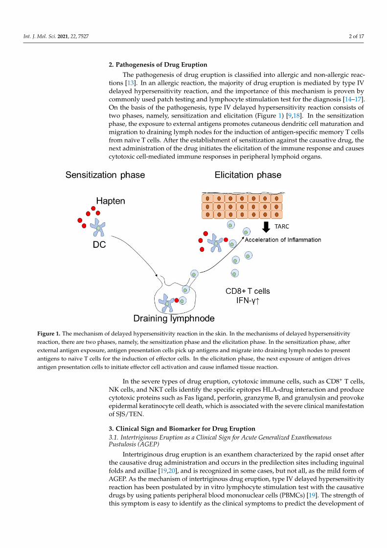
The pathogenesis of drug eruption is classified into allergic and non-allergic reac-tions [13]. In an allergic reaction, the majority of drug eruption is mediated by type IVdelayed hypersensitivity reaction, and the importance of this mechanism is proven bycommonly used patch testing and lymphocyte stimulation test for the diagnosis [14–17].On the basis of the pathogenesis, type IV delayed hypersensitivity reaction consists oftwo phases, namely, sensitization and elicitation (Figure 1) [9,18]. In the sensitizationphase, the exposure to external antigens promotes cutaneous dendritic cell maturation andmigration to draining lymph nodes for the induction of antigen-specific memory T cellsfrom naïve T cells. After the establishment of sensitization against the causative drug, thenext administration of the drug initiates the elicitation of the immune response and causescytotoxic cell-mediated immune responses in peripheral lymphoid organs.
Int. J. Mol. Sci. 2021, 22, x FOR PEER REVIEW 2 of 17
other minority types of drug eruption by searching keywords, “drug eruption”, “biomarker”, and “symptom” using Pubmed. These findings will be helpful for the further direction of these fields in the future.
2. Pathogenesis of Drug Eruption The pathogenesis of drug eruption is classified into allergic and non-allergic reactions
[13]. In an allergic reaction, the majority of drug eruption is mediated by type IV delayed hypersensitivity reaction, and the importance of this mechanism is proven by commonly used patch testing and lymphocyte stimulation test for the diagnosis [14–17]. On the basis of the pathogenesis, type IV delayed hypersensitivity reaction consists of two phases, namely, sensitization and elicitation (Figure 1) [9,18]. In the sensitization phase, the exposure to external antigens promotes cutaneous dendritic cell maturation and migration to draining lymph nodes for the induction of antigen-specific memory T cells from naïve T cells. After the establishment of sensitization against the causative drug, the next administration of the drug initiates the elicitation of the immune response and causes cytotoxic cell-mediated immune responses in peripheral lymphoid organs.
Figure 1. The mechanism of delayed hypersensitivity reaction in the skin. In the mechanisms of delayed hypersensitivity reaction, there are two phases, namely, the sensitization phase and the elicitation phase. In the sensitization phase, after external antigen exposure, antigen presentation cells pick up antigens and migrate into draining lymph nodes to present antigens to naïve T cells for the induction of effector cells. In the elicitation phase, the next exposure of antigen drives antigen presentation cells to initiate effector cell activation and cause inflamed tissue reaction.
In the severe types of drug eruption, cytotoxic immune cells, such as CD8+ T cells, NK cells, and NKT cells identify the specific epitopes HLA-drug interaction and produce cytotoxic proteins such as Fas ligand, perforin, granzyme B, and granulysin and provoke epidermal keratinocyte cell death, which is associated with the severe clinical manifestation of SJS/TEN.
3. Clinical Sign and Biomarker for Drug Eruption 3.1. Intertriginous Eruption as a Clinical Sign for Acute Generalized Exanthematous Pustulosis (AGEP)
Intertriginous drug eruption is an exanthem characterized by the rapid onset after the causative drug administration and occurs in the predilection sites including inguinal folds and axillae [19,20], and is recognized in some cases, but not all, as the mild form of
Figure 1. The mechanism of delayed hypersensitivity reaction in the skin. In the mechanisms of delayed hypersensitivityreaction, there are two phases, namely, the sensitization phase and the elicitation phase. In the sensitization phase, afterexternal antigen exposure, antigen presentation cells pick up antigens and migrate into draining lymph nodes to presentantigens to naïve T cells for the induction of effector cells. In the elicitation phase, the next exposure of antigen drivesantigen presentation cells to initiate effector cell activation and cause inflamed tissue reaction.
In the severe types of drug eruption, cytotoxic immune cells, such as CD8+ T cells,NK cells, and NKT cells identify the specific epitopes HLA-drug interaction and producecytotoxic proteins such as Fas ligand, perforin, granzyme B, and granulysin and provokeepidermal keratinocyte cell death, which is associated with the severe clinical manifestationof SJS/TEN.
3. Clinical Sign and Biomarker for Drug Eruption3.1. Intertriginous Eruption as a Clinical Sign for Acute Generalized ExanthematousPustulosis (AGEP)
Intertriginous drug eruption is an exanthem characterized by the rapid onset afterthe causative drug administration and occurs in the predilection sites including inguinalfolds and axillae [19,20], and is recognized in some cases, but not all, as the mild form ofAGEP. As the mechanism of intertriginous drug eruption, type IV delayed hypersensitivityreaction has been postulated by in vitro lymphocyte stimulation test with the causativedrugs by using patients peripheral blood mononuclear cells (PBMCs) [19]. The strength ofthis symptom is easy to identify as the clinical symptoms to predict the development of
Int. J. Mol. Sci. 2021, 22, 7527 3 of 17
AGEP. On the contrary, the weakness has been found by a single case report. Therefore, theaccumulation of multiple observational studies will be desired to determine how helpfulthis finding is for the detection of the early stage of AGEP.
3.2. Peripheral Blood Eosinophil Count
A previous study showed increased infiltration of eosinophils in the skin and the highfrequency of eosinophils in peripheral blood in patients with drug eruption. The presenceof eosinophils in peripheral blood differs in each type of drug eruption and the number ofeosinophils in patients with drug-induced erythema multiforme is increased, comparedwith other types of drug eruption [21]. A total of 113 patients were enrolled in this studyand an increased eosinophil number reflects an unfavorable clinical behavior, such as liverdysfunction, elongation of a duration of corticosteroid administration and hospitalization,suggesting that eosinophil is closely associated with the severity of drug eruption. Thestrongness of the peripheral blood eosinophil count is commonly examined in clinics andis easy to examine. On the contrary, the weakness is that this finding is not specific for drugeruption. Peripheral blood eosinophil also increases in atopic dermatitis with a positivecorrelation with the severity [22,23]. Peripheral blood eosinophil increase in patients withpsoriasis and urticaria [24]. From these findings, peripheral blood eosinophil is not specificin drug eruption; however, it might be easy to evaluate the severity of drug eruption.
3.3. Serum Eosinophilic Cationic Protein
Eosinophilic cationic protein is a cationic protein derived from eosinophil granulocyte,which is a marker for drug eruption [24]. Serum eosinophilic cationic protein levels weresignificantly increased in patients with drug eruption compared to healthy subjects. In amouse experiment, eosinophilic cationic protein increases 3 h after allergen exposure [25],suggesting that this might reflect an early time allergen reaction to the causative agents.Eosinophilic cationic protein does not correlate with the peripheral blood eosinophil count,however, serum eosinophilic cationic protein is increased with the positive correlation withthe activated eosinophil in the skin [26]. All 4 cases of drug eruption showed increasedserum eosinophilic cationic protein, while all 10 healthy subjects showed low levels ofserum eosinophilic cationic protein.
3.4. The Presence of CD4+CD25+ Cell in the Epidermis as a Sign of Desensitization in FixedDrug Eruption
Desensitization is one of the highlighted issues for clinical aspects in patients withdrug eruption. However, there is a limited number of findings to estimate the sign ofdesensitization. Although fixed drug eruption often shows the presence of CD8+ cells in theepidermis, CD4+CD25+ cells are also increased in the epidermis as a sign of desensitizationof fixed drug eruption [27]. Because this was investigated in only one subject, furtherlarge number studies will be desired to evaluate the actual impact on the desensitizationmechanisms. As a weakness, CD25+ cells do not directly mean regulatory T cells and thiscould not exclude the possibility of reactive T lymphocytes [28].
3.5. miR-18a-5p, miR-124, and miR-214
MicroRNA (miRNA) is a small single-stranded and non-coding RNA molecule and isobserved in various living things, such as animals and plants. miRNA is functionally silenc-ing RNA and suppresses post transcription of gene regulation [29]. Circulating miRNAsare released into the blood and have the potential as biomarkers in various diseases [30–32].An increased miR-18a-5p in the skin is observed in patients with TEN [33]. To evaluate theactual impact of miR-18a-5p, transfection of miR-18a-5p induces an increased apoptoticcell in keratinocytes and upregulates caspase-9 activity in the skin in patients with TEN. Inaddition, miR-18a-5p suppresses B-cell lymphoma/leukemia-2-like protein 10 (BCL2L10)gene expression, which is an anti-intrinsic apoptotic molecule. BCL2L10-silencing ker-atinocytes provoke both apoptosis and caspase-9 activity. Furthermore, serum miR-18a-5pincreases in patients with TEN with a positive correlation with body surface areas of skin
Int. J. Mol. Sci. 2021, 22, 7527 4 of 17
eruption. miR-18a-5p functionally promotes toxic reactions in the pathogenesis and is alsoa biomarker for patients with TEN. Serum samples were obtained from 8 patients withTEN, 10 patients with SJS, 15 patients with EM minor, and 22 healthy subjects in this study.
Both miR-124 and miR-214 expressions are increased in patients with severe drugeruptions and are reported as possible biomarkers of TEN [34]. The serum concentrationof miR-124 shows a positive correlation with the severity of drug eruption and the highscoring of SCORTEN. The miR-214 expression is increased in lesional skin in patientswith TEN. Serum samples were obtained from 7 TEN patients, 5 SJS patients, 11 EMminor patients, and 21 healthy subjects in this study. As a limitation, this study was asingle-institution study and was a relatively small number investigation.
The actual action of these miRNA in the pathogenesis of drug eruption remains unclear.However, the action of miRNA is considered to be: (1) specific changes in the methylationpattern, (2) post-transcriptional modifications of specific histone proteins, (3) differences inchromatic packing, in addition to (4) the role of the Polycomb and Thritorax proteins, whichcan be used as markers. Epigenetic changes can modulate gene transcription not to alterDNA sequencing information by histone modification and DNA methylation [35]. miRNAalso plays a pivotal role in the regulation of epigenetics [36,37]. Therefore, we speculatedthat these miRNA might also have an epigenetic modification and/or post-transcriptionalmodification in the drug eruption.
Actually, miR-18a-5p regulates the post-transcription and metastasis of cancer cells [38].miR-124 gene contributes to a condensed chromatin structure mediated by histone mod-ification [39], and also suppresses gene transcription [40]. Polycomb protein EZH2 isregulated by miR-124 and miR-214, which act as antitumor effects [41,42] and cell differen-tiation [43]. Although the contribution of epigenetic modification in drug eruption remainsunclear, these epigenetic changes might regulate the development of drug eruption.
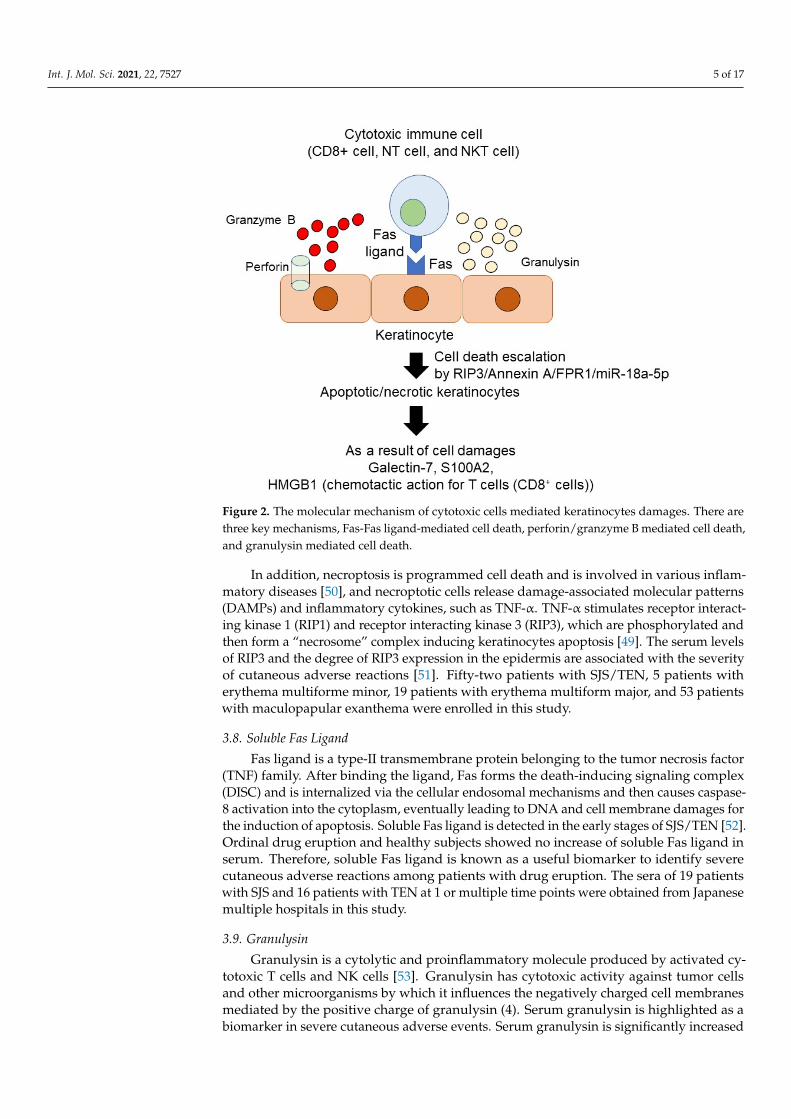
3.6. Perforin and Granzyme B
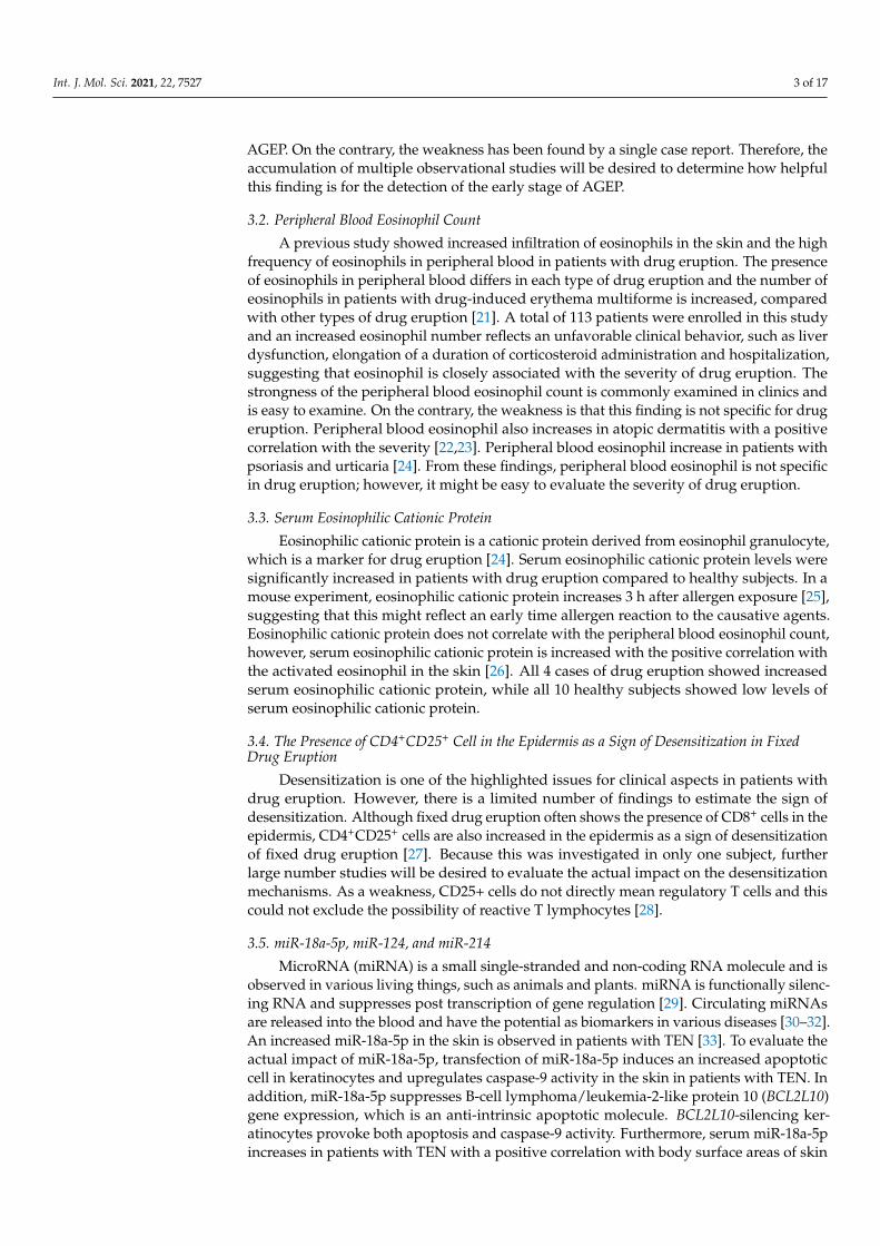
For the induction of the cytotoxic activity of immune cells, such as CD8+ cells and NKcells, they produce perforin, which is a cytolytic mediator and is released by cytoplasmicgranules in these immune cells (Figure 2). Perforin makes the pore in the cell targetedmembrane up to 20 nm in diameter and initiates cytotoxic molecules entering into thecytoplasm [44]. Granzyme B is a serine protease that initiates cell apoptosis in a perforin-dependent manner produced by cytotoxic cells [45]. Granzyme B cleaves and activatesvarious caspases, such as caspases 3 and 7, which trigger cell apoptosis [45]. Granzyme Balso has many substrates in the nucleus, leading to DNA disruption. In severe cutaneousadverse reactions, serum perforin and granzyme B are increased in patients with SJS/TENin an early time course [46,47]. Because their importance has been reported in severalstudies, perforin and granzyme B are reliable markers for SJS/TEN.
3.7. Annexin A1 and RIP3
Annexin A1 belongs to the danger-associated molecular patterns (DAMPs) and isreleased by the cytoplasm of apoptosis cells. Once annexin A1 binds formyl peptidereceptor 1 (FPR1) expressed on dendritic cells, this interaction enhances long-term betweenapoptosis cells and dendritic cells [48]. Annexin A1 is also one of the mediators forkeratinocyte death in severe cutaneous adverse drug eruption [49]. The depletion ofannexin A1 by antibody treatment impairs cytotoxicity against keratinocytes. Keratinocytesin patients with SJS/TEN highly express FPR1 in the skin. Therefore, annexin A is not onlya biomarker and but is also possibly a therapeutic target in SJS/TEN.
Int. J. Mol. Sci. 2021, 22, 7527 5 of 17Int. J. Mol. Sci. 2021, 22, x FOR PEER REVIEW 5 of 17
Figure 2. The molecular mechanism of cytotoxic cells mediated keratinocytes damages. There are three key mechanisms, Fas-Fas ligand-mediated cell death, perforin/granzyme B mediated cell death, and granulysin mediated cell death.
3.7. Annexin A1 and RIP3 Annexin A1 belongs to the danger-associated molecular patterns (DAMPs) and is
released by the cytoplasm of apoptosis cells. Once annexin A1 binds formyl peptide receptor 1 (FPR1) expressed on dendritic cells, this interaction enhances long-term between apoptosis cells and dendritic cells [48]. Annexin A1 is also one of the mediators for keratinocyte death in severe cutaneous adverse drug eruption [49]. The depletion of annexin A1 by antibody treatment impairs cytotoxicity against keratinocytes. Keratinocytes in patients with SJS/TEN highly express FPR1 in the skin. Therefore, annexin A is not only a biomarker and but is also possibly a therapeutic target in SJS/TEN.
In addition, necroptosis is programmed cell death and is involved in various inflammatory diseases [50], and necroptotic cells release damage-associated molecular patterns (DAMPs) and inflammatory cytokines, such as TNF-α. TNF-α stimulates receptor interacting kinase 1 (RIP1) and receptor interacting kinase 3 (RIP3), which are phosphorylated and then form a “necrosome” complex inducing keratinocytes apoptosis [49]. The serum levels of RIP3 and the degree of RIP3 expression in the epidermis are associated with the severity of cutaneous adverse reactions [51]. Fifty-two patients with SJS/TEN, 5 patients with erythema multiforme minor, 19 patients with erythema multiform major, and 53 patients with maculopapular exanthema were enrolled in this study.
3.8. Soluble Fas Ligand Fas ligand is a type-II transmembrane protein belonging to the tumor necrosis factor
(TNF) family. After binding the ligand, Fas forms the death-inducing signaling complex (DISC) and is internalized via the cellular endosomal mechanisms and then causes caspase-8 activation into the cytoplasm, eventually leading to DNA and cell membrane damages for the induction of apoptosis. Soluble Fas ligand is detected in the early stages of SJS/TEN [52]. Ordinal drug eruption and healthy subjects showed no increase of soluble
Figure 2. The molecular mechanism of cytotoxic cells mediated keratinocytes damages. There arethree key mechanisms, Fas-Fas ligand-mediated cell death, perforin/granzyme B mediated cell death,and granulysin mediated cell death.
In addition, necroptosis is programmed cell death and is involved in various inflam-matory diseases [50], and necroptotic cells release damage-associated molecular patterns(DAMPs) and inflammatory cytokines, such as TNF-α. TNF-α stimulates receptor interact-ing kinase 1 (RIP1) and receptor interacting kinase 3 (RIP3), which are phosphorylated andthen form a “necrosome” complex inducing keratinocytes apoptosis [49]. The serum levelsof RIP3 and the degree of RIP3 expression in the epidermis are associated with the severityof cutaneous adverse reactions [51]. Fifty-two patients with SJS/TEN, 5 patients witherythema multiforme minor, 19 patients with erythema multiform major, and 53 patientswith maculopapular exanthema were enrolled in this study.
3.8. Soluble Fas Ligand
Fas ligand is a type-II transmembrane protein belonging to the tumor necrosis factor(TNF) family. After binding the ligand, Fas forms the death-inducing signaling complex(DISC) and is internalized via the cellular endosomal mechanisms and then causes caspase-8 activation into the cytoplasm, eventually leading to DNA and cell membrane damages forthe induction of apoptosis. Soluble Fas ligand is detected in the early stages of SJS/TEN [52].Ordinal drug eruption and healthy subjects showed no increase of soluble Fas ligand inserum. Therefore, soluble Fas ligand is known as a useful biomarker to identify severecutaneous adverse reactions among patients with drug eruption. The sera of 19 patientswith SJS and 16 patients with TEN at 1 or multiple time points were obtained from Japanesemultiple hospitals in this study.
3.9. Granulysin
Granulysin is a cytolytic and proinflammatory molecule produced by activated cy-totoxic T cells and NK cells [53]. Granulysin has cytotoxic activity against tumor cellsand other microorganisms by which it influences the negatively charged cell membranesmediated by the positive charge of granulysin (4). Serum granulysin is highlighted as abiomarker in severe cutaneous adverse events. Serum granulysin is significantly increased
Int. J. Mol. Sci. 2021, 22, 7527 6 of 17
in SJS/TEN patients, and the immunochromatography examination showed positive re-sults of granulysin in 80% of SJS/TEN patients, with 1 out of 24 patients presenting ordinaldrug eruption [54]. Five patients with SJS/TEN, 24 patients with ordinal drug eruption,and 31 healthy subjects were enrolled and collected serum in this study. Another studyshowed that drug-induced hypersensitivity syndrome/drug reaction with eosinophiliaalso show high serum levels of granulysin [55]. Twenty-one patients with DRESS and29 healthy subjects were enrolled in this study.
3.10. High Mobility Group Box 1 Protein (HMGB1)
Likewise histone, HMGB1 is the important chromatin protein and interacts with nucle-osomes to regulate gene transcription [56]. In addition, HMGB1 has been recognized as adanger signal involved in the pathogenesis of various inflammatory diseases, malignancies,and injury [57–59]. On the contrary to these functional roles, HMGB1 is also useful as abiomarker in various diseases [60,61]. HMGB1 is noticed in serum in patients with severecutaneous adverse reactions [62,63]. Serum HMGB1 levels in patients with SJS/TEN from7 days before the onset and 21 after the onset of diseases were significantly increasedcompared with healthy controls and patients with ordinal drug eruption [62]. Twenty-twohealthy subjects and 13 patients with SJS/TEN were enrolled in this study and the serawere obtained from multiple institutions. In addition to the high serum level of HMGB1,HMGB1 concentration is also increased in bullous fluids in patients with SJS/TEN [63].This study was conducted by three independent SJS/TEN patient cohorts and was thenbrought together for the purpose of this study. Nine patients with SJS/TEN were enrolledin Nevirapine-induced cases study, 73 patients with SJS/TEN were enrolled in a Taiwanesestudy to evaluate the serum HMGB1 concentration. In addition, 22 patients with SJS/TENwere enrolled in a Spanish study to evaluate the bullous concentration of HMGB1.
3.11. Thymus and Activation-Regulated Chemokine (TARC)
TARC is a chemokine belonging to the CC chemokine family, namely CCL17, andis produced by immune cells in allergic diseases [64,65]. TARC binds to CCR4, which isdominant in Th2-dominant immune cells for their chemotaxis [66]. TARC levels in serumin patients with drug eruption can predict systemic inflammation and disease severity [67].Serum TARC levels positively correlated with the severity of cutaneous inflammation,such as white blood cell count, C-reactive protein, and the severity of the score. Twelvepatients with DRESS/DIHS, 18 patients with maculopapular exanthema, and 46 patientswith erythema multiforme were enrolled and SJS/TEN patients were eliminated fromthis study, indicating that TARC might not reflect the actual severity of drug eruption. Inaddition, TARC is not specific in drug eruption and is also increased in other inflammatoryskin diseases, such as atopic dermatitis [65,68] and psoriasis [69].
3.12. Th17
IL-17-producing cells are involved in various inflammatory diseases, such aspsoriasis [70–74], asthma [75–79], and atopic dermatitis [80–82], and contribute to thedevelopment of host defense against microorganisms [83–86]. As the importance of IL-17-producing cells in the human body, IL-17-targeted biologics treatment is proven by thecurrent advancement of this field. Th17 cells are known as the main source of IL-17 inhumans, and the percentages of Th17 cells are increased in DIHS (10 days after the onset)and SJS/TEN (2–6 days after the onset) as compared to normal subjects and maculopapu-lar drug eruption (2–6 days after the onset) [87]. Fifteen patients with maculopapulartype, 15 patients with erythema multiforme, 1 patient with SJS, 1 patient with TEN, and4 patients with DIHS were enrolled in this study. During the clinical course of DIHS, thefrequencies of Th17 cells are gradually decreased. Peripheral blood Th17 cells migrate intothe skin on day 12–21 after the onset. This information suggests to us that Th17 cells couldnot detect at the very early time point in the clinical time course in patients with severe typedrug eruption. In another study, the frequency of Th17 did not differ significantly between
Int. J. Mol. Sci. 2021, 22, 7527 7 of 17
SJS/TEN patients and healthy controls, possibly due to the missing time course point [88].Peripheral blood mononuclear cells were obtained from SJS/TEN patients (acute stage:n = 3, resolution stage: n = 7) and 24 healthy subjects. Therefore, Th17 cells might notbe useful for the biomarker in severe type drug eruption in an early time point, however,this might be a compliment for the lack of information of other important biomarkers forcutaneous adverse events such as SJS/TEN. As the importance in the pathogenesis, Th17might also promote an additional cutaneous inflammatory response in severe type drugeruption, because Th17 enhances subsequent inflammatory immune responses. Consis-tently, an increased frequency of Th17 cells has been reported in AGEP [89]. Peripheralblood mononuclear cells were obtained from 3 patients with AGEP in this study. Therefore,it is speculated that IL-17 and IL-22 cooperatively stimulate keratinocytes to enhance IL-8production, leading to the contribution of neutrophil infiltration in the lesional epidermisof AGEP. In addition, psoriasiform drug eruption mimicking as psoriasis vulgaris is some-times observed in the treatment of some biologic agents, such as TNF or IL-17 inhibitors,which are also involved in Th17 in the skin [90,91], suggesting that Th17 cells play somerole in the pathogenesis of drug eruption not in the main but in a part of the stream. Asfor the weakness, Th17 is observed in various other inflammatory skin diseases, such aspsoriasis and atopic dermatitis; therefore, Th17 is not a specific biomarker of drug eruption.
3.13. Serum Galectin-7
Galectin-7 is a β-galactoside-binding protein family and is expressed in epithelialcells [92]. Keratinocytes are one of the major cells to express galectin-7, which is closelyassociated with keratinocyte survival and growth [92]. Galectin-7 has been investigatedand used to discover biomarkers in various diseases [93–98]. Galectin-7 has examinedthe possible role of the biomarker in severe cutaneous adverse events [99]. The causativedrug was determined from lymphocyte stimulation test in SJS and TEN. The supernatantof lymphocyte stimulation test using peripheral blood mononuclear cells taken fromthese patients after 1 day was collected and subjected to the mass spectrometry analysis.Galectin-7 was identified as the possible proteins as a biomarker for SJS/TEN and theserum concentration of galectin-7 was increased in the 24 patients with SJS/TEN comparedwith that in 8 healthy subjects. The weakness of the galectin-7 is not a specific markerof drug eruption, and is also seen in other inflammatory skin diseases, such as atopicdermatitis [100] and UV radiation [101], while it is decreased in psoriasis [102].
3.14. S100A2
S100 calcium-binding protein A2 (S100A2) is a member of the EF-hand motif familyS100 [103] and is recognized as a tumor-suppressive role [104]. S100A2 is mainly expressedin epithelial cells and contributes to various cell functions [105], and is recognized as abiomarker in oncology fields [106–110]. However, a recent study identified that S100A2is a possible biomarker in severe cutaneous adverse events [111]. Forty-one cases ofmacular type, 14 cases of maculopapular type, and 9 cases of severe types of drug eruption(7 cases of Stevens–Johnson syndrome and 2 cases of toxic epidermal necrolysis) wereenrolled in this study. This study was conducted by using telaprevir or trichloroethylene,which causes high frequently cutaneous adverse events. Telaprevir or trichloroethyleneexposed keratinocytes highly enhance S100A2 proteins by microarray analysis and S100A2is increased in the skin in patients with severe type drug eruption. However, S100A2is not a specific biomarker because S100A2 is also upregulated in atopic dermatitis andpsoriasis patients.
3.15. The Difference of Generalized Bullous Fixed Drug Eruption Compared with SJS and TEN
Generalized bullous fixed drug eruption is a kind of particular clinical manifestationof fixed drug eruption and is characterized by wide-spreading blisters and erosions, whichmimic the clinical characteristics of SJS/TEN. Patients with generalized bullous fixed drugeruption exhibit shorter latent periods and less mucosal involvement, however, more
Int. J. Mol. Sci. 2021, 22, 7527 8 of 17
eosinophil infiltration and dermal melanophages. Twenty-three cases of GBFDE wereenrolled in this study [112], and increased infiltration of CD4+Foxp3+ regulatory T cellsin the dermis and reduced intraepidermal granulysin+ cell infiltration were observed ingeneralized bullous fixed drug eruption. In addition, the serum granulysin in generalizedbullous fixed drug eruption was significantly lower than that in SJS/TEN. Therefore, itmight be helpful to gain a clue of the diagnosis of generalized bullous fixed drug eruptionby evaluating the serum granulysin in addition to these histological analyses to distinguishbetween generalized bullous fixed drug eruption and SJS/TEN.
3.16. T Cell Receptor-Cβ1 (TCR-Cβ1) Gene Rearrangement for Pseudolymphoma CD30+ LargeCell Transformation
Lymphocytes acquire the diversity of antigen recognition through T-cell receptor generearrangement during cell differentiation. On the contrary, malignant lymphoma tumorcells exhibit a monoclonal change of tumor growth and T-cell receptorrearrangement [113–115]. Therefore, a monoclonal T-cell receptor gene band is observedin lymphoma cells. This method is commonly used in the clinical aspect to distinguish be-tween benign and malignant lymphoid tumors. Pseudolymphoma is one of the lymphoidcell proliferation diseases and is characterized by solid papules and nodules [116,117],which mimic lymphoma and lymphomatoid papulosis [118,119]. The clinical manifesta-tion is the solid popular with the presence of CD30+ large transformed atypical cells inthe skin. Although the pathogenesis of CD30+ pseudolymphoma remains unclear, thedrug administration is one of the triggers for this cutaneous reaction [120–122] and thereactive T cells are overstimulated during the causative agent’s treatment and as a result,the pseudolymphoma might cause after a long duration of the incubation period [120]. Asa limitation, these are case studies. For clinicians, lymphomatoid papulosis is a differentialdiagnosis that is clearly distinguished by the absence of monoclonal T-cell receptor-Cβ1gene rearrangement in the lesional skin [121].
3.17. Metalloproteinase
A metalloproteinase is any protease enzyme mediated by a catalytic mechanisminvolved in metals. Metalloproteinases play a crucial role in various cell functions, suchas cell migration [123]. Previous studies have identified the relationship between drugeruption and metalloproteinases. A high amount of MMP2 was observed in TEN blisterfluid [124]. Blister fluid from 6 patients with TEN patients was compared with 3 otherblistering conditions. Six patients with bullous pemphigoid, 13 patients with second-degree burn, and 3 patients with suction blister were examined in this study. In addition,the presence of MMP2, MMP9, and MMP11 was observed in the skin in patients withSJS/TEN [125,126]. One study was performed, where skin biopsies were taken from theedge of the blisters of 2 patients with TEN, 3 patients with SJS, and 2 healthy subjects [125],and another study conducted skin biopsied taken from 8 patients with erythema multiformeand 6 patients with SJS/TEN [126], respectively. MMP2 and MMP9 are not specific indrug eruption. MMP2 is increased in psoriasis [127] and MMP9 is increased in atopicdermatitis [128], psoriasis [129], and urticaria [130]. On the contrary, MMP11 has currentlynot been reported in inflammatory skin diseases, such as psoriasis and atopic dermatitis.Although further investigation is necessary, MMP11 might be a specific biomarker indurg eruption.
3.18. Prognostic Biomarkers for SJS and TEN
Since fatal cases and long-term complications are observed following cutaneous adversereactions, the predictive tools for these cases are helpful for clinicians. Severity-of-illness scorefor toxic epidermal necrolysis (SCORTEN) is a representative severity scoring for SJS/TEN toestimate their prognosis, based on seven independent clinical parameters; age > 40 years, thepresence of malignancy, tachycardia > 120/min, body surface area affected > 10%, serum urea> 28 mg/dL, serum glucose > 250 mg/dL, serum bicarbonate < 20 mEq/L [131]. SCORTEN
Int. J. Mol. Sci. 2021, 22, 7527 9 of 17
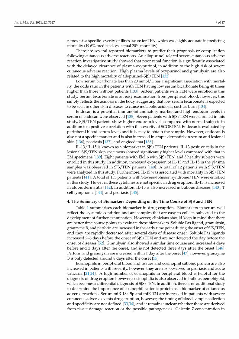
represents a specific severity-of-illness score for TEN, which was highly accurate in predictingmortality (19.6% predicted, vs. actual 20% mortality).
There are several reported biomarkers to predict their prognosis or complicationfollowing cutaneous adverse reactions. An allopurinol-related severe cutaneous adversereaction investigative study showed that poor renal function is significantly associatedwith the delayed clearance of plasma oxypurinol, in addition to the high risk of severecutaneous adverse reaction. High plasma levels of oxypurinol and granulysin are alsorelated to the high mortality of allopurinol-SJS/TEN [132].
Low serum bicarbonate less than 20 mmol/L has a significant association with mortal-ity, the odds ratio in the patients with TEN having low serum bicarbonate being 40 timeshigher than those without patients [133]. Sixteen patients with TEN were enrolled in thisstudy. Serum bicarbonate is an easy examination from peripheral blood; however, thissimply reflects the acidosis in the body, suggesting that low serum bicarbonate is expectedto be seen in other skin diseases to cause metabolic acidosis, such as burn [134].
Endocan is a potential immunoinflammatory marker, and high endocan levels inserum of endocan were observed [135]. Seven patients with SJS/TEN were enrolled in thisstudy. SJS/TEN patients show higher endocan levels compared with normal subjects inaddition to a positive correlation with the severity of SCORTEN. Endocan is evaluated byperipheral blood serum level, and it is easy to obtain the sample. However, endocan isalso not a specific marker and is also increased in atopic dermatitis in serum and lesionalskin [136], psoriasis [137], and angioedema [138].
IL-13/IL-15 is known as a biomarker in SJS/TEN patients. IL-13 positive cells in thelesional SJS/TEN skin specimens showed significantly higher levels compared with that inEM specimens [139]. Eight patients with EM, 6 with SJS/TEN, and 3 healthy subjects wereenrolled in this study. In addition, increased expression of IL-13 and IL-15 in the plasmasamples was observed in SJS/TEN patients [140]. A total of 12 patients with SJS/TENwere analyzed in this study. Furthermore, IL-15 was associated with mortality in SJS/TENpatients [141]. A total of 155 patients with Stevens-Johnson syndrome/TEN were enrolledin this study. However, these cytokines are not specific in drug eruption. IL-13 is increasedin atopic dermatitis [142]. In addition, IL-15 is also increased in bullous diseases [143], Tcell lymphoma [144], and psoriasis [145].
4. The Summary of Biomarkers Depending on the Time Course of SJS and TEN
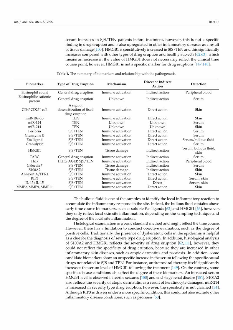
Table 1 summarises each biomarker in drug eruption. Biomarkers in serum wellreflect the systemic condition and are samples that are easy to collect, subjected to thedevelopment of further examination. However, clinicians should keep in mind that thereare better time course points to evaluate these biomarkers. Soluble Fas ligand, granulysin,granzyme B, and perforin are increased in the early time point during the onset of SJS/TEN,and they are rapidly decreased after several days of disease onset. Soluble Fas ligandsincreased 2–6 days before the onset of SJS/TEN and are not detected the day before theonset of diseases [52]. Granulysin also showed a similar time course and increased 4 daysbefore and 2 days after the onset, and is not detected three days after the onset [146].Perforin and granulysin are increased within 1 day after the onset [47], however, granzymeB is only detected around 8 days after the onset [55].
Eosinophils in peripheral blood and tissues and eosinophil cationic protein are alsoincreased in patients with severity, however, they are also observed in psoriasis and acuteurticaria [21,24]. A high number of eosinophils in peripheral blood is helpful for thediagnosis of drug eruption however, eosinophilia is also observed in bullous pemphigoid,which becomes a differential diagnosis of SJS/TEN. In addition, there is no additional studyto determine the importance of eosinophil cationic protein as a biomarker of cutaneousadverse reactions. Serum miR-18a-5p and miR-124 are increased in patients with severecutaneous adverse events drug eruption, however, the timing of blood sample collectionand specificity are not defined [33,34], and it remains unclear whether these are derivedfrom tissue damage reaction or the possible pathogenesis. Galectin-7 concentration in
Int. J. Mol. Sci. 2021, 22, 7527 10 of 17
serum increases in SJS/TEN patients before treatment, however, this is not a specificfinding in drug eruption and is also upregulated in other inflammatory diseases as a resultof tissue damage [100]. HMGB1 is constitutively increased in SJS/TEN and this significantlyincreases compared with other types of drug eruption and healthy subjects [62,63], whichmeans an increase in the value of HMGB1 does not necessarily reflect the clinical timecourse point, however, HMGB1 is not a specific marker for drug eruptions [147,148].
Table 1. The summary of biomarkers and relationship with the pathogenesis.
Biomarker Type of Drug Eruption Mechanism Direct or IndirectAction Detection
Eosinophil count General drug eruption Immune activation Indirect action Peripheral bloodEosinophilic cationic
protein General drug eruption Unknown Indirect action Serum
CD4+CD25+ cellA sign of
desensitization of fixeddrug eruption
Immune activation Direct action Skin
miR-18a-5p TEN Immune activation Direct action SkinmiR-124 TEN Unknown Unknown SerummiR-214 TEN Unknown Unknown SkinPerforin SJS/TEN Immune activation Direct action Serum
Granzyme B SJS/TEN Immune activation Direct action SerumFas ligand SJS/TEN Immune activation Direct action Serum, bullous fluidGranulysin SJS/TEN Immune activation Direct action Serum
HMGB1 SJS/TEN Tissue damage Indirect action Serum, bullous fluid,skin
TARC General drug eruption Immune activation Indirect action SerumTh17 DIHS, AGEP, SJS/TEN Immune activation Indirect action Peripheral blood
Galectin 7 SJS/TEN Tissue damage Indirect action SerumS100A2 SJS/TEN Tissue damage Indirect action Skin
Annexin A/FPR1 SJS/TEN Immune activation Direct action SkinRIP3 SJS/TEN Immune activation Direct action Serum, skin
IL-13/IL-15 SJS/TEN Immune activation Direct Serum, skinMMP2, MMP9, MMP11 SJS/TEN Immune activation Direct action Skin
The bullous fluid is one of the samples to identify the local inflammatory reaction toaccumulate the inflammatory response in the site. Indeed, the bullous fluid contains aboveearly time course biomarkers, such as soluble Fas ligands [63] and HMGB1 [63], however,they only reflect local skin site inflammation, depending on the sampling technique andthe degree of the local site inflammation.
Histological examination is a basic standard method and might reflect the time course.However, there has a limitation to conduct objective evaluation, such as the degree ofpositive cells. Traditionally, the presence of dyskeratotic cells in the epidermis is helpfulas a clue for the diagnosis of severe type drug eruption. In addition, histological analysisof S100A2 and HMGB1 reflects the severity of drug eruption [62,111], however, theycould not reflect the specificity of drug eruption, because they are increased in otherinflammatory skin diseases, such as atopic dermatitis and psoriasis. In addition, somecandidate biomarkers show an unspecific increase in the serum following the specific causaldrugs not related to SJS and TEN. For instance, antiretroviral therapy itself significantlyincreases the serum level of HMGB1 following the treatment [149]. On the contrary, somespecific disease conditions also affect the degree of these biomarkers. An increased serumHMGB1 level is observed in febrile seizures [150] and end stage renal disease [151]. S100A2also reflects the severity of atopic dermatitis, as a result of keratinocyte damages. miR-214is increased in severity type drug eruption, however, the specificity is not clarified [34].Although RIP3 is driven under a more specific condition, this could not also exclude otherinflammatory disease conditions, such as psoriasis [50].
Int. J. Mol. Sci. 2021, 22, 7527 11 of 17
As a limitation of biomarkers regarding the correlation between skin tissue reactionand serum/plasma and blister fluid levels of the biomarkers, skin tissue reaction does notalways reflect the serum levels of biomarkers. Therefore, systematic analysis is used toclarify the interaction between local skin tissue reaction and systemic reaction by peripheralblood and serum.
5. Conclusions
This review summarized the clinical signs, diagnostic tools, and biomarkers for drugeruption, in addition to a recent update of this field research. The presence of biomarkersdiffers in each time course of severe type drug eruption, and we should keep in mindthat the disease onset date-based examination of biomarkers is helpful for the diagnosis ofSJS/TEN with the combination of histological and serum examination. There are severaluseful markers for SJS/TEN diagnosis, however, specificity is also the next problem for thediagnosis of SJS and TEN. In the current options, soluble Fas ligands and granulysin aresome of the most reliable tools for the diagnosis of SJS/TEN. However, if we face the clinicalcases with a suspected diagnosis of SJS/TEN and there is a limitation to evaluate solubleFas ligands and granulysin, especially due to the delayed sampling, other biomarkers mightbe useful for the detection of SJS/TEN to predict the future clinical course in these patientsof drug eruption. As the prognostic estimation tool for SJS/TEN patients, SCORTEN is abetter tool in addition to other biomarkers, such as serum bicarbonate, endocan, and IL-15.These combination examinations will be helpful to determine the therapeutic approach fordrug eruption.
Author Contributions: Conceptualization and writing manuscript, M.Y. and Y.S. supervision, M.N.All authors have read and agreed to the published version of the manuscript.
Funding: This research received no external funding.
Institutional Review Board Statement: Not applicable.
Informed Consent Statement: Not applicable.
Conflicts of Interest: The authors declare no conflict of interest.
References1. Reen, F.J.; Romano, S.; Dobson, A.D.; O’Gara, F. The Sound of Silence: Activating Silent Biosynthetic Gene Clusters in Marine
Microorganisms. Mar. Drugs 2015, 13, 4754–4783. [CrossRef]2. Yamamoto, Y.; Wanibuchi, S.; Sato, A.; Kasahara, T.; Fujita, M. Precipitation of test chemicals in reaction solutions used in
the amino acid derivative reactivity assay and the direct peptide reactivity assay. J. Pharm. Toxicol. Methods 2019, 100, 106624.[CrossRef]
3. White, K.D.; Abe, R.; Ardern-Jones, M.; Beachkofsky, T.; Bouchard, C.; Carleton, B.; Chodosh, J.; Cibotti, R.; Davis, R.; Denny, J.C.;et al. SJS/TEN 2017: Building Multidisciplinary Networks to Drive Science and Translation. J. Allergy Clin. Immunol. Pract. 2018,6, 38–69. [CrossRef] [PubMed]
4. Sunaga, Y.; Kurosawa, M.; Ochiai, H.; Watanabe, H.; Sueki, H.; Azukizawa, H.; Asada, H.; Watanabe, Y.; Yamaguchi, Y.; Aihara, M.;et al. The nationwide epidemiological survey of Stevens-Johnson syndrome and toxic epidermal necrolysis in Japan, 2016–2018. J.Dermatol. Sci. 2020, 100, 175–182. [CrossRef]
5. Watanabe, T.; Go, H.; Saigusa, Y.; Takamura, N.; Watanabe, Y.; Yamane, Y.; Totsuka, M.; Ishikawa, H.; Nakamura, K.; Matsukura,S.; et al. Mortality and risk factors on admission in toxic epidermal necrolysis: A cohort study of 59 patients. Allergol. Int. 2020,70, 229–234. [CrossRef] [PubMed]
6. Dainichi, T.; Kitoh, A.; Otsuka, A.; Nakajima, S.; Nomura, T.; Kaplan, D.H.; Kabashima, K. The epithelial immune microenviron-ment (EIME) in atopic dermatitis and psoriasis. Nat. Immunol. 2018, 19, 1286–1298. [CrossRef] [PubMed]
7. Kabashima, K.; Honda, T.; Ginhoux, F.; Egawa, G. The immunological anatomy of the skin. Nat. Rev. Immunol. 2019, 19, 19–30.[CrossRef] [PubMed]
8. Sawada, Y.; Gallo, R.L. Role of Epigenetics in the Regulation of Immune Functions of the Skin. J. Investig. Dermatol. 2020, 141,1157–1166. [CrossRef] [PubMed]
9. Honda, T.; Egawa, G.; Grabbe, S.; Kabashima, K. Update of immune events in the murine contact hypersensitivity model: Towardthe understanding of allergic contact dermatitis. J. Investig. Dermatol. 2013, 133, 303–315. [CrossRef]
10. Johnstone, D.F. Fatal case of Stevens-Johnson syndrome. Lancet 1947, 2, 276. [CrossRef]
Int. J. Mol. Sci. 2021, 22, 7527 12 of 17
11. Silvestri, M.; Cristaudo, A.; Morrone, A.; Messina, C.; Bennardo, L.; Nisticò, S.P.; Mariano, M.; Cameli, N. Emerging Skin Toxicitiesin Patients with Breast Cancer Treated with New Cyclin-Dependent Kinase 4/6 Inhibitors: A Systematic Review. Drug Saf. 2021,44, 725–732. [CrossRef]
12. Roberti, R.; Iannone, L.F.; Palleria, C.; De Sarro, C.; Spagnuolo, R.; Barbieri, M.A.; Vero, A.; Manti, A.; Pisana, V.; Fries, W.; et al.Safety profiles of biologic agents for inflammatory bowel diseases: A prospective pharmacovigilance study in Southern Italy.Curr. Med. Res. Opin. 2020, 36, 1457–1463. [CrossRef]
13. Thong, B.Y.; Tan, T.C. Epidemiology and risk factors for drug allergy. Br. J. Clin. Pharm. 2011, 71, 684–700. [CrossRef] [PubMed]14. Sawada, Y.; Kabashima-Kubo, R.; Hino, R.; Nakamura, M. Fatal case of toxic epidermal necrolysis caused by cefozopran and
associated with psoriasis. Acta Derm.-Venereol. 2014, 94, 341–342. [CrossRef] [PubMed]15. Sawada, Y.; Kawakami, C.; Nakamura, M.; Tokura, Y.; Yoshiki, R. Toxic epidermal necrosis-like dermatosis induced by the first
course of methotrexate. Eur. J. Dermatol. 2009, 19, 397–398. [CrossRef]16. Sawada, Y.; Nakamura, M.; Tokura, Y. Generalized fixed drug eruption caused by pazufloxacin. Acta Derm.-Venereol. 2011, 91,
600–601. [CrossRef] [PubMed]17. Saito, R.; Sawada, Y.; Nakamura, M. Two cases of eczematous drug eruption caused by oral tacrolimus administration. Contact
Dermat. 2017, 77, 128–130. [CrossRef]18. Sawada, Y.; Honda, T.; Hanakawa, S.; Nakamizo, S.; Murata, T.; Ueharaguchi-Tanada, Y.; Ono, S.; Amano, W.; Nakajima, S.;
Egawa, G.; et al. Resolvin E1 inhibits dendritic cell migration in the skin and attenuates contact hypersensitivity responses. J. Exp.Med. 2015, 212, 1921–1930. [CrossRef]
19. Sawada, Y.; Sugita, K.; Fukamachi, S.; Bito, T.; Nakamura, M.; Tokura, Y. Doripenem-induced intertriginous drug eruption as amild form of AGEP. J. Eur. Acad. Dermatol. Venereol. 2009, 23, 974–976. [CrossRef]
20. Totonchy, M.B.; McNiff, J.M.; Bunick, C.G. Koebnerization of Hailey-Hailey disease into a cutaneous drug eruption of acutegeneralized exanthematous pustulosis associated with systemic symptoms. J. Cutan. Pathol. 2016, 43, 1031–1035. [CrossRef]
21. Yang, J.; Yang, X.; Li, M. Peripheral blood eosinophil counts predict the prognosis of drug eruptions. J. Investig. Allergol. Clin.Immunol. 2013, 23, 248–255.
22. Dhar, S.; Malakar, R.; Chattopadhyay, S.; Dhar, S.; Banerjee, R.; Ghosh, A. Correlation of the severity of atopic dermatitis withabsolute eosinophil counts in peripheral blood and serum IgE levels. Indian J. Dermatol. Venereol. Leprol. 2005, 71, 246–249.[CrossRef]
23. Hu, Y.; Liu, S.; Liu, P.; Mu, Z.; Zhang, J. Clinical relevance of eosinophils, basophils, serum total IgE level, allergen-specific IgE,and clinical features in atopic dermatitis. J. Clin. Lab. Anal. 2020, 34, e23214. [CrossRef] [PubMed]
24. Kim, T.Y.; Park, H.J.; Kim, C.W. Eosinophil cationic protein (ECP) level and its correlation with eosinophil number or IgE level ofperipheral blood in patients with various skin diseases. J. Dermatol. Sci. 1997, 15, 89–94. [CrossRef]
25. Nielsen, P.N.; Skov, P.S.; Poulsen, L.K.; Schmelz, M.; Petersen, L.J. Cetirizine inhibits skin reactions but not mediator release inimmediate and developing late-phase allergic cutaneous reactions. A double-blind, placebo-controlled study. Clin. Exp. Allergy J.Br. Soc. Allergy Clin. Immunol. 2001, 31, 1378–1384. [CrossRef]
26. Juhlin, L.; Venge, P. Eosinophilic cationic protein (ECP) in skin disorders. Acta Derm.-Venereol. 1991, 71, 495–501. [PubMed]27. Teraki, Y.; Shiohara, T. Successful desensitization to fixed drug eruption: The presence of CD25+CD4+ T cells in the epidermis
of fixed drug eruption lesions may be involved in the induction of desensitization. Dermatology 2004, 209, 29–32. [CrossRef][PubMed]
28. Jones, D.; Ibrahim, S.; Patel, K.; Luthra, R.; Duvic, M.; Medeiros, L.J. Degree of CD25 expression in T-cell lymphoma is dependenton tissue site: Implications for targeted therapy. Clin. Cancer Res. Off. J. Am. Assoc. Cancer Res. 2004, 10, 5587–5594. [CrossRef][PubMed]
29. Fabian, M.R.; Sonenberg, N. The mechanics of miRNA-mediated gene silencing: A look under the hood of miRISC. Nat. Struct.Mol. Biol. 2012, 19, 586–593. [CrossRef] [PubMed]
30. Wong, W.K.; Wong, S.H.D.; Bian, L. Long-Term Detection of Oncogenic MicroRNA in Living Human Cancer Cells by Gold@Polydopamine-Shell Nanoprobe. ACS Biomater. Sci. Eng. 2020, 6, 3778–3783. [CrossRef]
31. Evangelista, A.F.; Oliveira, R.J.; VA, O.S.; RA, D.C.V.; Reis, R.M.; MM, C.M. Integrated analysis of mRNA and miRNA profilesrevealed the role of miR-193 and miR-210 as potential regulatory biomarkers in different molecular subtypes of breast cancer.BMC Cancer 2021, 21, 76. [CrossRef]
32. Tang, S.; Li, S.; Liu, T.; He, Y.; Hu, H.; Zhu, Y.; Tang, S.; Zhou, H. MicroRNAs: Emerging oncogenic and tumor-suppressiveregulators, biomarkers and therapeutic targets in lung cancer. Cancer Lett. 2021, 502, 71–83. [CrossRef]
33. Ichihara, A.; Wang, Z.; Jinnin, M.; Izuno, Y.; Shimozono, N.; Yamane, K.; Fujisawa, A.; Moriya, C.; Fukushima, S.; Inoue, Y.; et al.Upregulation of miR-18a-5p contributes to epidermal necrolysis in severe drug eruptions. J. Allergy Clin. Immunol. 2014, 133,1065–1074. [CrossRef]
34. Sato, S.; Ichihara, A.; Jinnin, M.; Izuno, Y.; Fukushima, S.; Ihn, H. Serum miR-124 up-regulation as a disease marker of toxicepidermal necrolysis. Eur. J. Dermatol. 2015, 25, 457–462. [CrossRef]
35. Sawada, Y.; Nakatsuji, T.; Dokoshi, T.; Kulkarni, N.N.; Liggins, M.C.; Sen, G.; Gallo, R.L. Cutaneous innate immune tolerance ismediated by epigenetic control of MAP2K3 by HDAC8/9. Sci. Immunol. 2021, 6, eabe1935. [CrossRef]
36. Chuang, J.C.; Jones, P.A. Epigenetics and microRNAs. Pediatr. Res. 2007, 61 Pt 2, 24–29. [CrossRef]
Int. J. Mol. Sci. 2021, 22, 7527 13 of 17
37. Arif, K.M.T.; Elliott, E.K.; Haupt, L.M.; Griffiths, L.R. Regulatory Mechanisms of Epigenetic miRNA Relationships in HumanCancer and Potential as Therapeutic Targets. Cancers 2020, 12, 2922. [CrossRef]
38. Zhang, N.; Zhang, H.; Liu, Y.; Su, P.; Zhang, J.; Wang, X.; Sun, M.; Chen, B.; Zhao, W.; Wang, L.; et al. SREBP1, targeted bymiR-18a-5p, modulates epithelial-mesenchymal transition in breast cancer via forming a co-repressor complex with Snail andHDAC1/2. Cell Death Differ. 2019, 26, 843–859. [CrossRef] [PubMed]
39. Zhang, H.; Laux, A.; Stenmark, K.R.; Hu, C.J. Mechanisms Contributing to the Dysregulation of miRNA-124 in PulmonaryHypertension. Int. J. Mol. Sci. 2021, 22, 3852. [CrossRef] [PubMed]
40. Jiang, S.; Li, C.; McRae, G.; Lykken, E.; Sevilla, J.; Liu, S.Q.; Wan, Y.; Li, Q.J. MeCP2 reinforces STAT3 signaling and the generationof effector CD4+ T cells by promoting miR-124-mediated suppression of SOCS5. Sci. Signal. 2014, 7, ra25. [CrossRef] [PubMed]
41. Zheng, F.; Liao, Y.J.; Cai, M.Y.; Liu, Y.H.; Liu, T.H.; Chen, S.P.; Bian, X.W.; Guan, X.Y.; Lin, M.C.; Zeng, Y.X.; et al. The putativetumour suppressor microRNA-124 modulates hepatocellular carcinoma cell aggressiveness by repressing ROCK2 and EZH2. Gut2012, 61, 278–289. [CrossRef]
42. Xie, L.; Zhang, Z.; Tan, Z.; He, R.; Zeng, X.; Xie, Y.; Li, S.; Tang, G.; Tang, H.; He, X. MicroRNA-124 inhibits proliferation andinduces apoptosis by directly repressing EZH2 in gastric cancer. Mol. Cell. Biochem. 2014, 392, 153–159. [CrossRef] [PubMed]
43. Juan, A.H.; Kumar, R.M.; Marx, J.G.; Young, R.A.; Sartorelli, V. Mir-214-dependent regulation of the polycomb protein Ezh2 inskeletal muscle and embryonic stem cells. Mol. Cell 2009, 36, 61–74. [CrossRef] [PubMed]
44. Liu, C.C.; Walsh, C.M.; Young, J.D. Perforin: Structure and function. Immunol. Today 1995, 16, 194–201. [CrossRef]45. Velotti, F.; Barchetta, I.; Cimini, F.A.; Cavallo, M.G. Granzyme B in Inflammatory Diseases: Apoptosis, Inflammation, Extracellular
Matrix Remodeling, Epithelial-to-Mesenchymal Transition and Fibrosis. Front. Immunol. 2020, 11, 587581. [CrossRef]46. Lisi, P.; Pelliccia, S.; Bellini, V. Histopathological and immunohistochemical features of drug-induced exanthems. G Ital. Dermatol.
Venereol. 2014, 149, 237–241.47. Posadas, S.J.; Padial, A.; Torres, M.J.; Mayorga, C.; Leyva, L.; Sanchez, E.; Alvarez, J.; Romano, A.; Juarez, C.; Blanca, M. Delayed
reactions to drugs show levels of perforin, granzyme B, and Fas-L to be related to disease severity. J. Allergy Clin. Immunol. 2002,109, 155–161. [CrossRef] [PubMed]
48. Baracco, E.E.; Petrazzuolo, A.; Kroemer, G. Assessment of annexin A1 release during immunogenic cell death. Methods Enzym.2019, 629, 71–79.
49. Saito, N.; Qiao, H.; Yanagi, T.; Shinkuma, S.; Nishimura, K.; Suto, A.; Fujita, Y.; Suzuki, S.; Nomura, T.; Nakamura, H.; et al. Anannexin A1-FPR1 interaction contributes to necroptosis of keratinocytes in severe cutaneous adverse drug reactions. Sci. Transl.Med. 2014, 6, 245ra95. [CrossRef]
50. Honda, T.; Yamamoto, O.; Sawada, Y.; Egawa, G.; Kitoh, A.; Otsuka, A.; Dainichi, T.; Nakajima, S.; Miyachi, Y.; Kabashima,K. Receptor-interacting protein kinase 3 controls keratinocyte activation in a necroptosis-independent manner and promotespsoriatic dermatitis in mice. J. Allergy Clin. Immunol. 2017, 140, 619–622.e6. [CrossRef]
51. Hasegawa, A.; Shinkuma, S.; Hayashi, R.; Hama, N.; Watanabe, H.; Kinoshita, M.; Ogawa, Y.; Abe, R. RIP3 as a diagnosticand severity marker for Stevens-Johnson syndrome and toxic epidermal necrolysis. J. Allergy Clin. Immunol. Pract. 2020, 8,1768–1771.e7. [CrossRef] [PubMed]
52. Murata, J.; Abe, R.; Shimizu, H. Increased soluble Fas ligand levels in patients with Stevens-Johnson syndrome and toxicepidermal necrolysis preceding skin detachment. J. Allergy Clin. Immunol. 2008, 122, 992–1000. [CrossRef] [PubMed]
53. Krensky, A.M.; Clayberger, C. Biology and clinical relevance of granulysin. Tissue Antigens 2009, 73, 193–198. [CrossRef] [PubMed]54. Fujita, Y.; Yoshioka, N.; Abe, R.; Murata, J.; Hoshina, D.; Mae, H.; Shimizu, H. Rapid immunochromatographic test for serum
granulysin is useful for the prediction of Stevens-Johnson syndrome and toxic epidermal necrolysis. J. Am. Acad. Dermatol. 2011,65, 65–68. [CrossRef] [PubMed]
55. Yang, F.; Chen, S.A.; Wu, X.; Zhu, Q.; Luo, X. Overexpression of cytotoxic proteins correlates with liver function impairment inpatients with drug reaction with eosinophilia and systemic symptoms (DRESS). Eur. J. Dermatol. 2018, 28, 13–25.
56. Stros, M. HMGB proteins: Interactions with DNA and chromatin. Biochim. Biophys. Acta 2010, 1799, 101–113. [CrossRef] [PubMed]57. Schmohl, J.; Guenther, T.; Sutanto, W.; Schuster, F.; Kroell, T.; Hartmann, A.; Salih, H.; Stoetzer, O.; Schmetzer, H. Expression
profiles of HMGB1 on B-CLL related leukocytes contribute to prediction of relapse. Immunobiology 2020, 226, 152048. [CrossRef]58. Kader, M.; El Andaloussi, A.; Vorhaour, J.; Tamama, K.; Nieto, N.; Scott, M.J.; Ismail, N. Interferon Type I Regulates Inflammasome
Activation and High Mobility Group Box 1 Translocation in Hepatocytes During Ehrlichia-Induced Acute Liver Injury. HepatolCommun. 2021, 5, 33–51. [CrossRef]
59. Liu, Y.; Xu, Q.; Wang, Y.; Liang, T.; Li, X.; Wang, D.; Wang, X.; Zhu, H.; Xiao, K. Necroptosis is active and contributes to intestinalinjury in a piglet model with lipopolysaccharide challenge. Cell Death Dis. 2021, 12, 62. [CrossRef]
60. de Oliveira Gomes, C.G.; de Andrade, M.V.M.; Guedes, L.R.; Rocha, H.C.; Guimarães, R.G.; Carvalho, F.A.C.; Vilela, E.G.Evaluation of the Biomarkers HMGB1 and IL-6 as Predictors of Mortality in Cirrhotic Patients with Acute Kidney Injury. Mediat.Inflamm. 2020, 2020, 2867241. [CrossRef]
61. Watanabe, T.; Yamaguchi, Y.; Watanabe, Y.; Takamura, N.; Aihara, M. Increased level of high mobility group box 1 in the serumand skin in patients with generalized pustular psoriasis. J. Dermatol. 2020, 47, 1033–1036. [CrossRef]
62. Nakajima, S.; Watanabe, H.; Tohyama, M.; Sugita, K.; Iijima, M.; Hashimoto, K.; Tokura, Y.; Nishimura, Y.; Doi, H.; Tanioka, M.;et al. High-mobility group box 1 protein (HMGB1) as a novel diagnostic tool for toxic epidermal necrolysis and Stevens-Johnsonsyndrome. Arch. Dermatol. 2011, 147, 1110–1112. [CrossRef]
Int. J. Mol. Sci. 2021, 22, 7527 14 of 17
63. Carr, D.F.; Wang, C.W.; Bellón, T.; Ressel, L.; Nwikue, G.; Shrivastava, V.; Bergfeld, W.; Jorgensen, A.L.; Chung, W.H.; Pirmohamed,M. Serum and blister-fluid elevation and decreased epidermal content of high-mobility group box 1 protein in drug-inducedStevens-Johnson syndrome/toxic epidermal necrolysis. Br. J. Dermatol. 2019, 181, 166–174. [CrossRef]
64. Thijs, J.; Krastev, T.; Weidinger, S.; Buckens, C.F.; de Bruin-Weller, M.; Bruijnzeel-Koomen, C.; Flohr, C.; Hijnen, D. Biomarkersfor atopic dermatitis: A systematic review and meta-analysis. Curr. Opin. Allergy Clin. Immunol. 2015, 15, 453–460. [CrossRef][PubMed]
65. Sawada, Y.; Honda, T.; Nakamizo, S.; Nakajima, S.; Nonomura, Y.; Otsuka, A.; Egawa, G.; Yoshimoto, T.; Nakamura, M.; Narumiya,S.; et al. Prostaglandin E(2) (PGE(2))-EP2 signaling negatively regulates murine atopic dermatitis-like skin inflammation bysuppressing thymic stromal lymphopoietin expression. J. Allergy Clin. Immunol. 2019, 144, 1265–1273.e9. [CrossRef] [PubMed]
66. Iellem, A.; Mariani, M.; Lang, R.; Recalde, H.; Panina-Bordignon, P.; Sinigaglia, F.; D’Ambrosio, D. Unique chemotactic responseprofile and specific expression of chemokine receptors CCR4 and CCR8 by CD4+CD25+ regulatory T cells. J. Exp. Med. 2001, 194,847–853. [CrossRef] [PubMed]
67. Komatsu-Fujii, T.; Chinuki, Y.; Niihara, H.; Hayashida, K.; Ohta, M.; Okazaki, R.; Kaneko, S.; Morita, E. The thymus andactivation-regulated chemokine (TARC) level in serum at an early stage of a drug eruption is a prognostic biomarker of severityof systemic inflammation. Allergol. Int. 2018, 67, 90–95. [CrossRef]
68. Vestergaard, C.; Bang, K.; Gesser, B.; Yoneyama, H.; Matsushima, K.; Larsen, C.G. A Th2 chemokine, TARC, produced bykeratinocytes may recruit CLA+CCR4+ lymphocytes into lesional atopic dermatitis skin. J. Investig. Dermatol. 2000, 115, 640–646.[CrossRef]
69. Kawasaki, Y.; Kamata, M.; Shimizu, T.; Nagata, M.; Fukaya, S.; Hayashi, K.; Fukuyasu, A.; Tanaka, T.; Ishikawa, T.; Ohnishi, T.;et al. Thymus and activation-regulated chemokine (TARC) in patients with psoriasis: Increased serum TARC levels in patientswith generalized pustular psoriasis. J. Dermatol. 2020, 47, 1149–1156. [CrossRef]
70. Mease, P.J.; McInnes, I.B.; Kirkham, B.; Kavanaugh, A.; Rahman, P.; van der Heijde, D.; Landewé, R.; Nash, P.; Pricop, L.; Yuan,J.; et al. Secukinumab Inhibition of Interleukin-17A in Patients with Psoriatic Arthritis. N. Engl. J. Med. 2015, 373, 1329–1339.[CrossRef]
71. Yoshiki, R.; Kabashima, K.; Honda, T.; Nakamizo, S.; Sawada, Y.; Sugita, K.; Yoshioka, H.; Ohmori, S.; Malissen, B.; Tokura, Y.;et al. IL-23 from Langerhans cells is required for the development of imiquimod-induced psoriasis-like dermatitis by induction ofIL-17A-producing γδ T cells. J. Investig. Dermatol. 2014, 134, 1912–1921. [CrossRef]
72. Sawada, Y.; Honda, T.; Nakamizo, S.; Otsuka, A.; Ogawa, N.; Kobayashi, Y.; Nakamura, M.; Kabashima, K. Resolvin E1 attenuatesmurine psoriatic dermatitis. Sci. Rep. 2018, 8, 11873. [CrossRef]
73. Ueharaguchi, Y.; Honda, T.; Kusuba, N.; Hanakawa, S.; Adachi, A.; Sawada, Y.; Otsuka, A.; Kitoh, A.; Dainichi, T.; Egawa, G.; et al.Thromboxane A(2) facilitates IL-17A production from Vγ4+ γδ T cells and promotes psoriatic dermatitis in mice. J. Allergy Clin.Immunol. 2018, 142, 680–683.e2. [CrossRef]
74. Saito-Sasaki, N.; Sawada, Y.; Mashima, E.; Yamaguchi, T.; Ohmori, S.; Yoshioka, H.; Haruyama, S.; Okada, E.; Nakamura, M.Maresin-1 suppresses imiquimod-induced skin inflammation by regulating IL-23 receptor expression. Sci. Rep. 2018, 8, 5522.[CrossRef]
75. Östling, J.; van Geest, M.; Schofield, J.P.R.; Jevnikar, Z.; Wilson, S.; Ward, J.; Lutter, R.; Shaw, D.E.; Bakke, P.S.; Caruso, M.; et al.IL-17-high asthma with features of a psoriasis immunophenotype. J. Allergy Clin. Immunol. 2019, 144, 1198–1213. [CrossRef][PubMed]
76. Kim, D.; McAlees, J.W.; Bischoff, L.J.; Kaur, D.; Houshel, L.K.; Gray, J.; Hargis, J.; Davis, X.; Dudas, P.L.; Deshmukh, H.; et al.Combined administration of anti-IL-13 and anti-IL-17A at individually sub-therapeutic doses limits asthma-like symptoms in amouse model of Th2/Th17 high asthma. Clin. Exp. Allergy J. Br. Soc. Allergy Clin. Immunol. 2019, 49, 317–330. [CrossRef]
77. Busse, W.W. Asthma and psoriasis: What do they have in common? IL-17A! J. Allergy Clin. Immunol. 2019, 144, 1169–1171.[CrossRef] [PubMed]
78. Cai, T.; Qiu, J.; Ji, Y.; Li, W.; Ding, Z.; Suo, C.; Chang, J.; Wang, J.; He, R.; Qian, Y.; et al. IL-17-producing ST2+ group 2 innatelymphoid cells play a pathogenic role in lung inflammation. J. Allergy Clin. Immunol. 2019, 143, 229–244.e9. [CrossRef] [PubMed]
79. Lenberg, J.; Qian, Q.; Sun, Z.; Alam, R.; Gorska, M.M. Pre-pregnancy exposure to diesel exhaust predisposes offspring to asthmathrough IL-1β and IL-17A. J. Allergy Clin. Immunol. 2018, 141, 1118–1122.e3. [CrossRef] [PubMed]
80. Koga, C.; Kabashima, K.; Shiraishi, N.; Kobayashi, M.; Tokura, Y. Possible pathogenic role of Th17 cells for atopic dermatitis. J.Investig. Dermatol. 2008, 128, 2625–2630. [CrossRef] [PubMed]
81. Topal, F.A.; Zuberbier, T.; Makris, M.P.; Hofmann, M. The role of IL-17, IL-23 and IL-31, IL-33 in allergic skin diseases. Curr OpinAllergy Clin. Immunol. 2020, 20, 367–373. [CrossRef]
82. Nakajima, S.; Kitoh, A.; Egawa, G.; Natsuaki, Y.; Nakamizo, S.; Moniaga, C.S.; Otsuka, A.; Honda, T.; Hanakawa, S.; Amano,W.; et al. IL-17A as an inducer for Th2 immune responses in murine atopic dermatitis models. J. Investig. Dermatol. 2014, 134,2122–2130. [CrossRef]
83. Ryu, S.; Song, P.I.; Seo, C.H.; Cheong, H.; Park, Y. Colonization and infection of the skin by S. aureus: Immune system evasion andthe response to cationic antimicrobial peptides. Int. J. Mol. Sci. 2014, 15, 8753–8772. [CrossRef] [PubMed]
84. Eyerich, K.; Pennino, D.; Scarponi, C.; Foerster, S.; Nasorri, F.; Behrendt, H.; Ring, J.; Traidl-Hoffmann, C.; Albanesi, C.; Cavani,A. IL-17 in atopic eczema: Linking allergen-specific adaptive and microbial-triggered innate immune response. J. Allergy Clin.Immunol. 2009, 123, 59–66.e4. [CrossRef] [PubMed]
Int. J. Mol. Sci. 2021, 22, 7527 15 of 17
85. Sawada, Y.; Nakamura, M.; Kabashima-Kubo, R.; Shimauchi, T.; Kobayashi, M.; Tokura, Y. Defective epidermal innate immunityand resultant superficial dermatophytosis in adult T-cell leukemia/lymphoma. Clin. Cancer Res. Off. J. Am. Assoc. Cancer Res.2012, 18, 3772–3779. [CrossRef]
86. Sawada, Y.; Nakamura, M.; Kabashima-Kubo, R.; Shimauchi, T.; Kobayashi, M.; Tokura, Y. Defective epidermal inductionof S100A7/psoriasin associated with low frequencies of skin-infiltrating Th17 cells in dermatophytosis-prone adult T cellleukemia/lymphoma. Clin. Immunol. 2013, 148, 1–3. [CrossRef] [PubMed]
87. Fujiyama, T.; Kawakami, C.; Sugita, K.; Kubo-Kabashima, R.; Sawada, Y.; Hino, R.; Nakamura, M.; Shimauchi, T.; Ito, T.;Kabashima, K.; et al. Increased frequencies of Th17 cells in drug eruptions. J. Dermatol. Sci. 2014, 73, 85–88. [CrossRef] [PubMed]
88. Yoshioka, N.; Suto, A.; Abe, R.; Saito, N.; Murata, J.; Hayashi-Ujiie, I.; Hoshina, D.; Fujita, Y.; Shimizu, H. Disturbed balance inthree subpopulations of CD4+Foxp3+ regulatory T cells in Stevens-Johnson syndrome and toxic epidermal necrolysis patients.Clin. Immunol. 2013, 148, 89–91. [CrossRef] [PubMed]
89. Kabashima, R.; Sugita, K.; Sawada, Y.; Hino, R.; Nakamura, M.; Tokura, Y. Increased circulating Th17 frequencies and serum IL-22levels in patients with acute generalized exanthematous pustulosis. J. Eur. Acad. Dermatol. Venereol. 2011, 25, 485–488. [CrossRef]
90. Cohen, J.N.; Bowman, S.; Laszik, Z.G.; North, J.P. Clinicopathologic overlap of psoriasis, eczema, and psoriasiform dermatoses: Aretrospective study of T helper type 2 and 17 subsets, interleukin 36, and β-defensin 2 in spongiotic psoriasiform dermatitis,sebopsoriasis, and tumor necrosis factor α inhibitor-associated dermatitis. J. Am. Acad. Dermatol. 2020, 82, 430–439.
91. Saito-Sasaki, N.; Sawada, Y.; Ohmori, S.; Omoto, D.; Haruyama, S.; Yoshioka, M.; Nishio, D.; Okada, E.; Nakamura, M. A possiblerole of IL-23-producing cells in a patient with psoriasiform drug eruption due to tazobactam and piperacillin hydrate: A casestudy and literature review. Eur. J. Dermatol. 2017, 27, 88–89. [CrossRef]
92. Chen, H.L.; Chiang, P.C.; Lo, C.H.; Lo, Y.H.; Hsu, D.K.; Chen, H.Y.; Liu, F.T. Galectin-7 Regulates Keratinocyte Proliferation andDifferentiation through JNK-miR-203-p63 Signaling. J. Investig. Dermatol. 2016, 136, 182–191. [CrossRef]
93. Takagi, D.; Hato, N.; Okada, M.; Hakuba, N.; Gyo, K.; Shigemoto, K.; Toda, T.; Ogasawara, M.; Kameda, K. Galectin-7 as a markerof cholesteatoma residue and its detection during surgery by an immunofluorescent method—A preliminary study. Otol. Neurotol.2012, 33, 396–399. [CrossRef]
94. Matsukawa, S.; Morita, K.; Negishi, A.; Harada, H.; Nakajima, Y.; Shimamoto, H.; Tomioka, H.; Tanaka, K.; Ono, M.; Yamada, T.;et al. Galectin-7 as a potential predictive marker of chemo- and/or radio-therapy resistance in oral squamous cell carcinoma.Cancer Med. 2014, 3, 349–361. [CrossRef]
95. Mesquita, J.A.; Queiroz, L.M.; Silveira, É.J.; Gordon-Nunez, M.A.; Godoy, G.P.; Nonaka, C.F.; Alves, P.M. Association ofimmunoexpression of the galectins-3 and -7 with histopathological and clinical parameters in oral squamous cell carcinoma inyoung patients. Eur. Arch. Otorhinolaryngol. 2016, 273, 237–243. [CrossRef]
96. Grosset, A.A.; Poirier, F.; Gaboury, L.; St-Pierre, Y. Galectin-7 Expression Potentiates HER-2-Positive Phenotype in Breast Cancer.PLoS ONE 2016, 11, e0166731. [CrossRef]
97. Schulz, H.; Schmoeckel, E.; Kuhn, C.; Hofmann, S.; Mayr, D.; Mahner, S.; Jeschke, U. Galectins-1, -3, and -7 Are PrognosticMarkers for Survival of Ovarian Cancer Patients. Int. J. Mol. Sci. 2017, 18, 1230. [CrossRef] [PubMed]
98. Niiyama, S.; Yoshino, T.; Yasuda, C.; Yu, X.; Izumi, R.; Ishiwatari, S.; Matsukuma, S.; Mukai, H. Galectin-7 in the stratum corneum:A biomarker of the skin barrier function. Int. J. Cosmet. Sci. 2016, 38, 487–495. [CrossRef]
99. Hama, N.; Nishimura, K.; Hasegawa, A.; Yuki, A.; Kume, H.; Adachi, J.; Kinoshita, M.; Ogawa, Y.; Nakajima, S.; Nomura, T.;et al. Galectin-7 as a potential biomarker of Stevens-Johnson syndrome/toxic epidermal necrolysis: Identification by targetedproteomics using causative drug-exposed peripheral blood cells. J. Allergy Clin. Immunol. Pract. 2019, 7, 2894–2897.e7. [CrossRef][PubMed]
100. Umayahara, T.; Shimauchi, T.; Iwasaki, M.; Sakabe, J.I.; Aoshima, M.; Nakazawa, S.; Yatagai, T.; Yamaguchi, H.; Phadungsak-sawasdi, P.; Kurihara, K.; et al. Protective role of Galectin-7 for skin barrier impairment in atopic dermatitis. Clin. Exp. Allergy J.Br. Soc. Allergy Clin. Immunol. 2020, 50, 922–931. [CrossRef] [PubMed]
101. Yamaguchi, T.; Hiromasa, K.; Kabashima-Kubo, R.; Yoshioka, M.; Nakamura, M. Galectin-7, induced by cis-urocanic acid andultraviolet B irradiation, down-modulates cytokine production by T lymphocytes. Exp. Dermatol. 2013, 22, 840–842. [CrossRef]
102. Chen, H.L.; Lo, C.H.; Huang, C.C.; Lu, M.P.; Hu, P.Y.; Chen, C.S.; Chueh, D.Y.; Chen, P.; Lin, T.N.; Lo, Y.H.; et al. Galectin-7downregulation in lesional keratinocytes contributes to enhanced IL-17A signaling and skin pathology in psoriasis. J. Clin.Investig. 2021, 131, 131. [CrossRef] [PubMed]
103. Hountis, P.; Matthaios, D.; Froudarakis, M.; Bouros, D.; Kakolyris, S. S100A2 protein and non-small cell lung cancer. The dual roleconcept. Tumour Biol. 2014, 35, 7327–7333. [CrossRef] [PubMed]
104. Wicki, R.; Franz, C.; Scholl, F.A.; Heizmann, C.W.; Schafer, B.W. Repression of the candidate tumor suppressor gene S100A2 inbreast cancer is mediated by site-specific hypermethylation. Cell Calcium 1997, 22, 243–254. [CrossRef]
105. Nagy, N.; Hoyaux, D.; Gielen, I.; Schafer, B.W.; Pochet, R.; Heizmann, C.W.; Kiss, R.; Salmon, I.; Decaestecker, C. The Ca2+-bindingS100A2 protein is differentially expressed in epithelial tissue of glandular or squamous origin. Histol. Histopathol. 2002, 17,123–130.
106. Bachet, J.B.; Maréchal, R.; Demetter, P.; Bonnetain, F.; Cros, J.; Svrcek, M.; Bardier-Dupas, A.; Hammel, P.; Sauvanet, A.;Louvet, C.; et al. S100A2 is a predictive biomarker of adjuvant therapy benefit in pancreatic adenocarcinoma. Eur. J. Cancer 2013,49, 2643–2653. [CrossRef]
Int. J. Mol. Sci. 2021, 22, 7527 16 of 17
107. Wang, T.; Wang, N.; Zhang, L.; Liu, Y.; Thakur, A. S100A2: A potential biomarker to differentiate malignant from tuberculouspleural effusion. Indian J. Cancer 2021, 58, 241–247. [PubMed]
108. Gupta, S.; Hussain, T.; MacLennan, G.T.; Fu, P.; Patel, J.; Mukhtar, H. Differential expression of S100A2 and S100A4 duringprogression of human prostate adenocarcinoma. J. Clin. Oncol. 2003, 21, 106–112. [CrossRef]
109. Lauriola, L.; Michetti, F.; Maggiano, N.; Galli, J.; Cadoni, G.; Schäfer, B.W.; Heizmann, C.W.; Ranelletti, F.O. Prognostic significanceof the Ca2+ binding protein S100A2 in laryngeal squamous-cell carcinoma. Int. J. Cancer 2000, 89, 345–349. [CrossRef]
110. Luo, J.; Zhu, Y.; Yang, G.; Gong, L.; Wang, B.; Liu, H. Loss of Reprimo and S100A2 expression in human gastric adenocarcinoma.Diagn. Cytopathol. 2011, 39, 752–757. [CrossRef]
111. Yoshioka, M.; Sawada, Y.; Saito-Sasaki, N.; Yoshioka, H.; Hama, K.; Omoto, D.; Ohmori, S.; Okada, E.; Nakamura, M. HighS100A2 expression in keratinocytes in patients with drug eruption. Sci. Rep. 2021, 11, 5493. [CrossRef]
112. Cho, Y.T.; Lin, J.W.; Chen, Y.C.; Chang, C.Y.; Hsiao, C.H.; Chung, W.H.; Chu, C.Y. Generalized bullous fixed drug eruption isdistinct from Stevens-Johnson syndrome/toxic epidermal necrolysis by immunohistopathological features. J. Am. Acad. Dermatol.2014, 70, 539–548. [CrossRef] [PubMed]
113. Kobata, K.; Kimura, S.; Mihashi, Y.; Iwasaki, H.; Nonaka, S.; Matsumoto, S.; Takamatsu, Y.; Choi, I.; Kawauchi, S.; Ishitsuka, K.;et al. Clinical and cytopathological characteristics of HTLV-1+ hodgkin lymphoma. Cancer Med. 2020, 9, 5788–5797. [CrossRef][PubMed]
114. Piris, M.A.; Rodriguez-Pinilla, S.M.; Santonja, C.; Betancor, I.; Alonso-Alonso, R.; Gru, A.A.; Rodriguez, M. Update on peripheralT-cell lymphomas with T-helper phenotype: Are there too many subtypes? Semin. Diagn. Pathol. 2020, 37, 24–31. [CrossRef]
115. Sawada, Y.; Sugita, K.; Kabashima, R.; Hino, R.; Nakamura, M.; Koga, C.; Tokura, Y. CD8+ CD56+ mycosis fungoides with anindolent clinical behaviour: Case report and literature review. Acta Derm.-Venereol. 2010, 90, 525–526. [CrossRef]
116. Tian, Z.; Shiyu, Z.; Tao, W.; Li, L.; Yuehua, L.; Hongzhong, J. Lymphoma or pseudolymphoma: A report of six cases and review ofthe literature. Derm. Ther. 2019, 32, e12807. [CrossRef] [PubMed]
117. Nakamura, M.; Kabashima, K.; Tokura, Y. Pseudolymphomatous folliculitis presenting with multiple nodules. Eur. J. Dermatol.2009, 19, 263–264. [CrossRef]
118. Sawada, Y.; Hino, R.; Hama, K.; Ohmori, S.; Fueki, H.; Yamada, S.; Fukamachi, S.; Tajiri, M.; Kubo, R.; Yoshioka, M.; et al. Type ofskin eruption is an independent prognostic indicator for adult T-cell leukemia/lymphoma. Blood 2011, 117, 3961–3967. [CrossRef][PubMed]
119. Tokura, Y.; Sawada, Y.; Shimauchi, T. Skin manifestations of adult T-cell leukemia/lymphoma: Clinical, cytological andimmunological features. J. Dermatol. 2014, 41, 19–25. [CrossRef]
120. Sawada, Y.; Yoshiki, R.; Kawakami, C.; Fukamachi, S.; Sugita, K.; Nakamura, M.; Tokura, Y. Valsartan-induced drug eruptionfollowed by CD30+ pseudolymphomatous eruption. Acta Derm.-Venereol. 2010, 90, 521–522. [CrossRef]
121. Inoue, A.; Sawada, Y.; Ohmori, S.; Omoto, D.; Haruyama, S.; Yoshioka, M.; Nishio, D.; Nakamura, M. CD30-positive CutaneousPseudolymphoma Caused by Tocilizumab in a Patient with Rheumatoid Arthritis: Case Report and Literature Review. ActaDerm.-Venereol. 2016, 96, 570–571. [CrossRef]
122. Fukamachi, S.; Sugita, K.; Sawada, Y.; Bito, T.; Nakamura, M.; Tokura, Y. Drug-induced CD30+ T cell pseudolymphoma. Eur. J.Dermatol. 2009, 19, 292–294. [CrossRef] [PubMed]
123. Faveeuw, C.; Preece, G.; Ager, A. Transendothelial migration of lymphocytes across high endothelial venules into lymph nodes isaffected by metalloproteinases. Blood 2001, 98, 688–695. [CrossRef] [PubMed]
124. Paquet, P.; Nusgens, B.V.; Piérard, G.E.; Lapière, C.M. Gelatinases in drug-induced toxic epidermal necrolysis. Eur. J. Clin. Investig.1998, 28, 528–532. [CrossRef] [PubMed]
125. Gaultier, F.; Ejeil, A.L.; Igondjo-Tchen, S.; Dohan, D.; Dridi, S.M.; Maman, L.; Wierzba, C.B.; Stania, D.; Pellat, B.; Lafont, A.;et al. Possible involvement of gelatinase A (MMP2) and gelatinase B (MMP9) in toxic epidermal necrolysis or Stevens-Johnsonsyndrome. Arch. Dermatol. Res. 2004, 296, 220–225. [CrossRef] [PubMed]
126. Caproni, M.; Torchia, D.; Volpi, W.; Frezzolini, A.; Schena, D.; Marzano, A.; Quaglino, P.; De Simone, C.; Parodi, A.; Fabbri, P.Expression of matrix metalloproteinases 2, 9 and 11 in erythema multiforme: Immunohistochemical comparison with Stevens-Johnson syndrome/toxic epidermal necrolysis. Br. J. Dermatol. 2008, 158, 1163–1166. [CrossRef]
127. Fleischmajer, R.; Kuroda, K.; Hazan, R.; Gordon, R.E.; Lebwohl, M.G.; Sapadin, A.N.; Unda, F.; Iehara, N.; Yamada, Y. Basementmembrane alterations in psoriasis are accompanied by epidermal overexpression of MMP-2 and its inhibitor TIMP-2. J. Investig.Dermatol. 2000, 115, 771–777. [CrossRef]
128. Devillers, A.C.; van Toorenenbergen, A.W.; Klein Heerenbrink, G.J.; Muldert, P.G.; Oranje, A.P. Elevated levels of plasma matrixmetalloproteinase-9 in patients with atopic dermatitis: A pilot study. Clin. Exp. Dermatol. 2007, 32, 311–313. [CrossRef]
129. Buisson-Legendre, N.; Emonard, H.; Bernard, P.; Hornebeck, W. Relationship between cell-associated matrix metalloproteinase 9and psoriatic keratinocyte growth. J. Investig. Dermatol. 2000, 115, 213–218. [CrossRef] [PubMed]
130. Antiga, E.; Volpi, W.; Del Bianco, E.; Fabbri, P.; Caproni, M. Plasma levels of metalloproteinase-9 are elevated in patients withchronic autoimmune urticaria. Br. J. Dermatol. 2009, 161, 712–714. [CrossRef] [PubMed]
131. Bastuji-Garin, S.; Fouchard, N.; Bertocchi, M.; Roujeau, J.C.; Revuz, J.; Wolkenstein, P. SCORTEN: A severity-of-illness score fortoxic epidermal necrolysis. J. Investig. Dermatol. 2000, 115, 149–153.
Int. J. Mol. Sci. 2021, 22, 7527 17 of 17
132. Chung, W.H.; Chang, W.C.; Stocker, S.L.; Juo, C.G.; Graham, G.G.; Lee, M.H.; Williams, K.M.; Tian, Y.C.; Juan, K.C.;Jan Wu, Y.J.; et al. Insights into the poor prognosis of allopurinol-induced severe cutaneous adverse reactions: The impact ofrenal insufficiency, high plasma levels of oxypurinol and granulysin. Ann. Rheum. Dis. 2015, 74, 2157–2164. [CrossRef]
133. Yeong, E.K.; Lee, C.H.; Hu, F.C.; Wu, M. Z Serum bicarbonate as a marker to predict mortality in toxic epidermal necrolysis. J.Intensive Care Med. 2011, 26, 250–254. [CrossRef] [PubMed]
134. Pietsch, J.; Meakins, J.L. Complications of povidone-iodine absorption in topically treated burn patients. Lancet 1976, 307, 280–282.[CrossRef]
135. Syed, D.; Iqbal, O.; Mosier, M.; Mitchell, R.; Hoppensteadt, D.; Bouchard, C.; Fareed, J.; Gamelli, R. Elevated endocan levels andits association with clinical severity in Stevens-Johnson Syndrome and toxic epidermal necrolysis. Int. Angiol. J. Int. Union Angiol.2015, 34, 483–488.
136. Suzuki, H.; Miyagaki, T.; Otobe, S.; Nakajima, R.; Oka, T.; Takahashi, N.; Kabasawa, M.; Suga, H.; Yoshizaki, A.; Asano, Y.; et al.Increased endocan expression in lesional skin and decreased endocan expression in sera in atopic dermatitis. J. Dermatol. 2017, 44,1392–1395. [CrossRef]
137. Balta, I.; Balta, S.; Demirkol, S.; Mikhailidis, D.P.; Celik, T.; Akhan, M.; Kurt, O.; Kurt, Y.G.; Aydin, I.; Kilic, S. Elevated serumlevels of endocan in patients with psoriasis vulgaris: Correlations with cardiovascular risk and activity of disease. Br. J. Dermatol.2013, 169, 1066–1070. [CrossRef]
138. Demirturk, M.; Akpinar, T.S.; Kose, M.; Gelincik, A.; Colakoglu, B.; Buyukozturk, S. Endocan: A Novel Marker of EndothelialDysfunction in C1-Inhibitor-Deficient Hereditary Angioedema. Int. Arch. Allergy Immunol. 2017, 174, 104–107. [CrossRef][PubMed]
139. Caproni, M.; Torchia, D.; Schincaglia, E.; Volpi, W.; Frezzolini, A.; Schena, D.; Marzano, A.; Quaglino, P.; De Simone, C.; Parodi,A.; et al. Expression of cytokines and chemokine receptors in the cutaneous lesions of erythema multiforme and Stevens-Johnsonsyndrome/toxic epidermal necrolysis. Br. J. Dermatol. 2006, 155, 722–728. [CrossRef]
140. Sadek, M.; Iqbal, O.; Siddiqui, F.; Till, S.; Mazariegos, M.; Campbell, E.; Mudaliar, K.; Speiser, J.; Bontekoe, E.; Kouta, A.; et al. TheRole of IL-13, IL-15 and Granulysin in the Pathogenesis of Stevens-Johnson Syndrome/Toxic Epidermal Necrolysis. Clin. Appl.Thromb. Hemost. Off. J. Int. Acad. Clin. Appl. Thromb. Hemost. 2021, 27, 1076029620950831.
141. Su, S.C.; Mockenhaupt, M.; Wolkenstein, P.; Dunant, A.; Le Gouvello, S.; Chen, C.B.; Chosidow, O.; Valeyrie-Allanore, L.; Bellon,T.; Sekula, P.; et al. Interleukin-15 Is Associated with Severity and Mortality in Stevens-Johnson Syndrome/Toxic EpidermalNecrolysis. J. Investig. Dermatol. 2017, 137, 1065–1073. [CrossRef] [PubMed]
142. Hamid, Q.; Naseer, T.; Minshall, E.M.; Song, Y.L.; Boguniewicz, M.; Leung, D.Y. In vivo expression of IL-12 and IL-13 in atopicdermatitis. J. Allergy Clin. Immunol. 1996, 98, 225–231. [CrossRef]
143. D’Auria, L.; Bonifati, C.; Cordiali-Fei, P.; Leone, G.; Picardo, M.; Pietravalle, M.; Giacalone, B.; Ameglio, F. Increased seruminterleukin-15 levels in bullous skin diseases: Correlation with disease intensity. Arch. Dermatol. Res. 1999, 291, 354–356.[CrossRef] [PubMed]
144. Asadullah, K.; Haeussler-Quade, A.; Gellrich, S.; Hanneken, S.; Hansen-Hagge, T.E.; Döcke, W.D.; Volk, H.D.; Sterry, W. IL-15 andIL-16 overexpression in cutaneous T-cell lymphomas: Stage-dependent increase in mycosis fungoides progression. Exp. Dermatol.2000, 9, 248–251. [CrossRef]
145. Lesiak, A.; Bednarski, I.; Pałczynska, M.; Kumiszcza, E.; Kraska-Gacka, M.; Wozniacka, A.; Narbutt, J. Are interleukin-15 and -22a new pathogenic factor in pustular palmoplantar psoriasis? Postep. Dermatol. Alergol. 2016, 33, 336–339. [CrossRef]
146. Abe, R.; Yoshioka, N.; Murata, J.; Fujita, Y.; Shimizu, H. Granulysin as a marker for early diagnosis of the Stevens-Johnsonsyndrome. Ann. Intern. Med. 2009, 151, 514–515. [CrossRef] [PubMed]
147. Chen, T.; Guo, Z.P.; Li, L.; Wang, L.; Jia, R.Z.; Cao, N.; Qin, S.; Li, M.M. Increased HMGB1 serum levels and altered HMGB1expression in patients with psoriasis vulgaris. Arch. Dermatol. Res. 2013, 305, 263–267. [CrossRef]
148. Cuppari, C.; Manti, S.; Salpietro, A.; Valenti, S.; Capizzi, A.; Arrigo, T.; Salpietro, C.; Leonardi, S. HMGB1 levels in children withatopic eczema/dermatitis syndrome (AEDS). Pediatr. Allergy Immunol. 2016, 27, 99–102. [CrossRef] [PubMed]
149. Drozd, D.R.; Graham, S.M.; Crane, H.M.; Delaney, J.A.; Kitahata, M.M.; Liles, W.C. Short Communication: Effect of AntiretroviralTherapy on Circulating Damage-Associated Molecular Pattern Molecules and CD4 Immune Reconstitution in HIV-InfectedIndividuals. AIDS Res. Hum. Retrovir. 2016, 32, 876–878. [CrossRef]
150. Choi, J.; Min, H.J.; Shin, J.S. Increased levels of HMGB1 and pro-inflammatory cytokines in children with febrile seizures. J.Neuroinflamm. 2011, 8, 135. [CrossRef]
151. Chen, L.; Chen, G.; Kong, X. Serum level of high mobility group box protein-1 and prognosis of patients with end-stage renaldisease on hemodialysis and peritoneal dialysis. Medicine 2021, 100, e24275. [CrossRef] [PubMed]


![[Biomarkers of alcohol abuse. Part I. Traditional biomarkers and their interpretation]](https://img.pdfslide.net/doc/110x75/6338c558e5d84507a20c3ac6/biomarkers-of-alcohol-abuse-part-i-traditional-biomarkers-and-their-interpretation.jpg)


