Embed Size (px)
Citation preview

Diazeniumdiolate reactivity in model membrane systems
Bach T. Dinha, Stacy E. Pricea, Amr Majula, Mazen El-Hajja,b, Victor Morozovb, Joseph A.Hrabiec, and Keith M. Daviesa,*
a Department of Chemistry and Biochemistry, George Mason University, Fairfax, Virginia 22030
b National Center for Biodefense and Infectious Diseases, George Mason University, Manassas, Virginia20110
c Laboratory of Comparative Carcinogenesis, Basic Research Program, SAIC-Frederick, Inc., NationalCancer Institute at Frederick, Frederick, Maryland, 21702
AbstractThe effect of small unilamellar phospholipid vesicles on the acid-catalyzed dissociation of nitricoxide from diazeniumdiolate ions, R1R2N[N(O)NO]-, [1: R1 = H2N(CH2)3-, R2 = H2N(CH2)3NH(CH2)4-; 2: R1 = R2 = H2N(CH2)3-; 3: R1 = n-butyl-, R2 = n-butyl-NH2
+(CH2)6-; 4: R1 = R2 = nPr-] has been examined at pH 7.4 and 37 °C. NO release was catalyzed by anionic liposomes (DPPG,DOPG, DMPS, POPS and DOPA) and by mixed phosphatidylglycerol/phosphatidylcholine (DPPG/DPPC and DOPG/DPPC) covesicles, while cationic liposomes derived from 1,2-dioleoyl-3-trimethylammonium-propane (DOTAP) and the zwitterionic liposome DMPC did not significantlyaffect the dissociation rates of the substrates examined. Enhancement of the dissociation rate constantin DPPG liposome media (0.010M phosphate buffer, pH 7.4, 37 °C) at 10 mM phosphoglycerollevels, ranged from 37 for 1 to 1.2 for the anionic diazeniumdiolate 4, while DOPA effected thegreatest rate enhancement, achieving 49-fold rate increases with 1 under similar conditions. Theobserved catalysis decreases with increase in the bulk concentration of electrolytes in the reactionmedia. Quantitative analysis of catalytic effects has been obtained through the application ofpseudophase kinetic models and equilibrium binding constants at different liposome interfaces arecompared. The stoichiometry of nitric oxide release from 1 and 2 in DPPG/DPPC liposome mediahas been obtained through oxyhemoglobin assay. DPPG = 1,2-dipalmitoyl-sn-glycero-3-[phospho-rac-(1-glycerol)], DOPG = 1,2-dioleoyl-sn-glycero-3-[phospho-rac-(1-glycerol)], DMPS = 1,2-diacyl-sn-glycero-3-[phospho-L-serine], POPS= 1-palmitoyl-2-oleoyl-sn-glycero-3-[phospho-L-serine], DOPA = 1,2-dioleoyl-sn-glycero-3-phosphate; DPPC = 1,2-dipalmitoyl-sn-glycero-3-phosphocholine, DMPC = 1,2-dimyristoyl-sn-glycero-3-phosphocholine, DOTAP = 1,2-dioleoyl-3-trimethylammonium-propane.
KeywordsDiazeniumdiolates; nitric oxide; phospholipid liposomes; catalysis
To whom correspondence should be addressed at Chemistry Department and Biochemistry, George Mason University, 4400 UniversityDrive, Fairfax, VA 22030, U.S.A. Phone 703-993-1075. Fax: 703-993-1055. E-mail: [email protected]'s Disclaimer: This is a PDF file of an unedited manuscript that has been accepted for publication. As a service to our customerswe are providing this early version of the manuscript. The manuscript will undergo copyediting, typesetting, and review of the resultingproof before it is published in its final citable form. Please note that during the production process errors may be discovered which couldaffect the content, and all legal disclaimers that apply to the journal pertain.
NIH Public AccessAuthor ManuscriptNitric Oxide. Author manuscript; available in PMC 2009 March 1.
Published in final edited form as:Nitric Oxide. 2008 March ; 18(2): 113–121.
NIH
-PA Author Manuscript
NIH
-PA Author Manuscript
NIH
-PA Author Manuscript

IntroductionBecause of the widespread use of diazeniumdiolates for targeting NO at specific biologicalsites and cell types [1,2], there is an interest in the nature of their interactions with complexenvironments encountered biologically and how such interactions influence their NO releaserates [3-5]. Since both unilamellar and multilamellar phospholipid vesicles have been usedextensively as models for bilayer membranes in studies of a variety of membrane-mediatedprocesses, we have employed phospholipid liposomes as biomimetic media to explore theeffect of different lipid interfaces and bilayer structures on diazeniumdiolate reactivity.Previously we have used charge neutral phosphatidylcholine liposomes to promote theassociation of diazeniumdiolates that have significant hydrophobic structure with the liposomepseudo-phase so as to explore factors that may be influencing diazeniumdiolate reactivity inlipophilic media [4]. In the present study, the effectiveness of different anionic and cationicphospholipid vesicles in binding and catalyzing NO release from diazeniumdiolate ions hasbeen compared, and we have examined in more detail the influences that anionic liposomes,their phospholipid head groups, bilayer structures and the salt concentrations present in theirreaction media, have on reaction rates. This study furthers our understanding of medium effectson diazeniumdiolate reactivity, particularly factors influencing their reaction rates at interfaces.
Although phosphatidylcholines are the predominant lipids in most cell membranes, negativelycharged phospholipids are important components of bacterial cell membranes and ofpulmonary surfactants. The electrostatic potential generated by negatively chargedphospholipids is a key property of bilayer membranes and the interaction between the anionicmembrane and electrolytes plays an important role in regulating the concentration of protons,cations and other positively charged substrates at the membrane surface. Interest in anionicdiazeniumdiolates in phospholipid media extends also to their application as nitric oxidesources for the relief of pulmonary hypertension [6-10] since they have been successfullyemployed as pulmonary vasodilators in porcine animal models [11] and one has beenadministered to a human patient with acute respiratory distress syndrome [12]. The presentstudy provides fundamental information on the binding and reactivity of diazeniumdiolatesubstrates in phospholipid media, particularly negatively charged interfaces ofphosphoglycerol, phosphatidylserine and phosphatidic acid bilayers and phosphatidylcholine-phosphoglycerol aggregates that have important roles in multiphase transitions involved inlung surfactant function [13].
Experimental ProceduresMaterials
Zwitterionic diazeniumdiolates 1-3 were prepared, as described previously by treating theparent polyamine in CH3CN with NO at 60-80 psi for 24 h [14]. The product was isolated byfiltration, washed with CH3CN and ether and dried under vacuum. Compound 4 was similarlyprepared by the high pressure reaction of NO with di-n-propylamine [15]. The initialdialkylammonium salt obtained was converted to the sodium salt using sodium methoxide.Purities were checked by the ∼250 nm UV spectral band and by proton NMR. All phospholipidswere obtained from Avanti Polar Lipids (Alabaster, Alabama). Sephadex G-25 (PD-10)desalting columns were obtained from Amersham Biosciences and sodium hydrosulfite(dithionite) and dried human hemoglobin (Hb) were purchased from Sigma.
Rate measurementsRate constants were measured spectrophotometrically by following changes in absorption ofthe diazeniumdiolate chromophore at 250 nm, using a Hewlett Packard 8453 Diode Array UV-visible spectrophotometer. Substrate concentrations of 100 μM were employed in all cases.
Dinh et al. Page 2
Nitric Oxide. Author manuscript; available in PMC 2009 March 1.
NIH
-PA Author Manuscript
NIH
-PA Author Manuscript
NIH
-PA Author Manuscript

Phosphate (Na2HPO4.7H2O/NaH2PO4.H2O), Tris-HCl and PBS (9.57 mM phosphate, 137mM NaCl, 2.7 mM KCl) buffers were employed to maintain constant pH 7.4 in the kineticruns. pH values were checked against standard buffer solutions using an Accumet ResearchAR15 pH meter (Fisher).
Preparation of vesicle solutionsUnilamellar phospholipid vesicles were prepared from chloroform solutions of thephospholipids [16]. Lipid films were deposited on the walls of a round-bottom flask byevaporation of the solvent under a N2 flux followed by removal of trace solvent under highvacuum. The dry lipid films were hydrated in pH 7.4 phosphate buffer solutions by agitationin a rotary evaporator for 45 minutes. The resulting homogeneous vesicle suspensions weresonicated with a Branson 2510 thermostated bath-sonicator for one hour to obtain transparentliposome solutions. During hydration and sonication, solutions were maintained above theliquid crystalline phase transition temperature of the lipid. The desired lipid concentrations forkinetic runs were obtained by appropriate dilution of the liposome stock solutions with buffer.Liposome solutions were typically used within 2 hours of their preparation, and goodreproducibility of kinetic data was obtained from different liposome preparations.
Oxyhemoglobin assay of NO releaseNitric oxide was determined using the standard oxyhemoglobin assay method [17].Oxyhemoglobin (HbO2) was prepared by dissolving 10 mg of hemoglobin (Hb) with gentlestirring in 1 ml of phosphate buffer (0.01 M pH= 7.4). The hemoglobin solution was thentreated with 1 mg of sodium hydrosulfite to ensure complete reduction of the hemoglobinbefore oxidizing it completely to HbO2 by blowing O2 gas gently on the surface with stirring.Completion (within a few minutes) was signaled by the bright red color of the solution.Purification was achieved by passing the solution through a Sephadex G-25 (PD-10) desaltingcolumn before use in the kinetic experiments. The concentration of the Hb stock solution wasdetermined after dilution with buffer from its absorption at 415 nm (ε = 131 mM-1 cm-1). TheHbO2 stock was kept on ice and O2 gas was gently blown over the surface of the HbO2 beforeeach run to maintain its concentration at optimal level.
Liposome Size AnalysisThe sizes of the liposomes employed in the study were determined by dynamic light scattering(photon correlation spectroscopy) using a Beckman-Coulter N5 Sub-Micron particle sizeanalyzer. Measurements were made at 37°C at a measurement angle of 90°. Liposome sizewas determined both in the absence of substrate and after diazeniumdiolate was added andreaction allowed to go to completion, which typically took from 20 min – 90 min. Valuesreported are mean values of three measurements from the same sample.
ResultsCatalysis in anionic liposome media
Initial studies examined dissociation rates of the spermine-derived 1 in phosphate bufferedDPPG liposome solutions at pH 7.4 (0.010 M phosphate) and 37 °C. Reactions followed first-order behavior over several half-lives and yielded rate constants (kobs) that increased withliposome concentration and displayed a dependence on lipid concentration that approachedrate saturation at relatively low lipid levels. When the dissociation rates of a variety ofdiazeniumdiolate substrates were compared in DPPG liposome media, the rate enhancementsobserved were found to depend quite strongly on the structure of the diazeniumdiolateemployed. Figure 3 shows catalytic factors, kobs/kw, the ratio of the rate constant in the bufferedliposome solution (kobs) to that in the buffered solution alone (kw), plotted as a function of lipid
Dinh et al. Page 3
Nitric Oxide. Author manuscript; available in PMC 2009 March 1.
NIH
-PA Author Manuscript
NIH
-PA Author Manuscript
NIH
-PA Author Manuscript

concentration for zwitterionic substrates 1-3 and the anionic substrate 4. The extent of thecatalysis observed for the different substrates can be compared through kobs/kw values at 10mM phosphoglycerol levels, which ranged from 37 for 1 to 1.2 for 4.
The effect of varying the negative charge density at the liposome interface on the dissociationrate of 1, was examined through rate data obtained in the presence of covesicles prepared fromlipid mixtures comprised of DPPG and the charge-neutral phosphatidylcholine DPPC in whichthe DPPG/DPPC mole ratio was varied at constant total amphiphile ([DPPC] + [DPPG])concentration. The dissociation rate increased with increase in the anionic phosphoglycerolpresent in the covesicles and the larger number of anionic binding sites at the liposome surface.Catalysis observed in 100 % DPPG, 50/50 mol % DPPG/DPPC and 25/75 mol % DPPG/DPPCis compared in Figure 4.
To explore the effect of the anionic head group on substrate binding, rate data was obtainedfor 1 in 0.010 M phosphate buffer in the presence of liposomes prepared from thephosphatidylserine, DMPS, that has an amino acid zwitterion replacing the terminal glycerolof DPPG, and from the phosphatidic acid salt DOPA, that derives its negative charge from aterminal phosphate rather than from a bridging phosphate in the glycerophosphate acyl chain.Like DPPG, both had a marked catalytic effect on the dissociation rate of 1. The kobs-[lipid]rate profiles obtained in the presence of DMPS liposomes showed rate acceleration to beslightly less than with DPPG (data not shown) whereas significantly greater catalysis was foundwith DOPA whose rate profiles exhibited rate saturation at much lower lipid levels than wasapparent with the other anionic liposomes.
Effect of Salt Concentration on Reactivity in Anionic Liposome MediaOur initial findings strongly suggested that the catalysis of diazeniumdiolate dissociation ratesin anionic liposome media is dependent on substrate binding through electrostatic interactionswith the negatively charged liposome surface. For charged liposome systems, the magnitudeof the liposome surface potential is expected to decrease with increase in the bulk concentrationof electrolytes, and any catalytic effects arising from substrate binding would be expected tobe less marked in solutions with higher salt concentrations. Such an effect has beendemonstrated in rate data obtained for 1 in phosphate buffered DOPA solutions by employingdifferent concentrations of phosphate buffer to maintain the pH at 7.4 Rate profiles obtainedin DOPA solutions with 0.010 M, 0.020 M, 0.10 M total phosphate and phosphate bufferedsaline (PBS) containing 0.01 M phosphate, 137 mM NaCl and 2.7 mM KCl are compared inFigure 6. Measured dissociation rates decreased with increase in the buffer concentration ateach liposome concentration examined. Similar trends were observed for the catalysis of 1 byother anionic liposomes (data not shown), where the catalysis was also strongly reduced at thehigher salt concentrations.
Reaction in unsaturated DOPG and POPS mediaTo probe possible differences in behavior that might arise from the physical properties ofunsaturated lipids, rate data was obtained in the presence of liposomes prepared from theunsaturated lipids 1,2-dioleoyl phosphoglycerol, DOPG, and the phosphatidylserine, POPS.The rate profiles obtained with 1, in the presence of both DOPG and 50/50 DOPG/DPPCcovesicles (data not shown), closely resembled those observed with DPPG and correspondingDPPG/DPPC mixtures and there were no apparent changes in reaction rate arising from theincreased fluidity of the unsaturated bilayer. When a comparison was made of the effect ofunsaturated DOPG and POPS liposomes, on the dissociation rate of the more hydrophobicdibutyl-substrate, 3, although both effectively catalyzed NO dissociation, their dissociationrates at the higher lipid concentrations employed did not approach the rate saturation typicallyobserved with other anionic lipids. In Figure 5, the effect of POPS and DOPG liposomes on
Dinh et al. Page 4
Nitric Oxide. Author manuscript; available in PMC 2009 March 1.
NIH
-PA Author Manuscript
NIH
-PA Author Manuscript
NIH
-PA Author Manuscript

the dissociation rates of 3 is compared with that of the cationic DOTAP andphosphatidylcholine DMPC liposome media.
Pseudo-phase kinetic modelQuantitative analysis of the catalysis mediated by anionic liposomes has been obtained througha general pseudophase kinetic model (eq 2), widely used for reactions catalyzed by surfactantmicelles and synthetic vesicles, in which the diazeniumdiolate substrate (S) is partitionedbetween the aqueous and liposome pseudophase
while reacting simultaneously in both [18]. For such a two-state model, individual values ofkv and Ks are obtained by a non-linear regression fit of kobs-[CV] data to eq 2, where kv
(2)
is the first-order rate constant for reaction in the liposome phase, Ks is the association constantfor substrate binding and CV is the concentration of the sonicated lipid. The value of kw, thepseudo-first order rate constant for reaction in the bulk aqueous phase, is taken to be thatmeasured in pH 7.4 phosphate buffered solutions in the absence of lipid. The model providesa relative measure of the binding of diazeniumdiolate substrates at different vesicle interfacesalthough it is limited in some instances by our inability to follow reactions to rate saturationconditions due to the relatively low liposome concentrations available. The rate andequilibrium parameters for reactions of 1 – 4, provided by the model, are summarized in Table1.
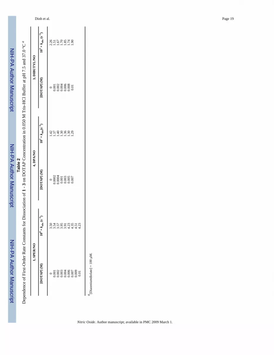
Rate Data in Cationic Liposome MediaCationic liposomes have found wide application in liposome-mediated gene transfertransfection studies due to their ability to bind anionic DNA and form complexes with highaffinity for cell membranes. The liposome formulations most frequently used have containedDOTAP usually mixed with a “helper lipid” such as the phosphatidylethanolamine DOPE toincrease the transfection potency. In our study, we looked at reactions of the polar 1, the morehydrophobic 3 and the anionic substrate 4 in the presence of cationic liposomes prepared fromDOTAP. The dissociation rates of all three substrates were unaffected by the presence of thecationic liposomes and no significant difference was noted in their behavior when comparedto that found in the Tris-buffered solutions alone. Data are summarized in Table 2.
Measurement of NO Release from 2 in DPPG/DPPC Liposome Media by OxyhemoglobinAssay
The stoichiometry of NO release from 1 and 2 in liposome media was determined by thestandard oxyhemoglobin assay method using the NO-mediated conversion of oxyhemoglobinto methemoglobin (HbO2 + NO → MetHb + NO3
- : k2 = 3.7 × 10⁷ M-1s-1), which is ∼ 26 times
Dinh et al. Page 5
Nitric Oxide. Author manuscript; available in PMC 2009 March 1.
NIH
-PA Author Manuscript
NIH
-PA Author Manuscript
NIH
-PA Author Manuscript

faster than its reaction with O2 at saturated oxygen concentrations [19]. Concentrations of NOwere calculated from absorbance changes at 401 nm using the literature value of 49mM-1cm-1 for the difference between the molar extinction coefficient of metHb and HbO2 atthat wavelength, Δε401(metHb-oxyHb) [17]. The yields of NO calculated by difference spectralmeasurements are summarized in Table 3. The mean experimental values for Δ[NO]/Δ[Diazeniumdiolate] determined for 1 and 2 were 1.47 ± 0.23 and 1.69 ± 0.24, respectively, in0.010M phosphate buffered 50/50 DPPC/DPPG media at 37 °C. The data indicate somewhatlower yields of NO in the liposome media than the 2.0 value expected for diazeniumdiolatedissociation reactions. The low precision in the data may be tied, in part, to the experimentalmethod requiring measurement of absolute absorbance values after blanking backgroundHbO2 and liposome absorbance.
Liposome Size Analysis by Photon Correlation Spectroscopy and Atomic Force MicroscopyImaging
Size analysis of the liposomes prepared in the study, as determined by photon correlationspectroscopy (PCS) immediately after preparation, revealed a mean vesicle diameter of 43.6± 0.29, 30.6 ± 0.69, 56.3 ± 0.31, and 38.8 ± 0.20 nm for DPPC, DPPG, DOPG and DOTAPvesicles, respectively. Some measurements of vesicle size were initiated after addingdiazeniumdiolate substrate and allowing its dissociation reaction to go to completion. Theseshowed similar values, indicating that the vesicle size did not significantly change during thecourse of the reaction. Data are summarized in Table 4. The vesicle sizes reported here aretypical of those reported for small unilamellar liposomes prepared by bath sonication [20].
DiscussionWe have shown that the acid-catalyzed dissociation of nitric oxide from diazeniumdiolate ionsis subject to catalysis by small unilamellar vesicles prepared from anionic lipids with thecatalytic activity arising from an increase in the local concentration of the diazeniumdiolatesubstrates and hydrogen ions at the negatively charged liposome interface. Substrate bindingoccurs through electrostatic interaction of the positively charged nitrogen centers in thediazeniumdiolate substrates with the negatively charged vesicle surface, as indicated by thegood correlation that exists between the values of the binding constants obtained for 1-3 inDPPG media and the positive polarity of the individual diazeniumdiolate substrates that havethree, two and one cationic nitrogen centers, respectively. A predominantly ionic associationis further supported by the small rate acceleration (< 2-fold at 10 mM DPPG) that was foundwith the anionic substrate, 4, that lacks any cationic nitrogen centers beyond that carrying the-N2O2
- group and by the reduced catalysis that is observed on decreasing the anionic lipidcontent of mixed DPPC/DPPG covesicles. Although KS values may reflect some additionalhydrophobic contributions to liposome binding, they appear to be minor for the substratesinvolved.
Binding constants for 1 in DPPG and DOPG liposome media are of comparable magnitudeand appear to reflect only the electrostatic interaction generated by the monoanionic surfacecharge of the lipids involved. The slightly lower binding constant obtained for 1 in DMPSmedia could be due to steric factors associated with the phosphatidylserine head group orrepulsion of the cationic nitrogen sites in 1 by the protonated serine amino group at the surface.pKa values of the functional groups in the phosphatidylserine head group indicate an overall-1 charge for DMPS at physiological pH similar to DPPG [21,22].
Since the vesicle bilayer provides both internal and external interfaces as potential reactionsites, several outcomes are possible for reactions in vesicle-containing solutions depending onreagent distribution between the different binding sites [23]. A kinetic distinction betweenendovesicular and exovesicular reactions can result from differences in reactivity at the inner
Dinh et al. Page 6
Nitric Oxide. Author manuscript; available in PMC 2009 March 1.
NIH
-PA Author Manuscript
NIH
-PA Author Manuscript
NIH
-PA Author Manuscript

and outer surfaces and by rate limiting permeation of the reactants through the membrane.Intrinsic differences in binding and reactivity at the inner and outer interfaces have been shownto arise from differences in the reaction media at the two locations and from the effect of vesiclesize and vesicle curvature on the pKa of both the lipid head groups and the bound substrate[24,25]. For the acid-catalyzed dissociation reactions in our study, the significant positivecharges carried by the zwitterionic substrates at pH 7.4, would be expected to inhibittransmembrane diffusion and we found no evidence of biphasic kinetic behavior whichgenerally characterizes transmembrane-limiting rate processes or modulation of chemicalreactivity by internal surfaces of vesicular membranes.
An interesting possibility (suggested by reviewer 2) is nitrosamine formation during ourliposome-catalyzed reactions due to the high solubility of our reaction products, NO and thefree amine, in the lipid bilayer following substrate dissociation at the liposome-solutioninterface. Rate enhancements up to 800-fold have been reported for the nitrosation ofdialkylamines by nitrite at pH 3.5 in the presence of cationic and neutral micelles [26], wherereaction is mediated by N2O3. We observed no spectral evidence for nitrosamine formation atpH 7.4 and no significant differences were noted in UV spectral scans (λ 200-400 nm) obtainedfor diazeniumdiolate dissociation reactions in liposome media and aqueous buffer.
In contrast to the behavior of 1, the more hydrophobic substrate 3 showed rate profiles inunsaturated DOPG and POPS media that suggested rate acceleration rather than rate saturationat the higher lipid concentrations employed. Aggregation of unsaturated POPS and DOPGliposomes, induced by interaction of the hydrophobic hexane-dibutyl structure of 3 with theunsaturated lipid bilayer, resulting in enhanced binding and catalysis by the larger phospholipidaggregates formed, may be responsible, at least in part, for the kinetic behavior displayed.Aggregation and fusion of phosphatidylserine and phosphatidylglycerol liposomes, inducedby a variety of reagents, has been previously reported [27-29].
Although phosphatidylcholine liposomes derived from DPPC have been shown to effect weakcatalysis of diazeniumdiolate dissociation rates [4], the hydrophobic substrate 3 was found tobe unaffected by the smaller DMPC liposomes. There is possibly insufficient flexibility in thephosphatidylcholine head group of the smaller DMPC lipid to permit significant ionicinteraction between the phosphate negative charge and cationic diazeniumdiolate centers. Themild catalysis found with DPPC may also result from slightly stronger hydrophobic associationbetween 3 and the choline methyl groups at the surface.
The larger KS value obtained with liposomes prepared from the phosphatidic acid salt, DOPAis consistent with the stronger electrostatic interaction expected from the higher negative chargeof the DOPA head group at pH 7.4. The pKa values reported for the first and seconddissociations of the terminal phosphate in the phosphatidic acid salt predict a net charge of∼1.7 for DOPA at pH 7.4 [24,30] compared to the -1 charge of the phosphate in DPPG (pKa= ∼1.1).
The sensitivity displayed by the measured binding constants in anionic liposome media, to theconcentration of electrolytes in the bulk aqueous solution, is consistent with changes in thereduction in the liposome surface charge density resulting from the effect of added salts on thediffuse double layer, as predicted by Guoy-Chapman-Stern models [31]. Although at typicalionic salt levels encountered near most cell membranes, substrate binding and its effect on NOrelease rates is expected to be small, at lipid bilayers encountered in certain “hydrophobic”protein or lipid environments more significant changes in diazeniumdiolate rate behavior maybe possible.
Dinh et al. Page 7
Nitric Oxide. Author manuscript; available in PMC 2009 March 1.
NIH
-PA Author Manuscript
NIH
-PA Author Manuscript
NIH
-PA Author Manuscript

Cationic DOTAP vesicles did not influence the acid-catalyzed dissociation rates of any of thediazeniumdiolate substrates examined consistent with reaction being confined to the aqueousphase due to the repulsion of H+
aq ions from the cationic surface. The absence of any rateinhibition in the presence of DOTAP vesicles further suggests that electrostatic attractionbetween the cationic surface and the negative charge of the diazeniumdiolate functional group,that would partition the diazeniumdiolate out of the aqueous phase, is outweighed by therepulsion of the positive centers by the cationic interface.
The limited oxyhemoglobin assay study of nitric oxide released in liposome media has foundyields of NO lower than the expected 2.0 mol of NO/mol of substrate. Although the loweramounts of NO detected could be due to the partial release or formation of alternative nitrogenoxide species such as NO- or NO2, nitroxyl formation would be unlikely for the secondaryamine-derived substrates involved and significant oxidation would be unexpected at theconcentration levels employed. Keynes et al have reported that NO delivered via adiazeniumdiolate was consumed by reaction with ingredients, Hepes buffer and riboflavin,present in a tissue-culture medium [32]. A mechanism involving formation of superoxideradical (O2 ▪-) through oxidation of Hepes followed by the rapid reaction of superoxide withNO to form peroxynitrite was proposed. It was suggested that riboflavin (in the light) providedan additional source of superoxide. Since both Hepes and riboflavin have hydroxyl functionalgroups similar to those present in the glycerol moiety of DPPG, a similar reaction may beresponsible for the low NO yields we have found in DPPG media. It should be mentioned that,although not considered by the previous authors, an additional quantity of NO may be lost ifthese strongly oxidizing conditions result in partial oxidation of the hydroxyls themselves toform small amounts of aldehyde- and/or ketone-containing impurities. Reaction of NO withcarbanions formed by abstraction of the acidic protons alpha to carbonyl groups (the Traubereaction) has been known for more than a century [33].
Previous experimental and theoretical studies have shown that diazeniumdiolate dissociationreactions are acid-catalyzed with equilibrium protonation of the N(1) amine nitrogen triggeringthe decomposition of the [N(O)NO]- functional group [34,35]. Although there is strongevidence that the enhanced NO dissociation rates in anionic liposome media are due to theconcentration of the reactants, H+(aq) ions and substrate, at the vesicle's aqueous interface, wehave explored the possibility that the catalysis of NO release in liposome media may also bedue, at least in part, to an increase in the intrinsic diazeniumdiolate dissociation rate constantin the polarizing environment of the negatively charged vesicle interface. This has beenachieved through application of the pseudo-phase ion exchange (PIE) model, previouslydescribed [3,36], that compares the intrinsic second-order rate constant, k2w (= kw/[H+]w) inthe aqueous phase with that estimated for the vesicle-bound substrate, k2v (= kv/[H+]v), where[H+]w and [H+]v are the local molarities of hydrogen ions expressed with respect to the volumesof the aqueous and vesicle pseudophase, respectively. In examining data obtained for thereaction of 1 in DPPG liposome media, we used V = 0.78 M-1 for the effective volume of theDPPG pseudo-phase, calculated by multiplying the lipid partial specific volume by itsmolecular weight [37] and the literature values, KH/Na = 1, for the equilibrium constant forexchange between bound Na+ counter ions and H+ ions in the interfacial region, β = 0.78 forthe fraction of the vesicle surface covered by Na+ ions [25,38]. [Na+]v/[Na+]w was estimatedassuming [Na+]v = β [DPPG] and [Na+]w = (1 - β) [DPPG] + [Na+]buffer. When data for 1 inDPPG media was applied to the PIE model, best fit values for k2v yielded k2v/ k2w = 0.64. Sinceestimated k2v values that are 2-3 fold smaller than k2w has been a common finding for manymicellar- and vesicle-catalyzed reactions [18], the relatively close agreement between k2v andk2w in this study does not support a significant change in the intrinsic dissociation rate constantin the polarizing microenvironment of the liposome interface.
Dinh et al. Page 8
Nitric Oxide. Author manuscript; available in PMC 2009 March 1.
NIH
-PA Author Manuscript
NIH
-PA Author Manuscript
NIH
-PA Author Manuscript

In summary, the information obtained in this study adds to our understanding of localenvironmental factors influencing diazeniumdiolate reactivity, particularly those existing nearnegatively charged aqueous interfaces of phospholipid bilayer membranes. Such membranesare important components of bacterial cell membranes and of pulmonary surfactants. Ofsignificance to the application of diazeniumdiolates as NO sources in vivo is the finding thatthe strongest binding and greatest catalysis of NO release occurs with polyamine-derivedzwitterionic substrates having multiple protonated nitrogen sites. Brilli et al have shown thatthe positive polarity of cationic tertiary amine sites in diazeniumdiolate can be utilized torestrict the movement of diazeniumdiolates across transmucosal membranes, thereby enablingthem to be more selective pulmonary vasodilators by inhibiting systemic hypertension [6,8].Previous studies involving inhaled NO gas for the treatment of acute lung injury in both animalsand children have also found a therapeutic synergy when NO exposure was combined withreplacement surfactant therapy [11] and there is interest in the effectiveness of soluble NOdonors when co-administered with surfactant. To the extent that the present study providesfundamental information on the interaction of diazeniumdiolates with anionic lipid interfacesprevalent in lung surfactant, particularly aqueous interfaces of phosphatidylcholine,phosphatidylglycerol and phosphatidylserine bilayers which are involved in the monolayer-to-multilayer transitions that are important to lung surfactant function, information obtained maybe of relevance to the use of diazeniumdiolates as pulmonary vasodilators in vivo.
Acknowledgements
Support of this work by the National Institutes of Health (Grant number R15-HL078750-01) and by the JeffressFoundation is gratefully acknowledged. This project has been funded in part with federal funds from the NationalCancer Institute, National Institutes of Health under Contract No. NO1-CO-12400. This research was supported inpart by the Intramural Research Program of the NIH, National Cancer Institute, Center for Cancer Research.
References1. Keefer LK, Nims RW, Davies KM, Wink DA. “NONOates” (1-substituted diazen-1-ium-1,2-diolates)
as nitric oxide donors: Convenient nitric oxide dosage forms. Methods Enzymol 1996;268:281–293.[PubMed: 8782594]
2. Keefer LK. Progress towards clinical applications of the nitric oxide-releasing diazeniumdiolates. AnnuRev Pharmacol Toxicol 2003;43:587–607.
3. Price SE, Jappar D, Lorenzo P, Saavedra JE, Hrabie JA, Davies KM. Micellar catalysis of nitric oxidedissociation from diazeniumdiolates. Langmuir 2003;19:2096–2102.
4. Dinh BT, Dove K, Jappar D, Hrabie JA, Davies KM. Effect of hydrophobic structure on the catalysisof nitric oxide release from zwitterionic diazeniumdiolates in surfactant and liposome media. NitricOxide 2005;13:204–209. [PubMed: 16122951]
5. Batchelor MM, Reoma SL, Fleser PS, Paul S, Nuthakki VK, Callahan RE, Shanley CJ, Politis JK,Elmore J, Mertz SI, Meyerhoff ME. More lipophilic dialkylamine-based diazeniumdiolates: Synthesis,characterization, and application in preparing thromboresistant nitric oxide release polymericmaterials. J Med Chem 2003;46:5153–5161. [PubMed: 14613318]
6. Brilli R, Krafte-Jacobs B, Smith DJ, Roselle D, Passerini D, Vromen A, Moore L, Szabo C, SalzmanA. Intratracheal instillation of a novel NO/nucleophile selectively reduces pulmonary hypertension. JAppl Physiol 1997;83:1968–1975. [PubMed: 9390970]
7. Brilli RJ, Krafte-Jacobs B, Smith DJ, Passerini D, Moore L, Ballard ET. Aerosolization of novel nitricoxide donors selectively reduces pulmonary hypertension. Crit Care Med 1998;26:1390–1396.[PubMed: 9710099]
8. Jacobs B, Brilli RJ, Moore L, Ballard E, Smith D. Areosolized soluble nitric oxide donor improvesoxygenation and pulmonary hypertension in acute lung injury. Am J Crit Care Med 1998;158:1536–1542.
9. Rossaint R, Falke KJ, Lopez F, Slama K, Pison U, Zapol WM. Inhaled nitric oxide for the adultrespiratory distress syndrome. N Eng J Med 1993;328:399–405.
Dinh et al. Page 9
Nitric Oxide. Author manuscript; available in PMC 2009 March 1.
NIH
-PA Author Manuscript
NIH
-PA Author Manuscript
NIH
-PA Author Manuscript

10. Schreber MD, Gin-Mestan K, Marks JD, Huo D, Lee G, Srisuparp P. Inhaled nitric oxide in prematureinfants with the respiratory distress syndrome. N Eng J Med 2003;349:2099–2107.
11. Jacobs BR, Smith DJ, Zingarelli B, Passerini DJ, Ballard ET, Brilli RJ. Soluble nitric oxide donorand surfactant improve oxygenation and pulmonary hypertension in porcine lung injury. Nitric Oxide2000;4:412–422. [PubMed: 10944426]
12. Lam CF, Van Heerden PV, Sviri S, Roberts BL, Ilett KF. The effects of inhalation of a novel nitricoxide donor, DETA/NO, in a patient with severe hypoxaemia due to acute respiratory distresssyndrome. Anaesth Intensive Care 2002;30:472–476. [PubMed: 12180587]
13. Notter, RH. Basic Science and Clinical Applications. 149. Marcel Dekker; New York: 2000. LungSurfactants.
14. Hrabie JA, Klose JR, Wink DA, Keefer LK. New nitric oxide-releasing zwitterions derived frompolyamines. J Org Chem 1993;58:1472–1476.
15. Drago RS, Karstetter BR. The Reaction of Nitrogen(II) Oxide with Various Primary and SecondaryAmines. J Am Chem Soc 1961;83:1819–1822.
16. New, RC. Liposomes: A Practical Approach. Oxford University Press; New York: 1990. p. 33-16.17. Feelisch, M.; Kubitzek, D.; Werringloer, J. The oxyhemoglobin assay. In: Feelisch, M.; Stamler, JS.,
editors. Methods in Nitric Oxide Research. John Wiley; 1996. p. 455-478.18. Bunton CA, Nome F, Quina FH, Romsted LS. Ionic binding and reactivity at charged aqueous
interfaces. Acc Chem Res 1991;24:357–364.19. Doyle MP, Hoekstra JW. Oxidation of nitrogen oxides by bound dioxygen in hemoproteins. J Inorg
Biochem 1981;14:351–358. [PubMed: 7276933]20. Jones, MN.; Chapman, D. Micelles, Monolayers and Biomembranes. Wiley-Liss; 1995. p. 117-121.21. Tsui FC, Ojcius DM, Hubbell WL. The intrinsic pKa values for phosphatidylserine and
phosphatidylethanolamine in phosphatidylcholine host bilayers. Biophys J 1986;49:459–468.[PubMed: 3955180]
22. Shapovalov ME, Mohwald VL, Brezesinski G. Ionization state and structure of 1,2-dipalmitoylphosphatidylglycerol monolayers at the liquid/air interface. J Phys Chem B2006;110:919–26. [PubMed: 16471624]
23. Chaimovich H, Cuccovia IM. Quantitative analysis of reagent distribution and reaction rates invesicles. Progr Colloid Polym Sci 1997;103:67–77.
24. Swairjo MA, Seaton BA, Roberts MF. Effect of vesicle composition and curvature on the dissociationof phosphatidic acid in small unilamellar vesicles - a 31P-NMR study. Biochim Biophys Acta1994;1191:354–61. [PubMed: 8172921]
25. Kawamuro MK, Chaimovich H, Abuin EB, Lissi EA, Cuccovia IM. Evidence that the effects ofsynthetic amphiphile vesicles on reaction rates depend on vesicle size. J Phys Chem 1991;95:1458–1463.
26. Okun JD, Archer MC. Kinetics of nitrosamine formation in the presence of micelle-formingsurfactants. J Natl Cancer Inst 1977;58:409–411. [PubMed: 833885]
27. Walter A, Siegel D. Divalent cation-induced lipid mixing between phosphatidylserine liposomesstudied by stopped-flow fluorescence measurements: Effects of temperature, comparison of bariumand calcium, and perturbation by DPX. Biochemistry 1993;32:3271–81. [PubMed: 8461294]
28. Ohki S, Duzgunes N, Leonards K. Phospholipid vesicle aggregation: Effect of monovalent anddivalent ions. Biochemistry 1982;21:2127–33. [PubMed: 7093233]
29. Lee G, Pollard HB. Highly sensitive and stable phosphatidylserine liposome aggregation assay forannexins. Anal Biochem 1997;252:160–164. [PubMed: 9324954]
30. Moncelli MR, Becucci L. The intrinsic pKa values for phosphatidic acid in monolayers deposited onmercury electrodes. J Electroanal Chem 1995;385:183–189.
31. McLaughlin S. The electrostatic properties of membranes. Annu Rev Biophys Biophys Chem1989;18:113–136. [PubMed: 2660821]
32. Keynes RG, Griffiths C, Garthwaite J. Superoxide-dependent consumption of nitric oxide inbiological media may confound in vitro experiments. Biochem J 2003;369:399–406. [PubMed:12366375]
Dinh et al. Page 10
Nitric Oxide. Author manuscript; available in PMC 2009 March 1.
NIH
-PA Author Manuscript
NIH
-PA Author Manuscript
NIH
-PA Author Manuscript

33. Hrabie JA, Keefer LK. Chemistry of the Nitric Oxide-Releasing Diazeniumdiolate(“Nitrosohydroxylamine”) Functional Group and Its Oxygen-Substituted Derivatives. Chem Rev2002;102:1135–1154. [PubMed: 11942789]
34. Davies KM, Wink DA, Saavedra JE, Keefer LK. Chemistry of Diazeniumdiolates. 2. Kinetics andmechanisms of dissociation to nitric oxide in a aqueous solution. J Am Chem Soc 2001;123:5473–5481. [PubMed: 11389629]
35. Dutton AS, Fukuto JM, Houk KN. The mechanism of NO formation from decomposition ofdialkylamino diazeniumdiolates: Density functional theory and CBS-QB3 predictions. Inorg Chem2004;43:1039–1045. [PubMed: 14753826]
36. Quina FH, Chaimovich H. Ion exchange in micellar solutions. 1. Conceptual framework for ionexchange in micellar solution. J Phys Chem 1979;83:1844–1850.
37. Newman GC, Huang CH. Structural studies on phosphatidylcholine-cholesterol mixed vesicles.Biochemistry 1975;14:3363–3370. [PubMed: 1170890]
38. Cuccovia IM, Feitosa E, Chaimovich H, Sepulveda L, Reed W. Size, electrophoretic mobility, andion dissociation of vesicles prepared with synthetic amphiphiles. J Phys Chem 1990;94:3722–3725.
Dinh et al. Page 11
Nitric Oxide. Author manuscript; available in PMC 2009 March 1.
NIH
-PA Author Manuscript
NIH
-PA Author Manuscript
NIH
-PA Author Manuscript

Figure 1.Diazeniumdiolates, R1R2N[N(O)NO]-, employed in the study. [1, SPER/NO: R1 = H2N(CH2)3-, R2 = H2N(CH2)3NH(CH2)4-; 2, DPTA/NO: R1 = R2 = H2N(CH2)3-; 3, DIBUTYL/NO: R1 = n-butyl-, R2 = n-butyl-NH2
+(CH2)6-; 4, DPA/NO: R1 = R2 = n Pr-]
Dinh et al. Page 12
Nitric Oxide. Author manuscript; available in PMC 2009 March 1.
NIH
-PA Author Manuscript
NIH
-PA Author Manuscript
NIH
-PA Author Manuscript

Figure 2.Lipids employed in the study. DPPG = 1,2-dipalmitoyl-sn-glycero-3-[phospho-rac-(1-glycerol)], DOPG = 1,2-dioleoyl-sn-glycero-3-[phospho-rac-(1-glycerol)], DMPS = 1,2-diacyl-sn-glycero-3-[phospho-L-serine], POPS= 1-palmitoyl-2-oleoyl-sn-glycero-3-[phospho-L-serine], DOPA = 1,2-dioleoyl-sn-glycero-3-phosphate; DPPC = 1,2-dipalmitoyl-sn-glycero-3-phosphocholine, DMPC = 1,2-dimyristoyl-sn-glycero-3-phosphocholine,DOTAP = 1,2-dioleoyl-3-trimethylammonium-propane.
Dinh et al. Page 13
Nitric Oxide. Author manuscript; available in PMC 2009 March 1.
NIH
-PA Author Manuscript
NIH
-PA Author Manuscript
NIH
-PA Author Manuscript

Figure 3.Effect of DPPG liposome concentration on dissociation rates of 1 – 4 in 0.010 M phosphatebuffer at pH 7.4 and 37 °C. [Diazeniumdiolate] = 100 μM.
Dinh et al. Page 14
Nitric Oxide. Author manuscript; available in PMC 2009 March 1.
NIH
-PA Author Manuscript
NIH
-PA Author Manuscript
NIH
-PA Author Manuscript

Figure 4.Effect of mixed DPPC/DPPG liposome composition on dissociation rate of 0.10 mM 1 in 0.010M phosphate buffer at pH 7.4 and 37 °C.
Dinh et al. Page 15
Nitric Oxide. Author manuscript; available in PMC 2009 March 1.
NIH
-PA Author Manuscript
NIH
-PA Author Manuscript
NIH
-PA Author Manuscript

Figure 5.Comparison of the effect of DOPG (□), POPS (Δ), DMPC (○) and DOTAP (▼) liposomes onthe dissociation rate of 3 in 0.010 M phosphate or 0.050 M Tris-HCl buffer (for DOTAP) atpH 7.4 and 37 °C. [3] = 100 μM
Dinh et al. Page 16
Nitric Oxide. Author manuscript; available in PMC 2009 March 1.
NIH
-PA Author Manuscript
NIH
-PA Author Manuscript
NIH
-PA Author Manuscript

Figure 6.Effect of phosphate buffer and salt concentration on the dissociation rate of 1 in DOPAliposome media at pH 7.4 and 37 °C. PBS (Δ) has 1.47 mM KH2PO4, 8.10 mM Na2HPO4, 2.7mM KCl and 137 mM NaCl. [1] = 100 μM.
Dinh et al. Page 17
Nitric Oxide. Author manuscript; available in PMC 2009 March 1.
NIH
-PA Author Manuscript
NIH
-PA Author Manuscript
NIH
-PA Author Manuscript

NIH
-PA Author Manuscript
NIH
-PA Author Manuscript
NIH
-PA Author Manuscript
Dinh et al. Page 18
Table 1Rate and Equilibrium Parameters from Liposome-Mediated Catalysis of Diazeniumdiolate Ions in 0.010 MPhosphate Buffered Solutions at pH 7.4 and 37 °C.
Substrate Reaction Medium kv/kw KS (M-1)
1 DPPG 37 477 ± 46DPPG/DPPC50/50 mol%
21 201 ± 54
DPPG/DPPC25/75 mol%
17 22 ± 5
1 DOPG 48 406 ± 1052 DPPG 40 136 ± 1073 DPPG 5.6 31 ± 51 DMPS 55 358 ± 135
DOPA 38 4040 ± 520DOPAb 32 947 ± 166DOPAc 8.4 245 ± 240
b0.020 M phosphate.
c0.10 M phosphate.
Nitric Oxide. Author manuscript; available in PMC 2009 March 1.
NIH
-PA Author Manuscript
NIH
-PA Author Manuscript
NIH
-PA Author Manuscript
Dinh et al. Page 19Ta
ble
2D
epen
denc
e of
Firs
t-Ord
er R
ate
Con
stan
ts fo
r Dis
soci
atio
n of
1 -
3 on
DO
TAP
Con
cent
ratio
n in
0.0
50 M
Tris
-HC
l Buf
fer a
t pH
7.5
and
37.
0 °C
a
1, S
PER
/NO
4, D
PA/N
O3,
DIB
UT
YL
/NO
[DO
TA
P] (M
)10
4 × k
obs (
s−1 )
[DO
TA
P] (M
)10
2 × k
obs(s
−1)
[DO
TA
P] (M
)10
3 × k
obs (
s−1 )
03.
500
1.42
02.
260.
001
3.54
0.00
021.
370.
001
1.51
0.00
23.
570.
0004
1.40
0.00
21.
670.
003
3.95
0.00
11.
300.
004
1.70
0.00
43.
910.
003
1.36
0.00
61.
850.
006
4.23
0.00
51.
300.
008
1.74
0.00
74.
350.
007
1.29
0.01
1.90
0.00
94.
510.
014.
23
a [Dia
zeni
umdi
olat
e] =
100
μM
.
Nitric Oxide. Author manuscript; available in PMC 2009 March 1.

NIH
-PA Author Manuscript
NIH
-PA Author Manuscript
NIH
-PA Author Manuscript
Dinh et al. Page 20
Table 3Yields of NO (ENO = mole of NO released / mole of substrate) during reaction of 1 and 2 in 0.010M phosphatebuffered 50/50 DPPC/DPPG media at 37 °C. [NO] = ΔAbs401/Δε401 obtained from difference spectra usingHbO2 as reference. Δε401 = 49 mM-1cm-1.
[Lipid]T μM [1], μM [NO] Released μM ENO
0 2.0 3.65 1.828.0 2.0 2.63 1.3220 2.0 2.53 1.27
[Lipid]T μM [2] μM [NO] Released μM ENO
0 2.0 4.27 2.138.0 2.0 3.14 1.5720 2.0 3.55 1.78100 2.0 2.41 1.20500 2.0 3.51 1.76
Nitric Oxide. Author manuscript; available in PMC 2009 March 1.

NIH
-PA Author Manuscript
NIH
-PA Author Manuscript
NIH
-PA Author Manuscript
Dinh et al. Page 21
Table 4Mean diameters of liposomes determined by dynamic light scattering at 37°C.
Liposome Mean Diameter (nm) a
DPPCb 43.6 ± 0.29DPPGb 30.6 ± 0.69DOPCb 56.3 ± 0.31
DOTAPc 38.8 ± 0.20DOTAP c,d 39.1 ± 0.21
aMean values of three repeat measurements.
b0.010 M phosphate buffer.
c0.050 M Tris.HCl buffer.
dMeasurement initiated after completion of reaction.
Nitric Oxide. Author manuscript; available in PMC 2009 March 1.


