Embed Size (px)
Citation preview

Cellular/Molecular
Different Presynaptic Roles of Synapsins at Excitatory andInhibitory Synapses
Daniel Gitler,1 Yoshiko Takagishi,3 Jian Feng,4,5 Yong Ren,5 Ramona M. Rodriguiz,2 William C. Wetsel,2
Paul Greengard,4 and George J. Augustine1
1Department of Neurobiology and 2Departments of Psychiatry and Behavioral Sciences, Medicine, and Cell Biology, Duke University Medical Center,Durham, North Carolina 27710, 3Research Institute of Environmental Medicine, Nagoya University, Nagoya 464-8601, Japan, 4Laboratory of Molecular andCellular Neuroscience, The Rockefeller University, New York, New York 10021, and 5Department of Physiology and Biophysics, State University of NewYork, Buffalo, New York 14214
The functions of synapsins were examined by characterizing the phenotype of mice in which all three synapsin genes were knocked out.Although these triple knock-out mice were viable and had normal brain anatomy, they exhibited a number of behavioral defects. Synaptictransmission was altered in cultured neurons from the hippocampus of knock-out mice. At excitatory synapses, loss of synapsins did notaffect basal transmission evoked by single stimuli but caused a threefold increase in the rate of synaptic depression during trains ofstimuli. This suggests that synapsins regulate the reserve pool of synaptic vesicles. This possibility was examined further by measuringsynaptic vesicle density in living neurons transfected with green fluorescent protein-tagged synaptobrevin 2, a marker of synapticvesicles. The relative amount of fluorescent synaptobrevin was substantially lower at synapses of knock-out neurons than of wild-typeneurons. Electron microscopy also revealed a parallel reduction in the number of vesicles in the reserve pool of vesicles �150 nm awayfrom the active zone at excitatory synapses. Thus, synapsins are required for maintaining vesicles in the reserve pool at excitatorysynapses. In contrast, basal transmission at inhibitory synapses was reduced by loss of synapsins, but the kinetics of synaptic depressionwere unaffected. In these terminals, there was a mild reduction in the total number of synaptic vesicles, but this was not restricted to thereserve pool of vesicles. Thus, synapsins maintain the reserve pool of glutamatergic vesicles but regulate the size of the readily releasablepool of GABAergic vesicles.
Key words: exocytosis; presynaptic; synapsin; synaptic depression; facilitation; synaptic vesicle; synaptic transmission; reserve pool
IntroductionThe synapsins are abundant phosphoproteins associated with themembranes of synaptic vesicles (Greengard et al., 1993; Hilfikeret al., 1999). Three mammalian synapsin genes have been identi-fied, with alternative splicing creating at least eight different neu-ronal synapsin isoforms (Sudhof et al., 1989; Kao et al., 1999;Porton et al., 1999). The functions of synapsins are not clear.Synapsins have been postulated to recruit synaptic vesicles to areserve pool (RP) (Li et al., 1995; Hilfiker et al., 1999), to regulatethe kinetics of membrane fusion (Hilfiker et al., 1998; Humeau et
al., 2001), to stabilize synaptic vesicles (Rosahl et al., 1995),and/or to regulate late steps of endocytosis (Bloom et al., 2003).
One reason why it has been difficult to assign precise roles tosynapsins is that interfering with synapsins with various experi-mental procedures has yielded different effects on synaptic func-tion. Acute interference with synapsins at the squid giant synapse,by microinjecting synapsin-derived peptides or dephosphory-lated synapsin, inhibits basal synaptic transmission, indicating arole for synapsins in neurotransmitter release (Llinas et al., 1985,1991; Hilfiker et al., 1998). Injecting synapsin peptides or anti-synapsin antibodies also changes the kinetics of postsynaptic cur-rents, suggesting that synapsin specifically regulates the fusion ofsynaptic vesicles with the presynaptic plasma membrane (Hil-fiker et al., 1998; Humeau et al., 2001). In contrast, deletion of theonly synapsin gene in Drosophila reportedly causes no measur-able change in synaptic transmission (Godenschwege et al.,2004). Genetic deletion of individual synapsin genes in mice alsodoes not eliminate synaptic transmission. None of these deletionsproduces a lethal phenotype, and effects on basal synaptic trans-mission, where measured, are reported to be small. Deletion ofsynapsin I decreases glutamate release during prolonged depolar-ization of synaptosomes, increases facilitation at excitatory syn-apses, and decreases transmission at inhibitory synapses (Rosahlet al., 1993, 1995; Li et al., 1995; Terada et al., 1999). Deletion of
Received Sept. 13, 2004; revised Nov. 4, 2004; accepted Nov. 5, 2004.This work was supported by National Institute of Mental Health Grants MH-39327 and MH-67044. D.G. was a
European Molecular Biology Organization Postdoctoral Fellow and a Pfizer Fellow of the Life Science ResearchFoundation. R.M.R. was an American Psychological Association Minority Postdoctoral Fellow in Neuroscience. Wethank F. Santamaria for help with EM image analysis software, W. Drake for assisting with EM image analysis, N. Cantfor assisting with histology, U. Ashery and J. Wesseling for helpful discussions, T. Dresbach for providing DNA forGFP-tagged synaptobrevin, B. Scalettar and A. Jeromin for providing DNA for GFP-tagged synaptophysin, and S.Hilfiker, J. Jovanovic, Y. Li, T. Sihra, and C. Villalba for their comments on this manuscript.
Correspondence should be addressed to George J. Augustine, Department of Neurobiology, Duke UniversityMedical Center, Box 3209, Durham, NC 27710. E-mail: [email protected].
D. Gitler’s present address: Department of Neurophysiology, Carl Ludwig Institute for Physiology, Leipzig Uni-versity, 04103 Leipzig, Germany.
DOI:10.1523/JNEUROSCI.3795-04.2004Copyright © 2004 Society for Neuroscience 0270-6474/04/2411368-13$15.00/0
11368 • The Journal of Neuroscience, December 15, 2004 • 24(50):11368 –11380

synapsin II (or of both I and II) apparently does not affect theamount of transmitter released by individual action potentials, asassessed by examining synaptic facilitation, but does acceleratesynaptic depression and increases post-tetanic potentiation (Ro-sahl et al., 1995). Finally, deletion of synapsin III decreases basaltransmission at inhibitory synapses but not excitatory synapses(Feng et al., 2002).
Some of the uncertainty concerning synapsin functions is at-tributable to the fact that mammals possess multiple synapsingenes. If only one or two of these three genes are deleted, theremaining gene products may compensate for the missing syn-apsins and mask a more fundamental phenotype. To determinewhether this was the case, we examined synaptic transmission inmice in which all three known synapsin genes were deleted. Fur-thermore, we used direct measurements of quantal content tomeasure basal transmitter release at excitatory and inhibitorysynapses. We found that loss of all three synapsin genes was notlethal but did cause substantial changes in behavior and synaptictransmission. These defects differ for excitatory and inhibitorysynapses, indicating that synapsins have unique functions at dif-ferent synapses.
Materials and MethodsGeneration of synapsin triple knock-out mice. Homozygous synapsin tripleknock-out (TKO) mice and matching triple wild-type (TWT) mice werederived by serially breeding previously generated synapsin I (Chin et al.,1995), synapsin II (Ferreira et al., 1998), and synapsin III (Feng et al.,2002) single knock-out mice, in this order. For genotyping, tail DNA wasextracted and analyzed by PCR. We used three primers that anneal to thetargeting vector, to the targeted sequence, and to the surrounding genomicDNA: synapsin I sense, AGGGAGTTTCGTTACTACAGGTCC; synapsin Iantisense, AGATTGGCCATGAAGTTGCTGTCC; synapsin II sense,TTCAGGTCTCAGCATACAAGGTGC; synapsin II antisense, GGAAGT-TCATCATCTGGCTTGAGG; synapsin I/II targeting vector antisense,CTACTTCCATTTGTCACGTCCTGC; synapsin III sense, GTCTAGAG-CAGAGTTGAACCTGTG; synapsin III antisense, CCGCCCTGGATGT-TAAGATCAGAT; synapsin III targeting vector sense, ACACTGCTCGA-CATTGGGTGGAAA.
Two to four mice were housed in each cage in a humidity- andtemperature-controlled room, under a 14/10 hr light/dark cycle, andwere provided with standard laboratory chow and water ad libitum. Allexperiments were conducted in accordance with National Institutes ofHealth guidelines and with animal protocols approved by the investiga-tors’ institutional animal care and use committees.
Western blots. Antibodies against synaptotagmin, syntaxin, synapto-physin, and �-tubulin were purchased from Sigma (St. Louis, MO). Anti-synaptobrevin 2 antibodies were a generous gift from R. Jahn (Max-Planck-Institute for Biophysical Chemistry, Gottingen, Germany).Antibodies against the NMDA receptor (NR) subunits were from Up-state Biotechnology (Lake placid, NY). Anti-postsynaptic density-95(PSD-95) was from Affinity Bioreagents (Golden, CO). Anti-synapsin Iclone 8 was from BD Transduction Labs (San Diego, CA). Anti-synapsinII was from Stressgen (San Diego, CA). Anti-synapsin III (RU486) wasgenerated previously (Feng et al., 2002).
Whole brains were homogenized in a 1% SDS solution and boiled for5 min. Protein concentration was measured by the DC Protein Assay kit(Bio-Rad, Hercules, CA). Equal amounts of total proteins from triplewild-type and triple knock-out mice were separated on 7.5 or 12% poly-acrylamide gels and transferred to nitrocellulose membranes (AmershamBiosciences, Piscataway, NJ) for Western blot analyses. Blots were incu-bated sequentially with primary and HRP-conjugated secondary antiseraand developed using the ECL detection method according to the manu-facturer’s protocol (Amersham Biosciences).
Culture and transfection. Hippocampal neurons were cultured fromnewborn pups (postnatal days 0 –2) and transfected as described previ-ously (Gitler et al., 2004). Microisland cultures of hippocampal neurons
were prepared as described (Nishiki and Augustine, 2004). Neurons wereallowed to mature for 7–14 d.
Histology. Cytochrome oxidase staining of brain sections was per-formed essentially as described in Wong-Riley (1979). Mice were anes-thetized and transcardially perfused with 0.9% saline, followed by ice-cold 4% paraformaldehyde in 0.1 M PBS, pH 7.4 (Sigma). The heads werepostfixed overnight, and the brain was removed and incubated in cold30% sucrose in PBS until sectioned. Brains were sectioned sagittally at athickness of 40 �m in a sliding microtome (American Optical) andstained in a 0.1 M phosphate buffer solution containing 4% sucrose, 0.025�g/ml cytochrome c (Sigma), and 0.5 mg/ml diaminobenzidine (Sigma).Sections were mounted on gelatin-coated slides, dehydrated by serialethanol solutions, cleared with xylene, and coverslipped with Permount(Fisher Scientific, Hampton, NH).
Electrophysiological acquisition and data analysis. Whole-cell patch-clamp recordings were made from single neurons on microislands, asdescribed in Nishiki and Augustine (2004). Patch pipettes (2.5– 4 M�)were filled with intracellular solution containing (in mM): 50K-glutamate, 71 K-gluconate (Fluka, Buchs, Switzerland), 15 NaCl, 6MgCl2, 2 EGTA, 5 Na2ATP, 0.3 Na2GTP, and 20 HEPES-KOH, pH 7.3(285 mOsm). The extracellular solution contained (in mM): 150 NaCl, 3KCl, 2 CaCl2, 2 MgCl2, 20 glucose, and 10 HEPES-NaOH, pH 7.3 (310mOsm). All materials were from Sigma, unless specified otherwise. Neu-rons were voltage clamped at �70 mV with a HEKA EPC-9D amplifier(HEKA, Lambrecht/Pfalz, Germany). All experiments were performed atroom temperature (�25°C). Electrophysiological data were sampled at25 kHz and filtered at 10 kHz. Axonal action potentials were evoked bydepolarizing the cell body to �40 mV for 0.5 msec every 15 sec, unlessspecified otherwise. Only cells with series resistances of �20 M� wereanalyzed (typically �10 M�), with 50% of this resistance compensatedelectronically. In each cell, we recorded evoked and spontaneous synapticcurrents, responses to pairs of stimuli, and sustained trains of stimuli.Only a single train was evoked in each neuron to avoid eliciting long-term forms of synaptic plasticity.
To calculate the peak current and the charge transfer during a train, wefirst subtracted an averaged trace containing the stimulus and the actionpotential current but lacking any discernable synaptic currents (i.e., syn-aptic failures). Such traces were easily identified toward the end of a trainof stimuli, when synaptic depression was maximal. We integrated thecurrent during each trace, discarding the stimulus and action potential,and then summed the charge for the whole train. For each event in a train,the baseline was defined as the final value of current during the precedingevent. In the case of excitatory events, the synaptic currents were wellseparated temporally, so that little summation occurred; however, be-cause inhibitory synaptic currents decay slowly, these responses summedto a significant extent, necessitating the baseline subtraction proceduredescribed above when calculating peak currents. Spontaneous synapticevents were semiautomatically analyzed using the MiniAnalysis program(Synaptosoft, Decatur, GA). Values were compared by the Student’s ttest, and error bars shown in the figures indicate the SEM.
Microscopy and fluorescence imaging. Cultured cells were imaged asdescribed in Gitler et al. (2004). Images were recorded with a CoolsnapFX CCD camera (Roper Scientific, Trenton, NJ) and acquired and pro-cessed with ISEE software (ISEE Imaging Systems, Raleigh, NC). Mea-surements of synaptobrevin 2 targeting were performed as describedpreviously for synapsins (Gitler et al., 2004), except that the neurons werecotransfected with pDsRed2C1 (Clontech, Palo Alto, CA) and a greenfluorescent protein (GFP)–synaptobrevin 2 construct based on thepEGFPC1 mammalian expression vector (a kind gift of ThomasDresbach, Magdeberg, Germany). For the purpose of counting synapses,we cotransfected the cells with pDsRed2C1 and with a pcDNA3 vectorencoding the synaptophysin I–GFP fusion protein (kindly provided byBethe Scalettar, Lewis and Clark College, Portland, OR, and AndreasJeromin, Baylor College of Medicine, Houston, TX). Transfected neu-rons that were spatially isolated from other transfected neurons werephotographed in their entirety using a 20� UApo 0.75 numerical aper-ture objective. The red channel of the image was used to merge the partialimages into a composite picture, using the semiautomatic photomergeoption of Photoshop Elements (Adobe Systems, San Jose, CA). Hence-
Gitler et al. • Presynaptic Functions of Synapsins J. Neurosci., December 15, 2004 • 24(50):11368 –11380 • 11369

forth, a segmenting algorithm was imple-mented by combining elements of the ISEEsoftware package. The green channel, contain-ing the synaptophysin–GFP images, wasclipped using the red channel as a mask. Toidentify the synaptic terminals, we iterativelyapplied successively increasing thresholds, thussegmenting the image into objects. Morpholog-ical criteria were applied to the objects to rejectthose that could not be synaptic terminals (bysize, roundness, and aspect ratio). The numberof positively identified objects was counted byresegmenting the image containing the accu-mulated objects.
Electron microscopy. Cells were grown onAclar film (Nisshin EM, Tokyo, Japan) for 19 dand fixed with 2.5% glutaraldehyde and 2%paraformaldehyde in 0.1 M phosphate buffer(PB) for 15–30 min. After washing in 0.1 M PBcontaining 4.5% sucrose, they were postfixedwith 1.0% OsO4 in 0.1 M PB containing 4.5%sucrose for 1 hr. They were then dehydrated inethanol and embedded in epoxy resin. Ultra-thin sections were prepared and examined witha JEOL 1210 electron microscope. Randomelectron micrographs were taken at a magnifi-cation of 10,000� and subsequently digitized(Dimage, Minolta, Japan). To measure the spa-tial distribution of synaptic vesicles, we usedcustom software routines written in Mathlab(The Mathworks, Natick MA). The location ofthe active zone was marked, and its minimallinear distance to the center of each individuallyidentified vesicle was calculated. If more thanone identifiable active zone was present in a ter-minal, each vesicle was counted only once bybeing included with the closest active zone. Ac-quisition of electron microscopy images andtheir analysis followed a double-blind protocol.
Behavioral assessment. The neurophysiologi-cal screen included examinations of orientationand reflexive behavior, postural and righting re-flexes, and general motor coordination (Rogerset al., 1997; Ribar et al., 2000). Most measureswere scored on a multipoint system. Normalresponses were scored as “0”; negative responses denoted deficiencies,and positive scores reflected more proficient responses relative to normalor “0” scores. Forepaw and hindpaw strength were examined with anUgo Basile grip-strength meter (Stoelting Company, Wood Dale, IL).Tests for ataxia and gait were evaluated by the foot-printing method(Robbins, 1985; Ribar et al., 2000). Linear strides of at least five matchedfootprints were used in the analyses.
Spatial working memory was examined in the eight-arm radial maze(Gainetdinov et al., 1999). Mice were food deprived �5– 6 hr beforetesting and maintained at �85–90% of their normal body weightthroughout the study. Animals were handled twice a day for 5 consecu-tive days for 5 min and then were trained to eat the food reinforcement(Fruit Loops) before maze testing. Animals were tested daily over 24 dwith a win-shift paradigm in which all eight arms were baited with thecereal. Individual mice were placed in a cylinder in the center of the mazefor 10 sec and then given free access to the maze for 300 sec. Only oneentry into an arm was reinforced. Performance was assessed in terms ofthe numbers of entries before an arm was reentered (entries to repeat),the amount of food eaten and latency to eat, activity in the maze (totaltime in maze divided by total arm entries), and number of perseverativeerrors (entry into a previously entered arm).
Statistics. The data for the neurophysiological screen are expressed asnormal, as delayed, as absent, or as minutes. All other results are ex-pressed as means and SEMs. Nominal data from the neurophysiological
assessment were analyzed by independent samples t tests. The radial armmaze results were analyzed by repeated measures ANOVA(RMANOVA). To determine the time points at which TWT and TKOperformance differed, tests of within-subjects contrasts with simple com-parisons were used with the first block of three test trials set as the controlgroup for each comparison. For all other comparisons, statistical signif-icance was assessed using the Student’s t test, unless specified otherwise.In all cases, p � 0.05 was considered significant.
ResultsPhenotype of synapsin TKO miceSynapsin TKO mice were generated by crossing preexisting linesof mice harboring null mutations in single synapsin genes (Chinet al., 1995; Ferreira et al., 1998; Feng et al., 2002). A line ofmatching wild-type mice was derived in parallel and were termedTWT mice. Although the usual complement of synapsin proteinswas present in total brain lysate from TWT mice, as demon-strated by Western blots using antibodies against each synapsingene product, the TKO mice lacked all synapsins (Fig. 1A).
Synapsin TKO mice were viable and fertile, arguing against arole for synapsins in survival; however, numerous behavioral def-icits were observed in these mice (Table 1). After the initial open-ing of the cage, TKO mice exhibited mild and transient seizures.
Figure 1. Brain-level effects of deleting the three synapsin genes. A, Western blot analysis of synapsins I, II, and III in whole-brain lysate from two TWT and two synapsin TKO mice. B, Spatial learning and memory were compared in synapsin TWT and TKOmice (n � 10 and 9, respectively) by using the win-shift paradigm in the eight-arm radial arm maze test in which all eight armswere baited. Significant differences in performance, as assessed by an RMANOVA, are indicated by asterisks. C, Cytochromeoxidase staining of sagittal sections of brains from TWT and TKO mice. D, Higher-resolution images of the hippocampal formationin the two types of mice.
11370 • J. Neurosci., December 15, 2004 • 24(50):11368 –11380 Gitler et al. • Presynaptic Functions of Synapsins

As a consequence, all behavioral tests were conducted at least 60min after the mice were disturbed, when these seizures had sub-sided. Homozygous mutants exhibited delayed reflexes whentheir eyes, ears, or whiskers were lightly touched with the edge ofa cotton swab. In each case, the TKO mice required several con-tacts with a cotton swab before demonstrating a reflex response.Although TWT mice demonstrated mild piloerection when han-dled, this characteristic was absent in the mutants. Horizontalposturing was also abnormal in the TKO mice because the ani-mals had difficulty maintaining balance with all four paws on thefloor when the cage was shaken gently in a horizontal plane.Although the grasping reflex for the front and rear paws wasnormal, TKO mice had a diminished ability to hang from a sus-pended wire. During the pole walking and climbing tests, TKOmice engaged the poles as rapidly as the TWT animals but tookapproximately twice as long to complete the task. The TKO micehad normal footprints, indicating an absence of ataxia. In sum-mary, the TKO mice displayed mild abnormalities in coordina-tion and balance, especially with their hindpaws.
To examine hippocampal function, mice were tested in a ra-dial arm maze for spatial memory (Gainetdinov et al., 1999).Initially, the performance of both TWT and TKO mice was verypoor (n � 10 and 9, respectively) (Fig. 1B). After training, TWTmice improved, and very high levels of performance wereachieved within 24 trials (F(7,63) � 8.099; p � 0.001). By compar-ison, the learning curve for the TKO mice was flat, suggesting that
spatial learning and memory were impaired in the mutants. Testsof within-subjects contrasts demonstrated that significant differ-ences in performance between TWT and TKO mice were evidenton block trials 5 through 8. The deficiency of TKO mice was notcaused by genotypic differences in motivation because mutantsshowed similar latencies to eat and consumed quantities of theFruit Loops similar to those of the TWT animals. Performancealso cannot be attributed to differences in activity in the mazebecause these levels were comparable between genotypes. Finally,the numbers of perseverative errors were low and not signifi-cantly different between TWT and TKO mice. This latter pointindicates that the TKO mice can at least remember the immediatearms that they had previously entered and could withhold re-sponses to these arms. Together, these findings suggest that theTKO mice are deficient in spatial learning and memory.
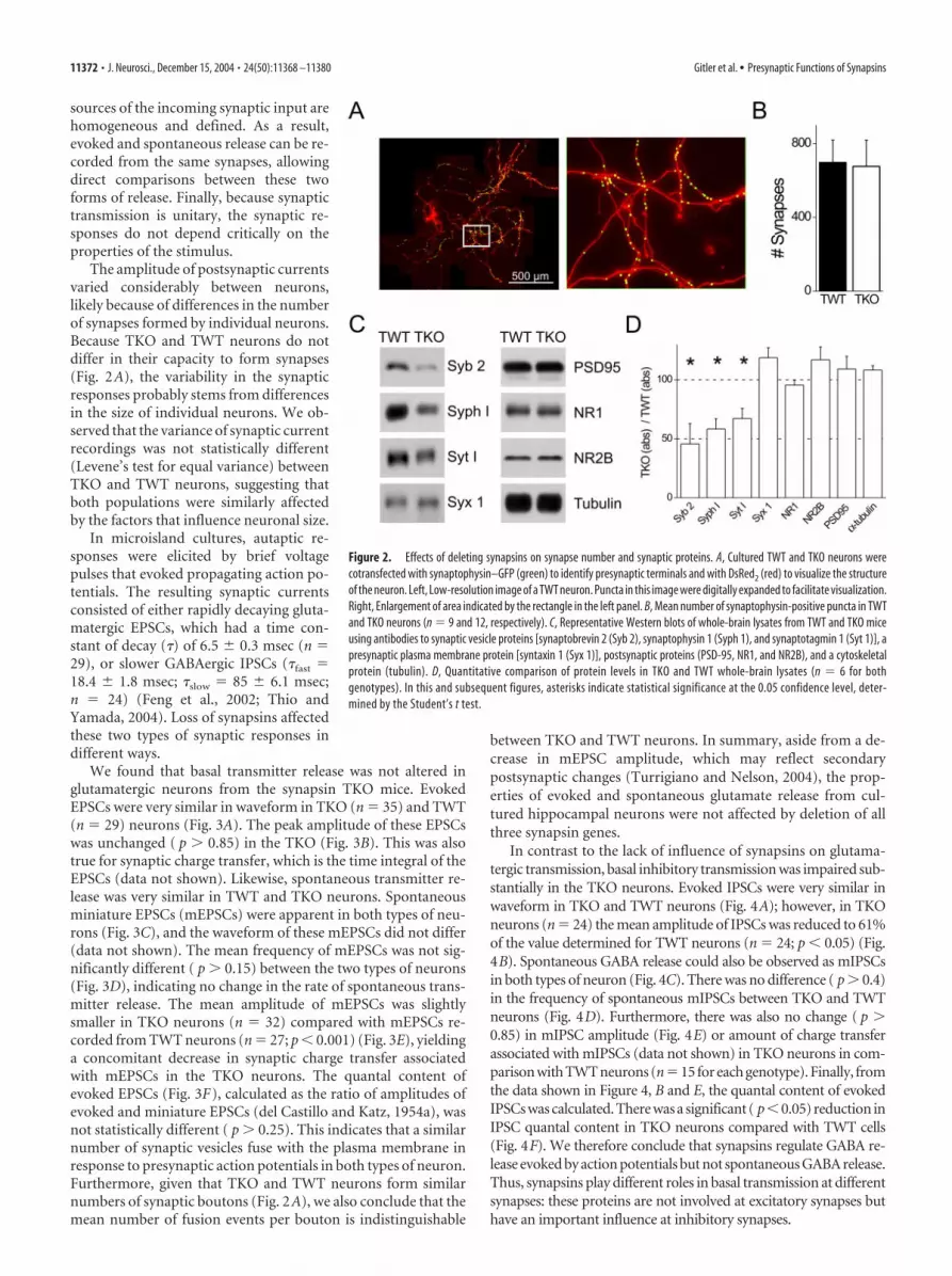
Comparison of the brains from wild-type and synapsin TKOmice revealed no perceptible differences in gross anatomy (Fig.1C). In particular, the structure of the hippocampus of TKO miceappeared normal (Fig. 1D). These results suggest that synapsinsare not collectively required for gross development of the adultbrain. Given that individual synapsin genes have been implicatedin synapse formation (Ferreira and Rapoport, 2002), we also con-sidered the number of synapses formed by TKO neurons. For thispurpose, we prepared cultures of neurons from the hippocampusof TKO and TWT mice. Synapse number was determined bytransfecting individual neurons with a GFP-tagged synaptophy-sin construct, which targets to synaptic vesicles and thereby pro-vides a means of visualizing presynaptic terminals (Fletcher et al.,1991; Sampo et al., 2003). In neurons from both TKO and TWTmice, fluorescent puncta were observed (Fig. 2A, left). Countingthe puncta revealed no differences in the mean number of syn-apses formed by the two types of neurons (Fig. 2B). This indicatesthat the complete absence of synapsins does not prevent synapsesfrom forming in normal numbers.
To determine what deletion of synapsins does to the levels ofsynaptic proteins, we examined these proteins in homogenatesprepared from TKO and TWT brains (Fig. 2C). In comparisonwith TWT mice, the TKO mice had selective reductions in levelsof several synaptic vesicle proteins, such as synaptobrevin 2, syn-aptotagmin I, and synaptophysin I (Fig. 2D). Several other syn-aptic proteins were unaffected. For example, syntaxin I, a SNARE[soluble N-ethylmaleimide-sensitive factor attachment protein(SNAP) receptor] protein associated with the presynaptic plasmamembrane, was present at normal levels. Likewise, several pro-teins associated with the postsynaptic membrane, such as PSD-95and NMDA receptor subunits NR1 and NR2B, were unaffected,as was the ubiquitous protein, �-tubulin. The normal comple-ment of postsynaptic membrane proteins, as well as syntaxin, isconsistent with the unchanged number of synapses observed inthe TKO mice (Fig. 2B). The selective reduction in synaptic ves-icle proteins suggests that loss of synapsins reduced the numberof synaptic vesicles, and this point is established in other experi-ments that are described below.
Differential regulation of basal transmitter releaseby synapsinsWe next asked whether synaptic function was impaired in theTKO mice. For this purpose, we measured synaptic currents inhippocampal neurons in microisland cultures (Segal and Fursh-pan, 1990; Bekkers and Stevens, 1991). This experimental systemoffers several advantages over brain slices and dense cell culturesfor quantifying neurotransmitter release. Most important is thatthe “autaptic” responses originate from the same cell, so that the
Table 1. Physiological characteristics of synapsin TWT and TKO mice
TWT TKO
Initial evaluation and general screenSkin color Normal NormalBody tone Normal NormalLacrimiation/palperbral closure Normal NormalExopthalmus Normal NormalConvulsions/tremor Absent PresentHeart rate Normal NormalRespiration rate Normal NormalPosture/tail elevation Normal NormalBarbering/hair loss Absent Absent
Orientation and reflexive behaviorVisual orientation to object Normal NormalVisual placement Normal NormalWhisker reflex Normal Delayed*Eye reflex Normal Delayed*Pinna (ear) reflex Normal Delayed*
Postural and righting reflexesPostural (vertical) Normal NormalPostural (horizontal) Normal Abnormal*Contact righting Normal Normal
Spinocerebellar functionForepaw grasp/strength Normal NormalHindpaw grasp/strength Normal NormalHindpaw coordination Normal Abnormal*Wire hang (duration) 52.0 7.5 sec 18.1 2.2 sec**Pole climbing down
Latency 10.2 0.9 sec 1.83 1.1 sec**Duration 8.4 1.9 sec 16.9 4.5 sec
Pole climbing upLatency 8.4 3.2 sec 4.8 1.5 secDuration 12.1 3.4 sec 22.4 4.5 sec
Pole walkingLatency 10.0 1.2 sec 16.1 2.3 sec*Duration 13.5 5.2 sec 31.9 4.9 sec**
Numerical data are expressed as meansSEM. The data were analyzed with an independent t test. n �10 mice pergenotype. * p� 0.05; ** p� 0.01.
Gitler et al. • Presynaptic Functions of Synapsins J. Neurosci., December 15, 2004 • 24(50):11368 –11380 • 11371
sources of the incoming synaptic input arehomogeneous and defined. As a result,evoked and spontaneous release can be re-corded from the same synapses, allowingdirect comparisons between these twoforms of release. Finally, because synaptictransmission is unitary, the synaptic re-sponses do not depend critically on theproperties of the stimulus.
The amplitude of postsynaptic currentsvaried considerably between neurons,likely because of differences in the numberof synapses formed by individual neurons.Because TKO and TWT neurons do notdiffer in their capacity to form synapses(Fig. 2A), the variability in the synapticresponses probably stems from differencesin the size of individual neurons. We ob-served that the variance of synaptic currentrecordings was not statistically different(Levene’s test for equal variance) betweenTKO and TWT neurons, suggesting thatboth populations were similarly affectedby the factors that influence neuronal size.
In microisland cultures, autaptic re-sponses were elicited by brief voltagepulses that evoked propagating action po-tentials. The resulting synaptic currentsconsisted of either rapidly decaying gluta-matergic EPSCs, which had a time con-stant of decay (�) of 6.5 0.3 msec (n �29), or slower GABAergic IPSCs (�fast �18.4 1.8 msec; �slow � 85 6.1 msec;n � 24) (Feng et al., 2002; Thio andYamada, 2004). Loss of synapsins affectedthese two types of synaptic responses indifferent ways.
We found that basal transmitter release was not altered inglutamatergic neurons from the synapsin TKO mice. EvokedEPSCs were very similar in waveform in TKO (n � 35) and TWT(n � 29) neurons (Fig. 3A). The peak amplitude of these EPSCswas unchanged ( p � 0.85) in the TKO (Fig. 3B). This was alsotrue for synaptic charge transfer, which is the time integral of theEPSCs (data not shown). Likewise, spontaneous transmitter re-lease was very similar in TWT and TKO neurons. Spontaneousminiature EPSCs (mEPSCs) were apparent in both types of neu-rons (Fig. 3C), and the waveform of these mEPSCs did not differ(data not shown). The mean frequency of mEPSCs was not sig-nificantly different ( p � 0.15) between the two types of neurons(Fig. 3D), indicating no change in the rate of spontaneous trans-mitter release. The mean amplitude of mEPSCs was slightlysmaller in TKO neurons (n � 32) compared with mEPSCs re-corded from TWT neurons (n � 27; p � 0.001) (Fig. 3E), yieldinga concomitant decrease in synaptic charge transfer associatedwith mEPSCs in the TKO neurons. The quantal content ofevoked EPSCs (Fig. 3F), calculated as the ratio of amplitudes ofevoked and miniature EPSCs (del Castillo and Katz, 1954a), wasnot statistically different ( p � 0.25). This indicates that a similarnumber of synaptic vesicles fuse with the plasma membrane inresponse to presynaptic action potentials in both types of neuron.Furthermore, given that TKO and TWT neurons form similarnumbers of synaptic boutons (Fig. 2A), we also conclude that themean number of fusion events per bouton is indistinguishable
between TKO and TWT neurons. In summary, aside from a de-crease in mEPSC amplitude, which may reflect secondarypostsynaptic changes (Turrigiano and Nelson, 2004), the prop-erties of evoked and spontaneous glutamate release from cul-tured hippocampal neurons were not affected by deletion of allthree synapsin genes.
In contrast to the lack of influence of synapsins on glutama-tergic transmission, basal inhibitory transmission was impaired sub-stantially in the TKO neurons. Evoked IPSCs were very similar inwaveform in TKO and TWT neurons (Fig. 4A); however, in TKOneurons (n � 24) the mean amplitude of IPSCs was reduced to 61%of the value determined for TWT neurons (n � 24; p � 0.05) (Fig.4B). Spontaneous GABA release could also be observed as mIPSCsin both types of neuron (Fig. 4C). There was no difference ( p � 0.4)in the frequency of spontaneous mIPSCs between TKO and TWTneurons (Fig. 4D). Furthermore, there was also no change ( p �0.85) in mIPSC amplitude (Fig. 4E) or amount of charge transferassociated with mIPSCs (data not shown) in TKO neurons in com-parison with TWT neurons (n�15 for each genotype). Finally, fromthe data shown in Figure 4, B and E, the quantal content of evokedIPSCs was calculated. There was a significant ( p�0.05) reduction inIPSC quantal content in TKO neurons compared with TWT cells(Fig. 4F). We therefore conclude that synapsins regulate GABA re-lease evoked by action potentials but not spontaneous GABA release.Thus, synapsins play different roles in basal transmission at differentsynapses: these proteins are not involved at excitatory synapses buthave an important influence at inhibitory synapses.
Figure 2. Effects of deleting synapsins on synapse number and synaptic proteins. A, Cultured TWT and TKO neurons werecotransfected with synaptophysin–GFP (green) to identify presynaptic terminals and with DsRed2 (red) to visualize the structureof the neuron. Left, Low-resolution image of a TWT neuron. Puncta in this image were digitally expanded to facilitate visualization.Right, Enlargement of area indicated by the rectangle in the left panel. B, Mean number of synaptophysin-positive puncta in TWTand TKO neurons (n � 9 and 12, respectively). C, Representative Western blots of whole-brain lysates from TWT and TKO miceusing antibodies to synaptic vesicle proteins [synaptobrevin 2 (Syb 2), synaptophysin 1 (Syph 1), and synaptotagmin 1 (Syt 1)], apresynaptic plasma membrane protein [syntaxin 1 (Syx 1)], postsynaptic proteins (PSD-95, NR1, and NR2B), and a cytoskeletalprotein (tubulin). D, Quantitative comparison of protein levels in TKO and TWT whole-brain lysates (n � 6 for bothgenotypes). In this and subsequent figures, asterisks indicate statistical significance at the 0.05 confidence level, deter-mined by the Student’s t test.
11372 • J. Neurosci., December 15, 2004 • 24(50):11368 –11380 Gitler et al. • Presynaptic Functions of Synapsins

Differential roles of synapsins in synaptic plasticitySynapsins have been implicated in various forms of synaptic plas-ticity. Impairment of synapsins has been reported to affect syn-aptic facilitation (Rosahl et al., 1995), a temporary enhancementof transmitter release caused by persistent actions of calciumwithin the presynaptic terminal (Zucker and Regehr, 2002). Inaddition, synapsins have been implicated in synaptic depressionvia the actions of these proteins on the reserve pool of synapticvesicles (Greengard et al., 1993; Li et al., 1995; Pieribone et al.,1995; Hilfiker et al., 1998, 1999; Humeau et al., 2001). Therefore,we next examined whether these forms of synaptic plasticity werealtered in the TKO mice.
We began by using pairs of stimuli separated by varying inter-vals and found that the neurons were not homogenous in theirresponses to such paired stimuli. At GABAergic synapses, theresponse to the second stimulus was always smaller than the re-sponse to the first stimulus, because of synaptic depression (seeFig. 7) (Kraushaar and Jonas, 2000). At glutamatergic synapses,the response to the second stimulus sometimes was larger than
the response to the first stimulus, because of synaptic facilitation.In other cases, synaptic depression was evident (Fig. 5A). Whenall excitatory responses were combined, we found no differencein the mean response of TKO (n � 14) and TWT (n � 20)neurons at any interstimulus interval examined (Fig. 5B). Be-cause the mechanisms responsible for depression and facilitationdiffer (del Castillo and Katz, 1954b; Chen et al., 2004), we parsedthe responses into those that showed facilitation and those thatdepressed. Approximately 35% of the TKO and 45% of the TWTneurons exhibited facilitation (not statistically different; p � 0.05in � 2 test). Even when separated in this way, the TKO and TWTneurons exhibited no differences in the magnitude or kinetics ofsynaptic facilitation or depression (Fig. 5C). The stimuli that wehave used release transmitter only from the “readily releasablepool” (RRP), a small subgroup of the total recycling pool of ves-icles. The RRP contains vesicles that are immediately available forrelease (Rosenmund and Stevens, 1996; Schikorski and Stevens,1997, 2001), so that treatments which change the probability oftransmitter release from this pool alter the response to such stim-
Figure 3. Basal neurotransmitter release at excitatory TWT and TKO synapses. A, Averagedevoked EPSCs from autaptic neurons from synapsin TWT and TKO mice (n � 29 and 35 neurons,respectively). B, Mean amplitude of evoked EPSCs of TWT and TKO neurons. C, Representativerecordings of spontaneous mEPSCs in TWT and TKO neurons. D, Mean mEPSC frequency of TWTand TKO neurons (n � 27 and 32, respectively). E, Mean mEPSC amplitude in TWT and TKOneurons. F, Mean quantal content of EPSCs recorded from TKO and TWT neurons.
Figure 4. Basal neurotransmitter release at inhibitory synapses. A, Averaged evoked IPSCsfrom autaptic neurons from synapsin TWT and TKO mice (n � 24 neurons for each genotype). B,Mean amplitude of evoked IPSCs of TWT and TKO neurons. C, Representative recordings ofspontaneous mIPSCs in TWT and TKO autapses. D, Mean mIPSC frequency in TWT and TKOneurons (n � 15 for each genotype). E, Mean mIPSC amplitude in TWT and TKO neurons. F,Mean quantal content of IPSCs recorded from TWT and TKO neurons.
Gitler et al. • Presynaptic Functions of Synapsins J. Neurosci., December 15, 2004 • 24(50):11368 –11380 • 11373

uli (Dobrunz and Stevens, 1997; Saviane et al., 2002). Thus, thefact that there was no change in the responses of TKO neuronsindicates that synapsins do not change the probability that vesi-cles are released from the RRP. Furthermore, given that the quan-tal content of EPSCs is the product of the release probability andthe size of the RRP (del Castillo and Katz, 1954a), our observation
that quantal content is unchanged (Fig. 3F) also indicates that thesize of the RRP is unchanged after loss of synapsins.
We next looked at the properties of synaptic depression bystimulating the neurons at 10 Hz for 50 sec, a physiologicallyrelevant stimulus comparable with patterns of activity that havebeen recorded from the hippocampus in vivo (Czurko et al., 1999;Hirase et al., 1999). During such stimuli, vesicles are depletedfrom the RRP and can then be replenished from the RP, whichrepresents the remainder of the pool of recycling vesicles. Undersuch conditions, the rate of mobilization of vesicles from the RPcannot match the rate of depletion of the RRP, leading to a netdepression of EPSC amplitude (Zucker and Regehr, 2002). Suchdepression was evident in both TWT (n � 20) and TKO (n � 22)excitatory neurons but was substantially faster in the TKO neu-
Figure 5. Short-term synaptic plasticity in TWT and TKO neurons. Pairs of stimuli were ap-plied to TWT and TKO autaptic neurons at various interstimulus intervals, and the ratio of theamplitudes of the two responses was calculated. A, Sample superimposed traces of responses oftwo TKO neurons, one illustrating synaptic depression (top) and another illustrating synapticfacilitation (bottom). B, Mean ratio calculated for all TWT and TKO neurons (n � 20 and 14,respectively). C, Mean ratios of cells parsed into those that exhibited facilitation (top) and thosethat depressed (bottom).
Figure 6. Characteristics of synaptic depression at excitatory synapses activated by trains of500 stimuli applied at 10 Hz. A, Mean EPSC amplitudes during the trains. Symbols representEPSC amplitudes normalized to the first response in the train and every fifth response is plotted(n � 20 TWT neurons; n � 22 TKO neurons). B, Expanded plot of the data in A, illustrating onlyresponses to the first 50 stimuli in the trains. C, Semi-log scale representation of the data shownin A. EPSC amplitudes after the 30th response are fit by a straight line, indicating the monoex-ponential kinetics of synaptic depression over this period of time. D, Mean time constants forsynaptic depression in TWT and TKO neurons, determined after the 30th stimulus as in C. E,Cumulative EPSC charge transfer in TWT and TKO neurons. F, Mean values for total EPSC chargetransfer after 500 stimuli were applied to TWT and TKO neurons.
11374 • J. Neurosci., December 15, 2004 • 24(50):11368 –11380 Gitler et al. • Presynaptic Functions of Synapsins

rons (Fig. 6A). Synaptic depression consisted of at least twophases. During the first phase, over the first 30 stimuli, the TKOneurons and TWT neurons were indistinguishable in their ratesof synaptic depression (Fig. 6B). During the subsequent secondphase, the rate of depression was much faster in the TKO neurons(Fig. 6A). For both TKO and TWT synapses, EPSC amplitudedeclined exponentially during this phase, appearing as linearfunctions when plotted on semilogarithmic coordinates (Fig.6C). The time constants of these exponential declines in EPSCamplitudes were dramatically different for TKO and TWT syn-apses ( p � 0.001), differing by a factor of �3 (Fig. 6D). Thus, thekinetics of the second phase of synaptic depression depends onsynapsins.
Our interpretation of these results is that the initial phase ofdepression, which should mainly involve depletion of vesiclesfrom the RRP (Wesseling and Lo, 2002), is unchanged becausethis pool is unaffected by loss of synapsins. This is consistent withour observations on basal release and responses to paired stimuliin the TKO synapses, as described above. The large difference inthe behavior of TKO and TWT neurons during the second phase,when mobilization from the RP should become more important,indicates that synapsins regulate the RP rather than the RRP.
We further examined this proposal for the dynamics of syn-aptic vesicle pools in TKO neurons by calculating the totalamount of charge carried by EPSCs during the train of high-frequency stimuli. The postsynaptic response to a quantum isstationary during such trains (data not shown) (Zhou et al.,2000), so that depression exclusively reflects presynaptic changes(del Castillo and Katz, 1954b; Wong et al., 2003). In this case,total EPSC charge will be proportional to the number of vesiclefusion events. The loss of synapsins substantially altered the totalamount of EPSC charge transfer during the trains (Fig. 6E). Al-though the initial rate of charge accumulation was identical inTKO and TWT synapses, later in the train the rate slowed greatlyfor TKO synapses. As a result, the total amount of charge trans-ferred during the stimulus train was reduced by �50% ( p �0.05) in the TKO neurons (Fig. 6F). This indicates that the cu-mulative number of exocytotic events during a train of activity isgreatly reduced after loss of synapsins, consistent with the con-clusion that synapsins are important for the RP of synapticvesicles.
Synaptic depression was more complex in GABAergic neu-rons than in glutamatergic neurons. In GABAergic cells, a 10 Hztrain of stimuli caused a very large and rapid initial depression ofIPSCs, which was followed by a second, slower phase of depres-sion (Fig. 7A). Throughout such trains, the mean amplitude ofIPSCs was smaller in TKO neurons than in TWT cells (Fig. 7A);however, normalizing the amplitudes of the first IPSC in eachtrain, to take into account the reduction in IPSC amplitudecaused by loss of synapsins (Fig. 4), revealed that the kinetics ofdepression was identical in TKO and TWT synapses (Fig. 7B).The slow phase of IPSC depression developed exponentially forboth TKO (n � 12) and TWT (n � 12) synapses (Fig. 7C), withtime constants that were not different between the two genotypes(Fig. 7D). Although the kinetics of IPSC depression were un-changed after loss of synapsins, IPSC charge transfer was reducedin the inhibitory TKO neurons (Fig. 7E). Both the initial accu-mulation of charge and the later, sustained accumulation ofcharge were greatly reduced for TKO synapses compared withTWT synapses. As a result, the total amount of charge transferredduring the stimulus train was reduced by �40% ( p � 0.05) in theTKO neurons (Fig. 7F). Thus, as was the case for glutamatergicsynapses, the cumulative number of exocytotic events during a
train of activity of GABAergic synapses was greatly reduced afterloss of synapsins; however, this reduction arose from differentcauses at the two types of synapses. At inhibitory synapses, thereduction in the cumulative amount of release was caused byeffects on the number of GABA quanta released during each stim-ulus, whereas at excitatory synapses, it was caused by a selectivereduction in release late in the train, attributable to loss of the RPof synaptic vesicles.
Synapsins differentially regulate synaptic vesicle distributionOur results are consistent with the notion that synapsins partic-ipate in maintaining the reserve pool of vesicles in glutamatergicneurons but play a role in basal transmission, and thus in the sizeof the RRP, at GABAergic synapses. To evaluate these possibilitiesmore directly, we examined the number and spatial distributionof synaptic vesicles in TKO neurons.
We first examined the density of vesicles in living synapses by
Figure 7. Characteristics of synaptic depression at inhibitory synapses activated by trains of500 stimuli applied at 10 Hz. A, Mean IPSC amplitudes during the train; every fifth response isplotted (n � 12 for each genotype). B, The data shown in A were normalized relative to theamplitude of the first IPSC in the train. C, Semi-log scale representation of the data shown in A.IPSC amplitudes after the 30th response are fit by a straight line, indicating exponential kinetics.D, Mean time constant for IPSC depression, measured after the 30th response, in TWT and TKOneurons. E, Cumulative IPSC charge transfer in TWT and TKO neurons. F, Mean values for totalIPSC charge transfer after 500 stimuli were applied to TWT and TKO neurons.
Gitler et al. • Presynaptic Functions of Synapsins J. Neurosci., December 15, 2004 • 24(50):11368 –11380 • 11375

fluorescently tagging synaptic vesicles. Cultured neurons weretransfected with the vesicle protein, synaptobrevin 2 (Baumert etal., 1989), tagged with GFP. This procedure indicates the locationof synaptic vesicles (Sankaranarayanan and Ryan, 2000). Anotherfluorescent protein, DsRed2, was coexpressed to visualize the cy-toplasmic volume of the neuron. In both TKO and TWT cells,these procedures revealed green synaptic boutons associated withred axons (Fig. 8).
To quantify the amount of synaptobrevin in the terminals, wecalculated a “targeting factor” (Gitler et al., 2004). This was cal-culated as the ratio of the intensity of GFP–synaptobrevin fluo-rescence in the terminals compared with that of the axon, cor-rected for the volume of the terminals and axons via the intensityof DsRed2 fluorescence. The targeting factor thus measures therelative density of synaptobrevin in the terminals, which shouldbe proportional to the number of synaptic vesicles. The targetingfactor of synaptobrevin in TKO neurons (n � 8;1.21 0.15) wassignificantly lower ( p � 0.001) than that measured in TWT neu-rons (n � 15;3.58 0.54). Because the mean expression level ofthe GFP–-synaptobrevin was similar in the TWT and TKO neu-rons, as determined by measuring the fluorescence of nonsynap-tic axonal segments ( p � 0.5), the differences observed betweensynaptobrevin targeting in TKO and TWT neurons cannot beattributed to possible effects of level of expression on synaptobre-vin targeting. Thus, the loss of synapsins apparently caused areduction in the relative number of vesicles in the TKO terminals.
We next used electron microscopy to examine directly thenumber and spatial distribution of vesicles within presynapticterminals. This method also allowed us to discriminate betweenglutamatergic and GABAergic synapses, with “asymmetrical”glutamatergic synapses found on dendritic spines and shafts and“symmetrical” GABAergic synapses found predominantly on so-mata and, occasionally, on thick dendrites (Bartlett and Banker,1984; Megias et al., 2001).
Glutamatergic presynaptic terminals from TKO and TWTneurons were fairly similar in appearance (Fig. 9A), although theTKO terminals appeared to have fewer synaptic vesicles. At in-creasing distances away from the active zone, there were fewervesicles in TKO terminals than in TWT terminals (Fig. 9B), as wasquantified by measuring the spatial distribution of synaptic ves-icles (Hess et al., 1993; Pieribone et al., 1995; Hilfiker et al., 1998).This difference between TKO and TWT terminals was seen mostreadily by calculating a ratio between the number of vesicles ineach spatial compartment of the two types of terminals (Fig. 9C).The calculated relative number of vesicles in the TKO terminalswas unchanged within 50 nm of the active zone but decreasedexponentially over greater distances, reaching a level of �0.3 that
of the TWT terminals at a distance 500 nm away from the activezone (Fig. 9C). The vesicles within 50 nm of the active zone areprimarily docked vesicles, which are thought to represent theRRP (Schikorski and Stevens, 1997, 2001). We quantified thenumber of docked vesicles by counting only vesicles that touchedthe plasma membrane (Bommert et al., 1993) and found thatthere was no difference in the number of docked vesicles in TWT(n � 47) and TKO terminals (n � 66; p � 0.3) (Fig. 9D). Todetermine the total number of synaptic vesicles present, we inte-grated the distributions shown in Figure 9B over the entire rangeof distances analyzed. Glutamatergic TKO terminals had a muchsmaller number of vesicles than did their TWT counterparts (Fig.9E). The 48% reduction in the number of vesicles is very similarto the degree of reduction in the cumulative number of quantareleased from TKO terminals during trains of activity (Fig. 6F). Itis also consistent with the reduction in synaptic vesicle numberinferred from our measurements of GFP–synaptobrevin 2 (Fig.8), which were made mainly from excitatory synapses (�75% ofthe cultured neurons are glutamatergic, based on electrophysio-logical measurements of postsynaptic currents). Thus, loss ofsynapsins causes a preferential loss of synaptic vesicles that areaway from the active zone, a population of vesicles that has beenattributed to the RP (Pieribone et al., 1995).
The structural consequences of deleting the synapsin geneswere quite different for GABAergic synapses (Fig. 9F). Examina-tion of the spatial distribution of synaptic vesicles within inhibi-tory terminals (Fig. 9G) revealed a roughly uniform reduction invesicle number throughout the terminal (Fig. 9H). This is in starkcontrast to the relationship observed in excitatory terminals (Fig.9C). In particular, the number of docked vesicles in the TKOinhibitory terminals (n � 57) was significantly smaller in com-parison with that in the TWT terminals (Fig. 9I) (n � 55; p �0.05). This also differs from the case of the excitatory synapses butis consistent with the smaller amplitude and quantal content ofinhibitory basal neurotransmission in TKO neurons (Fig. 4B,F).The total number of synaptic vesicles in the TKO inhibitory ter-minals was reduced by 34% in comparison with their number inTWT inhibitory terminals (Fig. 9J). This is a smaller reductionthan that observed in glutamatergic TKO terminals (Fig. 9E) butis consistent with the degree of reduction in the cumulative num-ber of quanta released from inhibitory TKO terminals duringtrains of activity (Fig. 7F). Thus, loss of synapsins has differenteffects on the structure of excitatory and inhibitory synapses, andthese structural changes can account for the changes in neuro-transmitter release caused by deletion of the synapsin genes.
DiscussionWe have used a molecular genetic approach to examine the func-tions of synapsins in the mammalian brain and have found thatsynapsins are involved in transmission at both glutamatergic andGABAergic synapses. Previous genetic studies of mammaliansynapsins have been confounded by the presence of multiple syn-apsin genes that might compensate for each other (Rosahl et al.,1993, 1995; Li et al., 1995; Ryan et al., 1996; Feng et al., 2002). Wehave avoided such issues by generating mice lacking all synapsins.Such mice are viable and fertile, indicating that synapsins are notessential for basic brain function. Lack of synapsins does lead tonumerous behavioral alterations, such as delayed reflexes anddeficits in spatial learning or memory (Silva et al., 1996). Ad-ditionally, the synapsin TKO mice suffer from epileptic-likeseizures, as do some of the single knock-outs, consistent with animbalance in excitability of the nervous system (Puranam andMcNamara, 1999). This parallels a recent report linking one type
Figure 8. Synaptobrevin 2 targeting to TWT and TKO presynaptic terminals. To examine therelative enrichment of synaptic vesicles in the presynaptic terminals of cultured neurons, neu-rons were cotransfected with a tagged synaptic vesicle protein, GFP–synaptobrevin 2 (green),and with DsRed2 (red). Shown are representative merged images of GFP and DsRed2 fluores-cence in axons and presynaptic terminals of transfected TWT and TKO neurons.
11376 • J. Neurosci., December 15, 2004 • 24(50):11368 –11380 Gitler et al. • Presynaptic Functions of Synapsins

of familial epilepsy to a mutation in the human synapsin I gene(Garcia et al., 2004).
Synapsins are not essential for synaptic transmissionOur main findings are that synapsins regulate synaptic transmis-sion but are not essential for synaptic function and that the roles
of these proteins are synapse specific. Wefound that synapsins have more effect onbasal transmission at inhibitory synapsesand more effect on depression at excita-tory synapses. In some experimental sys-tems, interference with synapsins appearsto have little effect on basal transmissionbut does affect synaptic plasticity (Li et al.,1995; Pieribone et al., 1995; Rosahl et al.,1995; Ryan et al., 1996; Humeau et al.,2001; Feng et al., 2002). At the squid giantsynapse, acute interference with synapsinsdisrupts synaptic plasticity but addition-ally inhibits basal synaptic transmission(Llinas et al., 1985, 1991; Hilfiker et al.,1998). It is possible that the synapse-specific differences in the roles of syn-apsins that we found could underlie someof the differences reported for the func-tions of synapsins at other synapses. Alter-natively, compensation of synapsin func-tion by other genes, which is not an issue inthe acute experiments in squid, could ob-scure the effects of synapsins on basal re-lease in other systems.
Differential regulation of vesicletrafficking by synapsinsSeveral of our results indicate that syn-apsins control the number of synaptic ves-icles in both excitatory and inhibitory ter-minals. First, in TKO brains the quantityof synaptic vesicle proteins was reduced,whereas nonvesicular synaptic proteinswere unchanged (Fig. 2D). This was alsoreported for mice in which synapsin I andII genes were knocked out (Rosahl et al.,1995). Second, synaptobrevin 2 targetingwas reduced in TKO presynaptic terminals(Fig. 8). Finally, electron microscopy indi-cated that the numbers of glutamatergic(Fig. 9E) and GABAergic (Fig. 9J) vesiclesin the terminals were lower in the synapsinTKO synapses. Thus, synapsins regulatesynaptic vesicle trafficking in both excita-tory and inhibitory terminals.
Although vesicle number was reducedin both types of terminals, deletion of syn-apsins affected the RP and RRP differentlyin each. Several lines of evidence indicatethat the RRP is intact at excitatory syn-apses of synapsin TKO mice. First, thequantal content of EPSCs, which is theproduct of vesicular release probabilityand the size of the RRP, was unchanged inTKO synapses (Fig. 3G). Second, re-sponses to paired stimuli were similar in
TKO and TWT neurons, suggesting no effect on release proba-bility (Dobrunz and Stevens, 1997). The conjunction of these twoobservations means that the RRP is unchanged after loss of syn-apsins. Also consistent with a lack of effect of synapsins on RRPsize is our observation that responses to the first 30 stimuli in atrain are equal in TKO and TWT synapses (Fig. 6B) (Murthy and
Figure 9. Spatial distribution of synaptic vesicles (SVs) in presynaptic terminals. A, Representative electron micrographs ofexcitatory terminals of TWT and TKO neurons. B, Mean number of synaptic vesicles located within 50-nm-wide concentric com-partments centered at the active zone of TWT and TKO neurons (n � 47 and 66 terminals, respectively). C, Ratio of the number ofvesicles in the various compartments, calculated from the data shown in B by dividing TKO values by the TWT values. Dashed linemarks equality between TWT and TKO data. D, Mean number of morphologically docked vesicles in TWT and TKO terminals. E,Mean total number of vesicles up to 500 nm from the active zone in TWT and TKO terminals. F–J, Comparable analysis of inhibitoryTWT and TKO terminals (n � 55 and 57 terminals, respectively).
Gitler et al. • Presynaptic Functions of Synapsins J. Neurosci., December 15, 2004 • 24(50):11368 –11380 • 11377

Stevens, 1999; Wesseling and Lo, 2002). Finally, the number ofdocked vesicles was virtually identical in TWT and TKO neurons(Fig. 9D), although the total number of vesicles was vastly differ-ent (Fig. 9E). Therefore, synapsins do not play a significant role indetermining the size of the RRP at excitatory synapses.
Although synapsins did not affect the size of the RRP, synapticvesicles away from the active zone were depleted in TKO excita-tory synapses (Fig. 9C). This population of vesicles has previouslybeen attributed to the RP (Pieribone et al., 1995). Furthermore,loss of synapsins accelerated synaptic depression, indicating animpaired RP. Similar results have been observed after severalother experimental perturbations of synapsin function (Pieri-bone et al., 1995; Rosahl et al., 1995; Hilfiker et al., 1998; Humeauet al., 2001). Thus, synapsins determine the size of the RP ofglutamatergic vesicles but do not influence the size of the RRP.Our results therefore indicate that synapsins, and consequentlythe size of the reserve pool, are critical for defining synaptic exci-tatory transmission at moderate rates of synaptic activity (Sara etal., 2002). Furthermore, our results are consistent with the con-ventional view that the RRP is segregated to a spatial compart-ment near the active zone (Schikorski and Stevens, 1997, 2001;Millar et al., 2002; Satzler et al., 2002), contrary to a recent report(Rizzoli and Betz, 2004).
At inhibitory terminals, the amount of GABA released by sin-gle stimuli was decreased in the TKOs, whereas the kinetics ofsynaptic depression was unaffected. These changes in GABA re-lease are consistent with the overall loss of synaptic vesicles inthese terminals (Fig. 9H), with the reduction in responses tosingle stimuli attributable to loss of docked synaptic vesicleswithin the RRP (Fig. 9I). Such a reduction in the size of the RRPmight be expected to hasten the rate of depletion of this poolduring depression (Elmqvist and Quastel, 1965; Betz, 1970; Ku-sano and Landau, 1975); however, the initial, fast component ofdepression of GABA release is reported to be caused by a use-independent mechanism (Hefft et al., 2002) (but see Jensen et al.,1999) rather than depletion of the RRP. Thus, our observationthat loss of synapsins does not affect the magnitude or rate ofdepression is consistent with the known properties of the RRP ofGABAergic vesicles.
Comparison of our results with those obtained from micedeficient in one or two synapsin genes provides a consistent pic-ture of the function of synapsins. Although there have been nomeasurements of quantal content at excitatory synapses wheresynapsin I or synapsin II are knocked out, our finding that quan-tal content at these synapses was not affected by deleting all threesynapsin genes (Fig. 3G) is consistent with a report that knock-out of synapsin III does not affect quantal content (Feng et al.,2002). The accelerated rate of depression of EPSCs at TKO syn-apses (Fig. 6C,D) is consistent with reports that deletion of syn-apsin II enhances depression (Rosahl et al., 1995). In contrast,disruption of the synapsin I gene does not affect the kinetics ofdepression at excitatory synapses (Rosahl et al., 1995), whereasdeletion of synapsin III slows the rate of depression (Feng et al.,2002). Thus, the actions of synapsins on the RP at mammalianglutamatergic synapses apparently are mediated primarily bysynapsin II.
GABAergic transmission has been examined in synapsin I(Terada et al., 1999) and synapsin III (Feng et al., 2002) null mice.In both cases, inhibitory synaptic transmission was reduced, sug-gesting that synapsin I and synapsin III each contribute to theRRP at inhibitory synapses; however, this deduction conflictswith the observation of Terada et al. (1999) that the number of
synaptic vesicles in unstimulated inhibitory terminals was unaf-fected by deletion of synapsin I.
The RRP and the RP also appear to interact differently atexcitatory and inhibitory synapses. At excitatory synapses, theloss of synapsins affects the number of vesicles in the RP but notthe RRP, indicating that the size of the RRP is not determined bythe total number of vesicles in the terminal, as would happenunder a mass-action model (Li and Schwarz, 1999). Instead, thesupply of vesicles to the RRP is uncoupled from the size of the RPand controlled by local events at the active zone, such as dockingreactions (Robinson and Martin, 1998). Uncoupling betweenthese pools also implies that detachment of vesicles from the RRPpool and their incorporation into the RP is rare; otherwise, thesetwo pools would equilibrate (but see Murthy and Stevens, 1999).At inhibitory synapses, the loss of synapsins decreases the RP andRRP in parallel, suggesting that a mass-action mechanism mayoperate at these terminals. The molecular underpinnings of thisdifference between excitatory and inhibitory terminals are un-clear but could be caused by expression of different synapsinisoforms in the two terminals (Sudhof et al., 1989). Alternatively,given that release of GABA appears to require SNAP-23, ratherthan SNAP-25 (Verderio et al., 2004), and that SNAP-23 enablesdepriming of vesicles more readily than SNAP-25 (Sorensen etal., 2003), impairment of the RP in inhibitory terminals couldresult in a reduction in the size of the RRP.
In summary, synapsins are important regulators of synapticvesicle trafficking in presynaptic terminals. At excitatory syn-apses, they determine the number of vesicles in the RP and atinhibitory synapses they determine the number of vesicles in boththe RRP and the RP. These findings reveal that the molecularmechanisms responsible for distributing synaptic vesicles to theRRP and RP must be different in these two types of terminals.These differences presumably arise from differences in the behav-ior of synapsin isoforms, and/or their numerous binding part-ners, at excitatory and inhibitory terminals, raising importantquestions for future investigations of synapsin action in presyn-aptic terminals.
ReferencesBartlett W, Banker G (1984) An electron microscopic study of the develop-
ment of axons and dendrites by hippocampal neurons in culture. II. Syn-aptic relationships. J Neurosci 4:1954 –1965.
Baumert M, Maycox PR, Navone F, De Camilli P, Jahn R (1989) Synapto-brevin: an integral membrane protein of 18,000 daltons present in smallsynaptic vesicles of rat brain. EMBO J 8:379 –384.
Bekkers J, Stevens C (1991) Excitatory and inhibitory autaptic currents inisolated hippocampal neurons maintained in cell culture. Proc Natl AcadSci USA 88:7834 –7838.
Betz WJ (1970) Depression of transmitter release at the neuromuscularjunction of the frog. J Physiol (Lond) 206:629 – 644.
Bloom O, Evergren E, Tomilin N, Kjaerulff O, Low P, Brodin L, Pieribone VA,Greengard P, Shupliakov O (2003) Colocalization of synapsin and actinduring synaptic vesicle recycling. J Cell Biol 161:737–747.
Bommert K, Charlton MP, DeBello WM, Chin GJ, Betz H, Augustine GJ(1993) Inhibition of neurotransmitter release by C2-domain peptidesimplicates synaptotagmin in exocytosis. Nature 363:163–165.
Chen G, Harata NC, Tsien RW (2004) Paired-pulse depression of unitaryquantal amplitude at single hippocampal synapses. Proc Natl Acad SciUSA 101:1063–1068.
Chin LS, Li L, Ferreira A, Kosik KS, Greengard P (1995) Impairment ofaxonal development and of synaptogenesis in hippocampal neurons ofsynapsin I-deficient mice. Proc Natl Acad Sci USA 92:9230 –9234.
Czurko A, Hirase H, Csicsvari J, Buzsaki G (1999) Sustained activation ofhippocampal pyramidal cells by “space clamping” in a running wheel. EurJ Neurosci 11:344 –352.
del Castillo J, Katz B (1954a) Quantal components of the end-plate poten-tial. J Physiol (Lond) 124:560 –573.
11378 • J. Neurosci., December 15, 2004 • 24(50):11368 –11380 Gitler et al. • Presynaptic Functions of Synapsins

del Castillo J, Katz B (1954b) Statistical factors involved in neuromuscularfacilitation and depression. J Physiol (Lond) 124:574 –585.
Dobrunz LE, Stevens CF (1997) Heterogeneity of release probability, facili-tation, and depletion at central synapses. Neuron 18:995–1008.
Elmqvist D, Quastel DM (1965) A quantitative study of end-plate potentialsin isolated human muscle. J Physiol (Lond) 178:505–529.
Feng J, Chi P, Blanpied TA, Xu Y, Magarinos AM, Ferreira A, Takahashi RH,Kao HT, McEwen BS, Ryan TA, Augustine GJ, Greengard P (2002) Regu-lation of neurotransmitter release by synapsin III. J Neurosci 22:4372–4380.
Ferreira A, Rapoport M (2002) The synapsins: beyond the regulation ofneurotransmitter release. Cell Mol Life Sci 59:589 –595.
Ferreira A, Chin LS, Li L, Lanier LM, Kosik KS, Greengard P (1998) Distinctroles of synapsin I and synapsin II during neuronal development. MolMed 4:22–28.
Fletcher TL, Cameron P, De Camilli P, Banker G (1991) The distribution ofsynapsin I and synaptophysin in hippocampal neurons developing inculture. J Neurosci 11:1617–1626.
Gainetdinov RR, Wetsel WC, Jones SR, Levin ED, Jaber M, Caron MG(1999) Role of serotonin in the paradoxical calming effect of psycho-stimulants on hyperactivity. Science 283:397– 401.
Garcia CC, Blair HJ, Seager M, Coulthard A, Tennant S, Buddles M, Curtis A,Goodship JA (2004) Identification of a mutation in synapsin I, a synap-tic vesicle protein, in a family with epilepsy. J Med Genet 41:183–186.
Gitler D, Xu Y, Kao H-T, Lin D, Lim S, Feng J, Greengard P, Augustine GJ(2004) Molecular determinants of synapsin targeting to presynaptic ter-minals. J Neurosci 24:3711–3720.
Godenschwege TA, Reisch D, Diegelmann S, Eberle K, Funk N, HeisenbergM, Hoppe V, Hoppe J, Klagges BR, Martin JR, Nikitina EA, Putz G,Reifegerste R, Reisch N, Rister J, Schaupp M, Scholz H, Schwarzel M,Werner U, Zars TD, et al. (2004) Flies lacking all synapsins are unexpectedlyhealthy but are impaired in complex behaviour. Eur J Neurosci 20:611–622.
Greengard P, Valtorta F, Czernik AJ, Benfenati F (1993) Synaptic vesicle phos-phoproteins and regulation of synaptic function. Science 259:780–785.
Hefft S, Kraushaar U, Geiger JRP, Jonas P (2002) Presynaptic short-termdepression is maintained during regulation of transmitter release at aGABAergic synapse in rat hippocampus. J Physiol (Lond) 539:201–208.
Hess SD, Doroshenko PA, Augustine GJ (1993) A functional role for GTP-binding proteins in synaptic vesicle cycling. Science 259:1169 –1172.
Hilfiker S, Schweizer FE, Kao HT, Czernik AJ, Greengard P, Augustine GJ(1998) Two sites of action for synapsin domain E in regulating neuro-transmitter release. Nat Neurosci 1:29 –35.
Hilfiker S, Pieribone VA, Czernik AJ, Kao HT, Augustine GJ, Greengard P(1999) Synapsins as regulators of neurotransmitter release. Philos TransR Soc Lond B Biol Sci 354:269 –279.
Hirase H, Czurko A, Csicsvari J, Buzsaki G (1999) Firing rate and theta-phase coding by hippocampal pyramidal neurons during “space clamp-ing”. Eur J Neurosci 11:4373– 4380.
Humeau Y, Doussau F, Vitiello F, Greengard P, Benfenati F, Poulain B(2001) Synapsin controls both reserve and releasable synaptic vesiclepools during neuronal activity and short-term plasticity in Aplysia. J Neu-rosci 21:4195– 4206.
Jensen K, Lambert JDC, Jensen MS (1999) Activity-dependent depressionof GABAergic IPSCs in cultured hippocampal neurons. J Neurophysiol82:42– 49.
Kao HT, Porton B, Hilfiker S, Stefani G, Pieribone VA, DeSalle R, GreengardP (1999) Molecular evolution of the synapsin gene family. J Exp Zool285:360 –377.
Kraushaar U, Jonas P (2000) Efficacy and stability of quantal GABA releaseat a hippocampal interneuron–principal neuron synapse. J Neurosci20:5594 –5607.
Kusano K, Landau EM (1975) Depression and recovery of transmission atthe squid giant synapse. J Physiol (Lond) 245:13–22.
Li J, Schwarz TL (1999) Genetic evidence for an equilibrium betweendocked and undocked vesicles. Philos Trans R Soc Lond B Biol Sci354:299 –306.
Li L, Chin LS, Shupliakov O, Brodin L, Sihra TS, Hvalby O, Jensen V, ZhengD, McNamara JO, Greengard P, Andersen P (1995) Impairment of syn-aptic vesicle clustering and of synaptic transmission, and increased sei-zure propensity, in synapsin I-deficient mice. Proc Natl Acad Sci USA92:9235–9239.
Llinas R, McGuinness TL, Leonard CS, Sugimori M, Greengard P (1985)Intraterminal injection of synapsin I or calcium/calmodulin-dependent
protein kinase II alters neurotransmitter release at the squid giant syn-apse. Proc Natl Acad Sci USA 82:3035–3039.
Llinas R, Gruner JA, Sugimori M, McGuinness TL, Greengard P (1991) Reg-ulation by synapsin I and Ca(2�)-calmodulin-dependent protein kinaseII of the transmitter release in squid giant synapse. J Physiol (Lond)436:257–282.
Megias M, Emri Z, Freund TF, Gulyas AI (2001) Total number and distri-bution of inhibitory and excitatory synapses on hippocampal CA1 pyra-midal cells. Neuroscience 102:527–540.
Millar AG, Bradacs H, Charlton MP, Atwood HL (2002) Inverse relation-ship between release probability and readily releasable vesicles in depress-ing and facilitating synapses. J Neurosci 22:9661–9667.
Murthy VN, Stevens CF (1999) Reversal of synaptic vesicle docking at cen-tral synapses. Nat Neurosci 2:503–507.
Nishiki T, Augustine GJ (2004) Synaptotagmin I synchronizes transmitterrelease in mouse hippocampal neurons. J Neurosci 24:6127– 6132.
Pieribone VA, Shupliakov O, Brodin L, Hilfiker-Rothenfluh S, Czernik AJ,Greengard P (1995) Distinct pools of synaptic vesicles in neurotrans-mitter release. Nature 375:493– 497.
Porton B, Kao HT, Greengard P (1999) Characterization of transcripts fromthe synapsin III gene locus. J Neurochem 73:2266 –2271.
Puranam RS, McNamara JO (1999) Seizure disorders in mutant mice: rele-vance to human epilepsies. Curr Opin Neurobiol 9:281–287.
Ribar TJ, Rodriguiz RM, Khiroug L, Wetsel WC, Augustine GJ, Means AR(2000) Cerebellar defects in Ca 2�/calmodulin kinase IV-deficient mice.J Neurosci 20:RC107(1–5).
Rizzoli SO, Betz WJ (2004) The structural organization of the readily releas-able pool of synaptic vesicles. Science 303:2037–2039.
Robbins LM (1985) Footprints: collection, analyses, and interpretation.Springfield, IL: Thomas.
Robinson LJ, Martin TF (1998) Docking and fusion in neurosecretion. CurrOpin Cell Biol 10:483– 492.
Rogers DC, Fisher EM, Brown SD, Peters J, Hunter AJ, Martin JE (1997)Behavioral and functional analysis of mouse phenotype: SHIRPA, a pro-posed protocol for comprehensive phenotype assessment. Mamm Ge-nome 8:711–713.
Rosahl TW, Geppert M, Spillane D, Herz J, Hammer RE, Malenka RC, SudhofTC (1993) Short-term synaptic plasticity is altered in mice lacking syn-apsin I. Cell 75:661– 670.
Rosahl TW, Spillane D, Missler M, Herz J, Selig DK, Wolff JR, Hammer RE,Malenka RC, Sudhof TC (1995) Essential functions of synapsins I and IIin synaptic vesicle regulation. Nature 375:488 – 493.
Rosenmund C, Stevens CF (1996) Definition of the readily releasable poolof vesicles at hippocampal synapses. Neuron 16:1197–1207.
Ryan TA, Li L, Chin LS, Greengard P, Smith SJ (1996) Synaptic vesicle re-cycling in synapsin I knock-out mice. J Cell Biol 134:1219 –1227.
Sampo B, Kaech S, Kunz S, Banker G (2003) Two distinct mechanisms tar-get membrane proteins to the axonal surface. Neuron 37:611– 624.
Sankaranarayanan S, Ryan TA (2000) Real-time measurements of vesicle-SNARE recycling in synapses of the central nervous system. Nat Cell Biol2:197–204.
Sara Y, Mozhayeva MG, Liu XR, Kavalali ET (2002) Fast vesicle recyclingsupports neurotransmission during sustained stimulation at hippocam-pal synapses. J Neurosci 22:1608 –1617.
Satzler K, Sohl LF, Bollmann JH, Borst JGG, Frotscher M, Sakmann B, LubkeJHR (2002) Three-dimensional reconstruction of a calyx of Held and itspostsynaptic principal neuron in the medial nucleus of the trapezoidbody. J Neurosci 22:10567–10579.
Saviane C, Savtchenko LP, Raffaelli G, Voronin LL, Cherubini E (2002)Frequency-dependent shift from paired-pulse facilitation to paired-pulsedepression at unitary CA3-CA3 synapses in the rat hippocampus.J Physiol (Lond) 544:469 – 476.
Schikorski T, Stevens CF (1997) Quantitative ultrastructural analysis of hip-pocampal excitatory synapses. J Neurosci 17:5858 –5867.
Schikorski T, Stevens CF (2001) Morphological correlates of functionallydefined synaptic vesicle populations. Nat Neurosci 4:391–395.
Segal MM, Furshpan EJ (1990) Epileptiform activity in microcultures con-taining small numbers of hippocampal neurons. J Neurophysiol64:1390 –1399.
Silva AJ, Rosahl TW, Chapman PF, Marowitz Z, Friedman E, Frankland PW,Cestari V, Cioffi D, Sudhof TC, Bourtchuladze R (1996) Impaired learn-ing in mice with abnormal short-lived plasticity. Curr Biol 6:1509 –1518.
Gitler et al. • Presynaptic Functions of Synapsins J. Neurosci., December 15, 2004 • 24(50):11368 –11380 • 11379

Sorensen JB, Nagy G, Varoqueaux F, Nehring RB, Brose N, Wilson MC,Neher E (2003) Differential control of the releasable vesicle pools bySNAP-25 splice variants and SNAP-23. Cell 114:75– 86.
Sudhof TC, Czernik AJ, Kao HT, Takei K, Johnston PA, Horiuchi A, KanazirSD, Wagner MA, Perin MS, De Camilli P (1989) Synapsins: mosaics ofshared and individual domains in a family of synaptic vesicle phospho-proteins. Science 245:1474 –1480.
Terada S, Tsujimoto T, Takei Y, Takahashi T, Hirokawa N (1999) Impair-ment of inhibitory synaptic transmission in mice lacking synapsin I. J CellBiol 145:1039 –1048.
Thio LL, Yamada KA (2004) Differential presynaptic modulation of excita-tory and inhibitory autaptic currents in cultured hippocampal neurons.Brain Res 1012:22–28.
Turrigiano GG, Nelson SB (2004) Homeostatic plasticity in the developingnervous system. Nat Rev Neurosci 5:97–107.
Verderio C, Pozzi D, Pravettoni E, Inverardi F, Schenk U, Coco S, Proux-
Gillardeaux V, Galli T, Rossetto O, Frassoni C, Matteoli M (2004) SNAP-25modulation of calcium dynamics underlies differences in GABAergic andglutamatergic responsiveness to depolarization. Neuron 41:599–610.
Wesseling JF, Lo DC (2002) Limit on the role of activity in controlling therelease-ready supply of synaptic vesicles. J Neurosci 22:9708 –9720.
Wong AYC, Graham BP, Billups B, Forsythe ID (2003) Distinguishing be-tween presynaptic and postsynaptic mechanisms of short-term depres-sion during action potential trains. J Neurosci 23:4868 – 4877.
Wong-Riley M (1979) Changes in the visual system of monocularly suturedor enucleated cats demonstrable with cytochrome oxidase histochemis-try. Brain Res 171:11–28.
Zhou Q, Petersen CCH, Nicoll RA (2000) Effects of reduced vesicular fillingon synaptic transmission in rat hippocampal neurones. J Physiol (Lond)525:195–206.
Zucker RS, Regehr WG (2002) Short-term synaptic plasticity. Annu RevPhysiol 64:355– 405.
11380 • J. Neurosci., December 15, 2004 • 24(50):11368 –11380 Gitler et al. • Presynaptic Functions of Synapsins


