Embed Size (px)
Citation preview
SHORT REPORT Open Access
Differential gene expression analysis of in vitroduck hepatitis B virus infected primary duckhepatocyte culturesSajith Nair, Devaki S Arathy, Aneesh Issac and Easwaran Sreekumar*
Abstract
Background: The human hepatitis B virus (HBV), a member of the hepadna viridae, causes acute or chronichepatitis B, and hepatocellular carcinoma (HCC). The duck hepatitis B virus (DHBV) infection, a dependable andreproducible model for hepadna viral studies, does not result in HCC unlike chronic HBV infection. Information ondifferential gene expression in DHBV infection might help to compare corresponding changes during HBVinfection, and to delineate the reasons for this difference.
Findings: A subtractive hybridization cDNA library screening of in vitro DHBV infected, cultured primary duckhepatocytes (PDH) identified cDNAs of 42 up-regulated and 36 down-regulated genes coding for proteins associatedwith signal transduction, cellular respiration, transcription, translation, ubiquitin/proteasome pathway, apoptosis, andmembrane and cytoskeletal organization. Those coding for both novel as well as previously reported proteins inHBV/DHBV infection were present in the library. An inverse modulation of the cDNAs of ten proteins, reported to playrole in human HCC, such as that of Y-box binding protein1, Platelet-activating factor acetylhydrolase isoform 1B,ribosomal protein L35a, Ferritin, a-enolase, Acid a-glucosidase and Caspase 3, copper-zinc superoxide dismutase(CuZnSOD), Filamin and Pyruvate dehydrogenase, was also observed in this in vitro study.
Conclusions: The present study identified cDNAs of a number of genes that are differentially modulated in in vitroDHBV infection of primary duck hepatocytes. Further correlation of this differential gene expression in in vivoinfection models would be valuable to understand the little known aspects of the hepadnavirus biology.
IntroductionThe human hepatitis B virus (HBV) and the duck hepati-tis B virus (DHBV), which are members of the same virusfamily, hepadnaviridae, share several features in common[1]. Unavailability of primary animal models susceptibleto HBV infection, and inefficiency and unreliability of theinfection process in in vitro systems [2] are major limita-tions in HBV research which restrain the study of thismajor human pathogen. But the establishment of the ani-mal model with domestic duck employing the DHBV hashelped greatly to overcome the shortcomings in HBVresearch [1,3]. However, this model has its own limita-tions as revealed by the differences in the clinical mani-festations of the disease in humans and birds infected bythese viruses. This mainly pertains to the chronicity in
DHBV infection without liver injury/hepatocellular carci-noma (HCC)/cirrhosis; spontaneous elimination of infec-tion in adult ducks; and at the molecular level, theexpression of only a cryptic X-protein [4]. A majorlacuna in HBV biology is the lack of sufficient informa-tion on the molecular mechanisms involved in the devel-opment of HCC in chronic HBV patients, which hasbecome a major medical challenge [5].A few studies have been performed comparing the gene
expression in HBV positive HCC and non-cancerous liver[6] and viral and non-viral HCC [7] in patient samples.However, no study has focused to identify the differentialgene expression in infection with DHBV either in vivo orin vitro to facilitate a comparative analysis. A recent invitro study has addressed the proteomic changes duringDHBV infection, which has brought to light a number ofgenes that are involved in the infection process [8]. How-ever, a purely proteome based approach might not reveal
* Correspondence: [email protected] Virology Laboratory, Rajiv Gandhi Centre for Biotechnology (RGCB),Thycaud P.O., Thiruvananthapuram-695014, Kerala, India
Nair et al. Virology Journal 2011, 8:363http://www.virologyj.com/content/8/1/363
© 2011 Nair et al; licensee BioMed Central Ltd. This is an Open Access article distributed under the terms of the Creative CommonsAttribution License (http://creativecommons.org/licenses/by/2.0), which permits unrestricted use, distribution, and reproduction inany medium, provided the original work is properly cited.
changes in the expression levels of many of the low abun-dant proteins due to technical limitations, which needs tobe complemented by mRNA/cDNA differential expres-sion based approaches. In this context, we carried out asubtractive hybridization cDNA library construction andscreening to identify the differential gene expression dur-ing DHBV infection in primary duck hepatocytes (PDH)in culture. The protocol we followed identified 42 up-regulated and 36 down-regulated genes in DHBVinfected PDH in culture.
MethodsPrimary duck hepatocytes (PDH) were isolated from 27-day old embryonated, un-hatched, duck eggs free ofduck hepatitis B virus (DHBV) infection as previouslydescribed [9] and maintained at 5 × 10⁶ cells/ml inDMEM+F12 (Sigma) and 5% FBS supplemented withglucose (0.5 gm/l), dexamethasone (10-5 M) and insulin(1 μg/ml) (all from Sigma) at 37°C in a 5% CO2 atmo-sphere. DHBV stock was concentrated from LMH-D2cell culture supernatant, a chicken hepatoma cell linethat constitutively replicate DHBV, (a kind gift from Dr.William S Mason, Fox Chase Cancer Centre, California),by precipitation with 10% polyethylene glycol 8000(USB, USA) [10]. The pellet was re-suspended inDMEM+F12 medium and this concentrated virus wasused to infect PDH at an MOI of 103 genome equiva-lents per hepatocyte, as previously described [11] in pre-sence of 1% DMSO (Sigma). DHBV infection wasconfirmed by PCR on the DNA obtained from the cul-ture supernatant using DHBV specific primers P1F andD2R (Additional File 1, Table 1).2 μg of polyA RNA each from DHBV infected and unin-fected PDH on zero and 4th day of infection was isolatedusing PolyATract mRNA isolation system-III (Promega,USA) and was used to construct forward and reversesubtracted cDNA libraries using Clontech PCR-SelectcDNA subtraction kit (Clontech, USA), as per kit proto-cols. PCR amplification of a house-keeping geneGAPDH (Additional File 1, Table 1) from subtractedand un-subtracted samples was used for confirmation ofthe subtraction efficiency. The subtracted cDNAs wereligated with the pGEM-T (Easy) vector (Promega), com-petent JM109 Escherichia coli cells (Promega) weretransformed and plasmids were isolated following stan-dard molecular biology protocols to obtain 137 forwardand 148 reverse subtracted clones.Macroarrays of these plasmids were generated by
vacuum transferring 100 ng each of the denatured cloneplasmid in duplicate spots onto nylon membranes(Hybond-N+, Amersham Biosciences UK) using a dot-blot apparatus (Bio-Dot, Bio-Rad). The arrays werehybridized with a32 P labelled forward and reverse sub-tracted cDNA mixtures as radioactive probes in a
reverse-northern procedure. The probes were radio-labelled in a 50 μl PCR reaction using [a32 P]-dCTP,dATP, dGTP, dTTP (0.2 mM each) and unlabelleddCTP (0.02 mM) using the nested PCR primers 1 and2R (10 μM each) (Additional File 1, Table 1) and theAdvantage 2 polymerase mix (Clontech). The adaptorregions common to both the probe and library cloneswere removed by digestion with RsaI restriction enzyme(NEB). The arrays were individually hybridized withboth forward and reverse radio-labelled probes. Subse-quent to a pre-hybridization of the membrane for 30min in the hybridization solution(10% Polyethylene gly-col, 1.5× SSPE and 7% sodium dodecyl sulphate), heatdenatured probe solution containing 100 μl of RsaIdigested radio-labelled probe, 250 μl of 10 mg/ml Her-ring sperm DNA(Promega) and 100 μl of 0.2N NaOHwas added. The probe solution was removed after 16hrs of hybridization at 65°C and the membrane waswashed twice in 2× SSC and 0.1%SDS for 10 min atroom temperature followed by two high stringencywashes using 0.2× SSC and 0.1%SDS at 65°C for 10 min,and exposure to a phosphor screen for 30 min. Theimages were captured in Molecular Imager FX (Bio-Rad). The hybridization intensity was measured in thecaptured images by densitometry analysis of the signalon individual clones using VisionWorksLS image acqui-sition and analysis software (UVP, USA). The relativeabundance ratio of gene expression was calculated usingthe following formulas.
Abundance Ratio(Up− regulated clones
)=Signal Intensity when hybridized with forward subtracted library probeSignal Intensity when hybridized with reverse subtracted library probe
Abundance Ratio(down − regulated clones
)=
Signal Intensity when hybridized with reverse subtracted library probeSignal Intensity when hybridized with forward subtracted library probe
All genes with an abundance ratio of more than one, acut-off fixed arbitrarily, were then short-listed as theones with true differential expression. These clones weresubjected to automated DNA sequencing in an ABIPrism 310 sequencer (Applied Biosystems) with the BigDye Terminator 3.0 kit (ABI Prism; Applied Biosystems)as per the manufacturer’s directions using the primersTvectF and TvectR (Additional File 1, Table 1). Thesequences thus obtained were analyzed using theBLAST online software (NCBI).Three genes, randomly selected from the top five
genes in Table 1 and 2 (with high abundance); one genefrom the bottom (with lower abundance) of the table;and one gene, which was not short-listed, were used forreal-time PCR analysis for validation of the short-listingprocedure. Specific primers for these 10 genes (fivefrom each of the up-regulated and down-regulatedlibrary) and primers for the house keeping geneGAPDH were designed (Additional File 1, Table 1) andused in the real-time PCR. cDNA was synthesized using
Nair et al. Virology Journal 2011, 8:363http://www.virologyj.com/content/8/1/363
Page 2 of 11
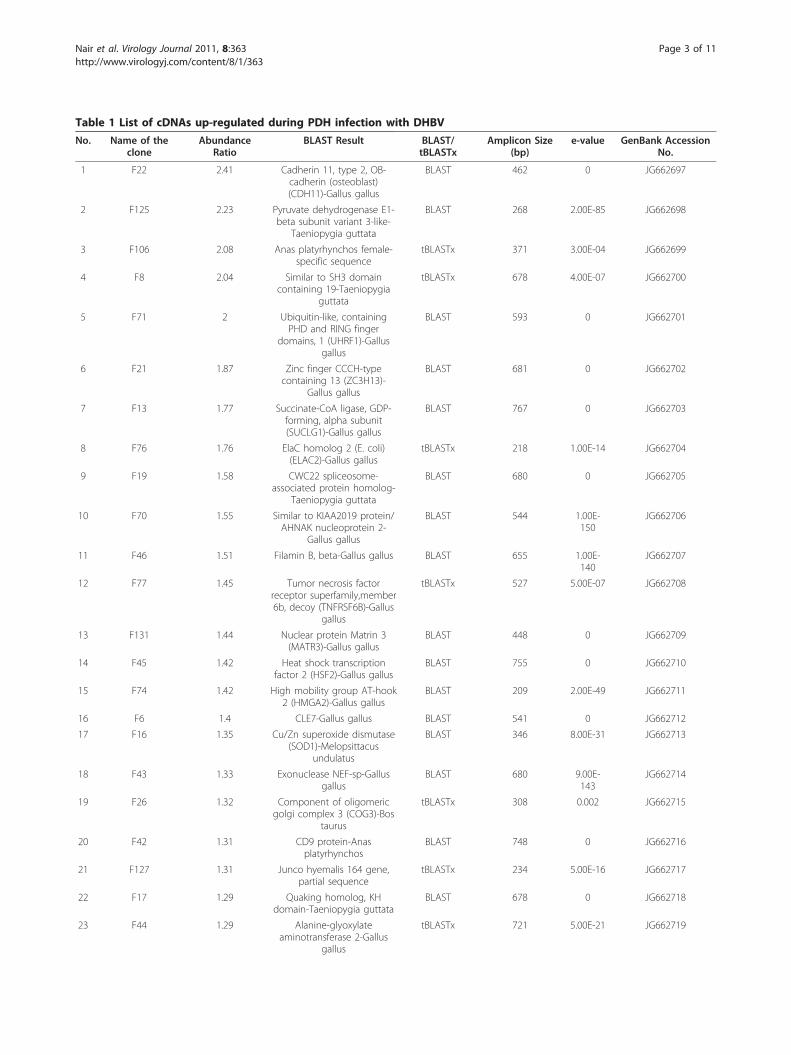
Table 1 List of cDNAs up-regulated during PDH infection with DHBV
No. Name of theclone
AbundanceRatio
BLAST Result BLAST/tBLASTx
Amplicon Size(bp)
e-value GenBank AccessionNo.
1 F22 2.41 Cadherin 11, type 2, OB-cadherin (osteoblast)(CDH11)-Gallus gallus
BLAST 462 0 JG662697
2 F125 2.23 Pyruvate dehydrogenase E1-beta subunit variant 3-like-
Taeniopygia guttata
BLAST 268 2.00E-85 JG662698
3 F106 2.08 Anas platyrhynchos female-specific sequence
tBLASTx 371 3.00E-04 JG662699
4 F8 2.04 Similar to SH3 domaincontaining 19-Taeniopygia
guttata
tBLASTx 678 4.00E-07 JG662700
5 F71 2 Ubiquitin-like, containingPHD and RING finger
domains, 1 (UHRF1)-Gallusgallus
BLAST 593 0 JG662701
6 F21 1.87 Zinc finger CCCH-typecontaining 13 (ZC3H13)-
Gallus gallus
BLAST 681 0 JG662702
7 F13 1.77 Succinate-CoA ligase, GDP-forming, alpha subunit(SUCLG1)-Gallus gallus
BLAST 767 0 JG662703
8 F76 1.76 ElaC homolog 2 (E. coli)(ELAC2)-Gallus gallus
tBLASTx 218 1.00E-14 JG662704
9 F19 1.58 CWC22 spliceosome-associated protein homolog-
Taeniopygia guttata
BLAST 680 0 JG662705
10 F70 1.55 Similar to KIAA2019 protein/AHNAK nucleoprotein 2-
Gallus gallus
BLAST 544 1.00E-150
JG662706
11 F46 1.51 Filamin B, beta-Gallus gallus BLAST 655 1.00E-140
JG662707
12 F77 1.45 Tumor necrosis factorreceptor superfamily,member6b, decoy (TNFRSF6B)-Gallus
gallus
tBLASTx 527 5.00E-07 JG662708
13 F131 1.44 Nuclear protein Matrin 3(MATR3)-Gallus gallus
BLAST 448 0 JG662709
14 F45 1.42 Heat shock transcriptionfactor 2 (HSF2)-Gallus gallus
BLAST 755 0 JG662710
15 F74 1.42 High mobility group AT-hook2 (HMGA2)-Gallus gallus
BLAST 209 2.00E-49 JG662711
16 F6 1.4 CLE7-Gallus gallus BLAST 541 0 JG662712
17 F16 1.35 Cu/Zn superoxide dismutase(SOD1)-Melopsittacus
undulatus
BLAST 346 8.00E-31 JG662713
18 F43 1.33 Exonuclease NEF-sp-Gallusgallus
BLAST 680 9.00E-143
JG662714
19 F26 1.32 Component of oligomericgolgi complex 3 (COG3)-Bos
taurus
tBLASTx 308 0.002 JG662715
20 F42 1.31 CD9 protein-Anasplatyrhynchos
BLAST 748 0 JG662716
21 F127 1.31 Junco hyemalis 164 gene,partial sequence
tBLASTx 234 5.00E-16 JG662717
22 F17 1.29 Quaking homolog, KHdomain-Taeniopygia guttata
BLAST 678 0 JG662718
23 F44 1.29 Alanine-glyoxylateaminotransferase 2-Gallus
gallus
tBLASTx 721 5.00E-21 JG662719
Nair et al. Virology Journal 2011, 8:363http://www.virologyj.com/content/8/1/363
Page 3 of 11
total RNA from fresh sets of primary duck hepatocytecultures either infected with DHBV or uninfected, asdescribed above, using Avian Myeloblastosis Virus(AMV) reverse transcription system (Promega). Real-time PCR was carried out as previously described [12].The experiments were repeated thrice, each in dupli-cates, and average fold change in gene expression wascalculated for individual genes.The threshold cycle (Ct) values obtained in the real-
time PCR analysis were normalized with the expressionof the house-keeping gene GAPDH, and the relative
expression of individual genes in infected and uninfectedcells were calculated by Pfaffl method [13] for Day 0and Day 4 of infection using the equation:
Ratio =(Etarget)
�CT,target(calibrator-test)
(Eref)�CT,ref(calibrator-test)
The ratios for day 0 and day 4 infected samples werecompared and analysed statistically by paired Student’st-test to validate the significance of gene expressionchanges. P-values < 0.05 were considered significant.
Table 1 List of cDNAs up-regulated during PDH infection with DHBV (Continued)
24 F135 1.27 Leucine-rich repeats andcalponin homology (CH)domain containing 4-Oryctolagus cuniculus
tBLASTx 288 0.4 JG662720
25 F14 1.24 Clathrin, light chain A (CLTA)-Gallus gallus
BLAST 673 0 JG662721
26 F83 1.2 Sequestosome 1-Gallus gallus BLAST 562 0 JG662722
27 F10 1.18 RAB 32, member of Rasoncogene-Gallus gallus
BLAST 743 0 JG662723
28 F30 1.16 Ribosomal protein L6 (RPL6)-Gallus gallus
BLAST 591 0 JG662724
29 F64 1.16 Holocytochrome c synthase(cytochrome c heme-lyase)-
Gallus gallus
tBLASTx 421 8.00E-52 JG662725
30 F32 1.16 Lysosomal-associatedmembrane protein 1-Taeniopygia guttata
tBLASTx 591 4.00E-93 JG662726
31 F7 1.14 Serine protease 23-Gallusgallus
BLAST 740 3.00E-168
JG662727
32 F18 1.09 Beta-catenin isolate 3-Anasplatyrhynchos
BLAST 710 0 JG662728
33 F52 1.09 Zebrafish DNA sequencefrom clone CH211-276C22 in
linkage group 6
tBLASTx 218 2.2 JG662729
34 F25 1.08 Gallus gallus finished cDNA,clone ChEST457d18
tBLASTx 696 2.00E-27 JG662730
35 F59 1.07 Leucine proline-enrichedproteoglycan (leprecan)1/prolyl 3-hydroxylase 1(P3H1)-Gallus gallus
BLAST 581 0 JG662731
36 F12 1.07 Ribophorin I-Gallus gallus BLAST 796 0 JG662732
37 F87 1.06 Gallus gallus finished cDNA,clone ChEST855m19
BLAST 505 3.00E-91 JG662733
38 F95 1.06 Spastic paraplegia 3A(autosomal dominant)-Gallus
gallus
BLAST 316 3.00E-54 JG662734
39 F107 1.06 High-mobility group box 3-Taeniopygia guttata
BLAST 276 2.00E-136
JG662735
40 F1 1.05 ATP synthase, H+transporting, mitochondrialF0 complex, subunit F2(ATP5J2)-Gallus gallus
tBLASTx 199 9.00E-18 JG662736
41 F78 1.03 Ubiquitin specific peptidase47 (USP47)-Gallus gallus
BLAST 695 0 JG662737
42 F88 1.03 No significant similarityfound
tBLASTx 360 - JG662738
Nair et al. Virology Journal 2011, 8:363http://www.virologyj.com/content/8/1/363
Page 4 of 11

Table 2 List of cDNAs down-regulated during PDH infection with DHBV
No. Name of the clone Abundance Ratio BLAST Result BLAST/tBLASTx
Amplicon Size (bp) e-value GenBank Accession No.
1 R73 1.52 Ferritin, heavy polypeptide 1(FTH1)-Gallus gallus
BLAST 342 4.00E-143 JG662661
2 R90 1.41 Zinc finger CCCH-type,antiviral 1 (ZC3HAV1)-Gallus
gallus
tBLASTx 328 1.00E-14 JG662662
3 R130 1.39 T-complex 1-Taeniopygiaguttata
BLAST 400 6.00E-172 JG662663
4 R108 1.39 Y box binding protein 1-Gallus gallus
BLAST 486 0 JG662664
5 R96 1.34 MYST/Esa1-associated factor6-Taeniopygia guttata
BLAST 646 1.00E-17 JG662665
6 R97 1.33 Similar to RGD-CAP-Gallusgallus
BLAST 743 0 JG662666
7 R111 1.31 PREDICTED: Gallus gallussimilar to Ankycorbin
BLAST 646 0 JG662667
8 R134 1.3 No significant similarityfound
tBLASTx 490 - JG662668
9 R123 1.25 ATPase8, ATPase6 genes forF0-ATP synthase subunit 8,F0-ATP synthase subunit 6-
Anas platyrhynchos
BLAST 462 0 JG662669
10 R133 1.24 Versican-Gallus gallus BLAST 257 3.00E-98 JG662670
11 R103 1.22 Platelet-activating factoracetylhydrolase isoform Ib,
alpha subunit 45kDa(PAFAH1B1)-Gallus gallus
BLAST 508 4.00E-145 JG662671
12 R16 1.18 UPF0308 protein-Gallus gallus BLAST 593 0 JG662672
13 R100 1.17 TRAF interacting protein(TRAIP)-Gallus gallus
BLAST 631 0 JG662673
14 R84 1.16 Acid alpha-glucosidase-Macaca mulatta
tBLASTx 756 3.8 JG662674
15 R15 1.15 Microtubule-associatedprotein RP/EB family,member 1-Taeniopygia
guttata
BLAST 438 5.00E-168 JG662675
16 R126 1.15 Catechol-O-methyltransferase-Gallus
gallus
tBLASTx 395 1.00E-25 JG662676
17 R135 1.14 Chromosome 15 hypotheticalATG/GTP binding protein-
Gallus gallus
tBLASTx 239 0.048 JG662677
18 R143 1.13 Ankyrin repeat domain 17(ANKRD17)-Gallus gallus
BLAST 546 0 JG662678
19 R45 1.13 Splicing factor, arginine/serine-rich 18 (SFRS18)-Gallus
gallus
tBLASTx 476 1.00E-145 JG662679
20 R141 1.11 Cytochrome oxidase subunitI (COI)-Anas platyrhynchos
BLAST 336 1.00E-152 JG662680
21 R10 1.11 Eukaryotic translationinitiation factor 5 (EIF5)-Gallus
gallus
BLAST 735 0 JG662681
22 R129 1.11 Beta-actin-Anasplatyrhynchos
BLAST 664 0 JG662682
23 R106 1.1 Alpha enolase-Peking Duck BLAST 381 0 JG662683
24 R93 1.1 No significant similarityfound
tBLASTx 488 - JG662684
25 R139 1.1 Similar to KIAA1824 protein/WD repeat domain 22-Gallus
gallus
BLAST 279 7.00E-100 JG662685
Nair et al. Virology Journal 2011, 8:363http://www.virologyj.com/content/8/1/363
Page 5 of 11

Results & DiscussionThe infection of PDH with DHBV did not produce anyvisible changes on the cell monolayer (Figure 1A). Thevirus infection was confirmed by PCR detection of a300 bp DHBV glycoprotein 1 (gp1) gene fragment in theDNA isolated from infected PDH culture supernatant(Figure 1B) and by sequence analysis. The establishmentof a productive infection was indicated by the increasingPCR amplification intensity of the gene fragment withevery successive day of culture for the total culture per-iod of eight days. For RNA isolation for subtractionlibrary construction, we selected an early time point of 4days as described in previous studies [14]. Two librarieswere generated- the forward subtracted or up-regulatedgenes and the reverse subtracted or down-regulatedgenes. The efficiency of subtraction procedure was indi-cated by a decrease in the intensity and appearance ofdiscrete banding patterns in the lanes with subtractedproducts (Figure 1C) and was confirmed by PCR detec-tion of the house-keeping gene GAPDH, the ampliconsof which appeared at an earlier time point (25 cycles) inun-subtracted samples compared to a later time point(30 cycles) in both forward and reverse subtractedlibraries (Figure 1D). Hybridization of macroarraysblotted with 137 up-regulated and 148 down-regulatedclones (Figure 1E) and short-listing only the ones with an
abundance ratio of more than 1, we obtained 42 non-redundant up-regulated clones and 36 non-redundantdown-regulated clones (Tables 1 and 2). Real-time PCRdone using the representative sets of short-listed clonesgave results confirming the reliability of the short-listingprocedure. Genes that topped the differential expressionamong the up-regulated genes (F22, F8, F71) showed asignificant (P < 0.05) increase in expression at 4-dayscompared to the 0 day in infected PDH (Figure 2A),while the reverse was the case of the down-regulatedgenes (R73, R90, R130) (Figure 2B), all of whose expres-sion decreased significantly (P < 0.05) at 4-day DHBVinfection. F88 and R86, which were selected from thebottom end of the up-regulated and down-regulatedgene-tables, respectively, also showed the expected mod-ulation albeit at a lower fold. F62 and R47, picked fromthe genes left-out did not show any significant differencein their expression pattern.Functional classification of the short-listed clones
using gene ontology based on BLAST results groupedthem mainly into those belonging to cellular processessuch as cellular respiration, signal transduction, tran-scription/translation, ubiquitin/proteasome pathway andapoptosis besides those coding for membrane and cytos-keletal proteins (Table 3). Among them, the categorythat was maximum up-regulated were the ones involved
Table 2 List of cDNAs down-regulated during PDH infection with DHBV (Continued)
26 R104 1.09 Caspase 3, apoptosis-relatedcysteine peptidase (CASP3)-
Gallus gallus
BLAST 792 0 JG662686
27 R95 1.09 Gallus gallus finished cDNA,clone ChEST757h13
tBLASTx 793 5.00E-19 JG662687
28 R79 1.07 Ral guanine nucleotidedissociation stimulator-like 1
(RGL1)-Gallus gallus
BLAST 333 1.00E-132 JG662688
29 R105 1.06 Gallus gallus similar toMGC53471 protein
BLAST 646 7.00E-89 JG662689
30 R128 1.05 Hydroxyacyl glutathionehydrolase-like,transcript
variant 2-Taeniopygia guttata
BLAST 382 4.00E-54 JG662690
31 R22 1.05 Proteasome (prosome,macropain) 26S subunit,ATPase,1 (PSMC1)-Gallus
gallus
BLAST 324 4.00E-133 JG662691
32 R99 1.04 Gallus gallus hypotheticalprotein
BLAST 362 2.00E-86 JG662692
33 R2 1.04 Ribosomal protein L35a-Gallus gallus
BLAST 90 3.00E-08 JG662693
34 R36 1.04 Gallus gallus finished cDNA,clone ChEST191i5
tBLASTx 414 0.025 JG662694
35 R124 1.02 Gallus gallus BAC cloneCH261-189F16 fromchromosome z
BLAST 524 0 JG662695
36 R86 1.02 Transmembrane protein 30A-Taeniopygia guttata
BLAST 220 4.00E-72 JG662696
Nair et al. Virology Journal 2011, 8:363http://www.virologyj.com/content/8/1/363
Page 6 of 11
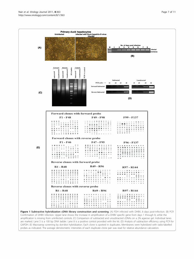
Figure 1 Subtractive hybridization cDNA library construction and screening. (A) PDH infected with DHBV, 8 days post-infection. (B) PCRConfirmation of DHBV infection. Upper lane shows the increase in amplification of a DHBV specific gene from days 1 through 8, while theamplification is missing from uninfected controls. (C) Comparison of subtracted and unsubtracted cDNAs on a 2% agarose gel. Individual lanesare marked. Lane 5 is a 100 bp DNA ladder. Lane 8 is a positive control provided with the kit. (D) Analysis of subtraction efficiency using PCR forGAPDH. (E) Macroarray screening by dot-blot hybridization. Each clone is spotted in duplicates. Membranes were hybridized with radio-labelledprobes as indicated. The average densitometric intensities of each duplicate clone pair was read for relative abundance calculation.
Nair et al. Virology Journal 2011, 8:363http://www.virologyj.com/content/8/1/363
Page 7 of 11
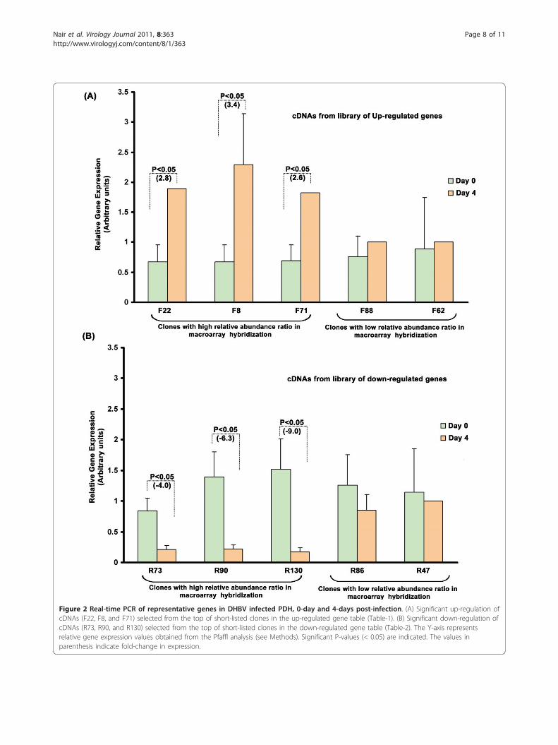
Figure 2 Real-time PCR of representative genes in DHBV infected PDH, 0-day and 4-days post-infection. (A) Significant up-regulation ofcDNAs (F22, F8, and F71) selected from the top of short-listed clones in the up-regulated gene table (Table-1). (B) Significant down-regulation ofcDNAs (R73, R90, and R130) selected from the top of short-listed clones in the down-regulated gene table (Table-2). The Y-axis representsrelative gene expression values obtained from the Pfaffl analysis (see Methods). Significant P-values (< 0.05) are indicated. The values inparenthesis indicate fold-change in expression.
Nair et al. Virology Journal 2011, 8:363http://www.virologyj.com/content/8/1/363
Page 8 of 11
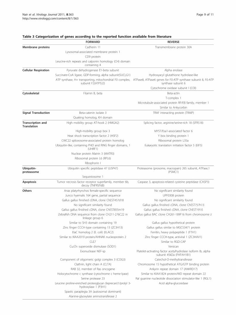
Table 3 Categorization of genes according to the reported function available from literature
FORWARD REVERSE
Membrane proteins Cadherin 11 Transmembrane protein 30A
Lysosomal-associated membrane protein 1
CD9 protein
Leucine-rich repeats and calponin homology (CH) domaincontaining 4
Cellular Respiration Pyruvate dehydrogenase E1-beta subunit Alpha enolase
Succinate-CoA ligase, GDP-forming, alpha subunit(SUCLG1) Hydroxyacyl glutathione hydrolase-like
ATP synthase, H+ transporting, mitochondrial F0 complex,subunit F2(ATP5J2)
ATPase8, ATPase6 genes for F0-ATP synthase subunit 8, F0-ATPsynthase subunit 6
Cytochrome oxidase subunit I (COI)
Cytoskeletal Filamin B, beta Beta-actin
T-complex 1
Microtubule-associated protein RP/EB family, member 1
Similar to Ankycorbin
Signal Transduction Beta-catenin isolate 3 TRAF interacting protein (TRAIP)
Quaking homolog, KH domain
Transcription andTranslation
High mobility group AT-hook 2 (HMGA2) Splicing factor, arginine/serine-rich 18 (SFRS18)
High-mobility group box 3 MYST/Esa1-associated factor 6
Heat shock transcription factor 2 (HSF2) Y box binding protein 1
CWC22 spliceosome-associated protein homolog Ribosomal protein L35a
Ubiquitin-like, containing PHD and RING finger domains, 1(UHRF1)
Eukaryotic translation initiation factor 5 (EIF5)
Nuclear protein Matrin 3 (MATR3)
Ribosomal protein L6 (RPL6)
Ribophorin I
Ubiquitin-proteasome
Ubiquitin specific peptidase 47 (USP47) Proteasome (prosome, macropain) 26S subunit, ATPase,1(PSMC1)
Sequestosome 1
Apoptosis Tumor necrosis factor receptor superfamily, member 6b,decoy (TNFRSF6B)
Caspase 3, apoptosis-related cysteine peptidase (CASP3)
Others Anas platyrhynchos female-specific sequence No significant similarity found
Junco hyemalis 164 gene, partial sequence UPF0308 protein
Gallus gallus finished cDNA, clone ChEST457d18 No significant similarity found
No significant similarity found Gallus gallus finished cDNA, clone ChEST757h13
Gallus gallus finished cDNA, clone ChEST855m19 Gallus gallus finished cDNA, clone ChEST191i5
Zebrafish DNA sequence from clone CH211-276C22 inlinkage group 6
Gallus gallus BAC clone CH261-189F16 from chromosome z
Similar to SH3 domain containing 19 Gallus gallus hypothetical protein
Zinc finger CCCH-type containing 13 (ZC3H13) Gallus gallus similar to MGC53471 protein
ElaC homolog 2 (E. coli) (ELAC2) Ferritin, heavy polypeptide 1 (FTH1)
Similar to KIAA2019 protein/AHNAK nucleoprotein 2 Zinc finger CCCH-type, antiviral 1 (ZC3HAV1)
CLE7 Similar to RGD-CAP
Cu/Zn superoxide dismutase (SOD1) Versican
Exonuclease NEF-sp Platelet-activating factor acetylhydrolase isoform Ib, alphasubunit 45kDa (PAFAH1B1)
Component of oligomeric golgi complex 3 (COG3) Catechol-O-methyltransferase
Clathrin, light chain A (CLTA) Chromosome 15 hypothetical ATG/GTP binding protein
RAB 32, member of Ras oncogene Ankyrin repeat domain 17 (ANKRD17)
Holocytochrome c synthase (cytochrome c heme-lyase) Similar to KIAA1824 protein/WD repeat domain 22
Serine protease 23 Ral guanine nucleotide dissociation stimulator-like 1 (RGL1)
Leucine proline-enriched proteoglycan (leprecan)1/prolyl 3-hydroxylase 1 (P3H1)
Acid alpha-glucosidase
Spastic paraplegia 3A (autosomal dominant)
Alanine-glyoxylate aminotransferase 2
Nair et al. Virology Journal 2011, 8:363http://www.virologyj.com/content/8/1/363
Page 9 of 11
in transcription/translation (19%), whereas the onesmaximum down regulated (11%) belonged to cytoskele-tal proteins. The former included the HMG Box pro-teins and Y-box binding proteins. Previous studies haveimplicated the Y-box binding protein1, Platelet-activat-ing factor acetylhydrolase isoform 1B (PAFAH1B1),Ribosomal Protein L35a, Ferritin, a-enolase, Caspase 3,CuZn Superoxide Dismutase (CuZnSOD), Filamin B,Pyruvate dehydrogenase 1-b, b-catenin, prolyl-3-hydro-xylase 1, b-actin, acid a-glucosidase, and clathrin, thecDNAs of which were identified to be up-regulated,with chronic HBV infections and HCC development[6,15-26]. In comparison to the earlier report based onproteome analysis in DHBV infected PDH [8], exceptfor b-actin and a-enolase, all the cDNAs identified inthe present study represented new genes. The differencecould be due to multiple reasons, and importantly itmight include the selective enrichment/elimination ofsome of the cDNAs during the process of RT-PCRamplification and cloning as part of the subtractionlibrary construction. Nevertheless, our data provides anew set of candidate genes worth further investigationin hepadnaviral infection.An interesting observation in this study was the inverse
pattern of differential expression of ten of these genes inin vitro DHBV infected cells as against the reports onHCC clinical samples [6,15-20]. The mRNAs for the Y-box binding protein1, PAFAH1B1, Ribosomal ProteinL35a, Ferritin, a-enolase, acid alpha-glucosidase and Cas-pase 3 were shown to be down-regulated during in vitroDHBV infection, whereas those of CuZnSOD, Filamin Band Pyruvate dehydrogenase were shown to be up-regu-lated, where as the reverse was the trend in human HCC.This observation may be purely coincidental owing to thefact that the experimental method we used was an in vitrosystem, and the changes in primary hapatocytes duringculture itself, such as de-differentiation, might have led tothese alterations in gene expression.
ConclusionsIn summary, the present study identified cDNAs of anumber of genes that are differentially modulated in cul-tured PDH, invitro infected with DHBV. cDNAs of bothnovel as well as already reported genes/proteins asso-ciated with HBV/DHBV infection or HCC were identi-fied in the library. The genes short-listed here could bevaluable leads for further studies in animal models,which might help to understand the pathology ofchronic HBV infections and pathogenesis of HCC.
Additional material
Additional file 1: Primers used in the study.
AcknowledgementsThe financial support by the Department of Biotechnology, Government ofIndia (Grant No. BT/PR8930/GBD/27/39/2006) is gratefully acknowledged.
Authors’ contributionsSN, DSA and AI carried out the experiments. SN drafted the manuscript. ESconceived the study, edited and completed the final version of themanuscript. All authors read and approved the final manuscript.
Competing interestsThe authors declare that they have no competing interests.
Received: 1 April 2011 Accepted: 23 July 2011 Published: 23 July 2011
References1. Funk A, Mhamdi M, Will H, Sirma H: Avian hepatitis B viruses: molecular
and cellular biology, phylogenesis, and host tropism. World JGastroenterol 2007, 13:91-103.
2. Gripon P, Rumin S, Urban S, Le Seyec J, Glaise D, Cannie I, Guyomard C,Lucas J, Trepo C, Guguen-Guillouzo C: Infection of a human hepatomacell line by hepatitis B virus. Proc Natl Acad Sci USA 2002, 99:15655-15660.
3. Schultz U, Grgacic E, Nassal M: Duck hepatitis B virus: an invaluablemodel system for HBV infection. Adv Virus Res 2004, 63:1-70.
4. Ganem D, Schneider RJ: Hepadnaviridae: The viruses and theirreplication. In Fields Virology. Volume 2. 4 edition. Edited by: Knipe DM,Howley PM. Philadelphia: Lippincott, Williams and Wilkins; 2001:2923-2969.
5. Kao JH, Chen PJ, Chen DS: Recent advances in the research of hepatitis Bvirus-related hepatocellular carcinoma: epidemiologic and molecularbiological aspects. Adv Cancer Res 108:21-72.
6. Xu XR, Huang J, Xu ZG, Qian BZ, Zhu ZD, Yan Q, Cai T, Zhang X, Xiao HS,Qu J, Liu F, Huang QH, Cheng ZH, Li NG, Du JJ, Hu W, Shen KT, Lu G, Fu G,Zhong M, Xu SH, Gu WY, Huang W, Zhao XT, Hu GX, Gu JR, Chen Z,Han ZG: Insight into hepatocellular carcinogenesis at transcriptome levelby comparing gene expression profiles of hepatocellular carcinoma withthose of corresponding noncancerous liver. Proc Natl Acad Sci USA 2001,98:15089-15094.
7. Bellodi-Privato M, Kubrusly MS, Stefano JT, Soares IC, Wakamatsu A,Oliveira AC, Alves VA, Bacchella T, Machado MC, D’Albuquerque LA:Differential gene expression profiles of hepatocellular carcinomasassociated or not with viral infection. Braz J Med Biol Res 2009,42:119-1127.
8. Zhao Y, Ben H, Qu S, Zhou X, Yan L, Xu B, Zhou S, Lou Q, Ye R, Zhou T,Yang P, Qu D: Proteomic analysis of primary duck hepatocytes infectedwith duck hepatitis B virus. Proteome Sci 8:28.
9. Tuttleman JS, Pugh JC, Summers JW: In vitro experimental infection ofprimary duck hepatocyte cultures with duck hepatitis B virus. J Virol1986, 58:17-25.
10. Summers J, Smith PM, Huang MJ, Yu MS: Morphogenetic and regulatoryeffects of mutations in the envelope proteins of an avian hepadnavirus.J Virol 1991, 65:1310-1317.
11. Turin F, Borel C, Benchaib M, Kay A, Jamard C, Guguen-Guillouzo C,Trepo C, Hantz O: n-Butyrate, a cell cycle blocker, inhibits earlyamplification of duck hepatitis B virus covalently closed circular DNAafter in vitro infection of duck hepatocytes. J Virol 1996, 70:2691-2696.
12. Arathy DS, Nair S, Soman SS, Issac A, Sreekumar E: Functionalcharacterization of the CC chemokine RANTES from Pekin duck (Anasplatyrhynchos). Dev Comp Immunol 35:142-150.
13. Pfaffl MW: A new mathematical model for relative quantification in real-time RT-PCR. Nucleic Acids Res 2001, 29:e45.
14. Qiao M, Scougall CA, Duszynski A, Burrell CJ: Kinetics of early molecularevents in duck hepatitis B virus replication in primary duck hepatocytes.J Gen Virol 1999, 80(Pt 8):2127-2135.
15. Yasen M, Kajino K, Kano S, Tobita H, Yamamoto J, Uchiumi T, Kon S,Maeda M, Obulhasim G, Arii S, Hino O: The up-regulation of Y-boxbinding proteins (DNA binding protein A and Y-box binding protein-1)as prognostic markers of hepatocellular carcinoma. Clin Cancer Res 2005,11:7354-7361.
16. Pan YS, Lee YS, Lee YL, Lee WC, Hsieh SY: Differentially profiling the low-expression transcriptomes of human hepatoma using a novel SSH/microarray approach. BMC Genomics 2006, 7:131.
Nair et al. Virology Journal 2011, 8:363http://www.virologyj.com/content/8/1/363
Page 10 of 11
17. Kim MY, Park E, Park JH, Park DH, Moon WS, Cho BH, Shin HS, Kim DG:Expression profile of nine novel genes differentially expressed inhepatitis B virus-associated hepatocellular carcinomas. Oncogene 2001,20:4568-4575.
18. Blumberg BS, Lustbader ED, Whitford PL: Changes in serum iron levelsdue to infection with hepatitis B virus. Proc Natl Acad Sci USA 1981,78:3222-3224.
19. Takashima M, Kuramitsu Y, Yokoyama Y, Iizuka N, Fujimoto M, Nishisaka T,Okita K, Oka M, Nakamura K: Overexpression of alpha enolase in hepatitisC virus-related hepatocellular carcinoma: association with tumorprogression as determined by proteomic analysis. Proteomics 2005,5:1686-1692.
20. Chang CS, Huang SM, Lin HH, Wu CC, Wang CJ: Different expression ofapoptotic proteins between HBV-infected and non-HBV-infectedhepatocellular carcinoma. Hepatogastroenterology 2007, 54:2061-2068.
21. Gottlob K, Fulco M, Levrero M, Graessmann A: The hepatitis B virus HBxprotein inhibits caspase 3 activity. J Biol Chem 1998, 273:33347-33353.
22. Hung JH, Yan CW, Su IJ, Wang HC, Lei HY, Lin WC, Chang WT, Huang W,Lu TJ, Lai MD: Hepatitis B virus surface antigen interacts with acid alpha-glucosidase and alters glycogen metabolism. Hepatol Res 40:633-640.
23. Elchuri S, Oberley TD, Qi W, Eisenstein RS, Jackson Roberts L, VanRemmen H, Epstein CJ, Huang TT: CuZnSOD deficiency leads to persistentand widespread oxidative damage and hepatocarcinogenesis later inlife. Oncogene 2005, 24:367-380.
24. Thompson MD, Monga SP: WNT/beta-catenin signaling in liver healthand disease. Hepatology 2007, 45:1298-1305.
25. Zhai B, Yan HX, Liu SQ, Chen L, Wu MC, Wang HY: Reduced expression ofE-cadherin/catenin complex in hepatocellular carcinomas. World JGastroenterol 2008, 14:5665-5673.
26. Risteli J, Tuderman L, Tryggvason K, Kivirikko KI: Effect of hepatic injury onprolyl 3-hydroxylase and 4-hydroxylase activities in rat liver and onimmunoreactive prolyl 4-hydroxylase concentrations in the liver andserum. Biochem J 1978, 170:129-135.
doi:10.1186/1743-422X-8-363Cite this article as: Nair et al.: Differential gene expression analysis of invitro duck hepatitis B virus infected primary duck hepatocyte cultures.Virology Journal 2011 8:363.
Submit your next manuscript to BioMed Centraland take full advantage of:
• Convenient online submission
• Thorough peer review
• No space constraints or color figure charges
• Immediate publication on acceptance
• Inclusion in PubMed, CAS, Scopus and Google Scholar
• Research which is freely available for redistribution
Submit your manuscript at www.biomedcentral.com/submit
Nair et al. Virology Journal 2011, 8:363http://www.virologyj.com/content/8/1/363
Page 11 of 11