Embed Size (px)
Citation preview

Dipeptidyl Peptidase IV Inhibitor Treatment Stimulates�-Cell Survival and Islet Neogenesis inStreptozotocin-Induced Diabetic RatsJ. Andrew Pospisilik,
1Jennifer Martin,
1Timothy Doty,
1Jan A. Ehses,
1Nathalie Pamir,
1
Francis C. Lynn,1
Shalea Piteau,1
Hans-Ulrich Demuth,2
Christopher H.S. McIntosh,1
and Raymond A. Pederson1
Recent studies into the physiology of the incretinsglucose-dependent insulinotropic polypeptide (GIP)and glucagon-like peptide-1 (GLP-1) have added stimu-lation of �-cell growth, differentiation, and cell survivalto well-documented, potent insulinotropic effects. Un-fortunately, the therapeutic potential of these hor-mones is limited by their rapid enzymatic inactivation invivo by dipeptidyl peptidase IV (DP IV). Inhibition of DPIV, so as to enhance circulating incretin levels, hasproved effective in the treatment of type 2 diabetes bothin humans and in animal models, stimulating improve-ments in glucose tolerance, insulin sensitivity, and�-cell function. We hypothesized that enhancement ofthe cytoprotective and �-cell regenerative effects of GIPand GLP-1 might extend the therapeutic potential of DPIV inhibitors to include type 1 diabetes. For testing thishypothesis, male Wistar rats, exposed to a single dose ofstreptozotocin (STZ; 50 mg/kg), were treated twicedaily with the DP IV inhibitor P32/98 for 7 weeks.Relative to STZ-injected controls, P32/98-treated ani-mals displayed increased weight gain (230%) and nutri-ent intake, decreased fed blood glucose (�26 vs. �20mmol/l, respectively), and a return of plasma insulinvalues toward normal (0.07 vs. 0.12 nmol/l, respective-ly). Marked improvements in oral glucose tolerance,suggesting enhanced insulin secretory capacity, werecorroborated by pancreas perfusion and insulin contentmeasurements that revealed two- to eightfold increasesin both secretory function and insulin content after 7weeks of treatment. Immunohistochemical analyses ofpancreatic sections showed marked increases in thenumber of small islets (�35%) and total �-cells(�120%) and in the islet �-cell fraction (12% control vs.
24% treated) in the treated animals, suggesting that DPIV inhibitor treatment enhanced islet neogenesis, �-cellsurvival, and insulin biosynthesis. In vitro studies usinga �-(INS-1) cell line showed a dose-dependent preven-tion of STZ-induced apoptotic cell-death by both GIPand GLP-1, supporting a role for the incretins in elicit-ing the in vivo results. These novel findings provideevidence to support the potential utility of DP IV inhib-itors in the treatment of type 1 and possibly late-stagetype 2 diabetes. Diabetes 52:741–750, 2003
Despite substantial advances in our understand-ing of type 1 diabetes, diagnosis of the condi-tion still carries with it a sentence of lifelongdaily insulin injection, a partially effective ther-
apy at best (1). New therapeutic strategies under investi-gation include islet transplantation (and associated stemcell–derived and xenogeneic islet technologies necessaryfor treatment en masse), the development of improvedinsulin analogues and delivery systems, gene therapy, andthe search for novel agents able to protect and/or stimu-late the proliferation and regeneration of islet �-cells (1).The importance of the latter strategy is underscored by theneed for an inexpensive, benign, preventive therapy thatlacks the considerable profile of side effects of mosttherapies studied to date (e.g., immunosuppressants).
A number of recent studies have highlighted the role ofthe incretin hormones glucose-dependent insulinotropicpolypeptide (GIP) and glucagon-like peptide-1 (GLP-1) in�-cell function and development (2,3). Together, the incre-tins are responsible for �50% of nutrient-stimulated insu-lin release and make up the endocrine arm of theenteroinsular axis (4). Apart from their insulinotropic role,GIP and GLP-1 have been shown to enhance �-cell glucosecompetence and, more recent, to stimulate �-cell growth,differentiation, proliferation, and cell survival (5–11). Inhi-bition of dipeptidyl peptidase IV (DP IV), a ubiquitousserine protease that rapidly cleaves and inactivates GIPand GLP-1 in vivo, has been shown to raise circulatingactive incretin levels and thus increase the effective con-centration of these peptides reaching target tissues (12–16). Studies in both humans and animal models haveestablished DP IV inhibition as a promising therapeuticstrategy for the treatment of type 2 diabetes (17–23). Werecently showed that long-term administration of the DPIV inhibitor P32/98 to VDF rats (model of type 2 diabetes)
From the 1Department of Physiology, University of British Columbia, Vancou-ver, British Columbia, Canada; and 2Probiodrug AG, Halle (Saale), Germany.
Address correspondence and reprint requests to Dr. R.A. Pederson, Depart-ment of Physiology, University of British Columbia, 2146 Health Sciences Mall,Vancouver, BC, Canada V6T 1Z3. E-mail: [email protected].
Received for publication 11 July 2002 and accepted in revised form 18November 2002.
H.U.D. is a Chief Executive Officer, the Chief Scientific Officer, and ashareholder in Probiodrug, a pharmaceutical company in the process ofdeveloping a DP IV inhibitor treatment for diabetes and its complications.R.A.P. and C.H.S.M. are members of a scientific advisory panel to Probiodrugand receive consulting fees for their participation and receive grant/researchsupport from Probiodrug to support studies on the drug candidate P32/98 andits utility in treating diabetes and its complications.
DP IV, dipeptidyl peptidase IV; GIP, glucose-dependent insulinotropicpolypeptide; GLP-1, glucagon-like peptide-1; IDGTT, intraduodenal glucosetolerance test; IR, immunoreactive; OGTT, oral glucose tolerance test; PDX-1,pancreatic-duodenum homeobox-1; PP, pancreatic polypeptide; SOM, soma-tostatin; STZ, streptozotocin.
DIABETES, VOL. 52, MARCH 2003 741

resulted in an enhancement of glucose tolerance, insulinsensitivity, and �-cell glucose responsiveness (19,23). Con-sidering the pleiotropy of noninsulinotropic incretin ef-fects that culminate in an overall promotion of �-cellfunction and survival, we hypothesized that DP IV inhibi-tor treatment of rats transiently exposed to the �-cell–specific toxin streptozotocin (STZ) would be able toprotect islet �-cells from the initial toxic insult and possi-bly to stimulate �-cell proliferation and function.
In the current study, we show that long-term DP IVinhibitor treatment of STZ rats improves glucose toler-ance, enhances pancreatic insulin content, and stimulatesislet neogenesis and the survival of pancreatic �-cells.Furthermore, using an in vitro �-cell model (INS-1 832/13),we show a dose-dependent prevention of STZ-inducedapoptosis by GIP and GLP-1, providing a potential mech-anism of action for the in vivo findings. These findingsprovide a basis on which to extend the therapeutic poten-tial of DP IV inhibitors to include type 1 and very-late-stagetype 2 diabetes.
RESEARCH DESIGN AND METHODS
Materials. Unless otherwise stated, all materials were obtained from SigmaChemical (St. Louis, MO). The specific DP IV inhibitor P32/98 (di-[2S,3S]-2-amino-3-methyl-pentanoic-1,3-thiazolidine fumarate) was synthesized as pre-viously described (24).Animals. Male Wistar rats (250 g) were randomly assigned to one of fourgroups: control, STZ control, early treatment, or late treatment (n � 15). Thetwo treatment groups were administered P32/98 (10 mg/kg) twice daily by oralgavage starting either 1 week before (early) or 1 week after (late) the STZinjection (day 0). The control groups were administered a 1% celluloseinjection vehicle. On day 0, all groups save the controls were administeredSTZ (50 mg/kg i.v., tail vein) while under halothane anesthesia. The controlgroup was administered citrate buffer alone. The techniques used in this studywere in compliance with the guidelines of the Canadian Council on AnimalCare and were approved by the UBC Council on Animal Care (CertificateA99-0006).Acute DP IV inhibition during intraduodenal glucose tolerance tests. Asecond set of Wistar rats (275 g) received an injection of STZ, as describedabove, to examine the effects of acute DP IV inhibition in the STZ model. Sixdays after STZ injection, animals were anesthetized (sodium pentobarbital 50mg/kg) and subjected to two successive intraduodenal glucose tolerance tests(IDGTT; 1 g/kg), first in the absence of a DP IV inhibitor and second after anintravenous bolus of P32/98 (10 mg/kg). Blood samples, taken at regularintervals from a carotid cannula, were collected directly into tubes containingP32/98 (final concentration 1 mmol/l; to prevent further DP IV degradation ofGLP-17–36) and placed immediately on ice. Plasma was separated and stored at�20°C within 1 h of collection. Samples were analyzed for active and totalGLP-1 using enzyme-linked immunosorbent assay– and radioimmunoassay–based kits, respectively (Linco Research).Weekly monitoring. During the long-term study, 500-�l blood samples werecollected from each animal into heparinized capillary tubes at 9:00 A.M. on aweekly basis. Samples were then centrifuged (15 min at 12,000 rpm) andstored at �20°C until analysis. Blood glucose values were determined using aSureStep Glucose analyzer (LifeScan Canada, Burnaby, BC, Canada). Plasmafructosamine measurements were performed using a LaRoche DiagnosticFructosamine kit (LaRoche Diagnostics) as per the manufacturer’s instruc-tions. Similarly, plasma insulin levels were determined using a rat-specificsensitive insulin kit (0.02–1.0 ng/ml; Linco Research). DP IV activity levelswere measured as previously described (19). Food and water intake wasmeasured by subtraction.Measurement of oral glucose tolerance. Seven weeks after STZ exposure,the animals underwent an oral glucose tolerance test (OGTT; 1 g/kg) after a16-h fast. In this case, P32/98 was not administered the morning of the OGTTto ensure complete drug washout (16 h from last drug administration; 1.5 hclearance half-life). Blood glucose and plasma insulin measurements weremade as above (250-�l samples).Measurment of pancreatic function. For assessing the effects of long-termP32/98 treatment on pancreatic function in the STZ model, a low-to-highglucose pancreas perfusion was performed as previously described (25). Inbrief, the pancreas was isolated through a ventral midline incision, all major
vasculature was ligated, and a glucose perfusate was introduced through theceliac artery. Perfusion effluent was collected at 1-min intervals via the portalvein with a perfusion rate of 4 ml/min. Samples were stored at �20°C untilanalysis for insulin.Total pancreatic insulin. Total pancreatic insulin content was determined inone-third of the experimental subjects using conventional techniques. Afterinduction of anesthesia with sodium pentobarbital (Somnotol; 50 mg/kg i.p.),the pancreas was excised, blotted dry, and weighed. Pancreata were thenhomogenized in 5 ml of ice-cold 2 N acetic acid, boiled, and centrifuged (10min, 15,000 rpm, 4°C). The supernatant was then assayed for insulin contentand normalized for protein concentration (BCA; Pierce, Rockford, IL).Immunohistochemistry. Pancreata removed under anesthesia were imme-diately placed in fixative (44% formaldehyde, 47% distilled H2O, 9% glacialacetic acid) for 48 h, after which they were washed and stored in 70% ethanol.To account for variation between pancreatic regions, the pancreata were cutinto �4-mm blocks that were then randomly inserted into a cassette andparaffin-embedded. �-Cell counts were made from all blocks (�10–15) in acassette, thereby ensuring assessment of a randomized set of cross-sections inall three planes throughout each pancreas. Once embedded, pancreata weresectioned (5 �m), and the sections were mounted onto slides and dried readyfor staining. Sections were stained for insulin (Rbt �-insulin H-86; Santa CruzBiotechnology, Santa Cruz, CA) as per the manufacturer’s instructions, andthe nuclei were counterstained using DAPI (0.3 �mol/l; Molecular Probes,Eugene, OR) to facilitate quantification of �-cell and islet parameters. Quan-tification was performed manually, as a high degree of variability wasobserved in insulin staining within islet sections from animals exposed to STZ.Determinations of insulin immunoreactive (IR) cell number and total cross-sectional islet cell number (nuclear stain) were made in islets from 250 mm2
of pancreatic tissue section per animal (�100 islets); three to four animalswere counted in each experimental group.Protection of �-(INS-1) cells in vitro. The rat insulinoma cell line INS-1(832/13) was obtained as a gift from Dr. C.B. Newgard (Duke University,Durham, NC). Cells were cultured in RPMI 1640 supplemented with 10% FBS(Cansera, Rexdale, ON), 11 mmol/l glucose, 100 units/ml penicillin, 100 �g/mlstreptomycin, 10 mmol/l HEPES (pH 7.4), 2 mmol/l L-glutamine, 1 mmol/lpyruvate, and 50 �mol/l �-mercaptoethanol. Cells were plated into either6-well (2 � 106 cells/well; caspase studies) or 96-well (7.5 � 104 cells/well;protection assays) coated tissue-culture plates (Becton Dickinson, LincolnPark, NJ) 24 h before experimentation. Unless otherwise stated, the glucoseconcentration during all cell survival assays was 3 mmol/l for the duration ofthe experiment to minimize complications associated with stimulation ofproliferation and glucose-dependent secretory products. Similarly, in allexperiments, cells were serum-starved overnight (3 mmol/l glucose RPMI 0.1% BSA) and subjected to a 30-min STZ exposure the next day. After STZexposure, medium was replaced and cells were allowed to recover for 24 h.GIP and/or GLP-1 (10�7M) was added 10 min before STZ (2 mmol/l) during thedose-response and caspase-3/8 experiments and 10 min before (Pre), imme-diately after (Post), or both 10 min before and immediately after (Pre Post)the STZ exposure for the cell protection assays. Post and Pre Post groupswere therefore exposed to incretins for the duration of the 24-h recoveryperiod. Quantification of live versus dead cell number was made manuallyusing a conventional trypan blue exclusion method. Caspase-3/8 determina-tions were made with the EnzChek Caspase Assay Kits (Molecular Probes)according to the manufacturer’s recommendations.Data analysis and presentation. Data, presented as mean SE, werecompared with Prism 3.02 data analysis software (GraphPad Software, LaJolla, CA) using ANOVA followed by a Dunnett’s multiple comparison testpost hoc (P � 0.05).
RESULTS
Acute inhibition of DP IV enhances plasma-active
GLP-1 and potentiates insulin secretion. In a separategroup of STZ rats, an IDGTT was used to demonstrate aDP IV inhibitor–mediated increase in circulating activeGLP-1 levels. A 10 mg/kg dose of P32/98 was sufficient toprotect an additional 40% of the total GLP-1 secretion,allowing peak glucose-stimulated active GLP-1 levels toreach 45 7 pmol/l (vs. 14 1 pmol/l for control; Fig. 1).Furthermore, an earlier and more robust plasma insulinpeak was evidenced after P32/98 treatment as was aconcomitant reduction in blood glucose (Fig. 1).
DP IV INHIBITION IN STZ RATS
742 DIABETES, VOL. 52, MARCH 2003

Effects of DP IV inhibitor treatment on weight gain
and nutrient intake. STZ-induced diabetes in rats isaccompanied by a marked reduction in weight gain despiteincreased caloric intake, a direct result of insulin insuffi-ciency (Fig. 2). Over the course of the present study, theearly and late treatment groups gained 129 7 g and 92 4 g, respectively (2.3- and 1.7-fold STZ-control; Fig. 2).Furthermore, the STZ-induced doubling in food intakeexhibited by the diabetic controls was decreased 48 and23% in the early and late groups, respectively. Changes infood and water intake paralleled one another, with im-provements evident in both treatment groups within 1week of treatment initiation (Fig. 2).Effects of DP IV inhibitor treatment on blood glu-
cose, plasma insulin, and plasma DP IV. The initialSTZ-induced increase in morning blood glucose valuesaveraged �20 mmol/l above normal (Fig. 3A). This rapidrise in glycemia, which stabilizes within 3–4 days, wasmarkedly attenuated in the early treatment group plateau-ing �5 mmol/l below the untreated controls. In contrast toa gradual deterioration in morning blood glucose in theuntreated STZ controls, both treatment groups displayedsignificant reductions in morning glycemia within 1 weekof treatment initiation. The reduction in morning glycemiaevident in the late treatment group, unlike the abrupt,immediate reduction seen in the early groups, was pro-gressive during the 6 weeks of treatment. Corroboratingthe reduced morning glucose values, plasma fructosaminelevels in both treatment groups were significantly de-creased (Fig. 3B). Once again, the early treatment groupexhibited an immediate blunting of the STZ-induced rise inplasma fructosamine, whereas the late group showed a
more progressive reduction, achieving significance duringthe final 2 weeks of the study.
Whereas untreated animals displayed a 0.08 nmol/l dropin plasma insulin within the first 2 weeks after STZexposure, the pretreated animals presented only a 0.04nmol/l drop, a 43% improvement over the untreated con-trols. As before, the late treatment group displayed a moregradual reversal, with a 40% elevation in morning plasmainsulin levels detected by week 6 of the experiment (Fig.3C). Morning plasma DP IV activity measured immediatelybefore P32/98 administration showed an STZ-induced ele-vation to a plateau level �50% above normal within 4 daysin all three STZ groups (Fig. 3D).Effects of DP IV inhibitor treatment on oral glucose
tolerance in STZ rats. Seven weeks after STZ injection,all groups underwent an OGTT. Fasting blood glucosevalues in the treated animals averaged 5.4 mmol/l less and2.0 mmol/l less than their untreated diabetic littermates(early and late, respectively), although neither groupachieved normalcy (Fig. 4A). Integrated blood glucoseresponses during the 120-min course of the OGTT weredecreased by 33 and 20% in the early and late treatmentgroups, respectively, with concomitant 240 and 45% in-creases in integrated insulin responses (Fig. 4, insets).Glucose responsiveness of insulin secretion, which waslargely ablated in the diabetic controls, was partiallyrestored in the early treatment group (Fig. 4B).Effects of DP IV inhibitor treatment on pancreatic
insulin secretion and content. The partial restoration ofglucose responsiveness (of insulin release) observed dur-ing the OGTT was accompanied by increased glucose-stimulated insulin secretion during pancreas perfusion.
FIG. 1. DP IV inhibition enhances active GLP-1 levels and early-phase insulin release and improves intraduodenal glucose tolerance. Blood glucose(A), plasma insulin (B), and plasma active (C) and total (D) GLP-1 levels measured during an IDGTT (1 g/kg) performed in anesthetized STZWistar rats either in the presence (open circles) or absence (closed squares) of the DP IV inhibitor P32/98 (10 mg/kg). *Statistical significance(P < 0.05) versus control.
J.A. POSPISILIK AND ASSOCIATES
DIABETES, VOL. 52, MARCH 2003 743

Under basal (4.4 mmol/l glucose) conditions, insulin secre-tion from pancreata of both treated groups was elevated(190 and 77% for the early and late treatment groups,respectively) compared with the untreated control levels(Fig. 5). Basal secretion from the early treatment groupequaled normal Wistar controls (0.21 nmol/l). Further-more, first-phase insulin release in response to a 4.4–8.8mmol/l glucose perfusion step was partially restored inboth treatment groups with peak insulin levels reaching0.87 and 0.59 nmol/l in the early and late groups, respec-tively (�70 and �50% of control, 400 and 250% of STZcontrol; Fig. 5A).
In keeping with the increases in basal insulin secretionevident during perfusion, whole pancreas insulin determi-nations revealed parallel increases in insulin content afterlong-term DP IV inhibitor treatment (Fig. 5B). The late andearly treatment groups exhibited greater than two- and
eightfold higher total pancreatic insulin levels than theuntreated STZ-injected group (corresponding to 5 and 17%of control). Data presented in Fig. 5, normalized againstprotein concentration, are representative of total pancre-atic insulin as no difference was evidenced in pancreaticmass between groups (control 1.91 0.10 g; STZ control1.93 0.11 g; early treatment 1.84 0.07 g; and latetreatment 1.86 0.06 g).Effects of DP IV inhibitor treatment on �-cell number
and islet morphology. Pursuant to evidence of increasedpancreatic insulin content, morphometric analysis of islet�-cell distribution was performed. Examination of isletnumber per unit area in all three STZ-exposed groupsshowed an increase in the number of small islets (1–20cells in cross-section; Fig. 6A). The effect was mostprofound in the treated groups, with marked increases inislets with a cross-sectional area �10 cells. Determinationof �-cell area as a fraction of cross-sectional islet cellnumber revealed that the majority of islets �20 cells incross-sectional area contained a near-normal fraction of�-cells (60–80%), whereas those of larger size showed amarked reduction in �-cell fraction (Fig. 6B). Significantincreases in �-cell fraction were evidenced in the earlytreatment group in all subsets of islets �10 cells incross-section, whereas a significant increase in the lategroup was seen in only the group 11–20 cells in cross-section (Figs. 6B and D). The logical interpretation ofthese two results, in functional terms, is that �-cells insmall islets make up a larger fraction of the functional�-cell mass (insulin-IR mass) in the three STZ groups.Expressed as the fraction of the total pancreatic �-cellnumber, �-cells of smaller islets were shown to compose asignificantly larger fraction of the total �-cell mass in allthree STZ-exposed groups (Fig. 6C). It is interesting thatislets with a cross-sectional area of 11–20 cells seemedparticularly sensitive both to the toxic effects of STZ andto the protective effects of the treatment, displayingmarked increases in �-cell fraction and fraction of totalpancreatic �-cell number in response to the DP IV inhibi-tor regimen (Fig. 6B, C, I, and J).
Qualitative analysis of insulin immunoreactivity re-vealed a number of interesting features. Whereas insu-lin-IR cells in the islets of STZ control animals displayedrelatively diffuse and weak staining (Fig. 6F), islets in bothtreatment groups contained a marked number of cellsdisplaying more intense insulin staining (Fig. 6G and H).These insulin-“bright” (INbright) cells appeared relativelyrotund and polarized, whereas those of STZ control isletsappeared diffuse and elongated and lacked distinct polar-ity (Fig. 6E–J).Incretin-mediated protection of �-cells from STZ in
vitro. In an attempt to provide a mechanism of action forthe immunohistochemical and the in vivo data, an exami-nation was made of the ability of GIP and GLP-1 topromote cell survival in INS-1 (832/13) cells transientlyexposed to STZ. After a 30-min STZ exposure followed bya 24-h recovery period, STZ was shown to elicit cytotoxiceffects in the low mmol/l range, with an EC50 of 2.1 mmol/l(Fig. 7A). GLP-1 and GIP were shown independently toreverse STZ-induced cytotoxicity partially with EC50 val-ues of 0.04 and 0.75 nmol/l, respectively (Fig. 7B). STZ-induced cytotoxicity under control conditions involved
FIG. 2. Body weight (A), food intake (B), and water intake (C)measurements from Wistar rats (n � 20) exposed to a single high doseof STZ (50 mg/kg) and treated either with or without the DP IVinhibitor P32/98. Control (solid squares) and STZ control (opensquares) animals were administered a 1% cellulose solution, whereasthe early treatment group (open triangles; treatment initiated 1 weekbefore STZ administration) and the late treatment group (open circles;treatment initiated 1 week after STZ administration) were adminis-tered 10 mg/kg P32/98 twice daily by oral gavage. Food and waterintake was measured by subtraction. *Statistical significance (P <0.05) for the early group versus STZ control; #statistical significancefor the late group versus STZ control.
DP IV INHIBITION IN STZ RATS
744 DIABETES, VOL. 52, MARCH 2003

FIG. 3. Weekly monitoring of blood glucose(A) and plasma fructosamine (B), insulin(C), and DP IV activity levels (D) in Wistarrats (n � 20) exposed to a single high dose ofSTZ (50 mg/kg) and treated either with orwithout the DP IV inhibitor P32/98. Control(solid squares) and STZ control (opensquares) animals were administered a 1%cellulose solution, whereas the early treat-ment group (open triangles; treatment initi-ated 1 week before STZ administration) andthe late treatment group (open circles; treat-ment initiated 1 week after STZ administra-tion) were administered 10 mg/kg P32/98twice daily by oral gavage. Blood glucose wasmeasured on a hand-held blood glucosemeter, insulin and fructosamine were mea-sured using commercially available kits, andDP IV activity was measured using an in-house assay. *Statistical significance (P <0.05) for the early group versus STZ control;#statistical significance for the late groupversus STZ control.
FIG. 4. OGTT performed on Wistar rats (n � 20) exposed to a singlehigh dose of STZ (50 mg/kg) and treated either with or without the DPIV inhibitor P32/98 for 7 weeks. Blood glucose (A) and plasma insulin(B) were measured during a 1 g/kg OGTT in control (solid squares) andSTZ control (open squares) animals that were administered a 1%cellulose solution and an early treatment group (open triangles;treatment initiated 1 week before STZ administration) and late treat-ment group (open circles; treatment initiated 1 week after STZ admin-istration) that received 10 mg/kg P32/98 twice daily by oral gavage for7 weeks after STZ administration. Insets show area under the curvecalculations. *Statistical significance (P < 0.05) for the early groupversus STZ control; #statistical significance for the late group versusSTZ control.
FIG. 5. Insulin response to glucose (A) and insulin content (B) inpancreata isolated from Wistar rats (n � 20) exposed to a single highdose of STZ (50 mg/kg) and treated either with or without the DP IVinhibitor P32/98. Glucose-stimulated insulin secretion was assessedduring an ex vivo pancreas perfusion, and pancreatic insulin contentdeterminations were made for entire pancreata excised under anesthe-sia. Inset shows area under the curve for the 5th to 10th minutes.*Statistical significance (P < 0.05) for the early group versus STZcontrol; #statistical significance for the late group versus STZ control.
J.A. POSPISILIK AND ASSOCIATES
DIABETES, VOL. 52, MARCH 2003 745

FIG. 6. Immunohistochemical analyses of islet sections stained for insulin. Islets were binned according to cross-sectional endocrine cell number(0–5, 6–10, 11–20, 21–50, 51–100, 100–350) and reported as the largest islet size of any given bin. A: Number of islets per unit area. B: Fractionof islet endocrine cells immunoreactive for insulin per size bin. C: Insulin-IR cells as a fraction of the total number of insulin-IR cells per subject.D: �-Cell fraction of total islet endocrine cells combined for all sizes and number of insulin-IR cells per unit pancreatic area. Insulin staining fromnormal Wistar rat (E), and STZ-control (F), early P32/98 treated (G), and late P32/98 treated (H) Wistar rats exposed to 50 mg/kg STZ 7 weeksearlier. Islets that contained �20 cells in cross-section appeared particularly sensitive to both STZ (I) and the protective effects of the DP IVinhibitor treatment (J; early treatment group).
DP IV INHIBITION IN STZ RATS
746 DIABETES, VOL. 52, MARCH 2003

three- and sevenfold increases in caspase-3 and -8 activity,respectively, increases that were largely ablated by theaddition of the incretins (Fig. 7C and D). Furthermore,only minimal differences in cytoprotection were observedwhen the interval of GLP-1 administration was variedbetween pretreatment and recovery treatment (Fig. 7E).Addition of GIP and GLP-1 in combination showed nosignificant additive effect (data not shown).
Effects of DP IV inhibitor treatment on plasma lipids
and hepatic PEPCK levels. Table 1 summarizes theresults of plasma triglyceride, free fatty acid, and choles-terol measurements made on fasting samples obtained 7weeks into the experiment. An indication of insulin insuf-ficiency and a characteristic of type 1 animal models, theSTZ controls displayed significant increases in circulatinglevels of all three lipids. Treatment with the DP IV
FIG. 7. GIP and GLP-1 stimulation of �-(INS-1 832/13) cells protects against STZ-induced apoptotic cell death. A: STZ dose-response curve inINS-1 832/13 cells. B: Dose dependence of GIP/GLP-1–mediated inhibition of STZ-induced cell death. C and D: GIP and GLP-1–mediatedsuppression of STZ-induced caspase-3 and -8 activation. Additional examination of prevention of STZ-induced cell death by GIP (E) and GLP-1(F) in the presence (open bars) or absence (closed bars) of the DP IV inhibitor P32/98 (10 �mol/l) showed no strong dependence on whetherpeptide was added before (Pre), immediately after (Post), or before and after (Pre � Post) a 30-min STZ (2 mmol/l) exposure.
J.A. POSPISILIK AND ASSOCIATES
DIABETES, VOL. 52, MARCH 2003 747
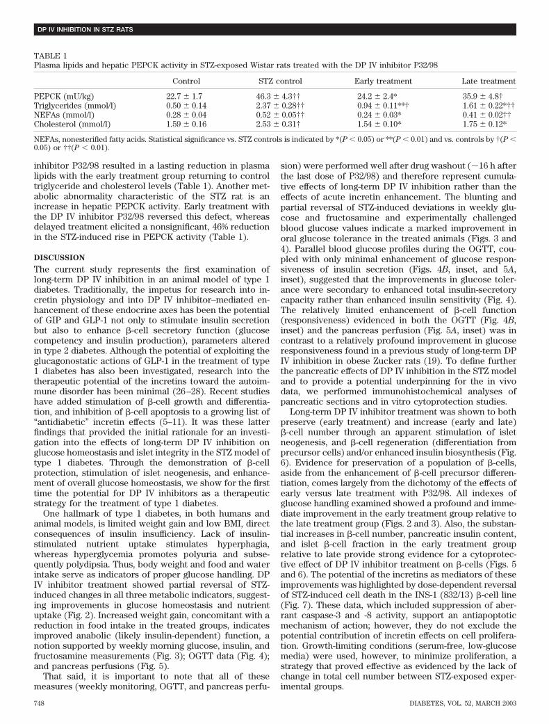
inhibitor P32/98 resulted in a lasting reduction in plasmalipids with the early treatment group returning to controltriglyceride and cholesterol levels (Table 1). Another met-abolic abnormality characteristic of the STZ rat is anincrease in hepatic PEPCK activity. Early treatment withthe DP IV inhibitor P32/98 reversed this defect, whereasdelayed treatment elicited a nonsignificant, 46% reductionin the STZ-induced rise in PEPCK activity (Table 1).
DISCUSSION
The current study represents the first examination oflong-term DP IV inhibition in an animal model of type 1diabetes. Traditionally, the impetus for research into in-cretin physiology and into DP IV inhibitor–mediated en-hancement of these endocrine axes has been the potentialof GIP and GLP-1 not only to stimulate insulin secretionbut also to enhance �-cell secretory function (glucosecompetency and insulin production), parameters alteredin type 2 diabetes. Although the potential of exploiting theglucagonostatic actions of GLP-1 in the treatment of type1 diabetes has also been investigated, research into thetherapeutic potential of the incretins toward the autoim-mune disorder has been minimal (26–28). Recent studieshave added stimulation of �-cell growth and differentia-tion, and inhibition of �-cell apoptosis to a growing list of“antidiabetic” incretin effects (5–11). It was these latterfindings that provided the initial rationale for an investi-gation into the effects of long-term DP IV inhibition onglucose homeostasis and islet integrity in the STZ model oftype 1 diabetes. Through the demonstration of �-cellprotection, stimulation of islet neogenesis, and enhance-ment of overall glucose homeostasis, we show for the firsttime the potential for DP IV inhibitors as a therapeuticstrategy for the treatment of type 1 diabetes.
One hallmark of type 1 diabetes, in both humans andanimal models, is limited weight gain and low BMI, directconsequences of insulin insufficiency. Lack of insulin-stimulated nutrient uptake stimulates hyperphagia,whereas hyperglycemia promotes polyuria and subse-quently polydipsia. Thus, body weight and food and waterintake serve as indicators of proper glucose handling. DPIV inhibitor treatment showed partial reversal of STZ-induced changes in all three metabolic indicators, suggest-ing improvements in glucose homeostasis and nutrientuptake (Fig. 2). Increased weight gain, concomitant with areduction in food intake in the treated groups, indicatesimproved anabolic (likely insulin-dependent) function, anotion supported by weekly morning glucose, insulin, andfructosamine measurements (Fig. 3); OGTT data (Fig. 4);and pancreas perfusions (Fig. 5).
That said, it is important to note that all of thesemeasures (weekly monitoring, OGTT, and pancreas perfu-
sion) were performed well after drug washout (�16 h afterthe last dose of P32/98) and therefore represent cumula-tive effects of long-term DP IV inhibition rather than theeffects of acute incretin enhancement. The blunting andpartial reversal of STZ-induced deviations in weekly glu-cose and fructosamine and experimentally challengedblood glucose values indicate a marked improvement inoral glucose tolerance in the treated animals (Figs. 3 and4). Parallel blood glucose profiles during the OGTT, cou-pled with only minimal enhancement of glucose respon-siveness of insulin secretion (Figs. 4B, inset, and 5A,inset), suggested that the improvements in glucose toler-ance were secondary to enhanced total insulin-secretorycapacity rather than enhanced insulin sensitivity (Fig. 4).The relatively limited enhancement of �-cell function(responsiveness) evidenced in both the OGTT (Fig. 4B,inset) and the pancreas perfusion (Fig. 5A, inset) was incontrast to a relatively profound improvement in glucoseresponsiveness found in a previous study of long-term DPIV inhibition in obese Zucker rats (19). To define furtherthe pancreatic effects of DP IV inhibition in the STZ modeland to provide a potential underpinning for the in vivodata, we performed immunohistochemical analyses ofpancreatic sections and in vitro cytoprotection studies.
Long-term DP IV inhibitor treatment was shown to bothpreserve (early treatment) and increase (early and late)�-cell number through an apparent stimulation of isletneogenesis, and �-cell regeneration (differentiation fromprecursor cells) and/or enhanced insulin biosynthesis (Fig.6). Evidence for preservation of a population of �-cells,aside from the enhancement of �-cell precursor differen-tiation, comes largely from the dichotomy of the effects ofearly versus late treatment with P32/98. All indexes ofglucose handling examined showed a profound and imme-diate improvement in the early treatment group relative tothe late treatment group (Figs. 2 and 3). Also, the substan-tial increases in �-cell number, pancreatic insulin content,and islet �-cell fraction in the early treatment grouprelative to late provide strong evidence for a cytoprotec-tive effect of DP IV inhibitor treatment on �-cells (Figs. 5and 6). The potential of the incretins as mediators of theseimprovements was highlighted by dose-dependent reversalof STZ-induced cell death in the INS-1 (832/13) �-cell line(Fig. 7). These data, which included suppression of aber-rant caspase-3 and -8 activity, support an antiapoptoticmechanism of action; however, they do not exclude thepotential contribution of incretin effects on cell prolifera-tion. Growth-limiting conditions (serum-free, low-glucosemedia) were used, however, to minimize proliferation, astrategy that proved effective as evidenced by the lack ofchange in total cell number between STZ-exposed exper-imental groups.
TABLE 1Plasma lipids and hepatic PEPCK activity in STZ-exposed Wistar rats treated with the DP IV inhibitor P32/98
Control STZ control Early treatment Late treatment
PEPCK (mU/kg) 22.7 1.7 46.3 4.3†† 24.2 2.4* 35.9 4.8†Triglycerides (mmol/l) 0.50 0.14 2.37 0.28†† 0.94 0.11**† 1.61 0.22*††NEFAs (mmol/l) 0.28 0.04 0.52 0.05†† 0.24 0.03* 0.41 0.02††Cholesterol (mmol/l) 1.59 0.16 2.53 0.31† 1.54 0.10* 1.75 0.12*
NEFAs, nonesterified fatty acids. Statistical significance vs. STZ controls is indicated by *(P � 0.05) or **(P � 0.01) and vs. controls by †(P �0.05) or ††(P � 0.01).
DP IV INHIBITION IN STZ RATS
748 DIABETES, VOL. 52, MARCH 2003

Protective effects aside, the immunohistochemical anal-ysis showed evidence for what was likely either enhancedinsulin biosynthesis or �-cell regeneration, as well as forislet neogenesis. Evidence for the latter included a signif-icant increase in islets of the smallest size subsets in bothDP IV inhibitor–treated groups (Fig. 6A). These islets werefound to contain a near-normal �-cell fraction (60–85%)and to be composed almost exclusively of intenselystained, morphologically normal �-cells (INbright). In con-trast, larger islets contained a broad range of insulin-IRcells, suggesting their presence during the STZ insult. Theincrease in number of very small, intensely stained islets inthe treated groups is consistent with previous reports ofGLP-1–stimulated �-cell differentiation, islet budding, andislet neogenesis (7,29,30)
What was the nature of the INbright cells containedwithin the larger, more mature islets? Teitelman andcolleagues (31,32) showed in two separate studies thepresence of a wave of intra-islet �-cell regeneration/differentiation that peaked within the first few days afterhigh-dose STZ administration in CD-1 mice and disap-peared 7–30 days after exposure. These intra-islet �-cellprecursor cells were shown to be of PP-cell or �-cellorigin, staining positive for the pancreatic-duodenum ho-meobox-1 (PDX-1) and either pancreatic polypeptide orsomatostatin (PDX-1/PP and PDX-1/SOM, respec-tively). These PDX-1/PP/IN and PDX-1/SOM/IN
�-cell precursors were found within 24 h of exposure tohigh-dose STZ and were shown to be preferentially in-duced by insulin-dependent normalization of hyperglyce-mia (31,32). If the INbright cells observed in the presentstudy are the mature product of these adult �-precursorcells, then it would seem as though DP IV inhibitortreatment, likely through the enhancement of GIP andGLP-1 levels, enhanced the differentiation of intra-isletprecursors and/or prolonged their survival in a severelyhyperglycemic environment. GLP-1 has been shown toenhance PDX-1 expression in vitro, and in vivo to stimu-late differentiation of �-cell precursors within the islets ofold glucose-intolerant rats (29). Additional data from thecurrent study that support such a conclusion include 1) adoubling of pancreatic insulin content despite a minimalincrease in insulin-IR cell number in the late treatment(versus STZ-control), 2) the finding that INbright cells weredetected in the STZ control animals, and 3) that there wasa 1.3-fold increase in islet �-cell fraction in larger islets(�50 cells in cross-section) in the late treatment groupversus the STZ controls (Fig. 6). Despite the strength ofthese findings, the data are still suggestive, and a confidentdescription of the mechanisms underlying the increases in�-cell and islet number and in pancreatic insulin contentwill require a detailed temporal examination of this modelat various stages of treatment.
Notwithstanding that GIP and GLP-1 are discussed asthe primary mediators of protective and reparative pro-cesses reported above, there remain a number of endoge-nous DP IV substrates whose enhancement might also playa role (33). For instance, glucagon (34), vasoactive intes-tinal polypeptide (35), and pituitary adenylate cyclase-activating polypeptide (35) all are relatively goodsubstrates for DP IV and have been shown to play signif-icant roles in the regulation of �-cell function and devel-
opment (36–38). The contribution of these and otherpeptides toward the present findings is very likely to besignificant. In addition to nonincretin substrate involve-ment, effects of DP IV inhibitors on immune function(including clearance of apoptotic �-cells) cannot be ruledout as contributing factors toward our findings. DP IVinhibitors have been shown to suppress a number ofT-cell–, B-cell–, and NK-cell–specific immune functions(39).
Although the functional relevance of DP IV in theimmune system has yet to be elucidated, its definitiveinvolvement makes the investigation of DP IV inhibition intype 1 diabetes intriguing. The potential for combinedimmunosuppressive and incretin-enhancing effects is aunique therapeutic paradigm. Having described the stimu-latory effects of DP IV inhibitors on �-cell survival andregeneration and on overall glucose tolerance in a type 1model that highlights insulin deficiency, �-cell apoptosis,and �-cell regeneration, this study now warrants an inves-tigation in an autoimmune model of the disease. Althoughboth have their own limitations with respect to autoimmu-nity, the NOD mouse and BB rat would provide suitablemodels for a study into DP IV inhibitor effects on theautoimmune aspect of type 1 diabetes.
In summary, we have shown that treatment of STZ-induced diabetic rats with the DP IV inhibitor P32/98stimulates islet neogenesis and �-cell regeneration, in-creases pancreatic insulin content, and significantly im-proves overall glucose tolerance. The findings set thefoundation for additional study into the application of DPIV inhibitors in the treatment of insulin-dependent diabe-tes (type 1 and late-stage type 2) perhaps filling the voidfor an inexpensive, benign, preventive therapy or to beused in combination with existing therapies (i.e., insulin)after diagnosis.
ACKNOWLEDGMENTS
This study was provided for by grants from the CanadianInstitutes for Health Research (CIHR), the Federal Minis-try of Education and Research (Germany; 0312302), andthe Department of Science and Technology of SachsenAnhalt (9704/00116; H.U.D.). J.A.P., J.A.E., and F.C.L. aregrateful to the CIHR, the Michael Smith Foundation forHealth Research, and the Killam Trust Foundation forscholarship support.
We thank Violet Yuen, Madeleine Speck, and CuilanNian for excellent technical support. As well, we areindebted to Diane T. Finegood for insightful criticismsduring the preparation of this manuscript.
REFERENCES
1. Atkinson M, Eisenbarth G: Type 1 diabetes: new perspectives on diseasepathogenesis and treatment. Lancet 358:221–229, 2001
2. Miyawaki K, Yamada Y, Yano H, Niwa H, Ban N, Ihara Y, Kubota A,Fujimoto S, Kajikawa M, Kuroe A, Tsuda K, Hashimoto H, Yamashita T,Jomori T, Tashiro F, Miyazaki J, Seino Y: Glucose intolerance caused by adefect in the entero-insular axis: a study in gastric inhibitory polypeptidereceptor knockout mice. Proc Natl Acad Sci U S A 96:14843–14847, 1999
3. Pederson RA, Satkunarajah M, McIntosh CH, Scrocchi LA, Flamez D,Schuit F, Drucker DJ, Wheeler MB: Enhanced glucose-dependent insuli-notropic polypeptide secretion and insulinotropic action in glucagon-likepeptide 1 receptor -/- mice. Diabetes 47:1046–1052, 1998
4. Kieffer TJ, Habener JF: The glucagon-like peptides. Endocr Rev 20:876–913, 1999
5. Trumper A, Trumper K, Trusheim H, Arnold R, Goke B, Horsch D:
J.A. POSPISILIK AND ASSOCIATES
DIABETES, VOL. 52, MARCH 2003 749

Glucose-dependent insulinotropic polypeptide is a growth factor for beta(INS-1) cells by pleiotropic signaling. Mol Endocrinol 15:1559–1570, 2001
6. Xu G, Stoffers DA, Habener JF, Bonner-Weir S: Exendin-4 stimulates bothbeta-cell replication and neogenesis, resulting in increased beta-cell massand improved glucose tolerance in diabetic rats. Diabetes 48:2270–2276,1999
7. Stoffers DA, Kieffer TJ, Hussain MA, Drucker DJ, Bonner-Weir S, HabenerJF, Egan JM: Insulinotropic glucagon-like peptide 1 agonists stimulateexpression of homeodomain protein IDX-1 and increase islet size in mousepancreas. Diabetes 49:741–748, 2000
8. Hui H, Wright C, Perfetti R: Glucagon-like peptide 1 induces differentiationof islet duodenal homeobox-1-positive pancreatic ductal cells into insulin-secreting cells. Diabetes 50:785–796, 2001
9. Buteau J, Roduit R, Susini S, Prentki M: Glucagon-like peptide-1 promotesDNA synthesis, activates phosphatidylinositol 3-kinase and increasestranscription factor pancreatic and duodenal homeobox gene 1 (PDX-1)DNA binding activity in beta (INS-1)-cells. Diabetologia 42:856–864, 1999
10. Dachicourt N, Serradas P, Giroix MH, Gangnerau MN, Portha B: Decreasedglucose-induced cAMP and insulin release in islets of diabetic rats:reversal by IBMX, glucagon, GIP. Am J Physiol 271:E725–E732, 1996
11. Holz GG 4th, Kuhtreiber WM, Habener JF: Pancreatic beta-cells arerendered glucose-competent by the insulinotropic hormone glucagon-likepeptide-1(7–37). Nature 361:362–365, 1993
12. Mentlein R, Gallwitz B, Schmidt WE: Dipeptidyl-peptidase IV hydrolysesgastric inhibitory polypeptide, glucagon-like peptide-1(7–36)amide, pep-tide histidine methionine and is responsible for their degradation in humanserum. Eur J Biochem 214:829–835, 1993
13. Kieffer TJ, McIntosh CH, Pederson RA: Degradation of glucose-dependentinsulinotropic polypeptide and truncated glucagon-like peptide 1 in vitroand in vivo by dipeptidyl peptidase IV. Endocrinology 136:3585–3596, 1995
14. Pauly RP, Rosche F, Wermann M, McIntosh CH, Pederson RA, Demuth HU:Investigation of glucose-dependent insulinotropic polypeptide-(1–42) andglucagon-like peptide-1-(7–36) degradation in vitro by dipeptidyl peptidaseIV using matrix-assisted laser desorption/ionization-time of flight massspectrometry. A novel kinetic approach. J Biol Chem 271:23222–23229,1996
15. Deacon CF, Nauck MA, Meier J, Hucking K, Holst JJ: Degradation ofendogenous and exogenous gastric inhibitory polypeptide in healthy and intype 2 diabetic subjects as revealed using a new assay for the intactpeptide. J Clin Endocrinol Metab 85:3575–3581, 2000
16. Hansen L, Deacon CF, Orskov C, Holst JJ: Glucagon-like peptide-1-(7–36)amide is transformed to glucagon-like peptide-1-(9–36)amide by dipep-tidyl peptidase IV in the capillaries supplying the L cells of the porcineintestine. Endocrinology 140:5356–5363, 1999
17. Pauly RP, Demuth HU, Rosche F, Schmidt J, White HA, Lynn F, McIntoshCH, Pederson RA: Improved glucose tolerance in rats treated with thedipeptidyl peptidase IV (CD26) inhibitor Ile-thiazolidide. Metabolism 48:385–389, 1999
18. Pederson RA, White HA, Schlenzig D, Pauly RP, McIntosh CH, Demuth HU:Improved glucose tolerance in Zucker fatty rats by oral administration ofthe dipeptidyl peptidase IV inhibitor isoleucine thiazolidide. Diabetes
47:1253–1258, 199819. Pospisilik J, Stafford S, Demuth H-U, Brownsey R, Parkhouse W, Finegood
D, McIntosh C, Pederson R: Long-term treatment with the DP IV inhibitorP32/98 causes sustained improvements in glucose tolerance, insulin sen-sitivity, hyperinsulinemia and �-cell glucose responsiveness in VDF (fa/fa)Zucker Rats. Diabetes 51:943–950, 2002
20. Ahren B, Simonsson E, Larsson H, Landin-Olsson M, Torgeirsson H,Jansson P-A, Sandqvist M, Bavenholm P, Efendic S, Eriksson J, DickinsonS, Holmes D: Inhibition of dipeptidyl peptidase IV improves metabolic
control over a 4-week study period in type 2 diabetes. Diabetes Care
25:869–875, 200221. Sudre B, Broqua P, White R, Ashworth D, Evans D, Haigh R, Junien J-L,
Aubert M: Chronic inhibition of circulating dipeptidyl peptidase IV by FE999011 delays the occurrence of diabetes in male Zucker Diabetic Fattyrats. Diabetes 51:1461–1469, 2002
22. Reimer MK, Holst JJ, Ahren B: Long-term inhibition of dipeptidyl peptidaseIV improves glucose tolerance and preserves islet function in mice. Eur J
Endocrinol 146:717–727, 200223. Pospisilik JA, Stafford SG, Demuth HU, McIntosh CH, Pederson RA:
Long-term treatment with dipeptidyl peptidase IV inhibitor improveshepatic and peripheral insulin sensitivity in the VDF Zucker rat: aeuglycemic-hyperinsulinemic clamp study. Diabetes 51:2677–2683, 2002
24. Demuth HU: Recent developments in inhibiting cysteine and serineproteases. J Enzyme Inhib 3:249–278, 1990
25. Jia X, Elliott R, Kwok YN, Pederson RA, McIntosh CH: Altered glucosedependence of glucagon-like peptide I(7–36)-induced insulin secretionfrom the Zucker (fa/fa) rat pancreas. Diabetes 44:495–500, 1995
26. Gutniak M, Orskov C, Holst JJ, Ahren B, Efendic S: Antidiabetogenic effectof glucagon-like peptide-1 (7–36)amide in normal subjects and patientswith diabetes mellitus. N Engl J Med 326:1316–1322, 1992
27. Dupre J, Behme MT, Hramiak IM, McFarlane P, Williamson MP, Zabel P,McDonald TJ: Glucagon-like peptide I reduces postprandial glycemicexcursions in IDDM. Diabetes 44:626–630, 1995
28. Creutzfeldt WO, Kleine N, Willms B, Orskov C, Holst JJ, Nauck MA:Glucagonostatic actions and reduction of fasting hyperglycemia by exog-enous glucagon-like peptide I(7–36) amide in type I diabetic patients.Diabetes Care 19:580–586, 1996
29. Perfetti R, Zhou J, Doyle ME, Egan JM: Glucagon-like peptide-1 inducescell proliferation and pancreatic-duodenum homeobox-1 expression andincreases endocrine cell mass in the pancreas of old, glucose-intolerantrats. Endocrinology 141:4600–4605, 2000
30. Tourrel C, Bailbe D, Meile MJ, Kergoat M, Portha B: Glucagon-likepeptide-1 and exendin-4 stimulate beta-cell neogenesis in streptozotocin-treated newborn rats resulting in persistently improved glucose homeosta-sis at adult age. Diabetes 50:1562–1570, 2001
31. Fernandes A, King LC, Guz Y, Stein R, Wright CV, Teitelman G: Differen-tiation of new insulin-producing cells is induced by injury in adultpancreatic islets. Endocrinology 138:1750–1762, 1997
32. Guz Y, Nasir I, Teitelman G: Regeneration of pancreatic beta cells fromintra-islet precursor cells in an experimental model of diabetes. Endocri-
nology 142:4956–4968, 200133. Mentlein R: Dipeptidyl-peptidase IV (CD26): role in the inactivation of
regulatory peptides. Regul Pept 85:9–24, 199934. Pospisilik JA, Hinke SA, Pederson RA, Hoffmann T, Rosche F, Schlenzig D,
Glund K, Heiser U, McIntosh CH, Demuth H: Metabolism of glucagon bydipeptidyl peptidase IV (CD26). Regul Pept 96:133–141, 2001
35. Lambeir A-M, Durinx C, Proost P, Van Damme J, Scharpe S, De Meester I:Kinetic study of the processing by dipeptidyl-peptidase IV/CD26 of neu-ropeptides involved in insulin secretion. FEBS Let 507:327–330, 2001
36. Inagaki N, Kuromi H, Seino S: PACAP/VIP receptors in pancreatic beta-cells: their roles in insulin secretion. Ann N Y Acad Sci 805:44–51;discussion 52–53, 1996
37. Jamen F, Persson K, Bertrand G, Rodriguez-Henche N, Puech R, BockaertJ, Ahren B, Brabet P: PAC1 receptor-deficient mice display impairedinsulinotropic response to glucose and reduced glucose tolerance. J Clin
Invest 105:1307–1315, 200038. Ahren B: Autonomic regulation of islet hormone secretion: implications for
health and disease. Diabetologia 43:393–410, 200039. De Meester I, Korom S, Van Damme J, Scharpe S: CD26, let it cut or cut it
down. Immunol Today 20:367–375, 1999
DP IV INHIBITION IN STZ RATS
750 DIABETES, VOL. 52, MARCH 2003


