Embed Size (px)
Citation preview
Directing Astroglia from the Cerebral Cortex intoSubtype Specific Functional NeuronsChristophe Heinrich1,2, Robert Blum1.¤, Sergio Gascon1,2., Giacomo Masserdotti1., Pratibha Tripathi2,
Rodrigo Sanchez1, Steffen Tiedt1, Timm Schroeder2, Magdalena Gotz1,2,3"*, Benedikt Berninger1,2"*
1 Department of Physiological Genomics, Institute of Physiology, Ludwig-Maximilians University Munich, Munich, Germany, 2 Institute for Stem Cell Research, National
Research Center for Environment and Health, Neuherberg, Germany, 3 Munich Center for Integrated Protein Science CiPSM, Munich, Germany
Abstract
Astroglia from the postnatal cerebral cortex can be reprogrammed in vitro to generate neurons following forced expressionof neurogenic transcription factors, thus opening new avenues towards a potential use of endogenous astroglia for brainrepair. However, in previous attempts astroglia-derived neurons failed to establish functional synapses, a severe limitationtowards functional neurogenesis. It remained therefore also unknown whether neurons derived from reprogrammedastroglia could be directed towards distinct neuronal subtype identities by selective expression of distinct neurogenic fatedeterminants. Here we show that strong and persistent expression of neurogenic fate determinants driven by silencing-resistant retroviral vectors instructs astroglia from the postnatal cortex in vitro to mature into fully functional, synapse-forming neurons. Importantly, the neurotransmitter fate choice of astroglia-derived neurons can be controlled by selectiveexpression of distinct neurogenic transcription factors: forced expression of the dorsal telencephalic fate determinantneurogenin-2 (Neurog2) directs cortical astroglia to generate synapse-forming glutamatergic neurons; in contrast, theventral telencephalic fate determinant Dlx2 induces a GABAergic identity, although the overall efficiency of Dlx2-mediatedneuronal reprogramming is much lower compared to Neurog2, suggesting that cortical astroglia possess a highercompetence to respond to the dorsal telencephalic fate determinant. Interestingly, however, reprogramming of astrogliatowards the generation of GABAergic neurons was greatly facilitated when the astroglial cells were first expanded asneurosphere cells prior to transduction with Dlx2. Importantly, this approach of expansion under neurosphere conditionsand subsequent reprogramming with distinct neurogenic transcription factors can also be extended to reactive astrogliaisolated from the adult injured cerebral cortex, allowing for the selective generation of glutamatergic or GABAergic neurons.These data provide evidence that cortical astroglia can undergo a conversion across cell lineages by forced expression of asingle neurogenic transcription factor, stably generating fully differentiated neurons. Moreover, neuronal reprogramming ofastroglia is not restricted to postnatal stages but can also be achieved from terminally differentiated astroglia of the adultcerebral cortex following injury-induced reactivation.
Citation: Heinrich C, Blum R, Gascon S, Masserdotti G, Tripathi P, et al. (2010) Directing Astroglia from the Cerebral Cortex into Subtype Specific FunctionalNeurons. PLoS Biol 8(5): e1000373. doi:10.1371/journal.pbio.1000373
Academic Editor: Ronald D. G. McKay, National Institutes of Health, United States of America
Received May 26, 2009; Accepted April 12, 2010; Published May 18, 2010
Copyright: � 2010 Heinrich et al. This is an open-access article distributed under the terms of the Creative Commons Attribution License, which permitsunrestricted use, distribution, and reproduction in any medium, provided the original author and source are credited.
Funding: This work was supported by grants from the Deutsche Forschungsgemeinschaft [http://www.dfg.de/] to BB and MG (BE 4182/1-3 and GO 640/9-1) andto TS (SCHR 1142/1-1), the Bavarian State Ministry of Sciences, Research, and the Arts (ForNeuroCell)[http://www.bayfor.org/de/geschaeftsbereiche/forschungsverbuende/welt-des-lebens/forneurocell.html] to BB and MG, and the Exzellenzcluster 114 Munich Center for Integrated Protein Science Munich(CIPSM) [http://www.cipsm.de/en/index.html], the SFB 596 (http://www.sfb596.med.uni-muenchen.de/index.html), the SFB 870 (http://www.sfb870.mcn.uni-muenchen.de/index.html), and European Transcriptome, Regulome & Cellular Commitment Consortium (EUTRACC) [http://www.eutracc.eu/] to MG. The fundershad no role in study design, data collection and analysis, decision to publish, or preparation of the manuscript.
Competing Interests: The authors have declared that no competing interests exist.
Abbreviations: AP, action potential; DPI, days post infection; GFP, green fluorescent protein; Mash1, mammalian achaete-schute homolog 1; Neurog2,neurogenin-2; vGaT, vesicular GABA transporter; vGluT1, vesicular glutamate transporter 1
* E-mail: [email protected] (MG); [email protected] (BB)
. These authors contributed equally to this work.
" These authors are joint senior authors on this work.
¤ Current address: Institute for Clinical Neurobiology, Julius-Maximilians University Wurzburg, Wurzburg, Germany
Introduction
While exerting diverse functions within the brain parenchyma
[1], astroglia are remarkable in that they also function as neural
stem or progenitor cells in specific regions of the postnatal and
adult brain [2], such as the ventricular subependymal zone [3] and
the subgranular zone of the hippocampus [4,5]. This raises the
possibility that even astroglia from non-neurogenic regions such as
the cerebral cortex may be reprogrammed towards neurogenesis
when provided with the appropriate transcriptional cues. Indeed,
we could previously show that astroglia from the early postnatal
cerebral cortex can be reprogrammed in vitro towards the
generation of neurons capable of action potential (AP) firing by
a single transcription factor, such as Pax6 or its target, the pro-
neural transcription factor neurogenin-2 (Neurog2) [6,7]. These
findings may open interesting avenues towards the potential
activation of endogenous astroglia for neuronal repair of injured
brain tissue.
PLoS Biology | www.plosbiology.org 1 May 2010 | Volume 8 | Issue 5 | e1000373
However, several major obstacles remained to be overcome to
fully exploit the potential of reprogrammed astroglia as an
endogenous cellular source for neuronal repair. Firstly, reprogram-
ming of astroglia towards neurons remained incomplete as the
astroglia-derived neurons failed to establish a functional presynaptic
output [7], an obvious hurdle towards functional repair that
requires participation in a neural network. Secondly, given the lack
of functional presynaptic output, we could not determine the
neuronal subtype generated by the reprogrammed astroglial cells
[7]. This raises the conceptual concern of whether neurons derived
from astroglial cells in a given brain region may be restricted
towards the generation of a respective neuronal subtype. During
development of the forebrain in rodents, stem/progenitor cells in
the dorsal telencephalon generate exclusively excitatory glutama-
tergic neurons, directed by Pax6 and Neurog1/2 [8–10], while
stem/progenitor cells in the ventral telencephalon give rise
primarily to inhibitory GABAergic neurons, governed by the fate
determinants mammalian achaete-schute homolog 1 (Mash1)
[11,12] and Dlx1/2 [13]. Region-specific fate restriction also seems
to apply for adult neural stem cells that are intrinsically specified
towards the generation of distinct neuronal subtypes [14]. This
implies that despite their multipotent nature in regard to generating
different glial cell types and neurons, the subtype identity of the
neurons generated from these stem cells is predetermined (see also
[15]) and is not altered following transplantation [14]. This raises
the important question of whether neuronal reprogramming of
astroglia derived from the cerebral cortex, a region derived from the
dorsal telencephalon, may be restricted towards the generation of
glutamatergic neurons, or whether this region-specific bias could be
overcome by forced expression of the appropriate neurogenic fate
determinants. Such an ability to generate both glutamatergic and
GABAergic neurons from astroglia may be a crucial step towards
restoring a damaged or imbalanced neuronal network.
Towards this end, we first aimed at a more potent neuronal
reprogramming by inducing higher and more persistent expression
of neurogenic fate determinants in astroglial cells. This allowed us
not only to obtain fully functional neurons that also establish
synapses from astroglial cells in vitro but also to demonstrate that
distinct neurogenic transcription factors, such as on the one hand
Neurog2 and on the other Dlx2 alone or in combination with
Mash1, can indeed instruct the selective generation of different
neuronal subtypes, such as glutamatergic and GABAergic
neurons, respectively. Moreover, we found that the reprogram-
ming efficiency of postnatal cortical astroglia towards GABAergic
neurons by Dlx2 could be enhanced by first expanding the
astroglial cells under neurosphere conditions prior to forced
expression of Dlx2. Given that following brain injury reactive
astroglia from the adult cerebral cortex de-differentiate, resume
proliferation, and can give rise to self-renewing neurospheres in
vitro [16], we finally show that neuronal reprogramming and
subtype specification are not restricted to postnatal stages but can
also be achieved from adult cortical astroglia responding to injury.
Results
Postnatal Cortical Astroglia Reprogrammed by ForcedExpression of Neurog2 Give Rise to Synapse-FormingGlutamatergic Neurons
Failure to establish a functional presynaptic compartment by
astroglia-derived neurons may be due to an incomplete repro-
gramming [7]. Here, we hypothesized that stronger and more
persistent expression of neurogenic fate determinants may be a
prerequisite for a more complete reprogramming of astroglia
towards synapse-forming neurons. We have previously shown that
expression levels from LTR (long terminal repeat)-driven MMLV
(Moloney Murine Leukemia Virus)-derived retroviral constructs,
which we employed in previous studies, are only about 2–3-fold of
the endogenous expression [6,17]. Moreover, these viral vectors are
prone to silencing [18] and we observed a severe decrease in
Neurog2 or green fluorescent protein (GFP) reporter expression
already 7–14 d after transduction [7,19]. Thus, in order to
overcome the limitations in synaptogenesis of neurons derived
from reprogrammed astroglia, we examined the effect of stronger
and more persistent expression of Neurog2 on neuronal repro-
gramming of astroglia from the cerebral cortex. We therefore
subcloned Neurog2 into a self-inactivating retroviral vector driving
gene expression under the control of a chicken beta-actin promoter
(pCAG) optimized for long-term expression over months in the
adult mouse brain [20]. Astroglia cultures were prepared from
postnatal day 5–7 (P5–P7) cerebral cortex as described previously
[7] and 1 wk later cells were passaged and subsequently transduced
with a retroviral vector encoding Neurog2 and DsRed (pCAG-
Neurog2-IRES-DsRed) or with a control virus encoding DsRed
only (pCAG-IRES-DsRed). Consistent with a stronger and more
persistent expression driven by the pCAG promoter, high levels of
Neurog2 and DsRed protein were still detected at 5–6 wk following
retroviral transduction of cortical astroglia (unpublished data).
In agreement with our previous observation on the high
efficiency of neurogenesis from astroglia following forced Neurog2
expression, the vast majority of Neurog2-transduced astroglia had
differentiated into bIII tubulin-positive, GFAP-negative neurons
after 10 d in culture (Figure S1A and S1A’; 70.2%66.3% at
9.863.1 days post-infection (DPI), 5 independent experiments,
n = 1,022 DsRed-positive cells counted), in contrast to control
retrovirus transduced cells (1.8%61.8% of bIII tubulin-positive
cells at 7.361.0 DPI, 3 independent experiments, n = 3,235 DsRed-
positive cells counted). Time-lapse video microscopy revealed that
the initial conversion of astroglia into neurons requires approxi-
mately 4 d, confirming previous results [7], and can occur at high
efficiency (Video S1). By 2–3 wk post-transduction, neurons derived
from Neurog2-transduced astroglia had acquired MAP2 immuno-
reactivity, indicative for dendritic maturation (Figure 1A and 1B).
Most strikingly, immunostaining for the vesicular glutamate
Author Summary
The brain consists of two major cell types: neurons, whichtransmit information, and glial cells, which support andprotect neurons. Interestingly, evidence suggests that someglial cells, including astroglia, can be directly converted intoneurons by specific proteins, a transformation that may aidin the functional repair of damaged brain tissue. However,in order for the repaired brain areas to function properly,it is important that astroglia be directed into appropriateneuronal subclasses. In this study, we show that non-neurogenic astroglia from the cerebral cortex can bereprogrammed in vitro using just a single transcriptionfactor to yield fully functional excitatory or inhibitoryneurons. We achieved this result through forced expressionof the same transcription factors that instruct the genesis ofthese distinct neuronal subtypes during embryonic forebraindevelopment. Moreover we demonstrate that reactiveastroglia isolated from the adult cortex after local injurycan be reprogrammed into synapse-forming excitatory orinhibitory neurons following a similar strategy. Our findingsprovide evidence that endogenous glial cells may prove apromising strategy for replacing neurons that have degen-erated due to trauma or disease.
Reprogramming Astroglia into Functional Neurons
PLoS Biology | www.plosbiology.org 2 May 2010 | Volume 8 | Issue 5 | e1000373
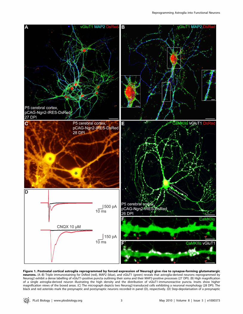
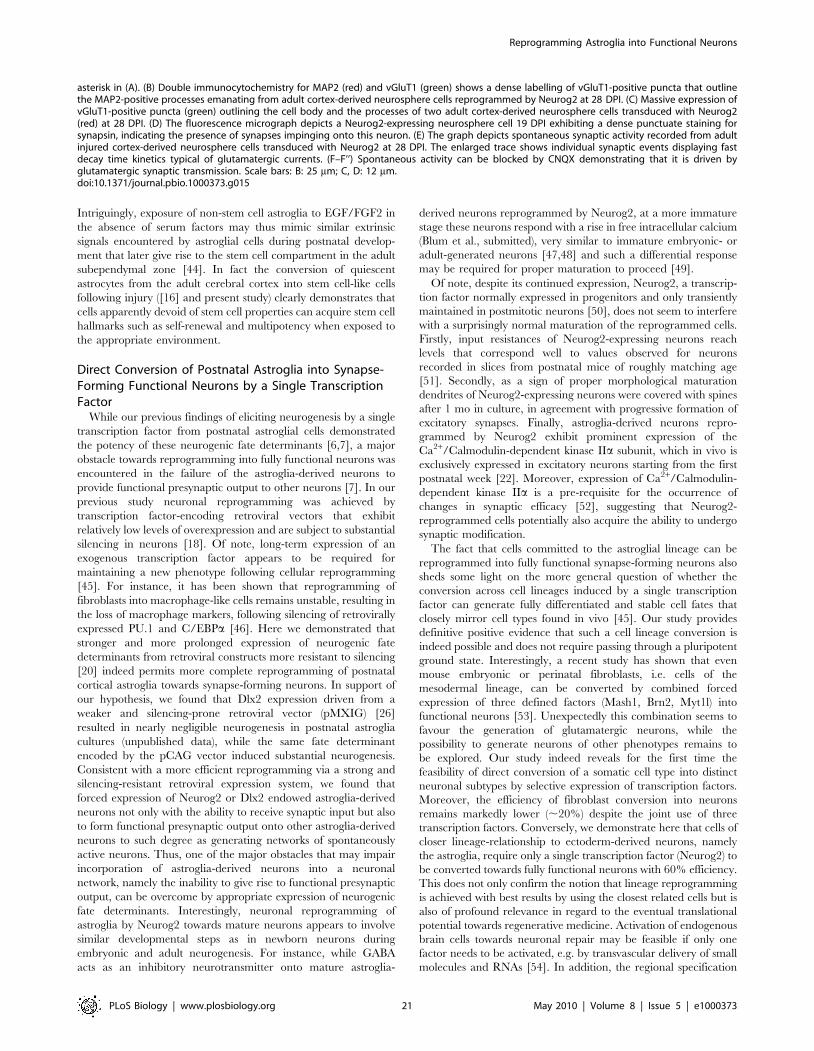
Figure 1. Postnatal cortical astroglia reprogrammed by forced expression of Neurog2 give rise to synapse-forming glutamatergicneurons. (A–B) Triple immunostaining for DsRed (red), MAP2 (blue), and vGluT1 (green) reveals that astroglia-derived neurons reprogrammed byNeurog2 exhibit a dense labelling of vGluT1-positive puncta outlining their soma and their MAP2-positive processes (27 DPI). (B) High magnificationof a single astroglia-derived neuron illustrating the high density and the distribution of vGluT1-immunoreactive puncta. Insets show highermagnification views of the boxed areas. (C) The micrograph depicts two Neurog2-transduced cells exhibiting a neuronal morphology (28 DPI). Theblack and red asterisks mark the presynaptic and postsynaptic neurons recorded in panel (D), respectively. (D) Step-depolarisation of a presynaptic
Reprogramming Astroglia into Functional Neurons
PLoS Biology | www.plosbiology.org 3 May 2010 | Volume 8 | Issue 5 | e1000373
transporter 1 (vGluT1), present in synaptic vesicles within
presynaptic terminals of glutamatergic neurons, revealed that the
vast majority of astroglia-derived neurons exhibited a dense
labelling with vGluT1-positive puncta outlining their soma and
their MAP2-positive processes 4 wk post-infection with Neurog2
(Figure 1A and 1B, 85.4%65.0% of DsRed-positive neurons at
26.362.2 DPI, n = 3 independent experiments, n = 170 DsRed-
positive neurons counted). This was in pronounced contrast to the
virtual absence of such staining upon transduction with the LTR-
driven construct (pCLIG-Neurog2) as described previously [7] and
also no vGluT1 immunoreactivity could be detected in astroglial
cultures transduced with the control vector (unpublished data).
Thus, these data suggest that astroglia reprogrammed with the
pCAG-Neurog2-containing retroviral vector acquire a glutamater-
gic phenotype forming presynaptic specializations.
As vGluT1 immunoreactivity does however not allow to ascertain
the neurotransmitter identity of an individual labelled neuron, as the
vGluT1-positive puncta may arise from other neurons in the next set
of experiments we assessed with single and pair electrophysiological
recordings whether astroglial cells reprogrammed by Neurog2 indeed
give rise to functional glutamatergic autapses or synapses after a
period of 14–32 DPI. As shown in Figure 1D, suprathreshold step-
depolarisation of a DsRed-positive neuron (i.e. presynaptic neuron,
black asterisk, Figure 1C) resulted in an autaptic response in the
stimulated neuron and an inward current in a nearby DsRed-positive
neuron with a short delay typical of a monosynaptic connection
(i.e. postsynaptic neuron, red asterisk, Figure 1C). In addition, the
AMPA/kainate glutamate receptor antagonist CNQX completely
abolished both the autaptic and the synaptic current, demonstrating
the glutamatergic nature of the presynaptic neuron (Figure 1D).
Among all the Neurog2-transduced astroglia-derived neurons
recorded (n = 36, average age of cells: 24.660.9 DPI), 58.3%
exhibited either glutamatergic autaptic connections onto themselves
or glutamatergic synapses onto nearby neurons (Figure S2A). In none
of the recordings from neurons derived from Neurog2-transduced
astroglia was a GABAergic connection observed (Figure S2A). In
accordance, cultures transduced with Neurog2 encoding retrovirus
were devoid of any vesicular GABA transporter (vGaT) immunore-
activity (unpublished data). Thus, these data provide evidence that
Neurog2 does not only induce a generic neuronal fate in postnatal
astroglia but selectively elicits differentiation along the glutamatergic
lineage, in exclusion of GABAergic neurogenesis. Consistent with the
specification of postnatal astroglia towards a glutamatergic identity,
forced expression of Neurog2 resulted in the induction of the T-box
transcription factors Tbr2 (Figure S1B and S1B’) in 20.7%61.9% of
the DsRed-positive cells at 4 DPI (n = 4 coverslips, n = 634 DsRed-
positive cells counted) and Tbr1 (48.2% of DsRed-positive neurons at
7 DPI, n = 112 DsRed/bIII tubulin-double positive cells counted;
Figure S1C and S1C’) as shown previously [7], hence of two well
characterised hallmarks of glutamatergic neurogenesis [21]. More-
over, by 4 wk of forced Neurog2 expression, astroglia-derived
neurons expressed high levels of the forebrain glutamatergic
neuron specific Ca2+/Calmodulin dependent kinase subunit
IIa [22], accumulating at dendritic spine-like structures which were
typically in opposition of vGluT1-positive presynaptic terminals
(Figure 1E–1F’).
Consistent with the development of excitatory networks in
Neurog2-reprogrammed astroglia cultures, we also observed the
emergence of self-driven synaptic activity, resulting eventually in the
occurrence of barrages of synaptic currents (Figure 2A). To monitor
such self-driven activity, we performed calcium imaging experi-
ments of neurons derived from Neurog2-reprogrammed astroglia.
Figure 2B illustrates two neurons that exhibited spontaneous,
recurrent, and synchronous Ca2+ transients (Figure 2B–2B’’). These
Ca2+ transients were completely abolished in the presence of
CNQX (Figure 2B’’). The majority of the DsRed-positive neurons
that we analysed (63.8%, n = 47 imaged neurons, 3 independent
experiments) exhibited Ca2+ transients at 14–43 d after transduc-
tion with Neurog2, thus indicating the high degree of incorporation
of Neurog2-transduced astroglia into excitatory neuronal networks.
These data clearly demonstrate that forced expression of Neurog2
driven by the pCAG retroviral vector is sufficient to instruct
postnatal cortical astroglia to generate fully functional synapse-
forming glutamatergic neurons.
Genetic Fate Mapping Demonstrates Reprogramming ofPostnatal Cortical Astroglia into Glutamatergic Neurons
In order to ascertain the astroglial nature of the cells that gave
rise to functional glutamatergic synapses following reprogramming
by Neurog2, we took advantage of a transgenic mouse line in
which GFP expression can be induced in astroglia and is
maintained in their progeny. Heterozygous mice in which the
expression of a tamoxifen-inducible Cre recombinase is driven by
the astroglia specific L-glutamate/L-aspartate transporter promot-
er (GLAST::CreERT2) [23] were crossed to a reporter mouse line
(Z/EG) [24] to generate double heterozygous mutants (GLAST::-
CreERT2/Z/EG) that were used in the present study. Cre-
mediated recombination of the reporter locus was induced via
tamoxifen administration from postnatal day 2 (P2) until sacrifice
(P5–P7). Astroglia cultures were prepared as described above and,
1 wk later, cells were passaged onto glass coverslips. The vast
majority of GFP reporter-positive cells were immunoreactive for
GFAP (98.7%60.7%) at 1 d after plating, with few cells being
positive for the oligodendroglial markers NG2/O4 (1.2%60.7%)
and none (0.1%60.1%) for the neuronal marker bIII tubulin
(Figure 3A; n = 3 independent experiments, n = 1,560 GFP-positive
cells counted). These data indicate that, under our culture
conditions, most reporter-positive cells at the time of transduction
possess an astroglial identity. These cells largely remain within
their astroglial lineage (86.9%612.7% of GFP-positive cells
expressing GFAP) when analysed at later stages (Figure 3A’;
n = 4 independent experiments, n = 1,363 GFP-positive cells
counted; 9–21 d following plating). We noted, however, a slight
increase in the number of NG2/O4-positive cells (13.0%612.8%),
likely due to the expansion of few reporter-positive clones of
oligodendrocyte precursors. Also at later stages reporter-positive
cells did not give rise to bIII tubulin-positive neurons
(0.1%60.1%; Figure 3A’).
To determine the identity of fate-mapped astroglial cells
following retroviral transduction, we performed immunostaining
for GFP (identifying cells of astroglial origin), DsRed (identifying
transduced cells), and either bIII tubulin, MAP2, or GFAP
neuron evokes both an autaptic (upper red trace) and a monosynaptic response (delay 2 ms) in a nearby postsynaptic neuron (lower red trace). Boththe synaptic and the autaptic responses were completely abolished in the presence of the AMPA/kainate receptor antagonist CNQX (10 mM; blacktraces), demonstrating the glutamatergic nature of the presynaptic neuron. (E) Triple immunostaining for DsRed (red), CaMKIIa (green), and vGluT1(white). (F–F’) High magnification views of the area boxed in (E) showing dendritic spines (red arrowheads) as revealed by CaMKIIa immunoreactivity(F), which are covered by vGluT1-positive puncta (F’, red arrowheads), suggestive of sites of synaptic contact. Scale bars: A: 40 mm; B: 20 mm; insets inB: 5 mm; E: 26 mm; F–F’: 5 mm.doi:10.1371/journal.pbio.1000373.g001
Reprogramming Astroglia into Functional Neurons
PLoS Biology | www.plosbiology.org 4 May 2010 | Volume 8 | Issue 5 | e1000373
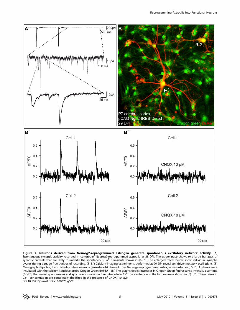
Figure 2. Neurons derived from Neurog2-reprogrammed astroglia generate spontaneous excitatory network activity. (A)Spontaneous synaptic activity recorded in cultures of Neurog2-reprogrammed astroglia at 28 DPI. The upper trace shows two large barrages ofsynaptic currents that are likely to underlie the spontaneous Ca2+ transients shown in (B–B’’). The enlarged traces below show individual synapticevents during barrage-free periods of recording. (B–B’’) Calcium imaging experiments performed at 29 DPI reveal self-driven network oscillations. (B)Micrograph depicting two DsRed-positive neurons (arrowheads) derived from Neurog2-reprogrammed astroglia recorded in (B’–B’’). Cultures wereincubated with the calcium-sensitive probe Oregon Green BAPTA1. (B’) The graphs depict increases in Oregon Green fluorescence intensity over time(DF/F0) that reveal spontaneous and synchronous raises in free intracellular Ca2+ concentration in the two neurons shown in (B). (B’’) These raises inCa2+ concentration are completely abolished in the presence of CNQX (10 mM).doi:10.1371/journal.pbio.1000373.g002
Reprogramming Astroglia into Functional Neurons
PLoS Biology | www.plosbiology.org 5 May 2010 | Volume 8 | Issue 5 | e1000373
Reprogramming Astroglia into Functional Neurons
PLoS Biology | www.plosbiology.org 6 May 2010 | Volume 8 | Issue 5 | e1000373
(identifying neuronal and astroglial cells, respectively). Notably,
the stochastic infection of the subset of genetically recombined cells
results in a limited number of double-targeted cells. When cultures
of adherent astroglia were transduced with the control retrovirus
encoding DsRed only, fate-mapped astroglial cells co-expressing
GFP and DsRed remained in the glial lineage, as revealed by their
astroglial morphology and GFAP expression 1 mo after transduc-
tion (Figure 3B–3B’’). In sharp contrast, when cultures of
tamoxifen-induced astroglia were transduced with the new
retrovirus encoding Neurog2 and DsRed, most GFP/DsRed-
double-positive fate-mapped astroglia were reprogrammed into
neurons expressing the neuronal markers bIII tubulin and MAP2
(67.3%612.7% among GFP/DsRed-double positive cells at
8.061.0 DPI, n = 3 independent experiments, n = 217 double-
positive cells counted; Figure 3C–3C’’). Single cell tracking of
GFP-reporter positive cells following Neurog2-transduction al-
lowed the direct visualisation of the glia-to-neuron conversion of
fate-mapped cells over the time course of 5 d (Figure 3D and 3D’;
Video S1 and Video S2).
Perforated patch clamp recordings of these fate-mapped
astroglia-derived cells reprogrammed by Neurog2 revealed their
functional neuronal identity as these cells fired APs following step-
current injection in current clamp (n = 8; Figure 4A–4C). In the
next set of experiments, we assessed whether neurons derived from
fate-mapped astroglia could give rise to functional glutamatergic
autapses (Figure 4D–4I). Step-depolarisation of GFP/DsRed-
double-positive neurons at 0.05 Hz evoked a sequence of both
autaptic and polysynaptic components (2 out of 8 cells recorded)
consistent with the excitatory nature of the recorded neurons
(average age of the cells: 18.162.2 DPI; Figure 4D–4I, insets),
while at higher stimulation frequency (1 Hz) the autaptic
component with a short decay time typical of glutamatergic
synaptic transmission [25] could be observed in isolation (Figure 4F
and 4I). Consistent with their glutamatergic nature, fate-mapped
astroglia reprogrammed by forced expression of Neurog2 also
exhibited a dense labelling of vGluT1-positive puncta (Figure 4J
and 4K). These data clearly demonstrate that Neurog2 instructs
fate-mapped astroglia from the postnatal cerebral cortex to
acquire a glutamatergic identity.
Cell Division Is Not a Sine Qua Non Condition forReprogramming of Astroglia by Neurog2
Given that our reprogramming strategy is based on retrovirally
mediated expression of neurogenic fate determinants, only cells
undergoing cell division will be targeted. In order to examine
whether cell division is required for fate conversion to occur, we
assessed whether neuronal reprogramming can be also achieved
when the Neurog2 and DsRed encoding plasmid is delivered to
the postnatal astroglia by transfection, i.e. a gene transfer strategy
which does not select for dividing cells, and tracked single
transfected cells by time-lapse video microscopy. Transfection with
the Neurog2 encoding plasmid resulted in a similar degree of
reprogramming after 4 d (14 cells out of 17, Figure 5) as obtained
after retroviral transduction. Of note, in four cases neurons were
generated directly from single astrocytes without a prior cell
division (Figure 5 and Video S3). Thus direct lineage conversion
can occur in the absence of cell division.
Dlx2 Directs Postnatal Cortical Astroglia towardsAcquiring a GABAergic Identity
Based on our finding that forced expression of Neurog2 can
selectively drive cortical astroglia towards the generation of
functional and synaptically integrated glutamatergic neurons, we
next asked whether cortical astroglia may also be directed towards
distinct neuronal subtypes. In particular, we asked whether
neuronal fate determinants known to instruct the genesis of
GABAergic neurons during embryonic development may be
sufficient to exert a similar effect on postnatal astroglia. As the
homeobox transcription factor Dlx2 is one of the key factors
involved in GABAergic neuron specification in the developing
ventral telencephalon [13] and in adult neurogenesis [26], we
examined whether forced expression of Dlx2 is also sufficient to
induce a neuronal and possibly a GABAergic fate in cortical
astroglia. To test this, astroglia cultures from P5–P7 cortex of
C57BL/6J or GLAST::CreERT2/Z/EG mice were transduced
with the same high-expressing retrovirus encoding in this case
Dlx2 and DsRed (pCAG-Dlx2-IRES-DsRed), and cells were
immunostained for GFP (to identify cells of astroglial origin),
DsRed (to identify Dlx2-transduced cells), and the neuronal
markers bIII tubulin or MAP2 after various differentiation time
periods in culture.
Upon forced expression of Dlx2, a substantial number of
postnatal cortical astroglia were redirected towards a neuronal
identity as revealed by bIII tubulin or MAP2 expression
(35.9%613.0% at 10.762.0 DPI, n = 3 independent experiments,
n = 392 DsRed-positive cells counted). Notably, however, the
efficacy of neurogenesis elicited by Dlx2 was significantly lower
than the one elicited by Neurog2 (see above). Fate-mapping
analysis confirmed the astroglial nature of the cells reprogrammed
by forced expression of Dlx2 as observed 22 DPI (Figure 6A–6A’’).
Next, to confirm the neuronal identity of the astroglia-derived
cells, we performed patch clamp recordings. All the cells
expressing Dlx2 and exhibiting a neuronal morphology, that we
recorded, were capable of AP firing in response to step current
injection (n = 33). In particular, this also held true for GFP-positive
neurons originating from fate-mapped astroglia that had been
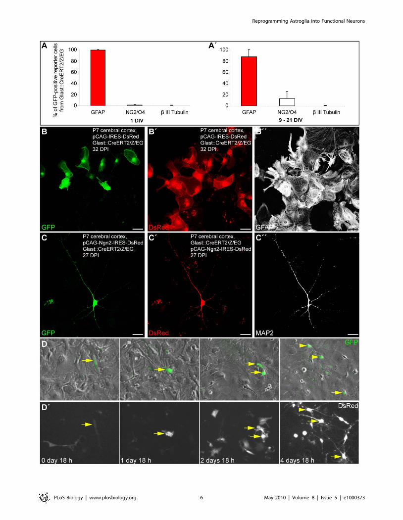
Figure 3. Fate-mapped astroglia from the postnatal cortex can be reprogrammed to generate neurons following forced expressionof Neurog2. (A–A’) The histograms show the percentage of GFP reporter-positive cells from GLAST::CreERT2/Z/EG mice immunoreactive for theastroglial marker GFAP, oligodendroglial markers NG2/O4, and the neuronal marker bIII tubulin 1 d after plating (A) and 9–21 d after plating (A’),respectively. (B–B’’) Cortical astroglia transduced with control retrovirus remain in the glial lineage. (B) The micrograph depicts GFP-positive, fate-mapped astroglia derived from postnatally induced GLAST::CreERT2/Z/EG mice. (B’–B’’) Micrographs of the same field of view as shown in (B) showingthat fate-mapped astroglia transduced with a control retrovirus encoding DsRed only (pCAG-IRES-DsRed) exhibit a glial morphology and expressGFAP (B’’). (C) Representative example of a GFP-positive neuron, i.e. derived from a fate-mapped astroglia, prepared from the postnatal cortex oftamoxifen-induced GLAST::CreERT2/Z/EG mice. (C’) DsRed expression in the same cell indicating forced expression of Neurog2. (C’’) The sameastroglia-derived neuron as shown in (C) and (C’) is immunoreactive for the mature neuronal marker MAP2 (27 DPI). (D–D’) Direct visualisation ofNeurog2-induced reprogramming of fate-mapped astroglia by time-lapse video microscopy. (D) Sequence of bright field images, overlaid with GFPreporter fluorescence, depicting the same field of view over the time course of 5 d as indicated in the corresponding panels below in (D’). The arrowpoints to a reporter-positive cell that divided once giving rise to two daughter cells that subsequently underwent reprogramming into neurons. Thearrowhead points to another fate-mapped reprogrammed cell that entered the field of view at a later time point. (D’) Corresponding sequence ofDsRed fluorescence images taken at the same time points as the bright field images shown in (D). Note the expression of DsRed (encoded byNeurog2-IRES-DsRed) in the fate-mapped cell (arrow). Note that besides fate-mapped cells, several other Neurog2-transduced cells also becamereprogrammed into neurons. Scale bars: B–B’’: 57 mm; C–C’’: 26 mm.doi:10.1371/journal.pbio.1000373.g003
Reprogramming Astroglia into Functional Neurons
PLoS Biology | www.plosbiology.org 7 May 2010 | Volume 8 | Issue 5 | e1000373
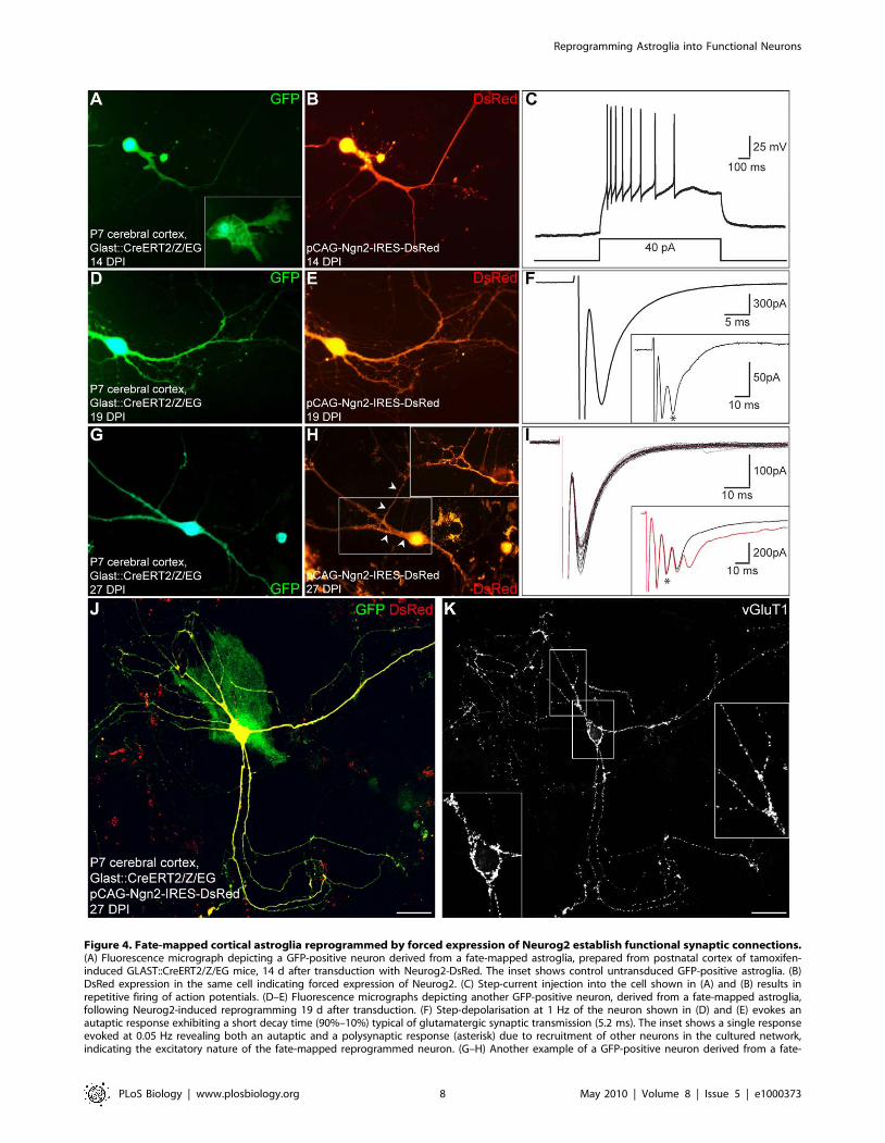
Figure 4. Fate-mapped cortical astroglia reprogrammed by forced expression of Neurog2 establish functional synaptic connections.(A) Fluorescence micrograph depicting a GFP-positive neuron derived from a fate-mapped astroglia, prepared from postnatal cortex of tamoxifen-induced GLAST::CreERT2/Z/EG mice, 14 d after transduction with Neurog2-DsRed. The inset shows control untransduced GFP-positive astroglia. (B)DsRed expression in the same cell indicating forced expression of Neurog2. (C) Step-current injection into the cell shown in (A) and (B) results inrepetitive firing of action potentials. (D–E) Fluorescence micrographs depicting another GFP-positive neuron, derived from a fate-mapped astroglia,following Neurog2-induced reprogramming 19 d after transduction. (F) Step-depolarisation at 1 Hz of the neuron shown in (D) and (E) evokes anautaptic response exhibiting a short decay time (90%–10%) typical of glutamatergic synaptic transmission (5.2 ms). The inset shows a single responseevoked at 0.05 Hz revealing both an autaptic and a polysynaptic response (asterisk) due to recruitment of other neurons in the cultured network,indicating the excitatory nature of the fate-mapped reprogrammed neuron. (G–H) Another example of a GFP-positive neuron derived from a fate-
Reprogramming Astroglia into Functional Neurons
PLoS Biology | www.plosbiology.org 8 May 2010 | Volume 8 | Issue 5 | e1000373
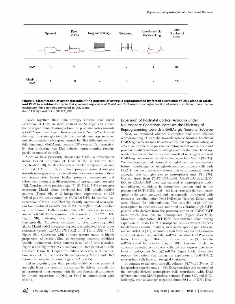
reprogrammed by Dlx2 (n = 9; Figure 7A–7C’’). Notably, neurons
derived from Dlx2-transduced astroglia exhibited distinct firing
patterns, with most of them revealing immature characteristics
(single to few spikes, 22 out of 30 cells recorded) (Figure 7A’’ and
Figure 8). The eight remainder cells exhibited firing patterns
which could be classified into three categories [27,28], namely
regular, stuttering, and low-threshold burst spiking (Figure 7B’’–
7C’’ and Figure 8), suggestive of the maturation into distinct types
of non-fast spiking interneurons [29]. Similarly, the majority of
fate-mapped astroglia reprogrammed by Dlx2 (7 out of 9 cells
recorded) exhibited immature firing patterns (Figure 7A–7A’’),
while 2 out of 9 fate-mapped cells developed more mature
interneuron-like firing (Figure 7B–7C’’). Consistent with the
generation of regular- and burst-spiking interneurons [29], we
observed calretinin immunoreactivity in a small subset of the Dlx2-
expressing cells (Figure 6C), while no parvalbumin immunoreac-
tivity could be detected.
The predominant appearance of immature firing patterns,
however, suggests an overall hampered maturation of Dlx2-
reprogrammed astroglia. Accordingly, astroglia-derived neurons
reprogrammed by Dlx2 exhibited much higher input resistance
values than Neurog2-derived neurons after the same time in culture
(Figure S2B and S2C; 2,319.26187.9 MV at 26.061.4 DPI (n = 26)
versus 1,111.86211.1 MV at 23.761.5 DPI (n = 20), Dlx2 versus
Neurog2, respectively). Surprisingly, the high input resistance of
Dlx2-expressing neurons did not decrease but even slightly increased
with time in culture (Figure S2B; 2,786.46440.3 MV at
35.961.2 DPI (n = 7)), while in the case of Neurog2-reprogrammed
astroglia input resistance decreased over time (608.66125.0 MV at
26.861.2 DPI; n = 14; Figure S2C). Taken together, these data
show that some postnatal cortical astroglia can be redirected by
forced expression of Dlx2 towards a neuronal identity; however, in
sharp contrast to the progressive maturation of Neurog2-transduced
cells, most of the astroglia-derived neurons reprogrammed by Dlx2
remain in a rather immature state, suggesting a comparatively less
efficient reprogramming by Dlx2.
Next we assessed whether some of these relatively immature
neurons derived from Dlx2-reprogrammed astroglia may neverthe-
less establish functional autaptic or synaptic connections. We first
performed immunocytochemistry for vGluT1 and for vGaT, the
latter known to be expressed in synaptic vesicles located in
presynaptic terminals of GABAergic neurons. In sharp contrast to
reprogramming by Neurog2, astroglia-derived neurons repro-
grammed by Dlx2 were devoid of vGluT1 immunoreactivity
(unpublished data), but some of them (33.7%63.6% at
22.060.6 DPI, n = 339 DsRed-positive neurons counted; n = 3
mapped astroglia following Neurog2-induced reprogramming 27 DPI. The inset in (H) shows an enlargement of the boxed area revealing a DsRed-positive axon (arrowheads) originating from another DsRed-positive neuron and meandering along the dendrites and the soma of the recorded cell,after the patch pipette had been withdrawn and the cell died. (I) Step-depolarisation of the neuron shown in (G) and (H) evokes a sequence of bothautaptic and polysynaptic responses. The black traces show individual autaptic responses observed in isolation when evoked at 1 Hz to eliminatepolysynaptic components (the average trace is shown in red). The autapse exhibited a short decay time (90%–10%) typical of glutamatergic synaptictransmission (7.7 ms). The inset shows two individual responses evoked at 0.05 Hz (black and red traces) revealing both autaptic and polysynapticresponses (asterisk) due to recruitment of other neurons in the cultured network, indicating the excitatory nature of the fate-mapped reprogrammedneuron. (J–K) Expression of the vesicular glutamate transporter 1 (vGluT1) by fate-mapped postnatal astroglia reprogrammed by Neurog2. (J)Micrograph depicting a GFP and DsRed double-positive neuron derived from a fate-mapped astroglia, 27 d after transduction with a retrovirus encodingNeurog2 and DsRed. Note the DsRed-negative fate-mapped astrocyte. (K) Immunocytochemistry for vGluT1 reveals that the fate-mapped astroglia-derived neuron reprogrammed by Neurog2 exhibits a dense labelling of vGluT1-positive puncta outlining its cell body and dendrites. The insets showhigher magnification views of the soma and dendrites illustrating the punctuate staining for vGluT1. Scale bars: J-K: 30 mm.doi:10.1371/journal.pbio.1000373.g004
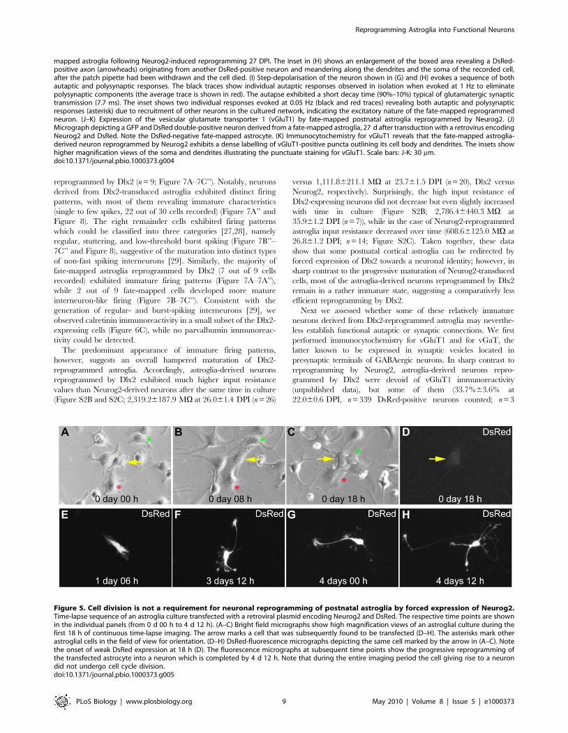
Figure 5. Cell division is not a requirement for neuronal reprogramming of postnatal astroglia by forced expression of Neurog2.Time-lapse sequence of an astroglia culture transfected with a retroviral plasmid encoding Neurog2 and DsRed. The respective time points are shownin the individual panels (from 0 d 00 h to 4 d 12 h). (A–C) Bright field micrographs show high magnification views of an astroglial culture during thefirst 18 h of continuous time-lapse imaging. The arrow marks a cell that was subsequently found to be transfected (D–H). The asterisks mark otherastroglial cells in the field of view for orientation. (D–H) DsRed-fluorescence micrographs depicting the same cell marked by the arrow in (A–C). Notethe onset of weak DsRed expression at 18 h (D). The fluorescence micrographs at subsequent time points show the progressive reprogramming ofthe transfected astrocyte into a neuron which is completed by 4 d 12 h. Note that during the entire imaging period the cell giving rise to a neurondid not undergo cell cycle division.doi:10.1371/journal.pbio.1000373.g005
Reprogramming Astroglia into Functional Neurons
PLoS Biology | www.plosbiology.org 9 May 2010 | Volume 8 | Issue 5 | e1000373
independent experiments) were found to exhibit labelling of vGaT-
positive puncta outlining both their soma and their processes
(Figure 6D). In addition, a small subset of DsRed-positive neurons
exhibited GAD67 immunoreactivity (Figure 6B and 6B’). These
findings therefore suggest that Dlx2 induces a GABAergic identity in
the reprogrammed astroglia. Consistent with an interneuron
phenotype, we could also record in 9 out of 33 neurons spontaneous
synaptic currents exhibiting a slow decay time, characteristic of
GABAergic synaptic events (Figure 7D and 7E). Finally, in few
cases, step-depolarisation in voltage clamp evoked an autaptic
response of the stimulated neuron (6.1% of the DsRed-positive
neurons recorded, n = 33, age of the cells: 26.961.4 DPI; Figures 7F
and S2A). In accordance with the above data, these autaptic
responses exhibited slow decay time kinetics and were abolished by
the GABAA receptor antagonist bicuculline (Figure 7F), thus
demonstrating the GABAergic nature of these autapses.
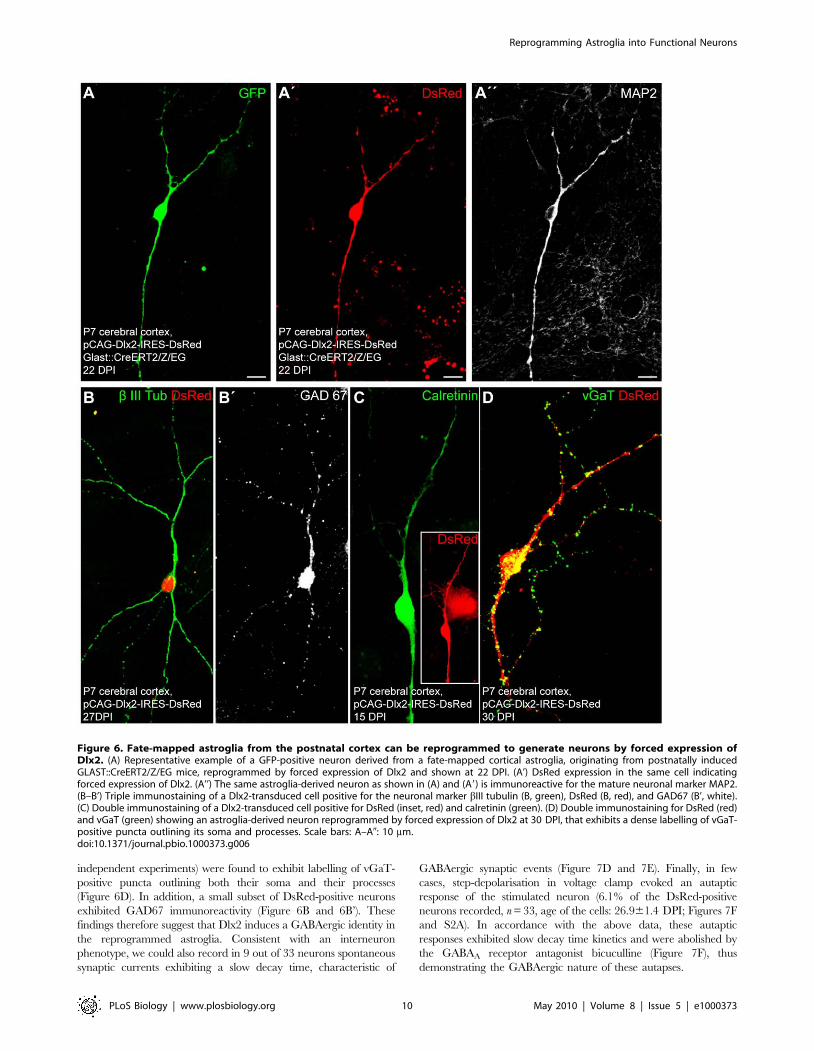
Figure 6. Fate-mapped astroglia from the postnatal cortex can be reprogrammed to generate neurons by forced expression ofDlx2. (A) Representative example of a GFP-positive neuron derived from a fate-mapped cortical astroglia, originating from postnatally inducedGLAST::CreERT2/Z/EG mice, reprogrammed by forced expression of Dlx2 and shown at 22 DPI. (A’) DsRed expression in the same cell indicatingforced expression of Dlx2. (A’’) The same astroglia-derived neuron as shown in (A) and (A9) is immunoreactive for the mature neuronal marker MAP2.(B–B’) Triple immunostaining of a Dlx2-transduced cell positive for the neuronal marker bIII tubulin (B, green), DsRed (B, red), and GAD67 (B’, white).(C) Double immunostaining of a Dlx2-transduced cell positive for DsRed (inset, red) and calretinin (green). (D) Double immunostaining for DsRed (red)and vGaT (green) showing an astroglia-derived neuron reprogrammed by forced expression of Dlx2 at 30 DPI, that exhibits a dense labelling of vGaT-positive puncta outlining its soma and processes. Scale bars: A–A0: 10 mm.doi:10.1371/journal.pbio.1000373.g006
Reprogramming Astroglia into Functional Neurons
PLoS Biology | www.plosbiology.org 10 May 2010 | Volume 8 | Issue 5 | e1000373
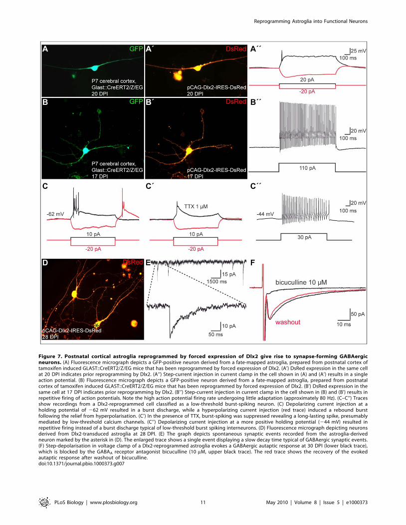
Figure 7. Postnatal cortical astroglia reprogrammed by forced expression of Dlx2 give rise to synapse-forming GABAergicneurons. (A) Fluorescence micrograph depicts a GFP-positive neuron derived from a fate-mapped astroglia, prepared from postnatal cortex oftamoxifen induced GLAST::CreERT2/Z/EG mice that has been reprogrammed by forced expression of Dlx2. (A’) DsRed expression in the same cellat 20 DPI indicates prior reprogramming by Dlx2. (A’’) Step-current injection in current clamp in the cell shown in (A) and (A’) results in a singleaction potential. (B) Fluorescence micrograph depicts a GFP-positive neuron derived from a fate-mapped astroglia, prepared from postnatalcortex of tamoxifen induced GLAST::CreERT2/Z/EG mice that has been reprogrammed by forced expression of Dlx2. (B’) DsRed expression in thesame cell at 17 DPI indicates prior reprogramming by Dlx2. (B’’) Step-current injection in current clamp in the cell shown in (B) and (B’) results inrepetitive firing of action potentials. Note the high action potential firing rate undergoing little adaptation (approximately 80 Hz). (C–C’’) Tracesshow recordings from a Dlx2-reprogrammed cell classified as a low-threshold burst-spiking neuron. (C) Depolarizing current injection at aholding potential of 262 mV resulted in a burst discharge, while a hyperpolarizing current injection (red trace) induced a rebound burstfollowing the relief from hyperpolarisation. (C’) In the presence of TTX, burst-spiking was suppressed revealing a long-lasting spike, presumablymediated by low-threshold calcium channels. (C’’) Depolarizing current injection at a more positive holding potential (244 mV) resulted inrepetitive firing instead of a burst discharge typical of low-threshold burst spiking interneurons. (D) Fluorescence micrograph depicting neuronsderived from Dlx2-transduced astroglia at 28 DPI. (E) The graph depicts spontaneous synaptic events recorded from the astroglia-derivedneuron marked by the asterisk in (D). The enlarged trace shows a single event displaying a slow decay time typical of GABAergic synaptic events.(F) Step-depolarisation in voltage clamp of a Dlx2-reprogrammed astroglia evokes a GABAergic autaptic response at 30 DPI (lower black trace),which is blocked by the GABAA receptor antagonist bicuculline (10 mM, upper black trace). The red trace shows the recovery of the evokedautaptic response after washout of bicuculline.doi:10.1371/journal.pbio.1000373.g007
Reprogramming Astroglia into Functional Neurons
PLoS Biology | www.plosbiology.org 11 May 2010 | Volume 8 | Issue 5 | e1000373
Taken together, these data strongly indicate that forced
expression of Dlx2, in sharp contrast to Neurog2, can induce
the reprogramming of astroglia from the postnatal cortex towards
a GABAergic phenotype. However, whereas Neurog2 redirected
the majority of astroglia towards functional glutamatergic neurons,
only few astroglial cells reprogrammed by Dlx2 differentiated into
fully functional, GABAergic neurons (58% versus 6%, respective-
ly), thus indicating that Dlx2-induced reprogramming remains
partial in most of the cells.
Since we have previously shown that Mash1, a transcription
factor located up-stream of Dlx2 in the interneuron fate
specification [30], the direct targets of which overlap only partially
with that of Mash1 [31], can also reprogram postnatal astroglia
towards neurogenesis [7], we tested whether co-expression of these
two transcription factors further promote neurogenesis and
subsequent interneuron differentiation of reprogrammed astroglia
[32]. Consistent with previous data [7], 33.5%617.8% of astroglia
expressing Mash1 alone developed into bIII tubulin-positive
neurons (Figure 9B, n = 3 independent experiments, n = 226
DsRed-positive cells counted at 10.762.0 DPI). In contrast, co-
expression of Mash1 and Dlx2 significantly augmented neurogen-
esis from postnatal astroglia (93.0%63.1% of bIII tubulin-positive
neurons amongst DsRed-positive cells, n = 3 independent exper-
iments, n = 548 DsRed-positive cells counted at 10.762.0 DPI;
Figure 9B), indicating that these two factors indeed act
synergistically. Moreover, compared to cells expressing Dlx2
alone, Mash1/Dlx2 co-expressing neurons exhibited lower input
resistance values (1,237.56278.8 MV at 18.461.0 DPI; n = 15;
Figure 9C). Consistent with a more mature status, a higher
proportion of Mash1/Dlx2 co-expressing neurons exhibited
specific interneuronal firing patterns (6 out of 15 cells recorded,
Figure 8 and Figure 9A–9A’’) compared to Dlx2 (8 out of 30 cells
recorded, Figure 8). Despite this enhanced degree of differentia-
tion, none of the recorded cells co-expressing Mash1 and Dlx2
showed an autaptic response (Figure S2A, n = 15).
Taken together, our data provide evidence that postnatal
astroglia from the cerebral cortex can be driven towards the
generation of interneurons with distinct functional properties
by forced expression of Dlx2 or Dlx2 in combination with
Mash1.
Expansion of Postnatal Cortical Astroglia underNeurosphere Conditions Increases the Efficiency ofReprogramming towards a GABAergic Neuronal Subtype
Next, we examined whether a complete and more efficient
reprogramming of astroglia towards synapse-forming functional
GABAergic neurons may be achieved by first expanding astroglial
cells as neurospheres in presence of mitogens that on the one hand
promote de-differentiation of astroglia and on the other hand up-
regulate fate determinants normally involved in the generation of
GABAergic neurons in the telencephalon, such as Mash1 [33–35].
We therefore cultured postnatal astroglial cells as neurospheres
before transducing the astroglia-derived neurosphere cells with
Dlx2. It has been previously shown that early postnatal cortical
astroglial cells can give rise to neurospheres until P11 [36].
Cortical tissue from P5–P7 C57BL/6J, GLAST::CreERT2/Z/
EG, or hGFAP-GFP mice was cultured as neurospheres under
non-adherent conditions in serum-free medium and in the
presence of EGF/FGF2, and 1 wk later, astroglia-derived neuro-
sphere cells were passaged and subsequently transduced with
retrovirus encoding either Dlx2-DsRed or Neurog2-DsRed, and
were allowed for differentiation. The astroglial origin of the
neurosphere founder cells was confirmed by culturing single GFP-
positive cells derived from the postnatal cortex of hGFAP-GFP
mice which gave rise to neurospheres (Figure S3A–S3D).
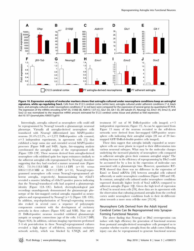
Moreover, quantitative RT-PCR demonstrated that during
expansion in EGF/FGF2 neurosphere cells expressed mRNAs
for different astroglial markers, such as the specific pan-astrocyte
marker Aldh1L1 [37], at similarly high levels as adherent astroglia
after 1 wk in culture, and the mRNA encoding GFAP at even
higher levels (Figure 10A–10E). In contrast, no bIII tubulin
mRNA could be detected (Figure 10F). Likewise, similar to
adherent astroglia neurosphere cells did not express detectable
levels of endogenous Neurog2 mRNA (Figure 10G). These data
support the notion that during the expansion in EGF/FGF2,
neurosphere cells have an astroglial character.
In contrast to adherent astroglia cultures, 94.7%60.3% (n = 3
independent experiments, n = 644 DsRed-positive cells counted) of
the astroglia-derived neurosphere cells transduced with Dlx2
differentiated into MAP2-positive neurons (Figure S4A and S4C).
Strikingly, even at younger stages in culture (20.161.9 DPI), Dlx2-
Figure 8. Classification of action potential firing patterns of astroglia reprogrammed by forced expression of Dlx2 alone or Mash1and Dlx2 in combination. Note that combined expression of Mash1 and Dlx2 results in a higher fraction of neurons exhibiting more matureinterneuron firing patterns compared to Dlx2 alone.doi:10.1371/journal.pbio.1000373.g008
Reprogramming Astroglia into Functional Neurons
PLoS Biology | www.plosbiology.org 12 May 2010 | Volume 8 | Issue 5 | e1000373
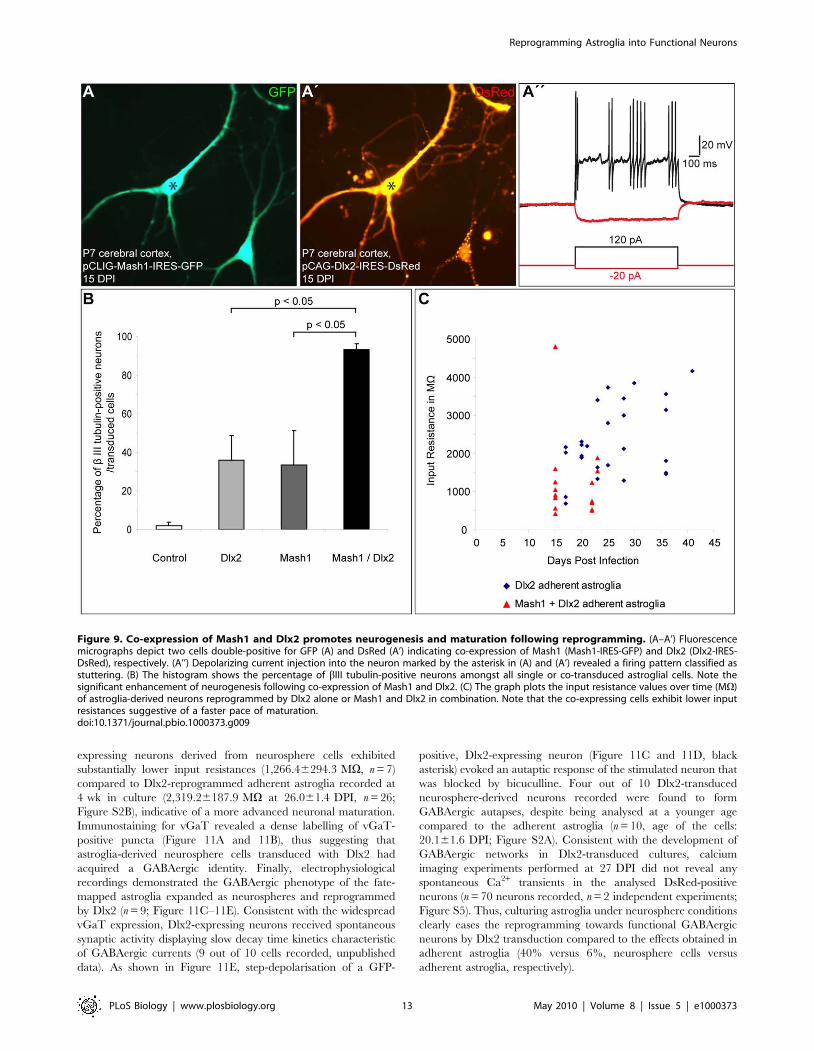
expressing neurons derived from neurosphere cells exhibited
substantially lower input resistances (1,266.46294.3 MV, n = 7)
compared to Dlx2-reprogrammed adherent astroglia recorded at
4 wk in culture (2,319.26187.9 MV at 26.061.4 DPI, n = 26;
Figure S2B), indicative of a more advanced neuronal maturation.
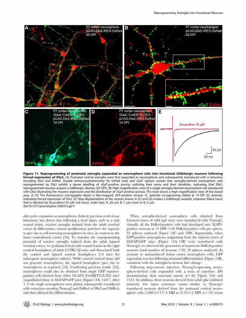
Immunostaining for vGaT revealed a dense labelling of vGaT-
positive puncta (Figure 11A and 11B), thus suggesting that
astroglia-derived neurosphere cells transduced with Dlx2 had
acquired a GABAergic identity. Finally, electrophysiological
recordings demonstrated the GABAergic phenotype of the fate-
mapped astroglia expanded as neurospheres and reprogrammed
by Dlx2 (n = 9; Figure 11C–11E). Consistent with the widespread
vGaT expression, Dlx2-expressing neurons received spontaneous
synaptic activity displaying slow decay time kinetics characteristic
of GABAergic currents (9 out of 10 cells recorded, unpublished
data). As shown in Figure 11E, step-depolarisation of a GFP-
positive, Dlx2-expressing neuron (Figure 11C and 11D, black
asterisk) evoked an autaptic response of the stimulated neuron that
was blocked by bicuculline. Four out of 10 Dlx2-transduced
neurosphere-derived neurons recorded were found to form
GABAergic autapses, despite being analysed at a younger age
compared to the adherent astroglia (n = 10, age of the cells:
20.161.6 DPI; Figure S2A). Consistent with the development of
GABAergic networks in Dlx2-transduced cultures, calcium
imaging experiments performed at 27 DPI did not reveal any
spontaneous Ca2+ transients in the analysed DsRed-positive
neurons (n = 70 neurons recorded, n = 2 independent experiments;
Figure S5). Thus, culturing astroglia under neurosphere conditions
clearly eases the reprogramming towards functional GABAergic
neurons by Dlx2 transduction compared to the effects obtained in
adherent astroglia (40% versus 6%, neurosphere cells versus
adherent astroglia, respectively).
Figure 9. Co-expression of Mash1 and Dlx2 promotes neurogenesis and maturation following reprogramming. (A–A’) Fluorescencemicrographs depict two cells double-positive for GFP (A) and DsRed (A’) indicating co-expression of Mash1 (Mash1-IRES-GFP) and Dlx2 (Dlx2-IRES-DsRed), respectively. (A’’) Depolarizing current injection into the neuron marked by the asterisk in (A) and (A’) revealed a firing pattern classified asstuttering. (B) The histogram shows the percentage of bIII tubulin-positive neurons amongst all single or co-transduced astroglial cells. Note thesignificant enhancement of neurogenesis following co-expression of Mash1 and Dlx2. (C) The graph plots the input resistance values over time (MV)of astroglia-derived neurons reprogrammed by Dlx2 alone or Mash1 and Dlx2 in combination. Note that the co-expressing cells exhibit lower inputresistances suggestive of a faster pace of maturation.doi:10.1371/journal.pbio.1000373.g009
Reprogramming Astroglia into Functional Neurons
PLoS Biology | www.plosbiology.org 13 May 2010 | Volume 8 | Issue 5 | e1000373
Interestingly, astroglia cultured as neurosphere cells could still
be reprogrammed by Neurog2 towards a glutamatergic neuronal
phenotype. Virtually all astroglia-derived neurosphere cells
transduced with Neurog2 differentiated into MAP2-positive
neurons (91.4%62.2%, n = 2,272 DsRed-positive cells counted,
n = 3 independent experiments, in agreement with [7]) that
exhibited a large soma size and extended several MAP2-positive
processes (Figure S4B and S4D). Again, fate-mapping analysis
corroborated the astroglial origin of the reprogrammed cells
(Figure 12D–12F). These neurons derived from astroglia-derived
neurosphere cells exhibited quite low input resistances similarly to
the adherent astroglial cells reprogrammed by Neurog2, therefore
suggesting that they had reached a mature neuronal state (Figure
S2C; 751.96118.8 MV at 15.061.4 DPI (n = 10) versus
608.66125.0 MV at 26.861.2 DPI (n = 14), Neurog2-repro-
grammed neurosphere cells versus Neurog2-reprogrammed ad-
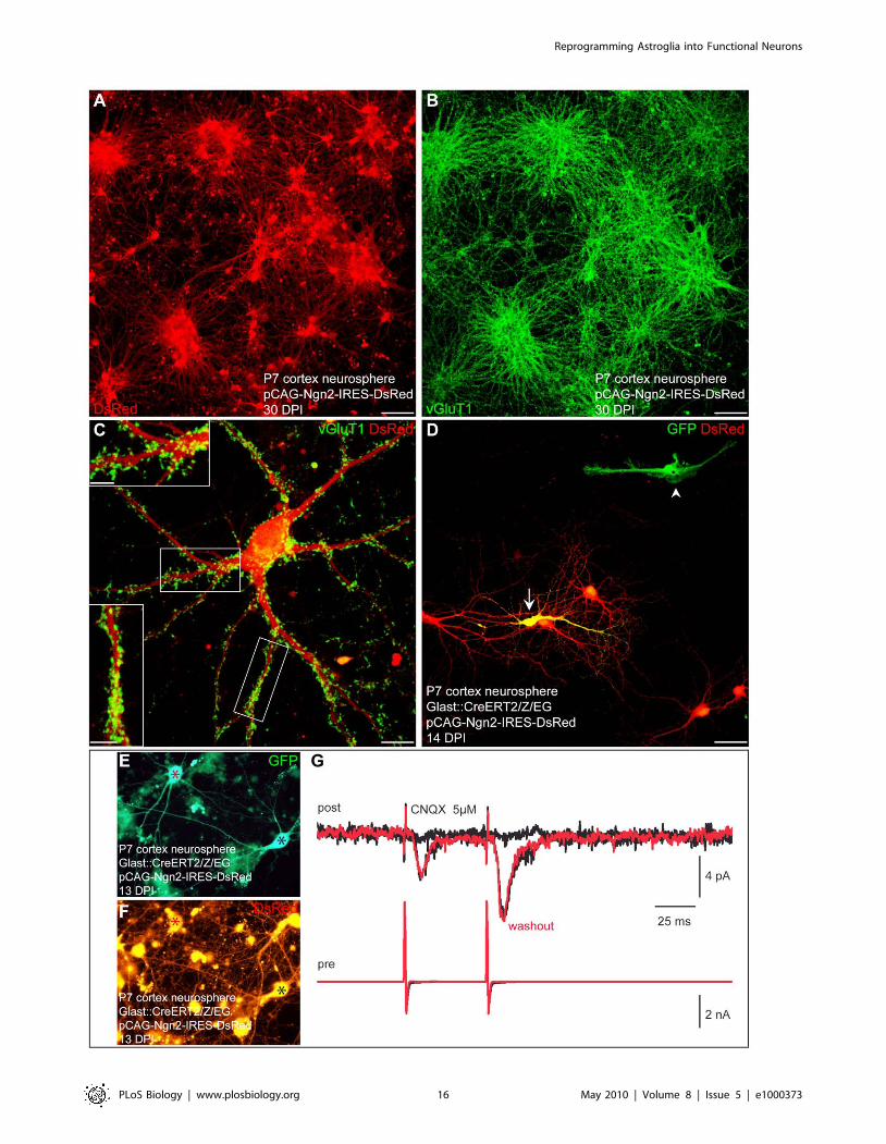
herent astroglia, respectively). Immunostaining for vGluT1
revealed a massive labelling of vGluT1-positive puncta indicating
that the Neurog2-transduced cells had acquired a glutamatergic
identity (Figure 12A–12C). Indeed, electrophysiological pair
recordings unambiguously demonstrated the glutamatergic phe-
notype of the fate-mapped cortical astroglia expanded as neuro-
spheres and reprogrammed by Neurog2 (n = 5) (Figure 12E–12G).
In addition, step-depolarization of Neurog2-expressing neurons
also evoked in several cases a sequence of polysynaptic
components consistent with the development of excitatory
networks in these cultures (Figure S3G and S3H). Nine out of
21 DsRed-positive neurons recorded exhibited glutamatergic
autaptic or synaptic connections (age of the cells: 14.260.7 DPI;
Figure S2A). In addition, calcium imaging experiments performed
3–4 wk post-infection in these Neurog2-reprogrammed cultures
revealed a high degree of self-driven, synchronous excitatory
network activity, which was blocked by CNQX and AP5
treatment (97 out of 98 DsRed-positive cells imaged; n = 3
independent experiments; Figure 13). As can be appreciated from
Figure 13 many of the neurons recruited to the self-driven
networks were derived from fate-mapped GFP-positive neuro-
sphere cells indicating their astroglial origin (28 out of 29 fate-
mapped GFP/DsRed-double-positive cells imaged).
These data suggest that astroglia initially expanded as neuro-
sphere cells are more plastic in regard to their differentiation into
various neuronal subtypes. What may be the molecular changes
underlying the increased plasticity of neurosphere cells compared
to adherent astroglia given their common astroglial origin? The
striking increase in the efficiency of reprogramming by Dlx2 could
be accounted for by a loss in the expression of molecular cues
associated with a glutamatergic bias. However, quantitative RT-
PCR showed that there was no difference in the expression of
Emx1 or Emx2 mRNAs [38] between astroglial cells cultured
adherently or under neurosphere conditions (Figure 10H and 10I).
In contrast, astroglial cells cultured under neurosphere conditions
expressed drastically higher levels of Sox2 mRNA compared to
adherent astroglia (Figure 10J). Given the high level of expression
of Sox2 in neural stem cells [39], these data are in agreement with
the observation that culturing postnatal astroglia from the cerebral
cortex under neurosphere conditions leads to their de-differenti-
ation towards a more stem cell-like state [33,34].
Neurosphere Cells Derived from the Adult InjuredCerebral Cortex Can Be Reprogrammed into Synapse-Forming Functional Neurons
The above finding that Neurog2 or Dlx2 overexpression can
induce with high efficiency the generation of functional neurons
from postnatal astroglia-derived neurosphere cells prompted us to
examine whether reactive astroglia from the adult cortex following
injury can also be reprogrammed to generate functional neurons
Figure 10. Expression analysis of molecular markers shows that astroglia cultured under neurosphere conditions keep an astroglialsignature, while up-regulating Sox2. Cells from the E12.5 cerebral cortex (white bars), astroglia cultured under adherent conditions (7 d, blackbars), and astroglia cultured under neurosphere conditions (7 d, red bars) were compared for the expression of several genes by quantitative RT-PCR.The expression of the mRNAs encoding GFAP (A), S100b (B), Aldh1L1 [37] (C), Glu1 (D), Glt-1 (E), bIII tubulin (F), Neurog2 (G), Emx1 (H), Emx2 (I), andSox2 (J) was normalized to the respective mRNA amount estimated for E12.5 cerebral cortex tissue and plotted as fold expression.doi:10.1371/journal.pbio.1000373.g010
Reprogramming Astroglia into Functional Neurons
PLoS Biology | www.plosbiology.org 14 May 2010 | Volume 8 | Issue 5 | e1000373
after prior expansion as neurospheres. Indeed, previous work of our
laboratory has shown that following a local injury such as a stab
wound lesion, reactive astroglia isolated from the adult cerebral
cortex de-differentiate, resume proliferation, and have the capacity
to give rise to self-renewing neurospheres in vitro, in contrast to the
intact contralateral cortex [16]. To examine the reprogramming
potential of reactive astroglia isolated from the adult injured
cerebral cortex, we performed local stab wound lesions in the right
cortical hemisphere of adult C57BL/6J mice and dissociated both
the control and injured cortical hemispheres 3 d later for
subsequent neurosphere cultures. While control cortical tissue did
not generate neurospheres, the injured hemisphere gave rise to
neurospheres as reported [16]. Confirming previous results [16],
neurospheres could also be obtained from single GFP reporter-
positive cells derived from either GLAST::CreERT2/Z/EG mice
(unpublished data) or hGFAP-GFP mice (Figure 14A–14A’’). After
1–2 wk, single neurospheres were plated, subsequently transduced
with retrovirus encoding Neurog2 and DsRed or Dlx2 and DsRed,
and then allowed for differentiation.
When astroglia-derived neurosphere cells obtained from
lesioned cortex of wild type mice were transduced with Neurog2,
virtually all the DsRed-positive cells had developed into MAP2-
positive neurons at 15 DPI (.50 DsRed-positive cells per sphere;
25 spheres analysed, Figure 14C and 14D). Importantly, when
GFP-positive neurospheres originating from the injured cortex of
hGFAP-GFP mice (Figure 14A–14B) were transduced with
Neurog2, we observed the generation of numerous DsRed-positive
neurons (total number of neurons .50, 10 spheres analysed). In
contrast to untransduced lesion cortex neurosphere cells, GFP
expression was lost following neuronal differentiation (Figure 14B),
consistent with the astroglia-to-neuron fate change.
Following step-current injection, Neurog2-expressing neuro-
sphere-derived cells responded with a train of repetitive APs
demonstrating their neuronal nature (n = 30; Figure 15A and
15A’). In addition, these neurons derived from adult glia exhibited
relatively low input resistance values similar to Neurog2-
transduced neurons derived from the postnatal cortical neuro-
sphere cells (1,009.26177.4 MV at 21.961.2 DPI (n = 19) versus
Figure 11. Reprogramming of postnatal astroglia expanded as neurosphere cells into functional GABAergic neurons followingforced expression of Dlx2. (A) Postnatal cortical astroglia were first expanded as neurospheres and subsequently transduced with a retrovirusencoding Dlx2 and DsRed. Double immunocytochemistry for DsRed (red) and vGaT (green) reveals that astroglia-derived neurosphere cellsreprogrammed by Dlx2 exhibit a dense labelling of vGaT-positive puncta outlining their soma and their dendrites, indicating that Dlx2-reprogrammed neurons acquire a GABAergic identity (30 DPI). (B) High magnification view of a single astroglia-derived neurosphere cell transducedwith Dlx2 illustrating the massive expression and the distribution of vGaT-positive puncta. The inset shows a high magnification view of the boxedarea. (C–D) The fluorescence micrographs depict a fate-mapped, GFP-positive neuron (C, asterisk) co-expressing DsRed at 14 DPI (D, asterisk),indicating forced expression of Dlx2. (E) Step-depolarisation of the neuron shown in (C) and (D) evokes a GABAergic autaptic response (black trace)that is blocked by bicuculline (10 mM, red trace). Scale bars: A: 20 mm; B: 7 mm; Inset in B: 2 mm.doi:10.1371/journal.pbio.1000373.g011
Reprogramming Astroglia into Functional Neurons
PLoS Biology | www.plosbiology.org 15 May 2010 | Volume 8 | Issue 5 | e1000373
Reprogramming Astroglia into Functional Neurons
PLoS Biology | www.plosbiology.org 16 May 2010 | Volume 8 | Issue 5 | e1000373
751.96118.8 MV at 15.061.4 DPI (n = 10), respectively). We
next assessed whether these neurons could establish functional
synaptic connections. Single adult cortex-derived neurospheres
showed a massive immunostaining of vGluT1-positive puncta as
shown 28 DPI, that outlined the dense network of intermingled
MAP2-positive processes (Figure 15B) and the soma of Neurog2-
transduced cells (Figure 15C). Consistent with vGluT1 expression,
immunostaining for the presynaptic protein synapsin also revealed
a dense labelling of synapsin-positive puncta, thus suggesting the
development of synaptic contacts between adult lesioned cortex-
derived neurosphere cells (Figure 15D). Furthermore electrophys-
iological recordings revealed the emergence of CNQX-sensitive
spontaneous synaptic currents in these neurons in accordance with
vGluT1 and synapsin expression (8 out of 30 cells recorded at
22.560.9 DPI; Figure 15E–15F’’). These data indicate that adult
astroglia-derived neurosphere cells transduced with Neurog2
mature into functional glutamatergic neurons.
To examine the extent of plasticity of glial cells derived from the
adult lesioned cortex we also tested as a proof-of-principle
experiment whether adult lesioned cortex-derived neurosphere
cells could also be directed by forced expression of Dlx2 towards
MAP2-expressing neurons (Figure S6A and S6A’). However, Dlx2
reprogrammed neurons were rather few and fragile due to their
rather small soma size, thus hampering extensive electrophysio-
logical analysis. Nevertheless we could record from one cell shown
in Figure S6, where step-depolarisation evoked an autaptic
response that was blocked by bicuculline, indicating the develop-
ment of functional GABAergic connections (Figure S6B and S6B’).
These data show that even adult cells isolated from the injured
cortex and expanded as neurospheres can be instructed by forced
expression of Neurog2 or Dlx2 to generate mature neurons able to
establish functional glutamatergic or GABAergic connections,
respectively.
Discussion
The present study provides four major findings: firstly, it
provides new independent experimental evidence based on genetic
fate-mapping that astroglia from the postnatal cerebral cortex can
be reprogrammed by a single transcription factor into functional
neurons; secondly, we have succeeded in overcoming the previous
limitations in synaptogenesis of neurons derived from postnatal
cortical astroglia by the use of retroviral vectors conveying higher
and more persistent levels of neurogenic fate determinants’
expression [7]; thirdly, based on the transcription factor used for
reprogramming, postnatal astroglia can be directed towards the
generation of glutamatergic and GABAergic neurons, providing
proof-of-principle evidence that selective subtypes can be gener-
ated from the same cells of origin; fourthly, reprogramming
efficiency is further enhanced by prior de-differentiation of the
astroglia as provided by the expansion under neurosphere
conditions. Importantly, using the latter procedure we even
succeeded in reprogramming reactive astroglia after adult brain
injury. Thus, reprogramming of astroglia towards functional
neurons with a single transcription factor is not restricted to
postnatal stages but can also be achieved from astroglia of the
adult cerebral cortex following injury-induced reactivation.
Neuronal Reprogramming of Cells Restricted to theAstroglial Lineage
We have previously shown that postnatal astroglia can be
reprogrammed into neurons [6,7]. However, as the astroglial
origin of the reprogrammed cells is a very important issue, here we
sought to provide new experimental evidence via genetic fate-
mapping of astroglia by using the GLAST::CreERT2/Z/EG
mouse line developed in our laboratory [23]. To ensure the
specificity of this mouse model we showed that virtually all fate-
mapped, i.e. GFP-reporter positive, cells remain in the glial lineage
under our culture conditions, with the vast majority being
identified as astrocytes based on GFAP expression as well as
GLAST (unpublished data) and a minor population as oligoden-
droglial cells, while none of the fate-mapped cells spontaneously
gave rise to neurons (Figure 3A and 3A’). These data support the
notion that the cells cultured under these conditions do not possess
an intrinsic neurogenic potential. This is consistent with the
finding of virtually absent endogenous Neurog2 expression
compared to cortical precursors isolated at the embryonic stage
(Figure 10G) and in agreement with the epigenetic silencing of the
neurogenin-1 and -2 loci at the transition between neurogenic and
astrogliogenic precursors [40]. These data do not rule out the
possibility that in vivo a small subset of astroglial cells can still give
rise to neurons as suggested by hGFAP::CreERT2-mediated fate
mapping showing that neurons can be generated at early postnatal
stages from genetically marked cells [41]. However, so far it could
not be experimentally distinguished whether the postnatal
generated neurons indeed had been derived from astroglia local
to the cerebral cortex or would be derived from astroglial stem
cells in the subependymal zone that had subsequently immigrated
into the cortex [42]. In any case our cultures do not sustain
conditions for the genesis of neurons from astroglia (even in the
presence of EGF/FGF2) without forced expression of neurogenic
fate determinants.
Yet despite generating only glia when adherently grown in
serum containing medium, postnatal astroglia exhibit a remark-
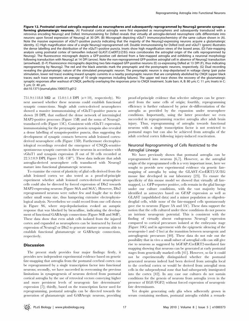
Figure 12. Postnatal cortical astroglia expanded as neurospheres and subsequently reprogrammed by Neurog2 generate synapse-forming glutamatergic neurons. (A) Postnatal cortical astroglia were first expanded as neurospheres and subsequently transduced with aretrovirus encoding Neurog2 and DsRed. Immunostaining for DsRed reveals that virtually all astroglia-derived neurosphere cells differentiate intoneurons upon forced expression of Neurog2 at 30 DPI. (B) Micrograph depicting vGluT1 immunocytochemistry of the same culture shown in (A).Note the massive expression of vGluT1-positive puncta indicating that the majority of the Neurog2-expressing neurons acquire a glutamatergicidentity. (C) High magnification view of a single Neurog2-reprogrammed cell. Double immunostaining for DsRed (red) and vGluT1 (green) illustratesthe dense labelling and the distribution of the vGluT1-positive puncta. Insets show high magnification views of the boxed areas. (D) Fate-mappinganalysis using postnatal cortex of tamoxifen induced GLAST::CreERT2/Z/EG mice corroborates the astroglial origin of the cells reprogrammed byNeurog2. The fluorescence micrograph depicts a GFP-positive cell derived from a fate-mapped astroglia and exhibiting a neuronal morphologyfollowing transduction with Neurog2 at 14 DPI (arrow). Note the non-reprogrammed GFP-positive astroglial cell in absence of Neurog2 transduction(arrowhead). (E–F) Fluorescence micrographs depicting two fate-mapped GFP-positive neurons (E) co-expressing DsRed at 13 DPI (F), thus indicatingreprogramming by Neurog2. The red and the black asterisks mark the presynaptic and the postsynaptic neurons, respectively. (G) Dual recordingrevealing the glutamatergic nature of the stimulated presynaptic neuron. The presynaptic neuron is step-depolarised in voltage clamp (pair pulsestimulation, lower red trace) evoking inward synaptic currents in a nearby postsynaptic neuron that are completely abolished by CNQX (upper blacktraces; each trace represents an average of 10 single responses including failures). The upper red trace shows the recovery of the glutamatergicsynaptic responses after washout of CNQX, perfectly overlaying the responses prior to CNQX treatment. Scale bars: A, B: 80 mm; C: 11 mm; Insets in C:5 mm; D: 40 mm.doi:10.1371/journal.pbio.1000373.g012
Reprogramming Astroglia into Functional Neurons
PLoS Biology | www.plosbiology.org 17 May 2010 | Volume 8 | Issue 5 | e1000373
able degree of plasticity as indicated by the fact that at least some
can give rise to self-renewing, multipotent neurospheres ([36] and
present study). The latter fact could be taken as evidence that early
postnatal astroglia possess stem cell character, particularly in the
light of the notion that cells from the postnatal cerebral cortex may
contribute to the pool of radial/astroglial stem cells within the
dorsal adult subependymal zone [14] (for review see [43]).
However, several lines of evidence argue against a stem cell
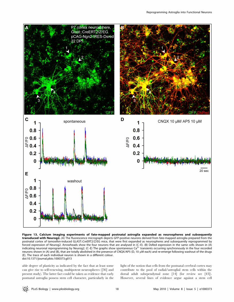
Figure 13. Calcium imaging experiments of fate-mapped postnatal astroglia expanded as neurospheres and subsequentlytransduced with Neurog2. (A) The fluorescence micrograph depicts GFP-positive neurons derived from fate-mapped astroglia prepared from thepostnatal cortex of tamoxifen-induced GLAST::CreERT2/Z/EG mice, that were first expanded as neurospheres and subsequently reprogrammed byforced expression of Neurog2. Arrowheads show the four neurons that are analysed in (C–E). (B) DsRed expression in the same cells shown in (A)indicating neuronal reprogramming by Neurog2. (C–E) The graphs show spontaneous Ca2+ transients occurring synchronously in the four recordedneurons shown in (A) and (B), that are totally abolished in the presence of CNQX/AP5 (D, 10 mM each) and re-emerge following washout of the drugs(E). The trace of each individual neuron is shown in a different colour.doi:10.1371/journal.pbio.1000373.g013
Reprogramming Astroglia into Functional Neurons
PLoS Biology | www.plosbiology.org 18 May 2010 | Volume 8 | Issue 5 | e1000373
character of the astroglial cells studied here. Firstly, neither wild-
type nor genetically fate-mapped astroglia spontaneously give rise
to neurons, which is inconsistent with the stem cell defining
hallmark of multipotency (Figure 3A–3B’’). Moreover, the large
number of neurons generated following forced expression of
Neurog2 argues against the possibility that the successfully
reprogrammed cells derive from rare stem cells within this culture
(Video S1). Secondly, while Dlx2 very efficiently directs adult
neural stem cells in vitro towards neurogenesis [26], the
responsiveness of adherent astroglia is much more limited,
suggesting a reduced susceptibility to Dlx2 transcriptional activity.
The third line of evidence is based on the striking difference in
Sox2 mRNA levels following expansion of the postnatal astroglia
as neurosphere cells. Sox2 is a transcription factor well known to
play a key role in neural stem cell self-renewal [39]; thus the
massive up-regulation of Sox2 following exposure of astroglia to
neurosphere culture conditions suggests that these cells undergo
de-differentiation eventually acquiring indeed stem cell properties,
while the comparatively lower levels of Sox2 in the adherent
cultures would be in agreement with their non-stem cell character.
Consistent with the higher degree of plasticity characterizing
neural stem cells, the efficiency of reprogramming of astroglia-
derived neurosphere cells by Dlx2 was found to reach levels
comparable to bona fide neural stem cells (Figure S4; [26]).
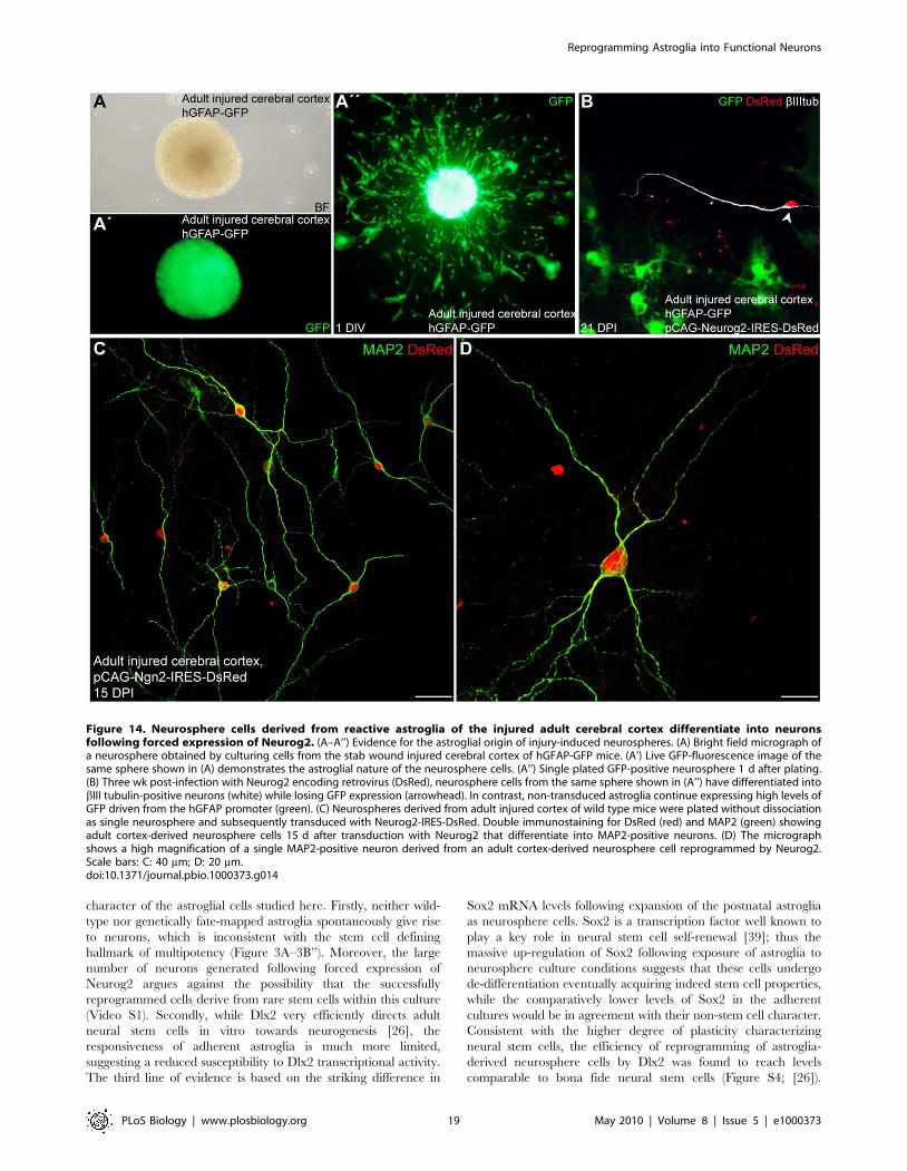
Figure 14. Neurosphere cells derived from reactive astroglia of the injured adult cerebral cortex differentiate into neuronsfollowing forced expression of Neurog2. (A–A’’) Evidence for the astroglial origin of injury-induced neurospheres. (A) Bright field micrograph ofa neurosphere obtained by culturing cells from the stab wound injured cerebral cortex of hGFAP-GFP mice. (A’) Live GFP-fluorescence image of thesame sphere shown in (A) demonstrates the astroglial nature of the neurosphere cells. (A’’) Single plated GFP-positive neurosphere 1 d after plating.(B) Three wk post-infection with Neurog2 encoding retrovirus (DsRed), neurosphere cells from the same sphere shown in (A’’) have differentiated intobIII tubulin-positive neurons (white) while losing GFP expression (arrowhead). In contrast, non-transduced astroglia continue expressing high levels ofGFP driven from the hGFAP promoter (green). (C) Neurospheres derived from adult injured cortex of wild type mice were plated without dissociationas single neurosphere and subsequently transduced with Neurog2-IRES-DsRed. Double immunostaining for DsRed (red) and MAP2 (green) showingadult cortex-derived neurosphere cells 15 d after transduction with Neurog2 that differentiate into MAP2-positive neurons. (D) The micrographshows a high magnification of a single MAP2-positive neuron derived from an adult cortex-derived neurosphere cell reprogrammed by Neurog2.Scale bars: C: 40 mm; D: 20 mm.doi:10.1371/journal.pbio.1000373.g014
Reprogramming Astroglia into Functional Neurons
PLoS Biology | www.plosbiology.org 19 May 2010 | Volume 8 | Issue 5 | e1000373
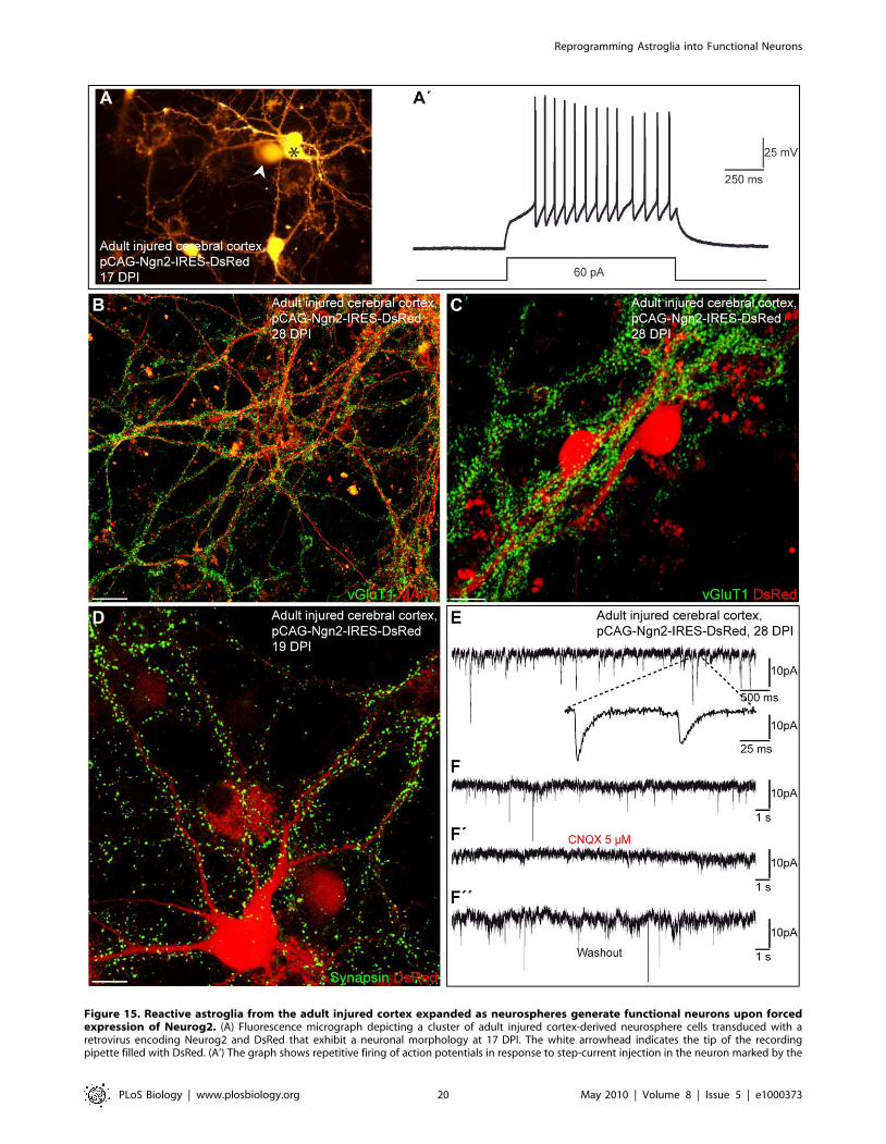
Figure 15. Reactive astroglia from the adult injured cortex expanded as neurospheres generate functional neurons upon forcedexpression of Neurog2. (A) Fluorescence micrograph depicting a cluster of adult injured cortex-derived neurosphere cells transduced with aretrovirus encoding Neurog2 and DsRed that exhibit a neuronal morphology at 17 DPI. The white arrowhead indicates the tip of the recordingpipette filled with DsRed. (A’) The graph shows repetitive firing of action potentials in response to step-current injection in the neuron marked by the
Reprogramming Astroglia into Functional Neurons
PLoS Biology | www.plosbiology.org 20 May 2010 | Volume 8 | Issue 5 | e1000373
Intriguingly, exposure of non-stem cell astroglia to EGF/FGF2 in
the absence of serum factors may thus mimic similar extrinsic
signals encountered by astroglial cells during postnatal develop-
ment that later give rise to the stem cell compartment in the adult
subependymal zone [44]. In fact the conversion of quiescent
astrocytes from the adult cerebral cortex into stem cell-like cells
following injury ([16] and present study) clearly demonstrates that
cells apparently devoid of stem cell properties can acquire stem cell
hallmarks such as self-renewal and multipotency when exposed to
the appropriate environment.
Direct Conversion of Postnatal Astroglia into Synapse-Forming Functional Neurons by a Single TranscriptionFactor
While our previous findings of eliciting neurogenesis by a single
transcription factor from postnatal astroglial cells demonstrated
the potency of these neurogenic fate determinants [6,7], a major
obstacle towards reprogramming into fully functional neurons was
encountered in the failure of the astroglia-derived neurons to
provide functional presynaptic output to other neurons [7]. In our
previous study neuronal reprogramming was achieved by
transcription factor-encoding retroviral vectors that exhibit
relatively low levels of overexpression and are subject to substantial
silencing in neurons [18]. Of note, long-term expression of an
exogenous transcription factor appears to be required for
maintaining a new phenotype following cellular reprogramming
[45]. For instance, it has been shown that reprogramming of
fibroblasts into macrophage-like cells remains unstable, resulting in
the loss of macrophage markers, following silencing of retrovirally
expressed PU.1 and C/EBPa [46]. Here we demonstrated that
stronger and more prolonged expression of neurogenic fate
determinants from retroviral constructs more resistant to silencing
[20] indeed permits more complete reprogramming of postnatal
cortical astroglia towards synapse-forming neurons. In support of
our hypothesis, we found that Dlx2 expression driven from a
weaker and silencing-prone retroviral vector (pMXIG) [26]
resulted in nearly negligible neurogenesis in postnatal astroglia
cultures (unpublished data), while the same fate determinant
encoded by the pCAG vector induced substantial neurogenesis.
Consistent with a more efficient reprogramming via a strong and
silencing-resistant retroviral expression system, we found that
forced expression of Neurog2 or Dlx2 endowed astroglia-derived
neurons not only with the ability to receive synaptic input but also
to form functional presynaptic output onto other astroglia-derived
neurons to such degree as generating networks of spontaneously
active neurons. Thus, one of the major obstacles that may impair
incorporation of astroglia-derived neurons into a neuronal
network, namely the inability to give rise to functional presynaptic
output, can be overcome by appropriate expression of neurogenic
fate determinants. Interestingly, neuronal reprogramming of
astroglia by Neurog2 towards mature neurons appears to involve
similar developmental steps as in newborn neurons during
embryonic and adult neurogenesis. For instance, while GABA
acts as an inhibitory neurotransmitter onto mature astroglia-
derived neurons reprogrammed by Neurog2, at a more immature
stage these neurons respond with a rise in free intracellular calcium
(Blum et al., submitted), very similar to immature embryonic- or
adult-generated neurons [47,48] and such a differential response
may be required for proper maturation to proceed [49].
Of note, despite its continued expression, Neurog2, a transcrip-
tion factor normally expressed in progenitors and only transiently
maintained in postmitotic neurons [50], does not seem to interfere
with a surprisingly normal maturation of the reprogrammed cells.
Firstly, input resistances of Neurog2-expressing neurons reach
levels that correspond well to values observed for neurons
recorded in slices from postnatal mice of roughly matching age
[51]. Secondly, as a sign of proper morphological maturation
dendrites of Neurog2-expressing neurons were covered with spines
after 1 mo in culture, in agreement with progressive formation of
excitatory synapses. Finally, astroglia-derived neurons repro-
grammed by Neurog2 exhibit prominent expression of the
Ca2+/Calmodulin-dependent kinase IIa subunit, which in vivo is
exclusively expressed in excitatory neurons starting from the first
postnatal week [22]. Moreover, expression of Ca2+/Calmodulin-
dependent kinase IIa is a pre-requisite for the occurrence of
changes in synaptic efficacy [52], suggesting that Neurog2-
reprogrammed cells potentially also acquire the ability to undergo
synaptic modification.
The fact that cells committed to the astroglial lineage can be
reprogrammed into fully functional synapse-forming neurons also
sheds some light on the more general question of whether the
conversion across cell lineages induced by a single transcription
factor can generate fully differentiated and stable cell fates that
closely mirror cell types found in vivo [45]. Our study provides
definitive positive evidence that such a cell lineage conversion is
indeed possible and does not require passing through a pluripotent
ground state. Interestingly, a recent study has shown that even
mouse embryonic or perinatal fibroblasts, i.e. cells of the
mesodermal lineage, can be converted by combined forced
expression of three defined factors (Mash1, Brn2, Myt1l) into
functional neurons [53]. Unexpectedly this combination seems to
favour the generation of glutamatergic neurons, while the
possibility to generate neurons of other phenotypes remains to
be explored. Our study indeed reveals for the first time the
feasibility of direct conversion of a somatic cell type into distinct
neuronal subtypes by selective expression of transcription factors.
Moreover, the efficiency of fibroblast conversion into neurons
remains markedly lower (,20%) despite the joint use of three
transcription factors. Conversely, we demonstrate here that cells of
closer lineage-relationship to ectoderm-derived neurons, namely
the astroglia, require only a single transcription factor (Neurog2) to
be converted towards fully functional neurons with 60% efficiency.
This does not only confirm the notion that lineage reprogramming
is achieved with best results by using the closest related cells but is
also of profound relevance in regard to the eventual translational
potential towards regenerative medicine. Activation of endogenous
brain cells towards neuronal repair may be feasible if only one
factor needs to be activated, e.g. by transvascular delivery of small
molecules and RNAs [54]. In addition, the regional specification
asterisk in (A). (B) Double immunocytochemistry for MAP2 (red) and vGluT1 (green) shows a dense labelling of vGluT1-positive puncta that outlinethe MAP2-positive processes emanating from adult cortex-derived neurosphere cells reprogrammed by Neurog2 at 28 DPI. (C) Massive expression ofvGluT1-positive puncta (green) outlining the cell body and the processes of two adult cortex-derived neurosphere cells transduced with Neurog2(red) at 28 DPI. (D) The fluorescence micrograph depicts a Neurog2-expressing neurosphere cell 19 DPI exhibiting a dense punctuate staining forsynapsin, indicating the presence of synapses impinging onto this neuron. (E) The graph depicts spontaneous synaptic activity recorded from adultinjured cortex-derived neurosphere cells transduced with Neurog2 at 28 DPI. The enlarged trace shows individual synaptic events displaying fastdecay time kinetics typical of glutamatergic currents. (F–F’’) Spontaneous activity can be blocked by CNQX demonstrating that it is driven byglutamatergic synaptic transmission. Scale bars: B: 25 mm; C, D: 12 mm.doi:10.1371/journal.pbio.1000373.g015
Reprogramming Astroglia into Functional Neurons
PLoS Biology | www.plosbiology.org 21 May 2010 | Volume 8 | Issue 5 | e1000373
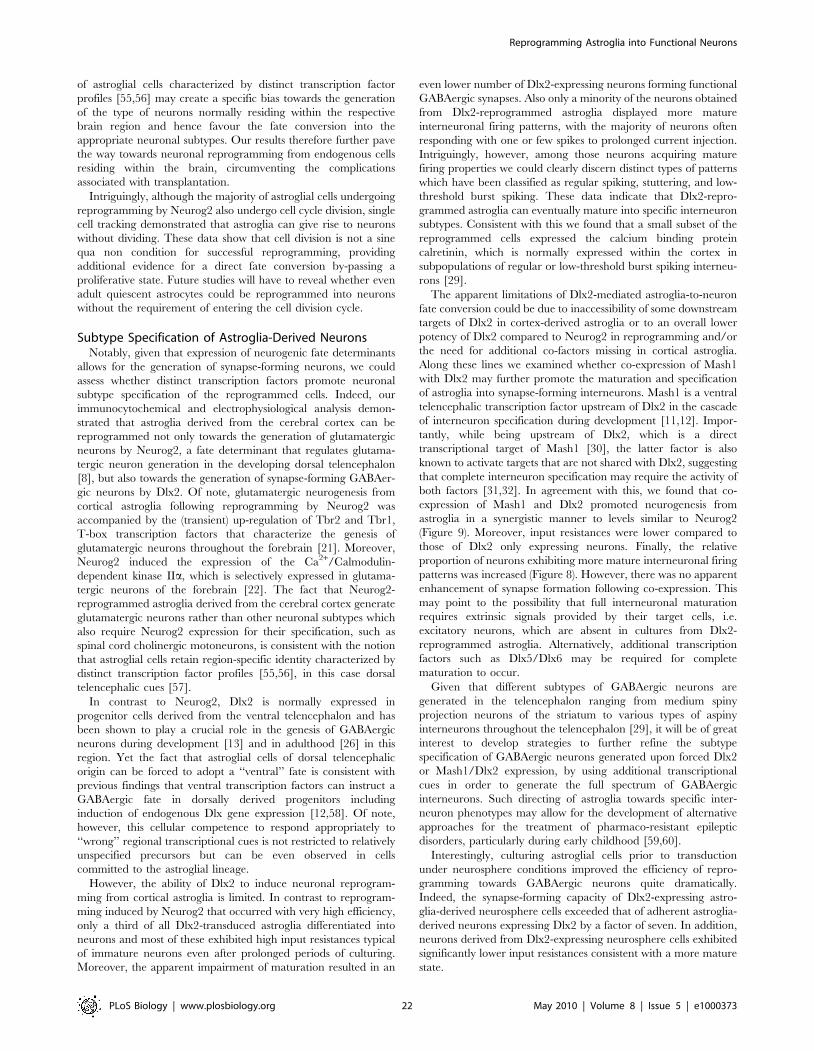
of astroglial cells characterized by distinct transcription factor
profiles [55,56] may create a specific bias towards the generation
of the type of neurons normally residing within the respective
brain region and hence favour the fate conversion into the
appropriate neuronal subtypes. Our results therefore further pave
the way towards neuronal reprogramming from endogenous cells
residing within the brain, circumventing the complications
associated with transplantation.
Intriguingly, although the majority of astroglial cells undergoing
reprogramming by Neurog2 also undergo cell cycle division, single
cell tracking demonstrated that astroglia can give rise to neurons
without dividing. These data show that cell division is not a sine
qua non condition for successful reprogramming, providing
additional evidence for a direct fate conversion by-passing a
proliferative state. Future studies will have to reveal whether even
adult quiescent astrocytes could be reprogrammed into neurons
without the requirement of entering the cell division cycle.
Subtype Specification of Astroglia-Derived NeuronsNotably, given that expression of neurogenic fate determinants
allows for the generation of synapse-forming neurons, we could
assess whether distinct transcription factors promote neuronal
subtype specification of the reprogrammed cells. Indeed, our
immunocytochemical and electrophysiological analysis demon-
strated that astroglia derived from the cerebral cortex can be
reprogrammed not only towards the generation of glutamatergic
neurons by Neurog2, a fate determinant that regulates glutama-
tergic neuron generation in the developing dorsal telencephalon
[8], but also towards the generation of synapse-forming GABAer-
gic neurons by Dlx2. Of note, glutamatergic neurogenesis from
cortical astroglia following reprogramming by Neurog2 was
accompanied by the (transient) up-regulation of Tbr2 and Tbr1,
T-box transcription factors that characterize the genesis of
glutamatergic neurons throughout the forebrain [21]. Moreover,
Neurog2 induced the expression of the Ca2+/Calmodulin-
dependent kinase IIa, which is selectively expressed in glutama-
tergic neurons of the forebrain [22]. The fact that Neurog2-
reprogrammed astroglia derived from the cerebral cortex generate
glutamatergic neurons rather than other neuronal subtypes which
also require Neurog2 expression for their specification, such as
spinal cord cholinergic motoneurons, is consistent with the notion
that astroglial cells retain region-specific identity characterized by
distinct transcription factor profiles [55,56], in this case dorsal
telencephalic cues [57].
In contrast to Neurog2, Dlx2 is normally expressed in
progenitor cells derived from the ventral telencephalon and has
been shown to play a crucial role in the genesis of GABAergic
neurons during development [13] and in adulthood [26] in this
region. Yet the fact that astroglial cells of dorsal telencephalic
origin can be forced to adopt a ‘‘ventral’’ fate is consistent with
previous findings that ventral transcription factors can instruct a
GABAergic fate in dorsally derived progenitors including
induction of endogenous Dlx gene expression [12,58]. Of note,
however, this cellular competence to respond appropriately to
‘‘wrong’’ regional transcriptional cues is not restricted to relatively
unspecified precursors but can be even observed in cells
committed to the astroglial lineage.
However, the ability of Dlx2 to induce neuronal reprogram-
ming from cortical astroglia is limited. In contrast to reprogram-
ming induced by Neurog2 that occurred with very high efficiency,
only a third of all Dlx2-transduced astroglia differentiated into
neurons and most of these exhibited high input resistances typical
of immature neurons even after prolonged periods of culturing.
Moreover, the apparent impairment of maturation resulted in an
even lower number of Dlx2-expressing neurons forming functional
GABAergic synapses. Also only a minority of the neurons obtained
from Dlx2-reprogrammed astroglia displayed more mature
interneuronal firing patterns, with the majority of neurons often
responding with one or few spikes to prolonged current injection.
Intriguingly, however, among those neurons acquiring mature
firing properties we could clearly discern distinct types of patterns
which have been classified as regular spiking, stuttering, and low-
threshold burst spiking. These data indicate that Dlx2-repro-
grammed astroglia can eventually mature into specific interneuron
subtypes. Consistent with this we found that a small subset of the
reprogrammed cells expressed the calcium binding protein
calretinin, which is normally expressed within the cortex in
subpopulations of regular or low-threshold burst spiking interneu-
rons [29].
The apparent limitations of Dlx2-mediated astroglia-to-neuron
fate conversion could be due to inaccessibility of some downstream
targets of Dlx2 in cortex-derived astroglia or to an overall lower
potency of Dlx2 compared to Neurog2 in reprogramming and/or
the need for additional co-factors missing in cortical astroglia.
Along these lines we examined whether co-expression of Mash1
with Dlx2 may further promote the maturation and specification
of astroglia into synapse-forming interneurons. Mash1 is a ventral
telencephalic transcription factor upstream of Dlx2 in the cascade
of interneuron specification during development [11,12]. Impor-
tantly, while being upstream of Dlx2, which is a direct
transcriptional target of Mash1 [30], the latter factor is also
known to activate targets that are not shared with Dlx2, suggesting
that complete interneuron specification may require the activity of
both factors [31,32]. In agreement with this, we found that co-
expression of Mash1 and Dlx2 promoted neurogenesis from
astroglia in a synergistic manner to levels similar to Neurog2
(Figure 9). Moreover, input resistances were lower compared to
those of Dlx2 only expressing neurons. Finally, the relative
proportion of neurons exhibiting more mature interneuronal firing
patterns was increased (Figure 8). However, there was no apparent
enhancement of synapse formation following co-expression. This
may point to the possibility that full interneuronal maturation
requires extrinsic signals provided by their target cells, i.e.
excitatory neurons, which are absent in cultures from Dlx2-
reprogrammed astroglia. Alternatively, additional transcription
factors such as Dlx5/Dlx6 may be required for complete
maturation to occur.
Given that different subtypes of GABAergic neurons are
generated in the telencephalon ranging from medium spiny
projection neurons of the striatum to various types of aspiny
interneurons throughout the telencephalon [29], it will be of great
interest to develop strategies to further refine the subtype
specification of GABAergic neurons generated upon forced Dlx2
or Mash1/Dlx2 expression, by using additional transcriptional
cues in order to generate the full spectrum of GABAergic
interneurons. Such directing of astroglia towards specific inter-
neuron phenotypes may allow for the development of alternative
approaches for the treatment of pharmaco-resistant epileptic
disorders, particularly during early childhood [59,60].
Interestingly, culturing astroglial cells prior to transduction
under neurosphere conditions improved the efficiency of repro-
gramming towards GABAergic neurons quite dramatically.
Indeed, the synapse-forming capacity of Dlx2-expressing astro-
glia-derived neurosphere cells exceeded that of adherent astroglia-
derived neurons expressing Dlx2 by a factor of seven. In addition,
neurons derived from Dlx2-expressing neurosphere cells exhibited
significantly lower input resistances consistent with a more mature
state.
Reprogramming Astroglia into Functional Neurons
PLoS Biology | www.plosbiology.org 22 May 2010 | Volume 8 | Issue 5 | e1000373
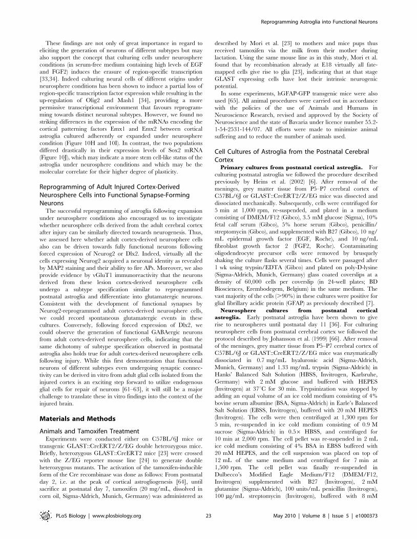
These findings are not only of great importance in regard to
eliciting the generation of neurons of different subtypes but may
also support the concept that culturing cells under neurosphere
conditions (in serum-free medium containing high levels of EGF
and FGF2) induces the erasure of region-specific transcription
[33,34]. Indeed culturing neural cells of different origins under
neurosphere conditions has been shown to induce a partial loss of
region-specific transcription factor expression while resulting in the
up-regulation of Olig2 and Mash1 [34], providing a more
permissive transcriptional environment that favours reprogram-
ming towards distinct neuronal subtypes. However, we found no
striking differences in the expression of the mRNAs encoding the
cortical patterning factors Emx1 and Emx2 between cortical
astroglia cultured adherently or expanded under neurosphere
condition (Figure 10H and 10I). In contrast, the two populations
differed drastically in their expression levels of Sox2 mRNA
(Figure 10J), which may indicate a more stem cell-like status of the
astroglia under neurosphere conditions and which may be the
molecular correlate for their higher degree of plasticity.
Reprogramming of Adult Injured Cortex-DerivedNeurosphere Cells into Functional Synapse-FormingNeurons
The successful reprogramming of astroglia following expansion
under neurosphere conditions also encouraged us to investigate
whether neurosphere cells derived from the adult cerebral cortex
after injury can be similarly directed towards neurogenesis. Thus,
we assessed here whether adult cortex-derived neurosphere cells
also can be driven towards fully functional neurons following
forced expression of Neurog2 or Dlx2. Indeed, virtually all the
cells expressing Neurog2 acquired a neuronal identity as revealed
by MAP2 staining and their ability to fire APs. Moreover, we also
provide evidence by vGluT1 immunoreactivity that the neurons
derived from these lesion cortex-derived neurosphere cells
undergo a subtype specification similar to reprogrammed
postnatal astroglia and differentiate into glutamatergic neurons.
Consistent with the development of functional synapses by
Neurog2-reprogrammed adult cortex-derived neurosphere cells,
we could record spontaneous glutamatergic events in these
cultures. Conversely, following forced expression of Dlx2, we
could observe the generation of functional GABAergic neurons
from adult cortex-derived neurosphere cells, indicating that the
same dichotomy of subtype specification observed in postnatal
astroglia also holds true for adult cortex-derived neurosphere cells
following injury. While this first demonstration that functional
neurons of different subtypes even undergoing synaptic connec-
tivity can be derived in vitro from adult glial cells isolated from the
injured cortex is an exciting step forward to utilize endogenous
glial cells for repair of neurons [61–63], it will still be a major
challenge to translate these in vitro findings into the context of the
injured brain.
Materials and Methods
Animals and Tamoxifen TreatmentExperiments were conducted either on C57BL/6J mice or
transgenic GLAST::CreERT2/Z/EG double heterozygous mice.
Briefly, heterozygous GLAST::CreERT2 mice [23] were crossed
with the Z/EG reporter mouse line [24] to generate double
heterozygous mutants. The activation of the tamoxifen-inducible
form of the Cre recombinase was done as follows: From postnatal
day 2, i.e. at the peak of cortical astrogliogenesis [64], until
sacrifice at postnatal day 7, tamoxifen (20 mg/mL, dissolved in
corn oil, Sigma-Aldrich, Munich, Germany) was administered as
described by Mori et al. [23] to mothers and mice pups thus
received tamoxifen via the milk from their mother during
lactation. Using the same mouse line as in this study, Mori et al.
found that by recombination already at E18 virtually all fate-
mapped cells give rise to glia [23], indicating that at that stage
GLAST expressing cells have lost their intrinsic neurogenic
potential.
In some experiments, hGFAP-GFP transgenic mice were also
used [65]. All animal procedures were carried out in accordance
with the policies of the use of Animals and Humans in
Neuroscience Research, revised and approved by the Society of
Neuroscience and the state of Bavaria under licence number 55.2-
1-54-2531-144/07. All efforts were made to minimize animal
suffering and to reduce the number of animals used.
Cell Cultures of Astroglia from the Postnatal CerebralCortex
Primary cultures from postnatal cortical astroglia. For
culturing postnatal astroglia we followed the procedure described
previously by Heins et al. (2002) [6]. After removal of the
meninges, grey matter tissue from P5–P7 cerebral cortex of
C57BL/6J or GLAST::CreERT2/Z/EG mice was dissected and
dissociated mechanically. Subsequently, cells were centrifuged for
5 min at 1,000 rpm, re-suspended, and plated in a medium
consisting of DMEM/F12 (Gibco), 3.5 mM glucose (Sigma), 10%
fetal calf serum (Gibco), 5% horse serum (Gibco), penicillin/
streptomycin (Gibco), and supplemented with B27 (Gibco), 10 ng/
mL epidermal growth factor (EGF, Roche), and 10 ng/mL
fibroblast growth factor 2 (FGF2, Roche). Contaminating
oligodendrocyte precursor cells were removed by brusquely
shaking the culture flasks several times. Cells were passaged after
1 wk using trypsin/EDTA (Gibco) and plated on poly-D-lysine
(Sigma-Aldrich, Munich, Germany) glass coated coverslips at a
density of 60,000 cells per coverslip (in 24-well plates; BD
Biosciences, Erembodegem, Belgium) in the same medium. The
vast majority of the cells (.90%) in these cultures were positive for
glial fibrillary acidic protein (GFAP) as previously described [7].
Neurosphere cultures from postnatal cortical
astroglia. Early postnatal astroglia have been shown to give
rise to neurospheres until postnatal day 11 [36]. For culturing
neurosphere cells from postnatal cerebral cortex we followed the
protocol described by Johansson et al. (1999) [66]. After removal
of the meninges, grey matter tissue from P5–P7 cerebral cortex of
C57BL/6J or GLAST::CreERT2/Z/EG mice was enzymatically
dissociated in 0.7 mg/mL hyaluronic acid (Sigma-Aldrich,
Munich, Germany) and 1.33 mg/mL trypsin (Sigma-Aldrich) in
Hanks’ Balanced Salt Solution (HBSS, Invitrogen, Karlsruhe,
Germany) with 2 mM glucose and buffered with HEPES
(Invitrogen) at 37uC for 30 min. Trypsinization was stopped by
adding an equal volume of an ice cold medium consisting of 4%
bovine serum albumine (BSA, Sigma-Aldrich) in Earle’s Balanced
Salt Solution (EBSS, Invitrogen), buffered with 20 mM HEPES
(Invitrogen). The cells were then centrifuged at 1,300 rpm for
5 min, re-suspended in ice cold medium consisting of 0.9 M
sucrose (Sigma-Aldrich) in 0.56 HBSS, and centrifuged for
10 min at 2,000 rpm. The cell pellet was re-suspended in 2 mL
ice cold medium consisting of 4% BSA in EBSS buffered with
20 mM HEPES, and the cell suspension was placed on top of
12 mL of the same medium and centrifuged for 7 min at
1,500 rpm. The cell pellet was finally re-suspended in
Dulbecco’s Modified Eagle Medium/F12 (DMEM/F12,
Invitrogen) supplemented with B27 (Invitrogen), 2 mM
glutamine (Sigma-Aldrich), 100 units/mL penicillin (Invitrogen),
100 mg/mL streptomycin (Invitrogen), buffered with 8 mM
Reprogramming Astroglia into Functional Neurons
PLoS Biology | www.plosbiology.org 23 May 2010 | Volume 8 | Issue 5 | e1000373
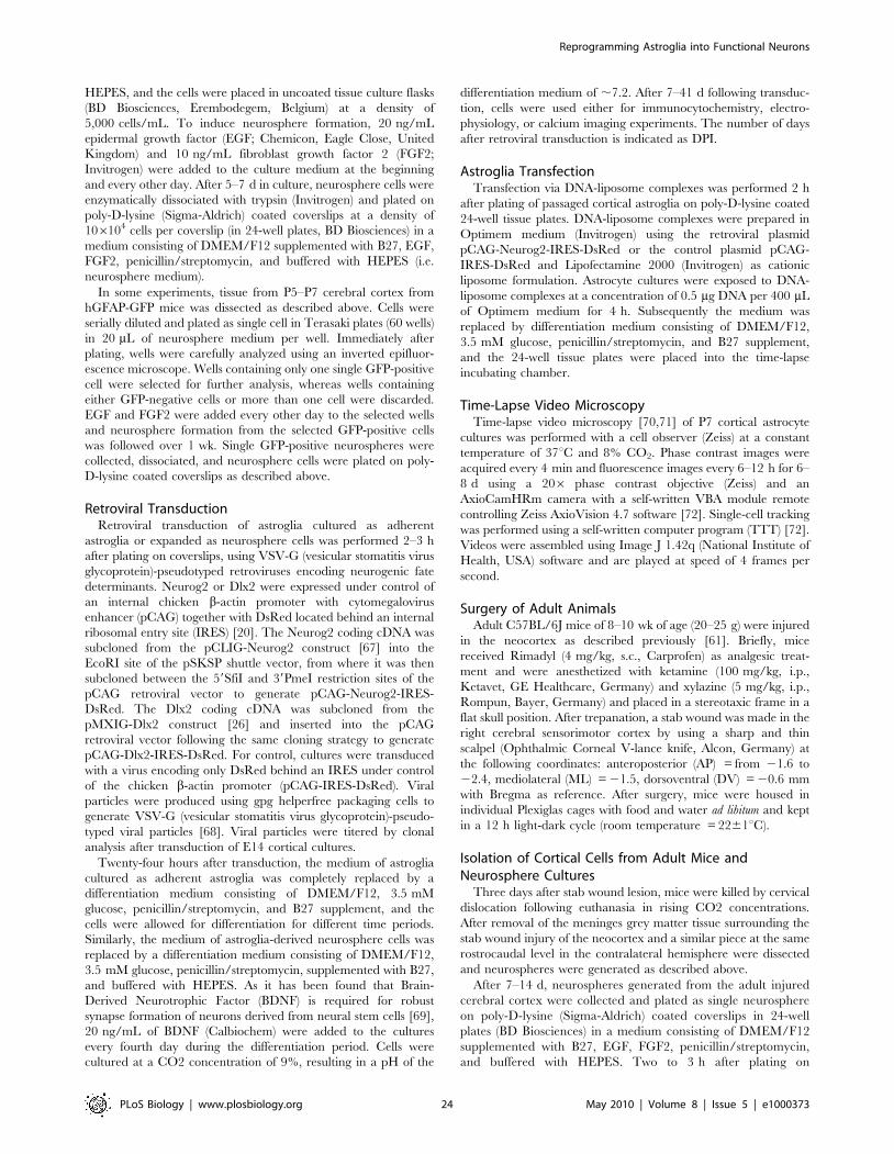
HEPES, and the cells were placed in uncoated tissue culture flasks
(BD Biosciences, Erembodegem, Belgium) at a density of
5,000 cells/mL. To induce neurosphere formation, 20 ng/mL
epidermal growth factor (EGF; Chemicon, Eagle Close, United
Kingdom) and 10 ng/mL fibroblast growth factor 2 (FGF2;
Invitrogen) were added to the culture medium at the beginning
and every other day. After 5–7 d in culture, neurosphere cells were
enzymatically dissociated with trypsin (Invitrogen) and plated on
poly-D-lysine (Sigma-Aldrich) coated coverslips at a density of
106104 cells per coverslip (in 24-well plates, BD Biosciences) in a
medium consisting of DMEM/F12 supplemented with B27, EGF,
FGF2, penicillin/streptomycin, and buffered with HEPES (i.e.
neurosphere medium).
In some experiments, tissue from P5–P7 cerebral cortex from
hGFAP-GFP mice was dissected as described above. Cells were
serially diluted and plated as single cell in Terasaki plates (60 wells)
in 20 mL of neurosphere medium per well. Immediately after
plating, wells were carefully analyzed using an inverted epifluor-
escence microscope. Wells containing only one single GFP-positive
cell were selected for further analysis, whereas wells containing
either GFP-negative cells or more than one cell were discarded.
EGF and FGF2 were added every other day to the selected wells
and neurosphere formation from the selected GFP-positive cells
was followed over 1 wk. Single GFP-positive neurospheres were
collected, dissociated, and neurosphere cells were plated on poly-
D-lysine coated coverslips as described above.
Retroviral TransductionRetroviral transduction of astroglia cultured as adherent
astroglia or expanded as neurosphere cells was performed 2–3 h
after plating on coverslips, using VSV-G (vesicular stomatitis virus
glycoprotein)-pseudotyped retroviruses encoding neurogenic fate
determinants. Neurog2 or Dlx2 were expressed under control of
an internal chicken b-actin promoter with cytomegalovirus
enhancer (pCAG) together with DsRed located behind an internal
ribosomal entry site (IRES) [20]. The Neurog2 coding cDNA was
subcloned from the pCLIG-Neurog2 construct [67] into the
EcoRI site of the pSKSP shuttle vector, from where it was then
subcloned between the 59SfiI and 39PmeI restriction sites of the
pCAG retroviral vector to generate pCAG-Neurog2-IRES-
DsRed. The Dlx2 coding cDNA was subcloned from the
pMXIG-Dlx2 construct [26] and inserted into the pCAG
retroviral vector following the same cloning strategy to generate
pCAG-Dlx2-IRES-DsRed. For control, cultures were transduced
with a virus encoding only DsRed behind an IRES under control
of the chicken b-actin promoter (pCAG-IRES-DsRed). Viral
particles were produced using gpg helperfree packaging cells to
generate VSV-G (vesicular stomatitis virus glycoprotein)-pseudo-
typed viral particles [68]. Viral particles were titered by clonal
analysis after transduction of E14 cortical cultures.
Twenty-four hours after transduction, the medium of astroglia
cultured as adherent astroglia was completely replaced by a
differentiation medium consisting of DMEM/F12, 3.5 mM
glucose, penicillin/streptomycin, and B27 supplement, and the
cells were allowed for differentiation for different time periods.
Similarly, the medium of astroglia-derived neurosphere cells was
replaced by a differentiation medium consisting of DMEM/F12,
3.5 mM glucose, penicillin/streptomycin, supplemented with B27,
and buffered with HEPES. As it has been found that Brain-
Derived Neurotrophic Factor (BDNF) is required for robust
synapse formation of neurons derived from neural stem cells [69],
20 ng/mL of BDNF (Calbiochem) were added to the cultures
every fourth day during the differentiation period. Cells were
cultured at a CO2 concentration of 9%, resulting in a pH of the
differentiation medium of ,7.2. After 7–41 d following transduc-
tion, cells were used either for immunocytochemistry, electro-
physiology, or calcium imaging experiments. The number of days
after retroviral transduction is indicated as DPI.
Astroglia TransfectionTransfection via DNA-liposome complexes was performed 2 h
after plating of passaged cortical astroglia on poly-D-lysine coated
24-well tissue plates. DNA-liposome complexes were prepared in
Optimem medium (Invitrogen) using the retroviral plasmid
pCAG-Neurog2-IRES-DsRed or the control plasmid pCAG-
IRES-DsRed and Lipofectamine 2000 (Invitrogen) as cationic
liposome formulation. Astrocyte cultures were exposed to DNA-
liposome complexes at a concentration of 0.5 mg DNA per 400 mL
of Optimem medium for 4 h. Subsequently the medium was
replaced by differentiation medium consisting of DMEM/F12,
3.5 mM glucose, penicillin/streptomycin, and B27 supplement,
and the 24-well tissue plates were placed into the time-lapse
incubating chamber.
Time-Lapse Video MicroscopyTime-lapse video microscopy [70,71] of P7 cortical astrocyte
cultures was performed with a cell observer (Zeiss) at a constant
temperature of 37uC and 8% CO2. Phase contrast images were
acquired every 4 min and fluorescence images every 6–12 h for 6–
8 d using a 206 phase contrast objective (Zeiss) and an
AxioCamHRm camera with a self-written VBA module remote
controlling Zeiss AxioVision 4.7 software [72]. Single-cell tracking
was performed using a self-written computer program (TTT) [72].
Videos were assembled using Image J 1.42q (National Institute of
Health, USA) software and are played at speed of 4 frames per
second.
Surgery of Adult AnimalsAdult C57BL/6J mice of 8–10 wk of age (20–25 g) were injured
in the neocortex as described previously [61]. Briefly, mice
received Rimadyl (4 mg/kg, s.c., Carprofen) as analgesic treat-
ment and were anesthetized with ketamine (100 mg/kg, i.p.,
Ketavet, GE Healthcare, Germany) and xylazine (5 mg/kg, i.p.,
Rompun, Bayer, Germany) and placed in a stereotaxic frame in a
flat skull position. After trepanation, a stab wound was made in the
right cerebral sensorimotor cortex by using a sharp and thin
scalpel (Ophthalmic Corneal V-lance knife, Alcon, Germany) at
the following coordinates: anteroposterior (AP) = from 21.6 to
22.4, mediolateral (ML) = 21.5, dorsoventral (DV) = 20.6 mm
with Bregma as reference. After surgery, mice were housed in
individual Plexiglas cages with food and water ad libitum and kept
in a 12 h light-dark cycle (room temperature = 2261uC).
Isolation of Cortical Cells from Adult Mice andNeurosphere Cultures
Three days after stab wound lesion, mice were killed by cervical
dislocation following euthanasia in rising CO2 concentrations.
After removal of the meninges grey matter tissue surrounding the
stab wound injury of the neocortex and a similar piece at the same
rostrocaudal level in the contralateral hemisphere were dissected
and neurospheres were generated as described above.
After 7–14 d, neurospheres generated from the adult injured
cerebral cortex were collected and plated as single neurosphere
on poly-D-lysine (Sigma-Aldrich) coated coverslips in 24-well
plates (BD Biosciences) in a medium consisting of DMEM/F12
supplemented with B27, EGF, FGF2, penicillin/streptomycin,
and buffered with HEPES. Two to 3 h after plating on
Reprogramming Astroglia into Functional Neurons
PLoS Biology | www.plosbiology.org 24 May 2010 | Volume 8 | Issue 5 | e1000373
coverslips, neurosphere cells were transduced as described above
with retroviral vectors encoding Neurog2 (pCAG-Neurog2-
IRES-DsRed), Dlx2 (pCAG-Dlx2-IRES-DsRed), or the control
retrovirus (pCAG-IRES-DsRed) and were then allowed for
differentiation.
ImmunocytochemistryFor immunocytochemistry, cultures were fixed in 4% parafor-
maldehyde (PFA) in phosphate buffered saline (PBS) for 15 min at
room temperature. Cells were first pretreated in 0.5% Triton X-
100 in PBS for 30 min, followed by incubation in 2% BSA and
0.5% Triton X-100 in PBS for 30 min. Primary antibodies were
incubated on specimen overnight at 4uC in 2% BSA, 0.5% Triton
X-100 in PBS. The following primary antibodies were used: anti-
GFP (GFP, chicken, 1:1000, Aves Labs, GFP-1020), polyclonal
anti-Glial Fibrillary Acidic Protein (GFAP, rabbit, 1:4000,
DakoCytomation, Z0334), polyclonal anti-Red Fluorescent Pro-
tein (RFP, rabbit, 1:500, Chemicon, AB3216), polyclonal anti-RFP
(rabbit, Rockland, 1:2000, 600-401-379), polyclonal anti-vesicular
glutamate transporter 1 (vGluT11, rabbit, 1:1000, Synaptic
Systems, 135302), monoclonal anti-Microtubule Associated Pro-
tein 2 (MAP2, mouse IgG1, 1:200, Sigma-Aldrich, M4403),
monoclonal anti-synapsin 1 (mouse IgG2, 1:2000, Synaptic
Systems, 106001), polyclonal anti-vGaT (guinea pig, 1:200,
Synaptic Systems, 131004), polyclonal anti-Tbr1 (rabbit, 1:1000,
Millipore, AB9616), polyclonal anti-Tbr2 (rabbit, 1:500, Millipore,
AB9618), monoclonal anti-bIII tubulin (mouse IgG2b, 1:500,
Sigma, T8660), polyclonal anti-GAD1(GAD67) (rabbit, 1:500,
Synaptic Systems, 198013), monoclonal anti-calretinin (mouse
IgG1, 1:200, Millipore, MAB1568), and monoclonal anti-CaM
kinase IIa (mouse IgG1, 1:200, Abcam, ab2725). After extensive
washing in PBS, cells were incubated with appropriate species- or
subclass-specific secondary antibodies conjugated to CyTM2,
CyTM3, CyTM5 (1:500, Jackson ImmunoResearch), Alexa Fluor
488 (1:500, Invitrogen), FITC (fluorescein isothiocyanate, 1:500,
Jackson ImmunoResearch), TRITC (tetramethyl rhodamine
isothiocyanate, 1:500, Jackson ImmunoResearch), or biotin
(1:500, Jackson ImmunoResearch or Vector Laboratories) for
2 h in the dark at room temperature, followed by extensive
washing in PBS. Following treatment with secondary antibodies
conjugated to biotin, cells were subsequently incubated for 2 h at
room temperature with AMCA streptavidin (1:200, Vector
Laboratories) or Alexa Fluor 647 streptavidin (1:500, Invitrogen).
Coverslips were finally mounted onto a glass slide with an anti-
fading mounting medium (Aqua Poly/Mount; Polysciences,
Warrington, PA).
Microscopy AnalysisStainings were first examined with an epifluorescence micro-
scope (BX61, Olympus, Hamburg, Germany) equipped with the
appropriate filter sets. Stainings were further analyzed with laser-
scanning confocal microscopes (SP5, Leica, Wetzlar, Germany or
LSM710, Carl Zeiss, Gottingen, Germany). Z-stacks of digital
images were captured using the LAS AF (Leica) or ZEN software
(Carl Zeiss). Single confocal images were then extracted from the
Z-stacks. Alternatively, the Z-stacks were collapsed in one resulting
picture using the maximum intensity projection function provided
by the above mentioned softwares.
Cell Counts and Statistical AnalysisCell counts were performed by taking pictures of several
randomly selected views per coverslip analysed by means of a Zeiss
LSM 710 confocal microscope using a 256 objective. Subse-
quently, pictures were analysed for cell quantification using Image
J 1.42q (National Institute of Health, USA) software. For each
quantification, values are given as mean 6 SEM. Cell counting
data from reprogramming induced by Dlx2, Mash2, and Mash1 in
combination with Dlx2 were subjected to a two-tailed Student’s t
test for statistical significance. Differences were considered
statistically significant when the probability value was ,0.05.
ElectrophysiologyElectrophysiological properties of neurons derived from repro-
grammed astroglial cells were analyzed 11–41 d following
retroviral transduction. Single or dual perforated patch-clamp
recordings [73,74] were performed at room temperature with
amphotericin-B (Calbiochem) for perforation. Micropipettes were
made from borosilicate glass capillaries (Garner, Claremont, CA,
USA). Pipettes were tip-filled with internal solution and back-filled
with internal solution containing 200 mg/mL amphotericin-B. The
electrodes had resistances of 2–2.5 MV. The internal solution
contained 136.5 mM K-gluconate, 17.5 mM KCl, 9 mM NaCl,
1 mM MgCl2, 10 mM HEPES, and 0.2 mM EGTA (pH 7.4) at
an osmolarity of 300 mOsm. The external solution contained
150 mM NaCl, 3 mM KCl, 3 mM CaCl2, 2 mM MgCl2, 10 mM
HEPES, and 5 mM glucose (pH 7.4) at an osmolarity of
310 mOsm. The recording chamber was continuously perfused
with external solution at a rate of 0.5 mL/min.
Cells were visualized with an epifluorescence microscope
(Axioskop2, Carl Zeiss) equipped with the appropriate filter sets.
For patch clamp recordings, virally transduced cells were selected
on the basis of their DsRed immunoreactivity. In addition, to
ascertain the astroglial origin of the recorded neurons, DsRed- and
GFP-expressing cells from GLAST::CreERT2/Z/EG animals
were also selected for patch clamp recordings. Digital pictures of
the recorded cells were acquired using a digital camera (AxioCam,
Carl Zeiss).
Signals were sampled at 10 kHz with Axopatch 200B patch-
clamp amplifiers (Axon Instruments, Foster City, CA, USA),
filtered at 5 kHz and analyzed with Clampfit 9.2 software (Axon
Instruments). For assessing a cell’s ability to fire APs, cells received
depolarizing step-current injections. AP amplitudes were mea-
sured by subtracting the threshold voltage of the AP from the AP
maximum amplitude. For determining input resistance, hyperpo-
larizing currents of small amplitudes were injected into the cells
under current clamp condition at a holding potential of 270 mV
and input resistances were calculated from the corresponding
voltage deviation.
To examine spontaneous synaptic input into a given neuron,
cells were kept in voltage clamp at a holding potential of 270 mV
and synaptic events were recorded throughout a period of 1 to
5 min. In order to assess autaptic connections, single cells were
step-depolarized in voltage clamp for 1 ms from 270 to +30 mV
at a frequency of 0.05 Hz and responses were recorded in the
same cell. Responses were considered to be autaptic when they
occurred within 3 ms after the step-depolarization [75]. Synaptic
connectivity was investigated by means of pair recordings in
voltage clamp mode. One neuron was stimulated at low frequency
(0.05–0.1 Hz) by a 1 ms step-depolarization from 270 to +30 mV
and the response was recorded from the other neuron, and vice
versa. To determine the nature of the autaptic or synaptic
responses, neurons were step-depolarized as described above and
we assessed whether responses could be abolished in the presence
of either the GABAA receptor antagonist bicuculline (10 mM) or
the AMPA/kainate receptor antagonist CNQX (5 mM). Finally,
the recovery of the autaptic or synaptic response was assessed
following washout of the pharmacological drugs.
Reprogramming Astroglia into Functional Neurons
PLoS Biology | www.plosbiology.org 25 May 2010 | Volume 8 | Issue 5 | e1000373
Confocal Calcium ImagingNeurons derived from reprogrammed astroglial cells were
further analyzed 22, 27, and 29 d after retroviral transduction
with calcium imaging experiments. A 5 mM stock solution of
Oregon-Green BAPTA1, AM (KD: 170 nM, Invitrogen, O6807)
was prepared in 8.9 mL 20% Pluronic F-127 (Invitrogen) in
dimethylsulfoxide (DMSO) by means of a sonifier bath (Bandelin,
Berlin, Germany) for 3 min. Reprogrammed astroglial cells were
incubated with 5 mM Oregon-BAPTA1, AM in artificial cerebro-
spinal fluid solution (ACSF: in mM: 127 NaCl, 4.5 KCl, 2.5
NaH2PO4, 2 CaCl2, 2 MgCl2, 23 NaHCO3, and 25 D-glucose.,
bubbled with 95% O2/5% CO2.). The incubation of neurons with
the dye-ACSF was performed in a cell culture incubator (37uC,
9% CO2) for a loading time of 10–15 min. Cells were washed with
ACSF in a perfusion chamber (Volume: 150–200 mL) for at least
10–20 min with a flow rate of approximately 2–3 mL/min. Cells
were imaged under continuous perfusion with ACSF solution at
26–30uC. Ligands (Ascent scientific) were bath-applied and used at
the following concentrations: CNQX (10 mM), D-AP5 (10 mM),
and tetrodotoxin (TTX, 500 nM). For confocal Ca2+ imaging
(2566256 pixels, 2.16 Hz), an inverted confocal microscope
(Olympus IX70, equipped with a Fluoview 300 laser scanning
system) was used in combination with an Olympus, UPlanApo
206/0.7 objective. Transduced cells were identified by means of
DsRed expression. Oregon Green-derived fluorescence was
excited with a 488 nm laser line (emission filter: band pass 510/
540 nm). DsRed was excited at 543 nm (emission filter: band pass:
580 nm640 nm).
In some experiments, astroglia from GLAST::CreERT2/Z/EG
mice, reprogrammed by forced expression of neurogenic fate
determinants, were used for calcium imaging experiments. To
ascertain the astroglial origin of the DsRed-positive reprogrammed
cells that will be analyzed, GFP expression was first pictured at the
confocal microscope and was carefully bleached by using the 488-
laser afterwards. The DsRed-positive reprogrammed astroglial cells
were subsequently incubated with Oregon Green and calcium
imaging experiments were processed as described above.
Images were analyzed using IMAGEJ software (WS Rasband,
IMAGEJ, US National Institutes of Health, Bethesda, Maryland,
USA, http://rsb.info.nih.gov/ij/, 1997–2006). XY-time Calcium
imaging results were analyzed by a region-of-interest analysis (pixel
intensity) in the extended TIFF format as described before [76].
Quantitative RT-PCRTotal RNA was extracted with RNeasy Plus MicroKit (Qiagen),
according to the manufacturer’s instructions. 1–1.5 mg of total
RNA was retro-transcribed using Super-ScriptIII Reverse Tran-
scriptase (Invitrogen) and random primers. Each cDNA was
diluted one to ten, and 2 ml was used for each real-time reaction.
mRNA quantitation was performed on a DNA Engine Opticon 2
System (Bio-Rad) following the manufacturer’s protocol using the
IQ SYBR Green SuperMix (Bio-Rad). The following oligonucle-
otide primers were used for the qPCR: Gapdh, Emx1, and Emx2
[34]; Sox2 [77], Ngn2 [78], bIIItub [79], Glt1 [80], Glu1 (Glutamine
Synthetase) (CCTGGACCCCAAGGCCCGTA; TGGCAGC-
CTGCACCATTCCAG), Aldh1l1 (TGTTTGGCCAGGAGGT-
TTAC; AGGTCACCAGTGTCCAGACC), S100b (GATGTC-
TTCCACCAGTACTCC; CTCATGTTCAAAGAACTCAT),
and Gfap [81]. The amount of each gene was analyzed in
triplicate, and the analysis was repeated on at least three
independent samples (n = 3 for NPC and postnatal neurospheres,
n = 5 for postnatal adherent astrocytes). Data analysis was
performed with the DDCt method [82].
Supporting Information
Figure S1 Neuronal reprogramming of astroglia in-volves down-regulation of GFAP and up-regulation ofTbr2 and Tbr1. (A–A’) Down-regulation of GFAP in astroglia-
derived neurons. Astroglia culture 6 d post-transduction with
Neurog2-IRES-DsRed encoding retrovirus. (A) Note that with the
exception of the two cells labelled by the arrowheads depicting
DAPI positive nuclei (blue) devoid of any GFAP immunoreactiv-
ity, all other cells labelled by nuclear DAPI staining are GFAP-
positive (green). (A’) The two GFAP-negative cells are DsRed-
positive (indicating Neurog2 expression) and are bIII tubulin-
positive (white) revealing their neuronal identity (arrowheads). (B–
B’) Expression of Tbr2 in astroglia undergoing reprogramming at
4 DPI. (B) Immunostaining for DsRed (red) and bIII tubulin
(green) shows five cells transduced with Neurog2-IRES-DsRed
encoding retrovirus, one of which already developed into a
neuron, while in the other cells bIII tubulin expression has barely
commenced. (B’) Immunostaining for Tbr2 (white, arrowheads)
reveals expression in those transduced astroglia which still exhibit
glia-like morphology (four out of five cells). This is consistent with
the early expression of Tbr2 in the cascade of transcription factors
steering glutamatergic neurogenesis [21]. (C–C’) Expression of
Tbr1 in neurons derived from Neurog2-transduced astroglia at
7 DPI. (C) Immunostaining for DsRed (red) and bIII tubulin
(green) reveals five reprogrammed cells. (C’) Immunostaining for
Tbr1 (white, arrowheads) reveals the up-regulation of Tbr1 by
Neurog2 in a large subset (four out of five cells) of Neurog2-
transduced cells as reported previously [7].
Found at: doi:10.1371/journal.pbio.1000373.s001 (7.08 MB TIF)
Figure S2 Type of synaptic responses and input resis-tances of postnatal astroglia-derived neurons repro-grammed by Neurog2 or Dlx2. (A) Summary of the type of
autaptic/synaptic responses that could be evoked from astroglia
reprogrammed by forced expression of Neurog2, Dlx2, or Mash1
in combination with Dlx2, following culturing under adherent
(Astro) or neurosphere conditions (NS). The number of neurons
exhibiting autaptic/synaptic connections is expressed as percent-
age of recorded neurons. Note that astroglia-derived neurons
reprogrammed by Neurog2 or Dlx2 in adherent astroglia cultures
were recorded at 24.660.9 DPI or 26.961.4 DPI, respectively.
Astroglia-derived neurosphere cells transduced with Neurog2 or
Dlx2 were recorded at 14.260.7 DPI or 20.161.6 DPI, respec-
tively. (B) The graph plots input resistance values over time (MV)
of astroglia-derived neurons reprogrammed by Dlx2, following
culturing under adherent (triangles) or neurosphere conditions
(squares), respectively. GFP reporter-positive fate-mapped cells are
depicted with green colour. (C) The graph plots input resistance
values over time (MV) of astroglia-derived neurons reprogrammed
by Neurog2, following culturing under adherent (triangles) or
neurosphere conditions (squares), respectively. GFP reporter-
positive fate-mapped cells are depicted with green colour.
Found at: doi:10.1371/journal.pbio.1000373.s002 (0.45 MB TIF)
Figure S3 Postnatal astroglia isolated from hGFAP-GFPmice and expanded as neurospheres generate glutama-tergic neurons following forced expression of Neurog2. (A)
To ascertain the astroglial origin of the neurosphere founder cells,
cortical tissue from P5–P7 hGFAP-GFP mice was cultured as single
cell in Terasaki plates. (B) Fluorescence micrograph depicting cell
division of a singly plated GFP-positive astroglial cell. (C–D) The
majority of singly plated GFP-positive astroglia gives rise to GFP-
positive neurospheres (C) that increase in size over time in culture
(D). (E–F) Double immunostaining for DsRed (red) and GFP (green)
Reprogramming Astroglia into Functional Neurons
PLoS Biology | www.plosbiology.org 26 May 2010 | Volume 8 | Issue 5 | e1000373
of astroglia-derived neurospheres as shown in (D), 17 d after
neurosphere dissociation and subsequent transduction with Neu-
rog2-IRES-DsRed. (E) Example of a non-transduced cell (i.e.,
DsRed-negative) expressing GFP and exhibiting a glial morphology.
(F) In contrast, following transduction with Neurog2-DsRed,
neurosphere cells lose GFP expression and differentiate into
neurons. (G) The fluorescence micrograph depicts a cluster of
Neurog2-expressing neurosphere cells, derived from singly plated
GFP-positive astroglia that lost GFP expression and exhibit a
neuronal morphology 17 DPI. (H) The graph shows an evoked
sequence of both autaptic and polysynaptic events following step-
depolarisation of the neuron marked by the asterisk in (G),
suggesting that Neurog2-transduced neurosphere cells acquire a
glutamatergic identity and give rise to excitatory networks.
Found at: doi:10.1371/journal.pbio.1000373.s003 (6.10 MB TIF)
Figure S4 Neuronal reprogramming of postnatal astro-glia expanded as neurospheres by forced expression ofNeurog2 or Dlx2. (A–B) Double immunostaining for DsRed
(red) and MAP2 (green) reveals that virtually all astroglia-derived
neurosphere cells transduced with Dlx2-DsRed (A) or Neurog2-
DsRed (B) differentiate into MAP2-positive neurons (14 DPI). (C–
D) Fluorescence micrographs at the same magnification showing
that astroglia-derived neurosphere cells transduced with Neurog2
(D) extend several MAP2-positive neurites, whereas Dlx2-repro-
grammed neurons show more simple dendritic arbors and often
exhibit a bipolar morphology (C). Scale bars: A, B: 60 mm; C, D:
20 mm.
Found at: doi:10.1371/journal.pbio.1000373.s004 (7.52 MB TIF)
Figure S5 Calcium imaging experiments of postnatalastroglia-derived neurosphere cells transduced withDlx2 do not reveal any spontaneous Ca2+ transients. (A)
Cultures of astroglia-derived neurosphere cells transduced with
Dlx2 (red) were incubated at 27 DPI with the calcium-sensitive
probe Oregon Green BAPTA1 (green). (B) Micrograph of the
same field of view as shown in (A) depicting five astroglia-derived
neurosphere cells transduced with Dlx2 exhibiting a neuronal
morphology (arrows) that are analysed in (C). (C) The graph shows
a virtual absence of any spontaneous Ca2+ transient in the five
DsRed-positive analysed neurons shown in (B). The trace of each
individual neuron is shown in a different colour.
Found at: doi:10.1371/journal.pbio.1000373.s005 (4.55 MB TIF)
Figure S6 Adult injured cortex-derived neurosphere cellsgenerate functional GABAergic neurons following forcedexpression of Dlx2. (A–A’) Micrographs showing an adult cortex-
derived neurosphere cell 28 d after transduction with Dlx2 (red) that
has differentiated into a MAP2-positive neuron (green). (B–B’) Step-
depolarisation of the adult cortex-derived neurosphere cell trans-
duced with Dlx2 shown in (B) evokes a GABAergic autapse at
27 DPI (black trace), which is blocked by the GABAA receptor
antagonist bicuculline (10 mM, red trace). Scale bars: A, B: 25 mm.
Found at: doi:10.1371/journal.pbio.1000373.s006 (1.32 MB TIF)
Video S1 Direct visualisation of efficient reprogram-ming of astroglia into neurons by Neurog2. Time-lapse
video-microscopy of an astroglia culture derived from GLAST::
CreERT2/Z/EG mice following recombination in vivo between
P2–P7 and transduced with a retrovirus encoding Neurog2-IRES-
DsRed. The right screen reveals those cells which successfully had
undergone recombination by GFP fluorescence. GFP fluorescence
images were taken every 12 h after imaging had commenced
(onset 18 h post-infection). The left screen shows DsRed
fluorescence to reveal transduced cells. Images were taken every
6 h. Tracking of each cell was performed with bright field images
taken every 4 min (not shown). DsRed- and GFP-double positive
cells are marked by the red and green arrows, respectively. The
beige crosses marked dead cells. High magnification views of one
DsRed/GFP double-positive cell are shown in Video S2. Note the
massive, partially still ongoing, reprogramming by 3 d 6 h. Time is
indicated in each image.
Found at: doi:10.1371/journal.pbio.1000373.s007 (4.03 MB
MPG)
Video S2 Direct visualisation of reprogramming of fate-mapped astroglia into neurons. Temporal sequence of
reprogramming by Neurog2 of a fate-mapped astrocyte derived
from GLAST::CreERT2/Z/EG mice following recombination in
vivo between P2 and P7. The video provides high magnification
views of the sequence shown in Video S1. The video starts with a
GFP-fluorescence and bright field image at the beginning of the
time-lapse experiment. The GFP-positive cell is marked with a red
arrow throughout the sequence. The cell is subsequently followed
using bright field images. At the point when DsRed fluorescence is
detectable, a GFP fluorescence image is shown to demonstrate the
co-expression of DsRed and GFP in the same cell. Subsequently
DsRed fluorescence images (grey scale) monitor the division of the
transduced cell and the gradual metamorphosis of the two
daughter cells into neurons. At the end of the imaging sequence
a GFP fluorescence image is shown to confirm that the two
neurons are reporter-positive. Time is indicated in each image.
Found at: doi:10.1371/journal.pbio.1000373.s008 (6.14 MB
MPG)
Video S3 Direct visualisation of the astroglia-to-neuronmetamorphosis in the absence of cell division over a timeperiod of 5 d. Temporal sequence of high magnification views of
an astrocyte culture following transfection with the retroviral
plasmid encoding Neurog2-IRES-DsRed. Prior to the onset of
DsRed expression, bright field images are shown to follow a cell
subsequently found to be transfected (red arrow). DsRed fluores-
cence images are used to follow the cell through the reprogramming
process. Note that the astroglial cell followed by the red arrow is
reprogrammed into a neuron without entering cell division.
Found at: doi:10.1371/journal.pbio.1000373.s009 (5.22 MB
MPG)
Acknowledgments
We thank Dr. Marcos Costa for critical reading of and helpful discussion
on the manuscript. We are also very grateful to Tatiana Simon-Ebert and
Joanna Zaisserer for their excellent technical assistance as well as Dr.
Alexandra Lepier and Simone Bauer for virus production. We thank
Ronny Stahl for dissection of embryonic cortical tissue.
Author Contributions
The author(s) have made the following declarations about their
contributions: Conceived and designed the experiments: CH MG BB.
Performed the experiments: CH RB SG GM PT RS ST BB. Analyzed the
data: CH RB SG GM ST BB. Contributed reagents/materials/analysis
tools: TS. Wrote the paper: CH MG BB.
References
1. Wang DD, Bordey A (2008) The astrocyte odyssey. Prog Neurobiol 86:
342–367.
2. Doetsch F (2003) The glial identity of neural stem cells. Nat Neurosci 6:
1127–1134.
Reprogramming Astroglia into Functional Neurons
PLoS Biology | www.plosbiology.org 27 May 2010 | Volume 8 | Issue 5 | e1000373
3. Doetsch F, Caille I, Lim DA, Garcia-Verdugo JM, Alvarez-Buylla A (1999)Subventricular zone astrocytes are neural stem cells in the adult mammalian
brain. Cell 97: 703–716.
4. Seri B, Garcia-Verdugo JM, McEwen BS, Alvarez-Buylla A (2001) Astrocytes
give rise to new neurons in the adult mammalian hippocampus. J Neurosci 21:
7153–7160.
5. Seri B, Garcia-Verdugo JM, Collado-Morente L, McEwen BS, Alvarez-Buylla A
(2004) Cell types, lineage, and architecture of the germinal zone in the adultdentate gyrus. J Comp Neurol 478: 359–378.
6. Heins N, Malatesta P, Cecconi F, Nakafuku M, Tucker KL, et al. (2002) Glialcells generate neurons: the role of the transcription factor Pax6. Nat Neurosci 5:
308–315.
7. Berninger B, Costa MR, Koch U, Schroeder T, Sutor B, et al. (2007) Functionalproperties of neurons derived from in vitro reprogrammed postnatal astroglia.
J Neurosci 27: 8654–8664.
8. Schuurmans C, Armant O, Nieto M, Stenman JM, Britz O, et al. (2004)
Sequential phases of cortical specification involve Neurogenin-dependent and -
independent pathways. Embo J 23: 2892–2902.
9. Guillemot F (2007) Spatial and temporal specification of neural fates by
transcription factor codes. Development 134: 3771–3780.
10. Guillemot F (2005) Cellular and molecular control of neurogenesis in the
mammalian telencephalon. Curr Opin Cell Biol 17: 639–647.
11. Casarosa S, Fode C, Guillemot F (1999) Mash1 regulates neurogenesis in the
ventral telencephalon. Development 126: 525–534.
12. Parras CM, Schuurmans C, Scardigli R, Kim J, Anderson DJ, et al. (2002)Divergent functions of the proneural genes Mash1 and Ngn2 in the specification
of neuronal subtype identity. Genes Dev 16: 324–338.
13. Petryniak MA, Potter GB, Rowitch DH, Rubenstein JL (2007) Dlx1 and Dlx2
control neuronal versus oligodendroglial cell fate acquisition in the developingforebrain. Neuron 55: 417–433.
14. Merkle FT, Mirzadeh Z, Alvarez-Buylla A (2007) Mosaic organization of neural
stem cells in the adult brain. Science 317: 381–384.
15. Simon H, Hornbruch A, Lumsden A (1995) Independent assignment of antero-
posterior and dorso-ventral positional values in the developing chick hindbrain.Curr Biol 5: 205–214.
16. Buffo A, Rite I, Tripathi P, Lepier A, Colak D, et al. (2008) Origin and progenyof reactive gliosis: a source of multipotent cells in the injured brain. Proc Natl
Acad Sci U S A 105: 3581–3586.
17. Heins N, Cremisi F, Malatesta P, Gangemi RM, Corte G, et al. (2001) Emx2promotes symmetric cell divisions and a multipotential fate in precursors from
the cerebral cortex. Mol Cell Neurosci 18: 485–502.
18. Gaiano N, Kohtz JD, Turnbull DH, Fishell G (1999) A method for rapid gain-
of-function studies in the mouse embryonic nervous system. Nat Neurosci 2:812–819.
19. Berninger B, Guillemot F, Gotz M (2007) Directing neurotransmitter identity of
neurones derived from expanded adult neural stem cells. Eur J Neurosci 25:2581–2590.
20. Zhao C, Teng EM, Summers RG, Jr., Ming GL, Gage FH (2006) Distinctmorphological stages of dentate granule neuron maturation in the adult mouse
hippocampus. J Neurosci 26: 3–11.
21. Hevner RF, Hodge RD, Daza RA, Englund C (2006) Transcription factors in
glutamatergic neurogenesis: conserved programs in neocortex, cerebellum, and
adult hippocampus. Neurosci Res 55: 223–233.
22. Liu XB, Jones EG (1996) Localization of alpha type II calcium calmodulin-
dependent protein kinase at glutamatergic but not gamma-aminobutyric acid(GABAergic) synapses in thalamus and cerebral cortex. Proc Natl Acad Sci U S A
93: 7332–7336.
23. Mori T, Tanaka K, Buffo A, Wurst W, Kuhn R, et al. (2006) Inducible gene
deletion in astroglia and radial glia–a valuable tool for functional and lineage
analysis. Glia 54: 21–34.
24. Novak A, Guo C, Yang W, Nagy A, Lobe CG (2000) Z/EG, a double reporter
mouse line that expresses enhanced green fluorescent protein upon Cre-mediated excision. Genesis 28: 147–155.
25. Schinder AF, Berninger B, Poo M (2000) Postsynaptic target specificity of
neurotrophin-induced presynaptic potentiation. Neuron 25: 151–163.
26. Brill MS, Snapyan M, Wohlfrom H, Ninkovic J, Jawerka M, et al. (2008) A dlx2-
and pax6-dependent transcriptional code for periglomerular neuron specifica-tion in the adult olfactory bulb. J Neurosci 28: 6439–6452.
27. Kawaguchi Y (1995) Physiological subgroups of nonpyramidal cells with specificmorphological characteristics in layer II/III of rat frontal cortex. J Neurosci 15:
2638–2655.
28. Ma Y, Hu H, Berrebi AS, Mathers PH, Agmon A (2006) Distinct subtypes ofsomatostatin-containing neocortical interneurons revealed in transgenic mice.
J Neurosci 26: 5069–5082.
29. Wonders CP, Anderson SA (2006) The origin and specification of cortical
interneurons. Nat Rev Neurosci 7: 687–696.
30. Poitras L, Ghanem N, Hatch G, Ekker M (2007) The proneural determinant
MASH1 regulates forebrain Dlx1/2 expression through the I12b intergenic
enhancer. Development 134: 1755–1765.
31. Long JE, Cobos I, Potter GB, Rubenstein JL (2009) Dlx1&2 and Mash1
transcription factors control MGE and CGE patterning and differentiationthrough parallel and overlapping pathways. Cereb Cortex 19 Suppl 1: i96–i106.
32. Yun K, Fischman S, Johnson J, Hrabe de Angelis M, Weinmaster G, et al.(2002) Modulation of the notch signaling by Mash1 and Dlx1/2 regulates
sequential specification and differentiation of progenitor cell types in the
subcortical telencephalon. Development 129: 5029–5040.
33. Gabay L, Lowell S, Rubin LL, Anderson DJ (2003) Deregulation of dorsoventral
patterning by FGF confers trilineage differentiation capacity on CNS stem cells
in vitro. Neuron 40: 485–499.
34. Hack MA, Sugimori M, Lundberg C, Nakafuku M, Gotz M (2004)
Regionalization and fate specification in neurospheres: the role of Olig2 and
Pax6. Mol Cell Neurosci 25: 664–678.
35. Yung SY, Gokhan S, Jurcsak J, Molero AE, Abrajano JJ, et al. (2002)
Differential modulation of BMP signaling promotes the elaboration of cerebral
cortical GABAergic neurons or oligodendrocytes from a common sonic
hedgehog-responsive ventral forebrain progenitor species. Proc Natl Acad
Sci U S A 99: 16273–16278.
36. Laywell ED, Rakic P, Kukekov VG, Holland EC, Steindler DA (2000)
Identification of a multipotent astrocytic stem cell in the immature and adult
mouse brain. Proc Natl Acad Sci U S A 97: 13883–13888.
37. Cahoy JD, Emery B, Kaushal A, Foo LC, Zamanian JL, et al. (2008) A
transcriptome database for astrocytes, neurons, and oligodendrocytes: a new
resource for understanding brain development and function. J Neurosci 28:
264–278.
38. Cecchi C, Boncinelli E (2000) Emx homeogenes and mouse brain development.
Trends Neurosci 23: 347–352.
39. Suh H, Consiglio A, Ray J, Sawai T, D’Amour KA, et al. (2007) In vivo fate
analysis reveals the multipotent and self-renewal capacities of Sox2+ neural stem
cells in the adult hippocampus. Cell Stem Cell 1: 515–528.
40. Hirabayashi Y, Suzki N, Tsuboi M, Endo TA, Toyoda T, et al. (2009) Polycomb
limits the neurogenic competence of neural precursor cells to promote astrogenic
fate transition. Neuron 63: 600–613.
41. Ganat YM, Silbereis J, Cave C, Ngu H, Anderson GM, et al. (2006) Early
postnatal astroglial cells produce multilineage precursors and neural stem cells in
vivo. J Neurosci 26: 8609–8621.
42. Fagel DM, Ganat Y, Cheng E, Silbereis J, Ohkubo Y, et al. (2009) Fgfr1 is
required for cortical regeneration and repair after perinatal hypoxia. J Neurosci
29: 1202–1211.
43. Kriegstein A, Alvarez-Buylla A (2009) The glial nature of embryonic and adult
neural stem cells. Annu Rev Neurosci 32: 149–184.
44. Ahn S, Joyner AL (2005) In vivo analysis of quiescent adult neural stem cells
responding to Sonic hedgehog. Nature 437: 894–897.
45. Hochedlinger K, Plath K (2009) Epigenetic reprogramming and induced
pluripotency. Development 136: 509–523.
46. Feng R, Desbordes SC, Xie H, Tillo ES, Pixley F, et al. (2008) PU.1 and C/
EBPalpha/beta convert fibroblasts into macrophage-like cells. Proc Natl Acad
Sci U S A 105: 6057–6062.
47. Berninger B, Marty S, Zafra F, da Penha Berzaghi M, Thoenen H, et al. (1995)
GABAergic stimulation switches from enhancing to repressing BDNF expression
in rat hippocampal neurons during maturation in vitro. Development 121:
2327–2335.
48. Ge S, Goh EL, Sailor KA, Kitabatake Y, Ming GL, et al. (2006) GABA regulates
synaptic integration of newly generated neurons in the adult brain. Nature 439:
589–593.
49. Ge S, Pradhan DA, Ming GL, Song H (2007) GABA sets the tempo for activity-
dependent adult neurogenesis. Trends Neurosci 30: 1–8.
50. Hand R, Bortone D, Mattar P, Nguyen L, Heng JI, et al. (2005) Phosphorylation
of Neurogenin2 specifies the migration properties and the dendritic morphology
of pyramidal neurons in the neocortex. Neuron 48: 45–62.
51. Zhu JJ (2000) Maturation of layer 5 neocortical pyramidal neurons: amplifying
salient layer 1 and layer 4 inputs by Ca2+ action potentials in adult rat tuft
dendrites. J Physiol 526 Pt 3: 571–587.
52. Silva AJ, Stevens CF, Tonegawa S, Wang Y (1992) Deficient hippocampal long-
term potentiation in alpha-calcium-calmodulin kinase II mutant mice. Science
257: 201–206.
53. Vierbuchen T, Ostermeier A, Pang ZP, Kokubu Y, Sudhof TC, et al. Direct
conversion of fibroblasts to functional neurons by defined factors. Nature.
54. Kumar P, Wu H, McBride JL, Jung KE, Kim MH, et al. (2007) Transvascular
delivery of small interfering RNA to the central nervous system. Nature 448:
39–43.
55. Hochstim C, Deneen B, Lukaszewicz A, Zhou Q, Anderson DJ (2008)
Identification of positionally distinct astrocyte subtypes whose identities are
specified by a homeodomain code. Cell 133: 510–522.
56. Denis-Donini S, Glowinski J, Prochiantz A (1984) Glial heterogeneity may
define the three-dimensional shape of mouse mesencephalic dopaminergic
neurones. Nature 307: 641–643.
57. Ma YC, Song MR, Park JP, Henry Ho HY, Hu L, et al. (2008) Regulation of
motor neuron specification by phosphorylation of neurogenin 2. Neuron 58:
65–77.
58. Stuhmer T, Anderson SA, Ekker M, Rubenstein JL (2002) Ectopic expression of
the Dlx genes induces glutamic acid decarboxylase and Dlx expression.
Development 129: 245–252.
59. Cross JH (2002) Epilepsy surgery in childhood. Epilepsia 43 Suppl 3: 65–70.
60. Richardson RM, Barbaro NM, Alvarez-Buylla A, Baraban SC (2008)
Developing cell transplantation for temporal lobe epilepsy. Neurosurg Focus
24: E17.
Reprogramming Astroglia into Functional Neurons
PLoS Biology | www.plosbiology.org 28 May 2010 | Volume 8 | Issue 5 | e1000373
61. Buffo A, Vosko MR, Erturk D, Hamann GF, Jucker M, et al. (2005) Expression
pattern of the transcription factor Olig2 in response to brain injuries:implications for neuronal repair. Proc Natl Acad Sci U S A 102: 18183–18188.
62. Ohori Y, Yamamoto S, Nagao M, Sugimori M, Yamamoto N, et al. (2006)
Growth factor treatment and genetic manipulation stimulate neurogenesis andoligodendrogenesis by endogenous neural progenitors in the injured adult spinal
cord. J Neurosci 26: 11948–11960.63. Magavi SS, Leavitt BR, Macklis JD (2000) Induction of neurogenesis in the
neocortex of adult mice. Nature 405: 951–955.
64. Sauvageot CM, Stiles CD (2002) Molecular mechanisms controlling corticalgliogenesis. Curr Opin Neurobiol 12: 244–249.
65. Nolte C, Matyash M, Pivneva T, Schipke CG, Ohlemeyer C, et al. (2001) GFAPpromoter-controlled EGFP-expressing transgenic mice: a tool to visualize
astrocytes and astrogliosis in living brain tissue. Glia 33: 72–86.66. Johansson CB, Momma S, Clarke DL, Risling M, Lendahl U, et al. (1999)
Identification of a neural stem cell in the adult mammalian central nervous
system. Cell 96: 25–34.67. Hatakeyama J, Tomita K, Inoue T, Kageyama R (2001) Roles of homeobox and
bHLH genes in specification of a retinal cell type. Development 128:1313–1322.
68. Ory DS, Neugeboren BA, Mulligan RC (1996) A stable human-derived
packaging cell line for production of high titer retrovirus/vesicular stomatitisvirus G pseudotypes. Proc Natl Acad Sci U S A 93: 11400–11406.
69. Song HJ, Stevens CF, Gage FH (2002) Neural stem cells from adulthippocampus develop essential properties of functional CNS neurons. Nat
Neurosci 5: 438–445.70. Costa MR, Wen G, Lepier A, Schroeder T, Gotz M (2008) Par-complex
proteins promote proliferative progenitor divisions in the developing mouse
cerebral cortex. Development 135: 11–22.71. Eilken HM, Nishikawa S, Schroeder T (2009) Continuous single-cell imaging of
blood generation from haemogenic endothelium. Nature 457: 896–900.
72. Rieger MA, Hoppe PS, Smejkal BM, Eitelhuber AC, Schroeder T (2009)
Hematopoietic cytokines can instruct lineage choice. Science 325: 217–
218.
73. Horn R, Marty A (1988) Muscarinic activation of ionic currents measured by a
new whole-cell recording method. J Gen Physiol 92: 145–159.
74. Rae J, Cooper K, Gates P, Watsky M (1991) Low access resistance perforated
patch recordings using amphotericin B. J Neurosci Methods 37: 15–26.
75. Fitzsimonds RM, Song HJ, Poo MM (1997) Propagation of activity-dependent
synaptic depression in simple neural networks. Nature 388: 439–448.
76. Rehberg M, Lepier A, Solchenberger B, Osten P, Blum R (2008) A new non-
disruptive strategy to target calcium indicator dyes to the endoplasmic reticulum.
Cell Calcium 44: 386–399.
77. Tanaka Y, Era T, Nishikawa S, Kawamata S (2007) Forced expression of Nanog
in hematopoietic stem cells results in a gammadeltaT-cell disorder. Blood 110:
107–115.
78. Du ZW, Li XJ, Nguyen GD, Zhang SC (2006) Induced expression of Olig2 is
sufficient for oligodendrocyte specification but not for motoneuron specification
and astrocyte repression. Mol Cell Neurosci 33: 371–380.
79. Lee MS, Jun DH, Hwang CI, Park SS, Kang JJ, et al. (2006) Selection of neural
differentiation-specific genes by comparing profiles of random differentiation.
Stem Cells 24: 1946–1955.
80. Delyfer MN, Forster V, Neveux N, Picaud S, Leveillard T, et al. (2005) Evidence
for glutamate-mediated excitotoxic mechanisms during photoreceptor degener-
ation in the rd1 mouse retina. Mol Vis 11: 688–696.
81. Carr DJ, Veress LA, Noisakran S, Campbell IL (1998) Astrocyte-targeted
expression of IFN-alpha1 protects mice from acute ocular herpes simplex virus
type 1 infection. J Immunol 161: 4859–4865.
82. Livak KJ, Schmittgen TD (2001) Analysis of relative gene expression data using
real-time quantitative PCR and the 2(-Delta Delta C(T)) Method. Methods 25:
402–408.
Reprogramming Astroglia into Functional Neurons
PLoS Biology | www.plosbiology.org 29 May 2010 | Volume 8 | Issue 5 | e1000373


