Embed Size (px)
Citation preview

at SciVerse ScienceDirect
European Journal of Medicinal Chemistry 67 (2013) 208e220
Contents lists available
European Journal of Medicinal Chemistry
journal homepage: http: / /www.elsevier .com/locate/ejmech
Original article
Discovery of the first inhibitors of bacterial enzyme D-aspartate ligasefrom Enterococcus faecium (Aslfm)
Veronika �Skedelj a, Andrej Perdih b,**, Matja�z Brvar b, Ana Krofli�c c,Vincent Dubbée d,e,f,g,h, i, j, Victoria Savagem, Alex J. O’Neill m, Tom Solmajer b,Marija Be�ster-Roga�c c, Didier Blanot k,l, Jean-Emmanuel Hugonnet d,e, f,g,h, i, j,Sophie Magnet d,e, f,g,h, i, j,1, Michel Arthur d,e, f,g,h, i, j,Jean-Luc Mainardi d,e, f,g,h, i, j, Jure Stojan n, Anamarija Zega a,*
a Faculty of Pharmacy, University of Ljubljana, A�sker�ceva 7, 1000 Ljubljana, SloveniabNational Institute of Chemistry, Hajdrihova 19, 1000 Ljubljana, Sloveniac Faculty of Chemistry and Chemical Technology, University of Ljubljana, A�sker�ceva 5, 1000 Ljubljana, SloveniadCentre de Recherche des Cordeliers, LRMA, Equipe 12, Université Pierre et Marie Curie, Paris 6, UMR S 872, Paris F-75006 Francee INSERM, U872, Paris F-75006 FrancefUniversité Paris Descartes, Sorbonne Paris Cité, UMR S 872, Paris F-75006 Franceg Institut Parisien de Chimie Moléculaire, équipe GOBS, Université Pierre et Marie Curie, Paris 6, FrancehCentre National de la Recherche Scientifique, UMR 7201, Paris F-75006 FranceiAP-HP, Hôpital Européen Georges Pompidou, Paris F-75015 FrancejRhode Island Hospital, Brown University, Providence, RI 02903-4923, USAk Laboratoire des Enveloppes Bactériennes et Antibiotiques, University Paris-Sud, IBBMC, UMR 8619, 91405 Orsay, FrancelCNRS, 91405 Orsay, FrancemAntimicrobial Research Centre and School of Molecular & Cellular Biology, University of Leeds, Leeds LS2 9JT, UKn Institute of Biochemistry, Medical Faculty, University of Ljubljana, Vrazov trg 2, 1000 Ljubljana, Slovenia
a r t i c l e i n f o
Article history:Received 28 February 2013Received in revised form1 June 2013Accepted 2 June 2013Available online 28 June 2013
Keywords:D-Aspartate ligaseEnterococcus faeciumATP binding sitePharmacophore modelingKinetic measurementsIsothermal titration calorimetryAntibacterial agentsDrug design
Abbreviations: ATP, adenosine-50-triphosphate; ITCSARs, structureeactivity relationships.* Corresponding author. Tel.: þ386 1 4769 673; fax** Corresponding author. Tel.: þ386 1 4760 376; fax
E-mail addresses: [email protected] (A. Perdih),1 Present address: INSERM U1070, Laboratoire de
BP633, France.
0223-5234/$ e see front matter � 2013 Elsevier Mashttp://dx.doi.org/10.1016/j.ejmech.2013.06.017
a b s t r a c t
The D-aspartate ligase of Enterococcus faecium (Aslfm) is an attractive target for the development ofnarrow-spectrum antibacterial agents that are active against multidrug-resistant E. faecium. Althoughthere is currently little available information regarding the structural characteristics of Aslfm, weexploited the knowledge that this enzyme belongs to the ATP-grasp superfamily to target its ATP bindingsite. In the first design stage, we synthesized and screened a small library of known ATP-competitiveinhibitors of ATP-grasp enzymes. A series of amino-oxazoles derived from bacterial biotin carboxylaseinhibitors showed low micromolar activity. The most potent inhibitor compound 12, inhibits Aslfm with aKi value of 2.9 mM. In the second design stage, a validated ligand-based pharmacophore modelingapproach was used, taking the newly available inhibition data of an initial series of compounds intoaccount. Experimental evaluation of the virtual screening hits identified two novel structural types ofAslfm inhibitors with 7-amino-9H-purine (18) and 7-amino-1H-pyrazolo[3,4-d]pyrimidine (30 and 34)scaffolds, and also with Ki values in the low micromolar range. Investigation the inhibitors modes ofaction confirmed that these compounds are competitive with respect to the ATP molecule. The binding ofinhibitors to the target enzyme was also studied using isothermal titration calorimetry (ITC). Compounds6, 12, 18, 30 and 34 represent the first inhibitors of Aslfm reported to date, and are an important stepforward in combating infections due to E. faecium.
� 2013 Elsevier Masson SAS. All rights reserved.
, isothermal titration calorimetry; MIC, minimal inhibitory concentration; RA, residual activity; PDB, Protein databank;
: þ386 1 4258031.: þ386 1 [email protected] (A. Zega).Pharmacologie des Antiinfectieux, Université de Poitiers, Pôle Biologie Santé, 1 rue Georges Bonnet, 86022 Poitiers,
son SAS. All rights reserved.

V. �Skedelj et al. / European Journal of Medicinal Chemistry 67 (2013) 208e220 209
1. Introduction
Despite the relentless increase in resistance to antimicrobialagents of the most important pathogens, the number of new anti-biotics approved for clinical use has shown a remarkable declineover the past two decades. Enterococci are one of the mostimportant pathogens for problems of resistance, and particularlyEnterococcus faecium, which belongs to the group of ’ESKAPE’pathogens that currently cause the majority of nosocomial in-fections [1,2].
Enterococci are among the most prevalent pathogens that havedeveloped resistance to vancomycin, the so-called antibiotic of lastresort. Vancomycin targets the bacterial cell wall by formingcomplexes with peptidoglycan precursors, thereby inhibitingpeptidoglycan biosynthesis [3]. Since the first reported cases ofvancomycin-resistant E. faecium in 1988, enterococcal resistance tovancomycin has dramatically increased [4].
Bacterial peptidoglycan is an attractive drug target, as itcompletely surrounds the bacterial cytoplasmic membrane andthus provides mechanical protection against the turgor pressure ofthe cytoplasm. It also serves as an attachment site for various sur-face polymers that interact with host cells and the immune system,and it is involved in cell division [5,6]. Peptidoglycan is a polymericstructure that is composed of b-linked N-acetylglucosamine(GlcNAc) and N-acetylmuramic acid (MurNAc) polysaccharidechains, which are cross-linked through variable peptide stems. Thepolymerization process is performed by glycosyltransferases thatcatalyze the formation of b-1,4 bonds, and D,D-transpeptidases thatcross-link the glycan strands [7]. The peptidoglycan assemblypathway involves a wide range of other bacterial enzymes that actat three distinct sites in the bacterial cell: the cytoplasm, and theinner and outer sides of the cytoplasmic membrane.
The synthesis of the UDP-MurNAc-pentapeptide precursor ofpeptidoglycan begins in the cytoplasm and involves six Mur en-zymes (MurA to MurF). The first two of these (MurA and MurB)catalyze the formation of UDP-MurNAc, onto which the followingfour enzymes (MurC to MurF) subsequently add five amino acids,with alternating L- and D-configurations except for terminal D-Ala-D-Ala [8]. The pathogenic Gram-positive bacteria of the generaStaphylococcus, Streptococcus and Enterococcus have a conserved L-alanyl-g-D-glutamyl-L-lysyl-D-alanyl-D-alanine peptide stem ontowhich a variable side chain is linked to the ε-amino group of L-Lys[3,5,9]. The side chain consists of one to seven amino acids fromboth the L- and D-series. The activation of L-amino acids and glycinefor incorporation into the peptide side-chain is performed byaminoacyl-tRNA synthetases, which form aminoacyl-tRNAs thatare then transferred to the precursors by the enzymes of the Femtransferase family [10,11].
While members of the Fem family have been extensively stud-ied and characterized, the incorporation of D-amino acids intopeptidoglycan precursors has not been well explored. TheE. faecium D-aspartate ligase, Aslfm, which was partially purified in
Fig. 1. Function of Aslfm in peptidoglycan
1972 (Fig. 1), activates D-aspartic acid as b-aspartyl-phosphate in anATP-dependent reaction, and links the activated amino acid to theε-amino group of L-Lys in the cytoplasmic precursor UDP-MurNAc-pentapeptide (Fig. 2) [12,13]. Structural studies have also indicatedthat in themature peptidoglycan of E. faecium, the a-carboxyl groupof the D-aspartic acid is partly amidated (D-iso-asparagine) [5,9].
The gene encoding the D-aspartate ligase in E. faecium (Aslfm)has been identified and enzyme activity was assessed followingpurification of the protein overexpressed in Escherichia coli. Thestructure of hexapeptide product of Aslfm was determined by tan-dem mass spectroscopy [14]. The aslfm gene was also expressed inEnterococcus faecalis to demonstrate the catalytic activity of Aslfmin vivo. Analysis of the specifity of Aslfm for its amino acid substrateshows that the enzyme is highly specific for D-Asp since increase inthe length of the side chain or substitution of the a-amino group bya hydroxyl group were not tolerated by the enzyme. D-iso-aspara-gine did not serve as a substrate for the enzyme, which indicatesthat its presence in the cross-linking of E. faecium originates fromamidation of the a-carboxyl group after incorporation into pre-cursors by Aslfm [14].
Close homologs of Aslfm can be found in the genomes of 10bacterial species that can produce precursors substituted by D-Asp,but there are none in bacteria that contain directly cross-linkedpeptidoglycan or side-chains containing L-amino acids andglycine. Low-level sequence similarity has indicated that Aslfm is amember of the ATP-grasp superfamily [14], which is characterizedby an unusual nucleotide-binding fold: the ATP-grasp fold. En-zymes of this family catalyze the formation of a carbonenitrogenbond by a common reaction mechanism, which requires Mg2þ andATP for the formation of an acylphosphate intermediate. The pro-tein superfamily includes biotin carboxylase, glutathione synthe-tase, carbamoyl phosphate synthetase, ribosomal protein S6modification enzyme (RimK), urea amidolyase, tubulin-tyrosineligase, and three enzymes of purine biosynthesis [15]. Additionalmembers include D-alanine:D-alanine ligase and the closely-relatedD-Ala-D-lactate-forming and D-Ala-D-Ser-forming enzymes found inglycopeptide-resistant Gram-positive bacteria. Aside from Aslfm,these are the only known members of the family that are involvedin peptidoglycan synthesis [16].
As Aslfm belongs to the ATP-grasp superfamily, this aided ourefforts in the development of what are, to the best of our knowl-edge, the first inhibitors of this target. Our two-stage drug designprocess and the biochemical and biophysical characterization aredepicted in Scheme 1. In the first stage, we designed and screened asmall library of known ATP-competitive inhibitors of bacterialmembers of the ATP-grasp superfamily. Among these, there wereamino-oxazoles that inhibited the target enzyme Aslfm with Kivalues in the lowmicromolar range. In the next design phase of ourinvestigation, a ligand-based pharmacophore model was built us-ing the available data that relate to the inhibitory action of theinitial series, in order to identify novel inhibitors of the targetenzyme that have different scaffolds. This approach enabled us to
cross-bridge formation in E. faecium.

Table 1Inhibitory activities of amino-oxazoles (5e12) and amino-thiazoles (13e14) againstAslfm.
Cmpd X R1 R2 R3 RA [%]a
5 O H 75
6 O H 43Ki ¼ 44 mM
7 O H 94
8 O H 74
9 O H 98
10 O H H 90
Fig. 2. Reaction catalyzed by Aslfm.
V. �Skedelj et al. / European Journal of Medicinal Chemistry 67 (2013) 208e220210
discover novel classes of Aslfm inhibitors that were further studiedand characterized by isothermal titration calorimetry. For selectedcompounds, we also evaluated the antimicrobial activity.
2. Results and discussion
2.1. Design stage 1: design and synthesis of amino-oxazoles andamino-thiazoles, starting from inhibitors of members of the ATP-grasp enzyme superfamily
Due to the currently limited knowledge relating to the structuralcharacteristics of our target enzyme, we took advantage of the onlyknown specific of the 3D structure of Aslfm: its affiliation to the ATP-grasp superfamily of enzymes [14]. In many cases, although there isno significant primary sequence homology between the enzymes,the structures of their ATP-binding domains are largely superim-posable [17].
Based on this information, we first, synthesized and screened asmall library of known ATP-competitive inhibitors 5e7 of thebacterial biotin carboxylase which is also a member of the sameATP-grasp superfamily of enzymes (Table 1) [18]. Evaluation oftheir biological activities against purified Aslfm using a pyruvatekinaseelactate dehydrogenase detection system revealed a prom-ising inhibitor of the target enzyme (compound 6, Fig. 3A, Table 1).As the aromatic moiety turned out to be important for the inhibi-tory activity, our next objective was the incorporation of moreelectron-rich amines, to enable additional potential interactionswith the Aslfm active site. Thus, we synthesized seven target com-pounds 8e12 (Table 1), with these characteristics. Finally, theamino-oxazole moiety was replaced by two amino-thiazoles, asthese were commercially available (compounds 13 and 14, Table 1).
The synthesis of the compounds reported in the present study isoutlined in Scheme 2. In the first step, hydrolysis of ethyl esterswith 2e5.2 equivalents of sodium hydroxide provided oxazole (1)and thiazole (2 and 3) acids. These were subsequently converted totarget compounds 5e14 using TBTU-promoted coupling withcommercially available primary or secondary amines. For com-pound 12, the appropriate secondary amine 4 was obtained priorto the coupling reaction, by sodium cyanoborohydride-effected
Scheme 1. Outline of the design steps and subsequent biochemical and biophysicalcharacterization of the hit compounds used in this study.
reductive amination of (1H-indol-5-yl)methanamine with 3,5-dibromobenzaldehyde (Scheme 2).
All of these synthesized compounds (Table 1) were assayed forin-vitro inhibitory activity toward recombinant Aslfm using a py-ruvate kinaseelactate dehydrogenase enzymatic assay [19e21].The results for the whole series are presented as residual activities(RAs) of the enzyme in the presence of 50 or 100 mM of the targetcompounds, and as Ki values with respect to ATP for the most activecompounds (Table 1).
With a Ki of 44 mM, compound 6 represents the first knowninhibitor of our target enzyme Aslfm (Table 1), and it was therefore
11 O H H 79
12 O H 44*Ki ¼ 2.9 mM
13 S H 80
14 S CH3 82
a Residual activity (RA, %) of the enzyme at 250 mM of the tested compounds,except for compound 12 marked with *, which was tested at 100 mM due to limitedsolubility. Data are means of two independent experiments. Standard deviationswere within �10% of the mean.

Fig. 3. (A) Chemical structures of the amino-oxazole compounds 6 and 12 used for theconstruction of the ligand-based pharmacophore model. (B) Active compounds 6 and12 aligned with the derived ligand-based pharmacophore model. The green sphererepresents the hydrogen bond donor, the two red spheres the hydrogen bond accep-tors. The two yellow spheres denote hydrophobic pharmacophoric features (left), andthe derived pharmacophore model with exclusion volumes is depicted as gray spheres(right). (For interpretation of the references to color in this figure legend, the reader isreferred to the web version of this article.)
V. �Skedelj et al. / European Journal of Medicinal Chemistry 67 (2013) 208e220 211
an important starting point for the further design and optimizationof these Aslfm inhibitors. Based on compounds 5e7 [18], andpromising initial inhibition data, we developed and synthesized afocused library of additional seven target compounds (compounds8e14; Table 1), to establish further structureeactivity relationships(SARs). The data from the enzymatic inhibition assay clearlyshowed that for retention of inhibitory activity toward the targetenzyme, secondary amines with aromatic substituents must becoupled to 2-amino-1,3-oxazole-5-carboxylic acid. If primary het-erocyclic amines were coupled instead, the enzymatic inhibitoryactivity significantly decreased (compound 11) or was even lost(compound 10). Replacing one phenyl ring with the aliphatic butylgroup (compound 5) resulted in very modest inhibition of Aslfm,whereas dibutylamine derivative 7 was devoid of Aslfm inhibitoryactivity. To retain aromatic substituents and possibly obtain anadditional hydrogen bond between the compound and target
Scheme 2. Reagents and conditions: (a) NaOH, EtOH or H2O or THF, room temperature. (b) Novernight.
enzyme, we replaced both phenyl rings by the pyridine moiety(compound 8). However, the enzymatic inhibitory activity wassignificantly decreased or was lost. This was the case forN-(pyridin-4-ylmethyl)ethanamine derivative 9, where only one pyridine ringwas introduced into themolecule. The introduction of two bromineatoms on one phenyl ring and the replacement of another phenylmoiety with indole led to the most potent inhibitor of Aslfm of thisseries: compound 12, with a Ki of 2.9 mM. Finally, compoundscontaining a thiazole ring (13 and 14) did not show any significantinhibition of the target enzyme.
2.2. Design stage 2: ligand-based pharmacophore modelingapproach and virtual screening directed toward novel chemicalclasses of Aslfm inhibitors
Pharmacophore modeling is a widely used method in moderndrug discovery [22,23], and it is particularly useful when the three-dimensional (3D) structure of the target has not been described.Several software tools are available to derive 3D pharmacophoremodels in an automated fashion. LigandScout is an establishedstructure-based or ligand-based pharmacophore generator, and itwas used for the pharmacophore modeling part of the presentstudy [24].
To begin with, two active compounds, 6 and 12 (Fig. 3A), wereused as the starting point for the ligand-based drug design (seeMethods for full molecular modeling details). For the generation ofthe initial pharmacophore models for each of 6 and 12, 500 uniqueconformations were calculated and aligned [25]. This yielded 10different ligand-based pharmacophore models, and the one withthe highest score (0.9311) was used in the large-scale virtualscreening investigation. The pharmacophore-derived model isdepicted in Fig. 3B, and this consists of two hydrophobic interactionspheres, reflecting both of the lipophilic moieties of the chosencompounds 6 and 12, two hydrogen bond acceptors (Fig. 3B, redspheres), which describe the interactions with the carbonyl oxygenand with the 5-membered ring nitrogen, and finally one hydrogenbond donor (Fig. 3B, green sphere) relative to the interactions of theamine group attached to the heterocyclic 5-membered ring. Theexclusion volume spheres were also created to approximate thesteric circumferences of the Aslfm binding site of these compounds(see Supplementary material Fig. S1 for the full pharmacophoremodel). The discriminatory performance of the derived pharma-cophore model was critically assessed by screening it against bothactive molecules 6 and 12 and 100 decoy molecules which weregenerated from both active compounds (50 decoys per activemolecule) using the Decoyfinder software [26]. The pharmaco-phore model successfully identified both active compounds in thecorrect conformation and none of the available decoy moleculeswere identified as virtual hits.
aCNBH3, MeOH, room temperature, overnight. (c) TBTU, NMM, DMF, room temperature,

V. �Skedelj et al. / European Journal of Medicinal Chemistry 67 (2013) 208e220212
Subsequently, a large-scale virtual screening campaign wasperformed by screening a library of approximately 5.5 millioncommercially available compounds. This library of virtual com-pounds was prepared as described in the Experimental section. Thecompounds were aligned with the investigated validated pharma-cophore using the alignment method available in the LigandScoutprogram [21,22]. The virtual screening campaign yielded 3200 hitcompounds. Several chemical classes were identified as fitting thescreening pharmacophore pattern, including: triazines, 9H-purines,pyrazolo[3,4-d]pyrimidines, 1H-benzo[d]imidazoles, imidazoles,triazoles, tetrazoles and thiazoles (Scheme 3). The hit compoundswere visually inspected for their fit with the pharmacophoremodel,and the most promising compounds (15e28) were purchased forfurther experimental investigations. The results of the inhibitionassay performed at 100 mM for selected commercially availablecompounds (except for compound 16 and 17 measured at 50 mM)are presented in Table 2 (see also Supplementary material, Table S1for the origins, details and vendor quality-control procedures forthese compounds, and Section 2 for the analysis performed and thepurity characterization).
After careful analysis of the conformational alignment to thepharmacophore model of the virtual screening hits obtained, wefocused our attention on nine different heterocyclic scaffolds thatmight serve as replacements for the amino-oxazoles interactionpattern: 7-amino-9H-purines (compounds 15e18), 7-amino-1H-pyrazolo[3,4-d]pyrimidine 19, 1,3,4-thiadiazol-2-amine 20, thiazole21, imidazole 22, pyridine-2-ol 23, pyrazine-2-ol 24, tetrazoles 25and 26 and (1,2,5-oxadiazol-3-yl)-1H-benzo[d]imidazoles 27 and28 (Scheme 2 and Table 2).
Only compounds from the first two classes were found to beactive against the enzyme (Table 2). From the group of 7-amino-9H-purines, only compound 18 showed promising inhibitory activity(Ki ¼ 3.2 mM), which was comparable to the previously describedinhibitor 12 from the series of amino-oxazoles. We can clearly seethe importance of only one benzyl group at position 5 of the7-amino-9H-purine scaffold for the inhibitory activity, as none of
Scheme 3. Different scaffolds identified by the ligand-based virtual screeni
the disubstituted compounds (15e17) showed any inhibition of thetarget enzyme. 7-Amino-1H-pyrazolo[3,4-d]pyrimidine 19was alsoa moderate inhibitor of Aslfm, with a residual activity of 79% at100 mM. From the biological activities of the compounds from thisseries, the importance of the amino group in position 7 can also beestablished, as it is present in both active compounds 18 and 19.
The conformation of hit compound 18 that was obtained in thevirtual screening procedure is depicted in Fig. 4. The compoundcontained a 7-amino-9H-purine heterocyclic moiety that enabledthe fulfillment of the required hydrogen bond acceptoredonoredonor pattern in the pharmacophore model derived for activecompounds 6 and 12. Two alternative positions of the 7-amino-9H-purine moiety flipped by 180� in compound 18 were identified bythe screening software. Due to the lack of structural informationregarding the ATP binding site of Aslfm, it is currently not feasible todifferentiate between the derived orientations (Fig. 4). Thesubstituted benzene moiety attached to the 7-amino-9H-purine ofhit molecule 18 enabled interactions with both hydrophobicspheres. The phenyl moiety was positioned between the hydro-phobic spheres, and the chlorine atoms were located in bothspheres. It should be noted that since investigated compounds withtwo lipophilic moieties and containing the same acceptoredonoredonor interaction pattern were inactive in the Aslfm inhibition as-says (e.g. compounds 15e17), the potential for another favorablebinding placement of an alternative positioning of this hydrophobicmoiety within the Aslfm binding site cannot be ruled out. Weanticipate that when structural data for the Aslfm enzyme becomeavailable, this will be investigated in more detail.
Based on the promising results from the virtual screeningcampaign, we selected 12 additional commercially available 7-amino-9H-purines (compounds 29e33) and 7-amino-1H-pyrazolo[3,4-d]pyrimidines (compounds 34e40), to provide more infor-mation about the SARs of the two discovered compound classes ofAslfm inhibitors. The inhibition of the selected compounds 29e40was assayed at 100 mM and the results are presented in Table 3 (seealso Supplementary material, Table S1 for the origins, details and
ng campaign, as suitable replacements of the amino-oxazole 6 and 12.

Table 2Inhibitory activities of the selected commercially available compounds derived from ligand-based virtual screening campaign against Aslfm.
Cmpd Structure RA [%]a Cmpd Structure RA [%]a
15 83 22 100
16 94* 23 93
17 110* 24 93
18 47Ki ¼ 3.2 mM
25 92
19 79 26 92
20 95 27 89
21 91 28 116
a Residual activity (RA, %) of the enzyme at 100 mMof the tested compound, except for compoundsmarked with *, which were tested at 50 mMdue to limited solubility. Dataare means of two independent experiments. Standard deviations were within �10% of the mean.
V. �Skedelj et al. / European Journal of Medicinal Chemistry 67 (2013) 208e220 213

Fig. 4. Two of the alternative conformations obtained of the active hit compound 18from the 7-amino-9H-purine chemical class identified in the virtual screeningcampaign using the LigandScout ligand-based pharmacophore model (exclusion vol-umes not shown). The green sphere represents the hydrogen bond donor, and the redspheres hydrogen bond acceptors, while the yellow spheres denote hydrophobicpharmacophoric features. (For interpretation of the references to color in this figurelegend, the reader is referred to the web version of this article.)
V. �Skedelj et al. / European Journal of Medicinal Chemistry 67 (2013) 208e220214
vendor quality-control procedures for these compounds, and Sec-tion 2 for the analysis performed and purity characterization).Among the selected analogs, compounds 30 and 34 were seen tobe promising inhibitors of the target enzyme. In the case of the
Table 3Inhibitory activities of commercially available 7-amino-9H-purines (29e33) and 7-amin
Cmpd Y R4 RA [%]a
29 NH 85
30 S 71Ki ¼ 35 mM
31 NH 89
32 NH 83
33 NH 84
a Residual activity (RA, %) of the enzyme at 100 mM of the tested compound (Cmpd).�10% of the mean.
7-amino-9H-purine scaffold, the importance of a sulfur atom atposition 5 is illustrated by the good inhibitory activity of compound30. Furthermore, we noted that the 2,6-disubstitution of the phenylring is a good choice for inhibitory activity. This could be attributedto the more favorable position of the lipophilic chlorine atoms, andconsequently better molecular recognition of the hydrophobic areawithin the ATP binding site.
Novel active compounds 30 and 34 from the 7-amino-9H-pu-rines and 7-amino-1H-pyrazolo[3,4-d]pyrimidine chemical classes,respectively, were also aligned to the pharmacophore model. Theseconformations are shown in Supplementary material Fig. S2.Similar observations were made as already described for com-pound 18. Again, two flipped positions of the heterocyclic moietieswere detected, along with analogous positions of the substitutedhydrophobic benzene moiety.
2.3. Kinetic determination of inhibitory constants
Catalysis by Aslfm was determined to proceed according to theso-called ter-bi reaction mechanism, as summarized in Scheme 4[27].
In the first step, the ATP molecule is bound to the enzyme(EA), followed by D-aspartate (EAB). Next, the D-aspartate is
o-1H-pyrazolo[3,4-d]pyrimidines (34e40) against Aslfm.
Cmpd R5 R6 RA [%]a
34 H 69Ki ¼ 22 mM
35 H 86
36 H 84
37 H 93
38 H 88
39 H 74
40 CH3 87
Data are means of two independent experiments. Standard deviations were within

Scheme 4. Putative reaction mechanism of Aslfm. In the scheme E represents freeenzyme, A is ATP, B is D-aspartic acid, C is phosphorylated D-aspartic acid, D is ADP, PPis UDP-MurNAc-pentapeptide and I stands for the inhibitor. The substrates written onthe right side represents productive complexes while on the left denotes non-productive complexes. All complexes with asterisk are assumed to evolve uponisomerization.
V. �Skedelj et al. / European Journal of Medicinal Chemistry 67 (2013) 208e220 215
phosphorylated (EDC) and an amide bond is formed upon theapproach of UDP-MurNAc-pentapeptide (EDCPP), by substitution ofthe phosphate with the ε-amino group of the lysine at position 3 ofUDP-MurNAc-pentapeptide. The catalytic cycle is completed by anordered release of newly synthesized hexapeptide, ADP and inor-ganic phosphate. The shape of the curves under continuous mea-surement suggests that slow conformational changes in Aslfm gowith the binding and release of the participating peptides.Accordingly, the nonproductive binding of UDP-MurNAc-pentape-tide is unmasked when the binding order is violated. In the pres-ence of ATP-competitive inhibitors 6, 12, 18, 30 and 34, the timecourse of product formation shows the expected double character:a moderate initial instantaneous decrease in activity is followed bya slow, but substantially stronger, inhibition, which results insloped asymptotes (see Fig. 5 for selected compounds 6 and 18).
Kinetic analysis of such a three-substrate reaction, coupled withtwo additional enzyme reactions for detection, is very complex.
Fig. 5. Time courses of product formation in the enzyme reaction by Aslfm in the absence andB, 50 mM; C, 75 mM; D, 100 mM; and E, 150 mM. The concentrations of 18 (B) were: curve A zereactants were for both cases: 100 mM ATP, 1 mM D-aspartate, 300 mM UDP-MurNAc-pentapwas 0.75 mM (6) or 1 mM (18), and those of pyruvate kinase and lactate dehydrogenase werethe ter-bi reaction mechanism using ENZO program [28].
To decrease the number of measurements without sacrificing theamount of information for reliable evaluation, we followed theprogress of the reaction in the absence and presence of putativeinhibitors for longer periods of time (10e15min). Subsequently, weanalyzed the progress curves obtained with ENZO [28], a web toolwritten in java, and designed for automatic generation and rapidanalysis of data by differential equations from different proposedreaction schemes, with the purpose being to find a suitable one forAslfm. In the first step, we analyzed the data in the absence of in-hibitors, to obtain characteristic dissociation and rate constants forthe Aslfm reaction itself [40]. In the second step, we fixed thedetermined constants from the previous step and evaluated onlythose characteristic for each individual tested inhibitor [41]. Theagreement between experimental curves and the theoretical onesas depicted in Fig. 5 for compounds 6 and 18 strongly supports theATP-competitive effects of investigated compounds (6, 12, 18, 30,34) all displaying analogous behavior. The estimated inhibitionconstants are summarized in Tables 1e3.
2.4. Antimicrobial activity
The antimicrobial activities of compounds 6, 12, 18, 30 and 34were evaluated against three enterococcal strains: E. faecium E1679,E. faecium E1636 and E. faecalis ATTC 29212. For all of these com-pounds, the minimum inhibitory concentrations (MICs) against allthree strains were >64 mg/mL. The antimicrobial activities ofcompounds 5e14 were also tested against the clinical strainE. faecium D344R in brain-heart infusion agar, using the diskdiffusion method. No zone of inhibition was observed. Activity wasfurther assessed in liquidmedium using themacrodilutionmethod.No growth inhibition was observed up to the maximal testedconcentration (200 mg/mL), even in the presence of subinhibitoryconcentrations of ampicillin (2 mg/mL) (Supplementary materialTable S3). The lack of activity of the compounds against thesetested bacteria might be due to problems associated with entry intothe bacterial cells. As our work presents the initial hit/lead phase ofthe drug design process the physical properties of these com-pounds were further assessed by comparing their Clog P and Clog D(at pH ¼ 7.4) values (see Supplementary material Table S4). Therange of the observed values between�0.5 and 4 for all compounds
presence of inhibitor 6 (A) and 18 (B). The concentrations of 6 (A) were: curve A, zero;ro, B 10 mM, C 25 mM, D 50 mM, E 75 mM and 125 mM. The starting concentrations of theeptide, 2 mM phosphoenolpyruvate, 105 mM NADH. The added concentration of Aslfm0.3 mM and 0.02 mM, respectively. Solid lines indicate the calculated curves based on

Table 4Determined standard thermodynamic parameters (enthalpy, DH0
b ; entropy, DS0b;and Gibbs free energy, DGS0b) of the binding of 6 and 18 to Aslfm, and the corre-sponding binding constants, Kb, obtained by isothermal titration calorimetry.a
Cmpd DH0b=Kcal mol�1 Kb/M�1 Ki/mM DG0
b=Kcal mol�1 TDS0b=Kcal mol�1
6 �4.2 2 � 105 5 �7.6 3.418 �13.8 2 � 105 5 �7.6 �6.2
a Ki ¼ 1=Kb;DG0b ¼ RTlnðKb=M
�1Þ;DG0b ¼ DH0
b � TDS0b.
V. �Skedelj et al. / European Journal of Medicinal Chemistry 67 (2013) 208e220216
except compounds 15 and 17 indicated the drug-like nature ofthese compounds. Furthermore, as interplay of several additionalfactors (e.g. permeability, efflux) could be responsible for the lack ofactivity of these compounds this aspect remains to be investigatedin the future.
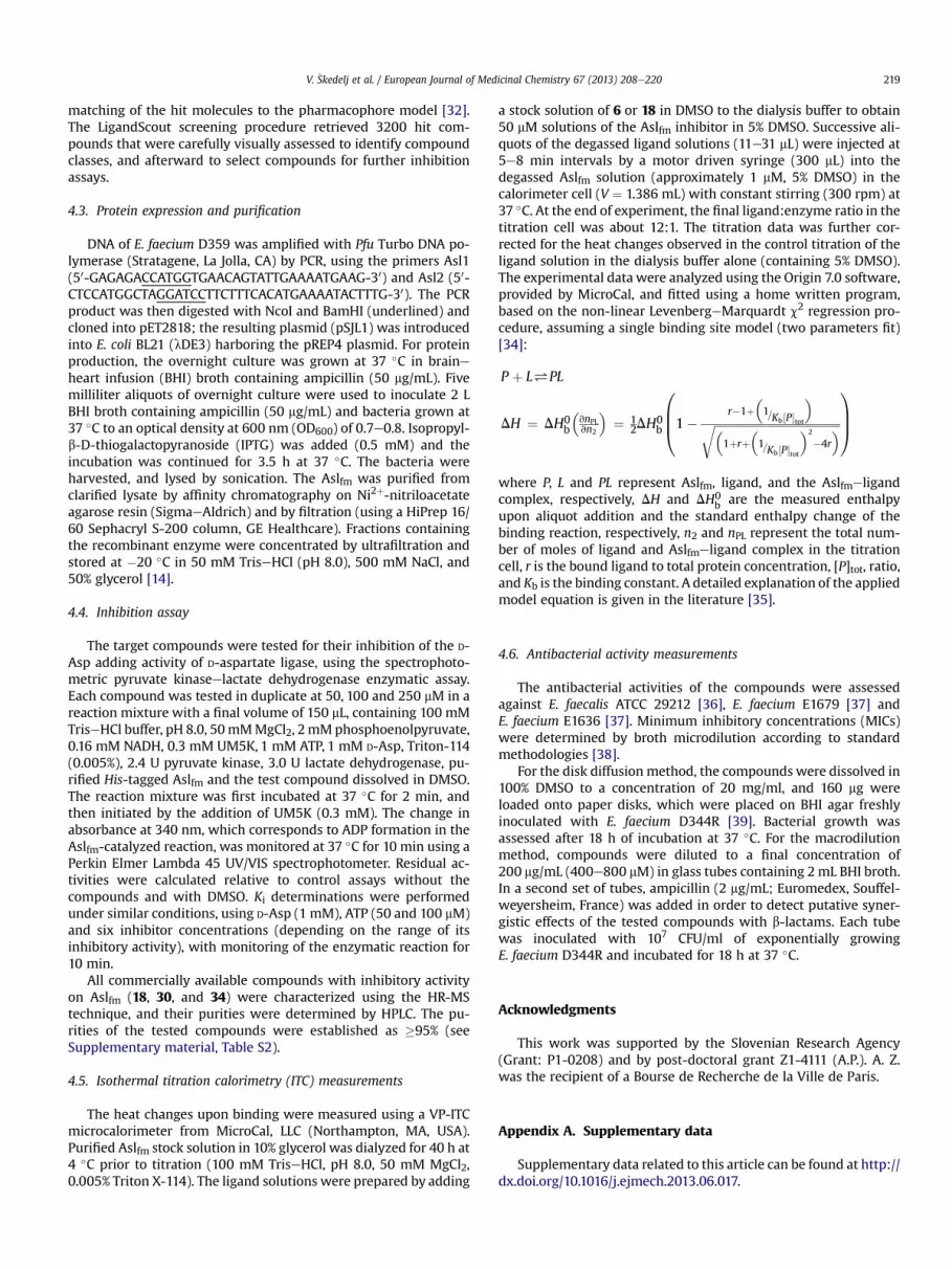
2.5. Biophysical characterization of the hit compounds 6 and 18 byisothermal titration calorimetry
Two of the compounds, 6 and 18, were tested by isothermaltitration calorimetry to obtain thermodynamic parameters of theirbinding into the Aslfm active site at 37 �C. Heat changes uponbinding were measured directly, corrected for the heats of dilution,and the model function was fitted to the experimental data points(see Experimental section).
Experiments for the two investigated compounds (6 and 18)yielded similar binding constants (Kb z 105 M�1) that are in fullagreement with their Ki values (Table 2) obtained in the kineticanalysis using ter-bi reaction mechanism. Interestingly, the ITCresults also revealed that the driving forces governing the bindingof these two different compound classes differ considerably(Table 4 and Fig. 6). The binding of compound 6 to Aslfm resultsfrom comparable and favorable enthalpic (DH¼�4.2 kcal/mol) andentropic (TDS ¼ 3.4 kcal/mol) contributions. On the other hand, thebinding of compound 18 is enthalpy driven (DH ¼ �13.8 kcal/mol)at the temperature investigated, and accompanied by a negativeentropy change (TDS¼�6.2 kcal/mol). Nevertheless, compensationof the opposing effects at 18 results in a binding affinity ðDG0
bÞsimilar to that of 6, although there are presumably more non-covalent bonds formed between 18 and the protein than for 6.Compound 18 obtained in the second drug design stage appears tobe more suitable for further optimization, as the increased rigidityof the molecule in its binding conformation should improve theentropy contributions upon retaining the specific and oriented in-teractions within the Aslfm active site [29,30]. Nevertheless,comparing of the Clog P values for compound 6 (Clog P ¼ 2.4) andcompound 18 (Clog P ¼ 4.0) a caveat must be stated that subse-quent optimization of this compound class must proceed toward
Fig. 6. Experimental titration curves and fits according to the single binding site model for b
compounds with comparable or preferably lower lipophilicitycharacter. The information that a single diclorophenyl substituentin compound 18 instead of two phenyl rings in compound 6 yieldedcomparable inhibitory activity is providing much optimism thataffinity of these compounds is not driven purely by lipophilicity. Forfurther rationally-driven optimization of these hits additionalstructural insights into their molecular recognition by the Aslfmenzyme will be of vital importance.
3. Conclusions
By combining classical screening and ligand-based pharmaco-phore modeling approaches in two drug-design stages, we havesuccessfully identified and experimentally characterized the firstinhibitors of the bacterial enzyme D-aspartate ligase from Entero-coccus faecium. Due to the limited knowledge of the structure ofAslfm, we focused on the ATP binding site of the enzyme andscreened a small collection of known ATP-competitive inhibitors ofthe large family of the ATP-grasp enzymes. A series of amino-oxazoles and thio-oxazoles derived from bacterial biotin carbox-ylase inhibitors showed low micromolar activity, with the mostpotent inhibitor (12) with a Ki of 2.9 mM with respect to ATP. In thesecond design stage, ligand-based pharmacophore modeling ap-proaches using newly acquired data for the inhibitory activities ofthe initially discovered series, followed by virtual screening,resulted in identification of new structural types of Aslfm inhibitors.This identified novel ATP-competitive inhibitors of Aslfm based on7-amino-9H-purines (18) and 7-amino-1H-pyrazolo[3,4-d]pyrimi-dine (30 and 34) scaffolds with Ki values in the low micromolarrange. The binding of two selected inhibitors (6 and 18) was sub-sequently confirmed and studied by isothermal titration calorim-etry, with determination of the standard thermodynamicparameters of the inhibitor binding to the enzyme active site.Compounds 6, 12, 18, 30 and 34 have Ki values in the low micro-molar range, and these represent the first inhibitors of Aslfm re-ported to date.We believe that this set of inhibitors discovered herewill provide novel lead compounds in antibacterial drug designefforts to combat E. faecium infections.
4. Experimental section
4.1. Chemistry
Chemicals were obtained from Acros, Aldrich Chemical Co.,Apollo Scientific, and Fluka, and they were used without furtherpurification. Analytical thin-layer chromatography was performedon silica gel Merck 60 F254 precoated plates (0.25 mm), visualizedwith ultraviolet light, ninhydrin and 2,4-dinitrophenylhydrazine.
inding of compounds 6 (a) and 18 (b) to the Aslfm active site at 37 �C. Scheme Caption.

V. �Skedelj et al. / European Journal of Medicinal Chemistry 67 (2013) 208e220 217
Flash column chromatography was carried out on silica gel 60(particle size 0.040e0.063 mm; Merck, Germany). Melting pointswere determined on a Reichert hot-stage microscope and are notcorrected. 1H NMR spectra were recorded on a Bruker Avance III400 MHz spectrometer at 295 K and 400 MHz, and are reported inppm using solvent as internal standard (DMSO-d6 at 2.50 ppm,CDCl3 at 7.26 ppm). 13C NMR spectra were recorded on a BrukerAvance III 400 MHz spectrometer at 295 K and 100 MHz, and arereported in ppm using solvent as the internal standard (DMSO-d6 at39.5 ppm). Mass spectra data were recorded using a Q-Tof Premierinstrument (Waters-Micromass, Manchester, UK). HPLC analyseswere performed on an Agilent Technologies HP 1100 instrument,with a G1365B UVevis detector, a G1316A thermostat, and aG1313A autosampler, using a Phenomenex Luna 5 mM C18 column(4.6 mm � 150 mm) at a flow rate of 1.0 mL/min. The eluent con-sisted of 0.1% trifluoroacetic acid inwater (A) andmethanol (B). Thegradient was 20% B to 80% B in 20 min. The purity of the testedcompounds was established to be �95%.
4.1.1. Synthesis of compound 1To the suspension of the oxazole ethyl ester (3.13 g, 20.0 mmol)
in water, 1 M NaOH (0.04 mL, 40.0 mmol) was added. The mixturewas stirred at room temperature for 3 h, and afterward adjusted topH 2e3 with 1 M HCl. The resulting precipitate 1 was filtered,washed with diethyl ether and dried overnight at 60 �C. Yield: 93%;white crystals, mp 210e212 �C. 1H NMR (400 MHz, DMSO-d6): 7.01(s, 2H, NH2), 7.13 (s. 1H, Ar-H), 12.11 (br s, 1H, COOH) ppm. HRMS(ESI): m/z [M þ H]þ calcd for C4H5N2O3 129.1347; found 129.1537.
4.1.2. Synthesis of compound 2The thiazole ethyl ester (1.0 g, 5.81 mmol) was treated with
0.5 M ethanolic sodium hydroxide solution (60 mL), overnight atroom temperature. The reaction mixture was then cooled in an icebath, and neutralized with acetic acid. The precipitated acid 2 wasfiltered, washed with diethyl ether, and dried overnight at 60 �C.Yield: 81%; off-white crystals, mp 225e227 �C (lit. [31] 223 �C). 1HNMR (400MHz, DMSO-d6): 2.35 (s, 3H, CH3), 7.61 (s, 2H, NH2),12.26(br s, 1H, COOH) ppm. HRMS (ESI): m/z [M þ H]þ calcd forC5H7N2O2S 159.1698; found 159.1663.
4.1.3. Synthesis of compound 3To the solution of the thiazole ethyl ester (0.80 g, 4.32 mmol) in
THF/water (2:1), NaOH was added (0.65 g, 16.3 mmol), and thereaction mixture was refluxed overnight. THF was removed underreduced pressure and afterward the reaction mixture wasneutralized with 1 M HCl. The resulting precipitate 1 was filtered,washed with diethyl ether and dried overnight at 60 �C. Yield: 86%;off-white crystals, mp 168e171 �C (lit. [32] 172e173 �C). 1H NMR(400 MHz, DMSO-d6): d 7.81 (s, 1H, oxazol-H), 8.99 (br s, 2H, NH2)ppm. HRMS (ESI): m/z [M þ H]þ calcd for C4H5N2O2S 145.1475;found 145.1502.
4.1.4. Synthesis of compound 4To the solutions of (1H-indol-5-yl)methanamine (0.50 g,
3.42 mmol) and 3,5-dibromobenzaldehyde (0.90 g, 3.42 mmol) inmethanol, NaCNBH3 (0.43 g, 6.84 mmol) was added, and themixture was stirred overnight under an argon atmosphere. Thesolvent was removed under reduced pressure and afterward theresidue was dissolved in ethyl acetate (50 mL) and washed withwater (3 � 20 mL). The organic phase was dried over Na2SO4, andfiltered, and the solvent was removed under reduced pressure. Thecrude product was purified by flash column chromatography usingpetrolether/ethyl acetate (3:1) as eluent. Yield: 70%, off-whitecrystals, mp 186e188 �C. 1H NMR (400 MHz, CDCl3): d 3.79 (s, 2H,CH2), 3.91 (s, 2H, CH2), 6.54e6.59 (m, 1H, indole-H-3), 7.20 (dd,
J ¼ 8.4, 1.6 Hz, 1H), 7.25 (t, J ¼ 2.7 Hz, 1H), 7.29 (s, 1H, NHCH2), 7.40(d, J ¼ 8.4 Hz, 1H, Ar-H), 7.48 (d, J ¼ 2.0 Hz, 2H, Ar-2,6), 7.57(t, J ¼ 2.0 Hz, 1H, Ar-4-H), 7.59e7.65 (m, 1H, indole-H), 10.29 (br s,1H, indole-H-1) ppm. HRMS (ESI): m/z [M þ H]þ calcd forC16H15N2Br2, 395.1254; found, 395.1259.
4.1.5. Synthesis of compounds 5e14The suspension of acid (2.00 mmol) in DMF (10 mL) was cooled
to 0 �C in an ice bath. N-methylmorpholine (0.49 mL, 4.40 mmol)and TBTU (0.84 g, 2.60mmol) were added, and the reactionmixturewas stirred at 0 �C for 0.5 h. Then it was allowed to reach roomtemperature, and amine (2.00 mmol) was added to the solution.The reaction mixture was stirred overnight at room temperature,after which the solvent was evaporated under reduced pressure.The residuewas dissolved in ethyl acetate (40mL) andwashedwitha saturated aqueous solution of NaHCO3 (3 � 20 mL), 10% citric acid(3 � 20 mL) and brine (20 mL). The organic phase was dried overNa2SO4, and filtered, and the solvent was removed under reducedpressure. The crude product was purified by crystallization or flashcolumn chromatography.
4.1.5.1. 2-Amino-N-benzyl-N-butyloxazole-5-carboxamide (5).The crude product was crystallized from CH2Cl2. Yield: 46%, off-white crystals, mp 134e137 �C. 1H NMR (400 MHz, DMSO-d6):d 0.85 (t, J ¼ 7.4 Hz, 3H, CH3), 1.19e1.31 (m, 2H, CH2CH3), 1.45e1.58(m, 2H, CH2CH2CH2), 3.29e3.43 (m, 2H, CH2CH2CH2), 4.69 (s, 2H,Ar-CH2), 7.18 (br s, 2H, NH2), 7.21e7.40 (m, 6H, Ar-H þ oxazole-H)ppm. 13C NMR (400 MHz, DMSO-d6): d 13.69, 19.45, 29.80, 46.45,49.28, 127.05, 128.53, 133.81, 137.32, 137.86, 158.04, 162.45 ppm.HRMS (ESI): m/z [M þ H]þ calcd for C15H20N3O2, 274.1556; found,274.1547. HPLC tR ¼ 15.965 min (96.22% at 220 nm, 98.78% at254 nm).
4.1.5.2. 2-Amino-N,N-dibenzyloxazole-5-carboxamide (6). Thecrude product was crystallized from CH2Cl2. Yield: 57%, whitecrystals, mp 216e219 �C. 1H NMR (400 MHz, DMSO-d6): d 5.77 (s,4H, 2� CH2), 7.12 (s, 1H, oxazole-H), 7.18e7.42 (m,12H, Ar-HþNH2)ppm. 13C NMR (400 MHz, DMSO-d6): d 49.50, 127.03, 127.81, 128.63,134.40, 136.89, 137.21, 158.47, 162.66 ppm. d HRMS (ESI): m/z[M þ H]þ calcd for C18H18N3O2, 308.1399; found, 308.1389. HPLCtR ¼ 16.662 min (98.19% at 220 nm, 99.34% at 254 nm).
4.1.5.3. 2-Amino-N,N-dibutyloxazole-5-carboxamide (7). The crudeproduct was crystallized from CH2Cl2. Yield: 38%, yellow crystals,mp 121e124 �C. 1H NMR (400 MHz, DMSO-d6): d 0.89 (t, J ¼ 7.4 Hz,6H, 2� CH3), 1.22e1.34 (m, 4H, 2� CH2CH3), 1.45e1.56 (m, 4H,CH2CH2CH2), 3.29e3.44 (m, 4H, CH2CH2CH2), 7.18 (br s, 2H, NH2),7.23 (s, 1H, oxazole-H) ppm. 13C NMR (400 MHz, DMSO-d6): 13.74,19.52, 30.09, 46.41, 133.14, 137.72, 157.52, 162.22 ppm. HRMS (ESI):m/z [M þ H]þ calcd for C12H22N3O2, 240.1712; found, 240.1710.HPLC tR ¼ 16.210 min (98.18% at 220 nm, 98.87% at 254 nm).
4.1.5.4. 2-Amino-N,N-bis(pyridin-2-ylmethyl)oxazole-5-carboxamide(8). The crude product was purified with flash column chroma-tography using chloroform/methanol (9:1) as eluent. Yield: 21%,yellow crystals, mp 125e128 �C. 1H NMR (400 MHz, DMSO-d6):d 4.82 (s, 4H, 2� CH2), 7.20 (s, 1H, oxazole-H), 7.23e7.40 (m, 6H, pyr-H þ NH2), 7.72e7.82 (m, 2H, pyr-H), 8.49e8.57 (m, 2H, pyr-H) ppm.13C NMR (400MHz, DMSO-d6): d 2.28,121.56,122.41,134.32,136.91,137.01, 149.23, 156.98, 158.60, 162.63 ppm. HRMS (ESI): m/z[M þ H]þ calcd for C16H16N5O2, 310.1304; found, 310.1309. HPLCtR ¼ 15.965 min (96.22% at 220 nm, 98.78% at 254 nm).
4.1.5.5. 2-Amino-N-ethyl-N-(pyridin-4-ylmethyl)oxazole-5-carboxamide (9). The crude product was purified with flash column

V. �Skedelj et al. / European Journal of Medicinal Chemistry 67 (2013) 208e220218
chromatography using chloroform/methanol (9:1) as eluent. Yield:39%, pale yellow crystals, mp 162e166 �C. 1H NMR (400 MHz,DMSO-d6): d 1.15 (t, J ¼ 6.8 Hz, 3H, CH2CH3), 3.48 (q, J ¼ 6.8 Hz, 2H,CH2CH3), 4.69 (s, 2H, CH2), 7.11 (s, 1H, oxazole-H), 7.24 (d, J¼ 6.1 Hz,pyr-H-3,5), 7.28 (br s, 2H, NH2), 8.52 (d, J ¼ 6.1 Hz, pyr-H-2,6) ppm.13C NMR (400 MHz, DMSO-d6): d 13.49, 42.35, 48.39, 122.04, 134.17,137.04, 147.31, 149.68, 157.96, 162.60 ppm. HRMS (ESI): m/z[M þ H]þ calcd for C12H15N4O2, 247.1195; found, 247.1203. HPLCtR ¼ 9.277 min (97.09% at 220 nm, 98.99% at 254 nm).
4.1.5.6. 2-Amino-N-(1H-benzo[d]imidazol-2-yl)oxazole-5-carboxamide (10). The crude product was purified with flash col-umn chromatography using ethyl acetate/methanol (8:1) as eluent.Yield: 26%, off-white crystals, mp > 300 �C. 1H NMR (400 MHz,DMSO-d6): d 7.11 (br s, 2H, NH2), 7.36e7.51 (m, 5H, Ar-H þ oxazole-H), 7.77 (s, 1H, CONH) ppm. 13C NMR (400 MHz, DMSO-d6): d 114.6,123.7, 135.8, 136.2, 142.5, 147.4, 162.7, 166.2 ppm. HRMS (ESI): m/z[M þ H]þ calcd for C11H10N5O2, 244.0834; found, 244.0838. HPLCtR ¼ 18.464 min (98.65% at 220 nm, 99.09% at 254 nm).
4.1.5.7. N-((1H-Indol-5-yl)methyl)-2-aminooxazole-5-carboxamide(11). The crude product was purified with flash column chroma-tography using chloroform/methanol (9:1) as eluent. Yield: 46%,light-brown crystals, mp 207e211 �C. 1H NMR (400 MHz, DMSO-d6): d 4.44 (d, J ¼ 6.0 Hz, 2H, CH2), 6.36e6.40 (m, 1H, indole-H-3),7.08 (dd, J ¼ 8.4, 1.6 Hz, 1H), 7.21 (br s, 2H, NH2), 7.33e7.42 (m,2H, oxazole-H þ indole-H), 7.45 (s, 1H, indole-H), 7.49 (s, 1H,indole-H), 8.46 (t, J¼ 6.0 Hz, 1H, CONH), 11.09 (br s, 1H, indole-H-1)ppm. 13C NMR (400 MHz, DMSO-d6): d 42.19, 100.85, 111.16, 118.69,120.97, 125.54, 127.51, 129.81, 131.32, 134.96, 138.36, 156.90,162.43 ppm. HRMS (ESI): m/z [M þ H]þ calcd for C13H13N4O2,257.1039; found, 257.1032. HPLC tR ¼ 8.482 min (98.97% at 220 nm,99.54% at 254 nm).
4.1.5.8. N-((1H-Indol-5-yl)methyl)-2-amino-N-(3,5-dibromobenzyl)oxazole-5-carboxamide (12). The crude product was purified withflash column chromatography using chloroform/ethyl acetate(1:10) as eluent. Yield: 68%, pink crystals, mp 187e191 �C. 1HNMR (400 MHz, DMSO-d6): d 4.62 (s, 2H, CH2), 4.80 (s, 2H, CH2),6.40 (m, 1H, indole-H-3), 6.98 (br s, 2H, NH2), 7.16 (s, 1H, oxazol-H), 7.27e7.49, (m, 6H, Ar-H), 7.72 (s, 1H, Ar-40-H), 11.11 (br s, 1H,indole-H-1) ppm. 13C NMR (400 MHz, DMSO-d6): d 51.14, 59.75,101.00, 111.70, 118.64, 120.49, 122.52, 125.83, 126.97, 127.75,131.96, 134.69, 135.25, 136.83, 140.62, 142.72, 158.47, 162.71 ppm.HRMS (ESI): m/z [M þ H]þ calcd for C20H17N4O2Br2, 502.9718;found, 502.9712. HPLC tR ¼ 18.464 min (98.65% at 220 nm,99.09% at 254 nm).
4.1.5.9. 2-Amino-N,N-dibenzylthiazole-5-carboxamide (13). Thecrude product was purified with flash column chromatographyusing ethyl acetate/hexane (1:1) as eluent. Yield: 41%, yellowcrystals, mp 114e118 �C. 1H NMR (400 MHz, DMSO-d6): d 4.67 (s,4H, 2� CH2), 7.16 (s, 1H, thiazole-H), 7.21e7.43 (m, 10H, Ar-H), 7.54(br s, 2H, NH2) ppm. 13C NMR (400 MHz, DMSO-d6): d 50.24, 119.63,126.99, 127.21, 128.68, 137.14, 142.68, 162.63, 171.61 ppm. HRMS(ESI): m/z [M þ H]þ calcd for C18H18N3OS, 324.1171; found,324.1176. HPLC tR ¼ 18.464 min (98.65% at 220 nm, 99.09% at254 nm).
4.1.5.10. 2-Amino-N,N-dibenzyl-4-methylthiazole-5-carboxamide(14). The crude product was purified with flash column chroma-tography using ethyl acetate as eluent. Yield: 53%, yellow crystals,mp 125e128 �C. 1H NMR (400 MHz, DMSO-d6): d 2.21 (s, 3H, CH3),4.56 (s, 4H, 2� CH2), 7.18e7.48 (m, 13H, Ar-H þ NH2 þ thiazole-H)ppm. 13C NMR (400 MHz, DMSO-d6): d 16.67, 49.51, 109.85, 127.31,
128.63, 132.32, 137.06, 151.04, 164.75, 167.77 ppm. HRMS (ESI): m/z[M þ H]þ calcd for C19H20N3OS, 338.1327; found, 338.1321. HPLCtR ¼ 15.613 min (100.0% at 220 nm, 100.0% at 254 nm).
4.2. Ligand-based pharmacophore modeling and virtual screeningprocedures
Ligand-based pharmacophore modeling and virtual screeningwere performed using the LigandScout software [21]. The initialconstructed conformations of the active compounds 6 and 12 wereminimized using the MMFF94 force field, and imported into theligand-based module available in Ligandscout. Five-hundredunique conformations were calculated for each structure byapplying the following settings of the LigandScout conformergenerator coupled to the OMEGA software: maximum number ofoutput conformers per molecule¼ 500; RMS threshold to duplicateconformers¼ 0.4�A; maximum number of all generated conformersper molecule ¼ 30,000; and maximum number of intermediateconformers per molecule ¼ 4000. Subsequently, the conformerswere dynamically aligned [22] to yield 10 ligand-based mergedpharmacophore models. A scoring function that combined phar-macophore fit and atom shape overlap was used to assess themodels produced. The ligand-based pharmacophore model withthe highest score of 0.9311 was selected for further use (seeSupplementary material, Fig. S1, for a complete merged Pharma-cophore model). Visual inspection revealed that most of theremaining nine derived pharmacophore models closely resembledthis model. The initial number of available merged pharmacophoricfeatures was reduced to obtain on the one hand sufficient molec-ular recognition pattern required for the ligand binding, and toincrease on the other hand the chemical space identified by thepharmacophore model [33]. The reduced ligand-based pharmaco-phore model was used in the large-scale virtual screeningcampaign. The derived pharmacophore model (see Fig. 3) consistedof two hydrophobic interaction spheres, two hydrogen bond ac-ceptors, and one hydrogen bond donor. Exclusion volume sphereswere also added, to approximate the steric circumference of theAslfm binding site. The discriminatory performance of the derivedpharmacophore model was validated by a screening experimentagainst 100 decoy molecules generated for both active compounds6 and 12 (50 decoys/molecule) using the Decoyfinder software [26].Decoyfinder generates sets of decoy molecules for defined activeligands. Decoys have similar number of rotational bonds, hydrogenbond acceptors, hydrogen bond donors, log P value and molecularweight as active molecules but are chemically different, which isdefined by a maximum Tanimoto value threshold between activeligand and decoy molecule MACCS fingerprints [26]. The pharma-cophore model successfully identified the both active compoundsin the correct orientation and none of the generated decoy mole-cules were identified as potential hits.
The pharmacophore model was used to screen approximately5.5 million commercially available compounds, all of which werepreviously converted into multifunctional format (25 conformersfor each compound in the database) using the LigandScoutscreening module. The conformers of the molecules in thescreening library were generated using the idbgen module that isavailable in Ligandscout, coupled with the OMEGA software. Thedefault high-throughput settings were used for the library gener-ation: maximum number of output conformers per molecule ¼ 25;RMS threshold to duplicate conformers ¼ 0.8�A; maximum numberof all generated conformers per molecule ¼ 30,000; and maximumnumber of intermediate conformers per molecule ¼ 4000. In thevirtual screening experiments, each compound had to fulfill all ofthe derived pharmacophore constraints to be identified as a virtualhit. Pharmacophore Fit scoring function was used to score the
V. �Skedelj et al. / European Journal of Medicinal Chemistry 67 (2013) 208e220 219
matching of the hit molecules to the pharmacophore model [32].The LigandScout screening procedure retrieved 3200 hit com-pounds that were carefully visually assessed to identify compoundclasses, and afterward to select compounds for further inhibitionassays.
4.3. Protein expression and purification
DNA of E. faecium D359 was amplified with Pfu Turbo DNA po-lymerase (Stratagene, La Jolla, CA) by PCR, using the primers Asl1(50-GAGAGACCATGGTGAACAGTATTGAAAATGAAG-30) and Asl2 (50-CTCCATGGCTAGGATCCTTCTTTCACATGAAAATACTTTG-30). The PCRproduct was then digested with NcoI and BamHI (underlined) andcloned into pET2818; the resulting plasmid (pSJL1) was introducedinto E. coli BL21 (lDE3) harboring the pREP4 plasmid. For proteinproduction, the overnight culture was grown at 37 �C in braineheart infusion (BHI) broth containing ampicillin (50 mg/mL). Fivemilliliter aliquots of overnight culture were used to inoculate 2 LBHI broth containing ampicillin (50 mg/mL) and bacteria grown at37 �C to an optical density at 600 nm (OD600) of 0.7e0.8. Isopropyl-b-D-thiogalactopyranoside (IPTG) was added (0.5 mM) and theincubation was continued for 3.5 h at 37 �C. The bacteria wereharvested, and lysed by sonication. The Aslfm was purified fromclarified lysate by affinity chromatography on Ni2þ-nitriloacetateagarose resin (SigmaeAldrich) and by filtration (using a HiPrep 16/60 Sephacryl S-200 column, GE Healthcare). Fractions containingthe recombinant enzyme were concentrated by ultrafiltration andstored at �20 �C in 50 mM TriseHCl (pH 8.0), 500 mM NaCl, and50% glycerol [14].
4.4. Inhibition assay
The target compounds were tested for their inhibition of the D-Asp adding activity of D-aspartate ligase, using the spectrophoto-metric pyruvate kinaseelactate dehydrogenase enzymatic assay.Each compound was tested in duplicate at 50, 100 and 250 mM in areaction mixture with a final volume of 150 mL, containing 100 mMTriseHCl buffer, pH 8.0, 50mMMgCl2, 2mMphosphoenolpyruvate,0.16 mM NADH, 0.3 mM UM5K, 1 mM ATP, 1 mM D-Asp, Triton-114(0.005%), 2.4 U pyruvate kinase, 3.0 U lactate dehydrogenase, pu-rified His-tagged Aslfm and the test compound dissolved in DMSO.The reaction mixture was first incubated at 37 �C for 2 min, andthen initiated by the addition of UM5K (0.3 mM). The change inabsorbance at 340 nm, which corresponds to ADP formation in theAslfm-catalyzed reaction, was monitored at 37 �C for 10 min using aPerkin Elmer Lambda 45 UV/VIS spectrophotometer. Residual ac-tivities were calculated relative to control assays without thecompounds and with DMSO. Ki determinations were performedunder similar conditions, using D-Asp (1 mM), ATP (50 and 100 mM)and six inhibitor concentrations (depending on the range of itsinhibitory activity), with monitoring of the enzymatic reaction for10 min.
All commercially available compounds with inhibitory activityon Aslfm (18, 30, and 34) were characterized using the HR-MStechnique, and their purities were determined by HPLC. The pu-rities of the tested compounds were established as �95% (seeSupplementary material, Table S2).
4.5. Isothermal titration calorimetry (ITC) measurements
The heat changes upon binding were measured using a VP-ITCmicrocalorimeter from MicroCal, LLC (Northampton, MA, USA).Purified Aslfm stock solution in 10% glycerol was dialyzed for 40 h at4 �C prior to titration (100 mM TriseHCl, pH 8.0, 50 mM MgCl2,0.005% Triton X-114). The ligand solutions were prepared by adding
a stock solution of 6 or 18 in DMSO to the dialysis buffer to obtain50 mM solutions of the Aslfm inhibitor in 5% DMSO. Successive ali-quots of the degassed ligand solutions (11e31 mL) were injected at5e8 min intervals by a motor driven syringe (300 mL) into thedegassed Aslfm solution (approximately 1 mM, 5% DMSO) in thecalorimeter cell (V ¼ 1.386 mL) with constant stirring (300 rpm) at37 �C. At the end of experiment, the final ligand:enzyme ratio in thetitration cell was about 12:1. The titration data was further cor-rected for the heat changes observed in the control titration of theligand solution in the dialysis buffer alone (containing 5% DMSO).The experimental data were analyzed using the Origin 7.0 software,provided by MicroCal, and fitted using a home written program,based on the non-linear LevenbergeMarquardt c2 regression pro-cedure, assuming a single binding site model (two parameters fit)[34]:
P þ L#PL
DH ¼ DH0b
�vnPLvn2
�¼ 1
2DH0b
0BB@1�
r�1þ�1=Kb½P�tot
�ffiffiffiffiffiffiffiffiffiffiffiffiffiffiffiffiffiffiffiffiffiffiffiffiffiffiffiffiffiffiffiffiffiffiffiffiffiffiffiffiffiffiffi�1þrþ
�1=Kb ½P�tot
�2
�4r�r1CCA
where P, L and PL represent Aslfm, ligand, and the Aslfmeligandcomplex, respectively, DH and DH0
b are the measured enthalpyupon aliquot addition and the standard enthalpy change of thebinding reaction, respectively, n2 and nPL represent the total num-ber of moles of ligand and Aslfmeligand complex in the titrationcell, r is the bound ligand to total protein concentration, [P]tot, ratio,and Kb is the binding constant. A detailed explanation of the appliedmodel equation is given in the literature [35].
4.6. Antibacterial activity measurements
The antibacterial activities of the compounds were assessedagainst E. faecalis ATCC 29212 [36], E. faecium E1679 [37] andE. faecium E1636 [37]. Minimum inhibitory concentrations (MICs)were determined by broth microdilution according to standardmethodologies [38].
For the disk diffusion method, the compounds were dissolved in100% DMSO to a concentration of 20 mg/ml, and 160 mg wereloaded onto paper disks, which were placed on BHI agar freshlyinoculated with E. faecium D344R [39]. Bacterial growth wasassessed after 18 h of incubation at 37 �C. For the macrodilutionmethod, compounds were diluted to a final concentration of200 mg/mL (400e800 mM) in glass tubes containing 2 mL BHI broth.In a second set of tubes, ampicillin (2 mg/mL; Euromedex, Souffel-weyersheim, France) was added in order to detect putative syner-gistic effects of the tested compounds with b-lactams. Each tubewas inoculated with 107 CFU/ml of exponentially growingE. faecium D344R and incubated for 18 h at 37 �C.
Acknowledgments
This work was supported by the Slovenian Research Agency(Grant: P1-0208) and by post-doctoral grant Z1-4111 (A.P.). A. Z.was the recipient of a Bourse de Recherche de la Ville de Paris.
Appendix A. Supplementary data
Supplementary data related to this article can be found at http://dx.doi.org/10.1016/j.ejmech.2013.06.017.

V. �Skedelj et al. / European Journal of Medicinal Chemistry 67 (2013) 208e220220
References
[1] H.W. Boucher, G.H. Talbot, J.S. Bradley, J.E. Edwards, D. Gilbert, L.B. Rice, et al.,Bad bugs, no drugs: no ESKAPE! an update from the Infectious Diseases So-ciety of America, Clin. Infect. Dis. 48 (2009) 1e12.
[2] R.C. Moellering Jr., Advances in antibacterial therapy, Transplant. Proc. 43(2011) 2441e2442.
[3] G.J. Patti, S.J. Kim, J. Schaefer, Characterization of the peptidoglycan ofvancomycin-susceptible Enterococcus faecium, Biochemistry 47 (2008) 8378e8385.
[4] R. Leclercq, E. Derlot, J. Duval, P. Courvalin, Plasmid-mediated resistance tovancomycin and teicoplanin in Enterococcus faecium, N. Engl. J. Med. 319(1988) 157e161.
[5] W. Vollmer, D. Blanot, M.A. de Pedro, Peptidoglycan structure and architec-ture, FEMS Microbiol. Rev. 32 (2008) 149e167.
[6] D. Mengin-Lecreulx, B. Lemaitre, Structure and metabolism of peptidoglycanand molecular requirements allowing its detection by the Drosophila innateimmune system, J. Endotoxin Res. 11 (2005) 105e111.
[7] P. Macheboeuf, C. Contreras-Martel, V. Job, O. Dideberg, A. Dessen, Penicillinbinding proteins: key players in bacterial cell cycle and drug resistance pro-cesses, FEMS Microbiol. Rev. 30 (2006) 673e691.
[8] H. Barreteau, A. Kovac, A. Boniface, M. Sova, S. Gobec, D. Blanot, Cytoplasmicsteps of peptidoglycan biosynthesis, FEMS Microbiol. Rev. 32 (2008) 168e207.
[9] K.H. Schleifer, O. Kandler, Peptidoglycan types of bacterial cell walls and theirtaxonomic implications, Bacteriol. Rev. 36 (1972) 407e477.
[10] T.E. Benson, D.B. Prince, V.T. Mutchler, K.A. Curry, A.M. Ho, R.W. Sarver, et al.,X-ray crystal structure of Staphylococcus aureus FemA, Structure 10 (2002)1107e1115.
[11] J.-L. Mainardi, R. Villet, T.D. Bugg, C. Mayer, M. Arthur, Evolution of peptido-glycan biosynthesis under the selective pressure of antibiotics in gram-positive bacteria, FEMS Microbiol. Rev. 32 (2008) 386e408.
[12] W. Staudenbauer, J.L. Strominger, Activation of D-aspartic acid for incorpora-tion into peptidoglycan, J. Biol. Chem. 247 (1972) 5095e5102.
[13] W. Staudenbauer, E. Willoughby, J.L. Strominger, Further studies of the D-aspartic acid-activating enzyme of Streptococcus faecalis and its attachment tothe membrane, J. Biol. Chem. 247 (1972) 5289e5296.
[14] S. Bellais, M. Arthur, L. Dubost, J.-E. Hugonnet, L. Gutmann, J. van Heijenoort,et al., Aslfm, the D-aspartate ligase responsible for the addition of D-asparticacid onto the peptidoglycan precursor of Enterococcus faecium, J. Biol. Chem.281 (2006) 11586e11594.
[15] M.Y. Galperin, E.V. Koonin, A diverse superfamily of enzymes with ATP-dependent carboxylate-amine/thiol ligase activity, Protein Sci. 6 (1997)2639e2643.
[16] S. Dutka-Malen, C. Molinas, M. Arthur, P. Courvalin, Sequence of the vanCgene of Enterococcus gallinarum BM4174 encoding a D-alanine:D-alanineligase-related protein necessary for vancomycin resistance, Gene 112(1992) 53e58.
[17] V. �Skedelj, T. Toma�si�c, L.P. Ma�si�c, A. Zega, ATP-binding site of bacterialenzymes as a target for antibacterial drug design, J. Med. Chem. 54 (2011)915e929.
[18] I. Mochalkin, J.R. Miller, L. Narasimhan, V. Thanabal, P. Erdman, P.B. Cox, et al.,Discovery of antibacterial biotin carboxylase inhibitors by virtual screeningand fragment-based approaches, ACS Chem. Biol. 4 (2009) 473e483.
[19] J.E. Lindsley, Use of a real-time, coupled assay to measure the ATPase activityof DNA topoisomerase II, Methods Mol. Biol. 95 (2001) 57e64.
[20] W.R. McClure, A kinetic analysis of coupled enzyme assays, Biochemistry 8(1969) 2782e2786.
[21] B. Hess, B. Wurster, Transient time of the pyruvate kinaseelactate dehydro-genase system of rabbit muscle in vitro, FEBS Lett. 9 (1970) 73e77.
[22] T. Langer, R.D. Hoffmann, Pharmacophores and Pharmacophore Searches,Wiley VCH, Weinheim, Germany, 2006.
[23] A. Perdih, A. Kovac, G. Wolber, D. Blanot, S. Gobec, T. Solmajer, Discovery ofnovel benzene 1,3-dicarboxylic acid inhibitors of bacterial MurD and MurEligases by structure-based virtual screening approach, Bioorg. Med. Chem.Lett. 19 (2009) 2668e2673.
[24] G. Wolber, T. Langer, LigandScout: 3-D pharmacophores derived fromprotein-bound ligands and their use as virtual screening filters, J. Chem. Inf.Model 45 (2005) 160e169.
[25] G.Wolber, A.A. Dornhofer, T. Langer, Efficient overlay of small organic moleculesusing 3D pharmacophores, J. Comput. Aided Mol. Des. 20 (2006) 773e788.
[26] A. Cereto-Massague, L. Guasch, C. Valls, M. Mulero, G. Pujadas, S. Garcia-Vallve, DecoyFinder: an easy-to-use python GUI application for buildingtarget-specific decoy sets, Bioinformatics 28 (2012) 1661e1662.
[27] V. �Skedelj, et al., in press.[28] S. Bevc, J. Konc, J. Stojan, M. Hodo�s�cek, M. Penca, M. Praprotnik, et al., ENZO: a
web tool for derivation and evaluation of kinetic models of enzyme catalyzedreactions, PLoS One 6 (2011) e22265.
[29] E. Edink, C. Jansen, R. Leurs, I.J.P. de Esch, The heat is on: thermodynamicanalysis in fragment-based drug discovery, Drug Discov. Today Technol. 7(2010) 189e201.
[30] J.E. Ladbury, G. Klebe, E. Freire, Adding calorimetric data to decision making inlead discovery: a hot tip, Nat. Rev. Drug Discov. 9 (2010) 23e27.
[31] W. Knauf, Chemotherapeutic nitro heterocycles. XXII. Synthesis and antimi-crobial activity of 2-nitrothiazole-5-carboxamide, Eur. J. Med. Chem. (1975)533e534.
[32] K.E. Rao, Y. Bathini, J.W. Lown, Synthesis of novel thiazole-containing DNAminor groove binding oligopeptides related to the antibiotic distamycin,J. Org. Chem. 55 (1990) 728e737.
[33] J. Kirchmair, P. Markt, S. Distinto, G. Wolber, T. Langer, Evaluation of theperformance of 3D virtual screening protocols: RMSD comparisons, enrich-ment assessments, and decoy selectionewhat can we learn from earliermistakes? J. Comput. Aided Mol. Des. 22 (2008) 213e228.
[34] W.H. Press, B.P. Flannery, S.A. Teukolsky, W.T. Vetterling, Numerical Recipes,Cambridge University Press, Oxford, 1992.
[35] V. �Skedelj, Emilija Arsovska, T. Toma�si�c, Ana Krofli�c, Vesna Hodnik,Martina Hrast, et al., 6-Arylpyrido[2,3-d]pyrimidines as novel ATP-competitive inhibitors of bacterial D-Alanine:D-Alanine ligase, PLoS One(2012) e39922.
[36] M.-T. Arias-Moliz, P. Baca, S. Ordóñez-Becerra, M.-P. González-Rodríguez, C.-M. Ferrer-Luque, Eradication of enterococci biofilms by lactic acid alone andcombined with chlorhexidine and cetrimide, Med. Oral Patol. Oral Cir. Bucal(2012).
[37] W. van Schaik, J. Top, D.R. Riley, J. Boekhorst, J.E.P. Vrijenhoek,C.M.E. Schapendonk, et al., Pyrosequencing-based comparative genomeanalysis of the nosocomial pathogen Enterococcus faecium and identificationof a large transferable pathogenicity island, BMC Genomics 11 (2010) 239.
[38] National Committee for Clinical Laboratory Standards (NCCLS), Methods forDilution Antimicrobial Susceptibility Tests for Bacteria that Grow Aerobically,fourth ed., vol. 17, 1997.
[39] W. Zorzi, X.Y. Zhou, O. Dardenne, J. Lamotte, D. Raze, J. Pierre, et al., Structureof the low-affinity penicillin-binding protein 5 PBP5fm in wild-type andhighly penicillin-resistant strains of Enterococcus faecium, J. Bacteriol. 178(1996) 4948e4957.
[40] A typical run in the absence of inhibitors was implemented under the followingproject number enzo.cmm.ki.si/kinetic.php?uwd¼120727444&load¼true (click’Set Parameters’ and ’Start’).
[41] See enzo.cmm.ki.si/kinetic.php?uwd¼120326994&load¼true for compound6; click ’Set Parameters’ and ’Start’.


