Embed Size (px)
Citation preview
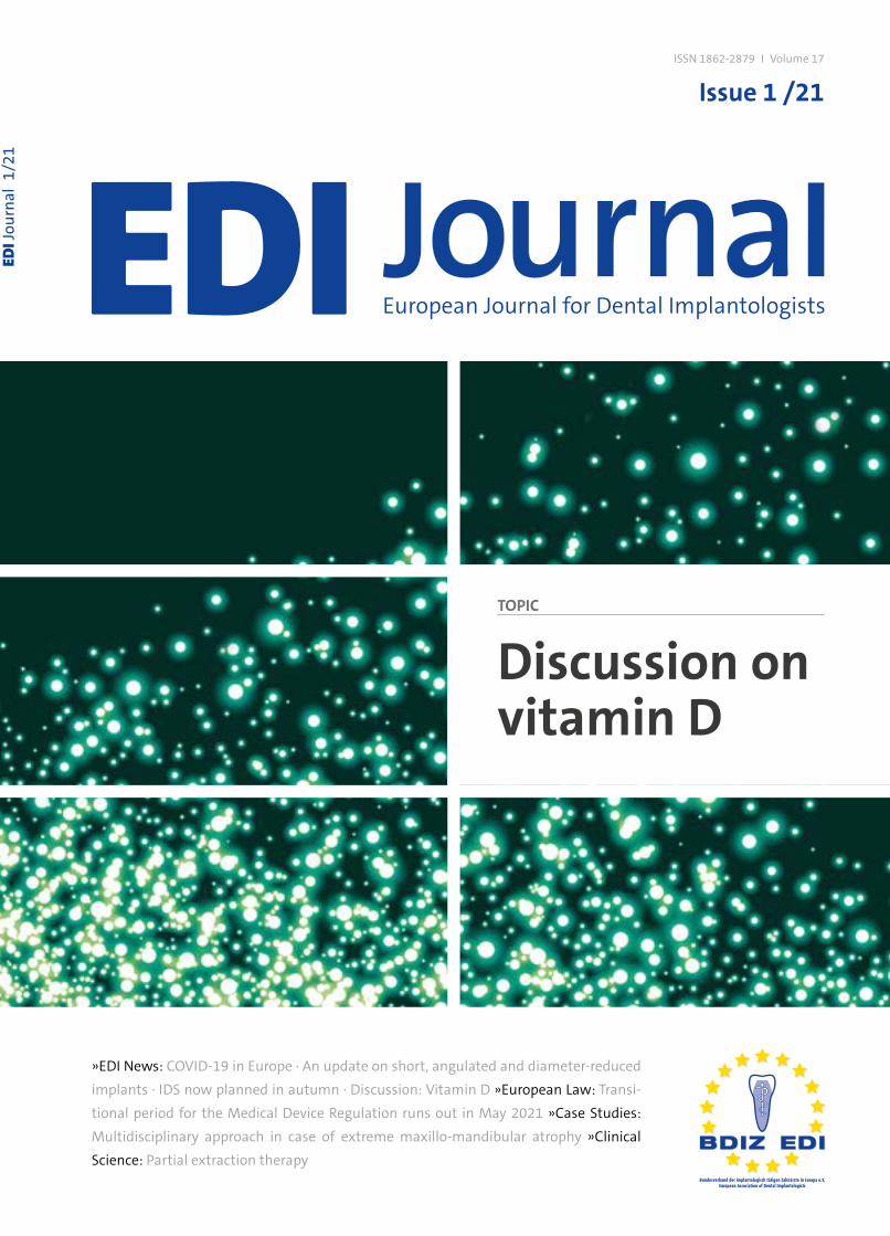
»EDI News: COVID-19 in Europe · An update on short, angulated and diameter-reduced implants · IDS now planned in autumn · Discussion: Vitamin D »European Law: Transi-tional period for the Medical Device Regulation runs out in May 2021 »Case Studies: Multidisciplinary approach in case of extreme maxillo-mandibular atrophy »Clinical Science: Partial extraction therapy
European Journal for Dental Implantologists
ISSN 1862-2879 I Volume 17
Issue 1 /21
EDI J
ourn
al 1
/21
TOPIC
Discussion on vitamin D
2EDITORIAL
A0030/EN/A/00 01/21
Experience Straumann® Zygomatic Implant System www.straumann.com/zygomatic
Straumann® Zygomatic Implant SystemDesigned by experts with the patient in mind.
STRAIGHTFORWARD PROSTHETICS³
Prosthetic portfolio with a single implant connection;
compatible with Straumann® Edentulous Solutions.
ANATOMICALLY BASED DESIGNS
Two designs with a unique combination of features to
respect patients’ anatomical structures.⁵
IMMEDIATE LOADING¹,²,⁴
Graftless solution that pro-vides primary stability and allows immediate loading.
FROM EXPERTS FOR EXPERTS
Bringing experts together to provide a solution you can
trust in.
REFERENCES
1 Tuminelli FJ, Walter LR, Neugarten J, Bedrossian E. Immediate loading of zygomatic implants: A systematic review of implant survival, prosthesis survival and potential complications. Eur J Oral Implantol. 2017;10 Suppl 1:79-87. 2 Fatigue Testing Report – ZAGA Implant System. 3 Boyes-Var-ley JG, Howes DG, Lownie JF, Blackbeard GA. Surgical Modifications to the Branemark Zygomaticus Protocol in the treatment of the severely resorbed Maxilla: A Clinical Report. Int. J Oral Maxillofac. Implants 2003;18: 232–237. 4 Jung RE, Al-Nawas B, Araujo M, Avila-Ortiz G, Barter S, Brodala N, Chap-puis V, Chen B, De Souza A, Almeida RF, Fickl S, Finelle G, Ganeles J, Gholami H, Hammerle C, Jensen S, Jokstad A, Katsuyama H, Kleinheinz J, Kunavisarut C, Mardas N, Monje A, Papaspyridakos P, Payer M, Schiegnitz E, Smeets R, Stefanini M, Ten Bruggenkate C, Vazouras K, Weber HP, Weingart D, Windisch P. Group 1 ITI Consensus Report: The influence of implant length and design and medications on clin-ical and patient-reported outcomes - Clin Oral Implants Res. 2018 Oct;29 Suppl 16:69-77. 5 Aparicio C, López-Piriz R, Albrektsson T. ORIS Criteria of Success for the Zygoma-Related Rehabilitation: The (Revisited) Zygoma Success Code. Int J Oral Maxillofac Implants 2020;35:366–378.
A0030_Zygomatic Implant_A4_Advert.indd 1A0030_Zygomatic Implant_A4_Advert.indd 1 28.01.21 13:2528.01.21 13:25
At the same time, more and more EU countries are urging for a common certificate that would enable benefits such as free travel or theater visits. The European Union is driving forward the work on a unified Corona vaccination passport. The joint system could be ready for use in time for the summer season. In the next three months, the EU Commission wants to create the technical conditions for linking national digital vaccination cards – at least that is the outcome of a special EU summit on the Corona pandemic.
Countries such as Austria, Bulgaria and Greece are putting pressure. They want to give more liberties back to vaccinated, tested, and recovered individuals. The model for the initiative is the “green passport” in Israel. There, people who have recovered from a Corona infection and those who have been vaccinated against the virus have recently been able to visit gyms, theaters, and sporting events, among other things. In the EU, especially southern countries are focusing on the tourism business, which is vital for them.
So far, the 27 EU member states had only agreed that there should be a mutually accepted proof of vaccination for medical reasons. A database for the registration of vaccinations and a personalized QR code for vaccinated persons are being planned. So, only persons who own a vaccination certificate will be al-lowed to travel in the future? Hardly any head of government in Europe wants to answer this question for now. Austria has signaled its agreement to a “green passport”. Greece and Cyprus have already concluded agreements with Israel on the future admission of vaccinated persons. It looks like individual coun-tries are going solo.
Anita WuttkeEditor-in-Chief
These are challenging times for individuals, for the economy, and for democracy. The lockdown is being eased in mini steps across Europe, but in most countries, restrictions like bans on travel, meetings and lodging still prevail. On the other hand, there is a rising fear of the mutants. The British variant of CO-VID-19 is now being joined by a South African and Brazilian one. Now, concerns are spreading that the three EMA-approved vaccines could lose their effectiveness.
The 7-day incidence is only falling slightly and has its peaks at weekends, when people are no longer staying indoors. So, there is obviously not any reason yet to sound the all-clear. Scientists and politicians fear a third wave.
Many EU member countries – except Sweden, which relies on personal responsibility – have opted for a somehow more pa-ternalistic way out of the crisis. This path is not without contro-versy, because it affects fundamental rights. Another noticeable aspect is the frequent lack of objectivity in political discussions, for example, when it comes to an excessive number of deaths and total mortality. There is merciless polarization, especially in the social media. Those for whom the measures have always gone too far are dispensing one conspiracy theory after another, while the others are raising the moral finger. Ever since vaccines became available, the emotions about the possible compulsory vaccination have been running high.
In his article on “Fundamental rights in the crisis”, published the 4/2020 issue of EDI Journal, Peter Knüpper, a Munich-based lawyer and former managing director of the Bavarian Dental Chamber, encouraged readers to reflect on the proportionality of the situation. He described the dilemma democracy is facing: “You don’t have to be a Corona denier, ‘lateral thinker,’ or con-spiracy theorist to be concerned that in the current crisis, prin-ciples of order are sliding, which could put our democracy-based society in a precarious position,” he wrote in his reflections. In order to make his assertion, he gives Germany as an example, where the Federal Minister of Health, Jens Spahn, wishes to see changes. He wants to achieve far-reaching competencies for the Federal Ministry of Health in the Infection Protection Act. In the future, it should be possible to issue legal decrees bypassing parliament and the Bundesrat.
Freedom versus safety
EDITORIAL3
A0030/EN/A/00 01/21
Experience Straumann® Zygomatic Implant System www.straumann.com/zygomatic
Straumann® Zygomatic Implant SystemDesigned by experts with the patient in mind.
STRAIGHTFORWARD PROSTHETICS³
Prosthetic portfolio with a single implant connection;
compatible with Straumann® Edentulous Solutions.
ANATOMICALLY BASED DESIGNS
Two designs with a unique combination of features to
respect patients’ anatomical structures.⁵
IMMEDIATE LOADING¹,²,⁴
Graftless solution that pro-vides primary stability and allows immediate loading.
FROM EXPERTS FOR EXPERTS
Bringing experts together to provide a solution you can
trust in.
REFERENCES
1 Tuminelli FJ, Walter LR, Neugarten J, Bedrossian E. Immediate loading of zygomatic implants: A systematic review of implant survival, prosthesis survival and potential complications. Eur J Oral Implantol. 2017;10 Suppl 1:79-87. 2 Fatigue Testing Report – ZAGA Implant System. 3 Boyes-Var-ley JG, Howes DG, Lownie JF, Blackbeard GA. Surgical Modifications to the Branemark Zygomaticus Protocol in the treatment of the severely resorbed Maxilla: A Clinical Report. Int. J Oral Maxillofac. Implants 2003;18: 232–237. 4 Jung RE, Al-Nawas B, Araujo M, Avila-Ortiz G, Barter S, Brodala N, Chap-puis V, Chen B, De Souza A, Almeida RF, Fickl S, Finelle G, Ganeles J, Gholami H, Hammerle C, Jensen S, Jokstad A, Katsuyama H, Kleinheinz J, Kunavisarut C, Mardas N, Monje A, Papaspyridakos P, Payer M, Schiegnitz E, Smeets R, Stefanini M, Ten Bruggenkate C, Vazouras K, Weber HP, Weingart D, Windisch P. Group 1 ITI Consensus Report: The influence of implant length and design and medications on clin-ical and patient-reported outcomes - Clin Oral Implants Res. 2018 Oct;29 Suppl 16:69-77. 5 Aparicio C, López-Piriz R, Albrektsson T. ORIS Criteria of Success for the Zygoma-Related Rehabilitation: The (Revisited) Zygoma Success Code. Int J Oral Maxillofac Implants 2020;35:366–378.
A0030_Zygomatic Implant_A4_Advert.indd 1A0030_Zygomatic Implant_A4_Advert.indd 1 28.01.21 13:2528.01.21 13:25
Case Studies
42 Multidisciplinary approach in case of extreme maxillo-mandibular atrophy Extra-short implants, narrow implants and transposition of the dental nerve
50 One-tooth-One-time technique 2-years follow up Immediately loaded single molar in 16 patients
Clinical Science
57 A graftless solution for adjacent implants in the aesthetic zone Partial extraction therapy
Business & Events
62 Learn better together – Osstem AIC Europe Osstem’s online platform to educate and network
63 BEGO Implant Systems organizes intensive course
63 Curasan: Technology-based and sales-oriented growth
64 CleanImplant Foundation: CeramTec Group awarded with the “Certified Production Quality”
65 Focus on bone regeneration Interview with Dr Peter Fairbairn, London
66 Dealing with the pandemic Online seminar on Osstell campus
67 A premiere in online training Online Congress Geistlich + YOU 2
68 10 years of innovation and passion for implant dentistry TRI Anniversary
69 Dental care for disadvantaged kids exocad supports “Overland for Smile”
News and Views
03 Editorial
06 Imprint
08 Partner Organizations of BDIZ EDI
70 Product Studies/Product Reports/Product News
74 Calendar of Events/Publishers Corner
EDI News
10 Seminar offers BDIZ EDI will provide online lectures in 2021
12 Never ending story? COVID-19 in Europe
14 Comparing apples with oranges Discussion: Vitamin D
18 The 2016 Practical Guide is still up to date An update on short, angulated and diameter-reduced implants
26 IDS now planned in autumn VDDI and Koelnmesse postpone the International Dental Show
27 16th Expert Symposium postponed to autumn The BDIZ EDI reacts to the ongoing corona situation
28 Implant cards for dental implants CED position on MDR
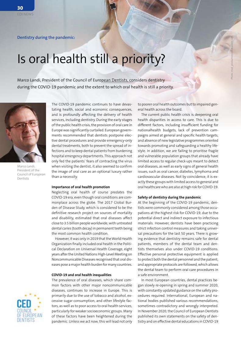
30 Is oral health still a priority? Dentistry during the pandemic:
32 Can proof of vaccination be a requirement? Statement made by Professor Wedde, Frankfurt
33 Newsticker
35 Technology meets excellence Save the date: 14th European Symposium postponed to May 2021
36 Did you ever know ... Facts about the BDIZ EDI
37 @bdiz_edi: stay tuned BDIZ EDI active on social media
38 Introducing the website of BDIZ EDI at www.bdizedi.org BDIZ EDI website continually improving
European Law
39 Transitional period runs out in May 2021 Medical Devices Regulation MDR
Volumetric analysis of the interdental papilla with adjacent implants in the aesthetic zone
The ISQ measurement is an important parameter to determine the implant stability.
5750
4TABLE OF CONTENT
TABLE OF CONTENT5
Scientific Board
Dr Iyad Abou-Rabii, Coventry
Dr Maher Almasri, Coventry
Professor Alberico Benedicenti, Genoa
Dr Eduardo Anitua,Vitoria-Gasteiz
Dr Marco Degidi, Bologna
Dr Eric van Dooren, Antwerp
Professor Rolf Ewers, Vienna
Professor Jens Fischer, Basel
Dr Roland Glauser, Zurich
Professor Ingrid Grunert, Innsbruck
Dr Detlef Hildebrand, Berlin
Dr Axel Kirsch, Filderstadt
Professor Ulrich Lotzmann, Marburg
Professor Edward Lynch, Coventry
Professor Antonio Felino, Porto
Dr Konrad Meyenberg, Zurich
Professor Georg Nent wig, Frankfurt
Dr Jörg Neugebauer, Landsberg a. Lech
Professor Hakan Özyuvaci, Istanbul
Professor Georgios Romanos, Stony Brook
Luc Rutten, MDT, Tessenderlo
Patrick Rutten, MDT, Tessenderlo
Dr Henry Salama, Atlanta
Dr Maurice Salama, Atlanta
Dr Ashok Sethi, London
Professor Joachim E. Zöller, Cologne
All case reports and scientific documentations are peer reviewed by the international editorial board of “teamwork – prosthetic dentistry and digital technologies in practice“.
Association: The European Journal for Dental Implantologists (EDI) is published in cooperation with BDIZ EDI.
Publisher board members: Christian Berger, Professor Joachim E. Zöller, Dr Detlef Hildebrand, Professor Thomas Ratajczak
Editor-in-Chief (responsible according to the press law): Anita Wuttke, phone: +49 89 72069-888, [email protected]
Managing editor: Dr Alina Ion, phone: +49 8243 9692-32, [email protected]
Advertising management: Daniela Wiedemann, Business Account Manager phone ++49 151 285 129 15, [email protected]
Publisher: teamwork media GmbH & Co. KG Betriebsstätte Fuchstal, Hauptstraße 1, D-86925 Fuchstal phone: +49 8243 9692-0, fax: +49 8243 9692-22 [email protected], www.teamwork-media.de Managing director: Bernd Müller Personally liable partner: Mediengruppe Oberfranken – Fachverlage Verwaltung GmbH E.-C.-Baumann-Straße 5, D-95326 Kulmbach
Subscription: Katharina Schäferle, phone: +49 8243 9692-16, fax: +49 8243 9692-22, [email protected]
Layout: Sigrid Eisenlauer, teamwork media GmbH & Co. KG
Printing: mgo360 GmbH & Co. KG, Gutenbergstr. 1, 96050 Bamberg
Publication dates: March, June, September, December
Subscription rates: Annual subscription: Germany € 40 including shipping and VAT. All other countries € 58 including shipping. Subscription payments must be made in advance. Ordering: in written form only to the publisher. Cancellation deadlines: in written form only, eight weeks prior to end of subscription year. Subscription is governed by German law. Past issues are available. Complaints regarding nonreceipt of issues will be accepted up to three months after date of publication. Current advertising rate list of 1/1/2019. ISSN 1862-2879
Payments: to teamwork media GmbH & Co KG, Sparkasse Bamberg, IBAN DE46 7705 0000 0303 3651 91, BIC BYLADEM1SKB
Copyright and publishing rights: All rights reserved. The magazine and all articles and illustrations therein are protected by copyright. Any utilization without the prior consent of editor and publisher is inadmissible and liable to prosecution. No part of this publication may be produced or transmitted in any form or by any means, elec-tronic or mechanical including by photocopy, recording, or information storage and retrieval system without permission in writing from the publisher. With acceptance of manuscripts the publisher has the right to publish, translate, permit reproduc-tion, electronically store in databases, produce reprints, photocopies and microcop-ies. No responsibility shall be taken for unsolicited books and manuscripts. Articles bearing symbols other than of the editorial department or which are distinguished by the name of the authors represent the opinion of the afore-mentioned, and do not have to comply with the views of BDIZ EDI or teamwork media GmbH. Respon-sibility for such articles shall be borne by the author. All information, results etc. con-tained in this publication are produced by the authors with best intentions and are carefully checked by the authors and the publisher. All cases of liability arising from inaccurate or faulty information are excluded. Responsibility for advertisements and other specially labeled items shall not be borne by the editorial department.
Copyright: teamwork media GmbH & Co. KG · Place of jurisdiction: Bayreuth
Imprint
6IMPRINT
M-0
126-
ADV-
EN-IN
T-BH
CL-0
0-11
2020
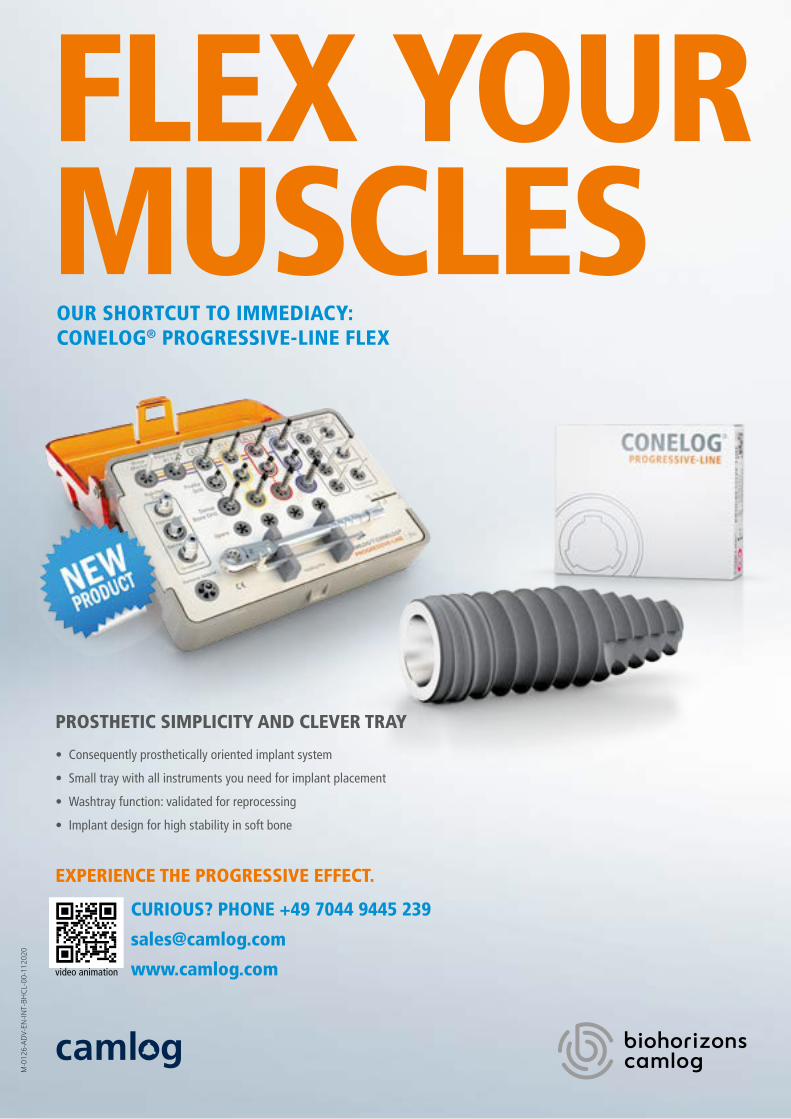
FLEX YOUR MUSCLES
EXPERIENCE THE PROGRESSIVE EFFECT.
CURIOUS? PHONE +49 7044 9445 239
www.camlog.comvideo animation
OUR SHORTCUT TO IMMEDIACY: CONELOG® PROGRESSIVE-LINE FLEX
PROSTHETIC SIMPLICITY AND CLEVER TRAY
• Consequently prosthetically oriented implant system
• Small tray with all instruments you need for implant placement
• Washtray function: validated for reprocessing
• Implant design for high stability in soft bone

Partner Organizations of BDIZ EDI
Association of Dental Implantology UK (ADI UK)
ADI UK, founded in 1987, is a registered charity committed to improving the standards of im plant dentistry by providing continuing education and ensuring scientific research. It is a member-ship-focused organization dedicated to providing the dental profession with continuing educa-tion, and the public with a greater understanding of the benefits of dental im plant treatment. Membership of the ADI is open to the whole dental team and industry, and offers a wealth of benefits, education and support for anyone wishing to start out or develop further in the field of dental implantology.
Ogolnopolskie Stowarzyszenie Implantologii Stomatologicznej (OSIS EDI)
OSIS EDI, founded in 1992, is a university-based organization of Polish scientific implantologi-cal associations that joined forces to form OSIS. The mission of OSIS EDI is to increase implant patients’ comfort and quality of life by promoting the state of the art and high standards of treatment among dental professionals. OSIS EDI offers a postgraduate education in dental implantology leading to receiving a Certificate of Skills (Certyfikat Umiejętności OSIS), which over 130 dental implantologists have already been awarded.
Sociedad Espanola de Implantes (SEI)
SEI is the oldest society for oral implantology in Europe. The pioneer work started in 1959 with great expectations. The concept of the founding fathers had been a bold one at the time, al-though a preliminary form of implantology had existed both in Spain and Italy for some time. Today, what was started by those visionaries has become a centrepiece of dentistry in Spain. SEI is the society of reference for all those who practice implantology in Spain and has been throughout the 50 years, during which the practice has been promoted and defended whereas many other societies had jumped on the bandwagon. In 2009 SEI celebrated its 50th anniversary and the board is still emphasizing the importance of cooperating with other recognized and renowned professional societies and associations throughout Europe.
Sociedade Portuguesa de Cirurgia Oral (SPCO)
The SPCO’s first international activity was the foundation – together with their counterparts in France, Italy, Spain and Germany – of the European Federation of Oral Surgery (EFOOS) in 1999. The Sociedade Portuguesa de Cirurgia Oral’s primary objective is the promotion of medical knowledge in the field of oral surgery and the training of its members.
Udruženje Stomatologa Implantologa Srbije-EDI (USSI EDI)
USSI EDI was founded in 2010 with the desire to enhance dentists’ knowledge of dental im-plants, as well as to provide the highest quality of continuing education in dentistry. The most important aims of the organization are to make postgraduate studies meeting the standards of the European Union available to dentists from Serbia and the region; to raise the level of education in the field of oral implantology; to develop forensic practice in implantology; and to cooperate with countries in the region striving to achieve similar goals.
8PARTNERS
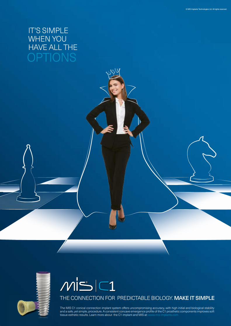
© MIS Implants Technologies Ltd. All rights reserved.
The MIS C1 conical connection implant system o�ers uncompromising accuracy, with high initial and biological stability and a safe, yet simple, procedure. A consistent concave emergence pro�le of the C1 prosthetic components improves soft tissue esthetic results. Learn more about the C1 implant and MIS at: www.mis-implants.com
THE CONNECTION FOR PREDICTABLE BIOLOGY. MAKE IT SIMPLE
IT'S SIMPLE WHEN YOU HAVE ALL THE OPTIONS
various online seminars, has been received so favor-ably that BDIZ EDI would now like to provide dental practices with fast and well-targeted information and further training even internationally, in English.
The presentations will be broadcast online and live. Participants will have an opportunity for discus-sion with the speakers. Participation is free of charge for members. All participants will receive a certificate of participation.
Please look at the 2021 programme of seminars in the “BDIZ EDI informs” series. Send your selec-tion either to the editorial office at office-munich@ bdizedi.org or by fax to +49 89 720 69 889. BDIZ EDI will of course spread the news via Facebook and Instagram as well. The online seminars will be held in the evenings between 7 and 8 p.m.
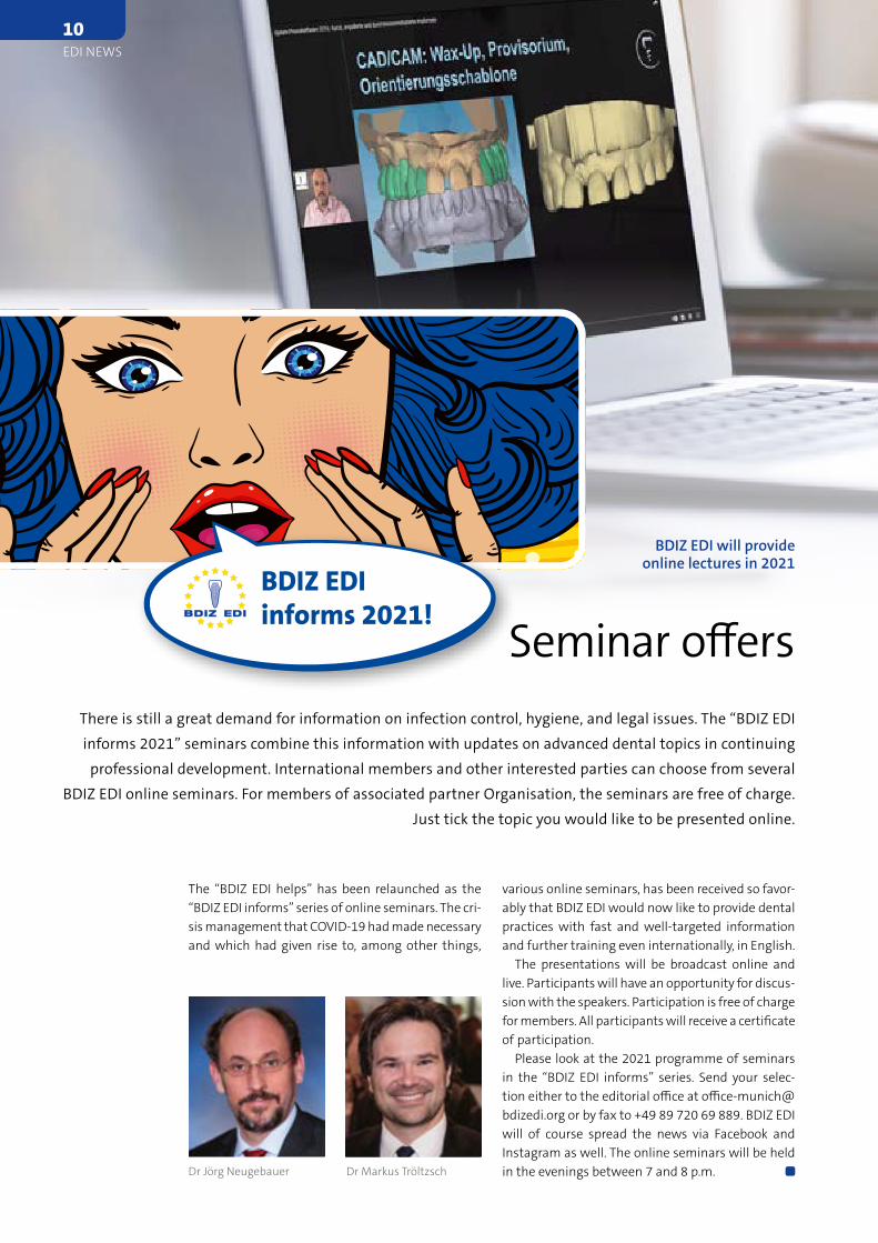
The “BDIZ EDI helps” has been relaunched as the “BDIZ EDI informs” series of online seminars. The cri-sis management that COVID-19 had made necessary and which had given rise to, among other things,
BDIZ EDI will provide online lectures in 2021
Seminar offers There is still a great demand for information on infection control, hygiene, and legal issues. The “BDIZ EDI informs 2021” seminars combine this information with updates on advanced dental topics in continuing
professional development. International members and other interested parties can choose from several BDIZ EDI online seminars. For members of associated partner Organisation, the seminars are free of charge.
Just tick the topic you would like to be presented online.
BDIZ EDI informs 2021!
Dr Jörg Neugebauer Dr Markus Tröltzsch
10EDI NEWS
Send your selection either to the editorial office at [email protected] or by fax to +49 89 720 69 889
www.bdizedi.org/seminare
Topic No. 1: Update on peri-implantitis; Guideline of the European Consensus Conference 2020 Presenter: Dr Jörg Neugebauer (Landsberg am Lech, Germany), Member of the BDIZ EDI Board, 7–8 p.m.
About this seminar: Biological complications cannot be avoided completely; they occur at different times following the delivery of the implant restoration. The etiology of these complications is as diverse as the way in which they manifest themselves. This issue has been addressed three times before by the European Consensus Conference; this panel of experts has now re-evaluated the current literature and updated the recommendations of the Guideline. Dr Neugebauer will present the most recent findings from the literature with numerous clinical examples to ensure the best possible care for patients with peri-implantitis, with a view to avoiding implant loss and eliminating risk factors.
Topic No. 2: Update on digital implantology Presenter: Dr Markus Tröltzsch (Ansbach, Germany), Chair of the Academy of Dentistry and Oral Medicine (APW)
of the German Society for Dentistry and Oral and Maxillofacial Surgery (DGZMK), 7–8 p.m.
About this seminar: In Germany, most implants are still planned the traditional way, using OPGs and physical models and placed without digital support, even though the tools for use in digital implantology are readily available. What do practitioners have to pay attention to, and what are the advantages and disadvantages of digital methods in oral implantology? Dr Tröltzsch addresses these questions and shows how the digital workflow can be integrated into everyday practice.
Topic No. 3: Update on bone augmentation surgery Presenter: Dr Markus Tröltzsch, 7–8 p.m.
About this seminar: For implantological restorations to achieve long-term stability, both hard and soft tissues must be avail-able in sufficient quantity and quality. There are many ways in which this can be achieved or maintained. In this online seminar, Dr Tröltzsch, who was in charge of the new DGI/DGZMK Guideline on implantological indications for the use of bone replacement materials, will highlight the various “minor” and “major” techniques. One of the topics Dr Tröltzsch will discuss is how tissue volume can be (re)built or maintained and which of the relevant techniques are suitable for practitioners with different types of practices and different levels of experience.
Topic No. 4: Update on short, angulated and diameter-reduced implants – Guideline of the European Consensus Conference 2016 Presenter: Dr Jörg Neugebauer (Landsberg am Lech, Germany), Member of the BDIZ EDI Board, 7–8 p.m.
About this seminar: “The use of short, angulated or diameter-reduced implants in case of reduced bone availability represents today – if the specific treatment parameters are taken into account – a reliable therapy option compared to the risks associated with the use of implants with standard dimensions in combination with augmentative procedures”. The conclusion of the BDIZ EDI Practice Guide from 2016 will be put to the test in this lecture. Do the statements still apply or do they need to be revised? Can complex augmentations be prevented by using the mentioned implants? How should ultra-short implants be rated today? When is augmentation the better choice? The lecture gives an update on the use of implants and com-pares the possible treatment options.
Please contact me via email at:
____________________________________________
Foto
: Afr
ica
Stud
io/s
tock
.ado
be.co
m
EDI NEWS11
The European Medicines Agency (EMA) released on 29 January its first safety update on a COVID-19 vaccine – Comirnaty, a vaccine produced by BioNTech and Pfizer. It concluded that safety data collected on Comirnaty use in vaccination campaigns was con-sistent with the known safety profile of the vaccine, and no new side effects were identified.• Reports of suspected severe allergic reaction
have not identified new aspects regarding the nature of this known side effect.
• No specific safety concern has been identified for vaccine use in frail elderly individuals.
• Its benefits in preventing COVID-19 continue to outweigh any risks, and there are no recom-mended changes regarding the use of the vac-cine.
Situation end of FebruaryThe European Centre for Disease Prevention and Control (ECDC) of the European Union reports end of February that transmission is still widespread in the EU/EEA even though most countries are experienc-ing stable or decreasing case rates. However, abso-lute numbers remain high, with increasing case rates among older age groups and increasing death rates in several countries. Around one third of countries
Vaccination against COVID-19 started on 27 Decem-ber 2020 across the European Union, in a moment of unity. “A safe and effective vaccine is our lasting way out of the pandemic”, says the European Commis-sion on their website. The EC states that the three approved COVID-19 vaccines, which were assessed as being safe and effective, are being sent to all EU countries, under the same conditions.
For now, people in priority groups are vaccinated - elderly people or healthcare professionals. But soon, the Commission promised end of February, there will be enough vaccines for the entire EU population. “Once enough people are vaccinated, we will be able to get our normal lives back, gradually!”
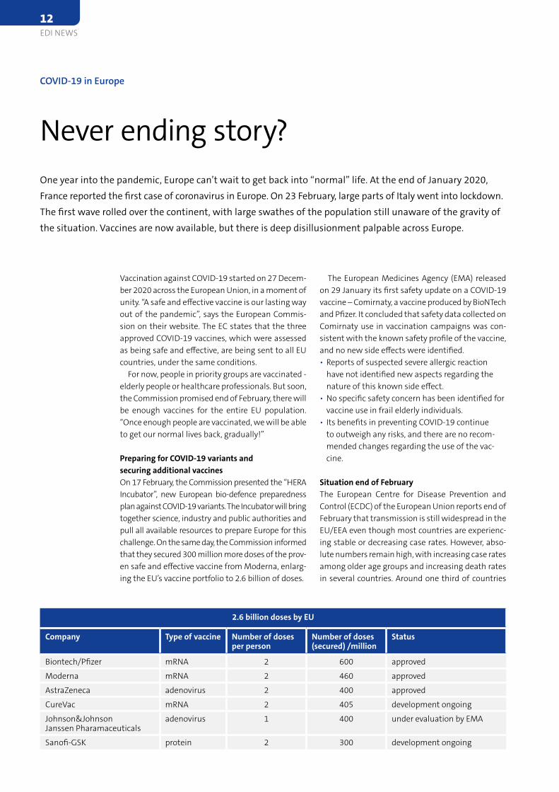
Preparing for COVID-19 variants and securing additional vaccinesOn 17 February, the Commission presented the “HERA Incubator”, new European bio-defence preparedness plan against COVID-19 variants. The Incubator will bring together science, industry and public authorities and pull all available resources to prepare Europe for this challenge. On the same day, the Commission informed that they secured 300 million more doses of the prov-en safe and effective vaccine from Moderna, enlarg-ing the EU’s vaccine portfolio to 2.6 billion of doses.
COVID-19 in Europe
Never ending story?One year into the pandemic, Europe can’t wait to get back into “normal” life. At the end of January 2020, France reported the first case of coronavirus in Europe. On 23 February, large parts of Italy went into lockdown. The first wave rolled over the continent, with large swathes of the population still unaware of the gravity of the situation. Vaccines are now available, but there is deep disillusionment palpable across Europe.
2.6 billion doses by EU
Company Type of vaccine Number of doses per person
Number of doses (secured) /million
Status
Biontech/Pfizer mRNA 2 600 approved
Moderna mRNA 2 460 approved
AstraZeneca adenovirus 2 400 approved
CureVac mRNA 2 405 development ongoing
Johnson&Johnson Janssen Pharamaceuticals
adenovirus 1 400 under evaluation by EMA
Sanofi-GSK protein 2 300 development ongoing
12EDI NEWS
are seeing increases in hospital or ICU admissions and/or occupancy due to COVID-19. This serves as a reminder of the importance of maintaining public health and physical distancing measures and that these measures should not be relaxed, even in coun-tries with decreasing trends. On February 18, ECDC reported more than 21 million cases in EU/EEA (107 million worldwide) and ½ million deaths.
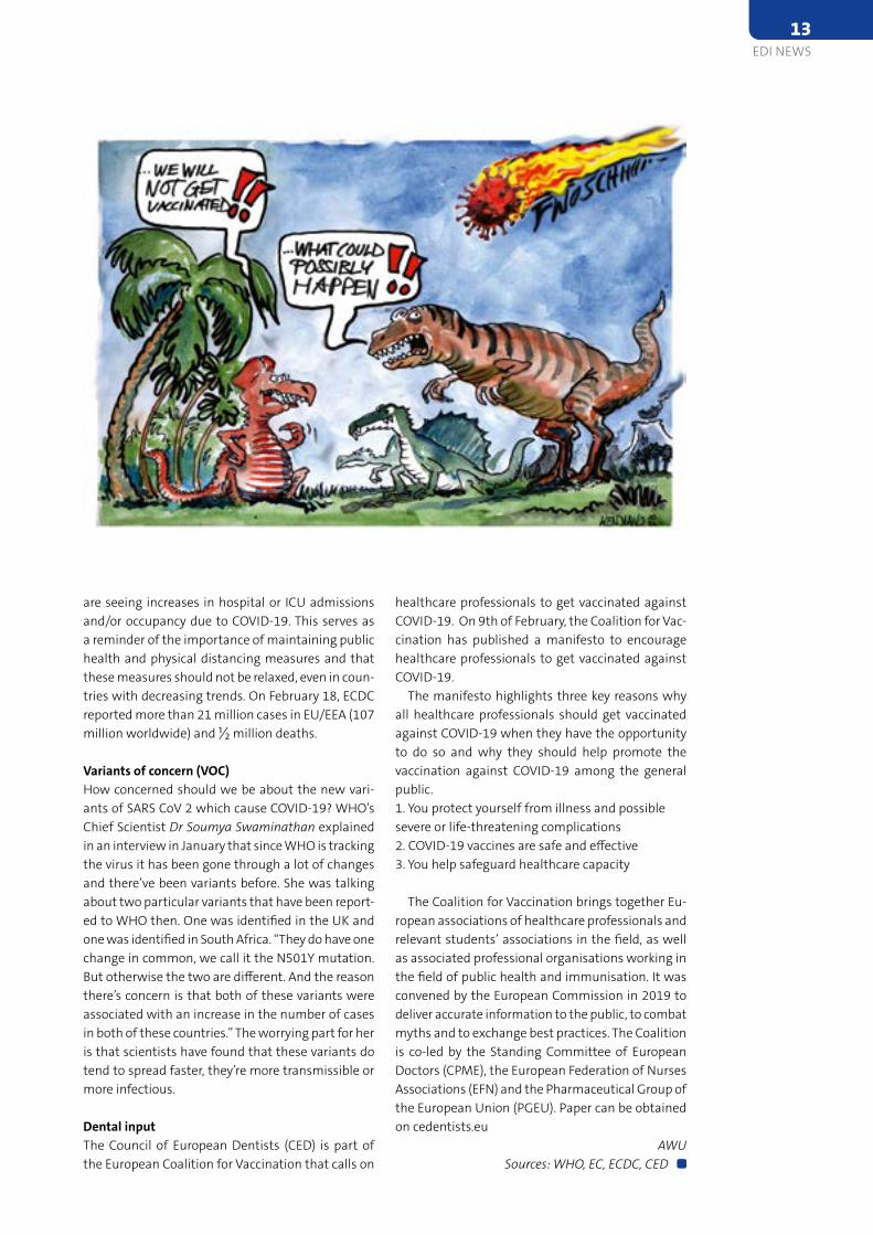
Variants of concern (VOC)How concerned should we be about the new vari-ants of SARS CoV 2 which cause COVID-19? WHO’s Chief Scientist Dr Soumya Swaminathan explained in an interview in January that since WHO is tracking the virus it has been gone through a lot of changes and there’ve been variants before. She was talking about two particular variants that have been report-ed to WHO then. One was identified in the UK and one was identified in South Africa. “They do have one change in common, we call it the N501Y mutation. But otherwise the two are different. And the reason there’s concern is that both of these variants were associated with an increase in the number of cases in both of these countries.” The worrying part for her is that scientists have found that these variants do tend to spread faster, they’re more transmissible or more infectious.
Dental inputThe Council of European Dentists (CED) is part of the European Coalition for Vaccination that calls on
healthcare professionals to get vaccinated against COVID-19. On 9th of February, the Coalition for Vac-cination has published a manifesto to encourage healthcare professionals to get vaccinated against COVID-19.
The manifesto highlights three key reasons why all healthcare professionals should get vaccinated against COVID-19 when they have the opportunity to do so and why they should help promote the vaccination against COVID-19 among the general public.1. You protect yourself from illness and possible severe or life-threatening complications2. COVID-19 vaccines are safe and effective3. You help safeguard healthcare capacity
The Coalition for Vaccination brings together Eu-
ropean associations of healthcare professionals and relevant students’ associations in the field, as well as associated professional organisations working in the field of public health and immunisation. It was convened by the European Commission in 2019 to deliver accurate information to the public, to combat myths and to exchange best practices. The Coalition is co-led by the Standing Committee of European Doctors (CPME), the European Federation of Nurses Associations (EFN) and the Pharmaceutical Group of the European Union (PGEU). Paper can be obtained on cedentists.eu
AWUSources: WHO, EC, ECDC, CED
EDI NEWS13
Vitamin D
Vitamin D
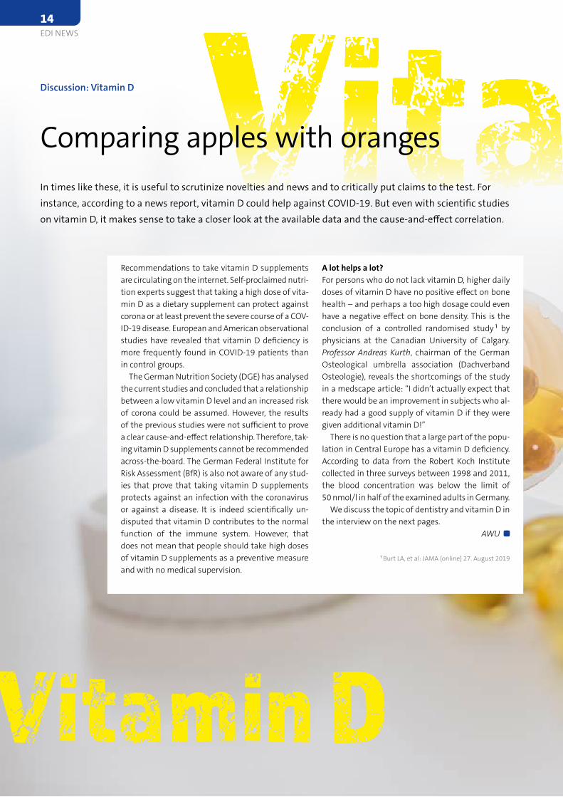
A lot helps a lot?For persons who do not lack vitamin D, higher daily doses of vitamin D have no positive effect on bone health – and perhaps a too high dosage could even have a negative effect on bone density. This is the conclusion of a controlled randomised study ¹ by physicians at the Canadian University of Calgary.Professor Andreas Kurth, chairman of the German Osteological umbrella association (Dachverband Osteologie), reveals the shortcomings of the study in a medscape article: “I didn’t actually expect that there would be an improvement in subjects who al-ready had a good supply of vitamin D if they were given additional vitamin D!”
There is no question that a large part of the popu-lation in Central Europe has a vitamin D deficiency. According to data from the Robert Koch Institute collected in three surveys between 1998 and 2011, the blood concentration was below the limit of 50 nmol/l in half of the examined adults in Germany.
We discuss the topic of dentistry and vitamin D in the interview on the next pages.
AWU
¹ Burt LA, et al: JAMA (online) 27. August 2019
Recommendations to take vitamin D supplements are circulating on the internet. Self-proclaimed nutri-tion experts suggest that taking a high dose of vita-min D as a dietary supplement can protect against corona or at least prevent the severe course of a COV-ID-19 disease. European and American observational studies have revealed that vitamin D deficiency is more frequently found in COVID-19 patients than in control groups.
The German Nutrition Society (DGE) has analysed the current studies and concluded that a relationship between a low vitamin D level and an increased risk of corona could be assumed. However, the results of the previous studies were not sufficient to prove a clear cause-and-effect relationship. Therefore, tak-ing vitamin D supplements cannot be recommended across-the-board. The German Federal Institute for Risk Assessment (BfR) is also not aware of any stud-ies that prove that taking vitamin D supplements protects against an infection with the coronavirus or against a disease. It is indeed scientifically un-disputed that vitamin D contributes to the normal function of the immune system. However, that does not mean that people should take high doses of vitamin D supplements as a preventive measure and with no medical supervision.
Discussion: Vitamin D
Comparing apples with orangesIn times like these, it is useful to scrutinize novelties and news and to critically put claims to the test. For instance, according to a news report, vitamin D could help against COVID-19. But even with scientific studies on vitamin D, it makes sense to take a closer look at the available data and the cause-and-effect correlation.
14EDI NEWS
Vitamin DAssistant Professor Johan Wölber
Dr Wölber, which of the vitamins in the D group is in the focus of dentistry and why?
Vitamin D3 – Cholecalciferol – is most frequently mentioned. Not only does it play an essential role in the resorption of calcium and phosphorus from the intestine and the assimilation of calcium into the bones, but also in immunological functions and inflammatory processes. From a dental point of view, this is of course highly interesting, as the tooth hard tissues and alveolar bones themselves consist largely of calcium compounds.Bone resorption and inflam-matory processes are the core of what occurs in periodontitis.
How do minerals in combination with vitamin D affect bone health and the teeth mineralisation?
Minerals play an essential role in every stage of human life. Let us start with the mineral status or the sufficient vitamin D supply of pregnant women. If vitamin D is lacking there, this could influence the development of the child’s teeth, who, for example, later show a higher susceptibility to caries. For in-stance, a prospective cohort study with 207 women showed that a low vitamin D level during pregnan-cy significantly increases the risk of caries in early childhood. Likewise, there are association studies that have found lower vitamin D levels in pregnant women with periodontitis than in pregnant women without periodontitis.
Further, there is also evidence of a lower incidence of caries in adolescens with sufficient vitamin D levels.
In the elderly, additional factors such as the ageing skin, which can no longer produce as much vitamin D, as well as possible overweight favour a vitamin D de-ficiency. Vitamin D is fat-soluble, it accumulates in fatty tissue and is no longer available in the serum.
Are there any studies that prove the efficacy of vitamin D?
From the point of view of evidence-based den-tistry, this is where things get tenuous. We do have many association studies that show correlations. But these studies only give hints and they cannot explain causality. Intervention studies in which vi-tamin D is actively administered and the effect is compared with a placebo are much more difficult (because more expensive) to be conducted. There is a quite recent study from Italy that could find a trend towards better therapy results in periodontitis therapy with adjuvant supplementation of 25,000 IU vitamin D per week.
A vitamin D deficiency does not seem to be rare in the population in our Central European climate zone and due to our way of life and work. How do you determine the vitamin D concentration in the organism and which level is “normal”?
[Wölber laughs]: Nowadays, in the age of the Anthropocene–the human-influenced age of the earth–is not so easy to answer what is “normal”. For a heavily dressed nun, a low vitamin D value will be more “normal” than for a lightly dressed lifeguard in an open-air swimming pool.
Interview on the significance of vitamin D for the human organism and in dentistry
An important instrument in the orchestra Is vitamin D a panacea or completely overrated? In the following interview, Assistant Pro-fessor Johan Wölber takes a stance on the significance of vitamin D for the human organ-ism and, of course, for dentistry. Dr Wölber is a dentist and nutritional physician (DAEM/DGEM) and works as deputy director of the Master Periodontology & Implant Therapy course at Freiburg University Hospital, Department of Oral and Maxillofacial Surgery, Clin-ic for Dental Preservation and Periodontology.
EDI NEWS15
Vitam
in D
to 100 micrograms (= 4000 I.U.) of vitamin D per day to be harmless. The Robert Koch Institute warns of increased calcium concentrations (hypercalcemia) in case of overdose, which can acutely lead to nausea, loss of appetite, abdominal cramps, vomiting or, in severe cases, to kidney damage, cardiac arrhythmia, unconsciousness and death. For this reason, I also leave the supplementation of higher doses in the hands of the physician.
Can the course of a jaw osteonecrosis be influenced by the administration of vitamins and/or minerals?
I cannot answer this question.
Are you familiar with any known interactions when administering vitamin D?
Caution seems to be advisable with digitalis medication (“cardiac insufficiency”) and simultane-ous administration of other medicines that increase the calcium level – such as hydrochlorothiazide for “dehydration”.
In your opinion, is the significance of vitamin D for dentistry widely known in dental practices?
No, I do not think so. But this applies to the whole range of nutritional medicine, from macro- to mi-cronutrients. The knowledge is extremely important, health-promoting, comprehensive and sometimes non-trivial. However, the change from a reparative (drilling and scratching) dentistry to a preventive dentistry is proceeding very slowly, so that there is still hardly any space for such topics in education. Moreover, prevention is not rewarded accordingly.
Finally, I dare to ask a bold question: Is vitamin D a panacea for dentistry?
If you followed some of the debates, you might think so. I personally think that sufficient levels of vitamin D are important, but I have not seen yet any miracle cures. It is just ONE instrument in the whole orchestra of micronutrients. If one instrument is miss-ing, it can make a difference in music ( or in health).
Dr Wölber, thank you very much for your insightful answers.
The interview was conducted by Editor-in-Chief Anita Wuttke
From the evolutionary angle, however, we could pos-tulate that Homo sapiens–regarding vitamin D – are not necessarily designed to sit dressed in houses all day, to be obese and to live far away from the equator.
In my search for the optimal vitamin D level, I was personally impressed by one study: it investigated what vitamin D level breastfeeding mothers need to have for the vitamin D level in breast milk to be sufficient to spare the infants rickets prophylaxis (in the form of 500 IU per day). The value was 40–50 ng/ml or 100–125 nmol/l in maternal serum.
The measurement is normally taken via the 25-hy-droxyvitamin D in the serum, which is a precursor vitamin. Unfortunately, two different units are still used here, which always complicates the interpreta-tion for patients.
Is the dentist able to diagnose vitamin D deficiency and if so, how?
I personally refer patients to their physician when I have diagnosed periodontitis. Even the laboratory tests are affected by test-related variations. There-fore, follow-up should also be done in the same place.
When is it advisable to administer vitamin D prior to bone augmentation and/or implantation?
[Wölber smirks] As a passionate and successful supporter of conservative dentistry, this question does not arise for me. I discuss vitamin D for pro-phylactic and tooth-preserving reasons. But I have learned from colleagues that it can also be useful for the above-mentioned interventions. I would recom-mend testing and targeting a value of 40–50 ng/ml.
How far in advance and for how long is it advisable to administer the substance?
This depends on how low the initial value was and how much and how often the supplementation is carried out. With 20,000 IU per week, a follow-up after six to eight weeks would make sense. If the physician prescribes more frequent supplementa-tion, the level may be reached earlier.
What dose do you recommend before and, if neces-sary, after surgery?
I leave that decision to the testing and prescribing physician.
According to the motto: a lot helps a lot: can you overdose on vitamin D and what will happen then?
Overdosing cannot be achieved via sunlight. But it is possible via supplements. Nevertheless, the “tolerable upper limits” seem to be quite high. The European Food Safety Authority (EFSA) considers up
Contact address Assistant Professor Johan Wölber
Department of Operative Dentistry and Periodontology Center for Dental Medicine Medical Center – University of Freiburg Germany
16EDI NEWS
EDI NEWS17
www.ethoss.dental [email protected]
+44 (0) 1535 843 106Join our Facebook community - ‘EthOss Case Studies’
The best dentists don’t stand still
E t h O s s i s c h a l l e n g i n g
G B R t o d r i v e a n e w e r a o f
T R U E B O N ER E G E N E R AT I O N
Grow Stronger
ses do not seem to be interesting any more because there have hardly been any new evaluations in recent years. On the other hand, the scientific reviews are more focused on what complications occur and what the patient-related outcomes are.
What has changed since then?The issue flapless surgery has become more pro-
found in the meantime. In the discussion of the paper at that time, the non-opening of the alveolar ridge was still classified as a high risk factor. We now know that we can trust surgical guides if the devia-tion is kept as small as possible during use. This is possible with restorations where there is a remain-ing dentition. Or we can use auxiliary implants, so-called anchor pins, to achieve a high degree of preci-sion in flapless implant placement. Another factor in flapless surgery is primary stability, which can be determined with resonance frequency analysis. The risk with flapless surgery is therefore lower.
Your conclusion on angulated implants?It is now acknowledged that angulated implants
do work, but we have a number of influential factors. The type of prosthetic restoration and how these prosthetic restorations work is also important. In the end, the patient evaluates the work in terms of the functional prosthesis.
In terms of horizontal bone volume, the reduced-diameter implants become important. What are the possibilities?
We have classified the diameter-reduced implants (DRI) into two groups in comparison to the standard diameter implants (SDI), which have an intraosse-ous diameter of less than 3.5 mm. If the diameter is smaller – less than 2.7 mm – we speak of so-called
Dr Neugebauer, what is the requirement for mini-mally invasive implant therapy?
In 2016, the European Consensus Conference – which takes place every year in Cologne on a cur-rent implantological topic – worked out that in the minimally invasive area, due to complex augmenta-tions, we must try to avoid too great impairments for the patient. At that time, we summarized that by means of 3D diagnostics, an estimation regarding bone quality and quantity is important. For this to work, we need to know where to place the implants. We can ideally use navigation guides for this, and it is important to consider the whole process from a prosthetic point of view.
At the 11th EuCC in 2016, angulated implants were the subject of the most intense and controversial discussion. The focus was on the prevention of com-plications. What did the EuCC agree on?
At that time we specified that immediately re-stored and angulated implants should be used with sufficient primary stability. Also significant was the recommended 3D diagnostics for anatomically and prosthetically correct implant placement. Another recommendation was aimed at the adequate train-ing of the implanting (dental) practitioner and the prosthetic practitioner. At the time, we worked with experience reports that included data from the past five to ten years and showed follow-up examinations with a maximum of 6.5 years. We observed that there was no difference in bone resorption between straight and angulated implants and no difference to conventional treatment.
What is the state of affairs today?A recent paper from 20181 shows that there are
still no differences. As for the science, meta-analy-
An update on short, angulated and diameter-reduced implants
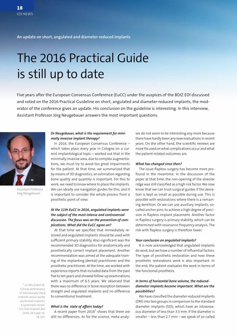
The 2016 Practical Guide is still up to dateFive years after the European Consensus Conference (EuCC) under the auspices of the BDIZ EDI discussed and voted on the 2016 Practical Guideline on short, angulated and diameter-reduced implants, the mod-erator of the conference gives an update. His conclusion on the guideline is interesting. In this interview, Assistant Professor Jörg Neugebauer answers the most important questions.
Assistant Professor Jörg Neugebauer
1 Lin WS, Eckert SE. Clinical performance of intentionally tilted
implants versus axially positioned implants: A systematic review.
Clin Oral Implants Res 2018; 29 Suppl 16:
78-105.
18EDI NEWS
and recommend removing short implants and are also critical of alternative procedures. Unfortunately, there is still too much from our colleagues to short, diameter-reduced and angulated implants . I hope that the current study situation and the reliable ex-periences will bring about some change here.
Could we conclude that the practical guide agreed in 2016 has lost nothing of its relevance?
Absolutely. The statements are still correct! Of course, there are changes in the study situation, especially regarding short implants, which could be included in the literature list to support the conclu-sions. But this practical guideline of the European Consensus Conference (EuCC) under the leadership of the BDIZ EDI is still very helpful as a recommen-dation for action. Next year, we should nevertheless consider updating it.
Dr Neugebauer, thank you very much for the inter-esting interview.
The interview was conducted by Editor-in-Chief Anita Wuttke.
mini-implants (MDI). From a scientific point of view, we can conclude that DRIs also work in the posterior region. However, the indication restrictions of the respective manufacturers should be considered. A recent study by Schiegnitz and Al-Nawas from 20182 shows that implants with diameter 3–3.25 mm and 3.3–3.5 mm diameter show no difference to the sur-vival rate of standard implants. For the mini implants smaller than 2.5 mms, unfortunately, it does not look so good in the literature. There are many retrospec-tive studies. Meta-analyses with prospective and/or randomised studies show only short-term results or increased error rates. But: MDI for prosthesis stabi-lisation can, under precise indication, significantly improve the quality of life of elderly patients.
Starting from which length do we speak of short implants?
Our paper from 2016 has now also become estab-lished on an international level. When we have im-plants that are less than or equal to 8 mm long and the diameter is greater than or equal to 3.75 mm, we speak of short implants. Ultra-short implants are de-fined as those that are smaller than 6 mm. The market for short implants has developed in recent years be-cause it has been discovered that it is not just a niche, but a concept. So you can definitely work with the short ones and accordingly fewer vertical augmenta-tions and open sinus floor elevations will be necessary.
When are the “short ones” used?The EuCC 2016 came to the conclusion that the
indication is mainly in the lateral region of the max-illa and mandible, due to the reduced vertical bone supply with still sufficient width of the bone, limited by anatomical neighbouring structures, such as the maxillary sinus and the mandibular canal. They are also used in the anterior region as a fixation for over-dentures and as single or multiple dentures, as an alternative to bone-building measures, although of course the whole matter becomes more difficult in the aesthetic area in particular.
What is the current state of the studies?The results are clearly better than with the angu-
lated and reduced-diameter implants. In the past two years there have been a number of meta-analyses ex-amining randomised clinical tests with the conclusion that the short implants show the same results as the long implants with augmentation. Short implants are now accepted and no longer have an outsider status.
What is the situation regarding the acceptance among the dental community?
Well, unfortunately there are quite a few col-leagues who stick to the experience of 30 years ago
The Practical Guide of the European Consensus Conference under the auspices of the BDIZ EDI is available online in German and English on the BDIZ EDI website: Deutsch: https://bdizedi.org/kurze-angulierte-und-durchmesserreduzierte-implantate/
English: https://bdizedi.org/en/european-consensus-conference/
Practical Guide 2016
2 Schiegnitz E, Al-Nawas B. Narrow-diameter implants: A systematic review and meta-analysis. Clin Oral Implants Res 2018; 29 Suppl 16: 21-40.
EDI NEWS19
2
Update on short, angulated and diameter-reduced implants
Guidelines 2016
Update on short, angulated and diameter-reduced implants
11th European Consensus Conference (EuCC) 2016 in Cologne
6 February 2016
Authors: Jörg Neugebauer, PhD, DMD Hans-Joachim Nickenig M.Sc., PhD, DMD Joachim E. Zöller, PhD, MD, DMD Department of Cranio-maxillofacial and Plastic Surgery and Interdisciplinary Department for Oral Surgery and Implantology Centre for Dentistry and Oral and Maxillofacial Surgery, University of Cologne, Germany Director: Professor DDr Joachim E. Zöller
Chairman: Dr J. Neugebauer (Germany) Protocol: Dr F. Vizethum (Germany) Participants: Ch. Berger (Germany)
Dr W. Bolz (Germany) Dr A. Bowen (Spain) Professor Dr D. Deporter (Canada) Professor DDr R. Ewers (Austria) Dr P. Fairbairn (United Kingdom) Professor Dr A. Felino (Portugal) Dr Th. Fortin (France) Dr V. Gowd (India) Professor Dr M. Kern (Germany) Professor Dr P. Kobler (Croatia) Professor Dr V. Konstantinovic (Serbia) Professor Dr M. Marincola (Italy) Dr H.J. Nickenig (Germany) Professor Dr H. Özyuvaci (Turkey) Professor Dr N. Schmedtmann (Germany) Professor DDr J.E. Zöller (Germany)
1. Methods
1.1 Objective The purpose of these guidelines is to offer recommendations for clinicians engaging in implant dentistry, enabling them to correctly assess potential indications (and any limitations thereof) for short, angulated or diameter-reduced implants.
BDIZ EDIAn der Esche 2D-53111 BonnGERMANYFon: +49-228-93592-44Fax: [email protected]
Bundesverband derimplantologischtätigen Zahnärztein Europa
EuropeanAssociation of DentalImplantologists
20EDI NEWS
3
Update on short, angulated and diameter-reduced implants
Guidelines: Update on short, angulated and diameter-reduced implants 11th European Consensus Conference in February 2016
Page 2 of 9
1.2 Introduction This consensus paper covers only titanium implants typically placed in accordance with the indications recommended by the European Consensus Conference (EuCC, Germany, 6 February 2016). All consensus recommendations in this paper should be considered as guidelines only. The patient’s specific situation is always an important consideration and may justify a deviation from the recommendations of this consensus paper.
1.3 BackgroundAvoiding bone augmentation through reduced-dimension implants and optimum utilization of available bone volume is often recommended being a minimally invasive treatment option [45].To ensure an acceptable treatment outcome, dimension and insertion type must be considered in addition to the number of implants.
1.4 Literature search The Cochrane Library, EMBASE, DIMDI and Medline literature databases were used to conduct a systematic search of recently published data on the use of short, angled or diameter-reduced implants. Selective search criteria were used, including terms such as “short implants”, “angulated implants”, “angled implants”, “tilted implants”, “outcome grafting procedure”, and “implant -failure”. The publications identified by the search were screened by reading their abstracts, and those irrelevant to the subject were identified and excluded. Publications found to be potentially relevant were obtained in full-text form. Multiple review papers with meta-analyses and randomized controlled trials (RCTs), and other prospective and retrospective systematic clinical studies were available on the subject.
1.5 Procedure for developing the Consensus Conference guidelines A preliminary version of this document on which the EuCC based its deliberations was prepared by Dr J. Neugebauer of the Interdisciplinary Policlinic for Oral Surgery and Implantology and Department of Oral and Maxillofacial Plastic Surgery at the University of Cologne/Germany. The preliminary report was then reviewed and discussed by the sitting committee members in five steps as follows:
• Reviewing the preliminary draft• Collecting alternative proposals• Voting on recommendations and levels of
recommendation• Discussing non-consensual issues• Final voting
The full text of all (potentially) relevant citations was obtained if necessary and reviewed. Numerous reviews, but few RCTs (randomised controlled trials) or other systematic clinical trials are available on this topic.
2. Problem
The application of standard implants in patients with atrophy of their alveolar ridges or large pneumatization of the maxillary sinus cavity often requires the use of hard tissue augmentation procedures [18, 17]. These procedures are established, and widely used with success. But depending on level of training of the user and the patient-specific risk factors, complications may occur and affect the postoperative quality of life [1, 9, 19, 18, 17, 33].
BDIZ EDIAn der Esche 2D-53111 BonnGERMANYFon: +49-228-93592-44Fax: [email protected]
EDI NEWS21
4
Update on short, angulated and diameter-reduced implants
Guidelines: Update on short, angulated and diameter-reduced implants 11th European Consensus Conference in February 2016
Page 3 of 9
3. Use of short implants
3.1 Introduction Short implants are increasingly being discussed as a treatment alternative in situations characterized by limited vertical bone height [3].Compared to the use of standard implants due to biomechanical considerations (e.g. crown-to-implant ratio, C/R) with short implants may lead to unfavourable loading conditions and complications, including excessive crestal bone loss and implant failure [23]. Improvements in implant design and surface along with the use of modified implant insertion methods all are intended to minimize these risks [14].
3.2 Definition of short implants Implants are usually referred to as short if their designed intrabony length measures ≤ 8 mm with diameters ≥ 3.75 mm. Standard implants are considered to be those with lengths > 8 mm and diameters ≥ 3.75 mm [43, 47]. “Ultra-short” implants are considered to be those with lengths less than 6 mm [13].
3.3 Indications for short implants Short implants are primarily used to avoid bone augmentation procedures in the maxillary and mandibular posterior segments of partially edentulous patients. They are applicable if vertical bone volume is limited by anatomical structures (maxillary sinus, mandibular canal), but there is sufficient alveolar ridge width to permit successful use of implant diameters ≥ 3.75 mm. They are also used to support removable overdentures as single or multiple tooth replacements in the anterior jaws [47, 48].
3.4 Current observations For ultra-short implants, there is insufficient evidence to make recommendations at this time. A review paper from 2015 summarized findings with RCTs on sinus floor elevation with standard length implants or short implants on their own. Five studies reported 16–18 months survival rates for long implants in combination with sinus elevation of 99.5 % (95 % CI: 97.6 – 99.98 %) and for short implants alone of 99.0 % (95 % CI: 96.4 – 99.8 %). For shorter observation periods of 8 – 9 months in three studies, survival rates for long implants were 100 % (95% CI: 97.1 – 100 %) and for short implants alone 98.2 % (95 % CI: 93.9 – 99.7 %) [53]. These results are supported by other RCTs [49, 52].
The number of RCTs on the use in the mandible is limited [42]. In these RCTs, no relevant differences in biological parameters between the use of short and long implants in the posterior mandible were found [20, 27]. One group has presented five-year results showing no significant difference for the application of short implants alone as compared to standard implants and vertical augmentation in the mandible [21].
A retrospective comparative analysis also showed no differences between short and long implants for an observation period of five years [24]. Meta-analysis showed high survival rates for short implants with moderately rough surfaces [37]. Long-term data for observation periods of 10 years for the posterior mandible of partially edentulous patients and 20 years for mandibular overdentures showed favourable results for short, sintered porous-surfaced implants [15, 16].
The literature does show, however, that short implants with a reduced diameter have failure rates of up to 10 % after three to five years [11].
BDIZ EDIAn der Esche 2D-53111 BonnGERMANYFon: +49-228-93592-44Fax: [email protected]
22EDI NEWS
5
Update on short, angulated and diameter-reduced implants
Guidelines: Update on short, angulated and diameter-reduced implants 11th European Consensus Conference in February 2016
Page 4 of 9
3.5 Prevention of complications Some authors have offered recommendations on how to avoid complications that are mainly biomechanical in nature. These recommendations include:
• Machine-surfaced, short implants should not be used [37]
• Short implants should only be used if bone-quality is favourable [48]
• Restoration with single crowns [2, 28, 38, 53]
• Primary splinting of threaded short implants [39]
• Guiding surfaces for lateral movements should be avoided [10]
• Insertion at or below bone level with tapered abutment design [30, 34]
• The implant surgeon and restorative dentist should have adequate training [53]
• For short implants no data available for immediate loading procedures
4. Use of angulated implants
4.1 Introduction Angulated standard implant designs or non-angulated ones placed in off-axis (tilted) positions are increasingly being used for the splinted reconstructions of edentulous jaws, again as an alternative treatment option to avoid hard tissue augmentation procedures, but also to increase primary stability for immediate loading procedures with longer implants [10]. The objective of having implants in a tilted position is to utilize as much bone as possible, while still avoiding vital adjacent structures (e.g. the mental foramen in the mandible or the maxillary sinus in the maxilla). They also increase the surface area for restorative support (through diverging implant axes) [4]. Restorations can be inserted on these implants via angulated abutments.
4.2 Current observations Studies of immediate loading concepts with angulated implants used to support full-arch reconstructions on four or six implants in the maxilla and mandible have provided five-to-ten-year data [6, 7, 22, 26, 32, 35]. Favourable survival rates were found following the use of primary splinting of angulated/tilted implants with fixed dental prostheses (FDP) for follow-up intervals of up to 6.5 years [36]. Various meta-analyses show no differences compared to conventional implant placement/loading in either survival rates or crestal bone loss in the restoration of atrophied edentulous jaws with FDP and angulated implants after a short and medium observation time [5, 10, 12, 40].
4.3 Restoration-related experiences Using a cantilevered, shortened dental arch with a lack of posterior support resulted in no increased prevalence of oro-mandibular malfunctions [46].
4.4 Prevention of complications
• Placement of angulated and immediately restored implants should be effected withsufficient primary stability (and splinted with immediate restoration)
• For anatomically and prosthetically correct angulated implant placement, a pre-operative 3D computer-based diagnosis is recommended
• The implant surgeon and restorative dentist must have adequate training
BDIZ EDIAn der Esche 2D-53111 BonnGERMANYFon: +49-228-93592-44Fax: [email protected]
EDI NEWS23
6
Update on short, angulated and diameter-reduced implants
Guidelines: Update on short, angulated and diameter-reduced implants 11th European Consensus Conference in February 2016
Page 5 of 9
5. Use of diameter-reduced implants
5.1 Definition Diameter-reduced implants (DRIs) can be defined as those with intraosseous diameters below 3.5 mm for placement in sites with reduced alveolar ridge bone width [47]. Implants with diameters less than 2.7 mm are referred to as mini-implants [25, 50].
5.2 Current observations Diameter-reduced implants generally have high survival rates (> 90 %), assuming careful patient selection, assessment of bone density, the clinical approach and the experience of the user [29, 31, 47, 51]. DRIs are also applicable in the posterior region with high success rates [31].
Many retrospective studies are available for mini-implants. However, meta-analyses with prospective and/or randomized trials show only short-term results and/or increased failure rates [8, 31, 44]. Furthermore, in a recent literature review, it was determined that short mini-implants (≤ 13 mm) will be lost more frequently under load than longer ones (> 13 mm) [51].
5.3 Prevention of complications
• Mini-implants have an increased risk of implant loss• Short mini-implants should be avoided [54]
6. Recommendations for short, angulated or diameter-reduced implants
Provided the specific treatment parameters are observed, the use of short, angulated or diameter-reduced implants in sites with reduced bone volume can be a reliable treatment option, given the risks associated with the use of standard-dimension implants in combination with augmentation procedures. The implant surgeon and the restorative dentist must have appropriate training to choose the best possible therapy for each patient [41].
Cologne, 6 February 2016
Professor DDr Joachim E. Zöller Dr Jörg Neugebauer Vice President Chairman of EuCC
BDIZ EDIAn der Esche 2D-53111 BonnGERMANYFon: +49-228-93592-44Fax: [email protected]
24EDI NEWS
EDI NEWS25
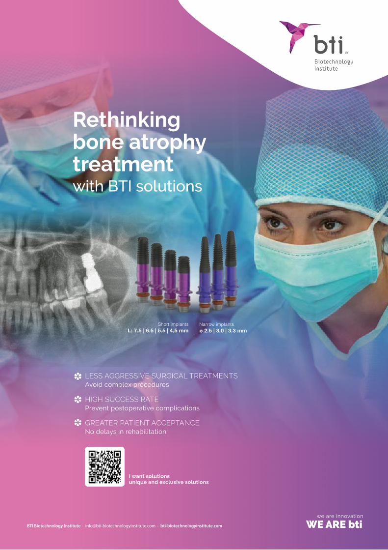
BTI Biotechnology Institute · [email protected] · bti-biotechnologyinstitute.com
Rethinking bone atrophy treatmentwith BTI solutions
LESS AGGRESSIVE SURGICAL TREATMENTSAvoid complex procedures
HIGH SUCCESS RATEPrevent postoperative complications
GREATER PATIENT ACCEPTANCENo delays in rehabilitation
Short implantsL: 7.5 | 6.5 | 5.5 | 4,5 mm
Narrow implantsø 2.5 | 3.0 | 3.3 mm
we are innovation
WE ARE bti
I want solutionsunique and exclusive solutions
210x297 MAXILAR ATROFICO EN 2.indd 1 9/2/21 18:49
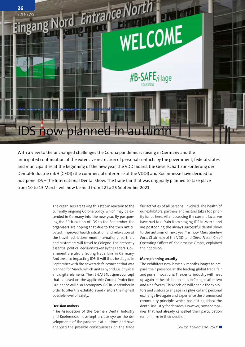
fair activities of all personal involved. The health of our exhibitors, partners and visitors takes top prior-ity for us here. After assessing the current facts, we have had to refrain from staging IDS in March and are postponing the always successful dental show to the autumn of next year,” is how Mark Stephen Pace, Chairman of the VDDI and Oliver Freser, Chief Operating Officer of Koelnmesse GmbH, explained their decision.
More planning securityThe exhibitors now have six months longer to pre-pare their presence at the leading global trade fair and push innovations. The dental industry will meet up again in the exhibition halls in Cologne after two and a half years: This decision will enable the exhibi-tors and visitors to engage in a physical and personal exchange live again and experience the pronounced community principle, which has distinguished the dental industry for decades. However, most compa-nies that had already cancelled their participation remain firm in their decision.
Source: Koelnmesse, VDDI
The organisers are taking this step in reaction to the currently ongoing Corona policy, which may be ex-tended in Germany into the new year. By postpon-ing the 39th edition of IDS to the September, the organisers are hoping that due to the then antici-pated, improved health situation and relaxation of the travel restrictions more international partners and customers will travel to Cologne. The presently essential political decisions taken by the Federal Gov-ernment are also affecting trade fairs in Germany. And are also impacting IDS. It will thus be staged in September with the new trade fair concept that was planned for March, which unites hybrid, i.e. physical and digital elements. The #B-SAFE4business concept that is based on the applicable Corona Protection Ordinance will also accompany IDS in September in order to offer the exhibitors and visitors the highest possible level of safety.
Decision makers“The Association of the German Dental Industry and Koelnmesse have kept a close eye on the de-velopments of the pandemic at all times and have analysed the possible consequences on the trade
VDDI and Koelnmesse postpone the International Dental Show
IDS now planned in autumnWith a view to the unchanged challenges the Corona pandemic is raising in Germany and the anticipated continuation of the extensive restriction of personal contacts by the government, federal states and municipalities at the beginning of the new year, the VDDI board, the Gesellschaft zur Förderung der Dental-Industrie mbH (GFDI) (the commercial enterprise of the VDDI) and Koelnmesse have decided to postpone IDS – the International Dental Show. The trade fair that was originally planned to take place from 10 to 13 March, will now be held from 22 to 25 September 2021.
Photo: Koelnmesse GmbH/Harald Fleissner
26EDI NEWS
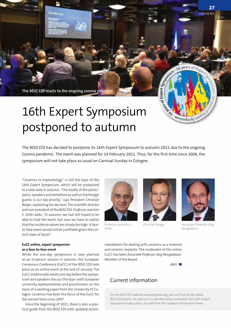
mendations for dealing with ceramics as a material and ceramic implants. The moderator of the online EuCC has been Associate Professor Jörg Neugebauer, Member of the Board.
AWU
“Ceramics in Implantology” is still the topic of the 16th Expert Symposium, which will be postponed to a later date in autumn. “The health of the partici-pants, speakers and exhibitors as well as the foreign guests is our top priority,” says President Christian Berger, explaining the decision. The scientific director and vice-president of the BDIZ EDI, Professor Joachim E. Zöller adds: “In autumn, we had still hoped to be able to hold the event, but now we have to realize that the incidence values are simply too high. A face-to-face event would not be justifiable given the cur-rent state of facts!”
EuCC online, expert symposium as a face-to-face eventWhile the one-day symposium is now planned as an in-person session in autumn, the European Consensus Conference (EuCC) of the BDIZ EDI took place as an online event at the end of January. The EuCC traditionally meets one day before the sympo-sium and speakers discuss the topic with European university representatives and practitioners on the basis of a working paper from the University of Co-logne. Ceramics has been the focus of the EuCC for the second time since 2007.
Since the beginning of 2021, there is also a prac-tical guide from the BDIZ EDI with updated recom-
The BDIZ EDI reacts to the ongoing corona situation
16th Expert Symposium postponed to autumnThe BDIZ EDI has decided to postpone its 16th Expert Symposium to autumn 2021 due to the ongoing Corona pandemic. The event was planned for 14 February 2021. Thus, for the first time since 2006, the symposium will not take place as usual on Carnival Sunday in Cologne.
Professor Joachim E. Zöller
Christian Berger Associate Professor Jörg Neugebauer
Current information
On the BDIZ EDI website www.bdizedi.org, you will find all the latest BDIZ EDI events. As soon as it is decided where and when the 16th Expert Symposium takes place, you will find the updated information there.
27EDI NEWS
“3. The following implants shall be exempted from the obligations laid down in this Article: sutures, staples, dental fillings, dental braces, tooth crowns, screws, wedges, plates, wires, pins, clips and connec-tors. The Commission is empowered to adopt del-egated acts in accordance with Article 115 to amend this list by adding other types of implants to it or by removing implants therefrom.”
It has been the CED’s understanding, the paper states, that under the MDR, all devices/materials that are affixed to the bone (such as dental implants) are not exempted from the obligation of an implant card. This would mirror the established shared inter-pretation of national Competent Authorities on clas-sification of dental implants under Council Directive 93/42/EEC on Medical Devices (MDD). The CED has no objections to this obligation. However, the CED has been recently informed of some inconsistencies in member states’ interpretations of Article 18 of the MDR.
Intended use and envisaged benefitsArticle 18(1) of the MDR lays out the information to be provided by the manufacturer on the implant card. The implant card must clearly identify the de-vice and provide additional relevant information. Article 18(2) lays down the obligation of Member States to require health institutions or healthcare providers to deliver the implant card to the patient in question. The paragraph reads,
“2. Member States shall require health institutions to make the information referred to in paragraph 1 available, by any means that allow rapid access to that information, to any patients who have been implanted with the device, together with the implant card, which shall bear their identity”.
Additionally, in order to further clarify Article 18 of the MDR, a guidance document on implant cards has been adopted by the European Medical Device Coordination Group (MDCG). It states that the im-
With the document at hand, the Council of European Dentists (CED) wishes to clarify its position regard-ing the implant card obligation in dental implants for safeguarding patient safety. This CED position specifically applies to dental implants and not to implant materials or any other device or material intended to be placed in the teeth.
European Regulatory FrameworkPursuant to Article 18(3) of the Medical Devices Regulation (MDR) (EU) 2017/745 i, published on 5 May 2017 in the Official Journal of the European Union (EU), and, as per Regulation (EU) 2020/561, applicable as of 26 May 2021:
CED position on MDR
Implant cards for dental implantsThe Council of European Dentists (CED), roof organisation of the dental chambers of all EU member states, adopted in its online general meeting 2020 a position paper on implant cards for dental implants which is required by the Medical Devices Regulation (MDR).
Profile
The CED is an European not-for-profit association which represents over 340,000 dental practitioners across Europe through 33 national dental associations and chambers in 31 European countries. Established in 1961 to advise the European Commission on matters relating to the dental profession, the CED key objectives are to promote high standards of oral healthcare and dentistry and effective patient-safety centred professional practice.
28EDI NEWS
plant card is meant to help the patient identify the implanted device and to get access to information related to it. Implant cards can also be used by emer-gency department personnel or first responders to receive information about medical treatment or pa-tient needs in emergency situations, this is also valid for dental emergencies.
CED positionThe CED’s mission is to promote the highest stan-dards of oral care to ensure patient safety at all stages of dental treatment. All measures aimed at improving traceability and patient’s access to rel-evant information about his/her health are strongly supported by the CED. At present, high standards of traceability and safety of dental implants may already be in place at the national level. Dentists should use appropriate and certified dental materi-als and medical devices for each patient’s individu-alised treatment plan. CED notices the different in-terpretations of the MDR and would like to stress
the importance of a shared single interpretation concerning the position of implant cards.
For previous mentioned reasons CED believes that implant cards for dental implants can contribute to high standards of traceability and patients access to information.
CED considers a high standard of traceability and access to information as paramount for a best prac-tice that promotes high standards of oral healthcare and effective patient-safety centred professional practice.
Therefore, in this context, the CED supports the view that it is best practice that a dentist who plac-es a dental implant should supply the patient with an implant card. This recommendation supports a best practice that promotes high standards of oral healthcare, dentistry and effective patient-safety centred professional practice.
Adopted at the CED General Meeting on 20 No-vember 2020
Source: CED
EDI NEWS29
CAD/CAM in digital dentistry
The publication “CAD/CAM in digital dentistry” in English closes an up to this point existing gap in the dental literature.The tremendous speed of development in digital dentistry requires profound knowledge in the various areas of the digital workflow. This book is a thread running from data acquisition to data processing through to digital production techniques. The target groups are dental technicians as well as dentists, trainees and students and also participants in postgraduate training courses.
Soft cover, 190 pages, ISBN 978-3-00-064987-5
by Josef Schweiger and Annett Kieschnick
[email protected] +49 8243 9692-16Fax +49 8243 9692-22
plus shipping
49,-€
to poorer oral health outcomes but to impaired gen-eral health across the board.
The current public health crisis is deepening oral health disparities in access to care. This is due to different factors, including insufficient funding for nationalhealth budgets, lack of prevention cam-paigns aimed at general and specific health targets, and absence of new legislative programmes oriented towards promoting and safeguarding a healthy life-style. In addition, we are failing to prioritise fragile and vulnerable population groups that already have limited access to regular check-ups meant to detect oral diseases, as well as early signs of general health issues, such as oral cancer, diabetes, lymphoma and cardiovascular diseases. Not by coincidence, it is ex-actly these groups with limited access to general and oral healthcare who are also at high risk for COVID-19.
Safety of dentistry during the pandemic At the beginning of the COVID-19 pandemic, den-tists were commonly considered among those occu-pations at the highest risk for COVID-19, due to the potential direct and indirect exposure to infectious materials. However, dentists have been practicing strict infection control measures and taking univer-sal precautions for the last 50 years. There is grow-ing evidence that dentistry remains safe for dental patients, members of the dental team and den-tists themselves also under COVID-19 conditions. Effective personal protective equipment is applied to protect both the dental personnel and the patient, and appropriate protocols are followed, which allows the dental team to perform oral care procedures in a safe environment.
In most European countries, dental practices be-gan slowly re-opening in spring and summer 2020, with constantly updated guidance on the safety pro-cedures required. International, European and na-tional bodies published various recommendations, sometimes contradictory and wrongly interpreted. In November 2020, the Council of European Dentists published its own statements on the safety of den-tistry and on effective dental educations in COVID-19
The COVID-19 pandemic continues to have devas-tating health, social and economic consequences, and is profoundly affecting the delivery of health services, including dentistry. During the early stages of the public health crisis, the provision of oral care in Europe was significantly curtailed. European govern-ments recommended that dentists postpone elec-tive dental procedures and provide emergency-only dental treatments, both to prevent the spread of in-fections and to keep dental patients from burdening hospital emergency departments. This approach not only fed the patients’ fears of contracting the virus when visiting the dentist, it also seemed to confirm the image of oral care as an optional luxury rather than a necessity.
Importance of oral health promotion Neglecting oral health of course predates the COVID-19 era, even though oral conditions are com-monplace across the globe. The 2017 Global Bur-den of Disease Study, which is considered to be the definitive research project on sources of mortality and disability, estimated that oral diseases affect close to 3.5 billion people worldwide, with untreated dental caries (tooth decay) in permanent teeth being the most common health condition.
However, it was only in 2019 that the World Health Organization finally included oral health in the Politi-cal Declaration on Universal Health Coverage, eight years after the United Nations High-Level Meeting on Noncommunicable Diseases recognised that oral dis-eases pose a major health burden for many countries.
COVID-19 and oral health inequalities The prevalence of oral diseases, which share com-mon factors with other major noncommunicable diseases, continues to increase in Europe. This is primarily due to the use of tobacco and alcohol, ex-cessive sugar consumption, and other lifestyle fac-tors, as well as to poor access to oral health services, particularly for weaker socioeconomic groups. Many of these factors have been heightened during the pandemic. Unless we act now, this will lead not only
Dentistry during the pandemic:
Is oral health still a priority?Marco Landi, President of the Council of European Dentists, considers dentistry during the COVID-19 pandemic and the extent to which oral health is still a priority.
Marco Landi, President of the Council of European Dentists
Phot
o: m
ad_p
rodu
ctio
n/st
ock.
adob
e.co
m
30EDI NEWS
times, strongly recommending the continuity of oral care provision during the pandemic.
Oral COVID-19 as an opportunity to refocus oral care The COVID-19 pandemic presents an overarching op-portunity to rethink the future of dentistry and ad-dress pressing issues faced by the field, rather than going back to business as usual.
Current circumstances provide a chance to reori-ent oral care towards a less invasive and more pre-ventive approach, an endeavour which tackles the shared risks for oral diseases and other noncommu-nicable, and embraces the non-surgical approach to caries management, as promoted by the Council of European Dentists in our White Paper on prevention. By focusing on the reduction of risk factors and pro-motion of a healthy lifestyle in community settings, as well as on increasing access to oral health, we pro-pose to shift the dental care paradigm from treat-ment to prevention. These methods present a major opportunity to expand the access to preventive and restorative care for vulnerable populations, but they must be combined with policy changes, sustainable
payment reform, and changes in the education of oral health professionals.
Health in the European context In this context, the Council of European Dentists welcomes what seems to be a change of pace in European health policies, with more importance attributed to health in the European Commission’s work, with a bold EU4Health Programme and mul-tiyear budget proposals. These initiatives aim to strengthen national health systems so that they can face epidemics as well as long-term challenges by stimulating disease prevention and health promo-tion, the digital transformation of health systems and increasing access to healthcare for vulnerable groups. The European Commissioner for Health’s ap-proach and priorities to work on urgent health issues such as the fight against cancer, reducing the num-ber of antimicrobial-resistant infections and improv-ing vaccination rates is an important step forward in building strong and resilient health systems all over Europe.
Marco Landi
Contact addressMarco Landi President of the Council of European Dentists (CED) Phone: +32 2 736 34 29 [email protected] www.cedentists.eu www.twitter.com/ CEDentists
EDI NEWS31
BONE SUBSTITUTE MATERIAL
The essence of nature in expert hands.BEGO IMPLANT SYSTEMS
Partners in Progress www.bego.com
[rhinoceros] synonymous with certainty, calm and power
In this context, employees do not have to inform the employer of an HIV infection, for instance,” ex-plains the lawyer. Employees can agree to the pro-cessing of their personal data, but it must be done willingly. “If they only consent because they other-wise have to fear negative consequences at work, this is not voluntary,” says Wedde.
If an employer nevertheless demands that em-ployees provide information about their vaccination status, they violate the terms of voluntary consent set out in the GDPR and risk the imposition of a financial penalty (Art. 83(5)(a) GDPR), for which there is a le-gal limit of up to €20 million. Additionally, individual claims for compensation may be made by employees (Art. 82(1) GDPR) if they are no longer assigned to better-paid specialized tasks due to the refusal to give information. Should an employer (despite the lack of information under labour or data protection law) re-fuse access to the company without a vaccination certificate, he would still be obligated to continue paying wages and salaries (due to the default of ac-ceptance that would then occur).
Wedde’s conclusion: “Since there is neither a legal obligation to vaccinate nor an entitlement to have employees present proof of vaccination, the only op-tion left to employers is, on the one hand, to push for other company protection measures against infections and, on the other hand, to convince em-ployees who are undecided of the usefulness of vaccination. This also applies to the care sector. In doing so, they must avoid putting pressure on those who do not want to be vaccinated or discriminate against them. Those who are not allowed or do not want to be vaccinated due to an existing allergy, for example, must not be compelled to inform the em-ployer directly or indirectly of the existence of this health condition. For this reason, companies may also have to live with stricter hygiene regulations on a permanent basis.”
Source: Frankfurt University of Applied Sciences, ZWP-Online
“This debate about vaccinations as an entry require-ment will not spare companies and offices and has already begun in the case of nursing staff,” says Pro-fessor Peter Wedde from the Frankfurt University of Applied Sciences (Frankfurt UAS).
Wedde is critical above all of the lack of legal regu-lation – as he was with the launch of the “Corona app”. The employer presumably justifies the require-ment for employees to be vaccinated with the need to protect colleagues, but also with the obligation to safeguard their own health and thus their work-ing capacity. Employees can argue against this with their right to physical integrity and to freely decide on medical interventions. “Since each of the parties can back up its position with fundamental rights, conflicts are inevitable here,” warns Wedde.
Data protection lawAlso, from a data protection point of view, the ques-tion about vaccination protection is inadmissible due to the lack of a legal or contractual basis. “According to the General Data Protection Regulation (GDPR), the answer to such a question would be the start of the processing of personal data (Art. 4 No. 2 GDPR).
As the special category ‘health of employees’ is at issue, this is generally prohibited (Art. 9 (1) GDPR). Legal derogations such as the fulfilment of obliga-tions under labour or social law or occupational healthcare (Art. 9(2) GDPR) do not apply here.
Statement made by Professor Wedde, Frankfurt
Can proof of vaccination be a requirement?With the beginning of COVID-19 vaccinations, the fight against the Corona virus has entered a new round. In Europe, it is evident that a considerable number of persons refuse to be vaccinated for different reasons. This is fueling the discussion on the question if proof of vaccination could be a requirement for entering public institutions, restaurants or means of transportation in the future.
About the author
Professor Peter Wedde is professor of labour law and information society law at the Frankfurt University of Applied Sciences (Frankfurt UAS). His research areas include individual and collective labour law as well as data and em-ployee data protection. He is the editor of legal comments on all aspects of individual labour law, industrial constitution law and data protection law, and the author of various book and journal articles and online publications. As a speaker, he regularly presents his main topics at specialist conferences and in practitioner forums.
Phot
o: A
ngel
o Es
slin
ger a
uf P
ixab
ay
Professor Peter Wedde
32EDI NEWS
Europe Ticker +++
start treatment later when their disease is more ad-vanced and harder to treat, meaning they are more likely to die from it or be left disabled. Dr Hans Kluge, the WHO’s regional director for Europe, said: ‘Due to travel restrictions and the enormous strain on health systems of fighting COVID-19, cancer services have been disrupted across the entire region, significantly delaying diagnosis and treatment, directly impact-ing the chances of a cure or survival for hundreds of thousands of cancer patients.’ Many more people will die in the coming years, particularly of breast and bowel cancer, for which screening appointments have been postponed, he said. Dr Kluge said in his statement from February 4th: ‘At the Kyrgyzstan National Center of Oncology, the number of cancers diagnosed in April last year dropped by 90 per cent, while in the Netherlands and Belgium in the first lockdown of 2020, it dropped by 30–40 per cent. ‘Delayed diagnosis and treatment in the United Kingdom are expected to result in an increase in the number of deaths from colorectal [bowel] can-cer by 15 per cent, and 9 per cent for breast cancer over the next 5 years. ‘A crisis of noncommunicable diseases, including cancer, is brewing, brought on by the pandemic.’ Source: Daily Mail, UK, WHO
New post-Brexit laws
Expensive work permits The EU has discovered not all member states are subject to the same rules and fees when applying for post-Brexit work visas in the UK. Five EU countries face more expensive charges than other member states, which has prompted an outcry from impact-ed countries. Having left the EU, the UK is no longer part of the bloc’s freedom of movement, which al-lows EU nationals to look and apply for jobs in an-
US-Researchers from Tufts University
No risk by touching surfaces
The risk of catching coronavirus from frequently touched surfaces such as ATMs, petrol pumps and pedestrian crossings is low, according to research-ers. Scientists swabbed hundreds of supermarket door handles, post boxes and bin lids between April and June last year, during the first wave. But they only detected COVID 29 times – or eight per cent of samples. And even when the virus was identi-fied it was in amounts so small the researchers said the risk of infection was ‘low’ – estimated to be as little as one in 2,000. The study carried out in the 700,000-strong city of Boston, Massachusetts, adds to an increasing body of evidence that COVID is not regularly spread by touch. Researchers say droplets containing the SARS-CoV-2 virus emitted when an infected person coughs, sneezes or talks are the main way the disease is passed on. This study adds to mounting evidence that aerosol droplets are the main way the virus jumps between people. Despite insisting contaminated surfaces play a minimal role in transmission, experts say people should still sa-nitise their hands to stay safe. They argue the virus can still enter the body when infected hands touch the nose and mouth. The researchers from Tufts Uni-versity, also in Massachusetts, swabbed the selected surfaces in the city almost every week to identify whether the virus was regularly present on surfaces. They found samples were more likely to be positive when there was a higher infection rate, but dropped when fewer residents were testing positive for the virus. ‘The estimated risk of infection from touching a contaminated surface was low (less than five in 10,000),’ they wrote. Source: Tufts University, USA
WHO: Coronavirus pandemic
Catastrophic for cancer careCOVID has had a ‘catastrophic’ impact on can-cer treatments and ‘a crisis is brewing’, the World Health Organization has warned. Millions of people across Europe saw their scans or treatment delayed because of lockdowns put in place to control the corona virus pandemic in 2020. As a result, many will
Phot
o: o
neph
oto/
stoc
k.ad
obe.
com
EDI NEWS33
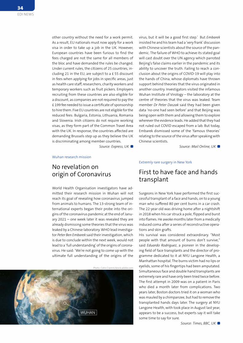
virus, but it will be a good first step.’ But Embarek insisted he and his team had a ‘very frank’ discussion with Chinese scientists about the source of the pan-demic. The failure of WHO to achieve its stated goal will cast doubt over the UN agency which parroted Beijing’s false claims earlier in the pandemic and its ability to uncover the truth. Failing to reach a con-clusion about the origins of COVID-19 will play into the hands of China, whose diplomats have thrown support behind theories that the virus originated in another country. Investigators visited the infamous Wuhan Institute of Virology – the laboratory at the centre of theories that the virus was leaked. Team member Dr Peter Daszak said they had been given data ‘no-one had seen before’ and that Beijing was being open with them and allowing them to explore wherever the evidence leads. He added that they had not ruled out COVID escaped from a lab. But today, Embarek dismissed some of the ‘famous theories’ relating to the source of the virus after speaking with Chinese scientists. Source: Mail Online, UK
Extremly rare surgery in New York
First to have face and hands transplant
Surgeons in New York have performed the first suc-cessful transplant of a face and hands, on to a young man who suffered 80 per cent burns in a car crash. The 22-year old was driving home after a nightshift in 2018 when his car struck a pole, flipped and burst into flames. He awoke months later from a medically induced coma after a series of reconstructive opera-tions and skin grafts.His survival was considered extraordinary. “Most people with that amount of burns don’t survive,” said Eduardo Rodriguez, a pioneer in the develop-ing field of face transplants and the director of pro-gramme dedicated to it at NYU Langone Health, a Manhattan hospital. The burns victim had no lips or eyelids, some of his fingertips had been amputated. Simultaneous face and double hand transplants are extremely rare and have only been tried twice before. The first attempt in 2009 was on a patient in Paris who died a month later from complications. Two years later, Boston doctors tried it on a woman who was mauled by a chimpanzee, but had to remove the transplanted hands days later. The surgery at NYU Langone Health, with took place in August last year, appears to be a success, but experts say it will take some time to say for sure. Source: Times, BBC, UK
other country without the need for a work permit. As a result, EU nationals must now apply for a work visa in order to take up a job in the UK. However, European countries have been furious to find the fees charged are not the same for all members of the bloc and have demanded the rules be changed. Under current rules, the citizens of 25 countries, in-cluding 21 in the EU, are subject to a £ 55 discount in fees when applying for jobs in specific areas, just as health care staff, researchers, charity workers and temporary workers such as fruit pickers. Employers recruiting from these countries are also eligible for a discount, as companies are not required to pay the £ 199 fee needed to issue a certificate of sponsorship to hire them. Five EU countries are not eligible for the reduced fees: Bulgaria, Estonia, Lithuania, Romania and Slovenia. Irish citizens do not require working visas, as they form part of the Common Travel Area with the UK. In response, the countries affected are demanding Brussels step up as they believe the UK is discriminating among member countries. Source: Express, UK
Wuhan research mission
No revelation on origin of Coronavirus
World Health Organisation investigators have ad-mitted their research mission in Wuhan will not reach its goal of revealing how coronavirus jumped from animals to humans. The 13-strong team of in-ternational experts began their probe into the ori-gins of the coronavirus pandemic at the end of Janu-ary 2021 – one week later it was revealed they are already dismissing some theories that the virus was leaked by a Chinese laboratory. WHO lead investiga-tor Peter Ben Embarek said their investigation, which is due to conclude within the next week, would not lead to a ‘full understanding’ of the origins of corona-virus. He said: ‘We’re not going to come up with the ultimate full understanding of the origins of the
Photo: Colin Cramm/stock.adobe.com
34EDI NEWS
Save the date: 14th European Symposium postponed to May 2021
Technology meets excellenceA highlight in the BDIZ EDI year is certainly always the European Symposium, which is held annually with a different partner in a different European country. Due to the corona pandemic, the event – which was planned for May 2020 in Skopje – has been postponed to 2021.
small beginnings and opportunities, an approach has been developed which allows the cooperation of European dentists to grow beyond national borders and thus intensifies the professional exchange within Europe. The European Symposium is based on the proven training concept of the BDIZ EDI to promote active dental exchange on a European level. The BDIZ EDI is the first cooperation partner of AIAM.
BDIZ EDI will be a cooperation partner of the Alba-nian Association for Implantology in Macedonia (ADSM-AIAM), the Macedonian Dental Association, the Association of Oral Surgeons (MDS-AOSS) and the associated partner association EDI Macedonia.
The international congress of AIAM offers work-shops and a scientific program, which mainly deals with oral implantology, but includes many other disciplines of dentistry: general, regenerative and aesthetic dentistry, oral surgery and implantology, functional therapy as well as topics like digitaliza-tion etc.
“The basis for the greatest ventures often lies in barely noticeable opportunities.” Demosthenes’ sen-tence from his Philippic speeches is characteristic for the history of the BDIZ EDI’s European symposia. From
More information and registration
Programme, registration and venue will soon be available on the BDIZ EDI website: https://bdizedi.org/en/14th- european-symposium-in-skopje/
EDI NEWS35
... that BDIZ EDI
… offers online seminars during the pandemic for all associated partner organisations for free? There’s still a strong need of information among dentists and implant dentists all over Europe. Topics can be infection control, hygiene, but also a need for updates on peri-implantitis, on digital implantology, short, angulated and diameter-reduced implants, on bone augmentation, and many more.
For more information see questionnaire in this issue and/or con-tact [email protected]
... that EDI Journal
… covers the hot topics in the field between science, human medicine and dentistry? For example: EDI Journal 4/2021 gave exciting insights into stem-cell research, especially when it comes to regrowing teeth. In this issue we give a glimpse on the value of vitamin D in dentistry and the possible necessity of supplementing the substance when it comes to augmentation and/or implantation. Don’t miss the editorials in EDI Journal für the dental office.
For more information see https://bdizedi.org/en/publications/ »EDI News: Update on COVID-19 pandemic in Europe · Will IDS 2021 take place?
BDIZ EDI Website relaunched · Save the date: 14th European Symposium postponed
to May 2021 »European Law: The European Court of Justice deals with the PIP scandal
»Case Studies: Immediate loading of extrashort implants »Clinical Science Cantilever
extensions of implant-supported superstructures
European Journal for Dental Implantologists
ISSN 1862-2879 I Volume 16
Issue 3/20
EDI J
ourn
al 3
/20
TOPIC
Bone and soft tissue
management
»EDI News: What’s going on in Europe? · Dealing with aerosol-borne pathogens in dental practice · Corona virus will not stop IDS · Place an implant – or grow a new tooth? »European Law: ECJ invalidates EU-US Data Protection Shield »Case Studies: One-year follow up of full arch treatment with new fully tapered Tissue Level implant system »Clinical Science: One-year clinical experience with Progressive-Line implants
European Journal for Dental Implantologists
ISSN 1862-2879 I Volume 16
Issue 4/20
EDI J
ourn
al 4
/20
TOPIC
Quo vadis, implantology?
»EDI News: COVID-19 in Europe · An update on short, angulated and diameter-reduced
implants · IDS now planned in autumn · Discussion: Vitamin D »European Law: Transi-
tional period for the Medical Device Regulation runs out in May 2021 »Case Studies:
Multidisciplinary approach in case of extreme maxillo-mandibular atrophy »Clinical
Science: Partial extraction therapy
European Journal for Dental Implantologists
ISSN 1862-2879 I Volume 17Issue 1 /21
EDI J
ourn
al 1
/21
TOPIC
Discussion onvitamin D
BDIZ EDI informs 2021!
... that BDIZ EDI
… had to postpone the 16th Expert Symposium on ceramics in implantology due to COVID-19 pandemic to fall 2021? However, the European Consensus Conference (EuCC) under the auspices of BDIZ EDI has taken place in February. The final guideline on above topic will be published in EDI Journal 2/2021 in June 2021. If you are interested in all guidelines of BDIZ EDI, you can download the papers from the website:
The brochure can be downloaded online for free: https://bdizedi.org/en/quality-guidelines/
Did you ever know ...
Cologne ABC Risk Score for Implant Treatment
7 thEuropean Consensus Conference of BDIZ EDI
February 2012
European Association of
Dental Implantologists (BDIZ EDI)
Lipowskystr. 12 · 81373 Munich/Germany
FON +49 (0) 89 720 69 888 · FAX +49 (0) 89 720 69 023
www.bdizedi.org
Update Periimplantitis –
periimplantäre Entzündungen und
periimplantäre Erkrankungen
15. Europäische Konsensuskonferenz (EuCC) 2020 in Köln
22. Februar 2020
Bundesverband der implantologisch
tätigen Zahnärzte in Europa (BDIZ EDI)
Mühlenstr. 18 · 51143 Köln
Tel. 02203/80 09 339 · Fax 02203/91 68 822
www.bdizedi.org
2020
Update peri-implantitis –peri-implant inflammation and peri-implant disease
15th European Consensus Conference (EuCC) 2020 in Cologne22 February 2020
European Association of DentalImplantologists (BDIZ EDI)
Mühlenstr. 18 · 51143 CologneTel. 02203/80 09 339 · Fax 02203/91 68 822
2020
36EDI NEWS
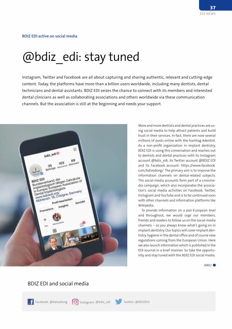
More and more dentists and dental practices are us-ing social media to help attract patients and build trust in their services. In fact, there are now several millions of posts online with the hashtag #dentist. As a non-profit organization in implant dentistry, BDIZ EDI is using this conversation and reaches out to dentists and dental practices with its Instagram account @bdiz_edi, its Twitter account @BDIZ EDI and its Facebook account: https://www.facebook.com/bdizediorg/. The primary aim is to improve the information channels on dental-related subjects. The social media accounts form part of a crossme-dia campaign, which also incorporates the associa-tion’s social media activities on Facebook, Twitter, Instagram and YouTube and is to be continued soon with other channels and information platforms like Wikipedia.
To provide information on a pan-European level and throughout, we would urge our members, friends and readers to follow us on the social media channels – so you always know what’s going on in implant dentistry. Our topics will cover implant den-tistry, hygiene in the dental office and of course new regulations coming from the European Union. Here we also launch information which is published in the EDI Journal in a brief manner. So take the opportu-nity and stay tuned with the BDIZ EDI social media.
AWU
BDIZ EDI active on social media
@bdiz_edi: stay tunedInstagram, Twitter and Facebook are all about capturing and sharing authentic, relevant and cutting-edge content. Today, the platforms have more than a billion users worldwide, including many dentists, dental technicians and dental assistants. BDIZ EDI seizes the chance to connect with its members and interested dental clinicians as well as collaborating associations and others worldwide via these communication channels. But the association is still at the beginning and needs your support.
BDIZ EDI and social media
Facebook: @bdizediorg Instagram: @bdiz_edi Twitter: @BDIZEDI
Phot
o: fr
eepi
k.co
m/d
an_d
ariu
s
EDI NEWS37
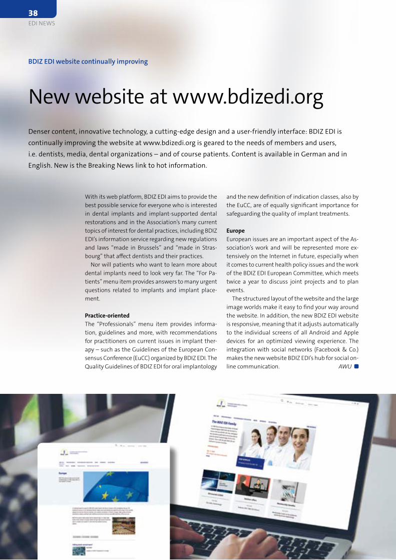
and the new definition of indication classes, also by the EuCC, are of equally significant importance for safeguarding the quality of implant treatments.
EuropeEuropean issues are an important aspect of the As-sociation’s work and will be represented more ex-tensively on the Internet in future, especially when it comes to current health policy issues and the work of the BDIZ EDI European Committee, which meets twice a year to discuss joint projects and to plan events.
The structured layout of the website and the large image worlds make it easy to find your way around the website. In addition, the new BDIZ EDI website is responsive, meaning that it adjusts automatically to the individual screens of all Android and Apple devices for an optimized viewing experience. The integration with social networks (Facebook & Co.) makes the new website BDIZ EDI’s hub for social on-line communication. AWU
With its web platform, BDIZ EDI aims to provide the best possible service for everyone who is interested in dental implants and implant-supported dental restorations and in the Association’s many current topics of interest for dental practices, including BDIZ EDI’s information service regarding new regulations and laws “made in Brussels” and “made in Stras-bourg” that affect dentists and their practices.
Nor will patients who want to learn more about dental implants need to look very far. The “For Pa-tients” menu item provides answers to many urgent questions related to implants and implant place-ment.
Practice-orientedThe “Professionals” menu item provides informa-tion, guidelines and more, with recommendations for practitioners on current issues in implant ther-apy – such as the Guidelines of the European Con-sensus Conference (EuCC) organized by BDIZ EDI. The Quality Guidelines of BDIZ EDI for oral implantology
BDIZ EDI website continually improving
New website at www.bdizedi.org Denser content, innovative technology, a cutting-edge design and a user-friendly interface: BDIZ EDI is continually improving the website at www.bdizedi.org is geared to the needs of members and users, i.e. dentists, media, dental organi zations – and of course patients. Content is available in German and in English. New is the Breaking News link to hot information.
38EDI NEWS
§Medical Device Regulation MDR
Transitional period runs out in May 2021Medical devices are products or equipment intended generally for a medical use and are regulated at Member State level. The Medical Device and the In-Vitro Diagnostic Device Regulations have introduced new responsibili-ties for the European Medicines Agency (EMA) and national competent authorities in the assessment of certain categories of medical devices. On 23 April 2020, the European Parliament and the Council of the EU adopted a proposal to extend the transitional period of the Medical Device Regulation by one year – until 26 May 2021.
EMA will update its regulatory guid-ance in the first quarter of 2021 to re-flect the new date of application of the Regulation.
The MDR has a transitional period of four years and will fully apply from 26 May 2021. The IVDR has a transitional period of five years and will fully apply from 26 May 2022.
During the transitional period, manu-facturers can opt to place medical devices on the market under the applicable EU Directives (93/42/EEC, 98/79/EC and 90/385/EEC) or under the new Regula-tions if they fully comply with these.
Combination productsSome medicines are used in combination with a medical device, usually to enable the delivery of the medicine. If the prin-ciple intended action of the combination product is achieved by the medicine, the entire product is regulated as a medicinal product under Directive 2001/83/EC or Regulation (EC) No 726/2004.
There are two types of combination:• integral: the medicinal product and
the device form a single integrated product e.g. pre-filled syringes and pens, patches for transdermal drug delivery and pre-filled inhalers;
• co-packaged: the medicinal product and the device are separate items contained in the same pack e.g. reus-able pen for insulin cartridges, tablet delivery system with controller for pain management.
Medical devices in the EU have to under-go a conformity assessment to demon-strate that they meet legal requirements to ensure they are safe and perform as intended. EU member states can desig-nate accredited notified bodies to con-duct conformity assessments.
The conformity assessment usually involves an audit of the manufacturer’s quality system and, depending on the type of device, a review of technical docu-mentation from the manufacturer on the safety and performance of the device.
Manufacturers can place a CE (Con-formité Européenne) mark on a medical device once it has passed a conformity assessment.
LegislationThe adoption in April 2017 of Regulation (EU) 2017/745 on Medical Device (MDR) and Regulation (EU) 2017/746 on In-Vitro Diagnostic Device (IVDR) changed the European legal framework for medical devices, introducing new responsibilities for EMA and for national competent au-thorities. Both Regulations entered into force in May 2017 and have a staggered transitional period.
On 23 April 2020, the European Parlia-ment and the Council of the EU adopted a proposal to extend the transitional pe-riod of the Medical Device Regulation by one year – until 26 May 2021.
This measure aims to avoid shortages of medical devices during the ongoing COVID-19 pandemic due to the limited ca-pacity of national competent authorities or notified bodies to implement the Regulation.
Medical devices that are co-packaged or obtained separately must be CE marked in accordance with the medical device legislation.
Article 117 of the MDR introduced a new requirement for medicines with an integral device. From 26 May 2021, the marketing authorisation application should include a CE certificate for the device or, if it is not CE marked but would need to be certified if marketed separate-ly, the applicant must include an opinion from a notified body on the conformity of the device. This requirement does not apply to Class I devices (non-sterile, non-measuring).
Role of EMAThe Agency is responsible for evaluating the quality, safety and efficacy of market-ing authorisation applications assessed through the centralised procedure, in-cluding the safety and performance of the medical device in relation to its use with the medicinal product.
On 3 June 2019, EMA released for a 3-month public consultation a guideline on quality requirements for regulatory submissions for medicines that include a medical device (drug-device combina-tions). The device can be integral, co-packaged or obtained separately.
The guideline clarifies expectations laid down in Directive 2001/83/EC and addresses the new obligations in the MDR, in particular the requirements under Article 117. EMA will finalise the guideline before the entry into force of the MDR on 26 May 2021.
EUROPEAN LAW39
Before it can issue a CE certificate, the notified body must seek a scientific opin-ion from EMA or a national competent authority on the compliance of the sub-stance with the requirements laid down in Annex I to Directive 2001/83/EC.
Borderline productsBorderline products are complex health-care products for which there is uncertainty over which regulatory framework applies.
Common borderlines are between medicinal products, medical devices, cos-metics, biocidal products, herbal medi-cines and food supplements.
National competent authorities classify borderline products either as medicinal products or, for example, as medical devic-es on a case-by-case basis. This determines the applicable regulatory framework.
Applicants who are unclear on the cor-rect classification of their product should consult a national competent authority and provide information on the product’s composition and constituents, a scientif-ic explanation of the mode of action and its intended purpose.
The Medical Device Regulation fore-sees that the European Commission may consult the Agency on products that bor-derline with medicines.
EMA’s Innovation Task Force provides ad-vice to medicine developers on eligibility to EMA procedures relating to the research and development of borderline products.
Source: European Medicines Agency (official website of the European Union:
ema.europe.eu
To what extent is dentistry or oral implantology affected by the EU regulation?
Answer was given by Professor Ulrich M. Gassner, Founding Director of the Research Center for Medical Device Law at the University of Augsburg in EDI Journal 2/2019:
Professor Gassner: Implantologists with in-office laboratories are manufacturers of custom-made products. There are no transitional arrangements that apply to them. As of 26 May 2020 (update: 26 May 2021), for example, they must meet exactly the same requirements that apply to industrial manufacturers of medical devices with regard to clinical evaluations.
And clinical evaluations are not even all there is to it. Under the heading of “post-market surveillance” (PMS), the MDR demands that implantologists continue to monitor whether their implants are safe and therapeutically useful once implanted and whether there are any incidents related to them. Further-more, they have to demonstrate that a person responsible for regulatory compliance is available on a permanent and continuous basis.
The In-Vitro Diagnostic Device Regu-lation introduces a new classification system for companion diagnostics and the obligation to undergo a conformity assessment by a notified body.
Before it can issue a CE certificate, the notified body must seek a scientific opin-ion from the Agency on the suitability of the companion diagnostic to the medici-nal product concerned if: • the medicinal product falls exclusively
within the scope of the centralised procedure for the authorisation of medicines, or
• the medicinal product is already authorised through the centralised procedure, or
• a marketing authorisation application for the medicinal product has been submitted through the centralised procedure.
For other substances, the notified body can seek the opinion from a national competent authority or EMA.
EMA will provide further information on the consultation procedure between the notified body and a competent au-thority or EMA.
Medical devices made of substances that are systemically absorbedSome medical devices are made of sub-stances that are absorbed by the human body to achieve their intended purpose.
These devices are normally introduced into the human body via an orifice or ap-plied to the skin.
Medical devices with an ancillary medicinal substanceA medical device may contain an ancil-lary medicinal substance to support the proper functioning of the device. These products fall under the medical devices legislation and must be CE marked.
Examples of medical devices with an ancillary medicinal substance include drug-eluting stents, bone cement con-taining an antibiotic, catheters coated with heparin or an antibiotic agent and condoms coated with spermicides.
Additional: scientific opinionBefore it can issue a CE certificate, the notified body must seek a scientific opinion from the Agency on the quality and safety of the ancillary substance if it is derived from human blood or human plasma, or if it is within the scope of the centralised procedure for the authorisa-tion of medicines.
For other substances, the notified body can seek the opinion from a national competent authority or EMA e.g. in cases where the Agency has already evaluated a medicine containing the same medici-nal substance.
EMA publishes consultation procedure public assessment reports (CPAR) on its scientific opinions.
In-vitro diagnosticsA companion diagnostic is an in-vitro diagnostic test that supports the safe and effective use of a specific medicinal product, by identifying patients that are suitable or unsuitable for treatment.
40EUROPEAN LAW
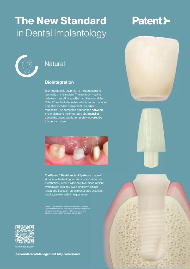
EUROPEAN LAW41
Natural
Biointegration
Biointegration is essential to the success and longevity of the implant. The optimum healing between the soft tissue, the hard tissue and the Patent™ Implant eliminates infections and reduces complications like periimplantitis and peri-mucositis. The cemented connection between the implant and the integrated abutment lies above the tissue and is completely covered by the dental crown.
mucositis. The cemented connection between the implant and the integrated abutment lies above the tissue and is completely covered by
Zircon Medical Management AG, Switzerland
The Patent™ Dental Implant System is made of zirconia with a hydrophilic surface and metal free prosthetics. Patent™ is the only two-piece implant system with peer-reviewed long term clinical research1. Based on our demonstrated excellent results, we o� er a lifetime guarantee.
1 Becker J, John G, Becker K, Mainusch S, Diedrichs G, Schwarz F.Clinical performance of two-piece zirconium implants in the posterior mandible and maxilla: a prospective cohort study over 2 years. Clin. Oral Impl. Res. 28, 2017, 29 – 35 doi: 10.1111/clr.12610
www.mypatent.com
The New Standardin Dental Implantology
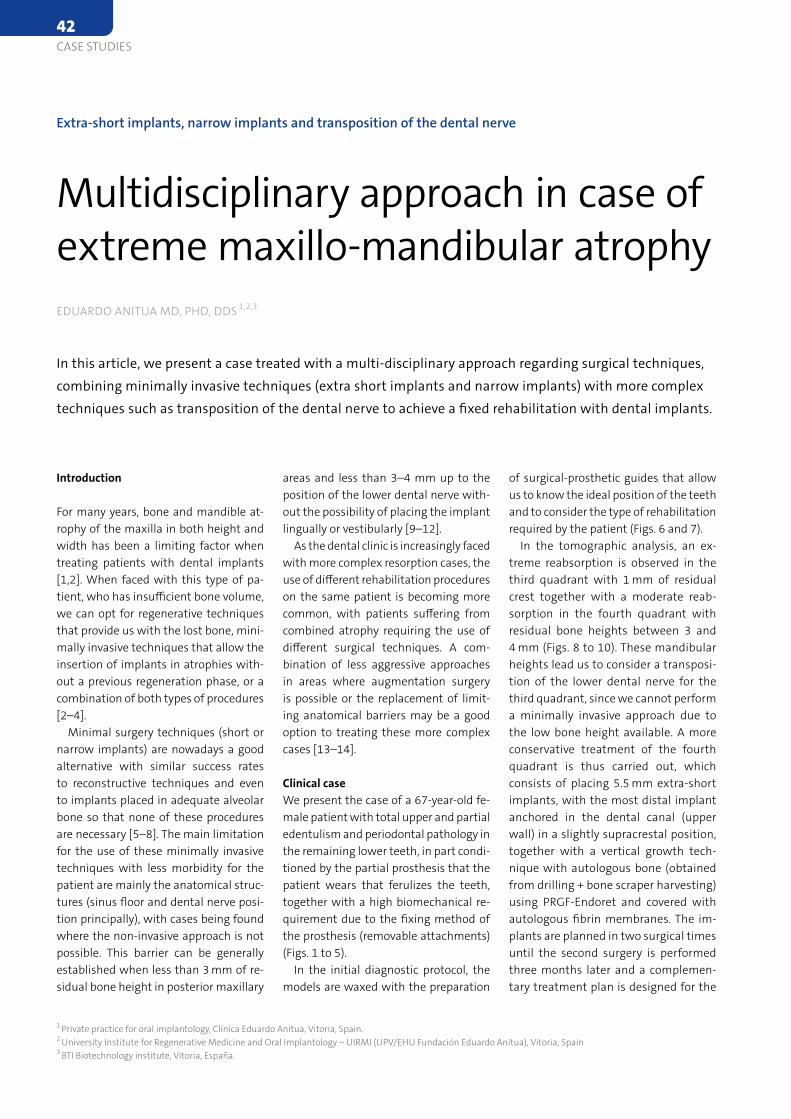
areas and less than 3–4 mm up to the position of the lower dental nerve with-out the possibility of placing the implant lingually or vestibularly [9–12].
As the dental clinic is increasingly faced with more complex resorption cases, the use of different rehabilitation procedures on the same patient is becoming more common, with patients suffering from combined atrophy requiring the use of different surgical techniques. A com-bination of less aggressive approaches in areas where augmentation surgery is possible or the replacement of limit-ing anatomical barriers may be a good option to treating these more complex cases [13–14].
Clinical case We present the case of a 67-year-old fe-male patient with total upper and partial edentulism and periodontal pathology in the remaining lower teeth, in part condi-tioned by the partial prosthesis that the patient wears that ferulizes the teeth, together with a high biomechanical re-quirement due to the fixing method of the prosthesis (removable attachments) (Figs. 1 to 5).
In the initial diagnostic protocol, the models are waxed with the preparation
Introduction
For many years, bone and mandible at-rophy of the maxilla in both height and width has been a limiting factor when treating patients with dental implants [1,2]. When faced with this type of pa-tient, who has insufficient bone volume, we can opt for regenerative techniques that provide us with the lost bone, mini-mally invasive techniques that allow the insertion of implants in atrophies with-out a previous regeneration phase, or a combination of both types of procedures [2–4].
Minimal surgery techniques (short or narrow implants) are nowadays a good alternative with similar success rates to reconstructive techniques and even to implants placed in adequate alveolar bone so that none of these procedures are necessary [5–8]. The main limitation for the use of these minimally invasive techniques with less morbidity for the patient are mainly the anatomical struc-tures (sinus floor and dental nerve posi-tion principally), with cases being found where the non-invasive approach is not possible. This barrier can be generally established when less than 3 mm of re-sidual bone height in posterior maxillary
Extra-short implants, narrow implants and transposition of the dental nerve
Multidisciplinary approach in case of extreme maxillo-mandibular atrophyEDUARDO ANITUA MD, PHD, DDS 1,2,3
In this article, we present a case treated with a multi-disciplinary approach regarding surgical techniques, combining minimally invasive techniques (extra short implants and narrow implants) with more complex techniques such as transposition of the dental nerve to achieve a fixed rehabilitation with dental implants.
of surgical-prosthetic guides that allow us to know the ideal position of the teeth and to consider the type of rehabilitation required by the patient (Figs. 6 and 7).
In the tomographic analysis, an ex-treme reabsorption is observed in the third quadrant with 1 mm of residual crest together with a moderate reab-sorption in the fourth quadrant with residual bone heights between 3 and 4 mm (Figs. 8 to 10). These mandibular heights lead us to consider a transposi-tion of the lower dental nerve for the third quadrant, since we cannot perform a minimally invasive approach due to the low bone height available. A more conservative treatment of the fourth quadrant is thus carried out, which consists of placing 5.5 mm extra-short implants, with the most distal implant anchored in the dental canal (upper wall) in a slightly supracrestal position, together with a vertical growth tech-nique with autologous bone (obtained from drilling + bone scraper harvesting) using PRGF-Endoret and covered with autologous fibrin membranes. The im-plants are planned in two surgical times until the second surgery is performed three months later and a complemen-tary treatment plan is designed for the
1 Private practice for oral implantology, Clínica Eduardo Anitua, Vitoria, Spain.2 University Institute for Regenerative Medicine and Oral Implantology – UIRMI (UPV/EHU Fundación Eduardo Anitua), Vitoria, Spain3 BTI Biotechnology institute, Vitoria, España.
42CASE STUDIES
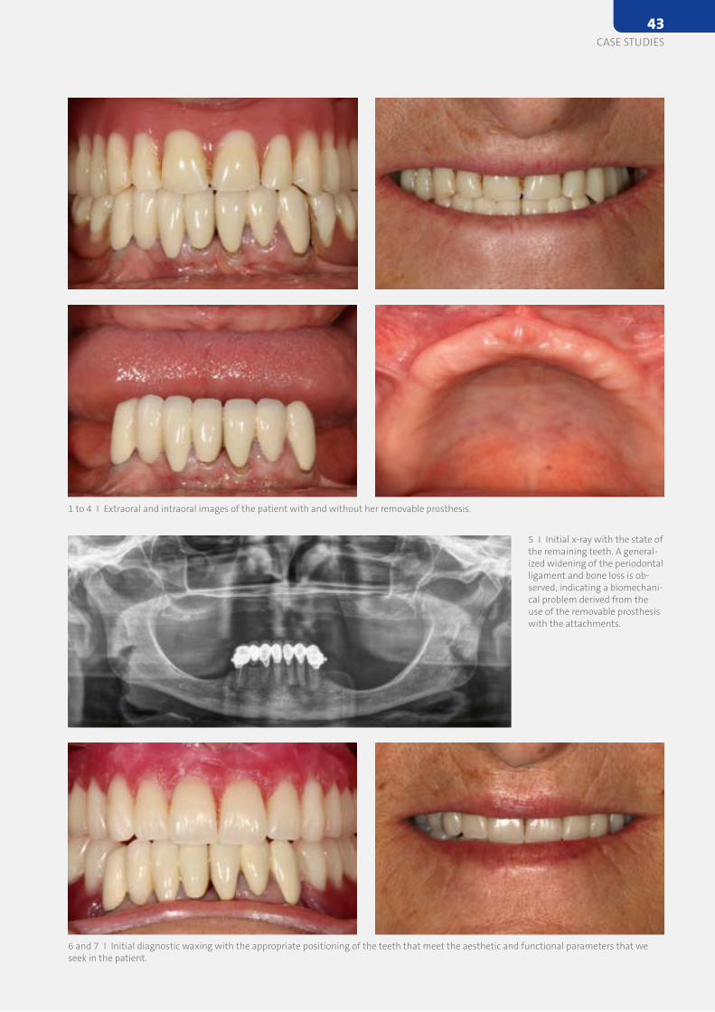
1 to 4 I Extraoral and intraoral images of the patient with and without her removable prosthesis.
5 I Initial x-ray with the state of the remaining teeth. A general-ized widening of the periodontal ligament and bone loss is ob-served, indicating a biomechani-cal problem derived from the use of the removable prosthesis with the attachments.
6 and 7 I Initial diagnostic waxing with the appropriate positioning of the teeth that meet the aesthetic and functional parameters that we seek in the patient.
CASE STUDIES43
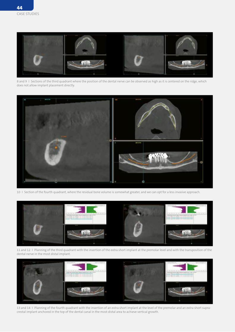
8 and 9 I Sections of the third quadrant where the position of the dental nerve can be observed as high as it is centered on the ridge, which does not allow implant placement directly.
11 and 12 I Planning of the third quadrant with the insertion of the extra short implant at the premolar level and with the transposition of the dental nerve in the most distal implant.
13 and 14 I Planning of the fourth quadrant with the insertion of an extra short implant at the level of the premolar and an extra short supra-crestal implant anchored in the top of the dental canal in the most distal area to achieve vertical growth.
10 I Section of the fourth quadrant, where the residual bone volume is somewhat greater, and we can opt for a less invasive approach.
44CASE STUDIES
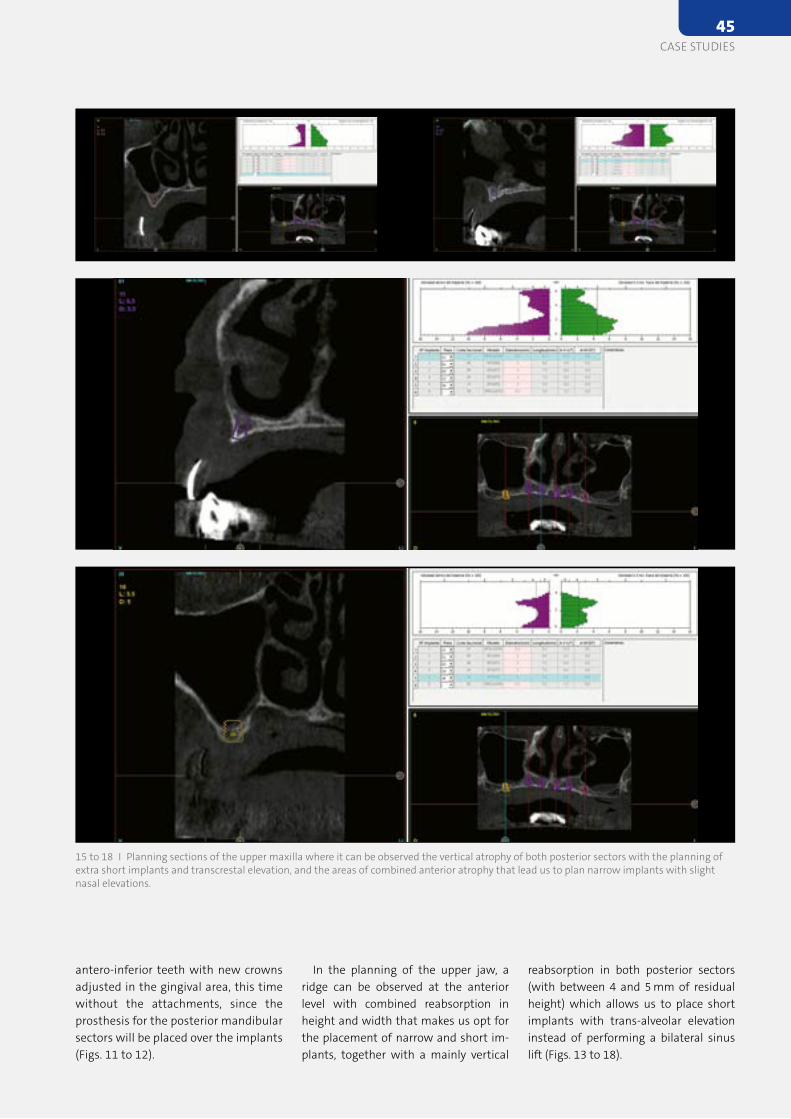
reabsorption in both posterior sectors (with between 4 and 5 mm of residual height) which allows us to place short implants with trans-alveolar elevation instead of performing a bilateral sinus lift (Figs. 13 to 18).
In the planning of the upper jaw, a ridge can be observed at the anterior level with combined reabsorption in height and width that makes us opt for the placement of narrow and short im-plants, together with a mainly vertical
antero-inferior teeth with new crowns adjusted in the gingival area, this time without the attachments, since the prosthesis for the posterior mandibular sectors will be placed over the implants (Figs. 11 to 12).
15 to 18 I Planning sections of the upper maxilla where it can be observed the vertical atrophy of both posterior sectors with the planning of extra short implants and transcrestal elevation, and the areas of combined anterior atrophy that lead us to plan narrow implants with slight nasal elevations.
CASE STUDIES45
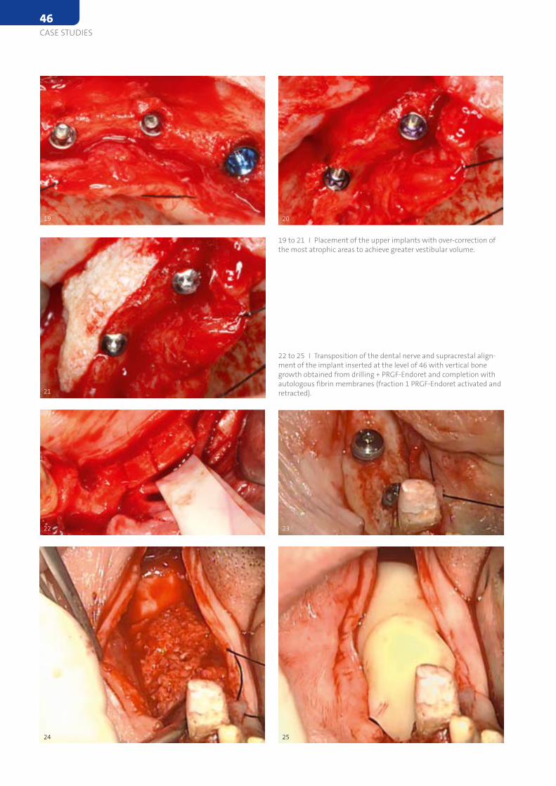
19
21
23
25
20
22
24
19 to 21 I Placement of the upper implants with over-correction of the most atrophic areas to achieve greater vestibular volume.
22 to 25 I Transposition of the dental nerve and supracrestal align-ment of the implant inserted at the level of 46 with vertical bone growth obtained from drilling + PRGF-Endoret and completion with autologous fibrin membranes (fraction 1 PRGF-Endoret activated and retracted).
46CASE STUDIES
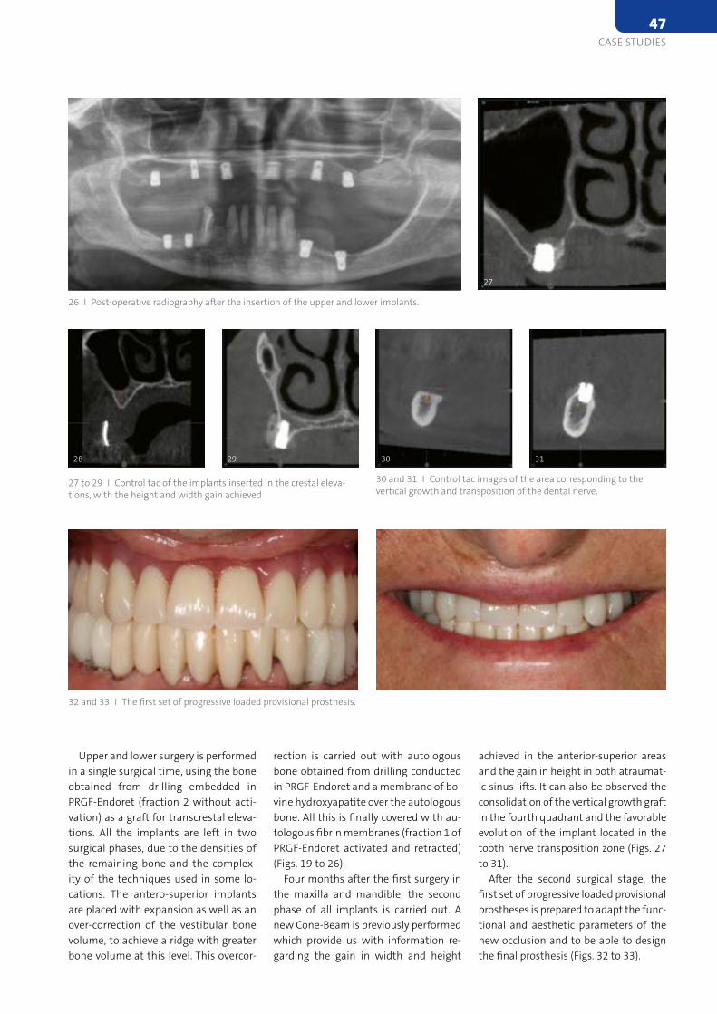
achieved in the anterior-superior areas and the gain in height in both atraumat-ic sinus lifts. It can also be observed the consolidation of the vertical growth graft in the fourth quadrant and the favorable evolution of the implant located in the tooth nerve transposition zone (Figs. 27 to 31).
After the second surgical stage, the first set of progressive loaded provisional prostheses is prepared to adapt the func-tional and aesthetic parameters of the new occlusion and to be able to design the final prosthesis (Figs. 32 to 33).
rection is carried out with autologous bone obtained from drilling conducted in PRGF-Endoret and a membrane of bo-vine hydroxyapatite over the autologous bone. All this is finally covered with au-tologous fibrin membranes (fraction 1 of PRGF-Endoret activated and retracted) (Figs. 19 to 26).
Four months after the first surgery in the maxilla and mandible, the second phase of all implants is carried out. A new Cone-Beam is previously performed which provide us with information re-garding the gain in width and height
Upper and lower surgery is performed in a single surgical time, using the bone obtained from drilling embedded in PRGF-Endoret (fraction 2 without acti-vation) as a graft for transcrestal eleva-tions. All the implants are left in two surgical phases, due to the densities of the remaining bone and the complex-ity of the techniques used in some lo-cations. The antero-superior implants are placed with expansion as well as an over-correction of the vestibular bone volume, to achieve a ridge with greater bone volume at this level. This overcor-
26 I Post-operative radiography after the insertion of the upper and lower implants.
27
30 3128 29
27 to 29 I Control tac of the implants inserted in the crestal eleva-tions, with the height and width gain achieved
30 and 31 I Control tac images of the area corresponding to the vertical growth and transposition of the dental nerve.
32 and 33 I The first set of progressive loaded provisional prosthesis.
CASE STUDIES47
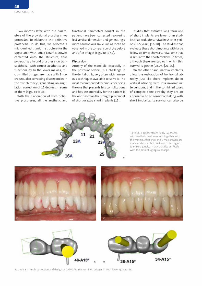
Studies that evaluate long term use of short implants are fewer than stud-ies that evaluate survival in shorter peri-ods (1-5 years) [16-20]. The studies that evaluate these short implants with large follow-up times show a survival time that is similar to the shorter follow-up times, although there are studies in which this survival is greater (98.9%) [21-25].
On the other hand, narrow implants allow the restoration of horizontal at-rophy, just like short implants do in vertical atrophy, with less invasive in-terventions, and in the combined cases of complex bone atrophy they are an alternative to be considered along with short implants. Its survival can also be
functional parameters sought in the patient have been corrected, recovering lost vertical dimension and generating a more harmonious smile line as it can be observed in the comparison of the before and after images (Figs. 40 to 42).
DiscussionAtrophy of the mandible, especially in the posterior sectors, is a challenge in the dental clinic, very often with numer-ous techniques available to solve it. The most recommended technique for being the one that presents less complications and has less morbidity for the patient is the one based on the straight placement of short or extra short implants [15].
Two months later, with the param-eters of the provisional prosthesis, we proceeded to elaborate the definitive prosthesis. To do this, we selected a micro-milled titanium structure for the upper arch with Emax ceramic crowns cemented onto the structure, thus generating a hybrid prosthesis on tran-sepithelial with correct aesthetics and functionality. In the lower maxilla, mi-cro-milled bridges are made with Emax crowns, also correcting discrepancies in the exit chimneys, generating an angu-lation correction of 15 degrees in some of them (Figs. 34 to 38).
With the elaboration of both defini-tive prostheses, all the aesthetic and
35
34
37 38
36
34 to 36 I Upper structure by CAD/CAM with aesthetic test in mouth together with the waxing. After that, the E-Max crowns are made and cemented on it and tested again to make a gingival mask that fits perfectly with the patient’s gingival margin.
37 and 38 I Angle correction and design of CAD/CAM micro-milled bridges in both lower quadrants.
48CASE STUDIES
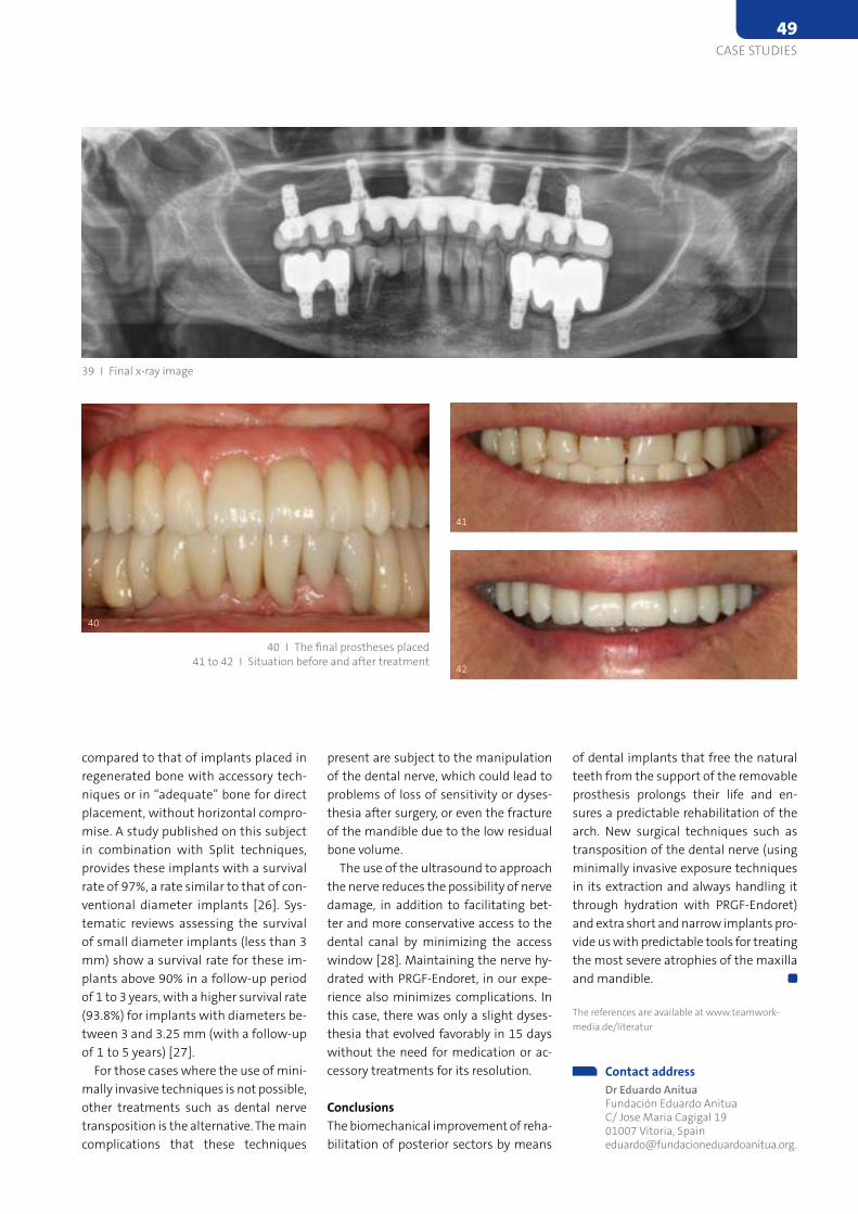
of dental implants that free the natural teeth from the support of the removable prosthesis prolongs their life and en-sures a predictable rehabilitation of the arch. New surgical techniques such as transposition of the dental nerve (using minimally invasive exposure techniques in its extraction and always handling it through hydration with PRGF-Endoret) and extra short and narrow implants pro-vide us with predictable tools for treating the most severe atrophies of the maxilla and mandible.
The references are available at www.teamwork-media.de/literatur
Contact address
Dr Eduardo Anitua Fundación Eduardo Anitua C/ Jose Maria Cagigal 19 01007 Vitoria, Spain [email protected].
present are subject to the manipulation of the dental nerve, which could lead to problems of loss of sensitivity or dyses-thesia after surgery, or even the fracture of the mandible due to the low residual bone volume.
The use of the ultrasound to approach the nerve reduces the possibility of nerve damage, in addition to facilitating bet-ter and more conservative access to the dental canal by minimizing the access window [28]. Maintaining the nerve hy-drated with PRGF-Endoret, in our expe-rience also minimizes complications. In this case, there was only a slight dyses-thesia that evolved favorably in 15 days without the need for medication or ac-cessory treatments for its resolution.
ConclusionsThe biomechanical improvement of reha-bilitation of posterior sectors by means
compared to that of implants placed in regenerated bone with accessory tech-niques or in “adequate” bone for direct placement, without horizontal compro-mise. A study published on this subject in combination with Split techniques, provides these implants with a survival rate of 97%, a rate similar to that of con-ventional diameter implants [26]. Sys-tematic reviews assessing the survival of small diameter implants (less than 3 mm) show a survival rate for these im-plants above 90% in a follow-up period of 1 to 3 years, with a higher survival rate (93.8%) for implants with diameters be-tween 3 and 3.25 mm (with a follow-up of 1 to 5 years) [27].
For those cases where the use of mini-mally invasive techniques is not possible, other treatments such as dental nerve transposition is the alternative. The main complications that these techniques
39 I Final x-ray image
40
41
42
40 I The final prostheses placed 41 to 42 I Situation before and after treatment
CASE STUDIES49
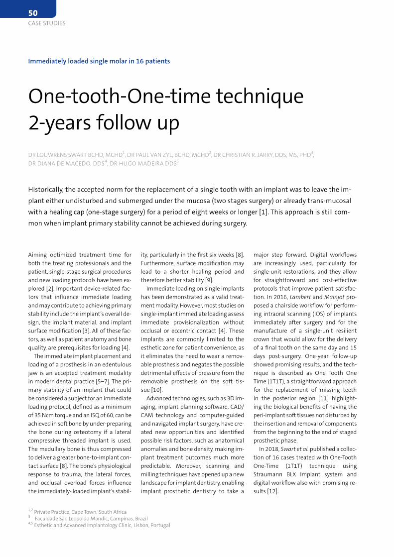
ity, particularly in the first six weeks [8]. Furthermore, surface modification may lead to a shorter healing period and therefore better stability [9].
Immediate loading on single implants has been demonstrated as a valid treat-ment modality. However, most studies on single-implant immediate loading assess immediate provisionalization without occlusal or eccentric contact [4]. These implants are commonly limited to the esthetic zone for patient convenience, as it eliminates the need to wear a remov-able prosthesis and negates the possible detrimental effects of pressure from the removable prosthesis on the soft tis-sue [10].
Advanced technologies, such as 3D im-aging, implant planning software, CAD/CAM technology and computer-guided and navigated implant surgery, have cre-ated new opportunities and identified possible risk factors, such as anatomical anomalies and bone density, making im-plant treatment outcomes much more predictable. Moreover, scanning and milling techniques have opened up a new landscape for implant dentistry, enabling implant prosthetic dentistry to take a
Aiming optimized treatment time for both the treating professionals and the patient, single-stage surgical procedures and new loading protocols have been ex-plored [2]. Important device-related fac-tors that influence immediate loading and may contribute to achieving primary stability include the implant’s overall de-sign, the implant material, and implant surface modification [3]. All of these fac-tors, as well as patient anatomy and bone quality, are prerequisites for loading [4].
The immediate implant placement and loading of a prosthesis in an edentulous jaw is an accepted treatment modality in modern dental practice [5–7]. The pri-mary stability of an implant that could be considered a subject for an immediate loading protocol, defined as a minimum of 35 Ncm torque and an ISQ of 60, can be achieved in soft bone by under-preparing the bone during osteotomy if a lateral compressive threaded implant is used. The medullary bone is thus compressed to deliver a greater bone-to-implant con-tact surface [8]. The bone’s physiological response to trauma, the lateral forces, and occlusal overload forces influence the immediately- loaded implant’s stabil-
Immediately loaded single molar in 16 patients
One-tooth-One-time technique 2-years follow upDR LOUWRENS SWART BCHD, MCHD1, DR PAUL VAN ZYL, BCHD, MCHD2, DR CHRISTIAN R. JARRY, DDS, MS, PHD3, DR DIANA DE MACEDO, DDS4, DR HUGO MADEIRA DDS5
Historically, the accepted norm for the replacement of a single tooth with an implant was to leave the im-plant either undisturbed and submerged under the mucosa (two stages surgery) or already trans-mucosal with a healing cap (one-stage surgery) for a period of eight weeks or longer [1]. This approach is still com-mon when implant primary stability cannot be achieved during surgery.
major step forward. Digital workflows are increasingly used, particularly for single-unit restorations, and they allow for straightforward and cost-effective protocols that improve patient satisfac-tion. In 2016, Lambert and Mainjot pro-posed a chairside workflow for perform-ing intraoral scanning (IOS) of implants immediately after surgery and for the manufacture of a single-unit resilient crown that would allow for the delivery of a final tooth on the same day and 15 days post-surgery. One-year follow-up showed promising results, and the tech-nique is described as One Tooth One Time (1T1T), a straightforward approach for the replacement of missing teeth in the posterior region [11] highlight-ing the biological benefits of having the peri-implant soft tissues not disturbed by the insertion and removal of components from the beginning to the end of staged prosthetic phase.
In 2018, Swart et al. published a collec-tion of 16 cases treated with One-Tooth One-Time (1T1T) technique using Straumann BLX Implant system and digital workflow also with promising re-sults [12].
1,2 Private Practice, Cape Town, South Africa3 Faculdade São Leopoldo Mandic, Campinas, Brazil4,5 Esthetic and Advanced Implantology Clinic, Lisbon, Portugal
50CASE STUDIES
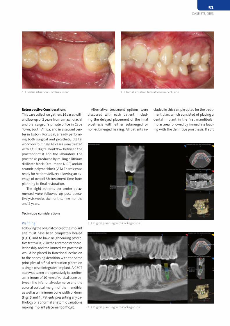
cluded in this sample opted for the treat-ment plan, which consisted of placing a dental implant in the first mandibular molar area followed by immediate load-ing with the definitive prosthesis. If soft
Alternative treatment options were discussed with each patient, includ-ing the delayed placement of the final prosthesis with either submerged or non-submerged healing. All patients in-
Retrospective ConsiderationsThis case collection gathers 16 cases with a follow up of 2 years from a maxillofacial and oral surgeon’s private office in Cape Town, South Africa, and in a second cen-ter in Lisbon, Portugal, already perform-ing both surgical and prosthetic digital workflow routinely. All cases were treated with a full digital workflow between the prosthodontist and the laboratory. The prosthesis produced by milling a lithium disilicate block (Straumann N!CE) and/or ceramic-polymer block (VITA Enamic) was ready for patient delivery allowing an av-erage of overall 5h treatment time from planning to final restoration.
The eight patients per center docu-mented were followed up post opera-tively six weeks, six months, nine months and 2 years.
Technique considerations
PlanningFollowing the original concept the implant site must have been completely healed (Fig. 1) and to have neighbouring protec-tive teeth (Fig. 2) in the anteroposterior re-lationship, and the immediate prosthesis would be placed in functional occlusion to the opposing dentition with the same principles of a final restoration placed on a single osseointegrated implant. A CBCT scan was taken pre-operatively to confirm a minimum of 10 mm of vertical bone be-tween the inferior alveolar nerve and the coronal cortical margin of the mandible, as well as a minimum bone width of 6mm (Figs. 3 and 4). Patients presenting any pa-thology or abnormal anatomic variations making implant placement difficult.
2 I Initial situation lateral view in occlusion1 I Initial situation – occlusal view
3 I Digital planning with CoDiagnostiX
4 I Digital planning with CoDiagnostiX
CASE STUDIES51
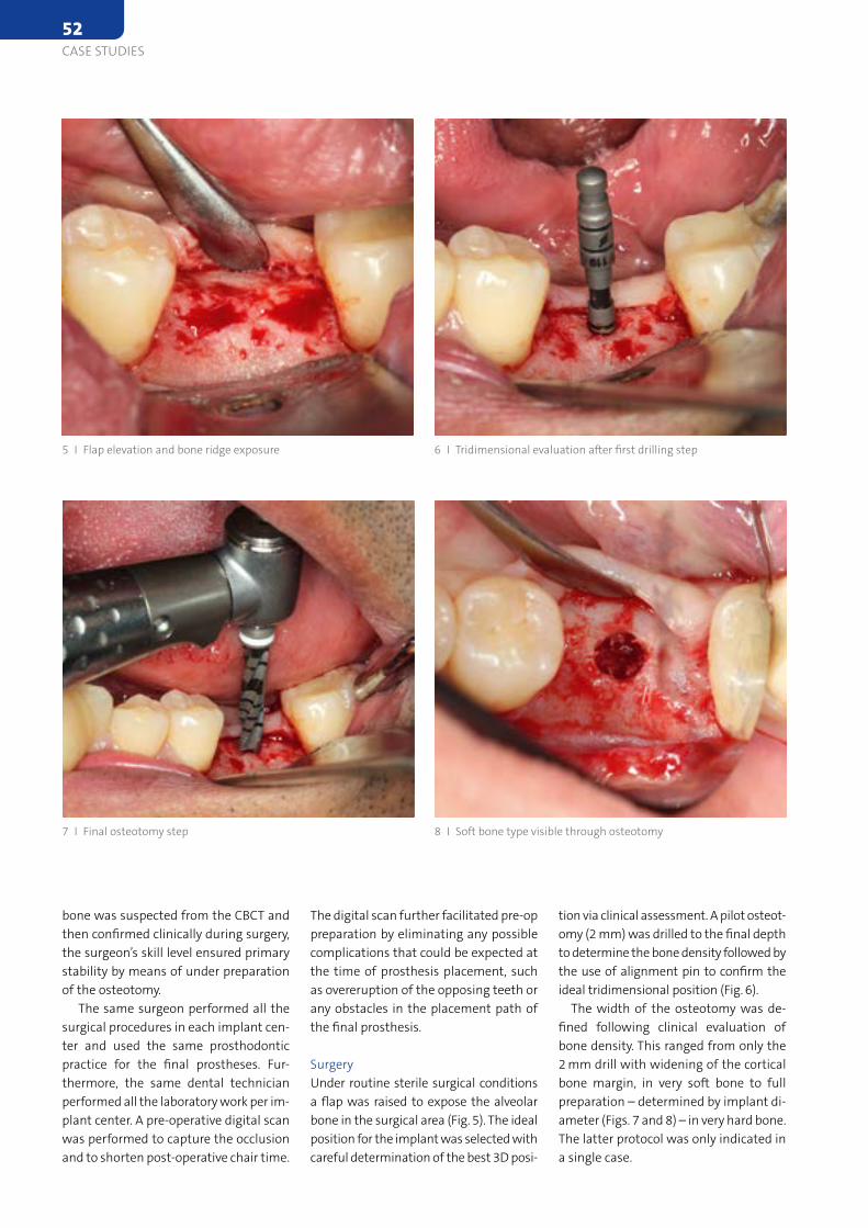
tion via clinical assessment. A pilot osteot-omy (2 mm) was drilled to the final depth to determine the bone density followed by the use of alignment pin to confirm the ideal tridimensional position (Fig. 6).
The width of the osteotomy was de-fined following clinical evaluation of bone density. This ranged from only the 2 mm drill with widening of the cortical bone margin, in very soft bone to full preparation – determined by implant di-ameter (Figs. 7 and 8) – in very hard bone. The latter protocol was only indicated in a single case.
The digital scan further facilitated pre-op preparation by eliminating any possible complications that could be expected at the time of prosthesis placement, such as overeruption of the opposing teeth or any obstacles in the placement path of the final prosthesis.
SurgeryUnder routine sterile surgical conditions a flap was raised to expose the alveolar bone in the surgical area (Fig. 5). The ideal position for the implant was selected with careful determination of the best 3D posi-
bone was suspected from the CBCT and then confirmed clinically during surgery, the surgeon’s skill level ensured primary stability by means of under preparation of the osteotomy.
The same surgeon performed all the surgical procedures in each implant cen-ter and used the same prosthodontic practice for the final prostheses. Fur-thermore, the same dental technician performed all the laboratory work per im-plant center. A pre-operative digital scan was performed to capture the occlusion and to shorten post-operative chair time.
5 I Flap elevation and bone ridge exposure
7 I Final osteotomy step
6 I Tridimensional evaluation after first drilling step
8 I Soft bone type visible through osteotomy
52CASE STUDIES
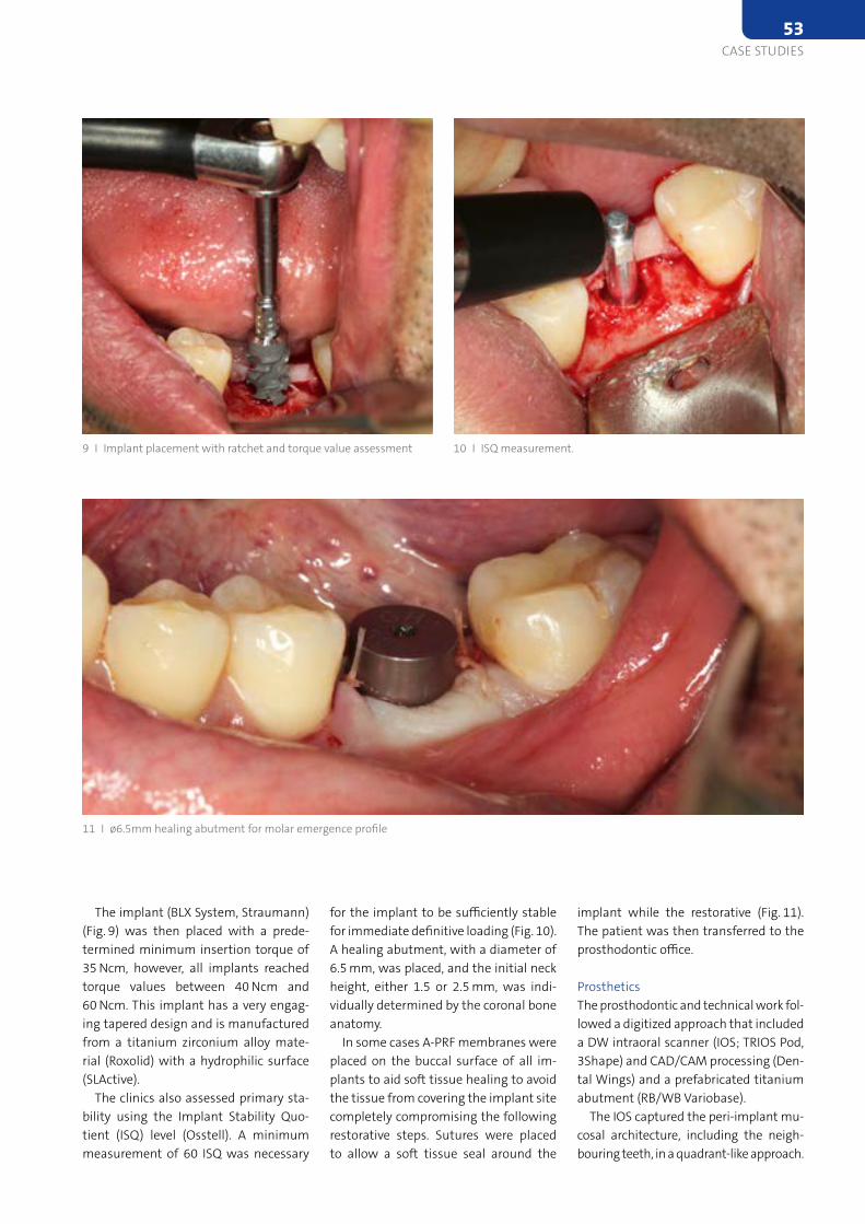
implant while the restorative (Fig. 11). The patient was then transferred to the prosthodontic office.
ProstheticsThe prosthodontic and technical work fol-lowed a digitized approach that included a DW intraoral scanner (IOS; TRIOS Pod, 3Shape) and CAD/CAM processing (Den-tal Wings) and a prefabricated titanium abutment (RB/WB Variobase).
The IOS captured the peri-implant mu-cosal architecture, including the neigh-bouring teeth, in a quadrant-like approach.
for the implant to be sufficiently stable for immediate definitive loading (Fig. 10). A healing abutment, with a diameter of 6.5 mm, was placed, and the initial neck height, either 1.5 or 2.5 mm, was indi-vidually determined by the coronal bone anatomy.
In some cases A-PRF membranes were placed on the buccal surface of all im-plants to aid soft tissue healing to avoid the tissue from covering the implant site completely compromising the following restorative steps. Sutures were placed to allow a soft tissue seal around the
The implant (BLX System, Straumann)(Fig. 9) was then placed with a prede-termined minimum insertion torque of 35 Ncm, however, all implants reached torque values between 40 Ncm and 60 Ncm. This implant has a very engag-ing tapered design and is manufactured from a titanium zirconium alloy mate-rial (Roxolid) with a hydrophilic surface (SLActive).
The clinics also assessed primary sta-bility using the Implant Stability Quo-tient (ISQ) level (Osstell). A minimum measurement of 60 ISQ was necessary
9 I Implant placement with ratchet and torque value assessment
11 I ø6.5mm healing abutment for molar emergence profile
10 I ISQ measurement.
CASE STUDIES53
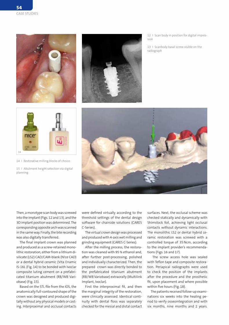
surfaces. Next, the occlusal scheme was checked statically and dynamically with Shimstock foil, achieving light occlusal contacts without dynamic interactions. The monolithic LS2 or dental hybrid ce-ramic restoration was screwed with a controlled torque of 35 Ncm, according to the implant provider’s recommenda-tions (Figs. 16 and 17).
The screw access hole was sealed with Teflon tape and composite restora-tion. Periapical radiographs were used to check the position of the implants after the procedure and the prosthetic fit, upon placement and where possible within five hours (Fig. 18).
The patients received follow-up exami-nations six weeks into the healing pe-riod to verify osseointegration and with six months, nine months and 2 years.
were defined virtually according to the threshold settings of the dental design software for chairside solutions (CARES C-Series).
The virtual crown design was processed and produced with 4-axis wet milling and grinding equipment (CARES C-Series).
After the milling process, the restora-tion was cleaned with 95 % ethanol and, after further post-processing, polished and individually characterized. Then, the prepared crown was directly bonded to the prefabricated titanium abutment (RB/WB Variobase) extraorally (Multilink Implant, Ivoclar).
First the interproximal fit, and then the marginal integrity of the restoration, were clinically assessed. Identical conti-nuity with dental floss was separately checked for the mesial and distal contact
Then, a monotype scan body was screwed into the implant (Figs. 12 and 13), and the 3D implant position was determined. The corresponding opposite arch was scanned in the same way. Finally, the bite recording was also digitally transferred.
The final implant crown was planned and produced as a screw-retained mono-lithic restoration, either from a lithium di-silicate (LS2) CAD/CAM-blank (N!ce CAD) or a dental hybrid ceramic (Vita Enamic IS-16L (Fig. 14) to be bonded with Ivoclar composite luting cement on a prefabri-cated titanium abutment (RB/WB Vari-obase) (Fig. 15).
Based on the STL file from the IOS, the anatomically full-contoured shape of the crown was designed and produced digi-tally without any physical models or cast-ing. Interproximal and occlusal contacts
12
14
13
12 I Scan body in position for digital impres-sion
13 I Scanbody basal screw visible on the radiograph
15
14 I Restorative milling blocks of choice.
15 I Abutment height selection via digital planning
54CASE STUDIES
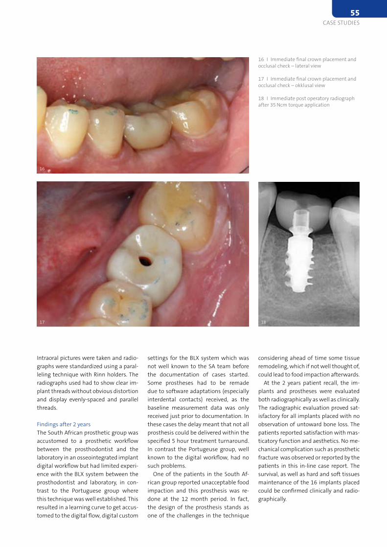
considering ahead of time some tissue remodeling, which if not well thought of, could lead to food impaction afterwards.
At the 2 years patient recall, the im-plants and prostheses were evaluated both radiographically as well as clinically. The radiographic evaluation proved sat-isfactory for all implants placed with no observation of untoward bone loss. The patients reported satisfaction with mas-ticatory function and aesthetics. No me-chanical complication such as prosthetic fracture was observed or reported by the patients in this in-line case report. The survival, as well as hard and soft tissues maintenance of the 16 implants placed could be confirmed clinically and radio-graphically.
settings for the BLX system which was not well known to the SA team before the documentation of cases started. Some prostheses had to be remade due to software adaptations (especially interdental contacts) received, as the baseline measurement data was only received just prior to documentation. In these cases the delay meant that not all prosthesis could be delivered within the specified 5 hour treatment turnaround. In contrast the Portugeuse group, well known to the digital workflow, had no such problems.
One of the patients in the South Af-rican group reported unacceptable food impaction and this prosthesis was re-done at the 12 month period. In fact, the design of the prosthesis stands as one of the challenges in the technique
Intraoral pictures were taken and radio-graphs were standardized using a paral-leling technique with Rinn holders. The radiographs used had to show clear im-plant threads without obvious distortion and display evenly-spaced and parallel threads.
Findings after 2 years The South African prosthetic group was accustomed to a prosthetic workflow between the prosthodontist and the laboratory in an osseointegrated implant digital workflow but had limited experi-ence with the BLX system between the prosthodontist and laboratory, in con-trast to the Portuguese group where this technique was well established. This resulted in a learning curve to get accus-tomed to the digital flow, digital custom
1817
16
16 I Immediate final crown placement and occlusal check – lateral view
17 I Immediate final crown placement and occlusal check – okklusal view
18 I Immediate post operatory radiograph after 35 Ncm torque application
CASE STUDIES55
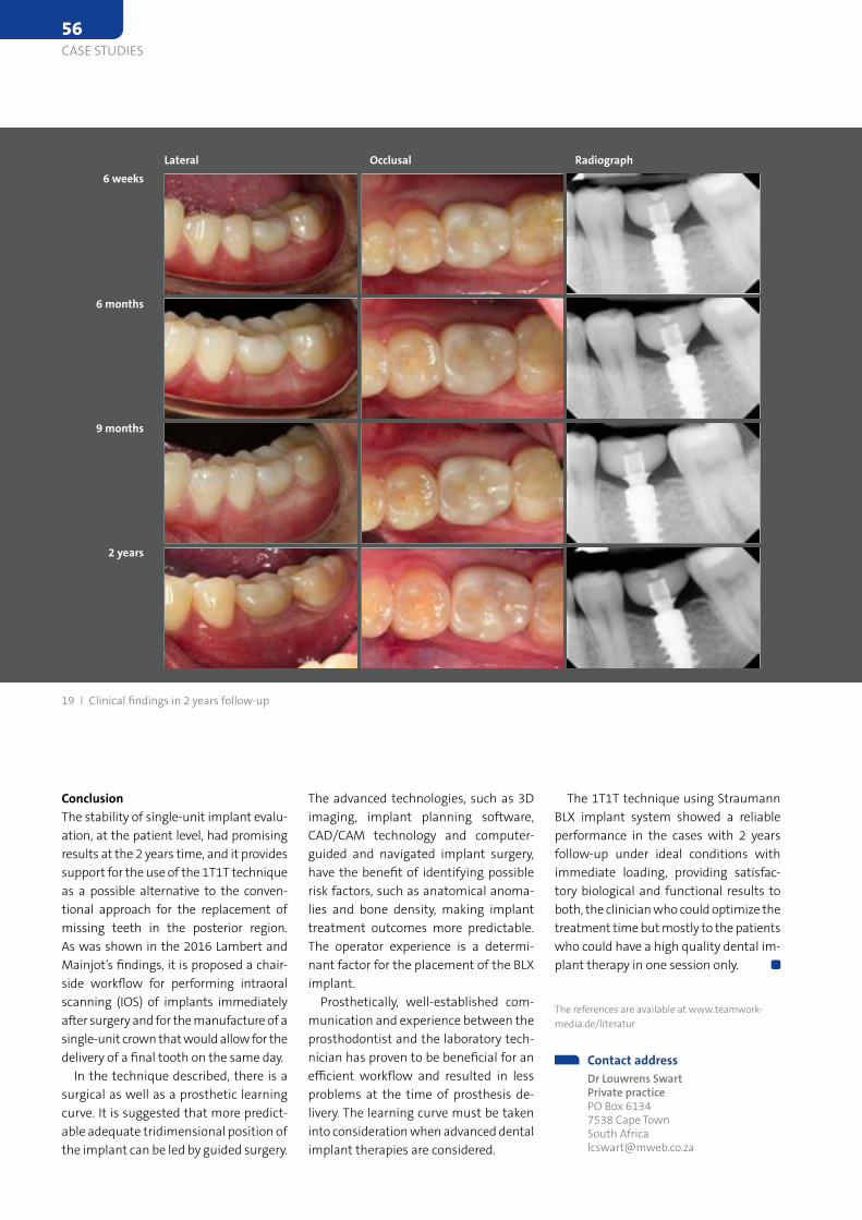
The 1T1T technique using Straumann BLX implant system showed a reliable performance in the cases with 2 years follow-up under ideal conditions with immediate loading, providing satisfac-tory biological and functional results to both, the clinician who could optimize the treatment time but mostly to the patients who could have a high quality dental im-plant therapy in one session only.
The references are available at www.teamwork-media.de/literatur
Contact address Dr Louwrens Swart
Private practice PO Box 6134 7538 Cape Town South Africa [email protected]
The advanced technologies, such as 3D imaging, implant planning software, CAD/CAM technology and computer-guided and navigated implant surgery, have the benefit of identifying possible risk factors, such as anatomical anoma-lies and bone density, making implant treatment outcomes more predictable. The operator experience is a determi-nant factor for the placement of the BLX implant.
Prosthetically, well-established com-munication and experience between the prosthodontist and the laboratory tech-nician has proven to be beneficial for an efficient workflow and resulted in less problems at the time of prosthesis de-livery. The learning curve must be taken into consideration when advanced dental implant therapies are considered.
ConclusionThe stability of single-unit implant evalu-ation, at the patient level, had promising results at the 2 years time, and it provides support for the use of the 1T1T technique as a possible alternative to the conven-tional approach for the replacement of missing teeth in the posterior region. As was shown in the 2016 Lambert and Mainjot’s findings, it is proposed a chair-side workflow for performing intraoral scanning (IOS) of implants immediately after surgery and for the manufacture of a single-unit crown that would allow for the delivery of a final tooth on the same day.
In the technique described, there is a surgical as well as a prosthetic learning curve. It is suggested that more predict-able adequate tridimensional position of the implant can be led by guided surgery.
Lateral6 weeks
6 months
9 months
2 years
Occlusal Radiograph
19 I Clinical findings in 2 years follow-up
56CASE STUDIES
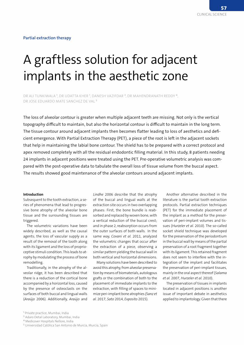
Lindhe 2006 describe that the atrophy of the buccal and lingual walls at the extraction site occurs in two overlapping phases: First, the bone bundle is reab-sorbed and replaced by woven bone, with a vertical reduction of the buccal crest; and in phase 2, reabsorption occurs from the outer surfaces of both walls. In the same way, Covani et al. 2011, analyzed the volumetric changes that occur after the extraction of a piece, observing a similar pattern yielding the buccal wall in both vertical and horizontal dimensions.
Many solutions have been described to avoid this atrophy, from alveolar preserva-tion by means of biomaterials, autologous grafts or the combination of both to the placement of immediate implants to the extraction, with filling of spaces to mini-mize peri-implant bone atrophies (Sanz et al. 2017, Sato 2014, Exposito 2015).
IntroductionSubsequent to the tooth extraction, a se-ries of phenomena that lead to progres-sive bone atrophy of the alveolar bone tissue and the surrounding tissues are triggered.
The volumetric variations have been widely described, as well as the causal agents; the loss of vascular supply as a result of the removal of the tooth along with its ligament and the loss of proprio-ceptive stimuli condition. This leads to at-rophy by modulating the process of bone remodelling.
Traditionally, in the atrophy of the al-veolar ridge, it has been described that there is a reduction of the cortical bone accompanied by a horizontal loss, caused by the presence of osteoclasts on the surfaces of both buccal and lingual walls (Araújo 2006). Additionally, Araújo and
Partial extraction therapy
A graftless solution for adjacent implants in the aesthetic zoneDR ALI TUNKIWALA ¹, DR UDATTA KHER ¹, DANESH VAZIFDAR ³, DR MAHENDRANATH REDDY ⁴, DR JOSE EDUARDO MATE SANCHEZ DE VAL ⁵
The loss of alveolar contour is greater when multiple adjacent teeth are missing. Not only is the vertical topography difficult to maintain, but also the horizontal contour is difficult to maintain in the long term. The tissue contour around adjacent implants then becomes flatter leading to loss of aesthetics and defi-cient emergence. With Partial Extraction Therapy (PET), a piece of the root is left in the adjacent sockets that help in maintaining the labial bone contour. The shield has to be prepared with a correct protocol and apex removed completely with all the residual endodontic filling material. In this study, 8 patients needing 24 implants in adjacent positions were treated using the PET. Pre-operative volumetric analysis was com-pared with the post-operative data to tabulate the overall loss of tissue volume from the buccal aspect. The results showed good maintenance of the alveolar contours around adjacent implants.
Another alternative described in the literature is the partial tooth extraction protocols. Partial extraction techniques (PET) for the immediate placement of the implant as a method for the preser-vation of peri-implant volumes and tis-sues (Hurzeler et al. 2010). The so-called socket shield technique was developed for the preservation of the periodontium in the buccal wall by means of the partial preservation of a root fragment together with its ligament. This retained fragment does not seem to interfere with the in-tegration of the implant and facilitates the preservation of peri-implant tissues, mainly in the oral aspect thereof (Salama et al. 2007, Hurzeler et al. 2010).
The preservation of tissues in implants located in adjacent positions is another issue of important debate in aesthetics applied to implantology. Given that there
¹ Private practice, Mumbai, India³ Adaro Detal Laboratory, Mumbai, India⁴ Medicover Hospitals Nellore, India¹ Universidad Católica San Antonio de Murcia, Murcia, Spain
CLINICAL SCIENCE57
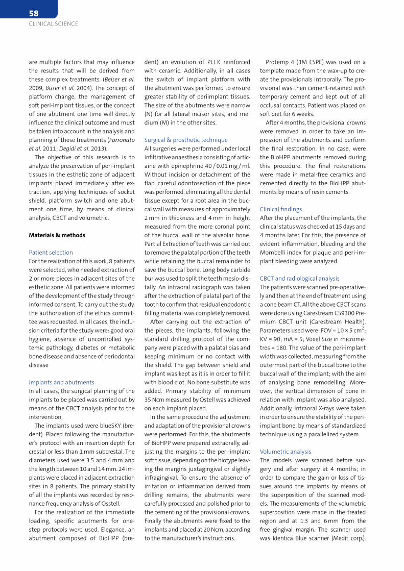
are multiple factors that may influence the results that will be derived from these complex treatments. (Belser et al. 2009, Buser et al. 2004). The concept of platform change, the management of soft peri-implant tissues, or the concept of one abutment one time will directly influence the clinical outcome and must be taken into account in the analysis and planning of these treatments (Farronato et al. 2011; Degidi et al. 2013).
The objective of this research is to analyze the preservation of peri-implant tissues in the esthetic zone of adjacent implants placed immediately after ex-traction, applying techniques of socket shield, platform switch and one abut-ment one time, by means of clinical analysis, CBCT and volumetric.
Materials & methods
Patient selectionFor the realization of this work, 8 patients were selected, who needed extraction of 2 or more pieces in adjacent sites of the esthetic zone. All patients were informed of the development of the study through informed consent. To carry out the study, the authorization of the ethics commit-tee was requested. In all cases, the inclu-sion criteria for the study were: good oral hygiene, absence of uncontrolled sys-temic pathology, diabetes or metabolic bone disease and absence of periodontal disease
Implants and abutmentsIn all cases, the surgical planning of the implants to be placed was carried out by means of the CBCT analysis prior to the intervention,
The implants used were blueSKY (bre-dent). Placed following the manufactur-er’s protocol with an insertion depth for crestal or less than 1 mm subcrestal. The diameters used were 3.5 and 4 mm and the length between 10 and 14 mm. 24 im-plants were placed in adjacent extraction sites in 8 patients. The primary stability of all the implants was recorded by reso-nance frequency analysis of Osstell.
For the realization of the immediate loading, specific abutments for one-step protocols were used. Elegance, an abutment composed of BioHPP (bre-
dent) an evolution of PEEK reinforced with ceramic. Additionally, in all cases the switch of implant platform with the abutment was performed to ensure greater stability of periimplant tissues. The size of the abutments were narrow (N) for all lateral incisor sites, and me-dium (M) in the other sites.
Surgical & prosthetic techniqueAll surgeries were performed under local infiltrative anaesthesia consisting of artic-aine with epinephrine 40 / 0.01 mg / ml. Without incision or detachment of the flap, careful odontosection of the piece was performed, eliminating all the dental tissue except for a root area in the buc-cal wall with measures of approximately 2 mm in thickness and 4 mm in height measured from the more coronal point of the buccal wall of the alveolar bone. Partial Extraction of teeth was carried out to remove the palatal portion of the teeth while retaining the buccal remainder to save the buccal bone. Long body carbide bur was used to split the teeth mesio-dis-tally. An intraoral radiograph was taken after the extraction of palatal part of the tooth to confirm that residual endodontic filling material was completely removed.
After carrying out the extraction of the pieces, the implants, following the standard drilling protocol of the com-pany were placed with a palatal bias and keeping minimum or no contact with the shield. The gap between shield and implant was kept as it is in order to fill it with blood clot. No bone substitute was added. Primary stability of minimum 35 Ncm measured by Ostell was achieved on each implant placed.
In the same procedure the adjustment and adaptation of the provisional crowns were performed. For this, the abutments of BioHPP were prepared extraorally, ad-justing the margins to the peri-implant soft tissue, depending on the biotype leav-ing the margins juxtagingival or slightly infragingival. To ensure the absence of irritation or inflammation derived from drilling remains, the abutments were carefully processed and polished prior to the cementing of the provisional crowns. Finally the abutments were fixed to the implants and placed at 20 Ncm, according to the manufacturer’s instructions.
Protemp 4 (3M ESPE) was used on a template made from the wax-up to cre-ate the provisionals intraorally. The pro-visional was then cement-retained with temporary cement and kept out of all occlusal contacts. Patient was placed on soft diet for 6 weeks.
After 4 months, the provisional crowns were removed in order to take an im-pression of the abutments and perform the final restoration. In no case, were the BioHPP abutments removed during this procedure. The final restorations were made in metal-free ceramics and cemented directly to the BioHPP abut-ments by means of resin cements.
Clinical findingsAfter the placement of the implants, the clinical status was checked at 15 days and 4 months later. For this, the presence of evident inflammation, bleeding and the Mombelli index for plaque and peri-im-plant bleeding were analyzed.
CBCT and radiological analysisThe patients were scanned pre-operative-ly and then at the end of treatment using a cone beam CT. All the above CBCT scans were done using Carestream CS9300 Pre-mium CBCT unit (Carestream Health). Parameters used were: FOV = 10 × 5 cm2; KV = 90; mA = 5; Voxel Size in microme-tres = 180. The value of the peri-implant width was collected, measuring from the outermost part of the buccal bone to the buccal wall of the implant; with the aim of analysing bone remodelling. More-over, the vertical dimension of bone in relation with implant was also analysed. Additionally, intraoral X-rays were taken in order to ensure the stability of the peri-implant bone, by means of standardized technique using a parallelized system.
Volumetric analysisThe models were scanned before sur-gery and after surgery at 4 months; in order to compare the gain or loss of tis-sues around the implants by means of the superposition of the scanned mod-els. The measurements of the volumetric superposition were made in the treated region and at 1.3 and 6 mm from the free gingival margin. The scanner used was Identica Blue scanner (Medit corp.).
58CLINICAL SCIENCE
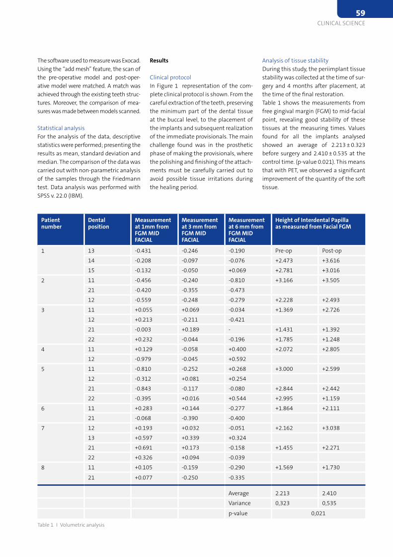
Analysis of tissue stabilityDuring this study, the periimplant tissue stability was collected at the time of sur-gery and 4 months after placement, at the time of the final restoration. Table 1 shows the measurements from free gingival margin (FGM) to mid-facial point, revealing good stability of these tissues at the measuring times. Values found for all the implants analysed showed an average of 2.213 ± 0.323 before surgery and 2.410 ± 0.535 at the control time. (p-value 0.021). This means that with PET, we observed a significant improvement of the quantity of the soft tissue.
Results
Clinical protocolIn Figure 1 representation of the com-plete clinical protocol is shown. From the careful extraction of the teeth, preserving the minimum part of the dental tissue at the buccal level, to the placement of the implants and subsequent realization of the immediate provisionals. The main challenge found was in the prosthetic phase of making the provisionals, where the polishing and finishing of the attach-ments must be carefully carried out to avoid possible tissue irritations during the healing period.
The software used to measure was Exocad. Using the “add mesh” feature, the scan of the pre-operative model and post-oper-ative model were matched. A match was achieved through the existing teeth struc-tures. Moreover, the comparison of mea-sures was made between models scanned.
Statistical analysisFor the analysis of the data, descriptive statistics were performed; presenting the results as mean, standard deviation and median. The comparison of the data was carried out with non-parametric analysis of the samples through the Friedmann test. Data analysis was performed with SPSS v. 22.0 (IBM).
Patient number
Dental position
Measurement at 1mm from FGM MID FACIAL
Measurement at 3 mm from FGM MID FACIAL
Measurement at 6 mm from FGM MID FACIAL
Height of Interdental Papilla as measured from Facial FGM
1 13 -0.431 -0.246 -0.190 Pre-op Post-op
14 -0.208 -0.097 -0.076 +2.473 +3.616
15 -0.132 -0.050 +0.069 +2.781 +3.016
2 11 -0.456 -0.240 -0.810 +3.166 +3.505
21 -0.420 -0.355 -0.473
12 -0.559 -0.248 -0.279 +2.228 +2.493
3 11 +0.055 +0.069 -0.034 +1.369 +2.726
12 +0.213 -0.211 -0.421
21 -0.003 +0.189 - +1.431 +1.392
22 +0.232 -0.044 -0.196 +1.785 +1.248
4 11 +0.129 -0.058 +0.400 +2.072 +2.805
12 -0.979 -0.045 +0.592
5 11 -0.810 -0.252 +0.268 +3.000 +2.599
12 -0.312 +0.081 +0.254
21 -0.843 -0.117 -0.080 +2.844 +2.442
22 -0.395 +0.016 +0.544 +2.995 +1.159
6 11 +0.283 +0.144 -0.277 +1.864 +2.111
21 -0.068 -0.390 -0.400
7 12 +0.193 +0.032 -0.051 +2.162 +3.038
13 +0.597 +0.339 +0.324
21 +0.691 +0.173 -0.158 +1.455 +2.271
22 +0.326 +0.094 -0.039
8 11 +0.105 -0.159 -0.290 +1.569 +1.730
21 +0.077 -0.250 -0.335
Average 2.213 2.410
Variance 0,323 0,535
p-value 0,021
Table 1 I Volumetric analysis
CLINICAL SCIENCE59
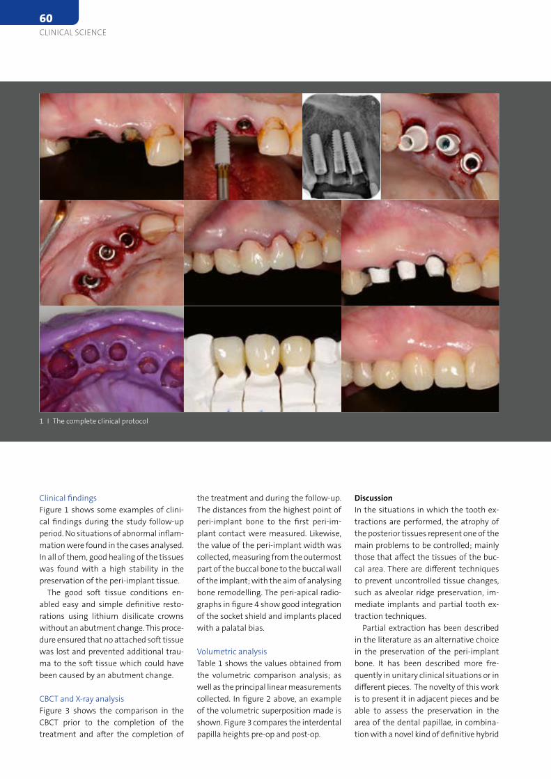
DiscussionIn the situations in which the tooth ex-tractions are performed, the atrophy of the posterior tissues represent one of the main problems to be controlled; mainly those that affect the tissues of the buc-cal area. There are different techniques to prevent uncontrolled tissue changes, such as alveolar ridge preservation, im-mediate implants and partial tooth ex-traction techniques.
Partial extraction has been described in the literature as an alternative choice in the preservation of the peri-implant bone. It has been described more fre-quently in unitary clinical situations or in different pieces. The novelty of this work is to present it in adjacent pieces and be able to assess the preservation in the area of the dental papillae, in combina-tion with a novel kind of definitive hybrid
the treatment and during the follow-up. The distances from the highest point of peri-implant bone to the first peri-im-plant contact were measured. Likewise, the value of the peri-implant width was collected, measuring from the outermost part of the buccal bone to the buccal wall of the implant; with the aim of analysing bone remodelling. The peri-apical radio-graphs in figure 4 show good integration of the socket shield and implants placed with a palatal bias.
Volumetric analysisTable 1 shows the values obtained from the volumetric comparison analysis; as well as the principal linear measurements collected. In figure 2 above, an example of the volumetric superposition made is shown. Figure 3 compares the interdental papilla heights pre-op and post-op.
Clinical findingsFigure 1 shows some examples of clini-cal findings during the study follow-up period. No situations of abnormal inflam-mation were found in the cases analysed. In all of them, good healing of the tissues was found with a high stability in the preservation of the peri-implant tissue.
The good soft tissue conditions en-abled easy and simple definitive resto-rations using lithium disilicate crowns without an abutment change. This proce-dure ensured that no attached soft tissue was lost and prevented additional trau-ma to the soft tissue which could have been caused by an abutment change.
CBCT and X-ray analysisFigure 3 shows the comparison in the CBCT prior to the completion of the treatment and after the completion of
1 I The complete clinical protocol
60CLINICAL SCIENCE
3 I Comparison of interdental papilla height at pre-op and post-op
2 I Showing the measurement protocol and the socket shield
combination with immediate restora-tion could be useful and predictable in situations of missing adjacent teeth to prevent the loss of vestibular tissues.
The references are available at www.teamwork-media.de/literatur
Contact address Dr Ali Tunkiwala
Smiles:By Design Ground Floor Nirant Building 19th Road, Khar West Mumbai 400052 India [email protected]
4 I Peri-apical radiographs showing implant position with palatal bias
abutment, which enables the immediate restoration with One-Time-Therapy with-out abutment change.
Neither abnormal inflammatory reac-tion nor situations of encap sulation of the dental tissue nor expulsion was observed, so it can be said that the pre-served tooth fragment is integrated into the structure of the buccal wall of peri-implant tissue. There seems to be a high correlation between the previous volume and the amount of tissue preservation achieved.
With the radiologic analysis, it has been possible to assess the vertical pres-ervation of the peri-implant bone, find-ing a preservation of it. Something that is complementary with other works of par-tial extraction therapy, where the conser-vation of peri-implant tissue is achieved satisfactorily.
Finally, the volumetric analysis tries to elucidate two factors: first, if there is a relation between the previous quan-tity and volume of the mouth and the preservation of the tissue a posteriori; and second, if the technique for volume maintenance is effective.
This assessment is made by volumetric superposition. It seems to indicate that in cases where the previous volume was greater, the conservation of the tissue is more evident, which suggests the impor-tant influence of both the width of the buccal bone and the gingival pattern.
Conclusion:In conclusion, after the analysis of the ini-tial clinical situation and the follow-up, as well as the results of the volumetric analysis, there seem to be an indication that the partial extraction technique in
CLINICAL SCIENCE61
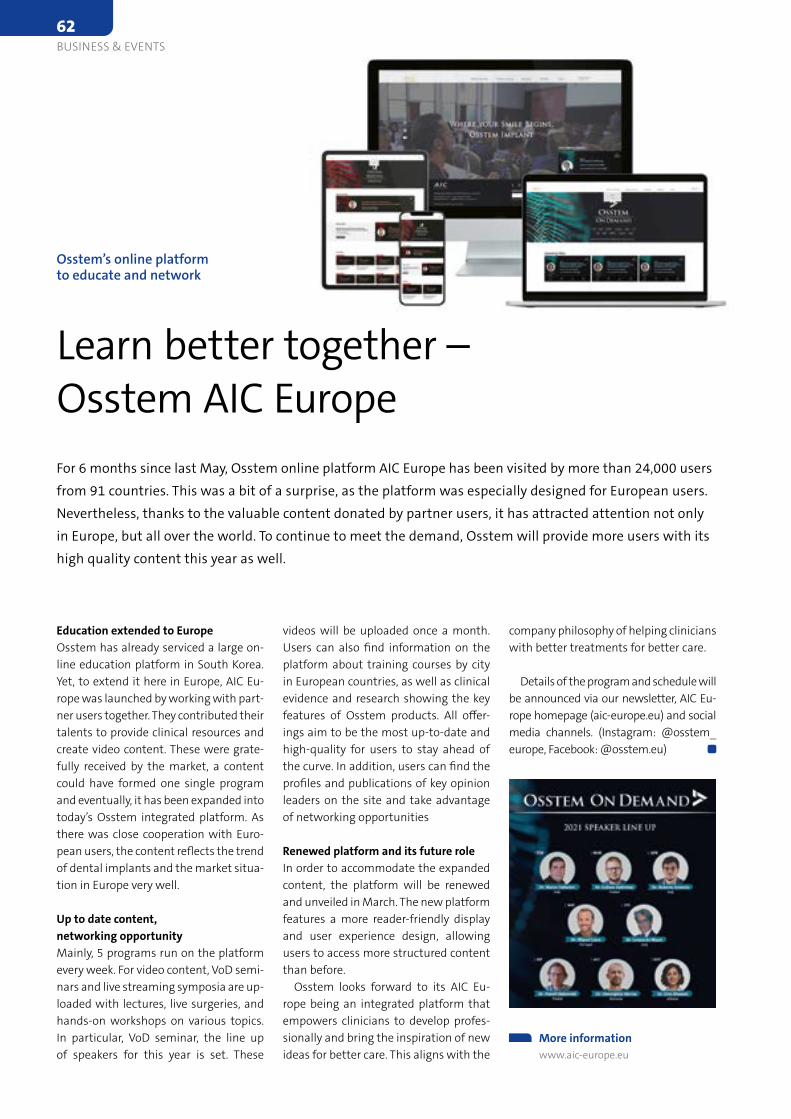
videos will be uploaded once a month. Users can also find information on the platform about training courses by city in European countries, as well as clinical evidence and research showing the key features of Osstem products. All offer-ings aim to be the most up-to-date and high-quality for users to stay ahead of the curve. In addition, users can find the profiles and publications of key opinion leaders on the site and take advantage of networking opportunities
Renewed platform and its future roleIn order to accommodate the expanded content, the platform will be renewed and unveiled in March. The new platform features a more reader-friendly display and user experience design, allowing users to access more structured content than before.
Osstem looks forward to its AIC Eu-rope being an integrated platform that empowers clinicians to develop profes-sionally and bring the inspiration of new ideas for better care. This aligns with the
Education extended to EuropeOsstem has already serviced a large on-line education platform in South Korea. Yet, to extend it here in Europe, AIC Eu-rope was launched by working with part-ner users together. They contributed their talents to provide clinical resources and create video content. These were grate-fully received by the market, a content could have formed one single program and eventually, it has been expanded into today’s Osstem integrated platform. As there was close cooperation with Euro-pean users, the content reflects the trend of dental implants and the market situa-tion in Europe very well.
Up to date content, networking opportunityMainly, 5 programs run on the platform every week. For video content, VoD semi-nars and live streaming symposia are up-loaded with lectures, live surgeries, and hands-on workshops on various topics. In particular, VoD seminar, the line up of speakers for this year is set. These
Osstem’s online platform to educate and network
Learn better together – Osstem AIC EuropeFor 6 months since last May, Osstem online platform AIC Europe has been visited by more than 24,000 users from 91 countries. This was a bit of a surprise, as the platform was especially designed for European users. Nevertheless, thanks to the valuable content donated by partner users, it has attracted attention not only in Europe, but all over the world. To continue to meet the demand, Osstem will provide more users with its high quality content this year as well.
More information www.aic-europe.eu
company philosophy of helping clinicians with better treatments for better care.
Details of the program and schedule will be announced via our newsletter, AIC Eu-rope homepage (aic-europe.eu) and social media channels. (Instagram: @osstem_europe, Facebook: @osstem.eu)
62BUSINESS & EVENTS
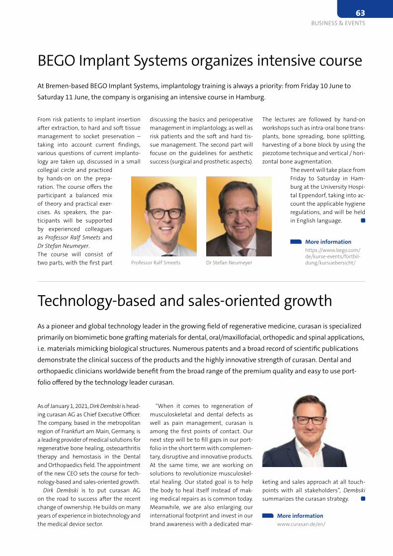
discussing the basics and perioperative management in implantology, as well as risk patients and the soft and hard tis-sue management. The second part will focuse on the guidelines for aesthetic success (surgical and prosthetic aspects).
From risk patients to implant insertion after extraction, to hard and soft tissue management to socket preservation – taking into account current findings, various questions of current implanto-logy are taken up, discussed in a small collegial circle and practiced by hands-on on the prepa-ration. The course offers the participant a balanced mix of theory and practical exer-cises. As speakers, the par-ticipants will be supported by experienced colleagues as Professor Ralf Smeets and Dr Stefan Neumeyer. The course will consist of two parts, with the first part
BEGO Implant Systems organizes intensive course At Bremen-based BEGO Implant Systems, implantology training is always a priority: from Friday 10 June to Saturday 11 June, the company is organising an intensive course in Hamburg.
Technology-based and sales-oriented growth As a pioneer and global technology leader in the growing field of regenerative medicine, curasan is specialized primarily on biomimetic bone grafting materials for dental, oral/maxillofacial, orthopedic and spinal applications, i.e. materials mimicking biological structures. Numerous patents and a broad record of scientific publications demonstrate the clinical success of the products and the highly innovative strength of curasan. Dental and orthopaedic clinicians worldwide benefit from the broad range of the premium quality and easy to use port-folio offered by the technology leader curasan.
The lectures are followed by hand-on workshops such as intra-oral bone trans-plants, bone spreading, bone splitting, harvesting of a bone block by using the piezotome technique and vertical / hori-zontal bone augmentation.
The event will take place from Friday to Saturday in Ham-burg at the University Hospi-tal Eppendorf, taking into ac-count the applicable hygiene regulations, and will be held in English language.
Professor Ralf Smeets Dr Stefan Neumeyer
More information https://www.bego.com/
de/kurse-events/fortbil-dung/kursuebersicht/
“When it comes to regeneration of musculoskeletal and dental defects as well as pain management, curasan is among the first points of contact. Our next step will be to fill gaps in our port-folio in the short term with complemen-tary, disruptive and innovative products. At the same time, we are working on solutions to revolutionize musculoskel-etal healing. Our stated goal is to help the body to heal itself instead of mak-ing medical repairs as is common today. Meanwhile, we are also enlarging our international footprint and invest in our brand awareness with a dedicated mar-
As of January 1, 2021, Dirk Dembski is head-ing curasan AG as Chief Executive Officer. The company, based in the metropolitan region of Frankfurt am Main, Germany, is a leading provider of medical solutions for regenerative bone healing, osteoarthritis therapy and hemostasis in the Dental and Orthopaedics field. The appointment of the new CEO sets the course for tech-nology-based and sales-oriented growth.
Dirk Dembski is to put curasan AG on the road to success after the recent change of ownership. He builds on many years of experience in biotechnology and the medical device sector.
More information www.curasan.de/en/
keting and sales approach at all touch-points with all stakeholders”, Dembski summarizes the curasan strategy.
BUSINESS & EVENTS63
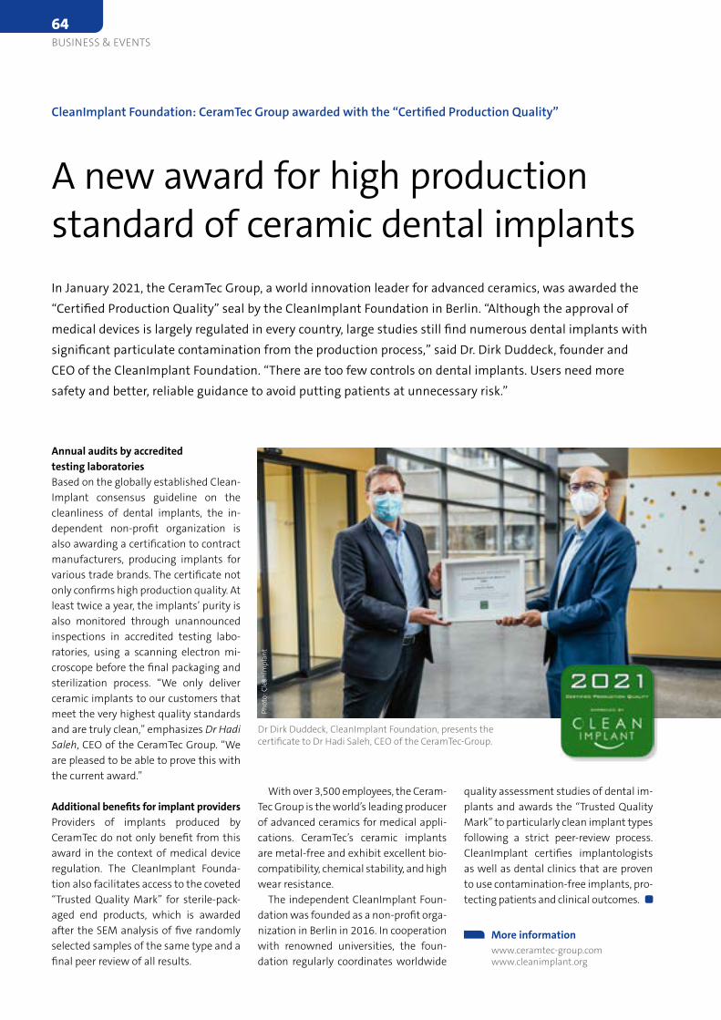
With over 3,500 employees, the Ceram-Tec Group is the world’s leading producer of advanced ceramics for medical appli-cations. CeramTec’s ceramic implants are metal-free and exhibit excellent bio-compatibility, chemical stability, and high wear resistance.
The independent CleanImplant Foun-dation was founded as a non-profit orga-nization in Berlin in 2016. In cooperation with renowned universities, the foun-dation regularly coordinates worldwide
Annual audits by accredited testing laboratories Based on the globally established Clean-Implant consensus guideline on the cleanliness of dental implants, the in-dependent non-profit organization is also awarding a certification to contract manufacturers, producing implants for various trade brands. The certificate not only confirms high production quality. At least twice a year, the implants’ purity is also monitored through unannounced inspections in accredited testing labo-ratories, using a scanning electron mi-croscope before the final packaging and sterilization process. “We only deliver ceramic implants to our customers that meet the very highest quality standards and are truly clean,” emphasizes Dr Hadi Saleh, CEO of the CeramTec Group. “We are pleased to be able to prove this with the current award.”
Additional benefits for implant providers Providers of implants produced by CeramTec do not only benefit from this award in the context of medical device regulation. The CleanImplant Founda-tion also facilitates access to the coveted “Trusted Quality Mark” for sterile-pack-aged end products, which is awarded after the SEM analysis of five randomly selected samples of the same type and a final peer review of all results.
CleanImplant Foundation: CeramTec Group awarded with the “Certified Production Quality”
A new award for high production standard of ceramic dental implantsIn January 2021, the CeramTec Group, a world innovation leader for advanced ceramics, was awarded the “Certified Production Quality” seal by the CleanImplant Foundation in Berlin. “Although the approval of medical devices is largely regulated in every country, large studies still find numerous dental implants with significant particulate contamination from the production process,” said Dr. Dirk Duddeck, founder and CEO of the CleanImplant Foundation. “There are too few controls on dental implants. Users need more safety and better, reliable guidance to avoid putting patients at unnecessary risk.”
More information www.ceramtec-group.com
www.cleanimplant.org
quality assessment studies of dental im-plants and awards the “Trusted Quality Mark” to particularly clean implant types following a strict peer-review process. CleanImplant certifies implantologists as well as dental clinics that are proven to use contamination-free implants, pro-tecting patients and clinical outcomes.
Dr Dirk Duddeck, CleanImplant Foundation, presents the certificate to Dr Hadi Saleh, CEO of the CeramTec-Group.
Phot
o: C
lean
impl
ant
64BUSINESS & EVENTS
ing driven by innovation and technologi-cal development and it will be very inter-esting to see where things go in the next few years.
What has driven the latest changes?As in the wider medical world, a move
to more minimal, less-invasive surgery and reducing the use of foreign-sourced materials seems to be an exciting area. These new materials and techniques which have emerged, show great promise for both practitioners and patients, with longer-term results. As the research and trials of these new products, materials and techniques increases, people’s confi-dence inevitably increases too, with more case studies and data showing long-term optimized outcomes and predictable re-sults, driving change at a faster pace.
You are accumulating more and more cases using alloplastic graft materials. Which are the long-term results?
I’ve placed implants in the same prac-tice for over 30 years meaning we get to see many exciting long-term outcomes. Having exclusively used synthetics for the last 20 years we are consistently pleas-antly surprised by the long-term stability of the cases. This should not really be a surprise anymore as, according to Wolff’s Law, if the bone is in function it will con-tinually remodel over time, giving a great support for the implant.
If you look at the Protocol I published in 2015, it was looking at patients between 6 months – 10 years after their proce-dure and the long-term follow up really
Dr Fairbairn, although you have regular-ly published studies and patient cases in our EDI Journal, it has been a while since our last interview. What can you tell us about your current work and about new developments?
It certainly has been a long time since we last spoke and a lot has happened since then! I have been continuing my work placing implants and performing graft procedures in London. I’ve complet-ed more than 6,500 successful grafts now using synthetic biomaterials, particularly B-TCP and Calcium Sulphate, over a 20 year period. I’ve also been lucky enough to deliver lectures around the world dur-ing the last few years, including on the main stage of the ICOI global congress in New York during 2019 looking at true bone regeneration.
During this time, I’ve travelled to more than 40 countries and met thousands of clinicians from all over the globe. It has been really encouraging to see the adop-tion of synthetic materials like EthOss ac-celerate as people move away from tradi-tional “GBR” and embrace the innovative new regenerative concepts and tech-niques that we have been consistently using for a long time. I often refer to these techniques as “true bone regeneration”, working with the latest generation of synthetic biomaterials to upregulate the host healing response and encouraging the patient to regenerate their own bone by optimizing periosteal function.
Looking more at implants, it’s also been interesting to see immediate placements increasing in popularity again. This is be-
Interview with Dr Peter Fairbairn, London
Focus on bone regenerationDr Peter Fairbairn is a global expert on bone regeneration, with extensive published research in materials and surgical protocols for bone repair without the use of donor animal and human products. These break-throughs have not only enabled the patients to have their own host healing for the restoration of the bone needed for dental implants but have also provided patients with a more ethical alternative. Dr Alina Ion from EDI Journal spoke with Dr Fairbairn about the latest developments in his career.
is there for these materials. We see a very high success rate and really predictable outcomes – exactly what the patient and clinician both want. Soft tissue response is a particularly interesting area. I’ve not done any specific research into this area yet, but it is very interesting to see the excellent soft tissue response that comes from using synthetic materials (particu-larly calcium sulphates).
The key point is the quality of the at-tached keratinised soft tissue that the body generates. I believe a large part of this is the quality of the bone underneath – healthy bone usually leads to healthy tissues. This was said many years ago by David Garber: “soft tissue is the issue but bone sets the tone.”
Your focus is bone regeneration. What is new in this field?
I realised recently that I’ve been lec-turing on ‘true bone regeneration’ for more than 20 years now and it’s been more than five years since our Protocol for Immediate-Delayed placements was published so, while these concepts are ‘new’ to lots of people, they have actu-ally been around for a long time. Over the last few years it has been interest-ing to read the wider medical research looking at the osteoinductive potential of synthetic materials – particularly B-TCP – and see how this can be applied to dentistry. There are now more than 300 papers in this area available on Pubmed, it is very well researched. It has also been fascinating to see how colleagues are pushing boundaries in this area. We are
BUSINESS & EVENTS65
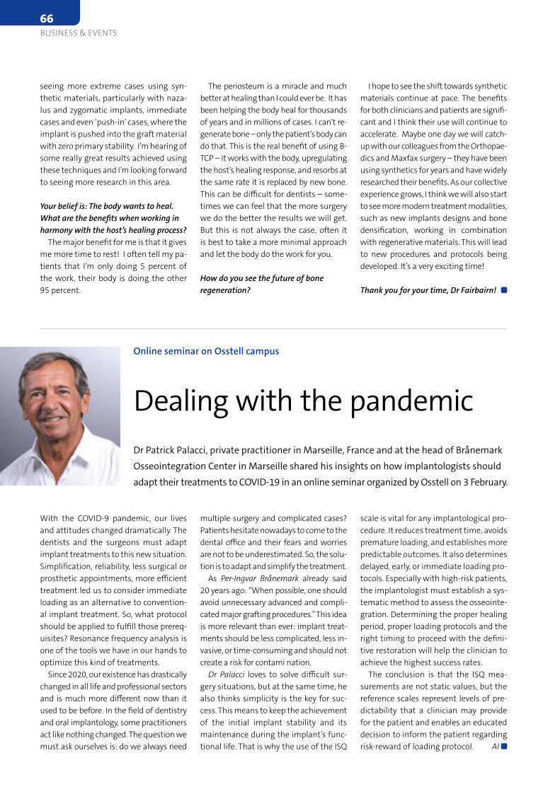
I hope to see the shift towards synthetic materials continue at pace. The benefits for both clinicians and patients are signifi-cant and I think their use will continue to accelerate. Maybe one day we will catch-up with our colleagues from the Orthopae-dics and Maxfax surgery – they have been using synthetics for years and have widely researched their benefits. As our collective experience grows, I think we will also start to see more modern treatment modalities, such as new implants designs and bone densification, working in combination with regenerative materials. This will lead to new procedures and protocols being developed. It’s a very exciting time!
Thank you for your time, Dr Fairbairn!
The periosteum is a miracle and much better at healing than I could ever be. It has been helping the body heal for thousands of years and in millions of cases. I can’t re-generate bone – only the patient’s body can do that. This is the real benefit of using B-TCP – it works with the body, upregulating the host’s healing response, and resorbs at the same rate it is replaced by new bone. This can be difficult for dentists – some-times we can feel that the more surgery we do the better the results we will get. But this is not always the case, often it is best to take a more minimal approach and let the body do the work for you.
How do you see the future of bone regeneration?
seeing more extreme cases using syn-thetic materials, particularly with naza-lus and zygomatic implants, immediate cases and even ‘push-in’ cases, where the implant is pushed into the graft material with zero primary stability. I’m hearing of some really great results achieved using these techniques and I’m looking forward to seeing more research in this area.
Your belief is: The body wants to heal. What are the benefits when working in harmony with the host’s healing process?
The major benefit for me is that it gives me more time to rest! I often tell my pa-tients that I’m only doing 5 percent of the work, their body is doing the other 95 percent.
multiple surgery and complicated cases? Patients hesitate nowadays to come to the dental office and their fears and worries are not to be underestimated. So, the solu-tion is to adapt and simplify the treatment.
As Per-Ingvar Brånemark already said 20 years ago: “When possible, one should avoid unnecessary advanced and compli-cated major grafting procedures.” This idea is more relevant than ever: implant treat-ments should be less complicated, less in-vasive, or time-consuming and should not create a risk for contami nation.
Dr Palacci loves to solve difficult sur-gery situations, but at the same time, he also thinks simplicity is the key for suc-cess. This means to keep the achievement of the initial implant stability and its maintenance during the implant’s func-tional life. That is why the use of the ISQ
With the COVID-9 pandemic, our lives and attitudes changed dramatically. The dentists and the surgeons must adapt implant treatments to this new situation. Simplification, reliability, less surgical or prosthetic appointments, more efficient treatment led us to consider immediate loading as an alternative to convention-al implant treatment. So, what protocol should be applied to fulfill those prereq-uisites? Resonance frequency analysis is one of the tools we have in our hands to optimize this kind of treatments.
Since 2020, our existence has drastically changed in all life and professional sectors and is much more different now than it used to be before. In the field of dentistry and oral implantology, some practitioners act like nothing changed. The question we must ask ourselves is: do we always need
scale is vital for any implantological pro-cedure. It reduces treatment time, avoids premature loading, and establishes more predictable outcomes. It also determines delayed, early, or immediate loading pro-tocols. Especially with high-risk patients, the implantologist must establish a sys-tematic method to assess the osseointe-gration. Determining the proper healing period, proper loading protocols and the right timing to proceed with the defini-tive restoration will help the clinician to achieve the highest success rates.
The conclusion is that the ISQ mea-surements are not static values, but the reference scales represent levels of pre-dictability that a clinician may provide for the patient and enables an educated decision to inform the patient regarding risk-reward of loading protocol. AI
Online seminar on Osstell campus
Dealing with the pandemicDr Patrick Palacci, private practitioner in Marseille, France and at the head of Brånemark Osseointegration Center in Marseille shared his insights on how implantologists should adapt their treatments to COVID-19 in an online seminar organized by Osstell on 3 February.
66BUSINESS & EVENTS
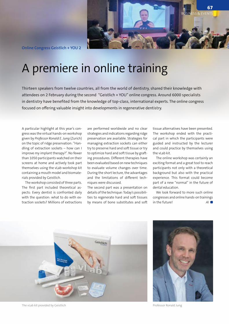
are performed worldwide and no clear strategies and indications regarding ridge preservation are available. Strategies for managing extraction sockets can either try to preserve hard and soft tissue or try to optimize hard and soft tissue by graft-ing procedures. Different therapies have been evaluated based on new techniques to evaluate volume changes over time. During the short lecture, the advantages and the limitations of different tech-niques were discussed.The second part was a presentation on details of the technique: Today’s possibili-ties to regenerate hard and soft tissues by means of bone substitutes and soft
A particular highlight at this year’s con-gress was the virtual hands-on workshop given by Professor Ronald E. Jung (Zurich) on the topic of ridge preservation: “Han-dling of extraction sockets – how can I improve my implant therapy?”. No fewer than 1050 participants watched on their screens at home and actively took part themselves using the vLab workshop kit containing a mouth model and biomate-rials provided by Geistlich.
The workshop consisted of three parts. The first part included theoretical as-pects: Every dentist is confronted daily with the question: what to do with ex-traction sockets? Millions of extractions
Online Congress Geistlich + YOU 2
A premiere in online trainingThirteen speakers from twelve countries, all from the world of dentistry, shared their knowledge with attendees on 2 February during the second “Geistlich + YOU” online congress. Around 6000 specialists in dentistry have benefited from the knowledge of top-class, international experts. The online congress focused on offering valuable insight into developments in regenerative dentistry.
tissue alternatives have been presented. The workshop ended with the practi-cal part in which the participants were guided and instructed by the lecturer and could practice by themselves using the vLab kit.
The online workshop was certainly an exciting format and a great tool to reach participants not only with a theoretical background but also with the practical experience. This format could become part of a new “normal” in the future of dental education.
We look forward to more such online congresses and online hands-on trainings in the future! AI
Professor Ronald JungThe vLab kit provided by Geistlich
67BUSINESS & EVENTS
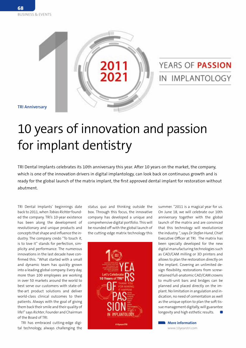
status quo and thinking outside the box. Through this focus, the innovative company has developed a unique and comprehensive digital portfolio. This will be rounded off with the global launch of the cutting-edge matrix technology this
TRI Dental Implants’ beginnings date back to 2011, when Tobias Richter found-ed the company. TRI’s 10-year existence has been along the development of revolutionary and unique products and concepts that shape and influence the in-dustry. The company credo “To touch it, is to love it” stands for perfection, sim-plicity and performance. The numerous innovations in the last decade have con-firmed this. “What started with a small and dynamic team has quickly grown into a leading global company. Every day, more than 100 employees are working in over 50 markets around the world to best serve our customers with state-of-the-art product solutions and deliver world-class clinical outcomes to their patients. Always with the goal of giving them back their smile and their quality of life!” says Richter, Founder and Chairman of the Board of TRI.
TRI has embraced cutting-edge digi-tal technology, always challenging the
TRI Anniversary
10 years of innovation and passion for implant dentistryTRI Dental Implants celebrates its 10th anniversary this year. After 10 years on the market, the company, which is one of the innovation drivers in digital implantology, can look back on continuous growth and is ready for the global launch of the matrix implant, the first approved dental implant for restoration without abutment.
More information www.10yearstri.com
summer. “2011 is a magical year for us. On June 18, we will celebrate our 10th anniversary together with the global launch of the matrix and are convinced that this technology will revolutionize the industry. “, says Dr Stefan Hund, Chief Executive Officer at TRI. The matrix has been specially developed for the new digital manufacturing technologies such as CAD/CAM milling or 3D printers and allows to plan the restoration directly on the implant. Covering an unlimited de-sign flexibility, restorations from screw-retained full-anatomic CAD/CAM crowns to multi-unit bars and bridges can be planned and placed directly on the im-plant. No limitation in angulation and in-dication, no need of cementation as well as the unique option to plan the soft tis-sue management digitally, will guarantee longevity and high esthetic results.
68BUSINESS & EVENTS
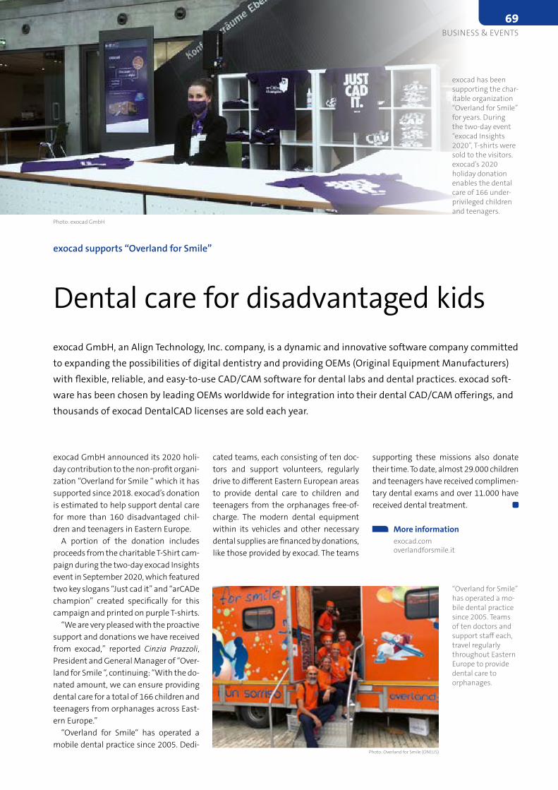
cated teams, each consisting of ten doc-tors and support volunteers, regularly drive to different Eastern European areas to provide dental care to children and teenagers from the orphanages free-of-charge. The modern dental equipment within its vehicles and other necessary dental supplies are financed by donations, like those provided by exocad. The teams
exocad GmbH announced its 2020 holi-day contribution to the non-profit organi-zation “Overland for Smile “ which it has supported since 2018. exocad’s donation is estimated to help support dental care for more than 160 disadvantaged chil-dren and teenagers in Eastern Europe.
A portion of the donation includes proceeds from the charitable T-Shirt cam-paign during the two-day exocad Insights event in September 2020, which featured two key slogans “Just cad it” and “arCADe champion” created specifically for this campaign and printed on purple T-shirts.
“We are very pleased with the proactive support and donations we have received from exocad,” reported Cinzia Prazzoli, President and General Manager of “Over-land for Smile “, continuing: “With the do-nated amount, we can ensure providing dental care for a total of 166 children and teenagers from orphanages across East-ern Europe.”
“Overland for Smile“ has operated a mobile dental practice since 2005. Dedi-
exocad supports “Overland for Smile”
Dental care for disadvantaged kidsexocad GmbH, an Align Technology, Inc. company, is a dynamic and innovative software company committed to expanding the possibilities of digital dentistry and providing OEMs (Original Equipment Manufacturers) with flexible, reliable, and easy-to-use CAD/CAM software for dental labs and dental practices. exocad soft-ware has been chosen by leading OEMs worldwide for integration into their dental CAD/CAM offerings, and thousands of exocad DentalCAD licenses are sold each year.
supporting these missions also donate their time. To date, almost 29.000 children and teenagers have received complimen-tary dental exams and over 11.000 have received dental treatment.
More information exocad.com
overlandforsmile.it
exocad has been supporting the char-itable organization “Overland for Smile” for years. During the two-day event “exocad Insights 2020”, T-shirts were sold to the visitors. exocad’s 2020 holiday donation enables the dental care of 166 under-privileged children and teenagers.
“Overland for Smile” has operated a mo-bile dental practice since 2005. Teams of ten doctors and support staff each, travel regularly throughout Eastern Europe to provide dental care to orphanages.
Photo: exocad GmbH
Photo: Overland for Smile (ONLUS)
BUSINESS & EVENTS69
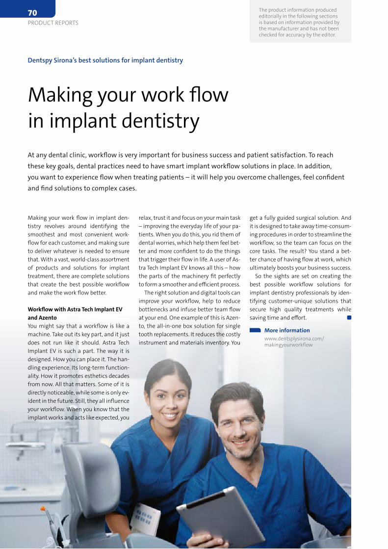
relax, trust it and focus on your main task – improving the everyday life of your pa-tients. When you do this, you rid them of dental worries, which help them feel bet-ter and more confident to do the things that trigger their flow in life. A user of As-tra Tech Implant EV knows all this – how the parts of the machinery fit perfectly to form a smoother and efficient process.
The right solution and digital tools can improve your workflow, help to reduce bottlenecks and infuse better team flow at your end. One example of this is Azen-to, the all-in-one box solution for single tooth replacements. It reduces the costly instrument and materials inventory. You
Making your work flow in implant den-tistry revolves around identifying the smoothest and most convenient work-flow for each customer, and making sure to deliver whatever is needed to ensure that. With a vast, world-class assortment of products and solutions for implant treatment, there are complete solutions that create the best possible workflow and make the work flow better.
Workflow with Astra Tech Implant EV and AzentoYou might say that a workflow is like a machine. Take out its key part, and it just does not run like it should. Astra Tech Implant EV is such a part. The way it is designed. How you can place it. The han-dling experience. Its long-term function-ality. How it promotes esthetics decades from now. All that matters. Some of it is directly noticeable, while some is only ev-ident in the future. Still, they all influence your workflow. When you know that the implant works and acts like expected, you
Dentspy Sirona’s best solutions for implant dentistry
Making your work flow in implant dentistry At any dental clinic, workflow is very important for business success and patient satisfaction. To reach these key goals, dental practices need to have smart implant workflow solutions in place. In addition, you want to experience flow when treating patients – it will help you overcome challenges, feel confident and find solutions to complex cases.
More information www.dentsplysirona.com/
makingyourworkflow
get a fully guided surgical solution. And it is designed to take away time-consum-ing procedures in order to streamline the workflow, so the team can focus on the core tasks. The result? You stand a bet-ter chance of having flow at work, which ultimately boosts your business success.
So the sights are set on creating the best possible workflow solutions for implant dentistry professionals by iden-tifying customer-unique solutions that secure high quality treatments while saving time and effort.
The product information produced editorially in the following sections is based on information provided by the manufacturer and has not been checked for accuracy by the editor.
70PRODUCT REPORTS

thread profile, creates the optimal con-dition for fast and reliable osseointegra-tion.
The Everguard Connection, with an in-ternal hex and external stabilization ring, ensures optimal long-term stabil-ity of the implant abutment connection. The connection design includes a verti-cal stop for consistent abutment posi-tion and preservation of the abutment screw’s preload to avoid screw loosen-ing. The clever design of the reduced size abutment screw enables thicker implant and abutment walls and protects im-plant integrity. The reduced screw access channel allows for improved restorative flexibility and aesthetics.
Integuard Matrix ensures an optimal hard tissue response. Faster osseointe-gration is promoted by the implant sur-face Inicell which is the superhydrophilic state of the proven sandblasted and acid etched Thommen implant surface. Inicell is generated in a few seconds with the chairside conditioning system.
The conditioning agent applied with Apliquiq has an antimicrobial effect which reduces the risk of infection in the early healing phase. The implant bed preparation with the Vectodrills, in combination with the Thommen implant
Thommen Implant System
Multiguard Protection SolutionWith a long-term view of clinical success, the product development at Thommen is driven by science, not trends. The company delivers a comprehensive implant system, based on a proven design – the Multiguard Protection Solution – which has been optimized over three decades of clinical success. A combination of innovative features work together to protect implant integrity by maintaining mechanical stability and an optimal bio-response for the life of the patient.
More information www.thommenmedical.com
The Tissueguard Collar encourages soft tissue adaptation to prevent bone loss and allows for surgical flexibility to place the machined collar into the bone de-pending on the clinical need. The ma-chined surface of the collar is optimal for soft tissue and is cleansable, which improves hygienic maintenance to re-duce the risk of biologic complications, including peri-implantitis. Supports opti-mal placement of the implant/abutment junction allowing for restorative flexibil-ity and easy impression taking. Smooth transition between implant and abut-ment leaves no void or ledge for bacteria and facilitates problem-free probing.
PRODUCT REPORTS71
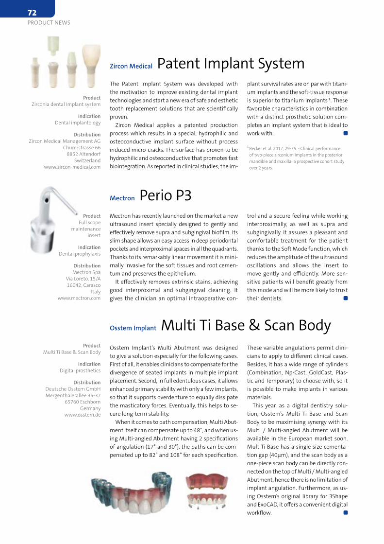
Zircon Medical Patent Implant SystemThe Patent Implant System was developed with the motivation to improve existing dental implant technologies and start a new era of safe and esthetic tooth replacement solutions that are scientifically proven.
Zircon Medical applies a patented production process which results in a special, hydrophilic and osteoconductive implant surface without process induced micro-cracks. The surface has proven to be hydrophilic and osteoconductive that promotes fast biointegration. As reported in clinical studies, the im-
plant survival rates are on par with titani-um implants and the soft-tissue response is superior to titanium implants 1. These favorable characteristics in combination with a distinct prosthetic solution com-pletes an implant system that is ideal to work with.
1 Becker et al. 2017, 29-35. - Clinical performance of two-piece zirconium implants in the posterior mandible and maxilla: a prospective cohort study over 2 years.
ProductZirconia dental Implant system
IndicationDental implantology
DistributionZircon Medical Management AG
Churerstrasse 66 8852 Altendorf
Switzerland www.zircon-medical.com
Mectron Perio P3 Mectron has recently launched on the market a new ultrasound insert specially designed to gently and effectively remove supra and subgingival biofilm. Its slim shape allows an easy access in deep periodontal pockets and interproximal spaces in all the quadrants.Thanks to its remarkably linear movement it is mini-mally invasive for the soft tissues and root cemen-tum and preserves the epithelium.
It effectively removes extrinsic stains, achieving good interproximal and subgingival cleaning. It gives the clinician an optimal intraoperative con-
trol and a secure feeling while working interproximally, as well as supra and subgingivally. It assures a pleasant and comfortable treatment for the patient thanks to the Soft Mode function, which reduces the amplitude of the ultrasound oscillations and allows the insert to move gently and efficiently. More sen-sitive patients will benefit greatly from this mode and will be more likely to trust their dentists.
ProductFull scope
maintenance insert
IndicationDental prophylaxis
DistributionMectron Spa
Via Loreto, 15/A 16042, Carasco
Italy www.mectron.com
Osstem Implant Multi Ti Base & Scan BodyOsstem Implant’s Multi Abutment was designed to give a solution especially for the following cases. First of all, it enables clinicians to compensate for the divergence of seated implants in multiple implant placement. Second, in full edentulous cases, it allows enhanced primary stability with only a few implants, so that it supports overdenture to equally dissipate the masticatory forces. Eventually, this helps to se-cure long-term stability.
When it comes to path compensation, Multi Abut-ment itself can compensate up to 48°, and when us-ing Multi-angled Abutment having 2 specifications of angulation (17° and 30°), the paths can be com-pensated up to 82° and 108° for each specification.
These variable angulations permit clini-cians to apply to different clinical cases. Besides, it has a wide range of cylinders (Combination, Np-Cast, GoldCast, Plas-tic and Temporary) to choose with, so it is possible to make implants in various materials.
This year, as a digital dentistry solu-tion, Osstem’s Multi Ti Base and Scan Body to be maximising synergy with its Multi / Multi-angled Abutment will be available in the European market soon. Mult Ti Base has a single size cementa-tion gap (40μm), and the scan body as a one-piece scan body can be directly con-nected on the top of Multi / Multi-angled Abutment, hence there is no limitation of implant angulation. Furthermore, as us-ing Osstem’s original library for 3Shape and ExoCAD, it offers a convenient digital workflow.
ProductMulti Ti Base & Scan Body
IndicationDigital prosthetics
DistributionDeutsche Osstem GmbH Mergenthalerallee 35-37
65760 Eschborn Germany
www.osstem.de
72PRODUCT NEWS

BDIZ EDI Mühlenstr. 18 D 51143 Köln Tel. ++49 (0) 2203-8009-339 Fax ++49 (0) 2203-9168-822 [email protected] www.bdizedi.org
MEMBERSHIP REGISTRATION FORM
I hereby apply for a membership in the BDIZ EDI (European Association of Dental Implantologists)
Name: ....................................................................................
First Name: ............................................................................
Country: ................................................................................
Zip code / City: …..................................................................
Street: ……………………………………………………….…...
Phone: …………………………………………………….….....
Fax: .........................................................................…..........
E-Mail: .......................................... @ …...............................
Homepage: ………………………….………………….……….
Date of Birth: .........................................................................
Practicing implantology since: …...........................................
Member of other Societies:
ICOI BDO DGI DGZI DGMKG EAO
Continuing education Courses: ................................................
...................................................................................................
Fellowship status / diplomate status in implantology
Yes No Organization ………...………..
Entry in BDIZ EDI Directory: Yes No(For information on BDIZ EDI Directory of Implant Dentists see overleaf)
The annual membership fee for:
FULL MEMBERSHIP
Full member - clinical 345,00 Euro
Assistant dentist / young professional(up to 5 years after graduation)
172,50 Euro
Second membership / family member 172,50 Euro
EXTRAORDINARY MEMBERSHIP
Co-operative Member (Professionals without practice and dental technicians)
165,00 Euro
Students non-contributory
Supporting Membership(Companies etc.)
530,00 Euro
Payment Membership cannot be confirmed until payment is processed. Method of payment is by bank transfer. Please use the following banking account.
Commerzbank Bonn
Account Number: 310 144 100 Bank Code: 380 400 07 IBAN: DE96 3804 0007 0310 1441 00 BIC: COBADEFFXXX
Membership cards will be sent upon receipt of the annual subscription fee.
City / Date : …………………………………………………
Seal / Signature: ....................................................................
Please return the completed registration form to:
European Association of Dental Implantologists e. V. Mühlenstr. 18 • D-51143 Köln Fon: + 49 (0) 2203-8009-339 Fax: + 49 (0) 2203-9168-822 E-Mail: [email protected]: www.bdizedi.org
The commercial processing of your personal data on this form is based on the EU General Data Protection Regulation (GDPR – Regulation (EU) 2016/679 of 27 April 2016), Article 6 f GDPR by the European Association of Dental Implantologists (BDIZ EDI), Mühlenstr. 18, D-51143 Cologne/Germany.
You have the right to obtain information about personal data concerning you (Article 15 of the GDPR). You can also request the correction (rectification) of incorrect data (Article 16 of the GDPR). More information: Privacy Statement on www.bdizedi.org.
EDI Journal – the interdisciplinary journal for prosthetic dental implantology is aimed at dentists and technicians interested in prosthetics implantology. All contributions sub-mitted should be focused on this aspect in content and form. Suggested contributions may include:· Original scientific research· Case studies· Product studies· Overviews
Manuscript submissionSubmissions should be made in digital form. Original articles will be considered for publication only on the condition that they have not been published elsewhere in part or in whole and are not simultaneously under consideration elsewhere.
ManuscriptsPages should be numbered consecutively, starting with the cover page. The cover page should include the title of the manuscript and the name and degree for all authors. Also included should be the full postal address, telephone number, and e-mail address of the contact author. Manuscripts can be organized in a manner that best fits the specific goals of the ar-ticle, but should always include an introductory section, the body of the article and a conclusion.
Illustrations and tablesEach article should contain a minimum of 20 and a maximum of 50 pictures, except in unusual circumstances. Our publishing house attaches great importance to high qual-ity illustrations. All illustrations should to be numbered, have a caption and be men-tioned in the text. The photos should have a size of 10x15 cm, the image or graphic files must have a reso-lution of 300dpi. Tiff, eps and jpg file formats are suitable. Radiographs, charts, graphs, and drawn figures are also accepted. Captions should be brief one or two-line descriptions of each illustration, typed on a separate page following the references. Captions must be numbered in the same nu-merical order as the illustrations.Tables should be typed on a separate page and num-bered consecutively, according to citation in the text. The title of the table and its cap-tion must be on the same page as the table itself.
ReferencesEach article should contain a minimum of 10 and a maximum of 30 references, except in unusual circumstances. Citations in the body of the text should be made in numeri-cal order. The reference list should be typed on a separate sheet and should provide complete bibliographical information in the format exemplified below:[1] Albrektsson, T.: A multicenter report on osseointegrated oral implants. J Prosthet Dent 1988; 60, 75-82. [2] Hildebrand, H. F., Veron, Chr., Martin, P.: Nickel, chromium, cobalt dental alloys and allergic reactions: an overview. Biomaterials 10, 545-548, (1989)
Review ProcessManuscripts will be reviewed by three members of the editorial board. Authors are not informed of the identity of the reviewers and reviewers are not provided with the identity of the author. The review cycle will be completed within 60 days. Publication is expected within nine months.
CopyrightWith acceptance of a manuscript, the publisher has the right to publish and translate the text, permit its reproduction, electronically store it in databases, produce reprints, photocopies and microcopies, and publish the text online. Any further utilization or publication of the article is subject to prior approval by teamwork media GmbH.
Page Charges and ReprintsThere are no page charges. The publisher will cover all costs of production. The journal will provide the primary author with a PDF file of the article and a free copy of the journal issue in which the article appears.
Editors office: teamwork media GmbH & Co. KGBetriebsstätte Fuchstal, Hauptstraße 1, 86925 Fuchstal/Germany Phone: +49 8243 9692-0Fax: +49 8243 [email protected]
EDI Journal – Information for authors
Calendar of Events
Event Location Date Details/Registration
3/2021 International Conference on Perio-dontology and Oral Implantology
Tokyo, Japan 25–26 March 2021 waset.org
4/2021 Euro Implanto 2021 Nice, France 8–9 April 2021 euroimplanto.fr
5/2021 International Conference on Perio-dontology and Oral Implantology
Barcelona, Spain 24–25 May 2021 waset.org
6/2021 ICOI Implant Symposium 2021 New York, USA 12–13 June 2021 icoi.org
9/2021 ITI World Symposium 2021 Singapore, Singapore 1–3 September 2021 iti.org
International Dental Show IDS Cologne, Germany 22–25 September 2021 ids-cologne.de
74CALENDAR OF EVENTS
Due to the Coronavirus spread, many events have been postponed or cancelled. Other events only take place online. Please check the official websites or contact the organizers to see if the dates are confirmed.
Stay healthy! The EDI Journal and “konkret“ team
Anita Wuttke Alina Ion Daniela Wiedemann
time to teethOptimizing
Osstell helps you to objectively determine implant stability and to assess the progress of osseointegration – without jeopardizing the healing process.
Let Osstell ISQ Guide You
Treat all your patients Shorten treatment timesAchieve more predictable outcomes
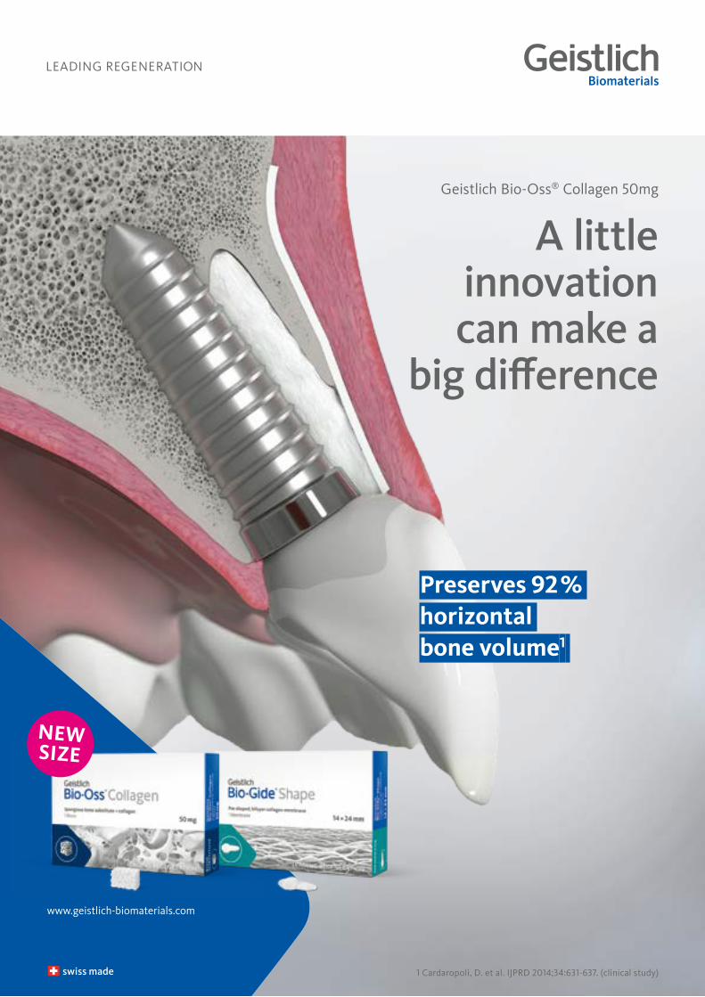
1 Cardaropoli, D. et al. IJPRD 2014;34:631-637. (clinical study)
NEW SIZE
Preserves 92 % horizontal bone volume1
Geistlich Bio-Oss® Collagen 50mg
A little innovation can make a
big difference
www.geistlich-biomaterials.com


