Embed Size (px)
Citation preview

Disease-relevant proteostasis regulation of cysticfibrosis transmembrane conductance regulator
VR Villella1, S Esposito1, EM Bruscia1,2, M Vicinanza3, S Cenci4, S Guido5, M Pettoello-Mantovani6, R Carnuccio7,MA De Matteis3, A Luini3,8, MC Maiuri7,9, V Raia*,10, G Kroemer*,9,11,12,13,14 and L Maiuri*,1,6
Mismanaged protein trafficking by the proteostasis network contributes to several conformational diseases, including cysticfibrosis, the most frequent lethal inherited disease in Caucasians. Proteostasis regulators, as cystamine, enable the beneficialaction of cystic fibrosis transmembrane conductance regulator (CFTR) potentiators in DF508-CFTR airways beyond drugwashout. Here we tested the hypothesis that functional CFTR protein can sustain its own plasma membrane (PM) stability.Depletion or inhibition of wild-type CFTR present in bronchial epithelial cells reduced the availability of the small GTPase Rab5by causing Rab5 sequestration within the detergent-insoluble protein fraction together with its accumulation in aggresomes.CFTR depletion decreased the recruitment of the Rab5 effector early endosome antigen 1 to endosomes, thus reducing the localgeneration of phosphatidylinositol-3-phosphate. This diverts recycling of surface proteins, including transferrin receptor andCFTR itself. Inhibiting CFTR function also resulted in its ubiquitination and interaction with SQSTM1/p62 at the PM, favoring itsdisposal. Addition of cystamine prevented the recycling defect of CFTR by enhancing BECN1 expression and reducing SQSTM1accumulation. Our results unravel an unexpected link between CFTR protein and function, the latter regulating the levels of CFTRsurface expression in a positive feed-forward loop, and highlight CFTR as a pivot of proteostasis in bronchial epithelial cells.Cell Death and Differentiation (2013) 20, 1101–1115; doi:10.1038/cdd.2013.46; published online 17 May 2013
Manipulating proteostasis by proteostasis regulators (PRs) hasemerged as a novel therapeutic approach for the treatment ofconformational diseases.1–5 Cystic fibrosis (CF) constitutes thequintessential example of a conformational disease.6–8 CF is amonogenic disease in which defective function of one singleprotein, cystic fibrosis transmembrane conductance regulator(CFTR), leads to major clinical manifestations, includingchronic lung inflammation with increased susceptibility torespiratory bacterial infections, pancreatic dysfunction, raisedelectrolyte levels in sweat and male infertility.9–11 CF is causedby loss-of-function mutations of CFTR, a cAMP-regulatedchloride channel that is primarily located at the apicalmembrane of epithelial cells.12,13 A single deletion, F508del,occurs in about 70–90% of CF patients in Northern Europe andNorth America.14 Due to its misfold, DF508-CFTR is prema-turely degraded and loses its essential ion channel activity atthe plasma membrane (PM).9,15 DF508-CFTR can retain
channel activity if rescued at the PM by molecules that correctits intracellular retention and degradation (correctors),16–20 aswell as by low temperature,21 but in this case DF508-CFTR israpidly dismissed from the PM and redirected from endosomalrecycling towards lysosomal degradation.22,23
We previously reported that defective CFTR functioninduces reactive oxygen species (ROS)-dependent andtransglutaminase-2 (TGM2)-mediated sequestration ofBECN1, leading to defective autophagy and increased lunginflammation.24 PRs such as cystamine or the superoxidedismutase/catalase-mimetic EUK-13425,26 enable the bene-ficial action of CFTR potentiators on DF508-CFTR mutanttissues well beyond drug washout through restoring BECN1expression and autophagy, thereby reducing the abundanceof SQSTM1/p62 (SQSTM1).24,27 This might suggest that,once peripheral proteostasis has been re-established, PM-resident DF508-CFTR could sustain its own PM stability.
1Division of Genetics and Cell Biology, San Raffaele Scientific Institute, European Institute for Research in Cystic Fibrosis, Milan 20132, Italy; 2Department of Pediatrics,Yale University School of Medicine, New Haven, CT 06509, USA; 3Telethon Institute of Genetic and Medicine (TIGEM), Naples 80131, Italy; 4Division of Genetics andCell Biology, San Raffaele Scientific Institute, Milan 20132, Italy; 5Faculty of Engineering, Federico II University, Naples 80125, Italy; 6Institute of Pediatrics, University ofFoggia, Foggia 71100, Italy; 7Department of Experimental Pharmacology, School of Biotechnological Sciences, Federico II University, Naples 80131, Italy; 8Institute ofProtein Biochemistry, National Research Council, Naples 80131, Italy; 9INSERM, U848, Villejuif, France; 10Cystic Fibrosis Unit, Department of Pediatrics, Federico IIUniversity, Naples 80131, Italy; 11Universite Paris Descartes, Paris, France; 12Metabolomics Platform, Institut Gustave Roussy, Villejuif, France; 13Equipe 11 labelliseeLigue contre le Cancer, Centre de Recherche des Cordeliers, Paris, France and 14Pole de Biologie, Hopital Europeen Georges Pompidou, AP-HP, Paris, France*Corresponding author: L Maiuri , European Institute for Research in Cystic Fibrosis, San Raffaele Scientific Institute, Division of Genetics and Cell Biology, via Olgettina58, Milan 20132, Italy. Tel: þ 39 02 26434301; Fax: þ 39 02 26434328; E-mail: [email protected] V Raia, Cystic Fibrosis Unit, Department of Pediatrics, Federico II University, via S. Pansini 5, 80131, Naples, Italy. Tel: þ 39 081 746 3273;Fax: þ 39 081 746 3273; E-mail: [email protected] G Kroemer, INSERM U848, Institut Gustave Roussi, Pavillon de Recherche 1, F-94805 Villejuif, France. Tel: þ 33 1 42116046 ; Fax: þ 33 1 42116047;E-mail: [email protected]
Received 22.11.12; revised 09.4.13; accepted 10.4.13; Edited by E Baehrecke; published online 17.5.13
Keywords: CFTR; conformational diseases; protein trafficking; proteostasis; SQSTM1Abbreviations: CF, cystic fibrosis; CFTR, cystic fibrosis transmembrane conductance regulator; PRs, proteostasis regulators; TGM2, transglutaminase-2; ROS,reactive oxygen species; PtdIns3K, type III phosphatidylinositol 3-kinase; PtdIns3P, phosphatidylinositol 3-phosphate; EEA-1, early endosome antigen 1; TfR, transferrinreceptor; PM, plasma membrane; EGFR, epidermal growth factor receptor; Vps, vacuolar protein sorting; 3-MA, 3-methyl-adenine; LE, late endosome; PNS, post-nuclear supernatant; MTT, 3-[4,5-dimethylthiazol-2-yl]-2,5 diphenyl tetrazolium bromide; UVRAG, UV-irradiation-resistance-associated gene; CHX, cycloheximide
Cell Death and Differentiation (2013) 20, 1101–1115& 2013 Macmillan Publishers Limited All rights reserved 1350-9047/13
www.nature.com/cdd
Based on these premises and incognita, we explored thehypothesis that a functional CFTR might orchestrate periph-eral proteostasis and ultimately regulate its own PM residenceand function.
Results
CFTR depletion reduces the availability of Rab5 andimpairs the recruitment of Rab5 effectors to earlyendosomes (EEs). The abundance of the PM pool of surfaceproteins/receptors that are internalized into endocytic vesiclesis determined by the balance between the rate of internaliza-tion and the formation and budding of recycling tubules atEEs, where the accuracy of receptor sorting is mediated bythe small GTPase Rab5 and its effectors early endosomeantigen 1 (EEA-1), type III phosphatidylinositol 3-kinase(PtdIns3K, also known as hVps34, a protein that belongs tothe BECN1 interactome) and its product phosphatidylinositol3-phosphate (PtdIns3P).28–31 Besides their effects on autop-hagy, PtdIns3K and its product PtdIns3P regulate endosomalfusion/maturation and trafficking.32–34 In addition, Rab5participates to autophagosome formation through activatinghVps34, as part of the BECN1 macromolecular complex.31
We have reported that defective CFTR leads to functionalsequestration of BECN1 interactors, including hVps34, as theresult of TG2-mediated BECN1 crosslinking.24 Therefore,we examined whether depleting CFTR from 16HBE14o-bronchial epithelial cells would also affect endocytic function.
In control conditions (i.e. 16HBE14o- cells transfected withscrambled siRNAs) the endosomal abundance of PtdIns3P,which can be detected with a GFP-FYVE fusion protein,35–38
was strongly reduced after CFTR depletion (Figure 1a andSupplementary Figures S1A and B). The TGM2 inhibitorcystamine restored the endosomal localization of PtdIns3P tothe normal level unless it was combined with the PtdIns3Kinhibitor 3-methyl-adenine (3-MA) (Figure 1a).
CFTR depletion from 16HBE14o- cells lowered the cellularlevels of Rab5 protein (Figure 1b). Simultaneous treatmentwith cystamine or transfection with HA-tagged BECN1 (HA-BECN1)24,27 abolished the reduction of Rab5 protein levelsinduced by CFTR depletion (Figure 1b). Depletion of BECN1,VPS34 (vacuolar protein sorting 34) or VPS15 (but notthat of the autophagy-related gene product ATG14)39,40
abrogated the capacity of cystamine to restore Rab5expression (Figure 1c; Supplementary Figure S1C), indicatingthat these cystamine effects required a functional BECN1/hVps34/hVps15 complex.
We previously reported that defective CFTR leads toaccumulation of the BECN1 interactome in the detergent-insoluble protein fraction and its sequestration within intracel-lular aggregates as the result of autophagy inhibition withaccumulation of SQSTM1 and proteasome overload.24 Herewe show that CFTR depletion from 16HBE14o- cells resultedin the accumulation of Rab5 in the detergent-insoluble proteinfraction, a phenomenon that was enhanced by the incuba-tion with the proteasomal inhibitor MG132 (Figure 2a;Supplementary Figures S2A and B). Moreover, CFTRdepletion resulted in Rab5 sequestration within HDAC6þaggresomes (Figure 2b). These features were reversed by
transfection with HA-BECN1 or addition of cystamine unlessthe latter treatment was combined with hVps34 depletion(Figure 2a). Similarly to Rab5, Rab7 protein levels weresignificantly decreased in CFTR-depleted 16HBE14o- cells(Figure 1b) and were restored by transfection with HA-BECN1or cystamine, unless BECN1, VPS34 or VPS15 (but notATG14) were depleted by siRNAs (Figure 1c). However,in contrast to Rab5, Rab7 did not redistribute intothe insoluble protein fraction, following CFTR depletion(Figure 2a; Supplementary Figure S2B), suggesting subtledifferences in the mechanisms causing the inactivation ofRab5 and Rab7.
The expression of SQSTM1 is well known to increase inconditions of autophagy impairment, because this protein is anautophagic substrate, and then favors aggresome forma-tion.41–43 SQSTM1 also accumulates in 16HBE14o- cells uponCFTR depletion as the result of disabled autophagy.24 Herewe demonstrate that in CFTR-depleted 16HBE14o- cells, Rab5co-localized with SQSTM1 in intracellular aggregates(Figure 2c) and siRNA-mediated depletion of SQSTM1,together with that of CFTR, prevented both the sequestrationof Rab5 in the insoluble protein fraction (Figure 2a) and thereduction of Rab5 and Rab7 protein levels (Figure 1b).
These results support the intimate connection betweenautophagy and endosomal trafficking.31,32 They also indicatethat CFTR depletion causes reduced Rab5 availability andlikely perturbs endosomal function.
CFTR depletion impairs endosomal trafficking of surfacereceptors in bronchial epithelial cells. We examined theeffects of CFTR depletion on the endocytic trafficking of twosurface receptors: (i) the transferrin receptor (TfR), whichrecycles from the early/recycling endosomes to the PM44 and(ii) the epidermal growth factor receptor (EGFR), which issorted from the early endosomes towards degradativecompartments (late endosomes (LEs)/lysosomes) followingEGF-induced internalization.45–47 As compared withscrambled siRNA-treated cells, CFTR-depleted 16HBE14o-cells exhibited reduced PM binding of Tf at 4 1C (Figure 3a),along with reduced cellular uptake of Tf (Figures 3b and c).This reduced uptake was due to impaired exposure of TfR tothe cell surface rather than due to a defect in theinternalization process per se (Figure 3d). Importantly, therecycling rates of internalized Tf were reduced by CFTRdepletion (Figure 3e), in line with the observation that, afterCFTR depletion, fluorescent-labelled Tf did not localize atRab11þ endosomes for up to 15 min after chase withunlabeled Tf, as this was the case in control conditions(Supplementary Figure S3A). Surface biotinylation and mem-brane fractionation confirmed the reduced exposure of TfR atthe cell surface following CFTR depletion (SupplementaryFigure S3B). The effects of CFTR depletion on TfR binding andrecycling were abrogated by the enforced expression of HA-BECN1 (Figure 3e), as well as by cystamine, unless 3-MA wasadded (Figure 3e; Supplementary Figures S3A and B).
To unravel whether the effects of CFTR depletion rely onthe loss of CFTR function, we used the pharmacologicalCFTR inhibitor CFTRinh-172. In spite of the fact that short-timeCFTR-unrelated effects of CFTRinh-172 have been describedin CFTR lacking HeLa cells,48 a long-term (24–48 h) treatment
CFTR regulates its own PM residence and functionVR Villella et al
1102
Cell Death and Differentiation

with CFTRinh-172 led to CFTR-specific effects.49,50
CFTRinh-172 recapitulated the effects of CFTR depletionas it increased ROS-mediated TGM-2 SUMOylation and
SQSTM1 levels only in 16HBE14o- but not in HeLa cells(Supplementary Figures S4A–C). In addition, CFTRinh-172 waseffective in favoring Rab5 sequestration within the insoluble
Scrambled
CFTR siRNA
+Cystamine
+3-MA
16HBE14o-
GF
P-F
YV
ES
AR
AE
EA
-1
GFP-FVYE
EEA-1
Scrambled CFTR siRNA
0
10
20
30
40
Cys
tam
ine
3-M
A
Num
ber
of F
YV
Esp
ots\
cell
0
10
20
30
40
Tot
al fl
uore
scen
cein
tens
ity\c
ell
Cys
tam
ine
3-M
A
* *
Rab7Rab5
Rab7Rab5
CellLysates
16HBE14o-
Rab-7
Rab-5
β-Actin
28
28
36KDa
CFTR siRNA
Scr
ambl
ed
Cys
tam
ine
Em
pty
vect
or
HA
-BE
CN
1
SQ
TM
1 si
RN
A
Blot:
Scr
ambl
ed
Con
trol
Cys
tam
ine
Em
pty
vect
or
HA
-BE
CN
1
SQ
TM
1 si
RN
A
CFTR siRNA
*
*
*
020406080
100120
*P
erce
ntag
e of
con
trol
020406080
100120
* # # #
Scr
ambl
ed
AT
G14
siR
NA
BE
CN
1 si
RN
A
VP
S34
siR
NA
VP
S15
siR
NA
Cystamine
CFTR siRNA
Per
cent
age
of c
ontr
ol
Cell Lysates
16HBE14o-
Rab-7
Rab-5
β-Actin
28
28
36
CFTR siRNA
Scr
ambl
ed
AT
G14
siR
NA
BE
CN
1 si
RN
A
VP
S34
siR
NA
VP
S15
siR
NA
Cystamine
Blot:
Figure 1 CFTR depletion reduces the availability of Rab5 and the recruitment of Rab5 effector EGFP-tagged-FYVESARA to early endosomes. (a) Confocal microscopicimages of 16HBE14o- cells transfected with a plasmid encoding EGFP-tagged-FYVESARA and with CFTR-specific or scrambled siRNA (50 nM) in the presence or absence ofcystamine (250mM) with/without 3-MA (3 mM). Left, confocal microscopic images of EEA1 and EGFP-tagged-FYVESARA. Insets: boxed area show merged images of EGFP-tagged-FYVESARA (green) and EEA1 (red). Scale bar, 10 mm. Right, number of FYVESARA spots per cell were counted using AnalySIS software. Means±S.D. (n¼ 30 cellsper experiment; analysis of three independent experiments). *Po0.05 compared with scrambled siRNA-treated cells. In this analysis, the total fluorescence intensity per cellwas used to monitor EGFP-tagged-FYVE transfection efficiency and was calculated for each sample using Zeiss LSM 510 software (version 3.2). (b) Effects of CFTR depletionon Rab5 and Rab7 protein levels. Effects of cystamine (250mM), HA-Beclin-1 overexpression or SQSTM1 siRNA (50 nM) in CFTR depleted 16HBE14o- cells. Left, Rab5 andRab7 protein expression versus whole cell lysates. b-actin expression was measured as a loading control. Right, densitometric analysis of protein levels expressed aspercentage of control. Mean±S.D., *Po0.05 compared with untreated cells, #Po0.01 versus CFTR siRNA (analysis of variance (ANOVA)). (c) The cells were treated with orwithout CFTR siRNA (50 nM) in the presence or absence of cystamine (250mM), after transfection with the indicated siRNAs (50 nM each). Left, expression of Rab5 and Rab7in 16HBE14o- cells. Right, densitometric analysis of protein levels expressed as percentage of control. Mean±S.D., *Po0.05, compared with untreated cells, #Po0.01compared with cystamine-treated cells (ANOVA)
CFTR regulates its own PM residence and functionVR Villella et al
1103
Cell Death and Differentiation

protein fraction (Supplementary Figures S4D and E) as well asin impairing recycling of TfR in primary human bronchialepithelial cells (Figure 3f), thus recapitulating the effects ofCFTR depletion from16HBE14o- cells.
We also investigated the effects of CFTR depletion onEGF-induced trafficking of the EGFR. In 16HBE14o- cellstransfected with scrambled siRNAs, EGF (and its receptor)was efficiently degraded after 30 and 60 min of stimulation. Bycontrast, CFTR depletion delayed the EGFR degradation in16HBE14o- cells, and EGFR was still retained up to60 min following internalization (Figures 4a and b). HA-BECN1 and cystamine neutralized these effects of CFTRdepletion on EGFR degradation in a 3-MA-inhibitable fashion(Figures 4a and b).
Perturbed endosomal trafficking of TfR and EGFR was alsoobserved in parental CFBE41o- cells, which lack a functionalCFTR due to the homozygous DF508-CFTR mutation(Supplementary Figures S5A and B). Furthermore, Rab5protein levels were significantly reduced in CFBE41o- cells, as
Rab5 accumulated in the insoluble protein fraction evenin the absence of incubation with the proteasomal inhibitorMG132 (Supplementary Figures S6A and B), in line withthe observation that CF epithelial cells show SQSTM1accumulation and proteasome overload as the result of theautophagy inhibition.24
Low Rab7 protein levels were detected in CFBE41o- cells,although Rab7 was not recovered from the detergent-insoluble protein fraction (Supplementary Figures S6A andB), suggesting impaired Rab5-Rab7 transition. Indeed, theRab network also involves other components of theBECN1 interactome, such as UV-irradiation-resistance-asso-ciated gene (UVRAG) that interacts with the HOPScomplex at the EEs, thus recruiting and activating Rab7 andfavoring the Rab5 to Rab7 transition.32,51 DecreasedUVRAG impairs endosomal fusion and delays the endocytictrafficking and degradation of EGFR.32,51 We found that inCFBE41o- cells UVRAG was sequestered within thedetergent-insoluble protein fraction (Supplementary Figure
Rab
-5H
DA
C-6
Mer
ge
CFTR siRNA
CystamineScrambled HA-BECN1
16HBE14o–, +MG132
Empty vector
Correlation Coefficient
0.11±0.010
0.88 ±0.020 *
0.10 ±0.009
0.12 ±0.005
Fol
d in
crea
se
Scr
ambl
ed
Cystamine
VP
S34
siR
NA
CFTR siRNA
*
AT
G14
siR
NA
SQ
ST
M1
siR
NA
16HBE14o–+ MG132
28
Insoluble protein fraction
KDa
Cystamine
Scr
ambl
ed
VP
S34
siR
NA
AT
G14
siR
NA16HBE14o–
+ MG132
CFTR siRNA
Rab-5
SQ
ST
M1
siR
NA
Cystamine
Rab
-5S
QS
TM
1M
erge
16HBE14o–, +MG132
Scrambled
CFTR siRNA
Correlation Coefficient
0.12 ±0.017
0.90 ±0.025 *
0.18 ±0.023
28Rab-7
Blot: 00.5
11.5
22.5
3#
Rab7Rab5
Figure 2 Effects of CFTR depletion on Rab5 distribution after proteasome inhibition in bronchial epithelial cells. (a–c) 16HBE14o- cells were treated with the proteasomeinhibitor MG132 (50 mM). (a) The cells were treated with or without CFTR siRNA (50 nM) in the presence or absence of cystamine (250 mM) after transfection with the indicatedsiRNAs (50 nM each) or transfected with CFTR siRNA (50 nM) together with SQSTM1 siRNA (50 nM). Left, immunoblots of Rab5 and Rab7 in the detergent-insolublefractions. Right, densitometric analysis of protein levels expressed as fold increase. Mean±S.D., *Po0.05 compared with scrambled siRNA-treated cells, #Po0.01compared with cystamine-treated cells, (analysis of variance). (b and c) Effects of HA-Beclin-1 overexpression or cystamine (250mM) in CFTR depleted 16HBE14o- cells.Cells were co-stained for Rab5 and HDAC6 (b) or with Rab5 and SQSTM1 (c). Upper, confocal microscopic images, bottom, quantitative measurement of co-localizations.Mean±S.D. of five independent experiments (n¼ 50 cells for each experiment). *Po0.05 compared with scrambled siRNA-treated cells. Bar¼ 10mm
CFTR regulates its own PM residence and functionVR Villella et al
1104
Cell Death and Differentiation

1h 4°C
Scrambled CFTR siRNA
5 min 37°C
Scrambled CFTR siRNA
A48
8-T
f upt
ake
(flu
ores
cenc
e in
tens
ity, a
.u.)
5min 15min 30min 60min
Time (min) at 37°C
30min 60min
A48
8-T
f upt
ake
(% o
f scr
ambl
ed)
1h at 37°C
Load Chase
Medium CFTR inh172
16HBE14o- 16HBE14o-
A48
8-T
f
A48
8-T
f
0
20
40
60
80
100
Scrambled CFTR siRNA
0
20
40
60
80
100
120
0
20
40
60
80
100
120
1hat 4°C
Bou
nd A
488-
Tf(
% o
f scr
ambl
ed)
5min 15min 30min 60min
Time (min) at 37°C
1h at 37°C
Load
A48
8-T
f upt
ake(
% o
f scr
ambl
ed)
Em
pty
vect
or
HA
-BE
CN
1
+3-
MA
cyst
amin
e
30min 60minTime (min) at 37°C
Chase
Res
idua
l-A48
8-T
f(%
of b
ound
Tf)
Em
pty
vect
or
+3-
MA
Em
pty
vect
or
+3-
MA
cyst
amin
e
cyst
amin
e
HA
-BE
CN
1
HA
-BE
CN
1
Human primary bronchial epithelial cells
16HBE14o-Scrambled CFTR siRNA16HBE14o-
Scrambled CFTR siRNA16HBE14o-
**
*
*#
*
*
*
#
*
*
Inte
rnal
ised
-A88
-Tf(
% o
f bou
nd T
f)
0
20
40
60
80
100
120
0
20
40
60
80
100
120
0
20
40
60
80
100
120
0
20
40
60
80
100
120
Res
idua
l-A48
8-T
f(%
of b
ound
Tf)
Figure 3 CFTR depletion impairs TfR recycling in bronchial epithelial cells. (a–d) 16HBE14o- cells were transfected with either 50 nM human CFTR siRNA or scrambledoligonucleotides. (a) The cells were exposed to Alexa-Fluor-488-Tf for 1 h at 4 1C (panel). Insets: enlargement of the boxed area, with images of Tf (green). (b) Then, the cellswere warmed to 37 1C in complete medium for 5 min. Bar¼ 10mm. (c) Quantification of cell-associated A488-Tf, evaluated as mean fluorescence intensities per cell at theindicated times, referred to the untreated samples and expressed as indicated (a.u., arbitrary units). Data are mean values±S.D. (n¼ 30 cells; three independentexperiments). *Po0.05 compared with scrambled siRNA-treated cells, (analysis of variance (ANOVA)). (d) The PM exposure of Tf (Bound Al488-Tf) and its internalization(internalized Al488-Tf) were measured through binding at 4 1C and internalization at 37 1C for 5, 15, 30 and 60 min. The fluorescence intensities were quantified and expressedas percentages of bound Tf. Data are mean values±S.D. (n¼ 30 cells; three independent experiments). *Po0.05 compared with scrambled siRNA-treated cells (ANOVA).(e) 16HBE14o- cells were transfected with either 50 nM human CFTR siRNA or scrambled oligonucleotides in the presence of either HA-tagged BECN1 or the empty vector orcultured with cystamine (250mM) followed by 3-MA (3 mM). For analysis of Tf recycling, the cells were loaded with Alexa-Fluor-488-Tf for 1 h at 37 1C (Load) and chased incomplete medium for 30 and 60 min (Chase). The fluorescence intensities remaining in the cells after 30 and 60 min of chase were quantified and expressed as percentages ofloaded Tf. Data are mean values± S.D. (n¼ 30 cells; three independent experiments). *Po0.05 compared with scrambled siRNA-treated cells, #Po0.01 compared withcystamine treated cells (ANOVA). (f) Normal human primary bronchial epithelial cells were cultured in the presence of CFTRinh172. For Tf recycling, the cells were loaded withAlexa-Fluor-488-Tf for 1 h at 37 1C (Load) and chased in complete medium for 30 and 60 min (Chase). The fluorescence intensities remaining in the cells after 30 and 60 min ofchase were quantified and expressed as percentages of loaded Tf. Data are mean values±S.D. (n¼ 30 cells; three independent experiments). *Po0.05 compared withscrambled siRNA-treated cells (ANOVA)
CFTR regulates its own PM residence and functionVR Villella et al
1105
Cell Death and Differentiation

CFTR siRNA
Scrambled Empty vector + 3-MA
+ Cystamine
A48
8-E
GF
30 m
in60
min
CFTR siRNA
ScrambledEmptyvector HA-BECN1 + 3-MA
+ Cystamine
16HBE14o-
+ E
GF
, 60m
in
EG
FR
Em
pty
vect
or
HA
-BE
CN
1
+3-
MA
cyst
amin
e
Em
pty
vect
or
HA
-BE
CN
1
+3-
MA
cyst
amin
e
Scrambled
CFTR siRNA
16HBE14o-
30min 60minTime (min) at 37°C
A48
8-E
GF
upt
ake
(flu
ores
cenc
e in
tens
ity, a
.u.)
0
20
40
60
80
100
Em
pty
vect
or
HA
-BE
CN
1
+3-
MA
cyst
amin
e
0
20
40
60
80
100
EG
FR
(flu
ores
cenc
e in
tens
ity, a
.u.)
**
#*
* #
*
HA-BECN1
Figure 4 CFTR depletion delays EGFR endosomal trafficking in bronchial epithelial cells. (a and b) 16HBE14o- cells were transfected with either 50 nM human CFTRsiRNA or scrambled oligonucleotides in the presence of either HA-BECN 1 or the empty vector or cultured with cystamine (250mM) with/without 3-MA (3 mM). (a) Upper, thecells were serum starved for 12 h at 37 1C and then incubated for 1 h at 4 1C with 100 ng/ml EGF–AlexaFluor-488. The uptake of EGF and its movement along the endocyticpathway were followed by incubating the cells at 37 1C for the indicated times. Bar¼ 10mm. Bottom, quantification of cell-associated A488-EGF, evaluated as meanfluorescence intensities per cell at the indicated times, and expressed as indicated (a.u., arbitrary units). Data are mean values±S.D. (n¼ 30 cells; three independentexperiments). *Po0.05 compared with scrambled siRNA-treated cells, #Po0.01 compared with cystamine-treated cells (analysis of variance). (b) The cells were stimulatedwith EGF (100 ng/ml) for 60 min, fixed, permeabilized and labelled with antibodies against the EGFR. Bar¼ 10mm. Bottom, quantification of cell-associated EGFR, evaluatedas mean fluorescence intensities per cell and expressed as indicated (a.u.). Data are mean values±S.D. (n¼ 30 cells; three independent experiments). *Po0.05 comparedwith scrambled siRNA-treated cells
CFTR regulates its own PM residence and functionVR Villella et al
1106
Cell Death and Differentiation
S6C) and accumulated in HDAC6þ aggregates (Supple-mentary Figure S6D). Cystamine effectively restoredthe soluble fraction of UVRAG protein (SupplementaryFigure S6C).
Altogether, these results indicate that a functional CFTR isrequired for optimal endosomal trafficking of surface recep-tors in respiratory epithelial cells, thus regulating theabundance of the PM pool of surface proteins.
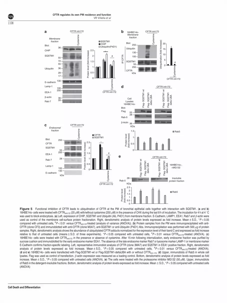
Functional inhibition of CFTR induces ubiquitination ofCFTR protein and its interaction with SQSTM1 at thePM. We examined whether the function of CFTR wouldregulate the recycling of CFTR protein and hence its own PMstability. The abundance of CFTR at the PM is mainlydetermined by its recycling through the Rab11þ endocyticpathway.52,53 It has been reported that ubiquitylated CFTRbecomes unstable at the PM and is diverted to lysosomaldegradation.54 Moreover, SQSTM1 can also function as acritical regulator of internalization, trafficking and sorting ofubiquitinylated surface proteins.55,56 As SQSTM1 proteinlevels increase after functional CFTR depletion in16HBE14o- cells (Supplementary Figure S4C), we wonderedwhether the functional CFTR inhibition might trigger theubiquitination of CFTR at the PM, increase PM levels ofSQSTM1 and hence stimulate CFTR/SQSTM1 interaction.
To this aim, we incubated 16HBE14o- cells with CFTRinh-172
to analyze CFTR by means of anti-CFTR Abs. Addition ofCFTRinh-172 to 16HBE14o- cells increased the PM abundanceof both SQSTM1 and the ubiquitin ligase carboxyl-terminusheat shock cognate 70 (Hsc70)–interacting protein (CHIP), amajor player of PM protein quality control in CF22 (Figure 5a).CFTRinh-172 caused a dramatic increase in CFTR ubiquitination(which was barely detected in untreated 16HBE14o- cells)and enhanced the interaction between PM-resident CFTRand SQSTM1, as determined by co-immunoprecipitation(Figure 5b).
Moreover, after CFTR inhibition, both CFTR and SQSTM1were detected within EEs, as revealed by subcellularfractionation followed by immunoblotting (Figure 5c). Accord-ingly, internalized CFTR co-localized with SQSTM1 at theepithelial surface as well as within enlarged vesicles in primaryhuman bronchial epithelial cells treated with CFTRinh-172
(Supplementary Figures S7A and B). Cystamine neutralizedthe effects of CFTRinh-172 as it prevented PM ubiquitination ofCFTR, SQSTM1 accumulation both at PM and in theendosomal compartment, as well as SQSTM1/CFTR interac-tion in bronchial epithelial cells (Figures 5a–c andSupplementary Figures S7A and B), unless 3-MA was addedto the system (Supplementary Figure S7A). Next, weinvestigated whether the accumulation of SQSTM1 at EEsfollowing CFTR inhibition could favor Rab5 sequestration. Tothis aim, we overexpressed either wild-type (wt)-SQSTM1 or aSQSTM1 mutant lacking the ubiquitin-binding domain (delta-UBA mutant) in 16HBE14o- cells in the presence or absenceof CFTRinh-172. The overexpression of wt-SQSTM1, but notthat of delta-UBA SQSTM1, recapitulated the effects of CFTRinhibition in causing the redistribution of Rab5 into thedetergent-insoluble protein fraction (Figures 5d and e).Moreover, the enforced expression of delta-UBA SQSTM1,but not that of wt-SQSTM1, abrogated these effects of CFTR
inhibition (Figures 5d and e). These data indicate that theubiquitin-binding activity of SQSTM1 is pivotal for inducingRab5 sequestration within the detergent-insoluble proteinfraction upon CFTR inhibition.
To determine whether functional inhibition of CFTR wouldlower its own recycling, we developed an assay in which16HBE14o- cells were either left untreated or were pretreatedwith CFTRinh-172 for 24 h, and cycloheximide (CHX) was addedduring the last 4 h of incubation to inhibit protein neosynth-esis.22,27 16HBE14o- cells were then shifted to 4 1C for 1 h,followed by incubation at 37 1C to induce internalization of PM-resident endogenous CFTR. In contrast to untreated cells,CFTRinh-172-treated cells manifested negligible CFTR localiza-tion in Rab11þ endosomes 30 min after internalization(Figure 6a). The deranged recycling of CFTR followingfunctional CFTR inhibition was reversed by cystamine unlessBECN1 was depleted during cystamine treatment (Figure 6a).Moreover, in CFTRinh-172-treated cells, CFTR was detectedwithin LAMP1þ vesicles as late as 90 min following internaliza-tion, unless SQSTM1 was depleted by siRNA (Figure 6b).Thus, functional inhibition of CFTR perturbs CFTR recycling asit diverts CFTR towards lysosomal degradation.
Altogether, these results indicate that the inhibition of CFTRfunction in bronchial epithelial cells enhances the function andabundance of SQSTM1 at the PM, thus favoring PM disposalof ubiquitylated CFTR and perturbing the endosomal routesthat mediate CFTR recycling.
CFTR function controls CFTR protein stability at the PMof bronchial epithelial cells. The aforementioned datasuggest that the function of CFTR may determine the stabilityof CFTR protein at the PM. To further explore this possibility,16HBE14o- cells were incubated with CFTRinh-172 for 24 h inthe presence or absence of EUK-134 or cystamine with/without 3-MA, with CHX (100 mg/ml), which was added twiceat 8 and 4 h before termination of the experiment.27 CHXtoxicity in the system was excluded by a 3-[4,5-dimethylthia-zol-2-yl]-2,5 diphenyl tetrazolium bromide (MTT) cell viabilityassay (Supplementary Figures S8A and B). Then theabundance of CFTR at the PM was determined by cell-surface biotinylation, purification of biotinylated proteins andimmunoblotting. CFTRinh-172 reduced the overall abundance ofPM-resident mature glycosylated CFTR (band C) by morethan two-thirds (27% of remaining band C versus 91% in cellstreated with CHX alone, considering 100% the value ofuntreated cells) (Figure 7a), and this effect was fully reversedby simultaneous addition of either cystamine or EUK-134 (88and 85% of the remaining band C, respectively) unless 3-MAwas added to the system or BECN1 was depleted by siRNAs(Figure 7a). Depletion of SQSTM1 also abrogated the capacityof CFTRinh-172 to reduce the PM abundance of CFTR(Figure 7a). Finally, CFTRinh-172 lowered the PM abundanceof CFTR in a cystamine-inhibitable fashion, in primary humanbronchial epithelial cells (Figure 7b), confirming the tight linkbetween CFTR function and CFTR stability.
CFTR sufficiency is required for PM stability of themisfolded DF508-CFTR mutant. We previously reportedthat DF508-CFTR mutant protein, which has been rescued atthe epithelial surface of DF508-CFTR homozygous epithelial
CFTR regulates its own PM residence and functionVR Villella et al
1107
Cell Death and Differentiation
β-actin
250
55250
100130
28
Blot:
CFTR
SQSTM1
EEA-1
Rab-7
Lamp-1
E-cadherin
Endosomalfraction
kDa
*
#
CFTRSQSTM1
0
0.5
1
1.5
2
2.5
Pro
tein
leve
l (fo
ld in
crea
se)
Cys
tam
ine
CFTR inh172
Cys
tam
ine
CFTR inh172
250
130
100
55250
Cys
tam
ine
CFTR inh172Membrane
fraction
16HBE14o-
Ubiquitin
SQSTM1
CFTR
IP:C
FT
R
kDa
Ubi
quiti
natio
n (f
old
incr
ease
)
Cys
tam
ine
*
CFTR inh172
0
5
10
15
20
CFTR inh172
Blot:
#
36
557255
17
36
28
130
36
Blot:
CHIP
SQSTM1
Ubiquitin
E-cadherin
Membranefraction
*
#
SQSTM1CHIP
Pro
tein
leve
l (fo
ld in
crea
se)
0.5
1
1.5
2
2.5
Cys
tam
ine
Cys
tam
ine
CFTR inh172
0
Ubiquitin(P4D1)
250EEA-1
100Lamp-1
28Rab-7
KDa
CellLysates
16HBE14o-
Rab-5
β-Actin
28
36
CFTR inh172
Fla
g- S
QS
TM
1 w
t
Blot: Fla
g- S
QS
TM
1de
lta U
BA
Fla
g- S
QS
TM
1 w
t
Fla
g- S
QS
TM
1de
lta U
BA
55Flag
* **
0
0.5
1
1.5
Pro
tein
leve
l(f
old
incr
ease
)Rab5
* **
00.5
11.5
22.5
CFTR inh172
Fla
g- S
QS
TM
1 w
t
Fla
g- S
QS
TM
1de
lta U
BA
Fla
g- S
QS
TM
1 w
t
Fla
g- S
QS
TM
1de
lta U
BA
Rab-528
KDa
16HBE14o-,+MG132
Insolubleprotein fraction
Pro
tein
leve
l(f
old
incr
ease
)
Rab5
Figure 5 Functional inhibition of CFTR leads to ubiquitination of CFTR at the PM of bronchial epithelial cells together with interaction with SQSTM1. (a and b)16HBE14o- cells were treated with CFTRinh172 (20mM) with/without cystamine (250mM) in the presence of CHX during the last 8 h of incubation. The incubation for 4 h at 4 1Cwas used to block endocytosis. (a) Left, expression of CHIP, SQSTM1 and Ubiquitin (Ab, P4D1) from membrane fraction. E-Cadherin, LAMP1, EEA1, Rab7 and b-actin wereused as control of the membrane cell-surface protein fractionation. Right, densitometric analysis of protein levels expressed as fold increase. Mean±S.D., *Po0.05compared with untreated cells, #Po0.01 versus CFTRinh172-treated (analysis of variance (ANOVA)). (b) Protein samples from the PM were immunoprecipitated with anti-CFTR (clone CF3) and immunoblotted with anti-CFTR (clone M3A7), anti-SQSTM1 or anti-Ubiquitin (P4D1) Abs. Immunoprecipitation was performed with 500mg of proteinsamples. Right, densitometric analysis shows the abundance of ubiquitylated CFTR adducts normalized for the expression level of their band C and expressed as fold increaserelative to that of untreated cells (means±S.D. of three experiments). *Po0.05 compared with untreated cells, #Po0.01 versus CFTRinh172-treated (ANOVA). (c)16HBE14o- cells were treated with CFTRinh172 in the presence or absence of cystamine. After 15 min following internalization, early endosome fraction was purified bysucrose cushion and immunoblotted for the early endosome marker EEA1. The absence of the late endosome marker Rab7 or lysosome marker LAMP-1 or membrane markerE-Cadherin confirms fraction-specific labeling. Left, representative immunoblot analysis of CFTR (clone 3MA7) and SQSTM1 in EEA1 positive fraction. Right, densitometricanalysis of protein levels expressed as fold increase. Mean±S.D., *Po0.05 compared with untreated cells, #Po0.01 versus CFTRinh172-treated (ANOVA).(d and e) 16HBE14o- cells were transfected with Flag-SQSTM1-wt or Flag-SQSTM1-deltaUBA with or without CFTRinh-172. (d) Upper, immunoblots of Rab5 in whole celllysates. Flag was used as control of transfection. b-actin expression was measured as a loading control. Bottom, densitometric analysis of protein levels expressed as foldincrease. Mean±S.D., *Po0.05 compared with untreated cells (ANOVA). (e) The cells were treated with the proteasome inhibitor MG132 (50 mM). Upper, immunoblotsof Rab5 in the detergent-insoluble fractions. Bottom, densitometric analysis of protein levels expressed as fold increase. Mean±S.D., *Po0.05 compared with untreated cells(ANOVA)
CFTR regulates its own PM residence and functionVR Villella et al
1108
Cell Death and Differentiation

16HBE14o-
Cystamine
CF
TR
Mer
ge
Medium SQTM1 siRNAScrambled Scrambled BECN1 siRNA
CFTR inh172
Rab
-11
Correlation Coefficient
0.80 ±0.004
0.20 ±0.002 *
0.83 ±0.003
0.88 ±0.006
0.18 ±0.005 *
16HBE14o-
Correlation Coefficient0.15 ±0.010
0.90 ±0.015 *
0.13 ±0.008
CF
TR
LAM
P-1
Mer
ge
SQSTM1 siRNA
CFTR inh172
Figure 6 Functional inhibition of CFTR impairs CFTR recycling in bronchial epithelial cells. (a) 16HBE14o- cells were treated with CFTRinh172 (20mM) and/or 50 nM ofscrambled or SQSTM1 siRNAs or cystamine in the presence or absence of BECN1 siRNAs. CHX (100mg/ml) was added during the last 4 h. The cells were then shifted at 4 1Cfor 1 h and then at 37 1C up to 30 min to allow internalization. Upper, confocal microscopic images of CFTR co-stained with Rab11, 30 min after internalization. Bottom,quantitative measurement of co-localization. Mean±S.D. of five independent experiments (n¼ 50 cells for each experiment). *Po0.05 compared with untreated cells.Bar¼ 10mm. (b) 16HBE14o- cells were treated with CFTRinh172 (20mM) and 50 nM of scrambled or SQSTM1 siRNAs. Upper, confocal microscopic images of CFTR (cloneCF3) co-stained with LAMP1, 90 min after internalization. Bottom, quantitative measurement of co-localization. Mean±S.D. of three independent experiments (n¼ 50 cellsfor each experiment). Representative of three different experiments. *Po0.05 compared with untreated cells. Bar¼ 10mm
CFTR regulates its own PM residence and functionVR Villella et al
1109
Cell Death and Differentiation

cells by cystamine treatment, can reside at the PM wellbeyond cystamine washout. This suggests that DF508-CFTRmight sustain its own PM stability. To address this issue, wetested whether a CFTR-sufficient context (i.e., untreated16HBE14o- cells) could also generate a permissive environ-ment for PM stability of the misfolded DF508-CFTRmutant.22,27 We transfected 16HBE14o- cells with HA-tagged DF508-CFTR at 37 1C, an experimental conditionthat does not allow DF508-CFTR to traffic to the PM in cells
that lack wt-CFTR expression.3,4,6 Mature DF508-CFTRband C was detected in immunoblots of whole 16HBE14o-cell lysates with an anti-HA Ab (Supplementary Figure S9A).Twenty-four hours post transfection, the cells were culturedfor 8 h in the presence of CHX (100 mg/ml, refreshed every4 h) to inhibit protein neosynthesis, as described above.Immunoblotting of surface biotinylated proteins revealedthat DF508-CFTR band C was stably expressed at thePM of 16HBE14o- even after 8 h following CXH treatment
Blot:CFTR band C
E-cadherin
250
130
36kDa
+CFTR inh172
SQ
ST
M1
siR
NA
Scr
ambl
ed
BE
CN
1 si
RN
A
+ Cystamine
3-M
A
+8 h, CHX
EU
K-1
34Surfacebiotinylated proteins
0
Exp
ress
ion
%of
ban
d C
at P
M
**
3-M
A
+8h, CHX
+Cystamine
Scr
ambl
edS
QS
TM
1si
RN
A
EU
K-1
34
*
β-actin
Cys
tam
ine
CFTR inh172
Blot:
E-cadherin
Surfacebiotinylated proteins
250
130
36kDa
CFTR band C
+8 h, CHX
0
Cys
tam
ine
+8h, CHX
*
#
Exp
ress
ion
% o
f b
and
C a
t PM
#
+CFTR inh172
Primary bronchialepithelial cells
+CFTR inh172
β-actin
BE
CN
1 si
RN
A
100
75
25
50
100755025
Figure 7 CFTR function controls CFTR protein stability at the PM of bronchial epithelial cells. (a) 16HBE14o- cells were incubated with/without CFTRinh172 in the presenceor absence of EUK-134 or cystamine with or without 3-MA or BECN1 siRNAs. CFTRinh172-treated cells were alternatively transfected with SQSTM1 siRNAs. CHX (100mg/ml)was added during the last 8 h of incubation. Left, Surface-biotinylated proteins from the PM and immunoblot with anti-CFTR Ab (clone M3A7). Right, densitometricmeasurement of the residual wt-CFTR band C at PM expressed as percentage of initial amount normalized to E-cadherin levels. Mean±S.D. of triplicates of three differentexperiments; *Po0.05, #Po0.01 compared with initial amount with CHX (analysis of variance (ANOVA)). (b) Normal human primary bronchial epithelial cells were treatedwith/without CFTRinh172 in the presence or absence of cystamine. CHX (100mg/ml) was added during the last 8 h of incubation. Left, surface-biotinylated proteins from the PMand immunoblot with anti-CFTR Abs (clone 3MA7). Right, densitometric measurement of the residual wt-CFTR band C at PM expressed as the percentage of initial amountnormalized to E-cadherin levels. Mean±S.D. of triplicates of three different experiments; *Po0.05 compared with untreated cell, #Po0.01 versus CFTRinh172-treated cells(ANOVA)
Figure 8 CFTR function is required for PM stability of DF508-CFTR in bronchial epithelial cells. (a) 16HBE14o- cells were transfected with HA-DF508-CFTR and thenincubated with/without CFTRinh-172 in the presence or absence of cystamine with/without 3-MA. CHX (100mg/ml) was added during the last 8 h of incubation. Left, surface-biotinylated proteins from the PM were immunoblotted with anti-HA Ab. Right, densitometric analysis of the residual band C at PM expressed as the percentage of initialamount normalized to E-cadherin levels. Mean± S.D. of triplicates of three different experiments; *Po0.05 compared with untreated cells, #Po0.01 versus cystamine-treated cells (analysis of variance (ANOVA)). (b) 16HBE14o- were transfected with HA-DF508-CFTR and/or with scrambled or SQSTM1 siRNAs (50nM) and then incubatedwith CFTRinh172 for 24 h. CHX was added during the last 8 h. Left, surface-biotinylated proteins from the PM of the transfected CFTR band C by anti-HA Abs. Right,densitometric analysis the residual band C at PM expressed as the percentage of initial amount normalized to E-cadherin levels. Mean±S.D. of triplicates of three differentexperiments; *Po0.05 compared with untreated cells, #Po0.01 versus scrambled treated cells incubated with CFTRinh172 (ANOVA). (c) Schematic representation of theresults. (1) CFTR inhibition leads to the sequestration of the BECN1 interactome as the result of TG2-mediated BECN1 crosslinking that reduces the availability of PtdIns3K(Vps34). This results in autophagy inhibition with accumulation of SQSTM1 (p62) leading to proteasome overload and accumulation of ubiquitin. The inhibition of CFTRfunction induces CFTR ubiquitination at the PM and favors the interaction between CFTR and p62. (2) The reduced PtdIns3K availability also decreases PtdIns3P at the earlyendosomes and reduces Rab5 protein levels, thus impairing endosomal function. The binding of p62 to ubiquitylated CFTR at the PM diverts CFTR recycling and targets CFTRto degradation. Moreover, p62 present at early endosomes favors Rab5 sequestration within insoluble protein aggregates and delays the progression of internalized CFTRthough the degradative route, thus causing its local accumulation
CFTR regulates its own PM residence and functionVR Villella et al
1110
Cell Death and Differentiation

(Figure 8a). However, pretreatment with CFTRinh-172 stronglyreduced the abundance of PM DF508-CFTR in thisexperimental setting, unless cystamine (but not cystaminecombined with 3-MA) was added as well (Figure 8a). Directdepletion of SQSTM1 by siRNAs mimicked the effects of
cystamine (Figure 8b). Cystamine did not influence the PMabundance of DF508-CFTR rescued by low temperature inCFTR-lacking HeLa cells54,57 (Supplementary Figure S10A),indicating that cystamine does not directly target DF508-CFTR and rather corrects the derangement of peripheral
Exp
ress
ion
% o
f ban
d C
at P
M
CFTRinh172
3-MA
8h, CHX
**
Cystamine
0
25
50
75
+CFTR inh172
3-M
A
Cystamine
+CHX, 8 h, 37°C
Blot:
HA
E-cadherin
250
130
36
kDa
16HBE14o-
band C
*
#
+CFTR inh172
Scrambled
+CHX, 8 h
SQ
ST
M1
siR
NA
Exp
ress
ion
%of
ban
d C
at P
M
130
250
36KDa
Blot:
HA
E-cadherin
β-actin
16HBE14o-+CFTR inh172
Scrambled
+CHX, 8 h, 37°C
SQ
ST
M1
siR
NA
band C
100
0
25
50
75
100
β-actin
-CHX
SurfaceBiotynalation
SurfaceBiotynalation
+HA-ΔF508
+HA-ΔF508
CFTR regulates its own PM residence and functionVR Villella et al
1111
Cell Death and Differentiation

proteostasis resulting from CFTR inhibition in bronchialepithelial cells. These results indicate that a CFTR-sufficientcontext generates permissive conditions for DF508-CFTRmutant to reside at the PM of airway epithelial cells.
Discussion
The proteostasis network is a homeostatic system thatresponds to multiple perturbations, be they genetically deter-mined or acquired due to environmental stress or aging. Theaccumulation of misfolded proteins secondary to destabilizinggenetic mutations or the aging-related decline of proteostasiscontributes to several human conformational diseases, includ-ing neurodegenerative disorders, type II diabetes and CF.1–5
This study indicates that CFTR is a key player of theproteostasis network of bronchial epithelial cells. Our resultsprovide evidence in favor of the hypothesis that functionalperturbation of CFTR affects endosomal trafficking of cell-surface proteins exemplified by TfR, EGFR and CFTR itself.This results from the functional sequestration of BECN1,which has two negative effects on intracellular traffickingrelated to CFTR.24 First, CFTR depletion or inhibition reducesthe local generation of PtdIns3P, which is pivotal in regulatingendosomal fusion/maturation and trafficking,28–32 by inducingfunctional sequestration of the BECN1-interactor hVps34.Second, CFTR depletion or inhibition and consequent BECN1inactivation disables autophagy, thereby enhancing theabundance of SQSTM1 both at the EEs (thus favoringsequestration of Rab5 in the detergent-insoluble proteinfraction and favoring its aggregation) and at the PM (thustargeting ubiquitinylated CFTR to degradation). SQSTM1 isan ubiquitin-binding protein that accumulates in conditions ofdefective autophagy, leading to proteasome overload andfavoring aggresome formation.42,43,58 Interestingly, directdepletion of SQSTM1 increases BECN1 protein levels(through reducing TGM-2 levels)24 and recapitulates theeffects of either genetic (by enforced expression) or pharma-cological (by cystamine) rescue of BECN1 as it can restoreDF508-CFTR at the epithelial surface and blunt lunginflammation in CF mice.27 Here, we show that SQSTM1depletion avoids the deleterious effects of the functionalCFTR inhibition on PM abundance and recycling of CFTRprotein. By contrast, enforced expression of SQSTM1, but notthat of the delta-UBA SQSTM1 mutant, recapitulated theeffects of CFTR inhibition on Rab5 sequestration. Theseresults highlight SQSTM1 as a pivot of CFTR-mediatedregulation of peripheral proteostasis (Figure 8c).
Altogether our data indicate that pharmacological or geneticinhibition of CFTR ignites the disposal of PM-resident CFTR.This may have important implications for CF, as it may initiate aCF-relevant vicious cycle in which the loss of CFTR functionnegatively impacts on the overall abundance of the proteinwithin the PM, hence further reducing the function ofCFTR. Such a scenario may explain how it is sufficient totemporarily interrupt this negative feed-forward loop, forinstance, by transient treatment with cystamine, to obtain along-lasting therapeutic effect that improves signs of lunginflammation in DF508-CFTR homozygous mice.27 Indeed,re-establishing and sustaining a functional CFTR at the PM ofbronchial epithelial cells generates permissive conditions
for misfolded DF508-CFTR to traffic to and reside at the PMof bronchial epithelial cells.27
Conformational diseases are characterized by the prema-ture degradation of unstable proteins or, on the contrary, theirunwarranted and harmful accumulation in intra- or extracellularcompartments. The common view is that such diseases arecaused by mutations that directly affect the structure of theprotein, resulting in abnormal proteostasis due to excessive ordeficient proteolysis. To our knowledge, CFTR constitutes thefirst example of a disease-relevant protein that must be fullyfunctional to avoid its premature degradation, hence unveilingan unexpected connection between protein function andproteostasis. The proteostasis network is probably unique toeach cell type and tissue.1,4,5 Indeed, many conformationaldiseases manifest as tissue-specific disorders, in spite of thefact that mutant proteins are expressed near-to-ubiquitously inmany different tissues. Understanding whether other disease-relevant proteins might orchestrate proteostasis in theirspecific context could unveil new strategies for drug discoveryin other conformational diseases.
Materials and MethodsCell lines and treatments. Normal 16HBE14o- or CFTR-mutatedCFBE41o- bronchial epithelial cells (kindly provided by D.C. Gruenert) werecultured with minimum essential medium (MEM) Earl’s salt (200 mM L-Glutamine,10% FBS and the appropriate amount of penicillin/streptomycin). Human, normalprimary bronchial/tracheal epithelial cells or HeLa adenocarcinoma cell lines (LGCPromochem, Milan, Italy) were cultured as recommended by the American TypeCulture Collection.
16HBE14o- cells were transfected with GFP-tagged FYVE domain of SARA(PtdIns3P probe, GFP-FYVE)46 and then with CFTR-specific or scrambled siRNAsor incubated with CFTRinh-172 for 24 h (20mM, refreshed every 4 h, Calbiochem,Darmstadt, Germany) in the presence or absence of cystamine (250mM, Sigma-Aldrich, St Louis, MO, USA) or EUK-134 (50mg/ml, Vinci-biochem, Florence, Italy)or HA-BECN1 overexpression24 or scrambled or SQSTM1 siRNA. The cells werealso treated with CFTR siRNA or CFTRinh172 and incubated with cystamine inpresence of BECN1, hVps34, hVps15 or ATG14 siRNAs or with cystamine with/without 3-MA (3 mM, Sigma-Aldrich). In another set of experiments, the cells werepreviously transfected with Flag-SQSTM1-wt or Flag-SQSTM1-DUBA(E396X) withor without CFTRinh-172. The cells were also incubated with CHX (100mg/ml,refreshed every 4 h, Sigma-Aldrich)22,55 or with MG132 (50mM for 6 h, Calbiochem)where indicated. CHX toxicity was excluded by a MTT cell viability assay, as previouslydescribed.27 In other experiments, the cells were incubated at 4 1C for 1 h to blockendocytosis and then shifted at 37 1C to allow CFTR internalization. In another set ofexperiments, the cells were previously transfected with HA-tagged-DF508-CFTR andthen treated with CFTRinh-172 with/without cystamine followed by 3-MA.
Human primary epithelial cells were incubated without/with CFTRinh-172 in thepresence or absence of cystamine.
CFBE41o- cells were transfected with HA-BECN1 or incubated with cystamine(250mM, Sigma-Aldrich).24,27
HeLa cells were transfected with HA-tagged-DF508-CFTR and then incubated at26 1C for 30 h with/without cystamine. CHX was added during the last 8 h.
Plasmids. The pcDNA3-HA-BECN 1 (gift from N. Mizushima), pcDNA3.1HA-DF508-CFTR (PRIMM), pCMV-Tag2bFLAG-SQSTM1 wt or pCMV-Tag2b-FLAG-SQSTM1-DUBA (E396X) (kind gift of Dr. Lynne J. Hocking, University ofAberdeen, UK)59 and the GFP-tagged FYVESARA domain (PtdIns3P probe) (kindlyprovided by S. Corvera) expression vectors were used for transfectionexperiments as described.24 Empty vectors were used as control.
Transfection and RNA interference. Cells were transfected with 50 nMof CFTR (Invitrogen), SQSTM1, BECN1, hVps15, ATG14 (PRIMM, Milan, Italy),hVPS34 (Sigma-Aldrich) siRNA (Supplementary Table SI) or scrambledoligonucleotides by Lipo RnaiMax (Invitrogen, Carlsbad, CA, USA) accordingto the manufacturer’s instructions or transfected with pcDNA3-HA-Beclin1,GFP-FYVESARA, pcDNA3-HA-DF508-CFTR, pCMV-Tag2bFLAG-SQSTM1wt or
CFTR regulates its own PM residence and functionVR Villella et al
1112
Cell Death and Differentiation

pCMV-Tag2bFLAG-SQSTM1-DUBA(E396X) expression vectors, with the help oflipofectamine 2000 (Invitrogen), according to the manufacturer’s instructions.
Immunoblot analysis, immunoprecipitation and CFTR ubiquiti-nation. The whole or membrane fraction proteins were obtained from treatedand untreated cells, harvested, lysed and the amounts of proteins weredetermined by a Bio-Rad protein assay (Bio-Rad, Hercules, CA, USA) to ensureequal protein loading before western blot analysis. Fifty micrograms of cell lysatewere loaded in each lane. Immunoprecipitation and CFTR ubiquitination wereperformed with proteins of membrane fraction incubated at 4 1C for 8–12 h withCFTR antibody (clone CF3, Abcam, Cambridge, UK) followed by the addition ofProtein A/G-agarose beads. After washing, the immunoprecipitated proteins weresubjected to electrophoresis through 8% polyacrylamide gels, transferred to blottingmembranes and analyzed. CFTR ubiquitination level was measured by densitometryand normalized to the CFTR level in the precipitate. Densitometric analysis wasperformed with Image J software (NIH, Bethesda, MD, USA); all data points wereexpressed as means±S.D. of triplicates of three independent experiments.
Cell-surface biotinylation assay and membrane fractionation.Cell-surface proteins were biotinylated using sulfosuccinimidyl-6-(biotinamido)hexanoate (sulfo-NHS-LC-Biotin, 1 mg/ml in PBS, pH 8.2; Pierce, Rockford, IL,USA), as described.27,60 Cells were homogenized with a Potter-Elvehjem pestleand centrifuged at 2300� g for 15 min at 4 1C. Supernatants that contains thecytoplasmic and PM fractions were centrifuged 1 h at 16 000� g at 4 1C; the pelletwas the intact membrane and was solubilized in BUFFER A (20 mM Tris-HCl pH7.4, 2 mM EDTA, 20 mM 2-ME, 1� PMSF, 1mg/ml inhibitor protease cocktail)þ 1% Triton X-100 and centrifuged 1 h at 60 000� g in the ultracentrifuge. Thesupernatants were collected as PM fraction. Equivalent amounts of protein(500mg) were used for streptavidin-agarose pull-down (Pierce). Biotinylatedproteins of PM fraction were immunoblotted against CFTR (clone M3A7) or TfRand E-cadherin or b–actin. For transferrin: cells were starved for 1 h at 37 1C inserum-free HEPES-buffered DMEM and then incubated for 1 h at 4 1C in thecontinuous presence of 5 mg/ml Tf-unconjugated (Invitrogen) HEPES-bufferedDMEM. The cell were incubated at 37 1C for 15 min in growth medium and thenprocessed for biotnylation assay. The densitometric analysis and percentageremaining was performed by Image J software and each data point is expressedas the mean±S.D. of triplicate of three independent experiments.
Differential fractionation and separation of endosomes. Theseparation of EE and LE fractions was performed as described.61 Briefly, cells werehomogenized gently to limit damage to endosomes, and a post-nuclear supernatant(PNS) was prepared by centrifugation for 10 min at 1000� g, and then the PNS wascentrifuged at 20 000� g for 20 min. The supernatant was collected, and the pelletwas resuspended with 1� homogenization buffer (250 mM sucrose, 1 mMNa2EDTA, 10 mM HEPES). The PNS was adjusted to 40.6% sucrose, loaded atthe bottom of an SW60 tube and then overlaid sequentially with 16% sucrose inD2O, 10% sucrose in D2O and finally with homogenization buffer. The gradient wascentrifuged for 60 min at 60 000� g using an SW60 rotor. EEs were then collectedat the 16%/10% interface and LEs at the top of the 10% cushion.
Soluble and insoluble fractions. Cells were lysed in buffer containg50 mM Tris- HCl pH 7.5, 150 mM NaCl, 0.5% Nonidet P40, 5 mM EDTA, 1 mMphenylmethylsulphonyl fluoride, 50 mM NaF, 10 mg/ml leupeptin and 10mg/mlapoprotein supplemented with protease inhibitors (Sigma-Aldrich) in presence of2% SDS and centrifuged at 16 000� g at 4 1C for 20 min. After centrifugation, thesoluble (supernatant) and insoluble (pellet) fraction were used in western blotanalysis with anti-Rab5 antibody. The pellet insoluble in Nonidet P40 wasdissolved five times in sample buffer, boiled at 95 1C for 5 min and resolved on a10% polyacrylamide gel.24
Antibodies. The following primary antibodies were used for blot analysis:Ubiquitin (P4D1), 1 : 1000, CHIP, 1 : 500, hVps15\p150 (Nterm), 1 : 300, UVRAG,1 : 500, Santa Cruz Biotechnology (Santa Cruz Inc, Santa Cruz, CA, USA);SQSTM1, 1 : 1000, hVps34, 1 : 500, ATG14L, 1 : 750, Flag 1:1500 (Sigma-Aldrich); CFTR clone M3A7, 1 : 500, EEA-1, 1 : 1000, Lamp-1, 1 : 1000, CFTR(CF3), 1 : 1000, Rab-5, 1 : 1000, Rab-7, 1 : 1000, BECN1, 1 : 1000, Abcam;E-Cadherin, 1 : 1000 and b-actin, 1 : 1000, Cell Signaling Technology (CellSignaling Inc., Danvers, MA, USA); and TfR, 1 : 800, Invitrogen.
The following primary antibodies were used for microscopy techniques: mousemonoclonal antibodies against CFTR (CF3), 1 : 200, Abcam; SQSTM1(D3), 1 : 100,
HDAC6 (D11), 1 : 100, UVRAG, 1 : 300, Santa Cruz Biotechnology; rabbit polyclonalantibodies against EEA-1, 1 : 400, Rab5, 1 : 400, LAMP-1, 1 : 400, Abcam; hVps-34,1 : 500, SQSTM1, 1 : 400, Sigma-Aldrich; Rab11(D4F5), 1 : 400, Cell Signaling;CFTR(H-182), 1 : 100, EGFR (1005), 1 : 100, Santa Cruz Biotechnology. TheAlexa-Fluor-488, Alexa-Fluor-546 secondary antibodies were obtained fromMolecular Probes (Invitrogen).
Immunofluorescence and confocal imaging. The cells were fixed for10 min with 4% paraformaldehyde (PFA, Sigma-Aldrich) in PBS, quenched with50 mM NH4Cl and permeabilized for 30 min in blocking buffer (0.1% (w/v) saponin,0.5% (w/v) BSA in PBS/Ca/Mg). The cells were incubated for 2 h with the primaryantibody, washed three times in PBS, incubated for 1 h with the secondary (Alexa-labelled) antibody, washed three times in PBS and were finally taken onVectashield-mounted coverslips. The samples were examined under a Zeiss LSM510 confocal laser-scanning microscope (Carl Zeiss MicroImaging, Thornwood,NY, USA) equipped with � 63 oil-immersion objective and image processing wasdone with Adobe Photoshop C2 (Adobe Sysytem Inc., San Jose, CA, USA). Toperform quantitative image analysis, 10–15 randomly chosen fields that included8–10 cells each were scanned, using the same setting parameters (i.e., pinhole,laser power, offset gain and detector amplification) below pixel saturation. Themean intensity per cell was determined using the histogram function in the ZeissLSM 510 Software (version 3.2), and all of the pixel values above backgroundlevels were quantified. All of the experiments were repeated at least three times,and representative images are shown. Quantification of number of FYVESARA
spots per cell was performed using the AnalySIS software (Soft Imaging SystemsGmbH, Muenster, Germany). To quantify the levels of co-localization, confocalserial sections were acquired from 8–10 cells per experimental condition, exportedin TIFF format and processed as previously described.62
Tf uptake and recycling. For transferrin uptake, cells were starved for 1 h at37 1C in serum-free HEPES-buffered DMEM and then incubated for 1 h at 4 1C in thecontinuous presence of 5mg/ml Tf–Alexa-Fluor-488 (Molecular Probes) HEPES-buffered DMEM. The uptake of Tf and its movement along the endocytic pathwaywere followed by incubating the cells at 37 1C for 5, 10, 15, 30 and 60 min in growthmedium. The cells were then treated with a mixture of 0.5 M NaCl and 0.5% aceticacid (this acid wash removes Tf that is non-specifically bound to the PM) for 30 sbefore being fixed and processed for immunofluorescence.
For transferrin recycling, cells were starved for 1 h at 37 1C, HEPES-bufferedDMEM and then incubated for 20 min at 37 1C in the continuous presence of 5 mg/mlTf–Alexa-Fluor-488 in serum-free, HEPES-buffered DMEM. After extensivewashing with serum-free HEPES-buffered DMEM, the recycling of Tf wasfollowed by incubating the cells at 37 1C in the presence of 50mg/ml unlabelledTf for 40 and 60 min in growth medium. The cells were then treated with a mixtureof 0.5 M NaCl and 0.5% acetic acid for 30 s before being fixed and processed forimmunofluorescence.
EGFR internalization and degradation. For EFGR internalization, thecells were serum starved for 12 h at 37 1C in serum-free HEPES-buffered DMEMto allow the EGFR to accumulate on the cell surface and then incubated for 1 h at4 1C in the continuous presence of 100 ng/ml EGF–Alexa-Fluor-488 in serum-freeHEPES-buffered DMEM. The uptake of EGF and its movement along theendocytic pathway were followed by incubating the cells at 37 1C for 5, 10,15, 30and 60 min in growth medium. The cells were then treated with a mixture of0.5 M NaCl and 0.5% acetic acid (this acid wash removes EGF that isnon-specifically bound to the PM) for 30 s before being fixed and processed forimmunofluorescence.
For EFGR degradation, the cells were stimulated by addition of EGF. Atdesignated intervals after addition of EGF, cells were fixed, permeabilized andlocalization of the receptor was determined by immunofluorescence analysis usingthe antibody against total EGFR.
Fluorescence resonance energy transfer microscopy. Cells wereimmunostained with Alexa 546-anti-TG2 (Molecular Probes/Invitrogen)/Cy5-anti-SUMO-1 (Santa Cruz Biotechnology), as previously described.24
ROS detection. Cells were pulsed with 10mM 5-(and-6)-chloromethyl-2070-dichlorodihydrofluorescein diacetate acetyl ester (Molecular Probes) and analyzedwith Wallac 1420 multilabel Counter (Perkin Elmer, Zaventem, Belgium), asdescribed.24
CFTR regulates its own PM residence and functionVR Villella et al
1113
Cell Death and Differentiation

Iodide efflux. The analysis of the iodide efflux was performed by a iodide-sensitive fluorescent indicator, SPQ (Molecular Probes, Invitrogen), as previouslydescribed.22,27,53,63 The rates were calculated using SigmaPlot Version 7.1 (SystatSoftware Inc., London, UK) for each mean fluorescence trace generated from the50 cells examined per population per coverslip, as described.27
Statistical analysis. Data are reported as arithmetic mean±S.D. Datadistribution was analyzed for normality and statistical analysis performed usingthe one-way ANOVA. Significant differences are indicated in the figures. All datawere obtained from independent measurements. Data were analyzed using SPSS13 software (SPSS, Milan, Italy). Statistical significance was defined as P-valueof o0.05.
Conflict of InterestThe authors declare no conflict of interest.
Acknowledgements. This work was supported by the European Institute forResearch in Cystic Fibrosis and Italian Cystic Fibrosis Association (LM), theProgramma di Ricerca Scientifica di Rilevante Interesse Nazionale(2008RMJB3A_004, 2008) of the ministero dell’Istruzione, dell’Universita e dellaRicerca (LM, VR), RO1 HL093004 (EMB), Telethon Grant No. GGP12128 (LM,EMB, VR, MCM), Ligue Nationale contre le Cancer (Equipe labellisee) (GK), AXAChair for Longevity Research, Agence Nationale pour la Recherche (ANR) (GK),European Commission (ArtForce) (GK), Fondation pour la Recherche Medicale(FRM), Institut National du Cancer (INCa), Canceropole Ile-de-France (GK),Fondation Bettencourt-Schueller and the LabEx Onco-Immunology (GK). We thankNoboru Mizushima (The Tokyo Metropolitan Institute of Medical Sciences, Tokyo,Japan) for the gift of the pcDNA3-HA-Beclin1 expression vectors, Dieter C. Gruenert(University of California, San Francisco, CA, USA) for the gift of CFBE41o- cell lines,S. Corvera (University of Massachusetts Medical School, Worcester, MA, USA) forthe gift of the GFP-FYVESARA. We thank Alessandro Luciani (European Institute forResearch in Cystic Fibrosis, Milan) for technical assistance in confocal microscopy.
Author contributionsVRV co-designed the research concept, performed surface biotinylation andmembrane fractionation, immunoblot and immunoprecipitation experiments, cellcultures and transfections and analyzed data. SE performed immunoblot andimmunoprecipitation experiments, confocal microscopy, cell cultures and transfec-tions and analyzed data. EMB provided the scientific knowledge, contributed to thediscussion, interpretation and analysis of the data. MV provided the scientificknowledge, supervised confocal microscopic studies and contributed to thediscussion, interpretation and analysis of the data. SC provided the SQSTM1plasmids, provided the scientific knowledge on SQSTM1 mutants and contributed tothe analysis of the data. ADM and AL provided the scientific knowledge andcontributed to the discussion, interpretation and analysis of the data. SG, MPM andRC contributed to the discussion of data. VR, MCM and GK co-designed theresearch concept, co-supervised the project, provided the scientific knowledge andcontributed to the discussion, interpretation and analysis of the data. LM designedthe research concept, planned the overall experimental design and supervised thestudy. LM, VR and GK wrote the paper.
1. Powers ET, Morimoto RI, Dillin A, Kelly JW, Balch WE. Biological and chemicalapproaches to diseases of proteostasis deficiency. Annu Rev Biochem 2009; 78: 959–991.
2. Hutt DM, Powers ET, Balch WE. The proteostasis boundary in misfolding diseases ofmembrane traffic. FEBS Lett 2009; 583: 2639–2646.
3. Roth DM, Balch WE. Modeling general proteostasis: proteome balance in health anddisease. Curr Opin Cell Biol 2011; 23: 126–134.
4. Gidalevitz T, Kikis EA, Morimoto RI. A cellular perspective on conformational disease:the role of genetic background and proteostasis networks. Curr Opin Struct Biol 2010; 20:23–32.
5. Balch WE, Morimoto RI, Dillin A, Kelly JW. Adapting proteostasis for disease intervention.Science 2008; 319: 916–919.
6. Balch WE, Roth DM, Hutt DM. Emergent properties of proteostasis in managing cysticfibrosis. Cold Spring Harb Perspect Biol 2011; 3: a004499.
7. Okiyoneda T, Apaja PM, Lukacs GL. Protein quality control at the plasma membrane.Curr Opin Cell Biol 2011; 23: 483–491.
8. Amaral MD. Targeting CFTR: how to treat cystic fibrosis by CFTR-repairing therapies.Curr Drug Targets 2011; 12: 683–693.
9. O’Sullivan BP, Freedman SD. Cystic fibrosis. Lancet 2009; 373: 1891–1904.
10. Rowe SM, Miller S, Sorscher EJ. Cystic fibrosis. N Engl J Med 2005; 352: 1992–2001.11. Accurso FJ. Update in cystic fibrosis 2005. Am J Respir Crit Care Med 2006; 173: 944–947.12. Park HW, Nam JH, Kim JY, Namkung W, Yoon JS, Lee JS et al. Dynamic regulation of
CFTR bicarbonate permeability by [Cl-]i and its role in pancreatic bicarbonate secretion.
Gastroenterology 2010; 139: 620–631.13. Welsh MJ, Smith AE. Molecular mechanisms of CFTR chloride channel dysfunction in
cystic fibrosis. Cell 1993; 73: 1251–1254.14. Bobadilla JL, Macek M, Fine JP, Farrell PM. Cystic fibrosis: a worldwide analysis of CFTR
mutations–correlation with incidence data and application to screening. Hum Mutat 2002;
19: 575–606.15. Kartner N, Augustinas O, Jensen TJ, Naismith AL, Riordan JR. Mislocalization of delta
F508 CFTR in cystic fibrosis sweat gland. Nat Genet 1992; 1: 321–327.16. Okiyoneda T, Lukacs GL. Cell surface dynamics of CFTR: the ins and outs. Biochim
Biophys Acta 2007; 1773: 476–479.17. Pedemonte N. Small-molecule correctors of defective [Delta]F508-CFTR cellular
processing identified by high-throughput screening. J Clin Invest 2005; 115: 2564–2571.18. Galietta LJ, Springsteel MF, Eda M, Niedzinski EJ, By K, Haddadin MJ et al. Novel CFTR
chloride channel activators identified by screening of combinatorial libraries based on
flavone and benzoquinolizinium lead compounds. J Biol Chem 2001; 276: 19723–19728.19. Van Goor F, Straley KS, Cao D, Gonzalez J, Hadida S, Hazlewood A et al. Rescue of
DeltaF508-CFTR trafficking and gating in human cystic fibrosis airway primary cultures by
small molecules. Am J Physiol Lung Cell Mol Physiol 2006; 290: L1117–L1130.20. Van Goor F, Hadida S, Grootenhuis PD, Burton B, Stack JH, Straley KS et al. Correction of
the F508del-CFTR protein processing defect in vitro by the investigational drug VX-809.
Proc Natl Acad Sci USA 2011; 108: 18843–18848.21. Denning GM, Anderson MP, Amara JF, Marshall J, Smith AE, Welsh MJ. Processing of
mutant cystic fibrosis transmembrane conductance regulator is temperature-sensitive.
Nature 1992; 358: 761–764.22. Okiyoneda T, Barriere H, Bagdany M, Rabeh WM, Du K, Hohfeld J et al. Peripheral protein
quality control removes unfolded CFTR from the plasma membrane. Science 2010; 329:
805–810.23. Lukacs GL, Verkman AS. CFTR: folding, misfolding and correcting the DeltaF508
conformational defect. Trends Mol Med 2012; 18: 81–91.24. Luciani A, Villella VR, Esposito S, Brunetti-Pierri N, Medina D, Settembre C et al. Defective
CFTR induces aggresome formation and lung inflammation in cystic fibrosis through ROS-
mediated autophagy inhibition. Nat Cell Biol 2010; 12: 863–875.25. Luciani A. SUMOylation of tissue transglutaminase as link between oxidative stress and
inflammation. J Immunol 2009; 183: 2775–2784.26. Maiuri L, Luciani A, Giardino I, Raia V, Villella VR, D’Apolito M et al. Tissue
transglutaminase activation modulates inflammation in cystic fibrosis via PPAR gamma
down-regulation. J Immunol 2008; 180: 7697–7705.27. Luciani A, Villella VR, Esposito S, Gavina M, Russo I, Silano M et al. Targeting autophagy
as a novel strategy for facilitating the therapeutic action of potentiators on DF508 cystic
fibrosis transmembrane conductance regulator. Autophagy 2012; 8: 1657–1672.28. Poteryaev D, Datta S, Ackema K, Zerial M, Spang A. Identification of the switch in early-to-
late endosome transition. Cell 2010; 141: 497–508.29. Cabrera M, Ungermann C. Guiding endosomal maturation. Cell 2010; 141: 404–406.30. Rink J, Ghigo E, Kalaidzidis Y, Zerial M. Rab conversion as a mechanism of progression
from early to late endosomes. Cell 2005; 122: 735–749.31. Ravikumar B, Imarisio S, Sarkar S, O’Kane CJ, Rubinsztein DC. Rab5 modulates
aggregation and toxicity of mutant huntingtin through macroautophagy in cell and fly
models of Huntington disease. J Cell Sci 2008; 121: 1649–1660.32. Thoresen SB, Pedersen NM, Liestol K, Stenmark H. A phosphatidylinositol 3-kinase
class III sub-complex containing VPS15, VPS34, Beclin 1, UVRAG and BIF-1
regulates cytokinesis and degradative endocytic traffic. Exp Cell Res 2010; 316:
3368–3378.33. Christoforidis S, Miaczynska M, Ashman K, Wilm M, Zhao L, Yip SC et al. Phosphati-
dylinositol-3-OH kinases are Rab5 effectors. Nat Cell Biol 1999; 1: 249–252.34. Simonsen A, Gaullier JM, D’Arrigo A, Stenmark H. The Rab5 effector EEA1 interacts
directly with syntaxin-6. J Biol Chem 1999; 274: 28857–28860.35. Hayakawa A, Hayes SJ, Lawe DC, Sudharshan E, Tuft R, Fogarty K et al. Structural basis
for endosomal targeting by FYVE domains. J Biol Chem 2004; 279: 5958–5966.36. Gillooly DJ, Morrow IC, Lindsay M, Gould R, Bryant NJ, Gaullier JM et al. Localization
of phosphatidylinositol 3-phosphate in yeast and mammalian cells. EMBO J 2000; 19:
4577–4588.37. Raiborg C, Bremnes B, Mehlum A, Gillooly DJ, D’Arrigo A, Stang E et al. FYVE and coiled-
coil domains determine the specific localisation of Hrs to early endosomes. J Cell Sci 2001;
114: 2255–2263.38. Hu Y, Chuang JZ, Xu K, McGraw TG, Sung CH. SARA, a FYVE domain protein, affects
Rab5-mediated endocytosis. J Cell Sci 2002; 115: 4755–4763.39. Sinha S, Levine B. The autophagy effector Beclin 1: a novel BH3-only protein. Oncogene
2008; 27: S137–S148.40. Kroemer G, Marino G, Levine B. Autophagy and the integrated stress response. Mol Cell
2010; 40: 280–293.41. Kirkin V, McEwan DG, Novak I, Dikic I. A role for ubiquitin in selective autophagy. Mol Cell
2009; 34: 259–269.
CFTR regulates its own PM residence and functionVR Villella et al
1114
Cell Death and Differentiation
42. Moscat J, Diaz-Meco MT. p62 at the crossroads of autophagy, apoptosis, and cancer. Cell2009; 137: 1001–1004.
43. Korolchuk VI, Mansilla A, Menzies FM, Rubinsztein DC. Autophagy inhibitioncompromises degradation of ubiquitin-proteasome pathway substrates. Mol Cell 2009;33: 517–527.
44. Maxfield FR, McGraw TE. Endocytic recycling. Nat Rev Mol Cell Biol 2004; 5: 121–132.45. Scita G, Di Fiore PP. The endocytic matrix. Nature 2010; 463: 464–473.46. Vicinanza M, Di Campli A, Polishchuk E, Santoro M, Di Tullio G, Godi A et al. OCRL
controls trafficking through early endosomes via PtdIns4,5P(2)-dependent regulation ofendosomal actin. EMBO J 2011; 30: 4970–4985.
47. Daniele T, Di Tullio G, Santoro M, Turacchio G, De Matteis MA. ARAP1 regulates EGFreceptor trafficking and signalling. Traffic 2008; 9: 2221–2235.
48. Kelly M, Trudel S, Brouillard F, Bouillaud F, Colas J, Nguyen-Khoa T et al. Cystic fibrosistransmembrane regulator inhibitors CFTR(inh)-172 and GlyH-101 target mitochondrialfunctions, independently of chloride channel inhibition. J Pharmacol Exp Ther 2010; 333:60–69.
49. Ma T, Thiagarajah JR, Yang H, Sonawane ND, Folli C, Galietta LJ et al. ThiazolidinoneCFTR inhibitor identified by high-throughput screening blocks cholera toxin-inducedintestinal fluid secretion. J Clin Invest 2002; 110: 1651–1658.
50. Baudouin-Legros M, Colas J, Moriceau S, Kelly M, Planelles G, Edelman A et al. Long-termCFTR inhibition modulates 15d-prostaglandin J2 in human pulmonary cells. Int J BiochemCell Biol 2012; 44: 1009–1018.
51. Liang C. Beclin1-binding UVRAG targets the class C Vps complex to coordinateautophagosome maturation and endocytic trafficking. Nat Cell Biol 2008; 10: 776–787.
52. Cholon DM, O’Neal WK, Randell SH, Riordan JR, Gentzsch M. Modulation of endocytictrafficking and apical stability of CFTR in primary human airway epithelial cultures. Am JPhysiol Lung Cell Mol Physiol 2010; 298: L304–L314.
53. Silvis MR, Bertrand CA, Ameen N, Golin-Bisello F, Butterworth MB, Frizzell RA et al.Rab11b regulates the apical recycling of the cystic fibrosis transmembrane conductanceregulator in polarized intestinal epithelial cells. Mol Biol Cell 2009; 20: 2337–2350.
54. Sharma M, Pampinella F, Nemes C, Benharouga M, So J, Du K et al. Misfolding diverts
CFTR from recycling to degradation: quality control at early endosomes. J Cell Biol 2004;
164: 923–933.55. Samuels IS, Seibenhener ML, Neidigh KB, Wooten MW. Nerve growth factor stimulates the
interaction of ZIP/p62 with atypical protein kinase C and targets endosomal localization:
evidence for regulation of nerve growth factor-induced differentiation. J Cell Biochem 2001;
82: 452–466.56. Geetha T, Wooten MW. TrkA receptor endolysosomal degradation is both ubiquitin and
proteasome dependent. Traffic 2008; 9: 1146–1156.57. Delaney SJ, Rich DP, Thomson SA, Hargrave MR, Lovelock PK, Welsh MJ et al. Cystic
fibrosis transmembrane conductance regulator splice variants are not conserved and fail to
produce chloride channels. Nat Genet 1993; 4: 426–431.58. Korolchuk VI, Menzies FM, Rubinsztein DC. Mechanisms of cross-talk between the ubiquitin-
proteasome and autophagy-lysosome systems. FEBS Lett 2010; 584: 1393–1398.59. Hocking LJ, Mellis DJ, McCabe PS, Helfrich MH, Rogers MJ. Functional
interaction between sequestosome-1/p62 and autophagy-linked FYVE-containing
protein WDFY3 in human osteoclasts. Biochem Biophys Res Commun 2010; 402:
543–548.60. Caohuy H, Jozwik C, Pollard HB. Rescue of [Delta]F508-CFTR by the SGK1/Nedd4-2
signaling pathway. J Biol Chem 2009; 284: 25241–25253.61. Aniento F, Emans N, Griffiths G, Gruenberg J. Cytoplasmic dynein-dependent vesicular
transport from early to late endosomes. J Cell Biol 1993; 123: 1373–1387.62. Maiuri L, Luciani A, Villella VR, Vasaturo A, Giardino I, Pettoello-Mantovani M et al.
Lysosomal accumulation of gliadin p31-43 peptide induces oxidative stress and tissue
transglutaminase-mediated PPAR gamma downregulation in intestinal epithelial cells and
coeliac mucosa. Gut 2010; 59: 311–319.63. Verkman AS, Galietta LJ. Chloride channels as drug targets. Nat Rev Drug Discov 2009
8: 153–171.
Supplementary Information accompanies this paper on Cell Death and Differentiation website (http://www.nature.com/cdd)
CFTR regulates its own PM residence and functionVR Villella et al
1115
Cell Death and Differentiation


