Embed Size (px)
Citation preview

Med Clin N Am 90 (2006) 453–479
Dyspnea
Joseph R. Shiber, MDa,b,c,*, Jose Santana, MDb
aDepartment of Medicine, East Carolina University, Greenville, NC, USAbDepartment of Emergency Medicine, East Carolina University, Greenville, NC, USA
cEmergency Medicine–Internal Medicine Combined Residency, East Carolina University,
Greenville, NC, USA
The sensation of breathlessness, dyspnea, is clinically important when it isrecognized by the patient as abnormal. The development of shortness ofbreath (SOB) or the inability to satisfy oxygen requirements is an expectedoutcome of overexertion, such as occurs after running or heavy lifting.When dyspnea occurs at rest or during exertion that is less than expected,it is considered pathologic and a symptom of a disease state. Multiple organsystems are involved in the differential diagnosis of dyspnea, most commonlythe cardiovascular and pulmonary systems. The type and severity of under-lying lung or heart disease have been shown to correlate with the descriptionoffered by the patient [1,2]. Box 1 illustrates the extensive differential diag-nosis for dyspnea.
The outpatient management of dyspnea has changed recently, with theadvent of new diagnostic tests and therapies. These innovations have al-lowed the outpatient clinician to more accurately diagnose the underlyingdisorder and initiate appropriate therapy. Now, clinical decisions basedon whether to continue outpatient management or admit a patient to thehospital can be augmented by treatment algorithms. This article constructsa decision and management protocol for physicians, allowing for a decisionto treat the adult dyspneic patient in the office or transfer the patient toa hospital in pursuit of a confirmed diagnosis and definitive care. The firststep is to begin with a rapid initial assessment of the airway, breathing,and circulation, while gathering a focused history and physical examination[3]. Once the initial examination and vital signs have been obtained and anemergent situation has been excluded, the patient can be placed in one ofthree categories: (1) distress with unstable vital signs; (2) distress with stable
* Corresponding author. Department of Medicine, East Carolina University, 600 Moye
Boulevard, Greenville, NC 27834.
E-mail address: [email protected] (J.R. Shiber).
0025-7125/06/$ - see front matter � 2006 Elsevier Inc. All rights reserved.
doi:10.1016/j.mcna.2005.11.006 medical.theclinics.com

454 SHIBER & SANTANA
Box 1. Differential diagnosis for dyspnea
Mechanical interference with ventilationAbdominal or chest massAsthma, emphysema, bronchitisEndobronchial tumorInterstitial fibrosis of any causeKyphoscoliosisLeft ventricular failureLymphangitic tumorObesityObstruction to airflow, central or peripheralPleural thickeningResistance to expansion of the chest wall or diaphragmResistance to expansion of the lungThoracic burn with eschar formationTracheal or laryngeal stenosis
Weakness of the respiratory pumpAbsoluteHyperinflationNeuromuscular diseaseObesityPleural effusionPneumothoraxPrevious poliomyelitisRelative
Increased respiratory driveDecreased cardiac outputDecreased effective hemoglobinHypoxemia of any causeMetabolic acidosisRenal diseaseStimulation of intrapulmonary receptors
Wasted ventilationCapillary destructionLarge-vessel obstruction
Psychologic dysfunctionAnxietyBodily preoccupation, somatization disorderDepressionSecondary gain, malingering

455DYSPNEA
vital signs; or (3) no distress and stable vital signs. Following this algorithm,a patient’s disposition can be determined expeditiously (Fig. 1).
Acute myocardial infarction (AMI) and congestive heart failure (CHF)are the most common cardiovascular disorders that lead to dyspnea. Pa-tients who have had an AMI usually describe retrosternal chest discomfortand an inability to catch their breath. This sensation occurs secondary toa reduction in cardiac output and pulmonary perfusion and is caused byreduced contractility of the ischemic or stunned myocardium. The adminis-tration of supplemental oxygen, therapy to decrease myocardial oxygenconsumption and increase coronary vasodilation, and the use of revascular-ization strategies such as fibrinolytic therapy or percutaneous coronaryintervention may readily relieve the symptoms. Patients who have anexacerbation of congestive heart failure may describe a sensation of SOBwith exertion, orthopnea, and paroxysmal nocturnal dyspnea. Symptomsgenerally resolve or are improved by giving the patient supplemental oxy-gen, diuretics, therapy aimed at decreasing preload and afterload, and med-ical or surgical treatments to improve cardiac contractility. Pulmonarydyspnea is commonly caused by chronic obstructive pulmonary disease(COPD), asthma, pneumothorax, and pneumonia. Chronic obstructive pul-monary disease and asthma are considered obstructive processes that may
Fig. 1. Algorithm for acute dyspnea management. The initial assessment includes airway,
breathing, and circulation (ABC’s), a brief history of the present illness, medical history, med-
ications, and allergies. Hx, history; Sx, symptoms; VS, vital signs.

456 SHIBER & SANTANA
produce chronic SOB punctuated by sudden acute worsening of symptoms.Often, the treatment of these diseases requires the use of steroids and in-haled bronchodilators to resolve the ensuing exacerbation. Table 1 describesthe symptoms of dyspnea according to the disease state. Primary care clini-cians should always consider other life-threatening causes of SOB such aspulmonary embolism (PE).
Although many of the causes that lead to dyspnea will be treated ulti-mately in the causes or inpatient ward, primary care physicians should beknowledgeable about the subtle and atypical presentations of potentiallylife-threatening diagnoses.
Congestive heart failure
Congestive heart failure is one of the most common causes of dyspnea seenin health care settings. Approximately 1.2% to 2% of the population in theUnited States has heart failure, and most patients (75%–80%) are olderthan 65 years of age. It has been estimated that approximately 20 million peo-ple unknowingly have depressed left ventricular function in the absence ofsymptoms and are likely to become symptomatic within a 1- to 5-year period.Patients who have CHF visit physicians more than 11 million times per yearand are responsible for 3.5 million hospitalizations per year, with one thirdrequiring rehospitalization within 3 months after discharge [4].
Patients may present to an outpatient clinic as an established, well-knownpatient with CHF or as a first-time visitor. Thus, primary care physicians areon the front line when it comes to tentatively diagnosing congestive heartfailure. The well-established patient usually has been diagnosed previouslywith heart failure or has significant predisposing risk factors, such as hyper-tension or coronary artery disease (CAD). The patient who is newly diag-nosed with CHF presents the added challenge of tailoring the outpatientworkup and investigation for the cause of the disease. The diagnosis is de-termined after a full history, physical examination, and diagnostic studieshave been performed. Once the diagnosis and severity of illness are deter-mined, the appropriate treatment can be initiated. In many cases, patientswho present to an outpatient clinic will be too ill to be taken care of inthe office and will require transfer to an emergency department (ED) fora definitive diagnosis, treatment, and possibly admission to the hospital.
Table 1
Symptoms associated with diagnosis
Diagnosis Symptoms
CHF Dyspnea on exertion, paroxymal nocturnal dypnea, orthopnea
AMI Radiating chest pressure, diaphoresis, SOB
PE Fever, pleuritic chest pain, sudden onset SOB, syncope
COPD/asthma Cough, smoker, SOB relieved with nebulizer or MDI
Pneumonia Fever, productive cough, SOB
Pneumothorax Sudden onset pleuritic chest pain, SOB not relieved with O2

457DYSPNEA
History
The patient’s history will help determine what elements of the physical ex-amination will aid in the appropriate diagnosis and what diagnostic studiesmay or may not be indicated. The onset, severity, duration, alleviating, ag-gravating, and associated symptoms are all pertinent to the history. To illus-trate, consider the following case: A 55-year-old man presents to his primarycare physicians clinic with a medical history significant for hypertension,with the chief complaint, ‘‘I’m unable to walk very far without becomingwinded.’’ He states that he now uses three pillows to keep from feeling shortof breath and wakes up frequently at night to catch his breath, and he hasnoticed that his legs are swollen.
This patient is like many of the people seen everyday in outpatient clinics.How is the practitioner to sort out this patient from the other patients seenthe same day with similar complaints? Simply, it is all in the history. Theimportant information to obtain includes onset, duration of symptoms,what activities result in SOB, and any other associated symptoms. In addi-tion, the history should focus on the other deadly causes of SOB. Has thepatient just taken a 10-hour plane ride? Has there been any associated chestpain, back pain, nausea, or diaphoresis? Has the patient experienced palpi-tations or any other symptoms?
It is also important to ask the patient about their medical, surgical, social,and family history to determine risk factors and possible causes, and askabout medication use and record the current dosage to initiate or modifytreatments.
Physical examination
The physical examination should be given due importance, despite inno-vations in objective laboratory and radiographic tests. Studies have shownthat the physical examination depends on the diagnosis and prognosis ofcongestive heart failure. The physical examination begins the first momentthe patient is encountered, during which their general appearance is ob-served. A person’s appearance speaks volumes about the severity of theirdyspnea. A patient who becomes short of breath after a few steps in the of-fice is obviously very different from the one who can walk half of a mile be-fore becoming dyspneic. This categorization can be further described byusing the New Heart Association’s Functional Classification of Heart Fail-ure. Rating the patient on this functional scale from I to V can be used asa comparison to the previous functional status, determine the acuity ofchange in an established patient, and the response to treatment [5]. The pa-tient’s disposition can often be determined by the initial general appearance;whether the patient can be treated with oral medication or he or she requirestransportation to the ED for intravenous (IV) medication administrationand hospital admission.
It is imperative to record and review the patient’s initial vital signs, in-cluding pulse oximetry, and to repeat assessments after interventions.

458 SHIBER & SANTANA
Obtaining an accurate body weight to compare with previous values, partic-ularly if the patient weighs him/herself regularly, will suggest whether therehas been a recent increase in weight. Tachypnea, tachycardia, and hypoxiaare expected findings during a CHF exacerbation. Hypertension may be sec-ondary to stress or present as a cause of failure; if hypotension is present,cardiac output is likely to be severely decreased. If hypotension and tachy-cardia are present, the physician must consider cardiogenic shock with in-adequate tissue perfusion. Ideally, this type of patient will be recognizedimmediately, and stabilization can begin as urgent supervised transportationto the hospital emergency department is arranged.
After the initial observation, the clinician should work through a system-atic evaluation. An examination of the neck may show jugular venousdistention, indicating increased intravascular volume or right-sided fillingpressures. Palpation and auscultation of the carotids may demonstrate signsof atherosclerotic disease, which may indicate that other vascular beds, suchas the coronary vessels, are involved with disease. An examination of thelungs may reveal the presence of rales or crackles from pulmonary edemaand wheezing from bronchial and interstitial edema. Decreased tactile frem-itus on palpation and dullness on percussion indicate pleural effusion. An S3gallop found on cardiac auscultation represents left ventricular systolic dys-function, whereas an S4 is associated with left ventricular diastolic dysfunc-tion with acute cardiac ischemia being the most worrisome cause. Anirregular heart rhythm may be detected that may indicate that an underlyingarrhythmia is present, or a new murmur may signal acute valvular dysfunc-tion. Quiet heart sounds may also be detected in some cases and be causedby a pericardial effusion.
An abdominal examination may demonstrate findings secondary to con-gestion of the hepatic venous system, resulting in hepatomegaly and jugulardistension (hepatojugular reflex) when depressing the liver edge. Ascites canalso be observed by a fluid wave on palpation. The extremities should beevaluated for perfusion, including an assessment of skin color and temper-ature, peripheral pulses, and capillary refill. Lower extremity pitting edemais a sign of right-sided heart failure, whereas decreased hair growth on thelower extremity is indicative of chronic edema (Table 2).
Diagnostic studies
Heart failure can be diagnosed by the history and constellation of clinicalfindings alone, but imaging and laboratory studies may be necessary to helpconfirm the definitive diagnosis as well as to stratify the severity of the ep-isode. The availability of these tests varies from the primary care office tothe urgent care setting. Pulse oximetry should be the first diagnostic modal-ity used while taking the vital signs. Based on these data, the patient’s levelof oxygenation and the reversibility of the disease process can be deter-mined. If a heart failure patient presents with symptomatic hypoxia thatis not improved with supplemental oxygen, this patient falls under the
459DYSPNEA
category of unstable and is in distress. In this category, the immediate trans-fer to the hospital is required. Patients who have chronic hypoxia are eligiblefor home oxygen therapy if their oxygenation on exertion falls below 88%,with coverage by private insurance and government agencies [6].
ECG is an invaluable tool and should be performed early on all patientsbeing evaluated for SOB. The ECG may help determine the cause of heartfailure and help define the anatomic location of CAD. Ischemic heart diseasemay be interpreted on the ECG as an ST-segment depression or a T-wave in-version, whereas an ST-segment elevation or Q waves indicate previous in-farction. Patients may also have arrhythmias and signs of electrolytedeficiency or excess. In the case of hypokalemia and hyperkalemia, flattenedT waves or Uwaves, or both, and peaked T waves may be found, respectively.Ventricular and atrial enlargement can be determined as anatomical featuresof heart failure by using recognized ECG criteria. Low QRS voltages, notedin precordial leads, are caused commonly by pericardial effusion, infiltrativeheart disease, COPD, hypothyroidism, and obesity. Chest radiographs(CXR) are used to look for pulmonary vascular congestion, interstitial fluid,and pulmonary edema. The presence of cardiomegaly indicates that the pro-cess did not occur suddenly, but differentiating the pericardial silhouettefrom the cardiac contour can be challenging by chest radiography. Echocar-diography is the most important noninvasive modality with which to deter-mine systolic and diastolic function, ventricular size, the presence of valvulardisease, and the presence of pericardial effusion. Systolic heart failure is iden-tified by a decreased ejection fraction and dilated chambers, whereas diastolicheart failure is more difficult to diagnose and less common than systolic dys-function. These patients have a normal or elevated ejection fraction but re-duced diastolic filling caused by impaired myocardial relaxation, and theyhave a specific secondary cause, such as hypertension or aortic stenosis.
Laboratory studies can add to the objective findings. Anemia can befound in chronic heart failure, and polycythemia may be encountered in
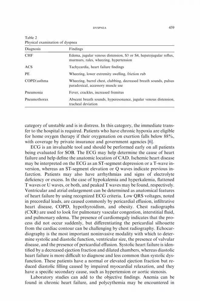
Table 2
Physical examination of dyspnea
Diagnosis Findings
CHF Edema, jugular venous distension, S3 or S4, hepatojugular reflux,
murmurs, rales, wheezing, hypertension
ACS Tachycardia, heart failure findings
PE Wheezing, lower extremity swelling, friction rub
COPD/asthma Wheezing, barrel chest, clubbing, decreased breath sounds, pulsus
paradoxical, accessory muscle use
Pneumonia Fever, crackles, increased fremitus
Pneumothorax Abscent breath sounds, hyperesonance, jugular venous distension,
tracheal deviation

460 SHIBER & SANTANA
those with chronic hypoxic states. A high platelet count can be an acutephase reactant, whereas for older fragile heart failure patients, pneumoniamay cause an exacerbation and present with a high white blood cell count.The measurement of serum digoxin level is important in patients taking thisdrug because of the relatively narrow therapeutic index. Prerenal acute renalfailure can be determined by finding an elevated blood urea nitrogen (BUN)level, and increased BUN-creatinine ratio, indicating hypoperfusion ofthe kidneys. Obtaining serum values of potassium, magnesium, calcium,sodium, chloride, and bicarbonate may assist in determining intravascularvolume status, whereas replacing depleted electrolytes may reduce the riskof cardiac arrhythmia. Brain natriuretic peptide (BNP) is a cardiac neuro-hormone secreted by the cardiac ventricles in response to ventricular walltension, pressure overload, and increased volume expansion. BNP can nowbe detected rapidly by a simple laboratory test, which is becoming morereadily available in the outpatient setting. This value allows for a more de-finitive diagnosis of heart failure by narrowing the differential for dyspnea.If this value is elevated by R100 pg/mL, the high sensitivity and specificityof this test allows for accurate diagnosis of heart failure. Values that are%100 pg/mL reliably rule out depressed left ventricular function [7,8]. Brainnatriuretic peptide has also been used as a predictor of mortality and can beused to follow improvement after CHF therapy in cases of acute cardiac de-compensation and for outpatients. The assessment of BNP values over timeallows the clinician to better estimate an exacerbation based on an individ-ualized value. BNP produces vasodilation, diuresis, and natriuresis, and in-hibits the renin-angiotensin-aldosterone axis. Although BNP is an availablemedication, nesiritide (recombinant human BNP), its IV formulation, hasbeen limited to inpatient use for severely decompensated CHF.
Management
The initial management of CHF begins after the patient has been eval-uated quickly and the level of acuteness has been determined. Using the cat-egories described earlier, patients are treated according to their specificcategory. If the patient is in acute distress, they should receive supplementaloxygen and preload reduction with sublingual, transcutaneous, or IV nitro-glycerin (NTG). Have them remain with their head elevated but their feetdependent, as in a reverse-Trendelenburg position, to further discourage ve-nous return. Oral or IV furosemide is recommended if the patient has evi-dence of volume overload. If the blood pressure is still elevated, thereduction in cardiac afterload should be accomplished with an arterial vaso-dilator, such as an angiotensin-converting enzyme inhibitor or hydralazine.The use of calcium channel blockers should be avoided in patients who haveCHF caused by reduced systolic function, and b-blockers should not be usedwhile the patient is in a decompensated episode. Obviously, many of thesepatients will require transportation to the nearest ED for definitive care.

461DYSPNEA
These patients are further classified into unstable and stable based on vi-tal signs. Unstable patient are initially given therapy while arrangements aremade for transfer to an inpatient facility. Stable patients, after initial treat-ment, can be observed for improvement; but if they become unstable, followthe same algorithm as above. The patients who improve significantly can bemanaged much in the same way as those that are initially found to be in noacute distress. These patients benefit from home instruction and education,medication modification, and close follow-up (Table 3).
Table 3
Distress classification and management
Initial treatment
Disease No acute distress Acute distress Stable Unstable
CHF No dyspnea
at rest
Dyspnea at rest
or minimal
exertion
Furosemide po,
Bp control, O2
Furosemide IV,
NTG, Bp
control, O2
CAD Stable angina Acute chest pain,
diaphoresis,
tachycardia or
bradycardia,
hypotension or
hypertension,
syncope
ASA, NTG, O2,
analgesia
ASA, NTG,
O2, analgesia,
G plavix
PE Asymptomatic
silent
Dyspnea at rest,
fever, hypoxia,
tachycardia,
hypertension
NA LMWH, O2,
analgesics
Pneumonia Normal O2
saturation, mild
dyspnea, low
PORT score
Decreased O2
saturation,
functional
debilitating
dyspnea,
tachycardia,
high/moderate
PORT score
O2,
acetominophen,
antibiotics po
O2,
acetominophen,
IM or IV
antibiotics
Pneumothorax Spontaneous Tension Analgesics Needle
decompression,
O2, analgesics
COPD/asthma O80% peak flow !80% peak flow O2, MDI
treatment
O2, MDI, or
nebulizer
treatment and
steroids
Abbreviations: ASA, aspirin; Bp, blood pressure; IM, intramuscular; LMWH, low molecular
weight heparin; NA, not applicable; po, by mouth.
Data from Steering Committee and Membership of the Advisory Council to Improve Out-
comes Nationwide in Heart Failure. Am J Cardiol 1999;83(Suppl 2A);1A–39A.

462 SHIBER & SANTANA
Acute coronary syndrome
CAD is the leading cause of death in the United States, caused acutely bymyocardial infarction and as a result of chronic problems such as congestiveheart failure. It is estimated that 13.7 million people in the United States suf-fer from CAD, with half having myocardial infarction and the other halfsuffering from chronic angina [9]. An acute coronary syndrome (ACS) refersto the rupture of an atherosclerotic plaque with the activation of plateletsand fibrin, resulting in thrombus formation and either reduced flow or theocclusion of a coronary artery. Many of these individuals have significantrisk factors identified before an acute event by their primary medical pro-viders; thus it is important for a physician in an outpatient environmentto recognize patients who have acute dyspnea caused by acute coronary syn-drome, so that prompt initial treatment can be started and transport initi-ated as soon as possible. The goal is for no more than 30 minutes to elapsefrom the time of initial contact until emergency medical services are calledor appropriate measures are instituted [10]. Once the patient arrives at thehospital, the goal is 90 minutes from door to reperfusion, in an attempt tolimit the myocardial ischemia and injury to 2 hours or less [11]. Anotherconsideration is that some patients, particularly women and the elderly, maypresent with atypical presentations of AMI, particularly isolated dyspnea. Ifan acute coronary syndrome is suspected, transport for definitive manage-ment should not be delayed.
History
The rapid assessment of dyspnea in acute coronary syndrome is initiatedwith a brief targeted interview and risk-factor stratification. If the patienthas had a syncopal event or has unstable vital signs, the initial assessmentstarts with an evaluation of airway, breathing, and circulation, while takinga brief history. It is necessary to distinguish an ACS patient from a patientwho simply has stable chronic angina, which does not have the same under-lying pathophysiology or associated morbidity or mortality and thereforedoes not require the same aggressive therapies.
Patients who complain of acute dyspnea and have acute coronary syn-drome often describe left-sided retrosternal chest pressure, diaphoresis,tachycardia, radiation of pain to the jaw, neck, or left or right arm, andnausea. These symptoms may last several minutes and be alleviated by sub-lingual NTG and aggravated by exertion. Importantly, a large percentage ofcardiac ischemic episodes may not have these specific symptoms [11]. It hasalso been documented that up to one third of myocardial infarction patientshave no chest pain on presentation to the hospital [12]. These patients maynot be diagnosed initially or at all, allowing for myocardial damage to ex-ceed that of an otherwise treated patient. Therefore, a thorough historythat includes past medical, surgical, social, and family history must be ob-tained so that those individuals with vague symptoms or several risk factorscan be evaluated appropriately.

463DYSPNEA
Risk factors for CAD include male ageR45 years, female ageR55 years,diabetes, obesity, a family history for CAD, tobacco use, hypertension, ahistory of atherosclerotic disease, hypertriglyceridemia, high levels of low-density lipoprotein cholesterol, and low levels of high-density lipoprotein(HDL) cholesterol (%40 mg/dL). High HDL (R60 mg/dL) is a negativerisk factor, which should be considered [13].
Physical examination
The physical examination should focus on the cardiovascular and respi-ratory systems. The examination will follow the same general sequence asfor congestive heart failure. Specific attention should be devoted to cardiacauscultation. An S4 sound may indicate ventricular stiffness caused by ische-mic stunning, and a new murmur may represent acute valvular dysfunction,causing regurgitation or, less commonly, rupture of the interventricular sep-tum, allowing for a new left-to-right shunt. Quiet heart sounds and other ev-idence of a symptomatic pericardial effusion may be caused by ventricularfree wall rupture, causing acute hemopericardium. Those patients presentingwith unstable vital signs in acute distress will first require rapid evaluation ofairway, breathing, and circulation.
Diagnosis
The diagnosis of acute myocardial infarction is based on signs and symp-toms, diagnostic electrocardiographic criteria, and an elevation of cardiacbiomarkers. Because the latter usually can be performed only in the hospi-tal, outpatient criteria are limited to ECG changes and patient presenta-tion. Other diagnostic tools in an inpatient facility may include furtherblood work, echocardiography, and chest radiography. Relief of chest painquickly after receiving supplemental oxygen, soluble aspirin, or sublingualNTG, is suggestive but not specific for acute CAD, but it should not beused as diagnostic criteria to confirm or exclude an ACS.
Laboratory tests may demonstrate anemia as the cause of the ischemia orinfarction. As described above, electrolytes may be deficient or in excess andpredispose to cardiac arrhythmias, which decrease blood flow while increas-ing demands on the myocardium. Rapid assays of the cardiac isomer ofcreatine phosphokinase and troponin are becoming more available, andalthough the assays are very sensitive and specific for cardiac injury, serumlevels require a minimum of several hours to rise during an acute coronaryevent. The ECG may demonstrate many types of changes that indicate ische-mia or injury and infarction. Typical changes include hyperacute T waves,T-wave inversion, ST-segment elevation, Q waves, or new onset left bundlebranch blocks in infarction and ST-segment depression in ischemia. It isimportant to identify the region of the myocardium that is affected whenevaluating an ECG. An echocardiogram can be used to identify wall motionabnormalities. This information, along with the anatomical description of
464 SHIBER & SANTANA
myocardial damage by ECG, will provide important information to the car-diac interventionalist when deciding which coronary artery to image first onpercutaneous coronary catheterization.
Management
When patients who have acute dyspnea present and ACS is suspected ordiagnosed, prompt initial therapy and transfer for further evaluation and de-finitive treatment are required. An IV line should be started, and the patientshould be placed on a cardiac monitor. Therapy begins with the administra-tion of oxygen, rapidly absorbed soluble aspirin (not enteric coated), andsublingual NTG (0.4 mg). If the patient develops hypotension in responseto NTG, an IV bolus of crystalloid should be given, and further NTGmust be used cautiously [14,15]. A b-blocker should be given to patientswithout bradycardia or heart block to decrease myocardial oxygen consump-tion [16]. The transfer of a patient suspected of having ACS should not beimpeded by further examinations, studies, or treatment. The goal is to obtaindefinitive treatment through reperfusion as soon as possible (see Table 3).
One of the most important skills in the outpatient setting is in recognizingand diagnosing an atypical acute coronary syndrome. Patients known tohave subtle, atypical, and even silent myocardial infarctions include the el-derly and diabetics. Female patients have also been found to present withatypical presentations. Primary care physicians should be on the alert forthese types of presentations in their patients.
Pulmonary embolism
PE is one of the most commonly missed lethal diagnoses. Untreated PEhas a mortality rate from 18% to 35% and therefore should always be con-sidered in the differential diagnosis of acute dyspnea [17]. In the UnitedStates, 1 in 1000 Americans is affected each year, making this disease a com-mon clinical entity that can be treated if identified correctly. Of the 600,000episodes of PE that occur each year, 50 to 100,000 patients die as a result ofthe disease [18]. Once the disease is diagnosed and treated, the incidence ofrecurrence and death is significantly reduced. The strategy in an outpatientclinic should be to immediately send any patient with significant clinical sus-picion to the hospital emergency department for a definitive diagnosis andpossible treatment. Because the objective data may not be available in theoutpatient clinic, clinical suspicion is highly regarded as sufficient evidenceto disposition early for definitive management. Considering that mostdeaths are attributed to the failure to diagnose, a quick, definitive diagnosisand management are imperative.
History
The history in patients who have PE may be misleading because the signsand symptoms are neither sensitive nor specific. An estimated 50% of cases

465DYSPNEA
of PE remain undiagnosed, underscoring the atypical nature of the disease[19]. The frequency of the most common signs and symptoms of PE havebeen studied. Dyspnea is the most common symptom, whereas tachypnea,pleuritic chest pain, and rales are also common signs and symptoms, at73%, 70%, 66%, and 51%, respectively [20]. Other frequent symptoms in-clude cough, hemoptysis, syncope, and fever. Although these symptomsare common in PE, they lack diagnostic sensitivity and specificity. It hasbeen shown that patients who do not have PE have a similar frequency ofsymptoms [21].
It is important to inquire about any recent unilateral leg swelling,warmth, redness, or pain because lower extremity deep venous thrombosis(DVT) is the most common cause of PE. Risk-factor stratification is animportant tool in gathering evidence to support suspicion of PE. Factorsidentified include immobilization, surgery within the past 3 months, stroke,history of venous thromboembolism, and malignancy [20]. Less commonrisk factors include increasing age, lower extremity trauma, extended travel,oral contraceptive pills, hormone replacement therapy, pregnancy, recentdelivery, recent joint replacement, lower extremity fractures, and pelvic frac-tures. Other studies have added obesity, hypertension, severe CHF, pulmo-nary hypertension, and cigarette smoking. The medical and family historyshould also account for genetic or acquired causes of thrombophilia, includ-ing antithrombin III deficiency, lupus anticoagulant, protein C and S defi-ciency, factor V Leiden, and mutation in prothrombin G20210A, as wellas nephrotic syndrome and inflammatory bowel disease.
Physical examination
A rapid examination of a dyspneic patient who has possible pulmonarythromboembolism is essential for disposition. The physical examination isused to further support a suspicious history and provides rapid supportfor the need to transport the patient to the ED. Vital signs may demonstratefever, tachypnea, tachycardia, hypoxia, and hypotension. Of these findings,hypoxia is the most specific, although this can be masked by hyperventila-tion from a tachypneic patient. After the vital signs have been evaluated,the patient’s general appearance is noted, followed by a focused cardiovas-cular and pulmonary physical examination. Patients who appear to be inacute distress may have rapid shallow breathing at rest, diaphoresis, andtachycardia. Their airway, breathing, and circulation should be assessedquickly, before they are transported to a hospital.
A focused respiratory examination consists of auscultation of the lungs.Examination findings may include normal, decreased breath sounds, a pleu-ral friction rub, or rales. In patients who have pulmonary consolidationcaused by an associated lung infarction, the presence of egophony can beappreciated on auscultation. Dullness to percussion and decreased tactilefremitus on palpation indicate a pleural effusion. Patients may also have re-producible chest pain on deep breathing. The cardiovascular examination
466 SHIBER & SANTANA
may reveal signs of right-sided heart failure from pulmonary hypertension.Tachycardia, a right-sided S4, and an increased pulmonic component of sec-ond heart sound may be auscultated. The remainder of the vascular exam-ination consists of examining the lower extremities for signs of deep veinthrombosis as a possible cause. These examination findings include unilat-eral lower extremity edema, warmth, erythema, a palpable cord, and poste-rior leg pain on dorsiflexion of the ipsilateral foot.
Diagnosis
The diagnosis of PE is challenging because of vague, nonspecific symp-toms or lack of symptoms. PE is diagnosed definitively by imaging studies,once the patient has been sent to the ED for evaluation. Laboratory tests,echocardiograms, and chest radiographs are modalities used to help providesupporting evidence but do not provide concrete evidence of PE. The diag-nosis of a deep venous thromboembolism by ultrasonography may allowa clinician to assume PE is present with suggestive pulmonary symptomsor signs. Thus, a patient who is being evaluated for SOB who is found tohave a DVT needs no further workup. The diagnosis is PE.
Laboratory test findings may be abnormal but are nonspecific. Leukocy-tosis can occur as a reactive phenomenon, as well as an elevated sedimenta-tion rate. Respiratory alkalosis with an elevated A-a gradient is often presenton arterial blood gas but is also nonspecific. The CXR is abnormal in themajority of patients who have PE with infarction, but these findings are of-ten nonspecific. These findings may include infiltrates, atelectasis, and pleu-ral effusion. More specific radiographic signs include Westermark’s andHampton’s sign, although they are uncommon. Westermark’s sign is theloss of pulmonary markings distal to the PE, from dilated pulmonary vascu-lature proximal to the embolus, with oligemia distally. Hampton’s ‘‘hump’’is pleural-based wedge-shaped opacity facing the hilum.
The electrocardiogram is also often abnormal and nonspecific. Sinustachycardia and nonspecific ST-T wave changes are the most common ab-normalities. Signs of right-sided heart strain, such as S1Q3T3, T-wave in-version in leads V1 to V3, and a new right bundle branch block, arespecific but uncommon [20]. Echocardiography is used to assess for signsof right heart strain and to look for thrombus in the right ventricle. Signsof pulmonary hypertension may include increased pressure over the tricus-pid valve and the pulmonary artery. The definitive tests are ventilation per-fusion imaging, multidetector pulmonary CT angiography, and, in rarecircumstances, pulmonary angiography. Any patient who requires thistype of examination should be transferred to the hospital to rule out thediagnosis.
Management
The definitive diagnosis cannot be made in most outpatient facilities, thusrapid transfer to a hospital is crucial to effective management. Patients who

467DYSPNEA
appear to be in acute distress should have only their airway, breathing, andcirculation examined before transfer measures have begun. Those who ap-pear to be in no acute distress may undergo a more detailed examination.Patients in acute distress should have an IV line started, placed on oxygen,and put on a cardio-pulmonary monitor.
After the history and physical have been completed, the physician shoulddesignate the patient’s pretest probability for having a PE. The probabilitymay be low, intermediate, or high, although there is no standard method formeasurement of pretest probability. Some researchers have based this onobjective data not available in the outpatient environment. The outpatientdesignation should reflect findings on history, examination, and other objec-tive data. This pretest probability can be relayed to the admitting physicianas a means to assist in determining what diagnostic test to perform. If thepatient is believed to be at high risk for a PE and has no contraindicationsfor anticoagulation, then heparin administration should be considered.From the outpatient view, the most important management point is to im-mediately transfer any patient suspected of having a PE (see Table 3).
Asthma
Asthma is characterized by reversible airflow obstruction, bronchial hy-perresponsiveness, airway inflammation, submucosal edema, and increasedmucus production caused by hypertrophy and hyperplasia of goblet cells[22–24]. Asthma is the most common chronic lung disease in developedand developing countries, with a current adult prevalence of 5%, andhas been increasing in prevalence over the last 20 years [25,26]. It is alsothe most common cause of respiratory emergency, resulting in almost 2million ED visits each year, 10% to 20% of which require hospital admis-sion [22–28]. Between 4% and 10% of asthma admissions will require carein the ICU, but the presentations of severe acute asthma requiring ICUcare have decreased over the last decade [26–28]. Although the death ratecaused by asthma is low, 0.3 per 100,000 people, it is still responsible forapproximately 5500 deaths per year, with the overwhelming majority ofthese patients suffering respiratory arrest before arriving at the hospital[25,27].
History
During an exacerbation, patients commonly report dyspnea, wheezing,tightness in the chest, and coughing (usually nonproductive). Frequentcauses include viral respiratory infections, bacterial infections (most com-monly by Mycoplasma pneumoniae and Chlamydia pneumoniae), exposureto allergens or respiratory irritants (tobacco smoke, perfumes, and fumesfrom cleaning products), exercise, exposure to cold, emotional distress, med-ications (aspirin, nonsteroidal anti-inflammatory drugs, or b-blockers), and

468 SHIBER & SANTANA
noncompliance to medication [22–24,29]. Risk factors for increased morbid-ity and mortality include two or more ED visits or a hospital admissionwithin the past year, any previous episode requiring mechanical ventilation,chronic steroid use, extremely rapid onset of symptoms, association of ana-phylaxis, low socioeconomic and educational status, psychosocial disorders,and language barriers [22–24,29]. A large proportion of the asthma fatalitiesthat occur are believed to be caused by extensive mucous plugging of the air-ways, with such air trapping that the lungs remain inflated at autopsy afterremoval from the thorax [22–24]. The failure of the patient or physician torecognize the severity of the asthma attack is also believed to be responsiblefor many of the deaths [25,30].
Physical examination
Asthma, meaning ‘‘panting’’ in Greek, was described accurately in an-cient times for the most obvious clinical feature: tachypnea [22]. Wheezingis often heard diffusely and may be appreciated throughout the respiratorycycle or more prominently with expiration. Because airflow is required tocause turbulence, which produces wheezing, a patient with severe airway ob-struction may have minimal airflow and therefore no wheezing. A brief,focused history and physical examination should be performed initially toidentify any patient who may be unstable or need immediate airway in-terventions or assisted ventilation. An initial peak expiratory flow rate(PEFR) should be obtained. A focused examination, including vital signs,mental status, cardiopulmonary examination, and PEFR should be repeatedfrequently and after interventions to assess response to treatment.
A patient with only a mild exacerbation, defined as an initial PEFRR200L/min or R50% of the predicted best may have only end-expiratory wheez-ing and a slightly increased respiratory rate without other physical findings.During a moderate asthma exacerbation, a PEFR of 80 to 200 L/min or25% to 50% of the predicted best, the patient may not be able to speak con-versationally without breathlessness, they may not be able to recline on thestretcher and instead choose a more upright posture, respiratory rate will beincreased, and wheezing will be more prominent. With a severe exacerba-tion, a PEFR of %80 L/min or %25% of the predicted best, the patientwill likely have diaphoresis, supraclavicular retractions, accessory muscleuse, halting speech, a tripodding posture (sitting or standing while leaningforward to brace the arms outward on the knees or a fixed object), and se-vere tachypnea. Beware of agitation or confusion, cyanosis, fatigue, or a quietchest because these are signs of impending respiratory arrest [22–24,29,30].An increased pulsus paradoxus, R25 mm Hg, may be found with severeasthmatic exacerbations. Because this abnormality depends on forceful in-spiratory and expiratory excursions, the pulsus may diminish as the clinicalcondition improves, airway obstruction resolves, and respiratory effortnormalizes or as the condition worsens and the respiratory effort becomesfatiguing [22–28].

469DYSPNEA
Diagnostic studies
In addition to the assessment of PEFR and oxygen saturation, the phy-sician should consider obtaining a chest radiograph if the patient has not re-sponded adequately to therapy or is in extremis. The film findings may benormal or may show signs of air trapping (increased lung volumes and flat-tened diaphragms). The actual value of the chest radiograph is to determinewhether other serious complications, such as a pneumothorax or pulmonaryinfiltrate, may also be involved. Laboratory tests, if any, should be orderedspecifically to each individual patient. One should consider checking electro-lyte levels if the patient is given frequent or continuous b-agonist treatmentsor if underlying deficiencies of potassium, magnesium, or phosphate are sus-pected because this therapy will cause an intracellular shift of these ions,thereby further reducing serum levels [25]. Arterial blood gas (ABG) analy-sis may be performed to confirm a respiratory alkalosis condition caused bytachypnea or follow the resolution of hypercapnea once mechanical ventila-tion has been instituted, but it should be stressed that the need for an arti-ficial airway and assisted ventilation is based on clinical assessments andmust not be delayed while waiting for ABG or other diagnostic results [28].
Management
Oxygen should be administered to keep the arterial oxygen saturation(SaO2) at R90% and R95% in pregnant patients or those who have signif-icant CAD [25]. Inhaled b-agonists are the first-line therapy in all asthma ex-acerbations. They may be delivered with equivalent success by nebulizationor by metered-dose-inhaler (MDI) with or without a spacer device [26–28].A typical nebulized dose of albuterol would be 5 mg every 15 to 20 minutestimes three doses. A similar effect can be obtained by giving a continuous neb-ulized treatment of 15 to 20 mg of albuterol over 1 hour. If aMDI is used, 6 to8 actuations, 90 mg each, should be given at each treatment to deliver anequivalent dose [26,27,30]. Levalbuterol, the all R-isomer of albuterol (as op-posed to the equal mixture of R and S in the standard racemic albuterol) hasbeen shown to be as safe and effective at a dose of 1.25 mg per nebulized treat-ment and may even be more effective in certain subsets of asthmatics [25].Ipratropium bromide should also be administered, either by nebulized treat-ment, 500 mg, or by 4 to 6 MDI actuations. Parenteral b-agonists should beused only if the patient is moribund, coughing excessively, or continues toworsen despite inhaled treatments [26,27,30]. The indicated dosage for epi-nephrine is 0.2 to 0.5 mg subcutaneously of a 1:1000 solution for three dosesevery 20 to 30 minutes. If the patient is in shock, it should be given as an IVbolus of 0.2 to 0.5 mg of 1:10,000 solution, followed by an infusion titratedbetween 1 and 20 mg/min. An alternative option for the subcutaneous routeis terbutaline, 0.25 mg every 20 minutes for three doses [25,28].
Glucocorticoid steroid administration has been shown to decrease admis-sion rates and prevent relapses and should be given to the majority of pa-tients presenting with an asthmatic exacerbation. The one group that does

470 SHIBER & SANTANA
not require steroids are those with very mild symptoms of short duration,which resolve completely with one inhaled bronchodilator, reflecting onlybronchoconstriction without airway inflammation and edema [25,28]. Therecommended oral dose of prednisone is 40 to 60 mg (0.5–1.0 mg/kg) dailyfor 3 to 14 days, with or without a taper. If the patient cannot toleratethe oral administration of prednisone, then methylprednisolone IV, 125 to250 mg can be given [22–25,27,30].
Dehydration is common, especially if the exacerbation has been ongoingfor several days, so that IV fluid administration is recommended by mostexperts for moderate to severe asthma attacks [3,27]. If an infiltrate is foundon chest radiography or empiric antibiotics are given, coverage should in-clude Mycoplasma and Chlamydia spp [1]. In severe exacerbations, the IVrapid infusion of magnesium sulfate, 2 g over 10 to 20 minutes, has beenfound to give additional clinical benefit [3,14,16]. There may be some rolefor the initiation of inhaled steroids or oral leukotriene antagonists in acuteexacerbations, but more data need to be collected before these agents can berecommended without reservations [25,26,28]. A mixture of helium and ox-ygen (heliox) has been found to decrease the work of breathing and improveoxygenation in severe attacks by decreasing airway turbulence and resis-tance. To be effective, the formulation needs to be 70% to 80% heliumand the remainder oxygen, so that heliox is not indicated if the patient re-quires high oxygen concentrations to maintain an adequate SaO2 [25,28].There is no role for sedatives, mucolytics, or opiate cough suppressants inthe treatment of acute asthma [25,27].
Most patients will improve after treatments, with only 20% of all asth-matics ever requiring hospitalization [28]. Patients presenting with aPEFR %25% of their predicted rate before treatment will likely require ad-mission; if their PEFR remains below 25% despite aggressive therapy, thenan ICU stay is warranted. If they improve but PEFR is still%40%, then ad-mission is also indicated. Patients who show improvement but not resolutionof their symptoms and a PEFR from 40% to 60% should be observed in theoffice or sent to the emergency department for continued treatment; after anadditional 2 to 3 hours, if they have improved, then they can be sent home;but if not, then they should be admitted. Patients who reach a PEFR R60%of their predicted best will most likely experience a resolution of subjectivesymptoms and can be sent home [2,28]. Given the mortality rate associatedwith asthma, any patient who does not respond to outpatient therapy shouldbe considered for emergency department care to help determine the bestcourse of therapy and to help determine disposition.
Chronic obstructive pulmonary disease
COPD encompasses a group of chronic lung disorders, but emphysemaand chronic bronchitis are the disorders most commonly encountered inclinical practice and will be the focus of this discussion. A reduction inbronchial airflow caused by nonreversible disorders is the underlying

471DYSPNEA
pathophysiology. Permanent enlargement of air spaces distal to the terminalbronchiole with wall destruction but without fibrosis defines emphysema.The loss of elastic recoil of lung tissue allows the collapse of distal airways,promoting air trapping [1]. COPD has an incidence of 34.1 per 1000 peopleover the age 65 years, affecting 14 to 20 million people in the United States,making it the fourth leading cause of death [27,31]. It is responsible for 4million office visits and 500,000 hospital admissions yearly. COPD patientstypically experience one to three exacerbations per year, in which between3% and 16% of the episodes require hospitalization; the mortality rate is3% to 10% for each hospital stay [31].
History
The typical baseline symptoms of COPD patients include dyspnea andchronic cough with sputum production. During an exacerbation, the dysp-nea will worsen, with an increase in tachypnea, a prolonged expiratoryphase, and audible wheezing, and the sputum will often increase in volumewhile becoming purulent [22–24,31]. Approximately 80% of COPD exacer-bations are believed to be caused by respiratory infections, with half beingviral and half being bacterial [22–24]. Pulmonary irritants (cigarette smoke,smog, and ozone) and medication noncompliance are also common causes.The four elements responsible for the reduction in airflow during an exacer-bation are mucosal edema, increased secretions, bronchial smooth muscleconstriction, and airway inflammation [22–24].
Physical examination
COPD patients presenting with an acute exacerbation typically have an in-creased respiratory and heart rate and pursed-lip breathing with a prolongedexpiratory phase of the respiratory cycle. The patient’s thorax is often in-creased in the anteroposterior dimension to accommodate the increasedlung volumes. Patients will position themselves to maximize respiratory me-chanics, often bracing their hands on their knees to stabilize the upper extrem-ities and recruit accessory muscles. An examination of their lungs may revealexpiratory wheezing or may simply be quiet, with markedly diminished airexchange. Other life-threatening disorders that are associated with COPDshould be considered, such as pneumothorax, which may produce asymmet-ric or absent breath sounds on a hemithorax, distended neck veins, and tra-cheal deviation, whereas pneumonia may cause signs of focal pulmonaryconsolidation or pleural effusion. Acute agitation may be a sign of hypox-emia, whereas somnolence or lethargy may indicate an elevated partial pres-sure of carbon dioxide (PcO2) caused by hypercarbic respiratory insufficiency.
Diagnostic studies
Continuous pulse oximetry is necessary to ensure an adequate level ofSaO2 (R88%–90%), especially if supplemental oxygen is being

472 SHIBER & SANTANA
administered, without overshooting and encouraging CO2 retention by sup-pression of the hypoxic respiratory drive [22–24]. Chest radiographs typicallyreveal hyperinflation with flattened diaphragms and may also show bullaeand pulmonary oligemia [22–24]. During a presumed COPD exacerbation,the additional value of the CXR is to evaluate for other causes of acutedyspnea, including pneumothorax, pneumonia, and congestive heart failure.Between 16% and 21% of COPD patients have CXR findings that were be-lieved to be helpful by physicians, and treatments were then modified by thefindings [31]. Serum electrolytes may be indicated to determine potassiumand phosphate level because these are commonly depleted in COPD pa-tients, and serum levels may drop further from intracellular shifts inducedby b-agonists. ABG analysis, if available urgently, may be particularly help-ful to evaluate for acute CO2 retention indicated by an elevated PcO2 withan acidosis. Although they are not commonly performed at the time of anexacerbation, if pulmonary function tests (PFTs) are obtained, they willconfirm an obstructive pattern: a decreased ratio of forced expiratory vol-ume in 1 second (FEV1)/forced vital capacity (FVC), an increased totallung capacity and residual volume, and decreased diffusing capacity [22–24].
Management
Oxygen administration is recommended to keep the SaO2 level at R88%to 90% and the PO2 at R60 mm Hg to maintain tissue oxygenation whilepreventing pulmonary vasoconstriction and right ventricular strain or pres-sure overload [22–24,31]. Inhaled b-agonists and anticholinergics should begiven by nebulizer or MDI. Albuterol and ipratropium are the most com-mon representatives of these respective classes in the United States. Theireffects are additive, so they should be used together, and although albuterolwill often give muscular tremor and tachycardia, ipratropium rarely produ-ces any significant side effects [22–24]. Theophylline has been in use for over70 years and has been found to improve FEV1 and exercise performance byabout 10% in COPD patients by increasing diaphragm strength and endur-ance and mucociliary clearance. Because theophylline has a narrow thera-peutic index, the associated side effects of nausea, vomiting, and tremorare common, whereas life-threatening toxicity includes seizures and cardiacarrhythmias. For these reasons, theophylline has little role acutely but mayhave a role in outpatient chronic care if used cautiously [22–24,27].
Glucocortical steroids are beneficial acutely and improve outcomes inCOPD exacerbations. Either a short course, 4 to 7 days without taper, ora 10- to 14-day course with a taper is effective. Prednisone, 0.5 to 1 mg/kgorally each day, can be used, but an initial dose of IV methylprednisoloneor dexamethasone can be given if patients are unable to tolerate oral medica-tions. There is currently not enough evidence to recommend initiating inhaledsteroids for acute COPD episodes, but they may also prove to be helpful [22–24,27,31]. Prescribing antibiotics for an exacerbation is believed to decreasebacterial counts in the chronically colonized respiratory tracts of COPD

473DYSPNEA
patients. Currently, the agents recommended for 7- to 10-day outpatient ther-apy include trimethoprim-sulfamethoxazole, ampicillin, doxycycline, anderythromycin. If the patient has an infiltrate on CXR or has fever or signsof systemic infection, then IV antibiotics effective against Streptococcus pneu-moniae and Haemophilus influenzae should be started [22–24]. Expectorantsand mucolytics have been found to provide little or no benefit [22–24,27].
Noninvasive positive pressure ventilation, using either continuous posi-tive airway pressure or bi-level positive airway pressure, may decrease thework of breathing and improve ventilation, but patient cooperation is re-quired to be effective. If acute respiratory failure occurs with hypoxemia(PO2 %50 mm Hg) and respiratory acidosis despite aggressive medical ther-apy, tracheal intubation and mechanical ventilation are indicated. These pa-tients should be ventilated cautiously because overzealous correction ofacute or chronic hypercarbia may cause severe alkalemia, precipitating seiz-ures or ventricular arrhythmias [22–24].
The majority of COPD patients will improve after therapy and can safelyreturn home. Patients who continue to have dyspnea increased over theirbaseline will need to be hospitalized if they also experience an inability towalk (if previously ambulatory), loss of appetite or sleep caused by dyspnea,comorbid pulmonary (eg, pneumonia) or nonpulmonary (eg, anemia) con-ditions, worsened hypoxemia or hypercarbia, new or worsened cor pulmo-nale, and an inability to manage at home with health care resources (eg,home oxygen) not readily available. Patients will need to be admitted toan ICU if they have alterations in mental status (confusion or lethargy), re-spiratory fatigue, worsening hypoxemia despite supplemental oxygen, wors-ening respiratory acidosis (pH %7.30), and the need for mechanicalventilation (invasive or noninvasive) [31].
Pneumonia
Pneumonia is inflammation, most often from infection, affecting the lungparenchyma (respiratory bronchioles and alveolar units) [22–24]. Althoughthere are multiple distinct causes, including viral, fungal, and mycobacterialpneumonias, aspiration pneumonia, ventilator or hospital-acquired pneu-monia, and opportunistic infections in immunodeficient individuals, thisdiscussion focuses on community-acquired bacterial pneumonia in immuno-competent adults. An infectious agent is identified in 30% to 40% of thecases of community-acquired pneumonia (CAP), in which the most commonbacterial organism is S pneumoniae, followed by C pneumoniae, M pneumo-niae, and H influenzae. Legionella pneumonia is a rare causative agent in thevery elderly (1% in those R80 years old) but is more common in youngeradults (8% in those %80 years old). Gram-negative enteric organisms aremore common in older patients who have comorbid conditions, particularlydiabetes, malignancy, central nervous system disease, and renal, hepatic, orcardiopulmonary diseases [32–34].
474 SHIBER & SANTANA
The incidence of pneumonia in the United States is 12 cases per 1000 pa-tients per year, resulting in 4 to 5.6 million cases annually. Approximately25% of these patients will require hospitalization, and 10% of those will re-quire an ICU stay. The risk for developing pneumonia is 10-fold higher inresidents of long-term care facilities than for older adults living in the com-munity [27,29,32,35,36]. Pneumonia is the leading cause of infectious deathsand the sixth overall cause of mortality in the United States; in people overage 65, pneumonia moves up to the fifth leading cause of death, resulting in60 thousand deaths per year. The mortality rate for pneumonia varies de-pending on the location of treatment, which is a marker for the severityof the disease. For those treated as outpatients, the rate is very low, between%1% and 9%, but it is higher for inpatients and approaches 50% in an ICU[32,36,37].
History
Cough, purulent sputum, dyspnea, and pleuritic thoracic pain are re-ported commonly, localizing symptoms associated with pneumonia; nonspe-cific symptoms include fever and chills, headache, and myalgias. Elderlypatients may have fewer of these symptoms and are more likely than areyounger adults to present with a change in mental status. If the lower lobesof the lungs are involved, diaphragmatic irritation can cause upper abdom-inal pain, referred shoulder or scapular pain, or singultus [32,37,38].
Physical examination
Fever, increased heart rate and respiratory rate, inspiratory crackles, andsigns of consolidation (bronchial breath sounds, egophony, increased tactilefremitus) on lung examination are found typically. The patient may appearfatigued, but a rapid evaluation of the mental status is vital to evaluate forpossible impending respiratory failure. The skin, mucous membranes, andjugular veins should be examined for evidence of dehydration. Abdominaldistension and diminished bowel sounds may be found, caused by paralyticileus, particularly with lower-lobe pneumonia. SaO2 may also be decreased,requiring supplemental oxygen to maintain normal values [37,38].
Diagnostic studies
Chest radiography is necessary to confirm the presence and location ofthe pneumonia. It may show focal consolidation of air-space pneumoniaor a diffuse interstitial pattern; it is also helpful in evaluating for other con-ditions that may present similarly, such as CHF of a neoplasm. A leukocy-tosis with an increased percentage of PMNs and immature band forms iscommon, but leukopenia may occur. ABG analysis, if available, may revealrespiratory acidosis and an elevated PaO2/FIO2 ratio, showing evidence ofrespiratory failure caused by severe pneumonia [37,38]. The Infectious

475DYSPNEA
Disease Society of America [34] recommends obtaining a sputum specimenfor Gram staining and culture on all inpatients treated for pneumonia,whereas the American Thoracic Society recommends ordering a specimenonly if an organism is suspected to be drug resistant or not susceptible tothe usual empiric therapy. Blood cultures yield low results and may notneed to be ordered routinely, based on studies showing that there were nosignificant differences in culture rates among patients who have differingpneumonia severity scores and that the rates of bacteremia in pneumoniapatients did not significantly influence their hospital stay or mortalityrate. Furthermore, blood culture results only rarely offer guidance for clin-ical decisions or changes in therapy [34,37,39]. A new urinary antigen assayfor S pneumoniae, which has an 82% sensitivity for diagnosing bacteremia,is now available and may be helpful in identifying these patients [40].
Management
Most patients diagnosed with pneumonia can be treated with oral antibi-otics, but an IV dose should be administered if the patient is unable to takemedications orally or is suspected to be bacteremic or acutely ill. The currentrecommendation by the American Infectious Disease Society of America forthe treatment of CAP in adults is a second- or third-generation cephalospo-rin plus a macrolide or an extended spectrum fluoroquinolone. If the patientis admitted to an ICU, broad-spectrum antibiotics are indicated and shouldinclude coverage for Pseudomonas organisms if risk factors are present(bronchiectasis, chronic steroid therapy, or recent antibiotic administration)[27,35,37,40,41]. The early initiation of antibiotics (within 8 hours of the pa-tient’s arrival) has been shown to decrease the length of stay in the hospitaland reduce inpatient and 30-day mortality in patients older than 65 years.The optimum duration of antibiotic therapy is not well defined but is typi-cally from 7 to 21 days [27,35,37,41,42]. Supplemental oxygen should besupplied to maintain a normal SaO2, and IV fluids should be administeredif the patient has signs of significant dehydration.
Numerous grading systems and scoring scales have been created in an at-tempt to predict which patients are at higher risk for complications andmortality and require hospitalization or even an ICU stay versus those pa-tients at low risk who can be safely treated as outpatients. Some of thesesystems include the Pneumonia Severity Index, the Pneumonia OutcomeResearch Trial (PORT), the British Thoracic Society Scoring System, andthe American Thoracic Society Criteria for Severe CAP. These scoring sys-tems generally include similar data (vital signs, comorbidities, CXR, andlaboratory studies) to separate patients into different risk classes. Patientsare considered to be at high risk and should be hospitalized if they haveany of the following: age R65 years, respiratory rate R30 breaths/min, bi-lateral or multilobar infiltrates, hypotension (systolic %90 mm Hg, diastolic%60 mm Hg), acute renal failure, and altered mental status. If the patienthas respiratory failure (defined by PaO2/FIO2 ratio R250 or the need for

476 SHIBER & SANTANA
mechanical ventilation) or is in septic shock requiring vasopressors, thenICU care will certainly be required. The study result of concern, however,is that in one review of patients in the ICU for CAP, 11% had PneumoniaSeverity Index class I scores, and 13% had class II scores (I–V, with I leastand V most severe) [35–38,41,43].
Miscellaneous disorders
Many other less prevalent disorders may lead to dyspnea, some of whichcan be life threatening and require immediate treatment, whereas others aresubacute or chronic conditions for which patients may be followed andtreated as outpatients. The history and physical examination often assist indiagnosing these entities, but there may be a need for diagnostic studies(chest radiograph, laboratory studies, referral for pulmonary function stud-ies, laryngoscopy, or bronchoscopy).
Spontaneous pneumothorax can occur without trauma or iatrogenic lunginjury and can be further divided into primary (young patients who havenormal lung parenchyma but congenital apical blebs) and secondary (olderpatients who have underlying lung disease such as emphysema or pulmo-nary fibrosis). The risk is higher for smokers and is greater for men thanwomen. Pleuritic chest pain and dyspnea are the most common complaints.Diminished or asymmetric breath sounds over the affected hemithorax arefound on examination. A CXR, specifically an upright expiratory film, isthe confirmatory test. Chest tube thoracostomy placement or anterior nee-dle decompression should be performed emergently if the patient appearsto have a tension pneumothorax (distended neck veins, tracheal deviationto the opposite side, and hypotension). For small, stable pneumothoracies,simple aspiration is effective but takes longer until resolution than the tubethoracostomy does [27]. These therapies will in most cases require transferto the ED.
Airway obstruction may be organic, occurring as a result of laryngeal tu-mors, polyps, or vocal cord paralysis, or be caused by functional vocal corddisorders, such as paradoxical vocal cord movement. Dyspnea and stridorare common features, and visualization of the glottis may be needed to di-agnose the obstruction [1,44]. Patients who have neuromuscular diseases re-sulting in acute weakness, such as Guillain-Barre syndrome or myastheniagravis, will report dyspnea because they may be using a much greater per-centage of their respiratory strength than previously to accomplish thoracicexpansion and diaphragmatic excursion. Anxiety disorders often have dysp-nea as a central complaint, and hyperventilation syndrome may be identi-fied. Patients who have COPD have a 3-fold increase in the prevalence ofanxiety disorders than the general population, so that it may be difficultto readily make this diagnosis [45].
Central nervous system events such as strokes or intracranial hemor-rhages will often cause tachypnea and a resultant respiratory alkalosis,but dyspnea is not usually reported [46]. Liver disease and pregnancy are

477DYSPNEA
also associated with an increase in minute ventilation. Moderate to severeanemia will typically cause dyspnea on exertion because of reduced oxygendelivery to peripheral tissues. Metabolic acidosis of any cause (sepsis, in-creased lactate, or diabetic or alcoholic ketosis) will cause a compensatoryincrease in minute ventilation that is sensed as SOB by the patient. Toxins,including carbon monoxide and salicylate poisoning, will also cause a symp-tomatic increase in respiratory rate and tidal volume. These patients, unlessa secondary disorder is involved, will all show normal findings on lungexaminations.
Summary
When evaluating a dyspneic patient in the office, a quick initial assess-ment of the airway, breathing, and circulation, while gathering a briefhistory and focused physical examination are necessary. Most often, anacute cardiopulmonary disorder, such as CHF, cardiac ischemia, pneumo-nia, asthma, or COPD exacerbation, can be identified and treated. Stablepatients who improve can be sent home, but those in acute distress with un-stable or impending unstable conditions need to be transferred emergentlyto definitive care. Because of the difficult logistics involved in attemptingto work up an outpatient for new onset of SOB, some patients will need tobe transferred to the nearest ED for a definitive diagnosis.
Further readings
Shilon Y, Shitrit AB-G, Rudensky B, et al. A rapid quantitative D-dimer assay at admission cor-
relates with the severity of community acquired pneumonia. BloodCoagul Fibrinolysis 2003;
14(8):745–8.
Stein PD, Terrin ML, Hales CA, et al. Clinical, laboratory, roentgenographic and electrocardio-
graphic findings in patients with acute pulmonary embolism and no pre-existing cardiac or
pulmonary disease. Chest 1991;100:598.
References
[1] Zoorob RJ, Cambell JS. Acute dyspnea in the office. Am Fam Physician 2003;68:1803–10.
[2] Kunitoh H, Wantanabe K, Sajima Y. Clinical features to predict hypoxia and/or hypercap-
nea in acute asthma attacks. J Asthma 1994;31:401–7.
[3] Hazinski MF, Cummins RO, Field JM. 2000 Handbook of emergency cardiovascular care
for healthcare providers. Dallas (TX): American Heart Association; 2000.
[4] Packer M, Cohn JN; for the Steering Committee and Membership of the Advisory Council
to Improve Outcomes Nationwide in Heart Failure. Consensus recommendations for the
management of chronic heart failure, II: management of heart failure: approaches to the pre-
vention of heart failure. Am J Cardiol 1999;83(Suppl 2A):9A–38A.
[5] The Criteria Committee of the New York Heart Association. Physical capacity with heart
disease. In: Diseases of the heart and blood vessels, nomenclature and criteria for diagnosis.
6th edition. Boston: Little, Brown & Co; 1964. p. 110–4.
[6] Cutaia M. Ambulatory monitoring of oxygen saturation in chronic lung disease: optimizing
long-term oxygen therapy. Clinical Pulmonary Medicine 2002;9(6):297–305.

478 SHIBER & SANTANA
[7] McDonagh TA, Robb SD, Murdoch DR, et al. Biochemical detection of left-ventricular
systolic dysfunction. Lancet 1998;351:9–13.
[8] Maisel AS, Koon J, Krishnaswamy P, et al. Utility of B-natriuretic peptide as a rapid, point-
of-care test for screening patients undergoing echocardiography to determine left ventricular
dysfunction. Am Heart J 2001;141:367–74.
[9] American Heart Association. Heart and stroke facts: 1995 statistical supplement. Dallas
(TX): American Heart Association; 1994.
[10] Tucker NHB, Doty D, Gilmour K, et al, for the Early Diagnosis Steering Committee. As-
sessment and triage of unstable angina and acute MI. 1998.
[11] Canto JG, Every NR,Magid DJ, et al, for the National Registry of Myocardial Infarction 2
Investigators. The volume of primary angioplasty procedures and survival after acute myo-
cardial infarction. N Engl J Med 2000;342:1573–80.
[12] Deedwania PC, Carbajal E. Silent myocardial ischemia: a clinical perspective. Arch Intern
Med 1991;151:2373–82.
[13] Canto JG, ShlipakMG, Rogers WJ, et al. Prevalence, clinical characteristics, and mortality
among patients with myocardial infarction presenting without chest pain. JAMA 2000;283:
3223.
[14] Yusuf S, Hawken S, Ounpuu S, et al. Effect of potentially modifiable risk factors associated
with myocardial infarction in 52 countries (the INTERHEART study): case-control study.
Lancet 2004;364:937.
[15] Antman EM,AnbeDT, Armstrong PW, et al. ACC/AHA guidelines for the management of
patients with ST-elevation myocardial infarction–executive summary: a report of the Amer-
ican College of Cardiology/American Heart Association Task Force on Practice Guidelines
(Writing Committee to Revise the 1999 Guidelines for the Management of Patients With
Acute Myocardial Infarction). Circulation 2004;110:588.
[16] Parker JO. Nitrates and angina pectoris. Am J Cardiol 1993;72:3C–8C.
[17] TeoKK,Yusuf S, Furberg CD. Effects of prophylactic antiarrhythmic drug therapy in acute
myocardial infarction: An overview of results from randomized controlled trials. JAMA
1993;270:1589.
[18] Calder KK, Herbert M, Henderson SO. The mortality of untreated pulmonary embolism in
emergency department patients. Ann Emerg Med 2005;45:302–10.
[19] Fedullo PF, Tapson VF. The evaluation of suspected pulmonary embolism. N Engl J Med
2003;349:1247–56.
[20] Horlander KT, Mannino DM, Leeper KV. Pulmonary embolism mortality in the United
States, 1979–1998: an analysis using multiple-cause mortality data. Arch Intern Med 2003;
163:1711.
[21] Investigators PIOPED. Value of the ventilation/perfusion scan in acute pulmonary embo-
lism: results of the prospective investigation of pulmonary embolism diagnosis (PIOPED).
JAMA 1990;263:2753–9.
[22] AdamsL, StulbargMS.Dyspnea. In:Murray JF,Nadel JA, editors. Textbook of respiratory
medicine. 3rd edition. Philadelphia: WB Saunders; 2000. p. 542–9.
[23] Rennard SI, Shapiro SD, SniderGL. COPD. In:Murray JF, Nadel JA, editors. Textbook of
respiratory medicine. 3rd edition. Philadelphia: WB Saunders; 2000. p. 1188–230.
[24] Bushey HA, Burchard EG, Corry DB, et al. Asthma. In: Murray JF, Nadel JA, editors.
Textbook of respiratory medicine. 3rd edition. Philadelphia: WB Saunders; 2000.
p. 1250–77.
[25] Jaroslaw P, Nowak RM, Zoratti EM. The evaluation and management of acute, severe
asthma. Med Clin North Am 2002;86:1049–71.
[26] Cates C, Fitzgerald M. Asthma. In: Clinical evidence. Vol. 4. London: BMJ Publishing
Group; 2000. p. 828–64.
[27] Gibbs MA, Camargo CA, Rowe BH, et al. State of the art: therapeutic controversies in se-
vere acute asthma. Acad Emerg Med 2000;7(7):800–15.

479DYSPNEA
[28] Rodrigo GJ, Rodrigo C, Hall JB. Acute asthma in adults: a review. Chest 2004;125(3):
1081–102.
[29] Sherman S. Acute asthma in adults. In: Tintinalli J, Ruiz E, Krome RL, editors. Emergency
medicine: a comprehensive study guide. 4th edition. New York: McGraw-Hill Co; 1996.
p. 430–7.
[30] Beveridge RC, Grunfeld AF, Hodder RV, et al. Guidelines for the emergency management
of asthma in adults. CMAJ 1996;155(1):25–37.
[31] Soto FJ, Varkey B. Evidence-based approach to acute exacerbations of COPD. Curr Opin
Pulm Med 2003;9(2):117–24.
[32] Loeb M. Pneumonia in the elderly. Curr Opin Infect Dis 2004;17(2):127–30.
[33] Bjerre LM, Verheij TJM, Kochen MM. Antibiotics for community acquired pneumonia in
adult outpatients. Cochrane Database Syst Rev 2004;3:CD002109.
[34] Garcia-Vazquez E,MarcosMA,Mensa J, et al. Assessment of the usefulness of sputum cul-
ture for diagnosis of community-acquired pneumonia using the PORT predictive scoring
system. Arch Intern Med 2004;164(16):1807–11.
[35] Wilkinson M, Woodhead MA. Guidelines for community-acquired pneumonia in the ICU.
Curr Opin Crit Care 2004;10(1):59–64.
[36] Riley PD, Aronsky D, Dean NC. Validation of the 2001 American Thoracic Society criteria
for severe community-acquired pneumonia. Crit Care Med 2004;32(12):2398–402.
[37] Alves DW, Kennedy MT. Community-acquired pneumonia in casualty: etiology, clinical
features, diagnosis, and management (or a look at the ‘‘new’’ in pneumonia since 2002).
Curr Opin Pulm Med 2004;10(3):166–70.
[38] Metlay JP, FineMJ.Testing strategies in the initialmanagement of patientswith community-
acquired pneumonia. Ann Intern Med 2003;138(2):109–18.
[39] Ioachimescu OC, Ioachimescu AG, Iannini PB. Severity scoring in community-acquired
pneumonia caused by Streptococcus pneumoniae: a 5-year experience. Int J Antimicrob
Agents 2004;24(5):485–90.
[40] BattlemanDS, CallahanM, ThalerHT. Rapid antibiotic delivery and appropriate antibiotic
selection reduce length of hospital stay of patients with community-acquired pneumonia.
Arch Intern Med 2002;162:682–8.
[41] Alvarez-Lerma F, Torres A. Severe community-acquired pneumonia. Current Opinion in
Critical Care 2004;10(5):369–74.
[42] Silber SH,Garrett C, SinghR, et al. Early administration of antibiotics does not shorten time
to clinical stability in patients with moderate-to-severe community-acquired pneumonia.
Chest 2003;124(5):1798–804.
[43] Oosterheert JJ. Comparison of guidelines for diagnosis of severe community-acquired pneu-
monia. Curr Opin Infect Dis 2003;16(2):153–9.
[44] McGeehan M, Busse WW. Refractory asthma. Med Clin North Am 2002;86:1073–90.
[45] Brenes GA. Anxiety and chronic obstructive pulmonary disease: prevalence, impact, and
treatment. Psychosom Med 2003;65(6):963–70.
[46] Rose BD, editor. Respiratory acidosis. In: Clinical physiology of acid-base electrolyte disor-
ders. 4th edition. New York: McGraw-Hill Co; 1994. p. 540–603.


